a reduction in npas4 expression results in delayed neural differentiation of mouse embryonic stem...
TRANSCRIPT

RESEARCH Open Access
A reduction in Npas4 expression results indelayed neural differentiation of mouseembryonic stem cellsThomas S Klaric1,2, Paul Q Thomas1, Mirella Dottori3, Wai Khay Leong1,2, Simon A Koblar1,2† and Martin D Lewis1,2*†
Abstract
Introduction: Npas4 is a calcium-dependent transcription factor expressed within neurons of the brain where itregulates the expression of several genes that are important for neuronal survival and synaptic plasticity. It is knownthat in the adult brain Npas4 plays an important role in several key aspects of neurobiology including inhibitorysynapse formation, neuroprotection and memory, yet very little is known about the role of Npas4 duringneurodevelopment. The aim of this study was to examine the expression and function of Npas4 during nervoussystem development by using a combination of in vivo experiments in the developing mouse embryo and neuraldifferentiation of embryonic stem cells (ESCs) as an in vitro model of the early stages of embryogenesis.
Methods: Two different neural differentiation paradigms were used to investigate Npas4 expression duringneurodevelopment in vitro; adherent monolayer differentiation of mouse ESCs in N2B27 medium and Noggin-induced differentiation of human ESCs. This work was complemented by direct analysis of Npas4 expression in themouse embryo. The function of Npas4 in the context of neurodevelopment was investigated using loss-of-functionexperiments in vitro. We created several mouse ESC lines in which Npas4 expression was reduced during neuraldifferentiation through RNA interference and we then analyzed the ability of these Npas4 knockdown mouse ESCslines to undergo neural differentiation.
Results: We found that while Npas4 is not expressed in undifferentiated ESCs, it becomes transiently up-regulatedduring neural differentiation of both mouse and human ESCs at a stage of differentiation that is characterized byproliferation of neural progenitor cells. This was corroborated by analysis of Npas4 expression in the mouse embryowhere the Npas4 transcript was detected specifically in the developing forebrain beginning at embryonic day 9.5.Finally, knockdown of Npas4 expression in mouse ESCs undergoing neural differentiation affected their ability todifferentiate appropriately, resulting in delayed neural differentiation.
Conclusions: Here we provide the first evidence that Npas4 is expressed during embryonic development and thatit may have a developmental role that is unrelated to its function in the adult brain.
IntroductionNeuronal PAS domain protein 4 (Npas4) is a transcriptionfactor belonging to the basic helix-loop-helix Per-Arnt-Sim (bHLH PAS) family of regulatory proteins [1-3].Members of this family are involved in a wide range of
biological processes including embryonic development,cellular response to environmental stresses and circa-dian rhythm [4]. In the adult organism, expression ofNpas4 is largely restricted to the brain [2,5,6] where it isexpressed predominantly in excitatory neurons in re-sponse to neuronal activity-mediated calcium (Ca2+)signaling [7]. Unlike most other bHLH PAS proteins,which are continually expressed but are regulated at thepost-translational level [4], Npas4 expression is regulatedprimarily at the level of transcription. Up-regulation ofNpas4 mRNA is independent of de novo protein synthesis,occurs within minutes and is dependent on nuclear Ca2+
* Correspondence: [email protected]†Equal contributors1School of Molecular and Biomedical Science, The University of Adelaide,Adelaide, SA 5005, Australia2Stroke Research Program, The University of Adelaide, Adelaide, SA 5005,AustraliaFull list of author information is available at the end of the article
© 2014 Klaric et al.; licensee BioMed Central Ltd. This is an Open Access article distributed under the terms of the CreativeCommons Attribution License (http://creativecommons.org/licenses/by/2.0), which permits unrestricted use, distribution, andreproduction in any medium, provided the original work is properly credited. The Creative Commons Public DomainDedication waiver (http://creativecommons.org/publicdomain/zero/1.0/) applies to the data made available in this article,unless otherwise stated.
Klaric et al. Stem Cell Research & Therapy 2014, 5:64http://stemcellres.com/content/5/3/64

signaling [8] making it an immediate-early gene that actsas a direct link between changes in intracellular Ca2+
levels and rapid changes in gene expression.Through its regulation of various activity-dependent
transcriptional programs, Npas4 is involved in severalaspects of neuronal homeostasis. A number of studieshave established the importance of Npas4 in activity-dependent neuroprotection [5,9,10], but there is nowmounting evidence that Npas4 also plays a critical rolein modulating synaptic plasticity. Npas4 was first impli-cated in synaptic plasticity when it was identified as amaster regulator of inhibitory synapse formation [7].More recently, it has been shown that Npas4 is inducedby learning and also has a functional role in memoryformation [8,11].To date, there are few Npas4 target genes that have
been experimentally validated, although several of thegenes identified thus far have been implicated in neuronalplasticity. One component of the transcriptional programregulated by Npas4 is brain-derived neurotrophic factor(Bdnf ) [7,12], a multifaceted neurotrophin that is im-portant in the development and function of the nervoussystem due to its roles in neuronal survival, differentiationand synaptic plasticity [13-16]. Another is developmentallyregulated brain protein (Drebrin) [3]. The Drebrins areactin binding proteins that have roles in early synapto-genesis and synaptic function through modulation ofdendritic spine morphology [17,18].While significant progress has been made in under-
standing the expression, function and targets of Npas4 inthe adult brain, relatively little is known about its expres-sion during embryogenesis and whether or not it may beinvolved in embryonic development. Only two studieshave investigated the expression of Npas4 during develop-ment and the reports were inconsistent. Despite cloningthe Npas4 complementary DNA (cDNA) from a humanfetal brain library, Ooe et al. were unable to detect Npas4mRNA expression in the developing mouse embryo usingeither in situ hybridization (ISH) or reverse transcriptionpolymerase chain reaction (RT-PCR) [3]. In contrast,Hester et al. found Npas4 protein to be expressed in thedorsal root ganglion of the mouse embryo at embryonicday 10.5 (E10.5) [5]. The paucity of available data andthe lack of a clear consensus indicate that more researchis required before a definitive conclusion can be maderegarding the expression of Npas4 in the embryo.Although experimental evidence is still lacking, several
lines of evidence suggest that Npas4 may have a develop-mental role. Firstly, it is not uncommon for bHLH PAStranscription factors to have roles in both adulthood anddevelopment. For example, in addition to regulating thehypoxic response in the adult organism [19-22], hypoxiainducible factor 1 alpha (Hif1α) is also required for propervascularization of the embryo and, consequently, mice
lacking Hif1α die in utero due to vascularization defects[23]. Indeed, some researchers have suggested a possibledevelopmental role for Npas4 due to its similarity toother bHLH PAS proteins that are involved in regulat-ing various aspects of embryonic development. Npas4falls within a subset of bHLH PAS proteins that have atissue-restricted expression pattern and are not dependenton pre-activation (such as ligand binding or proteinstabilization) to form transcriptionally active dimers.Other factors belonging to this subgroup include theD. melanogaster proteins single-minded and trachealess,both of which have critical roles in development [24-26].Moser et al. have proposed that since Npas4 shares asimilar mode of action to these proteins it could also beimportant for embryonic development [2].Secondly, consideration of evolutionarily related genes
also supports the notion that Npas4 may have a roleduring development. The D. melanogaster homolog ofNpas4, dysfusion (Dys), is expressed during embryogenesisand, indeed, is essential for normal development [27,28].Given that Npas4 and Dys are descended from a commonancestral protein, it is possible that some aspects ofthe developmental expression or function of Dys areconserved in the mammalian Npas4 proteins.Thirdly, several Npas4 target genes are known to be
important for nervous system development. The embry-onic isoform of Drebrin, Drebrin E, plays a role in axonalgrowth during development [29,30] while disruption ofBdnf signaling has profound consequences for neuronalsurvival resulting in death during the first few weeks oflife [31,32].Lastly, a computational analysis which examined net-
works of interacting bHLH transcription factors predictedthat Npas4, together with NeuroD6, a neurogenic bHLHtranscription factor important for neuronal differentiationand survival [33,34], may be part of a transcriptionalregulatory module which is important in mouse braindevelopment [35].In this study, we used neural differentiation of embry-
onic stem cells (ESCs) as a model of neurogenesis to in-vestigate the expression and function of Npas4 from aneurodevelopmental point of view. Differentiation of ESCsin vitro recapitulates many of the fundamental cellularand molecular events occurring during the early stages ofembryogenesis [36,37] making them a useful model inwhich complex processes can be studied in a simplifiedand much more accessible way without the complexity ofa whole organism. Here, we show that Npas4 is transientlyup-regulated during neural differentiation of both mouseand human ESCs at a time which coincides with theproliferation of neural progenitor cells in culture. Wealso demonstrate that Npas4 is expressed specifically inthe developing forebrain of the mouse embryo at a stageof development that is crucial for patterning of the
Klaric et al. Stem Cell Research & Therapy 2014, 5:64 Page 2 of 14http://stemcellres.com/content/5/3/64

telencephalon. Finally, we show that reduced Npas4 ex-pression in mouse ESCs undergoing neural conversionimpairs their ability to differentiate appropriately result-ing in a delayed neural differentiation phenotype. Over-all, our results indicate a presumptive role for Npas4 inearly mammalian central nervous system (CNS) devel-opment that has been hitherto undefined.
Materials and methodsEthics statementAll animals were housed and treated in accordance withthe Australian Code of Practice for the Care and Use ofAnimals for Scientific Purposes. The University of AdelaideAnimal Ethics Committee approved all experiments priorto their commencement.
Cell cultureCell lines used in this study were: D3 [38] and 46C [39]mouse embryonic stem cells (mESCs) and the Envy [40]human embryonic stem cell (hESC) line. Routine cultureof mESCs has been described previously [41].N2B27 differentation of mESCs: the method used for
neural differentiation of mESCs as a monolayer has beendescribed previously [42]. Briefly, mESCs were platedonto 0.1% gelatin coated dishes in N2B27 medium at adensity of 1 × 10⁴ cells/cm2. Cells were cultured in a 37°Cincubator containing a mixture of 95% air/5% CO2 andN2B27 medium was replaced every two days.Noggin-induced differentiation of hESCs: the methods
used for routine culture and neural differentiation of hESCshave been described previously [43]. Briefly, neural inductionof hESCs was initiated by treatment with recombinant Nog-gin (500 ng/mL) for 14 days. To form neurospheres, Noggintreated hESC colonies were dissected and transferred toserum free medium supplemented with epidermal growthfactor (20 ng/mL) and fibroblast growth factor (20 ng/mL).
Flow cytometryCells were dissociated using trypsin, resuspended in PBSand the suspension was passed through a 70 μm cellstrainer to remove any aggregates. Cells were then analyzedfor GFP expression using a FACSCanto™ Flow Cytometer(BD Biosciences, Franklin Lakes, New Jersey, USA) anddata were analyzed using FACSDiva™ (BD Biosciences,Franklin Lakes, New Jersey, USA) software. Cells were firstgated to remove any cell debris or doublets and subse-quently were gated to exclude the negative control D3mESC line from the GFP positive pool - the threshold GFPfluorescence was set such that 99% of D3 cells were ex-cluded and only cells whose fluorescence intensity wasgreater than this level were scored as GFP+.
ImmunocytochemistryCells were fixed in 4% paraformaldehyde (PFA) for20 minutes at room temperature and then permeabilizedusing blocking solution (10% (v/v) horse serum in PBSwith 0.1% Tween 20 (0.1% PBST)) for 60 minutes atroom temperature. Cells were then incubated with theanti-Nestin antibody (Abcam, Cambridge, England,Ab5968; 1/800 in blocking solution) overnight at 4°C.After washing in 0.1% PBST, cells were incubatedwith the anti-rabbit Cy2 secondary antibody (Jackson,Pennsylvania, USA, 711-225-152; 1/200 in blocking solution)for two hours at room temperature. After washing in 0.1%PBST, cells were then incubated in 300 nM 4′,6-diamidino-2-phenylindole (DAPI) for 10 minutes at room temperatureafter which they were washed twice in 0.1% PBSTand viewedunder fluorescence microscopy. For quantification of Nestinexpression, ImageJ was used to calculate the Nestin:DAPI ra-tio. For each field, the overall fluorescence intensity was calcu-lated for each color channel and from this the ratio of Nestin:DAPI fluorescence was determined.
ImmunoblottingCells were lysed using lysis buffer (50 mM Tris–HClpH8.0, 150 mM NaCl, 1% (v/v) NP-40, 1% (w/v) sodiumdeoxycholate, 0.1% (w/v) sodium dodecyl sulfate) containingprotease inhibitor cocktail (Roche, Basel, Switzerland).Following sodium dodecyl sulfate polyacrylamide gelelectrophoresis (SDS-PAGE), proteins were transferredonto a 0.45 μm Immobilon P PVDF membrane (Millipore™,Billerica, Massachusetts, USA) and, after blocking, themembranes were incubated overnight at 4°C with theappropriate primary antibody diluted in 0.1% PBST (Actin(Sigma-Aldrich, St. Louis, Missouri, USA, A2066; 1/5,000),GFP (Rockland, Pennsylvania, USA, 600-101-215; 1/2,500),Npas4 (kind gift from Yingxi Lin; 1/15,000)). After washing,membranes were incubated with the appropriate horse radishperoxidase (HRP)-conjugated secondary antibody diluted in0.1% PBST (anti-goat-HRP (Rockland, 605-4302; 1/60,000),anti-mouse-HRP (Rockland, 610-703-124; 1/60,000), anti-rabbit-HRP (Rockland, 611-703-127; 1/60,000)) for two hoursat room temperature. Proteins were detected using Immobi-lon™Western Chemiluminescent HRP Substrate (Millipore™)according to the manufacturer’s instructions. Chemilumines-cent signal intensity was quantified by densitometry analysisusing ImageJ software.
Alkaline phosphatase stainingThe Alkaline Phosphatase Detection Kit (Millipore™) wasused to assay for alkaline phosphatase activity accordingto the manufacturer’s instructions.
Growth rate assaysUndifferentiated mESCs: cells were plated in six-wellplates (9 × 10³ cells/cm2) and the number of living cells
Klaric et al. Stem Cell Research & Therapy 2014, 5:64 Page 3 of 14http://stemcellres.com/content/5/3/64

was counted at 24 hour intervals over a period of fourdays using the vital stain trypan blue.N2B27 differentiation: on Day 0, ESCs were plated onto
35 mm dishes (8 × 10⁴ cells/dish) in N2B27 medium andthe number of living cells was counted at 24 hour intervalsover a period of ten days using the vital stain trypan blue.
In situ hybridizationN2B27 cultures in vitro: The ISH probes used to detectNpas4 mRNA expression have been used previously [1].A 991 bp segment of Npas4 cDNA was amplified usingthe following primers (TATGAGAAGTTGCCCCCAAG;CGGTGAGGAAGTGAGACTCC) and was cloned intothe multiple cloning site of the pGEM® T Easy vector(Promega, Fitchburg, Wisconsin, USA). The probe se-quence was confirmed by sequencing using pUC/M13primers (GTTTTCCCAGTCACGAC; CAGGAAACAGCTATGAC) and 50 ng of plasmid DNA was used as atemplate to amplify a linear DNA fragment containingthe Npas4 probe sequence by PCR using pUC/M13primers. PCR product (10 μg) was digested with eitherSacI or SacII. Digested PCR fragments were purified byagarose gel electrophoresis and excision using QIAquick®Gel Extraction Kit (QIAGEN®), Hilden, Germany accord-ing to the manufacturer’s instructions. Purified DNA(3.5 μg) was used as template to make a DIG labeledriboprobe using in vitro transcription. Following this,unbound DIG-11-UTP was removed using a Nanosep®Centrifugal Device (Pall Corportation, PortWashington, NewYork, USA) according to themanufacturer’s instructions.Cells were fixed in 4% PFA for 20 minutes, washed
twice with PBS and dehydrated through a series of as-cending methanol/PBS washes (25%, 50%, 75%, 100%).Cells were immersed in 100% methanol and left over-night at 20°C and the following day cells were rehy-drated with a series of descending methanol/PBS washes(75%, 50%, 25%, 0%). Cells were then permeabilized bywashing in 0.1% PBST for 30 minutes after which theywere again fixed in 4% PFA for 20 minutes. This wasfollowed by three washes of five minutes each in 0.1%PBST and then one five minute wash in a 1:1 mixture ofhybridization buffer (50% (v/v) formamide, 5X salinesodium citrate (SSC), 0.1% (v/v) Tween 20, 40 μg/mLsalmon sperm DNA) and PBS. Subsequently, cells wereincubated in hybridization buffer for 60 minutes at 65°Cafter which this was replaced with fresh hybridizationbuffer containing the DIG-labeled riboprobe (175 ng/mL)which was heated for two minutes at 80°C and thencooled for five minutes on ice prior to addition to thehybridization buffer. Hybridization was allowed to proceedovernight at 65°C in a humidified chamber. The followingday, cells were washed three times in washing solution(50% (v/v) formamide, 2X SSC, 0.1% (v/v) Tween 20) for30 minutes at 65°C and then twice in maleic acid buffer
(0.1 M maleic acid, 0.15 M NaCl) for 30 minutes. Afterthis, cells were incubated in blocking solution (1% (w/v)blocking reagent (Roche, 11-096-176-001), 10% (v/v) horseserum in maleic acid buffer) for 60 minutes after whichthis was replaced with fresh blocking solution containingthe anti-DIG antibody (Roche, 11-093-274-910; 1/5,000).Cells were incubated with the anti-DIG antibody over-night at 4°C and the following day were washed threetimes in maleic acid buffer for 20 minutes, followed bytwo washes of 10 minutes each in detection buffer (0.1 MTris-Cl, 0.1 M NaCl). Coverslips were placed face up ontoa microscope slide and detection solution (2% (v/v) NBT/BCIP stock solution (Roche, 11-681-451-001) in detectionbuffer) was added to the slides (500 μL/slide) and the colorreaction was allowed to develop overnight at roomtemperature in the dark. Once color development wascomplete, cells were washed three times in PBS, fixed in4% PFA for 20 minutes and washed twice more in PBS.Following this, coverslips were placed upside down onto adrop of mounting medium which has been placed onto amicroscope slide. The mounting medium was allowed tocure overnight and the following day slides were viewedmicroscopically.Whole-mount mouse embryo: After embryos were
isolated, they were washed three times in PBS and dehy-drated through a series of ascending methanol/0.1% PBSTwashes (25%, 50%, 75%, 100%) of five minutes each. Em-bryos were immersed in 100% methanol (2x five minutes),left overnight at 20°C and the following day were rehy-drated with a series of descending methanol/PBS washes(75%, 50%, 25%, 0%) of five minutes each. Endogenoushydrogen peroxide activity was then quenched by incubat-ing embryos in 6% H2O2 in PBT for one hour at roomtemperature. Embryos were then permeabilized with Pro-teinase K (Cat. # P6556. Sigma-Alrich (St. Louis, Missouri,USA) (10 μg/mL in PBT) treatment (25 minutes at roomtemperature for E8.0, 30 minutes for E9.5 and 35 mi-nutes for E10.5). Proteinase K activity was arrested bywashing for five minutes at room temperature withfresh glycine (2 mg/mL in PBT). Embryos were then re-fixed in 0.2% gluteraldehyde/4% PFA in PBT for 20minutes at room temperature. Embryos were then washedtwice for five minutes in PBT and rinsed once in a 1:1mixture of PBT:hybridization solution (hybridization solu-tion: 50% formamide, 5X SSC, 1% SDS, 50 μg/mL heparin,50 μg/mL yeast RNA, 0.1% Tween20 in RNase-free water).Embryos were then rinsed in hybridization solution beforebeing incubated in hydridization solution for four hours at70°C with rotation. After blocking, the solution was replacedwith hybridization solution containing DIG-labeled LinkedNucleic Acid (LNA™, Exiqon (Copenhagen, Denmark))probes diluted to 20 nM (Npas4 TGATCTGCTGAATGAGACGAT; Scrambled GTGTAACACGTCTATACGCCCA,Exiqon 300512–15) and hybridization was allowed to
Klaric et al. Stem Cell Research & Therapy 2014, 5:64 Page 4 of 14http://stemcellres.com/content/5/3/64

proceed overnight at 57°C with rotation. The followingday, embryos were washed twice in pre-heated Solution#1 (50% formamide, 4X SSC, 1% SDS in water) for 30 mi-nutes at 70°C. Next, embryos were washed once in a 1:1mixture of Solution #1:Solution #2 (0.5 M NaCl, 10 mMTris pH7.5, 0.1% Tween20 in water) for 10 minutes at 70°C. Embryos were washed three more times in Solution #2for five minutes at room temperature. Unbound probe wasthen degraded by incubating embryos twice in RNase A(100 ug/mL in Solution #2) for 30 minutes at 37°C. Em-bryos were then washed once in Solution #2 for five mi-nutes at room temperature, once in Solution #3 (50%formamide, 2X SSC, in water) for five minutes at roomtemperature, twice in Solution #3 for 30 minutes at 65°Cand three times in Tris buffered saline with 0.1% Tween20(TBST) for five minutes at room temperature. Embryoswere then blocked by incubating for 1.5 hours in 10% heat-inactivated sheep serum in TBST. Following this, the block-ing solution was replaced with fresh blocking solution con-taining the anti-DIG antibody (Roche, 11-093-274-910; 1/2,000) and embryos were incubated overnight at 4°C withrocking. The following day, embryos were washed threetimes for five minutes in TBST at room temperature, thenfive times for one hour in TBST at room temperature andthen overnight in TBST at 4°C with rocking. The followingday, embryos were washed three times in NTT solution(0.1 M NaCl, 0.1 M Tris pH9.5, 0.1% Tween20) for 10 mi-nutes at room temperature. To develop the color reaction,embryos were incubated at 4°C and protected from light inNTT solution containing NBT/BCIP stock solution (Roche,11681451001, 1/50) for several days until staining was vis-ible. Embryos were then washed twice for 10 minutes inNTT solution before being washed overnight in PBT atroom temperature. The following day, embryos were fixedin 0.2% glutaraldehyde/4% PFA for one hour at roomtemperature, washed twice for five minutes in PBS and thentransferred to 80% glycerol for imaging.
Generation of stable Npas4 knockdown mESC lines bylentiviral transductionMISSION® shRNA constructs (Sigma-Aldrich: pLKO.1-puro control vector, SHC001; Npas4, SHGLY-NM_153553)were used to generate Npas4 knockdown and control lenti-virus. 46C mESCs were transduced and clones were iso-lated using antibiotic selection (3 μg/mL puromycin for10 days. Although the 46C mESC line does already inher-ently carry the puromycin N-acetyl-transferase gene withinits genome, it is under the control of the endogenousSry-related high mobility group box 1 (Sox1) promoterand, therefore, is not actively expressed in undifferenti-ated mESCs. Thus, we were able to isolate transducedcells harboring integrated constructs using puromycinselection while they remained undifferentiated. The se-quences targeted by the Npas4 knockdown constructs are
as follows; Npas4 KD1 - CCTGGATCTTAAACCCTGGAA, Npas4 KD2 - CGTTTCTGAAAGTGTCCTAAT.
Nucleic acid isolationIsolation of genomic DNA (gDNA) was performed usingthe DNeasy Blood and Tissue Kit (QIAGEN) according tothe manufacturer’s instructions. RNA was isolated usingthe High Pure RNA Isolation Kit (Roche) according to themanufacturer’s instructions. Included in the protocol is aDNaseI treatment step to remove traces of gDNA.
Reverse transcriptionRT was performed using the SuperScript™ III reversetranscriptase enzyme (Invitrogen™, Carlsbad, California,USA) according to the manufacturer’s instructions.
Polymerase chain reactionPrimer sequences and PCR conditions are described indetail in Additional file 1.
Quantitative RT PCRQuantitative RT PCR (qRT-PCR) reactions were performedusing SYBR® Green PCR Master Mix (Applied Biosystems®,California, USA) according to the manufacturer’s instruc-tions using 50 ng of template cDNA. Samples were loadedin triplicate and thermal cycling was performed using anABI PRISM® 7000 Sequence Detection System driven byABI PRISM SDS v1.1 software (Applied Biosystems®).β-actin was used as an internal reference to facilitaterelative quantification using the comparative Ct method[44]. Primers directed to the Npas4 transcript weresimilar in reaction efficiency to the β-actin internalreference primers.
Mouse embryo collectionUteri of pregnant mice were collected at the indicatedtimes and the embryos dissected from the decidua incold PBS. For ISH experiments, the C57BL6 strain wasused and embryos were immediately fixed in 4% PFAovernight at 4°C, while for RT-PCR experiments theB6CBAF1 strain was used and embryos from the samelitter were pooled and RNA extracted immediately.
ResultsNpas4 is transiently expressed during neuraldifferentiation of ESCsTo characterize the temporal expression profile of Npas4during neural differentiation of mouse ESCs (mESCs),46C mESCs were differentiated using the N2B27 method[42] and RT-PCR was used to assay for Npas4 mRNAexpression at 48 hour intervals over the course of tendays. Undifferentiated mESCs (Day 0) were also analyzedto determine whether Npas4 is expressed by pluripotentmESCs. To place the Npas4 expression profile in context,
Klaric et al. Stem Cell Research & Therapy 2014, 5:64 Page 5 of 14http://stemcellres.com/content/5/3/64

the expression of various marker genes was also examinedincluding genes that are expressed by pluripotent ESCs(Pou5f1), neural progenitor cells (NPCs) (Sox1, Nestin)and neurons (NF-M). We observed that Npas4 was tran-siently up-regulated during neural differentiation withexpression commencing at approximately Day 4, onceexpression of Pou5f1 was down-regulated, and decliningat around Day 8 just prior to the onset of NF-M expres-sion (Figure 1A). In this way, the expression pattern ofNpas4 was similar to that of the NPC marker gene Nestin.When the change in Npas4 expression was quantifiedusing qRT-PCR, we found that the highest expression ofNpas4 occurred at Day 4 when a 10-fold induction wasobserved relative to undifferentiated mESCs (Figure 1B).We then investigated the spatial expression pattern of
Npas4 during neural differentiation of mESCs using ISH.After four days of differentiation, a strong signal was ob-served in virtually all colonies with most of the cells ineach colony showing staining (Figure 1C). In negativecontrol experiments where a sense probe was used, nosignal was detected in any colonies.Next, Npas4 protein expression during N2B27 differ-
entiation of mESCs was investigated using immuno-blotting. Similar to the mRNA profile, Npas4 proteinexpression was transient reaching a peak at around Day4 of differentiation (Figure 1D). The specificity of theanti-Npas4 antibody was validated by transfecting HEK293 T cells, which do not express Npas4 protein endogen-ously, with an Npas4 expression construct (Additionalfile 2). Once again, expression of various marker genes
Figure 1 Npas4 expression during neural differentiation of ESCs. (A) RT-PCR analysis was used to determine the temporal expression profile ofNpas4 mRNA in relation to various marker genes during N2B27 differentiation of mESCs (n = 3). Primers to the reference gene β-actin were used as aloading control. The negative control reaction (−) contained water in place of template cDNA. (B) The changes in Npas4 expression during N2B27differentiation were quantified using qRT-PCR. At each time point Npas4 expression was normalized to β-actin expression and fold changes are relative toDay 0 (undifferentiated mESCs). Mean values and standard deviations of three independent experiments (n = 3) are displayed. (C) In situ hybridizationanalysis of Npas4 mRNA expression at Day 4 of N2B27 differentiation of mESCs. Representative images of differentiating colonies from two independentexperiments (n = 2) are shown. Top panel - Npas4 antisense probe; Bottom panel - Npas4 sense probe. Scale bar = 100 μm. (D) Immunoblotting was usedto determine the temporal expression profile of the Npas4 protein during N2B27 differentiation of mESCs. An antibody to the reference protein β-actinwas used as a loading control (n = 3). (E) Immunoblotting was used to determine the temporal expression profile of Sox1 in the 46C cell line (using anantibody to GFP) and βIII tubulin during N2B27 differentiation of mESCs. An antibody to the reference protein β-actin was used as a loading control(n = 3). (F) RT-PCR analysis of NPAS4 mRNA in relation to various marker genes during Noggin-induced neural differentiation of hESCs (n = 3). Primers tothe reference gene β-ACTIN were used as a loading control. The negative control reaction (−) contained water in place of template cDNA. hESCs,human embryonic stem cells; mESCs, mouse embryonic stem cells; NS, neurospheres.
Klaric et al. Stem Cell Research & Therapy 2014, 5:64 Page 6 of 14http://stemcellres.com/content/5/3/64
was also investigated to place Npas4 protein expressionin context. The 46C mESC line [39] is a transgenic GFPknock-in cell line in which the open reading frame ofthe Sox1 gene has been replaced with a dual reporter/selection cassette containing the sequence coding forenhanced GFP followed by an internal ribosome entrysite (IRES)-linked puromycin resistance gene such thatboth genes are under the control of the endogenousSox1 promoter and are expressed concurrently in Sox1+
cells. Thus, using the 46C mESC line we were able toindirectly assess expression of Sox1 by using an anti-body to GFP or by using fluorescence microscopy. GFPexpression was present at low levels after four days ofdifferentiation, peaked between Days 6 and 8 and thengradually declined from Day 8 onwards (Figure 1E,Additional file 3). In contrast, the neuronal marker βIIItubulin was not expressed at significant levels untilaround Day 8 (Figure 1E). Thus, Npas4 protein expressionoverlapped slightly with that of Sox1 but was down-regulated prior to the onset of βIII tubulin expression.To determine whether the pattern of Npas4 expression
observed during neural differentiation of mESCs wasconserved in other mammalian species, the temporalprofile of NPAS4 expression was also investigated duringNoggin-induced neural differentiation of hESCs usingRT-PCR. Samples corresponding to three stages of differ-entiation were analyzed; undifferentiated hESCs, hESCstreated with Noggin for 14 days and neurospheres derivedfrom Noggin-treated hESCs that had been cultured for19 days. Once again, expression of well-characterizedmarker genes was used as a reference point; these in-cluded markers of pluripotent ESCs (POU5F1), earlyNPCs (PAX6, PAX7) and late NPCs (PAX3).As in the mESC model, we found that NPAS4 mRNA
expression was transiently up-regulated during neuraldifferentiation of hESCs with NPAS4 mRNA expressionbeing detected specifically in Noggin-treated hESCs,but absent in undifferentiated hESCs or neurospheres(Figure 1F). This was different from the expression of NPCmarker genes PAX6 and PAX7, which were expressed inboth Noggin-treated hESCs and neurospheres.
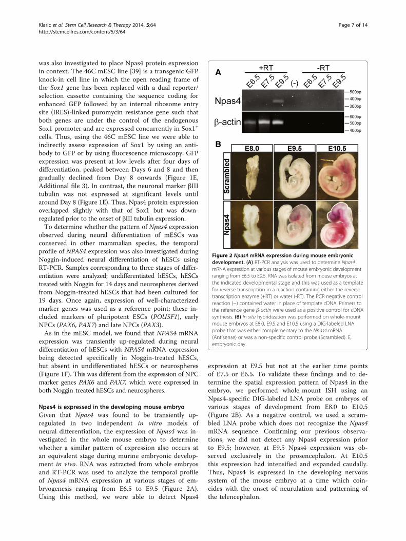
Npas4 is expressed in the developing mouse embryoGiven that Npas4 was found to be transiently up-regulated in two independent in vitro models ofneural differentiation, the expression of Npas4 was in-vestigated in the whole mouse embryo to determinewhether a similar pattern of expression also occurs atan equivalent stage during murine embryonic develop-ment in vivo. RNA was extracted from whole embryosand RT-PCR was used to analyze the temporal profileof Npas4 mRNA expression at various stages of em-bryogenesis ranging from E6.5 to E9.5 (Figure 2A).Using this method, we were able to detect Npas4
expression at E9.5 but not at the earlier time pointsof E7.5 or E6.5. To validate these findings and to de-termine the spatial expression pattern of Npas4 in theembryo, we performed whole-mount ISH using anNpas4-specific DIG-labeled LNA probe on embryos ofvarious stages of development from E8.0 to E10.5(Figure 2B). As a negative control, we used a scram-bled LNA probe which does not recognize the Npas4mRNA sequence. Confirming our previous observa-tions, we did not detect any Npas4 expression priorto E9.5; however, at E9.5 Npas4 expression was ob-served exclusively in the prosencephalon. At E10.5this expression had intensified and expanded caudally.Thus, Npas4 is expressed in the developing nervoussystem of the mouse embryo at a time which coin-cides with the onset of neurulation and patterning ofthe telencephalon.
Figure 2 Npas4 mRNA expression during mouse embryonicdevelopment. (A) RT-PCR analysis was used to determine Npas4mRNA expression at various stages of mouse embryonic developmentranging from E6.5 to E9.5. RNA was isolated from mouse embryos atthe indicated developmental stage and this was used as a templatefor reverse transcription in a reaction containing either the reversetranscription enzyme (+RT) or water (-RT). The PCR negative controlreaction (−) contained water in place of template cDNA. Primers tothe reference gene β-actin were used as a positive control for cDNAsynthesis. (B) In situ hybridization was performed on whole-mountmouse embryos at E8.0, E9.5 and E10.5 using a DIG-labeled LNAprobe that was either complementary to the Npas4 mRNA(Antisense) or was a non-specific control probe (Scrambled). E,embryonic day.
Klaric et al. Stem Cell Research & Therapy 2014, 5:64 Page 7 of 14http://stemcellres.com/content/5/3/64

Knockdown of Npas4 expression during neuraldifferentiation of 46C mESCsTo investigate the role of Npas4 during neural differenti-ation, RNA interference (RNAi) was used to reduce Npas4expression in mESCs undergoing differentiation. Giventhe pattern of Npas4 expression during embryonic de-velopment and neural differentiation of ESCs, the 46CmESC line was selected as the parental cell line forNpas4 knockdown experiments to facilitate detection ofpossible phenotypes affecting Sox1 expression and/orNPC formation. Lentiviral transduction was used tointroduce small hairpin RNA (shRNA) expression con-structs into 46C mESCs and antibiotic selection wasused to generate stable, clonally-derived Npas4 knock-down mESC lines. Two different Npas4 shRNA constructswere used to create Npas4 knockdown mESC lines (KD1and KD2) and, in addition, an Empty vector control mESCline was created to control for the transduction processand for the introduction of foreign DNA into cells. Im-munoblotting was used to assess Npas4 expression in thetransduced mESC lines after four days of neural differenti-ation. Expression of the endogenous Npas4 protein wasreduced in both the Npas4 knockdown mESC lines whencompared to the Empty vector control (approximately54% reduction in KD1 and 62% reduction in KD2)(Figure 3A) and, when analyzed using a one-way analysisof variance (ANOVA), this difference was statistically sig-nificant (P <0.001) (Figure 3B). In the undifferentiatedstate, all of the transduced mESC lines retained normalgrowth rates, morphology and expression of several keypluripotency genes (Additional file 4) suggesting that thegenomic integration of shRNA constructs had not inter-fered with normal ESC biology and that interpretation ofresults would not be affected by off-target integration ef-fects. Furthermore, no difference was observed betweenthe Empty vector control mESC line and those constitu-tively expressing Npas4-specific shRNAs suggesting thatNpas4 is dispensable for normal ESC biology.
Reduced Npas4 expression during neural differentiationresults in delayed neural differentiationTo determine whether reduced Npas4 expression affectedneural differentiation of mESCs, the Npas4 knockdownmESC lines were compared to the Empty vector controlmESC line in their ability to undergo neural differenti-ation. We first assessed the proliferation rate of the Npas4knockdown lines to determine whether knockdown ofNpas4 resulted in a gross change in proliferation or celldeath during neural differentiation. We compared the pro-liferation rate of Npas4 knockdown mESC lines to that ofthe Empty vector control mESC line by counting the totalnumber of viable cells at 24 hour intervals over ten daysof differentiation for each cell line. We found that there
was no statistically significant difference in proliferationrate between the Npas4 knockdown lines and the Emptyvector control mESC line when analyzed using non-linearregression, P = 0.2739 (Figure 4A).
Figure 3 Knockdown of endogenous Npas4 protein duringneural differentiation of mESCs. (A) Immunoblotting was used todetermine the level of Npas4 protein expression in different mESClines (n = 3 biological replicates). Lysates were harvested after fourdays of differentiation in N2B27 medium and blots were probedwith an anti-Npas4 antibody. β-actin served as a loading control.(B) Npas4 knockdown was quantified by densitometry analysis.Npas4 protein expression in each sample was normalized to β-actinexpression and is relative to Npas4 expression in the Empty vectorcontrol line which was given an arbitrary value of 1. Mean valuesand standard deviations of three independent experiments (n = 3biological replicates) are shown (**P <0.01, one-way ANOVA).ANOVA, analysis of variance; mESC, mouse embryonic stem cells.
Klaric et al. Stem Cell Research & Therapy 2014, 5:64 Page 8 of 14http://stemcellres.com/content/5/3/64

The distinctive temporal expression profile of Npas4during neural differentiation of mESCs (Figure 1A, 1B, 1D)suggested that Npas4 may be expressed by a transient pro-genitor cell type as the period of peak Npas4 expressioncorresponded to the phase of differentiation marked bythe proliferation of NPCs [39]. Thus, to determine theeffect of Npas4 knockdown on NPCs formation, flow cy-tometry was used to evaluate the occurrence of Sox1+
NPCs in both Npas4 knockdown and control mESCs
during neural differentiation. To ensure that backgroundauto-fluorescence did not interfere with detection ofSox1GFP expression, the D3 mESC line [38], which doesnot carry the gene coding for GFP in its genome, was usedas a negative control.The Sox1GFP expression profile of the Empty vector
control mESC line was comparable to that of the 46Cparental mESC line and no statistically significant dif-ference was detected when the two cell lines were
Figure 4 The effect of reduced Npas4 expression on Sox1 expression during neural differentiation of mESCs. (A) The cell proliferationrate during neural differentiation was measured by counting the total number of viable cells at 24 hour intervals. Means and standard deviationsof three independent experiments are shown (n = 3). There was no statistically significant difference between the Npas4 knockdown lines andthe Empty vector control mESC line (P = 0.2739, non-linear regression). (B) Temporal analysis of Sox1GFP expression during N2B27 differentiationof Npas4 knockdown and control mESCs as measured by flow cytometry. Means and standard deviations of three independent experiments areshown (n = 3). There was a statistically significant difference between each of the Npas4 knockdown mESC lines and the Empty vector controlmESC line (****P ≤0.0001, two-way ANOVA). There was no statistically significant difference between the Empty vector control mESC line and theparental 46C mESC line (P = 0.1898 , two-way ANOVA). (C) Sox1GFP expression at Day 7 of neural differentiation. Representative flow cytometryhistograms are shown for each mESC line. The y-axis shows the number of cells counted while the x-axis shows the GFP fluorescence intensity.The gate used to count GFP+ cells is shown. ANOVA, analysis of variance; mESC, mouse embryonic stem cells.
Klaric et al. Stem Cell Research & Therapy 2014, 5:64 Page 9 of 14http://stemcellres.com/content/5/3/64

compared using two-way ANOVA, P = 0.1898 (Figure 4B).In both cell lines, the proportion of GFP+ cells steadilyincreased between Days 3 and 5 reaching a peak ofapproximately 80% GFP+ cells between Days 6 and 7before gradually declining. Analysis of Npas4 knockdownmESC cultures revealed that the Sox1GFP expression pro-files for these cell lines had a similar pattern to those ofthe control mESC lines although the maximum percent-age of GFP+ cells attained was considerably lower. Thehighest percentage of GFP+ cells recorded in KD1 cultureswas 60%, while in KD2 cultures this figure was only 40%.Over the course of the differentiation period, the differ-ence in the percentage of GFP+ cells generated betweenthe Empty vector control mESC line and each of theNpas4 knockdown mESC lines was statistically significantwhen tested using a two-way ANOVA (P <0.0001). Repre-sentative histogram plots of GFP fluorescence intensity re-corded on Day 7 of differentiation are shown in Figure 4C.These data suggest that reduced Npas4 expression duringneural differentiation of mESCs resulted in a decrease inthe proportion of cells expressing Sox1.We next analyzed Npas4 knockdown and Empty vector
control cultures for expression of another NPC marker,Nestin, using immunohistochemistry. The Nestin gene en-codes an intermediate filament protein whose expressionis specific to CNS progenitor cells [45]; however, in theembryonic CNS Nestin is expressed at a later stage ofdifferentiation than Sox1 [46] and thus Nestin+ cellsrepresent a more mature type of NPC. After four daysof differentiation, no statistically significant differencewas observed between mESC lines in the percentage ofcells expressing Nestin (Additional file 5). However,during the later stages of differentiation a clear trendemerged. From Day 6 to Day 9, there was a steady de-cline in the proportion of Nestin+ cells present in Emptyvector control cultures as these NPCs differentiated intopost-mitotic neurons, which was in contrast to the Npas4knockdown cultures where the proportion of Nestin+ cellsremained constant (KD1) or even increased (KD2) overthe same period (Figure 5). Over the course of this fourday differentiation period, there was a statistically signifi-cant difference in the Nestin:DAPI ratio between theEmpty vector control line and each of the Npas4 knock-down lines (Empty versus KD1, **P <0.01; Empty versusKD2, **P <0.01) when analyzed using a two-way ANOVA.These data indicate that Nestin remains expressed for alonger period of time when Npas4 expression is reducedwhich suggests that neural differentiation may be stalledor delayed in Npas4 knockdown cultures.
DiscussionThe possible role of Npas4 during neurodevelopmentand neuronal differentiation is a topic that has received
little attention to this point, despite there being evidencethat it may indeed be involved in these processes. Yunet al. showed that Npas4 mRNA is up-regulated duringneuronal differentiation of the Neuro2a neuroblastomacell line and that Npas4 plays an important role in pro-moting neurite outgrowth in both Neuro2a cells andhippocampal neurons [47]. Here, we expand on theseobservations and demonstrate for the first time thatNpas4 is also expressed during neural differentiation ofboth mouse and human ESCs and that it may play a rolein the progression of neural progenitor cells along the dif-ferentiation route. In both neural differentiation models,Npas4 expression was transient and occurred prior to theemergence of neurons in culture. We speculate that Npas4
Figure 5 The effect of reduced Npas4 expression on Nestinexpression during neural differentiation of mESCs. (A)Immunocytochemical analysis of Nestin expression (green) duringneural differentiation. Representative images of cultures are shownfor each mESC line at Day 6 to 9 of N2B27 differentiation. Cells werecounterstained with DAPI to visualize nuclei (blue). Scale bar = 100 μm.(B) Quantification of Nestin expression in Npas4 knockdown andcontrol mESC lines at various stages of neural differentiation. For eachfield, the overall fluorescence intensity was calculated for each colorchannel and from this the ratio of Nestin:DAPI fluorescence wasdetermined. Means and standard deviations of three independentexperiments are shown (n = 3). Over the course of the differentiationperiod, there was a statistically significant difference in the Nestin:DAPIratio between the Empty vector control line and each of the Npas4knockdown lines (Empty versus KD1, **P <0.01; Empty versus KD2,**P <0.01) when analyzed using a two-way ANOVA. ANOVA, analysisof variance; DAPI, 4',6-diamidino-2-phenylindole; mESC, mouseembryonic stem cells.
Klaric et al. Stem Cell Research & Therapy 2014, 5:64 Page 10 of 14http://stemcellres.com/content/5/3/64

expression occurs in an early NPC population that existstransiently prior to terminal differentiation into post-mitotic neurons and glia. This assertion is supported bydata from the hESC model where NPAS4 was selectivelyexpressed in Noggin-treated hESCs but was down-regulated once cells had progressed to the neurospherestage. Treatment of hESCs with Noggin converts them intoa population of committed early NPCs; after 14 days oftreatment the majority of cells in culture are PAX6+/SOX1− cells with very few PAX6+/SOX1+ cells present[43]. These PAX6+/SOX1− NPCs cells have a distinctanterior identity and have been referred to as ‘primitiveanterior neurectoderm’ (PAN) [48]. Replating the Noggin-treated cells triggers further differentiation and formationof neurospheres, which represent a more mature typeof NPC population that also expresses PAX3. Wehypothesize that during neural differentiation Npas4 isexpressed by the PAN cell population and this is sup-ported by the specific expression of Npas4 that is seenin the developing forebrain of the mouse embryo.Interestingly, it has been reported that a small numberof Npas4+ cells in the dentate gyrus of adult mice co-express markers of dividing cells such as Sox2 anddoublecortin [49] which suggests that expression ofNpas4 in NPCs is not limited to the developmentalperiod.We also demonstrate that reduced Npas4 expression
in mESCs undergoing neural induction resulted in de-layed neural differentiation. In these cultures we foundthat there were fewer Sox1+ cells and that expression ofNestin persisted longer than in control cultures. Inter-estingly, in both cases we observed a more pronouncedeffect in the cell line having a greater reduction in Npas4expression. This suggests that the severity of the pheno-type is proportional to the level of Npas4 knockdown.Given that we have achieved only partial knockdown ofNpas4 expression in this system, it is possible that aneven more significant effect would be observed by fur-ther reducing Npas4 levels or by using mESCs obtainedfrom Npas4−/−mice.Differentiation of ESCs is commonly used to model
the events occurring in the embryo during the earlystages of development [36,37]. Accordingly, our obser-vations in vitro suggested that Npas4 may be involvedin aspects of early nervous system development in vivo.This was substantiated by our analysis of Npas4 expres-sion in the mouse embryo where we detected mRNAexpression in the developing forebrain at E9.5 andE10.5. No expression was observed at the earlier stageof E8.0 and as later embryonic stages were not investi-gated it is not known whether Npas4 expression ismaintained until birth or is only transiently expressedas was seen in vitro. More research is needed to addressthis question.
Curiously, the temporal relationship between Npas4and Sox1 expression differed in the ESC differentiationmodels as compared to the mouse embryo. While Npas4appears to be expressed prior to or concurrently withSox1 in the in vitro differentiation systems, this seemedto be reversed in vivo. During embryogenesis Sox1 expres-sion coincides with the neural induction of ectoderm atE7.5 [50] when it is expressed specifically in the neuralplate [51] and it continues to be expressed in the neuraltube along the entire anterior/posterior axis at later stagesof neurodevelopment [50]. This places Sox1 expressionseveral days ahead of Npas4 expression which we wereunable to detect prior to E9.5 (Figure 2). While this seemsto contradict the in vitro data, it should be noted that dif-ferentiation of ESCs in vitro is not a perfect model of em-bryonic development and that some differences may existbetween the simplified in vitro model and the complexityof the whole embryo.Nevertheless, these observations suggest that our loss-
of-function studies in vitro may have relevance in vivo.The period between E7.5 to E10.5 is a critical time inthe embryo when development of the nervous systembegins and it is marked by several important events. AtE7.5, secretion of diffusible dorsalizing signals, such asNoggin, triggers neural induction in which a region ofthe ectoderm becomes specifically defined as the neu-roectoderm [52]. By E8.5, neural specification is largelycomplete and the resulting neural plate begins to foldonto itself to form the neural tube in a process knownas neurulation which terminates at around E10 with thefinal closure event [53]. The timing of Npas4 expressionand its specific localization in the rudimentary forebrainstructures raise the possibility that it may be involved inthe specification of forebrain identity. However, unravel-ling the role of Npas4 in embryogenesis and determiningprecisely which cell populations express this transcrip-tion factor will require further investigation. A thoroughexploration of the developmental biology of Npas4-nullembryos, particularly the expression patterns of Sox1 andNestin, may provide valuable information towards this end.While the findings from this study suggest that Npas4
could have a role in early developmental processes, thestudy of Npas4−/−mice has thus far not revealed anyclear embryological function for Npas4. Npas4−/−micedo not show any gross morphological abnormality atbirth and they are able to generate offspring in theexpected Mendelian ratios [7,9]. While these obser-vations demonstrate that the Npas4 gene is not essen-tial for embryonic survival, we would suggest that,based on our results, further detailed investigation ofNpas4−/−mice during the early stages of embryogenesisis warranted. There are numerous examples of genesthat have roles in important developmental processesbut do not show a profound developmental phenotype
Klaric et al. Stem Cell Research & Therapy 2014, 5:64 Page 11 of 14http://stemcellres.com/content/5/3/64

when deleted, particularly when there may be com-pensation by other genes having similar functions.For instance, the Sox1 gene is specifically expressedin the neural plate during early CNS development[51] and is involved in maintenance of NPCs [46,54], yetSox1−/−mice are viable, born in the expected Mendelianratios and show no obvious signs of developmentalabnormalities [55].Indeed, there is evidence to suggest that alternative
pathways are activated in Npas4−/−mice to compensate forthe lack of Npas4 protein. For example, Lin and colleaguesremarked that although frequencies of miniature inhibi-tory post-synaptic currents (mIPSCs) were similar in hip-pocampal slices prepared from Npas4+/+and Npas4−/−mice,a clear difference was seen between Npas4 knockdownand controls when RNAi was used to acutely reduceNpas4 expression in wildtype slices [7]. Similarly,Ramamoorthi et al. noted that while there was reduc-tion in the expression of certain Npas4 target genes inNpas4 global knockout mice, conditional deletion ofNpas4 in adult mice resulted in an even more pro-nounced reduction in target gene expression [8]. Bothof these observations imply that the presence orabsence of Npas4 during embryonic development caninfluence which developmental programs are activated.A complete absence of Npas4 during embryonic devel-opment appears to trigger various Npas4-independentcompensatory pathways that allow embryonic develop-ment to proceed relatively normally such that the resultinganimal can function without the need for Npas4 activity.On the other hand, it seems that in wildtype animals,where Npas4 is an integral part of development, the neuralpathways that are produced are reliant on Npas4 functionand the sudden removal of Npas4 from the system hasdevastating effects. Presumably, this type of gene compen-sation is more likely to occur in the intact organism wherethere are many feedback mechanisms in place to ensurethat development can proceed normally. In contrast to thecomplex environment that is an embryo, a simplified two-dimensional cellular model of development that lacksthese sophisticated feedback mechanisms is likely to bemuch more susceptible to perturbations in gene expres-sion. This may explain why sometimes certain phenotypescan be elicited by gene knockdown in vitro, yet they arenot detectable in the knockout embryo.In mESCs cultures having reduced levels of Npas4 we
observed that expression of Sox1 was affected whichraises the possibility that Npas4 acts upstream of Sox1and may participate in the regulation of Sox1 expression.If Npas4 does indeed influence expression of Sox1, thenit would be expected that in the Npas4−/−mouse therewould be some evidence of disrupted Sox1 function.Interestingly, there is some similarity between theNpas4−/−and Sox1−/−mice; both knockout mice display
a hyperexcitability phenotype. One of the features of theSox1−/−phenotype is enhanced synaptic activity in the ol-factory cortex which manifests as spontaneous epilepti-form discharges and ultimately results in spontaneouslimbic seizures beginning at four to six weeks of age[55,56]. Given that Npas4−/−mice display a similar hyper-excitability phenotype and are also prone to seizures [7], itis tempting to speculate that there may be a commonunderlying mechanism. In summary, our work highlightsa potential role for Npas4 in neural differentiation duringCNS development.
ConclusionsOur findings provide the first evidence that Npas4 isexpressed by a progenitor cell type in the context ofmammalian development and that Npas4 expression isrequired for the normal progression of neural progenitorcells along the differentiation route. While these findingssuggest that Npas4 may be involved in aspects of NPCbiology, such as NPC maintenance and/or differentiation,further research is needed to confirm the developmentalrole of Npas4 in vivo.
Additional files
Additional file 1: Primer sequences and PCR conditions used foreach primer set. aThese primers were designed for mouse transcriptsand therefore contained some nucleotide mismatches to human cDNAsequences (underlined); however, they successfully generated RT-PCRproducts of the expected size (Npas4 primers - 348 bp; β actin primers -511 bp) when used in reactions containing cDNA derived fromhuman samples.
Additional file 2: Verification of anti-Npas4 antibody specificity. Theanti-Npas4 antibody was able to detect a protein of approximately100 kDa in HEK 293 T cells transfected with a recombinant Npas4expression construct (rNpas4) while no signal was observed inuntransfected HEK 293 T cells (n = 3). The expression vector containedthe cDNA sequence coding for the mouse Npas4 protein in which thestop codon had been removed and replaced with two copies of thesequence coding for a Myc epitope placed in tandem. The Mycsequences were positioned in frame and were followed by a stop codonsuch that translation of the resulting mRNA would yield the mouseNpas4 protein fused to two C-terminal Myc tags. An antibody to thereference protein β-actin was used as a loading control.
Additional file 3: Temporal expression of GFP during N2B27differentiation of the 46C transgenic mESC line. GFP expression isunder the control of the endogenous Sox1 promoter as visualized byfluorescence microscopy (n ≥3). Scale bars = 100 μm.
Additional file 4: Assessment of the pluripotency status oftransduced mESC lines. (A) Stable integration of shRNA constructs intothe mESC genome was confirmed by genomic PCR using primersspecific to the integration fragment. After 10 passages or more, genomicDNA was isolated from mESC lines that were virally transduced withshRNA expression constructs and also from the untransduced 46Cparental mESC line. Primers to the β-actin gene were used as a positivecontrol for genomic DNA isolation. The negative control reaction (−)contained water in place of template genomic DNA. (B) mESC lines wereassessed for expression of the pluripotency genes Pou5f1 and Nanogusing RT-PCR (n = 3). (C) Percentage of colonies expressing alkalinephosphatase in undifferentiated mESC lines as determined by an alkalinephosphatase detection assay. Means and standard deviations of three
Klaric et al. Stem Cell Research & Therapy 2014, 5:64 Page 12 of 14http://stemcellres.com/content/5/3/64

independent experiments are shown (n = 3). No statistically significantdifference was observed between mESC lines. P = 0.5078 (one-wayANOVA). (D) Representative images of colonies from each mESC line afteralkaline phosphatase detection assay (Fast Red Violet staining). Scalebar = 100 μm. (E) Comparison of the morphology of undifferentiatedmESC lines. Scale bar = 100 μm. (F) Comparison of growth rate betweenmESC lines. Means and standard deviations of four independentexperiments are shown (n = 4). No statistically significant difference wasobserved between slopes using linear regression. P = 0.115.
Additional file 5: Analysis of Nestin expression in various mESClines after four days of neural differentiation in N2B27 medium.(A) Immunocytochemical analysis of Nestin expression (red) during neuraldifferentiation. Representative images of cultures after four days ofdifferentiation are shown for each mESC line. Cells were counterstainedwith DAPI to visualize nuclei (blue). Scale bar = 100 μm. (B) Quantificationof Nestin expression in Npas4 knockdown and control mESC lines afterfour days of differentiation. The number of Nestin-expressing cells(Nestin+, DAPI+) was counted manually by a blinded researcher andexpressed as a percentage of total cells (DAPI+). Means and standarddeviations of four independent experiments are shown (n = 4). Nostatistically significant difference was observed between the Empty vectorcontrol and the Npas4 KD1 mESC line (P = 0.4203, unpaired t test) or theNpas4 KD2 mESC line (P = 0.1614, unpaired t test).
AbbreviationsANOVA: analysis of variance; Bdnf: brain-derived neurotrophic factor; bHLHPAS: basic helix-loop-helix Per-Arnt-Sim; bp: base pair; CNS: central nervoussystem; DAPI: 4',6-diamidino-2-phenylindole; E: embryonic day;ESC: embryonic stem cell; GFP: green fluorescent protein; hESC: human ESC;HRP: horseradish peroxidase; ISH: in situ hybridization; mESC: mouse ESC;Npas4: neuronal PAS domain protein 4; NPCs: neural progenitor cells;PAN: primitive anterior neuroectoderm; PBS: phosphate-buffered saline;PBST: PBS with 0.1% Tween 20; PFA: paraformaldehyde; RNAi: RNAinterference; Sox1: Sry-related high mobility group box 1; SSC: saline sodiumcitrate; TBST: Tris buffered saline with 0.1% Tween20.
Competing interestsThe authors declare that they have no competing interests.
Authors’ contributionsTK conceived and designed research, acquired data, analyzed andinterpreted data and results, performed statistical analysis, and drafted themanuscript. PT provided study materials, contributed to analysis andinterpretation of data, and was involved in drafting the manuscript andproviding critical review. MD provided study materials, contributed toanalysis and interpretation of data, and was involved in drafting themanuscript and providing critical review. WKL contributed to analysis andinterpretation of data, and was involved in drafting the manuscript andproviding critical review. SK conceived and designed research, contributed toanalysis and interpretation of data, and was involved in drafting themanuscript and providing critical review. ML conceived and designedresearch, contributed to analysis and interpretation of data, and was involvedin drafting the manuscript and providing critical review. All authors read andapproved the final manuscript.
Authors’ informationSimon A Koblar and Martin D Lewis are co-senior authors.
AcknowledgementsWe are grateful to Yingxi Lin for her generous gift of the anti-Npas4antibody. We also thank Don Anson for his assistance with performing thelentiviral transduction experiments and Murray Whitelaw for providing theNpas4 expression construct.
Author details1School of Molecular and Biomedical Science, The University of Adelaide,Adelaide, SA 5005, Australia. 2Stroke Research Program, The University ofAdelaide, Adelaide, SA 5005, Australia. 3Centre for Neural Engineering, TheUniversity of Melbourne, Melbourne, VIC 3010, Australia.
Received: 2 December 2013 Revised: 24 January 2014Accepted: 11 April 2014 Published: 8 May 2014
References1. Flood WD, Moyer RW, Tsykin A, Sutherland GR, Koblar SA: Nxf and Fbxo33:
novel seizure-responsive genes in mice. Eur J Neurosci 2004,20:1819–1826.
2. Moser M, Knoth R, Bode C, Patterson C: LE-PAS, a novel Arnt-dependentHLH-PAS protein, is expressed in limbic tissues and transactivates theCNS midline enhancer element. Brain Res Mol Brain Res 2004, 128:141–149.
3. Ooe N, Saito K, Mikami N, Nakatuka I, Kaneko H: Identification of a novelbasic helix-loop-helix-PAS factor, NXF, reveals a Sim2 competitive,positive regulatory role in dendritic-cytoskeleton modulator drebringene expression. Mol Cell Biol 2004, 24:608–616.
4. Kewley RJ, Whitelaw ML, Chapman-Smith A: The mammalian basichelix-loop-helix/PAS family of transcriptional regulators. Int J Biochem CellBiol 2004, 36:189–204.
5. Hester I, McKee S, Pelletier P, Thompson C, Storbeck C, Mears A, Schulz JB,Hakim AA, Sabourin LA: Transient expression of Nxf, a bHLH-PAStransactivator induced by neuronal preconditioning, confersneuroprotection in cultured cells. Brain Res 2007, 1135:1–11.
6. Shamloo M, Soriano L, von Schack D, Rickhag M, Chin DJ, Gonzalez-ZuluetaM, Gido G, Urfer R, Wieloch T, Nikolich K: Npas4, a novel helix-loop-helixPAS domain protein, is regulated in response to cerebral ischemia. Eur JNeurosci 2006, 24:2705–2720.
7. Lin Y, Bloodgood BL, Hauser JL, Lapan AD, Koon AC, Kim TK, Hu LS, MalikAN, Greenberg ME: Activity-dependent regulation of inhibitory synapsedevelopment by Npas4. Nature 2008, 455:1198–1204.
8. Ramamoorthi K, Fropf R, Belfort GM, Fitzmaurice HL, McKinney RM, Neve RL,Otto T, Lin Y: Npas4 regulates a transcriptional program in CA3 requiredfor contextual memory formation. Science 2011, 334:1669–1675.
9. Ooe N, Motonaga K, Kobayashi K, Saito K, Kaneko H: Functionalcharacterization of basic helix-loop-helix-PAS type transcription factorNXF in vivo: putative involvement in an “on demand” neuroprotectionsystem. J Biol Chem 2009, 284:1057–1063.
10. Zhang SJ, Zou M, Lu L, Lau D, Ditzel DA, Delucinge-Vivier C, Aso Y, DescombesP, Bading H: Nuclear calcium signaling controls expression of a large genepool: identification of a gene program for acquired neuroprotectioninduced by synaptic activity. PLoS Genet 2009, 5:e1000604.
11. Ploski JE, Monsey MS, Nguyen T, Dileone RJ, Schafe GE: The neuronal PASdomain protein 4 (Npas4) is required for new and reactivated fearmemories. PLoS One 2011, 6:e23760.
12. Pruunsild P, Sepp M, Orav E, Koppel I, Timmusk T: Identification ofcis-elements and transcription factors regulating neuronal activity-dependent transcription of human BDNF gene. J Neurosci 2011,31:3295–3308.
13. Bibel M, Barde YA: Neurotrophins: key regulators of cell fate and cellshape in the vertebrate nervous system. Genes Dev 2000, 14:2919–2937.
14. Binder DK, Scharfman HE: Brain-derived neurotrophic factor. GrowthFactors 2004, 22:123–131.
15. Huang EJ, Reichardt LF: Neurotrophins: roles in neuronal developmentand function. Annu Rev Neurosci 2001, 24:677–736.
16. Waterhouse EG, Xu B: New insights into the role of brain-derivedneurotrophic factor in synaptic plasticity. Mol Cell Neurosci 2009, 42:81–89.
17. Majoul I, Shirao T, Sekino Y, Duden R: Many faces of drebrin: from buildingdendritic spines and stabilizing gap junctions to shaping neurite-like cellprocesses. Histochem Cell Biol 2007, 127:355–361.
18. Sekino Y, Kojima N, Shirao T: Role of actin cytoskeleton in dendritic spinemorphogenesis. Neurochem Int 2007, 51:92–104.
19. Firth JD, Ebert BL, Pugh CW, Ratcliffe PJ: Oxygen-regulated controlelements in the phosphoglycerate kinase 1 and lactate dehydrogenaseA genes: similarities with the erythropoietin 3′ enhancer. Proc Natl AcadSci U S A 1994, 91:6496–6500.
20. Semenza GL, Roth PH, Fang HM, Wang GL: Transcriptional regulation ofgenes encoding glycolytic enzymes by hypoxia-inducible factor 1.J Biol Chem 1994, 269:23757–23763.
21. Semenza GL, Wang GL: A nuclear factor induced by hypoxia via de novoprotein synthesis binds to the human erythropoietin gene enhancer at asite required for transcriptional activation. Mol Cell Biol 1992,12:5447–5454.
Klaric et al. Stem Cell Research & Therapy 2014, 5:64 Page 13 of 14http://stemcellres.com/content/5/3/64

22. Wang GL, Jiang BH, Rue EA, Semenza GL: Hypoxia-inducible factor 1 is abasic-helix-loop-helix-PAS heterodimer regulated by cellular O2 tension.Proc Natl Acad Sci U S A 1995, 92:5510–5514.
23. Ryan HE, Lo J, Johnson RS: HIF-1 alpha is required for solid tumorformation and embryonic vascularization. EMBO J 1998, 17:3005–3015.
24. Nambu JR, Franks RG, Hu S, Crews ST: The single-minded gene ofDrosophila is required for the expression of genes important for thedevelopment of CNS midline cells. Cell 1990, 63:63–75.
25. Nambu JR, Lewis JO, Wharton KA Jr, Crews ST: The Drosophila single-minded gene encodes a helix-loop-helix protein that acts as a masterregulator of CNS midline development. Cell 1991, 67:1157–1167.
26. Wilk R, Weizman I, Shilo BZ: Trachealess encodes a bHLH-PAS protein thatis an inducer of tracheal cell fates in Drosophila. Genes Dev 1996,10:93–102.
27. Jiang L, Crews ST: The Drosophila dysfusion basic helix-loop-helix(bHLH)-PAS gene controls tracheal fusion and levels of the trachealessbHLH-PAS protein. Mol Cell Biol 2003, 23:5625–5637.
28. Jiang L, Crews ST: Dysfusion transcriptional control of Drosophila trachealmigration, adhesion, and fusion. Mol Cell Biol 2006, 26:6547–6556.
29. Dun XP, Chilton JK: Control of cell shape and plasticity duringdevelopment and disease by the actin-binding protein Drebrin. HistolHistopathol 2010, 25:533–540.
30. Mizui T, Kojima N, Yamazaki H, Katayama M, Hanamura K, Shirao T: DrebrinE is involved in the regulation of axonal growth through actin-myosininteractions. J Neurochem 2009, 109:611–622.
31. Alcantara S, Frisen J, del Rio JA, Soriano E, Barbacid M, Silos-Santiago I: TrkBsignaling is required for postnatal survival of CNS neurons and protectshippocampal and motor neurons from axotomy-induced cell death.J Neurosci 1997, 17:3623–3633.
32. Ernfors P, Lee KF, Jaenisch R: Mice lacking brain-derived neurotrophicfactor develop with sensory deficits. Nature 1994, 368:147–150.
33. Schwab MH, Bartholomae A, Heimrich B, Feldmeyer D, Druffel-Augustin S,Goebbels S, Naya FJ, Zhao S, Frotscher M, Tsai MJ, Nave KA: Neuronal basichelix-loop-helix proteins (NEX and BETA2/Neuro D) regulate terminalgranule cell differentiation in the hippocampus. J Neurosci 2000,20:3714–3724.
34. Uittenbogaard M, Chiaramello A: The basic helix-loop-helix transcriptionfactor Nex-1/Math-2 promotes neuronal survival of PC12 cells bymodulating the dynamic expression of anti-apoptotic and cell cycleregulators. J Neurochem 2005, 92:585–596.
35. Li J, Liu ZJ, Pan YC, Liu Q, Fu X, Cooper NG, Li Y, Qiu M, Shi T: Regulatorymodule network of basic/helix-loop-helix transcription factors in mousebrain. Genome Biol 2007, 8:R244.
36. Rathjen J, Rathjen PD: Mouse ES cells: experimental exploitation ofpluripotent differentiation potential. Curr Opin Genet Dev 2001,11:587–594.
37. Rodda SJ, Kavanagh SJ, Rathjen J, Rathjen PD: Embryonic stem celldifferentiation and the analysis of mammalian development. Int J DevBiol 2002, 46:449–458.
38. Doetschman TC, Eistetter H, Katz M, Schmidt W, Kemler R: The in vitrodevelopment of blastocyst-derived embryonic stem cell lines: formationof visceral yolk sac, blood islands and myocardium. J Embryol ExpMorphol 1985, 87:27–45.
39. Ying QL, Stavridis M, Griffiths D, Li M, Smith A: Conversion of embryonicstem cells into neuroectodermal precursors in adherent monoculture.Nat Biotechnol 2003, 21:183–186.
40. Costa M, Dottori M, Ng E, Hawes SM, Sourris K, Jamshidi P, Pera MF, ElefantyAG, Stanley EG: The hESC line Envy expresses high levels of GFP in alldifferentiated progeny. Nat Methods 2005, 2:259–260.
41. Rathjen J, Lake JA, Bettess MD, Washington JM, Chapman G, Rathjen PD:Formation of a primitive ectoderm like cell population, EPL cells, from EScells in response to biologically derived factors. J Cell Sci 1999,112:601–612.
42. Ying QL, Smith AG: Defined conditions for neural commitment anddifferentiation. Methods Enzymol 2003, 365:327–341.
43. Dottori M, Pera MF: Neural differentiation of human embryonic stemcells. Methods Mol Biol 2008, 438:19–30.
44. Schmittgen TD, Livak KJ: Analyzing real-time PCR data by the comparativeC(T) method. Nat Protoc 2008, 3:1101–1108.
45. Lendahl U, Zimmerman LB, McKay RD: CNS stem cells express a new classof intermediate filament protein. Cell 1990, 60:585–595.
46. Tanaka S, Kamachi Y, Tanouchi A, Hamada H, Jing N, Kondoh H: Interplay ofSOX and POU factors in regulation of the Nestin gene in neuralprimordial cells. Mol Cell Biol 2004, 24:8834–8846.
47. Yun J, Nagai T, Furukawa-Hibi Y, Kuroda K, Kaibuchi K, Greenberg ME,Yamada K: Neuronal Per Arnt Sim (PAS) domain protein 4 (NPAS4)regulates neurite outgrowth and phosphorylation of synapsin I. J BiolChem 2013, 288:2655–2664.
48. Pankratz MT, Li XJ, Lavaute TM, Lyons EA, Chen X, Zhang SC: Directedneural differentiation of human embryonic stem cells via an obligatedprimitive anterior stage. Stem Cells 2007, 25:1511–1520.
49. Yun J, Koike H, Ibi D, Toth E, Mizoguchi H, Nitta A, Yoneyama M, Ogita K,Yoneda Y, Nabeshima T, Nagai T, Yamada K: Chronic restraint stressimpairs neurogenesis and hippocampus-dependent fear memory inmice: possible involvement of a brain-specific transcription factor Npas4.J Neurochem 2010, 114:1840–1851.
50. Wood HB, Episkopou V: Comparative expression of the mouse Sox1, Sox2and Sox3 genes from pre-gastrulation to early somite stages. Mech Dev1999, 86:197–201.
51. Pevny LH, Sockanathan S, Placzek M, Lovell-Badge R: A role for SOX1 inneural determination. Development 1998, 125:1967–1978.
52. Levine AJ, Brivanlou AH: Proposal of a model of mammalian neuralinduction. Dev Biol 2007, 308:247–256.
53. Copp AJ, Greene ND, Murdoch JN: The genetic basis of mammalianneurulation. Nat Rev Genet 2003, 4:784–793.
54. Graham V, Khudyakov J, Ellis P, Pevny L: SOX2 functions to maintain neuralprogenitor identity. Neuron 2003, 39:749–765.
55. Nishiguchi S, Wood H, Kondoh H, Lovell-Badge R, Episkopou V: Sox1directly regulates the gamma-crystallin genes and is essential for lensdevelopment in mice. Genes Dev 1998, 12:776–781.
56. Malas S, Postlethwaite M, Ekonomou A, Whalley B, Nishiguchi S, Wood H,Meldrum B, Constanti A, Episkopou V: Sox1-deficient mice suffer fromepilepsy associated with abnormal ventral forebrain development andolfactory cortex hyperexcitability. Neuroscience 2003, 119:421–432.
doi:10.1186/scrt453Cite this article as: Klaric et al.: A reduction in Npas4 expression resultsin delayed neural differentiation of mouse embryonic stem cells. StemCell Research & Therapy 2014 5:64.
Submit your next manuscript to BioMed Centraland take full advantage of:
• Convenient online submission
• Thorough peer review
• No space constraints or color figure charges
• Immediate publication on acceptance
• Inclusion in PubMed, CAS, Scopus and Google Scholar
• Research which is freely available for redistribution
Submit your manuscript at www.biomedcentral.com/submit
Klaric et al. Stem Cell Research & Therapy 2014, 5:64 Page 14 of 14http://stemcellres.com/content/5/3/64