a santos-carvalho et.al 2013 neuropeptide y receptors activation protects rat retinal neural cells...
DESCRIPTION
fTRANSCRIPT
OPEN
Neuropeptide Y receptors activation protects rat retinalneural cells against necrotic and apoptotic cell deathinduced by glutamate
A Santos-Carvalho1,2, F Elvas2,3,4, AR Alvaro5, AF Ambrosio1,3,4 and C Cavadas*,1,2
It has been claimed that glutamate excitotoxicity might have a role in the pathogenesis of several retinal degenerative diseases,including glaucoma and diabetic retinopathy. Neuropeptide Y (NPY) has neuroprotective properties against excitotoxicity in thehippocampus, through the activation of Y1, Y2 and/or Y5 receptors. The principal objective of this study is to investigate the potentialprotective role of NPY against glutamate-induced toxicity in rat retinal cells (in vitro and in an animal model), unraveling the NPYreceptors and intracellular mechanisms involved. Rat retinal neural cell cultures were prepared from newborn Wistar rats (P3-P5)and exposed to glutamate (500 lM) for 24 h. Necrotic cell death was evaluated by propidium iodide (PI) assay and apoptotic celldeath using TUNEL and caspase-3 assays. The cell types present in culture were identified by immunocytochemistry. Theinvolvement of NPY receptors was assessed using selective agonists and antagonists. Pre-treatment of cells with NPY (100 nM)inhibited both necrotic cell death (PI-positive cells) and apoptotic cell death (TUNEL-positive cells and caspase 3-positive cells)triggered by glutamate, with the neurons being the cells most strongly affected. The activation of NPY Y2, Y4 and Y5 receptorsinhibited necrotic cell death, while apoptotic cell death was only prevented by the activation of NPY Y5 receptor. Moreover, NPYneuroprotective effect was mediated by the activation of PKA and p38K. In the animal model, NPY (2.35 nmol) was intravitreallyinjected 2 h before glutamate (500 nmol) injection into the vitreous. The protective role of NPY was assessed 24 h after glutamate(or saline) injection by TUNEL assay and Brn3a (marker of ganglion cells) immunohistochemistry. NPY inhibited the increase in thenumber of TUNEL-positive cells and the decrease in the number of Brn3a-positive cells induced by glutamate. In conclusion, NPYand NPY receptors can be considered potential targets to treat retinal degenerative diseases, such as glaucoma and diabeticretinopathy.Cell Death and Disease (2013) 4, e636; doi:10.1038/cddis.2013.160; published online 16 May 2013Subject Category: Neuroscience
Neuropeptide Y (NPY) is one of the most abundant peptides in
the mammalian central nervous system (CNS).1–3 NPY is a
highly conserved peptide containing 36 amino acids. Its
biological effects are mediated by six G-protein-coupled
receptors Y1, Y2, Y3, Y4, Y5 and y6.3–5 The retina is a
specialized nervous tissue where NPY and its receptors are
expressed in the retina of different species.6,7 The presence of
mRNA for Y1, Y2, Y4 and Y5 NPY receptors has been detected
in rat retinas8,9 and in cultured rat retinal neural cells,8 but their
distribution in different cell types and their function in the retina
is poorly understood.Glutamate is the main excitatory neurotransmitter in the
CNS, including in retina.10 Excitotoxicity, which is considered asan overactivation of glutamate receptors triggering neuronalcell death, has been associated with several acute andchronic neurodegenerative disorders11,12 and in retinal
degenerative disorders, such as glaucoma13–15 and diabetic
retinopathy.16–18
NPY has been linked to several physiological and patho-logical functions, such as feeding behaviour, memory
processing, pain, anxiety, cell proliferation and many other
processes in the central and peripheral nervous systems.19,20
Some studies have demonstrated putative neuroprotective
effects of NPY in various regions of the CNS. In particular,
NPY inhibits the glutamate release in rat hippocampus and is
neuroprotective in rat hippocampus and striatum.2,21–25
Moreover, the activation of NPY Y1, Y2 and Y5 receptors
mediates the neuroprotective effect of NPY against
AMPA- and kainate-induced excitotoxicity in organotypic rat
hippocampal slice cultures.21 It has also been suggested that
selective activation of Y1 and Y2 receptors protects mousehippocampal cells from excitotoxic lesions.24 Similarly, NPY
1CNC – Center for Neuroscience and Cell Biology, University of Coimbra; Largo Marques de Pombal, Coimbra, Portugal; 2Faculty of Pharmacy, University of Coimbra,Polo das Ciencias da Saude, Azinhaga de Santa Comba, Coimbra, Portugal; 3Centre of Ophthalmology and Visual Sciences, IBILI, Faculty of Medicine, University ofCoimbra, Polo das Ciencias da Saude, Azinhaga de Santa Comba, Coimbra, Portugal; 4AIBILI – Association for Innovation and Biomedical Research on Light andImage, Azinhaga de Santa Comba, Coimbra, Portugal and 5Department of Biology and Environment; University of Tras-os-Montes and Alto Douro, Vila Real, Portugal*Corresponding author: C Cavadas, Faculty of Pharmacy, University of Coimbra, Polo das Ciencias da Saude, Azinhaga de Santa Comba, Coimbra 3000-548, Portugal.Tel: +351 239859950; Fax: +351 239827126; E-mail: [email protected]
Received 03.12.12; revised 27.3.13; accepted 08.4.13; Edited by A Verkhratsky
Keywords: Retinal cells; neuropeptide Y; NPY receptors; neuroprotection; glutamateAbbreviations: GCL, ganglion cell layer; GFAP glial fibrillary acidic protein; INL, inner nuclear layer; L-NAME, L-NG-nitroarginine methyl ester; CNS, central nervoussystem; MDMA, 3,4-methylenedioxy-N-methylamphetamine; NOS, nitric oxide synthase; NPY, neuropeptide Y; ONL, outer nuclear layer; PI, propidium iodide; RT, roomtemperature; BSA, bovine serum albumin; PBS, phosphate-buffered saline; PFA, paraformaldehyde; PI3K, phosphatidylinositol 3-kinase; PKA, protein kinase A; PKC,protein kinase C; TUNEL, terminal deoxynucleotidyl transferase (TdT) dUTP nick end labeling
Citation: Cell Death and Disease (2013) 4, e636; doi:10.1038/cddis.2013.160& 2013 Macmillan Publishers Limited All rights reserved 2041-4889/13
www.nature.com/cddis
Y2 and Y5 are implicated in the neuroprotective role againstkainate-induced excitotoxicity in hippocampus even afterdelayed application of the respective agonists. Specificactivation of NPY Y2 receptor is also effective in a transient
middle cerebral artery occlusion model of ischemia.23
Recently, it was shown that NPY, also through NPY Y2
receptor activation, mediates the survival of dopaminergicneurons in Parkinson’s disease models.26 In addition, NPY
Neuroprotection by NPY receptors in retinal cellsA Santos-Carvalho et al
2
Cell Death and Disease

was suggested as a potential neuroprotective agent inAlzheimer’s disease by counteracting the toxic effect ofb-amyloid in an in vitro model.27,28
We have also shown that NPY in the retina presentsneuroprotective properties. Specifically, NPY protected ratretinal cells in culture against 3,4-methylenedioxy-N-methy-lamphetamine (MDMA)-induced toxicity,29 although the NPYreceptor subtype(s) involved in this neuroprotective effect isunknown.
As the retina is affected by various degenerative diseases,where glutamate excitotoxicity might eventually have arole,13,17 our major goal in the present work is to evaluatethe putative neuroprotective role of NPY and NPY receptorsagainst glutamate excitotoxicity in retinal cells. We haveevaluated the involvement of the different NPY receptors, aswell as the possible intracellular signaling pathways involvedin the neuroprotective effects of NPY in retinal cells, usingprimary rat retinal neural cell cultures.
Results
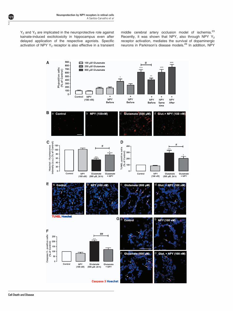
NPY protects neurons against necrotic and apoptoticcell death induced by glutamate. Necrotic and lateapoptotic cell death of rat retinal neural cells was evaluatedby propidium iodide (PI) uptake assay. Retinal cells wereexposed to 100, 250 or 500mM glutamate for 24 h (Figures 1Aand B). The number of PI-positive cells in coverslips exposedto 100, 250 or 500mM of glutamate was 175.7±27.1%,364.7±64.4% and 617.3±71.7% of control, respectively.These results indicate that cell viability decreases significantlywith increased glutamate concentrations. To investigate thepotential neuroprotective role of NPY against glutamate-induced toxicity, retinal cells were incubated with NPY(100 nM) at three different times: 1 h before the incubationwith glutamate (100, 250 and 500mM), simultaneously withthe addition of glutamate (500mM) and 30 min after exposureto glutamate (500mM). NPY did not affect the increase in thenumber of PI-positive cells induced by exposure to 100mMglutamate, as the number of PI-positive cells (179.9± 25.0%of control—NPY applied 1 h before glutamate) was similar toglutamate alone. When cells were exposed to 250mMglutamate, there was a tendency, although not significant,
for a protective effect of NPY when applied before glutamate.A neuroprotective effect of NPY was observed when 500mMglutamate stimulus was applied. When cells were exposed to500mM glutamate, and NPY (100 nM) was added 1 h beforeglutamate, there was a significant neuroprotective effect ofNPY, as shown by a decrease in the number of PI-positivecells to 409.4±41.8% of control (Figures 1A and Bd), whichcan be compared with the glutamate condition (617.3±71.7%of control), indicating a decrease in the number of PI-positivecells of 34%. However, when cells were exposed to NPY,either simultaneously or 30 min after adding 500mMglutamate, the neuroprotective effect was lost. Under thetwo conditions, the number of PI-positive cells was614.7±80.5% and 756.9±78.0% of control, respectively,similar to percentage found when cells were exposed to500mM glutamate (617.3±71.7% of control). Based on theseresults, NPY was applied 1 h before glutamate (500mM) forthe subsequent experiments.
The effects of glutamate and/or NPY treatments on the totalnumber of cells were also assessed (Figure 1C). Cells werestained with Hoechst33342, and the fluorescence intensity(arbitrary units) was measured. Glutamate (500 mM, 24 h) wasfound to decrease the Hoechst33342-fluorescence intensityto 50.8±7.0% of control (untreated cells). NPY partiallyprevented this effect triggered by glutamate, as the decreasein fluorescence intensity was attenuated by NPY (75.4±9.8%of control).
Apoptotic cell death was assessed using the TUNEL(terminal deoxynucleotidyl transferase (TdT) dUTP nick endlabeling) assay to obtain a better characterization of theprotective role of NPY against retinal cell death caused byexposure to glutamate (Figures 1D and E). Glutamate(500 mM) increased the number of apoptotic cells to294.1±41.7% of control. When NPY (100 nM) was applied1 h before glutamate, the increase in the number of TUNEL-positive cells triggered by glutamate was reduced to206.2±32.6% of control, representing a 30% reduction. Inaddition, although glutamate (500 mM, 24 h) increased thenumber of active caspase 3-positive cells to 201.9±12.8% ofcontrol (Figures 1F and Gc), NPY pre-treatment reduced theincrease in the number of caspase 3-positive cells triggeredby glutamate to 120.7±16.7% of control (Figure 1Gd).
Figure 1 NPY protects against necrotic and apoptotic retinal cell death induced by glutamate. (A and B) Necrotic cells were assessed by PI incorporation assay. (C) Cellnuclei were stained by Hoechst 33342. Apoptotic cells were assessed by (D and E) TUNEL assay and (F and G) cleaved caspase 3- immunocytochemistry. (A) Quantificationof PI-positive cells (percentage of control). Retinal cells were exposed to different concentrations of glutamate (100, 250 and 500mM) for 24 h and treated with NPY (100 nM) atthree different time points: 1 h before, simultaneously, and 30 min after glutamate, as indicated below bars. The results represent the mean±S.E.M of n¼ 4–11 independentexperiments; ***Po0.001, **Po0.01, *Po0.05, compared with control; #Po0.05, compared with glutamate (500mM); one-way analysis of variance (ANOVA) followed byBonferroni’s post-hoc test. (B) Representative images of (a) control and cultures treated with (b) NPY, (c) glutamate or (d) glutamateþNPY (1 h before), showing PI-positivecells (red spots), Bar¼ 100mm. (C) Quantification of fluorescence intensity (arbitrary units) of cells stained with Hoechst 33342 (nucleus marker), compared with control(no drug). These results represent the mean±S.E.M. of n¼ 21–27 independent experiments; ***Po0.001, *Po0.05, compared with control; #Po0.05, compared withglutamate; one-way ANOVA followed by Bonferroni’s post-hoc test. (D) Quantification of TUNEL-positive cells (percentage of control). Cultured retinal cells were exposed toglutamate and treated with NPY (1 h before glutamate exposure), as indicated below bars. Data represent the mean±S.E.M. of n¼ 5–6 independent experiments;***Po0.001, *Po0.05, compared with control; #Po0.05, compared with glutamate; one-way ANOVA followed by Bonferroni’s post-hoc test. (E) Representative images of(a) control and cultures treated with (b) NPY, (c) glutamate or (d) glutamateþNPY (1 h before), showing TUNEL-positive cells (purple spots, indicated by white arrows) andcell nuclei stained with Hoechst 33342 (blue); Bar¼ 50mm. (F) Quantification of cleaved caspase-3 positive cells (red) per field compared with control conditions (100%; nodrug, Ga). Rat retinal cells were exposed to glutamate and treated with NPY (1 h before glutamate exposure), as indicated below bars. The results represent themean±S.E.M. of n¼ 5–6 independent experiments; ***Po0.001, *Po0.05, compared with control; #Po0.05, compared with glutamate; one-way ANOVA followed byBonferroni’s post-hoc test. (G) Representative images of (a) control and cultures treated with (b) NPY, (c) glutamate or (d) glutamateþNPY (1 h before), showing cleavedcaspase 3-positive cells (purple spots). Cell nuclei were stained with Hoechst 33342 (blue). NPY per se had no effect on the number of PI-, Hoechst 33342-, TUNEL-, orcleaved caspase 3-positive cells compared with control. Bar¼ 50mm
Neuroprotection by NPY receptors in retinal cellsA Santos-Carvalho et al
3
Cell Death and Disease

To further elucidate the protective effect of NPY againstglutamate-induced cell death and considering that these cellcultures are composed of neurons, macroglial and microglialcells, we evaluated, by immunocytochemistry, which celltypes could be most strongly affected by glutamate and,eventually, protected by NPY (Figures 2–4). To quantify theeffects of glutamate and NPY on different cell types, theimmunoreactivity (fluorescence intensity) and/or the numberof positive cells to different cell markers were evaluated.Under control conditions, a normal distribution ofTUJ1-positive neurons was observed (Figures 2Ca). Whencells were exposed to 500mM glutamate for 24 h, the numberof neurons decreased and their neurites integrity wasdramatically affected (Figure 2Cc). The quantification of
TUJ1-positive cells (Figure 2A) revealed that glutamateinduced a significant decrease in the number of neurons inculture to 33.4±3.8% of control. The application of NPYbefore glutamate inhibited significantly the decrease in thenumber of TUJ1-positive cells to 51.4±3.6% of control.Additionally, by analyzing the TUJ-1-immunoreactivity (Figure2B), we also found that glutamate induced a significantdecrease in the content of this neuronal marker to 26.0±4.9%of control (Figure 2B). In cells incubated with NPY beforeglutamate application, the decrease in TUJ-1 immunoreactiv-ity was attenuated (49.2±8.5% of control), compared withcells just exposed to glutamate. In rat retinal cell cultures,among several neuronal markers, TUJ1 presents the bestimmunoreactivity profile. However, TUJ1 is considered an
Figure 2 NPY protects neuronal cell death induced by glutamate in rat retinal neural cell cultures. Neurons were identified with (C) anti-TUJ1 (green) or (E) anti-NeuN(green) antibodies, respectively. (A) Quantification of TUJ1-positive cells per z-stack. The results were normalized and are presented as percentage of control condition. Theresults represent the mean±S.E.M. of n¼ 5–7 independent experiments; ***Po0.001, compared with control; ##Po0.01, compared with glutamate; one-way analysis ofvariance (ANOVA) followed by Bonferroni’s post-hoc test. (B) Quantification of TUJ 1-immunoreactivity by fluorescence intensity (arbitrary units), compared with controlconditions (100%; no drug, Ca). The results represent the mean±S.E.M. of n¼ 4–8 independent experiments ***Po0.001, compared with control; #Po0.05, compared withglutamate; one-way ANOVA followed by Bonferroni’s post-hoc test. (C) Representative images of (a) control cultures and cultures treated with (b) NPY, (c) glutamate or (d)glutamateþNPY, showing TUJ1-positive cells (green). Cell nuclei were identified by Hoechst 33342 staining (blue). (D) Quantification of NeuN-positive cells per z-stack. Theresults were normalized and are presented as percentage of control condition. The results represent the mean±S.E.M. of n¼ 3–5 independent experiments; ***Po0.001,compared with control; #Po0.05, compared with glutamate; one-way ANOVA followed by Bonferroni’s post-hoc test. (E) Representative images of (a) control cultures andcultures treated with (b) NPY, (c) glutamate or (d) glutamateþNPY, showing NeuN-positive cells (green). Cell nuclei were stained with Hoechst 33342 (blue). NPY per se didnot affect the number of TUJ1- or NeuN-positive cells or the TUJ1-immunoreactivity compared with control. Bar¼ 50mm
Neuroprotection by NPY receptors in retinal cellsA Santos-Carvalho et al
4
Cell Death and Disease

immature neuronal marker, and therefore the expression ofNeuN, a marker of mature neurons, was also evaluated in thesame conditions. Similar results were found (Figures 2D andE). The number of NeuN-positive cells dramatically decreasedin the presence of 500 mM glutamate to 19.8±4.0% of control.The pre-incubation of the retinal cells with NPY inhibited thedecrease in the number of NeuN-positive cells triggered byglutamate (38.1±7.2% of control). To evaluate the effects ofglutamate and NPY on macroglial cells, we analyzed theimmunoreactivity of glial fibrillary acidic protein (GFAP), amacroglial cell (astrocytes and Muller cells) marker (Figure 3).The number of GFAP-positive cells (Figure 3A) and the GFAPimmunoreactivity (fluorescence intensity) were evaluated(Figure 3B). We found that exposure of retinal cells toglutamate induced a slight change in the morphology of someGFAP-positive cells, compared with control cells (Figure3Bc), namely a decrease in the number of cell processes andan increase of their thickness (Figure 3Bc). However, byevaluating the number of GFAP-positive cells and thequantification of GFAP immunoreactivity (fluorescence inten-sity) revealed no significant differences between cellsexposed to glutamate and controls. These small alterationsin GFAP-positive cell morphology triggered by glutamateappeared to be partially prevented by NPY (Figure 3Bd). Theeffects of glutamate and NPY on microglial cells wereassessed by analyzing the immunoreactivity of two microglialcell markers: CD11b and CD68/ED1 (Figure 4). CD11b labelsresting and activated microglial cells, while ED1 is a marker ofactivated microglia.30 Two different parameters were evalu-ated for these markers: the number of CD11b- and CD68/ED1-positive cells per field, and the CD11b or CD68/ED1immunoreactivity. NPY increased the number of microglialcells (resting and activated; Figures 4Cb and Fb). Similarly,glutamate or NPY plus glutamate also increased the number
of CD11b- and CD68/ED1-positive cells. As with the resultsobtained for the number of CD11b-positive cells, thefluorescence intensity measurements showed thatNPY, glutamate and NPY plus glutamate increased theimmunoreactivity of CD11b- and CD68/ED1-positive cells(Figures 4B and E).
Activation of NPY Y2, Y4 or Y5 receptors inhibits theincrease in necrotic cell death induced by glutamate.We evaluated the effects of NPY receptor agonists andantagonists to determine which NPY receptors could bemediating the protective role of NPY against necrotic celldeath induced by glutamate (Figures 5A and B). In thisanalysis, we compared the number of PI-positive cells foreach experimental condition with the number of PI-positivecells in cultures exposed to glutamate, taken as 100%. NPYdecreased the number of PI-positive cells to 72.4±3.7%relative to glutamate. The NPY Y1 receptor agonist ([Leu,31
Pro34]NPY) did not inhibit glutamate-induced necrotic celldeath (Figures 5A and B). However, the NPY Y2 receptoragonist (NPY13–36) inhibited the increase in PI-positive cells(68.8±6.4%, compared with glutamate; Figure 5A). Thisprotective effect was partially prevented by the NPY Y2
receptor antagonist BIIE0246 (83.4±7.2% compared withglutamate). Furthermore, the NPY Y4 agonist (r-PP, 100 nM)also partially protected retinal cells exposed to glutamate, asshown by the number of PI-positive cells decreasing to60.2±15.5% relative to glutamate. In addition, NPY Y5
receptor agonist (Gly,1Ser,3,22Gln,4,34Thr,6Arg,19Tyr,21
Ala,23,31Aib32)PP also exerted a protective effect, as seenby the increase in the number of PI-positive cells induced byglutamate, which was attenuated to 73.0±4.4%, comparedwith glutamate (Figures 5A and B). This effect wascompletely blocked by NPY Y5 receptor antagonist. The
Figure 3 NPY has no effect in glial cells. Microglial cells were identified with (C) anti-GFAP (red) antibody. (A) Quantification of GFAP-positive cells per z-stack.(B) Quantification of GFAP-immunoreactivity by fluorescence intensity (arbitrary units), compared with control conditions (100%; no drug, Ca). The results were normalizedand are presented as percentage of control condition. (C) Representative images of (a) control and cultures treated with (b) NPY, (c) glutamate or (d) glutamateþNPY,showing GFAP-positive cells (red). Cell nuclei were stained with Hoechst 33342 (blue). NPY per se did not affect the number of GFAP-positive cells or the GFAP-immunoreactivity compared with control. Bar¼ 50mm
Neuroprotection by NPY receptors in retinal cellsA Santos-Carvalho et al
5
Cell Death and Disease

NPY receptor agonists or antagonists per se did not increasethe number of PI-positive cells, compared with control (datanot shown).
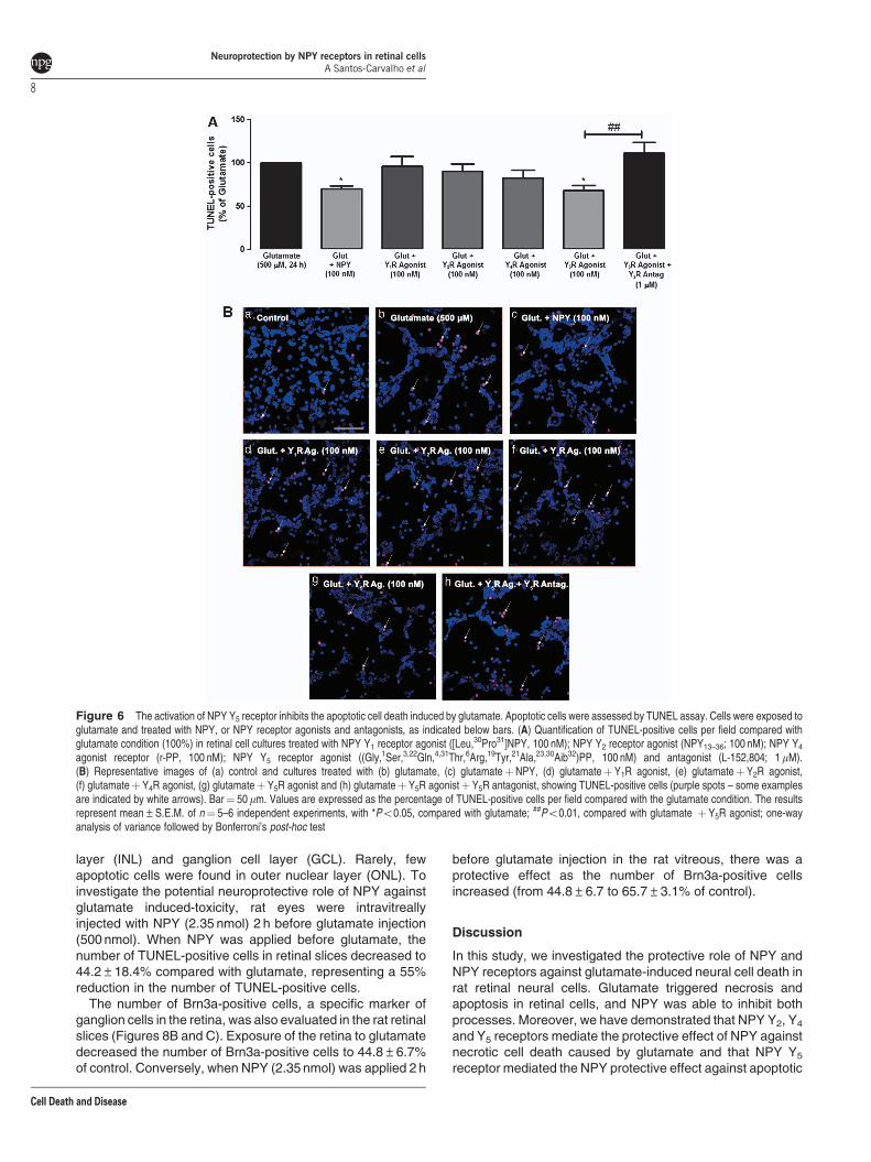
NPY Y5 receptor activation inhibits apoptotic retinalcell death induced by glutamate. We have evaluatedthe potential neuroprotective effect of NPY receptor agonistsagainst the increase in apoptotic cell (TUNEL-positivecells) number by exposure to glutamate. NPY reduced30% the number of apoptotic cells to 69.7±3.8%,compared with glutamate. NPY receptor agonists andantagonists were used to investigate those involved in this
neuroprotective effect (Figures 6A and B). The NPY Y5
receptor agonist mimicked the effect of NPY, inhibiting theincrease in the number of TUNEL-positive cells triggeredby glutamate; the percentage of apoptotic cells decreasedto 68.2±6.0%, compared with glutamate. This effect wascompletely blocked by the NPY Y5 receptor antagonist(L-152,804). The selective NPY Y1, Y2 or Y4 receptorsagonists did not decrease the number of TUNEL-positivecells in cultures exposed to glutamate. NPY receptoragonists and antagonists alone did not increase thenumber of TUNEL-positive cells, compared with control(data not shown).
Figure 4 Glutamate and NPY increase the proliferation and activation of retinal microglial cells. Microglial cells were identified by immunocytochemistry using(C) anti-CD11b (green) and (F) anti-CD68/ED1 (red) antibodies. (A) Quantification of CD11b-positive cells (green) per field, compared with control conditions (no drug, Ca).(B) Quantification of fluorescence intensity (arbitrary units) of CD11b-immunoreactivity, compared with control (100%; no drug, Ca). These results (A and B) represent themean±S.E.M. of n¼ 8 independent experiments, with **Po0.01, *Po0.05, compared with control; one-way analysis of variance (ANOVA) followed by Bonferroni’s post-hoc test. (C) Representative images of (a) control and cultures treated with (b) NPY, (c) glutamate or (d) glutamateþNPY, showing CD11b- positive cells (green). Cell nucleiwere stained by Hoechst 33342 (blue). Bar¼ 50mm. (D) Quantification of CD68/ED1-positive cells per field, compared with control (100%; no drug, Da). (E) Quantification offluorescence intensity (arbitrary units) of CD68/ED1-immunoreactivity, compared with control (100%; no drug, Da). These results (D and E) represent the mean±S.E.M. ofn¼ 5 independent experiments; ***Po0.001, **Po0.01, *Po0.05, compared with control; one-way ANOVA followed by Bonferroni’s post-hoc test. (F) Representativeimages of (a) control, and cultures treated with (b) NPY, (c) glutamate or (d) glutamateþNPY, showing CD 68/ED1-positive cells. Cell nuclei were stained by Hoechst 33342(blue). Bar¼ 50mm
Neuroprotection by NPY receptors in retinal cellsA Santos-Carvalho et al
6
Cell Death and Disease

Protein kinase A (PKA) and p38K proteins mediate theneuroprotective effect of NPY against glutamate-induced necrotic retinal neural cell death. Inhibitors ofkey proteins in different intracellular pathways were used toelucidate the intracellular pathways that mediate the neuro-protective effect of NPY when cells are exposed to glutamateor/and NPY (Figure 7). The PKA inhibitor, H89 (1 mM),prevented the neuroprotective effect of NPY (63.2±5.5%,compared with glutamate). The number of PI-positive cellsexposed to glutamate, or to glutamate plus NPY and H89,was similar. In order to confirm the involvement of PKA in theneuroprotective effect of NPY against glutamate-inducedexcitotoxicity, we have evaluated the effect of the PKAactivator, forskolin (10 mM), with cells exposed to glutamate.Forskolin decreased the number of PI-positive cells(69.6±7.1%, compared with glutamate) to a similar extentas NPY (63.2±5.5%, compared with glutamate). Theprotective effect of NPY against the increase of PI-positivecells triggered upon exposure to glutamate was also partially
prevented (85.6±2.7%, compared with glutamate) by thepresence of the p38K inhibitor (SB203580). The inhibitors ofnitric oxide synthase (NOS), protein kinase C (PKC),phosphoinositide 3-kinase (PI3K) and MEK1/2, namelyL-NG-nitroarginine methyl ester (L-NAME), calphostin C,LY294002 and U0126, respectively, did not affect theneuroprotective effect of NPY against glutamate-inducedtoxicity (Figure 7). The inhibitors per se did not increase thenumber of PI-positive cells, compared with control (data notshown).
NPY protects rat retina from apoptotic cell deathinduced by glutamate excitotoxicity. Rat retinas wereexposed to 500 nmol glutamate for 24 h (Figure 8). Apoptoticcell death was assessed by TUNEL assay (Figures 8Aand C). The number of TUNEL-positive cells in retinal slicesobtained from retinas exposed to 500 nmol glutamate was159.0±23.1 cells per field. The TUNEL-positive cells weremainly located in the inner retina, especially in inner nuclear
Figure 5 The activation of NPY Y2, Y4 and Y5 receptors inhibits the necrotic cell death induced by glutamate. Necrotic cells were evaluated by PI incorporation assay.Cells were exposed to glutamate, and treated with NPY, or NPY receptor agonists and antagonists, indicated below bars. (A) Quantification of PI-positive cells (percentage ofglutamate condition) per field in retinal cell cultures treated with NPY Y1 receptor agonist ([Leu,31Pro34]NPY;100 nM); NPY Y2 receptor agonist (NPY13–36; 100 nM) andantagonist (BIIE 0246; 1mM); NPY Y4 agonist receptor (r-PP, 100 nM); NPY Y5 receptor agonist ((Gly,1Ser,3,22Gln,4,34Thr,6Arg,19Tyr,21Ala,23,31Aib32)PP) and antagonist(L-152,804; 1 mM). (B) Representative images of (a) control and cultures treated with (b) glutamate, (c) glutamateþNPY, (d) glutamateþ Y1R agonist, (e) glutamateþY2Ragonist, (f) glutamateþY2R agonistþ Y2R antagonist, (g) glutamateþ Y4R agonist, (h) glutamateþ Y5R agonist and (i) glutamateþY5R agonistþY5R, showingPI-positive cells (red spots). Bar¼ 100mm. Values are expressed as the percentage of PI-positive cells per field compared with the glutamate condition. The results representmean±S.E.M. of n¼ 4–11 independent experiments; ***Po0.001, **Po0.01, compared with glutamate; ###Po0.001, ##Po0.01, compared with glutamateþNPYreceptor agonist; one-way analysis of variance followed by Bonferroni’s post-hoc test
Neuroprotection by NPY receptors in retinal cellsA Santos-Carvalho et al
7
Cell Death and Disease
layer (INL) and ganglion cell layer (GCL). Rarely, fewapoptotic cells were found in outer nuclear layer (ONL). Toinvestigate the potential neuroprotective role of NPY againstglutamate induced-toxicity, rat eyes were intravitreallyinjected with NPY (2.35 nmol) 2 h before glutamate injection(500 nmol). When NPY was applied before glutamate, thenumber of TUNEL-positive cells in retinal slices decreased to44.2±18.4% compared with glutamate, representing a 55%reduction in the number of TUNEL-positive cells.
The number of Brn3a-positive cells, a specific marker ofganglion cells in the retina, was also evaluated in the rat retinalslices (Figures 8B and C). Exposure of the retina to glutamatedecreased the number of Brn3a-positive cells to 44.8±6.7%of control. Conversely, when NPY (2.35 nmol) was applied 2 h
before glutamate injection in the rat vitreous, there was aprotective effect as the number of Brn3a-positive cellsincreased (from 44.8±6.7 to 65.7±3.1% of control).
Discussion
In this study, we investigated the protective role of NPY andNPY receptors against glutamate-induced neural cell death inrat retinal neural cells. Glutamate triggered necrosis andapoptosis in retinal cells, and NPY was able to inhibit bothprocesses. Moreover, we have demonstrated that NPY Y2, Y4
and Y5 receptors mediate the protective effect of NPY againstnecrotic cell death caused by glutamate and that NPY Y5
receptor mediated the NPY protective effect against apoptotic
Figure 6 The activation of NPY Y5 receptor inhibits the apoptotic cell death induced by glutamate. Apoptotic cells were assessed by TUNEL assay. Cells were exposed toglutamate and treated with NPY, or NPY receptor agonists and antagonists, as indicated below bars. (A) Quantification of TUNEL-positive cells per field compared withglutamate condition (100%) in retinal cell cultures treated with NPY Y1 receptor agonist ([Leu,30Pro31]NPY, 100 nM); NPY Y2 receptor agonist (NPY13–36; 100 nM); NPY Y4
agonist receptor (r-PP, 100 nM); NPY Y5 receptor agonist ((Gly,1Ser,3,22Gln,4,31Thr,6Arg,19Tyr,21Ala,23,30Aib32)PP, 100 nM) and antagonist (L-152,804; 1mM).(B) Representative images of (a) control and cultures treated with (b) glutamate, (c) glutamateþNPY, (d) glutamateþY1R agonist, (e) glutamateþY2R agonist,(f) glutamateþ Y4R agonist, (g) glutamateþ Y5R agonist and (h) glutamateþY5R agonistþ Y5R antagonist, showing TUNEL-positive cells (purple spots – some examplesare indicated by white arrows). Bar¼ 50mm. Values are expressed as the percentage of TUNEL-positive cells per field compared with the glutamate condition. The resultsrepresent mean±S.E.M. of n¼ 5–6 independent experiments, with *Po0.05, compared with glutamate; ##Po0.01, compared with glutamate þ Y5R agonist; one-wayanalysis of variance followed by Bonferroni’s post-hoc test
Neuroprotection by NPY receptors in retinal cellsA Santos-Carvalho et al
8
Cell Death and Disease

cell death induced by glutamate. Additionally, we have shownthat the neuroprotective effect of NPY is mediated by PKA andp38K. Finally, using an animal model, we have demonstratedthat NPY also has a protective role against glutamate-inducedexcitotoxicity in the retina. These findings suggest that NPYcan be viewed as a potential new target to protect retinal cellsin retinal corresponding degenerative diseases, such asglaucoma or diabetic retinopathy.
Previous studies have shown that NPY can exert neuro-protective effects against excitotoxicity triggered by glutamate
or glutamate receptor agonists in various regions of the CNS,such as hippocampus and striatum.2,21,23,25,26 In addition, wehave previously shown that NPY protects against MDMA(ecstasy) toxicity in cultured rat retinal cells.29 We extend thisto show, for the first time, that NPY is able to protect necroticand apoptotic cell death induced by glutamate in retinal cells.However, the neuroprotective effect of NPY only occurs whenthe peptide is applied before the excitotoxic stimulus and is notpresent when it is added simultaneously or after glutamate.This is consistent with the majority of studies describing a
Figure 7 PKA and protein 38 kinase (p38K) mediate the neuroprotective effect of NPY against retinal neuronal cell death triggered by glutamate. The involvement ofdifferent intracellular pathways in the neuroprotective effect of NPY against glutamate-induced excitotoxicity was assessed by PI uptake (PI-positive cells), using differentinhibitors of proteins involved on those pathways. Retinal cell cultures were exposed to NPY (100 nM), glutamate (500mM) and the inhibitors indicated below bars.Quantification of PI-positive cells (compared with glutamate condition) in retinal cells treated with H89 (1 mM; PKA inhibitor), forskolin (10mM; PKA activator), SD203580(1mM; p38K inhibitor), L-NAME (500mM; NOS inhibitor), calphostin C (10 nM; PKC inhibitor), LY294002 (1 mM; PI3K inhibitor) and U0126 (1 mM; MEK1/2 inhibitor). Valuesare expressed as the percentage of PI-positive cells (per field), compared with the glutamate condition. The results represent the mean±S.E.M. of n¼ 7–9 independentexperiments, with ***Po0.001, **Po0.01, *Po0.05, compared with glutamate; ##Po0.01, #Po0.05, compared with glutamateþNPY; one-way analysis of variancefollowed by Bonferroni’s post-hoc test
Figure 8 NPY protects against apoptotic cell death in the rat retina induced by glutamate. Cells undergoing apoptosis were identified by TUNEL assay, and ganglion cellswere identified by immunohistochemistry against Brn3a (ganglion cell marker). (A) Quantification of TUNEL-positive cells in rat retinal slices (presented as percentage ofglutamate condition). Retinas were exposed to glutamate (500 nmol; intravitreal injection) and treated (or not) with NPY (2.35 nmol, 2 h before intravitreal injection ofglutamate), as indicated below bars. Data represent the mean±S.E.M. of n¼ 3–5 independent experiments (animals); ***Po0.001 compared with control; #Po0.05,compared with glutamate; one-way analysis of variance (ANOVA) followed by Bonferroni’s post-hoc test. (B) Quantification of Brn3a-positive cells in rat retinal slices(presented as percentage of control). Retinas were exposed to glutamate (500 nmol; intravitreal injection) and treated (or not) with NPY (2.35 nmol, 2 h before glutamateexposure), as indicated below bars. Data represent the mean±S.E.M. of n¼ 3–5 independent experiments; ***Po0.001, *Po0.05, compared with control; #Po0.05,compared with glutamate; one-way ANOVA followed by Bonferroni’s post-hoc test. (C) Representative images of retinal slices obtained from eyes exposed to differentconditions (intravitreal injection): (a) saline (0.9% NaCl), treated with (b) NPY, (c) glutamate (500 nmol) or (d) glutamateþNPY (2.35 nmol, 2 h before glutamate), showingTUNEL-positive cells (green), Brn3a-positive cells (red), and cell nuclei stained with Hoechst 33342 (blue). NPY per se had no effect on the number of TUNEL- or Brn3a-positive cells compared with control. IPL, inner plexiform layer; OPL, outer plexiform layer. Bar¼ 50mm
Neuroprotection by NPY receptors in retinal cellsA Santos-Carvalho et al
9
Cell Death and Disease

protective role of NPY, where the peptide was applied beforethe toxic stimulus.21,29,31 However, some reports have alsoindicated that NPY is effective when it is applied a few hoursafter the excitotoxic stimulus.23,25,32 Similarly, NPY was alsoable to protect against cell death induced by glutamate in theretina. We have shown, for the first time, that NPY exertsneuroprotective effects in the retina in vitro and using ananimal model.
Glutamate excitotoxicity is characterized morphologicallyby a decrease in the number of neurons and a reduction in thelength of neuronal processes.33,34 It induced a similar effect inrat retinal cells, which was partially prevented by NPY,specifically in neurons. In fact, NPY protected immature(TUJ1-positive cells) and mature (NeuN-positive cells) neu-rons in culture. Some studies indicate that Muller cells have adual role under toxic conditions. When threatened, these cellscan be either neuroprotective or contribute to exacerbate theexcitotoxic stimuli (reviewed in Bringmann and Wiede-mann35). In the present study, glutamate slightly changedthe morphology of few GFAP-positive cells, increasing thethickness of their processes and decreasing the number ofcell processes.
The microglial cell response to glutamate exposure wascompletely different. Glutamate increased the number ofCD11b- and CD68/ED1-positive cells, as well as theimmunoreactivity of these two markers. Glutamate andglutamate receptor agonists are known to activate microglialcells in CNS, such as the hippocampus.36,37 In the presentwork, both glutamate and NPY increased microglial cellproliferation, as well as microglia activation. When NPY andglutamate were present, the effect on microglia proliferationand activation was not enhanced. In contrast, other groupshave reported inhibition of microglia phagocytosis and cellmotility by NPY upon inflammatory challenge through theactivation of NPY Y1 receptor.38,39 In addition, NPY, via Y2
receptors, has a protective role against methamphetamine-induced microgliosis.40 In the early stages of neurodegen-erative processes, the activation of microglia contributes toneuronal protection and tissue regeneration. However,continuous retinal microglial overactivation may lead tochronic inflammation, loss of autoregulatory mechanisms,irreversible neuronal loss and photoreceptor apoptosis.41–44
Microglial activation is involved in the initiation and perpetua-tion of degenerative process in many diseases, such as retinaldystrophies.42,45
Using pharmacological tools, we have shown that NPYprotects retinal cells against necrotic cell death induced byglutamate through the activation of NPY Y2, Y4 and Y5
receptors. In another study, using hippocampal slice cultures,the anti-necrotic effect of NPY was also seen to be mediatedby Y2 and Y5 receptors. However, NPY Y1 receptorscontributed to the neuroprotective effect of NPY as well.21
Although other studies have suggested that only the NPY Y1
or Y2 receptors are involved in the rescue of neurons fromexcitotoxic cell death,24,25 the involvement of NPY Y4 receptorhas not been evaluated in majority of these.
In the present study, we show that only the NPY Y5 receptoractivation protects against glutamate-induced apoptotic celldeath. Another study has linked the antiapoptotic effect ofNPY in the hippocampus to the activation of NPY Y2 and Y5
receptors.23 The difference between ours and these resultsmight be due to the differential expression of NPY receptors inretinal and hippocampal cultures, as well as to theinvolvement of different signaling pathways underlying theneuroprotective effects.
We have also found that three different NPY receptors areinvolved in the neuroprotective effect against necrotic celldeath induced by glutamate in rat retinal cell cultures.Similarly, other groups have also shown that activation ofdifferent NPY receptors can induce the same biologicaleffect.23,25,46,47 For example, NPY inhibits KCl-evoked[Ca2þ ]i increase in retinal neurons through the activation ofNPY Y1,Y4 and Y5 receptors. There are two possible mainexplanations for this: (1) the formation of homo- orhetero-dimers between different NPY receptors;48–51 and(2) heterogenous distribution of these receptors through thedifferent cell types present in the culture.
To obtain a better understanding of the intracellularmechanisms underlying the NPY neuroprotective role againstnecrotic cell death induced by glutamate, we have looked atthe possible involvement of various pathways. The NPYprotective effect may be linked to its inhibitory effect onglutamate release, as found previously in hippocampus.21,22
In rat retinal cultures, NPY inhibits both the [Ca2þ ]i increaseinduced by KCl46 and the aspartate release in these cultures(unpublished observations). NPY neuroprotection has alsobeen associated with the involvement of ERK1/2 and Aktpathways in a Parkinson’s disease model.26 In this study, wehave suggested that the NPY neuroprotective role is mediatedby PKA and p38K activation. The PKA inhibitor, H89, blockedthe neuroprotective effect of NPY, while forskolin, a PKAactivator, presented a similar protective effect to NPY,suggesting the involvement of this particular kinase in theneuroprotective effect of NPY against glutamate-inducednecrotic cell death in rat retinal cells. PKA activation by NPYhas been previously shown. For example, NPY has a biphasicmodulatory effect on [Ca2þ ]i increases induced by ATP,mediates the upregulated mRNA expression of gonadotropin-releasing hormone in a neuroblastoma cell line and inducescathecolamine release in human adrenal chromaffin cells,through the activation of PKA.52–54 However, there is alsoevidence showing that NPY inhibits PKA. The activation ofNPY receptors inhibits both the axonal transport in sensoryneurons and cell proliferation in vascular smooth muscle cells,with these effects being mediated by PKA inhibition.4,47,55–57
We also show that NPY activates p38K, and this enzyme,as PKA, appears to mediate, at least partially, the neuropro-tective role of NPY against glutamate-induced cell death. Inretinal Muller cells, the activation of NPY Y1 receptorsactivates p38 MAPK.58 Moreover, p38K activation protectsARPE-19 cells (retinal pigment epithelium cells) against celldeath triggered by pro-oxidant conditions.59
In conclusion, NPY can have a neuroprotective role againstnecrotic and apoptotic cell death induced by glutamate in ratretinal cells both in cultured cells and in situ in the retina. NPY,by activating NPY Y2, Y4 and Y5 receptors, protects retinalcells against glutamate-induced necrosis and is also able toprotect against retinal cell apoptosis by activating NPY Y5
receptors. In addition, PKA and p38K mediate the neuropro-tective effects of NPY. We believe these results might be
Neuroprotection by NPY receptors in retinal cellsA Santos-Carvalho et al
10
Cell Death and Disease

useful to devise novel pharmacologic targets and therapies totreat retinal degenerative diseases, such as glaucoma anddiabetic retinopathy.
Materials and MethodsPrimary rat retinal neural cell cultures. Three-to-five-day oldWistar rat pups were used to prepare primary rat retinal cell cultures, aspreviously described.8,60 All procedures involving animals were in agreement withthe Association for Research in Vision and Ophthalmology (ARVO) statement onvision and ophthalmic research for experimental models. Briefly, rat retinaswere dissected under sterile conditions, using a light microscope, in Ca2þ - andMg2þ -free Hanks’ balanced salt solution (in mM: 137 NaCl, 5.4 KCl, 0.45KH2PO4, 0.34 Na2HPO4, 4 NaHCO3, 5 glucose, pH 7.4) and digestedwith 0.1% trypsin (w/v, Gibco, Life Technologies Corporation, Paisley, UK) for15 min at 37 1C. Cells were plated on glass coverslips coated with poly-D-lysine(0.1 mg/ml, Sigma-Aldrich Co. LLC, St. Louis, MO, USA) using MinimumEssential Medium Eagle (Sigma-Aldrich), supplemented with 25 mM HEPES(Sigma-Aldrich), 26 mM NaHCO3, 10% fetal bovine serum (Gibco) and penicillin(100 U/ml)–streptomycin (100 mg/ml, Gibco) for 8/9 days (37 1C, 5% CO2), at adensity of 2� 106cells/cm2.
Animals. Adult male Wistar rats (250–300 g bodyweight, Charles River,France) were housed in a temperature- and humidity-controlled environment andwere provided with standard rodent diet and water ad libitum, while kept on a 12 hlight/12 h dark cycle. All procedures involving the animals were in agreement to theARVO Statement for the Use of Animals in Ophthalmic and Vision Research. Fourdifferent groups of animals were used: control (saline) injected, glutamate injected,glutamateþNPY injected, and NPY injected.
Intravitreal injections. The rats were anesthetized by isoflurane inhalationusing a gas-anesthetizing system (VetEquip, Pleasanton, CA, USA). Then,oxybuprocaine (4 mg/ml; Laboratorios Edol, Linda-a-Velha, Portugal) anestheticwas applied topically to the eyes and the pupils were dilated with tropicamide(10 mg/ml; Laboratorios Edol). Using a Hamilton syringe (Hamilton, Reno, NV,USA) with 33-gauge needle, 3ml of 0.78 mM NPY (total amount 2.35 nmol) or 3 mlof 167 mM glutamate (total amount 500 nmol) or sterile saline solution (0.9%sodium chloride; Fresenius Kabi, Carnaxide, Portugal) were intravitreally injected.Control group was injected with saline solution while NPY group was injected with2.35 nmol NPY. Glutamate group was injected with saline and 2 h later with500 nmol glutamate. Finally, glutamateþNPY group was injected with 2.35 nmolNPY and 2 h later with 500 nmol glutamate. Fusidic acid (10 mg/g; LeoPharmaceutical, Ballerup, Denmark) ointment was applied in the conjunctivalsac at the end of the experiment. The animals were killed 24 h after glutamate (orsaline) injection.
Frozen retinal sections. Under deep anesthesia (75 mg/kg ketamine and10 mg/kg xylazine), rats were transcardially perfused with phosphate-bufferedsaline (PBS; pH 7.4), followed by 4% (w/v) paraformaldehyde (PFA) in PBS. Theeyes were enucleated, washed in PBS and then transferred to PFA for 1 h. Thecornea was removed and the eye cup was further fixed for 1 h in PFA. Afterwashing in PBS, the eyes were cryopreserved by placing the eye cup in 15% (w/v)sucrose in PBS for 1 h and then in 30% (w/v) sucrose in PBS overnight at 4 1C.Finally, the eye cup was embedded in tissue-freezing medium (OCT; SakuraFinetek Europe B.V., AJ Alphen aan den Rijn, The Netherlands), the frozen blockswere cut into 10mm thickness sections in a cryostat and the cryosections werethen collected on SuperFrost Plus glass slides (Menzel-Glaser, Braunschweig,Germany) and stored at � 20 1C.
Immunocytochemistry. After treatment, cells cultured on glass coverslipswere washed twice with PBS and fixed in 4% PFA (20 min; room temperature(RT)). The cells were then permeabilized with 1% Triton X-100 for 5 min, andblocked with 3% (w/v) fatty acid-free bovine serum albumin (BSA, Sigma-Aldrich),supplemented with 0.2% Tween 20, to prevent nonspecific binding, for 1 h at RT.Cells were incubated with primary antibodies for 90 min at RT: rabbit anti-GFAP(1 : 400; Dako, Glostrup, Denmark); mouse anti-GFAP protein (1 : 500, Sigma-Aldrich); rat anti-CD11b or mouse anti-CD11b (1 : 200; AbD Serotec, Kidlington,UK); mouse anti-TUJ1 (1 : 500, Covance Research Products Inc, Berkeley, CA,USA); anti-vimentin (1 : 400, Thermo Fisher Scientific, Waltham, MA, USA); rabbitanti-cleaved caspase 3 (1 : 1600, Cell Signaling Technology, Danvers, MA, USA);
mouse anti-CD68/ED1 (1 : 200, AbD Serotec); mouse anti-NeuN (1 : 400, MerckMillipore, Billerica, MA, USA). All antibody solutions were prepared in 3% fattyacid-free BSA solution.
After washing, the cells were incubated for 1 h at RT with secondary antibodies:Alexa 488 anti-mouse IgG, Alexa 594 anti-rat IgG or Alexa 594 anti-rabbit IgG(1 : 200, Invitrogen, Life Technologies Corporation, Paisley, UK). Finally, after 5 minwashing, cell nuclei were stained with Hoechst 33342 (1 mg/ml in PBS, MolecularProbes, Eugene, OR, USA) for 5 min, and, following rinsing twice with PBS, thecoverslips were mounted on glass slides using Dako Fluorescent mounting medium(Dako). Cells were visualized using a fluorescence microscope (Zeiss Axioshop 2Plus) coupled to a digital camera (Axiocam HRc) and a scanning laser confocalmicroscope LSM 510 META (Zeiss, Jena, Germany). Images were analyzed usingAdobe Photoshop (Adobe Systems Incorporated, San Jose, CA, USA) or ImageJ(National Institutes of Health, Bethesda, MD, USA), as indicated in figure legends.
The number of cleaved caspase 3-positive cells was counted in 10–12 randomfields on each coverslip, while CD11b and CD68/ED 1-positive cells were counted in6 random fields. The number of TUJ1-, GFAP- and NeuN-positive cells was countedin 10 random z-stacks. The average number of cleaved caspase 3-, CD11b- orCD68/ED 1-, TUJ1-, GFAP- and NeuN-positive cells per random field wasdetermined for each condition tested (control – no drug; NPY; glutamate; andglutamateþNPY).
Immunoreactivity was quantified on micrographs taken after immunocytochem-ical experiments. Images were acquired using identical settings. The fluorescencelevels (arbitrary units) were quantified using image analysis software (Image J),considering the mean grey value in six random fields per coverslip of at least threeindependent experiments. Negative controls were stained without primaryantibodies per each immunocytochemistry performed.
Immunohistochemistry. Eye sections were fixed with acetone for 10 min at� 20 1C, permeabilized in PBS containing 0.25% Triton X-100 (Sigma) for 30 min,blocked in PBS containing 10% newborn goat serum (Gibco) and 1% BSA for30 min and incubated with a mouse anti-Brn3a (retinal ganglion cell marker;1 : 200; Millipore) overnight at 4 1C, in a closed humidified plastic container. Afterwashing, slices were incubated with Alexa 568 anti-mouse IgG (1 : 200, Invitrogen)for 1 h at RT.
Cell viability studies
Hoechst staining: The Hoechst33342 marker was used to label cell nuclei inrat retinal cells in culture. The Hoechst33342 fluorescence intensity was evaluatedin images captured in a confocal microscope (LSM 510 Meta; Zeiss) usingidentical settings and image analysis software (Image J). The fluorescenceintensity was obtained by the mean grey value in six random fields per coverslip ofat least three independent experiments. The average of mean grey value wasdetermined in arbitrary units in each experimental condition, and the results wereexpressed as a percentage of control.
PI staining: PI [3,8-Diamino-5-[3-(diethylmethylammonio)propyl]-6-phenylphe-nanthridinium diiodide, Sigma-Aldrich] is a polar substance that only stains thenucleus of dead or dying cells with disrupted cell membranes. In cells undergoingnecrosis or late apoptosis, PI binds to DNA, emitting a bright red fluorescence(630 nm) when excited by blue–green light (493 nm). Cells plated on coverslipswere exposed to 100, 250 or 500mM glutamate (Sigma-Aldrich) for 24 h at 37 1C.In order to test for a potential protective role of NPY, cells were incubated with100 nM NPY (Novabiochem, Laufelfingen, Switzerland) at three different times: 1 hbefore exposure to glutamate (500mM), simultaneously with the addition ofglutamate and 30 min after exposure to glutamate. The agonists for NPY receptors(Y1 ([Leu31Pro34]NPY), Y2 (NPY13–36), Y4 (r-PP) and Y5 ((Gly,1Ser,3,22Gln,4,34
Thr,6Arg,19Tyr,21Ala,23,31Aib32)PP), 100 nM, Bachem, Bubendorf, Switzerland)were also tested 1 h before exposure to glutamate. Cells were incubated withantagonists of NPY receptors (Y1 (BIBP3226); Y2 (BIIE0246) and Y5 (L-152,804),1 mM, Tocris Bioscience, Bristol, UK) 30 min before incubation with the agonists ofthese receptors.
Inhibitors of key proteins in important signaling pathways were used to elucidatethe signaling pathways mediating the neuroprotective effect of NPY againstglutamate. The inhibitors were introduced 1 h before glutamate addition. H89 (1 mM,Tocris Bioscience was used as a PKA inhibitor. SB203580 (1mM), L-NAME(500mM), calphostin C (10 nM), LY294002 (1 mM) and U0126 (1 mM) were used asinhibitors of p38K inhibitor, NOS, PKC, PI3K and MEK1/2 proteins (Merck Millipore),respectively. After 24 h exposure to glutamate, cells were washed twice and
Neuroprotection by NPY receptors in retinal cellsA Santos-Carvalho et al
11
Cell Death and Disease

incubated with PI (7.5mM) for 10 min, washed again twice and fixed with 4% PFA for20 min. Cells were then observed with a fluorescence microscope(Zeiss Axioshop 2 Plus) coupled to an Axiocam HRc camera. The number of PI-positive cells was counted in six random fields on each coverslip (two per condition),and the average number of PI-positive cells per random field was determined foreach condition tested.
TUNEL assay
In primary cell cultures: Cells cultured on coverslips were exposed to 500 mMglutamate for 24 h, with the drugs described above. After incubation, cells werewashed twice and then incubated for 1 h at 37 1C with the TUNEL mix (in situ celldeath kit; Roche Applied Science, Mannheim, Germany), washed again and,finally, nuclei were stained with Hoechst33342 for 5 min. Coverslips were mountedin Dako mounting media and images were acquired on a Zeiss PALM Microscope.The number of TUNEL-positive cells was counted in six random fields on eachcoverslip (two per condition), and the average number of TUNEL-positive cells perrandom field was determined for each condition tested.
In frozen retinal slices: After immunostaining with an anti-Brn3a antibody andthe corresponding secondary antibody, TUNEL assay was performed in retinalsections according to the manufacturer’s instructions (Promega, Madison, WI,USA). Nuclei were counterstained with DAPI (1 : 2000). The sections werecoverslipped using Glycergel mounting medium (Dako) and visualized in afluorescence microscope (Leica DM IRE2, Wetzlar, Germany). Images wereacquired from the four retinal sections distanced 50 mm from each other per eachgroup. Images from six random fields were taken along each retinal section. Thenumber of TUNEL-positive cells in the ONL, INL and GCL was counted andexpressed as an average number of TUNEL-positive cells per random field fromthe four retinal sections. The number of Brn3a immunoreactive cell bodies wasdetermined in images from six random fields per retinal section of a total of fourretinal sections, and expressed as an average number of Brn3a-positive cells perrandom field from the four retinal sections.
Statistical analysis. All data are presented as mean±S.E.M. Statisticalanalysis was performed using analysis of variance followed by Bonferroni’s post-hoc test, as indicated in the figure legends.
Conflict of InterestThe authors declare no conflict of interest.
Acknowledgements. This work was supported by the Portuguese Founda-tion for Science and Technology, FEDER, and COMPETE (SFRH/BD/45311/2008,PTDC/SAU-NEU/73119/2006; PTDC/SAU-NEU/099075/2008; PTDC/NEU-OSD/1113/2012; PEst-/SAU/LA0001/2011; PEst-C/SAU/UI3282/2011).
1. Dumont Y, Martel JC, Fournier A, St-Pierre S, Quirion R. Neuropeptide Y and neuropeptideY receptor subtypes in brain and peripheral tissues. Prog Neurobiol 1992; 38: 125–167.
2. Silva AP, Xapelli S, Grouzmann E, Cavadas C. The putative neuroprotective role ofneuropeptide Y in the central nervous system. Curr Drug Targets CNS Neurol Disord 2005;4: 331–347.
3. Tatemoto K, Carlquist M, Mutt V. Neuropeptide Y—a novel brain peptide with structuralsimilarities to peptide YY and pancreatic polypeptide. Nature 1982; 296: 659–660.
4. Michel MC. Receptors for neuropeptide Y: multiple subtypes and multiple secondmessengers. Trends Pharmacol Sci 1991; 12: 389–394.
5. Silva AP, Cavadas C, Grouzmann E. Neuropeptide Y and its receptors as potentialtherapeutic drug targets. Clin Chim Acta 2002; 326: 3–25.
6. Bruun A, Tornqvist K, Ehinger B. Neuropeptide Y (NPY) immunoreactive neurons in theretina of different species. Histochemistry 1986; 86: 135–140.
7. Hutsler JJ, Chalupa LM. Development of neuropeptide Y immunoreactive amacrine andganglion cells in the pre- and postnatal cat retina. J Comp Neurol 1995; 361: 152–164.
8. Alvaro AR, Rosmaninho-Salgado J, Santiago AR, Martins J, Aveleira C, Santos PF et al.NPY in rat retina is present in neurons, in endothelial cells and also in microglial and Mullercells. Neurochem Int 2007; 50: 757–763.
9. D’Angelo I, Brecha NC. Y2 receptor expression and inhibition of voltage-dependent Ca2þinflux into rod bipolar cell terminals. Neuroscience 2004; 125: 1039–1049.
10. Kishida K, Naka KI. Amino acids and the spikes from the retinal ganglion cells. Science1967; 156: 648–650.
11. Lucas DR, Newhouse JP. The toxic effect of sodium L-glutamate on the inner layers of theretina. AMA Arch Ophthalmol 1957; 58: 193–201.
12. Ozawa S, Kamiya H, Tsuzuki K. Glutamate receptors in the mammalian central nervoussystem. Prog Neurobiol 1998; 54: 581–618.
13. Gupta N, Yucel YH. Glaucoma as a neurodegenerative disease. Curr Opin Ophthalmol2007; 18: 110–114.
14. Dkhissi O, Chanut E, Wasowicz M, Savoldelli M, Nguyen-Legros J, Minvielle F et al. RetinalTUNEL-positive cells and high glutamate levels in vitreous humor of mutant quail with aglaucoma-like disorder. Invest Ophthalmol Vis Sci 1999; 40: 990–995.
15. Martin KR, Levkovitch-Verbin H, Valenta D, Baumrind L, Pease ME, Quigley HA. Retinalglutamate transporter changes in experimental glaucoma and after optic nerve transectionin the rat. Invest Ophthalmol Vis Sci 2002; 43: 2236–2243.
16. Kowluru RA, Abbas SN. Diabetes-induced mitochondrial dysfunction in the retina. InvestOphthalmol Vis Sci 2003; 44: 5327–5334.
17. Santiago AR, Hughes JM, Kamphuis W, Schlingemann RO, Ambrosio AF. Diabeteschanges ionotropic glutamate receptor subunit expression level in the human retina. BrainRes 2008; 1198: 153–159.
18. Lieth E, Barber AJ, Xu B, Dice C, Ratz MJ, Tanase D et al. Glial reactivity and impairedglutamate metabolism in short-term experimental diabetic retinopathy. Penn State RetinaResearch Group. Diabetes 1998; 47: 815–820.
19. Wettstein JG, Earley B, Junien JL. Central nervous system pharmacology of neuropeptideY. Pharmacol Ther 1995; 65: 397–414.
20. Hokfelt T, Broberger C, Zhang X, Diez M, Kopp J, Xu Z et al. Neuropeptide Y: someviewpoints on a multifaceted peptide in the normal and diseased nervous system. BrainRes Brain Res Rev 1998; 26: 154–166.
21. Silva AP, Pinheiro PS, Carvalho AP, Carvalho CM, Jakobsen B, Zimmer J et al. Activationof neuropeptide Y receptors is neuroprotective against excitotoxicity in organotypichippocampal slice cultures. FASEB J 2003; 17: 1118–1120.
22. Silva AP, Xapelli S, Pinheiro PS, Ferreira R, Lourenco J, Cristovao A et al. Up-regulation ofneuropeptide Y levels and modulation of glutamate release through neuropeptide Yreceptors in the hippocampus of kainate-induced epileptic rats. J Neurochem 2005; 93:163–170.
23. Smialowska M, Domin H, Zieba B, Kozniewska E, Michalik R, Piotrowski P et al.Neuroprotective effects of neuropeptide Y-Y2 and Y5 receptor agonists in vitro and in vivo.Neuropeptides 2009; 43: 235–249.
24. Xapelli S, Bernardino L, Ferreira R, Grade S, Silva AP, Salgado JR et al. Interactionbetween neuropeptide Y (NPY) and brain-derived neurotrophic factor in NPY-mediatedneuroprotection against excitotoxicity: a role for microglia. Eur J Neurosci 2008; 27:2089–2102.
25. Xapelli S, Silva AP, Ferreira R, Malva JO. Neuropeptide Y can rescue neurons from celldeath following the application of an excitotoxic insult with kainate in rat organotypichippocampal slice cultures. Peptides 2007; 28: 288–294.
26. Decressac M, Pain S, Chabeauti PY, Frangeul L, Thiriet N, Herzog H et al. Neuroprotectionby neuropeptide Y in cell and animal models of Parkinson’s disease. Neurobiol Aging 2012;33: 2125–2137.
27. Croce N, Dinallo V, Ricci V, Federici G, Caltagirone C, Bernardini S et al. Neuroprotectiveeffect of neuropeptide Y against beta-amyloid 25-35 toxicity in SH-SY5Y neuroblastomacells is associated with increased neurotrophin production. Neurodegener Dis 2011; 8:300–309.
28. Rose JB, Crews L, Rockenstein E, Adame A, Mante M, Hersh LB et al. Neuropeptide Yfragments derived from neprilysin processing are neuroprotective in a transgenic model ofAlzheimer’s disease. J Neurosci 2009; 29: 1115–1125.
29. Alvaro AR, Martins J, Costa AC, Fernandes E, Carvalho F, Ambrosio AF et al.Neuropeptide Y protects retinal neural cells against cell death induced by ecstasy.Neuroscience 2008; 152: 97–105.
30. Streit WJ. Neuroglia. 2nd edn. Kettenmann H, Ransom BR, (eds). Oxford University Press:New York, NY, USA, 2005.
31. Thiriet N, Deng X, Solinas M, Ladenheim B, Curtis W, Goldberg SR et al. NeuropeptideY protects against methamphetamine-induced neuronal apoptosis in the mouse striatum.J Neurosci 2005; 25: 5273–5279.
32. Wu YF, Li SB. Neuropeptide Y expression in mouse hippocampus and its role in neuronalexcitotoxicity. Acta Pharmacol Sin 2005; 26: 63–68.
33. Mattson MP, Dou P, Kater SB. Outgrowth-regulating actions of glutamate in isolatedhippocampal pyramidal neurons. J Neurosci 1988; 8: 2087–2100.
34. Doble A. The role of excitotoxicity in neurodegenerative disease: implications for therapy.Pharmacol Ther 1999; 81: 163–221.
35. Bringmann A, Wiedemann P. Muller glial cells in retinal disease. Ophthalmologica 2011;227: 1–19.
36. Christensen RN, Ha BK, Sun F, Bresnahan JC, Beattie MS. Kainate induces rapid redistributionof the actin cytoskeleton in ameboid microglia. J Neurosci Res 2006; 84: 170–181.
37. Heppner FL, Skutella T, Hailer NP, Haas D, Nitsch R. Activated microglial cells migratetowards sites of excitotoxic neuronal injury inside organotypic hippocampal slice cultures.Eur J Neurosci 1998; 10: 3284–3290.
38. Ferreira R, Santos T, Cortes L, Cochaud S, Agasse F, Silva AP et al. NeuropeptideY inhibits interleukin-1 beta (IL-1beta)-induced microglia motility. J Neurochem 2011; 120:93–105.
39. Ferreira R, Santos T, Viegas M, Cortes L, Bernardino L, Vieira OV et al. Neuropeptide Yinhibits interleukin-1beta-induced phagocytosis by microglial cells. J Neuroinflammation2011; 8: 169.
Neuroprotection by NPY receptors in retinal cellsA Santos-Carvalho et al
12
Cell Death and Disease
40. Goncalves J, Ribeiro CF, Malva JO, Silva AP. Protective role of neuropeptide Y Y(2)receptors in cell death and microglial response following methamphetamine injury.Eur J Neurosci 2012; 36: 3173–3183.
41. Hanisch UK, Kettenmann H. Microglia: active sensor and versatile effector cells in thenormal and pathologic brain. Nat Neurosci 2007; 10: 1387–1394.
42. Langmann T. Microglia activation in retinal degeneration. J Leukoc Biol 2007; 81:1345–1351.
43. Zeiss CJ, Johnson EA. Proliferation of microglia, but not photoreceptors, in the outernuclear layer of the rd-1 mouse. Invest Ophthalmol Vis Sci 2004; 45: 971–976.
44. Zeng HY, Zhu XA, Zhang C, Yang LP, Wu LM, Tso MO. Identification of sequential eventsand factors associated with microglial activation, migration, and cytotoxicity in retinaldegeneration in rd mice. Invest Ophthalmol Vis Sci 2005; 46: 2992–2999.
45. Karlstetter M, Ebert S, Langmann T. Microglia in the healthy and degenerating retina:insights from novel mouse models. Immunobiology 2010; 215: 685–691.
46. Alvaro AR, Rosmaninho-Salgado J, Ambrosio AF, Cavadas C. Neuropeptide Y inhibits[Ca2þ ]i changes in rat retinal neurons through NPY Y1, Y4, and Y5 receptors.J Neurochem 2009; 109: 1508–1515.
47. Son MY, Kim MJ, Yu K, Koo DB, Cho YS. Involvement of neuropeptide Y and its Y1 and Y5receptors in maintaining self-renewal and proliferation of human embryonic stem cells.J Cell Mol Med 2011; 15: 152–165.
48. Dinger MC, Bader JE, Kobor AD, Kretzschmar AK, Beck-Sickinger AG. Homodimerizationof neuropeptide y receptors investigated by fluorescence resonance energy transfer inliving cells. J Biol Chem 2003; 278: 10562–10571.
49. Berglund MM, Schober DA, Esterman MA, Gehlert DR, Neuropeptide Y. Y4 receptorhomodimers dissociate upon agonist stimulation. J Pharmacol Expe Ther 2003; 307: 1120–1126.
50. Movafagh S, Hobson JP, Spiegel S, Kleinman HK, Zukowska Z. Neuropeptide Y inducesmigration, proliferation, and tube formation of endothelial cells bimodally via Y1, Y2, and Y5receptors. FASEB J 2006; 20: 1924–1926.
51. Silva AP, Carvalho AP, Carvalho CM, Malva JO. Functional interaction betweenneuropeptide Y receptors and modulation of calcium channels in the rat hippocampus.Neuropharmacology 2003; 44: 282–292.
52. Dhillon SS, Gingerich S, Belsham DD. Neuropeptide Y induces gonadotropin-releasinghormone gene expression directly and through conditioned medium from mHypoE-38 NPYneurons. Regul Pept 2009; 156: 96–103.
53. Rosmaninho-Salgado J, Araujo IM, Alvaro AR, Mendes AF, Ferreira L, Grouzmann E et al.Regulation of catecholamine release and tyrosine hydroxylase in human adrenalchromaffin cells by interleukin-1beta: role of neuropeptide Y and nitric oxide. J Neurochem2009; 109: 911–922.
54. Soares Lemos V, Bucher B, Takeda K. Neuropeptide Y modulates ATP-induced increasesin internal calcium via the adenylate cyclase/protein kinase A system in a humanneuroblastoma cell line. Biochem J 1997; 321(Pt 2): 439–444.
55. Pons J, Kitlinska J, Jacques D, Perreault C, Nader M, Everhart L et al. Interactions ofmultiple signaling pathways in neuropeptide Y-mediated bimodal vascular smooth musclecell growth. Can J Physiol Pharmacol 2008; 86: 438–448.
56. Hiruma H, Saito A, Kusakabe T, Takenaka T, Kawakami T. Neuropeptide Y inhibits axonaltransport of particles in neurites of cultured adult mouse dorsal root ganglion cells. J Physiol2002; 543(Pt 1): 85–97.
57. Pellieux C, Sauthier T, Domenighetti A, Marsh DJ, Palmiter RD, Brunner HR et al.Neuropeptide Y (NPY) potentiates phenylephrine-induced mitogen-activated proteinkinase activation in primary cardiomyocytes via NPY Y5 receptors. Proc Natl Acad Sci USA2000; 97: 1595–1600.
58. Milenkovic I, Weick M, Wiedemann P, Reichenbach A, Bringmann A. NeuropeptideY-evoked proliferation of retinal glial (Muller) cells. Graefes Arch Clin Exp Ophthalmol 2004;242: 944–950.
59. Pocrnich CE, Liu H, Feng M, Peng T, Feng Q, Hutnik CM. p38 mitogen-activated proteinkinase protects human retinal pigment epithelial cells exposed to oxidative stress. Can JOphthalmol 2009; 44: 431–436.
60. Santiago AR, Pereira TS, Garrido MJ, Cristovao AJ, Santos PF, Ambrosio AF. Highglucose and diabetes increase the release of [3H]-d-aspartate in retinal cell cultures and inrat retinas. Neurochem Int 2006; 48: 453–458.
Cell Death and Disease is an open-access journalpublished by Nature Publishing Group. This work is
licensed under a Creative Commons Attribution-NonCommercial-NoDerivs 3.0 Unported License. To view a copy of this license, visithttp://creativecommons.org/licenses/by-nc-nd/3.0/
Neuroprotection by NPY receptors in retinal cellsA Santos-Carvalho et al
13
Cell Death and Disease