a unique midgut-associated bacterial community hosted by the cave
TRANSCRIPT
Paoletti et al. BMC Microbiology 2013, 13:129http://www.biomedcentral.com/1471-2180/13/129
RESEARCH ARTICLE Open Access
A unique midgut-associated bacterial communityhosted by the cave beetle Cansiliella servadeii(Coleoptera: Leptodirini) reveals parallelphylogenetic divergences from universalgut-specific ancestorsMaurizio G Paoletti1, Luca Mazzon2, Isabel Martinez-Sañudo2, Mauro Simonato2, Mattia Beggio1,Angelo Leandro Dreon1, Alberto Pamio1, Mauro Brilli3, Luca Dorigo4, Annette Summers Engel5,Alessandra Tondello1, Barbara Baldan1, Giuseppe Concheri2 and Andrea Squartini2*
Abstract
Background: Cansiliella servadeii (Coleoptera) is an endemic troglobite living in deep carbonate caves in North-Eastern Italy. The beetle constantly moves and browses in its preferred habitat (consisting in flowing water andmoonmilk, a soft speleothem colonized by microorganisms) self-preens to convey material from elytra, legs, andantennae towards the mouth. We investigated its inner and outer microbiota using microscopy and DNA-basedapproaches.
Results: Abundant microbial cell masses were observed on the external appendages. Cansiliella’s midgut is fullycolonized by live microbes and culture-independent analyses yielded nearly 30 different 16S phylotypes that haveno overlap with the community composition of the moonmilk. Many of the lineages, dominated by Gram positivegroups, share very low similarity to database sequences. However for most cases, notwithstanding their very limitedrelatedness with existing records, phylotypes could be assigned to bacterial clades that had been retrieved frominsect or other animals’ digestive traits.
Conclusions: Results suggest a history of remote separation from a common ancestor that harboured a set of gut-specific bacteria whose functions are supposedly critical for host physiology. The phylogenetic and coevolutionaryimplications of the parallel occurrences of these prokaryotic guilds appear to apply throughout a broad spectrum ofanimal diversity. Their persistence and conservation underlies a possibly critical role of precise bacterial assemblagesin animal-bacteria interactions.
Keywords: Cansiliella servadeii, Gut bacteria, Animal-bacteria coevolution, Cave, Moonmilk, Food web
* Correspondence: [email protected] di Agronomia Animali Alimenti Risorse Naturali e Ambiente,Università di Padova - Agripolis, Viale dell’Università, 16 - 35020, LegnaroPadova, ItalyFull list of author information is available at the end of the article
© 2013 Paoletti et al.; licensee BioMed Central Ltd. This is an Open Access article distributed under the terms of the CreativeCommons Attribution License (http://creativecommons.org/licenses/by/2.0), which permits unrestricted use, distribution, andreproduction in any medium, provided the original work is properly cited.

Paoletti et al. BMC Microbiology 2013, 13:129 Page 2 of 16http://www.biomedcentral.com/1471-2180/13/129
BackgroundThe associations between microorganisms and insects arewidespread in nature [1,2]. Relationships between obligatesymbioses and instances of co-evolution have been reportedfor mealybugs [3], whiteflies [4], weevils [5], tsetse flies [6],cockroaches and termites [7], aphids [8], planthoppers [9],carpenter ants [10]. In previous work of ours we haveexamined a number of symbiotic occurrences withindipterans, describing the novel species ‘Candidatus Erwiniadacicola’ dwelling in the oesophageal bulb of the olive fly[11,12] and the novel genus Stammerula, [13]; forwhich we highlighted evidences of joint evolution withthe insects [14,15].Hosting bacteria can result in different benefits for in-
sects, among which a specific nutritional complementa-tion is critical for those living on a markedly imbalanceddiet, e.g. aphids [16] or ants. In the latter exampletrophic metabolism has been recognized as a major con-tributor of evolutionary shifts [17], as in the case of theTetraponera ants [18]. In these ants the onset of herbiv-ory has been postulated to be the result of the link withinternal bacteria. Further examples include other hy-menoptera whereby members of the characteristic bac-terial microbiota of the honey bee Apis mellifera wereabsent from most species outside of the corbiculate bees,and a specific co-evolution between these hymenopteraand a defined bacterial guild was postulated to explainsuch association [19]. All of these relationships have also
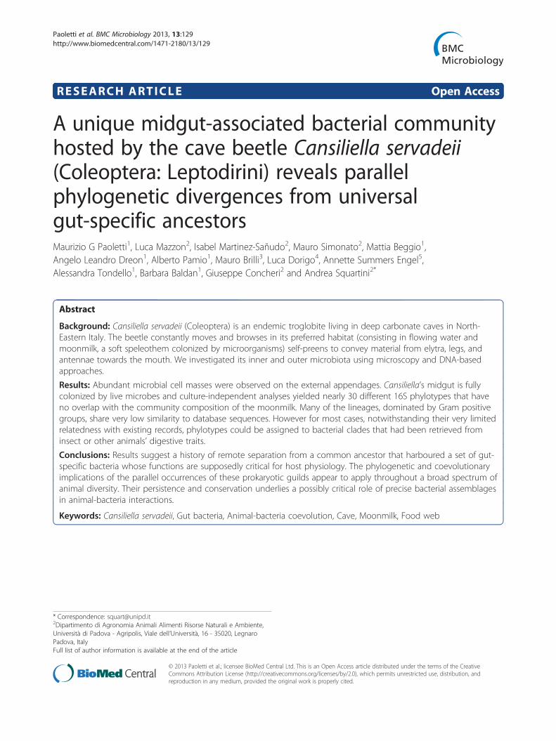
Figure 1 Cansiliella servadeii and its habitat. a) Top view of the adult incoiling; c) insect browsing on moonmilk in Grotta della Foos cave floor. d)and passing it through mouthparts.
been hypothesized to involve oxidative recycling ofnitrogen-rich metabolic waste and are encaged in spe-cialized hindgut- or midgut-derived pouches. Stinkbugshost Burkholderia in their midgut crypts [20,21], whilethe medicinal leech carries Aeromonas and a member ofthe Rickenellaceae in its intestinal assemblage [22,23].For invertebrates that permanently live in secluded habi-
tats with little exchange with the external biota, such ascave environments, the importance of microsymbionts canbe particularly critical for host adaptation and survival.Some cave-dwelling animals owe their life to symbioseswith chemolithoautotrophic bacteria [24,25]. We previouslydescribed a novel genus and two species of a troglobiticbeetle, Cansiliella tonielloi [26,27] and Cansiliella servadeii(Figure 1a) [28], which are endemic of few karst caves inNorthern Italy. The latter has been the object of more de-tailed studies [29,30], where we further described thephysico-chemical features of its environment.The beetles live in a hygropetric habitat in the pres-
ence of a peculiar, soft speleothem called moonmilk,which consists of carbonate minerals that are constantlycovered by a thin layer of running water [31]. Thishabitat type is common in air-filled caves, and is typifiedby dripwaters or sheetflow that bring allochthonous,surface-derived organic matter [32]. Hydrological isola-tion for some cave hygropetric habitats may restrict theinflux of organic matter, and this can lead to nutritionallimitations for troglobites and troglophiles over extended
sect. b) detail of the abdomen with indication of the gut position andsequence showing C.servadeii on location, preening its left antenna
Paoletti et al. BMC Microbiology 2013, 13:129 Page 3 of 16http://www.biomedcentral.com/1471-2180/13/129
periods of time and be a major driver for evolutionaryadaptation for troglobites [32].Moonmilk usually carries high amounts of microbial
biomass [33-38]. In the Grotta della Foos, one of thecave systems being studied, the wet moonmilk contains~108 microbial cells/ml and ~104 meiofaunal cells/m2
and its bacterial community characterization is describedin a parallel study of ours [39]. The insect spends mostof its time browsing the moonmilk surface andfrequently self-preening. Videos of live C. servadeiiin Grotta della Foos (http://www.youtube.com/watch?v=iXF5pDrF2J0) were taken, and its activities and be-haviour were recorded. The mouthparts are consistentwith reported models of adaptation for browsing/filter-ing organic particles in semi-aquatic environments[40], and differ markedly from those of the majority ofother troglobitic Leptodirini [32,41-43]. The mouth-parts have modified hoe-shaped mandibles and spoon-like galeae covered by dense setae spaced 1–1.5 μm.This distance could effectively rake particles ofcompatible size, such as bacteria [29,30].Our previous stable isotope investigations [30] demon-
strated that C. servadeii derives its nutritional require-ments from the moonmilk and from dissolved organicmatter in the percolating waters. To our knowledge,there are no molecular studies of the gut microbiota ofcave invertebrates. The current project aimed at charac-terizing the feeding behaviour of C. servadeii fromGrotta della Foos and the nature of its gut microbiota.The results provided insights pointing towards the exist-ence of a universal guild of bacteria which appears to becommon to many animal digestive systems and that sug-gests to have shared ancestors established prior to theirhosts evolution.
MethodsSampling site, specimen observation and collectionThe Grotta della Foos cave system formed within MonteCiaurlec located in north-eastern Italy, and is underlainby Cretaceous and Triassic limestone units [44] Thecave contains over 2600 m of passages. Ten sampling lo-cations within the cave were used for the investigationsof behaviour and insect collection. the sites coveredaltogether 13.3, square meters, which is the whole areawhich Cansiliella is regularly found in Grotta de la Fooscave. The density monitored varied from 1.4 to 1.8 spec-imens per square meter. Examined specimen were alladults and included both sexes. Live C. servadeii werecollected in sterile falcon tubes and transported to thelaboratory.
Microscopy, insect dissection, and gut content evaluationInsects external teguments were stained with DAPI (5 μg/ml) and observed in visible light and in epifluorescence
using a Leica DM4000 inverted microscope equipped witha DFC300 FX camera. Images were acquired by using theLAS software.Insects were dissected to remove the midgut to
analyze the intestinal microflora. Before dissection, spec-imens were stunned by keeping vials at 4°C for 20 min.To extract the midgut, the insect’s abdomen was openedunder a stereomicroscope (Figure 1b) in a laminar flowhood using sterile equipment and sterile water. The mid-gut was transferred in a sterile Eppendorf tube and usedfor both bacterial culturability tests and bacterial DNAextraction and amplification, and was stored at −20°Cuntil extraction.A segment of each midgut was observed under mi-
croscopy after staining with the LIVE/DEAD® BacLightBacterial Viability Kit (Molecular Probes, California,USA). Slides were also prepared for Gram staining andmorphological characterization, which was performedunder an Olympus BX60 microscope.
Bacterial cultivationIn order to examine external bacteria adhering to the in-sect exoskeletal tegument, live specimens collected withcave water in falcon tubes were handled with sterile for-ceps and gently touched over the surface of Plate CountAgar (PCA) (Oxoid) plates.The possible culturability of the microorganisms
hosted in the insect midgut was verified by plating ali-quots of resuspended, dissected gut material extractedonto PCA plates.All plates were incubated in the dark at 20°C for up to
10 days.
DNA extraction, 16S rRNA gene amplification, cloning,and sequencingDNA was extracted from the content of the midguts, aspreviously described [45]. PCR amplification targetingthe 16S rRNA gene was carried out in 20 μl, 1x PCRGoTaqFlexi Buffer (Promega), 2.5 mM MgCl2, 0.1 mMdNTPs, 0.5 μM of each primer, 1 U of GoTaq FlexiDNA polymerase (Promega), and 1 μl of a 1:30 dilutionof the DNA extraction. The universal bacterial 16SrRNA primers used were 63f and 1389r [46] to yield anexpected amplicon of ~1300 bp. The cycling programconsisted of a 95°C 2 min step followed by 35 cycles at96°C for 30 s, 56°C for 30 s, 72°C for 90 s, and a final ex-tension at 72°C for 10 min. PCR products were checkedby 1.0% agarose gel stained with SYBR®Safe (Invitrogen)and purified with the ExoSAP-IT kit (Amersham Biosci-ences) before sequencing.Amplicons (1300 bp) were cloned into JM109 compe-
tent cells using the P-GEM-T Easy vectors (Promega),following the manufacturer’s recommendations. Thirtyclones from each of the three gut specimen samples
Paoletti et al. BMC Microbiology 2013, 13:129 Page 4 of 16http://www.biomedcentral.com/1471-2180/13/129
were picked. Transformation was verified using PCRassays with the M13-T7 universal primers pair. Theamplification products were sequenced by capillaryelectrophoretic sanger sequencing using M13 and T7primers at the BMR Genomics service (Padova, Italy).Restriction enzyme (BsaI, Euroclone) digestion patternsof the amplified 16S gene (ARDRA) were used asa parallel clone screening in addition to nucleotidesequencing.
Sequence analysisSequence chromatograms were visually inspected andsequences were edited and aligned by using MEGA 4.0.2(http://www.megasoftware.net/).Chimeras were searched with the CHIMERA CHECK
program of the Ribosomal Database Project II (http://rdp.cme.msu.edu).A BLASTN GenBank analysis of all the sequences was
done through the NCBI website (http://www.ncbi.nlm.nih.gov/) and closely related sequences from thedatabases were retrieved and added to the alignment.Phylogenetic relationships among newly retrieved gutmicrobiota sequences to close relatives were estimatedusing a maximum likelihood analysis (ML) with a GTR+I+G model.The software package DOTUR [47] was used to assign
sequences to operational taxonomic units (OTUs) forthe bacterial identities found in the midgut of C.servadeii. This program assigns sequences to OTUsbased on sequence data by using values that are lessthan the cut-off level, which were at the 97% and 95%identity thresholds. The Chao1 richness estimator [48]was also calculated using DOTUR. The richness esti-mates are reported for 3% difference between sequences.16S rRNA gene sequences of clones from the guts of
C. servadeii are accessible under numbers JQ308110 toJQ308155 and from JX463074 to JX463100.The sequences from the culturable microbial commu-
nity from the midgut and the external tegument areaccessible under numbers JQ308156 to JQ308165.
ResultsObservations of insect behaviourLive activities were monitored for C. servadeii individ-uals within Grotta della Foos on six different expeditions(Figure 1). Consistent behavioural patterns could bedefined from a continuous 24-hour period from eightspecimens. The insect spends 44% of the time at a depthbetween 4 and 20 mm under the water that flows overthe moonmilk speleothem. During this activity, themouthparts and head are engaged in a prolonged brows-ing to rubbing motion (Figure 1c). Nearly half of thetime was dedicated to self-preening of the head, legs,elytra and antennae; this behaviour is suggestive of a
feeding activity as it moves organic particulates from thebody towards the mouth. Typically, during preening, theinsect passed the posterior legs over the elytra, then themiddle legs brushed the posterior ones, the forelegsbrushed the middle ones, each antenna, and then theforelegs passed between the mandibles and galeae.Antennae were combed for their entire length, as shown bythe consecutive frames of the sequential series (Figure 1d),taken from footage available at http://www.youtube.com/watch?v=iXF5pDrF2J0. The observed aquatic and semi-aquatic movement actively displaced superficial sedimentgranules and disrupted moonmilk into trenches ~0.2 to 3mm long.In support of the hypothesis that the browsing and
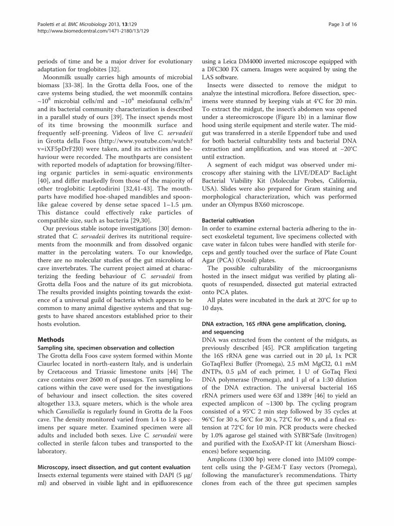
preening activities are related to feeding, possibly to ac-quire organic matter or cellular material from the wetmoonmilk, the DAPI fluorescent stain shows that thehair-covered upper underside and interior legs of the in-sect body parts, that are continuously rubbed duringpreening, are covered by masses of bacteria-containingmaterial (Figure 2). Crawling across the soft moonmilk,and passing the antennae tightly by the mouthparts, asshown by the sequence in Figure 1d, contributes toscooping up abundant organic material visible on theventral segment of the body (Figure 2c).
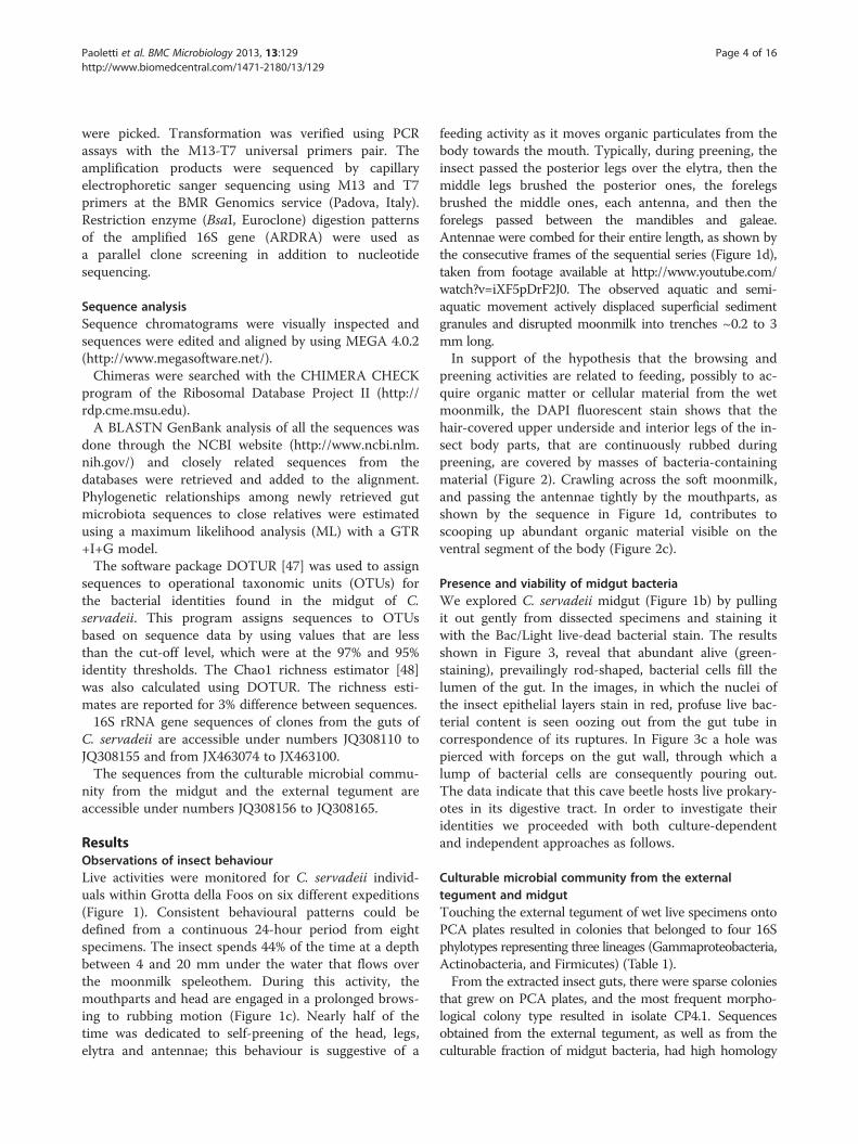
Presence and viability of midgut bacteriaWe explored C. servadeii midgut (Figure 1b) by pullingit out gently from dissected specimens and staining itwith the Bac/Light live-dead bacterial stain. The resultsshown in Figure 3, reveal that abundant alive (green-staining), prevailingly rod-shaped, bacterial cells fill thelumen of the gut. In the images, in which the nuclei ofthe insect epithelial layers stain in red, profuse live bac-terial content is seen oozing out from the gut tube incorrespondence of its ruptures. In Figure 3c a hole waspierced with forceps on the gut wall, through which alump of bacterial cells are consequently pouring out.The data indicate that this cave beetle hosts live prokary-otes in its digestive tract. In order to investigate theiridentities we proceeded with both culture-dependentand independent approaches as follows.
Culturable microbial community from the externaltegument and midgutTouching the external tegument of wet live specimens ontoPCA plates resulted in colonies that belonged to four 16Sphylotypes representing three lineages (Gammaproteobacteria,Actinobacteria, and Firmicutes) (Table 1).From the extracted insect guts, there were sparse colonies
that grew on PCA plates, and the most frequent morpho-logical colony type resulted in isolate CP4.1. Sequencesobtained from the external tegument, as well as from theculturable fraction of midgut bacteria, had high homology

Figure 2 Cansiliella servadeii observation under epifluorescence stereomicroscope after staining with the DNA-specific DAPIfluorochrome. a), c): head and torso view; b), d) detail of foreleg underside. a), b): white illumination; c), d): UV illumination. The presence ofmasses of bacteria staining with DAPI on the insect head, limbs, antennae and ventral side of body is visible. Scale bars: a), c): 250 μm; b), d):50 μm.
Paoletti et al. BMC Microbiology 2013, 13:129 Page 5 of 16http://www.biomedcentral.com/1471-2180/13/129
values (in most cases 99-100%) to bacterial taxa featuringmultiple occurrencies in the nucleotide sequence database(for references consult the GenBank accession numbersgiven in the fourth column of Table 1), although there wasno close affinity to sequences previously retrieved from in-sect guts.
Culture-independent analysis of the midgut microbialcommunityUnder the limitations posed by working with a rare en-demic and protected species with minimum samplingallowed, we analyzed three specimens from which separ-ate clone libraries of 16S rRNA gene amplicons weregenerated and 87 clones screened. Sequences from thethree different guts are labeled with the suffixes A, B,and C, respectively, on Table 2. At this resolution levelthe number of Dotur-defined species was 29 and theChao1 estimator [48] predicted a total number of speciesof 51,7. We also calculated the estimated coverage byapplying the Good’s index [49] which, at species level,resulted 81.6 %. In order to check with an independentmethod whether the sampling size had been truly effect-ive in yielding an adequate representation of the com-munity, we compared the cluster analysis dendrogramobtained with the first 46 clones screened (Additionalfile 1: Material S1 and Additional file 2: Material S2)with those generated with the whole set of 87 (Figures 4and 5), from whose comparison it can be observed thatthe community structure was already fully delineatedfrom the first stepwise subset of randomly selected
clones. Further, considering the phylum rank as a morefunctional assessment of population diversity we run rar-efaction curves with OTUs defined at a phylum levelsimilarity threshold (81%). The result obtained indicateda saturating curve and is shown in the supplementaryAdditional file 3: Figure S3.Phylogenetic analyses revealed the presence of six dis-
tinct major phylogenetic groups from the sequencedclones.The sequences showed a range of homology values
with the GenBank database records that for most caseswas remarkably low (Table 2).Considering the totality of the 87 clones, the Firmicutes
phylum represented 58,6% of all retrieved sequences, andover 60% of the clones showed homologies as low as 92-94% with existing database subjects. Bacteroidetes represented16.1% of the sequences, with homologies 89-94% toGenBank entries. Only few clones of the Actinobacteria(whose phylum represented 11.5% of the retrieved se-quences) displayed similarity values qualifying for specieslevel relatedness (≥97%) with described records.The remainder of the clones were affiliated with
the Deltaproteobacteria (8.0%) and with the Alpha-and Betaproteobacteria, classes (<5% each). Althoughculturable strains affiliated to the Gammaproteobacteriawere obtained from the gut (Table 1), no clone sequencesaffiliated with this class were retrieved, presumably due totheir rarity within the total community.The taxonomical groups resulted homogeneously dis-
tributed through the samples analyzed. There was no
Figure 3 BacLight staining of dissected Cansiliella servadeii midgut resuspended material. Live bacterial cells stain in green while insectepithelial nuclei stain in red. In a) clumps of bacteria are seen flowing out from the rupture of the bent gut tract. In b) a different portion isshown and the abundant masses of extracted bacteria. In c) individual bacterial cells are released from the gut epithelium through a hole piercedwith forceps. In d) a region of the gut from which several distinct bacterial cells can be seen along with others in more clustered formations.Scale bars: a),b): 350 μm,c),d): 50 μm.
Paoletti et al. BMC Microbiology 2013, 13:129 Page 6 of 16http://www.biomedcentral.com/1471-2180/13/129
statistical difference in the distribution of the phylogen-etic groups of Firmicutes, Actinobacteria, Proteobacteriaand Bacteroidetes from the different midgut samples(Fisher’s exact test, P = 0.22). All guts had an outstand-ing majority of OTUs belonging to the Firmicutes.Although the BLAST analysis gave similarities that in
most cases were below the species and even genus limit(respectively for the 89.04% and 63% of the samples),nevertheless the best matches of a vast majority ofclones corresponded to bacteria occurring in differentinsects gut, including ants, termites, and beetles
Table 1 Taxonomical assignment based on 16S rRNA gene seexoskeleton of Cansiliella servadeii (non-surface sterilized spespecimens)
Taxonomy Isolate, GenBank code Top databas
Tegument γ-Proteobacteria InGrP, (JQ308165) (100) Pseudom
Actinobacteria InGrG, (JQ3081649) (99.4) Streptom
Actinobacteria InGrA3, (JQ308163) (99.4) Rhodoc
Firmicutes InGrA1, (JQ308162) (96.8) Unc.bac
Midgut γ-Proteobacteria CP1a, (JQ308158) (100) Pseudom
γ-Proteobacteria CP1b, CP2b, (JQ308159) (100) Pseudom
Actinobacteria CP2a, CP3aL, (JQ308160) (100) Streptom
γ-Proteobacteria CP3a, (JQ308161) (100) Unc. Pse
Firmicutes CP4.1, CP4.2, (JQ308156) (99.4) Unc. Fir
Firmicutes CP4.3, (JQ308157) (98.6) Unc.bac1Description of GenBank subjects displaying the top-scoring BLAST alignment resul2Animal host or other environment in which the subject having homology with the
(Table 2). It is worth adding that more than 80% of thesehosts spend at least part of their life cycle in the soil,and ~46% of them belong to the Coleoptera order(Carabidae, Scarabaeidae and Geotrupidae). Another keyfinding is the fact that groups of taxonomically distinctclones from C. servadeii have their respective GenBankmatches in sequences that were found also in the sameinsect host species. For example, three non-identicalClostridiales clones are closely related to three differentbacteria that all come from the coleopteran Pachnodaepipphiata, [50] which also hosts the closest relatives to
quencing of culturable isolates from the externalcimens) or from its midgut content (surface-sterilized
e similarities (%)1 Habitat of subject2
onas sp. EU182834 Soil
yces sp. JF292927 Endophyte in Lobularia sp.
occussp. HQ256783 Cloud water from mountain summit
terium JF107304 Human skin, antecubital fossa
onas sp. AB569967 Chitinolitic biota in rhizosphere soil
onas sp. AJ243602 Lumbricus rubellus gut (Annelida)
yces champavatii HQ143637 Soil
udomonas sp. JF500897 Rye grass rhizosphere
micutes EU005283 Inert surfaces immersed in marine water
terium DQ860054 Anchovy intestinal microflora
ts of sequence similarity.present sequence s described in GenBank records.
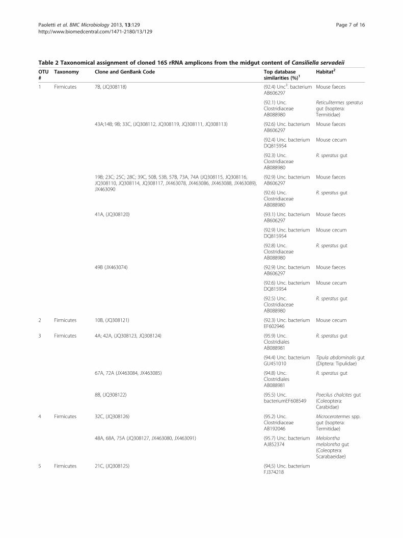
Table 2 Taxonomical assignment of cloned 16S rRNA amplicons from the midgut content of Cansiliella servadeii
OTU#
Taxonomy Clone and GenBank Code Top databasesimilarities (%)1
Habitat2
1 Firmicutes 7B, (JQ308118) (92.4) Unc3. bacteriumAB606297
Mouse faeces
(92.1) Unc.ClostridiaceaeAB088980
Reticulitermes speratusgut (Isoptera:Termitidae)
43A;14B; 9B; 33C, (JQ308112, JQ308119, JQ308111, JQ308113) (92.6) Unc. bacteriumAB606297
Mouse faeces
(92.4) Unc. bacteriumDQ815954
Mouse cecum
(92.3) Unc.ClostridiaceaeAB088980
R. speratus gut
19B; 23C; 25C; 28C; 39C, 50B, 53B, 57B, 73A, 74A (JQ308115, JQ308116,JQ308110, JQ308114, JQ308117, JX463078, JX463086, JX463088, JX463089),JX463090
(92.9) Unc. bacteriumAB606297
Mouse faeces
(92.6) Unc.ClostridiaceaeAB088980
R. speratus gut
41A, (JQ308120) (93.1) Unc. bacteriumAB606297
Mouse faeces
(92.9) Unc. bacteriumDQ815954
Mouse cecum
(92.8) Unc.ClostridiaceaeAB088980
R. speratus gut
49B (JX463074) (92.9) Unc. bacteriumAB606297
Mouse faeces
(92.6) Unc. bacteriumDQ815954
Mouse cecum
(92.5) Unc.ClostridiaceaeAB088980
R. speratus gut
2 Firmicutes 10B, (JQ308121) (92.3) Unc. bacteriumEF602946
Mouse cecum
3 Firmicutes 4A; 42A, (JQ308123, JQ308124) (95.9) Unc.ClostridialesAB088981
R. speratus gut
(94.4) Unc. bacteriumGU451010
Tipula abdominalis gut(Diptera: Tipulidae)
67A, 72A (JX463084, JX463085) (94.8) Unc.ClostridialesAB088981
R. speratus gut
8B, (JQ308122) (95.5) Unc.bacteriumEF608549
Poecilus chalcites gut(Coleoptera:Carabidae)
4 Firmicutes 32C, (JQ308126) (95.2) Unc.ClostridiaceaeAB192046
Microcerotermes spp.gut (Isoptera:Termitidae)
48A, 68A, 75A (JQ308127, JX463080, JX463091) (95.7) Unc. bacteriumAJ852374
Melolonthamelolontha gut(Coleoptera:Scarabaeidae)
5 Firmicutes 21C, (JQ308125) (94,5) Unc. bacteriumFJ374218
Paoletti et al. BMC Microbiology 2013, 13:129 Page 7 of 16http://www.biomedcentral.com/1471-2180/13/129
Table 2 Taxonomical assignment of cloned 16S rRNA amplicons from the midgut content of Cansiliella servadeii(Continued)
Pachnoda spp. gut(Coleoptera:Scarabaeidae)
6 Firmicutes 2A;12B, (JQ308128, JQ308129) (97.1) Unc.ClostridiaceaeAB192046
Microcerotermes spp.gut (Isoptera:Termitidae)
6B, (JQ308130) (96.9) Unc. bacteriumFJ374218
Pachnoda spp. larvalgut (Coleoptera:Scarabaeidae)
46A, 63A (JQ308131, JX463079) (94.5) Unc. bacteriumFJ374218
Pachnoda spp. gut(Coleoptera:Scarabaeidae)
7 Firmicutes 15B, (JQ308133) (91.7) Unc. bacteriumEU465991
African elephantfaeces
(90.5) Unc. bacteriumAY654956
Chicken gut
29C, (JQ308132) (91.9) Unc. bacteriumEU465991
African elephantfaeces
(90.7) Unc. bacteriumAY654956
Chicken gut
8 Firmicutes 5A, (JQ308134) (93.8) Unc.ClostridialesAB231035
Hodotermopsissjoestedti gut (Isoptera:Termitidae)
9 Firmicutes 69A (JX463081) (94.7) Unc. bacteriumAB088973
R. speratus gut
10 Firmicutes 71A(JX463087) (92.7) Unc. bacteriumAB088973
R. speratus gut
11 Firmicutes 24C, 30C, (JQ308135, JQ308136) (92.6) Unc. FirmicutesGQ275112
Leptogenys spp. gut(Hymenoptera:Formicidae)
12 Actinobacteria 61A (JX463076) (93.2) Unc. BacteriumFR687129
Paddy soil
13 Actinobacteria 22C; 36C, 51B, 54B (JQ308137, JQ308138, JX463075, JX463083) (97.2) Unc. bacteriumDQ521505
Lake Vida ice cover
(96.9) Unc. bacteriumAM940404
Rhagium inquisitor gut(Coleoptera:Cerambycidae)
52B (JX463077) (96.7) Unc. bacteriumDQ521505
Lake Vida, ice cover
(96.5) Unc. bacteriumAM940404
Rhagium inquisitor gut(Coleoptera:Cerambycidae)
65A (JX463082) (97) Unc. bacteriumDQ521505
Lake Vida, ice cover
(96.7) Unc. bacteriumAM940404
Rhagium inquisitor gut(Coleoptera:Cerambycidae
14 Actinobacteria 45A, (JQ308139) (99.5) Sanguibacterinulinus HQ326836
Thorectes lusitanicusgut (Coleoptera:Geotrupidae)
15 α-Proteobacteria 13B, (JQ308142) (96.2) Unc.α-proteobacteriumCU920098
Mesophilic anaerobicdigester treatingwastewater sludge
(93.7) Unc. bacteriumFN659093
Lumbricus terrestris gut
Paoletti et al. BMC Microbiology 2013, 13:129 Page 8 of 16http://www.biomedcentral.com/1471-2180/13/129
Table 2 Taxonomical assignment of cloned 16S rRNA amplicons from the midgut content of Cansiliella servadeii(Continued)
16 α-Proteobacteria 58B (JX463098) (100) Brevundimonassp.JQ316297
Soil
17 α-Proteobacteria 44A (JQ308143) (92.5) Unc. bacteriumEF667926
Epithelium Hydravulgaris
(88.2) Unc. bacteriumHM779996
Adult zebrafish gut
(87.9) Unc. bacteriumEU148629
Agrilus planipennis gut(Coleoptera:Buprestidae)
18 δ-Proteobacteria 3A; 20A, 62A (JQ308144, JQ308145, JX463096) (94.3) Unc. δ-proteobacteriumDQ307712
Macrotermesmichaelsenigut(Isoptera: Termitidae)
19 δ-Proteobacteria 60B (JX463100) (96) Unc.DesulfovibrionaceaeJN653048
Gut of millipedeTachypodoiulus niger
20 δ-Proteobacteria 66A, 70A (JX463092, JX463093) (94.1) Unc. bacteriumFJ374259
P. ephippiata gut(Coleoptera:Scarabaeidae)
21 β-Proteobacteria 27C, (JQ308141) (95.2) Unc.bacteriumAJ852369
Melolonthamelolontha gut(Coleoptera:Scarabaeidae)
22 β-Proteobacteria 26C, (JQ308140) (96.5) Burkholderialesbacterium EU073950
Dermolepidaalbohirtum gut(Coleoptera:Scarabaeidae)
23 Bacteroidetes 11B, (JQ308146) (91.9) Unc. bacteriumAJ576327
Pachnoda ephippiatagut (Coleoptera:Scarabaeidae)
18B, (JQ308147) (92.1) Unc. bacteriumHQ728219
Microbial fuel cell
(91.9) Unc. bacteriumAJ576327
P. ephippiata gut(Coleoptera:Scarabaeidae)
24 Bacteroidetes 16B, (JQ308148) (92.5) Unc. bacteriumFJ674429
Cattle feedlot
(91.9) Unc.BacteroidetesAB522123
R. santonensis gut(Isoptera: Termitidae)
(89.2) Unc. bacteriumEF176896
Tipula abdominalis gut(Diptera: Tipulidae)
25 Bacteroidetes 35C, (JQ308149) (96.2) Unc. bacteriumAJ576327
P. ephippiata gut(Coleoptera:Scarabaeidae)
26 Bacteroidetes 64A (JX463097) (94.2) Unc. bacteriumHQ728219
Anode of a glucose-fed microbial fuel cell
(93.7) Unc. bacteriumAJ576361
P. ephippiata gut(Coleoptera:Scarabaeidae)
27 Bacteroidetes 31C, (JQ308150) (93.1) Unc. bacteriumDQ447343
Urban biowaste
(89.3) Elizabethkingiasp. GU45829
R. speratus gut(Isoptera: Termitidae)
Paoletti et al. BMC Microbiology 2013, 13:129 Page 9 of 16http://www.biomedcentral.com/1471-2180/13/129

Table 2 Taxonomical assignment of cloned 16S rRNA amplicons from the midgut content of Cansiliella servadeii(Continued)
40C, (JQ308151) (92.8) Unc. bacteriumDQ447343
Urban biowaste
(89.2) Unc.BacteroidetesHM215036
Bumble bee gut(Hymenoptera:Apidae)
28 Bacteroidetes 17B; 37C; 34C, 59B (JQ308154, JQ308155, JQ308153, JX463099) (94.9) Unc.BacteroidetesDQ837639
Apis mellifera gut(Hymenoptera:Apidae)
55B (JX463095) (94.6) Unc.BacteroidetesDQ837639
Apis mellifera gut(Hymenoptera:Apidae)
56B (JX463094) (94.8) Unc.BacteroidetesDQ837639
Apis mellifera gut(Hymenoptera:Apidae)
29 Bacteroidetes 38C, (JQ308152) (94.3) Unc.BacteroidetesDQ837639
Apis mellifera gut(Hymenoptera:Apidae)
1Description of GenBank subjects displaying the top-scoring BLAST alignment results of sequence similarity.2Animal host or other environment in which the subject having homology with the present sequence is described in GenBank records.3Unc. = ‘Uncultured’.OTUs are defined at 97% similarity threshold. Clones ID are followed by letters A,B or C to identify the three insect guts specimens.
Paoletti et al. BMC Microbiology 2013, 13:129 Page 10 of 16http://www.biomedcentral.com/1471-2180/13/129
some of the Bacteroidetes clones (Table 2). Also, closestsequences to the clones affiliated with the Clostridialesand some Proteobacteria have been retrieved from thegut of the Melolontha melolontha beetle, while severalClostridiaceae clones and one Bacteroidetes clone wereclosely related to sequences that were all retrievedfrom the same dipteran host Tipula abdominalis gut(GU451010). Given the low homologies and the recur-ring multiple instances it appears highly unlikely thatthese occurrences could be coincidental, constituting asignificant element in favour of distant but conservedhost-bacteria interactive relationships, in which givensubsets of bacterial taxa seem to co-occur in a numberof parallel situations hosted by very different insects.In order to better visualize the distribution of bacterial
phyla found in C. servadeii along with that of the hosts/habitats where their closest GenBank relatives had beenfound, in Figure 6 we plotted these across the span of16S homology at which the BLAST match was found foreach clone or isolate. Interestingly, for the midgutclones, the identity levels show a bimodal distribution.Figure 6a shows the distribution of the bacterialtaxonomical divisions found within Cansiliella’s gut as-semblages. When the same are inspected as regards thehabitat of the nearest database subject (Figure 6b), a dis-tinction arises separating the insect-related cases (higherhomology region, peaking at 95%) from the rest of non-insect environments including mammal guts/faeces, etc.,(more distant homology region peaking at 93%). Thetwo peaks (93% and 95%) are significantly different(Wilcoxon Mann–Whitney test, p<0.01) (Figure 6b). Thefraction of culturable bacteria instead (Figure 6c)
displays high levels of similarity shared in all cases withnon-insect GenBank subjects.
DiscussionCansiliella spp. mouthparts are distinct from other cavebeetles, in general and from the large majority of theLeptodirini, and show features uncommon to beetleswith more saprophagous diets [28]. The beetles inGrotta della Foos have also a semi-aquatic lifestyle asso-ciated with moonmilk, which is a rich microbiologicalsubstrate mixed with carbonate minerals. Our previousstable isotope investigations, and observations of moonmilkparticles in beetle mouths, reveal that C. servadeii fromGrotta della Foos derives nutrition from moonmilk andhabitat waters which contain dissolved organic carbon at aconcentration of 10.11 mg/l [30]. The present data showthat the insect midgut hosts a bacterial community whosemembers, as far as it can be judged from the sequencedclones, appear to belong to heterotrophic guilds. Themidgut of the insect contains live bacterial cells whoseculture-independent analysis yielded a bacterial assemblagedominated by the phyla Firmicutes and featuring presencesof Bacteoridetes, Actinobacteria, together with Alpha-,Beta- and Deltaproteobacteria. A possible role of these bac-teria in nutritional physiology with activities within the ni-trogen metabolism could be postulated on the basis ofparallel examples in other gut systems.The sampling depth proved suitable as this community
structure was already fully outlined in terms of phylaand their proportions from the first round of 46 clones.Upon nearly doubling the number, the whole set of87 clones maintained the same pattern as the new
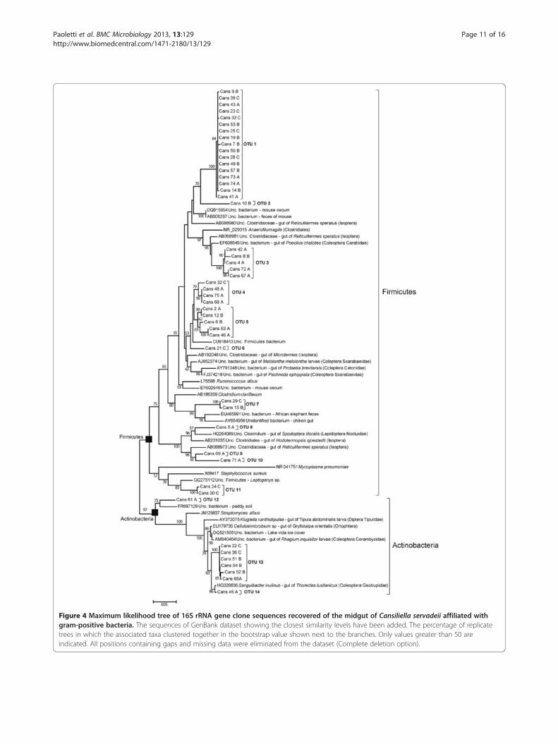
Figure 4 Maximum likelihood tree of 16S rRNA gene clone sequences recovered of the midgut of Cansiliella servadeii affiliated withgram-positive bacteria. The sequences of GenBank dataset showing the closest similarity levels have been added. The percentage of replicatetrees in which the associated taxa clustered together in the bootstrap value shown next to the branches. Only values greater than 50 areindicated. All positions containing gaps and missing data were eliminated from the dataset (Complete deletion option).
Paoletti et al. BMC Microbiology 2013, 13:129 Page 11 of 16http://www.biomedcentral.com/1471-2180/13/129
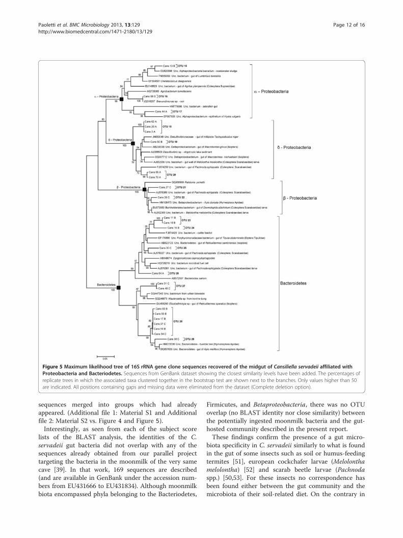
Figure 5 Maximum likelihood tree of 16S rRNA gene clone sequences recovered of the midgut of Cansiliella servadeii affiliated withProteobacteria and Bacteriodetes. Sequences from GenBank dataset showing the closest similarity levels have been added. The percentages ofreplicate trees in which the associated taxa clustered together in the bootstrap test are shown next to the branches. Only values higher than 50are indicated. All positions containing gaps and missing data were eliminated from the dataset (Complete deletion option).
Paoletti et al. BMC Microbiology 2013, 13:129 Page 12 of 16http://www.biomedcentral.com/1471-2180/13/129
sequences merged into groups which had alreadyappeared. (Additional file 1: Material S1 and Additionalfile 2: Material S2 vs. Figure 4 and Figure 5).Interestingly, as seen from each of the subject score
lists of the BLAST analysis, the identities of the C.servadeii gut bacteria did not overlap with any of thesequences already obtained from our parallel projecttargeting the bacteria in the moonmilk of the very samecave [39]. In that work, 169 sequences are described(and are available in GenBank under the accession num-bers from EU431666 to EU431834). Although moonmilkbiota encompassed phyla belonging to the Bacteriodetes,
Firmicutes, and Betaproteobacteria, there was no OTUoverlap (no BLAST identity nor close similarity) betweenthe potentially ingested moonmilk bacteria and the gut-hosted community described in the present report.These findings confirm the presence of a gut micro-
biota specificity in C. servadeii similarly to what is foundin the gut of some insects such as soil or humus-feedingtermites [51], european cockchafer larvae (Melolonthamelolontha) [52] and scarab beetle larvae (Pachnodaspp.) [50,53]. For these insects no correspondence hasbeen found either between the gut community and themicrobiota of their soil-related diet. On the contrary in

Figure 6 Phylotype and host partitioning in GenBank subjects with similarity to Cansiliella-associated bacteria. a) Abundance of 16SrDNA phylotypes found from the midgut using a culture-independent approach and respective GenBank homology percentage classes. b)Proportions of insects orders or other environments hosting bacterial subjects resulting in different degrees of sequence homology (x axis) withclones of the non-culturable microbial community from the midgut. The smaller diagram in the upper right corner shows the same data as linegraphs and by pooling the insect orders together to put in evidence the separation from the cases found in non-insect environments. c)Proportions of insects orders or other environments hosting bacterial subjects resulting in different degrees of sequence homology (x axis) withculturable microbial community isolates from the midgut and external tegument. The definition ‘other’ includes all non-insect guts, faeces, andother habitats as reported in Table 2.
Paoletti et al. BMC Microbiology 2013, 13:129 Page 13 of 16http://www.biomedcentral.com/1471-2180/13/129
Paoletti et al. BMC Microbiology 2013, 13:129 Page 14 of 16http://www.biomedcentral.com/1471-2180/13/129
insects having a more diverse and richer diet such ascrickets and cockroaches higher correspondence be-tween diet and gut bacterial flora has been identified inculture-dependent studies [54,55].While the uncultured clone library community had
such far divergence from known database entries, theculturable bacteria isolated from external tegument andmidgut showed a much higher sequence similarity topreviously retrieved sequences available in GenBank.Approximately 86% of these sequences have close orequal to 100% sequence similarity (average 97%)(Table 1). In contrast, the uncultured gut clone se-quences have lower homology to any previously de-scribed bacterial species or environmental sequences,with some as low as 92% (Table 2, Figure 6). Among thedominant OTUs groups, belonging mostly to Firmicutesand Bacteriodetes phyla, sequence similarity with de-scribed taxa is ~92% and 94%, respectively, which sug-gests novel bacterial lineages at the genus-level, if nothigher taxonomic ranks. Such result is nowadays an un-usual occurrence as the GenBank database contains alarge, ever-expanding number of sequences obtainedfrom many different microbiological environments, andit is therefore no longer common to find such low se-quence homology, especially when working with a set ofseveral different sequences, all of which turned out con-sistently distant from known records. Except for twoclones corresponding to OTU 14 and OTU 16 that show100% identity with the Actinobacteria Sanguibacterinulinus isolated from the gut of Thorectes lusitanicus(Coleoptera Geotrupidae) and Brevundimonas sp. iso-lated from the soil, the rest of the bacterial communitiesisolated from the gut of C. servadeii are highly differentfrom bacteria typical of other gut systems studied untilnow by culture-independent methods.Noteworthy, for a number of different groups of taxo-
nomically distinct bacteria hosted by the cave beetle, theinsect hosting the closest relatives of each case turnedout to be the same (Table 2). For example, the sequencesof given firmicutes, bacteroidetes and betaproteobacteriahappen to have their top matching GenBank subjectsall occurring within the Melolontha scarab. Others,also encompassing different phyla have their relativescoinciding within a coleopteran of the Pachnodagenus, other clusters co-occur in the Dipteran Tipulaabdominalis, others within the termite Reticulitermessperatus. Given the peculiarity of the sequences, theserepeated occurrences appear non-coincidental and sup-port the hypothesis of a selection ensuring the mainten-ance of a given microbial assemblage for a relevantphysiological scope.Because of the semi-aquatic feeding behaviour of C.
servadeii, it has been speculated that its ancestor, likethat of other hygropetric coleopterans, may have been
aquatic [32]. Neverthelesss, considering that the C.servadeii gut microbiota having the highest degrees ofhomology (95-98%) to previously retrieved sequencesfrom invertebrate gut bacteria that spend at least a partof their biological cycle in the soil (Table 2, Figure 4),and mainly to insects belonging to the Isoptera andColeoptera orders, one could in alternative speculatethat the C. servadeii ancestor had a terrestrial origin.However in available databases, bacteria from aquatic in-sects could be still poorly represented to enable a thor-ough assessment in this regard. About these aspects, asurvey of microbial phylotypes from the guts of theother species in the genus, and a barcoding comparisonof the insect genes are envisaged as parts of futureresearch.Considering the evolutionary history of the C.
servadeii and its gut symbiont system, a long history ofseparation from other invertebrates and microorganismsappears to have occurred. At the same time its situationreveals the existence of phylogenetic similarities acrossthe digestive tracts of many different hosts (Table 2). Itis conceivable that there may be a common ancestry in-volving a functional guild of bacteria that has enduredthe host lineage separation, as well as the erosion of se-quence identities, through the paths of independent evo-lution. The dual pattern of homology among clonesequences from gut bacteria in Cansiliella to other in-sects further suggests this scenario (Figure 6b); a pro-gressive phenomenon of divergence from commonancestries is suggested by the double-peaking instance ofhomology existing between C. servadeii’s sequence quer-ies and GenBank subjects, that set the insect-dwellingcases separated from the general intestinal/faecal cases.It is noteworthy that, while the hosts are set apart by se-quence homology thresholds, the taxonomical groups ofthe bacteria found in Cansiliella are rather evenly repre-sented across the different homology span (Figure 6a). Itcan be seen that Firmicutes, Bacteroidetes andProteobacteria are almost equally present throughoutthe sequence similarity gradient, underscoring the needof the whole functional assemblage to be conserved bothin distantly- as well as in recently-diverged hosts. Thisemphasizes a supposedly crucial role of a well-definedset of prokaryotic taxa that appear to have remained incharge within the alimentary tract of animals in spite ofages of separation of their hosts.More recent acquisitions across different hosts appear
to correspond to higher degrees of homology for bacter-ial symbionts, while acquisitions and symbiotic associa-tions that are older would correspond to lower degreesof homology (Figure 6). The evidences depicted inFigure 6 appear to fit the contour of an evolutionarypath of separation of the midgut bacteria from those ofother insects; it appears that matching bacteria that are
Paoletti et al. BMC Microbiology 2013, 13:129 Page 15 of 16http://www.biomedcentral.com/1471-2180/13/129
hosted in other insects (i.e. hosts that are closer toCansiliella) share higher homology with its symbionts(peak at 95%), while those living in animals which areevolutionarily more distant from the beetle, or in otherhabitats, have undergone a correspondingly higher diver-gence from them (peak at 93%). These instances supportthe existence of a group of common ancestors for a setof different bacteria and a history of isolation and coevo-lution within the hosts. The same analysis performedwith the culturable biota isolated from the external tegu-ment or, as a minority, from the midgut, shows the op-posite scenario (Figure 6c) i.e. a high level of similaritywith non-insect environments (Table 1), suggesting thatplate-culturable taxa are also more prone to spread/re-produce and be part of a more diffuse cosmopolitanism.
ConclusionsThe insects hereby examined feature a defined gut com-munity of bacteria suggesting a long history of inherit-ance and a coevolution.with their hosts. Corresponding,but genetically diverged, microbial assortments appearto exist, in parallel, in a series of other animals’ digestivesystems. It appears that the reproductive boundariesarisen between the hosts at their speciation stages, have,at the same pace, prevented the exchange of their gutbacteria. The conservation of these sets of prokaryotictaxa suggests a relevant role in animal physiology.The evidence of such patterns casts light on their biol-
ogy at both physiological and evolutionary scales. Eluci-dating, in future studies, the details of the bacterialtransmission in C. servadeii will offer useful insights tofurther interpret bacterial evolution and the criticalroles of prokaryotes in the animal-microbe interactionsecology.
Additional files
Additional file 1: Cluster analysis dendrogram obtained with thefirst 46 screened clones, Gram-negative portion.
Additional file 2: Cluster analysis dendrogram obtained with thefirst 46 screened clones, Gram-positive portion.
Additional file 3: Rarefaction curve for OTUs defined at 81%similarity.
Competing interestsThe authors declare that they have no competing interests.
Authors’ contributionsMGP defined the whole experimental plan of the research, organized thefieldwork and identified the zoological samples; LM, MS and IMS performedthe gut microscopy and the cloning and sequencing of microbial 16S genesand constructed the phylogeny trees; ALD, AP, MB and LD organized thelogistics of the speleological expedition into the cave, collected the insectsamples and recorded their in-situ behaviour, ASE provided the data ofmicrobial colonization of the cave substrate moonmilk and discussed itssimilarity with the Cansiliella microbiota; AT and BB performed thefluorescent stereomicroscopy detection of bacteria on external appendagesof the insect; GC performed the water chemical analysis of the cave
environment; AS performed the bioinformatical analyses, the microbialecology assessment and wrote the manuscript. All authors read andapproved the final manuscript.
AcknowledgementsThe authors thank Enrico Ruzzier for his collaboration to the present study.
Author details1Dipartimento di Biologia, Università di Padova, via U. Bassi 58/B, 35131Padova, Italy. 2Dipartimento di Agronomia Animali Alimenti Risorse Naturali eAmbiente, Università di Padova - Agripolis, Viale dell’Università, 16 - 35020,Legnaro Padova, Italy. 3Istituto di Geologia Ambientale e Geoingegneria,Dipartimento di Scienze della Terra, Università La Sapienza di Roma, 00185Rome, Italy. 4Natural History Museum, Via Marangoni 39, 33100 Udine, Italy.5Department of Earth and Planetary Sciences, University of Tennessee,Knoxville, TN 37996, USA.
Received: 27 January 2013 Accepted: 28 May 2013Published: 10 June 2013
References1. Buchner P: Endosymbiosis of animals with plant microorganisms. New York:
Interscience Publishers, Inc; 1965.2. Baumann P, Moran NA: Non-cultivable microorganisms from symbiotic
associations of insects and other hosts. Antonie van Leeuwenhoek 1997,72:39–48.
3. Munson MA, Baumann P, Moran NA: Phylogenetic relationships ofendosymbionts of mealybugs (Homoptera: Pseudococcidae) based on16S rDNA sequences. Mol Phylogen Evol 1992, 1:26–30.
4. Clark MA, Baumann L, Munson MA, Baumann P, Campbell BC, Duffus JE,Osborne LS, Moran NA: The eubacterial endosymbionts of whiteflies(Homoptera: Aleyrodoidea) constitute a lineage distinct from theendosymbionts of aphids and mealybugs. Curr Microbiol 1992,25:119–123.
5. Campbell BC, Bragg TS, Turner CE: Phylogeny of symbiotic bacteria of fourweevil species (Coleoptera: Curculionidae) based on analysis of 16Sribosomal DNA. Insect Biochem Mol Biol 1992, 22:415–421.
6. Aksoy S Molecular analysis of the endosymbionts of tsetse flies: 16S rDNAlocus and over-expression of a chaperonin. Insect Mol Biol 1994, 4:23–29.
7. Bandi C, Damiani G, Magrassi L, Gigolo A, Fani R, Sacchi L: Flavobacteria asintracellular symbionts in cockroaches. Proc R Soc Lond B 1994, 257:43–48.
8. Baumann P, Lai C, Baumann L, Rouhbakhsh D, Moran NA, Clark MA:Mutalistic associations of aphid and prokaryotes: biology of the genusBuchnera. Appl Environ Microbiol 1995, 61:1–7.
9. Noda H, Nakashima N, Koizumi M: Phylogenetic position of yeast-likesymbiotes of rice planthoppers based on partial 18S rDNA sequences.Insect Biochem Mol Biol 1995, 25:639–646.
10. Schröder D, Deppisch H, Obermayer M, Krohne G, Stackebrandt E,Hölldobler B, Goebel W, Gross R: Intracellular endosymbiotic bacteria ofCamponotus species (carpenter ants): systematics, evolution andultrastructural analysis. Mol Microbiol 1996, 21:479–489.
11. Capuzzo C, Firrao G, Mazzon L, Squartini A, Girolami V: ‘Candidatus Erwiniadacicola’, a coevolved symbiotic bacterium of the olive fly Bactroceraoleae (Gmelin). Int J Syst Evol Microbiol 2005, 55:1641–1647.
12. Savio C, Mazzon L, Martinez-Sañudo I, Simonato M, Squartini A, Girolami V:Evidence of two lineages of the symbiont "Candidatus Erwinia dacicola"in Italian populations of Bactrocera oleae (Rossi) based on 16S rRNAgene sequence. Int J Syst Evol Microbiol 2011, 72:179–187.
13. Mazzon L, Piscedda A, Simonato M, Martinez-Sañudo I, Squartini A, GirolamiV: Presence of specific symbiotic bacteria in flies of the subfamilyTephritinae (Diptera Tephritidae) and their phylogenetic relationships:proposal of ‘Candidatus Stammerula tephritidis’. Int J Syst Evol Microbiol2008, 58:1277–1287.
14. Mazzon L, Martinez-Sañudo I, Simonato M, Squartini A, Savio C, Girolami V:Phylogenetic relationships between flies of the Tephritinae subfamily(Diptera, Tephritidae) and their symbiotic bacteria. Molecular Phylogeneticsand Evolution 2010, 56:312–326.
15. Mazzon L, Martinez-Sañudo I, Savio C, Simonato M, Squartini A, In:Manipulative Tenants: Bacteria Associated with Arthropods: Stammerula andother symbiotic bacteria within the fruit flies inhabiting Asteraceaeflowerheads. CRC Press: Edited by Zchori-Fein E, Bourtzis K; 2011:89–111.

Paoletti et al. BMC Microbiology 2013, 13:129 Page 16 of 16http://www.biomedcentral.com/1471-2180/13/129
16. Rouhbaksh D, Lai C-Y, von Dohlen CD, Baumann L, Baumann P, Moran NA,Voegtlin DJ: The tryptophan biosynthetic pathway of aphidendosymbionts (Buchnera): genetics and evolution of plasmid-associatedtrpEG within the Aphididae. J Mol Evol 1996, 42:414–421.
17. Russell JA, Moreau CS, Goldman-Huertas B, Fujiwara M, Lohman DJ, PierceNE: Bacterial gut symbionts are tightly linked with the evolution ofherbivory in ants. Proc Nat Acad Sci 2009, 106:21236–21241.
18. van Borm S, Buschinger A, Boomsma JJ, Billen J: Tetraponera ants havegut-symbionts related to nitrogen-fixing symbionts. Proc R Soc Lond B2002, 269:2023–2027.
19. Martinson VG, Danforth BN, Minckley RL, Rueppell O, Tingek S, Moran NA:A simple and distinctive microbiota associated with honey bees andbumble bees. Mol Ecol 2011, 20:619–628.
20. Kikuchi Y, Hosokawa T, Fukatsu T: Specific Developmental Window forEstablishment of an Insect-Microbe Gut Symbiosis. Appl Environ Microbiol2011, 77:4075–4081.
21. Prado SS, Almeida RPP: Phylogenetic Placement of Pentatomid Stink BugGut Symbionts. Curr Microbiol 2009, 58:64–69.
22. Worthen PL, Gode CJ, Graf J: Culture-independent characterization of thedigestive-tract microbiota of the medicinal leech reveals a tripartitesymbiosis. Appl Environ Microbiol 2006, 72:4775–4781.
23. Graf J: Symbiosis of Aeromonas veronii Biovar sobria and Hirudomedicinalis, the Medicinal Leech: a Novel Model for Digestive TractAssociations. Infection and Immunity 1999, 67:1–7.
24. Sârbu SM, Kane TC, Kinkle BK: A chemoautotrophically based caveecosystem. Science 1996, 272:1953–1955.
25. Engel AS, Meisinger DB, Porter ML, Payn RA, Schmid M, Stern LA, SchleiferK-H, Lee NM: Linking phylogenetic and functional diversity to nutrientspiraling in microbial mats from Lower Kane Cave (USA). ISME J 2010,4:98–110.
26. Paoletti MG: Un nuovo Catopide pholeuonoide del Cansiglio (PrealpiCarniche) (Col. Bathysciinae). Boll Mus Civ St Nat Venezia 1972(XXII-XXIII):119––131.
27. Paoletti MG: Notizie sistematiche ed ecologiche su di un nuovointeressante genere del Cansiglio Cansiliella. Suppl Boll Mus Civ S Na.Venezia 1973, 24:81–88.
28. Paoletti MG: Dati aggiuntivi alla conoscenza del genere CansiliellaPaoletti (Col. Bathysciinae). Redia Firenze 1980, 63:67–80.
29. Paoletti MG, Beggio M, Pamio A, Gomiero T, Brilli M, Dreon AL, Toniello V,Engel AS: Comparison of three moonmilk cave habitats associated withtroglobitic beetles. In Proc 15th Int Cong Speleol. 1st edition. Edited byWhite WB. Kerrville, Texas; 2009:400–403.
30. Paoletti MG, Beggio M, Dreon AL, Pamio A, Gomiero T, Brilli M, Dorigo L,Concheri G, Squartini A, Engel AS: A new foodweb based on microbes incalcitic caves: The Cansiliella (Beetles) case in northern Italy. Int J Speleol2011, 40:45–52.
31. Hill CA, Forti P: Cave Minerals of the World. Huntsville, Alabama: NationalSpeleological Society; 1997:446.
32. Sket B: The cave hygropetric - a little known habitat and its inhabitants.Arch Hydrobiol 2004, 160:413–425.
33. Borsato A, Frisia S, Jones B, van der Borg K: Calcite moonmilk: crystalmorphology and environment of formation in caves in the Italian Alps.J Sediment Res 2000, 70:1179–1190.
34. Northup DE, Dahm CN, Melim LA, Crossey LJ, Lavoie KH, Mallory L, BostonPJ, Cunningham KI, Barn SM: Evidence for geomicrobiological interactionsin Guadalupe caves. J Cave Karst Stud 2000, 62:80–90.
35. Northup DE, Lavoie K: Geomicrobiology of caves: a review. Geomicrobiol J2001, 18:199–222.
36. Mulec J, Zalar P, Zupan–Hajna N, Rupnik M: Screening for culturablemicroorganisms from cave environments (Slovenia). Acta Carsologica2002, 31:177–187.
37. Cañaveras JC, Cuezva S, Sanchez-Moral S, Lario J, Laiz L, Gonzalez JM,Saiz-Jimenez C: On the origin of fiber calcite crystals in moonmilkdeposits. Naturwissenschaften 2006, 93:27–32.
38. Blyth AJ, Frisia S: Molecular evidence for bacterial mediation of calciteformation in cold high-altitude caves. Geomicrobiol J 2008, 25:101–111.
39. Engel AS, Paoletti MG, Beggio M, Dorigo L, Pamio A, Gomiero T, Furlan C,Brilli M, Dreon AL, Bertoni R, Squartini A: Comparative microbialcommunity composition from secondary 1 carbonate (moonmilk)deposits: implications for the Cansiliella servadeii cave hygropetric foodweb. International Journal of Speleology 2013. in press.
40. Moldovan OT, Jalzic B, Erichsen E: Adaptation of the mouthparts in somesubterranean Cholevinae (Coleoptera, Leiodidae). Nat Coroat 2004,13:1–18.
41. Jeannel R: Monographie des Bathsyciinae. Arch Zool Exp Gén 1924,63:1–436.
42. Remy P: Sur le mode de vie des Hadesia dans la grotte Vjetrenica. RevFrance Entomol 1940, 7:1–8.
43. Giachino PM, Vailati D: Kircheria beroni, a new genus and new species ofsubterranean hygropetricolous Leptodirinae from Albania. SubterraneanBiol 2006, 4:103–116.
44. Gasparo F: La grotta della Foos presso Campone (Prealpi Carniche).Mondo Sotterraneo 1971, 1:37–52.
45. Palmano S, Firrao G, Locci R: Sequence analysis of domains III and IV ofthe 23S rRNA gene of verticillate streptomycetes. Int J Syst Evol Microbiol2000, 50:1187–1191.
46. Osborn AM, Moore ERB, Timmis KN: An evaluation of terminal-restrictionfragment length polymorphism (T-RFLP) analysis for the study ofmicrobial community structure dynamics. Environ Microbiol 2000, 2:39–50.
47. Schloss PD, Handelsman J: Introducing DOTUR, a computer program fordefining operational taxonomic units and estimating species richness.Appl Environ Microbiol 2005, 71:1501–1506.
48. Chao A: Non-parametric estimation of the classes in a population. ScandJ Stat 1984, 11:265–270.
49. Magurran AE: Measuring biological diversity. Oxford, UK: Blackwell Publishing;2004:256.
50. Andert J, Marten A, Brandl R, Brune A: Inter- and intraspecific comparisonof the bacterial assemblages in the hindgut of humivorous scarab beetlelarvae (Pachnoda spp.). FEMS Microbiol. Ecol. 2010, 74:439–449.
51. Schmitt-Wagner D, Friedrich MW, Wagner B, Brune A: Phylogeneticdiversity, abundance, and axial distribution of bacteria in the intestinaltract of two soil-feeding termites (Cubitermes spp.). Appl Environ Microbiol2003, 69:6007–6017.
52. Egert M, Stingl U, Dyhrberg Bruun L, Pommerenke B, Brune A, Friedrich MW:Structure and topology of microbial communities in the major gutcompartments of Melolontha melolontha larvae (Coleoptera:Scarabaeidae). Appl Environ Microbiol 2005, 71:4556–4566.
53. Egert M, Wagner B, Lemke T, Brune A, Friedrich MW: Microbial communitystructure in midgut and hindgut of the humus-feeding larva ofPachnoda ephippiata (Coleoptera: Scarabaeidae). Appl Environ Microbiol2003, 69:6659–6668.
54. Kane MD: Breznak JA Effect of host diet on production of organic acids andmethane by cockroach gut bacteria Appl Environ Microbiol 1991,57:2628–2634.
55. Santo-Domingo JW, Kaufman MG, Klug MJ, Holben WE, Haris D, Tiedje JM:Influence of diet on the structure and function of the bacterial hindgutcommunity of crickets. Mol Ecol 1998, 7:761–767.
doi:10.1186/1471-2180-13-129Cite this article as: Paoletti et al.: A unique midgut-associated bacterialcommunity hosted by the cave beetle Cansiliella servadeii (Coleoptera:Leptodirini) reveals parallel phylogenetic divergences from universalgut-specific ancestors. BMC Microbiology 2013 13:129.
Submit your next manuscript to BioMed Centraland take full advantage of:
• Convenient online submission
• Thorough peer review
• No space constraints or color figure charges
• Immediate publication on acceptance
• Inclusion in PubMed, CAS, Scopus and Google Scholar
• Research which is freely available for redistribution
Submit your manuscript at www.biomedcentral.com/submit