aaomp case challenge: a nonulcerated, slowly … · the following case challenge is provided by the...
TRANSCRIPT
1The Journal of Contemporary Dental Practice Volume 10 No 5 September 1 2009copy2009 Seer Publishing LLC
AAOMP CASE CHALLENGE A Nonulcerated Slowly Growing Mass of the MandibleSiema A Eljack DMD Renee Reich DDS
Case Summary
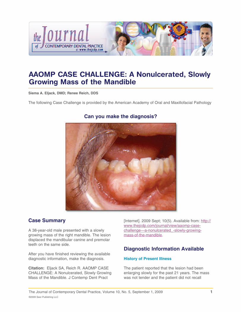
A 38-year-old male presented with a slowly growing mass of the right mandible The lesion displaced the mandibular canine and premolar teeth on the same side
After you have finished reviewing the available diagnostic information make the diagnosis
Citation Eljack SA Reich R AAOMP CASE CHALLENGE A Nonulcerated Slowly Growing Mass of the Mandible J Contemp Dent Pract
The following Case Challenge is provided by the American Academy of Oral and Maxillofacial Pathology
Can you make the diagnosis
[Internet] 2009 Sept 10(5) Available from httpwwwthejcdpcomjournalviewaaomp-case-challenge---a-nonulcerated_-slowly-growing-mass-of-the-mandible
Diagnostic Information Available
History of Present Illness
The patient reported that the lesion had been enlarging slowly for the past 21 years The mass was not tender and the patient did not recall
2The Journal of Contemporary Dental Practice Volume 10 No 5 September 1 2009copy2009 Seer Publishing LLC
any history of trauma No other information was available
Medical History
The patientrsquos medical history was unremarkable He denied alcohol and tobacco use
Clinical Findings
The clinical exam revealed a 3times2 cm ldquobony hardrdquo swelling of the right mandible buccal to 27 and 28 The overlying mucosa was intact and no lingual extension was noted (Figure 1) The remainder of the oral cavity was within normal limits
Radiographic Findings
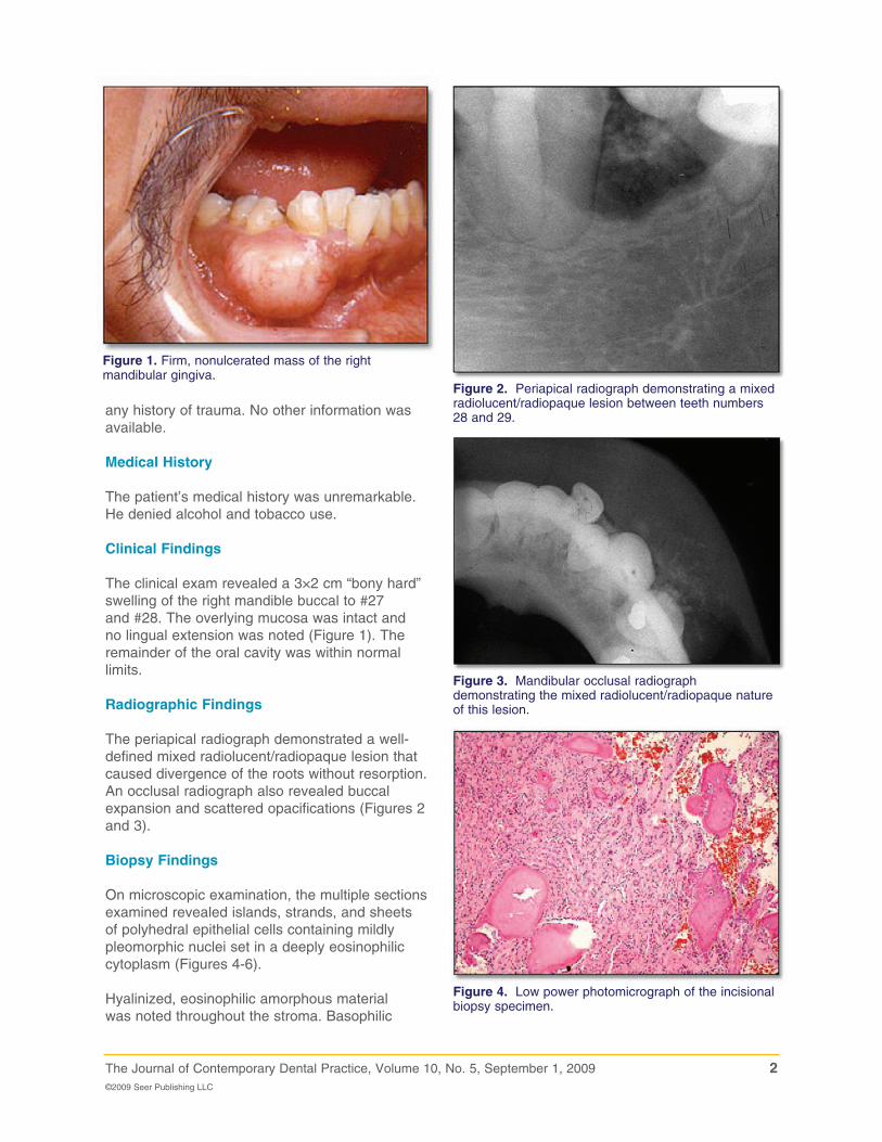
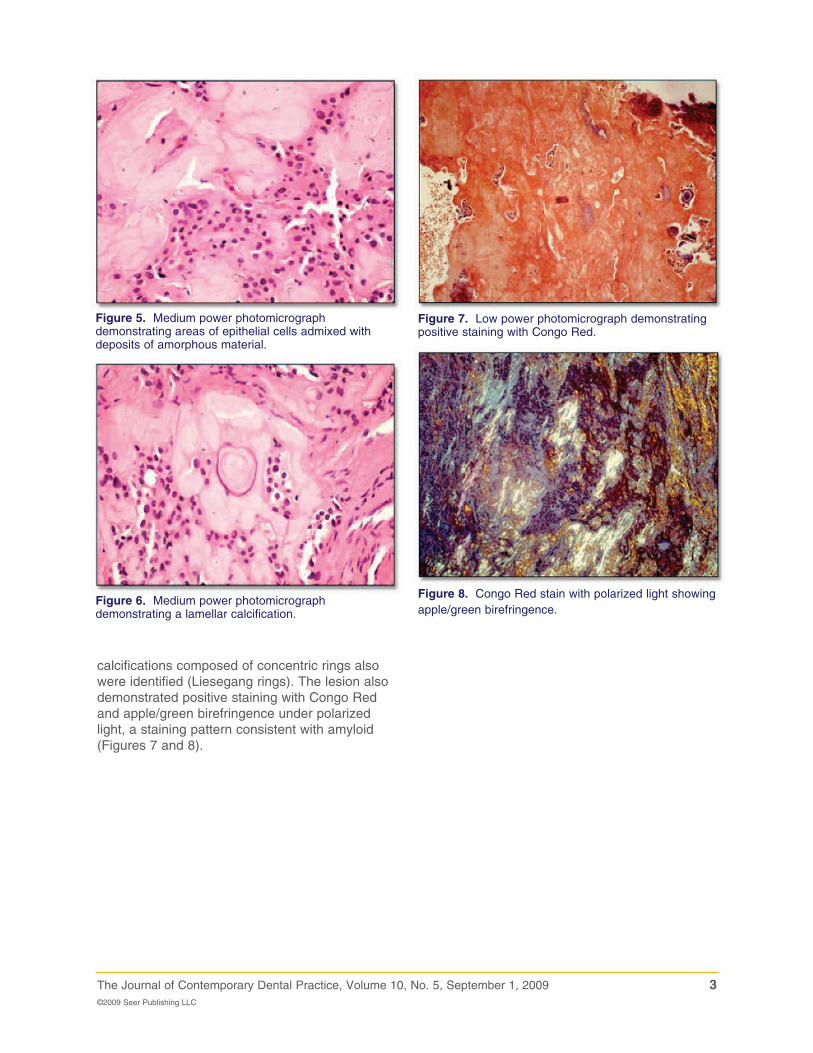
The periapical radiograph demonstrated a well-defined mixed radiolucentradiopaque lesion that caused divergence of the roots without resorption An occlusal radiograph also revealed buccal expansion and scattered opacifications (Figures 2 and 3)
Biopsy Findings
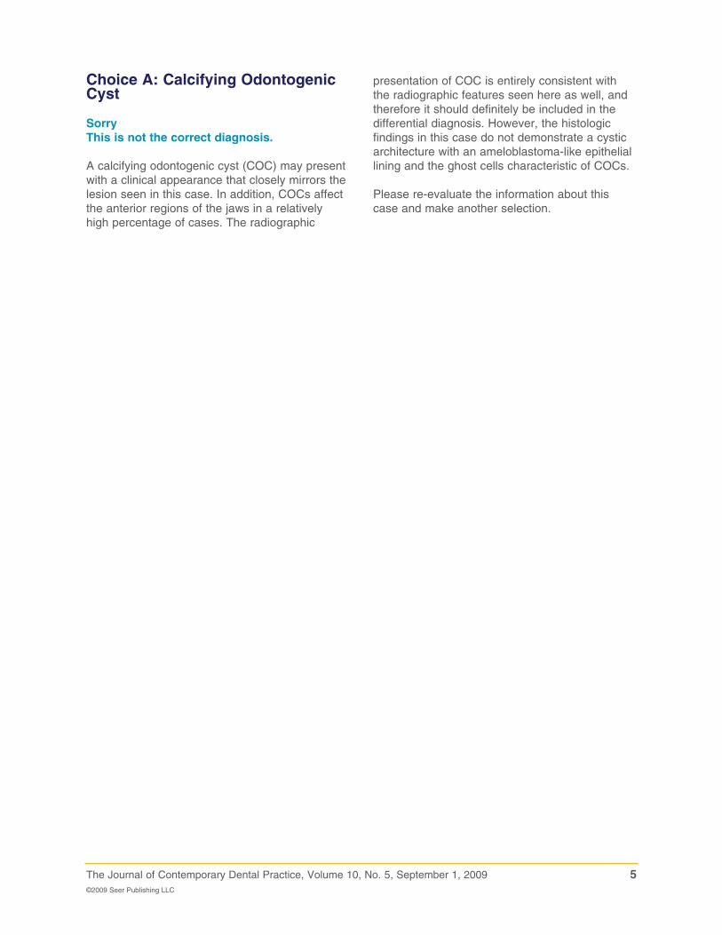
On microscopic examination the multiple sections examined revealed islands strands and sheets of polyhedral epithelial cells containing mildly pleomorphic nuclei set in a deeply eosinophilic cytoplasm (Figures 4-6)
Hyalinized eosinophilic amorphous material was noted throughout the stroma Basophilic
Figure 2 Periapical radiograph demonstrating a mixed radiolucentradiopaque lesion between teeth numbers 28 and 29
Figure 1 Firm nonulcerated mass of the right mandibular gingiva
Figure 3 Mandibular occlusal radiograph demonstrating the mixed radiolucentradiopaque nature of this lesion
Figure 4 Low power photomicrograph of the incisional biopsy specimen
3The Journal of Contemporary Dental Practice Volume 10 No 5 September 1 2009copy2009 Seer Publishing LLC
calcifications composed of concentric rings also were identified (Liesegang rings) The lesion also demonstrated positive staining with Congo Red and applegreen birefringence under polarized light a staining pattern consistent with amyloid (Figures 7 and 8)
Figure 5 Medium power photomicrograph demonstrating areas of epithelial cells admixed with deposits of amorphous material
Figure 6 Medium power photomicrograph demonstrating a lamellar calcification
Figure 7 Low power photomicrograph demonstrating positive staining with Congo Red
Figure 8 Congo Red stain with polarized light showing applegreen birefringence
4The Journal of Contemporary Dental Practice Volume 10 No 5 September 1 2009copy2009 Seer Publishing LLC
Can you make the diagnosis
Select the correct diagnosis
A Calcifying Odontogenic Cyst (COC)B Calcifying Epithelial Odontogenic Tumor
(CEOTC Cemento-Ossifying Fibroma (COF)D Osteosarcoma
5The Journal of Contemporary Dental Practice Volume 10 No 5 September 1 2009copy2009 Seer Publishing LLC
presentation of COC is entirely consistent with the radiographic features seen here as well and therefore it should definitely be included in the differential diagnosis However the histologic findings in this case do not demonstrate a cystic architecture with an ameloblastoma-like epithelial lining and the ghost cells characteristic of COCs
Please re-evaluate the information about this case and make another selection
Choice A Calcifying Odontogenic Cyst
SorryThis is not the correct diagnosis
A calcifying odontogenic cyst (COC) may present with a clinical appearance that closely mirrors the lesion seen in this case In addition COCs affect the anterior regions of the jaws in a relatively high percentage of cases The radiographic
6The Journal of Contemporary Dental Practice Volume 10 No 5 September 1 2009copy2009 Seer Publishing LLC
polymorphous squamous cells often showing intercellular bridges1 Variation in nuclear size shape and hyperchromaticity are often noted but do not indicate malignancy Mitoses are very uncommon3
A characteristic feature of the CEOT is the presence of an amorphous eosinophilic extracellular material The precise nature of this material is still uncertain but is thought to be a form of amyloid which results from degradation of lamina densa material (basal lamina) secreted by the tumor epithelial cells4 It generally stains as amyloid (ie positive staining with Congo Red) and will exhibit apple-green birefringence when viewed with polarized light5 A pattern of calcification in a droplet-like fashion often showing appositional basophilic concentric rings (Lisegang rings) is common but not always present6
CEOT does not appear to be an aggressive lesion and conservative excision with a narrow rim of surrounding bone appears to be the treatment of choice5 Lesions in the posterior maxilla should probably be treated more aggressively5 Maxillary lesions are considered more aggressive and are more difficult to treat because the thick compact bone found in the mandible that confines tumor growth is not found in the maxilla The combination of thin fragile bone with the proximity of the maxilla to the nasal cavity paranasal sinuses orbit and vital structures at the base of the skull add a clinical dimension not present with mandibular tumors7 Recurrence rate is reported to be about 155 Malignant transformation is rare and the prognosis is good
Congratulations This is the correct diagnosis
The clinical radiographic and microscopic findings are all consistent with this diagnosisChoice B Calcifying Epithelial Odontogenic Tumor (Pindborg tumor)
Discussion
The calcifying epithelial odontogenic tumor (CEOT) was first described by Pindborg in 1955 It is a benign neoplasm commonly occurring intraosseously but it also may occur in an extraosseous location The tumor often presents as a painless mass with slow growth1 CEOT accounts for less than 1 of all odontogenic tumors These tumors are seen more commonly in males than females and the average age is 369 years1 They are most often located in the premolar-molar region of the mandible and 50 are associated with an unerupted tooth2
Clinically the intra-osseous CEOT appears as a painless slow-growing mass The peripheral (extra-osseous) variant appears as a painless firm gingival mass1 The characteristic radiographic appearance is a uni- or multilocular radiolucency containing variably sized radiopacities In early lesions the calcified component may be undetectable on radiographs and the tumor will appear radiolucent Some tumors remain radiolucent and do not develop opacities3
Histologically the tumor typically consists of sheets and strands of polyhedral and
7The Journal of Contemporary Dental Practice Volume 10 No 5 September 1 2009copy2009 Seer Publishing LLC
site5 Radiographically presentation of COF is also consistent with the radiographic features seen here However histologically COFs demonstrate replacement of normal bone by fibrous tissue and irregular woven bone trabeculae lamellar bone or cementum-like deposits In the present case the microscopic features did not demonstrate any new bone formation In addition epithelial islands and amyloid-like material should not be seen in COF
Please re-evaluate the information about this case and make another selection
Choice C Cemento-Ossifying Fibroma (COF)
SorryThis is not the correct diagnosis
Cemento-Ossifying Fibroma (COF) may present clinically with features that resemble the lesion seen in this case It appears COFs occur across a wide age range with the greatest number of cases encountered during the third and fourth decades of life In addition the mandibular premolar and molar area is the most common
8The Journal of Contemporary Dental Practice Volume 10 No 5 September 1 2009copy2009 Seer Publishing LLC
or a moth-eaten appearance additional features commonly associated with osteosarcoma in the jaws Histologically osteosarcoma demonstrates osteoid production by malignant mesenchymal cells features that are absent in this case Also osteosarcomas are mesenchymal tumors and this tumor shows a prominent epithelial component
Please re-evaluate the information about this case and make another selection
Choice D Osteosarcoma
SorryThis is not the correct diagnosis
Osteosarcoma is an unlikely choice due to the long history of this lesion The symmetrical growth pattern of the clinical lesion and the lack of surface ulceration and pain would be less consistent with a diagnosis of osteosarcoma Radiographically this lesion is well circumscribed and does not demonstrate irregular root resorption
9The Journal of Contemporary Dental Practice Volume 10 No 5 September 1 2009copy2009 Seer Publishing LLC
About the Authors
Siema A Eljack DMD (Corresponding Author)
Dr Eljack is a second-year resident in Oral and Maxillofacial Pathology at New York HospitalndashQueens in New York NY USA
e-mail seljackhotmailcom
Renee Reich DDS
Dr Reich is the Associate Director of the Oral and Maxillofacial Pathology Residency Program at New York HospitalndashQueens in New York NY USA
e-mail rfr2001nyporg
References
1 Reichart P Philipsen HP Odontogenic tumors and allied lesions Berlin Germany Quintessence Publishing 2004 p 93-9
2 Patintildeo B Fernaacutendez-Alba J Garcia-Rozado A Martin R Loacutepez-Cedruacuten JL Sanromaacuten B Calcifying epithelial odontogenic (Pindborg) tumor a series of 4 distinctive cases and a review of the literature J Oral Maxillofac Surg 2005 63(9)1361-8
3 Marx RE Stern D Oral and maxillofacial pathology a rationale for treatment Berlin Germany Quintessence Publishing 2002 p 661
4 el-Labban NG Cementum-like material in a case of Pindborg tumor J Oral Pathol Med 1990 19(4)166-9
5 Neville BW Damm DD Allen CM Bouquot JE Oral and maxillofacial pathology 3rd ed Philadelphia WB Saunders 2008 p 625
6 Sciubba J Fantasia J Kahn L Atlas of tumor pathology tumors and cysts of the jaw Washington DC Armed Forces Institute of Pathology p 87
7 Zane R Maxillary ameloblastoma Baylor College of Medicine Department of OtolaryngologymdashHead and Neck Surgery 1991 Aug
2The Journal of Contemporary Dental Practice Volume 10 No 5 September 1 2009copy2009 Seer Publishing LLC
any history of trauma No other information was available
Medical History
The patientrsquos medical history was unremarkable He denied alcohol and tobacco use
Clinical Findings
The clinical exam revealed a 3times2 cm ldquobony hardrdquo swelling of the right mandible buccal to 27 and 28 The overlying mucosa was intact and no lingual extension was noted (Figure 1) The remainder of the oral cavity was within normal limits
Radiographic Findings
The periapical radiograph demonstrated a well-defined mixed radiolucentradiopaque lesion that caused divergence of the roots without resorption An occlusal radiograph also revealed buccal expansion and scattered opacifications (Figures 2 and 3)
Biopsy Findings
On microscopic examination the multiple sections examined revealed islands strands and sheets of polyhedral epithelial cells containing mildly pleomorphic nuclei set in a deeply eosinophilic cytoplasm (Figures 4-6)
Hyalinized eosinophilic amorphous material was noted throughout the stroma Basophilic
Figure 2 Periapical radiograph demonstrating a mixed radiolucentradiopaque lesion between teeth numbers 28 and 29
Figure 1 Firm nonulcerated mass of the right mandibular gingiva
Figure 3 Mandibular occlusal radiograph demonstrating the mixed radiolucentradiopaque nature of this lesion
Figure 4 Low power photomicrograph of the incisional biopsy specimen
3The Journal of Contemporary Dental Practice Volume 10 No 5 September 1 2009copy2009 Seer Publishing LLC
calcifications composed of concentric rings also were identified (Liesegang rings) The lesion also demonstrated positive staining with Congo Red and applegreen birefringence under polarized light a staining pattern consistent with amyloid (Figures 7 and 8)
Figure 5 Medium power photomicrograph demonstrating areas of epithelial cells admixed with deposits of amorphous material
Figure 6 Medium power photomicrograph demonstrating a lamellar calcification
Figure 7 Low power photomicrograph demonstrating positive staining with Congo Red
Figure 8 Congo Red stain with polarized light showing applegreen birefringence
4The Journal of Contemporary Dental Practice Volume 10 No 5 September 1 2009copy2009 Seer Publishing LLC
Can you make the diagnosis
Select the correct diagnosis
A Calcifying Odontogenic Cyst (COC)B Calcifying Epithelial Odontogenic Tumor
(CEOTC Cemento-Ossifying Fibroma (COF)D Osteosarcoma
5The Journal of Contemporary Dental Practice Volume 10 No 5 September 1 2009copy2009 Seer Publishing LLC
presentation of COC is entirely consistent with the radiographic features seen here as well and therefore it should definitely be included in the differential diagnosis However the histologic findings in this case do not demonstrate a cystic architecture with an ameloblastoma-like epithelial lining and the ghost cells characteristic of COCs
Please re-evaluate the information about this case and make another selection
Choice A Calcifying Odontogenic Cyst
SorryThis is not the correct diagnosis
A calcifying odontogenic cyst (COC) may present with a clinical appearance that closely mirrors the lesion seen in this case In addition COCs affect the anterior regions of the jaws in a relatively high percentage of cases The radiographic
6The Journal of Contemporary Dental Practice Volume 10 No 5 September 1 2009copy2009 Seer Publishing LLC
polymorphous squamous cells often showing intercellular bridges1 Variation in nuclear size shape and hyperchromaticity are often noted but do not indicate malignancy Mitoses are very uncommon3
A characteristic feature of the CEOT is the presence of an amorphous eosinophilic extracellular material The precise nature of this material is still uncertain but is thought to be a form of amyloid which results from degradation of lamina densa material (basal lamina) secreted by the tumor epithelial cells4 It generally stains as amyloid (ie positive staining with Congo Red) and will exhibit apple-green birefringence when viewed with polarized light5 A pattern of calcification in a droplet-like fashion often showing appositional basophilic concentric rings (Lisegang rings) is common but not always present6
CEOT does not appear to be an aggressive lesion and conservative excision with a narrow rim of surrounding bone appears to be the treatment of choice5 Lesions in the posterior maxilla should probably be treated more aggressively5 Maxillary lesions are considered more aggressive and are more difficult to treat because the thick compact bone found in the mandible that confines tumor growth is not found in the maxilla The combination of thin fragile bone with the proximity of the maxilla to the nasal cavity paranasal sinuses orbit and vital structures at the base of the skull add a clinical dimension not present with mandibular tumors7 Recurrence rate is reported to be about 155 Malignant transformation is rare and the prognosis is good
Congratulations This is the correct diagnosis
The clinical radiographic and microscopic findings are all consistent with this diagnosisChoice B Calcifying Epithelial Odontogenic Tumor (Pindborg tumor)
Discussion
The calcifying epithelial odontogenic tumor (CEOT) was first described by Pindborg in 1955 It is a benign neoplasm commonly occurring intraosseously but it also may occur in an extraosseous location The tumor often presents as a painless mass with slow growth1 CEOT accounts for less than 1 of all odontogenic tumors These tumors are seen more commonly in males than females and the average age is 369 years1 They are most often located in the premolar-molar region of the mandible and 50 are associated with an unerupted tooth2
Clinically the intra-osseous CEOT appears as a painless slow-growing mass The peripheral (extra-osseous) variant appears as a painless firm gingival mass1 The characteristic radiographic appearance is a uni- or multilocular radiolucency containing variably sized radiopacities In early lesions the calcified component may be undetectable on radiographs and the tumor will appear radiolucent Some tumors remain radiolucent and do not develop opacities3
Histologically the tumor typically consists of sheets and strands of polyhedral and
7The Journal of Contemporary Dental Practice Volume 10 No 5 September 1 2009copy2009 Seer Publishing LLC
site5 Radiographically presentation of COF is also consistent with the radiographic features seen here However histologically COFs demonstrate replacement of normal bone by fibrous tissue and irregular woven bone trabeculae lamellar bone or cementum-like deposits In the present case the microscopic features did not demonstrate any new bone formation In addition epithelial islands and amyloid-like material should not be seen in COF
Please re-evaluate the information about this case and make another selection
Choice C Cemento-Ossifying Fibroma (COF)
SorryThis is not the correct diagnosis
Cemento-Ossifying Fibroma (COF) may present clinically with features that resemble the lesion seen in this case It appears COFs occur across a wide age range with the greatest number of cases encountered during the third and fourth decades of life In addition the mandibular premolar and molar area is the most common
8The Journal of Contemporary Dental Practice Volume 10 No 5 September 1 2009copy2009 Seer Publishing LLC
or a moth-eaten appearance additional features commonly associated with osteosarcoma in the jaws Histologically osteosarcoma demonstrates osteoid production by malignant mesenchymal cells features that are absent in this case Also osteosarcomas are mesenchymal tumors and this tumor shows a prominent epithelial component
Please re-evaluate the information about this case and make another selection
Choice D Osteosarcoma
SorryThis is not the correct diagnosis
Osteosarcoma is an unlikely choice due to the long history of this lesion The symmetrical growth pattern of the clinical lesion and the lack of surface ulceration and pain would be less consistent with a diagnosis of osteosarcoma Radiographically this lesion is well circumscribed and does not demonstrate irregular root resorption
9The Journal of Contemporary Dental Practice Volume 10 No 5 September 1 2009copy2009 Seer Publishing LLC
About the Authors
Siema A Eljack DMD (Corresponding Author)
Dr Eljack is a second-year resident in Oral and Maxillofacial Pathology at New York HospitalndashQueens in New York NY USA
e-mail seljackhotmailcom
Renee Reich DDS
Dr Reich is the Associate Director of the Oral and Maxillofacial Pathology Residency Program at New York HospitalndashQueens in New York NY USA
e-mail rfr2001nyporg
References
1 Reichart P Philipsen HP Odontogenic tumors and allied lesions Berlin Germany Quintessence Publishing 2004 p 93-9
2 Patintildeo B Fernaacutendez-Alba J Garcia-Rozado A Martin R Loacutepez-Cedruacuten JL Sanromaacuten B Calcifying epithelial odontogenic (Pindborg) tumor a series of 4 distinctive cases and a review of the literature J Oral Maxillofac Surg 2005 63(9)1361-8
3 Marx RE Stern D Oral and maxillofacial pathology a rationale for treatment Berlin Germany Quintessence Publishing 2002 p 661
4 el-Labban NG Cementum-like material in a case of Pindborg tumor J Oral Pathol Med 1990 19(4)166-9
5 Neville BW Damm DD Allen CM Bouquot JE Oral and maxillofacial pathology 3rd ed Philadelphia WB Saunders 2008 p 625
6 Sciubba J Fantasia J Kahn L Atlas of tumor pathology tumors and cysts of the jaw Washington DC Armed Forces Institute of Pathology p 87
7 Zane R Maxillary ameloblastoma Baylor College of Medicine Department of OtolaryngologymdashHead and Neck Surgery 1991 Aug