abbott molecular oncology and genetics · pdf file5 chromosome, as described in iscn 19951)....
TRANSCRIPT

D E S C R I P TO R , 9/ 12 , A L L C A P S
Area for placed imageryOnly use imagery that is relevant
to the communication
ABBOTT MOLECULAR ONCOLOGY AND GENETICS2016 U.S. Product Catalog
CHOOSE TRANSFORMATION See where it will take you at AbbottMolecular.com

2
3
All products manufactured and/or distributed by Abbott Molecular should be used in accordance with the products’ labeled intended use. Products labeled “Research Use Only” should be used for research applications, and are not for use in diagnostic procedures.
CEP, LSI, AneuVysion, MultiVysion, PathVysion and Vysis are registered trademarks of Vysis, Inc., AutoVysion, ProbeChek, SpectrumAqua, SpectrumBlue, SpectrumGreen, SpectrumGold, SpectrumOrange, SpectrumRed, SpectrumFRed, TelVysion, ToTelVysion, UroVysion and VP 2000 are trademarks of Abbott Molecular in various jurisdictions. All other trademarks are the property of their respective owners.
ASR Analyte Specific Reagent
GPR General Purpose Reagent
IVD In Vitro Diagnostic
RUO Research Use Only

4
Our commitment to exploring new clinical frontiers is evident in the development and delivery of innovative systems and assay solutions that aid physicians in the diagnosis of disease, selection of therapies and monitoring of disease.
The new product offerings in this catalog and those coming throughout the remainder of 2016 have been designed in partnership with laboratories, directly incorporating the feedback we’ve gathered from you. These options expand the Vysis FISH portfolio and increase productivity by driving improvements in laboratory efficiency and enabling customization of solutions on a lab by lab basis.
E X P E R I E N C E A FA S T E R , M O R E CO M P R E H E N S I V E C H R O M O S O M E S E A R C H .The Chromosome Search Tool at abbottmolecular.com has been transformed to provide faster access to the most up-to-date Vysis FISH probe information.
Clear guidance for laboratory professionals.
The Chromosome Search is a web-based tool that organizes all Vysis DNA FISH probes according to their chromosome and specific locus. Each of the 24 human chromosomes are listed by chromosome number and represented by a chromosome ideogram (diagrammatic representation of a
As a leader in molecular diagnostics, Abbott is committed to providing solution-oriented offerings built on FISH and PCR. Building on a proven track record of service to the worldwide community of researchers and clinicians, Abbott Molecular continues to deliver patented Vysis FISH technology and real-time PCR assays that enable your laboratory to partner with health care providers.
Abbott Molecular is Transforming Laboratory Partnerships and Productivity—Today and into the Future

5
chromosome, as described in ISCN 19951). Each ideogram is illustrated at the 550 band level. Some ideogram bands may be further subdivided in order to provide greater resolution for purposes of graphic representation.
Chromosomes 1-22, X and Y are displayed at the top of the Chromosome Search web page. Each chromosome can be accesed by clicking on the corresponding chromosome number of interest. The web page will update to the chromosome selected and the following information will be displayed from left to right:
• Chromosome number and ideogram
• Locus designation
• Product name
• Fluorophore designation
“Mousing” over the probe produces a highlight on the ideogram corresponding to the specific product locus and fluorophore.
Additional quick links are embedded within each probe description and provide single click, direct access to:
• Associated FISH product page
• FISH hybridization images
• Product specific ideograms
• Direct online purchase

6
F U N C T I O N A L I T Y AT T H E T I P O F YO U R F I N G E R S .Check out the Vysis FISH Chromosome Search iPad Application. Get fast and mobile access to the most up-to-date Vysis FISH probe information, organized according to their chromosome and specific locus. Each of the 24 human chromosomes are listed by chromosome number and represented by a chromosome ideogram illustrated at the 550 band level. This patented, interactive tool provides access from FISH probe descriptions directly to the following:
• FISH Probe Maps
• FISH Hybridization Images
• Associated product page and ordering information through links to this website
D O W N LOA D A D I G I TA L CO P Y O F T H E A B B OT T M O L E C U L A R O N CO LO G Y A N D G E N E T I C S C ATA LO G AT :
www.AbbottMolecularOncologyCatalog.com
Visit the Apple iTunes store to download your copy!

7
TABLE OF CONTENTSVYSIS FISH :
8 Solid Tumor
46 Hematology
140 Genetics
186 Instruments
198 Accessories/General Purpose Reagents
204 Vysis FISH Probes by Chromosome
FILTERS & ORDERING:
216 Microscope Filters
224 Ordering/Contact Information
7

8
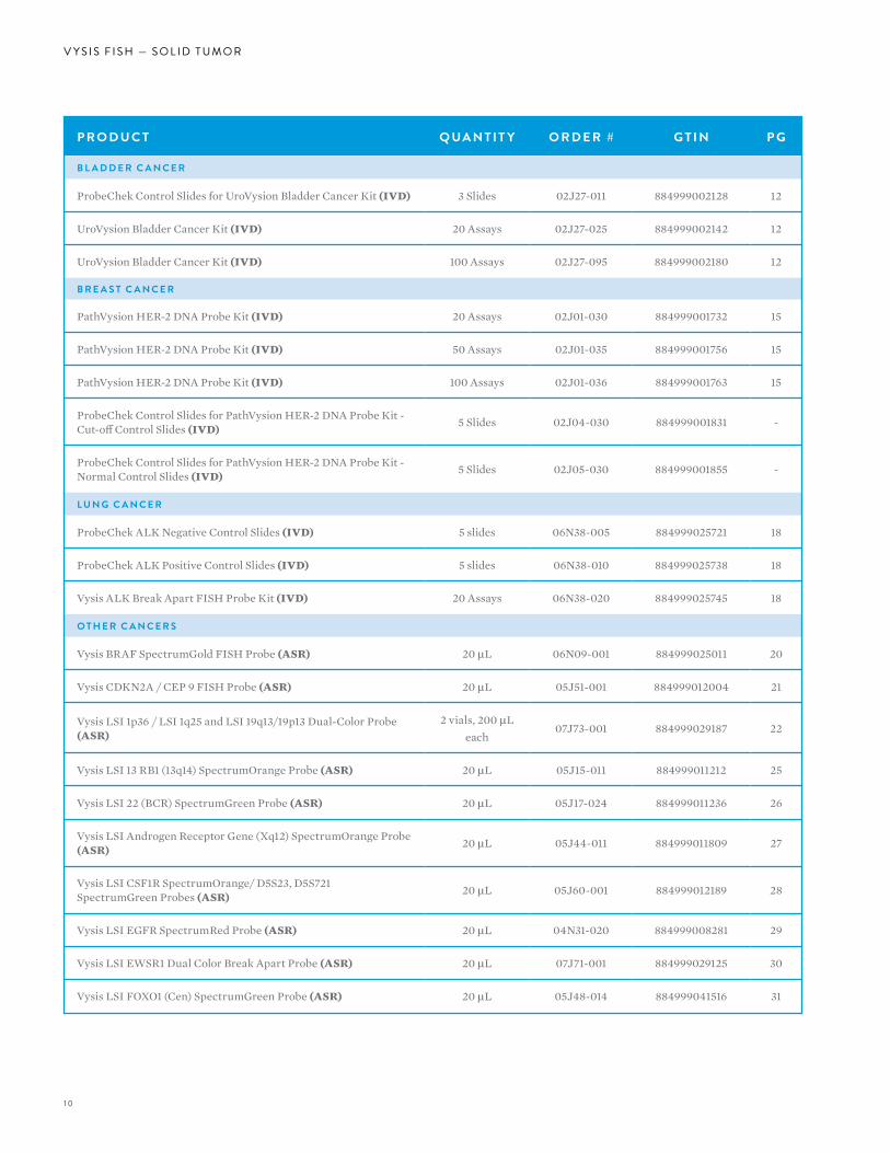
V Y S I S F I S H — S O L I D T U M O R
VYSIS FISH:SOLID TUMOR
8
V Y S I S F I S H — S O L I D T U M O R
Accurate and reliable detection of genetic aberrations in solid tumors with DNA fluorescence in situ Hybridization (FISH) probe technology is a powerful means to aid in diagnoses and treatment decisions. Abbott Molecular offers a comprehensive line of direct-labeled Vysis DNA probes for solid tumor assessment. Single- and multi-color probe sets offer researchers and clinicians a variety of ways to identify chromosome or locus deletions, gains, or
translocations that have been associated with specific types of solid tumors. These probes can be applied to a variety of sample types prepared for metaphase or interphase analysis.
99
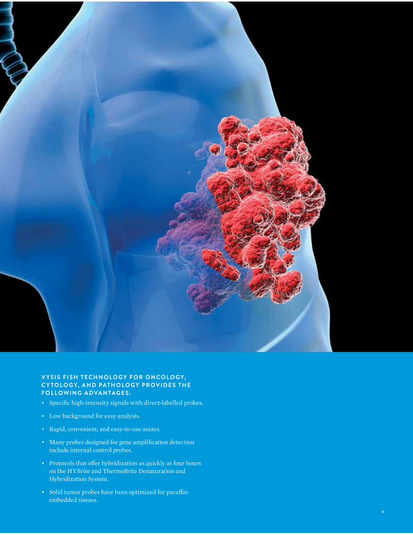
V Y S I S F I S H T E C H N O LO G Y F O R O N CO LO G Y, C Y TO LO G Y, A N D PAT H O LO G Y P R OV I D E S T H E F O L LO W I N G A DVA N TAG E S :• Specific high-intensity signals with direct-labelled probes.
• Low background for easy analysis.
• Rapid, convenient, and easy-to-use assays.
• Many probes designed for gene amplification detection include internal control probes.
• Protocols that offer hybridization as quickly as four hours on the HYBrite and ThermoBrite Denaturation and Hybridization System.
• Solid tumor probes have been optimized for paraffin-embedded tissues.
10
V Y S I S F I S H — S O L I D T U M O R
P R O D U C T Q UA N T I T Y O R D E R # G T I N P G
B L A D D E R C A N C E R
ProbeChek Control Slides for UroVysion Bladder Cancer Kit (IVD) 3 Slides 02J27-011 884999002128 12
UroVysion Bladder Cancer Kit (IVD) 20 Assays 02J27-025 884999002142 12
UroVysion Bladder Cancer Kit (IVD) 100 Assays 02J27-095 884999002180 12
B R E A S T C A N C E R
PathVysion HER-2 DNA Probe Kit (IVD) 20 Assays 02J01-030 884999001732 15
PathVysion HER-2 DNA Probe Kit (IVD) 50 Assays 02J01-035 884999001756 15
PathVysion HER-2 DNA Probe Kit (IVD) 100 Assays 02J01-036 884999001763 15
ProbeChek Control Slides for PathVysion HER-2 DNA Probe Kit - Cut-off Control Slides (IVD) 5 Slides 02J04-030 884999001831 -
ProbeChek Control Slides for PathVysion HER-2 DNA Probe Kit - Normal Control Slides (IVD) 5 Slides 02J05-030 884999001855 -
L U N G C A N C E R
ProbeChek ALK Negative Control Slides (IVD) 5 slides 06N38-005 884999025721 18
ProbeChek ALK Positive Control Slides (IVD) 5 slides 06N38-010 884999025738 18
Vysis ALK Break Apart FISH Probe Kit (IVD) 20 Assays 06N38-020 884999025745 18
O T H E R C A N C E R S
Vysis BRAF SpectrumGold FISH Probe (ASR) 20 µL 06N09-001 884999025011 20
Vysis CDKN2A / CEP 9 FISH Probe (ASR) 20 µL 05J51-001 884999012004 21
Vysis LSI 1p36 / LSI 1q25 and LSI 19q13/19p13 Dual-Color Probe (ASR)
2 vials, 200 µLeach
07J73-001 884999029187 22
Vysis LSI 13 RB1 (13q14) SpectrumOrange Probe (ASR) 20 µL 05J15-011 884999011212 25
Vysis LSI 22 (BCR) SpectrumGreen Probe (ASR) 20 µL 05J17-024 884999011236 26
Vysis LSI Androgen Receptor Gene (Xq12) SpectrumOrange Probe (ASR) 20 µL 05J44-011 884999011809 27
Vysis LSI CSF1R SpectrumOrange/ D5S23, D5S721 SpectrumGreen Probes (ASR) 20 µL 05J60-001 884999012189 28
Vysis LSI EGFR SpectrumRed Probe (ASR) 20 µL 04N31-020 884999008281 29
Vysis LSI EWSR1 Dual Color Break Apart Probe (ASR) 20 µL 07J71-001 884999029125 30
Vysis LSI FOXO1 (Cen) SpectrumGreen Probe (ASR) 20 µL 05J48-014 884999041516 31
1 1
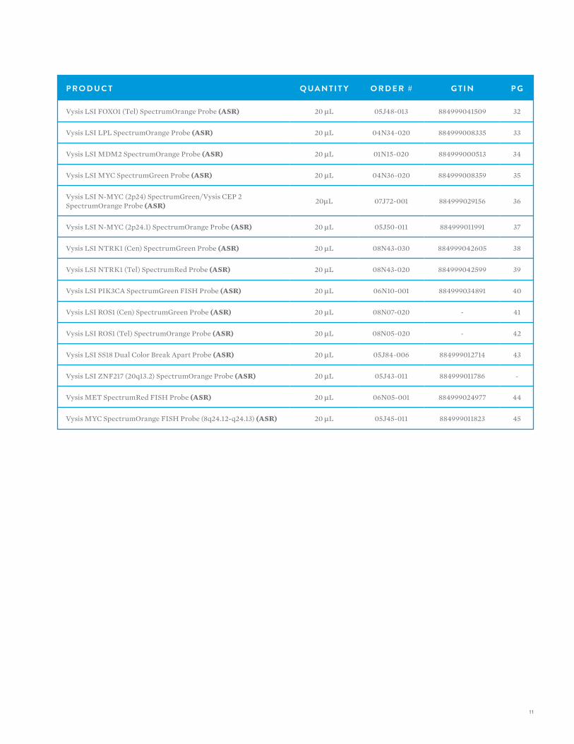
P R O D U C T Q UA N T I T Y O R D E R # G T I N P G
Vysis LSI FOXO1 (Tel) SpectrumOrange Probe (ASR) 20 µL 05J48-013 884999041509 32
Vysis LSI LPL SpectrumOrange Probe (ASR) 20 µL 04N34-020 884999008335 33
Vysis LSI MDM2 SpectrumOrange Probe (ASR) 20 µL 01N15-020 884999000513 34
Vysis LSI MYC SpectrumGreen Probe (ASR) 20 µL 04N36-020 884999008359 35
Vysis LSI N-MYC (2p24) SpectrumGreen/Vysis CEP 2 SpectrumOrange Probe (ASR) 20µL 07J72-001 884999029156 36
Vysis LSI N-MYC (2p24.1) SpectrumOrange Probe (ASR) 20 µL 05J50-011 884999011991 37
Vysis LSI NTRK1 (Cen) SpectrumGreen Probe (ASR) 20 µL 08N43-030 884999042605 38
Vysis LSI NTRK1 (Tel) SpectrumRed Probe (ASR) 20 µL 08N43-020 884999042599 39
Vysis LSI PIK3CA SpectrumGreen FISH Probe (ASR) 20 µL 06N10-001 884999034891 40
Vysis LSI ROS1 (Cen) SpectrumGreen Probe (ASR) 20 µL 08N07-020 - 41
Vysis LSI ROS1 (Tel) SpectrumOrange Probe (ASR) 20 µL 08N05-020 - 42
Vysis LSI SS18 Dual Color Break Apart Probe (ASR) 20 µL 05J84-006 884999012714 43
Vysis LSI ZNF217 (20q13.2) SpectrumOrange Probe (ASR) 20 µL 05J43-011 884999011786 -
Vysis MET SpectrumRed FISH Probe (ASR) 20 µL 06N05-001 884999024977 44
Vysis MYC SpectrumOrange FISH Probe (8q24.12-q24.13) (ASR) 20 µL 05J45-011 884999011823 45
12
V Y S I S F I S H — S O L I D T U M O RV Y S I S F I S H — S O L I D T U M O R
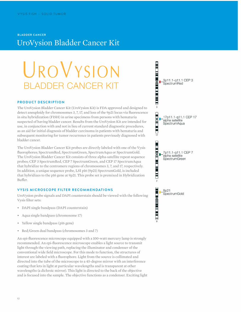
P R O D U C T D E S C R I P T I O NThe UroVysion Bladder Cancer Kit (UroVysion Kit) is FDA approved and designed to detect aneuploidy for chromosomes 3, 7, 17, and loss of the 9p21 locus via fluorescence in situ hybridization (FISH) in urine specimens from persons with hematuria suspected of having bladder cancer. Results from the UroVysion Kit are intended for use, in conjunction with and not in lieu of current standard diagnostic procedures, as an aid for initial diagnosis of bladder carcinoma in patients with hematuria and subsequent monitoring for tumor recurrence in patients previously diagnosed with bladder cancer.
The UroVysion Bladder Cancer Kit probes are directly labeled with one of the Vysis fluorophores; SpectrumRed, SpectrumGreen, SpectrumAqua or SpectrumGold. The UroVysion Bladder Cancer Kit consists of three alpha-satellite repeat sequence probes; CEP 3 SpectrumRed, CEP 7 SpectrumGreen, and CEP 17 SpectrumAqua that hybridize to the centromere regions of chromosomes 3, 7, and 17, respectively. In addition, a unique sequence probe, LSI p16 (9p21) SpectrumGold, is included that hybridizes to the p16 gene at 9p21. This probe set is premixed in Hybridization Buffer.
V Y S I S M I C R O S CO P E F I LT E R R E CO M M E N DAT I O N SUroVysion probe signals and DAPI counterstain should be viewed with the following Vysis filter sets:
• DAPI single bandpass (DAPI counterstain)
• Aqua single bandpass (chromosome 17)
• Yellow single bandpass (p16 gene)
• Red/Green dual bandpass (chromosomes 3 and 7)
An epi-fluorescence microscope equipped with a 100-watt mercury lamp is strongly recommended. An epi-fluorescence microscope enables a light source to transmit light through the viewing path, replacing the illuminator and condenser of the conventional wide field microscope. For this mode to function, the structures of interest are labeled with a fluorophore. Light from the source is collimated and directed into the tube of the microscope to a 45-degree mirror with an interference coating that lets in light at particular wavelengths and is transparent at other wavelengths (a dichroic mirror). This light is directed to the back of the objective and is focused into the sample. The objective functions as a condenser. Exciting light
B L A D D E R C A N C E R
UroVysion Bladder Cancer Kit
13
is absorbed by the fluorophore in the sample. The energy of the absorbed photon causes an electron in the dye molecule to jump to a higher energy orbital state. The electron rapidly jumps back to its ground orbital state, with the energy released becoming a photon of less energy compared to the exciting photon. The emitted photon has a color shifted toward red. The eyepiece collects the image in an identical manner as is transmitted in wide field microscopy.
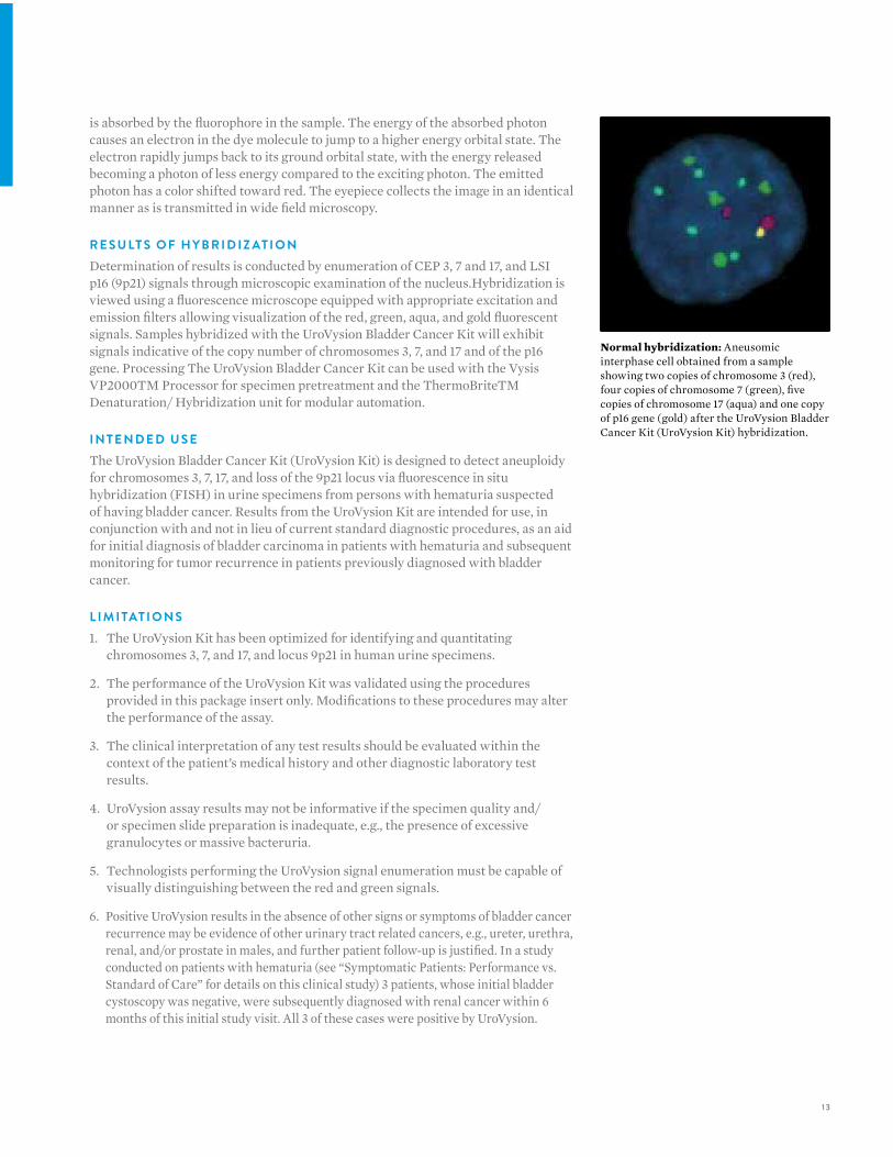
R E S U LT S O F H Y B R I D I Z AT I O NDetermination of results is conducted by enumeration of CEP 3, 7 and 17, and LSI p16 (9p21) signals through microscopic examination of the nucleus.Hybridization is viewed using a fluorescence microscope equipped with appropriate excitation and emission filters allowing visualization of the red, green, aqua, and gold fluorescent signals. Samples hybridized with the UroVysion Bladder Cancer Kit will exhibit signals indicative of the copy number of chromosomes 3, 7, and 17 and of the p16 gene. Processing The UroVysion Bladder Cancer Kit can be used with the Vysis VP2000TM Processor for specimen pretreatment and the ThermoBriteTM Denaturation/ Hybridization unit for modular automation.
I N T E N D E D U S EThe UroVysion Bladder Cancer Kit (UroVysion Kit) is designed to detect aneuploidy for chromosomes 3, 7, 17, and loss of the 9p21 locus via fluorescence in situ hybridization (FISH) in urine specimens from persons with hematuria suspected of having bladder cancer. Results from the UroVysion Kit are intended for use, in conjunction with and not in lieu of current standard diagnostic procedures, as an aid for initial diagnosis of bladder carcinoma in patients with hematuria and subsequent monitoring for tumor recurrence in patients previously diagnosed with bladder cancer.
L I M I TAT I O N S1. The UroVysion Kit has been optimized for identifying and quantitating
chromosomes 3, 7, and 17, and locus 9p21 in human urine specimens.
2. The performance of the UroVysion Kit was validated using the procedures provided in this package insert only. Modifications to these procedures may alter the performance of the assay.
3. The clinical interpretation of any test results should be evaluated within the context of the patient’s medical history and other diagnostic laboratory test results.
4. UroVysion assay results may not be informative if the specimen quality and/or specimen slide preparation is inadequate, e.g., the presence of excessive granulocytes or massive bacteruria.
5. Technologists performing the UroVysion signal enumeration must be capable of visually distinguishing between the red and green signals.
6. Positive UroVysion results in the absence of other signs or symptoms of bladder cancer recurrence may be evidence of other urinary tract related cancers, e.g., ureter, urethra, renal, and/or prostate in males, and further patient follow-up is justified. In a study conducted on patients with hematuria (see “Symptomatic Patients: Performance vs. Standard of Care” for details on this clinical study) 3 patients, whose initial bladder cystoscopy was negative, were subsequently diagnosed with renal cancer within 6 months of this initial study visit. All 3 of these cases were positive by UroVysion.
Normal hybridization: Aneusomic interphase cell obtained from a sample showing two copies of chromosome 3 (red), four copies of chromosome 7 (green), five copies of chromosome 17 (aqua) and one copy of p16 gene (gold) after the UroVysion Bladder Cancer Kit (UroVysion Kit) hybridization.
14
V Y S I S F I S H — S O L I D T U M O R
7. If UroVysion results are negative but standard clinical or diagnostic tests (e.g., cytology, cystoscopy) are positive, the standard procedures take precedence over the UroVysion test. Although the UroVysion Kit was designed to detect genetic changes associated with most bladder cancers, there will be some bladder cancers whose genetic changes cannot be detected by the UroVysion test.
8. Ta stage solitary tumors smaller than 5mm could not be detected by UroVysion FISH.23 UroVysion FISH results are dependent on the amount of tumor cells that are deposited on the slide.
P R O D U C T Q UA N T I T Y O R D E R # G T I N
UroVysion Bladder Cancer Kit (IVD) 20 Assays100 Assays
02J27-02502J27-095
884999002142884999002180
ProbeChek Control Slides for UroVysion Bladder Cancer Kit (IVD) 3 Slides 02J27-011 884999002128
15
B R E A S T C A N C E R
PathVysion HER-2 DNA Probe Kit
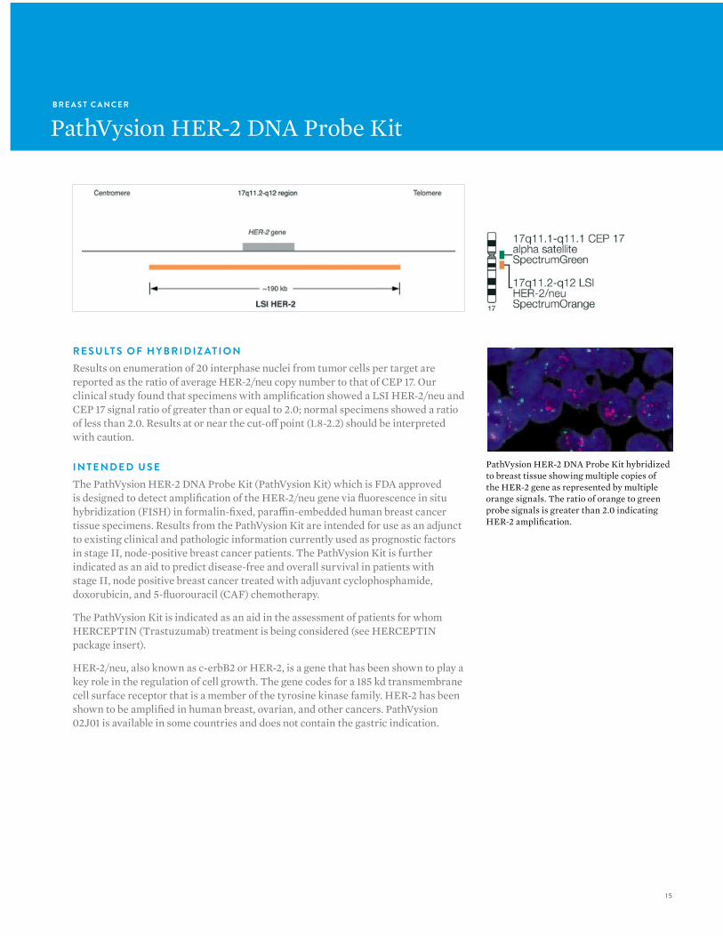
R E S U LT S O F H Y B R I D I Z AT I O N Results on enumeration of 20 interphase nuclei from tumor cells per target are reported as the ratio of average HER-2/neu copy number to that of CEP 17. Our clinical study found that specimens with amplification showed a LSI HER-2/neu and CEP 17 signal ratio of greater than or equal to 2.0; normal specimens showed a ratio of less than 2.0. Results at or near the cut-off point (1.8-2.2) should be interpreted with caution.
I N T E N D E D U S EThe PathVysion HER-2 DNA Probe Kit (PathVysion Kit) which is FDA approved is designed to detect amplification of the HER-2/neu gene via fluorescence in situ hybridization (FISH) in formalin-fixed, paraffin-embedded human breast cancer tissue specimens. Results from the PathVysion Kit are intended for use as an adjunct to existing clinical and pathologic information currently used as prognostic factors in stage II, node-positive breast cancer patients. The PathVysion Kit is further indicated as an aid to predict disease-free and overall survival in patients with stage II, node positive breast cancer treated with adjuvant cyclophosphamide, doxorubicin, and 5-fluorouracil (CAF) chemotherapy.
The PathVysion Kit is indicated as an aid in the assessment of patients for whom HERCEPTIN (Trastuzumab) treatment is being considered (see HERCEPTIN package insert).
HER-2/neu, also known as c-erbB2 or HER-2, is a gene that has been shown to play a key role in the regulation of cell growth. The gene codes for a 185 kd transmembrane cell surface receptor that is a member of the tyrosine kinase family. HER-2 has been shown to be amplified in human breast, ovarian, and other cancers. PathVysion 02J01 is available in some countries and does not contain the gastric indication.
PathVysion HER-2 DNA Probe Kit hybridized to breast tissue showing multiple copies of the HER-2 gene as represented by multiple orange signals. The ratio of orange to green probe signals is greater than 2.0 indicating HER-2 amplification.
16
V Y S I S F I S H — S O L I D T U M O R
I N T E N D E D U S EThe PathVysion HER-2 DNA Probe Kit (PathVysion Kit) is designed to detect amplification of the HER-2/neu gene via fluorescence in situ hybridization (FISH) in formalin-fixed, paraffin-embedded human breast cancer tissue specimens. Results from the PathVysion Kit are intended for use as an adjunct to existing clinical and pathologic information currently used as prognostic factors in stage II, node-positive breast cancer patients. The PathVysion Kit is further indicated as an aid to predict disease-free and overall survival in patients with stage II, node-positive breast cancer treated with adjuvant cyclophosphamide, doxorubicin and 5-fluorouracil (CAF) chemotherapy.
The PathVysion Kit is indicated as an aid in the assessment of patients for whom HERCEPTIN (Trastuzumab) treatment is being considered (see HERCEPTIN package insert).
WA R N I N GThe Vysis PathVysion Kit is not intended for use to screen for or diagnose breast cancer. It is intended to be used as an adjunct to other prognostic factors currently used to predict disease-free and overall survival in stage II, node-positive breast cancer patients. In making decisions regarding adjuvant CAF treatment, all other available clinical information should also be taken into consideration, such as tumor size, number of involved lymph nodes, and steroid receptor status.No treatment decision for stage II, node-positive breast cancer patients should be based on HER-2/neu gene amplification status alone.
The PathVysion HER-2 DNA Probe Kit consists of two labeled DNA probes. The LSI HER-2 probe that spans the entire HER-2 gene is labeled in SpectrumOrange. The CEP 17 probe is labeled in SpectrumGreen and hybridizes to the alpha satellite DNA located at the centromere of chromosome 17 (17p11.1-q11.1). Inclusion of the CEP 17 probe allows for the relative copy number of the HER-2 gene to be determined.
WA R N I N GHERCEPTIN therapy selection
NOTE: All of the patients in the HERCEPTIN clinical trials were selected using an investigational immunohistochemical assay (CTA). None of the patients in those trials were selected using the PathVysion assay. The PathVysion assay was compared to the CTA on a subset of clinical trial samples and found to provide acceptably concordant results. The actual correlation of the PathVysion assay to HERCEPTIN clinical outcome in prospective clinical trials has not been established.
Adjuvant therapy selection
The PathVysion Kit is not intended for use to screen for or diagnose breast cancer. It is intended to be used as an adjunct to other prognostic factors currently used to predict disease-free and overall survival in stage II, node-positive breast cancer patients and no treatment decision for stage II, node-positive breast cancer patients should be based on HER-2/neu gene amplification status alone. Selected patients with breast cancers shown to lack amplification of HER-2/neu may still benefit from CAF (cyclophosphamide, doxorubicin, 5-fluorouracil) adjuvant therapy on the basis of other prognostic factors that predict poor outcome (e.g. tumor size, number of involved lymph nodes, and hormone receptor status). Conversely, selected patients with breast cancers shown to contain gene amplification may not be candidates for CAF therapy due to preexisting or intercurrent medical illnesses. Required Training
Abbott Molecular will provide training in specimen preparation, assay procedure, and interpretation of FISH testing of the Her-2 gene for inexperienced users. It is also recommended that a laboratory that has previously received training but now has new personnel performing the assay request training for the new users.
17
P R O D U C T Q UA N T I T Y O R D E R # G T I N
PathVysion HER-2 DNA Probe Kit (IVD) 20 Assays50 Assays
100 Assays
02J01-03002J01-03502J01-036
884999001732884999001756884999001763
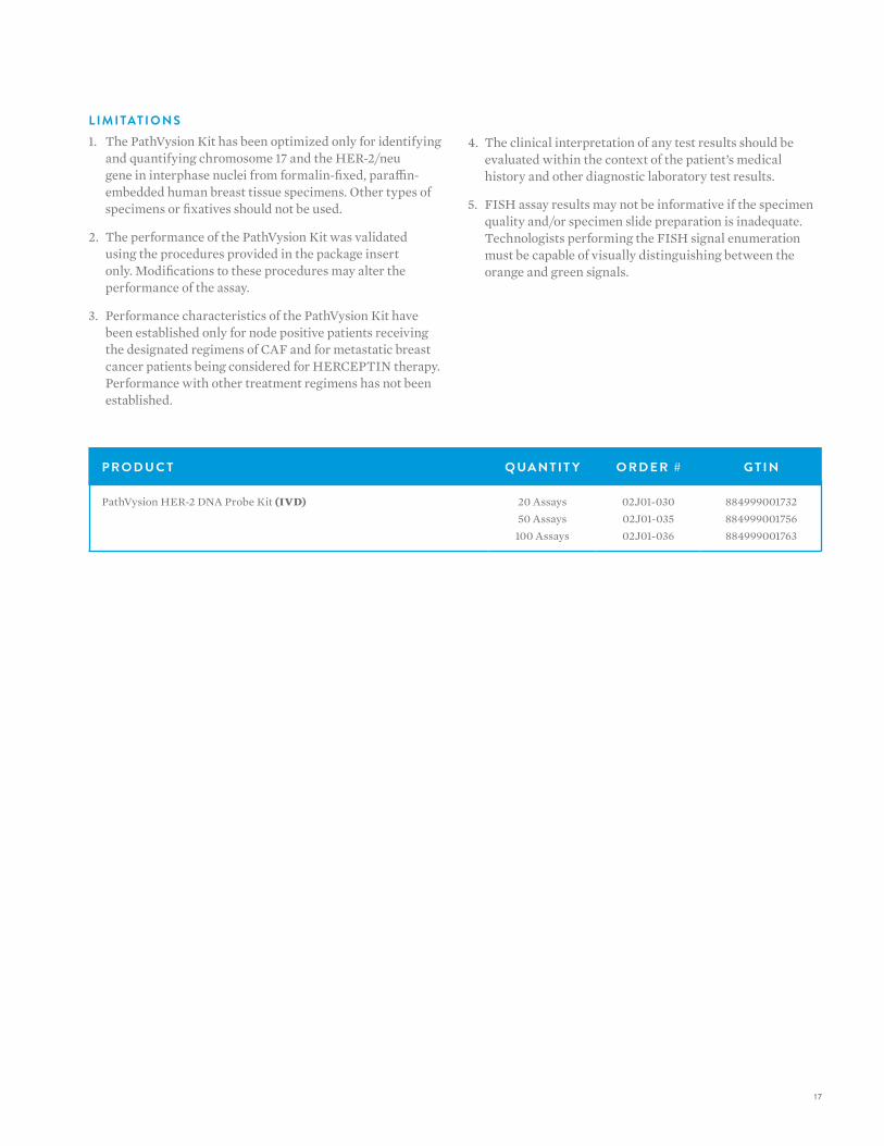
L I M I TAT I O N S1. The PathVysion Kit has been optimized only for identifying
and quantifying chromosome 17 and the HER-2/neu gene in interphase nuclei from formalin-fixed, paraffin-embedded human breast tissue specimens. Other types of specimens or fixatives should not be used.
2. The performance of the PathVysion Kit was validated using the procedures provided in the package insert only. Modifications to these procedures may alter the performance of the assay.
3. Performance characteristics of the PathVysion Kit have been established only for node positive patients receiving the designated regimens of CAF and for metastatic breast cancer patients being considered for HERCEPTIN therapy. Performance with other treatment regimens has not been established.
4. The clinical interpretation of any test results should be evaluated within the context of the patient’s medical history and other diagnostic laboratory test results.
5. FISH assay results may not be informative if the specimen quality and/or specimen slide preparation is inadequate. Technologists performing the FISH signal enumeration must be capable of visually distinguishing between the orange and green signals.
18
V Y S I S F I S H — S O L I D T U M O RV Y S I S F I S H — S O L I D T U M O R
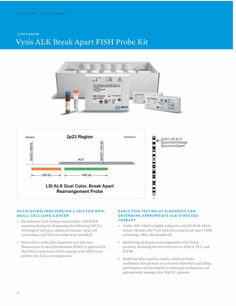
E A R LY F I S H T E S T I N G AT D I AG N O S I S C A N D E T E R M I N E A P P R O P R I AT E A L K-D I R E C T E D T H E R A P Y• Unlike IHC which is highly subjective and RT/PCR which
misses variants, the Vysis ALK test using break-apart FISH technology offers the benefits of:
• Identifying ALK gene rearrangements with fusion partners, including but not restricted to: EML4, TFG, and KIF5B
• Reducing false negative results, which provides confidence that patients are correctly identified and aiding pathologists and oncologists in their goal to diagnose and appropriately manage their NSCLC patients
L U N G C A N C E R
Vysis ALK Break Apart FISH Probe Kit
N CC N G U I D E L I N E S V E R S I O N 2 . 2013 F O R N O N -S M A L L C E L L LU N G C A N C E R• Recommend ALK testing concurrently with EGFR
mutation testing for diagnosing the following NSCLC histological subtypes: adenocarcinomas, large cell carcinomas, and NOS (not otherwise specified)
• States that a molecular diagnostic test that uses fluorescence in situ hybridization (FISH) is approved by the FDA to determine which patients with NSCLS are positive for ALK rearrangements
19
P E R S O N A L I Z E D M E D I C I N E A DVA N C E S• The first companion diagnostic for a novel subclass of non-
small cell lung cancer patients.
• Detection of ALK-positive NSCLC is necessary for selection of patients for treatment with XALKORI (crizotinib).
• Abbott announced the simultaneous approval of Pfizer’s XALKORI (crizotinib) and Abbott’s Vysis ALK FISH Probe Kit
I N T E N D E D U S EThe Vysis ALK Break Apart FISH Probe Kit is a qualitative test to detect rearrangements involving the ALK gene via fluorescence in situ hybridization (FISH) in formalin-fixed paraffin-embedded (FFPE) non-small cell lung cancer (NSCLC) tissue specimens to aid in identifying those patients eligible for treatment with Xalkori® (Crizotinib).
WA R N I N G S A N D L I M I TAT I O N S1. The Vysis ALK Break Apart FISH Probe Kit has
been optimized only for identifying and quantifying rearrangements of the ALK gene from formalin-fixed, paraffin-embedded human NSCLC tissue specimens. The assay should be only on 10% neutral buffered FFPE human lung tumor tissue. Other types of specimens or fixatives should not be used.
2. The performance of the Vysis ALK Break Apart FISH Probe Kit was established using the procedures provided in the package insert only. Modifications to these procedures may alter the performance of the assay.
3. The clinical interpretation of any test results should be evaluated within the context of the patient’s medical history and other diagnostic laboratory test results.
4. FISH assay results may not be informative if the specimen quality and/or specimen slide preparation is inadequate.
5. Technologists performing the FISH signal enumeration must be capable of visually distinguishing between the orange, green and yellow signals.̀
P R O D U C T Q UA N T I T Y O R D E R # G T I N
ProbeChek ALK Negative Control Slides (IVD) 5 slides 06N38-005 884999025721
ProbeChek ALK Positive Control Slides (IVD) 5 slides 06N38-010 884999025738
Vysis ALK Break Apart FISH Probe Kit (IVD)* 20 Assays 06N38-020 884999025745
Vysis Paraffin Pretreatment IV & Post-Hybridization Wash Buffer Kit (IVD) 1 Kit 01N31-005 884999000735
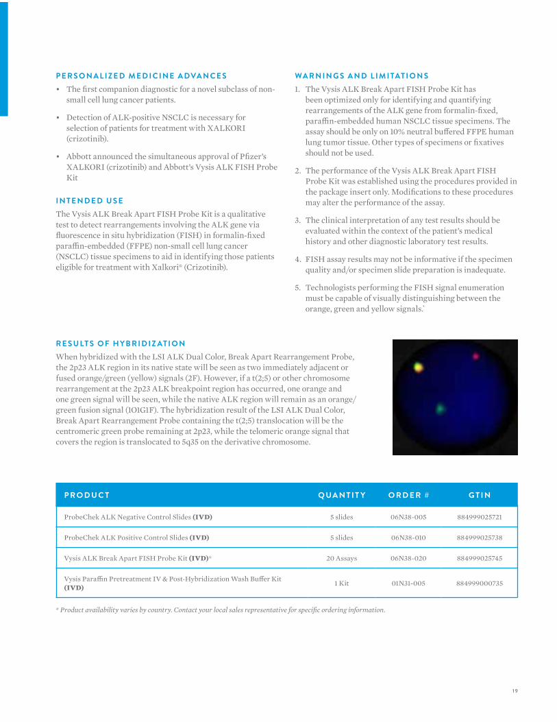
R E S U LT S O F H Y B R I D I Z AT I O NWhen hybridized with the LSI ALK Dual Color, Break Apart Rearrangement Probe, the 2p23 ALK region in its native state will be seen as two immediately adjacent or fused orange/green (yellow) signals (2F). However, if a t(2;5) or other chromosome rearrangement at the 2p23 ALK breakpoint region has occurred, one orange and one green signal will be seen, while the native ALK region will remain as an orange/green fusion signal (1O1G1F). The hybridization result of the LSI ALK Dual Color, Break Apart Rearrangement Probe containing the t(2;5) translocation will be the centromeric green probe remaining at 2p23, while the telomeric orange signal that covers the region is translocated to 5q35 on the derivative chromosome.
* Product availability varies by country. Contact your local sales representative for specific ordering information.

20
V Y S I S F I S H — S O L I D T U M O RV Y S I S F I S H — S O L I D T U M O R
O T H E R C A N C E R S
Vysis BRAF SpectrumGold FISH Probe
R E S U LT S O F H Y B R I D I Z AT I O NNormal diploid nuclei or metaphase chromosome sets are expected to exhibit two gold fluorescent BRAF signals, which correspond to two target loci on chromosome homologues to which the BRAF fluorescent probe is bound: 7q34. A chromosome set that has an extra copy (copies) of BRAF (7q34) will exhibit more than two gold fluorescent signals.
P R O D U C T Q UA N T I T Y O R D E R # G T I N
Vysis BRAF SpectrumGold FISH Probe (ASR) 20 µL 06N09-001 884999025011

2 1
O T H E R C A N C E R S
Vysis CDKN2A/CEP 9 FISH Probe
P R O D U C T D E S C R I P T I O NVysis LSI CDKN2A/CEP 9 Probes are provided in one vial as a mixture of the LSI CDKN2A (p16) probe labeled with SpectrumOrange and the CEP 9 probe labeled with SpectrumGreen. The LSI CDKN2A probe spans approximately222 kb and contains a number of genetic loci including D9S1749, DS1747, p16 (INK4B), p14 (ARF), D9S1748, p15(INK4B), and D9S1752. The CEP 9 SpectrumGreen probe hybridizes to alpha satellite sequences specific to chromosome 9.
R E S U LT S O F H Y B R I D I Z AT I O NIn a normal sample, the expected pattern for a nucleus hybridized with the Vysis LSI CDKN2A / CEP 9 Probe is the two orange, two green (2O2G) signal pattern. If a deletion at the 190 kb region covered by the LSI p16 probe occurs on one chromosome 9 homolog and both centromeres from chromosome 9 are retained, the one orange, two green (1O2G) signal pattern is expected.Very small deletions may occur that do not delete the entire LSI p16 probe target and therefore will not be detected.
P R O D U C T Q UA N T I T Y O R D E R # G T I N
Vysis CDKN2A / CEP 9 FISH Probe (ASR) 20 µL 05J51-001 884999012004
Abormal hybridization: Vysis LSI CDKN2A / CEP 9 Probe hybridized to a nucleus exhibiting the one orange and two green signal (1O2G) pattern. One p16 gene locus is deleted and both chromosome 9 homologs are present as indicated by one orange and two green signals, respectively.
22
V Y S I S F I S H — S O L I D T U M O RV Y S I S F I S H — S O L I D T U M O R
O T H E R C A N C E R S
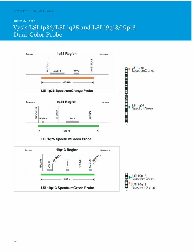
Vysis LSI 1p36/LSI 1q25 and LSI 19q13/19p13Dual-Color Probe

23
Normal hybridization: Result of the hybridization of the LSI 1p36 / LSI 1q25 Dual-Color Probe vial 1 as observed in normal interphase cells.
Abnormal hybridization: An abnormal cell hybridized with the LSI 1p36 / LSI 1q25 Dual-Color Probe vial 1. The cell in this image shows the one orange, two green signal pattern indicative of the 1p36 deletion.
Normal hybridization: Result of the hybridization of the LSI 19p13 / LSI 19q13 Dual-Color Probe vial 2 as observed in normal interphase cells.
Abnormal hybridization: An abnormal cell hybridized with the LSI 19p13 / LSI 19q13 Dual-Color Probe vial 2. The cell in this image shows the one orange, two green signal pattern indicative of the 19q13 deletion.
P R O D U C T D E S C R I P T I O NThe Vysis LSI 1p36 SpectrumOrange/1q25 SpectrumGreen Probes are provided in one vial as a mixture of a ~435 kb SpectrumOrange-labeled 1p36 probe and a ~618 kb SpectrumGreen-labeled 1q25 probe premixed in hybridization buffer. The LSI 1p36 probe contains sequences that extend from near SHGC 57243 locus, through the TP73 and MEGF6 genes, and ends at a point telomeric to the MEGF6 locus. The LSI 1q25 probe contains sequences that extend from a point telomeric to the ABL2 gene, through the ABL2 and ANGPTL1 genes, and ends proximally near the SHGC-1322 locus.
The Vysis LSI 19q13 SpectrumOrange/19p13 SpectrumGreen Probes are provided in one vial as a mixture of a ~380 kb SpectrumOrange-labeled 19q13 probe and a ~502 kb SpectrumGreen-labeled 19p13 probe premixed in hybridization buffer. The LSI 19p13 probe contains sequences that extend from a point centromeric to the MAN2B1 locus, through MAN2B1, ZNF443 and ZNF44 genes, and ends at a point telomeric to the ZNF44 locus. The LSI 19q13 probe contains sequences that extend from a point telomeric to the CRX locus, through the CRX, GLTSCR2 and GLTSCR1 genes, and ends proximally at a point centromeric to the GLTSCR1 locus.

24
V Y S I S F I S H — S O L I D T U M O R
R E S U LT S O F H Y B R I D I Z AT I O N
V I A L 1This probe allows status assessment of the following two chromosome regions: 1p36 and 1q25. In a normal cell hybridized with the LSI 1p36 and LSI 1q25, two orange and two green signals will be observed indicative of two intact copies of chromosome 1. In an abnormal cell with a deletion in the 1p36 region fewer than two orange signals will be observed.
V I A L 2This probe allows status assessment of the following two chromosome regions: 19q13 and 19p13. In a normal cell with two intact copies of chromosome 19, two orange and two green signals will be observed. In an an abnormal cell with a deletion in the 19q13 region fewer than two orange orange signals will be observed.
P R O D U C T Q UA N T I T Y O R D E R # G T I N
Vysis LSI 1p36 / LSI 1q25 and LSI 19q13/19p13 Dual-Color Probe (ASR) 2 vials, 200 µL each 07J73-001 884999029187

25
O T H E R C A N C E R S
Vysis LSI 13 RB1 (13q14) SpectrumOrange Probe
Normal hybridization: LSI 13 (RB1) 13q14 Probe hybridized to a normal nucleus showing a two orange (2O) signal pattern.
P R O D U C T Q UA N T I T Y O R D E R # G T I N
Vysis LSI 13 RB1 (13q14) SpectrumOrange Probe (ASR) 20 µL 05J15-011 884999011212
P R O D U C T D E S C R I P T I O NThe LSI 13 (RB1) 13q14 SpectrumOrange Probe contains unique DNA sequences specific to the RB1 gene within the 13q14 region of chromosome 13. The presence or absence of the RB1 gene region may be detected using the LSI 13 (RB1) 13q14 Probe. This probe may be used to detect deletion (not mutation) of the RB1 gene locus.
The LSI 13 (RB1) 13q14 SpectrumOrange Probe is approximately 220 kb and contains sequences that target the entire RB1 gene.
R E S U LT S O F H Y B R I D I Z AT I O NIn a normal cell, the expected result for a nucleus hybridized with the LSI 13 (RB1) probe is a two orange (2O) signal pattern. In a hybridized abnormal cell containing the deletion, a one orange (1O) signal pattern will be observed.
26
V Y S I S F I S H — S O L I D T U M O RV Y S I S F I S H — S O L I D T U M O R
O T H E R C A N C E R S
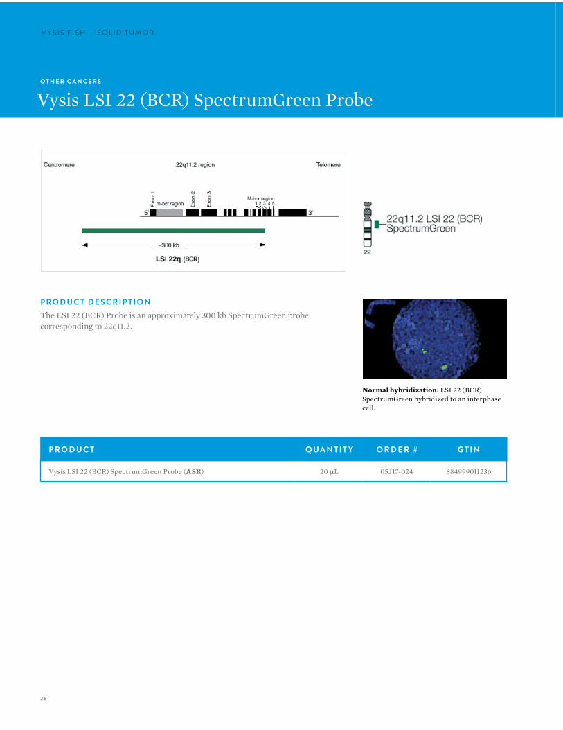
Vysis LSI 22 (BCR) SpectrumGreen Probe
P R O D U C T D E S C R I P T I O NThe LSI 22 (BCR) Probe is an approximately 300 kb SpectrumGreen probe corresponding to 22q11.2.
P R O D U C T Q UA N T I T Y O R D E R # G T I N
Vysis LSI 22 (BCR) SpectrumGreen Probe (ASR) 20 µL 05J17-024 884999011236
Normal hybridization: LSI 22 (BCR) SpectrumGreen hybridized to an interphase cell.
27
O T H E R C A N C E R S
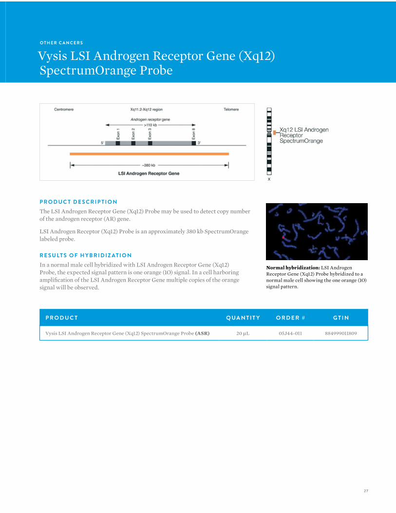
Vysis LSI Androgen Receptor Gene (Xq12) SpectrumOrange Probe
P R O D U C T D E S C R I P T I O NThe LSI Androgen Receptor Gene (Xq12) Probe may be used to detect copy number of the androgen receptor (AR) gene.
LSI Androgen Receptor (Xq12) Probe is an approximately 380 kb SpectrumOrange labeled probe.
R E S U LT S O F H Y B R I D I Z AT I O NIn a normal male cell hybridized with LSI Androgen Receptor Gene (Xq12) Probe, the expected signal pattern is one orange (1O) signal. In a cell harboring amplification of the LSI Androgen Receptor Gene multiple copies of the orange signal will be observed.
P R O D U C T Q UA N T I T Y O R D E R # G T I N
Vysis LSI Androgen Receptor Gene (Xq12) SpectrumOrange Probe (ASR) 20 µL 05J44-011 884999011809
Normal hybridization: LSI Androgen Receptor Gene (Xq12) Probe hybridized to a normal male cell showing the one orange (1O) signal pattern.
28
V Y S I S F I S H — S O L I D T U M O RV Y S I S F I S H — S O L I D T U M O R
O T H E R C A N C E R S
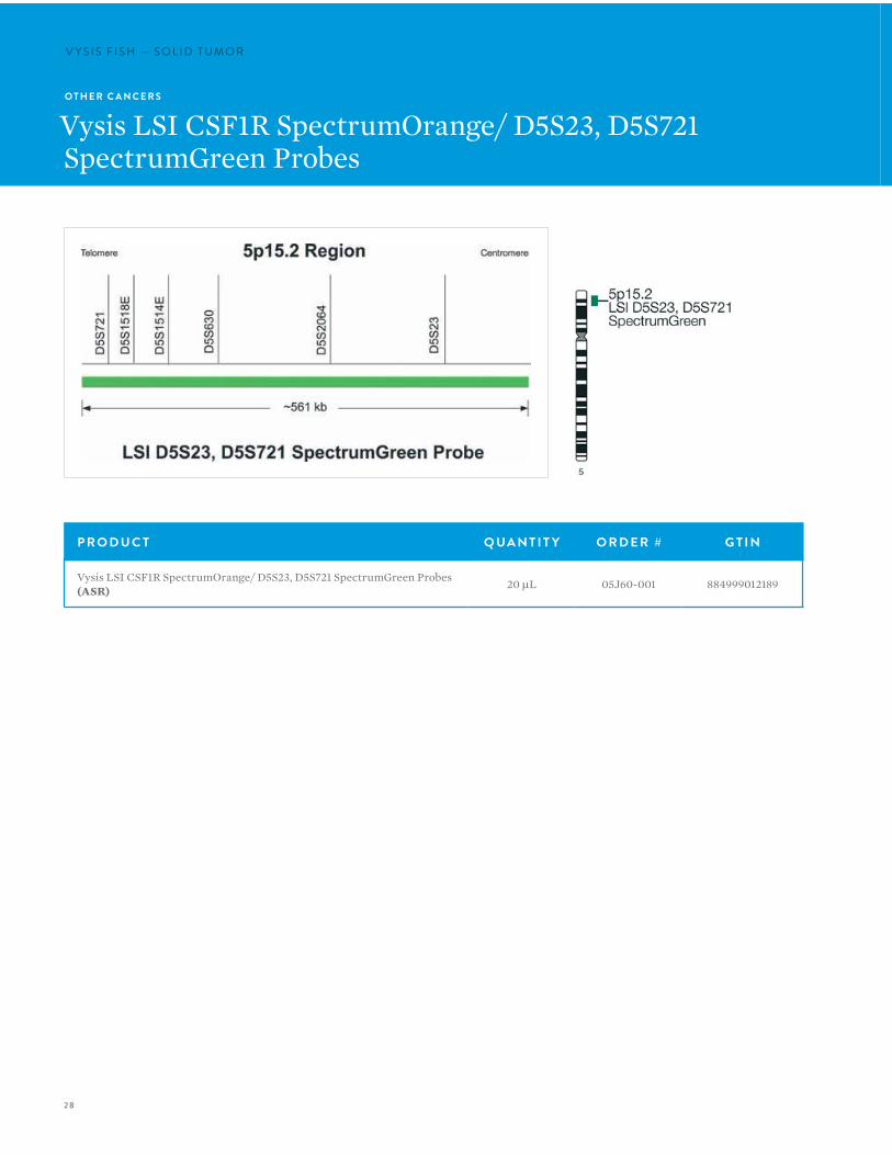
Vysis LSI CSF1R SpectrumOrange/ D5S23, D5S721 SpectrumGreen Probes
P R O D U C T Q UA N T I T Y O R D E R # G T I N
Vysis LSI CSF1R SpectrumOrange/ D5S23, D5S721 SpectrumGreen Probes (ASR) 20 µL 05J60-001 884999012189
29
P R O D U C T Q UA N T I T Y O R D E R # G T I N
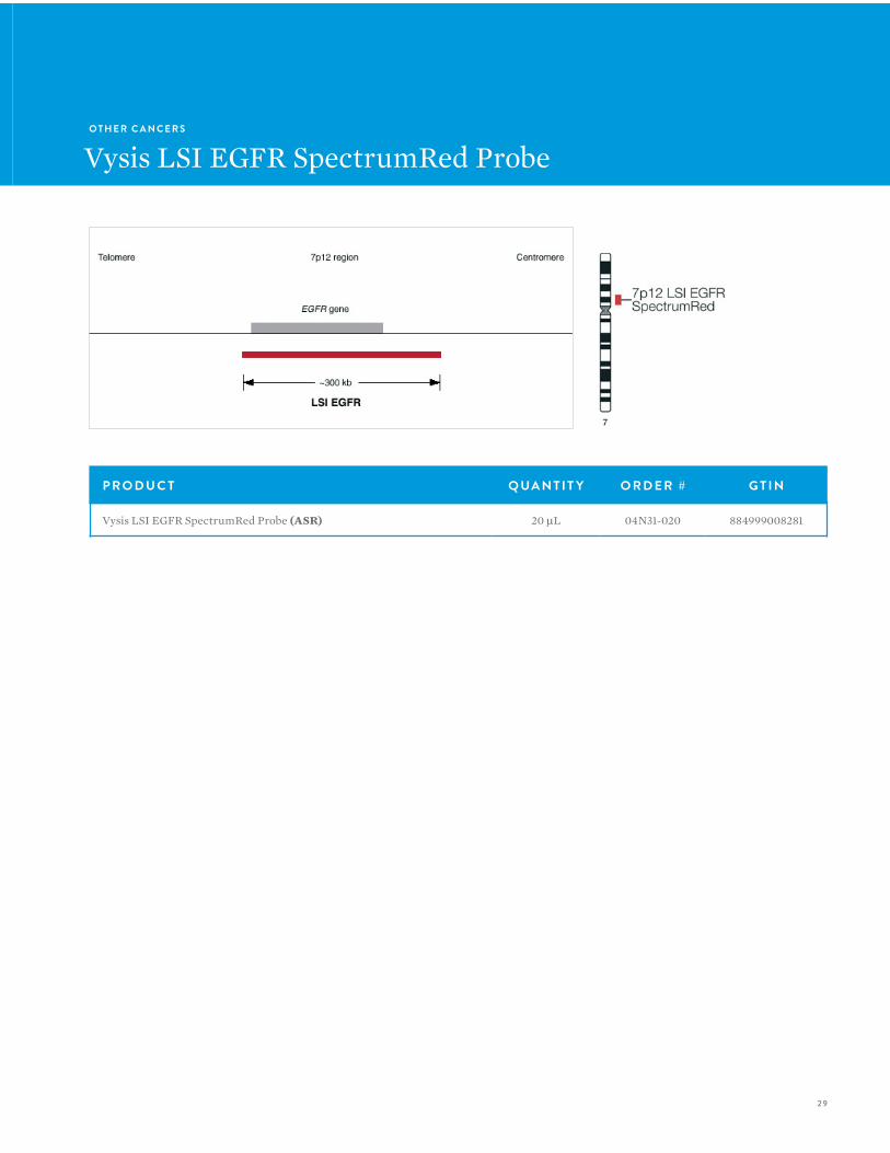
Vysis LSI EGFR SpectrumRed Probe (ASR) 20 µL 04N31-020 884999008281
O T H E R C A N C E R S
Vysis LSI EGFR SpectrumRed Probe
30
V Y S I S F I S H — S O L I D T U M O RV Y S I S F I S H — S O L I D T U M O R
O T H E R C A N C E R S
Vysis LSI EWSR1 Dual Color Break Apart Probe
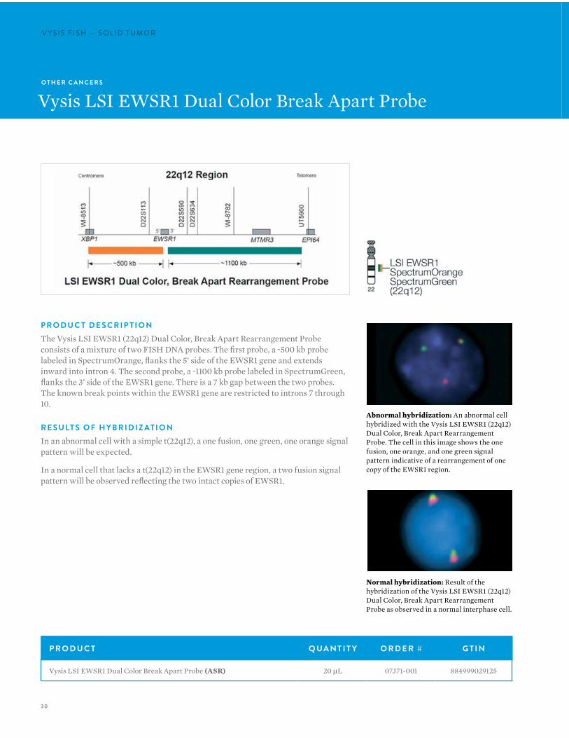
P R O D U C T D E S C R I P T I O NThe Vysis LSI EWSR1 (22q12) Dual Color, Break Apart Rearrangement Probe consists of a mixture of two FISH DNA probes. The first probe, a ~500 kb probe labeled in SpectrumOrange, flanks the 5’ side of the EWSR1 gene and extends inward into intron 4. The second probe, a ~1100 kb probe labeled in SpectrumGreen, flanks the 3’ side of the EWSR1 gene. There is a 7 kb gap between the two probes. The known break points within the EWSR1 gene are restricted to introns 7 through 10.
R E S U LT S O F H Y B R I D I Z AT I O NIn an abnormal cell with a simple t(22q12), a one fusion, one green, one orange signal pattern will be expected.
In a normal cell that lacks a t(22q12) in the EWSR1 gene region, a two fusion signal pattern will be observed reflecting the two intact copies of EWSR1.
Abnormal hybridization: An abnormal cell hybridized with the Vysis LSI EWSR1 (22q12) Dual Color, Break Apart Rearrangement Probe. The cell in this image shows the one fusion, one orange, and one green signal pattern indicative of a rearrangement of one copy of the EWSR1 region.
Normal hybridization: Result of the hybridization of the Vysis LSI EWSR1 (22q12) Dual Color, Break Apart Rearrangement Probe as observed in a normal interphase cell.
P R O D U C T Q UA N T I T Y O R D E R # G T I N
Vysis LSI EWSR1 Dual Color Break Apart Probe (ASR) 20 µL 07J71-001 884999029125

31
O T H E R C A N C E R S
Vysis LSI FOXO1 (Cen) SpectrumGreen Probe
P R O D U C T D E S C R I P T I O NThe Vysis LSI FOXO1 (Cen) SpectrumGreen Probe is based on the ability of the FOXO1 (Cen) locus-specific identifier (LSI) probe to identify rearrangements of the chromosomal locus 13q14 containing the FOXO1 gene when used in a FISH test with Vysis LSI FOXO1 (Tel) SpectrumOrange Probe. It is approximately ~724 kb in size and is labeled in SpectrumGreen.
Normal hybridization: LSI FOXO1 (Cen) SpectrumGreen probes hybridized to a interphase cell.
P R O D U C T Q UA N T I T Y O R D E R # G T I N
Vysis LSI FOXO1 (Cen) SpectrumGreen Probe (ASR) 20 µL 05J48-014 884999041516
32
V Y S I S F I S H — S O L I D T U M O RV Y S I S F I S H — S O L I D T U M O R
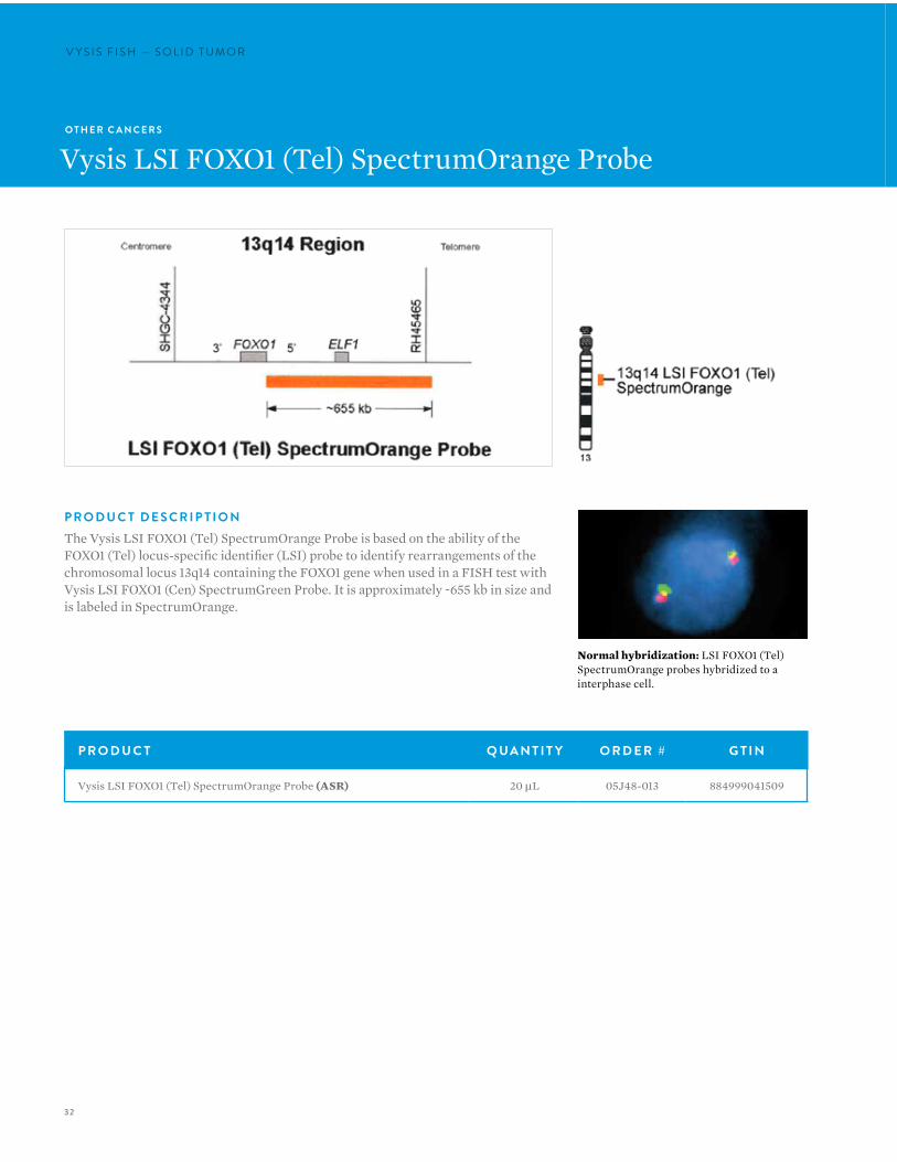
Normal hybridization: LSI FOXO1 (Tel) SpectrumOrange probes hybridized to a interphase cell.
O T H E R C A N C E R S
Vysis LSI FOXO1 (Tel) SpectrumOrange Probe
P R O D U C T D E S C R I P T I O NThe Vysis LSI FOXO1 (Tel) SpectrumOrange Probe is based on the ability of the FOXO1 (Tel) locus-specific identifier (LSI) probe to identify rearrangements of the chromosomal locus 13q14 containing the FOXO1 gene when used in a FISH test with Vysis LSI FOXO1 (Cen) SpectrumGreen Probe. It is approximately ~655 kb in size and is labeled in SpectrumOrange.
P R O D U C T Q UA N T I T Y O R D E R # G T I N
Vysis LSI FOXO1 (Tel) SpectrumOrange Probe (ASR) 20 µL 05J48-013 884999041509
33
O T H E R C A N C E R S
Vysis LSI LPL SpectrumOrange Probe
P R O D U C T Q UA N T I T Y O R D E R # G T I N
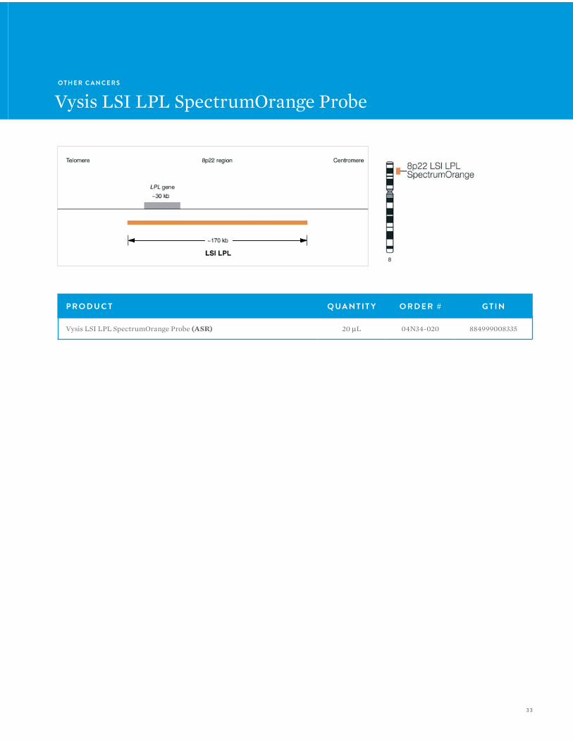
Vysis LSI LPL SpectrumOrange Probe (ASR) 20 µL 04N34-020 884999008335
34
V Y S I S F I S H — S O L I D T U M O RV Y S I S F I S H — S O L I D T U M O R
O T H E R C A N C E R S
Vysis LSI MDM2 SpectrumOrange Probe
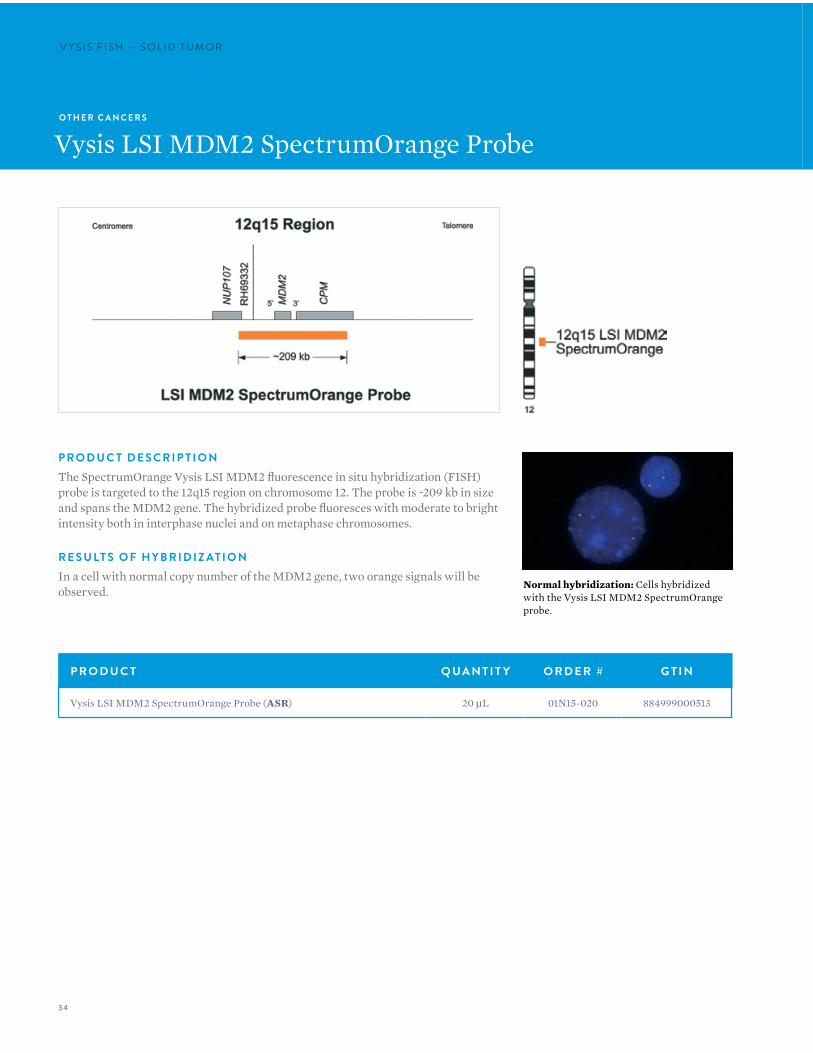
P R O D U C T D E S C R I P T I O NThe SpectrumOrange Vysis LSI MDM2 fluorescence in situ hybridization (FISH) probe is targeted to the 12q15 region on chromosome 12. The probe is ~209 kb in size and spans the MDM2 gene. The hybridized probe fluoresces with moderate to bright intensity both in interphase nuclei and on metaphase chromosomes.
R E S U LT S O F H Y B R I D I Z AT I O NIn a cell with normal copy number of the MDM2 gene, two orange signals will be observed.
P R O D U C T Q UA N T I T Y O R D E R # G T I N
Vysis LSI MDM2 SpectrumOrange Probe (ASR) 20 µL 01N15-020 884999000513
Normal hybridization: Cells hybridized with the Vysis LSI MDM2 SpectrumOrange probe.
35
O T H E R C A N C E R S
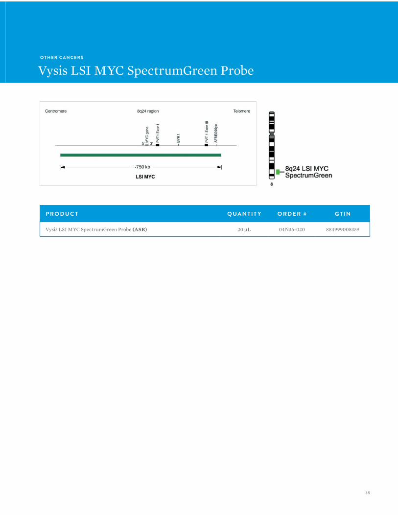
Vysis LSI MYC SpectrumGreen Probe
P R O D U C T Q UA N T I T Y O R D E R # G T I N
Vysis LSI MYC SpectrumGreen Probe (ASR) 20 µL 04N36-020 884999008359
36
V Y S I S F I S H — S O L I D T U M O RV Y S I S F I S H — S O L I D T U M O R
O T H E R C A N C E R S
Vysis LSI N-MYC (2p24) SpectrumGreen/Vysis CEP 2 SpectrumOrange Probe
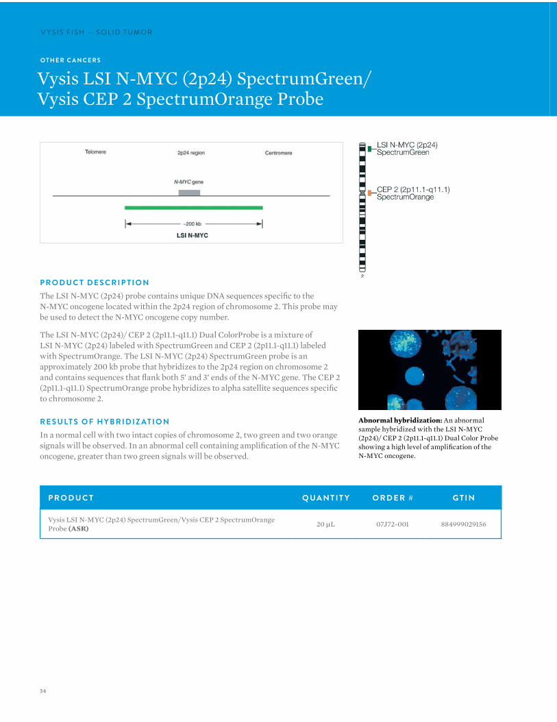
P R O D U C T D E S C R I P T I O NThe LSI N-MYC (2p24) probe contains unique DNA sequences specific to the N-MYC oncogene located within the 2p24 region of chromosome 2. This probe may be used to detect the N-MYC oncogene copy number.
The LSI N-MYC (2p24)/ CEP 2 (2p11.1-q11.1) Dual ColorProbe is a mixture of LSI N-MYC (2p24) labeled with SpectrumGreen and CEP 2 (2p11.1-q11.1) labeled with SpectrumOrange. The LSI N-MYC (2p24) SpectrumGreen probe is an approximately 200 kb probe that hybridizes to the 2p24 region on chromosome 2 and contains sequences that flank both 5’ and 3’ ends of the N-MYC gene. The CEP 2 (2p11.1-q11.1) SpectrumOrange probe hybridizes to alpha satellite sequences specific to chromosome 2.
R E S U LT S O F H Y B R I D I Z AT I O NIn a normal cell with two intact copies of chromosome 2, two green and two orange signals will be observed. In an abnormal cell containing amplification of the N-MYC oncogene, greater than two green signals will be observed.
P R O D U C T Q UA N T I T Y O R D E R # G T I N
Vysis LSI N-MYC (2p24) SpectrumGreen/Vysis CEP 2 SpectrumOrange Probe (ASR) 20 µL 07J72-001 884999029156
Abnormal hybridization: An abnormal sample hybridized with the LSI N-MYC (2p24)/ CEP 2 (2p11.1-q11.1) Dual Color Probe showing a high level of amplification of the N-MYC oncogene.
37
O T H E R C A N C E R S
Vysis LSI N-MYC (2p24.1) SpectrumOrange Probe
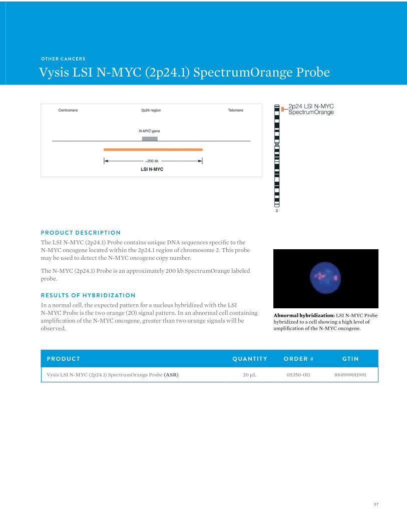
P R O D U C T D E S C R I P T I O NThe LSI N-MYC (2p24.1) Probe contains unique DNA sequences specific to the N-MYC oncogene located within the 2p24.1 region of chromosome 2. This probe may be used to detect the N-MYC oncogene copy number.
The N-MYC (2p24.1) Probe is an approximately 200 kb SpectrumOrange labeled probe.
R E S U LT S O F H Y B R I D I Z AT I O NIn a normal cell, the expected pattern for a nucleus hybridized with the LSI N-MYC Probe is the two orange (2O) signal pattern. In an abnormal cell containing amplification of the N-MYC oncogene, greater than two orange signals will be observed.
P R O D U C T Q UA N T I T Y O R D E R # G T I N
Vysis LSI N-MYC (2p24.1) SpectrumOrange Probe (ASR) 20 µL 05J50-011 884999011991
Abnormal hybridization: LSI N-MYC Probe hybridized to a cell showing a high level of amplification of the N-MYC oncogene.

38
V Y S I S F I S H — S O L I D T U M O RV Y S I S F I S H — S O L I D T U M O R
P R O D U C T D E S C R I P T I O NThe 1q23 NTRK1 (Cen) SpectrumGreen probe is approximately 440 kb in size and contains the entire NTRK1 gene. The hybridized probe fluoresces with moderate to bright intensity both in interphase nuclei and metaphase chromosomes.
P R O D U C T Q UA N T I T Y O R D E R # G T I N
Vysis LSI NTRK1 (Cen) SpectrumGreen Probe (ASR) 20 µL 08N43-030 884999042605
O T H E R C A N C E R S
Vysis LSI NTRK1 (Cen) SpectrumGreen Probe
39
O T H E R C A N C E R S
Vysis LSI NTRK1 (Tel) SpectrumRed Probe
P R O D U C T Q UA N T I T Y O R D E R # G T I N
Vysis LSI NTRK1 (Tel) SpectrumRed Probe (ASR) 20 µL 08N43-020 884999042599
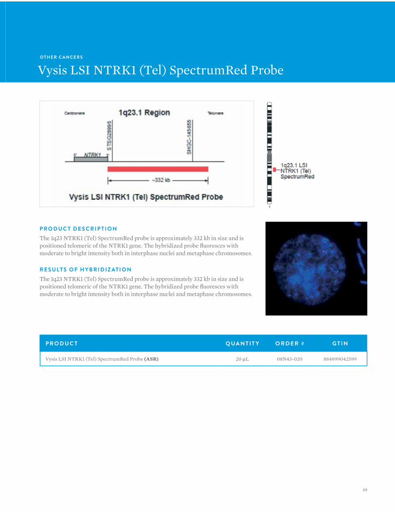
P R O D U C T D E S C R I P T I O NThe 1q23 NTRK1 (Tel) SpectrumRed probe is approximately 332 kb in size and is positioned telomeric of the NTRK1 gene. The hybridized probe fluoresces with moderate to bright intensity both in interphase nuclei and metaphase chromosomes.
R E S U LT S O F H Y B R I D I Z AT I O NThe 1q23 NTRK1 (Tel) SpectrumRed probe is approximately 332 kb in size and is positioned telomeric of the NTRK1 gene. The hybridized probe fluoresces with moderate to bright intensity both in interphase nuclei and metaphase chromosomes.
40
V Y S I S F I S H — S O L I D T U M O RV Y S I S F I S H — S O L I D T U M O R
O T H E R C A N C E R S
Vysis LSI PIK3CA SpectrumGreen FISH Probe
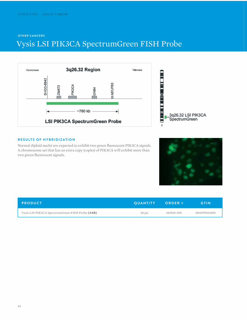
R E S U LT S O F H Y B R I D I Z AT I O NNormal diploid nuclei are expected to exhibit two green fluorescent PIK3CA signals. A chromosome set that has an extra copy (copies) of PIK3CA will exhibit more than two green fluorescent signals.
P R O D U C T Q UA N T I T Y O R D E R # G T I N
Vysis LSI PIK3CA SpectrumGreen FISH Probe (ASR) 20 µL 06N10-001 884999034891
41
P R O D U C T Q UA N T I T Y O R D E R # G T I N
Vysis LSI ROS1 (Cen) SpectrumGreen Probe (ASR) 20 µL 08N07-020 -
O T H E R C A N C E R S
Vysis LSI ROS1 (Cen) SpectrumGreen Probe
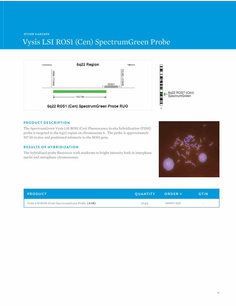
P R O D U C T D E S C R I P T I O NThe SpectrumGreen Vysis LSI ROS1 (Cen) Fluorescence in situ hybridization (FISH) probe is targeted to the 6q22 region on chromosome 6. The probe is approximately 557 kb in size and positioned telomeric to the ROS1 gene.
R E S U LT S O F H Y B R I D I Z AT I O N The hybridized probe fluoresces with moderate to bright intensity both in interphase nuclei and metaphase chromosomes.
42
V Y S I S F I S H — S O L I D T U M O RV Y S I S F I S H — S O L I D T U M O R
P R O D U C T Q UA N T I T Y O R D E R # G T I N
Vysis LSI ROS1 (Tel) SpectrumOrange Probe (ASR) 20 µL 08N05-020 -
O T H E R C A N C E R S
Vysis LSI ROS1 (Tel) SpectrumOrange Probe
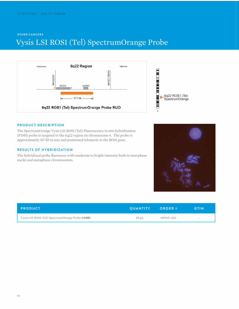
P R O D U C T D E S C R I P T I O NThe SpectrumOrange Vysis LSI ROS1 (Tel) Fluorescence in situ hybridization (FISH) probe is targeted to the 6q22 region on chromosome 6. The probe is approximately 317 kb in size and positioned telomeric to the ROS1 gene.
R E S U LT S O F H Y B R I D I Z AT I O NThe hybridized probe fluoresces with moderate to bright intensity both in interphase nuclei and metaphase chromosomes.
43
O T H E R C A N C E R S
Vysis LSI SS18 Dual Color Break Apart Probe
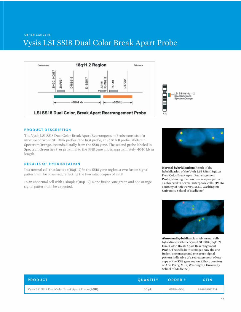
P R O D U C T D E S C R I P T I O NThe Vysis LSI SS18 Dual Color Break Apart Rearrangement Probe consists of a mixture of two FISH DNA probes. The first probe, an ~650 KB probe labeled in SpectrumOrange, extends distally from the SS18 gene. The second probe labeled in SpectrumGreen lies 3’ or proximal to the SS18 gene and is approximately ~1040 kb in length.
R E S U LT S O F H Y B R I D I Z AT I O NIn a normal cell that lacks a t(18q11.2) in the SS18 gene region, a two fusion signal pattern will be observed, reflecting the two intact copies of SS18
In an abnormal cell with a simple t(18q11.2), a one fusion, one green and one orange signal pattern will be expected.
Normal hybridization: Result of the hybridization of the Vysis LSI SS18 (18q11.2) Dual Color Break Apart Rearrangement Probe, showing the two fusion signal pattern as observed in normal interphase cells. (Photo courtesy of Arie Perrry, M.D., Washington University School of Medicine.)
Abnormal hybridization: Abnormal cells hybridized with the Vysis LSI SS18 (18q11.2) Dual Color, Break Apart Rearrangement Probe. The cells in this image show the one fusion, one orange and one green signal pattern indicative of a rearrangement of one copy of the SS18 gene region. (Photo courtesy of Arie Perry, M.D., Washington University School of Medicine.)
P R O D U C T Q UA N T I T Y O R D E R # G T I N
Vysis LSI SS18 Dual Color Break Apart Probe (ASR) 20 µL 05J84-006 884999012714

44
V Y S I S F I S H — S O L I D T U M O RV Y S I S F I S H — S O L I D T U M O R
O T H E R C A N C E R S
Vysis MET SpectrumRed FISH Probe
R E S U LT S O F H Y B R I D I Z AT I O NIn a nucleus with normal copy number of the MET gene, two red signals will be observed. Abnormal copy number of the MET gene is indicated by more than two copies of the red probe signal.
P R O D U C T Q UA N T I T Y O R D E R # G T I N
Vysis MET SpectrumRed FISH Probe (ASR) 20 µL 06N05-001 884999024977
45
O T H E R C A N C E R S
Vysis MYC SpectrumOrange FISH Probe (8q24.12-q24.13)
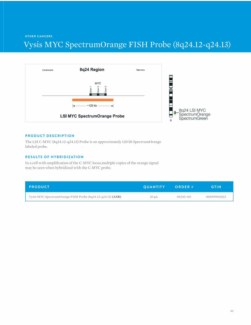
P R O D U C T D E S C R I P T I O NThe LSI C-MYC (8q24.12-q24.13) Probe is an approximately 120 kb SpectrumOrange labeled probe.
R E S U LT S O F H Y B R I D I Z AT I O NIn a cell with amplification of the C-MYC locus,multiple copies of the orange signal may be seen when hybridized with the C-MYC probe.
P R O D U C T Q UA N T I T Y O R D E R # G T I N
Vysis MYC SpectrumOrange FISH Probe (8q24.12-q24.13) (ASR) 20 µL 05J45-011 884999011823

46
V Y S I S F I S H — H E M ATO LO G Y
VYSIS FISH:HEMATOLOGY
46
V Y S I S F I S H — H E M ATO LO G Y
Abbott Molecular offers a wide range of DNA Fluorescence in situ Hybridization (FISH) products for the effective and rapid identification of genetic aberrations associated with hematopoietic disorders. Used as single probes, or in multi-color probe sets, these products are designed to identify various chromosome translocations, deletions, chromosomal gains, as well as other rearrangements associated with specific hematopoietic
disorders. These probes can be applied to a variety of sample types prepared for metaphase or interphase analysis.
4747
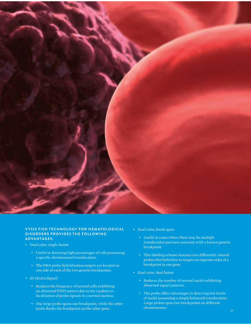
V Y S I S F I S H T E C H N O LO G Y F O R H E M ATO LO G I C A L D I S O R D E R S P R OV I D E S T H E F O L LO W I N G A DVA N TAG E S :• Dual color, single fusion
• Useful in detecting high percentages of cells possessing a specific chromosomal translocation.
• The DNA probe hybridization targets are located on one side of each of the two genetic breakpoints.
• ES (Extra Signal)
• Reduces the frequency of normal cells exhibiting an abnormal FISH pattern due to the random co-localization of probe signals in a normal nucleus.
• One large probe spans one breakpoint, while the other probe flanks the breakpoint on the other gene.
• Dual color, break apart
• Useful in cases where there may be multiple translocation partners associate with a known genetic breakpoint.
• This labeling scheme features two differently colored probes that hybridize to targets on opposite sides of a breakpoint in one gene
• Dual color, dual fusion
• Reduces the number of normal nuclei exhibiting abnormal signal patterns.
• The probe offers advantages in detecting low levels of nuclei possessing a simple balanced translocation. Large probes span two breakpoints on different chromosomes.
48
V Y S I S F I S H — H E M ATO LO G Y
P R O D U C T Q UA N T I T Y O R D E R # G T I N P G
A C U T E LYM P H O C Y T I C L E U K E M I A
Vysis CDKN2A / CEP 9 FISH Probe (ASR) 20 µL 05J51-001 884999012004 52
Vysis LSI ETV6 (CEN) SpectrumGreen (ASR) 20 µL 07J77-004 884999041530 53
Vysis LSI ETV6 (TEL) SpectrumOrange (ASR) 20 µL 07J77-003 884999041523 53
Vysis LSI ETV6 Dual Color Break Apart Rearrangement Probe (ASR) 20 µL 07J77-001 884999029262 53
Vysis LSI BCR, ABL Dual Color, Single Fusion Translocation Probe (ASR) 20 µL 05J77-001 884999012462 54
Vysis LSI BCR, ABL ES Dual Color Translocation Probe (ASR) 20 µL 05J78-001 884999012479 55
Vysis LSI BCR/ABL Dual Color, Dual Fusion Translocation Probe (ASR) 20 µL 05J82-001 884999012592 56
Vysis LSI BCR/ABL Dual Color, Dual Fusion Translocation Probe (ASR) 50 µL 05J82-010 884999012615 56
Vysis LSI CBFB Break Apart Rearrangement Probe (ASR) 20 µL 05J65-001 884999012240 58
Vysis LSI MLL Dual Color, Break Apart Rearrangement Probe (ASR) 20 µL 05J90-001 884999012837 59
Vysis LSI MYB (6q23) SpectrumAqua Probe (ASR) 20 µL 07J86-011 884999029392 60
Vysis LSI MYC Dual Color Break Apart Rearrangement Probe (ASR) 20 µL 05J91-001 884999012844 61
Vysis LSI RUNX1 SpectrumGreen Probe (ASR) 10 µL 05N98-010 884999015500 62
Vysis LSI TCF3/PBX1 Dual Color, Dual Fusion Translocation Probe (ASR) 20 µL 01N24-020 884999000605 63
Vysis LSI TEL/AML1 ES Dual Color Translocation Probe (ASR) 20 µL 05J62-001 884999012202 65
Vysis LSI TRA/D Dual Color Break Apart Rearrangement Probe (ASR) 20 µL 01N78-020 884999001015 66
A C U T E MY E L O G E N O U S L E U K E M I A
Vysis CEP 8 SpectrumOrange Direct Labeled Fluorescent DNA Probe Kit (IVD) 20 Assays 07J22-008 884999027077 67
Vysis CEP 8 SpectrumOrange Direct Labeled Fluorescent DNA Probe Kit (without control slides) (IVD) 20 Assays 07J20-008 884999027008 67
Vysis D7S486/ Vysis CEP 7 FISH Probe Kit (IVD) 20 µL 04N78-020 884999036406 69
Vysis EGR1 FISH Probe Kit - SC (Specimen Characterization) (IVD) 20 µL 04N37-001 884999038165 70
49
P R O D U C T Q UA N T I T Y O R D E R # G T I N P G
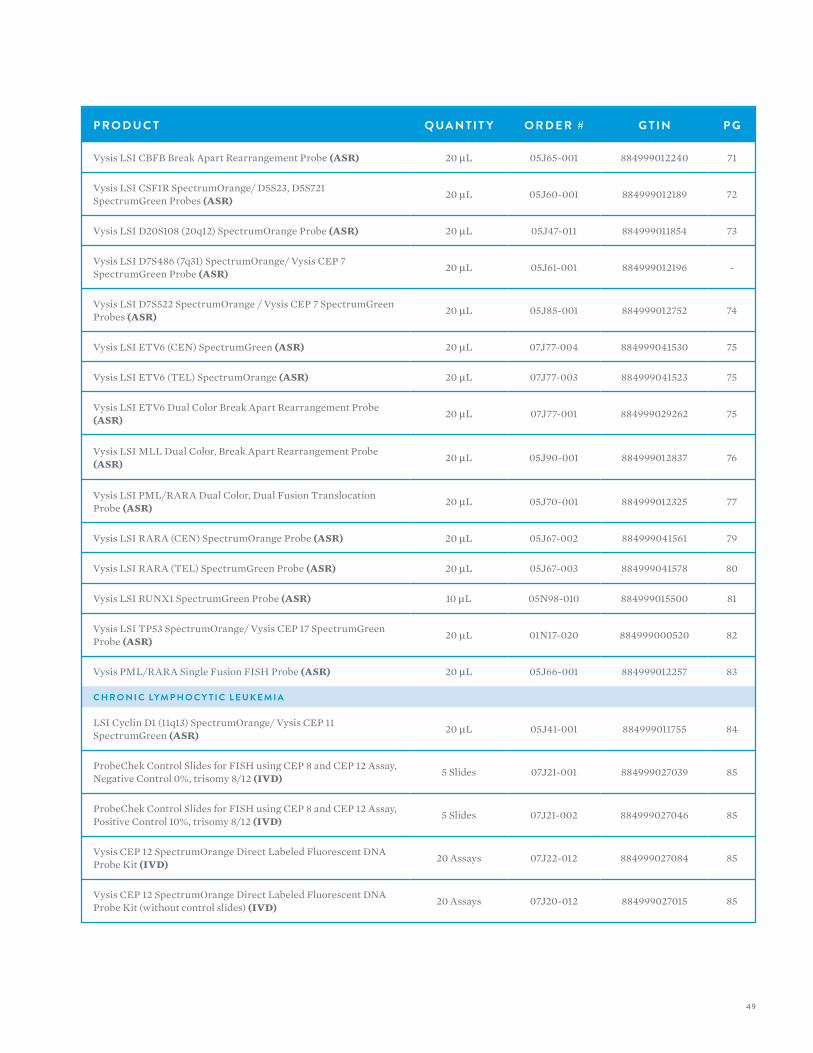
Vysis LSI CBFB Break Apart Rearrangement Probe (ASR) 20 µL 05J65-001 884999012240 71
Vysis LSI CSF1R SpectrumOrange/ D5S23, D5S721 SpectrumGreen Probes (ASR) 20 µL 05J60-001 884999012189 72
Vysis LSI D20S108 (20q12) SpectrumOrange Probe (ASR) 20 µL 05J47-011 884999011854 73
Vysis LSI D7S486 (7q31) SpectrumOrange/ Vysis CEP 7 SpectrumGreen Probe (ASR) 20 µL 05J61-001 884999012196 -
Vysis LSI D7S522 SpectrumOrange / Vysis CEP 7 SpectrumGreen Probes (ASR) 20 µL 05J85-001 884999012752 74
Vysis LSI ETV6 (CEN) SpectrumGreen (ASR) 20 µL 07J77-004 884999041530 75
Vysis LSI ETV6 (TEL) SpectrumOrange (ASR) 20 µL 07J77-003 884999041523 75
Vysis LSI ETV6 Dual Color Break Apart Rearrangement Probe (ASR) 20 µL 07J77-001 884999029262 75
Vysis LSI MLL Dual Color, Break Apart Rearrangement Probe (ASR) 20 µL 05J90-001 884999012837 76
Vysis LSI PML/RARA Dual Color, Dual Fusion Translocation Probe (ASR) 20 µL 05J70-001 884999012325 77
Vysis LSI RARA (CEN) SpectrumOrange Probe (ASR) 20 µL 05J67-002 884999041561 79
Vysis LSI RARA (TEL) SpectrumGreen Probe (ASR) 20 µL 05J67-003 884999041578 80
Vysis LSI RUNX1 SpectrumGreen Probe (ASR) 10 µL 05N98-010 884999015500 81
Vysis LSI TP53 SpectrumOrange/ Vysis CEP 17 SpectrumGreen Probe (ASR) 20 µL 01N17-020 884999000520 82
Vysis PML/RARA Single Fusion FISH Probe (ASR) 20 µL 05J66-001 884999012257 83
C H R O N I C LYM P H O C Y T I C L E U K E M I A
LSI Cyclin D1 (11q13) SpectrumOrange/ Vysis CEP 11 SpectrumGreen (ASR) 20 µL 05J41-001 884999011755 84
ProbeChek Control Slides for FISH using CEP 8 and CEP 12 Assay, Negative Control 0%, trisomy 8/12 (IVD) 5 Slides 07J21-001 884999027039 85
ProbeChek Control Slides for FISH using CEP 8 and CEP 12 Assay, Positive Control 10%, trisomy 8/12 (IVD) 5 Slides 07J21-002 884999027046 85
Vysis CEP 12 SpectrumOrange Direct Labeled Fluorescent DNA Probe Kit (IVD) 20 Assays 07J22-012 884999027084 85
Vysis CEP 12 SpectrumOrange Direct Labeled Fluorescent DNA Probe Kit (without control slides) (IVD) 20 Assays 07J20-012 884999027015 85

50
V Y S I S F I S H — H E M ATO LO G Y
P R O D U C T Q UA N T I T Y O R D E R # G T I N P G
Vysis CLL FISH Probe Kit (IVD) 20 Assays 04N02-020 884999007772 87
Vysis D13S319 (13q14.3) SpectrumOrange Probe (ASR) 20 µL 05J86-011 884999012776 91
Vysis LSI 4q12 Tricolor, Rearrangement Probe (ASR) 20 µL 01N79-020 884999001039 92
Vysis LSI ATM (11q22.3) SpectrumOrange Probe (ASR) 20 µL 05J64-011 884999012233 93
Vysis LSI ATM SpectrumOrange/Vysis CEP 11 SpectrumGreen Probes (ASR) 20 µL 01N18-020 884999000537 94
Vysis LSI D13S319 SpectrumOrange/ 13q34 SpectrumGreen Probes (ASR) 20 µL 01N20-020 884999000544 95
Vysis LSI IGH/CCND1 XT Dual Color, Dual Fusion Translocation Probes (ASR) 20 µL 05J72-001 884999012370 96
Vysis LSI MYB (6q23) SpectrumAqua Probe (ASR) 20 µL 07J86-011 884999029392 98
Vysis LSI p53 (17p13.1) SpectrumOrange Probe (ASR) 20 µL 05J52-011 884999012035 99
C H R O N I C MY E L O G E N O U S L E U K E M I A
Vysis CEP 8 SpectrumOrange Direct Labeled Fluorescent DNA Probe Kit (IVD) 20 Assays 07J22-008 884999027077 100
Vysis CEP 8 SpectrumOrange Direct Labeled Fluorescent DNA Probe Kit (without control slides) (IVD) 20 Assays 07J20-008 884999027008 100
Vysis LSI 4q12 Tricolor Rearrangement Probe (ASR) 20 µL 01N79-020 884999001039 102
Vysis LSI 9q34 SpectrumAqua Probe (ASR) 20 µL 05J79-011 884999012530 103
Vysis LSI ATM (11q22.3) SpectrumOrange Probe (ASR) 20 µL 05J64-011 884999012233 105
Vysis LSI IGH Dual Color, Break Apart Rearrangement Probe (ASR) 20 µL 05J73-001 884999012394 106
M U LT I P L E MY E L O M A
Vysis LSI (13q34) SpectrumGreen Probe (ASR) 20 µL 05J80-011 884999012561 107
Vysis LSI 13 (13q14) SpectrumGreen Probe (ASR) 20 µL 05J14-028 884999011199 108
Vysis LSI 13 RB1 (13q14) SpectrumOrange Probe (ASR) 20 µL 05J15-011 884999011212 109
Vysis LSI 13q34 SpectrumGreen Probe (ASR) 20 µL 05J80-011 884999012561 110
Vysis LSI IGH Dual Color, Break Apart Rearrangement Probe (ASR) 20 µL 05J73-001 884999012394 111
Vysis LSI IGH/BCL2 Dual Color, Dual Fusion Translocation Probe (ASR) 20 µL 05J71-001 884999012356 113
Vysis LSI IGH/CCND1 XT Dual Color, Dual Fusion Translocation Probes (ASR) 20 µL 05J72-001 884999012370 114

51
P R O D U C T Q UA N T I T Y O R D E R # G T I N P G
Vysis LSI IGH/FGFR3 Dual Color Dual Fusion Probes (ASR) 20 µL 05J74-001 884999012417 116
Vysis LSI IGH/MAF Dual Color, Dual Fusion Probe (ASR) 20 µL 05J84-004 884999012691 118
MY E L O DY S P L A S T I C S Y N D R O M E
ProbeChek Control Slides for FISH using CEP 8 and CEP 12 Assay, Negative Control 0%, trisomy 8/12 (IVD) 5 Slides 07J21-001 884999027039 120
ProbeChek Control Slides for FISH using CEP 8 and CEP 12 Assay, Positive Control 10%, trisomy 8/12 (IVD) 5 Slides 07J21-002 884999027046 120
Vysis CEP 8 SpectrumOrange Direct Labeled Fluorescent DNA Probe Kit (IVD) 20 Assays 07J22-008 884999027077 120
Vysis CEP 8 SpectrumOrange Direct Labeled Fluorescent DNA Probe Kit (without control slides) (IVD) 20 Assays 07J20-008 884999027008 120
Vysis D7S486/ Vysis CEP 7 FISH Probe Kit (IVD) 20 µL 04N78-020 884999036406 122
Vysis LSI D20S108 (20q12) SpectrumOrange Probe (ASR) 20 µL 05J47-011 884999011854 123
Vysis LSI ETV6 (CEN) SpectrumGreen (ASR) 20 µL 07J77-004 884999041530 124
Vysis LSI ETV6 (TEL) SpectrumOrange (ASR) 20 µL 07J77-003 884999041523 124
Vysis LSI ETV6 Dual Color Break Apart Rearrangement Probe (ASR) 20 µL 07J77-001 884999029262 124
N O N -H O D G K I N S LYM P H O M A
Vysis LSI 21 SpectrumOrange (ASR) 20 µL 05J13-002 884999011168 126
Vysis LSI BCL2 Break Apart FISH Probe (ASR) 20 µL 07J75-001 884999029231 127
Vysis LSI BCL6 (ABR) Dual Color Break Apart Rearrangement Probe (ASR) 20 µL 01N23-020 884999000582 128
Vysis LSI IGH Dual Color, Break Apart Rearrangement Probe (ASR) 20 µL 05J73-001 884999012394 129
Vysis LSI IGH/BCL2 Dual Color, Dual Fusion Translocation Probe (ASR) 20 µL 05J71-001 884999012356 131
Vysis LSI IGH/CCND1 XT Dual Color, Dual Fusion Translocation Probes (ASR) 20 µL 05J72-001 884999012370 133
Vysis LSI IGH/MALT1 Dual Color Dual Fusion Probes (ASR) 20 µL 05J84-001 884999012660 135
Vysis LSI MALT1 Break Apart FISH Probe (ASR) 20 µL 05J87-001 884999012783 137
Vysis LSI MYC Dual Color Break Apart Rearrangement Probe (ASR) 20 µL 05J91-001 884999012844 139

52
V Y S I S F I S H — H E M ATO LO G YV Y S I S F I S H — H E M ATO LO G Y
A C U T E LYM P H O C Y T I C L E U K E M I A
Vysis CDKN2A/CEP 9 FISH Probe
P R O D U C T D E S C R I P T I O NVysis LSI CDKN2A/CEP 9 Probes are provided in one vial as a mixture of the LSI CDKN2A (p16) probe labeled with SpectrumOrange and the CEP 9 probe labeled with SpectrumGreen. The LSI CDKN2A probe spans approximately222 kb and contains a number of genetic loci including D9S1749, DS1747, p16 (INK4B), p14 (ARF), D9S1748, p15(INK4B), and D9S1752. The CEP 9 SpectrumGreen probe hybridizes to alpha satellite sequences specific to chromosome 9.
R E S U LT S O F H Y B R I D I Z AT I O NIn a normal sample, the expected pattern for a nucleus hybridized with the Vysis LSI CDKN2A / CEP 9 Probe is the two orange, two green (2O2G) signal pattern. If a deletion at the 190 kb region covered by the LSI p16 probe occurs on one chromosome 9 homolog and both centromeres from chromosome 9 are retained, the one orange, two green (1O2G) signal pattern is expected.Very small deletions may occur that do not delete the entire LSI p16 probe target and therefore will not be detected.
P R O D U C T Q UA N T I T Y O R D E R # G T I N
Vysis CDKN2A / CEP 9 FISH Probe (ASR) 20 µL 05J51-001 884999012004
Abormal hybridization: Vysis LSI CDKN2A / CEP 9 Probe hybridized to a nucleus exhibiting the one orange and two green signal (1O2G) pattern. One p16 gene locus is deleted and both chromosome 9 homologs are present as indicated by one orange and two green signals, respectively.
53
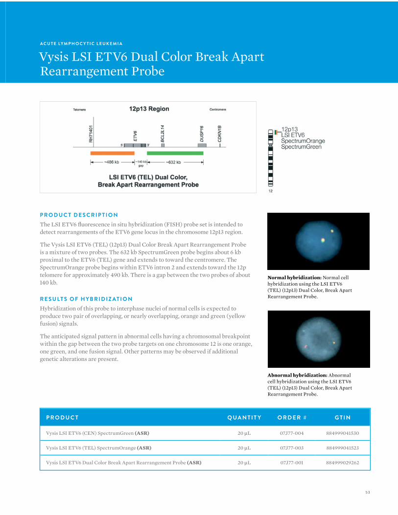
P R O D U C T D E S C R I P T I O NThe LSI ETV6 fluorescence in situ hybridization (FISH) probe set is intended to detect rearrangements of the ETV6 gene locus in the chromosome 12p13 region.
The Vysis LSI ETV6 (TEL) (12p13) Dual Color Break Apart Rearrangement Probe is a mixture of two probes. The 632 kb SpectrumGreen probe begins about 6 kb proximal to the ETV6 (TEL) gene and extends to toward the centromere. The SpectrumOrange probe begins within ETV6 intron 2 and extends toward the 12p telomere for approximately 490 kb. There is a gap between the two probes of about 140 kb.
R E S U LT S O F H Y B R I D I Z AT I O NHybridization of this probe to interphase nuclei of normal cells is expected to produce two pair of overlapping, or nearly overlapping, orange and green (yellow fusion) signals.
The anticipated signal pattern in abnormal cells having a chromosomal breakpoint within the gap between the two probe targets on one chromosome 12 is one orange, one green, and one fusion signal. Other patterns may be observed if additional genetic alterations are present.
A C U T E LYM P H O C Y T I C L E U K E M I A
Vysis LSI ETV6 Dual Color Break ApartRearrangement Probe
Abnormal hybridization: Abnormal cell hybridization using the LSI ETV6 (TEL) (12p13) Dual Color, Break Apart Rearrangement Probe.
Normal hybridization: Normal cell hybridization using the LSI ETV6 (TEL) (12p13) Dual Color, Break Apart Rearrangement Probe.
P R O D U C T Q UA N T I T Y O R D E R # G T I N
Vysis LSI ETV6 (CEN) SpectrumGreen (ASR) 20 µL 07J77-004 884999041530
Vysis LSI ETV6 (TEL) SpectrumOrange (ASR) 20 µL 07J77-003 884999041523
Vysis LSI ETV6 Dual Color Break Apart Rearrangement Probe (ASR) 20 µL 07J77-001 884999029262

54
V Y S I S F I S H — H E M ATO LO G YV Y S I S F I S H — H E M ATO LO G Y
P R O D U C T D E S C R I P T I O NThe LSI BCR/ABL Dual Color, Single Fusion Translocation Probe is a mixture of the LSI ABL probe labeled with SpectrumOrange and the LSI BCR probe labeled with SpectrumGreen. The ABL probe begins between exons 4 and 5 and continues for about 300 kb toward the telomere of chromosome 9. The LSI BCR probe begins between BCR exons 13 and 14 (M-bcr exons 2 and 3) and extends toward the centromere on chromosome 22 for approximately 300 kb, extending well beyond the m-bcr region.
R E S U LT S O F H Y B R I D I Z AT I O NA nucleus lacking the t(9;22) will exhibit the two orange, two green (2O2G) signal pattern. In a cell harboring the t(9;22), one orange, one green, and one orange/green (yellow) fusion signal pattern (1O1G1F) will be observed. This simple probe design detects the 5’ BCR/3’ ABL gene fusion and is useful for detecting samples with a high percentage of cells possessing this translocation.
A C U T E LYM P H O C Y T I C L E U K E M I A
Vysis LSI BCR, ABL Dual Color, Single Fusion Translocation Probe
LSI BCR/ABL Dual Color, Single Fusion Translocation Probe hybridized to a nucleus containing the t(9;22). One orange, one green and one fusion (IOIGIF) signal pattern is observed.
P R O D U C T Q UA N T I T Y O R D E R # G T I N
Vysis LSI BCR, ABL Dual Color, Single Fusion Translocation Probe (ASR) 20 µL 05J77-001 884999012462
55
A C U T E LYM P H O C Y T I C L E U K E M I A
Vysis LSI BCR, ABL ES Dual ColorTranslocation Probe
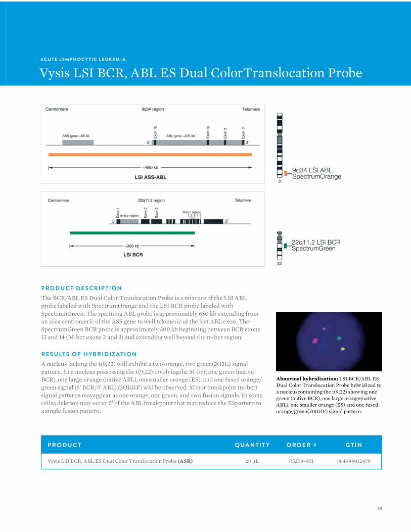
P R O D U C T D E S C R I P T I O NThe BCR/ABL ES Dual Color Translocation Probe is a mixture of the LSI ABL probe labeled with SpectrumOrange and the LSI BCR probe labeled with SpectrumGreen. The spanning ABL probe is approximately 650 kb extending from an area centromeric of the ASS gene to well telomeric of the last ABL exon. The SpectrumGreen BCR probe is approximately 300 kb beginning between BCR exons 13 and 14 (M-bcr exons 2 and 3) and extending well beyond the m-bcr region.
R E S U LT S O F H Y B R I D I Z AT I O NA nucleus lacking the t(9;22) will exhibit a two orange, two green(2O2G) signal pattern. In a nucleus possessing the t(9;22) involvingthe M-bcr, one green (native BCR), one large orange (native ABL), onesmaller orange (ES), and one fused orange/green signal (5’ BCR/3’ ABL),(2O1G1F) will be observed. Minor breakpoint (m-bcr) signal patterns mayappear as one orange, one green, and two fusion signals. In some cellsa deletion may occur 5’ of the ABL breakpoint that may reduce the ESpattern to a single fusion pattern.
P R O D U C T Q UA N T I T Y O R D E R # G T I N
Vysis LSI BCR, ABL ES Dual Color Translocation Probe (ASR) 20 µL 05J78-001 884999012479
Abnormal hybridization: LSI BCR/ABL ES Dual Color Translocation Probe hybridized to a nucleuscontaining the t(9;22) showing one green (native BCR), one large orange(native ABL), one smaller orange (ES) and one fused orange/green(20IGIF) signal pattern.
56
V Y S I S F I S H — H E M ATO LO G YV Y S I S F I S H — H E M ATO LO G Y
A C U T E LYM P H O C Y T I C L E U K E M I A
Vysis LSI BCR/ABL Dual Color, Dual Fusion Translocation Probe
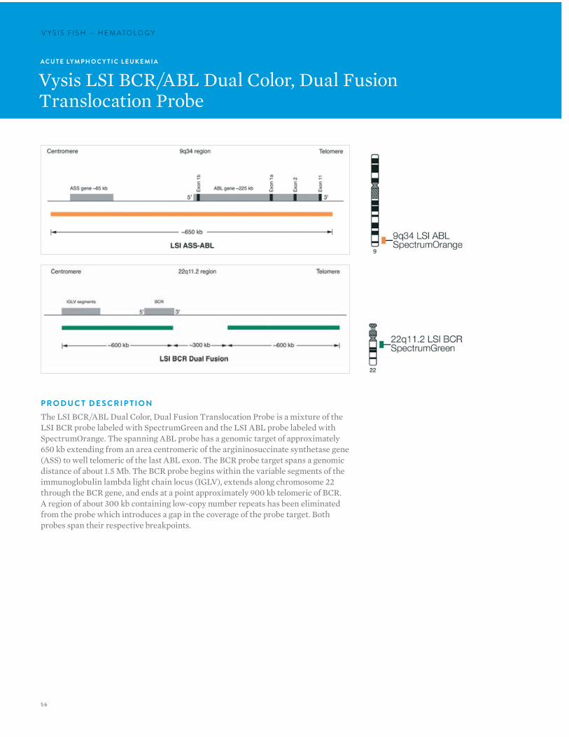
P R O D U C T D E S C R I P T I O NThe LSI BCR/ABL Dual Color, Dual Fusion Translocation Probe is a mixture of the LSI BCR probe labeled with SpectrumGreen and the LSI ABL probe labeled with SpectrumOrange. The spanning ABL probe has a genomic target of approximately 650 kb extending from an area centromeric of the argininosuccinate synthetase gene (ASS) to well telomeric of the last ABL exon. The BCR probe target spans a genomic distance of about 1.5 Mb. The BCR probe begins within the variable segments of the immunoglobulin lambda light chain locus (IGLV), extends along chromosome 22 through the BCR gene, and ends at a point approximately 900 kb telomeric of BCR. A region of about 300 kb containing low-copy number repeats has been eliminated from the probe which introduces a gap in the coverage of the probe target. Both probes span their respective breakpoints.

57
P R O D U C T Q UA N T I T Y O R D E R # G T I N
Vysis LSI BCR/ABL Dual Color, Dual Fusion Translocation Probe (ASR) 20 µL50 µL
05J82-00105J82-010
884999012592884999012615
Abnormal hybridization: LSI BCR/ABL Dual Color, Dual Fusion Translocation Probe hybridized to a nucleus containing a simple balanced t(9;22). One orange, one green and two orange/green fusion signals are observed (1O1G2F).
R E S U LT S O F H Y B R I D I Z AT I O NA nucleus lacking the t(9;22) translocation will exhibit the two orange, two green (2O2G) signal pattern. In a nucleus containing a simple balanced t(9;22), one orange and one green signal from the normal 9 and 22 chromosomes and two orange/green (yellow) fusion signals, one each from the derivative 9 and 22 chromosomes, will be observed (1O1G2F). In some instances, deletions may occur 3’ of the BCR breakpoint and/or 5’ of the ABL breakpoint resulting in either an ES (extra orange or green) signal pattern or a single fusion pattern.
58
V Y S I S F I S H — H E M ATO LO G YV Y S I S F I S H — H E M ATO LO G Y
A C U T E LYM P H O C Y T I C L E U K E M I A
Vysis LSI CBFB Break Apart Rearrangement Probe
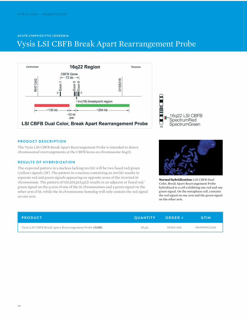
Normal hybridization: LSI CBFB Dual Color, Break Apart Rearrangement Probe hybridized to a cell exhibiting one red and one green signal. On the metaphase cell, contains the red signal on one arm and the green signal on the other arm.
P R O D U C T D E S C R I P T I O NThe Vysis LSI CBFB Break Apart Rearrangement Probe is intended to detect chromosomal rearrangements at the CBFB locus on chromosome 16q22.
R E S U LT S O F H Y B R I D I Z AT I O NThe expected pattern in a nucleus lacking inv(16) will be two fused red/green (yellow) signals (2F). The pattern in a nucleus containing an inv(16) results in separate red and green signals appearing on opposite arms of the inverted 16 chromosome. The pattern of t(16;16)(p13;q22) results in an adjacent or fused red/green signal on the q arm of one of the 16 chromosomes and a green signal on the other arm of 16, while the 16 chromosome homolog will only contain the red signal on one arm.
P R O D U C T Q UA N T I T Y O R D E R # G T I N
Vysis LSI CBFB Break Apart Rearrangement Probe (ASR) 20 µL 05J65-001 884999012240
59
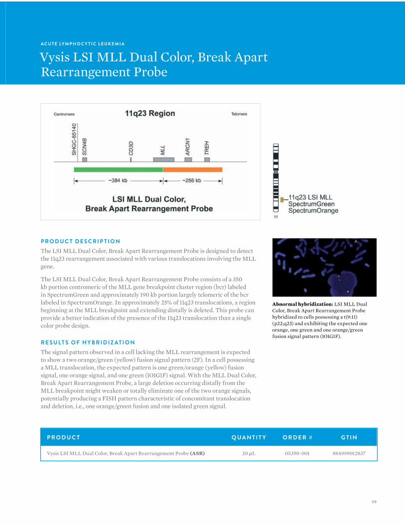
P R O D U C T D E S C R I P T I O NThe LSI MLL Dual Color, Break Apart Rearrangement Probe is designed to detect the 11q23 rearrangement associated with various translocations involving the MLL gene.
The LSI MLL Dual Color, Break Apart Rearrangement Probe consists of a 350 kb portion centromeric of the MLL gene breakpoint cluster region (bcr) labeled in SpectrumGreen and approximately 190 kb portion largely telomeric of the bcr labeled in SpectrumOrange. In approximately 25% of 11q23 translocations, a region beginning at the MLL breakpoint and extending distally is deleted. This probe can provide a better indication of the presence of the 11q23 translocation than a single color probe design.
R E S U LT S O F H Y B R I D I Z AT I O NThe signal pattern observed in a cell lacking the MLL rearrangement is expected to show a two orange/green (yellow) fusion signal pattern (2F). In a cell possessing a MLL translocation, the expected pattern is one green/orange (yellow) fusion signal, one orange signal, and one green (1O1G1F) signal. With the MLL Dual Color, Break Apart Rearrangement Probe, a large deletion occurring distally from the MLL breakpoint might weaken or totally eliminate one of the two orange signals, potentially producing a FISH pattern characteristic of concomitant translocation and deletion, i.e., one orange/green fusion and one isolated green signal.
A C U T E LYM P H O C Y T I C L E U K E M I A
Vysis LSI MLL Dual Color, Break ApartRearrangement Probe
Abnormal hybridization: LSI MLL Dual Color, Break Apart Rearrangement Probe hybridized to cells possessing a t(9:11)(p22;q23) and exhibiting the expected one orange, one green and one orange/green fusion signal pattern (1O1G1F).
P R O D U C T Q UA N T I T Y O R D E R # G T I N
Vysis LSI MLL Dual Color, Break Apart Rearrangement Probe (ASR) 20 µL 05J90-001 884999012837
60
V Y S I S F I S H — H E M ATO LO G YV Y S I S F I S H — H E M ATO LO G Y
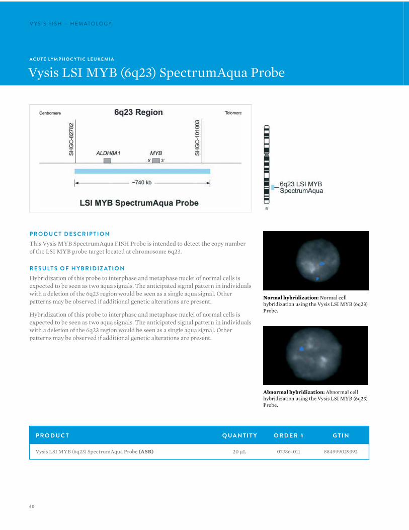
Normal hybridization: Normal cell hybridization using the Vysis LSI MYB (6q23) Probe.
Abnormal hybridization: Abnormal cell hybridization using the Vysis LSI MYB (6q23) Probe.
A C U T E LYM P H O C Y T I C L E U K E M I A
Vysis LSI MYB (6q23) SpectrumAqua Probe
P R O D U C T D E S C R I P T I O NThis Vysis MYB SpectrumAqua FISH Probe is intended to detect the copy number of the LSI MYB probe target located at chromosome 6q23.
R E S U LT S O F H Y B R I D I Z AT I O NHybridization of this probe to interphase and metaphase nuclei of normal cells is expected to be seen as two aqua signals. The anticipated signal pattern in individuals with a deletion of the 6q23 region would be seen as a single aqua signal. Other patterns may be observed if additional genetic alterations are present.
Hybridization of this probe to interphase and metaphase nuclei of normal cells is expected to be seen as two aqua signals. The anticipated signal pattern in individuals with a deletion of the 6q23 region would be seen as a single aqua signal. Other patterns may be observed if additional genetic alterations are present.
P R O D U C T Q UA N T I T Y O R D E R # G T I N
Vysis LSI MYB (6q23) SpectrumAqua Probe (ASR) 20 µL 07J86-011 884999029392
61
A C U T E LYM P H O C Y T I C L E U K E M I A
Vysis LSI MYC Dual Color Break ApartRearrangement Probe
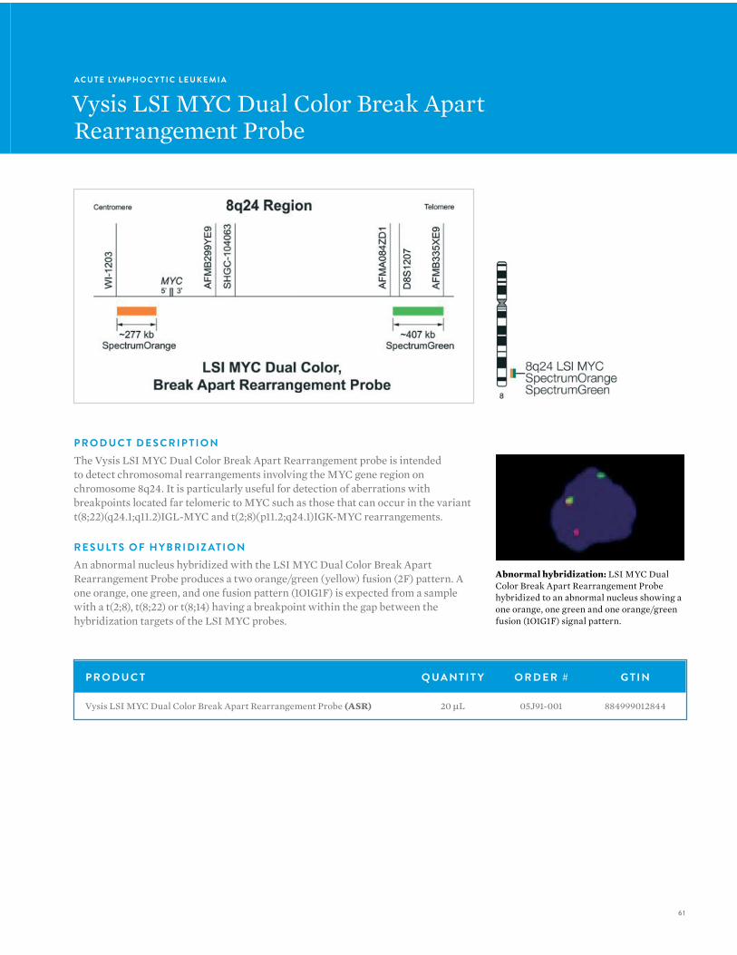
P R O D U C T D E S C R I P T I O NThe Vysis LSI MYC Dual Color Break Apart Rearrangement probe is intended to detect chromosomal rearrangements involving the MYC gene region on chromosome 8q24. It is particularly useful for detection of aberrations with breakpoints located far telomeric to MYC such as those that can occur in the variant t(8;22)(q24.1;q11.2)IGL-MYC and t(2;8)(p11.2;q24.1)IGK-MYC rearrangements.
R E S U LT S O F H Y B R I D I Z AT I O NAn abnormal nucleus hybridized with the LSI MYC Dual Color Break Apart Rearrangement Probe produces a two orange/green (yellow) fusion (2F) pattern. A one orange, one green, and one fusion pattern (1O1G1F) is expected from a sample with a t(2;8), t(8;22) or t(8;14) having a breakpoint within the gap between the hybridization targets of the LSI MYC probes.
P R O D U C T Q UA N T I T Y O R D E R # G T I N
Vysis LSI MYC Dual Color Break Apart Rearrangement Probe (ASR) 20 µL 05J91-001 884999012844
Abnormal hybridization: LSI MYC Dual Color Break Apart Rearrangement Probe hybridized to an abnormal nucleus showing a one orange, one green and one orange/green fusion (1O1G1F) signal pattern.
62
V Y S I S F I S H — H E M ATO LO G YV Y S I S F I S H — H E M ATO LO G Y
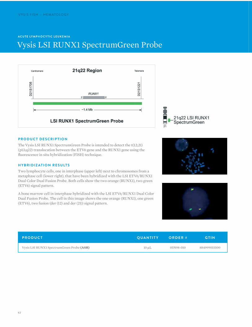
P R O D U C T D E S C R I P T I O NThe Vysis LSI RUNX1 SpectrumGreen Probe is intended to detect the t(12;21)(p13;q22) translocation between the ETV6 gene and the RUNX1 gene using the fluorescence in situ hybridization (FISH) technique.
H Y B R I D I Z AT I O N R E S U LT STwo lymphocyte cells, one in interphase (upper left) next to chromosomes from a metaphase cell (lower right), that have been hybridized with the LSI ETV6/RUNX1 Dual Color Dual Fusion Probe. Both cells show the two orange (RUNX1), two green (ETV6) signal pattern.
A bone marrow cell in interphase hybridized with the LSI ETV6/RUNX1 Dual Color Dual Fusion Probe. The cell in this image shows the one orange (RUNX1), one green (ETV6), two fusion (der (12) and der (21)) signal pattern.
A C U T E LYM P H O C Y T I C L E U K E M I A
Vysis LSI RUNX1 SpectrumGreen Probe
P R O D U C T Q UA N T I T Y O R D E R # G T I N
Vysis LSI RUNX1 SpectrumGreen Probe (ASR) 10 µL 05N98-010 884999015500
63
A C U T E LYM P H O C Y T I C L E U K E M I A
Vysis LSI TCF3/PBX1 Dual Color, Dual Fusion Translocation Probe
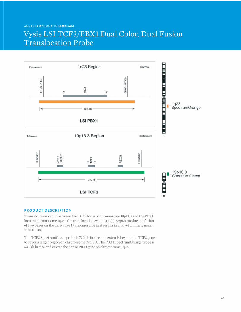
P R O D U C T D E S C R I P T I O NTranslocations occur between the TCF3 locus at chromosome 19p13.3 and the PBX1 locus at chromosome 1q23. The translocation event t(1;19)(q23;p13) produces a fusion of two genes on the derivative 19 chromosome that results in a novel chimeric gene, TCF3/PBX1.
The TCF3 SpectrumGreen probe is 730 kb in size and extends beyond the TCF3 gene to cover a larger region on chromosome 19p13.3. The PBX1 SpectrumOrange probe is 635 kb in size and covers the entire PBX1 gene on chromosome 1q23.
64
V Y S I S F I S H — H E M ATO LO G Y
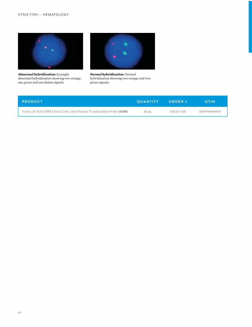
Normal hybridization: Normal hybridization showing two orange and two green signals.
Abnormal hybridization: Example abnormal hybridization showing two orange, one green and one fusion signals.
P R O D U C T Q UA N T I T Y O R D E R # G T I N
Vysis LSI TCF3/PBX1 Dual Color, Dual Fusion Translocation Probe (ASR) 20 µL 01N24-020 884999000605
65
A C U T E LYM P H O C Y T I C L E U K E M I A
Vysis LSI TEL/AML1 ES Dual Color Translocation Probe
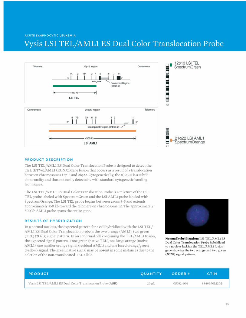
P R O D U C T D E S C R I P T I O NThe LSI TEL/AML1 ES Dual Color Translocation Probe is designed to detect the TEL (ETV6)/AML1 (RUNX1)gene fusion that occurs as a result of a translocation between chromosomes 12p13 and 21q22. Cytogenetically, the t(12;21) is a subtle abnormality and thus not easily detectable with standard cytogenetic banding techniques.
The LSI TEL/AML1 ES Dual Color Translocation Probe is a mixture of the LSI TEL probe labeled with SpectrumGreen and the LSI AML1 probe labeled with SpectrumOrange. The LSI TEL probe begins between exons 3-5 and extends approximately 350 kb toward the telomere on chromosome 12. The approximately 500 kb AML1 probe spans the entire gene.
R E S U LT S O F H Y B R I D I Z AT I O NIn a normal nucleus, the expected pattern for a cell hybridized with the LSI TEL/AML1 ES Dual Color Translocation probe is the two orange (AML1), two green (TEL) (2O2G) signal pattern. In an abnormal cell containing the TEL/AML1 fusion, the expected signal pattern is one green (native TEL), one large orange (native AML1), one smaller orange signal (residual AML1) and one fused orange/green (yellow) signal. The green native signal may be absent in some instances due to the deletion of the non-translocated TEL allele.
Normal hybridization: LSI TEL/AML1 ES Dual Color Translocation Probe hybridized to a nucleus lacking the TEL/AML1 fusion gene showing the two orange and two green (2O2G) signal pattern.
P R O D U C T Q UA N T I T Y O R D E R # G T I N
Vysis LSI TEL/AML1 ES Dual Color Translocation Probe (ASR) 20 µL 05J62-001 884999012202
66
V Y S I S F I S H — H E M ATO LO G YV Y S I S F I S H — H E M ATO LO G Y
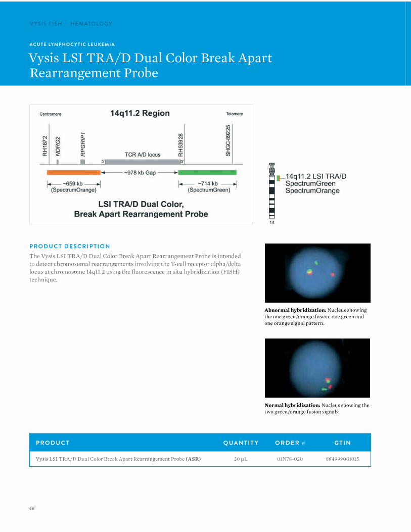
Normal hybridization: Nucleus showing the two green/orange fusion signals.
Abnormal hybridization: Nucleus showing the one green/orange fusion, one green and one orange signal pattern.
A C U T E LYM P H O C Y T I C L E U K E M I A
Vysis LSI TRA/D Dual Color Break ApartRearrangement Probe
P R O D U C T D E S C R I P T I O NThe Vysis LSI TRA/D Dual Color Break Apart Rearrangement Probe is intended to detect chromosomal rearrangements involving the T-cell receptor alpha/delta locus at chromosome 14q11.2 using the fluorescence in situ hybridization (FISH) technique.
P R O D U C T Q UA N T I T Y O R D E R # G T I N
Vysis LSI TRA/D Dual Color Break Apart Rearrangement Probe (ASR) 20 µL 01N78-020 884999001015
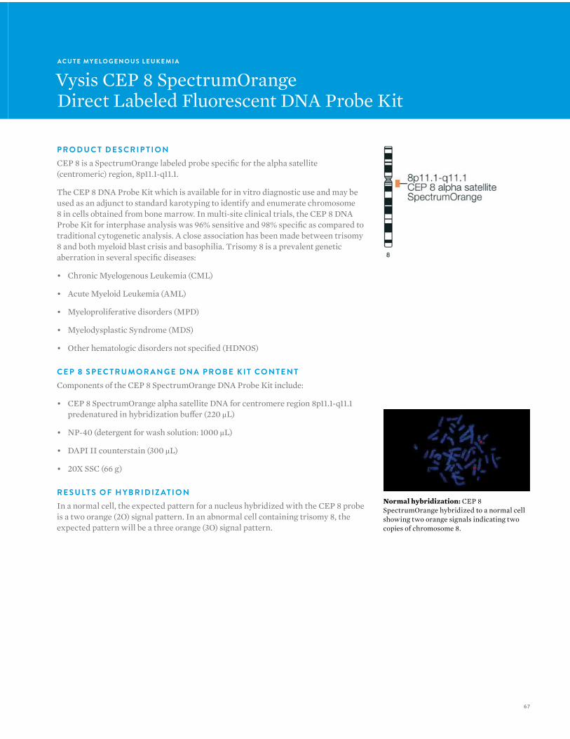
67
A C U T E MY E L O G E N O U S L E U K E M I A
Vysis CEP 8 SpectrumOrangeDirect Labeled Fluorescent DNA Probe Kit
P R O D U C T D E S C R I P T I O NCEP 8 is a SpectrumOrange labeled probe specific for the alpha satellite (centromeric) region, 8p11.1-q11.1.
The CEP 8 DNA Probe Kit which is available for in vitro diagnostic use and may be used as an adjunct to standard karotyping to identify and enumerate chromosome 8 in cells obtained from bone marrow. In multi-site clinical trials, the CEP 8 DNA Probe Kit for interphase analysis was 96% sensitive and 98% specific as compared to traditional cytogenetic analysis. A close association has been made between trisomy 8 and both myeloid blast crisis and basophilia. Trisomy 8 is a prevalent genetic aberration in several specific diseases:
• Chronic Myelogenous Leukemia (CML)
• Acute Myeloid Leukemia (AML)
• Myeloproliferative disorders (MPD)
• Myelodysplastic Syndrome (MDS)
• Other hematologic disorders not specified (HDNOS)
C E P 8 S P E C T R U M O R A N G E D N A P R O B E K I T CO N T E N TComponents of the CEP 8 SpectrumOrange DNA Probe Kit include:
• CEP 8 SpectrumOrange alpha satellite DNA for centromere region 8p11.1-q11.1 predenatured in hybridization buffer (220 µL)
• NP-40 (detergent for wash solution: 1000 µL)
• DAPI II counterstain (300 µL)
• 20X SSC (66 g)
R E S U LT S O F H Y B R I D I Z AT I O NIn a normal cell, the expected pattern for a nucleus hybridized with the CEP 8 probe is a two orange (2O) signal pattern. In an abnormal cell containing trisomy 8, the expected pattern will be a three orange (3O) signal pattern.
Normal hybridization: CEP 8 SpectrumOrange hybridized to a normal cell showing two orange signals indicating two copies of chromosome 8.
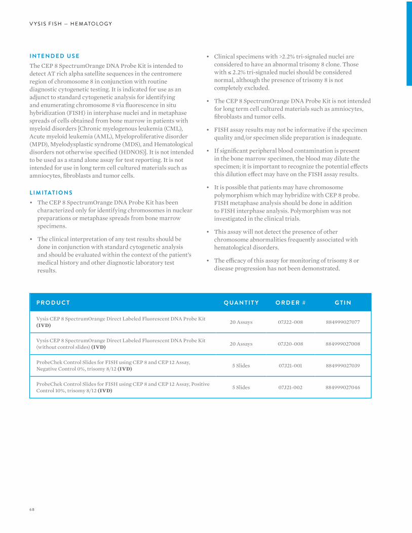
68
V Y S I S F I S H — H E M ATO LO G Y
I N T E N D E D U S EThe CEP 8 SpectrumOrange DNA Probe Kit is intended to detect AT rich alpha satellite sequences in the centromere region of chromosome 8 in conjunction with routine diagnostic cytogenetic testing. It is indicated for use as an adjunct to standard cytogenetic analysis for identifying and enumerating chromosome 8 via fluorescence in situ hybridization (FISH) in interphase nuclei and in metaphase spreads of cells obtained from bone marrow in patients with myeloid disorders [Chronic myelogenous leukemia (CML), Acute myeloid leukemia (AML), Myeloproliferative disorder (MPD), Myelodysplastic syndrome (MDS), and Hematological disorders not otherwise specified (HDNOS)]. It is not intended to be used as a stand alone assay for test reporting. It is not intended for use in long term cell cultured materials such as amniocytes, fibroblasts and tumor cells.
L I M I TAT I O N S • The CEP 8 SpectrumOrange DNA Probe Kit has been
characterized only for identifying chromosomes in nuclear preparations or metaphase spreads from bone marrow specimens.
• The clinical interpretation of any test results should be done in conjunction with standard cytogenetic analysis and should be evaluated within the context of the patient’s medical history and other diagnostic laboratory test results.
• Clinical specimens with >2.2% tri-signaled nuclei are considered to have an abnormal trisomy 8 clone. Those with ≤ 2.2% tri-signaled nuclei should be considered normal, although the presence of trisomy 8 is not completely excluded.
• The CEP 8 SpectrumOrange DNA Probe Kit is not intended for long term cell cultured materials such as amniocytes, fibroblasts and tumor cells.
• FISH assay results may not be informative if the specimen quality and/or specimen slide preparation is inadequate.
• If significant peripheral blood contamination is present in the bone marrow specimen, the blood may dilute the specimen; it is important to recognize the potential effects this dilution effect may have on the FISH assay results.
• It is possible that patients may have chromosome polymorphism which may hybridize with CEP 8 probe. FISH metaphase analysis should be done in addition to FISH interphase analysis. Polymorphism was not investigated in the clinical trials.
• This assay will not detect the presence of other chromosome abnormalities frequently associated with hematological disorders.
• The efficacy of this assay for monitoring of trisomy 8 or disease progression has not been demonstrated.
P R O D U C T Q UA N T I T Y O R D E R # G T I N
Vysis CEP 8 SpectrumOrange Direct Labeled Fluorescent DNA Probe Kit (IVD) 20 Assays 07J22-008 884999027077
Vysis CEP 8 SpectrumOrange Direct Labeled Fluorescent DNA Probe Kit (without control slides) (IVD) 20 Assays 07J20-008 884999027008
ProbeChek Control Slides for FISH using CEP 8 and CEP 12 Assay, Negative Control 0%, trisomy 8/12 (IVD) 5 Slides 07J21-001 884999027039
ProbeChek Control Slides for FISH using CEP 8 and CEP 12 Assay, Positive Control 10%, trisomy 8/12 (IVD) 5 Slides 07J21-002 884999027046
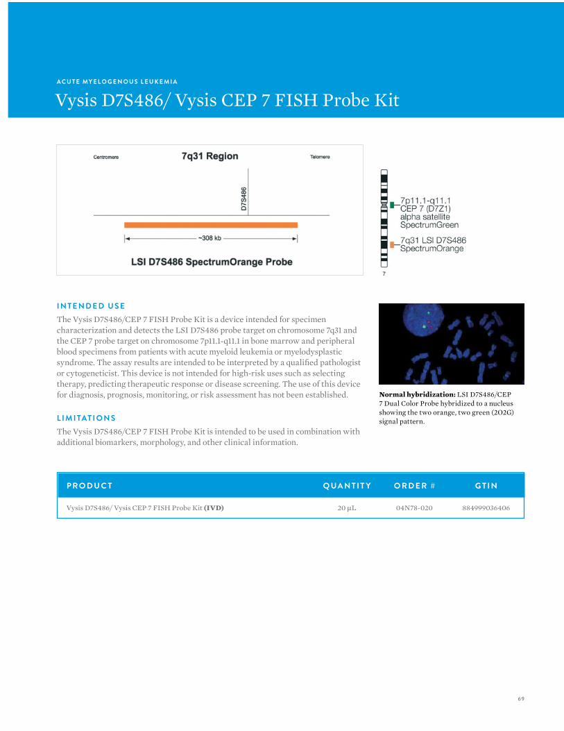
69
A C U T E MY E L O G E N O U S L E U K E M I A
Vysis D7S486/ Vysis CEP 7 FISH Probe Kit
I N T E N D E D U S EThe Vysis D7S486/CEP 7 FISH Probe Kit is a device intended for specimen characterization and detects the LSI D7S486 probe target on chromosome 7q31 and the CEP 7 probe target on chromosome 7p11.1-q11.1 in bone marrow and peripheral blood specimens from patients with acute myeloid leukemia or myelodysplastic syndrome. The assay results are intended to be interpreted by a qualified pathologist or cytogeneticist. This device is not intended for high-risk uses such as selecting therapy, predicting therapeutic response or disease screening. The use of this device for diagnosis, prognosis, monitoring, or risk assessment has not been established.
L I M I TAT I O N SThe Vysis D7S486/CEP 7 FISH Probe Kit is intended to be used in combination with additional biomarkers, morphology, and other clinical information.
Normal hybridization: LSI D7S486/CEP 7 Dual Color Probe hybridized to a nucleus showing the two orange, two green (2O2G) signal pattern.
P R O D U C T Q UA N T I T Y O R D E R # G T I N
Vysis D7S486/ Vysis CEP 7 FISH Probe Kit (IVD) 20 µL 04N78-020 884999036406
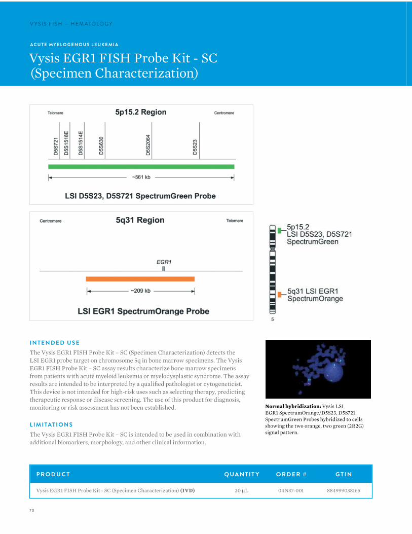
70
V Y S I S F I S H — H E M ATO LO G YV Y S I S F I S H — H E M ATO LO G Y
I N T E N D E D U S EThe Vysis EGR1 FISH Probe Kit – SC (Specimen Characterization) detects the LSI EGR1 probe target on chromosome 5q in bone marrow specimens. The Vysis EGR1 FISH Probe Kit – SC assay results characterize bone marrow specimens from patients with acute myeloid leukemia or myelodysplastic syndrome. The assay results are intended to be interpreted by a qualified pathologist or cytogeneticist. This device is not intended for high-risk uses such as selecting therapy, predicting therapeutic response or disease screening. The use of this product for diagnosis, monitoring or risk assessment has not been established.
L I M I TAT I O N SThe Vysis EGR1 FISH Probe Kit – SC is intended to be used in combination with additional biomarkers, morphology, and other clinical information.
Normal hybridization: Vysis LSI EGR1 SpectrumOrange/D5S23, D5S721 SpectrumGreen Probes hybridized to cells showing the two orange, two green (2R2G) signal pattern.
A C U T E MY E L O G E N O U S L E U K E M I A
Vysis EGR1 FISH Probe Kit - SC(Specimen Characterization)
P R O D U C T Q UA N T I T Y O R D E R # G T I N
Vysis EGR1 FISH Probe Kit - SC (Specimen Characterization) (IVD) 20 µL 04N37-001 884999038165

7 1
A C U T E MY E L O G E N O U S L E U K E M I A
Vysis LSI CBFB Break Apart Rearrangement Probe
P R O D U C T D E S C R I P T I O NThe Vysis LSI CBFB Break Apart Rearrangement Probe is intended to detect chromosomal rearrangements at the CBFB locus on chromosome 16q22.
R E S U LT S O F H Y B R I D I Z AT I O NThe expected pattern in a nucleus lacking inv(16) will be two fused red/green (yellow) signals (2F). The pattern in a nucleus containing an inv(16) results in separate red and green signals appearing on opposite arms of the inverted 16 chromosome. The pattern of t(16;16)(p13;q22) results in an adjacent or fused red/green signal on the q arm of one of the 16 chromosomes and a green signal on the other arm of 16, while the 16 chromosome homolog will only contain the red signal on one arm.
Abnormal hybridization: LSI CBFB Dual Color Break Apart Rearrangement Probe hybridized to a cell exhibiting one red and one green signal. On the metaphase cell, contains the red signal on one arm and the green signal on the other arm.
P R O D U C T Q UA N T I T Y O R D E R # G T I N
Vysis LSI CBFB Break Apart Rearrangement Probe (ASR) 20 µL 05J65-001 884999012240
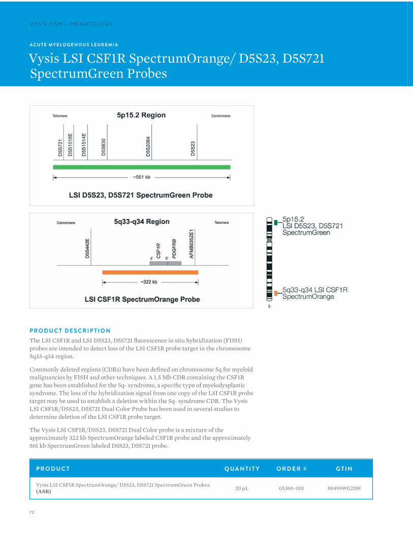
72
V Y S I S F I S H — H E M ATO LO G YV Y S I S F I S H — H E M ATO LO G Y
A C U T E MY E L O G E N O U S L E U K E M I A
Vysis LSI CSF1R SpectrumOrange/ D5S23, D5S721 SpectrumGreen Probes
P R O D U C T D E S C R I P T I O NThe LSI CSF1R and LSI D5S23, D5S721 fluorescence in situ hybridization (FISH) probes are intended to detect loss of the LSI CSF1R probe target in the chromosome 5q33-q34 region.
Commonly deleted regions (CDRs) have been defined on chromosome 5q for myeloid malignancies by FISH and other techniques. A 1.5 Mb CDR containing the CSF1R gene has been established for the 5q- syndrome, a specific type of myelodysplastic syndrome. The loss of the hybridization signal from one copy of the LSI CSF1R probe target may be used to establish a deletion within the 5q- syndrome CDR. The Vysis LSI CSF1R/D5S23, D5S721 Dual Color Probe has been used in several studies to determine deletion of the LSI CSF1R probe target.
The Vysis LSI CSF1R/D5S23, D5S721 Dual Color probe is a mixture of the approximately 322 kb SpectrumOrange labeled CSF1R probe and the approximately 561 kb SpectrumGreen labeled D5S23, D5S721 probe.
P R O D U C T Q UA N T I T Y O R D E R # G T I N
Vysis LSI CSF1R SpectrumOrange/ D5S23, D5S721 SpectrumGreen Probes (ASR) 20 µL 05J60-001 884999012189
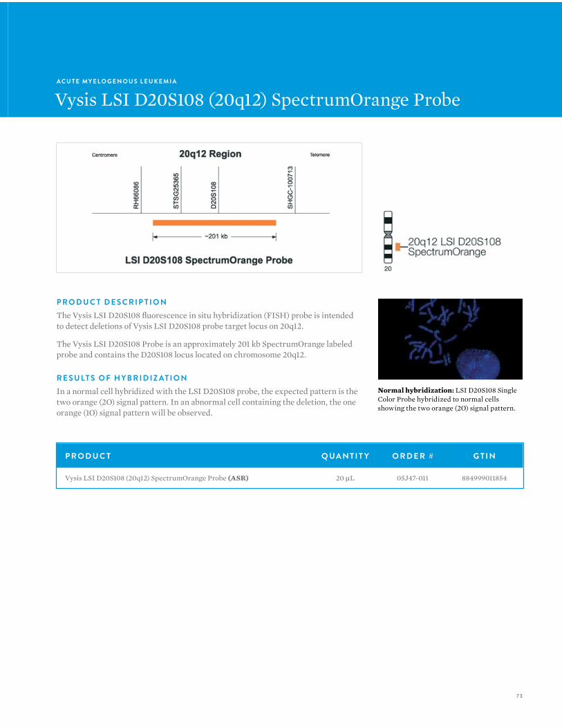
73
A C U T E MY E L O G E N O U S L E U K E M I A
Vysis LSI D20S108 (20q12) SpectrumOrange Probe
P R O D U C T D E S C R I P T I O NThe Vysis LSI D20S108 fluorescence in situ hybridization (FISH) probe is intended to detect deletions of Vysis LSI D20S108 probe target locus on 20q12.
The Vysis LSI D20S108 Probe is an approximately 201 kb SpectrumOrange labeled probe and contains the D20S108 locus located on chromosome 20q12.
R E S U LT S O F H Y B R I D I Z AT I O NIn a normal cell hybridized with the LSI D20S108 probe, the expected pattern is the two orange (2O) signal pattern. In an abnormal cell containing the deletion, the one orange (1O) signal pattern will be observed.
Normal hybridization: LSI D20S108 Single Color Probe hybridized to normal cells showing the two orange (2O) signal pattern.
P R O D U C T Q UA N T I T Y O R D E R # G T I N
Vysis LSI D20S108 (20q12) SpectrumOrange Probe (ASR) 20 µL 05J47-011 884999011854
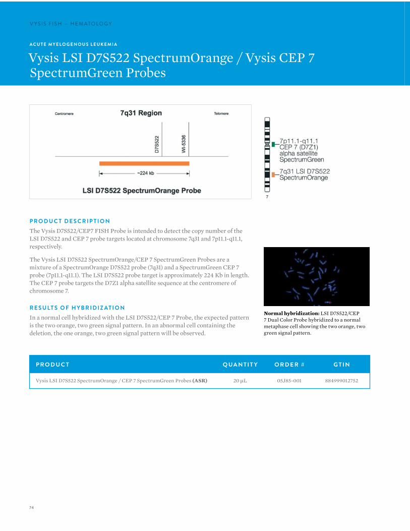
74
V Y S I S F I S H — H E M ATO LO G YV Y S I S F I S H — H E M ATO LO G Y
A C U T E MY E L O G E N O U S L E U K E M I A
Vysis LSI D7S522 SpectrumOrange / Vysis CEP 7 SpectrumGreen Probes
P R O D U C T D E S C R I P T I O NThe Vysis D7S522/CEP7 FISH Probe is intended to detect the copy number of the LSI D7S522 and CEP 7 probe targets located at chromosome 7q31 and 7p11.1-q11.1, respectively.
The Vysis LSI D7S522 SpectrumOrange/CEP 7 SpectrumGreen Probes are a mixture of a SpectrumOrange D7S522 probe (7q31) and a SpectrumGreen CEP 7 probe (7p11.1-q11.1). The LSI D7S522 probe target is approximately 224 Kb in length. The CEP 7 probe targets the D7Z1 alpha satellite sequence at the centromere of chromosome 7.
R E S U LT S O F H Y B R I D I Z AT I O NIn a normal cell hybridized with the LSI D7S522/CEP 7 Probe, the expected pattern is the two orange, two green signal pattern. In an abnormal cell containing the deletion, the one orange, two green signal pattern will be observed.
Normal hybridization: LSI D7S522/CEP 7 Dual Color Probe hybridized to a normal metaphase cell showing the two orange, two green signal pattern.
P R O D U C T Q UA N T I T Y O R D E R # G T I N
Vysis LSI D7S522 SpectrumOrange / CEP 7 SpectrumGreen Probes (ASR) 20 µL 05J85-001 884999012752

75
P R O D U C T D E S C R I P T I O NThe LSI ETV6 fluorescence in situ hybridization (FISH) probe set is intended to detect rearrangements of the ETV6 gene locus in the chromosome 12p13 region.
The Vysis LSI ETV6 (TEL) (12p13) Dual Color Break Apart Rearrangement Probe is a mixture of two probes. The 632 kb SpectrumGreen probe begins about 6 kb proximal to the ETV6 (TEL) gene and extends to toward the centromere. The SpectrumOrange probe begins within ETV6 intron 2 and extends toward the 12p telomere for approximately 490 kb. There is a gap between the two probes of about 140 kb.
R E S U LT S O F H Y B R I D I Z AT I O NHybridization of this probe to interphase nuclei of normal cells is expected to produce two pair of overlapping, or nearly overlapping, orange and green (yellow fusion) signals.
The anticipated signal pattern in abnormal cells having a chromosomal breakpoint within the gap between the two probe targets on one chromosome 12 is one orange, one green, and one fusion signal. Other patterns may be observed if additional genetic alterations are present.
A C U T E MY E L O G E N O U S L E U K E M I A
Vysis LSI ETV6 Dual Color Break ApartRearrangement Probe
Abnormal hybridization: Abnormal cell hybridization using the LSI ETV6 (TEL) (12p13) Dual Color, Break Apart Rearrangement Probe.
Normal hybridization: Normal cell hybridization using the LSI ETV6 (TEL) (12p13) Dual Color, Break Apart Rearrangement Probe.
P R O D U C T Q UA N T I T Y O R D E R # G T I N
Vysis LSI ETV6 (CEN) SpectrumGreen (ASR) 20 µL 07J77-004 884999041530
Vysis LSI ETV6 (TEL) SpectrumOrange (ASR) 20 µL 07J77-003 884999041523
Vysis LSI ETV6 Dual Color Break Apart Rearrangement Probe (ASR) 20 µL 07J77-001 884999029262
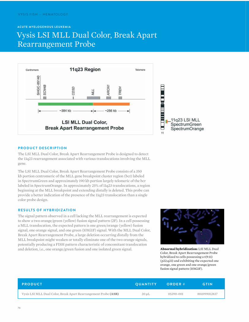
76
V Y S I S F I S H — H E M ATO LO G YV Y S I S F I S H — H E M ATO LO G Y
A C U T E MY E L O G E N O U S L E U K E M I A
Vysis LSI MLL Dual Color, Break ApartRearrangement Probe
P R O D U C T D E S C R I P T I O NThe LSI MLL Dual Color, Break Apart Rearrangement Probe is designed to detect the 11q23 rearrangement associated with various translocations involving the MLL gene.
The LSI MLL Dual Color, Break Apart Rearrangement Probe consists of a 350 kb portion centromeric of the MLL gene breakpoint cluster region (bcr) labeled in SpectrumGreen and approximately 190 kb portion largely telomeric of the bcr labeled in SpectrumOrange. In approximately 25% of 11q23 translocations, a region beginning at the MLL breakpoint and extending distally is deleted. This probe can provide a better indication of the presence of the 11q23 translocation than a single color probe design.
R E S U LT S O F H Y B R I D I Z AT I O NThe signal pattern observed in a cell lacking the MLL rearrangement is expected to show a two orange/green (yellow) fusion signal pattern (2F). In a cell possessing a MLL translocation, the expected pattern is one green/orange (yellow) fusion signal, one orange signal, and one green (1O1G1F) signal. With the MLL Dual Color, Break Apart Rearrangement Probe, a large deletion occurring distally from the MLL breakpoint might weaken or totally eliminate one of the two orange signals, potentially producing a FISH pattern characteristic of concomitant translocation and deletion, i.e., one orange/green fusion and one isolated green signal. Abnormal hybridization: LSI MLL Dual
Color, Break Apart Rearrangement Probe hybridized to cells possessing a t(9:11)(p22;q23) and exhibiting the expected one orange, one green and one orange/green fusion signal pattern (1O1G1F).
P R O D U C T Q UA N T I T Y O R D E R # G T I N
Vysis LSI MLL Dual Color, Break Apart Rearrangement Probe (ASR) 20 µL 05J90-001 884999012837
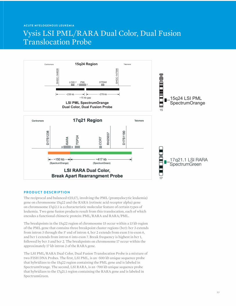
7 7
A C U T E MY E L O G E N O U S L E U K E M I A
Vysis LSI PML/RARA Dual Color, Dual Fusion Translocation Probe
Centromere Telomere
~239 kb
SH
GC
-144
626
LSI PML SpectrumOrange
Dual Color, Dual Fusion Probe
15q24 Region
SH
GC
-147
595
PML5’ 3’
~278 kb~15 kb gap
LOXL1 STRA6
15
15q24 LSI PMLSpectrumOrange
P R O D U C T D E S C R I P T I O NThe reciprocal and balanced t(15;17), involving the PML (promyelocytic leukemia) gene on chromosome 15q22 and the RARA (retinoic acid receptor alpha) gene on chromosome 17q12.1 is a characteristic molecular feature of certain types of leukemia. Two gene fusion products result from this translocation, each of which encodes a functional chimeric protein: PML/RARA and RARA/PML.
The breakpoints in the 15q22 region of chromosome 15 occur within a 13 kb region of the PML gene that contains three breakpoint cluster regions (bcr): bcr 3 extends from intron 3 through the 5’ end of intron 4, bcr 2 extends from exon 5 to exon 6, and bcr 1 extends from intron 6 into exon 7. Break frequency is highest in bcr 1, followed by bcr 3 and bcr 2. The breakpoints on chromosome 17 occur within the approximately 17 kb intron 2 of the RARA gene.
The LSI PML/RARA Dual Color, Dual Fusion Translocation Probe is a mixture of two FISH DNA Probes. The first, LSI PML, is an ~500 kb unique sequence probe that hybridizes to the 15q22 region containing the PML gene and is labeled in SpectrumOrange. The second, LSI RARA, is an ~700 kb unique sequence probe that hybridizes to the 17q21.1 region containing the RARA gene and is labeled in SpectrumGreen.
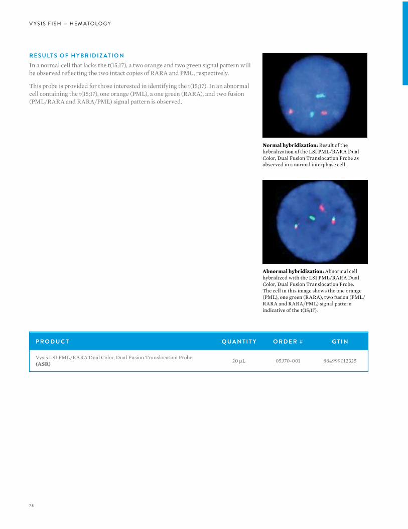
78
V Y S I S F I S H — H E M ATO LO G Y
Normal hybridization: Result of the hybridization of the LSI PML/RARA Dual Color, Dual Fusion Translocation Probe as observed in a normal interphase cell.
Abnormal hybridization: Abnormal cell hybridized with the LSI PML/RARA Dual Color, Dual Fusion Translocation Probe. The cell in this image shows the one orange (PML), one green (RARA), two fusion (PML/RARA and RARA/PML) signal pattern indicative of the t(15;17).
P R O D U C T Q UA N T I T Y O R D E R # G T I N
Vysis LSI PML/RARA Dual Color, Dual Fusion Translocation Probe (ASR) 20 µL 05J70-001 884999012325
R E S U LT S O F H Y B R I D I Z AT I O NIn a normal cell that lacks the t(15;17), a two orange and two green signal pattern will be observed reflecting the two intact copies of RARA and PML, respectively.
This probe is provided for those interested in identifying the t(15;17). In an abnormal cell containing the t(15;17), one orange (PML), a one green (RARA), and two fusion (PML/RARA and RARA/PML) signal pattern is observed.
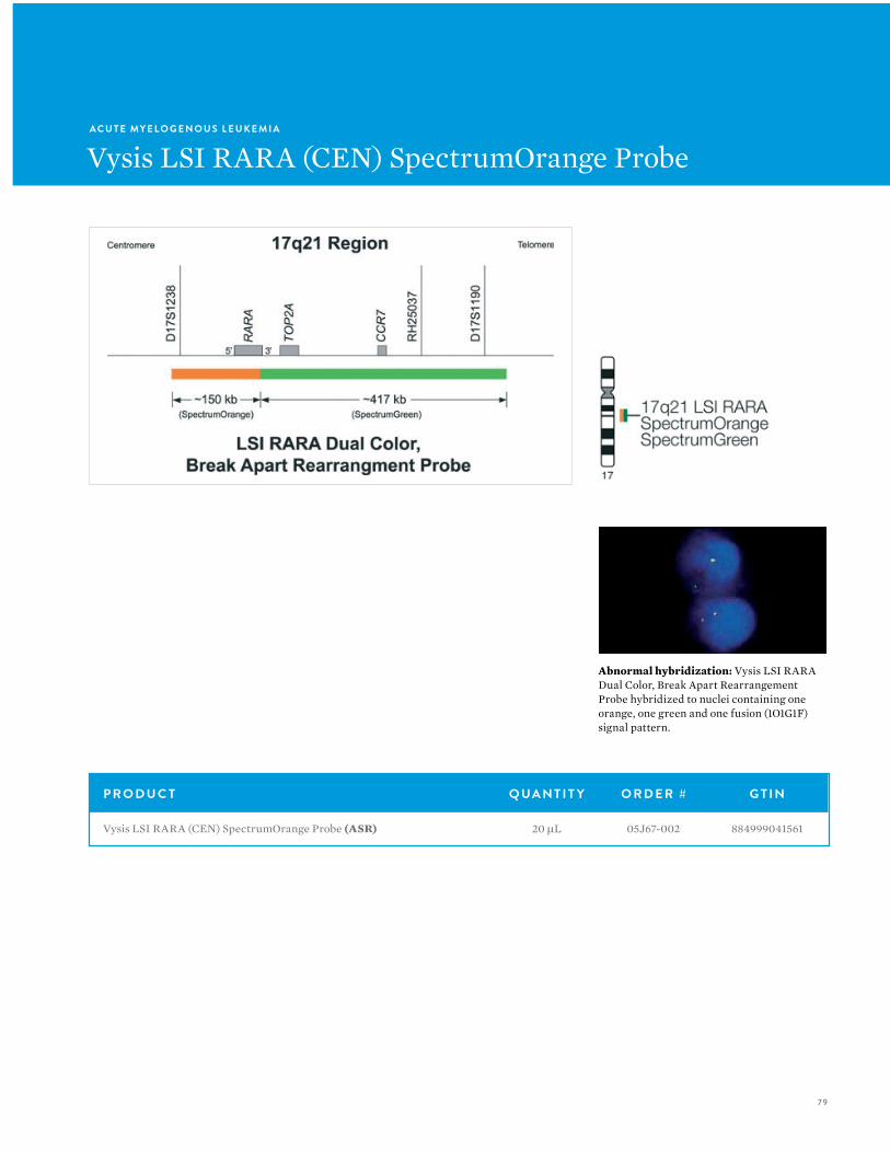
79
A C U T E MY E L O G E N O U S L E U K E M I A
Vysis LSI RARA (CEN) SpectrumOrange Probe
Abnormal hybridization: Vysis LSI RARA Dual Color, Break Apart Rearrangement Probe hybridized to nuclei containing one orange, one green and one fusion (1O1G1F) signal pattern.
P R O D U C T Q UA N T I T Y O R D E R # G T I N
Vysis LSI RARA (CEN) SpectrumOrange Probe (ASR) 20 µL 05J67-002 884999041561

80
V Y S I S F I S H — H E M ATO LO G YV Y S I S F I S H — H E M ATO LO G Y
A C U T E MY E L O G E N O U S L E U K E M I A
Vysis LSI RARA (TEL) SpectrumGreen Probe
Abnormal hybridization: Vysis LSI RARA Dual Color, Break Apart Rearrangement Probe hybridized to nuclei containing one orange, one green and one fusion (1O1G1F) signal pattern.
P R O D U C T Q UA N T I T Y O R D E R # G T I N
Vysis LSI RARA (TEL) SpectrumGreen Probe (ASR) 20 µL 05J67-003 884999041578
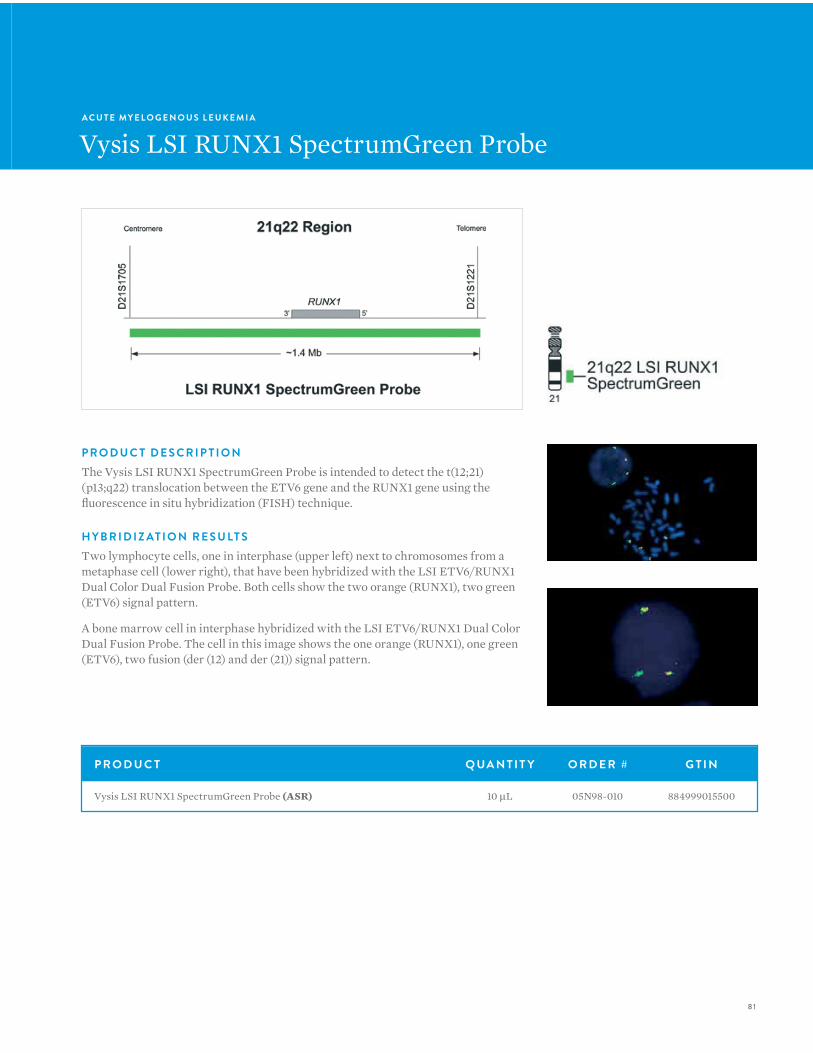
81
A C U T E MY E L O G E N O U S L E U K E M I A
Vysis LSI RUNX1 SpectrumGreen Probe
P R O D U C T D E S C R I P T I O NThe Vysis LSI RUNX1 SpectrumGreen Probe is intended to detect the t(12;21)(p13;q22) translocation between the ETV6 gene and the RUNX1 gene using the fluorescence in situ hybridization (FISH) technique.
H Y B R I D I Z AT I O N R E S U LT STwo lymphocyte cells, one in interphase (upper left) next to chromosomes from a metaphase cell (lower right), that have been hybridized with the LSI ETV6/RUNX1 Dual Color Dual Fusion Probe. Both cells show the two orange (RUNX1), two green (ETV6) signal pattern.
A bone marrow cell in interphase hybridized with the LSI ETV6/RUNX1 Dual Color Dual Fusion Probe. The cell in this image shows the one orange (RUNX1), one green (ETV6), two fusion (der (12) and der (21)) signal pattern.
P R O D U C T Q UA N T I T Y O R D E R # G T I N
Vysis LSI RUNX1 SpectrumGreen Probe (ASR) 10 µL 05N98-010 884999015500
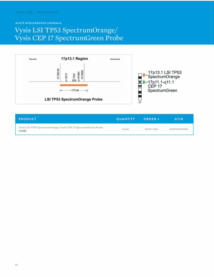
82
V Y S I S F I S H — H E M ATO LO G YV Y S I S F I S H — H E M ATO LO G Y
A C U T E MY E L O G E N O U S L E U K E M I A
Vysis LSI TP53 SpectrumOrange/Vysis CEP 17 SpectrumGreen Probe
P R O D U C T Q UA N T I T Y O R D E R # G T I N
Vysis LSI TP53 SpectrumOrange/Vysis CEP 17 SpectrumGreen Probe (ASR) 20 µL 01N17-020 884999000520
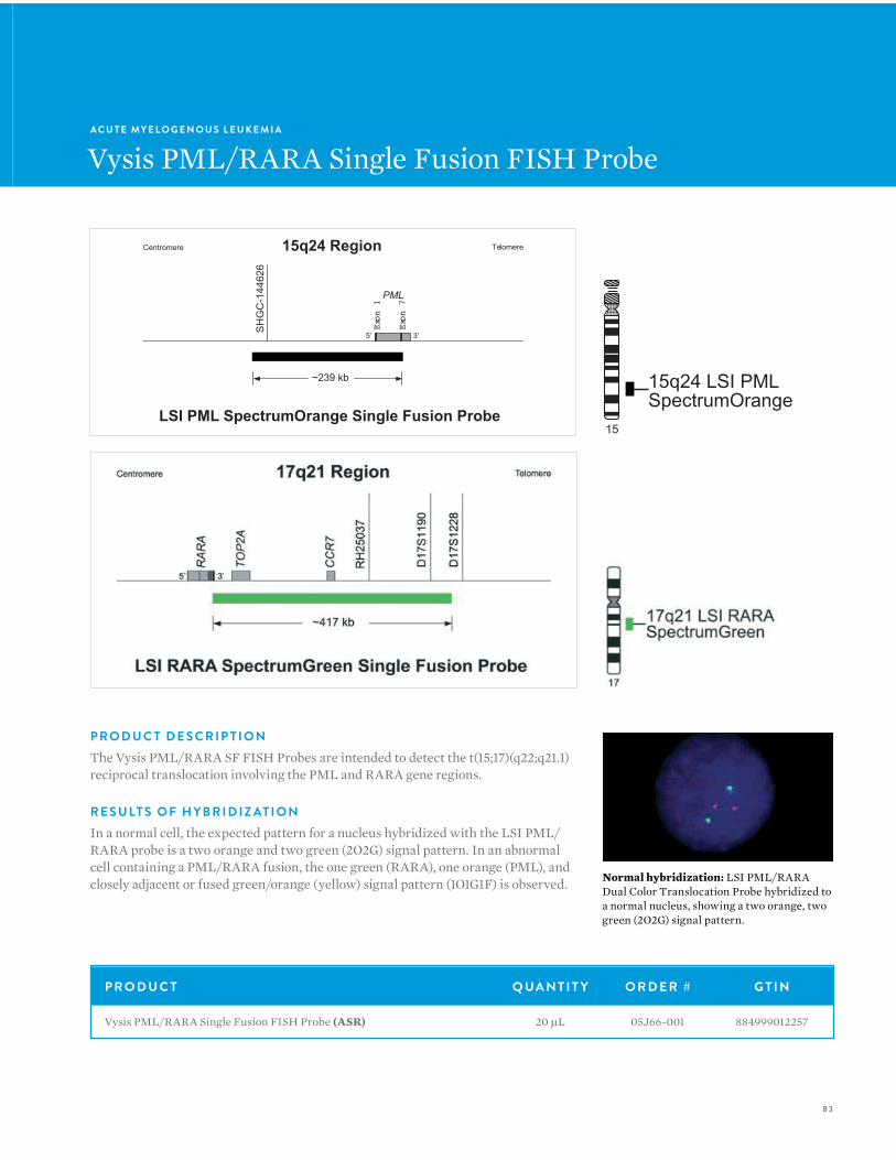
83
A C U T E MY E L O G E N O U S L E U K E M I A
Vysis PML/RARA Single Fusion FISH Probe
Centromere Telomere
~239 kb
LSI PML SpectrumOrange Single Fusion Probe
15q24 RegionS
HG
C-1
4462
6
PML
Exon 1
Exon 7
5’ 3’
15
15q24 LSI PMLSpectrumOrange
P R O D U C T D E S C R I P T I O NThe Vysis PML/RARA SF FISH Probes are intended to detect the t(15;17)(q22;q21.1) reciprocal translocation involving the PML and RARA gene regions.
R E S U LT S O F H Y B R I D I Z AT I O NIn a normal cell, the expected pattern for a nucleus hybridized with the LSI PML/RARA probe is a two orange and two green (2O2G) signal pattern. In an abnormal cell containing a PML/RARA fusion, the one green (RARA), one orange (PML), and closely adjacent or fused green/orange (yellow) signal pattern (1O1G1F) is observed. Normal hybridization: LSI PML/RARA
Dual Color Translocation Probe hybridized to a normal nucleus, showing a two orange, two green (2O2G) signal pattern.
P R O D U C T Q UA N T I T Y O R D E R # G T I N
Vysis PML/RARA Single Fusion FISH Probe (ASR) 20 µL 05J66-001 884999012257
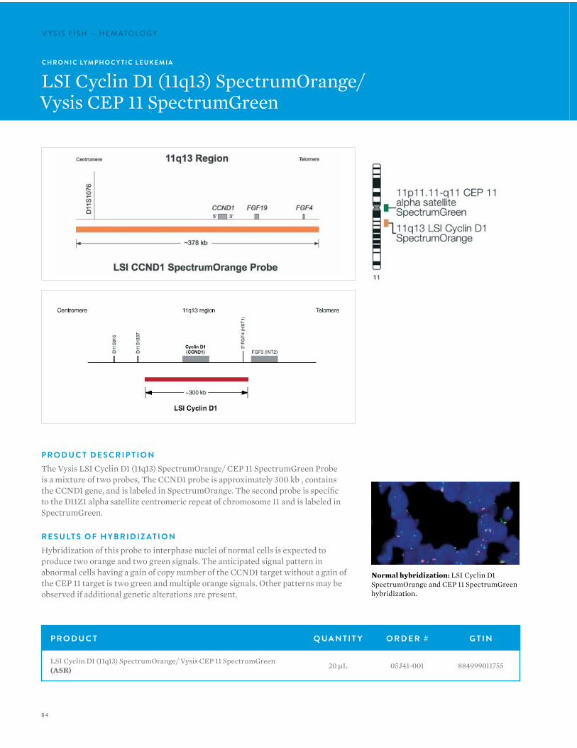
84
V Y S I S F I S H — H E M ATO LO G YV Y S I S F I S H — H E M ATO LO G Y
C H R O N I C LYM P H O C Y T I C L E U K E M I A
LSI Cyclin D1 (11q13) SpectrumOrange/Vysis CEP 11 SpectrumGreen
P R O D U C T D E S C R I P T I O NThe Vysis LSI Cyclin D1 (11q13) SpectrumOrange/ CEP 11 SpectrumGreen Probe is a mixture of two probes, The CCND1 probe is approximately 300 kb , contains the CCND1 gene, and is labeled in SpectrumOrange. The second probe is specific to the D11Z1 alpha satellite centromeric repeat of chromosome 11 and is labeled in SpectrumGreen.
R E S U LT S O F H Y B R I D I Z AT I O NHybridization of this probe to interphase nuclei of normal cells is expected to produce two orange and two green signals. The anticipated signal pattern in abnormal cells having a gain of copy number of the CCND1 target without a gain of the CEP 11 target is two green and multiple orange signals. Other patterns may be observed if additional genetic alterations are present.
Normal hybridization: LSI Cyclin D1 SpectrumOrange and CEP 11 SpectrumGreen hybridization.
P R O D U C T Q UA N T I T Y O R D E R # G T I N
LSI Cyclin D1 (11q13) SpectrumOrange/ Vysis CEP 11 SpectrumGreen (ASR) 20 µL 05J41-001 884999011755
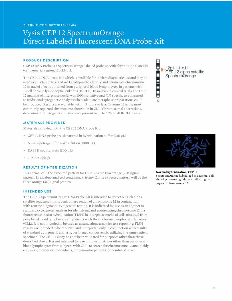
85
C H R O N I C LYM P H O C Y T I C L E U K E M I A
Vysis CEP 12 SpectrumOrangeDirect Labeled Fluorescent DNA Probe Kit
P R O D U C T D E S C R I P T I O NCEP 12 DNA Probe is a SpectrumOrange labeled probe specific for the alpha satellite (centromeric) region, 12p11.1-q11.
The CEP 12 DNA Probe Kit which is available for in vitro diagnostic use and may be used as an adjunct to standard karotyping to identify and enumerate chromosome 12 in nuclei of cells obtained from peripheral blood lymphocytes in patients with B-cell chronic lymphocytic leukemia (B-CLL). In multi-site clinical trials, the CEP 12 analysis of interphase nuclei was 100% sensitive and 91% specific as compared to traditional cytogenetic analysis when adequate metaphase preparations could be produced. Results are available within 3 hours or less. Trisomy 12 is the most commonly reported chromosome aberration in CLL. Chromosomal aberrations, determined by cytogenetic analysis are present in up to 55% of all B-CLL cases.
M AT E R I A L S P R OV I D E DMaterials provided with the CEP 12 DNA Probe Kit:
• CEP 12 DNA probe pre-denatured in hybridization buffer (220 µL)
• NP-40 (detergent for wash solution: 1000 µL)
• DAPI II counterstain (300 µL)
• 20X SSC (66 g)
R E S U LT S O F H Y B R I D I Z AT I O NIn a normal cell, the expected pattern for CEP 12 is the two orange (2O) signal pattern. In an abnormal cell containing trisomy 12, the expected pattern will be the three orange (3O) signal pattern.
I N T E N D E D U S EThe CEP 12 SpectrumOrange DNA Probe Kit is intended to detect AT rich alpha satellite sequences in the centromere region of chromosome 12 in conjunction with routine diagnostic cytogenetic testing. It is indicated for use as an adjunct to standard cytogenetic analysis for identifying and enumerating chromosome 12 via fluorescence in situ hybridization (FISH) in interphase nuclei of cells obtained from peripheral blood lymphocytes in patients with B-cell chronic lymphocytic leukemia (CLL). It is not intended to be used as a stand alone assay for test reporting; FISH results are intended to be reported and interpreted only in conjunction with results of standard cytogenetic analysis, performed concurrently, utilizing the same patient specimen. The CEP 12 assay has not been validated for purposes other than those described above. It is not intended for use with test matrices other than peripheral blood lymphocytes from subjects with CLL, to screen for chromosome 12 aneuploidy, e.g., in asymptomatic individuals, or to monitor patients for residual disease.
Normal hybridization: CEP 12 SpectrumOrange hybridized to a normal cell showing two orange signals indicating two copies of chromosome 12.
86
V Y S I S F I S H — H E M ATO LO G Y
L I M I TAT I O N S1. The CEP 12 DNA Probe Kit has been optimized only for
identifying chromosome 12 in interphase nuclei from peripheral blood specimens from patients with B-cell chronic lymphocytic leukemia.
2. The clinical interpretation of any abnormality or its absence by FISH should be done in conjunction with standard cytogenetic analysis and proper controls, and should be evaluated within the context of the patient’s medical history and past diagnostic laboratory test results.
3. Clinical specimens with >2.0% tri-signaled nuclei are considered to have an abnormal trisomy 12 clone. Those with ≤ 2.0% tri-signaled nuclei should be considered normal, although the presence of trisomy 12 is not completely excluded. When the percentage of tri-signaled interphase nuclei are near the cutoff point (1.5-2.5%), the results should be interpreted with caution.
4. The CEP 12 assay has been validated only for use with peripheral blood lymphocytes obtained from patients with B-cell CLL leukemia. It is not intended to be used for chromosome 12 enumeration in other patient populations or with other test matrices such as amniocytes, chorionic villi, fibroblasts, tumor cells, long term cultures, among others.
5. FISH assay results may not be informative if the specimen quality and/or specimen slide preparation is inadequate; the CEP 12 DNA Probe Kit has been optimized for archived peripheral blood specimens stored only at -20ºC or -80ºC, as recommended.
6. The CEP 12 DNA Probe Kit has not been validated for monitoring disease status.
7. It is possible that some individuals may have target sequences at an alternate chromosomal location that may hybridize with CEP 12. This has not been investigated for this device; the user should assess this in metaphase spreads from each subject tested.
8. The CEP 12 assay is intended only to aid in the enumeration of chromosome 12 centromeres; it is not designed to detect chromosome 12 structural abnormalities.
P R O D U C T Q UA N T I T Y O R D E R # G T I N
Vysis CEP 12 SpectrumOrange Direct Labeled Fluorescent DNA Probe Kit (IVD) 20 Assays 07J22-012 884999027084
Vysis CEP 12 SpectrumOrange Direct Labeled Fluorescent DNA Probe Kit (without control slides) (IVD) 20 Assays 07J20-012 884999027015
ProbeChek Control Slides for FISH using CEP 8 and CEP 12 Assay, Negative Control 0%, trisomy 8/12 (IVD) 5 Slides 07J21-001 884999027039
ProbeChek Control Slides for FISH using CEP 8 and CEP 12 Assay, Positive Control 10%, trisomy 8/12 (IVD) 5 Slides 07J21-002 884999027046
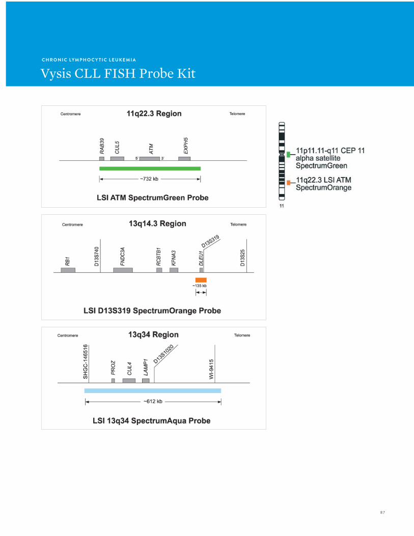
87
C H R O N I C LYM P H O C Y T I C L E U K E M I A
Vysis CLL FISH Probe Kit
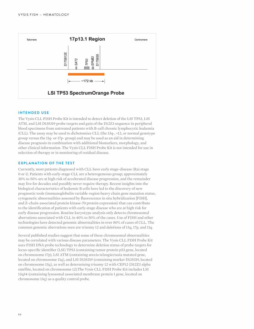
88
V Y S I S F I S H — H E M ATO LO G Y
I N T E N D E D U S EThe Vysis CLL FISH Probe Kit is intended to detect deletion of the LSI TP53, LSI ATM, and LSI D13S319 probe targets and gain of the D12Z3 sequence in peripheral blood specimens from untreated patients with B-cell chronic lymphocytic leukemia (CLL). The assay may be used to dichotomize CLL (the 13q-, +12, or normal genotype group versus the 11q- or 17p- group) and may be used as an aid in determining disease prognosis in combination with additional biomarkers, morphology, and other clinical information. The Vysis CLL FISH Probe Kit is not intended for use in selection of therapy or in monitoring of residual disease.
E X P L A N AT I O N O F T H E T E S TCurrently, most patients diagnosed with CLL have early stage-disease (Rai stage 0 or 1). Patients with early-stage CLL are a heterogeneous group; approximately 30% to 50% are at high risk of accelerated disease progression, and the remainder may live for decades and possibly never require therapy. Recent insights into the biological characteristics of leukemic B cells have led to the discovery of new prognostic tools (immunoglobulin variable-region heavy chain gene mutation status, cytogenetic abnormalities assessed by fluorescence in situ hybridization [FISH], and Z-chain-associated protein kinase-70 protein expression) that can contribute to the identification of patients with early-stage disease who are at high risk for early disease progression. Routine karyotype analysis only detects chromosomal aberrations associated with CLL in 40% to 50% of the cases. Use of FISH and other technologies have detected genomic abnormalities in over 80% of cases of CLL. The common genomic aberrations seen are trisomy 12 and deletions of 13q, 17p, and 11q.
Several published studies suggest that some of these chromosomal abnormalities may be correlated with various disease parameters. The Vysis CLL FISH Probe Kit uses FISH DNA probe technology to determine deletion status of probe targets for locus-specific identifier (LSI) TP53 (containing tumor protein p53 gene, located on chromosome 17p), LSI ATM (containing ataxia telangiectasia mutated gene, located on chromosome 11q), and LSI D13S319 (containing marker D13S319, located on chromosome 13q), as well as determining trisomy 12 with CEP12 (D12Z3 alpha satellite, located on chromosome 12).The Vysis CLL FISH Probe Kit includes LSI 13q34 (containing lysosomal associated membrane protein 1 gene, located on chromosome 13q) as a quality control probe.
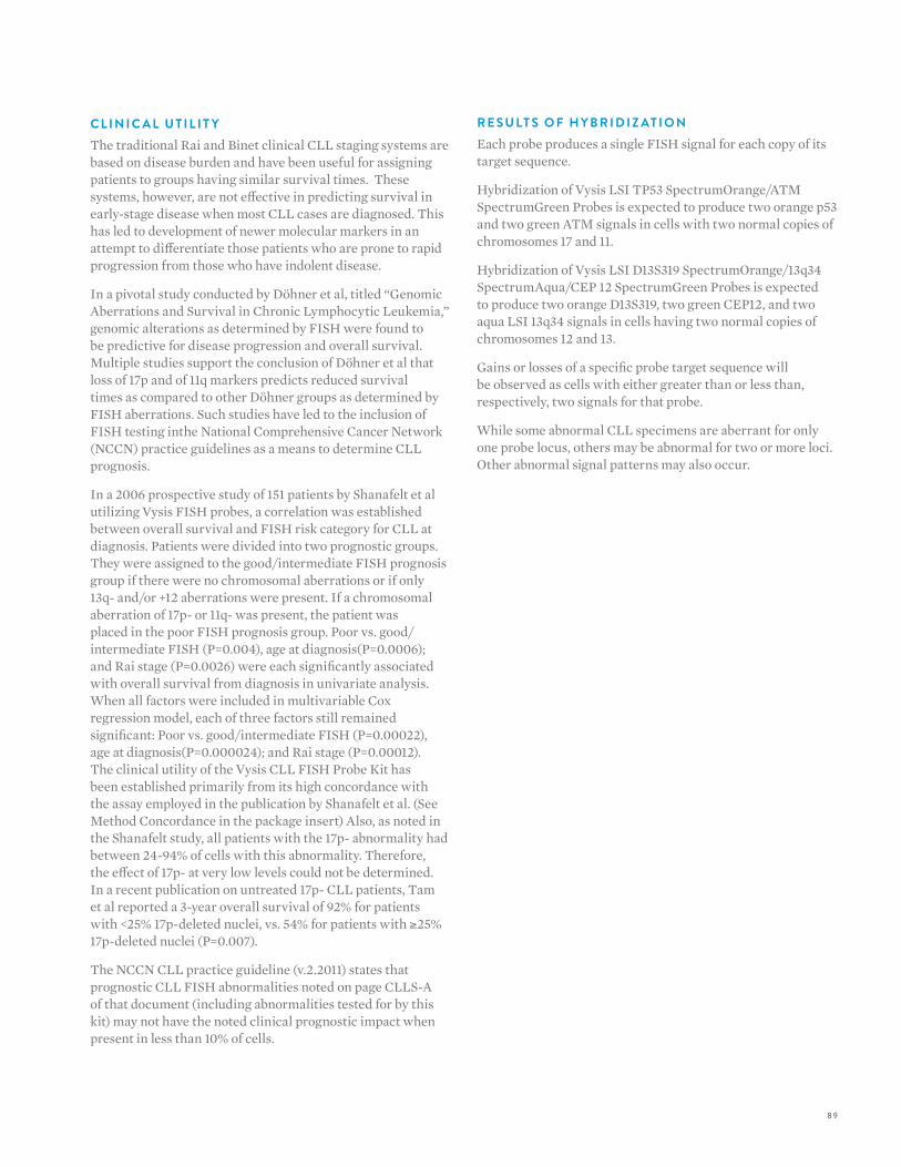
89
C L I N I C A L U T I L I T YThe traditional Rai and Binet clinical CLL staging systems are based on disease burden and have been useful for assigning patients to groups having similar survival times. These systems, however, are not effective in predicting survival in early-stage disease when most CLL cases are diagnosed. This has led to development of newer molecular markers in an attempt to differentiate those patients who are prone to rapid progression from those who have indolent disease.
In a pivotal study conducted by Döhner et al, titled “Genomic Aberrations and Survival in Chronic Lymphocytic Leukemia,” genomic alterations as determined by FISH were found to be predictive for disease progression and overall survival. Multiple studies support the conclusion of Döhner et al that loss of 17p and of 11q markers predicts reduced survival times as compared to other Döhner groups as determined by FISH aberrations. Such studies have led to the inclusion of FISH testing inthe National Comprehensive Cancer Network (NCCN) practice guidelines as a means to determine CLL prognosis.
In a 2006 prospective study of 151 patients by Shanafelt et al utilizing Vysis FISH probes, a correlation was established between overall survival and FISH risk category for CLL at diagnosis. Patients were divided into two prognostic groups. They were assigned to the good/intermediate FISH prognosis group if there were no chromosomal aberrations or if only 13q- and/or +12 aberrations were present. If a chromosomal aberration of 17p- or 11q- was present, the patient was placed in the poor FISH prognosis group. Poor vs. good/intermediate FISH (P=0.004), age at diagnosis(P=0.0006); and Rai stage (P=0.0026) were each significantly associated with overall survival from diagnosis in univariate analysis. When all factors were included in multivariable Cox regression model, each of three factors still remained significant: Poor vs. good/intermediate FISH (P=0.00022), age at diagnosis(P=0.000024); and Rai stage (P=0.00012).The clinical utility of the Vysis CLL FISH Probe Kit has been established primarily from its high concordance with the assay employed in the publication by Shanafelt et al. (See Method Concordance in the package insert) Also, as noted in the Shanafelt study, all patients with the 17p- abnormality had between 24-94% of cells with this abnormality. Therefore, the effect of 17p- at very low levels could not be determined. In a recent publication on untreated 17p- CLL patients, Tam et al reported a 3-year overall survival of 92% for patients with <25% 17p-deleted nuclei, vs. 54% for patients with ≥25% 17p-deleted nuclei (P=0.007).
The NCCN CLL practice guideline (v.2.2011) states that prognostic CLL FISH abnormalities noted on page CLLS-A of that document (including abnormalities tested for by this kit) may not have the noted clinical prognostic impact when present in less than 10% of cells.
R E S U LT S O F H Y B R I D I Z AT I O NEach probe produces a single FISH signal for each copy of its target sequence.
Hybridization of Vysis LSI TP53 SpectrumOrange/ATM SpectrumGreen Probes is expected to produce two orange p53 and two green ATM signals in cells with two normal copies of chromosomes 17 and 11.
Hybridization of Vysis LSI D13S319 SpectrumOrange/13q34 SpectrumAqua/CEP 12 SpectrumGreen Probes is expected to produce two orange D13S319, two green CEP12, and two aqua LSI 13q34 signals in cells having two normal copies of chromosomes 12 and 13.
Gains or losses of a specific probe target sequence will be observed as cells with either greater than or less than, respectively, two signals for that probe.
While some abnormal CLL specimens are aberrant for only one probe locus, others may be abnormal for two or more loci.Other abnormal signal patterns may also occur.
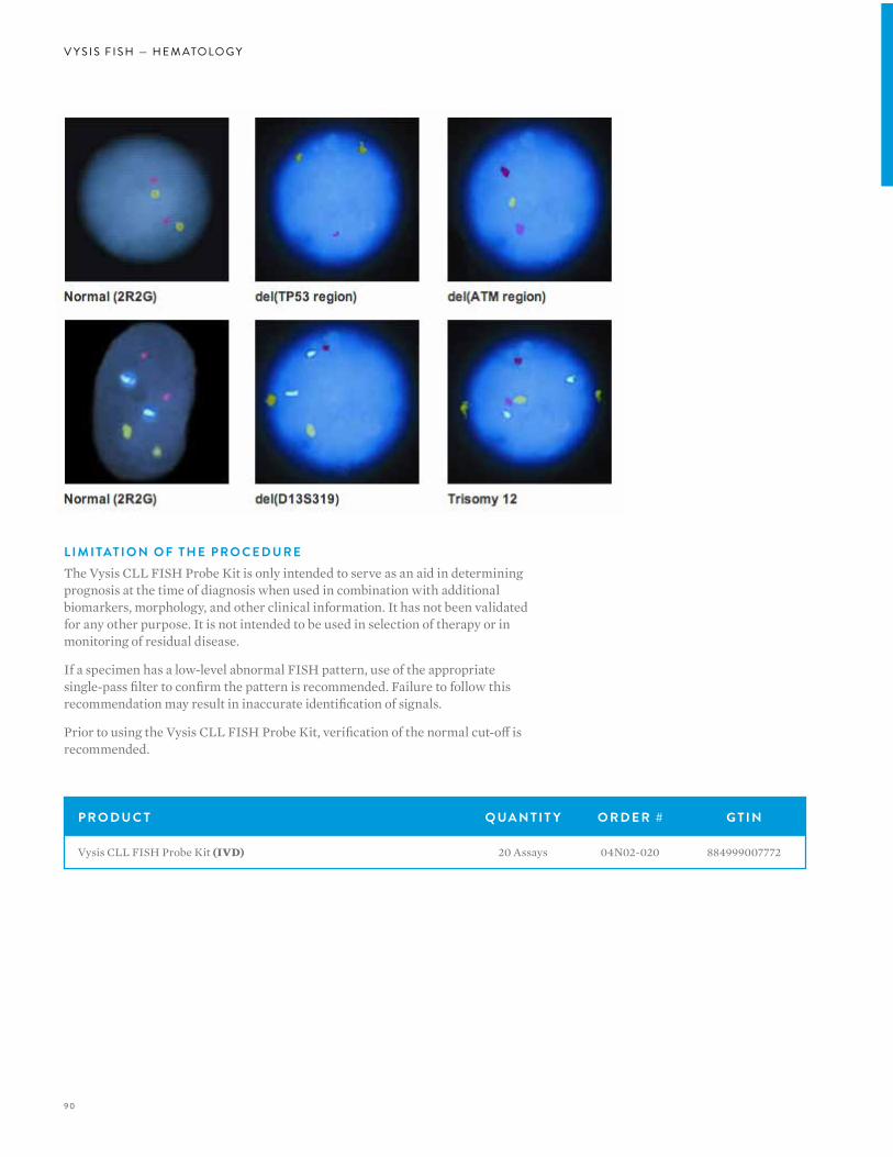
90
V Y S I S F I S H — H E M ATO LO G Y
P R O D U C T Q UA N T I T Y O R D E R # G T I N
Vysis CLL FISH Probe Kit (IVD) 20 Assays 04N02-020 884999007772
L I M I TAT I O N O F T H E P R O C E D U R EThe Vysis CLL FISH Probe Kit is only intended to serve as an aid in determining prognosis at the time of diagnosis when used in combination with additional biomarkers, morphology, and other clinical information. It has not been validated for any other purpose. It is not intended to be used in selection of therapy or in monitoring of residual disease.
If a specimen has a low-level abnormal FISH pattern, use of the appropriate single-pass filter to confirm the pattern is recommended. Failure to follow this recommendation may result in inaccurate identification of signals.
Prior to using the Vysis CLL FISH Probe Kit, verification of the normal cut-off is recommended.
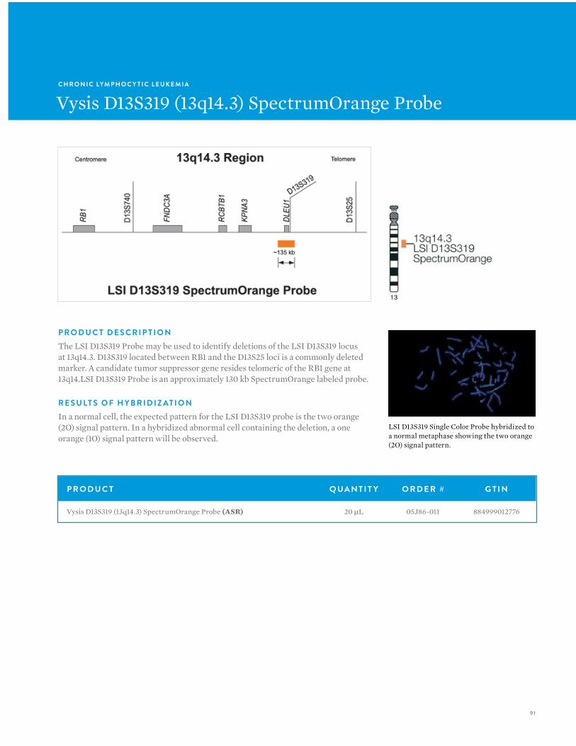
91
C H R O N I C LYM P H O C Y T I C L E U K E M I A
Vysis D13S319 (13q14.3) SpectrumOrange Probe
P R O D U C T Q UA N T I T Y O R D E R # G T I N
Vysis D13S319 (13q14.3) SpectrumOrange Probe (ASR) 20 µL 05J86-011 884999012776
P R O D U C T D E S C R I P T I O NThe LSI D13S319 Probe may be used to identify deletions of the LSI D13S319 locus at 13q14.3. D13S319 located between RB1 and the D13S25 loci is a commonly deleted marker. A candidate tumor suppressor gene resides telomeric of the RB1 gene at 13q14.LSI D13S319 Probe is an approximately 130 kb SpectrumOrange labeled probe.
R E S U LT S O F H Y B R I D I Z AT I O NIn a normal cell, the expected pattern for the LSI D13S319 probe is the two orange (2O) signal pattern. In a hybridized abnormal cell containing the deletion, a one orange (1O) signal pattern will be observed.
LSI D13S319 Single Color Probe hybridized to a normal metaphase showing the two orange (2O) signal pattern.
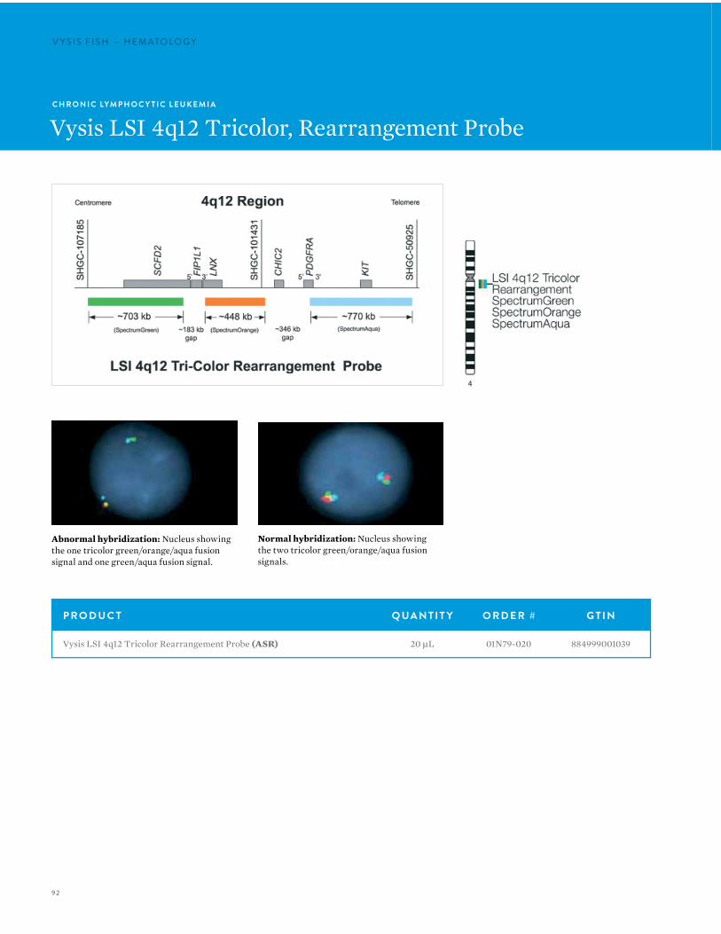
92
V Y S I S F I S H — H E M ATO LO G YV Y S I S F I S H — H E M ATO LO G Y
Normal hybridization: Nucleus showing the two tricolor green/orange/aqua fusion signals.
Abnormal hybridization: Nucleus showing the one tricolor green/orange/aqua fusion signal and one green/aqua fusion signal.
C H R O N I C LYM P H O C Y T I C L E U K E M I A
Vysis LSI 4q12 Tricolor, Rearrangement Probe
P R O D U C T Q UA N T I T Y O R D E R # G T I N
Vysis LSI 4q12 Tricolor Rearrangement Probe (ASR) 20 µL 01N79-020 884999001039
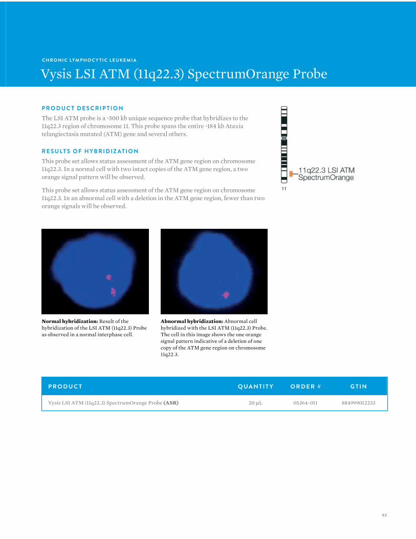
93
C H R O N I C LYM P H O C Y T I C L E U K E M I A
Vysis LSI ATM (11q22.3) SpectrumOrange Probe
P R O D U C T D E S C R I P T I O NThe LSI ATM probe is a ~500 kb unique sequence probe that hybridizes to the 11q22.3 region of chromosome 11. This probe spans the entire ~184 kb Ataxia telangiectasia mutated (ATM) gene and several others.
R E S U LT S O F H Y B R I D I Z AT I O NThis probe set allows status assessment of the ATM gene region on chromosome 11q22.3. In a normal cell with two intact copies of the ATM gene region, a two orange signal pattern will be observed.
This probe set allows status assessment of the ATM gene region on chromosome 11q22.3. In an abnormal cell with a deletion in the ATM gene region, fewer than two orange signals will be observed.
P R O D U C T Q UA N T I T Y O R D E R # G T I N
Vysis LSI ATM (11q22.3) SpectrumOrange Probe (ASR) 20 µL 05J64-011 884999012233
Normal hybridization: Result of the hybridization of the LSI ATM (11q22.3) Probe as observed in a normal interphase cell.
Abnormal hybridization: Abnormal cell hybridized with the LSI ATM (11q22.3) Probe. The cell in this image shows the one orange signal pattern indicative of a deletion of one copy of the ATM gene region on chromosome 11q22.3.
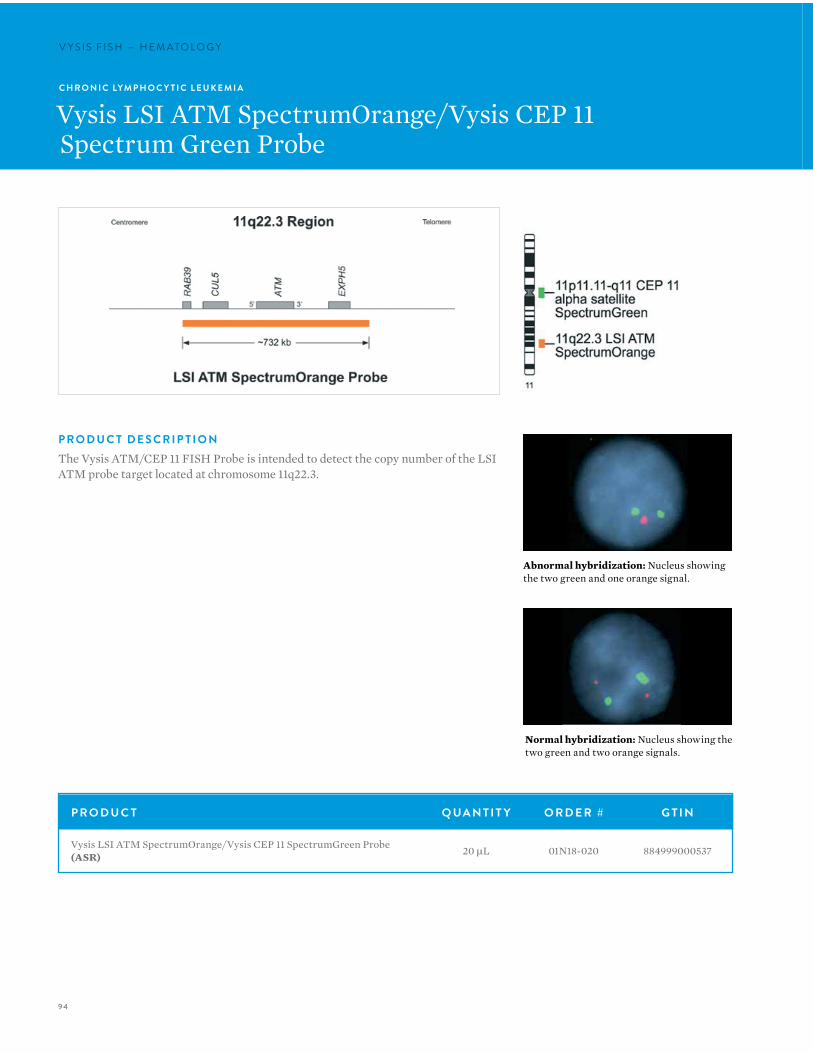
94
V Y S I S F I S H — H E M ATO LO G YV Y S I S F I S H — H E M ATO LO G Y
P R O D U C T D E S C R I P T I O NThe Vysis ATM/CEP 11 FISH Probe is intended to detect the copy number of the LSI ATM probe target located at chromosome 11q22.3.
C H R O N I C LYM P H O C Y T I C L E U K E M I A
Vysis LSI ATM SpectrumOrange/Vysis CEP 11Spectrum Green Probe
Normal hybridization: Nucleus showing the two green and two orange signals.
Abnormal hybridization: Nucleus showing the two green and one orange signal.
P R O D U C T Q UA N T I T Y O R D E R # G T I N
Vysis LSI ATM SpectrumOrange/Vysis CEP 11 SpectrumGreen Probe (ASR) 20 µL 01N18-020 884999000537
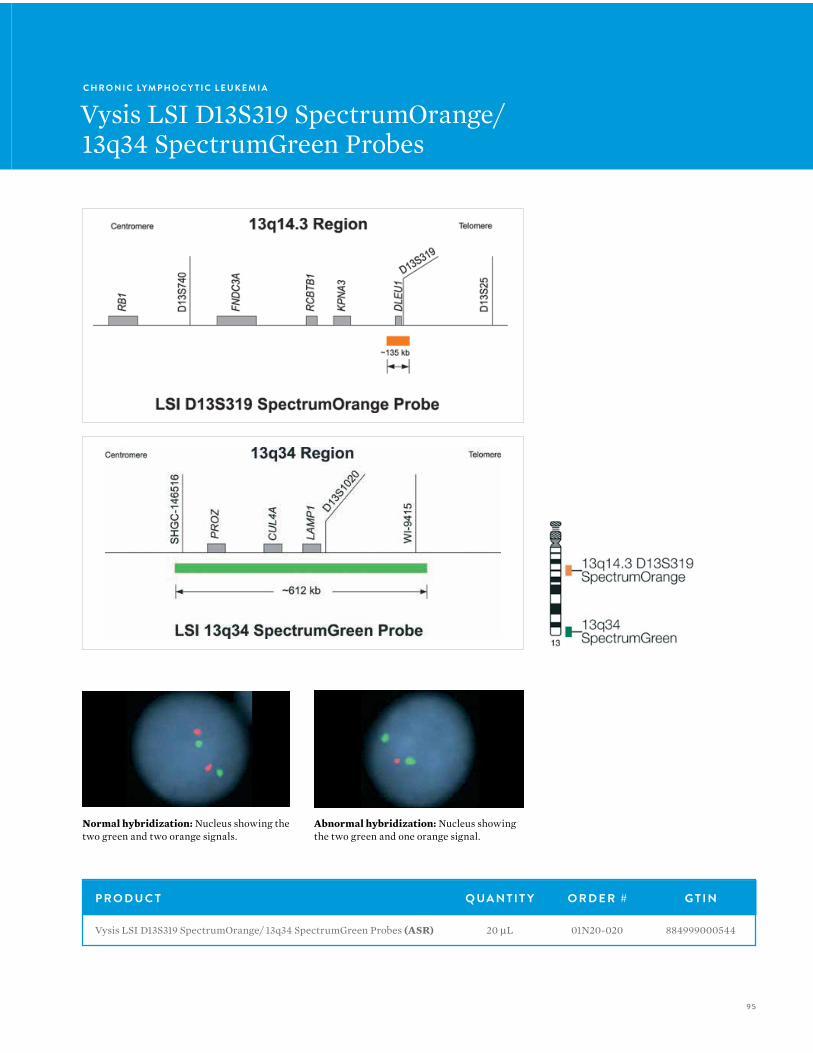
95
C H R O N I C LYM P H O C Y T I C L E U K E M I A
Vysis LSI D13S319 SpectrumOrange/13q34 SpectrumGreen Probes
Normal hybridization: Nucleus showing the two green and two orange signals.
Abnormal hybridization: Nucleus showing the two green and one orange signal.
P R O D U C T Q UA N T I T Y O R D E R # G T I N
Vysis LSI D13S319 SpectrumOrange/ 13q34 SpectrumGreen Probes (ASR) 20 µL 01N20-020 884999000544

96
V Y S I S F I S H — H E M ATO LO G YV Y S I S F I S H — H E M ATO LO G Y
C H R O N I C LYM P H O C Y T I C L E U K E M I A
Vysis LSI IGH/CCND1 XT Dual Color, Dual Fusion Translocation Probes
P R O D U C T D E S C R I P T I O NThese fluorescence in situ hybridization (FISH) probes are intended to detect t(11;14)(q13;q32) reciprocal translocation involving the IGH and CCND1 gene regions.
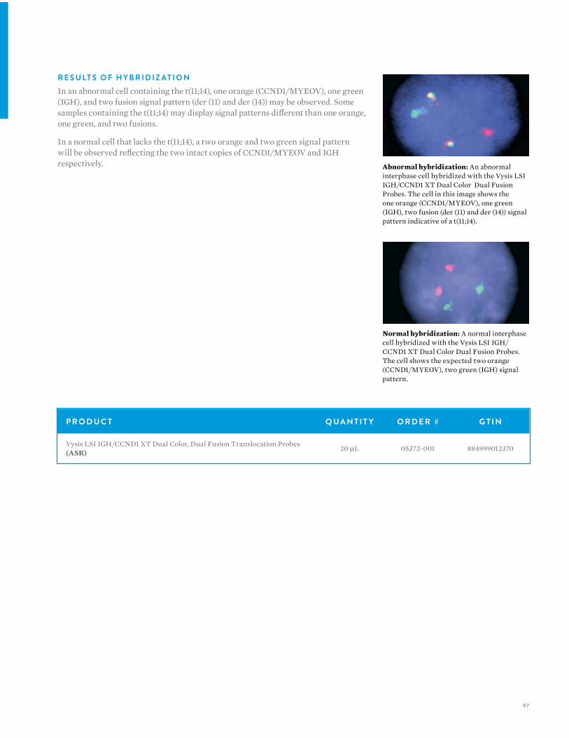
97
R E S U LT S O F H Y B R I D I Z AT I O NIn an abnormal cell containing the t(11;14), one orange (CCND1/MYEOV), one green (IGH), and two fusion signal pattern (der (11) and der (14)) may be observed. Some samples containing the t(11;14) may display signal patterns different than one orange, one green, and two fusions.
In a normal cell that lacks the t(11;14), a two orange and two green signal pattern will be observed reflecting the two intact copies of CCND1/MYEOV and IGH respectively.
Normal hybridization: A normal interphase cell hybridized with the Vysis LSI IGH/CCND1 XT Dual Color Dual Fusion Probes. The cell shows the expected two orange (CCND1/MYEOV), two green (IGH) signal pattern.
Abnormal hybridization: An abnormal interphase cell hybridized with the Vysis LSI IGH/CCND1 XT Dual Color Dual Fusion Probes. The cell in this image shows the one orange (CCND1/MYEOV), one green (IGH), two fusion (der (11) and der (14)) signal pattern indicative of a t(11;14).
P R O D U C T Q UA N T I T Y O R D E R # G T I N
Vysis LSI IGH/CCND1 XT Dual Color, Dual Fusion Translocation Probes (ASR) 20 µL 05J72-001 884999012370
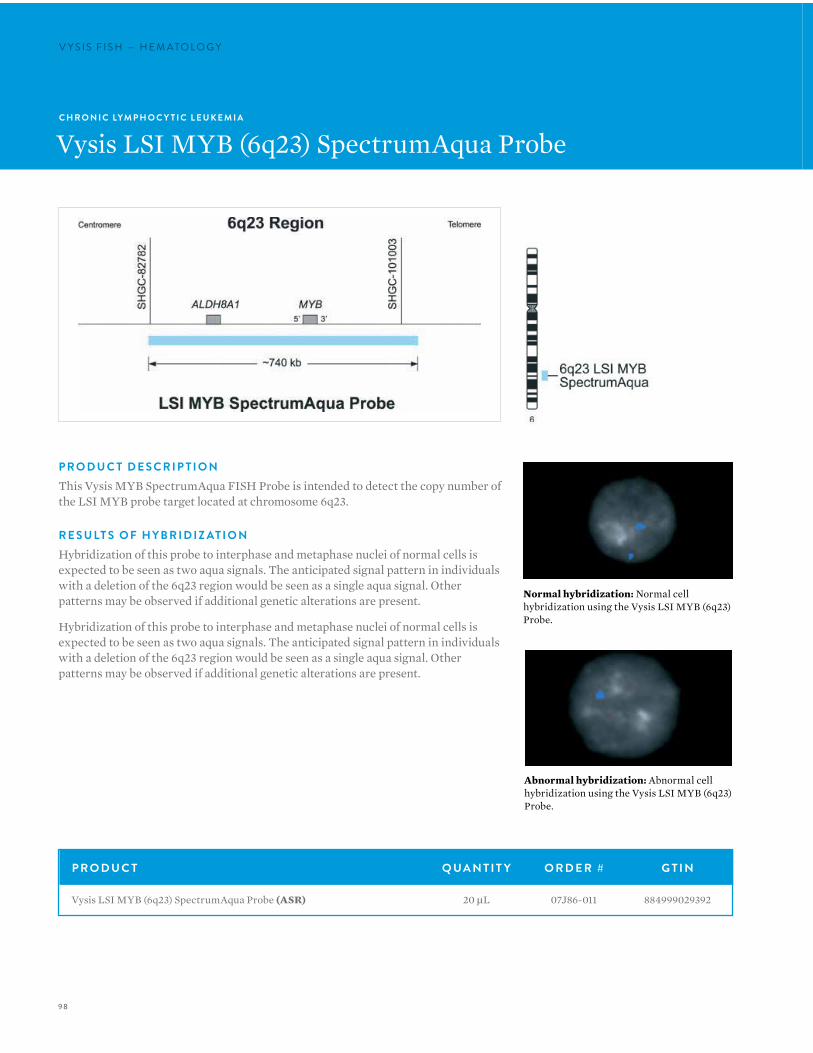
98
V Y S I S F I S H — H E M ATO LO G YV Y S I S F I S H — H E M ATO LO G Y
Normal hybridization: Normal cell hybridization using the Vysis LSI MYB (6q23) Probe.
Abnormal hybridization: Abnormal cell hybridization using the Vysis LSI MYB (6q23) Probe.
C H R O N I C LYM P H O C Y T I C L E U K E M I A
Vysis LSI MYB (6q23) SpectrumAqua Probe
P R O D U C T D E S C R I P T I O NThis Vysis MYB SpectrumAqua FISH Probe is intended to detect the copy number of the LSI MYB probe target located at chromosome 6q23.
R E S U LT S O F H Y B R I D I Z AT I O NHybridization of this probe to interphase and metaphase nuclei of normal cells is expected to be seen as two aqua signals. The anticipated signal pattern in individuals with a deletion of the 6q23 region would be seen as a single aqua signal. Other patterns may be observed if additional genetic alterations are present.
Hybridization of this probe to interphase and metaphase nuclei of normal cells is expected to be seen as two aqua signals. The anticipated signal pattern in individuals with a deletion of the 6q23 region would be seen as a single aqua signal. Other patterns may be observed if additional genetic alterations are present.
P R O D U C T Q UA N T I T Y O R D E R # G T I N
Vysis LSI MYB (6q23) SpectrumAqua Probe (ASR) 20 µL 07J86-011 884999029392
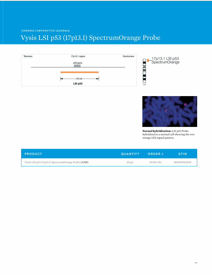
99
C H R O N I C LYM P H O C Y T I C L E U K E M I A
Vysis LSI p53 (17p13.1) SpectrumOrange Probe
Normal hybridization: LSI p53 Probe hybridized to a normal cell showing the two orange (2O) signal pattern.
P R O D U C T Q UA N T I T Y O R D E R # G T I N
Vysis LSI p53 (17p13.1) SpectrumOrange Probe (ASR) 20 µL 05J52-011 884999012035
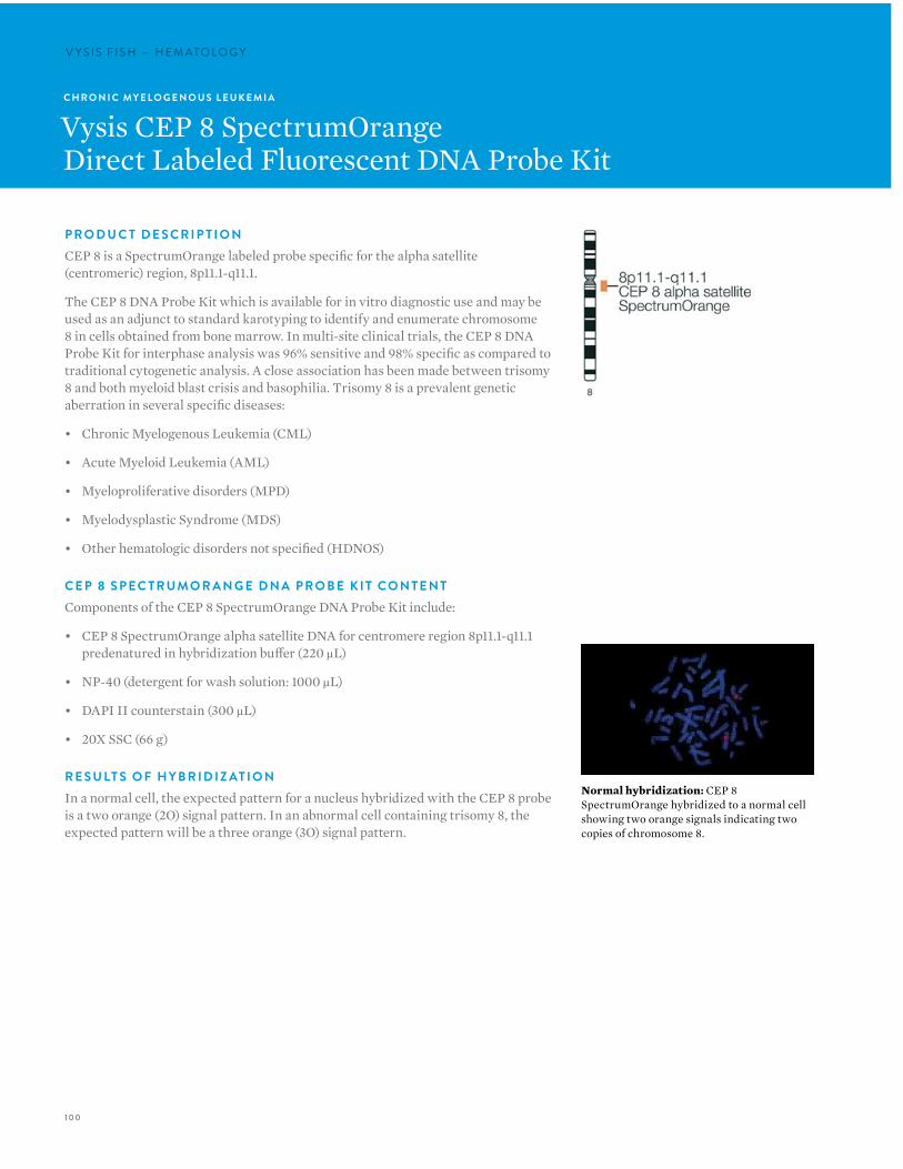
100
V Y S I S F I S H — H E M ATO LO G YV Y S I S F I S H — H E M ATO LO G Y
C H R O N I C MY E L O G E N O U S L E U K E M I A
Vysis CEP 8 SpectrumOrangeDirect Labeled Fluorescent DNA Probe Kit
P R O D U C T D E S C R I P T I O NCEP 8 is a SpectrumOrange labeled probe specific for the alpha satellite (centromeric) region, 8p11.1-q11.1.
The CEP 8 DNA Probe Kit which is available for in vitro diagnostic use and may be used as an adjunct to standard karotyping to identify and enumerate chromosome 8 in cells obtained from bone marrow. In multi-site clinical trials, the CEP 8 DNA Probe Kit for interphase analysis was 96% sensitive and 98% specific as compared to traditional cytogenetic analysis. A close association has been made between trisomy 8 and both myeloid blast crisis and basophilia. Trisomy 8 is a prevalent genetic aberration in several specific diseases:
• Chronic Myelogenous Leukemia (CML)
• Acute Myeloid Leukemia (AML)
• Myeloproliferative disorders (MPD)
• Myelodysplastic Syndrome (MDS)
• Other hematologic disorders not specified (HDNOS)
C E P 8 S P E C T R U M O R A N G E D N A P R O B E K I T CO N T E N TComponents of the CEP 8 SpectrumOrange DNA Probe Kit include:
• CEP 8 SpectrumOrange alpha satellite DNA for centromere region 8p11.1-q11.1 predenatured in hybridization buffer (220 µL)
• NP-40 (detergent for wash solution: 1000 µL)
• DAPI II counterstain (300 µL)
• 20X SSC (66 g)
R E S U LT S O F H Y B R I D I Z AT I O NIn a normal cell, the expected pattern for a nucleus hybridized with the CEP 8 probe is a two orange (2O) signal pattern. In an abnormal cell containing trisomy 8, the expected pattern will be a three orange (3O) signal pattern.
Normal hybridization: CEP 8 SpectrumOrange hybridized to a normal cell showing two orange signals indicating two copies of chromosome 8.
101
I N T E N D E D U S EThe CEP 8 SpectrumOrange DNA Probe Kit is intended to detect AT rich alpha satellite sequences in the centromere region of chromosome 8 in conjunction with routine diagnostic cytogenetic testing. It is indicated for use as an adjunct to standard cytogenetic analysis for identifying and enumerating chromosome 8 via fluorescence in situ hybridization (FISH) in interphase nuclei and in metaphase spreads of cells obtained from bone marrow in patients with myeloid disorders [Chronic myelogenous leukemia (CML), Acute myeloid leukemia (AML), Myeloproliferative disorder (MPD), Myelodysplastic syndrome (MDS), and Hematological disorders not otherwise specified (HDNOS)]. It is not intended to be used as a stand alone assay for test reporting. It is not intended for use in long term cell cultured materials such as amniocytes, fibroblasts and tumor cells.
L I M I TAT I O N S • The CEP 8 SpectrumOrange DNA Probe Kit has been
characterized only for identifying chromosomes in nuclear preparations or metaphase spreads from bone marrow specimens.
• The clinical interpretation of any test results should be done in conjunction with standard cytogenetic analysis and should be evaluated within the context of the patient’s medical history and other diagnostic laboratory test results.
• Clinical specimens with >2.2% tri-signaled nuclei are considered to have an abnormal trisomy 8 clone. Those with ≤ 2.2% tri-signaled nuclei should be considered normal, although the presence of trisomy 8 is not completely excluded.
• The CEP 8 SpectrumOrange DNA Probe Kit is not intended for long term cell cultured materials such as amniocytes, fibroblasts and tumor cells.
• FISH assay results may not be informative if the specimen quality and/or specimen slide preparation is inadequate.
• If significant peripheral blood contamination is present in the bone marrow specimen, the blood may dilute the specimen; it is important to recognize the potential effects this dilution effect may have on the FISH assay results.
• It is possible that patients may have chromosome polymorphism which may hybridize with CEP 8 probe. FISH metaphase analysis should be done in addition to FISH interphase analysis. Polymorphism was not investigated in the clinical trials.
• This assay will not detect the presence of other chromosome abnormalities frequently associated with hematological disorders.
• The efficacy of this assay for monitoring of trisomy 8 or disease progression has not been demonstrated.
P R O D U C T Q UA N T I T Y O R D E R # G T I N
Vysis CEP 8 SpectrumOrange Direct Labeled Fluorescent DNA Probe Kit (IVD) 20 Assays 07J22-008 884999027077
Vysis CEP 8 SpectrumOrange Direct Labeled Fluorescent DNA Probe Kit (without control slides) (IVD) 20 Assays 07J20-008 884999027008
ProbeChek Control Slides for FISH using CEP 8 and CEP 12 Assay, Negative Control 0%, trisomy 8/12 (IVD) 5 Slides 07J21-001 884999027039
ProbeChek Control Slides for FISH using CEP 8 and CEP 12 Assay, Positive Control 10%, trisomy 8/12 (IVD) 5 Slides 07J21-002 884999027046
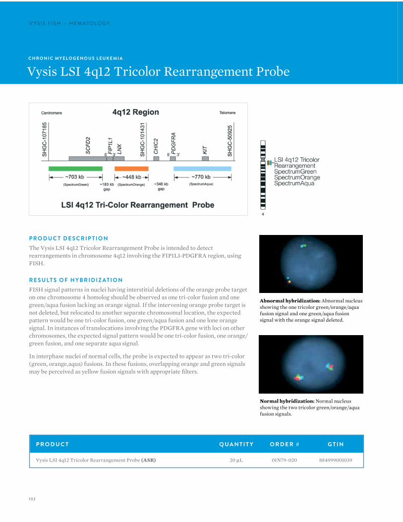
102
V Y S I S F I S H — H E M ATO LO G YV Y S I S F I S H — H E M ATO LO G Y
C H R O N I C MY E L O G E N O U S L E U K E M I A
Vysis LSI 4q12 Tricolor Rearrangement Probe
P R O D U C T D E S C R I P T I O NThe Vysis LSI 4q12 Tricolor Rearrangement Probe is intended to detect rearrangements in chromosome 4q12 involving the FIP1L1-PDGFRA region, using FISH.
R E S U LT S O F H Y B R I D I Z AT I O NFISH signal patterns in nuclei having interstitial deletions of the orange probe target on one chromosome 4 homolog should be observed as one tri-color fusion and one green/aqua fusion lacking an orange signal. If the intervening orange probe target is not deleted, but relocated to another separate chromosomal location, the expected pattern would be one tri-color fusion, one green/aqua fusion and one lone orange signal. In instances of translocations involving the PDGFRA gene with loci on other chromosomes, the expected signal pattern would be one tri-color fusion, one orange/green fusion, and one separate aqua signal.
In interphase nuclei of normal cells, the probe is expected to appear as two tri-color (green, orange,aqua) fusions. In these fusions, overlapping orange and green signals may be perceived as yellow fusion signals with appropriate filters.
P R O D U C T Q UA N T I T Y O R D E R # G T I N
Vysis LSI 4q12 Tricolor Rearrangement Probe (ASR) 20 µL 01N79-020 884999001039
Abnormal hybridization: Abnormal nucleus showing the one tricolor green/orange/aqua fusion signal and one green/aqua fusion signal with the orange signal deleted.
Normal hybridization: Normal nucleus showing the two tricolor green/orange/aqua fusion signals.
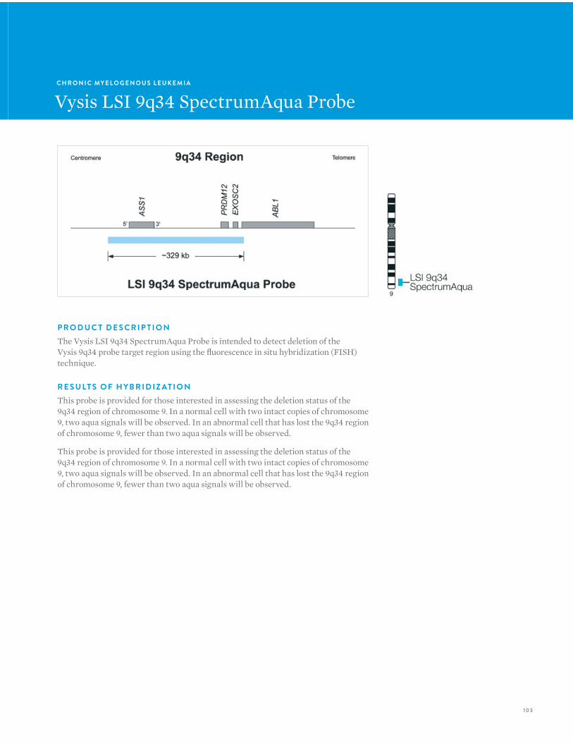
103
C H R O N I C MY E L O G E N O U S L E U K E M I A
Vysis LSI 9q34 SpectrumAqua Probe
P R O D U C T D E S C R I P T I O NThe Vysis LSI 9q34 SpectrumAqua Probe is intended to detect deletion of the Vysis 9q34 probe target region using the fluorescence in situ hybridization (FISH) technique.
R E S U LT S O F H Y B R I D I Z AT I O NThis probe is provided for those interested in assessing the deletion status of the 9q34 region of chromosome 9. In a normal cell with two intact copies of chromosome 9, two aqua signals will be observed. In an abnormal cell that has lost the 9q34 region of chromosome 9, fewer than two aqua signals will be observed.
This probe is provided for those interested in assessing the deletion status of the 9q34 region of chromosome 9. In a normal cell with two intact copies of chromosome 9, two aqua signals will be observed. In an abnormal cell that has lost the 9q34 region of chromosome 9, fewer than two aqua signals will be observed.
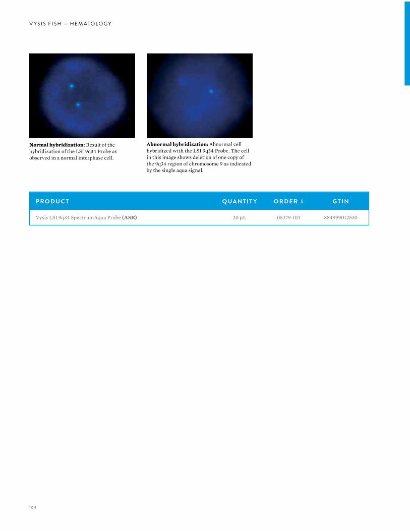
104
V Y S I S F I S H — H E M ATO LO G Y
P R O D U C T Q UA N T I T Y O R D E R # G T I N
Vysis LSI 9q34 SpectrumAqua Probe (ASR) 20 µL 05J79-011 884999012530
Abnormal hybridization: Abnormal cell hybridized with the LSI 9q34 Probe. The cell in this image shows deletion of one copy of the 9q34 region of chromosome 9 as indicated by the single aqua signal.
Normal hybridization: Result of the hybridization of the LSI 9q34 Probe as observed in a normal interphase cell.
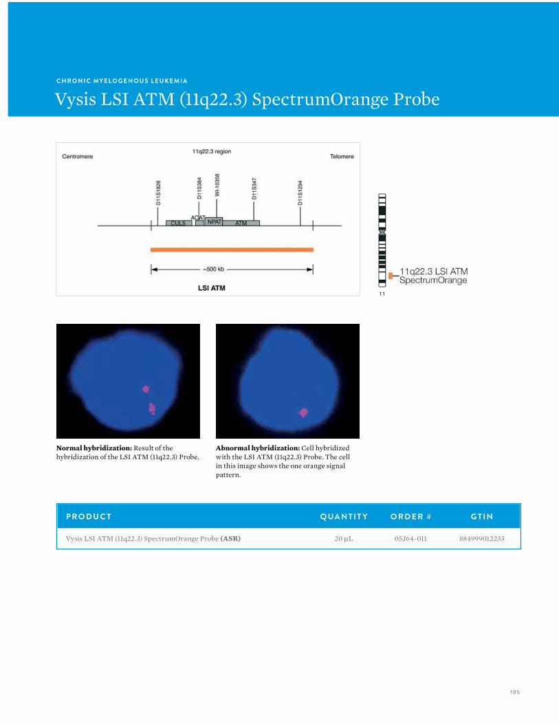
105
C H R O N I C MY E L O G E N O U S L E U K E M I A
Vysis LSI ATM (11q22.3) SpectrumOrange Probe
P R O D U C T Q UA N T I T Y O R D E R # G T I N
Vysis LSI ATM (11q22.3) SpectrumOrange Probe (ASR) 20 µL 05J64-011 884999012233
Normal hybridization: Result of the hybridization of the LSI ATM (11q22.3) Probe.
Abnormal hybridization: Cell hybridized with the LSI ATM (11q22.3) Probe. The cell in this image shows the one orange signal pattern.

106
V Y S I S F I S H — H E M ATO LO G YV Y S I S F I S H — H E M ATO LO G Y
C H R O N I C MY E L O G E N O U S L E U K E M I A
Vysis LSI IGH Dual Color,Break Apart Rearrangement Probe
P R O D U C T D E S C R I P T I O NThe LSI IGH Dual Color, Break Apart Rearrangement Probe is designed to detect chromosomal breakage of the immunoglobulin heavy chain (IGH) locus that is associated with 14q32 translocations involving a variety of other loci. Breakpoints within the IGH locus may occur at either the J segments [e.g., breakpoints commonly observed with t(14;18)] or within switch sequences located within the constant gene segments.
The LSI IGH Dual Color, Break Apart Rearrangement Probe is a mixture of two probes that hybridize to opposite sides of the J through constant regions of the IGH locus. The approximately 900 kb SpectrumGreen labeled LSI IGHV probe covers essentially the entire IGH variable region. The hybridization target of the approximately 250 kb SpectrumOrange labeled LSI IGH 3’ flanking probe lies completely 3’ to the IGH locus. As a result of this probe design, any translocation with a breakpoint at the J segments or within switch sequences should produce separate orange and green signals.
R E S U LT S O F H Y B R I D I Z AT I O NWhen hybridized to a normal nucleus, the LSI IGH Dual Color, Break Apart Rearrangement Probe produces a two orange/green (yellow) fusion (2F) signal pattern. As there is no probe targeted to the J or constant regions, a slight gap between the two differently colored probe signals may sometimes be observed in nuclei from normal cells. When the IGH Dual Color, Break Apart Translocation Probe is hybridized to a nucleus containing an IGH translocation, one orange, one green, and one orange/green fusion signal pattern is observed (1O1G1F). This signal pattern indicates that the genomic targets for the LSI IGHV and LSI IGH 3’ flanking probes have been physically separated as a result of the translocation. As V(D)J rearrangements may occur on either, or both, of the translocated and non-translocated IGH alleles, the green LSI IGHV probe signal intensity on either, or both, of the alleles may be diminished as a result of probe target deletion in some samples.
Normal hybridization: LSI IGH Dual Color, Break Apart Rearrangement Probe hybridized to nuclei exhibiting the expected two fusion (2F) signal pattern.
P R O D U C T Q UA N T I T Y O R D E R # G T I N
Vysis LSI IGH Dual Color, Break Apart Rearrangement Probe (ASR) 20 µL 05J73-001 884999012394
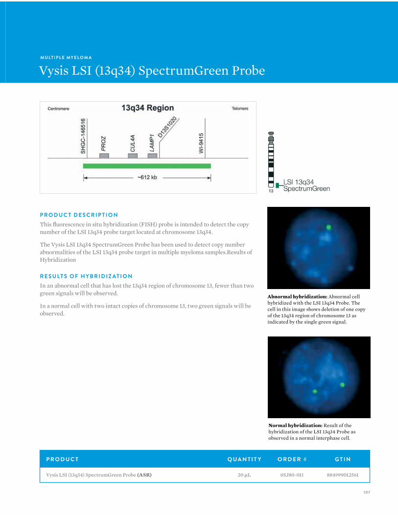
107
M U LT I P L E MY E L O M A
Vysis LSI (13q34) SpectrumGreen Probe
P R O D U C T D E S C R I P T I O NThis fluorescence in situ hybridization (FISH) probe is intended to detect the copy number of the LSI 13q34 probe target located at chromosome 13q34.
The Vysis LSI 13q34 SpectrumGreen Probe has been used to detect copy number abnormalities of the LSI 13q34 probe target in multiple myeloma samples.Results of Hybridization
R E S U LT S O F H Y B R I D I Z AT I O NIn an abnormal cell that has lost the 13q34 region of chromosome 13, fewer than two green signals will be observed.
In a normal cell with two intact copies of chromosome 13, two green signals will be observed.
Abnormal hybridization: Abnormal cell hybridized with the LSI 13q34 Probe. The cell in this image shows deletion of one copy of the 13q34 region of chromosome 13 as indicated by the single green signal.
Normal hybridization: Result of the hybridization of the LSI 13q34 Probe as observed in a normal interphase cell.
P R O D U C T Q UA N T I T Y O R D E R # G T I N
Vysis LSI (13q34) SpectrumGreen Probe (ASR) 20 µL 05J80-011 884999012561
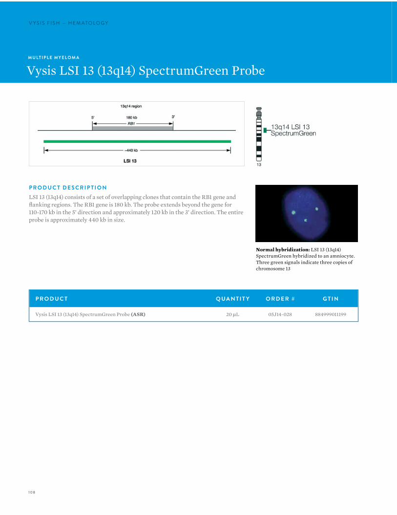
108
V Y S I S F I S H — H E M ATO LO G YV Y S I S F I S H — H E M ATO LO G Y
M U LT I P L E MY E L O M A
Vysis LSI 13 (13q14) SpectrumGreen Probe
P R O D U C T Q UA N T I T Y O R D E R # G T I N
Vysis LSI 13 (13q14) SpectrumGreen Probe (ASR) 20 µL 05J14-028 884999011199
P R O D U C T D E S C R I P T I O NLSI 13 (13q14) consists of a set of overlapping clones that contain the RB1 gene and flanking regions. The RB1 gene is 180 kb. The probe extends beyond the gene for 110-170 kb in the 5’ direction and approximately 120 kb in the 3’ direction. The entire probe is approximately 440 kb in size.
Normal hybridization: LSI 13 (13q14) SpectrumGreen hybridized to an amniocyte. Three green signals indicate three copies of chromosome 13
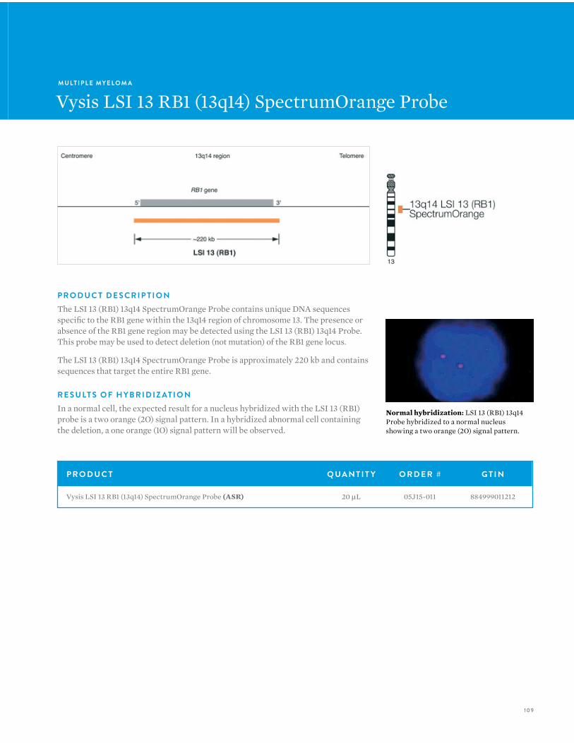
109
M U LT I P L E MY E L O M A
Vysis LSI 13 RB1 (13q14) SpectrumOrange Probe
Normal hybridization: LSI 13 (RB1) 13q14 Probe hybridized to a normal nucleus showing a two orange (2O) signal pattern.
P R O D U C T Q UA N T I T Y O R D E R # G T I N
Vysis LSI 13 RB1 (13q14) SpectrumOrange Probe (ASR) 20 µL 05J15-011 884999011212
P R O D U C T D E S C R I P T I O NThe LSI 13 (RB1) 13q14 SpectrumOrange Probe contains unique DNA sequences specific to the RB1 gene within the 13q14 region of chromosome 13. The presence or absence of the RB1 gene region may be detected using the LSI 13 (RB1) 13q14 Probe. This probe may be used to detect deletion (not mutation) of the RB1 gene locus.
The LSI 13 (RB1) 13q14 SpectrumOrange Probe is approximately 220 kb and contains sequences that target the entire RB1 gene.
R E S U LT S O F H Y B R I D I Z AT I O NIn a normal cell, the expected result for a nucleus hybridized with the LSI 13 (RB1) probe is a two orange (2O) signal pattern. In a hybridized abnormal cell containing the deletion, a one orange (1O) signal pattern will be observed.
1 10
V Y S I S F I S H — H E M ATO LO G YV Y S I S F I S H — H E M ATO LO G Y
M U LT I P L E MY E L O M A
Vysis LSI 13q34 SpectrumGreen Probe
P R O D U C T Q UA N T I T Y O R D E R # G T I N
Vysis LSI 13q34 SpectrumGreen Probe (ASR) 20 µL 05J80-011 884999012561

1 1 1
M U LT I P L E MY E L O M A
Vysis LSI IGH Dual Color,Break Apart Rearrangement Probe
P R O D U C T D E S C R I P T I O NThe LSI IGH Dual Color, Break Apart Rearrangement Probe is designed to detect chromosomal breakage of the immunoglobulin heavy chain (IGH) locus that is associated with 14q32 translocations involving a variety of other loci. Breakpoints within the IGH locus may occur at either the J segments [e.g., breakpoints commonly observed with t(14;18)] or within switch sequences located within the constant gene segments.
The LSI IGH Dual Color, Break Apart Rearrangement Probe is a mixture of two probes that hybridize to opposite sides of the J through constant regions of the IGH locus. The approximately 900 kb SpectrumGreen labeled LSI IGHV probe covers essentially the entire IGH variable region. The hybridization target of the approximately 250 kb SpectrumOrange labeled LSI IGH 3’ flanking probe lies completely 3’ to the IGH locus. As a result of this probe design, any translocation with a breakpoint at the J segments or within switch sequences should produce separate orange and green signals.
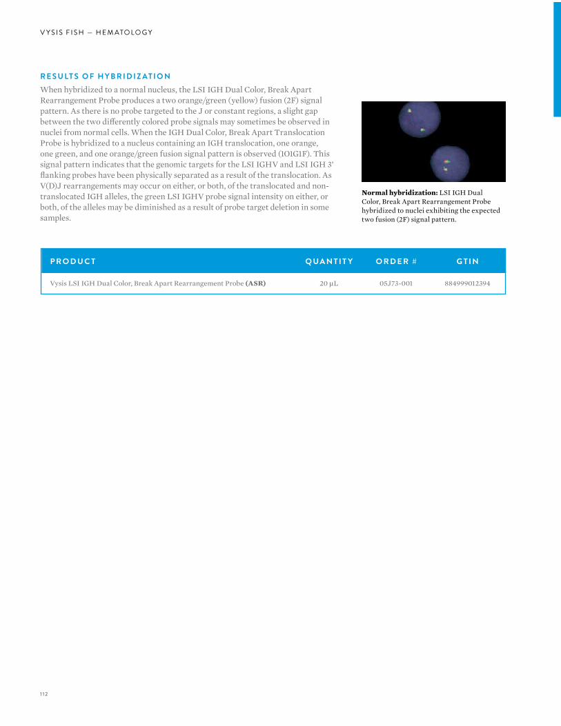
1 12
V Y S I S F I S H — H E M ATO LO G Y
R E S U LT S O F H Y B R I D I Z AT I O NWhen hybridized to a normal nucleus, the LSI IGH Dual Color, Break Apart Rearrangement Probe produces a two orange/green (yellow) fusion (2F) signal pattern. As there is no probe targeted to the J or constant regions, a slight gap between the two differently colored probe signals may sometimes be observed in nuclei from normal cells. When the IGH Dual Color, Break Apart Translocation Probe is hybridized to a nucleus containing an IGH translocation, one orange, one green, and one orange/green fusion signal pattern is observed (1O1G1F). This signal pattern indicates that the genomic targets for the LSI IGHV and LSI IGH 3’ flanking probes have been physically separated as a result of the translocation. As V(D)J rearrangements may occur on either, or both, of the translocated and non-translocated IGH alleles, the green LSI IGHV probe signal intensity on either, or both, of the alleles may be diminished as a result of probe target deletion in some samples.
Normal hybridization: LSI IGH Dual Color, Break Apart Rearrangement Probe hybridized to nuclei exhibiting the expected two fusion (2F) signal pattern.
P R O D U C T Q UA N T I T Y O R D E R # G T I N
Vysis LSI IGH Dual Color, Break Apart Rearrangement Probe (ASR) 20 µL 05J73-001 884999012394
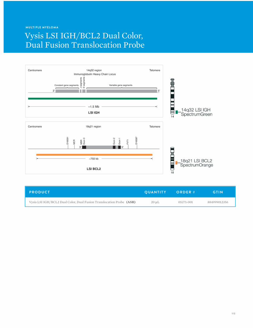
1 13
M U LT I P L E MY E L O M A
Vysis LSI IGH/BCL2 Dual Color,Dual Fusion Translocation Probe
P R O D U C T Q UA N T I T Y O R D E R # G T I N
Vysis LSI IGH/BCL2 Dual Color, Dual Fusion Translocation Probe (ASR) 20 µL 05J71-001 884999012356
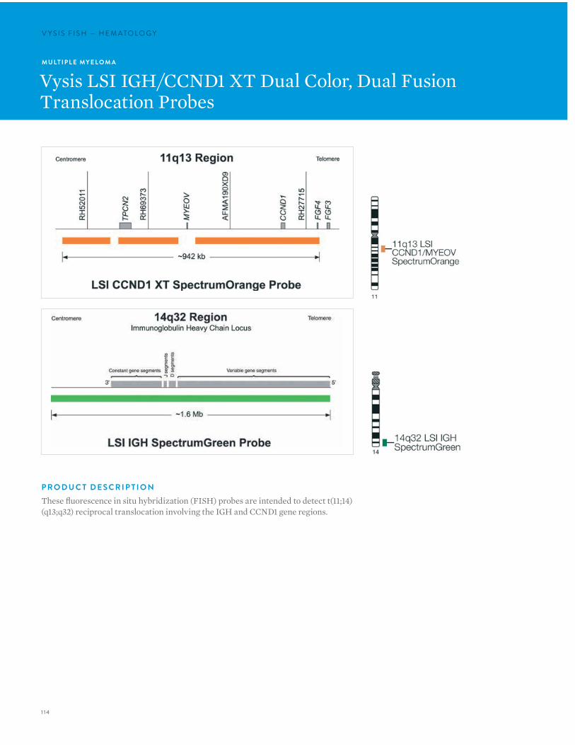
1 14
V Y S I S F I S H — H E M ATO LO G YV Y S I S F I S H — H E M ATO LO G Y
M U LT I P L E MY E L O M A
Vysis LSI IGH/CCND1 XT Dual Color, Dual Fusion Translocation Probes
P R O D U C T D E S C R I P T I O NThese fluorescence in situ hybridization (FISH) probes are intended to detect t(11;14)(q13;q32) reciprocal translocation involving the IGH and CCND1 gene regions.

1 15
R E S U LT S O F H Y B R I D I Z AT I O NIn an abnormal cell containing the t(11;14), one orange (CCND1/MYEOV), one green (IGH), and two fusion signal pattern (der (11) and der (14)) may be observed. Some samples containing the t(11;14) may display signal patterns different than one orange, one green, and two fusions.
In a normal cell that lacks the t(11;14), a two orange and two green signal pattern will be observed reflecting the two intact copies of CCND1/MYEOV and IGH respectively.
Normal hybridization: A normal interphase cell hybridized with the Vysis LSI IGH/CCND1 XT Dual Color Dual Fusion Probes. The cell shows the expected two orange (CCND1/MYEOV), two green (IGH) signal pattern.
Abnormal hybridization: An abnormal interphase cell hybridized with the Vysis LSI IGH/CCND1 XT Dual Color Dual Fusion Probes. The cell in this image shows the one orange (CCND1/MYEOV), one green (IGH), two fusion (der (11) and der (14)) signal pattern indicative of a t(11;14).
P R O D U C T Q UA N T I T Y O R D E R # G T I N
Vysis LSI IGH/CCND1 XT Dual Color, Dual Fusion Translocation Probes (ASR) 20 µL 05J72-001 884999012370
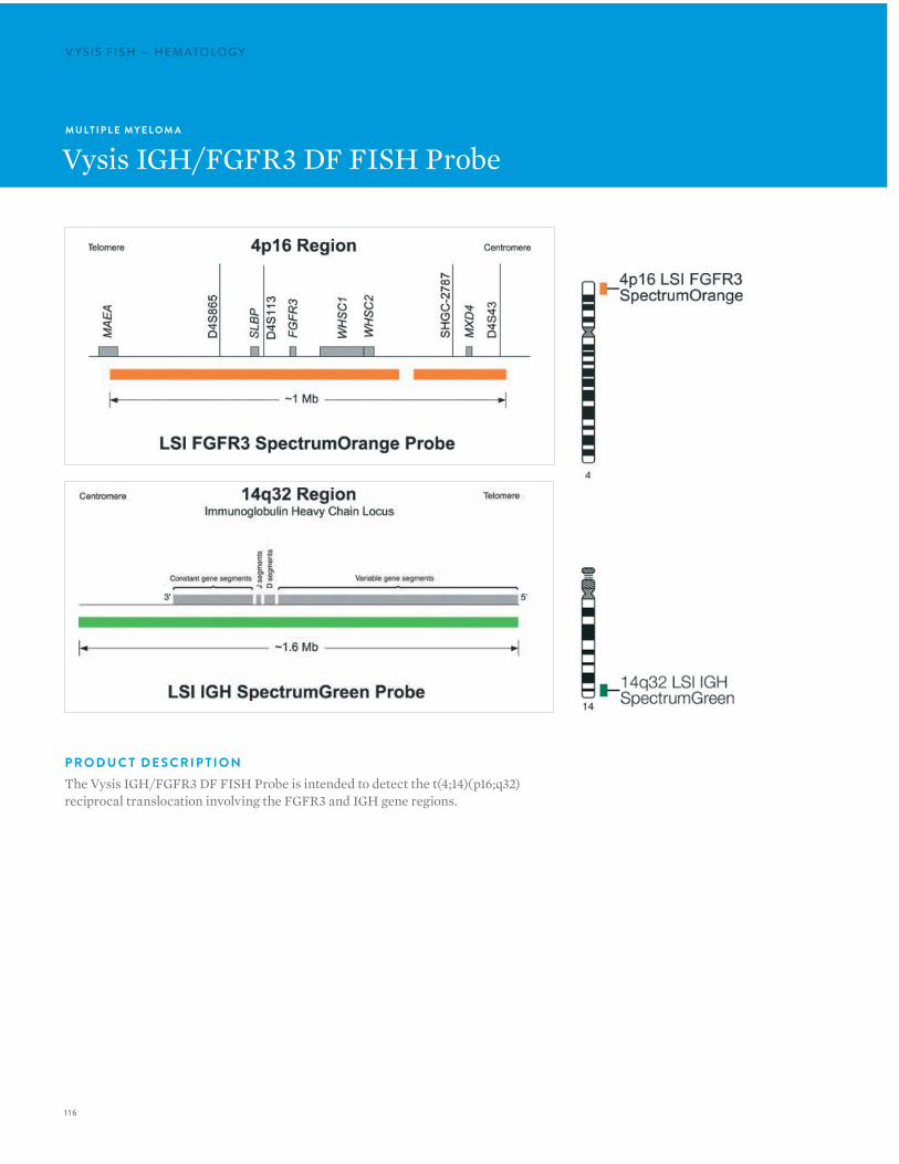
1 16
V Y S I S F I S H — H E M ATO LO G YV Y S I S F I S H — H E M ATO LO G Y
M U LT I P L E MY E L O M A
Vysis IGH/FGFR3 DF FISH Probe
P R O D U C T D E S C R I P T I O NThe Vysis IGH/FGFR3 DF FISH Probe is intended to detect the t(4;14)(p16;q32) reciprocal translocation involving the FGFR3 and IGH gene regions.
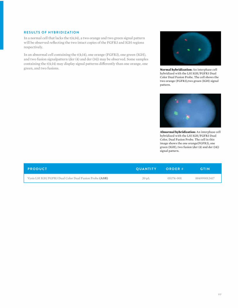
1 17
R E S U LT S O F H Y B R I D I Z AT I O NIn a normal cell that lacks the t(4;14), a two orange and two green signal pattern will be observed reflecting the two intact copies of the FGFR3 and IGH regions respectively.
In an abnormal cell containing the t(4;14), one orange (FGFR3), one green (IGH), and two fusion signalpattern (der (4) and der (14)) may be observed. Some samples containing the t(4;14) may display signal patterns differently than one orange, one green, and two fusions. Normal hybridization: An interphase cell
hybridized with the LSI IGH/FGFR3 Dual Color Dual Fusion Probe. The cell shows the two orange (FGFR3),two green (IGH) signal pattern.
Abnormal hybridization: An interphase cell hybridized with the LSI IGH/FGFR3 Dual Color, Dual Fusion Probe. The cell in this image shows the one orange(FGFR3), one green (IGH), two fusion (der (4) and der (14)) signal pattern.
P R O D U C T Q UA N T I T Y O R D E R # G T I N
Vysis LSI IGH/FGFR3 Dual Color Dual Fusion Probe (ASR) 20 µL 05J74-001 884999012417
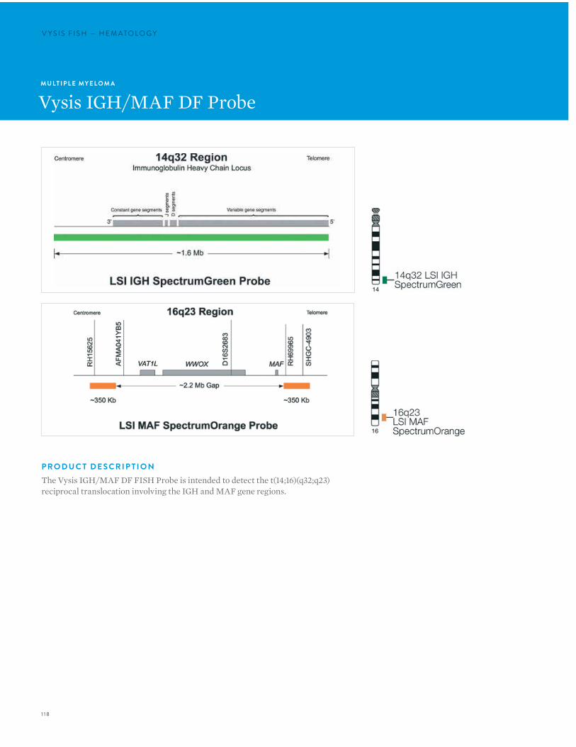
1 18
V Y S I S F I S H — H E M ATO LO G YV Y S I S F I S H — H E M ATO LO G Y
M U LT I P L E MY E L O M A
Vysis IGH/MAF DF Probe
P R O D U C T D E S C R I P T I O NThe Vysis IGH/MAF DF FISH Probe is intended to detect the t(14;16)(q32;q23) reciprocal translocation involving the IGH and MAF gene regions.
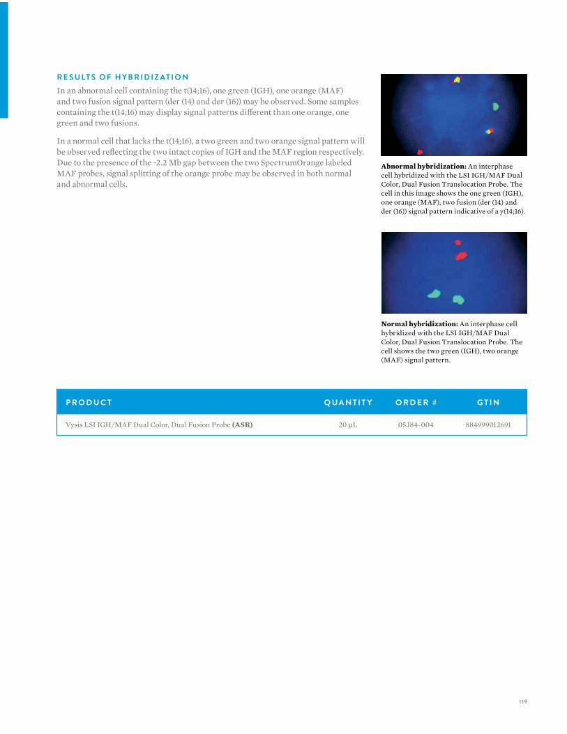
1 19
P R O D U C T Q UA N T I T Y O R D E R # G T I N
Vysis LSI IGH/MAF Dual Color, Dual Fusion Probe (ASR) 20 µL 05J84-004 884999012691
R E S U LT S O F H Y B R I D I Z AT I O NIn an abnormal cell containing the t(14;16), one green (IGH), one orange (MAF) and two fusion signal pattern (der (14) and der (16)) may be observed. Some samples containing the t(14;16) may display signal patterns different than one orange, one green and two fusions.
In a normal cell that lacks the t(14;16), a two green and two orange signal pattern will be observed reflecting the two intact copies of IGH and the MAF region respectively. Due to the presence of the ~2.2 Mb gap between the two SpectrumOrange labeled MAF probes, signal splitting of the orange probe may be observed in both normal and abnormal cells.
Abnormal hybridization: An interphase cell hybridized with the LSI IGH/MAF Dual Color, Dual Fusion Translocation Probe. The cell in this image shows the one green (IGH), one orange (MAF), two fusion (der (14) and der (16)) signal pattern indicative of a y(14;16).
Normal hybridization: An interphase cell hybridized with the LSI IGH/MAF Dual Color, Dual Fusion Translocation Probe. The cell shows the two green (IGH), two orange (MAF) signal pattern.

120
V Y S I S F I S H — H E M ATO LO G YV Y S I S F I S H — H E M ATO LO G Y
MY E L O DY S P L A S T I C S Y N D R O M E
Vysis CEP 8 SpectrumOrangeDirect Labeled Fluorescent DNA Probe Kit
P R O D U C T D E S C R I P T I O NCEP 8 is a SpectrumOrange labeled probe specific for the alpha satellite (centromeric) region, 8p11.1-q11.1.
The CEP 8 DNA Probe Kit which is available for in vitro diagnostic use and may be used as an adjunct to standard karotyping to identify and enumerate chromosome 8 in cells obtained from bone marrow. In multi-site clinical trials, the CEP 8 DNA Probe Kit for interphase analysis was 96% sensitive and 98% specific as compared to traditional cytogenetic analysis. A close association has been made between trisomy 8 and both myeloid blast crisis and basophilia. Trisomy 8 is a prevalent genetic aberration in several specific diseases:
• Chronic Myelogenous Leukemia (CML)
• Acute Myeloid Leukemia (AML)
• Myeloproliferative disorders (MPD)
• Myelodysplastic Syndrome (MDS)
• Other hematologic disorders not specified (HDNOS)
C E P 8 S P E C T R U M O R A N G E D N A P R O B E K I T CO N T E N TComponents of the CEP 8 SpectrumOrange DNA Probe Kit include:
• CEP 8 SpectrumOrange alpha satellite DNA for centromere region 8p11.1-q11.1 predenatured in hybridization buffer (220 µL)
• NP-40 (detergent for wash solution: 1000 µL)
• DAPI II counterstain (300 µL)
• 20X SSC (66 g)
R E S U LT S O F H Y B R I D I Z AT I O NIn a normal cell, the expected pattern for a nucleus hybridized with the CEP 8 probe is a two orange (2O) signal pattern. In an abnormal cell containing trisomy 8, the expected pattern will be a three orange (3O) signal pattern.
Normal hybridization: CEP 8 SpectrumOrange hybridized to a normal cell showing two orange signals indicating two copies of chromosome 8.

12 1
I N T E N D E D U S EThe CEP 8 SpectrumOrange DNA Probe Kit is intended to detect AT rich alpha satellite sequences in the centromere region of chromosome 8 in conjunction with routine diagnostic cytogenetic testing. It is indicated for use as an adjunct to standard cytogenetic analysis for identifying and enumerating chromosome 8 via fluorescence in situ hybridization (FISH) in interphase nuclei and in metaphase spreads of cells obtained from bone marrow in patients with myeloid disorders [Chronic myelogenous leukemia (CML), Acute myeloid leukemia (AML), Myeloproliferative disorder (MPD), Myelodysplastic syndrome (MDS), and Hematological disorders not otherwise specified (HDNOS)]. It is not intended to be used as a stand alone assay for test reporting. It is not intended for use in long term cell cultured materials such as amniocytes, fibroblasts and tumor cells.
L I M I TAT I O N S • The CEP 8 SpectrumOrange DNA Probe Kit has been
characterized only for identifying chromosomes in nuclear preparations or metaphase spreads from bone marrow specimens.
• The clinical interpretation of any test results should be done in conjunction with standard cytogenetic analysis and should be evaluated within the context of the patient’s medical history and other diagnostic laboratory test results.
• Clinical specimens with >2.2% tri-signaled nuclei are considered to have an abnormal trisomy 8 clone. Those with ≤ 2.2% tri-signaled nuclei should be considered normal, although the presence of trisomy 8 is not completely excluded.
• The CEP 8 SpectrumOrange DNA Probe Kit is not intended for long term cell cultured materials such as amniocytes, fibroblasts and tumor cells.
• FISH assay results may not be informative if the specimen quality and/or specimen slide preparation is inadequate.
• If significant peripheral blood contamination is present in the bone marrow specimen, the blood may dilute the specimen; it is important to recognize the potential effects this dilution effect may have on the FISH assay results.
• It is possible that patients may have chromosome polymorphism which may hybridize with CEP 8 probe. FISH metaphase analysis should be done in addition to FISH interphase analysis. Polymorphism was not investigated in the clinical trials.
• This assay will not detect the presence of other chromosome abnormalities frequently associated with hematological disorders.
• The efficacy of this assay for monitoring of trisomy 8 or disease progression has not been demonstrated.
P R O D U C T Q UA N T I T Y O R D E R # G T I N
Vysis CEP 8 SpectrumOrange Direct Labeled Fluorescent DNA Probe Kit (IVD) 20 Assays 07J22-008 884999027077
Vysis CEP 8 SpectrumOrange Direct Labeled Fluorescent DNA Probe Kit (without control slides) (IVD) 20 Assays 07J20-008 884999027008
ProbeChek Control Slides for FISH using CEP 8 and CEP 12 Assay, Negative Control 0%, trisomy 8/12 (IVD) 5 Slides 07J21-001 884999027039
ProbeChek Control Slides for FISH using CEP 8 and CEP 12 Assay, Positive Control 10%, trisomy 8/12 (IVD) 5 Slides 07J21-002 884999027046

122
V Y S I S F I S H — H E M ATO LO G YV Y S I S F I S H — H E M ATO LO G Y
MY E L O DY S P L A S T I C S Y N D R O M E
Vysis D7S486/ Vysis CEP 7 FISH Probe Kit
I N T E N D E D U S EThe Vysis D7S486/CEP 7 FISH Probe Kit is a device intended for specimen characterization and detects the LSI D7S486 probe target on chromosome 7q31 and the CEP 7 probe target on chromosome 7p11.1-q11.1 in bone marrow and peripheral blood specimens from patients with acute myeloid leukemia or myelodysplastic syndrome. The assay results are intended to be interpreted by a qualified pathologist or cytogeneticist. This device is not intended for high-risk uses such as selecting therapy, predicting therapeutic response or disease screening. The use of this device for diagnosis, prognosis, monitoring, or risk assessment has not been established.
L I M I TAT I O N SThe Vysis D7S486/CEP 7 FISH Probe Kit is intended to be used in combination with additional biomarkers, morphology, and other clinical information.
Normal hybridization: LSI D7S486/CEP 7 Dual Color Probe hybridized to a nucleus showing the two orange, two green (2O2G) signal pattern.
P R O D U C T Q UA N T I T Y O R D E R # G T I N
Vysis D7S486/ Vysis CEP 7 FISH Probe Kit (IVD) 20 µL 04N78-020 884999036406
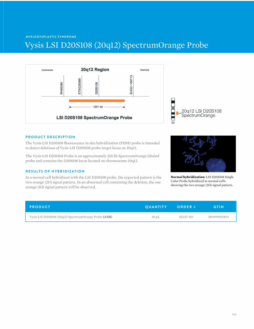
123
MY E L O DY S P L A S T I C S Y N D R O M E
Vysis LSI D20S108 (20q12) SpectrumOrange Probe
P R O D U C T D E S C R I P T I O NThe Vysis LSI D20S108 fluorescence in situ hybridization (FISH) probe is intended to detect deletions of Vysis LSI D20S108 probe target locus on 20q12.
The Vysis LSI D20S108 Probe is an approximately 201 kb SpectrumOrange labeled probe and contains the D20S108 locus located on chromosome 20q12.
R E S U LT S O F H Y B R I D I Z AT I O NIn a normal cell hybridized with the LSI D20S108 probe, the expected pattern is the two orange (2O) signal pattern. In an abnormal cell containing the deletion, the one orange (1O) signal pattern will be observed.
Normal hybridization: LSI D20S108 Single Color Probe hybridized to normal cells showing the two orange (2O) signal pattern.
P R O D U C T Q UA N T I T Y O R D E R # G T I N
Vysis LSI D20S108 (20q12) SpectrumOrange Probe (ASR) 20 µL 05J47-011 884999011854
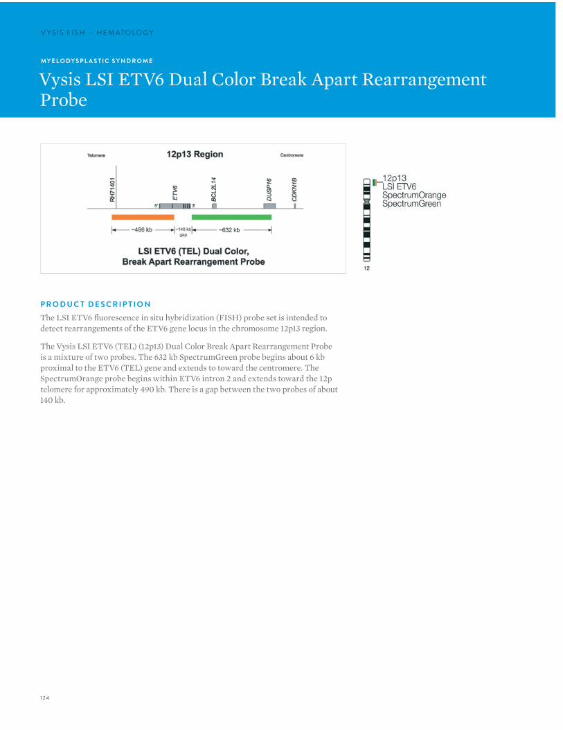
124
V Y S I S F I S H — H E M ATO LO G YV Y S I S F I S H — H E M ATO LO G Y
P R O D U C T D E S C R I P T I O NThe LSI ETV6 fluorescence in situ hybridization (FISH) probe set is intended to detect rearrangements of the ETV6 gene locus in the chromosome 12p13 region.
The Vysis LSI ETV6 (TEL) (12p13) Dual Color Break Apart Rearrangement Probe is a mixture of two probes. The 632 kb SpectrumGreen probe begins about 6 kb proximal to the ETV6 (TEL) gene and extends to toward the centromere. The SpectrumOrange probe begins within ETV6 intron 2 and extends toward the 12p telomere for approximately 490 kb. There is a gap between the two probes of about 140 kb.
MY E L O DY S P L A S T I C S Y N D R O M E
Vysis LSI ETV6 Dual Color Break Apart Rearrangement Probe
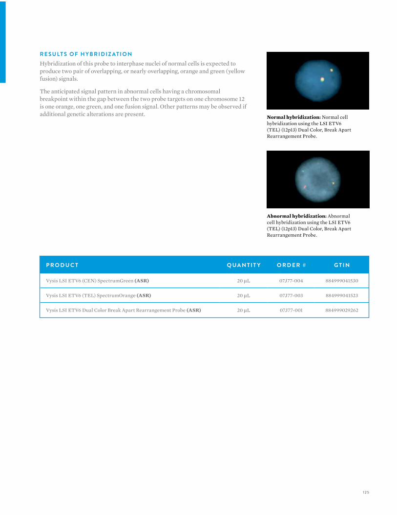
125
Abnormal hybridization: Abnormal cell hybridization using the LSI ETV6 (TEL) (12p13) Dual Color, Break Apart Rearrangement Probe.
Normal hybridization: Normal cell hybridization using the LSI ETV6 (TEL) (12p13) Dual Color, Break Apart Rearrangement Probe.
P R O D U C T Q UA N T I T Y O R D E R # G T I N
Vysis LSI ETV6 (CEN) SpectrumGreen (ASR) 20 µL 07J77-004 884999041530
Vysis LSI ETV6 (TEL) SpectrumOrange (ASR) 20 µL 07J77-003 884999041523
Vysis LSI ETV6 Dual Color Break Apart Rearrangement Probe (ASR) 20 µL 07J77-001 884999029262
R E S U LT S O F H Y B R I D I Z AT I O NHybridization of this probe to interphase nuclei of normal cells is expected to produce two pair of overlapping, or nearly overlapping, orange and green (yellow fusion) signals.
The anticipated signal pattern in abnormal cells having a chromosomal breakpoint within the gap between the two probe targets on one chromosome 12 is one orange, one green, and one fusion signal. Other patterns may be observed if additional genetic alterations are present.
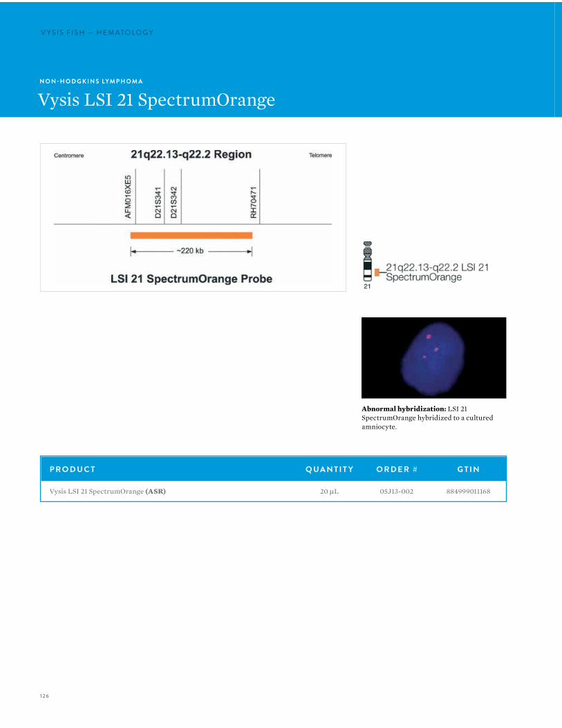
126
V Y S I S F I S H — H E M ATO LO G YV Y S I S F I S H — H E M ATO LO G Y
P R O D U C T Q UA N T I T Y O R D E R # G T I N
Vysis LSI 21 SpectrumOrange (ASR) 20 µL 05J13-002 884999011168
N O N -H O D G K I N S LYM P H O M A
Vysis LSI 21 SpectrumOrange
Abnormal hybridization: LSI 21 SpectrumOrange hybridized to a cultured amniocyte.
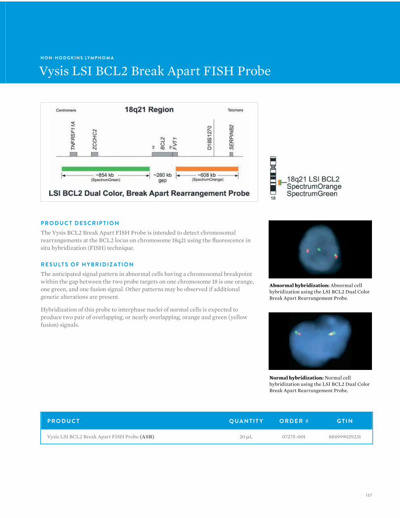
127
N O N -H O D G K I N S LYM P H O M A
Vysis LSI BCL2 Break Apart FISH Probe
P R O D U C T D E S C R I P T I O NThe Vysis BCL2 Break Apart FISH Probe is intended to detect chromosomal rearrangements at the BCL2 locus on chromosome 18q21 using the fluorescence in situ hybridization (FISH) technique.
R E S U LT S O F H Y B R I D I Z AT I O NThe anticipated signal pattern in abnormal cells having a chromosomal breakpoint within the gap between the two probe targets on one chromosome 18 is one orange, one green, and one fusion signal. Other patterns may be observed if additional genetic alterations are present.
Hybridization of this probe to interphase nuclei of normal cells is expected to produce two pair of overlapping, or nearly overlapping, orange and green (yellow fusion) signals.
Abnormal hybridization: Abnormal cell hybridization using the LSI BCL2 Dual Color Break Apart Rearrangement Probe.
Normal hybridization: Normal cell hybridization using the LSI BCL2 Dual Color Break Apart Rearrangement Probe.
P R O D U C T Q UA N T I T Y O R D E R # G T I N
Vysis LSI BCL2 Break Apart FISH Probe (ASR) 20 µL 07J75-001 884999029231
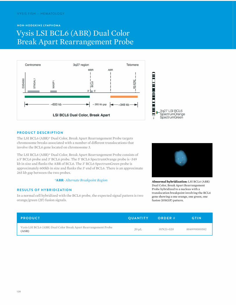
128
V Y S I S F I S H — H E M ATO LO G YV Y S I S F I S H — H E M ATO LO G Y
N O N -H O D G K I N S LYM P H O M A
Vysis LSI BCL6 (ABR) Dual ColorBreak Apart Rearrangement Probe
P R O D U C T D E S C R I P T I O NThe LSI BCL6 (ABR)* Dual Color, Break Apart Rearrangement Probe targets chromosome breaks associated with a number of different translocations that involve the BCL6 gene located on chromosome 3.
The LSI BCL6 (ABR)* Dual Color, Break Apart Rearrangement Probe consists of a 5’ BCL6 probe and 3’ BCL6 probe. The 5’ BCL6 SpectrumOrange probe is ~349 kb in size and flanks the ABR of BCL6. The 3’ BCL6 SpectrumGreen probe is approximately 600kb in size and flanks the 3’ end of BCL6. There is an approximate 265 kb gap between the two probes.
*ABR- Alternate Breakpoint Region
R E S U LT S O F H Y B R I D I Z AT I O NIn a normal cell hybridized with the BCL6 probe, the expected signal pattern is two orange/green (2F) fusion signals.
P R O D U C T Q UA N T I T Y O R D E R # G T I N
Vysis LSI BCL6 (ABR) Dual Color Break Apart Rearrangement Probe (ASR) 20 µL 01N23-020 884999000582
Abnormal hybridization: LSI BCL6 (ABR) Dual Color, Break Apart Rearrangement Probe hybridized to a nucleus with a translocation breakpoint involving the BCL6 gene showing a one orange, one green, one fusion (1O1G1F) pattern.
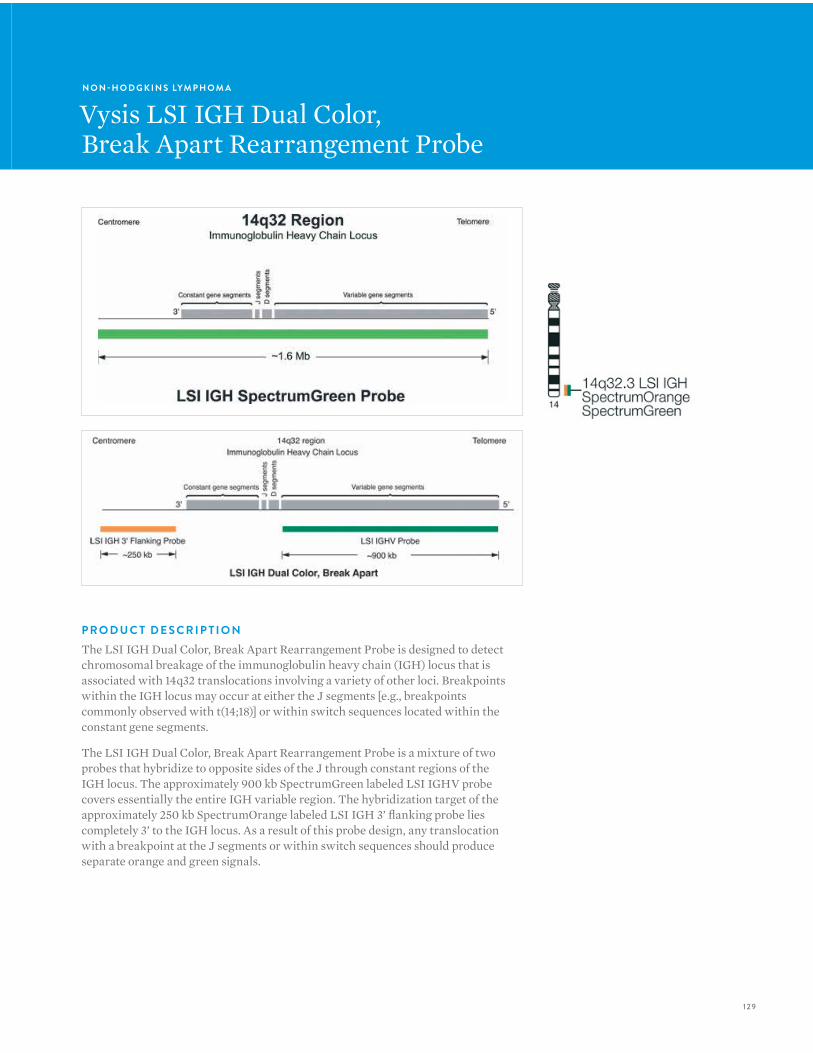
129
N O N -H O D G K I N S LYM P H O M A
Vysis LSI IGH Dual Color,Break Apart Rearrangement Probe
P R O D U C T D E S C R I P T I O NThe LSI IGH Dual Color, Break Apart Rearrangement Probe is designed to detect chromosomal breakage of the immunoglobulin heavy chain (IGH) locus that is associated with 14q32 translocations involving a variety of other loci. Breakpoints within the IGH locus may occur at either the J segments [e.g., breakpoints commonly observed with t(14;18)] or within switch sequences located within the constant gene segments.
The LSI IGH Dual Color, Break Apart Rearrangement Probe is a mixture of two probes that hybridize to opposite sides of the J through constant regions of the IGH locus. The approximately 900 kb SpectrumGreen labeled LSI IGHV probe covers essentially the entire IGH variable region. The hybridization target of the approximately 250 kb SpectrumOrange labeled LSI IGH 3’ flanking probe lies completely 3’ to the IGH locus. As a result of this probe design, any translocation with a breakpoint at the J segments or within switch sequences should produce separate orange and green signals.
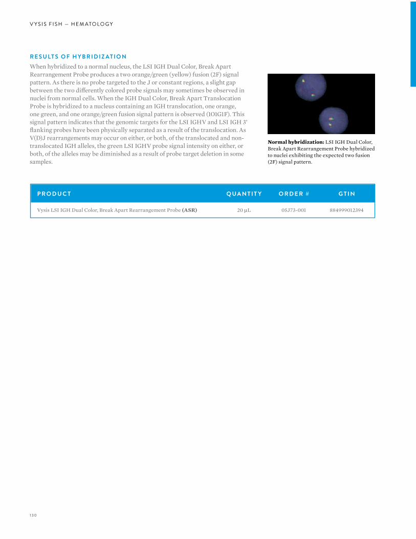
130
V Y S I S F I S H — H E M ATO LO G Y
R E S U LT S O F H Y B R I D I Z AT I O NWhen hybridized to a normal nucleus, the LSI IGH Dual Color, Break Apart Rearrangement Probe produces a two orange/green (yellow) fusion (2F) signal pattern. As there is no probe targeted to the J or constant regions, a slight gap between the two differently colored probe signals may sometimes be observed in nuclei from normal cells. When the IGH Dual Color, Break Apart Translocation Probe is hybridized to a nucleus containing an IGH translocation, one orange, one green, and one orange/green fusion signal pattern is observed (1O1G1F). This signal pattern indicates that the genomic targets for the LSI IGHV and LSI IGH 3’ flanking probes have been physically separated as a result of the translocation. As V(D)J rearrangements may occur on either, or both, of the translocated and non-translocated IGH alleles, the green LSI IGHV probe signal intensity on either, or both, of the alleles may be diminished as a result of probe target deletion in some samples.
Normal hybridization: LSI IGH Dual Color, Break Apart Rearrangement Probe hybridized to nuclei exhibiting the expected two fusion (2F) signal pattern.
P R O D U C T Q UA N T I T Y O R D E R # G T I N
Vysis LSI IGH Dual Color, Break Apart Rearrangement Probe (ASR) 20 µL 05J73-001 884999012394

13 1
N O N -H O D G K I N S LYM P H O M A
Vysis LSI IGH/BCL2 Dual Color,Dual Fusion Translocation Probe
P R O D U C T D E S C R I P T I O NThe LSI IGH/BCL2 Dual Color, Dual Fusion Translocation Probe is designed to detect the juxtaposition of immunoglobulin heavy chain (IGH) locus and BCL gene sequences. The translocation involving IGH at 14q32 and BCL2 at 18q21, t(14;18)(q32;q21) is common. Relocation of an IGH transcriptional enhancer next to the BCL2 gene as a result of the t(14;18) translocation is thought to cause constitutive over-expression of the anti-apoptotic BCL2 protein. The breakpoints at 14q32 occur at the IGH J segments and about 75% of the breaks at 18q21 occur in either the 2.8 kb major breakpoint region (MBR) 3’ of BCL2 exon 3 or in the minor cluster region (MCR) located about 30 kb 3’ to the MBR. The remaining BCL2 breakpoints are thought to lie between the MBR and MCR regions, or 5’ of the BCL2 gene.
The LSI IGH/BCL2 Dual Color, Dual Fusion Translocation Probe is a mixture of the LSI IGH probe labeled with SpectrumGreen spanning approximately 1.5 Mb and containing sequences homologous to essentially the entire IGH locus, as well as sequences extending about 300 kb beyond the 3’ end of the IGH locus. The LSI BCL2 probe labeled with SpectrumOrange covers an approximate 750 kb region, including the entire BCL2 gene with additional sequences extending approximately 250 kb both distal and proximal to the gene.
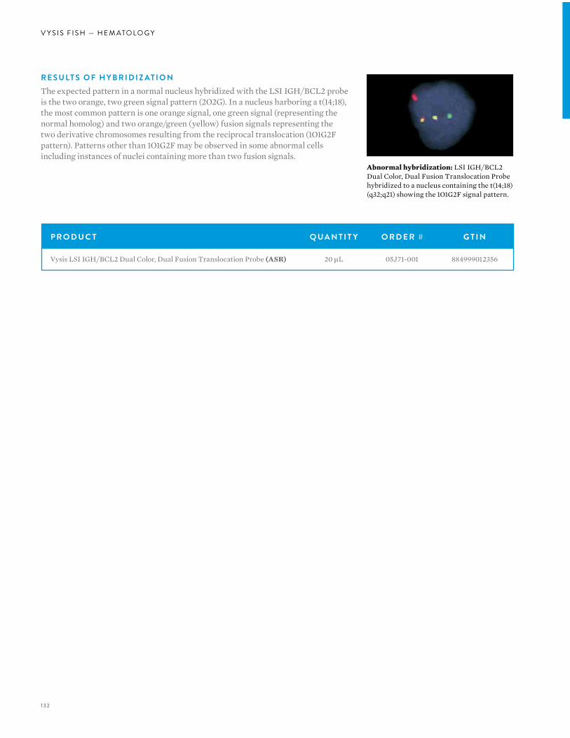
132
V Y S I S F I S H — H E M ATO LO G Y
P R O D U C T Q UA N T I T Y O R D E R # G T I N
Vysis LSI IGH/BCL2 Dual Color, Dual Fusion Translocation Probe (ASR) 20 µL 05J71-001 884999012356
R E S U LT S O F H Y B R I D I Z AT I O NThe expected pattern in a normal nucleus hybridized with the LSI IGH/BCL2 probe is the two orange, two green signal pattern (2O2G). In a nucleus harboring a t(14;18), the most common pattern is one orange signal, one green signal (representing the normal homolog) and two orange/green (yellow) fusion signals representing the two derivative chromosomes resulting from the reciprocal translocation (1O1G2F pattern). Patterns other than 1O1G2F may be observed in some abnormal cells including instances of nuclei containing more than two fusion signals.
Abnormal hybridization: LSI IGH/BCL2 Dual Color, Dual Fusion Translocation Probe hybridized to a nucleus containing the t(14;18) (q32;q21) showing the 1O1G2F signal pattern.
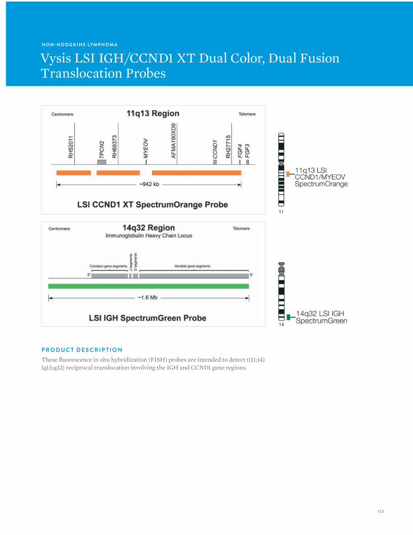
133
N O N -H O D G K I N S LYM P H O M A
Vysis LSI IGH/CCND1 XT Dual Color, Dual Fusion Translocation Probes
P R O D U C T D E S C R I P T I O NThese fluorescence in situ hybridization (FISH) probes are intended to detect t(11;14)(q13;q32) reciprocal translocation involving the IGH and CCND1 gene regions.
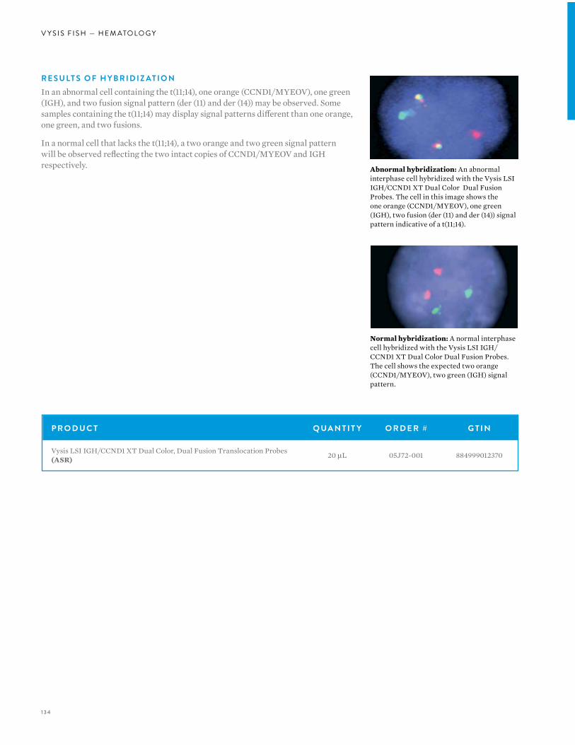
134
V Y S I S F I S H — H E M ATO LO G Y
R E S U LT S O F H Y B R I D I Z AT I O NIn an abnormal cell containing the t(11;14), one orange (CCND1/MYEOV), one green (IGH), and two fusion signal pattern (der (11) and der (14)) may be observed. Some samples containing the t(11;14) may display signal patterns different than one orange, one green, and two fusions.
In a normal cell that lacks the t(11;14), a two orange and two green signal pattern will be observed reflecting the two intact copies of CCND1/MYEOV and IGH respectively.
Normal hybridization: A normal interphase cell hybridized with the Vysis LSI IGH/CCND1 XT Dual Color Dual Fusion Probes. The cell shows the expected two orange (CCND1/MYEOV), two green (IGH) signal pattern.
Abnormal hybridization: An abnormal interphase cell hybridized with the Vysis LSI IGH/CCND1 XT Dual Color Dual Fusion Probes. The cell in this image shows the one orange (CCND1/MYEOV), one green (IGH), two fusion (der (11) and der (14)) signal pattern indicative of a t(11;14).
P R O D U C T Q UA N T I T Y O R D E R # G T I N
Vysis LSI IGH/CCND1 XT Dual Color, Dual Fusion Translocation Probes (ASR) 20 µL 05J72-001 884999012370
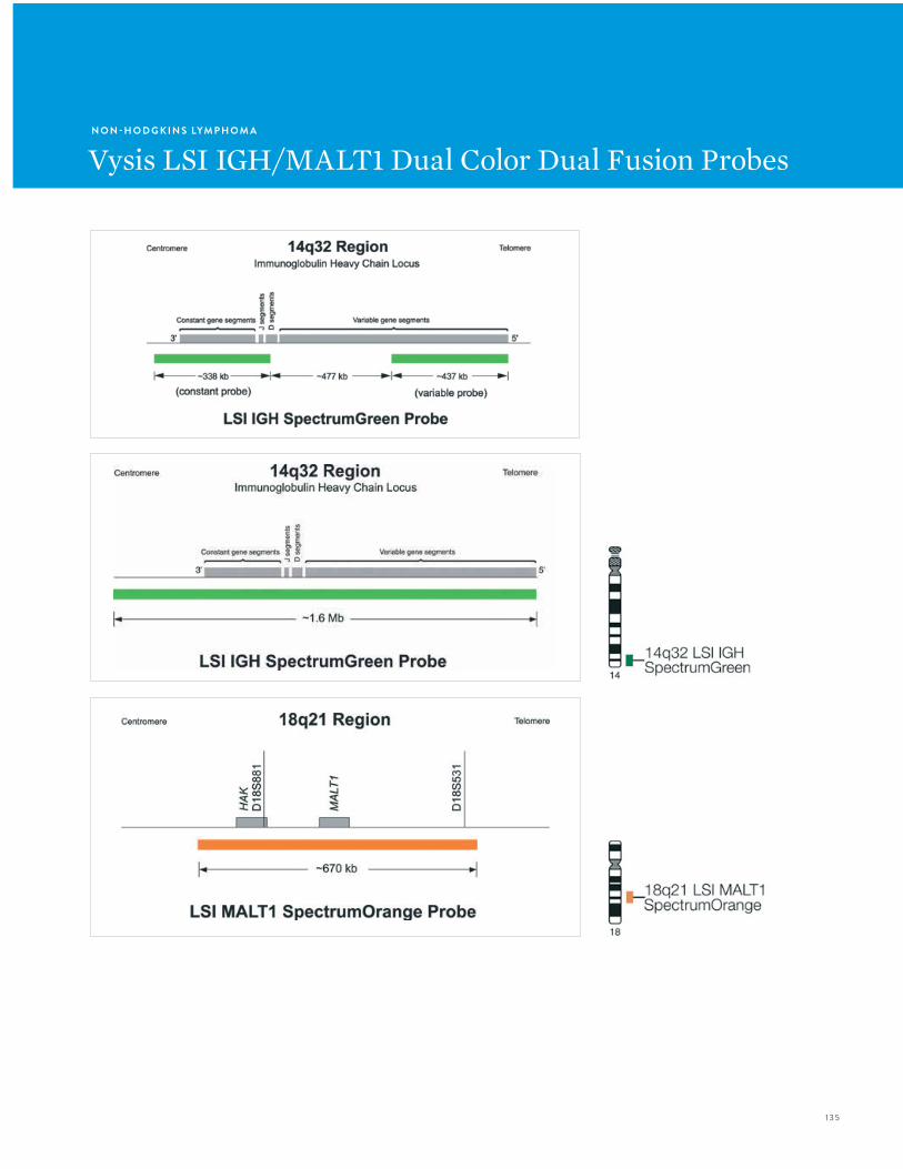
135
N O N -H O D G K I N S LYM P H O M A
Vysis LSI IGH/MALT1 Dual Color Dual Fusion Probes
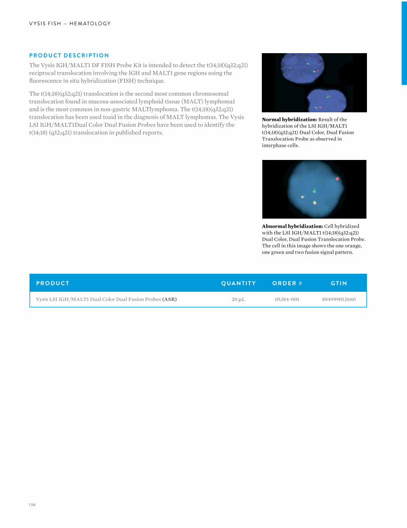
136
V Y S I S F I S H — H E M ATO LO G Y
P R O D U C T D E S C R I P T I O NThe Vysis IGH/MALT1 DF FISH Probe Kit is intended to detect the t(14;18)(q32;q21) reciprocal translocation involving the IGH and MALT1 gene regions using the fluorescence in situ hybridization (FISH) technique.
The t(14;18)(q32;q21) translocation is the second most common chromosomal translocation found in mucosa-associated lymphoid tissue (MALT) lymphoma1 and is the most common in non-gastric MALTlymphoma. The t(14;18)(q32;q21) translocation has been used toaid in the diagnosis of MALT lymphomas. The Vysis LSI IGH/MALT1Dual Color Dual Fusion Probes have been used to identify the t(14;18) (q32;q21) translocation in published reports.
Normal hybridization: Result of the hybridization of the LSI IGH/MALT1 t(14;18)(q32;q21) Dual Color, Dual Fusion Translocation Probe as observed in interphase cells.
Abnormal hybridization: Cell hybridized with the LSI IGH/MALT1 t(14;18)(q32;q21) Dual Color, Dual Fusion Translocation Probe. The cell in this image shows the one orange, one green and two fusion signal pattern.
P R O D U C T Q UA N T I T Y O R D E R # G T I N
Vysis LSI IGH/MALT1 Dual Color Dual Fusion Probes (ASR) 20 µL 05J84-001 884999012660
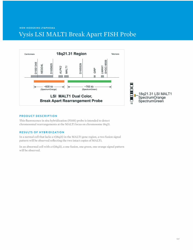
137
P R O D U C T D E S C R I P T I O NThis fluorescence in situ hybridization (FISH) probe is intended to detect chromosomal rearrangements at the MALT1 locus on chromosome 18q21.
R E S U LT S O F H Y B R I D I Z AT I O NIn a normal cell that lacks a t(18q21) in the MALT1 gene region, a two fusion signal pattern will be observed reflecting the two intact copies of MALT1.
In an abnormal cell with a t(18q21), a one fusion, one green, one orange signal pattern will be observed.
N O N -H O D G K I N S LYM P H O M A
Vysis LSI MALT1 Break Apart FISH Probe
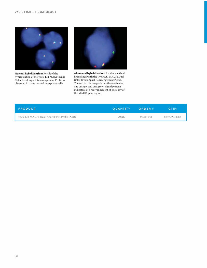
138
V Y S I S F I S H — H E M ATO LO G Y
P R O D U C T Q UA N T I T Y O R D E R # G T I N
Vysis LSI MALT1 Break Apart FISH Probe (ASR) 20 µL 05J87-001 884999012783
Normal hybridization: Result of the hybridization of the Vysis LSI MALT1 Dual Color Break Apart Rearrangement Probe as observed in three normal interphase cells.
Abnormal hybridization: An abnormal cell hybridized with the Vysis LSI MALT1 Dual Color Break Apart Rearrangement Probe. The cell in this image shows the one fusion, one orange, and one green signal pattern indicative of a rearrangement of one copy of the MALT1 gene region.
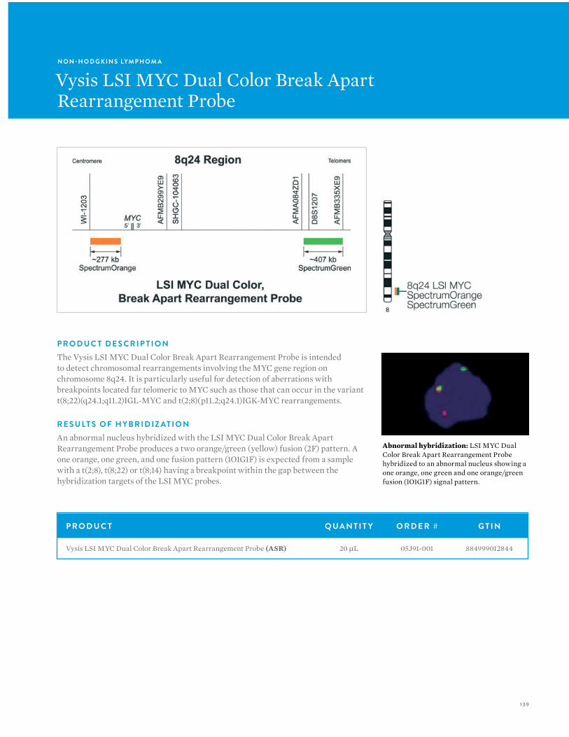
139
N O N -H O D G K I N S LYM P H O M A
Vysis LSI MYC Dual Color Break ApartRearrangement Probe
P R O D U C T D E S C R I P T I O NThe Vysis LSI MYC Dual Color Break Apart Rearrangement Probe is intended to detect chromosomal rearrangements involving the MYC gene region on chromosome 8q24. It is particularly useful for detection of aberrations with breakpoints located far telomeric to MYC such as those that can occur in the variant t(8;22)(q24.1;q11.2)IGL-MYC and t(2;8)(p11.2;q24.1)IGK-MYC rearrangements.
R E S U LT S O F H Y B R I D I Z AT I O NAn abnormal nucleus hybridized with the LSI MYC Dual Color Break Apart Rearrangement Probe produces a two orange/green (yellow) fusion (2F) pattern. A one orange, one green, and one fusion pattern (1O1G1F) is expected from a sample with a t(2;8), t(8;22) or t(8;14) having a breakpoint within the gap between the hybridization targets of the LSI MYC probes.
P R O D U C T Q UA N T I T Y O R D E R # G T I N
Vysis LSI MYC Dual Color Break Apart Rearrangement Probe (ASR) 20 µL 05J91-001 884999012844
Abnormal hybridization: LSI MYC Dual Color Break Apart Rearrangement Probe hybridized to an abnormal nucleus showing a one orange, one green and one orange/green fusion (1O1G1F) signal pattern.

140
V Y S I S F I S H — G E N E T I C S
VYSIS FISH:GENETICS
140
V Y S I S F I S H — G E N E T I C S
Identification and characterization of chromosome anomalies in preimplantation, prenatal, and postnatal genetics is critical for managing quality of life. FISH is a powerful tool for determining many types of chromosome anomalies. In addition to AneuVysion, the only FDA-cleared product for rapid detection of aneusomy in amniotic fluid samples, Abbott Molecular offers an expansive line of DNA FISH probes for
preimplantation, prenatal and postnatal genetic testing and research.

141141
A B B OT T M O L E C U L A R P R O D U C T S , P O W E R E D B Y V Y S I S F I S H T E C H N O LO G Y, P R OV I D E T H E F O L LO W I N G A DVA N TAG E S :• Rapid, sensitive, and specific detection and
characterization of chromosome abnormalities.
• Ability to test metaphase chromosomes from cultured samples and interphase cells from specimens that cannot be cultured.
• Direct-labeled probes, as compared to indirect labeling methods, provide:
• Less background signal, thereby simplifying interpretation.
• Reduced costs associated with labeling reagents and technician time.
• Dual and Tri Colored probe mixes for many microdeletion detection tests.
• Each mix includes a probe specific for the critical chromosome region implicated in the disease of interest and a control probe to another region on the same chromosome labeled with a different fluorophore.
• Inclusion of a control probe in most products ensures proper hybridization and facilitates identification of the chromosome of interest.
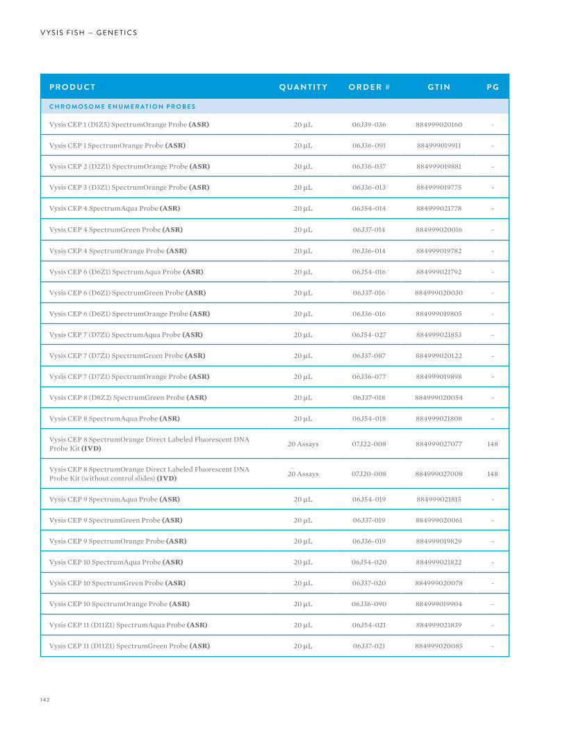
142
V Y S I S F I S H — G E N E T I C S
P R O D U C T Q UA N T I T Y O R D E R # G T I N P G
C H R O M O S O M E E N U M E R AT I O N P R O B E S
Vysis CEP 1 (D1Z5) SpectrumOrange Probe (ASR) 20 µL 06J39-036 884999020160 -
Vysis CEP 1 SpectrumOrange Probe (ASR) 20 µL 06J36-091 884999019911 -
Vysis CEP 2 (D2Z1) SpectrumOrange Probe (ASR) 20 µL 06J36-037 884999019881 -
Vysis CEP 3 (D3Z1) SpectrumOrange Probe (ASR) 20 µL 06J36-013 884999019775 -
Vysis CEP 4 SpectrumAqua Probe (ASR) 20 µL 06J54-014 884999021778 -
Vysis CEP 4 SpectrumGreen Probe (ASR) 20 µL 06J37-014 884999020016 -
Vysis CEP 4 SpectrumOrange Probe (ASR) 20 µL 06J36-014 884999019782 -
Vysis CEP 6 (D6Z1) SpectrumAqua Probe (ASR) 20 µL 06J54-016 884999021792 -
Vysis CEP 6 (D6Z1) SpectrumGreen Probe (ASR) 20 µL 06J37-016 884999020030 -
Vysis CEP 6 (D6Z1) SpectrumOrange Probe (ASR) 20 µL 06J36-016 884999019805 -
Vysis CEP 7 (D7Z1) SpectrumAqua Probe (ASR) 20 µL 06J54-027 884999021853 -
Vysis CEP 7 (D7Z1) SpectrumGreen Probe (ASR) 20 µL 06J37-087 884999020122 -
Vysis CEP 7 (D7Z1) SpectrumOrange Probe (ASR) 20 µL 06J36-077 884999019898 -
Vysis CEP 8 (D8Z2) SpectrumGreen Probe (ASR) 20 µL 06J37-018 884999020054 -
Vysis CEP 8 SpectrumAqua Probe (ASR) 20 µL 06J54-018 884999021808 -
Vysis CEP 8 SpectrumOrange Direct Labeled Fluorescent DNA Probe Kit (IVD) 20 Assays 07J22-008 884999027077 148
Vysis CEP 8 SpectrumOrange Direct Labeled Fluorescent DNA Probe Kit (without control slides) (IVD) 20 Assays 07J20-008 884999027008 148
Vysis CEP 9 SpectrumAqua Probe (ASR) 20 µL 06J54-019 884999021815 -
Vysis CEP 9 SpectrumGreen Probe (ASR) 20 µL 06J37-019 884999020061 -
Vysis CEP 9 SpectrumOrange Probe (ASR) 20 µL 06J36-019 884999019829 -
Vysis CEP 10 SpectrumAqua Probe (ASR) 20 µL 06J54-020 884999021822 -
Vysis CEP 10 SpectrumGreen Probe (ASR) 20 µL 06J37-020 884999020078 -
Vysis CEP 10 SpectrumOrange Probe (ASR) 20 µL 06J36-090 884999019904 -
Vysis CEP 11 (D11Z1) SpectrumAqua Probe (ASR) 20 µL 06J54-021 884999021839 -
Vysis CEP 11 (D11Z1) SpectrumGreen Probe (ASR) 20 µL 06J37-021 884999020085 -
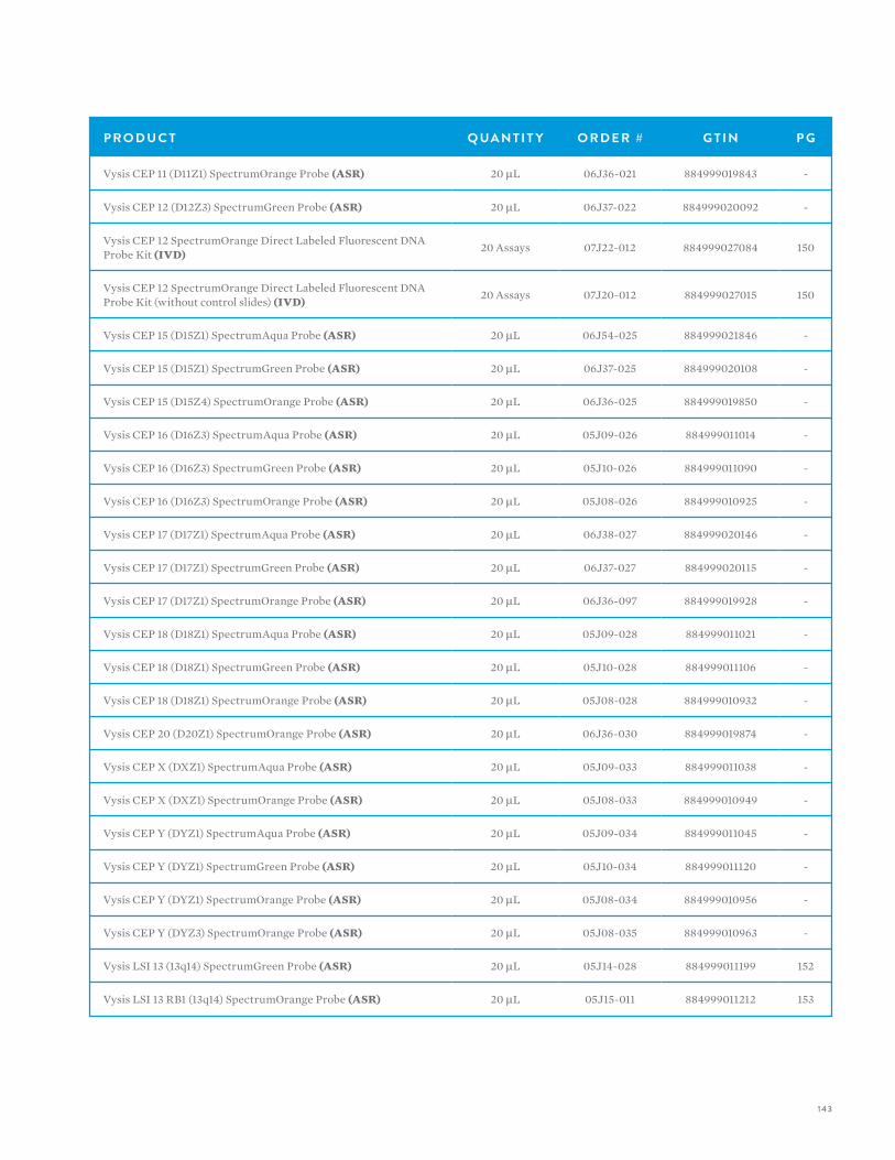
143
P R O D U C T Q UA N T I T Y O R D E R # G T I N P G
Vysis CEP 11 (D11Z1) SpectrumOrange Probe (ASR) 20 µL 06J36-021 884999019843 -
Vysis CEP 12 (D12Z3) SpectrumGreen Probe (ASR) 20 µL 06J37-022 884999020092 -
Vysis CEP 12 SpectrumOrange Direct Labeled Fluorescent DNA Probe Kit (IVD) 20 Assays 07J22-012 884999027084 150
Vysis CEP 12 SpectrumOrange Direct Labeled Fluorescent DNA Probe Kit (without control slides) (IVD) 20 Assays 07J20-012 884999027015 150
Vysis CEP 15 (D15Z1) SpectrumAqua Probe (ASR) 20 µL 06J54-025 884999021846 -
Vysis CEP 15 (D15Z1) SpectrumGreen Probe (ASR) 20 µL 06J37-025 884999020108 -
Vysis CEP 15 (D15Z4) SpectrumOrange Probe (ASR) 20 µL 06J36-025 884999019850 -
Vysis CEP 16 (D16Z3) SpectrumAqua Probe (ASR) 20 µL 05J09-026 884999011014 -
Vysis CEP 16 (D16Z3) SpectrumGreen Probe (ASR) 20 µL 05J10-026 884999011090 -
Vysis CEP 16 (D16Z3) SpectrumOrange Probe (ASR) 20 µL 05J08-026 884999010925 -
Vysis CEP 17 (D17Z1) SpectrumAqua Probe (ASR) 20 µL 06J38-027 884999020146 -
Vysis CEP 17 (D17Z1) SpectrumGreen Probe (ASR) 20 µL 06J37-027 884999020115 -
Vysis CEP 17 (D17Z1) SpectrumOrange Probe (ASR) 20 µL 06J36-097 884999019928 -
Vysis CEP 18 (D18Z1) SpectrumAqua Probe (ASR) 20 µL 05J09-028 884999011021 -
Vysis CEP 18 (D18Z1) SpectrumGreen Probe (ASR) 20 µL 05J10-028 884999011106 -
Vysis CEP 18 (D18Z1) SpectrumOrange Probe (ASR) 20 µL 05J08-028 884999010932 -
Vysis CEP 20 (D20Z1) SpectrumOrange Probe (ASR) 20 µL 06J36-030 884999019874 -
Vysis CEP X (DXZ1) SpectrumAqua Probe (ASR) 20 µL 05J09-033 884999011038 -
Vysis CEP X (DXZ1) SpectrumOrange Probe (ASR) 20 µL 05J08-033 884999010949 -
Vysis CEP Y (DYZ1) SpectrumAqua Probe (ASR) 20 µL 05J09-034 884999011045 -
Vysis CEP Y (DYZ1) SpectrumGreen Probe (ASR) 20 µL 05J10-034 884999011120 -
Vysis CEP Y (DYZ1) SpectrumOrange Probe (ASR) 20 µL 05J08-034 884999010956 -
Vysis CEP Y (DYZ3) SpectrumOrange Probe (ASR) 20 µL 05J08-035 884999010963 -
Vysis LSI 13 (13q14) SpectrumGreen Probe (ASR) 20 µL 05J14-028 884999011199 152
Vysis LSI 13 RB1 (13q14) SpectrumOrange Probe (ASR) 20 µL 05J15-011 884999011212 153
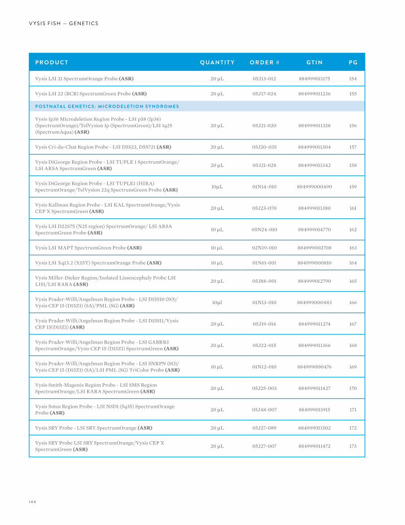
144
V Y S I S F I S H — G E N E T I C S
P R O D U C T Q UA N T I T Y O R D E R # G T I N P G
Vysis LSI 21 SpectrumOrange Probe (ASR) 20 µL 05J13-012 884999011175 154
Vysis LSI 22 (BCR) SpectrumGreen Probe (ASR) 20 µL 05J17-024 884999011236 155
P O S T N ATA L G E N E T I C S : M I C R O D E L E T I O N S Y N D R O M E S
Vysis 1p36 Microdeletion Region Probe - LSI p58 (1p36) (SpectrumOrange)/TelVysion 1p (SpectrumGreen)/LSI 1q25 (SpectrumAqua) (ASR)
20 µL 05J21-020 884999011328 156
Vysis Cri-du-Chat Region Probe - LSI D5S23, D5S721 (ASR) 20 µL 05J20-035 884999011304 157
Vysis DiGeorge Region Probe - LSI TUPLE 1 SpectrumOrange/LSI ARSA SpectrumGreen (ASR) 20 µL 05J21-028 884999011342 158
Vysis DiGeorge Region Probe - LSI TUPLE1 (HIRA) SpectrumOrange/TelVysion 22q SpectrumGreen Probe (ASR) 10µL 01N14-010 884999000490 159
Vysis Kallman Region Probe - LSI KAL SpectrumOrange/Vysis CEP X SpectrumGreen (ASR) 20 µL 05J23-070 884999011380 161
Vysis LSI D22S75 (N25 region) SpectrumOrange/ LSI ARSA SpectrumGreen Probe (ASR) 10 µL 05N24-010 884999014770 162
Vysis LSI MAPT SpectrumGreen Probe (ASR) 10 µL 02N19-010 884999002708 163
Vysis LSI Xq13.2 (XIST) SpectrumOrange Probe (ASR) 10 µL 01N61-001 884999000810 164
Vysis Miller-Dieker Region/Isolated Lissencephaly Probe LSI LIS1/LSI RARA (ASR) 20 µL 05J88-001 884999012790 165
Vysis Prader-Willi/Angelman Region Probe - LSI D15S10 (SO)/Vysis CEP 15 (D15Z1) (SA)/PML (SG) (ASR) 10µl 01N13-010 884999000483 166
Vysis Prader-Willi/Angelman Region Probe - LSI D15S11/Vysis CEP 15(D15Z1) (ASR) 20 µL 05J19-014 884999011274 167
Vysis Prader-Willi/Angelman Region Probe - LSI GABRB3 SpectrumOrange/Vysis CEP 15 (D15Z1) SpectrumGreen (ASR) 20 µL 05J22-015 884999011366 168
Vysis Prader-Willi/Angelman Region Probe - LSI SNRPN (SO)/Vysis CEP 15 (D15Z1) (SA)/LSI PML (SG) TriColor Probe (ASR) 10 µL 01N12-010 884999000476 169
Vysis Smith-Magenis Region Probe - LSI SMS Region SpectrumOrange/LSI RARA SpectrumGreen (ASR) 20 µL 05J25-003 884999011427 170
Vysis Sotos Region Probe - LSI NSD1 (5q35) SpectrumOrange Probe (ASR) 20 µL 05J48-007 884999011915 171
Vysis SRY Probe - LSI SRY SpectrumOrange (ASR) 20 µL 05J27-089 884999011502 172
Vysis SRY Probe LSI SRY SpectrumOrange/Vysis CEP X SpectrumGreen (ASR) 20 µL 05J27-007 884999011472 173
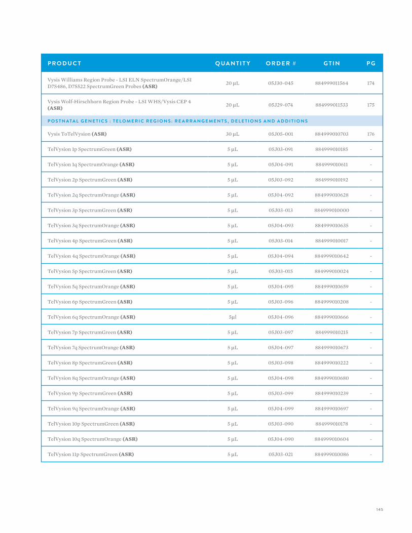
145
P R O D U C T Q UA N T I T Y O R D E R # G T I N P G
Vysis Williams Region Probe - LSI ELN SpectrumOrange/LSI D7S486, D7S522 SpectrumGreen Probes (ASR) 20 µL 05J30-045 884999011564 174
Vysis Wolf-Hirschhorn Region Probe - LSI WHS/Vysis CEP 4 (ASR) 20 µL 05J29-074 884999011533 175
P O S T N ATA L G E N E T I C S : T E L O M E R I C R E G I O N S : R E A R R A N G E M E N T S , D E L E T I O N S A N D A D D I T I O N S
Vysis ToTelVysion (ASR) 30 µL 05J05-001 884999010703 176
TelVysion 1p SpectrumGreen (ASR) 5 µL 05J03-091 884999010185 -
TelVysion 1q SpectrumOrange (ASR) 5 µL 05J04-091 884999010611 -
TelVysion 2p SpectrumGreen (ASR) 5 µL 05J03-092 884999010192 -
TelVysion 2q SpectrumOrange (ASR) 5 µL 05J04-092 884999010628 -
TelVysion 3p SpectrumGreen (ASR) 5 µL 05J03-013 884999010000 -
TelVysion 3q SpectrumOrange (ASR) 5 µL 05J04-093 884999010635 -
TelVysion 4p SpectrumGreen (ASR) 5 µL 05J03-014 884999010017 -
TelVysion 4q SpectrumOrange (ASR) 5 µL 05J04-094 884999010642 -
TelVysion 5p SpectrumGreen (ASR) 5 µL 05J03-015 884999010024 -
TelVysion 5q SpectrumOrange (ASR) 5 µL 05J04-095 884999010659 -
TelVysion 6p SpectrumGreen (ASR) 5 µL 05J03-096 884999010208 -
TelVysion 6q SpectrumOrange (ASR) 5µl 05J04-096 884999010666 -
TelVysion 7p SpectrumGreen (ASR) 5 µL 05J03-097 884999010215 -
TelVysion 7q SpectrumOrange (ASR) 5 µL 05J04-097 884999010673 -
TelVysion 8p SpectrumGreen (ASR) 5 µL 05J03-098 884999010222 -
TelVysion 8q SpectrumOrange (ASR) 5 µL 05J04-098 884999010680 -
TelVysion 9p SpectrumGreen (ASR) 5 µL 05J03-099 884999010239 -
TelVysion 9q SpectrumOrange (ASR) 5 µL 05J04-099 884999010697 -
TelVysion 10p SpectrumGreen (ASR) 5 µL 05J03-090 884999010178 -
TelVysion 10q SpectrumOrange (ASR) 5 µL 05J04-090 884999010604 -
TelVysion 11p SpectrumGreen (ASR) 5 µL 05J03-021 884999010086 -
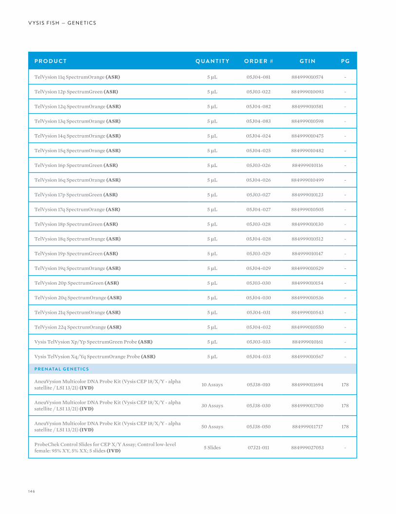
146
V Y S I S F I S H — G E N E T I C S
P R O D U C T Q UA N T I T Y O R D E R # G T I N P G
TelVysion 11q SpectrumOrange (ASR) 5 µL 05J04-081 884999010574 -
TelVysion 12p SpectrumGreen (ASR) 5 µL 05J03-022 884999010093 -
TelVysion 12q SpectrumOrange (ASR) 5 µL 05J04-082 884999010581 -
TelVysion 13q SpectrumOrange (ASR) 5 µL 05J04-083 884999010598 -
TelVysion 14q SpectrumOrange (ASR) 5 µL 05J04-024 884999010475 -
TelVysion 15q SpectrumOrange (ASR) 5 µL 05J04-025 884999010482 -
TelVysion 16p SpectrumGreen (ASR) 5 µL 05J03-026 884999010116 -
TelVysion 16q SpectrumOrange (ASR) 5 µL 05J04-026 884999010499 -
TelVysion 17p SpectrumGreen (ASR) 5 µL 05J03-027 884999010123 -
TelVysion 17q SpectrumOrange (ASR) 5 µL 05J04-027 884999010505 -
TelVysion 18p SpectrumGreen (ASR) 5 µL 05J03-028 884999010130 -
TelVysion 18q SpectrumOrange (ASR) 5 µL 05J04-028 884999010512 -
TelVysion 19p SpectrumGreen (ASR) 5 µL 05J03-029 884999010147 -
TelVysion 19q SpectrumOrange (ASR) 5 µL 05J04-029 884999010529 -
TelVysion 20p SpectrumGreen (ASR) 5 µL 05J03-030 884999010154 -
TelVysion 20q SpectrumOrange (ASR) 5 µL 05J04-030 884999010536 -
TelVysion 21q SpectrumOrange (ASR) 5 µL 05J04-031 884999010543 -
TelVysion 22q SpectrumOrange (ASR) 5 µL 05J04-032 884999010550 -
Vysis TelVysion Xp/Yp SpectrumGreen Probe (ASR) 5 µL 05J03-033 884999010161 -
Vysis TelVysion Xq/Yq SpectrumOrange Probe (ASR) 5 µL 05J04-033 884999010567 -
P R E N ATA L G E N E T I C S
AneuVysion Multicolor DNA Probe Kit (Vysis CEP 18/X/Y - alpha satellite / LSI 13/21) (IVD) 10 Assays 05J38-010 884999011694 178
AneuVysion Multicolor DNA Probe Kit (Vysis CEP 18/X/Y - alpha satellite / LSI 13/21) (IVD) 30 Assays 05J38-030 884999011700 178
AneuVysion Multicolor DNA Probe Kit (Vysis CEP 18/X/Y - alpha satellite / LSI 13/21) (IVD) 50 Assays 05J38-050 884999011717 178
ProbeChek Control Slides for CEP X/Y Assay; Control low-level female: 95% XY, 5% XX; 5 slides (IVD) 5 Slides 07J21-011 884999027053 -
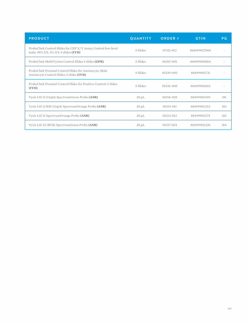
147
P R O D U C T Q UA N T I T Y O R D E R # G T I N P G
ProbeChek Control Slides for CEP X/Y Assay; Control low-level male: 95% XX, 5% XY; 5 slides (IVD) 5 Slides 07J21-012 884999027060 -
ProbeChek MultiVysion Control Slides 5 slides (GPR) 5 Slides 05J07-001 884999010864 -
ProbeChek Prenatal Control Slides for Amniocyte; Male Amniocyte Control Slides; 5 slides (IVD) 5 Slides 05J39-005 884999011731 -
ProbeChek Prenatal Control Slides for Positive Control; 5 slides (IVD) 5 Slides 05J36-005 884999011663 -
Vysis LSI 13 (13q14) SpectrumGreen Probe (ASR) 20 µL 05J14-028 884999011199 181
Vysis LSI 13 RB1 (13q14) SpectrumOrange Probe (ASR) 20 µL 05J15-011 884999011212 182
Vysis LSI 21 SpectrumOrange Probe (ASR) 20 µL 05J13-012 884999011175 183
Vysis LSI 22 (BCR) SpectrumGreen Probe (ASR) 20 µL 05J17-024 884999011236 184
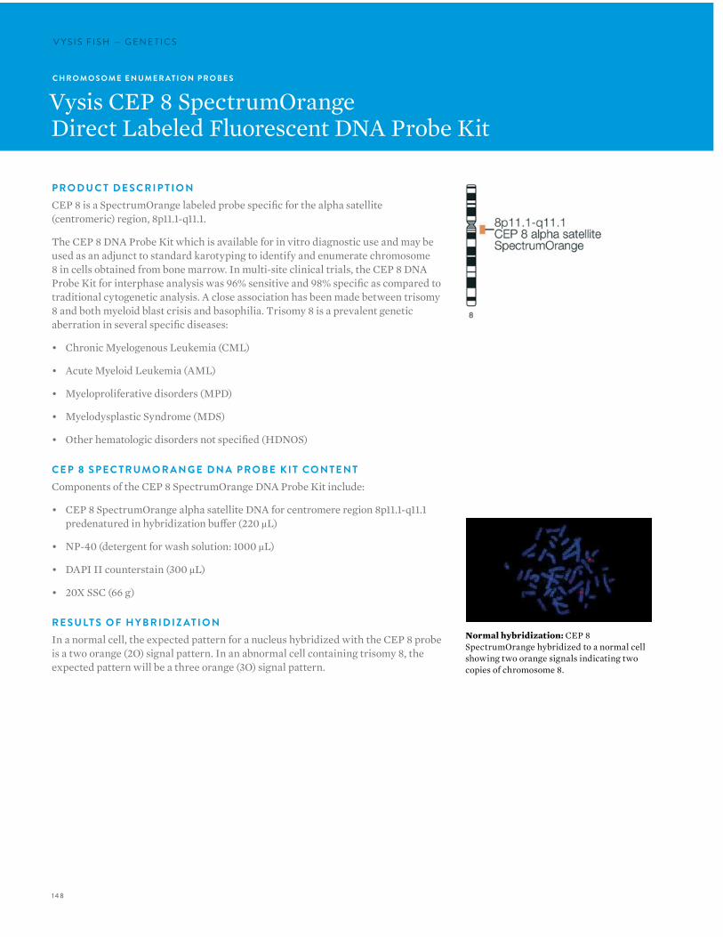
148
V Y S I S F I S H — G E N E T I C SV Y S I S F I S H — G E N E T I C S
C H R O M O S O M E E N U M E R AT I O N P R O B E S
Vysis CEP 8 SpectrumOrangeDirect Labeled Fluorescent DNA Probe Kit
P R O D U C T D E S C R I P T I O NCEP 8 is a SpectrumOrange labeled probe specific for the alpha satellite (centromeric) region, 8p11.1-q11.1.
The CEP 8 DNA Probe Kit which is available for in vitro diagnostic use and may be used as an adjunct to standard karotyping to identify and enumerate chromosome 8 in cells obtained from bone marrow. In multi-site clinical trials, the CEP 8 DNA Probe Kit for interphase analysis was 96% sensitive and 98% specific as compared to traditional cytogenetic analysis. A close association has been made between trisomy 8 and both myeloid blast crisis and basophilia. Trisomy 8 is a prevalent genetic aberration in several specific diseases:
• Chronic Myelogenous Leukemia (CML)
• Acute Myeloid Leukemia (AML)
• Myeloproliferative disorders (MPD)
• Myelodysplastic Syndrome (MDS)
• Other hematologic disorders not specified (HDNOS)
C E P 8 S P E C T R U M O R A N G E D N A P R O B E K I T CO N T E N TComponents of the CEP 8 SpectrumOrange DNA Probe Kit include:
• CEP 8 SpectrumOrange alpha satellite DNA for centromere region 8p11.1-q11.1 predenatured in hybridization buffer (220 µL)
• NP-40 (detergent for wash solution: 1000 µL)
• DAPI II counterstain (300 µL)
• 20X SSC (66 g)
R E S U LT S O F H Y B R I D I Z AT I O NIn a normal cell, the expected pattern for a nucleus hybridized with the CEP 8 probe is a two orange (2O) signal pattern. In an abnormal cell containing trisomy 8, the expected pattern will be a three orange (3O) signal pattern.
Normal hybridization: CEP 8 SpectrumOrange hybridized to a normal cell showing two orange signals indicating two copies of chromosome 8.
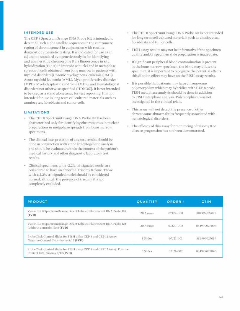
149
I N T E N D E D U S EThe CEP 8 SpectrumOrange DNA Probe Kit is intended to detect AT rich alpha satellite sequences in the centromere region of chromosome 8 in conjunction with routine diagnostic cytogenetic testing. It is indicated for use as an adjunct to standard cytogenetic analysis for identifying and enumerating chromosome 8 via fluorescence in situ hybridization (FISH) in interphase nuclei and in metaphase spreads of cells obtained from bone marrow in patients with myeloid disorders [Chronic myelogenous leukemia (CML), Acute myeloid leukemia (AML), Myeloproliferative disorder (MPD), Myelodysplastic syndrome (MDS), and Hematological disorders not otherwise specified (HDNOS)]. It is not intended to be used as a stand alone assay for test reporting. It is not intended for use in long term cell cultured materials such as amniocytes, fibroblasts and tumor cells.
L I M I TAT I O N S • The CEP 8 SpectrumOrange DNA Probe Kit has been
characterized only for identifying chromosomes in nuclear preparations or metaphase spreads from bone marrow specimens.
• The clinical interpretation of any test results should be done in conjunction with standard cytogenetic analysis and should be evaluated within the context of the patient’s medical history and other diagnostic laboratory test results.
• Clinical specimens with >2.2% tri-signaled nuclei are considered to have an abnormal trisomy 8 clone. Those with ≤ 2.2% tri-signaled nuclei should be considered normal, although the presence of trisomy 8 is not completely excluded.
• The CEP 8 SpectrumOrange DNA Probe Kit is not intended for long term cell cultured materials such as amniocytes, fibroblasts and tumor cells.
• FISH assay results may not be informative if the specimen quality and/or specimen slide preparation is inadequate.
• If significant peripheral blood contamination is present in the bone marrow specimen, the blood may dilute the specimen; it is important to recognize the potential effects this dilution effect may have on the FISH assay results.
• It is possible that patients may have chromosome polymorphism which may hybridize with CEP 8 probe. FISH metaphase analysis should be done in addition to FISH interphase analysis. Polymorphism was not investigated in the clinical trials.
• This assay will not detect the presence of other chromosome abnormalities frequently associated with hematological disorders.
• The efficacy of this assay for monitoring of trisomy 8 or disease progression has not been demonstrated.
P R O D U C T Q UA N T I T Y O R D E R # G T I N
Vysis CEP 8 SpectrumOrange Direct Labeled Fluorescent DNA Probe Kit (IVD) 20 Assays 07J22-008 884999027077
Vysis CEP 8 SpectrumOrange Direct Labeled Fluorescent DNA Probe Kit (without control slides) (IVD) 20 Assays 07J20-008 884999027008
ProbeChek Control Slides for FISH using CEP 8 and CEP 12 Assay, Negative Control 0%, trisomy 8/12 (IVD) 5 Slides 07J21-001 884999027039
ProbeChek Control Slides for FISH using CEP 8 and CEP 12 Assay, Positive Control 10%, trisomy 8/12 (IVD) 5 Slides 07J21-002 884999027046
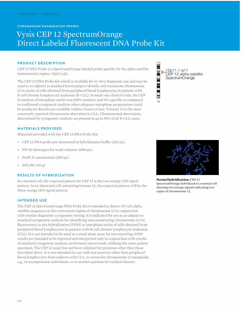
150
V Y S I S F I S H — G E N E T I C SV Y S I S F I S H — G E N E T I C S
C H R O M O S O M E E N U M E R AT I O N P R O B E S
Vysis CEP 12 SpectrumOrangeDirect Labeled Fluorescent DNA Probe Kit
P R O D U C T D E S C R I P T I O NCEP 12 DNA Probe is a SpectrumOrange labeled probe specific for the alpha satellite (centromeric) region, 12p11.1-q11.
The CEP 12 DNA Probe Kit which is available for in vitro diagnostic use and may be used as an adjunct to standard karotyping to identify and enumerate chromosome 12 in nuclei of cells obtained from peripheral blood lymphocytes in patients with B-cell chronic lymphocytic leukemia (B-CLL). In multi-site clinical trials, the CEP 12 analysis of interphase nuclei was 100% sensitive and 91% specific as compared to traditional cytogenetic analysis when adequate metaphase preparations could be produced. Results are available within 3 hours or less. Trisomy 12 is the most commonly reported chromosome aberration in CLL. Chromosomal aberrations, determined by cytogenetic analysis are present in up to 55% of all B-CLL cases.
M AT E R I A L S P R OV I D E DMaterials provided with the CEP 12 DNA Probe Kit:
• CEP 12 DNA probe pre-denatured in hybridization buffer (220 µL)
• NP-40 (detergent for wash solution: 1000 µL)
• DAPI II counterstain (300 µL)
• 20X SSC (66 g)
R E S U LT S O F H Y B R I D I Z AT I O NIn a normal cell, the expected pattern for CEP 12 is the two orange (2O) signal pattern. In an abnormal cell containing trisomy 12, the expected pattern will be the three orange (3O) signal pattern.
I N T E N D E D U S EThe CEP 12 SpectrumOrange DNA Probe Kit is intended to detect AT rich alpha satellite sequences in the centromere region of chromosome 12 in conjunction with routine diagnostic cytogenetic testing. It is indicated for use as an adjunct to standard cytogenetic analysis for identifying and enumerating chromosome 12 via fluorescence in situ hybridization (FISH) in interphase nuclei of cells obtained from peripheral blood lymphocytes in patients with B-cell chronic lymphocytic leukemia (CLL). It is not intended to be used as a stand alone assay for test reporting; FISH results are intended to be reported and interpreted only in conjunction with results of standard cytogenetic analysis, performed concurrently, utilizing the same patient specimen. The CEP 12 assay has not been validated for purposes other than those described above. It is not intended for use with test matrices other than peripheral blood lymphocytes from subjects with CLL, to screen for chromosome 12 aneuploidy, e.g., in asymptomatic individuals, or to monitor patients for residual disease.
Normal hybridization: CEP 12 SpectrumOrange hybridized to a normal cell showing two orange signals indicating two copies of chromosome 12.
151
L I M I TAT I O N S1. The CEP 12 DNA Probe Kit has been optimized only for
identifying chromosome 12 in interphase nuclei from peripheral blood specimens from patients with B-cell chronic lymphocytic leukemia.
2. The clinical interpretation of any abnormality or its absence by FISH should be done in conjunction with standard cytogenetic analysis and proper controls, and should be evaluated within the context of the patient’s medical history and past diagnostic laboratory test results.
3. Clinical specimens with >2.0% tri-signaled nuclei are considered to have an abnormal trisomy 12 clone. Those with ≤ 2.0% tri-signaled nuclei should be considered normal, although the presence of trisomy 12 is not completely excluded. When the percentage of tri-signaled interphase nuclei are near the cutoff point (1.5-2.5%), the results should be interpreted with caution.
4. The CEP 12 assay has been validated only for use with peripheral blood lymphocytes obtained from patients with B-cell CLL leukemia. It is not intended to be used for chromosome 12 enumeration in other patient populations or with other test matrices such as amniocytes, chorionic villi, fibroblasts, tumor cells, long term cultures, among others.
5. FISH assay results may not be informative if the specimen quality and/or specimen slide preparation is inadequate; the CEP 12 DNA Probe Kit has been optimized for archived peripheral blood specimens stored only at -20ºC or -80ºC, as recommended.
6. The CEP 12 DNA Probe Kit has not been validated for monitoring disease status.
7. It is possible that some individuals may have target sequences at an alternate chromosomal location that may hybridize with CEP 12. This has not been investigated for this device; the user should assess this in metaphase spreads from each subject tested.
8. The CEP 12 assay is intended only to aid in the enumeration of chromosome 12 centromeres; it is not designed to detect chromosome 12 structural abnormalities.
P R O D U C T Q UA N T I T Y O R D E R # G T I N
Vysis CEP 12 SpectrumOrange Direct Labeled Fluorescent DNA Probe Kit (IVD) 20 Assays 07J22-012 884999027084
Vysis CEP 12 SpectrumOrange Direct Labeled Fluorescent DNA Probe Kit (without control slides) (IVD) 20 Assays 07J20-012 884999027015
ProbeChek Control Slides for FISH using CEP 8 and CEP 12 Assay, Negative Control 0%, trisomy 8/12 (IVD) 5 Slides 07J21-001 884999027039
ProbeChek Control Slides for FISH using CEP 8 and CEP 12 Assay, Positive Control 10%, trisomy 8/12 (IVD) 5 Slides 07J21-002 884999027046
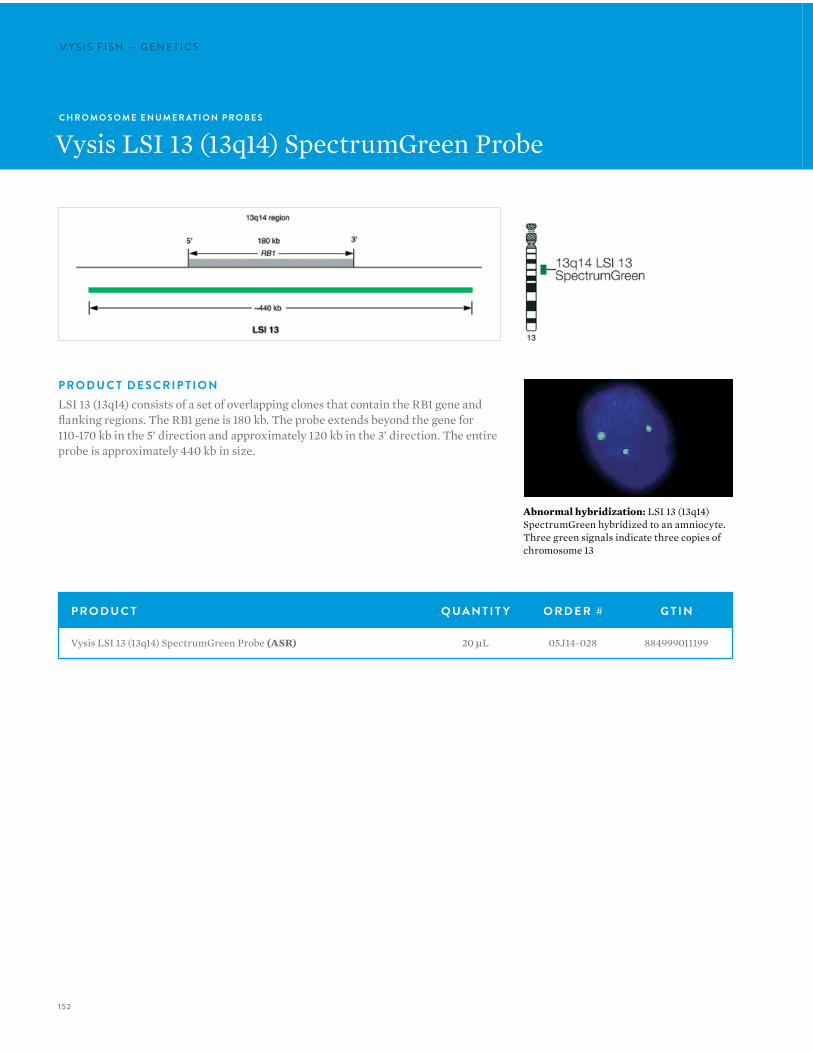
152
V Y S I S F I S H — G E N E T I C SV Y S I S F I S H — G E N E T I C S
C H R O M O S O M E E N U M E R AT I O N P R O B E S
Vysis LSI 13 (13q14) SpectrumGreen Probe
P R O D U C T Q UA N T I T Y O R D E R # G T I N
Vysis LSI 13 (13q14) SpectrumGreen Probe (ASR) 20 µL 05J14-028 884999011199
P R O D U C T D E S C R I P T I O NLSI 13 (13q14) consists of a set of overlapping clones that contain the RB1 gene and flanking regions. The RB1 gene is 180 kb. The probe extends beyond the gene for 110-170 kb in the 5’ direction and approximately 120 kb in the 3’ direction. The entire probe is approximately 440 kb in size.
Abnormal hybridization: LSI 13 (13q14) SpectrumGreen hybridized to an amniocyte. Three green signals indicate three copies of chromosome 13
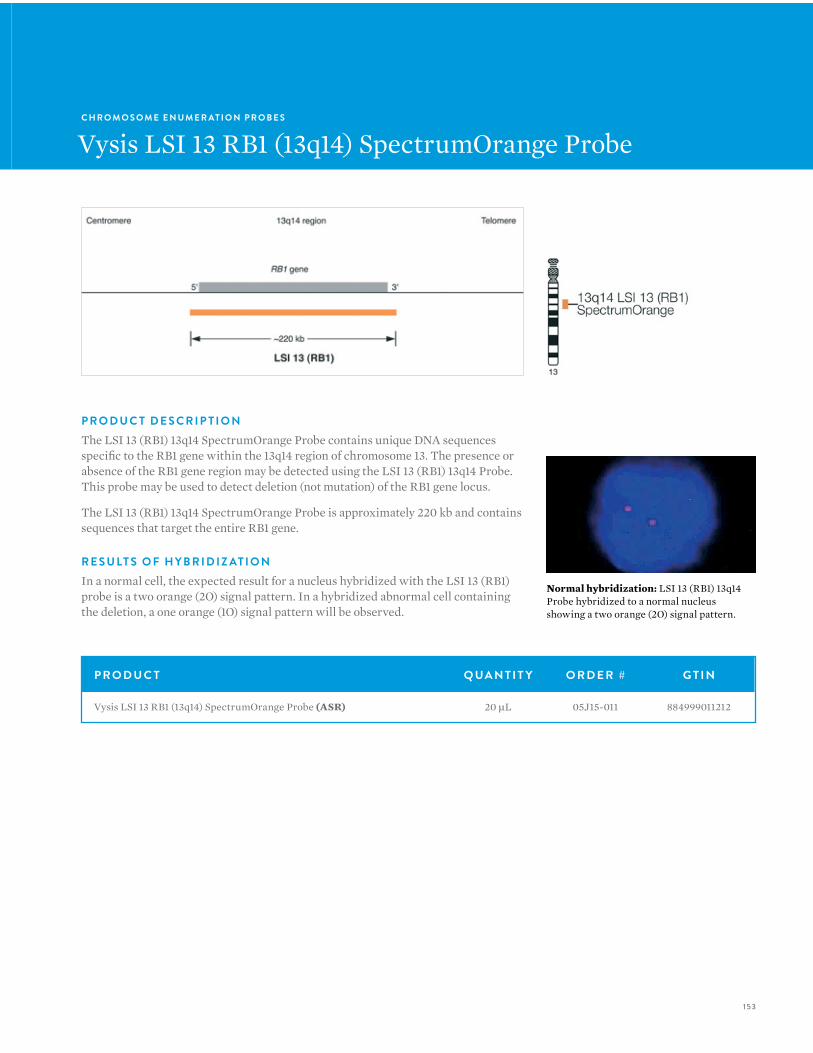
153
C H R O M O S O M E E N U M E R AT I O N P R O B E S
Vysis LSI 13 RB1 (13q14) SpectrumOrange Probe
Normal hybridization: LSI 13 (RB1) 13q14 Probe hybridized to a normal nucleus showing a two orange (2O) signal pattern.
P R O D U C T Q UA N T I T Y O R D E R # G T I N
Vysis LSI 13 RB1 (13q14) SpectrumOrange Probe (ASR) 20 µL 05J15-011 884999011212
P R O D U C T D E S C R I P T I O NThe LSI 13 (RB1) 13q14 SpectrumOrange Probe contains unique DNA sequences specific to the RB1 gene within the 13q14 region of chromosome 13. The presence or absence of the RB1 gene region may be detected using the LSI 13 (RB1) 13q14 Probe. This probe may be used to detect deletion (not mutation) of the RB1 gene locus.
The LSI 13 (RB1) 13q14 SpectrumOrange Probe is approximately 220 kb and contains sequences that target the entire RB1 gene.
R E S U LT S O F H Y B R I D I Z AT I O NIn a normal cell, the expected result for a nucleus hybridized with the LSI 13 (RB1) probe is a two orange (2O) signal pattern. In a hybridized abnormal cell containing the deletion, a one orange (1O) signal pattern will be observed.
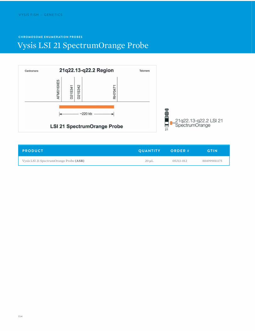
154
V Y S I S F I S H — G E N E T I C SV Y S I S F I S H — G E N E T I C S
C H R O M O S O M E E N U M E R AT I O N P R O B E S
Vysis LSI 21 SpectrumOrange Probe
P R O D U C T Q UA N T I T Y O R D E R # G T I N
Vysis LSI 21 SpectrumOrange Probe (ASR) 20 µL 05J13-012 884999011175

155
C H R O M O S O M E E N U M E R AT I O N P R O B E S
Vysis LSI 22 (BCR) SpectrumGreen Probe
P R O D U C T D E S C R I P T I O NThe LSI 22 (BCR) Probe is an approximately 300 kb SpectrumGreen probe corresponding to 22q11.2.
P R O D U C T Q UA N T I T Y O R D E R # G T I N
Vysis LSI 22 (BCR) SpectrumGreen Probe (ASR) 20 µL 05J17-024 884999011236
Normal hybridization: LSI 22 (BCR) SpectrumGreen hybridized to an interphase cell.
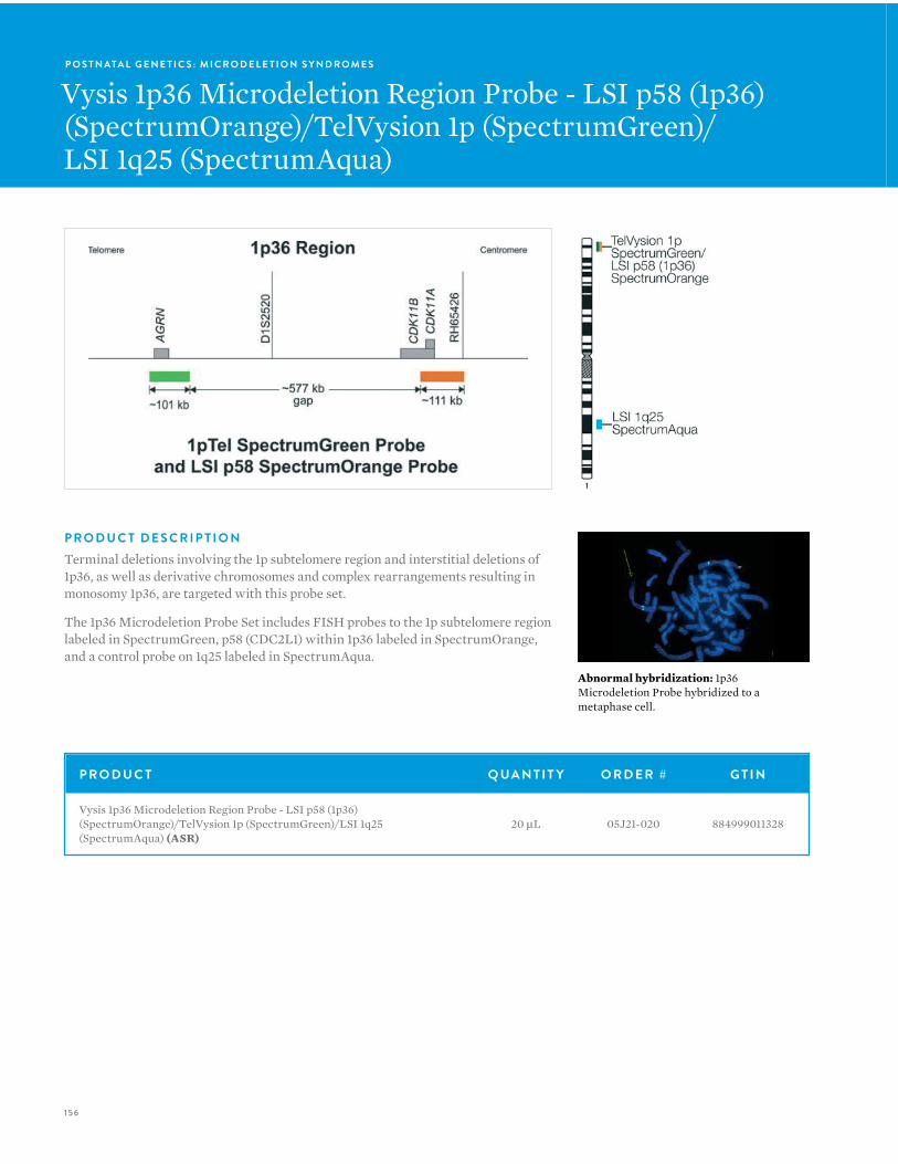
156
V Y S I S F I S H — G E N E T I C S
P R O D U C T D E S C R I P T I O NTerminal deletions involving the 1p subtelomere region and interstitial deletions of 1p36, as well as derivative chromosomes and complex rearrangements resulting in monosomy 1p36, are targeted with this probe set.
The 1p36 Microdeletion Probe Set includes FISH probes to the 1p subtelomere region labeled in SpectrumGreen, p58 (CDC2L1) within 1p36 labeled in SpectrumOrange, and a control probe on 1q25 labeled in SpectrumAqua.
P R O D U C T Q UA N T I T Y O R D E R # G T I N
Vysis 1p36 Microdeletion Region Probe - LSI p58 (1p36) (SpectrumOrange)/TelVysion 1p (SpectrumGreen)/LSI 1q25 (SpectrumAqua) (ASR)
20 µL 05J21-020 884999011328
P O S T N ATA L G E N E T I C S : M I C R O D E L E T I O N S Y N D R O M E S
Vysis 1p36 Microdeletion Region Probe - LSI p58 (1p36) (SpectrumOrange)/TelVysion 1p (SpectrumGreen)/LSI 1q25 (SpectrumAqua)
Abnormal hybridization: 1p36 Microdeletion Probe hybridized to a metaphase cell.
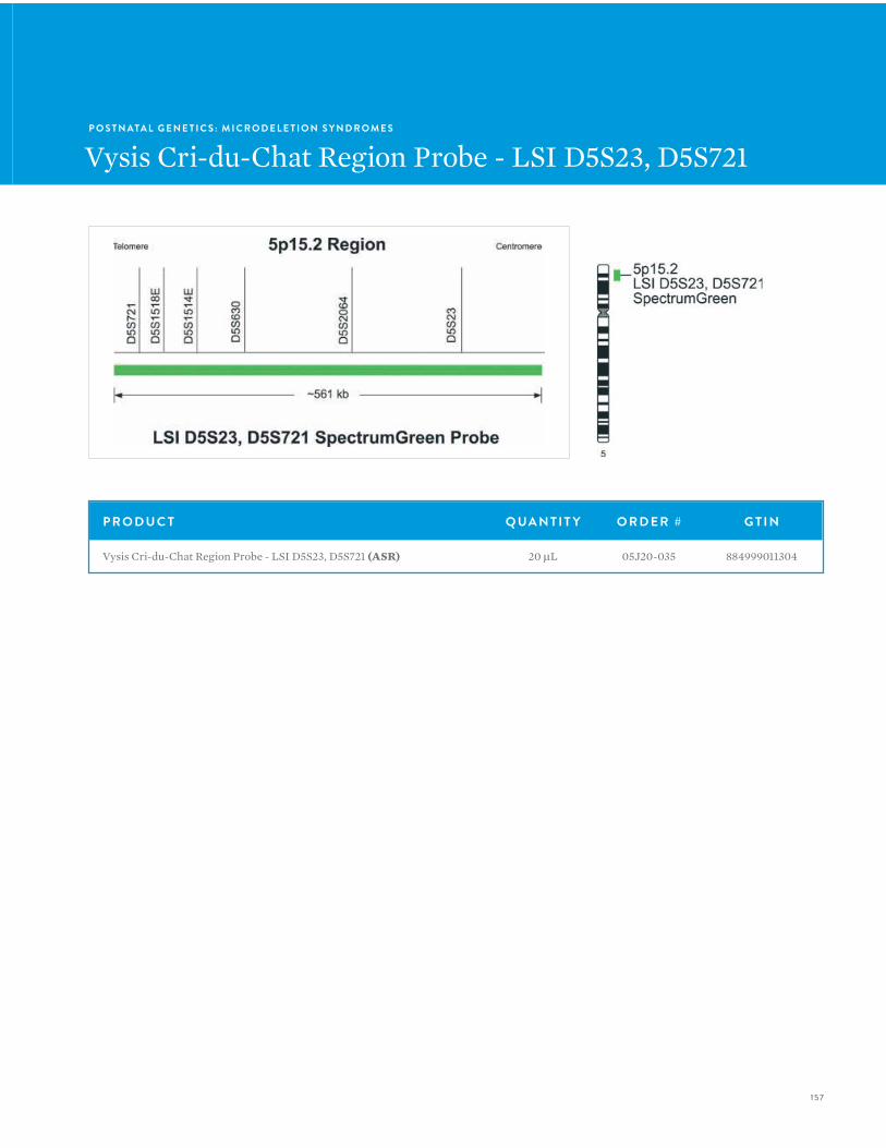
157
P O S T N ATA L G E N E T I C S : M I C R O D E L E T I O N S Y N D R O M E S
Vysis Cri-du-Chat Region Probe - LSI D5S23, D5S721
P R O D U C T Q UA N T I T Y O R D E R # G T I N
Vysis Cri-du-Chat Region Probe - LSI D5S23, D5S721 (ASR) 20 µL 05J20-035 884999011304
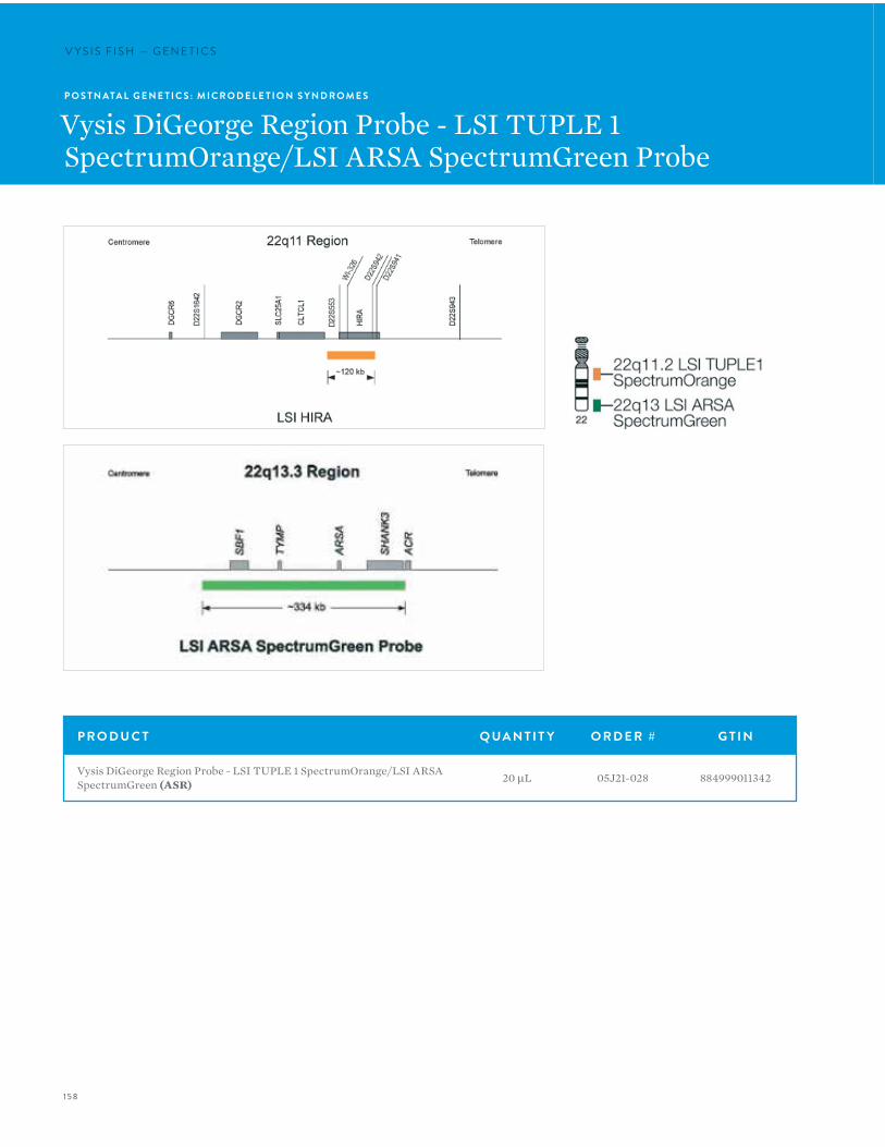
158
V Y S I S F I S H — G E N E T I C SV Y S I S F I S H — G E N E T I C S
P O S T N ATA L G E N E T I C S : M I C R O D E L E T I O N S Y N D R O M E S
Vysis DiGeorge Region Probe - LSI TUPLE 1 SpectrumOrange/LSI ARSA SpectrumGreen Probe
P R O D U C T Q UA N T I T Y O R D E R # G T I N
Vysis DiGeorge Region Probe - LSI TUPLE 1 SpectrumOrange/LSI ARSA SpectrumGreen (ASR) 20 µL 05J21-028 884999011342

159
P O S T N ATA L G E N E T I C S : M I C R O D E L E T I O N S Y N D R O M E S
Vysis DiGeorge Region Probe - LSI TUPLE1 (HIRA) SpectrumOrange/TelVysion 22q SpectrumGreen

160
V Y S I S F I S H — G E N E T I C S
P R O D U C T Q UA N T I T Y O R D E R # G T I N
Vysis DiGeorge Region Probe - LSI TUPLE1 (HIRA) SpectrumOrange/TelVysion 22q SpectrumGreen Probe (ASR) 10 µL 01N14-010 884999000490
P R O D U C T D E S C R I P T I O NVysis LSI TUPLE1 (HIRA) is a 117 kb SpectrumOrange probe that hybridizes to the 22q11 region of chromosome 22. The hybridization target spans from 87 kb centromeric to the HIRA gene to a point within the gene, 13 kb from from its telomeric end. TelVysion 22q is 96 kb in size, labeled in SpectrumGreen and hybridizes to the 22q13 subtelomeric region of chromosome 22.
Normal hybridization: Normal metaphase pattern showing two green 22q13.3 and two orange 22q11.2 signals.
Abnormal hybridization: Abnormal metaphase pattern following hybridization to chromosome 22.
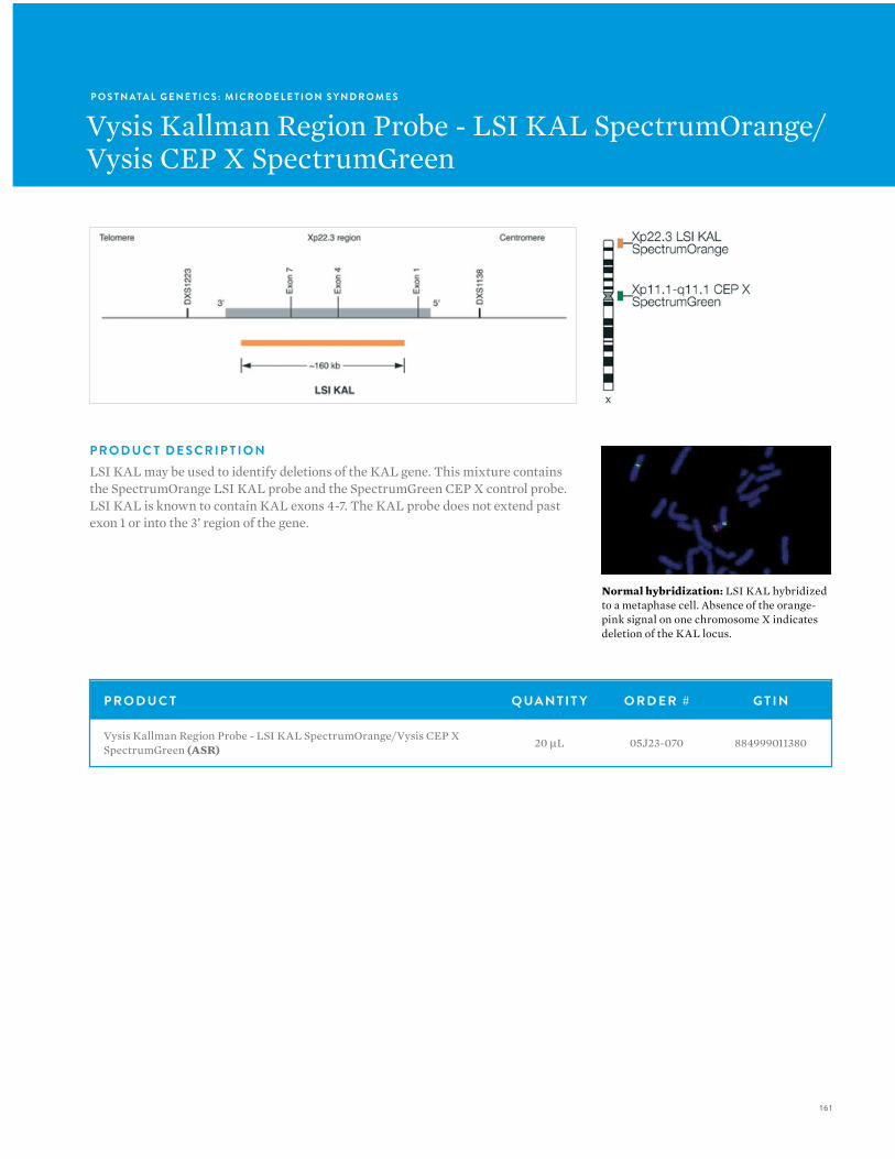
161
P O S T N ATA L G E N E T I C S : M I C R O D E L E T I O N S Y N D R O M E S
Vysis Kallman Region Probe - LSI KAL SpectrumOrange/Vysis CEP X SpectrumGreen
P R O D U C T Q UA N T I T Y O R D E R # G T I N
Vysis Kallman Region Probe - LSI KAL SpectrumOrange/Vysis CEP X SpectrumGreen (ASR) 20 µL 05J23-070 884999011380
P R O D U C T D E S C R I P T I O NLSI KAL may be used to identify deletions of the KAL gene. This mixture contains the SpectrumOrange LSI KAL probe and the SpectrumGreen CEP X control probe. LSI KAL is known to contain KAL exons 4-7. The KAL probe does not extend past exon 1 or into the 3’ region of the gene.
Normal hybridization: LSI KAL hybridized to a metaphase cell. Absence of the orange-pink signal on one chromosome X indicates deletion of the KAL locus.
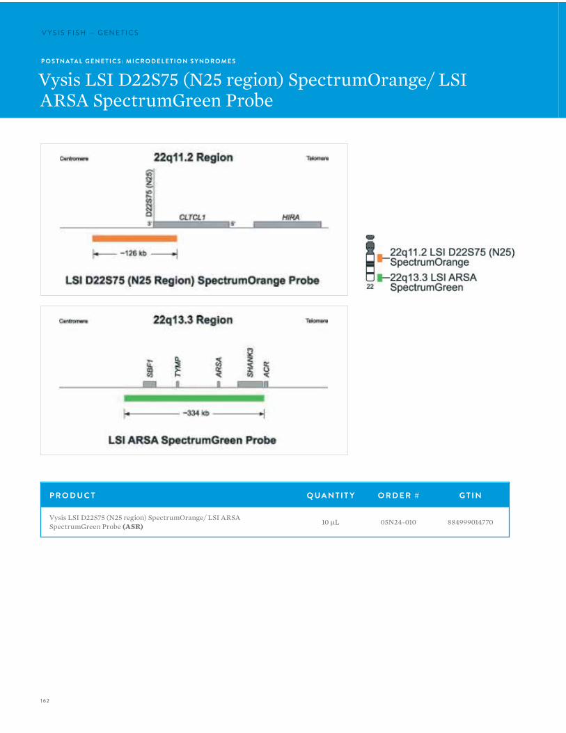
162
V Y S I S F I S H — G E N E T I C SV Y S I S F I S H — G E N E T I C S
P O S T N ATA L G E N E T I C S : M I C R O D E L E T I O N S Y N D R O M E S
Vysis LSI D22S75 (N25 region) SpectrumOrange/ LSI ARSA SpectrumGreen Probe
P R O D U C T Q UA N T I T Y O R D E R # G T I N
Vysis LSI D22S75 (N25 region) SpectrumOrange/ LSI ARSA SpectrumGreen Probe (ASR) 10 µL 05N24-010 884999014770
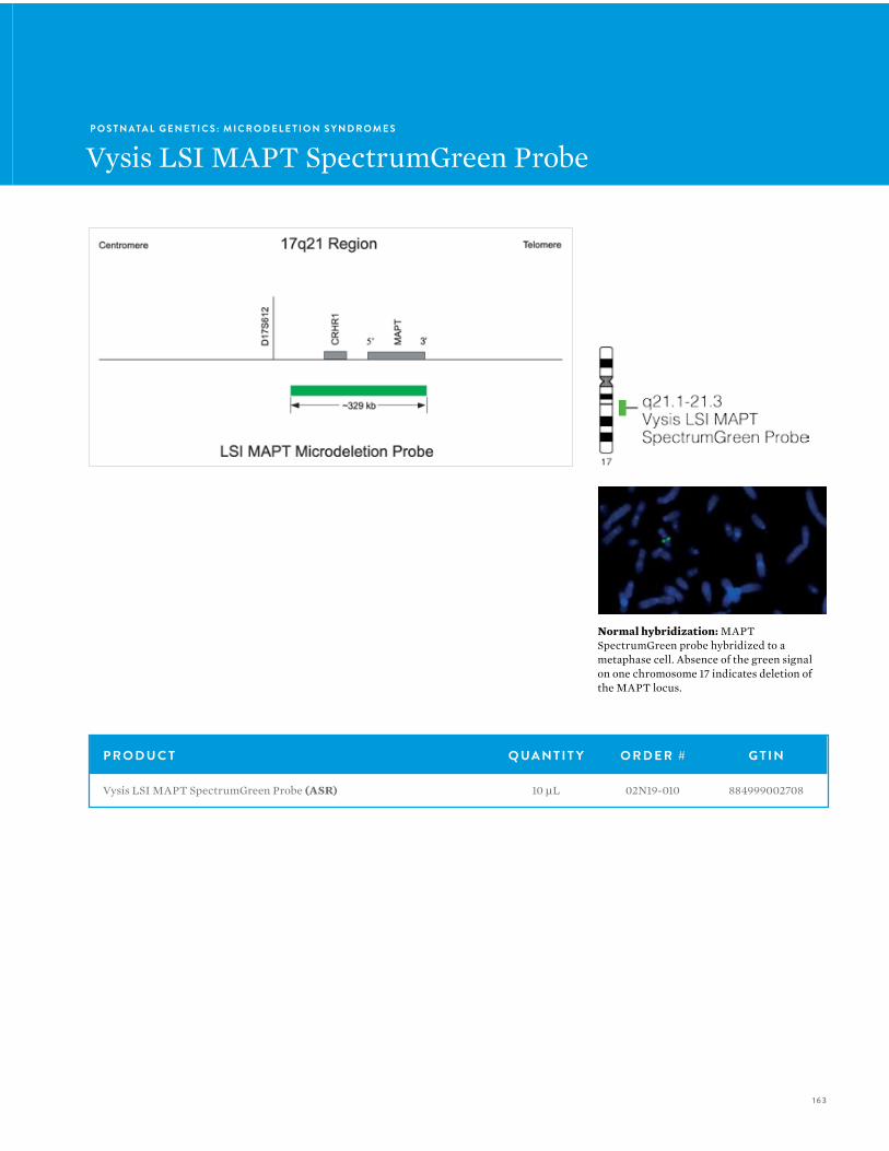
163
P O S T N ATA L G E N E T I C S : M I C R O D E L E T I O N S Y N D R O M E S
Vysis LSI MAPT SpectrumGreen Probe
Normal hybridization: MAPT SpectrumGreen probe hybridized to a metaphase cell. Absence of the green signal on one chromosome 17 indicates deletion of the MAPT locus.
P R O D U C T Q UA N T I T Y O R D E R # G T I N
Vysis LSI MAPT SpectrumGreen Probe (ASR) 10 µL 02N19-010 884999002708
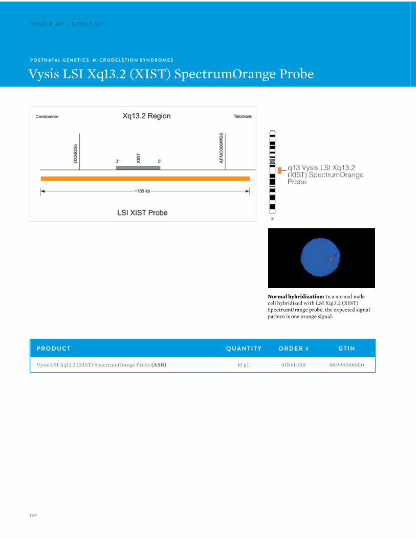
164
V Y S I S F I S H — G E N E T I C SV Y S I S F I S H — G E N E T I C S
P O S T N ATA L G E N E T I C S : M I C R O D E L E T I O N S Y N D R O M E S
Vysis LSI Xq13.2 (XIST) SpectrumOrange Probe
Normal hybridization: In a normal male cell hybridized with LSI Xq13.2 (XIST) SpectrumOrange probe, the expected signal pattern is one orange signal.
P R O D U C T Q UA N T I T Y O R D E R # G T I N
Vysis LSI Xq13.2 (XIST) SpectrumOrange Probe (ASR) 10 µL 01N61-001 884999000810
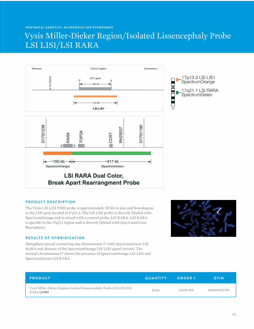
165
P O S T N ATA L G E N E T I C S : M I C R O D E L E T I O N S Y N D R O M E S
Vysis Miller-Dieker Region/Isolated Lissencephaly Probe LSI LIS1/LSI RARA
P R O D U C T Q UA N T I T Y O R D E R # G T I N
Vysis Miller-Dieker Region/Isolated Lissencephaly Probe LSI LIS1/LSI RARA (ASR) 20 µL 05J88-001 884999012790
P R O D U C T D E S C R I P T I O NThe Vysis LSI LIS1 FISH probe is approximately 110 kb in size and homologous to the LIS1 gene located at 17p13.3. The LSI LIS1 probe is directly labeled with SpectrumOrange and is mixed with a control probe, LSI RARA. LSI RARA is specific to the 17q21.1 region and is directly labeled with SpectrumGreen fluorophore.
R E S U LT S O F H Y B R I D I Z AT I O NMetaphase spread containing one chromosome 17 with SpectrumGreen LSI RARA and absence of the SpectrumOrange LSI LIS1 signal (arrow). The normal chromosome 17 shows the presence of SpectrumOrange LSI LIS1 and SpectrumGreen LSI RARA.
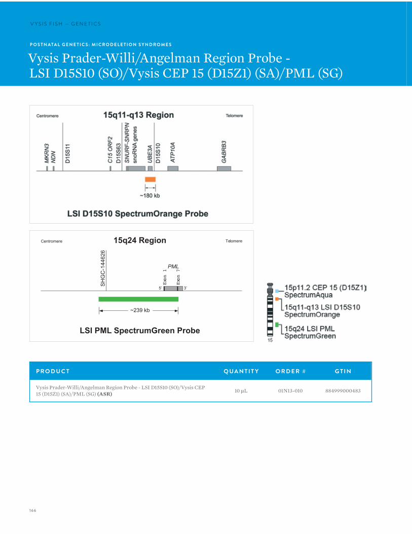
166
V Y S I S F I S H — G E N E T I C SV Y S I S F I S H — G E N E T I C S
P O S T N ATA L G E N E T I C S : M I C R O D E L E T I O N S Y N D R O M E S
Vysis Prader-Willi/Angelman Region Probe -LSI D15S10 (SO)/Vysis CEP 15 (D15Z1) (SA)/PML (SG)
P R O D U C T Q UA N T I T Y O R D E R # G T I N
Vysis Prader-Willi/Angelman Region Probe - LSI D15S10 (SO)/Vysis CEP 15 (D15Z1) (SA)/PML (SG) (ASR) 10 µL 01N13-010 884999000483
Centromere Telomere
~239 kb
LSI PML SpectrumGreen Probe
15q24 Region
SH
GC
-144
626
PML
Exon 1
Exon 7
5’ 3’

167
P O S T N ATA L G E N E T I C S : M I C R O D E L E T I O N S Y N D R O M E S
Vysis Prader-Willi/Angelman Region Probe -LSI D15S11/Vysis CEP 15 (D15Z1)
P R O D U C T Q UA N T I T Y O R D E R # G T I N
Vysis Prader-Willi/Angelman Region Probe - LSI D15S11/Vysis CEP 15 (D15Z1) (ASR) 20 µL 05J19-014 884999011274
P R O D U C T D E S C R I P T I O NFour products are offered for the detection or characterization of abnormalities involving 15q11-q13. Vysis 15q11-q13 probes are premixed with a CEP 15 control probe. The SNRPN and D15S10 probe mixes also include LSI PML (15q22), a control probe useful for detecting rare translocations in AS and PWS.

168
V Y S I S F I S H — G E N E T I C SP O S T N ATA L G E N E T I C S : M I C R O D E L E T I O N S Y N D R O M E S
Vysis Prader-Willi/Angelman Region Probe -LSI GABRB3 SpectrumOrange/Vysis CEP 15 (D15Z1) SpectrumGreen
P R O D U C T Q UA N T I T Y O R D E R # G T I N
Vysis Prader-Willi/Angelman Region Probe - LSI GABRB3 SpectrumOrange/Vysis CEP 15 (D15Z1) SpectrumGreen (ASR) 20 µL 05J22-015 884999011366
P R O D U C T D E S C R I P T I O NFour products are offered for the detection or characterization of abnormalities involving 15q11-q13. Vysis 15q11-q13 probes are premixed with a CEP 15 control probe. The SNRPN and D15S10 probe mixes also include LSI PML (15q22), a control probe useful for detecting rare translocations in AS and PWS.
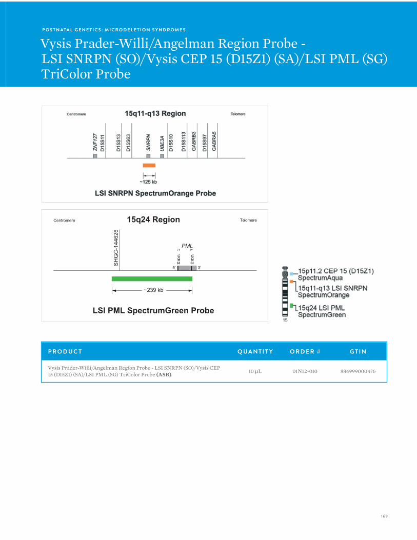
169
Centromere Telomere
~239 kb
LSI PML SpectrumGreen Probe
15q24 Region
SH
GC
-144
626
PML
Exon 1
Exon 7
5’ 3’
P O S T N ATA L G E N E T I C S : M I C R O D E L E T I O N S Y N D R O M E S
Vysis Prader-Willi/Angelman Region Probe -LSI SNRPN (SO)/Vysis CEP 15 (D15Z1) (SA)/LSI PML (SG)TriColor Probe
P R O D U C T Q UA N T I T Y O R D E R # G T I N
Vysis Prader-Willi/Angelman Region Probe - LSI SNRPN (SO)/Vysis CEP 15 (D15Z1) (SA)/LSI PML (SG) TriColor Probe (ASR) 10 µL 01N12-010 884999000476
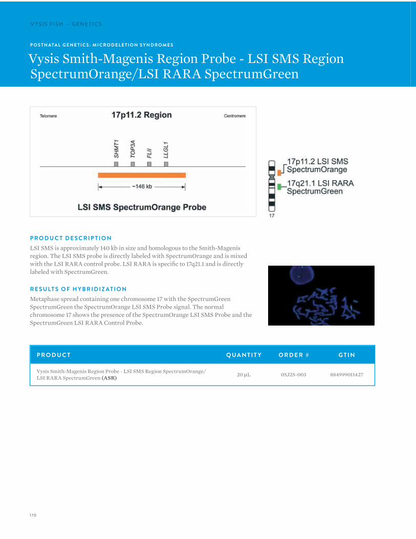
170
V Y S I S F I S H — G E N E T I C SV Y S I S F I S H — G E N E T I C S
P O S T N ATA L G E N E T I C S : M I C R O D E L E T I O N S Y N D R O M E S
Vysis Smith-Magenis Region Probe - LSI SMS Region SpectrumOrange/LSI RARA SpectrumGreen
P R O D U C T D E S C R I P T I O NLSI SMS is approximately 140 kb in size and homologous to the Smith-Magenis region. The LSI SMS probe is directly labeled with SpectrumOrange and is mixed with the LSI RARA control probe. LSI RARA is specific to 17q21.1 and is directly labeled with SpectrumGreen.
R E S U LT S O F H Y B R I D I Z AT I O NMetaphase spread containing one chromosome 17 with the SpectrumGreen SpectrumGreen the SpectrumOrange LSI SMS Probe signal. The normal chromosome 17 shows the presence of the SpectrumOrange LSI SMS Probe and the SpectrumGreen LSI RARA Control Probe.
P R O D U C T Q UA N T I T Y O R D E R # G T I N
Vysis Smith-Magenis Region Probe - LSI SMS Region SpectrumOrange/LSI RARA SpectrumGreen (ASR) 20 µL 05J25-003 884999011427
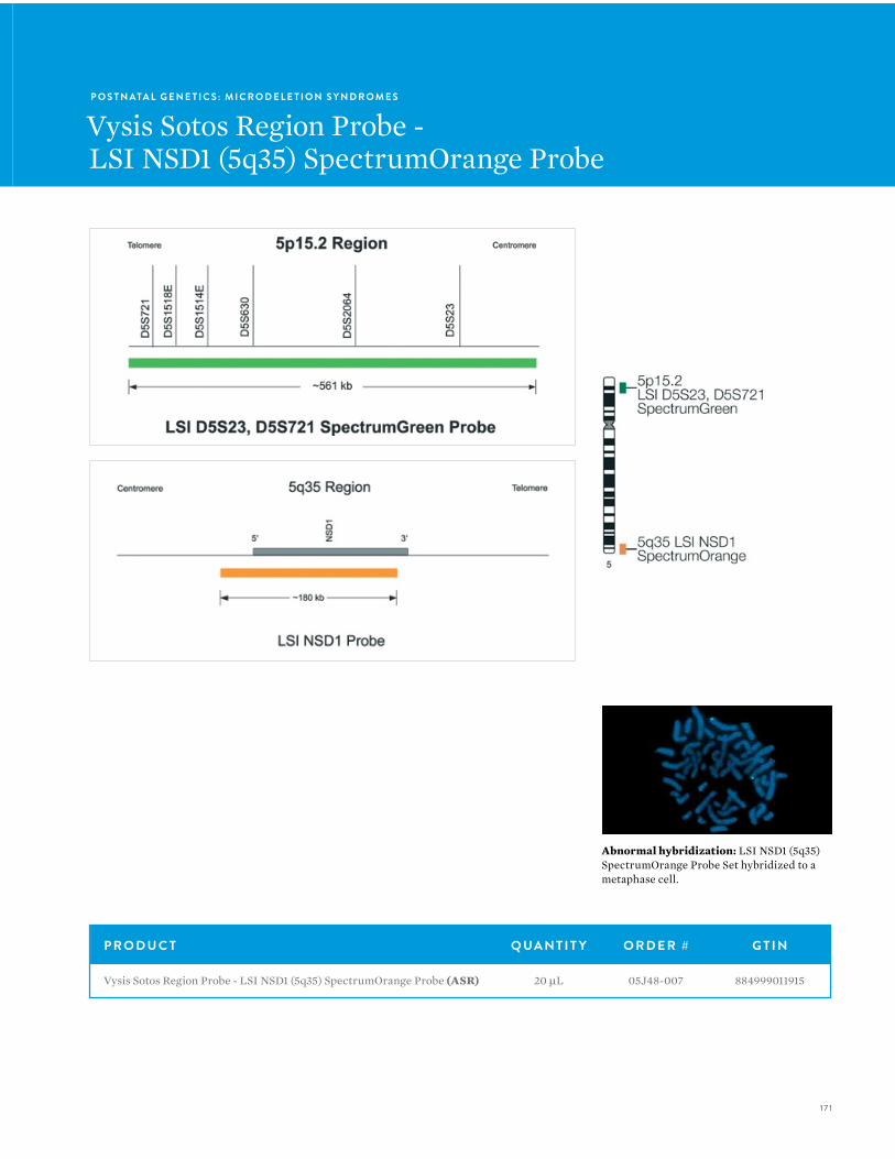
17 1
Abnormal hybridization: LSI NSD1 (5q35) SpectrumOrange Probe Set hybridized to a metaphase cell.
P R O D U C T Q UA N T I T Y O R D E R # G T I N
Vysis Sotos Region Probe - LSI NSD1 (5q35) SpectrumOrange Probe (ASR) 20 µL 05J48-007 884999011915
P O S T N ATA L G E N E T I C S : M I C R O D E L E T I O N S Y N D R O M E S
Vysis Sotos Region Probe -LSI NSD1 (5q35) SpectrumOrange Probe
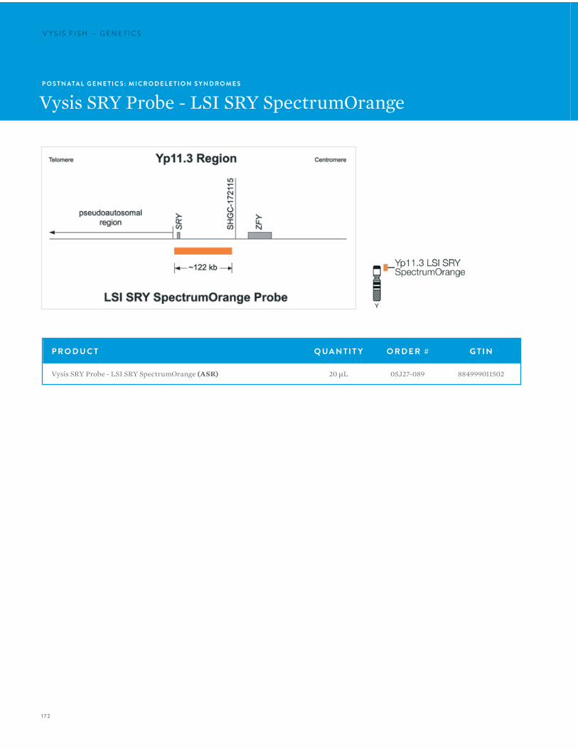
172
V Y S I S F I S H — G E N E T I C SV Y S I S F I S H — G E N E T I C S
P O S T N ATA L G E N E T I C S : M I C R O D E L E T I O N S Y N D R O M E S
Vysis SRY Probe - LSI SRY SpectrumOrange
P R O D U C T Q UA N T I T Y O R D E R # G T I N
Vysis SRY Probe - LSI SRY SpectrumOrange (ASR) 20 µL 05J27-089 884999011502

173
Abnormal hybridization: LSI SRY SpectrumOrange/CEP X SpectrumGreen metaphase hybridization.
P O S T N ATA L G E N E T I C S : M I C R O D E L E T I O N S Y N D R O M E S
Vysis SRY Probe LSI SRY Spectrum Orange/Vysis CEP X Spectrum Green Probe
P R O D U C T D E S C R I P T I O NThe SRY gene is located within 10 kb of the pseudoautosomal region of Yp. The LSI SRY probe is useful in detecting deletions of SRY or presence of the gene in rearrangements involving the X chromosome, autosomes and marker chromosomes.
The LSI SRY DNA FISH probe is an approximately 120 kb probe specific to the SRY gene and flanking sequences. This probe is direct labeled with SpectrumOrange and is available as a single probe or mixed with the CEP X SpectrumGreen probe.
P R O D U C T Q UA N T I T Y O R D E R # G T I N
Vysis SRY Probe LSI SRY SpectrumOrange/Vysis CEP X SpectrumGreen (ASR) 20 µL 05J27-007 884999011472
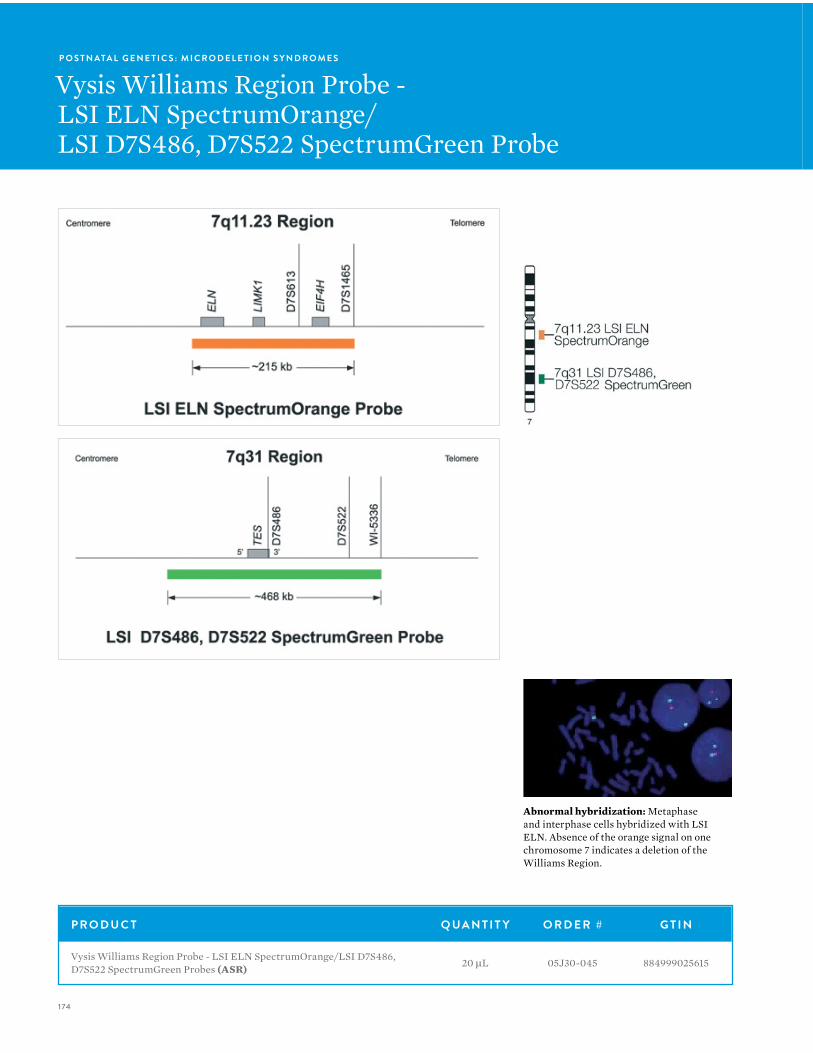
174
V Y S I S F I S H — G E N E T I C SP O S T N ATA L G E N E T I C S : M I C R O D E L E T I O N S Y N D R O M E S
Vysis Williams Region Probe -LSI ELN SpectrumOrange/LSI D7S486, D7S522 SpectrumGreen Probe
P R O D U C T Q UA N T I T Y O R D E R # G T I N
Vysis Williams Region Probe - LSI ELN SpectrumOrange/LSI D7S486, D7S522 SpectrumGreen Probes (ASR) 20 µL 05J30-045 884999025615
Abnormal hybridization: Metaphase and interphase cells hybridized with LSI ELN. Absence of the orange signal on one chromosome 7 indicates a deletion of the Williams Region.
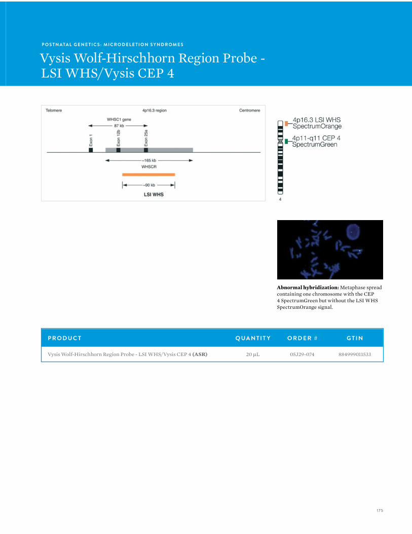
175
Abnormal hybridization: Metaphase spread containing one chromosome with the CEP 4 SpectrumGreen but without the LSI WHS SpectrumOrange signal.
P O S T N ATA L G E N E T I C S : M I C R O D E L E T I O N S Y N D R O M E S
Vysis Wolf-Hirschhorn Region Probe -LSI WHS/Vysis CEP 4
P R O D U C T Q UA N T I T Y O R D E R # G T I N
Vysis Wolf-Hirschhorn Region Probe - LSI WHS/Vysis CEP 4 (ASR) 20 µL 05J29-074 884999011533

176
V Y S I S F I S H — G E N E T I C SV Y S I S F I S H — G E N E T I C S
P O S T N ATA L G E N E T I C S : T O T E LV Y S I O N
Vysis ToTelVysion
P R O D U C T D E S C R I P T I O NToTelVysion consists of 41 TelVysion probes, including various LSI and CEP probes (64 probes in total).
• The 41 TelVysion probes are specific to:
• p and q subtelomeres of chromosomes 1-12 and 16-20
• q subtelomeres of the acrocentric chromosomes (13, 14, 15, 21, and 22)
• Xp/Yp and Xq/Yq pseudo-autosomal region subtelomeres
• A unique region within 300 kb of each chromosome telomere
The probes in ToTelVysion are provided in 15 mixtures. All probes are direct labeled, providing bright signals with minimal background noise. By utilizing SpectrumOrange, SpectrumGreen, SpectrumAqua, and a combination of SpectrumOrange and SpectrumGreen (to yield a yellow signal), each probe within a mixture is labeled with a unique color.
V Y S I S T E LV Y S I O N P R O B E STelomeres are DNA-protein complexes that cap the ends of eukaryotic chromosomes. Every telomere contains 3 to 20 kb of tandem TTAGGG repeats. The telomere associated repeats (TAR), also known as the subtelomeric repeats, are immediately proximal to the TTAGGG repeats. They contain regions of shared homology between subsets of certain chromosomes.
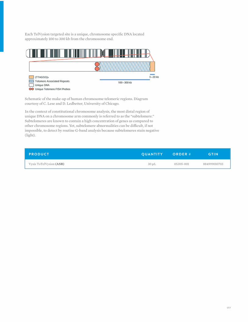
17 7
Each TelVysion targeted site is a unique, chromosome specific DNA located approximately 100 to 300 kb from the chromosome end.
Schematic of the make-up of human chromosome telomeric regions. Diagram courtesy of C. Lese and D. Ledbetter, University of Chicago.
In the context of constitutional chromosome analysis, the most distal region of unique DNA on a chromosome arm commonly is referred to as the “subtelomere.” Subtelomeres are known to contain a high concentration of genes as compared to other chromosome regions. Yet, subtelomere abnormalities can be difficult, if not impossible, to detect by routine G-band analysis because subtelomeres stain negative (light).
P R O D U C T Q UA N T I T Y O R D E R # G T I N
Vysis ToTelVysion (ASR) 30 µL 05J05-001 884999010703
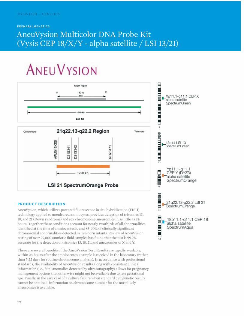
178
V Y S I S F I S H — G E N E T I C SV Y S I S F I S H — G E N E T I C S
P R E N ATA L G E N E T I C S
AneuVysion Multicolor DNA Probe Kit(Vysis CEP 18/X/Y - alpha satellite / LSI 13/21)
P R O D U C T D E S C R I P T I O NAneuVysion, which utilizes patented fluorescence in situ hybridization (FISH) technology applied to uncultured amniocytes, provides detection of trisomies 13, 18, and 21 (Down syndrome) and sex chromosome aneusomies in as little as 24 hours. Together these conditions account for nearly twothirds of all abnormalities identified at the time of amniocentesis, and 85-90% of clinically significant chromosomal abnormalities detected in live-born infants. Review of AneuVysion testing of over 29,000 amniotic fluid samples has found that the test is 99.9% accurate for the detection of trisomies 13, 18, 21, and aneusomies of X and Y.
There are several benefits of the AneuVysion Test. Results are rapidly available, within 24 hours after the amniocentesis sample is received in the laboratory (rather than 7-22 days for routine chromosome analysis). In accordance with professional standards, the availability of AneuVysion results along with consistent clinical information (i.e., fetal anomalies detected by ultrasonography) allows for pregnancy management options that otherwise might not be available due to late gestational age. Finally, in the rare case of a culture failure when standard cytogenetic results cannot be obtained, information on chromosome number for the most likely aneusomies is available.
179
P R O B E M I X T U R E #1• CEP 18: D18Z1 alpha satellite DNA probe corresponding to
18p11.1-q11.1 labeled with SpectrumAqua
• CEP X: DXZ1 alpha satellite DNA probe corresponding to Xp11.1-q11.1 labeled with SpectrumGreen
• CEP Y: DYZ3 alpha satellite DNA probe corresponding to Yp11.1-q11.1 labeled with SpectrumOrange
P R O B E M I X T U R E #2• LSI 18: DNA probe corresponding to the RB1 gene (13q14)
labeled with SpectrumGreen.
• LSI 21: DNA probe corresponding to loci D21S259, D21S341, and D21S342(21q22.13-q22.2) labeled with SpectrumOrange.
Mixture #1 is complete with labeled probes and non-labeled blocking DNA in hybridization buffer.
R E L AT E D P R O D U C T SProducts for use with AneuVysion
ProbeChek Prenatal Control Slides for Amniocyte;• Male Amniocyte Control
• 30-805010 — 5 Slides
• Fixed biological specimen derived from normal human male amniocytes applied to glass microscope slides.
ProbeChek Prenatal Control Slides for Positive Control• 30-805017 — 5 Slides
• Fixed biological specimen derived from human triploid fibroblast cells applied to glass microscope slides.
• Control slides are excellent training and validation tools for the AneuVysion Test.
I N T E N D E D U S EFor In Vitro Diagnostic Use
The AneuVysion (Vysis CEP 18, X, Y-alpha satellite, LSI 13 and 21) Multicolor Probe Panel is intended to use CEP 18/X/Y probe to detect alpha satellite sequences in the centromere regions of chromosomes 18, X, and Y, and LSI 13/21 probe to detect the 13q14 region and the 21q22.13 to 21q22.2 region. The AneuVysion kit is indicated for identifying and enumerating chromosomes 13, 18, 21, X, and Y via fl uorescence in situ hybridization (FISH) in metaphase cells and interphase nuclei
obtained from amniotic fl uid in subjects with presumed high risk pregnancies. It is not intended to be used as a stand alone assay for making clinical decisions. FISH results are intended to be used as an aid in the diagnosis of numerical abnormalities of chromosomes 13, 18, 21, X and/or Y in conjunction with other information currently used in prenatal diagnosis, consistent with professional standards of practice [1]. This device is intended for use only with amniocyte cells; it is not intended for and has not been validated for use with other test matrices. This FISH assay will not detect the presence of structural chromosome abnormalities that can also result in birth defects. This FISH assay will be performed in cytogenetics laboratories.
L I M I TAT I O N S 1. The AneuVysion kit has been characterized only for
identifying targeted regions of chromosomes X,Y,18,13, and 21 in interphase nuclei from cultured and uncultured amniocyte specimens.
2. The clinical interpretation of any test result(s) should be made in conjunction with other diagnostic laboratory test results and should be evaluated within the context of the patient’s medical history and current risk factors. Patient management decisions should be made based on information from two or three of the following parameters: FISH results, routine chromosome analysis, or clinical information [1].
3. FISH assay results may not be informative if the specimen quality and/or specimen slide preparation is inadequate.
4. This assay will not detect the presence of structural abnormalities frequently associated with birth defects. The frequency of these occurrences may be population and gestational age dependent.
5. This assay should not be performed on amniocyte specimens with moderate to severe maternal cell contamination. FISH test results on amniocyte specimens with mild maternal cell contaminations should be interpreted with caution.
6. No irreversible therapeutic action should be initiated based on the FISH assay lone. Positive results should be further characterized using traditional chromosome analysis to determine the mutational mechanism accounting for the abnormality detected by FISH. This information may aid in the counseling for the risk that the detected abnormality may occur in future pregnancies [1].
180
V Y S I S F I S H — G E N E T I C S
P R O D U C T Q UA N T I T Y O R D E R # G T I N
AneuVysion Multicolor DNA Probe Kit (Vysis CEP 18/X/Y - alpha satellite / LSI 13/21) (IVD)
10 Assays30 Assays50 Assays
05J38-01005J38-03005J38-050
884999011694884999011700884999011717
7. Physicians, counselors, and other healthcare providers should understand the risk of abnormalities that the test is not designed to detect. The patient should be informed that there is still a very small risk of low level mosaicsim, cryptic translocations, or other undetectable events that may not be demonst4rated by FISH or standard cytogenetic. Additionally, there is a very small risk that some individuals carry a genetic polymorphism that may affect the intensity, presence or absence of the probe signal that may result in a missed diagnosis [17].
8. When the specimen volume is not sufficient to meet the minimum requirements for processing both FISH and standard cytogenetic procedures, the user must carefully weigh the risks and benefits of utilizing any material for FISH. Consultation between the laboratory geneticist and or genetic counselor and the patient’s physician may aid in clarifying what information is desired, and which testing method should be used [1].
9. Technologists performing the FISH signal enumeration must be capable of visually distinguishing between orange, green and aqua signals.
10. Although the probe for enumerating chromosome 13 spans the Rb1 locus, this probe has not been validated for detecting mutations associated with retinoblastoma. In rare cases, the Rb1 locus may be deleted; this could complicate interpretation of FISH test results.
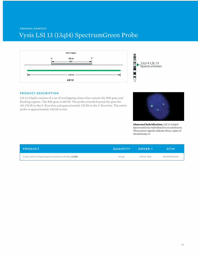
18 1
P R E N ATA L G E N E T I C S
Vysis LSI 13 (13q14) SpectrumGreen Probe
P R O D U C T Q UA N T I T Y O R D E R # G T I N
Vysis LSI 13 (13q14) SpectrumGreen Probe (ASR) 20 µL 05J14-028 884999011199
P R O D U C T D E S C R I P T I O NLSI 13 (13q14) consists of a set of overlapping clones that contain the RB1 gene and flanking regions. The RB1 gene is 180 kb. The probe extends beyond the gene for 110-170 kb in the 5’ direction and approximately 120 kb in the 3’ direction. The entire probe is approximately 440 kb in size.
Abnormal hybridization: LSI 13 (13q14) SpectrumGreen hybridized to an amniocyte. Three green signals indicate three copies of chromosome 13
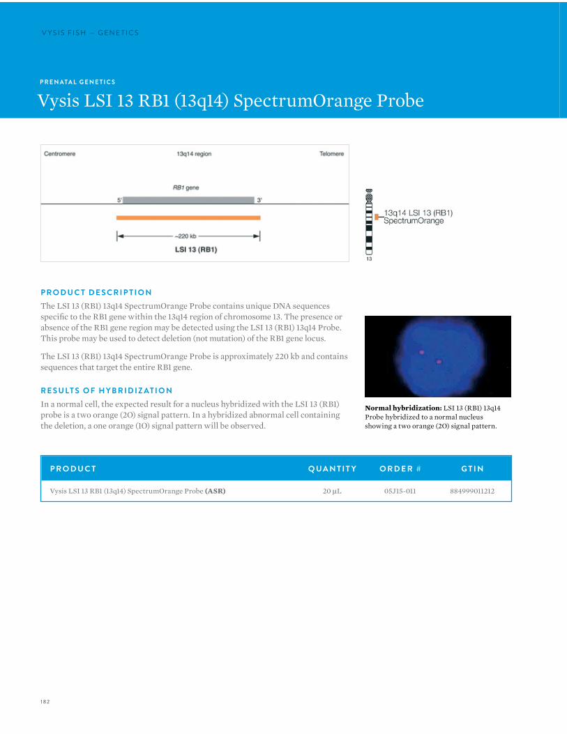
182
V Y S I S F I S H — G E N E T I C SV Y S I S F I S H — G E N E T I C S
P R E N ATA L G E N E T I C S
Vysis LSI 13 RB1 (13q14) SpectrumOrange Probe
Normal hybridization: LSI 13 (RB1) 13q14 Probe hybridized to a normal nucleus showing a two orange (2O) signal pattern.
P R O D U C T Q UA N T I T Y O R D E R # G T I N
Vysis LSI 13 RB1 (13q14) SpectrumOrange Probe (ASR) 20 µL 05J15-011 884999011212
P R O D U C T D E S C R I P T I O NThe LSI 13 (RB1) 13q14 SpectrumOrange Probe contains unique DNA sequences specific to the RB1 gene within the 13q14 region of chromosome 13. The presence or absence of the RB1 gene region may be detected using the LSI 13 (RB1) 13q14 Probe. This probe may be used to detect deletion (not mutation) of the RB1 gene locus.
The LSI 13 (RB1) 13q14 SpectrumOrange Probe is approximately 220 kb and contains sequences that target the entire RB1 gene.
R E S U LT S O F H Y B R I D I Z AT I O NIn a normal cell, the expected result for a nucleus hybridized with the LSI 13 (RB1) probe is a two orange (2O) signal pattern. In a hybridized abnormal cell containing the deletion, a one orange (1O) signal pattern will be observed.
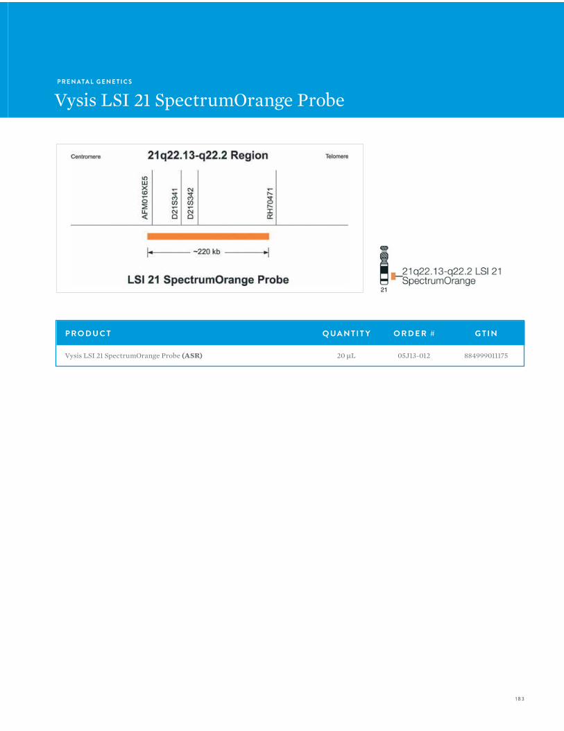
183
P R E N ATA L G E N E T I C S
Vysis LSI 21 SpectrumOrange Probe
P R O D U C T Q UA N T I T Y O R D E R # G T I N
Vysis LSI 21 SpectrumOrange Probe (ASR) 20 µL 05J13-012 884999011175
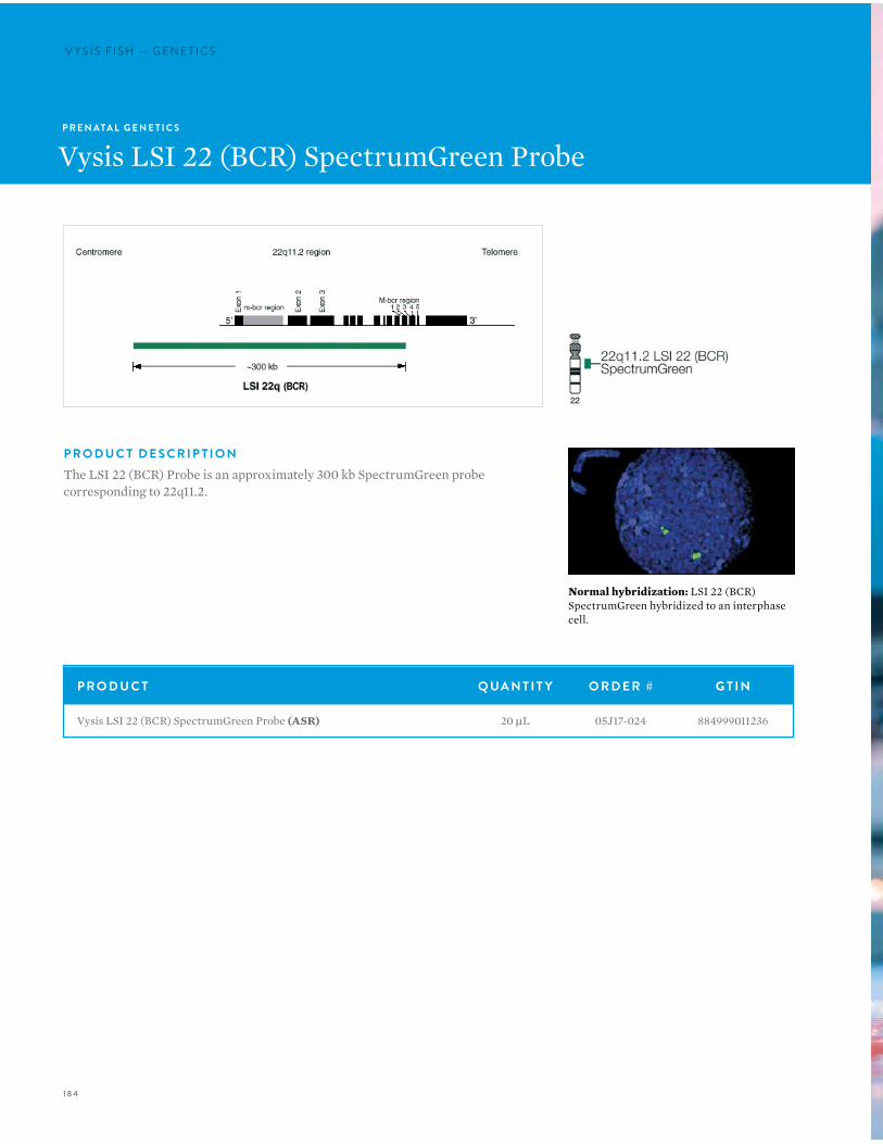
184
V Y S I S F I S H — G E N E T I C SV Y S I S F I S H — G E N E T I C S
P R E N ATA L G E N E T I C S
Vysis LSI 22 (BCR) SpectrumGreen Probe
P R O D U C T D E S C R I P T I O NThe LSI 22 (BCR) Probe is an approximately 300 kb SpectrumGreen probe corresponding to 22q11.2.
P R O D U C T Q UA N T I T Y O R D E R # G T I N
Vysis LSI 22 (BCR) SpectrumGreen Probe (ASR) 20 µL 05J17-024 884999011236
Normal hybridization: LSI 22 (BCR) SpectrumGreen hybridized to an interphase cell.

185
186
V Y S I S F I S H — I N S T R U M E N T S
VYSIS FISH:INSTRUMENTS
186
V Y S I S F I S H — I N S T R U M E N T S
Automation is essential for laboratories interested in reducing the amount of hands-on-time required to run FISH assays, while increasing laboratory throughput, flexibility, reproducibility and productivity. Abbott Molecular is pleased to offer FISH automation options to suit the needs of your laboratory.

187187
V Y S I S F I S H I N S T R U M E N T S P R OV I D E S T H E F O L LO W I N G A DVA N TAG E S :• Our VP 2000 Processor is designed to automate tedious
front end FISH preparation procedures such as de-paraffinization and slide pretreatment. With the flexibility to perform batches of up to 50 slides at a time, and 3 heated and 12 ambient temperature basins, the VP 2000 is well suited for the needs of today’s FISH laboratory.
• The ThermoBrite Slide Processing System is a temperature programmable, humidity controlled instrument designed to automate denaturation and hybridization steps for FISH. Rapid temperature ramping and accuracy within +/– 1 °C ensure superior temperature uniformity across all 12 slide positions. Up to 40 user defined protocols and 3 operating modes ensure ease of use and flexibility.

188
V Y S I S F I S H — I N S T R U M E N T S
P R O D U C T Q UA N T I T Y O R D E R # G T I N P G
B I O V I E W AU T O M AT E D I M A G I N G A N D A N A LY S I S S Y S T E M
Accord Plus Automated Scanner with Single Slide Motorized Stage 1 08N52-03 7290107652780 -
Accord Semi-Automatic Scanner (Manual Stage) 1 08N52-01 7290107652773 -
Allegro Plus Automated Scanner with 8 Slides Stage 1 08N52-05 7290107652797 -
Amniotic Fluid Scanning Applications 1 08N52-26 7290107653039 190
AneuVysion Application 1 08N52-36 7290107653091 190
Automated Brightfield Scanning 1 08N52-16 7290107652971 190
Bladder Cancer Scanning Application 1 08N52-53 7290107653244 190
Cell Suspension Target FISH Dual Mode Application 1 08N52-31 7290107653060 190
Cervical Probe Scanning application includes Oral Cancer Scanning 1 08N52-39 7290107653121 190
Consecutive Cut Tissue FISH Application 1 08N52-32 7290107653077 190
Duet 3 Automated Scanner with 50 slides loader 1 08N52-07 7290107652803 -
Encore 100 Automatic Motorized 100 Slide Loader 1 08N52-10 7290107652827 -
Encore 200 Automatic Motorized 200 Slide Loader 1 08N52-09 7290107652810 -
ERG Probe Scanning Application 1 08N52-41 7290107653145 190
Fluorescent TMA mapping application 1 08N52-44 7290107653176 190
G/R-Band automated high resolution metaphase capture for Karyotyping 1 08N52-45 7290107653183 190
G/R-Band Brightfield Metaphase Finder 1 08N52-17 7290107652988 190
Hematological Scanning Application 1 08N52-54 7290107653251 190
ImmunoFluorescence Applications 1 08N52-42 7290107653152 190
Karyotyping Software 1 08N52-24 7290107653015 190
Manual Brightfield Imaging 1 08N52-14 7290107652957 190
Manual Karyotype Capture 1 08N52-15 7290107652964 190
Post-Natal Scanning Applications 1 08N52-40 7290107653138 190
Research Aid Software 1 08N52-43 7290107653169 190
SOLO WEB for 10 Concurrent Users 1 08N52-55 7290107652940 190
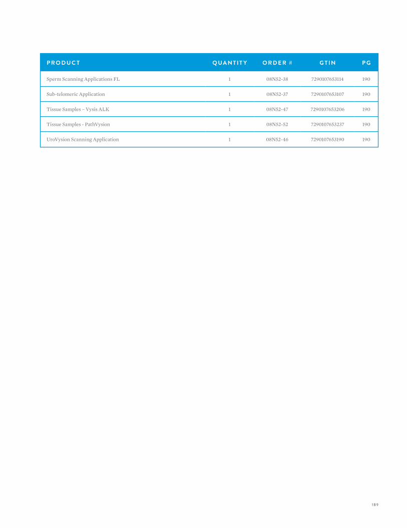
189
P R O D U C T Q UA N T I T Y O R D E R # G T I N P G
Sperm Scanning Applications FL 1 08N52-38 7290107653114 190
Sub-telomeric Application 1 08N52-37 7290107653107 190
Tissue Samples – Vysis ALK 1 08N52-47 7290107653206 190
Tissue Samples - PathVysion 1 08N52-52 7290107653237 190
UroVysion Scanning Application 1 08N52-46 7290107653190 190
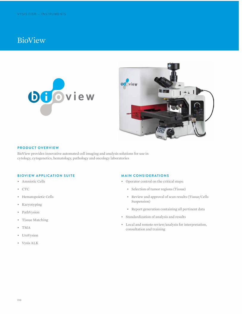
190
V Y S I S F I S H — I N S T R U M E N T SV Y S I S F I S H — I N S T R U M E N T S
BioView
B I OV I E W A P P L I C AT I O N S U I T E• Amniotic Cells
• CTC
• Hematopoietic Cells
• Karyotyping
• PathVysion
• Tissue Matching
• TMA
• UroVysion
• Vysis ALK
P R O D U C T OV E R V I E WBioView provides innovative automated cell imaging and analysis solutions for use in cytology, cytogenetics, hematology, pathology and oncology laboratories
M A I N CO N S I D E R AT I O N S• Operator control on the critical steps:
• Selection of tumor regions (Tissue)
• Review and approval of scan results (Tissue/Cells Suspension)
• Report generation containing all pertinent data
• Standardization of analysis and results
• Local and remote review/analysis for interpretation, consultation and training
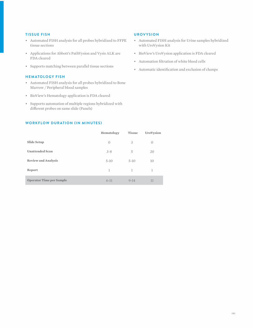
19 1
T I S S U E F I S H• Automated FISH analysis for all probes hybridized to FFPE
tissue sections
• Applications for Abbott’s PathVysion and Vysis ALK are FDA cleared
• Supports matching between parallel tissue sections
H E M ATO LO G Y F I S H• Automated FISH analysis for all probes hybridized to Bone
Marrow / Peripheral blood samples
• BioView’s Hematology application is FDA cleared
• Supports automation of multiple regions hybridized with different probes on same slide (Panels)
U R OV Y S I O N• Automated FISH analysis for Urine samples hybridized
with UroVysion Kit
• BioView’s UroVysion application is FDA cleared
• Automation filtration of white blood cells
• Automatic identification and exclusion of clumps
Hematology Tissue UroVysion
Slide Setup 0 3 0
Unattended Scan 3-8 5 20
Review and Analysis 5-10 5-10 10
Report 1 1 1
Operator Time per Sample 6-11 9-14 11
W O R K F LO W D U R AT I O N ( I N M I N U T E S )
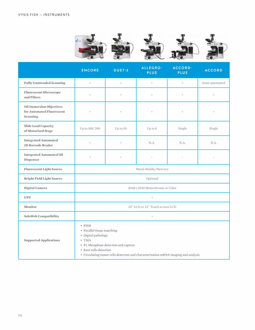
192
V Y S I S F I S H — I N S T R U M E N T S
E N CO R E D U E T-3 A L L E G R O -P LU S
ACCO R D -P LU S ACCO R D
Fully Unattended Scanning + + + + Semi-automated
Fluorescent Microscopeand Filters
+ + + + +
Oil Immersion Objectivesfor Automated FluorescentScanning
+ + + + +
Slide Load Capacityof Motorized Stage
Up to 100/#200 Up to 50 Up to 8 Single Single
Integrated Automated2D Barcode Reader
+ + N.A. N.A. N.A.
Integrated Automated OilDispenser
+ + - - -
Fluorescent Light Source Metal-Halide/$Mercury
Bright Field Light Source Optional
Digital Camera 2048#x#2048 Monochrome or Color
UPS +
Monitor 24’’ LCD or 22’’ Touch screen LCD
SoloWeb Compatibility +
Supported Applications
• FISH• Parallel tissue matching• Digital pathology• TMA• FL Metaphase detection and capture• Rare cells detection• Circulating tumor cells detection and characterization mRNA imaging and analysis
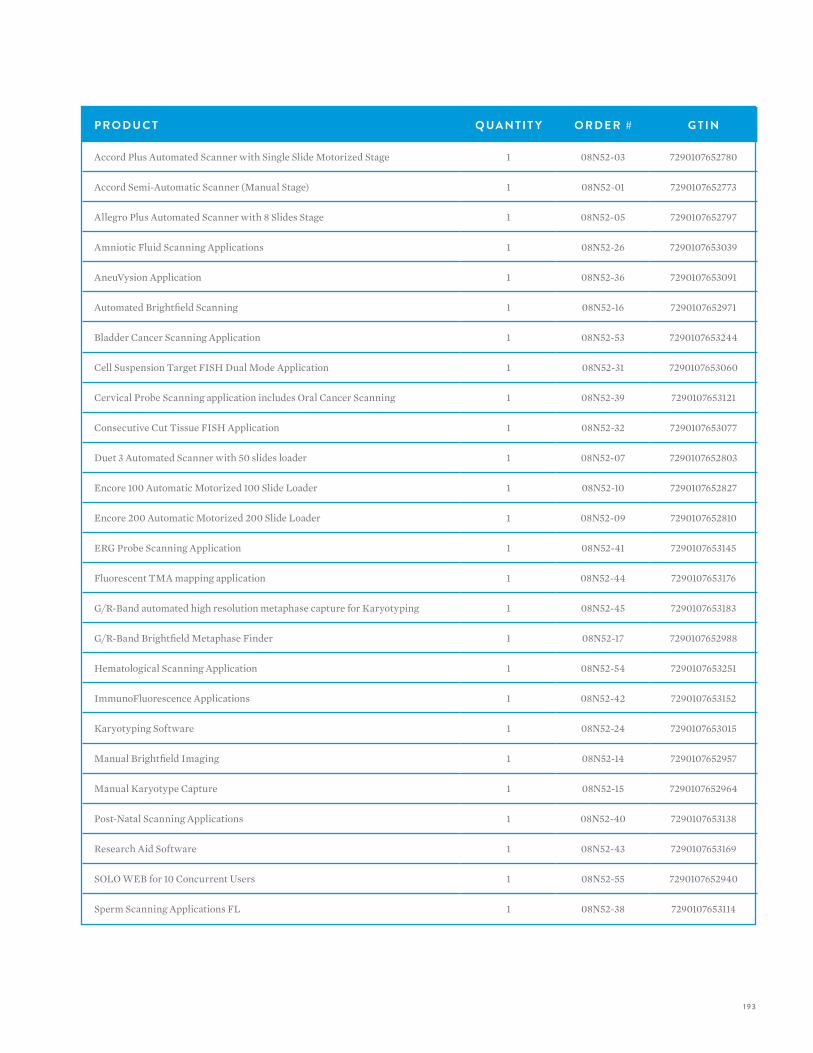
193
P R O D U C T Q UA N T I T Y O R D E R # G T I N
Accord Plus Automated Scanner with Single Slide Motorized Stage 1 08N52-03 7290107652780
Accord Semi-Automatic Scanner (Manual Stage) 1 08N52-01 7290107652773
Allegro Plus Automated Scanner with 8 Slides Stage 1 08N52-05 7290107652797
Amniotic Fluid Scanning Applications 1 08N52-26 7290107653039
AneuVysion Application 1 08N52-36 7290107653091
Automated Brightfield Scanning 1 08N52-16 7290107652971
Bladder Cancer Scanning Application 1 08N52-53 7290107653244
Cell Suspension Target FISH Dual Mode Application 1 08N52-31 7290107653060
Cervical Probe Scanning application includes Oral Cancer Scanning 1 08N52-39 7290107653121
Consecutive Cut Tissue FISH Application 1 08N52-32 7290107653077
Duet 3 Automated Scanner with 50 slides loader 1 08N52-07 7290107652803
Encore 100 Automatic Motorized 100 Slide Loader 1 08N52-10 7290107652827
Encore 200 Automatic Motorized 200 Slide Loader 1 08N52-09 7290107652810
ERG Probe Scanning Application 1 08N52-41 7290107653145
Fluorescent TMA mapping application 1 08N52-44 7290107653176
G/R-Band automated high resolution metaphase capture for Karyotyping 1 08N52-45 7290107653183
G/R-Band Brightfield Metaphase Finder 1 08N52-17 7290107652988
Hematological Scanning Application 1 08N52-54 7290107653251
ImmunoFluorescence Applications 1 08N52-42 7290107653152
Karyotyping Software 1 08N52-24 7290107653015
Manual Brightfield Imaging 1 08N52-14 7290107652957
Manual Karyotype Capture 1 08N52-15 7290107652964
Post-Natal Scanning Applications 1 08N52-40 7290107653138
Research Aid Software 1 08N52-43 7290107653169
SOLO WEB for 10 Concurrent Users 1 08N52-55 7290107652940
Sperm Scanning Applications FL 1 08N52-38 7290107653114
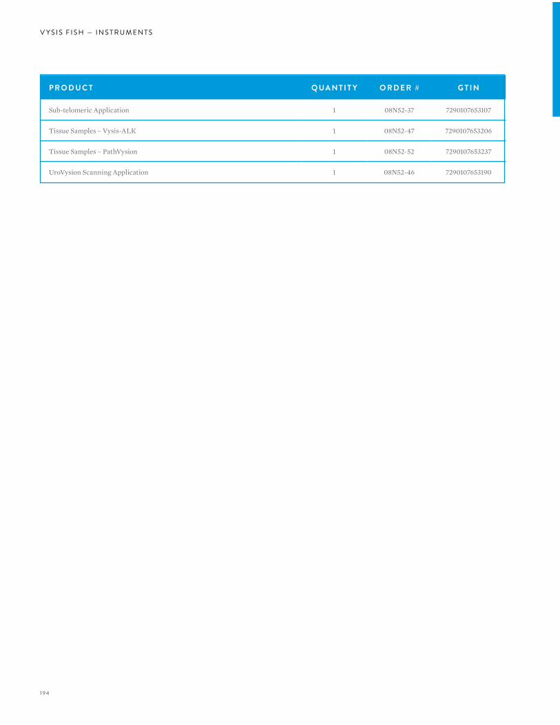
194
V Y S I S F I S H — I N S T R U M E N T S
P R O D U C T Q UA N T I T Y O R D E R # G T I N
Sub-telomeric Application 1 08N52-37 7290107653107
Tissue Samples – Vysis-ALK 1 08N52-47 7290107653206
Tissue Samples – PathVysion 1 08N52-52 7290107653237
UroVysion Scanning Application 1 08N52-46 7290107653190
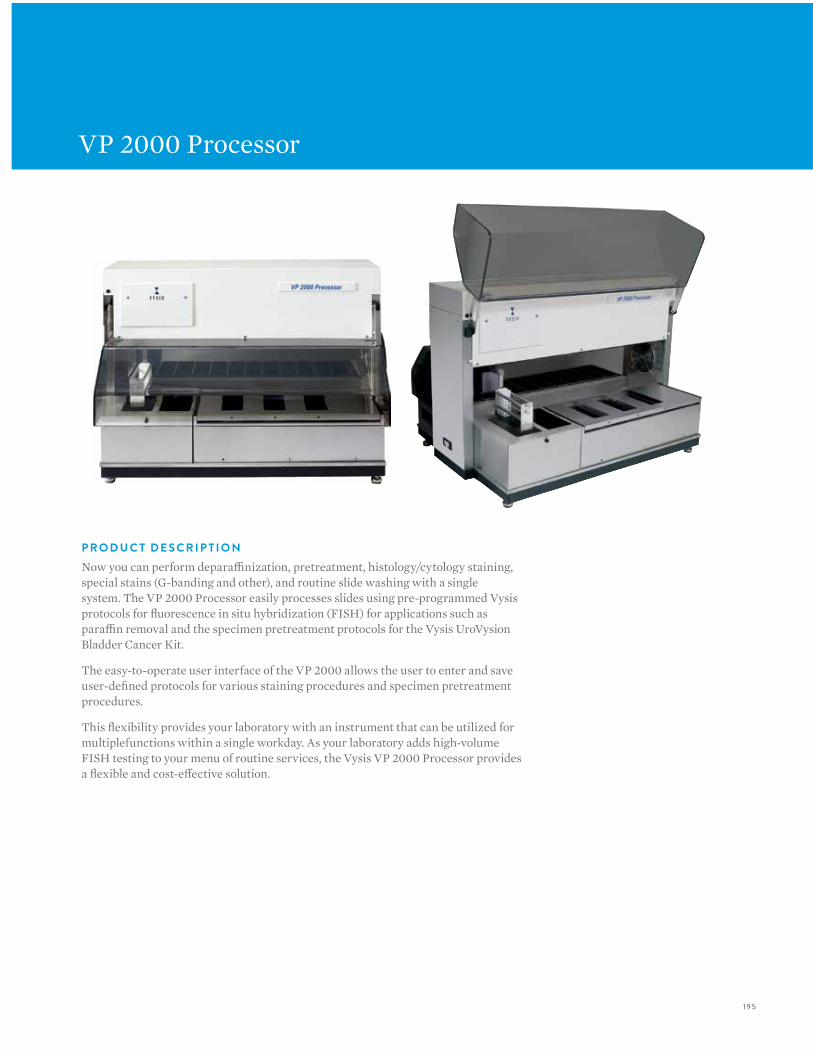
195
VP 2000 Processor
P R O D U C T D E S C R I P T I O NNow you can perform deparaffinization, pretreatment, histology/cytology staining, special stains (G-banding and other), and routine slide washing with a single system. The VP 2000 Processor easily processes slides using pre-programmed Vysis protocols for fluorescence in situ hybridization (FISH) for applications such as paraffin removal and the specimen pretreatment protocols for the Vysis UroVysion Bladder Cancer Kit.
The easy-to-operate user interface of the VP 2000 allows the user to enter and save user-defined protocols for various staining procedures and specimen pretreatment procedures.
This flexibility provides your laboratory with an instrument that can be utilized for multiplefunctions within a single workday. As your laboratory adds high-volume FISH testing to your menu of routine services, the Vysis VP 2000 Processor provides a flexible and cost-effective solution.
196
V Y S I S F I S H — I N S T R U M E N T S
A DVA N TAG E S O F T H E V P 2000 P R O C E S S O R :
• Convenient walk-away automation to reduce laboratory labor and costs
• Performs more consistent and standardized FISH assay deparaffinization and pretreatment
• Validated for use with Vysis FISH pretreatment protocols including solid tumor and cytological specimens, such as amniocytes and bladder tumor cells
• Full user programmability of events for maximum flexibility
• Open system is compatible with reagents used in today’s laboratories
• Bulk reagents available for added economy and ease-of-use
• Driven by a PC with Windows user interface (included in package)
• Five-way safety protection
• Reliability to stay on the job processing slides, year after year
T H E V Y S I S S Y S T E M S A P P R OAC H TO AU TO M AT E D F I S H T E S T I N G
The VP 2000 Processor, in conjunction with the Vysis ThermoBrite Denaturation/Hybridization unit provides a modular systems approach to automated FISH testing.
Software Proprietary VP 2000
Slide Capacity per Run 50
Ambient Reagent Basins 12
Heated Reagent Basins 3
Program Capacity >1000
Events per Program >100
Water Bath Flow Rate 1L/min
Dimensions (L x W x H) Processing Unit
31 x 24 x 22 in. (79 x 61 x 56 cm)
Weight - Processing Unit 115 VAC - 127 lb. (58 kg)100/230 VAC - 140 lb. (64. kg)
Computer Configuration Pentium Class PC, 600 MB or greater
Heated Reagent Basin Temperature
Ambient to 80 ºC
Operating Temperature 15-30 ºC
Drying Station Temperature
Ambient to 80 ºC
Systems 117 VAC, 50/60 Hz 230 VAC, 50/60 Hz 100 VAC, 50/60 Hz
T E C H N I C A L S P E C I F I C AT I O N S
P R O D U C T Q UA N T I T Y O R D E R # G T I N
VP 2000 Reagents Pretreatment Reagent (GPR) 500 mL 02J06-030 884999001862
VP 2000 Reagents Protease Buffer (GPR) 500 mL 02J07-030 884999001879
VP 2000 Reagents Protease I (GPR) 250 mg x 2 02J08-032 884999001886
VP 2000 Reagents Protease II (GPR) 750 mg 06J93-001 884999023468
VP 2000 Reagents 20X SSC (GPR) 500 g 02J10-032 884999001909
VP 2000 Reagents 2M MgCL 2 (GPR) 120 mL 02J09-030 884999001763

197

198
V Y S I S F I S H — A C C E S S O R I E S / G E N E R A L P U R P O S E R E AG E N T S
VYSIS FISH:ACCESSORIES/GENERAL PURPOSE REAGENTS
198
V Y S I S F I S H — ACC E S S O R I E S / G E N E R A L P U R P O S E R E AG E N T S
Abbott Molecular delivers best in class products and services to meet the diverse needs of your laboratory. In addition to FISH automation, Abbott Molecular provides quality instrumentation and reagents that optimize laboratory effectiveness when processingFISH probes.
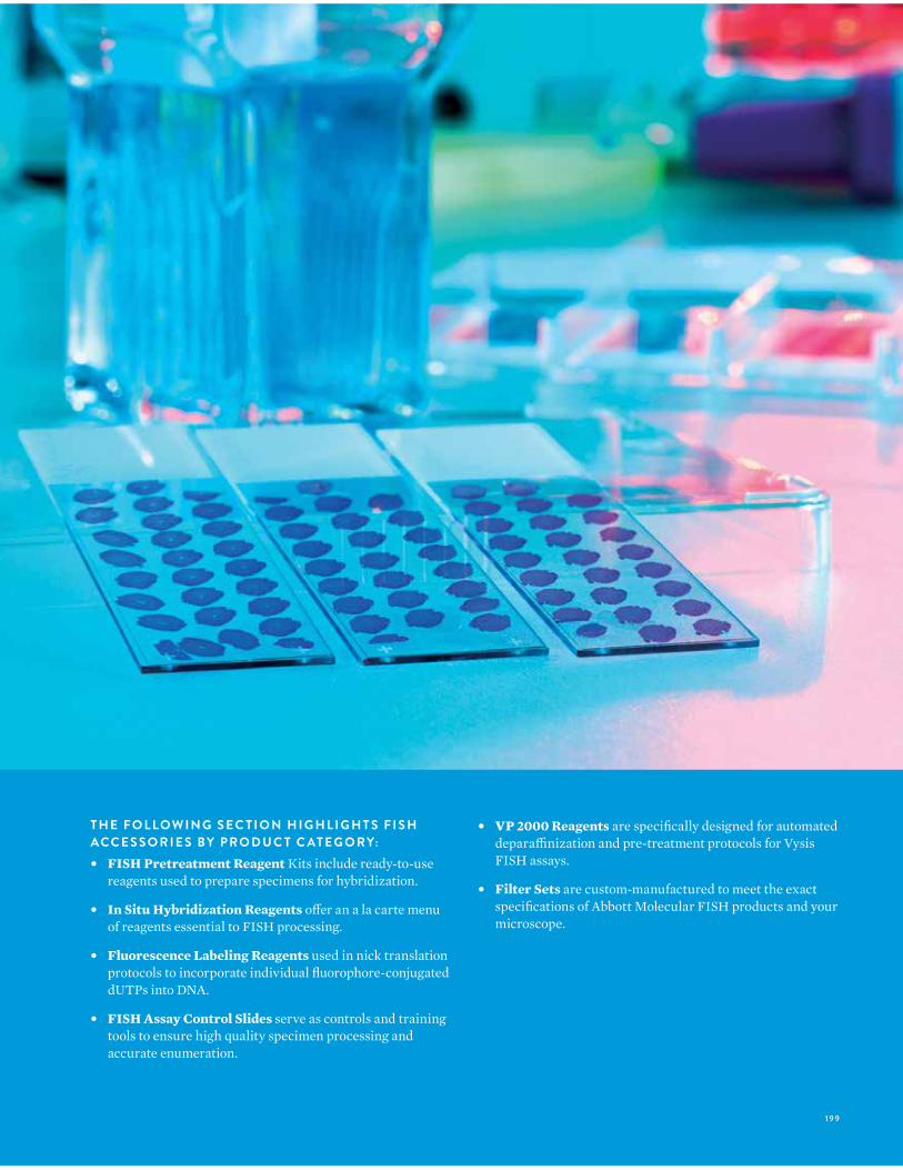
199199
T H E F O L LO W I N G S E C T I O N H I G H L I G H T S F I S H ACC E S S O R I E S B Y P R O D U C T C AT E G O R Y:• FISH Pretreatment Reagent Kits include ready-to-use
reagents used to prepare specimens for hybridization.
• In Situ Hybridization Reagents offer an a la carte menu of reagents essential to FISH processing.
• Fluorescence Labeling Reagents used in nick translation protocols to incorporate individual fluorophore-conjugated dUTPs into DNA.
• FISH Assay Control Slides serve as controls and training tools to ensure high quality specimen processing and accurate enumeration.
• VP 2000 Reagents are specifically designed for automated deparaffinization and pre-treatment protocols for Vysis FISH assays.
• Filter Sets are custom-manufactured to meet the exact specifications of Abbott Molecular FISH products and your microscope.
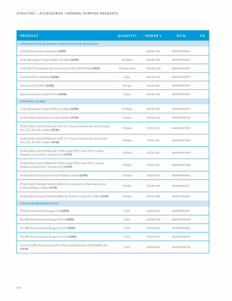
200
V Y S I S F I S H — A C C E S S O R I E S / G E N E R A L P U R P O S E R E AG E N T S
P R O D U C T Q UA N T I T Y O R D E R # G T I N P G
C O M PA R AT I V E G E N O M I C H Y B R I D I Z AT I O N R E A G E N T S
CGH Hybridization Reagents (GPR) 06J40-010 884999020184
CGH Metaphase Target Slides; 10 slides (GPR) 10 Slides 06J96-001 884999023512 -
CGH Nick Translation Kit with control DNA (MPE 600) (GPR) 50 Reactions 06J40-020 884999020207 -
Control DNA Unlabeled (GPR) 15 µL 06J40-001 884999020177 -
Human COT-1 DNA (GPR) 250 µL 06J31-001 884999019379 -
SpectrumGreen Control DNA (GPR) 25 µL 06J45-001 884999021587 -
C O N T R O L S L I D E S
CGH Metaphase Target Slides; 10 slides (GPR) 10 Slides 06J96-001 884999023512 -
ProbeChek ALK Positive Control Slides (IVD) 5 slides 06N38-010 884999025738 -
ProbeChek Control Slides for CEP X/Y Assay; Control low-level female: 95% XY, 5% XX; 5 slides (IVD) 5 Slides 07J21-011 884999027053 -
ProbeChek Control Slides for CEP X/Y Assay; Control low-level male: 95% XX, 5% XY; 5 slides (IVD) 5 Slides 07J21-012 884999027060 -
ProbeChek Control Slides for FISH using CEP 8 and CEP 12 Assay, Negative Control 0%, trisomy 8/12 (IVD) 5 Slides 07J21-001 884999027039 -
ProbeChek Control Slides for FISH using CEP 8 and CEP 12 Assay, Positive Control 10%, trisomy 8/12 (IVD) 5 Slides 07J21-002 884999027046 -
ProbeChek MultiVysion Control Slides 5 slides (GPR) 5 Slides 05J07-001 884999010864 -
ProbeChek Prenatal Control Slides for Amniocyte; Male Amniocyte Control Slides; 5 slides (IVD) 5 Slides 05J39-005 884999011731 -
ProbeChek Prenatal Control Slides for Positive Control; 5 slides (IVD) 5 Slides 05J36-005 884999011663 -
F I S H P R E T R E ATM E N T K I T S
FISH Pretreatment Reagent Kit (GPR) 1 Kit 02J03-032 884999001817 -
Paraffin Pretreatment Reagent Kit I (GPR) 1 Kit 02J02-032 884999001800 -
Paraffin Pretreatment Reagent Kit II (GPR) 1 Kit 07J02-002 884999026186 -
Paraffin Pretreatment Reagent Kit III (GPR) 1 Kit 07J02-003 884999026193 -
Vysis Paraffin Pretreatment IV & Post-Hybridization Wash Buffer Kit (IVD) 1 Kit 01N31-005 884999000735 -

201
P R O D U C T Q UA N T I T Y O R D E R # G T I N P G
G E N E R A L P U R P O S E R E A G E N T S / V P 20 0 0 R E A G E N T S
20X SSC 500 g 02J10-032 884999001909
2M MgCL 2 120 mL 02J09-030 884999001763
VP 2000 Reagents Pretreatment Reagent 500 mL 02J06-030 884999001862 -
VP 2000 Reagents Protease Buffer 500 mL 02J07-030 884999001879 -
VP 2000 Reagents Protease I 250 mg x 2 02J08-032 884999001886 -
VP 2000 Reagents Protease II 750 mg 06J93-001 884999023468 -
H Y B R I D I Z AT I O N R E A G E N T S
20X SSC 500 g 02J10-032 884999001909
2M MgCL 2 120 mL 02J09-030 884999001763
Antifade II Solution (GPR) 60 µL x 2 06J29-001 884999019324 -
Antifade Solution (GPR) 240 µL x 2 06J29-010 884999019331 -
DAPI I Counterstain (GPR) 500 µL x 2 06J49-001 884999021624 -
DAPI II Counterstain (GPR) 500 µL x 2 06J50-001 884999021648 -
DAPI III Counterstain (GPR) 500 µL x 2 06J49-010 884999021631 -
Hybridization Buffer (GPR) 2 tubes, 6900 µL 06L44-01 884999024250 -
LSI/WCP Hybridization Buffer (GPR) 2 x 150 µL 06J67-001 884999021983 -
LSI/WCP Hybridization Buffer (GPR) 2 x 500 µL 06J67-011 884999021990 -
NP-40 (GPR) 1000 µL x 2 07J05-001 884999026247 -
Propidium Iodide Counterstain (GPR) 500 µL x 2 07J06-001 884999026254 -
Vysis CEP Hybridization Buffer (GPR) 2 x 150 µL 07J36-001 884999027565 -
L A B E L I N G R E F E R E N C E D N A
Aqua dUTP (RUO) 50nmol, lyophilized 02N35-050 884999002944 -
Gold dUTP (RUO) 50nmol, lyophilized 05N18-050 884999014640 -
Green dUTP (RUO) 50nmol, lyophilized 02N32-050 884999002913 -
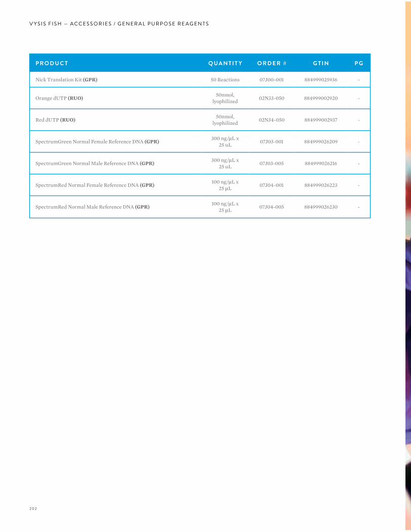
202
V Y S I S F I S H — A C C E S S O R I E S / G E N E R A L P U R P O S E R E AG E N T S
P R O D U C T Q UA N T I T Y O R D E R # G T I N P G
Nick Translation Kit (GPR) 50 Reactions 07J00-001 884999025936 -
Orange dUTP (RUO) 50nmol, lyophilized 02N33-050 884999002920 -
Red dUTP (RUO) 50nmol, lyophilized 02N34-050 884999002937 -
SpectrumGreen Normal Female Reference DNA (GPR) 300 ng/µL x 25 uL 07J03-001 884999026209 -
SpectrumGreen Normal Male Reference DNA (GPR) 300 ng/µL x 25 uL 07J03-005 884999026216 -
SpectrumRed Normal Female Reference DNA (GPR) 100 ng/µL x 25 µL 07J04-001 884999026223 -
SpectrumRed Normal Male Reference DNA (GPR) 100 ng/µL x 25 µL 07J04-005 884999026230 -

203
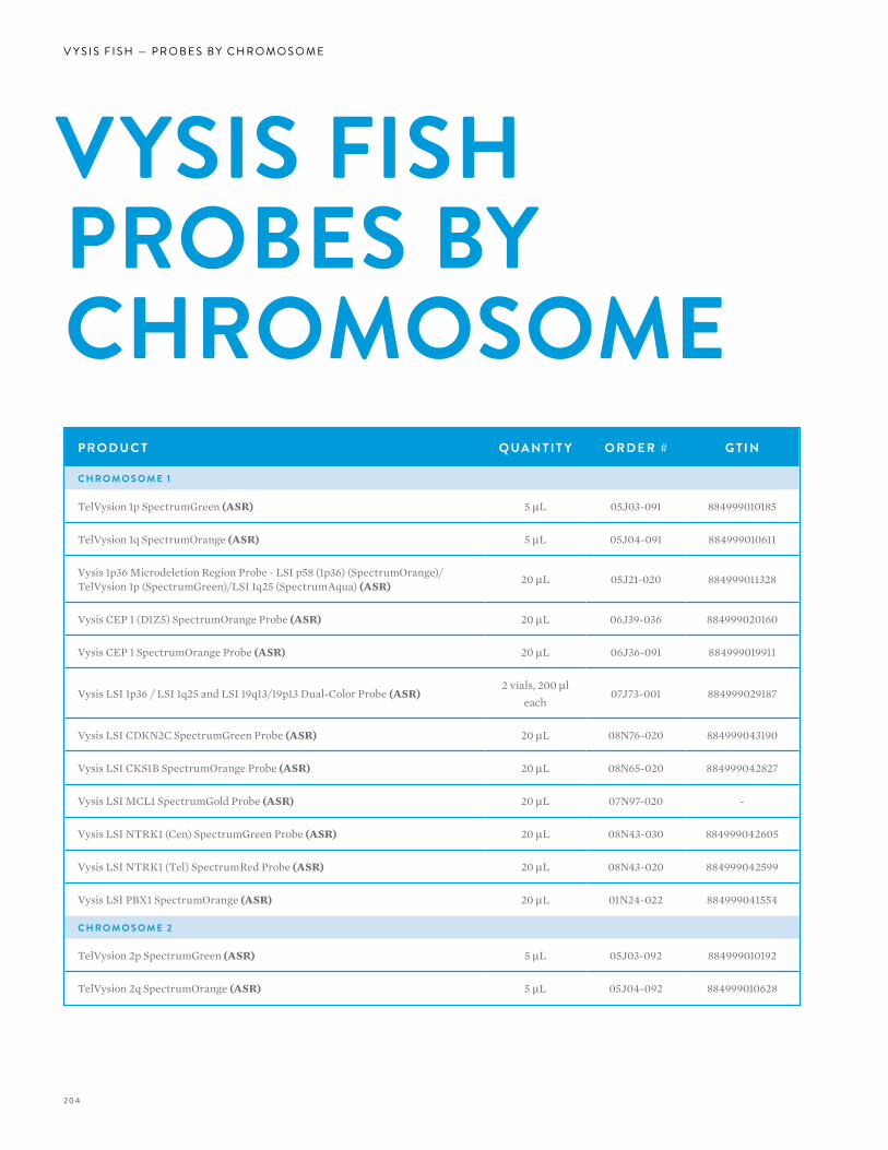
204
V Y S I S F I S H — P R O B E S B Y C H R O M O S O M E
P R O D U C T Q UA N T I T Y O R D E R # G T I N
C H R O M O S O M E 1
TelVysion 1p SpectrumGreen (ASR) 5 µL 05J03-091 884999010185
TelVysion 1q SpectrumOrange (ASR) 5 µL 05J04-091 884999010611
Vysis 1p36 Microdeletion Region Probe - LSI p58 (1p36) (SpectrumOrange)/TelVysion 1p (SpectrumGreen)/LSI 1q25 (SpectrumAqua) (ASR) 20 µL 05J21-020 884999011328
Vysis CEP 1 (D1Z5) SpectrumOrange Probe (ASR) 20 µL 06J39-036 884999020160
Vysis CEP 1 SpectrumOrange Probe (ASR) 20 µL 06J36-091 884999019911
Vysis LSI 1p36 / LSI 1q25 and LSI 19q13/19p13 Dual-Color Probe (ASR)2 vials, 200 µl
each07J73-001 884999029187
Vysis LSI CDKN2C SpectrumGreen Probe (ASR) 20 µL 08N76-020 884999043190
Vysis LSI CKS1B SpectrumOrange Probe (ASR) 20 µL 08N65-020 884999042827
Vysis LSI MCL1 SpectrumGold Probe (ASR) 20 µL 07N97-020 -
Vysis LSI NTRK1 (Cen) SpectrumGreen Probe (ASR) 20 µL 08N43-030 884999042605
Vysis LSI NTRK1 (Tel) SpectrumRed Probe (ASR) 20 µL 08N43-020 884999042599
Vysis LSI PBX1 SpectrumOrange (ASR) 20 µL 01N24-022 884999041554
C H R O M O S O M E 2
TelVysion 2p SpectrumGreen (ASR) 5 µL 05J03-092 884999010192
TelVysion 2q SpectrumOrange (ASR) 5 µL 05J04-092 884999010628
VYSIS FISH PROBES BY CHROMOSOME
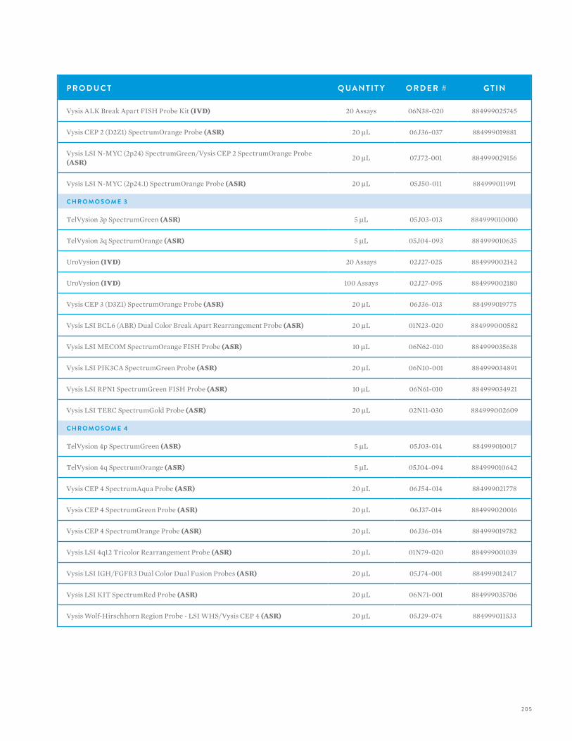
205
P R O D U C T Q UA N T I T Y O R D E R # G T I N
Vysis ALK Break Apart FISH Probe Kit (IVD) 20 Assays 06N38-020 884999025745
Vysis CEP 2 (D2Z1) SpectrumOrange Probe (ASR) 20 µL 06J36-037 884999019881
Vysis LSI N-MYC (2p24) SpectrumGreen/Vysis CEP 2 SpectrumOrange Probe (ASR) 20 µL 07J72-001 884999029156
Vysis LSI N-MYC (2p24.1) SpectrumOrange Probe (ASR) 20 µL 05J50-011 884999011991
C H R O M O S O M E 3
TelVysion 3p SpectrumGreen (ASR) 5 µL 05J03-013 884999010000
TelVysion 3q SpectrumOrange (ASR) 5 µL 05J04-093 884999010635
UroVysion (IVD) 20 Assays 02J27-025 884999002142
UroVysion (IVD) 100 Assays 02J27-095 884999002180
Vysis CEP 3 (D3Z1) SpectrumOrange Probe (ASR) 20 µL 06J36-013 884999019775
Vysis LSI BCL6 (ABR) Dual Color Break Apart Rearrangement Probe (ASR) 20 µL 01N23-020 884999000582
Vysis LSI MECOM SpectrumOrange FISH Probe (ASR) 10 µL 06N62-010 884999035638
Vysis LSI PIK3CA SpectrumGreen Probe (ASR) 20 µL 06N10-001 884999034891
Vysis LSI RPN1 SpectrumGreen FISH Probe (ASR) 10 µL 06N61-010 884999034921
Vysis LSI TERC SpectrumGold Probe (ASR) 20 µL 02N11-030 884999002609
C H R O M O S O M E 4
TelVysion 4p SpectrumGreen (ASR) 5 µL 05J03-014 884999010017
TelVysion 4q SpectrumOrange (ASR) 5 µL 05J04-094 884999010642
Vysis CEP 4 SpectrumAqua Probe (ASR) 20 µL 06J54-014 884999021778
Vysis CEP 4 SpectrumGreen Probe (ASR) 20 µL 06J37-014 884999020016
Vysis CEP 4 SpectrumOrange Probe (ASR) 20 µL 06J36-014 884999019782
Vysis LSI 4q12 Tricolor Rearrangement Probe (ASR) 20 µL 01N79-020 884999001039
Vysis LSI IGH/FGFR3 Dual Color Dual Fusion Probes (ASR) 20 µL 05J74-001 884999012417
Vysis LSI KIT SpectrumRed Probe (ASR) 20 µL 06N71-001 884999035706
Vysis Wolf-Hirschhorn Region Probe - LSI WHS/Vysis CEP 4 (ASR) 20 µL 05J29-074 884999011533
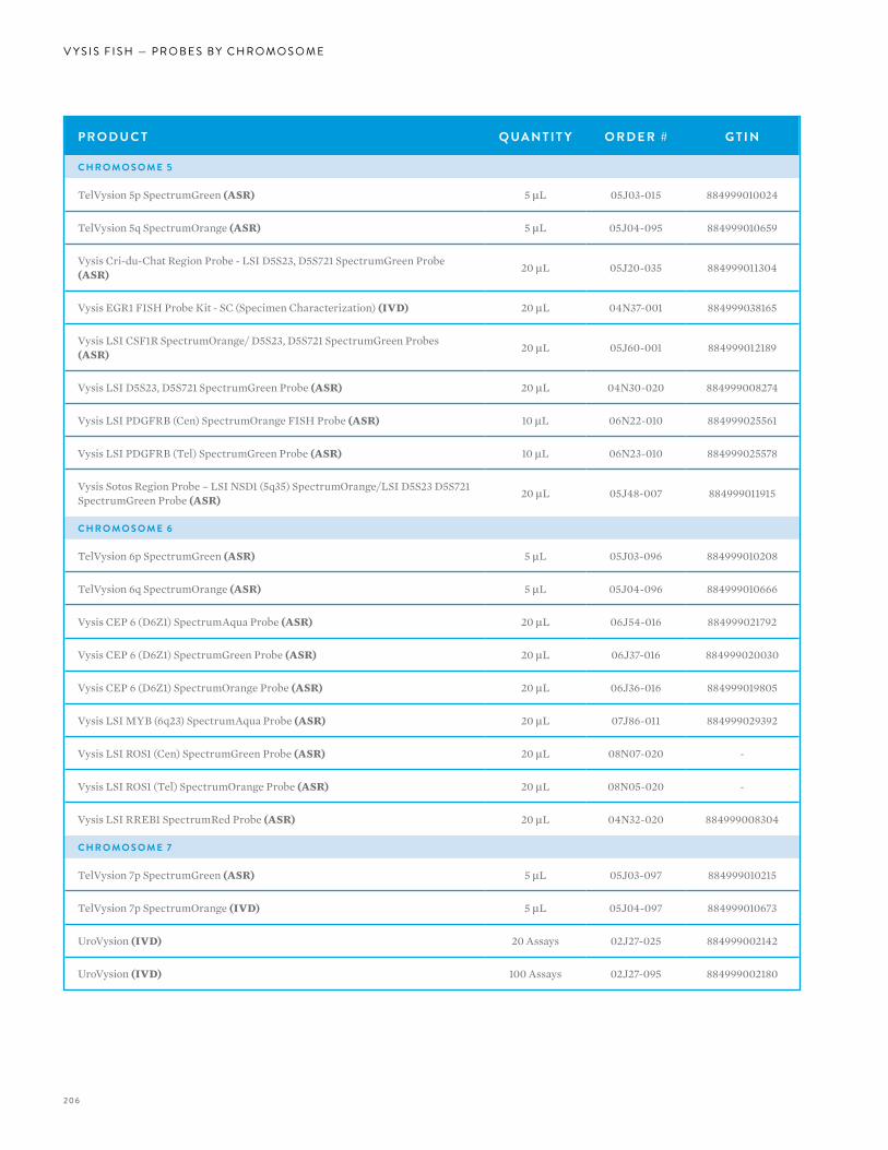
206
V Y S I S F I S H — P R O B E S B Y C H R O M O S O M E
P R O D U C T Q UA N T I T Y O R D E R # G T I N
C H R O M O S O M E 5
TelVysion 5p SpectrumGreen (ASR) 5 µL 05J03-015 884999010024
TelVysion 5q SpectrumOrange (ASR) 5 µL 05J04-095 884999010659
Vysis Cri-du-Chat Region Probe - LSI D5S23, D5S721 SpectrumGreen Probe (ASR) 20 µL 05J20-035 884999011304
Vysis EGR1 FISH Probe Kit - SC (Specimen Characterization) (IVD) 20 µL 04N37-001 884999038165
Vysis LSI CSF1R SpectrumOrange/ D5S23, D5S721 SpectrumGreen Probes (ASR) 20 µL 05J60-001 884999012189
Vysis LSI D5S23, D5S721 SpectrumGreen Probe (ASR) 20 µL 04N30-020 884999008274
Vysis LSI PDGFRB (Cen) SpectrumOrange FISH Probe (ASR) 10 µL 06N22-010 884999025561
Vysis LSI PDGFRB (Tel) SpectrumGreen Probe (ASR) 10 µL 06N23-010 884999025578
Vysis Sotos Region Probe – LSI NSD1 (5q35) SpectrumOrange/LSI D5S23 D5S721 SpectrumGreen Probe (ASR) 20 µL 05J48-007 884999011915
C H R O M O S O M E 6
TelVysion 6p SpectrumGreen (ASR) 5 µL 05J03-096 884999010208
TelVysion 6q SpectrumOrange (ASR) 5 µL 05J04-096 884999010666
Vysis CEP 6 (D6Z1) SpectrumAqua Probe (ASR) 20 µL 06J54-016 884999021792
Vysis CEP 6 (D6Z1) SpectrumGreen Probe (ASR) 20 µL 06J37-016 884999020030
Vysis CEP 6 (D6Z1) SpectrumOrange Probe (ASR) 20 µL 06J36-016 884999019805
Vysis LSI MYB (6q23) SpectrumAqua Probe (ASR) 20 µL 07J86-011 884999029392
Vysis LSI ROS1 (Cen) SpectrumGreen Probe (ASR) 20 µL 08N07-020 -
Vysis LSI ROS1 (Tel) SpectrumOrange Probe (ASR) 20 µL 08N05-020 -
Vysis LSI RREB1 SpectrumRed Probe (ASR) 20 µL 04N32-020 884999008304
C H R O M O S O M E 7
TelVysion 7p SpectrumGreen (ASR) 5 µL 05J03-097 884999010215
TelVysion 7p SpectrumOrange (IVD) 5 µL 05J04-097 884999010673
UroVysion (IVD) 20 Assays 02J27-025 884999002142
UroVysion (IVD) 100 Assays 02J27-095 884999002180
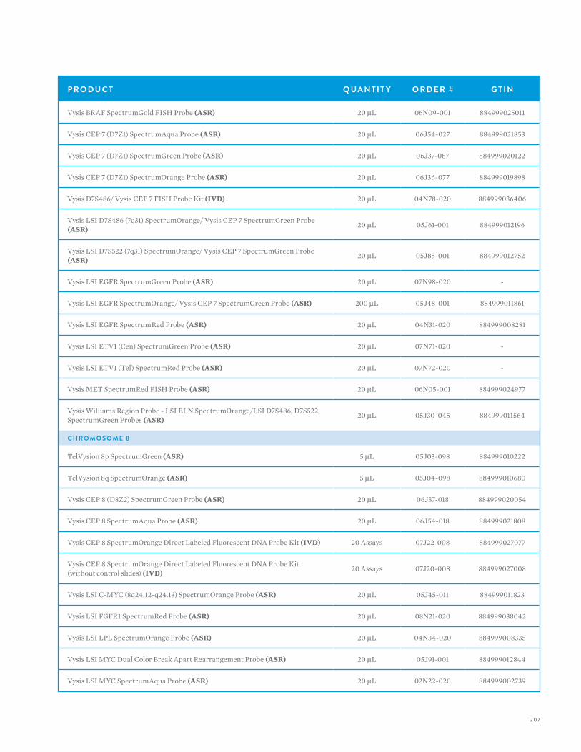
207
P R O D U C T Q UA N T I T Y O R D E R # G T I N
Vysis BRAF SpectrumGold FISH Probe (ASR) 20 µL 06N09-001 884999025011
Vysis CEP 7 (D7Z1) SpectrumAqua Probe (ASR) 20 µL 06J54-027 884999021853
Vysis CEP 7 (D7Z1) SpectrumGreen Probe (ASR) 20 µL 06J37-087 884999020122
Vysis CEP 7 (D7Z1) SpectrumOrange Probe (ASR) 20 µL 06J36-077 884999019898
Vysis D7S486/ Vysis CEP 7 FISH Probe Kit (IVD) 20 µL 04N78-020 884999036406
Vysis LSI D7S486 (7q31) SpectrumOrange/ Vysis CEP 7 SpectrumGreen Probe (ASR) 20 µL 05J61-001 884999012196
Vysis LSI D7S522 (7q31) SpectrumOrange/ Vysis CEP 7 SpectrumGreen Probe (ASR) 20 µL 05J85-001 884999012752
Vysis LSI EGFR SpectrumGreen Probe (ASR) 20 µL 07N98-020 -
Vysis LSI EGFR SpectrumOrange/ Vysis CEP 7 SpectrumGreen Probe (ASR) 200 µL 05J48-001 884999011861
Vysis LSI EGFR SpectrumRed Probe (ASR) 20 µL 04N31-020 884999008281
Vysis LSI ETV1 (Cen) SpectrumGreen Probe (ASR) 20 µL 07N71-020 -
Vysis LSI ETV1 (Tel) SpectrumRed Probe (ASR) 20 µL 07N72-020 -
Vysis MET SpectrumRed FISH Probe (ASR) 20 µL 06N05-001 884999024977
Vysis Williams Region Probe - LSI ELN SpectrumOrange/LSI D7S486, D7S522 SpectrumGreen Probes (ASR) 20 µL 05J30-045 884999011564
C H R O M O S O M E 8
TelVysion 8p SpectrumGreen (ASR) 5 µL 05J03-098 884999010222
TelVysion 8q SpectrumOrange (ASR) 5 µL 05J04-098 884999010680
Vysis CEP 8 (D8Z2) SpectrumGreen Probe (ASR) 20 µL 06J37-018 884999020054
Vysis CEP 8 SpectrumAqua Probe (ASR) 20 µL 06J54-018 884999021808
Vysis CEP 8 SpectrumOrange Direct Labeled Fluorescent DNA Probe Kit (IVD) 20 Assays 07J22-008 884999027077
Vysis CEP 8 SpectrumOrange Direct Labeled Fluorescent DNA Probe Kit (without control slides) (IVD) 20 Assays 07J20-008 884999027008
Vysis LSI C-MYC (8q24.12-q24.13) SpectrumOrange Probe (ASR) 20 µL 05J45-011 884999011823
Vysis LSI FGFR1 SpectrumRed Probe (ASR) 20 µL 08N21-020 884999038042
Vysis LSI LPL SpectrumOrange Probe (ASR) 20 µL 04N34-020 884999008335
Vysis LSI MYC Dual Color Break Apart Rearrangement Probe (ASR) 20 µL 05J91-001 884999012844
Vysis LSI MYC SpectrumAqua Probe (ASR) 20 µL 02N22-020 884999002739
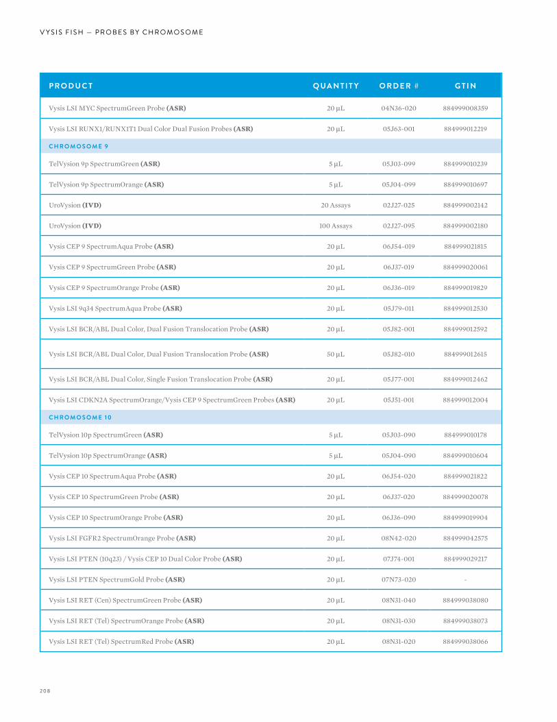
208
V Y S I S F I S H — P R O B E S B Y C H R O M O S O M E
P R O D U C T Q UA N T I T Y O R D E R # G T I N
Vysis LSI MYC SpectrumGreen Probe (ASR) 20 µL 04N36-020 884999008359
Vysis LSI RUNX1/RUNX1T1 Dual Color Dual Fusion Probes (ASR) 20 µL 05J63-001 884999012219
C H R O M O S O M E 9
TelVysion 9p SpectrumGreen (ASR) 5 µL 05J03-099 884999010239
TelVysion 9p SpectrumOrange (ASR) 5 µL 05J04-099 884999010697
UroVysion (IVD) 20 Assays 02J27-025 884999002142
UroVysion (IVD) 100 Assays 02J27-095 884999002180
Vysis CEP 9 SpectrumAqua Probe (ASR) 20 µL 06J54-019 884999021815
Vysis CEP 9 SpectrumGreen Probe (ASR) 20 µL 06J37-019 884999020061
Vysis CEP 9 SpectrumOrange Probe (ASR) 20 µL 06J36-019 884999019829
Vysis LSI 9q34 SpectrumAqua Probe (ASR) 20 µL 05J79-011 884999012530
Vysis LSI BCR/ABL Dual Color, Dual Fusion Translocation Probe (ASR) 20 µL 05J82-001 884999012592
Vysis LSI BCR/ABL Dual Color, Dual Fusion Translocation Probe (ASR) 50 µL 05J82-010 884999012615
Vysis LSI BCR/ABL Dual Color, Single Fusion Translocation Probe (ASR) 20 µL 05J77-001 884999012462
Vysis LSI CDKN2A SpectrumOrange/Vysis CEP 9 SpectrumGreen Probes (ASR) 20 µL 05J51-001 884999012004
C H R O M O S O M E 1 0
TelVysion 10p SpectrumGreen (ASR) 5 µL 05J03-090 884999010178
TelVysion 10p SpectrumOrange (ASR) 5 µL 05J04-090 884999010604
Vysis CEP 10 SpectrumAqua Probe (ASR) 20 µL 06J54-020 884999021822
Vysis CEP 10 SpectrumGreen Probe (ASR) 20 µL 06J37-020 884999020078
Vysis CEP 10 SpectrumOrange Probe (ASR) 20 µL 06J36-090 884999019904
Vysis LSI FGFR2 SpectrumOrange Probe (ASR) 20 µL 08N42-020 884999042575
Vysis LSI PTEN (10q23) / Vysis CEP 10 Dual Color Probe (ASR) 20 µL 07J74-001 884999029217
Vysis LSI PTEN SpectrumGold Probe (ASR) 20 µL 07N73-020 -
Vysis LSI RET (Cen) SpectrumGreen Probe (ASR) 20 µL 08N31-040 884999038080
Vysis LSI RET (Tel) SpectrumOrange Probe (ASR) 20 µL 08N31-030 884999038073
Vysis LSI RET (Tel) SpectrumRed Probe (ASR) 20 µL 08N31-020 884999038066
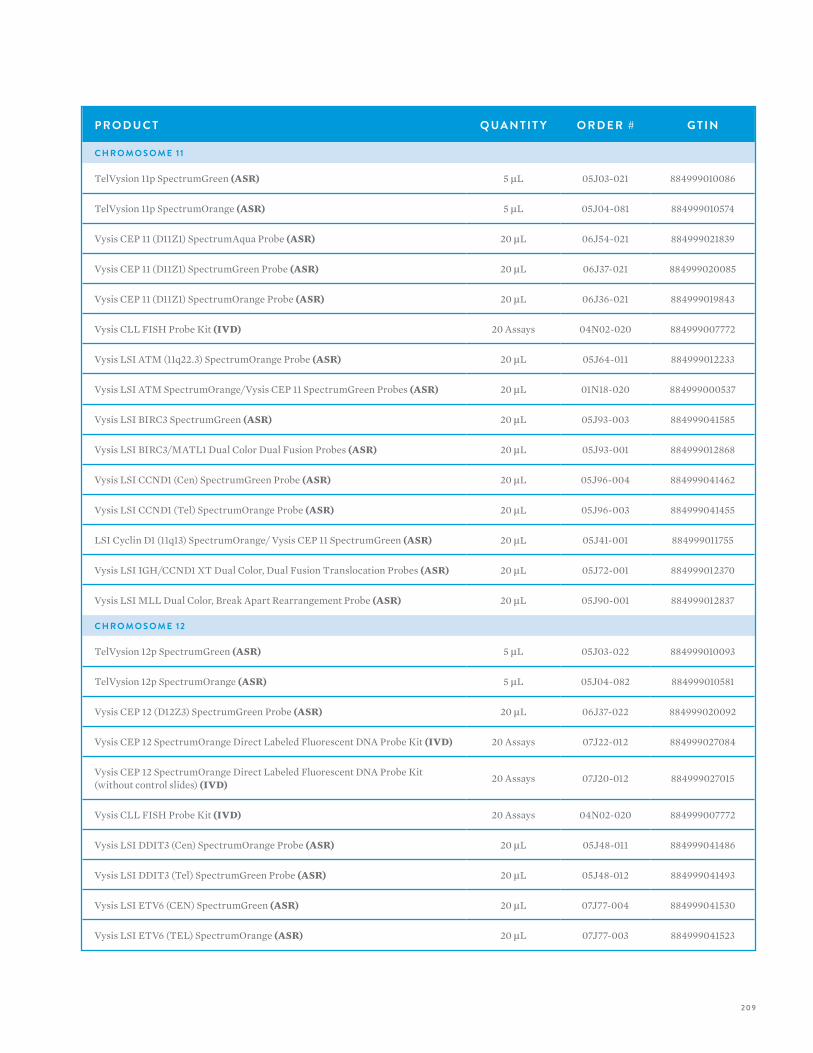
209
P R O D U C T Q UA N T I T Y O R D E R # G T I N
C H R O M O S O M E 11
TelVysion 11p SpectrumGreen (ASR) 5 µL 05J03-021 884999010086
TelVysion 11p SpectrumOrange (ASR) 5 µL 05J04-081 884999010574
Vysis CEP 11 (D11Z1) SpectrumAqua Probe (ASR) 20 µL 06J54-021 884999021839
Vysis CEP 11 (D11Z1) SpectrumGreen Probe (ASR) 20 µL 06J37-021 884999020085
Vysis CEP 11 (D11Z1) SpectrumOrange Probe (ASR) 20 µL 06J36-021 884999019843
Vysis CLL FISH Probe Kit (IVD) 20 Assays 04N02-020 884999007772
Vysis LSI ATM (11q22.3) SpectrumOrange Probe (ASR) 20 µL 05J64-011 884999012233
Vysis LSI ATM SpectrumOrange/Vysis CEP 11 SpectrumGreen Probes (ASR) 20 µL 01N18-020 884999000537
Vysis LSI BIRC3 SpectrumGreen (ASR) 20 µL 05J93-003 884999041585
Vysis LSI BIRC3/MATL1 Dual Color Dual Fusion Probes (ASR) 20 µL 05J93-001 884999012868
Vysis LSI CCND1 (Cen) SpectrumGreen Probe (ASR) 20 µL 05J96-004 884999041462
Vysis LSI CCND1 (Tel) SpectrumOrange Probe (ASR) 20 µL 05J96-003 884999041455
LSI Cyclin D1 (11q13) SpectrumOrange/ Vysis CEP 11 SpectrumGreen (ASR) 20 µL 05J41-001 884999011755
Vysis LSI IGH/CCND1 XT Dual Color, Dual Fusion Translocation Probes (ASR) 20 µL 05J72-001 884999012370
Vysis LSI MLL Dual Color, Break Apart Rearrangement Probe (ASR) 20 µL 05J90-001 884999012837
C H R O M O S O M E 12
TelVysion 12p SpectrumGreen (ASR) 5 µL 05J03-022 884999010093
TelVysion 12p SpectrumOrange (ASR) 5 µL 05J04-082 884999010581
Vysis CEP 12 (D12Z3) SpectrumGreen Probe (ASR) 20 µL 06J37-022 884999020092
Vysis CEP 12 SpectrumOrange Direct Labeled Fluorescent DNA Probe Kit (IVD) 20 Assays 07J22-012 884999027084
Vysis CEP 12 SpectrumOrange Direct Labeled Fluorescent DNA Probe Kit (without control slides) (IVD) 20 Assays 07J20-012 884999027015
Vysis CLL FISH Probe Kit (IVD) 20 Assays 04N02-020 884999007772
Vysis LSI DDIT3 (Cen) SpectrumOrange Probe (ASR) 20 µL 05J48-011 884999041486
Vysis LSI DDIT3 (Tel) SpectrumGreen Probe (ASR) 20 µL 05J48-012 884999041493
Vysis LSI ETV6 (CEN) SpectrumGreen (ASR) 20 µL 07J77-004 884999041530
Vysis LSI ETV6 (TEL) SpectrumOrange (ASR) 20 µL 07J77-003 884999041523
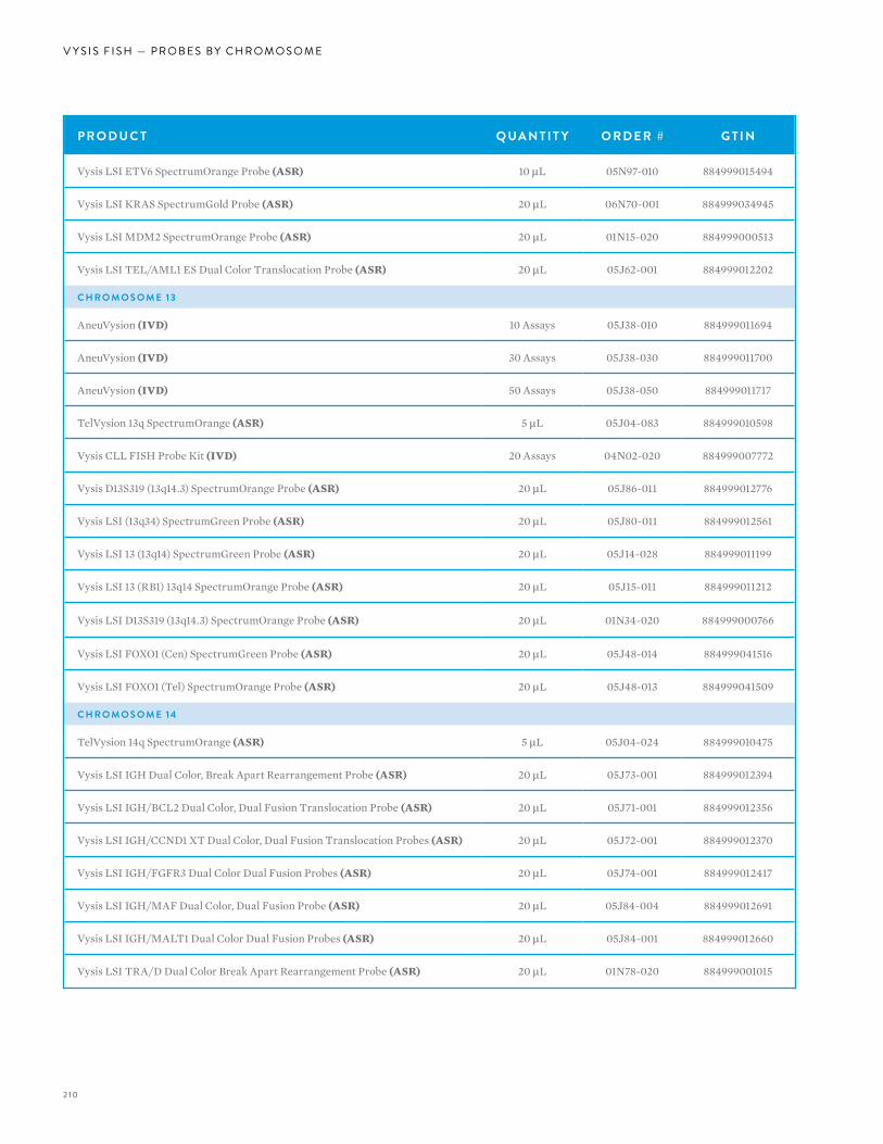
2 10
V Y S I S F I S H — P R O B E S B Y C H R O M O S O M E
P R O D U C T Q UA N T I T Y O R D E R # G T I N
Vysis LSI ETV6 SpectrumOrange Probe (ASR) 10 µL 05N97-010 884999015494
Vysis LSI KRAS SpectrumGold Probe (ASR) 20 µL 06N70-001 884999034945
Vysis LSI MDM2 SpectrumOrange Probe (ASR) 20 µL 01N15-020 884999000513
Vysis LSI TEL/AML1 ES Dual Color Translocation Probe (ASR) 20 µL 05J62-001 884999012202
C H R O M O S O M E 13
AneuVysion (IVD) 10 Assays 05J38-010 884999011694
AneuVysion (IVD) 30 Assays 05J38-030 884999011700
AneuVysion (IVD) 50 Assays 05J38-050 884999011717
TelVysion 13q SpectrumOrange (ASR) 5 µL 05J04-083 884999010598
Vysis CLL FISH Probe Kit (IVD) 20 Assays 04N02-020 884999007772
Vysis D13S319 (13q14.3) SpectrumOrange Probe (ASR) 20 µL 05J86-011 884999012776
Vysis LSI (13q34) SpectrumGreen Probe (ASR) 20 µL 05J80-011 884999012561
Vysis LSI 13 (13q14) SpectrumGreen Probe (ASR) 20 µL 05J14-028 884999011199
Vysis LSI 13 (RB1) 13q14 SpectrumOrange Probe (ASR) 20 µL 05J15-011 884999011212
Vysis LSI D13S319 (13q14.3) SpectrumOrange Probe (ASR) 20 µL 01N34-020 884999000766
Vysis LSI FOXO1 (Cen) SpectrumGreen Probe (ASR) 20 µL 05J48-014 884999041516
Vysis LSI FOXO1 (Tel) SpectrumOrange Probe (ASR) 20 µL 05J48-013 884999041509
C H R O M O S O M E 14
TelVysion 14q SpectrumOrange (ASR) 5 µL 05J04-024 884999010475
Vysis LSI IGH Dual Color, Break Apart Rearrangement Probe (ASR) 20 µL 05J73-001 884999012394
Vysis LSI IGH/BCL2 Dual Color, Dual Fusion Translocation Probe (ASR) 20 µL 05J71-001 884999012356
Vysis LSI IGH/CCND1 XT Dual Color, Dual Fusion Translocation Probes (ASR) 20 µL 05J72-001 884999012370
Vysis LSI IGH/FGFR3 Dual Color Dual Fusion Probes (ASR) 20 µL 05J74-001 884999012417
Vysis LSI IGH/MAF Dual Color, Dual Fusion Probe (ASR) 20 µL 05J84-004 884999012691
Vysis LSI IGH/MALT1 Dual Color Dual Fusion Probes (ASR) 20 µL 05J84-001 884999012660
Vysis LSI TRA/D Dual Color Break Apart Rearrangement Probe (ASR) 20 µL 01N78-020 884999001015
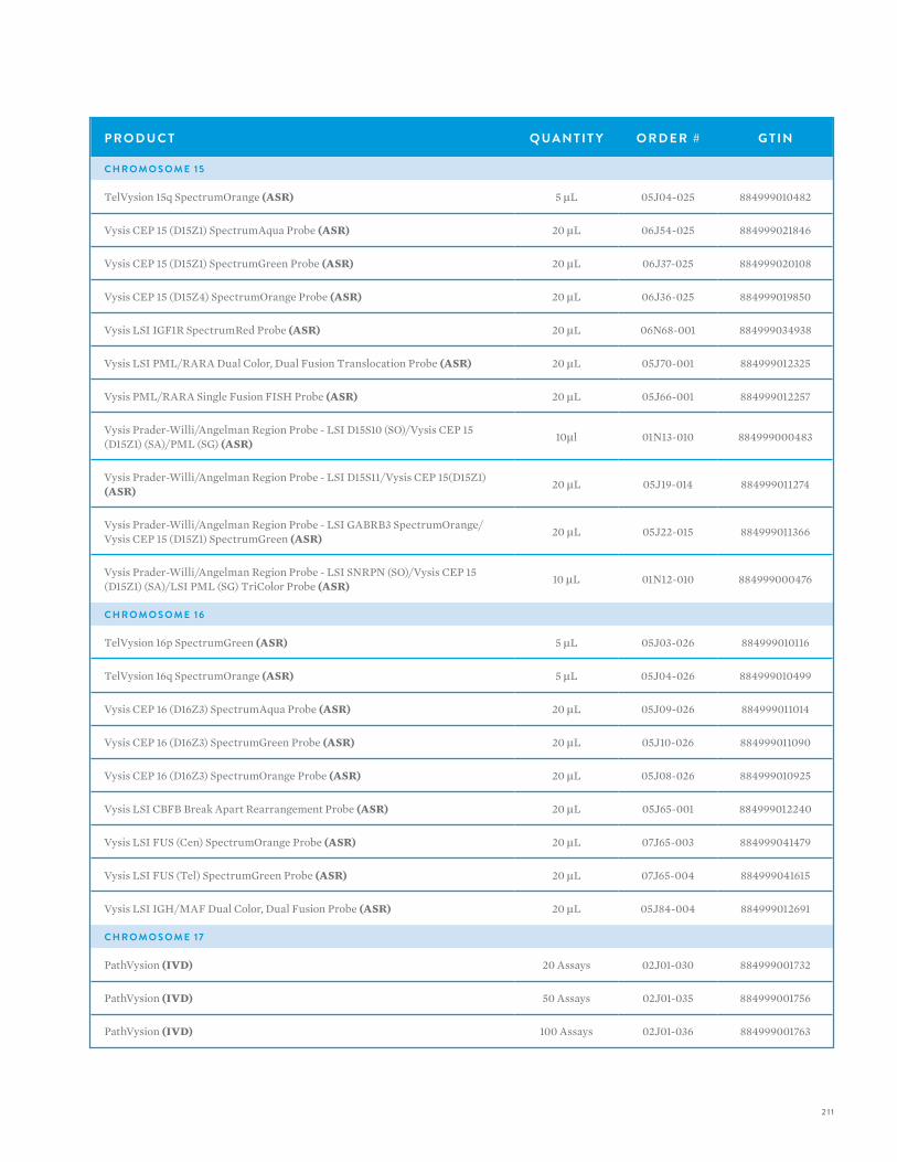
2 11
P R O D U C T Q UA N T I T Y O R D E R # G T I N
C H R O M O S O M E 1 5
TelVysion 15q SpectrumOrange (ASR) 5 µL 05J04-025 884999010482
Vysis CEP 15 (D15Z1) SpectrumAqua Probe (ASR) 20 µL 06J54-025 884999021846
Vysis CEP 15 (D15Z1) SpectrumGreen Probe (ASR) 20 µL 06J37-025 884999020108
Vysis CEP 15 (D15Z4) SpectrumOrange Probe (ASR) 20 µL 06J36-025 884999019850
Vysis LSI IGF1R SpectrumRed Probe (ASR) 20 µL 06N68-001 884999034938
Vysis LSI PML/RARA Dual Color, Dual Fusion Translocation Probe (ASR) 20 µL 05J70-001 884999012325
Vysis PML/RARA Single Fusion FISH Probe (ASR) 20 µL 05J66-001 884999012257
Vysis Prader-Willi/Angelman Region Probe - LSI D15S10 (SO)/Vysis CEP 15 (D15Z1) (SA)/PML (SG) (ASR) 10µl 01N13-010 884999000483
Vysis Prader-Willi/Angelman Region Probe - LSI D15S11/Vysis CEP 15(D15Z1) (ASR) 20 µL 05J19-014 884999011274
Vysis Prader-Willi/Angelman Region Probe - LSI GABRB3 SpectrumOrange/Vysis CEP 15 (D15Z1) SpectrumGreen (ASR) 20 µL 05J22-015 884999011366
Vysis Prader-Willi/Angelman Region Probe - LSI SNRPN (SO)/Vysis CEP 15 (D15Z1) (SA)/LSI PML (SG) TriColor Probe (ASR) 10 µL 01N12-010 884999000476
C H R O M O S O M E 1 6
TelVysion 16p SpectrumGreen (ASR) 5 µL 05J03-026 884999010116
TelVysion 16q SpectrumOrange (ASR) 5 µL 05J04-026 884999010499
Vysis CEP 16 (D16Z3) SpectrumAqua Probe (ASR) 20 µL 05J09-026 884999011014
Vysis CEP 16 (D16Z3) SpectrumGreen Probe (ASR) 20 µL 05J10-026 884999011090
Vysis CEP 16 (D16Z3) SpectrumOrange Probe (ASR) 20 µL 05J08-026 884999010925
Vysis LSI CBFB Break Apart Rearrangement Probe (ASR) 20 µL 05J65-001 884999012240
Vysis LSI FUS (Cen) SpectrumOrange Probe (ASR) 20 µL 07J65-003 884999041479
Vysis LSI FUS (Tel) SpectrumGreen Probe (ASR) 20 µL 07J65-004 884999041615
Vysis LSI IGH/MAF Dual Color, Dual Fusion Probe (ASR) 20 µL 05J84-004 884999012691
C H R O M O S O M E 17
PathVysion (IVD) 20 Assays 02J01-030 884999001732
PathVysion (IVD) 50 Assays 02J01-035 884999001756
PathVysion (IVD) 100 Assays 02J01-036 884999001763
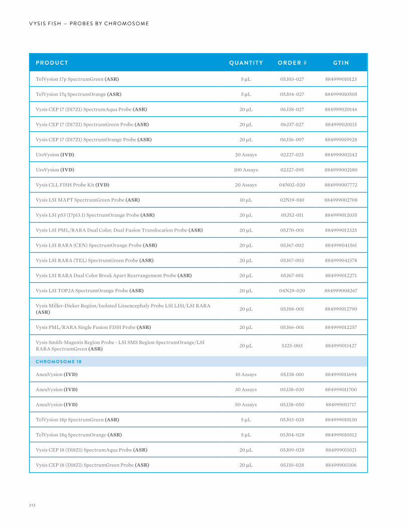
2 12
V Y S I S F I S H — P R O B E S B Y C H R O M O S O M E
P R O D U C T Q UA N T I T Y O R D E R # G T I N
TelVysion 17p SpectrumGreen (ASR) 5 µL 05J03-027 884999010123
TelVysion 17q SpectrumOrange (ASR) 5 µL 05J04-027 884999010505
Vysis CEP 17 (D17Z1) SpectrumAqua Probe (ASR) 20 µL 06J38-027 884999020146
Vysis CEP 17 (D17Z1) SpectrumGreen Probe (ASR) 20 µL 06J37-027 884999020115
Vysis CEP 17 (D17Z1) SpectrumOrange Probe (ASR) 20 µL 06J36-097 884999019928
UroVysion (IVD) 20 Assays 02J27-025 884999002142
UroVysion (IVD) 100 Assays 02J27-095 884999002180
Vysis CLL FISH Probe Kit (IVD) 20 Assays 04N02-020 884999007772
Vysis LSI MAPT SpectrumGreen Probe (ASR) 10 µL 02N19-010 884999002708
Vysis LSI p53 (17p13.1) SpectrumOrange Probe (ASR) 20 µL 05J52-011 884999012035
Vysis LSI PML/RARA Dual Color, Dual Fusion Translocation Probe (ASR) 20 µL 05J70-001 884999012325
Vysis LSI RARA (CEN) SpectrumOrange Probe (ASR) 20 µL 05J67-002 884999041561
Vysis LSI RARA (TEL) SpectrumGreen Probe (ASR) 20 µL 05J67-003 884999041578
Vysis LSI RARA Dual Color Break Apart Rearrangement Probe (ASR) 20 µL 05J67-001 884999012271
Vysis LSI TOP2A SpectrumOrange Probe (ASR) 20 µL 04N29-020 884999008267
Vysis Miller-Dieker Region/Isolated Lissencephaly Probe LSI LIS1/LSI RARA (ASR) 20 µL 05J88-001 884999012790
Vysis PML/RARA Single Fusion FISH Probe (ASR) 20 µL 05J66-001 884999012257
Vysis Smith-Magenis Region Probe - LSI SMS Region SpectrumOrange/LSI RARA SpectrumGreen (ASR) 20 µL 5J25-003 884999011427
C H R O M O S O M E 1 8
AneuVysion (IVD) 10 Assays 05J38-010 884999011694
AneuVysion (IVD) 30 Assays 05J38-030 884999011700
AneuVysion (IVD) 50 Assays 05J38-050 884999011717
TelVysion 18p SpectrumGreen (ASR) 5 µL 05J03-028 884999010130
TelVysion 18q SpectrumOrange (ASR) 5 µL 05J04-028 884999010512
Vysis CEP 18 (D18Z1) SpectrumAqua Probe (ASR) 20 µL 05J09-028 884999011021
Vysis CEP 18 (D18Z1) SpectrumGreen Probe (ASR) 20 µL 05J10-028 884999011106
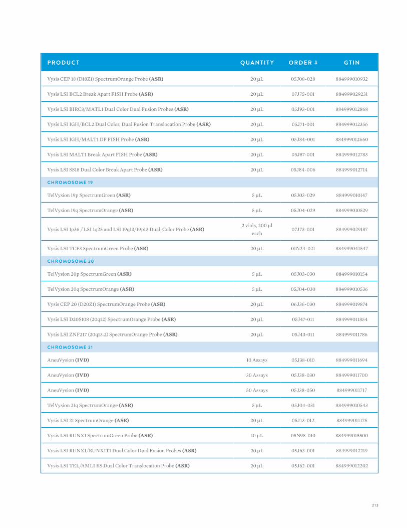
2 13
P R O D U C T Q UA N T I T Y O R D E R # G T I N
Vysis CEP 18 (D18Z1) SpectrumOrange Probe (ASR) 20 µL 05J08-028 884999010932
Vysis LSI BCL2 Break Apart FISH Probe (ASR) 20 µL 07J75-001 884999029231
Vysis LSI BIRC3/MATL1 Dual Color Dual Fusion Probes (ASR) 20 µL 05J93-001 884999012868
Vysis LSI IGH/BCL2 Dual Color, Dual Fusion Translocation Probe (ASR) 20 µL 05J71-001 884999012356
Vysis LSI IGH/MALT1 DF FISH Probe (ASR) 20 µL 05J84-001 884999012660
Vysis LSI MALT1 Break Apart FISH Probe (ASR) 20 µL 05J87-001 884999012783
Vysis LSI SS18 Dual Color Break Apart Probe (ASR) 20 µL 05J84-006 884999012714
C H R O M O S O M E 1 9
TelVysion 19p SpectrumGreen (ASR) 5 µL 05J03-029 884999010147
TelVysion 19q SpectrumOrange (ASR) 5 µL 05J04-029 884999010529
Vysis LSI 1p36 / LSI 1q25 and LSI 19q13/19p13 Dual-Color Probe (ASR)2 vials, 200 µl
each07J73-001 884999029187
Vysis LSI TCF3 SpectrumGreen Probe (ASR) 20 µL 01N24-021 884999041547
C H R O M O S O M E 20
TelVysion 20p SpectrumGreen (ASR) 5 µL 05J03-030 884999010154
TelVysion 20q SpectrumOrange (ASR) 5 µL 05J04-030 884999010536
Vysis CEP 20 (D20Z1) SpectrumOrange Probe (ASR) 20 µL 06J36-030 884999019874
Vysis LSI D20S108 (20q12) SpectrumOrange Probe (ASR) 20 µL 05J47-011 884999011854
Vysis LSI ZNF217 (20q13.2) SpectrumOrange Probe (ASR) 20 µL 05J43-011 884999011786
C H R O M O S O M E 2 1
AneuVysion (IVD) 10 Assays 05J38-010 884999011694
AneuVysion (IVD) 30 Assays 05J38-030 884999011700
AneuVysion (IVD) 50 Assays 05J38-050 884999011717
TelVysion 21q SpectrumOrange (ASR) 5 µL 05J04-031 884999010543
Vysis LSI 21 SpectrumOrange (ASR) 20 µL 05J13-012 884999011175
Vysis LSI RUNX1 SpectrumGreen Probe (ASR) 10 µL 05N98-010 884999015500
Vysis LSI RUNX1/RUNX1T1 Dual Color Dual Fusion Probes (ASR) 20 µL 05J63-001 884999012219
Vysis LSI TEL/AML1 ES Dual Color Translocation Probe (ASR) 20 µL 05J62-001 884999012202
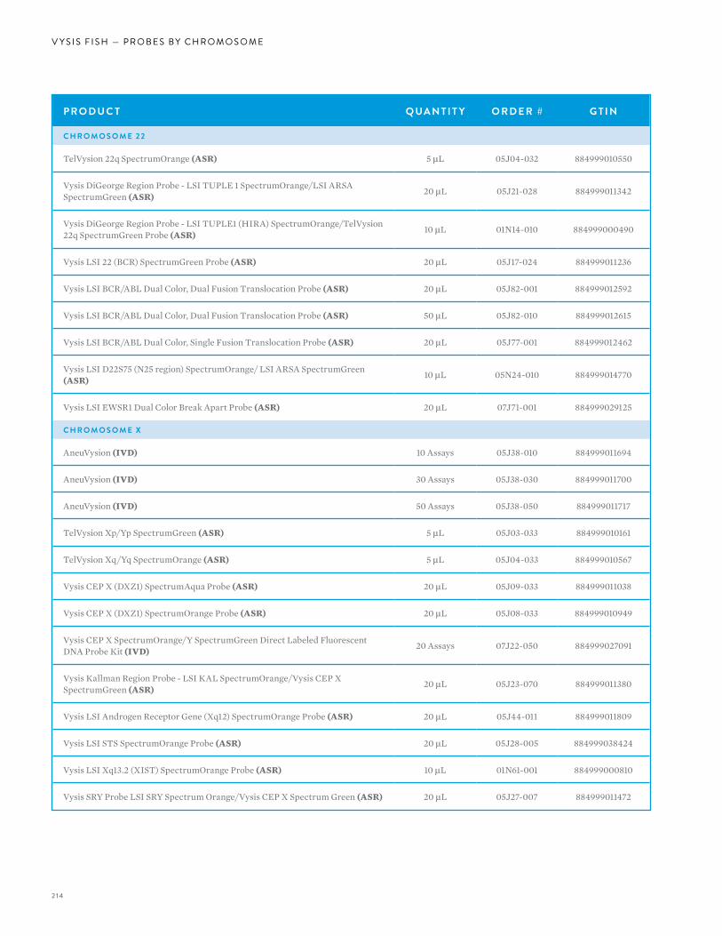
2 14
V Y S I S F I S H — P R O B E S B Y C H R O M O S O M E
P R O D U C T Q UA N T I T Y O R D E R # G T I N
C H R O M O S O M E 2 2
TelVysion 22q SpectrumOrange (ASR) 5 µL 05J04-032 884999010550
Vysis DiGeorge Region Probe - LSI TUPLE 1 SpectrumOrange/LSI ARSA SpectrumGreen (ASR) 20 µL 05J21-028 884999011342
Vysis DiGeorge Region Probe - LSI TUPLE1 (HIRA) SpectrumOrange/TelVysion 22q SpectrumGreen Probe (ASR) 10 µL 01N14-010 884999000490
Vysis LSI 22 (BCR) SpectrumGreen Probe (ASR) 20 µL 05J17-024 884999011236
Vysis LSI BCR/ABL Dual Color, Dual Fusion Translocation Probe (ASR) 20 µL 05J82-001 884999012592
Vysis LSI BCR/ABL Dual Color, Dual Fusion Translocation Probe (ASR) 50 µL 05J82-010 884999012615
Vysis LSI BCR/ABL Dual Color, Single Fusion Translocation Probe (ASR) 20 µL 05J77-001 884999012462
Vysis LSI D22S75 (N25 region) SpectrumOrange/ LSI ARSA SpectrumGreen (ASR) 10 µL 05N24-010 884999014770
Vysis LSI EWSR1 Dual Color Break Apart Probe (ASR) 20 µL 07J71-001 884999029125
C H R O M O S O M E X
AneuVysion (IVD) 10 Assays 05J38-010 884999011694
AneuVysion (IVD) 30 Assays 05J38-030 884999011700
AneuVysion (IVD) 50 Assays 05J38-050 884999011717
TelVysion Xp/Yp SpectrumGreen (ASR) 5 µL 05J03-033 884999010161
TelVysion Xq/Yq SpectrumOrange (ASR) 5 µL 05J04-033 884999010567
Vysis CEP X (DXZ1) SpectrumAqua Probe (ASR) 20 µL 05J09-033 884999011038
Vysis CEP X (DXZ1) SpectrumOrange Probe (ASR) 20 µL 05J08-033 884999010949
Vysis CEP X SpectrumOrange/Y SpectrumGreen Direct Labeled Fluorescent DNA Probe Kit (IVD) 20 Assays 07J22-050 884999027091
Vysis Kallman Region Probe - LSI KAL SpectrumOrange/Vysis CEP X SpectrumGreen (ASR) 20 µL 05J23-070 884999011380
Vysis LSI Androgen Receptor Gene (Xq12) SpectrumOrange Probe (ASR) 20 µL 05J44-011 884999011809
Vysis LSI STS SpectrumOrange Probe (ASR) 20 µL 05J28-005 884999038424
Vysis LSI Xq13.2 (XIST) SpectrumOrange Probe (ASR) 10 µL 01N61-001 884999000810
Vysis SRY Probe LSI SRY Spectrum Orange/Vysis CEP X Spectrum Green (ASR) 20 µL 05J27-007 884999011472
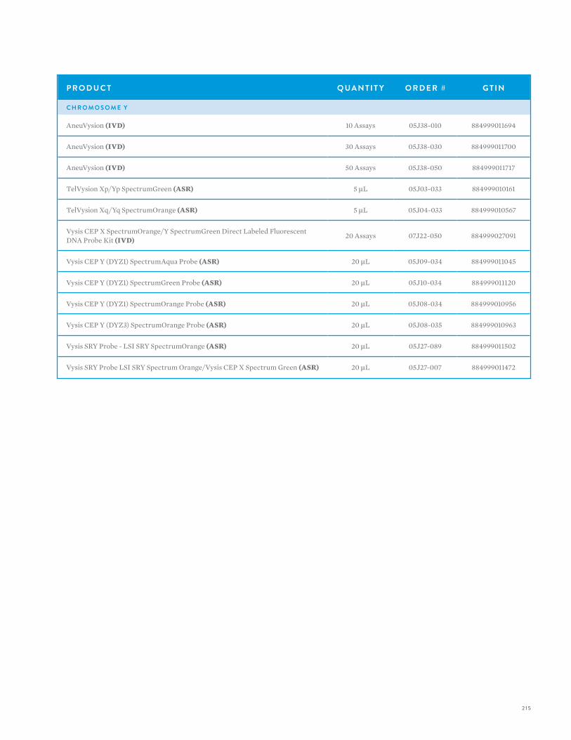
2 15
P R O D U C T Q UA N T I T Y O R D E R # G T I N
C H R O M O S O M E Y
AneuVysion (IVD) 10 Assays 05J38-010 884999011694
AneuVysion (IVD) 30 Assays 05J38-030 884999011700
AneuVysion (IVD) 50 Assays 05J38-050 884999011717
TelVysion Xp/Yp SpectrumGreen (ASR) 5 µL 05J03-033 884999010161
TelVysion Xq/Yq SpectrumOrange (ASR) 5 µL 05J04-033 884999010567
Vysis CEP X SpectrumOrange/Y SpectrumGreen Direct Labeled Fluorescent DNA Probe Kit (IVD) 20 Assays 07J22-050 884999027091
Vysis CEP Y (DYZ1) SpectrumAqua Probe (ASR) 20 µL 05J09-034 884999011045
Vysis CEP Y (DYZ1) SpectrumGreen Probe (ASR) 20 µL 05J10-034 884999011120
Vysis CEP Y (DYZ1) SpectrumOrange Probe (ASR) 20 µL 05J08-034 884999010956
Vysis CEP Y (DYZ3) SpectrumOrange Probe (ASR) 20 µL 05J08-035 884999010963
Vysis SRY Probe - LSI SRY SpectrumOrange (ASR) 20 µL 05J27-089 884999011502
Vysis SRY Probe LSI SRY Spectrum Orange/Vysis CEP X Spectrum Green (ASR) 20 µL 05J27-007 884999011472
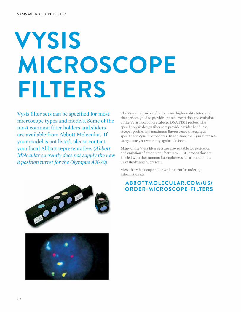
2 16
V Y S I S M I C R O S C O P E F I LT E R S
The Vysis microscope filter sets are high-quality filter sets that are designed to provide optimal excitation and emission of the Vysis fluorophore labeled DNA FISH probes. The specific Vysis design filter sets provide a wider bandpass, steeper profile, and maximum fluorescence throughput specific for Vysis fluorophores. In addition, the Vysis filter sets carry a one year warranty against defects.
Many of the Vysis filter sets are also suitable for excitation and emission of other manufacturers’ FISH probes that are labeled with the common fluorophores such as rhodamine, TexasRed®, and fluorescein.
View the Microscope Filter Order Form for ordering information at:
ABBOT TMOLECUL AR.COM/US/ORDER-MICROSCOPE-FILTERS
Vysis filter sets can be specified for most microscope types and models. Some of the most common filter holders and sliders are available from Abbott Molecular. If your model is not listed, please contact your local Abbott representative. (Abbott Molecular currently does not supply the new 8 position turret for the Olympus AX-70)
VYSIS MICROSCOPE FILTERS
2 17
Single Bandpass Filter SetsO R A N G EThe Orange filter set is designed to excite and transmit SpectrumOrange fluorescence. All of the Vysis SpectrumOrange labeled DNA FISH probes can be viewed, analyzed, and imaged using this filter set. The SpectrumRed fluorophore will be visible using this filter set, but will be dim.
R E DThe Red filter set is designed to excite and transmit SpectrumRed fluorescence. This filter set can be used to view, analyze, and image SpectrumRed labeled probes, or for TexasRed labeled DNA FISH probes that are available from other probe manufacturers. The SpectrumRed filter set is not recommended for viewing SpectrumOrange labeled probes.
Y E L LO WThe Yellow filter set is designed to excite and transmit SpectrumGold fluorescence. The Vysis SpectrumGold labeled DNA FISH probes (Vysis UroVysion) can be viewed, analyzed, and imaged using this filter set. SpectrumOrange probe fluorescence will also be visible with the Yellow filter set. SpectrumRed fluorescence may be visible, yet very dim.
G R E E NThe Green filter set is designed to excite and transmit SpectrumGreen fluorescence. This filter set can be used to view, analyze, and image SpectrumGreen and fluorescence labeled DNA FISH probes.
AQ UAThe Aqua filter set is designed to excite and transmit SpectrumAqua fluorescence. This filter set is specifically required for viewing, analyzing, and imaging Vysis SpectrumAquaTM DNA probes. Filter sets that excite and transmit DAPI fluorescence are not appropriate for SpectrumAqua labeled probes. In some instances,when hybridization signal is very intense for the SpectrumAqua labeled DNA probe, the aqua fluorescence may be visible through a DAPI filter set.However, this will not provide a reliable method for analysis of SpectrumAqua labeled probes. In addition, on specimens counterstained with DAPI, extremely weak DAPI fluorescence may be observed when viewing or imaging through an Aqua single bandpass filter set.
B LU EThe Blue filter set is designed to excite and transmit SpectrumBlue fluorescence and is useful when viewing the SpectrumBlueTM fluorophore alone.DAPI fluorescence will also be visible with this filter set. SpectrumAqua fluorescence will be visible through the Blue filter set, but will be dim. In order to view the SpectrumBlue and SpectrumAqua fluorophores simultaneously, the Aqua/Blue dual bandpass filter set should be used.
DA P I LO N G PA S SThe DAPI longpass filter set is designed to excite and transmit DAPI counterstain fluorescence. This filter set transmits more in the red range of the color spectrum than the DAPI bandpass filter set.
DA P I B A N D PA S SThe DAPI bandpass filter set is designed to excite and transmit DAPI counterstain. This filter set does not transmit as much of the spectrum as the DAPI Longpass filter set, and is recommended over the DAPI Longpass for imaging applications.
2 18
V Y S I S M I C R O S C O P E F I LT E R S
Triple Bandpass Filter SetsDA P I/G R E E N/O R A N G EThe DAPI/Green/Orange filter set is designed to excite and transmit SpectrumGreen, SpectrumOrange, and DAPI counterstain fluorescence simultaneously. This filter is useful when the nuclear and chromosomal DNA, counterstained with DAPI, and the two fluorophores SpectrumGreen and SpectrumOrange must be viewed concurrently. This filter set is recommended for most of the Vysis dual color probe mixtures when hybridized to specimens that are not imbedded in paraffin. Products such as CEP X/Y should be viewed using this filter set.
DA P I/G R E E N/O R A N G E (V. 2 )The DAPI/Green/Orange (V.2) filter set is designed to excite and transmit SpectrumGreen, SpectrumOrange and the DAPI counterstain fluorescence simultaneously. This filter is useful when the nuclear and chromosomal DNA, counterstained with DAPI, and the SpectrumGreen and SpectrumOrange fluorophores must be viewed simultaneously.
The DAPI/Green/Orange (V.2) filter design may provide better color distinction and brightness of the SpectrumOrange and SpectrumGreen fluorophores when viewing paraffin-embedded specimens. This filter set is not optimized for viewing dual-color translocation probes where a blending of the SpectrumGreen and SpectrumOrange fluorophores creates a yellow color. This is the recommended triple bandpass filter set for viewing and analyzing the PathVysion HER-2 DNA Assay.
DA P I/G R E E N/R E DThe DAPI/Green/Red filter set is designed to excite and transmit SpectrumGreen, SpectrumRed, and the DAPI counterstain fluorescence simultaneously. This filter is optimal for viewing probes labeled with SpectrumRed fluorophore while concurrently viewing SpectrumGreen and DAPI.
Dual Bandpass Filter SetsDA P I/O R A N G EThe DAPI/Orange filter set is designed to excite and transmit SpectrumOrange™ and DAPI counterstain fluorescence simultaneously. This filter is useful when the nuclear and chromosomal DNA, counterstained with DAPI, and the SpectrumOrange fluorophore must be viewed concurrently. This filter set is recommended for many of the Vysis® SpectrumOrange labeled DNA FISH probes that can be analyzed simultaneously while viewing the DAPI counterstain.
DA P I/9 -O R A N G E (N B )The DAPI/9-Orange (NB) filter set is designed to excite and transmit SpectrumOrange and the DAPI counterstain fluorescence simultaneously. This filter set is designed to minimize autofluorescence from paraffin-embedded specimens and is recommended for the PathVysion® HER-2 DNA Assay. This filter is useful when the nuclear and chromosomal DNA, counterstained with DAPI, and the SpectrumOrange fluorophore must be viewed concurrently.
DA P I/G R E E NThe DAPI/Green filter set is designed to excite and transmit SpectrumGreen™ and DAPI fluorescence simultaneously and is recommended for the PathVysion® DNA Assay. This filter is useful when the nuclear and chromosomal DNA, counterstained with DAPI, and the SpectrumGreen fluorophore must be viewed concurrently. This filter set is recommended for many of the Vysis SpectrumGreen labeled DNA FISH probes that can be analyzed simultaneously while viewing the DAPI counterstain.
B LU E /AQ UAThe Blue/Aqua filter set is designed to excite and transmit SpectrumBlue™ and SpectrumAquaTM fluorophores simultaneously.
2 19
Quad Bandpass Filter SetsDA P I/AQ UA /G R E E N/O R A N G EThe quad bandpass filter set is designed to excite and transmit DAPI, SpectrumAqua, SpectrumOrange and SpectrumGreen fluorophores simultaneously. This filter set is optimal for viewing all three probe labels simultaneously, plus the DAPI counterstain. This filter set is not recommended for viewing the AneuVysion® Assay on uncultured amniocytes.
220
V Y S I S M I C R O S C O P E F I LT E R S
V Y S I S F I LT E R S E T F LU O R O P H O R E S D E T E C T E D
DAPI/9-Orange (NB) dual bandpass SpectrumOrange and DAPI
DAPI/Green dual bandpass SpectrumGreen and DAPI
DAPI/Green/Orange (V.2) triple bandpass SpectrumGreen/SpectrumOrange/DAPI (specifically designed for viewing paraffin sections)
V Y S I S F I LT E R S E T F LU O R O P H O R E S D E T E C T E D
Aqua single bandpass SpectrumAqua
Green single bandpass SpectrumGreen
Orange single bandpass SpectrumOrange
DAPI/Green/Orange triple bandpass SpectrumGreen/SpectrumOrange/ DAPI
Required Vysis Filter Set ConfigurationsThe following filter set configurations are required for the specific Vysis Assays, as indicated. The recommended filter sets provide the most optimal viewing conditions. If not indicated, contact your local Abbott Molecular Technical Service representative for more information on appropriate filter set configurations for imaging.
PAT H V Y S I O N H E R-2 D N A A S S AY F I LT E R S E T SThe following filter sets are recommended for viewing and enumeration of the PathVysion HER-2 Assay. These filter sets are optimized both for Vysis SpectrumGreen and SpectrumOrange fluorophores and for paraffin-embedded specimen autofluorescence. The dual bandpass filter sets allow the user to view signals of each respective individual color fluorophore and the DAPI counterstain. The triple bandpass filter set allows the user to visualize the SpectrumGreen, SpectrumOrange, and DAPI fluorescent signals simultaneously.
A N E U V Y S I O N A S S AY F I LT E R S E T SThe following filter set configuration provides the best microscope filter set-up for viewing and analysis of the AneuVysion Assay on uncultured amniocytes.
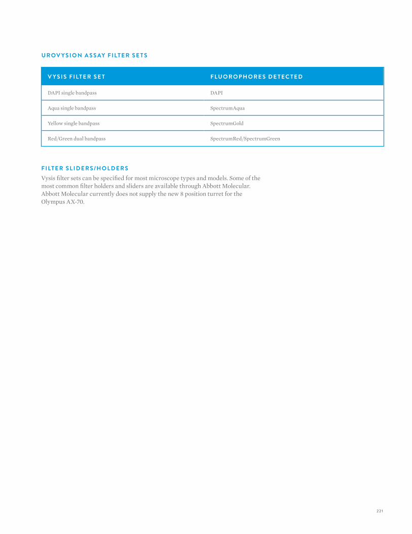
22 1
V Y S I S F I LT E R S E T F LU O R O P H O R E S D E T E C T E D
DAPI single bandpass DAPI
Aqua single bandpass SpectrumAqua
Yellow single bandpass SpectrumGold
Red/Green dual bandpass SpectrumRed/SpectrumGreen
F I LT E R S L I D E R S /H O L D E R SVysis filter sets can be specified for most microscope types and models. Some of the most common filter holders and sliders are available through Abbott Molecular. Abbott Molecular currently does not supply the new 8 position turret for the Olympus AX-70.
U R OV Y S I O N A S S AY F I LT E R S E T S
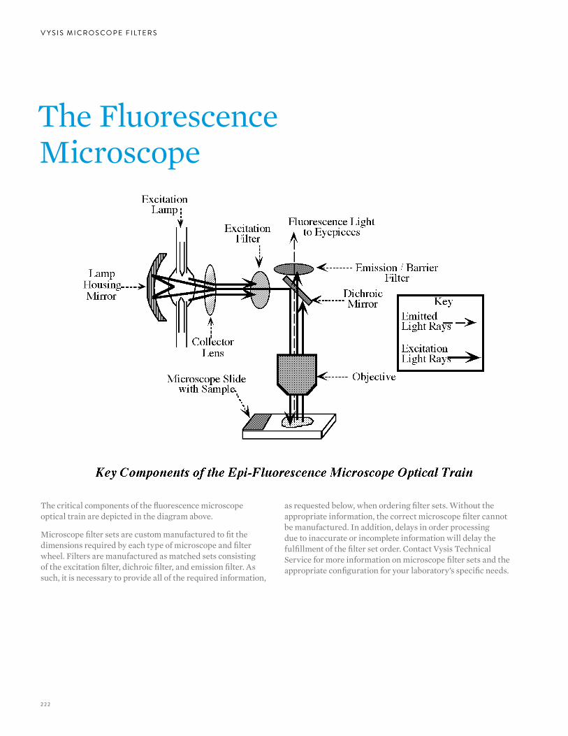
222
V Y S I S M I C R O S C O P E F I LT E R S
The Fluorescence Microscope
The critical components of the fluorescence microscope optical train are depicted in the diagram above.
Microscope filter sets are custom manufactured to fit the dimensions required by each type of microscope and filter wheel. Filters are manufactured as matched sets consisting of the excitation filter, dichroic filter, and emission filter. As such, it is necessary to provide all of the required information,
as requested below, when ordering filter sets. Without the appropriate information, the correct microscope filter cannot be manufactured. In addition, delays in order processing due to inaccurate or incomplete information will delay the fulfillment of the filter set order. Contact Vysis Technical Service for more information on microscope filter sets and the appropriate configuration for your laboratory’s specific needs.

223

224
O R D E R B Y P H O N ETel: 800 553-7042, option 1Tel: 224 361-7800
Call Monday through Friday between 8 a.m. and 5 p.m. CST(Voice mail 24 hours a day)
O R D E R O N L I N Ewww.abbottmolecular.com
Create an eCommerce account atwww.abbottmolecular.com/ecommerce
O R D E R B Y FA XFax: 224 361-7138
Order lines open 24 hours a day
G E N E R A L I N F O R M AT I O NTel: 800 553-7042Tel: 224 [email protected]
Call Monday through Friday between 8 a.m. and 5 p.m. CST(Voice mail 24 hours a day)
M A I L I N G A D D R E S S1300 E. Touhy AvenueDes Plaines, IL 60018
T E C H N I C A L A S S I S TA N C ETel: 800 553-7042, option [email protected]
Call Monday through Friday between 8 a.m. and 5 p.m. CST(Voice mail 24 hours a day)
R E I M B U R S E M E N T A N D CO D I N G A S S I S TA N C ETel: 800 553-7042, option 6
Call Monday through Friday between 8 a.m. and 5 p.m. CST(Voice mail 24 hours a day)
ORDERING/CONTACT INFORMATION
O R D E R I N G / CO N TAC T I N FO R M AT I O N

225

226