aby asics ommon oncerns of the infant years
TRANSCRIPT

Baby Basics - Common Concerns of the
Infant Years Jennifer W. Swoyer, DO


7/28/2014
1
Baby Basics- Common Health
Concerns of the Infant Years
Jennifer W. Swoyer, DO
Photos posted by permission of Danielle Campbell, DO
Overview
Cases representative of common health
concerns of the neonate and infant
Diagnosis
Pathophysiology
Management
Answer boards style question
**Key words/clues in bold throughout**
Case #1
You are reviewing the lab work of a neonate at 24 hours of life. The total serum bilirubin is 15 mg/dL with a direct bilirubin of 0.2 mg/dL. The child was born at 40 weeks gestation with apgars of 9/9 and is doing well. On exam the child’s sclera, face, and chest appear yellow and there is a one centimeter cephalohematoma on the R parietal region. The child is being exclusively breast fed and is feeding well. What is the appropriate management for this child?

7/28/2014
2
Neonatal Jaundice
Caused by:
◦ increased bilirubin production
◦ decreased bilirubin clearance
◦ increased enterohepatic circulation of
bilirubin
Kernicterus
Term used to describe the chronic and
permanent sequela of bilirubin toxicity
Severe hyperbilirubinemia- TB >25 to 30
mg/dL
At this level unconjugated bilirubin can cross the
blood-brain barrier and cause cell death
Hyperbilirubinemia
Defined as TB >95th percentile on the hour-specific Bhutani nomogram in infants ≥35 weeks gestation
Treatment Options
◦ Phototherapy Intensive
Home
Sunlight
◦ Exchange Transfusion
◦ IVIG, Phenobarb, Ursodeoxycholic acid, Metalloporphyrins

7/28/2014
3
Risk Factors for the Development
of Severe Hyperbilirubinemia Pre-discharge TB >95th percentile for age
Jaundice within the first 24 hours of life
Cephalohematoma or significant bruising from birth trauma
Exclusive breastfeeding ◦ nursing is not going well and weight loss is
excessive (>12 percent of birth weight)
Hemolytic disease
Gestational age 35 to 36 weeks or less
Previous sibling who received phototherapy
East Asian race
Albumin < 3 g/dL

7/28/2014
4
Indications for Phototherapy
For infants at low risk (≥38 weeks gestation and without risk factors), intensive phototherapy is started at the following TB values:
◦ 24 hours of age: >12 mg/dL
◦ 48 hours of age: >15 mg/dL
◦ 72 hours of age: >18 mg/dL
Infants in this category who have TB levels 2 to 3 mg/dL below the recommended levels may be treated with fiber optic or conventional phototherapy at home
Indications for Phototherapy
For infants at medium risk (≥38 weeks gestation with risk factors or 35 to 38 weeks gestation without risk factors), intensive phototherapy is started at the following TB values:
◦ 24 hours of age: >10 mg/dL
◦ 48 hours of age: >13 mg/dL
◦ 72 hours of age: >15 mg/dL
The threshold for intervention may be lowered for infants closer to 35 weeks and raised for those closer to 37 6/7 weeks.
Indications for Phototherapy
For infants at high risk (35 to 38 weeks
gestation with risk factors), phototherapy
is initiated at the following TB values:
◦ 24 hours of age: > 8 mg/dL
◦ 48 hours of age: >11 mg/dL
◦ 72 hours of age: >13.5 mg/dL

7/28/2014
5
Red Flag
JAUNDICE IN THE FIRST 24
HOURS OF LIFE!
◦ Most likely due to hemolysis and will most
likely need phototherapy or other
interventions

7/28/2014
6
Case #1
You are reviewing the lab work of a neonate
at 24 hours of life. The total serum
bilirubin is 15 mg/dL with a direct
bilirubin of 0.2 mg/dL. The child was born at
40 weeks gestation with apgars of 9/9 and
is doing well. On exam the child’s sclera,
face, and chest appear yellow and there
is a one centimeter cephalohematoma
on the R parietal region. What is the
appropriate management for this child?
Answer Choices
A. IVIG
B. initiate phototherapy
C. exchange transfusion
D. re-check TB in 48 hours
E. do nothing
Answer Choices
A. IVIG
B. initiate phototherapy
C. exchange transfusion
D. re-check TB in 48 hours
E. do nothing

7/28/2014
7
Case #2
During a well baby exam on a six month
old infant that is new to your practice you
assess for the red reflex. You are unable
to illicit a red reflex in the infant’s L eye
and the reflex actually seems to be white.
What is your initial step in management?
Leukocoria
Differential ◦ Retinoblastoma- 47 percent of cases in one
series
◦ Persistent fetal vasculature
◦ Retinopathy of prematurity
◦ Cataract
◦ Coloboma (fissure or cleft) of choroid or optic disc
◦ Uveitis
◦ Toxocariasis
◦ Coats' disease
◦ Vitreous hemorrhage
◦ Retinal dysplasia

7/28/2014
8
Retinoblastoma
most common intraocular malignancy of
childhood
approximately 300 new cases per year
usually diagnosed in children < 2 y/o
sporadic and heritable forms
◦ If bilateral then always inherited
◦ Unilateral is usually sporadic
Pathophysiology
mutational inactivation of both alleles of
the retinoblastoma (RB1) gene
untreated retinoblastoma grows to fill the
eye and destroys the internal architecture
of the globe
metastasizes after six months
death occurs within years
Case #2
During a well baby exam on a six month
old infant that is new to your practice
you assess for the red reflex. You are
unable to illicit a red reflex in the
infant’s L eye and the reflex actually
seems to be white. What is your initial
step in management?

7/28/2014
9
Answer Choices
A. re check at 9 month well check
B. dilate the eye in the office to better
assess the retina
C. send for ophthalmology evaluation
D. start antibiotic drops and re check in
one week
E. do nothing
Answer Choices
A. re check at 9 month well check
B. dilate the eye in the office to better
assess the retina
C. send for ophthalmology
evaluation
D. start antibiotic drops and re check in
one week
E. do nothing
Case #3
At a routine two week well check you are
examining an otherwise healthy full term
female and notice a +Galeazzi test on the
R and feel a “clunk of entry” with the
Ortolani maneuver on the R. What is
your first step in management?

7/28/2014
10
Developmental Dysplasia of the Hip
Developmental dysplasia of the hip
(DDH)
◦ abnormal development of the hip with
respect to instability of the hip joint and
dysplasia of the acetabulum
Pathophysiology
Ligamentous laxity predisposes the
developing hip to mechanical forces that
cause the femoral head to move outside
of the acetabulum (dislocation)
◦ dysplasia appears to be the result of
dislocation
Risk Factors
Female predominate- 4:1 F:M
Breech positioning
Family history of DDH
Limited fetal mobility
◦ oligohydramnios
◦ firstborn infants

7/28/2014
11
Screening Guidelines
USPSTF
◦ evidence is insufficient to recommend routine
screening for DDH as a means to prevent
adverse outcomes
◦ newborn screening leads to over diagnosis of
hips that do not benefit and may be harmed
by treatment
Screening Guidelines
Pediatric Orthopaedic Society of North
America (POSNA)
◦ Responds to USPSTF by pointing to the value
of early diagnosis
◦ Recommends following the AAP Clinical
Practice Guidelines
assessment for DDH at every well-child visit
until the child is walking normally
Physical Exam Findings
Asymmetry
◦ Apparent shortening of one femur
+ Galeazzi test
◦ Asymmetry of inguinal, thigh, or gluteal skin folds
◦ gait asymmetry
Hip instability
◦ + Ortolani and Barlow maneuvers
> 3 mos old: limitation of abduction
(<45º) is the most reliable sign of DDH

7/28/2014
12
Barlow Ortolani

7/28/2014
13
Appropriate Management
Definite signs of instability
◦ ≤ two weeks: directly to orthopedics
without imaging
◦ > two weeks: age specific imaging or ortho
AAP recommends US < 3 mos and XR > 3 mos
Subtle or nonspecific findings
◦ Newborn: re-examine in two weeks
◦ Two-weeks old: re-examine in two weeks or US
or refer to ortho
◦ > two weeks: age specific imaging
Case #3
At a routine two week well check you
are examining an otherwise healthy full
term female and notice a +Galeazzi
test on the R and feel a “clunk of
entry” with the Ortolani maneuver on
the R. What is your first step in
management?
Answer Options
A. refer to ortho
B. CT R hip
C. US L hip
D. Xray R hip
E. Xray L hip

7/28/2014
14
Answer Options
A. refer to ortho
B. CT R hip
C. US L hip
D. Xray R hip
E. Xray L hip
Case #4
You receive a call from a worried mom about her 4 month old son vomiting after feeds. The patient is exclusively breastfed and mom reports he spits up approximately one ounce of non-bloody, non-bilious vomitus after each feed. The infant is at the 50th percentile for both height and weight, which has been consistent since birth. He is not excessively irritable, and has no other complaints or medical problems. What is the most appropriate treatment at this point?
GERD
Extremely common in healthy infants
◦ gastric fluids reflux into the esophagus 30 or
more times daily normally
Often results in regurgitation into the oral cavity
Frequency decreases with increasing age
Very uncommon in children > 18 mos

7/28/2014
15
GERD- Diagnostic Approach
Uncomplicated
◦ Good weight gain
◦ feeds well
◦ not unusually irritable
◦ “happy spitter”
Complicated
◦ Failure to thrive
◦ GI blood loss
◦ Recurrent PNA
Warning Signs
◦ Bilious vomiting
◦ GI bleeding: hematemesis, hematochezia
◦ Consistently forceful vomiting
◦ Onset of vomiting after six months of life
◦ Constipation, Diarrhea
◦ Abdominal tenderness, distension
◦ Hepatosplenomegaly
◦ Bulging fontanelle
◦ Macro/microcephaly
◦ Seizures
◦ Genetic disorders (eg, Trisomy 21)
◦ Other chronic disorders (eg, HIV)
◦ Fever, Lethargy, Failure to thrive
Uncomplicated GERD
“happy spitter”
Warning signs of complication absent
No intervention is required
If the reflux is causing significant adverse effects on quality of life ◦ trial of a milk-free diet
◦ thickening of feeds
Acid suppression and prokinetic agents ◦ not valuable in treating children < 1 y/o
with uncomplicated GERD
◦ Trial only if above measures fail and QOL an issue

7/28/2014
16
Indications for Pharmacotherapy
Esophagitis documented by endoscopic
biopsies
◦ PPI most effective
Eosinophilic esophagitis
◦ PPI + leukotriene inhibitor
Case #4
You receive a call from a worried mom about her 4 month old son vomiting after feeds. The patient is exclusively breastfed and mom reports he spits up approximately one ounce of non-bloody, non-bilious vomitus after each feed. The infant is at the 50th percentile for both height and weight, which has been consistent since birth, he is not excessively irritable, and has no other complaints or medical problems. What is the most appropriate treatment at this point?
Answer Choices
A. Stop breastfeeding and start soy
formula immediately
B. Order an upper GI series
C. Reassure and follow
D. Refer to GI
E. Start PPI

7/28/2014
17
Answer
A. Stop breastfeeding and start soy
formula immediately
B. Order an upper GI series
C. Reassure and follow
D. Refer to GI
E. Start PPI
Case #5
You are evaluating a 3 week old male neonate in the office due to slow weight gain and vomiting. He has not yet reached birth weight. Mom says she is feeding him every one to two hours, but he vomits after each feeding and is still hungry afterwards. The vomitus is non-bloody and non-bilious. Mom describes the episodes of vomiting to be projectile. On exam you can palpate a small mass in the RUQ. What is the most appropriate treatment for this child?
Pyloric Stenosis
Hypertrophy of the pylorus eventually
progressing to near-complete
obstruction of the gastric outlet
Male predominant- M:F = 4:1 to 6:1
Peak incidence of dx: 3-5 weeks of age
30% occur in firstborn children

7/28/2014
18
Pyloric Stenosis
Etiology
◦ Unknown
◦ May have a genetic predisposition
Classic presentation
◦ 3- to 6-week-old male baby
◦ immediate postprandial, non-bilious,
often projectile vomiting
◦ demands to be re-fed soon afterwards
"hungry vomiter"
Plyoric Stenosis
Physical exam findings
◦ emaciated and dehydrated
◦ palpable "olive-like" mass at the lateral
edge of the rectus abdominus in the RUQ
Lab findings
◦ hypochloremic metabolic alkalosis
◦ hypokalemia may develop after 3 weeks of
vomiting
Pyloric Stenosis
Diagnosis
◦ US vs upper GI
Recommendation of which modality to chose varies
from center to center and case to case
Treatment
◦ Definitive treatment for pyloric stenosis is
surgery
Pyloromyotomy

7/28/2014
19
Case #5
You are evaluating a 3 week old male neonate in the office due to slow weight gain and vomiting. He has not yet reached birth weight. Mom says she is feeding him every one to two hours, but he vomits after each feeding and is still hungry afterwards. The vomitus is non-bloody and non-bilious. Mom describes the episodes of vomiting to be projectile. On exam you can palpate a small mass in the RUQ. What is the most appropriate treatment for this child?
Answer Choices
A. increase frequency of feeds and
decrease amount of each feed
B. fundoplication
C. start PPI
D. re evaluate in two weeks
E. pyloromyotomy
Answer Choices
A. increase frequency of feeds and
decrease amount of each feed
B. fundoplication
C. start PPI
D. re evaluate in two weeks
E. pyloromyotomy

7/28/2014
20
Case #6
A two year old male without significant past medical history taking no medications presents to your office c/o abdominal pain which began three hours ago. Pain is described as intermittent and crampy. Episodes are becoming more frequent and the patient tends to pull his legs up to his chest during the episodes. No n/v/d but his last stool did have some blood and mucous in it. On exam vital signs are stable, the abdomen is soft, mildly distended, and without peritoneal signs. You notice a sausage shaped mass on palpation of the RLQ. What is your diagnosis and treatment?
Intussusception
Invagination of a part of the intestine into itself
Most common abdominal emergency in early childhood ◦ particularly in children younger than two years of
age
Most common cause of intestinal obstruction in infants between 6 and 36 months of age ◦ 60% before one y/o
◦ 80% before two y/o
Male predominant- M:F = 3:2
Intussusception
Occurs most often near the ileocecal junction
Proximal segment of bowel, telescopes into a distal segment dragging the associated mesentery with it
◦ Leads to intestinal edema Can ultimately lead to ischemia, perforation, and
peritonitis
Etiology- most cases thought to be idiopathic but some increased incidence post-viral illness
7/28/2014
21
Intussusception
Concept of “lead point”
◦ lesion or variation in the intestine that is trapped by peristalsis and dragged into a distal segment of the intestine
meckel diverticulum
polyp
tumor
hematoma- HSP
vascular malformation
thick inspisssated stool- CF
◦ Must be vigilant for pathological lead points
Intussusception
Presentation
◦ sudden onset- intermittent, severe, crampy,
progressive abdominal pain
◦ inconsolable crying
◦ drawing up of the legs toward the
abdomen
◦ Episodes occur at 15-20 minute intervals
Become more frequent
Vomiting may follow episode
Pain free between episodes
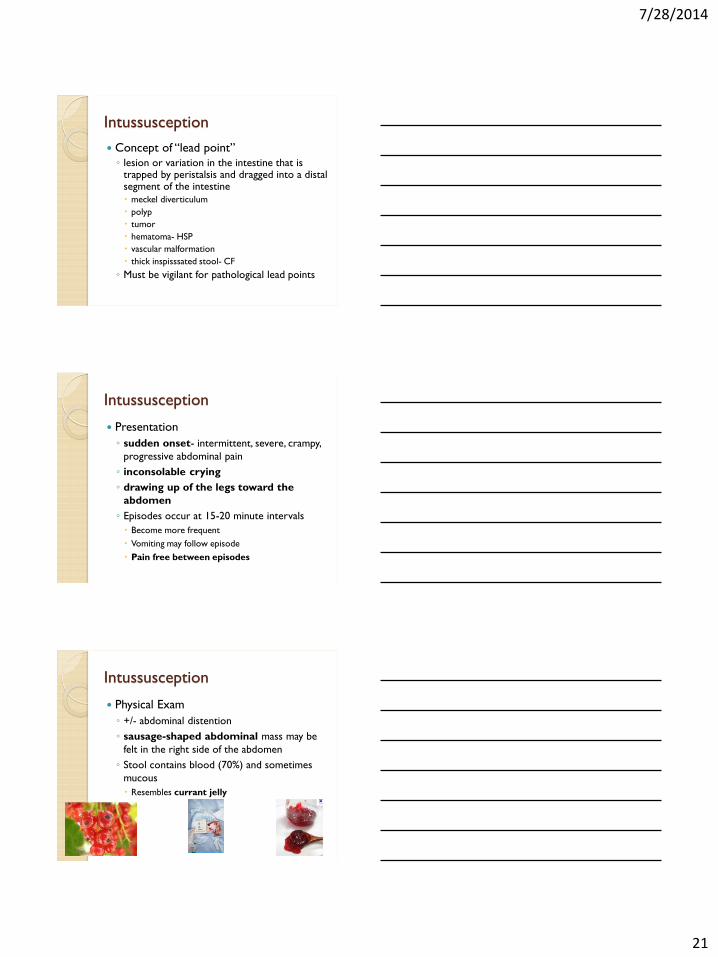
Intussusception
Physical Exam
◦ +/- abdominal distention
◦ sausage-shaped abdominal mass may be
felt in the right side of the abdomen
◦ Stool contains blood (70%) and sometimes
mucous
Resembles currant jelly

7/28/2014
22
Intussusception
Classic triad
◦ Abdominal pain
◦ Sausage-shaped palpable mass
◦ Currant jelly stool
Only seen in 15% of patients at presentation,
so high index of suspicion necessary
Intussusception
Patients with classic presentation and no suspicion for perforation may proceed directly to contrast enema for dx and tx
If diagnosis in question: ◦ Radiological studies
US- modality of choice in most institutions Approaches 100% sensitivity/specificity
Will see a “bull's eye" or "coiled spring" lesion
Plain film Less sensitive/specific than US
signs of intestinal obstruction
target sign- two concentric radiolucent circles superimposed on the right kidney
crescent sign- soft tissue density projecting into the gas of the large bowel

7/28/2014
23
Intussusception
Treatment ◦ No perforation Nonoperative reduction using hydrostatic or pneumatic
pressure by enema Water soluble contrast enema under fluoroscopic
guidance
◦ Perforation suspected Laparotomy
Recurrence ◦ recurs in approximately 10 percent of
children after successful nonoperative reduction Should prompt search for pathological lead point

7/28/2014
24
Case #6
A two year old male without significant past medical history taking no medications presents to your office c/o abdominal pain which began three hours ago. Pain is described as intermittent and crampy. Episodes are becoming closer together and the patient tends to pull his legs up to his chest during the episodes. No n/v/d but his last stool did have some blood and mucous in it. On exam vital signs are stable, the abdomen is soft, mildly distended, and without peritoneal signs. You notice a sausage shaped mass on palpation of the RLQ. What is your diagnosis and treatment?
Answer Choices
A. Mekels Diverticulum- watchful waiting
B. Intussusception- laparotomy
C. Mekels Diverticulum- laparatomy
D. Intussusception- contrast enema
E. Volvulus- laparotomy
Answer Choices
A. Mekels Diverticulum- watchful waiting
B. Intussusception- laparotomy
C. Mekels Diverticulum- laparatomy
D. Intussusception- contrast enema
E. Volvulus- laparotomy

7/28/2014
25
Questions???
References
www.uptodate.com
Hay, Current Diagnosis and Treatment: Pediatrics, 19th edition, 2009
Le, First Aid for the Family Medicine Boards, 2008
Rakel, Textbook of Family Medicine, 8th edition, 2011
Sotirios, Transcutaneous Bilirubin Levels for the First 120 Postnatal Hours in Healthy Neonates, Pedaitrics, Vol. 125 No. 1, 1/1/2010.
Waickus, Family Medicine Board Review, 4th edition, 2010