acid hydrolysis of wheat gluten induces formation of new...
TRANSCRIPT
General rights Copyright and moral rights for the publications made accessible in the public portal are retained by the authors and/or other copyright owners and it is a condition of accessing publications that users recognise and abide by the legal requirements associated with these rights.
• Users may download and print one copy of any publication from the public portal for the purpose of private study or research. • You may not further distribute the material or use it for any profit-making activity or commercial gain • You may freely distribute the URL identifying the publication in the public portal
If you believe that this document breaches copyright please contact us providing details, and we will remove access to the work immediately and investigate your claim.
Downloaded from orbit.dtu.dk on: Jul 06, 2018
Acid Hydrolysis of Wheat Gluten Induces Formation of New Epitopes but Does NotEnhance Sensitizing Capacity by the Oral Route: A Study in “Gluten Free” BrownNorway Rats
Kroghsbo, Stine; Andersen, Nanna Birch; Rasmussen, Tina Frid; Jacobsen, Susanne; Madsen, CharlotteBernhardPublished in:P L o S One
Link to article, DOI:10.1371/journal.pone.0107137
Publication date:2014
Document VersionPublisher's PDF, also known as Version of record
Link back to DTU Orbit
Citation (APA):Kroghsbo, S., Andersen, N. B., Rasmussen, T. F., Jacobsen, S., & Madsen, C. B. (2014). Acid Hydrolysis ofWheat Gluten Induces Formation of New Epitopes but Does Not Enhance Sensitizing Capacity by the OralRoute: A Study in “Gluten Free” Brown Norway Rats. P L o S One, 9(9), [e107137]. DOI:10.1371/journal.pone.0107137

Acid Hydrolysis of Wheat Gluten Induces Formation ofNew Epitopes but Does Not Enhance Sensitizing Capacityby the Oral Route: A Study in ‘‘Gluten Free’’ BrownNorway RatsStine Kroghsbo1¤, Nanna B. Andersen1, Tina F. Rasmussen1, Susanne Jacobsen2{, Charlotte B. Madsen1*
1 National Food Institute, Technical University of Denmark, Søborg, Denmark, 2 Enzyme and Protein Chemistry, Department of Systems Biology, Technical University of
Denmark, Lyngby, Denmark
Abstract
Background: Acid hydrolyzed wheat proteins (HWPs) are used in the food and cosmetic industry as emulsifiers. Cases ofsevere food allergic reactions caused by HWPs have been reported. Recent data suggest that these reactions are caused byHWPs produced by acid hydrolysis.
Objectives: To examine the sensitizing capacity of gluten proteins per se when altered by acid or enzymatic hydrolysisrelative to unmodified gluten in rats naıve to gluten.
Methods: High IgE-responder Brown Norway (BN) rats bred on a gluten-free diet were sensitized without the use ofadjuvant to three different gluten products (unmodified, acid hydrolyzed and enzymatic hydrolyzed). Rats were sensitizedby intraperitoneal (i.p.) immunization three times with 200 mg gluten protein/rat or by oral dosing for 35 days with 0.2, 2 or20 mg gluten protein/rat/day. Sera were analyzed for specific IgG and IgE and IgG-binding capacity by ELISA. IgEfunctionality was measured by rat basophilic leukemia (RBL) assay.
Results: Regardless of the route of dosing, all products had sensitizing capacity. When sensitized i.p., all three glutenproducts induced a strong IgG1 response in all animals. Acid hydrolyzed gluten induced the highest level of specific IgE butwith a low functionality. Orally all three gluten products induced specific IgG1 and IgE but with different dose-responserelations. Sensitizing rats i.p. or orally with unmodified or enzymatic hydrolyzed gluten induced specific IgG1 responses withsimilar binding capacity which was different from that of acid hydrolyzed gluten indicating that acid hydrolysis of glutenproteins induces formation of ‘new’ epitopes.
Conclusions: In rats not tolerant to gluten acid hydrolysis of gluten enhances the sensitizing capacity by the i.p. but not bythe oral route. In addition, acid hydrolysis induces formation of new epitopes. This is in contrast to the enzymatichydrolyzed gluten having an epitope pattern similar to unmodified gluten.
Citation: Kroghsbo S, Andersen NB, Rasmussen TF, Jacobsen S, Madsen CB (2014) Acid Hydrolysis of Wheat Gluten Induces Formation of New Epitopes but DoesNot Enhance Sensitizing Capacity by the Oral Route: A Study in ‘‘Gluten Free’’ Brown Norway Rats. PLoS ONE 9(9): e107137. doi:10.1371/journal.pone.0107137
Editor: Nicholas J. Mantis, New York State Dept. Health, United States of America
Received April 8, 2014; Accepted August 12, 2014; Published September 10, 2014
Copyright: � 2014 Kroghsbo et al. This is an open-access article distributed under the terms of the Creative Commons Attribution License, which permitsunrestricted use, distribution, and reproduction in any medium, provided the original author and source are credited.
Data Availability: The authors confirm that all data underlying the findings are fully available without restriction. All relevant data are within the paper.
Funding: This work was financially supported by Tereos Syral, Aalst, Belgium who also provided the gluten proteins used, Danish Veterinary and FoodAdministration, and the Danish Research Council grant 0603-00199B. The funders had no role in study design, data collection and analysis, decision to publish, orpreparation of the manuscript.
Competing Interests: Tereos Syral, a company producing wheat gluten and proteins under the name Meripro, provided funding towards this study. There areno further patents, products in development or marketed products to declare. This does not alter the authors’ adherence to all the PLOS ONE policies on sharingdata and materials.
{ Deceased.
* Email: [email protected]
¤ Current address: Unisensor A/S, Allerød, Denmark
Introduction
Gluten proteins are wheat storage proteins, constituting about
10% of wheat, and give wheat dough functional properties such as
water absorption capacity, viscosity and elasticity, which contrib-
ute to the unique baking properties [1–3]. Gluten proteins are
among the most complex proteins in nature containing hundreds
of components present as monomers, oligomers and polymers.
Oligomers and polymers are linked by disul-phide bonds between
the S-containing amino acid cysteine, which make up approxi-
mately 2% of gluten. Based on their physical characteristics, gluten
proteins can be divided into gliadins and glutenins. Gliadins, also
known as wheat prolamins, are soluble in alcohol and are mainly
present in gluten as monomers interacting by non-covalent forces.
PLOS ONE | www.plosone.org 1 September 2014 | Volume 9 | Issue 9 | e107137
Glutenins are soluble in dilute acid and are mainly present as
aggregated proteins linked by intra- and interchain disulphide
bonds. Gliadins and glutenins consist mainly of glutamine, proline
and the essential amino acid phenylalanine [3,4]. Another unique
feature about gluten proteins is their structures, which are
dynamic, comprise interchanging conformations and do not
unfold when exposed to heating like most other proteins [5].
Gluten proteins are by definition not soluble in water but
enzymatic or acid hydrolysis can alter their structure and size and
thereby resulting in soluble protein hydrolysates. Acid hydrolyses
also result in partial deamidation of the proteins [6]. Acid
hydrolyzed wheat gluten obtain emulsifying properties which
make them useful in food products such as coffee creamers and
shake-and-bake products but also in cosmetic products for hair
and body [6,7].
The digestibility of wheat depends on the degree of processing
and grinding [8]. Results from in vitro digestion studies of wheat
proteins indicate that gluten proteins partially resist digestion in
the stomach and intestine and thus may be absorbed as
structurally intact proteins [9]. However, when gluten proteins
are deamidated, it may result in increased digestibility [10]. In
general, hydrolysis can alter the way proteins are broken down,
digested and absorbed across the gut mucosal barrier [5]. Hence,
there is a possibility that the mucosal immune system is not
exposed to hydrolyzed gluten proteins in the same way as gluten
proteins [5,11].
The default immunological reaction to ingested food proteins is
tolerance. However, in susceptible individuals an otherwise
harmless food or food component can induce an IgE-mediated
food allergy, which involves an abnormal response of the immune
system to specific food proteins. IgE-mediated wheat allergy
appears in three different types; wheat food allergy, wheat-
dependent exercise-induced anaphylaxis (WDEIA) and baker’s
asthma, which must be distinguished from each other [12].
Recently allergy to HWPs was recognized as a fourth type of IgE-
mediated allergy to wheat. Most interestingly, HWPs have been
reported to cause food allergic reactions in patients who are
tolerant to wheat [11,13–17]. Allergic skin reactions after
application of cosmetic products containing HWPs have also been
described [7,14,15,18,19]. However, the type of hydrolysis
(enzymatic or acid) in the papers on HWP’s is not always clearly
defined. In Japan the use of a facial soap containing acid
hydrolyzed wheat protein has caused allergic skin symptoms and
WDEIA i.e. allergic reactions to wheat products [20,21]. A wide
spectrum of wheat proteins belonging to the albumin/globulin
fraction (salt-soluble), gliadin fraction (alcohol-soluble) and glute-
nin fraction (soluble in dilute acid) [9,11,13–15,19,22,23] have
been reported to be involved in IgE-mediated allergic reactions to
wheat. However, at present no study has clearly demonstrated a
correlation between the type of IgE-mediated wheat allergy and
pattern of IgE-reactivity to a specific fraction of wheat proteins. In
general, food allergens have a high resistance to heat. This is also
the case of gluten proteins that contain potentially thermo-stable
IgE-epitopes [5,9,25]. The route of sensitization can occur via the
gastrointestinal tract, via the respiratory tract (wheat flour) or via
the skin [12,25–27].
Solubility increases when gluten is hydrolyzed by enzymes. This
type of product is used in food as a protein supplement. When
gluten is hydrolyzed by acid, emulsifying properties are created.
This type of product is used as emulsifier in food and in cosmetics.
To investigate if hydrolysis alters the immunogenicity of gluten
proteins we have studied the sensitization capacity of unmodified,
enzymatic hydrolyzed and acid hydrolyzed wheat gluten. To be
able to compare the immune response per se the influence from
gluten tolerance was excluded by performing the studies in Brown
Norway rats naıve to gluten (bred for more than three generations
on a gluten free diet). Rats were dosed intraperitoneally or orally.
Material and Methods
Ethics statementAnimal experiments were carried out at the DTU Food
(Mørkhøj, Denmark) facilities. Ethical approval was given by the
Danish Animal Experiments Inspectorate. The authorization
number given: 2009/561-01710. The experiments were overseen
by the National Food Institutes in-house Animal Welfare
Committee for animal care and use.
Wheat gluten productsWheat gluten (unmodified), enzymatic hydrolyzed wheat gluten
and acid hydrolyzed wheat gluten were provided by Tereos Syral,
Aalst, Belgium. The enzyme used in the production of enzyme
hydrolyzed gluten was an endoprotease with no transglutaminase
activity. Compared to the conditions of acid hydrolysis, the
enzymatic reaction occurs at more neutral pH and moderate
temperature. The acid hydrolyzed gluten is produced at very low
pH (below pH 2.5) and at high temperature (above 80uC). For
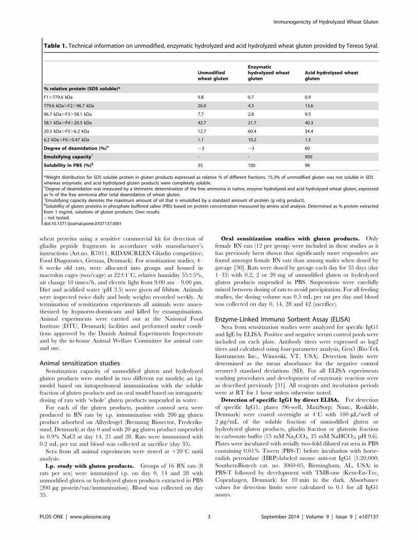
technical information on gluten products see Table 1. It can be
seen that acid hydrolysis results in substantial deamidation. The
level of endotoxin in gluten proteins were analyzed according to
manufacturer’s instructions (Pierce LAL Chromogenic Endotoxin
Quantitation kit; Fisher Scientific ApS, Slangerup, Denmark).
Endotoxin levels were below 50 EU/mg gluten protein for all
products.
Preparation of soluble fractions of wheat gluten productsfor analysis of antibody response
Gluten products were dissolved by carefully adding 1 mg/mL
unmodified gluten or hydrolyzed gluten products to phosphate
buffered saline (PBS; 137 mM NaCl, 3 mM KCl, 8 mM
Na2HPO4, 1 mM KH2PO4; pH 7.2) before magnetic stirring for
2 hours at room temperature (RT) followed by ultrasonication for
3–5 hours. After centrifugation at 20006g for 20 min. at 4uC,
supernatants were collected and stored at 420uC. Protein
concentrations in supernatants were determined by amino acid
analysis [28].
As approximately 35% of proteins in unmodified gluten were
soluble in PBS, gluten proteins were extracted by ethanol (gliadin
fraction) followed by sodium oleate (glutenin fraction) for
measurement of the specific antibody response to ‘total’ gluten
proteins. Briefly, unmodified gluten were suspended in 50%
ethanol (2 mg gluten protein/mL), mixed carefully for 10 min.
before ultrasonication for 2–3 hours. The gliadin fraction was
obtained by collecting the supernatant after centrifugation
(20006g, RT, 5 min.). The glutenin fraction was obtained by
suspending the remaining pellet in 0.1% (w/v) sodium oleate
solution before carefully mixing for 10 min. followed by ultra-
sonication for 2 hours and collection of supernatant after
centrifugation (20006g, RT, 15 min.). Gliadin and glutenin
fractions were stored at RT.
AnimalsInbred high IgE-responder Brown Norway (BN) rats were from
our in-house colony at the National Food Institute (DTU,
Denmark). Rats were bred and kept for at least three generations
on a gluten free diet developed and produced at the National Food
Institute (DTU, Denmark) [29] to ensure immunologically naıve
animals with respect to wheat proteins. The diet was analyzed for
Immunogenicity of Hydrolyzed Wheat Gluten
PLOS ONE | www.plosone.org 2 September 2014 | Volume 9 | Issue 9 | e107137
wheat proteins using a sensitive commercial kit for detection of
gliadin peptide fragments in accordance with manufacturer’s
instructions (Art.no. R7011, RIDASCREEN Gliadin competitive;
Food Diagnostics, Grenaa, Denmark). For sensitization studies, 4–
6 weeks old rats, were allocated into groups and housed in
macrolon cages (two/cage) at 2261uC, relative humidity 5565%,
air change 10 times/h, and electric light from 9.00 am – 9.00 pm.
Diet and acidified water (pH 3.5) were given ad libitum. Animals
were inspected twice daily and body weights recorded weekly. At
termination of sensitization experiments all animals were anaes-
thetized by hypnorm-dormicum and killed by exsanguinations.
Animal experiments were carried out at the National Food
Institute (DTU, Denmark) facilities and performed under condi-
tions approved by the Danish Animal Experiments Inspectorate
and by the in-house Animal Welfare Committee for animal care
and use.
Animal sensitization studiesSensitization capacity of unmodified gluten and hydrolyzed
gluten products were studied in two different rat models; an i.p.
model based on intraperitoneal immunization with the soluble
fraction of gluten products and an oral model based on intragastric
dosing of rats with ‘whole’ gluten products suspended in water.
For each of the gluten products, positive control sera were
produced in BN rats by i.p. immunization with 200 mg gluten
product adsorbed on Alhydrogel (Brenntag Biosector, Frederiks-
sund, Denmark) at day 0 and with 20 mg gluten product suspended
in 0.9% NaCl at day 14, 21 and 28. Rats were immunized with
0.2 mL per rat and blood was collected at sacrifice (day 35).
Sera from all animal experiments were stored at 420uC until
analysis.
I.p. study with gluten products. Groups of 16 BN rats (8
rats per sex) were immunized i.p. on day 0, 14 and 28 with
unmodified gluten or hydrolyzed gluten products extracted in PBS
(200 mg protein/rat/immunization). Blood was collected on day
35.
Oral sensitization studies with gluten products. Only
female BN rats (12 per group) were included in these studies as it
has previously been shown that significantly more responders are
found amongst female BN rats than among males when dosed by
gavage [30]. Rats were dosed by gavage each day for 35 days (day
1–35) with 0.2, 2 or 20 mg of unmodified gluten or hydrolyzed
gluten products suspended in PBS. Suspensions were carefully
mixed between dosing of rats to avoid precipitation. For all feeding
studies, the dosing volume was 0.5 mL per rat per day and blood
was collected on day 0, 14, 28 and 42 (sacrifice).
Enzyme-Linked Immuno Sorbent Assay (ELISA)Sera from sensitization studies were analyzed for specific IgG1
and IgE by ELISA. Positive and negative serum control pools were
included on each plate. Antibody titers were expressed as log2
titers and calculated using four-parameter analysis, Gen5 (Bio-Tek
Instruments Inc., Winooski, VT, USA). Detection limits were
determined as the mean absorbance for the negative control
serum+3 standard deviations (SD). For all ELISA experiments
washing procedures and development of enzymatic reaction were
as described previously [31]. All reagents and incubation periods
were at RT for 1 hour unless otherwise noted.
Detection of specific IgG1 by direct ELISA. For detection
of specific IgG1, plates (96-well, MaxiSorp; Nunc, Roskilde,
Denmark) were coated overnight at 4uC with 100 mL/well of
2 mg/mL of the soluble fraction of unmodified gluten or
hydrolyzed gluten products, gliadin fraction or glutenin fraction
in carbonate buffer (15 mM Na2CO3, 35 mM NaHCO3; pH 9.6).
Plates were incubated with serially two-fold diluted rat sera in PBS
containing 0.01% Tween (PBS-T) before incubation with horse-
radish peroxidase (HRP)-labeled mouse anti-rat IgG1 (1:20,000;
SouthernBiotech cat. no. 3060-05, Birmingham, AL, USA) in
PBS-T followed by development with TMB-one (Kem-En-Tec,
Copenhagen, Denmark) for 10 min in the dark. Absorbance
values for detection limits were calculated to 0.1 for all IgG1
assays.
Table 1. Technical information on unmodified, enzymatic hydrolyzed and acid hydrolyzed wheat gluten provided by Tereos Syral.
Unmodifiedwheat gluten
Enzymatichydrolyzed wheatgluten
Acid hydrolyzed wheatgluten
% relative protein (SDS soluble)*
F1.779.6 kDa 9.8 0.7 0.9
779.6 kDa.F2.96.7 kDa 26.0 4.3 13.6
96.7 kDa.F3.58.1 kDa 7.7 2.8 9.5
58.1 kDa.F4.20.5 kDa 42.7 21.7 40.3
20.5 kDa.F5.6.2 kDa 12.7 60.4 34.4
6.2 kDa.F6.0.47 kDa 1.1 10.2 1.3
Degree of deamidation (%)¤ ,3 ,3 60
Emulsifying capacity{ - - 950
Solubility in PBS (%)` 35 100 98
*Weight distribution for SDS soluble protein in gluten products expressed as relative % of different fractions. 15.3% of unmodified gluten was not soluble in SDSwhereas enzymatic and acid hydrolyzed gluten products were completely soluble.¤Degree of deamidation was measured by a titrimetric determination of the free ammonia in native, enzyme hydrolyzed and acid hydrolyzed wheat gluten, expressedas % of the free ammonia after total deamidation of wheat gluten.{Emulsifying capacity denotes the maximum amount of oil that is emulsified by a standard amount of protein (g oil/g product).`Solubility of gluten proteins in phosphate buffered saline (PBS) based on protein concentration measured by amino acid analysis. Determined as % protein extractedfrom 1 mg/mL solutions of gluten products. Own results.-: not tested.doi:10.1371/journal.pone.0107137.t001
Immunogenicity of Hydrolyzed Wheat Gluten
PLOS ONE | www.plosone.org 3 September 2014 | Volume 9 | Issue 9 | e107137
Inhibition ELISAs for examination of IgG1-binding
capacity. Assay procedures were as described for measurement
of specific IgG1 except that sera were preincubated with inhibitor
solutions. Sera from individual rats were diluted to reach an OD
between 0.8 and 1.0 and preincubated for 1 hour at RT with serial
ten-fold dilutions of the soluble fraction of gluten products (50 pg/
mL to 50 mg/mL) before triplicates of serum/inhibitor mix (and
sera with no inhibitor as a control) were added to the wells. Results
were expressed as % B/B0 where B corresponds to the specific
IgG1-binding to immobilized gluten protein when a known
concentration of inhibitor is present and B0 corresponds to the
binding in the absence of inhibitor. For each serum pool, the
concentration of inhibitor that inhibits 50% of the binding to the
immobilized antigen (IC50) was determined, where an increase in
IC50 value is correlated to a lower IgG1-reactivity of the product
used as inhibitor. Inhibition curves were analyzed by GraphPad
Prism (GraphPad Software, San Diego, CA, USA) to examine if
curves were parallel (no significant difference between slopes)
which is important for appropriate comparison of IC50 values.
Detection of specific IgE by antibody-capture
ELISAs. To avoid the interference of the much higher level of
IgG than IgE in rat sera, assays based on selective IgE capture
were established for detection of specific IgE responses. Plates were
coated overnight at 4uC with 0.5 mg/mL of mouse anti-rat IgE
(HPMAB-123 HybriDomus, Cytotech, Hellebæk, Denmark) in
carbonate buffer. After blocking of remaining active sites for 1
hour at 37uC with PBS-T containing 3% bovine serum albumin
(BSA; A2153, Sigma, Copenhagen, Denmark), plates were
incubated with serially two-fold diluted rat sera and then with
biotin-coupled gluten products or gliadin diluted in PBS-T
containing 3% BSA (biotin-gluten 1:200, biotin-enzymatic hydro-
lyzed gluten 1:1500, biotin-acid hydrolyzed gluten 1:800 and
biotin-gliadin 1:250). After washing, plates were incubated with
HRP-labeled NeutrAvidin (cat. no. 31030; ThermoScientific,
Slangerup, Denmark) diluted in PBS-T containing 3% BSA
(diluted 1:4000 for detection of anti-gluten IgE, anti-acid
hydrolyzed gluten IgE and anti-gliadin IgE or diluted 1:8000 for
detection of anti-enzymatic hydrolyzed gluten IgE). Plates were
developed for 10 min in the dark and absorbance values for
detection limits were calculated to 0.1 for all IgE assays.
Biotin was coupled to each of the gluten products or gliadin
using a biotin protein-labeling kit (Pierce EZ-Link Sulfo-NHS-LC-
Biotinylation Kit; ThermoScientific) in accordance with the
manufacturer’s instructions. For gluten products, proteins soluble
in PBS were used for biotinylation whereas the gliadin fraction of
unmodified gluten was dissolved in 50% ethanol before biotinyla-
tion. The highest stability of biotin-coupled gluten proteins was
obtained when biotin-gliadin was stored at 420uC and biotiny-
lated gluten products were stored at 4uC.
Rat Basophilic Leukemia (RBL) AssayThe biological functionality of specific IgE was examined by
RBL assay as described previously [29] with minor modifications.
Briefly, RBL-2H3 cells (kindly provided by Prof. Stefan Vieths,
Paul-Ehrlich Institute, Langen, Germany) were harvested in the
stationary phase, after over-confluence had been reached. Cells
were re-suspended (16106 cells/mL) in assay medium (Eagle
MEM supplemented with 100 U/100 mg/mL penicillin/strepto-
mycin solution, 2.5 mg/mL amphotericin, and 2 mM L-gluta-
mine) and incubated overnight (100 mL/well) in flat-bottomed cell
culture microtitre plates (Nunc) for attachment. Attached cells
were sensitized passively with 50 mL/well of undiluted rat serum
pools for 2 hours (37uC, 5% CO2) and then washed twice before
incubation with 100 mL/well of five-fold diluted extracts of gluten
products (0.02–350 mg protein/mL) for 60 min for cross-linking.
After incubation plates were centrifuged (2006g, 5 min) and
50 mL of supernatant (Specific release) was transferred to a
microtitre plate (Nunc) for measurement of b-hexosaminidase
enzymatic activity. Total release from remaining intact cells was
measured for each well by addition of detergent (50 mL/well of
0.1% Triton X-100; X100, Sigma), incubated for 30 min before
centrifugation and transfer of 50 mL supernatant (Total release) to
a second microtitre plate.
For control of IgE-mediated degranulation (total biological
release), serum-sensitized cells were stimulated with 1.25 mg/mL
of mouse anti-rat IgE (553914, BD Pharmingen, San Diego, CA,
USA). Negative controls were included on each plate to measure
spontaneous release (equivalent to background reading).
For each serum sample b-hexosaminidase release was calculated
according to the following equation:
% Specific release~
Specific release (stimulated with extracts of gluten products)
Total release (lysed with detergent)|100%
As IgE-mediated degranulation (Total IgE release) for serum
pools were 50–60% of total release for all serum pools and no
statistically significant difference was found between groups, results
are expressed as percent of maximum biological release:
b{hexosamindase release (%)~% Specific release
% Total IgE release|100%
Statistical analysisStatistical analysis of data was carried out using GraphPad
Prism version 4.00 for Windows (GraphPad Software, San Diego,
CA, USA). ELISA results expressed as antibody titers or IC50
values were examined using non-parametric statistical analysis
when distributions were not normal (Kruskal-Wallis test followed
by Dunn’s multiple comparison test). One-way ANOVA followed
by Tukey’s multiple comparison test was used for comparison
between groups with normal distributions. Differences between
experimental groups were regarded as significant when p#0.05.
Results
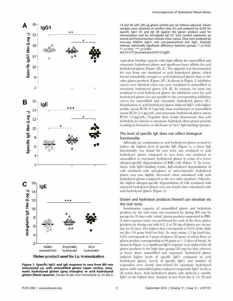
Acid hydrolyzed gluten induces the highest level ofspecific IgE by the i.p. route
Although unmodified gluten induced a statistically significant
higher IgG1 response compared to the acid hydrolyzed gluten all
three products induced a high level of specific IgG1 in all rats
when immunized i.p. (Figure 1A). In contrast, the highest level of
specific IgE was obtained by immunization with acid hydrolyzed
gluten (Figure 1B). ELISAs for measurement of specific IgE
against the different gluten products are not necessarily compa-
rable as it is uncertain whether biotin has been equally coupled to
the mixture of proteins (and hydrolysis products) present in the
gluten products. However, acid hydrolyzed gluten also induced the
highest level of anti-gliadin IgE (Figure 1C).
Acid hydrolysis of gluten discloses or produces newepitopes
Examination of IgG1-binding capacity showed that sera from
rats sensitized to unmodified or enzymatic hydrolyzed gluten had
Immunogenicity of Hydrolyzed Wheat Gluten
PLOS ONE | www.plosone.org 4 September 2014 | Volume 9 | Issue 9 | e107137
equivalent binding capacity with high affinity for unmodified and
enzymatic hydrolyzed gluten and significant lower affinity for acid
hydrolyzed gluten (Figure 2D, E). The opposite was demonstrated
for sera from rats sensitized to acid hydrolyzed gluten, which
bound remarkably stronger to acid hydrolyzed gluten than to the
other gluten products (Figure 2F). As shown in Figure 2, inhibition
curves were identical when rats were sensitized to unmodified or
enzymatic hydrolyzed gluten (2A, B). In contrast, for most rats
sensitized to acid hydrolyzed gluten, the inhibition curve for acid
hydrolyzed gluten was not parallel to the corresponding inhibition
curves for unmodified and enzymatic hydrolyzed gluten (2C).
Sensitization to acid hydrolyzed gluten induced IgG1 with higher
avidity (mean IC50: 0.3 mg/mL) than sensitization to unmodified
(mean IC50: 2.4 mg/mL) and enzymatic hydrolyzed gluten (mean
IC50: 1.2 mg/mL). Together these results demonstrate that acid
hydrolysis in contrast to enzymatic hydrolysis alters gluten proteins
resulting in formation or disclosure of ‘new’ IgG-binding epitopes.
The level of specific IgE does not reflect biologicalfunctionality
Although i.p. sensitization to acid hydrolyzed gluten seemed to
induce the highest level of specific IgE (Figure 1), a lower IgE
functionality was found for sera from rats sensitized to acid
hydrolyzed gluten compared to sera from rats sensitized to
unmodified or enzymatic hydrolyzed gluten in terms of a lower
allergen-specific degranulation of RBL cells (Figure 3). In accor-
dance with IgG1-binding results, IgE-mediated degranulation of
cells sensitized with anti-gluten or anti-enzymatic hydrolyzed
gluten sera was slightly decreased when stimulated with acid
hydrolyzed gluten compared to the two other products. Likewise,
the highest allergen-specific degranulation of cells sensitized with
anti-acid hydrolyzed gluten sera was found when stimulated with
acid hydrolyzed gluten (Figure 3).
Gluten and hydrolysis products thereof can sensitize viathe oral route
Sensitization capacity of unmodified gluten and hydrolysis
products by the oral route was examined by dosing BN rats by
gavage for 35 days with ‘whole’ gluten products suspended in PBS.
A dose-response study was performed for each of the three gluten
products by dosing rats with 0.2, 2 or 20 mg of gluten per rat per
day for 35 days. The highest dose corresponds to 0.2% of the daily
rat diet (10 gram feed/rat/day). In man eating 1.5 kg food/day,
0.2% corresponds to 3 gram of gluten (30 gram of wheat flour) or
gluten product corresponding to 60 gram or 1–2 slices of bread. As
shown in Figure 4, a significant IgG1 response were induced for all
gluten products in the high dose groups (20 mg/rat/day) whereas
at lower doses unmodified and enzymatic hydrolyzed gluten
induced higher levels of specific IgG1 compared to acid
hydrolyzed gluten. Levels of specific IgG1 and number of
responders were clearly dose-related for enzymatic hydrolyzed
gluten while unmodified gluten induced comparable IgG1 levels at
all tested doses. Acid hydrolyzed gluten only induced a notable
IgG1 at the highest dose. Analysis of sera from day 0, 14, 28 and
Figure 1. Specific IgG1 and IgE response in sera from BN ratsimmunized i.p. with unmodified gluten (open circles), enzy-matic hydrolyzed gluten (grey triangles) or acid hydrolyzedgluten (black squares). Groups of rats were immunized i.p. on day 0,
14 and 28 with 200 mg gluten protein per rat without adjuvant. Serumsamples were obtained at sacrifice (day 35) and analyzed by ELISA forspecific IgG1 (A) and IgE (B) against the gluten product used forimmunization and for anti-gliadin IgE (C). Each symbol represents ananimal and horizontal bars indicate mean values. Data were analyzed byone-way ANOVA (IgG1) and non-parametrical test (IgE). Asterisksindicate statistically significant difference between groups; *: p#0.05,**: p#0.01, ***: p#0.001.doi:10.1371/journal.pone.0107137.g001
Immunogenicity of Hydrolyzed Wheat Gluten
PLOS ONE | www.plosone.org 5 September 2014 | Volume 9 | Issue 9 | e107137
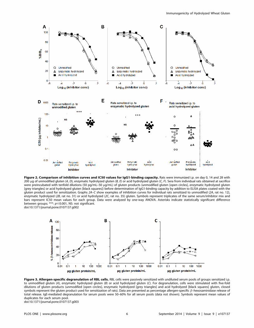
Figure 2. Comparison of inhibition curves and IC50 values for IgG1-binding capacity. Rats were immunized i.p. on day 0, 14 and 28 with200 mg of unmodified gluten (A, D), enzymatic hydrolyzed gluten (B, E) or acid hydrolyzed gluten (C, F). Sera from individual rats obtained at sacrificewere preincubated with tenfold dilutions (50 pg/mL–50 mg/mL) of gluten products (unmodified gluten [open circles], enzymatic hydrolyzed gluten[grey triangles] or acid hydrolyzed gluten [black squares]) before determination of IgG1-binding capacity by addition to ELISA plates coated with thegluten product used for sensitization. Graphs 2A–C show examples of inhibition curves for individual rats sensitized to unmodified (2A, rat no. 12),enzymatic hydrolyzed (2B, rat no. 31) or acid hydrolyzed (2C, rat no. 35) gluten. Symbols represent triplicates of the same serum/inhibitor mix andbars represent IC50 mean values for each group. Data were analyzed by one-way ANOVA. Asterisks indicate statistically significant differencebetween groups; ***: p#0.001, NS: not significant.doi:10.1371/journal.pone.0107137.g002
Figure 3. Allergen-specific degranulation of RBL cells. RBL cells were passively sensitized with undiluted serum pools of groups sensitized i.p.to unmodified gluten (A), enzymatic hydrolyzed gluten (B) or acid hydrolyzed gluten (C). For degranulation, cells were stimulated with five-folddilutions of gluten products (unmodified [open circles], enzymatic hydrolyzed [grey triangles] and acid hydrolyzed [black squares] gluten, closedsymbols represent the gluten product used for sensitization of rats). Data are presented as percentage allergen-specific b–hexosaminidase release oftotal release. IgE-mediated degranulation for serum pools were 50–60% for all serum pools (data not shown). Symbols represent mean values ofduplicates for each serum pool.doi:10.1371/journal.pone.0107137.g003
Immunogenicity of Hydrolyzed Wheat Gluten
PLOS ONE | www.plosone.org 6 September 2014 | Volume 9 | Issue 9 | e107137
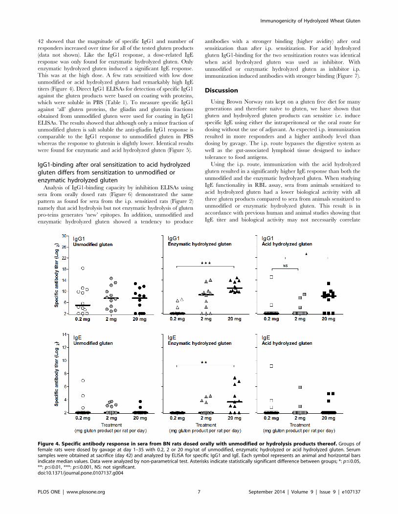
42 showed that the magnitude of specific IgG1 and number of
responders increased over time for all of the tested gluten products
(data not shown). Like the IgG1 response, a dose-related IgE
response was only found for enzymatic hydrolyzed gluten. Only
enzymatic hydrolyzed gluten induced a significant IgE response.
This was at the high dose. A few rats sensitized with low dose
unmodified or acid hydrolyzed gluten had remarkably high IgE
titers (Figure 4). Direct IgG1 ELISAs for detection of specific IgG1
against the gluten products were based on coating with proteins,
which were soluble in PBS (Table 1). To measure specific IgG1
against ‘all’ gluten proteins, the gliadin and glutenin fractions
obtained from unmodified gluten were used for coating in IgG1
ELISAs. The results showed that although only a minor fraction of
unmodified gluten is salt soluble the anti-gliadin IgG1 response is
comparable to the IgG1 response to unmodified gluten in PBS
whereas the response to glutenin is slightly lower. Identical results
were found for enzymatic and acid hydrolyzed gluten (Figure 5).
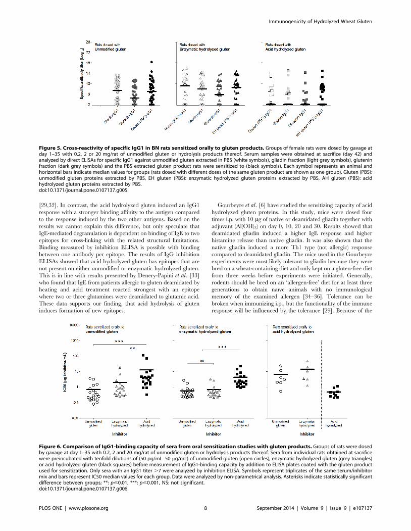
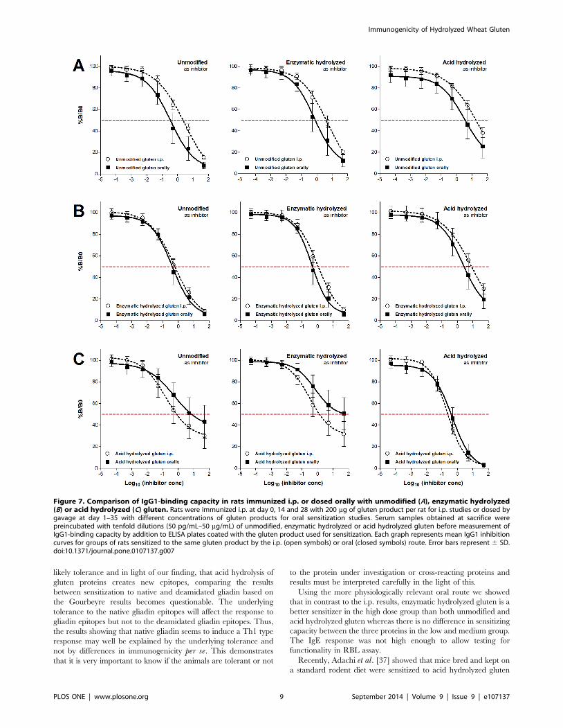
IgG1-binding after oral sensitization to acid hydrolyzedgluten differs from sensitization to unmodified orenzymatic hydrolyzed gluten
Analysis of IgG1-binding capacity by inhibition ELISAs using
sera from orally dosed rats (Figure 6) demonstrated the same
pattern as found for sera from the i.p. sensitized rats (Figure 2)
namely that acid hydrolysis but not enzymatic hydrolysis of gluten
pro-teins generates ‘new’ epitopes. In addition, unmodified and
enzymatic hydrolyzed gluten showed a tendency to produce
antibodies with a stronger binding (higher avidity) after oral
sensitization than after i.p. sensitization. For acid hydrolyzed
gluten IgG1-binding for the two sensitization routes was identical
when acid hydrolyzed gluten was used as inhibitor. With
unmodified or enzymatic hydrolyzed gluten as inhibitor i.p.
immunization induced antibodies with stronger binding (Figure 7).
Discussion
Using Brown Norway rats kept on a gluten free diet for many
generations and therefore naıve to gluten, we have shown that
gluten and hydrolyzed gluten products can sensitize i.e. induce
specific IgE using either the intraperitoneal or the oral route for
dosing without the use of adjuvant. As expected i.p. immunization
resulted in more responders and a higher antibody level than
dosing by gavage. The i.p. route bypasses the digestive system as
well as the gut-associated lymphoid tissue designed to induce
tolerance to food antigens.
Using the i.p. route, immunization with the acid hydrolyzed
gluten resulted in a significantly higher IgE response than both the
unmodified and the enzymatic hydrolyzed gluten. When studying
IgE functionality in RBL assay, sera from animals sensitized to
acid hydrolyzed gluten had a lower biological activity with all
three gluten products compared to sera from animals sensitized to
unmodified or enzymatic hydrolyzed gluten. This result is in
accordance with previous human and animal studies showing that
IgE titer and biological activity may not necessarily correlate
Figure 4. Specific antibody response in sera from BN rats dosed orally with unmodified or hydrolysis products thereof. Groups offemale rats were dosed by gavage at day 1–35 with 0.2, 2 or 20 mg/rat of unmodified, enzymatic hydrolyzed or acid hydrolyzed gluten. Serumsamples were obtained at sacrifice (day 42) and analyzed by ELISA for specific IgG1 and IgE. Each symbol represents an animal and horizontal barsindicate median values. Data were analyzed by non-parametrical test. Asterisks indicate statistically significant difference between groups; *: p#0.05,**: p#0.01, ***: p#0.001, NS: not significant.doi:10.1371/journal.pone.0107137.g004
Immunogenicity of Hydrolyzed Wheat Gluten
PLOS ONE | www.plosone.org 7 September 2014 | Volume 9 | Issue 9 | e107137
[29,32]. In contrast, the acid hydrolyzed gluten induced an IgG1
response with a stronger binding affinity to the antigen compared
to the response induced by the two other antigens. Based on the
results we cannot explain this difference, but only speculate that
IgE-mediated degranulation is dependent on binding of IgE to two
epitopes for cross-linking with the related structural limitations.
Binding measured by inhibition ELISA is possible with binding
between one antibody per epitope. The results of IgG inhibition
ELISAs showed that acid hydrolyzed gluten has epitopes that are
not present on either unmodified or enzymatic hydrolyzed gluten.
This is in line with results presented by Denery-Papini et al. [33]
who found that IgE from patients allergic to gluten deamidated by
heating and acid treatment reacted strongest with an epitope
where two or three glutamines were deamidated to glutamic acid.
These data supports our finding, that acid hydrolysis of gluten
induces formation of new epitopes.
Gourbeyre et al. [6] have studied the sensitizing capacity of acid
hydrolyzed gluten proteins. In this study, mice were dosed four
times i.p. with 10 mg of native or deamidated gliadin together with
adjuvant (Al(OH)3) on day 0, 10, 20 and 30. Results showed that
deamidated gliadin induced a higher IgE response and higher
histamine release than native gliadin. It was also shown that the
native gliadin induced a more Th1 type (not allergic) response
compared to deamidated gliadin. The mice used in the Gourbeyre
experiments were most likely tolerant to gliadin because they were
bred on a wheat-containing diet and only kept on a gluten-free diet
from three weeks before experiments were initiated. Generally,
rodents should be bred on an ‘allergen-free’ diet for at least three
generations to obtain naıve animals with no immunological
memory of the examined allergen [34–36]. Tolerance can be
broken when immunizing i.p., but the functionality of the immune
response will be influenced by the tolerance [29]. Because of the
Figure 5. Cross-reactivity of specific IgG1 in BN rats sensitized orally to gluten products. Groups of female rats were dosed by gavage atday 1–35 with 0.2, 2 or 20 mg/rat of unmodified gluten or hydrolysis products thereof. Serum samples were obtained at sacrifice (day 42) andanalyzed by direct ELISAs for specific IgG1 against unmodified gluten extracted in PBS (white symbols), gliadin fraction (light grey symbols), gluteninfraction (dark grey symbols) and the PBS extracted gluten product rats were sensitized to (black symbols). Each symbol represents an animal andhorizontal bars indicate median values for groups (rats dosed with different doses of the same gluten product are shown as one group). Gluten (PBS):unmodified gluten proteins extracted by PBS, EH gluten (PBS): enzymatic hydrolyzed gluten proteins extracted by PBS, AH gluten (PBS): acidhydrolyzed gluten proteins extracted by PBS.doi:10.1371/journal.pone.0107137.g005
Figure 6. Comparison of IgG1-binding capacity of sera from oral sensitization studies with gluten products. Groups of rats were dosedby gavage at day 1–35 with 0.2, 2 and 20 mg/rat of unmodified gluten or hydrolysis products thereof. Sera from individual rats obtained at sacrificewere preincubated with tenfold dilutions of (50 pg/mL–50 mg/mL) of unmodified gluten (open circles), enzymatic hydrolyzed gluten (grey triangles)or acid hydrolyzed gluten (black squares) before measurement of IgG1-binding capacity by addition to ELISA plates coated with the gluten productused for sensitization. Only sera with an IgG1 titer .7 were analyzed by inhibition ELISA. Symbols represent triplicates of the same serum/inhibitormix and bars represent IC50 median values for each group. Data were analyzed by non-parametrical analysis. Asterisks indicate statistically significantdifference between groups; **: p#0.01, ***: p#0.001, NS: not significant.doi:10.1371/journal.pone.0107137.g006
Immunogenicity of Hydrolyzed Wheat Gluten
PLOS ONE | www.plosone.org 8 September 2014 | Volume 9 | Issue 9 | e107137
likely tolerance and in light of our finding, that acid hydrolysis of
gluten proteins creates new epitopes, comparing the results
between sensitization to native and deamidated gliadin based on
the Gourbeyre results becomes questionable. The underlying
tolerance to the native gliadin epitopes will affect the response to
gliadin epitopes but not to the deamidated gliadin epitopes. Thus,
the results showing that native gliadin seems to induce a Th1 type
response may well be explained by the underlying tolerance and
not by differences in immunogenicity per se. This demonstrates
that it is very important to know if the animals are tolerant or not
to the protein under investigation or cross-reacting proteins and
results must be interpreted carefully in the light of this.
Using the more physiologically relevant oral route we showed
that in contrast to the i.p. results, enzymatic hydrolyzed gluten is a
better sensitizer in the high dose group than both unmodified and
acid hydrolyzed gluten whereas there is no difference in sensitizing
capacity between the three proteins in the low and medium group.
The IgE response was not high enough to allow testing for
functionality in RBL assay.
Recently, Adachi et al. [37] showed that mice bred and kept on
a standard rodent diet were sensitized to acid hydrolyzed gluten
Figure 7. Comparison of IgG1-binding capacity in rats immunized i.p. or dosed orally with unmodified (A), enzymatic hydrolyzed(B) or acid hydrolyzed (C) gluten. Rats were immunized i.p. at day 0, 14 and 28 with 200 mg of gluten product per rat for i.p. studies or dosed bygavage at day 1–35 with different concentrations of gluten products for oral sensitization studies. Serum samples obtained at sacrifice werepreincubated with tenfold dilutions (50 pg/mL–50 mg/mL) of unmodified, enzymatic hydrolyzed or acid hydrolyzed gluten before measurement ofIgG1-binding capacity by addition to ELISA plates coated with the gluten product used for sensitization. Each graph represents mean IgG1 inhibitioncurves for groups of rats sensitized to the same gluten product by the i.p. (open symbols) or oral (closed symbols) route. Error bars represent 6 SD.doi:10.1371/journal.pone.0107137.g007
Immunogenicity of Hydrolyzed Wheat Gluten
PLOS ONE | www.plosone.org 9 September 2014 | Volume 9 | Issue 9 | e107137
but not to unmodified gluten after transdermal exposure.
Administration of gluten together with SDS increased sensitization
capacity of gluten thus indicating that an increased solubility or
transdermal permeability of gluten may be important for
sensitization. In Japan, the use of facial soap containing acid
hydrolyzed wheat has resulted in many cases of sensitization
pointing to the skin or mucosal surfaces as the likely route of
sensitization [20,21]. The most important risk factor for develop-
ing wheat allergy in persons using facial soap with acid hydrolyzed
wheat was the number of soap bars used [38]. In contrast to the
European experience, Japanese patients also developed allergy to
wheat as WDEIA [20,21]. In an experiment, Strid et al. [39] were
able to break oral tolerance in mice when the allergen (peanut) was
dosed on tape stripped skin. It seems that in the Japanese patients,
allergic skin reactions caused by the acid hydrolyzed wheat protein
have contributed to break the oral tolerance to epitopes on native
wheat.
We have used commercial enzyme and commercial acid
hydrolyzed gluten. Although our results on acid hydrolyzed gluten
are supported by others [6,21,24], generalization of results
between different enzyme hydrolyzed products or different acid
hydrolyzed products may be done with caution. The results by
Denery-Papini et al. [24] show the degree of deamidation to be
crucial for the changed allergenic properties of (acid) hydrolyzed
gluten. Our results support these findings.
In conclusion, we have shown that in rats not tolerant to gluten
i.p. immunization results in statistically significant difference in the
specific IgE response with acid hydrolyzed . enzymatic hydro-
lyzed . unmodified gluten. This result is not reflected in the
biological reactivity where unmodified = nzymatic hydrolyzed .
acid hydrolyzed gluten. In the more physiological relevant oral
route enzymatic hydrolyzed gluten gives a higher IgE response in
the high dose group. There is no difference between the proteins in
the low and medium dose groups. As we do not know the dose-
response relationship in sensitization it is difficult to interpret the
significance of the different dose-response relations in IgE response
between unmodified and acid hydrolyzed gluten (no dose
response) and enzymatic hydrolyzed gluten (clear dose response).
The results also support the existence of ‘new’ epitopes formed
by deamidation on acid hydrolyzed gluten compared to unmod-
ified and enzymatic hydrolyzed gluten. This makes it possible to be
tolerant to gluten (be able to eat wheat products) and develop an
allergic reaction to acid hydrolyzed gluten. There are several
reports describing sensitization in wheat tolerant humans to
hydrolyzed gluten via the skin [7,14,15,18–21]. It is not always
very clear whether sensitization in wheat tolerant subjects was
caused by acid hydrolyzed or enzyme hydrolyzed gluten because
the type of hydrolysis is not always clearly specified. Our results
indicate that sensitization to acid hydrolyzed gluten is possible in
wheat tolerant subjects because of new epitopes. Likewise our
results indicate that sensitization to enzyme hydrolyzed gluten is
unlikely in wheat tolerant subjects because of the identical
epitopes. As with other proteins, our results show that a
sensitization route bypassing the gut is more efficient for all three
gluten products tested. This difference is most striking for acid
hydrolyzed gluten.
Acknowledgments
The authors thank Anne Ørngren, Eva Ferdinansen, Elise Navntoft, Maja
Danielsen, Eigil Frank and Kenneth Worm (National Food Institute, DTU,
Denmark) for excellent technical assistance and Anne Blicher (Enzyme and
Protein Chemistry, Department of Systems Biology, DTU, Denmark) for
performing amino acid analysis.
Author Contributions
Conceived and designed the experiments: SK CBM. Performed the
experiments: SK NBA TFR. Analyzed the data: SK NBA TFR SJ CBM.
Contributed reagents/materials/analysis tools: SJ. Contributed to the
writing of the manuscript: SK NBA TFR SJ CBM.
References
1. Belitz H-D, Grosch W, Schieberle P (2004) Food Chemistry, 3rd revised Edn,Berlin: Springer.
2. Shils ME, Shike M, Ross AC, Caballero B, Cousins RJ (2006) Modern nutrition
in health and disease, 10th Edition, 50th anniversary Edition. Philadelphia:
Lippincott Williams and Wilkins, Wolters Kluwer.
3. Wieser H (2007) Chemistry of gluten proteins. Food Microbiol 24: 115–119.
4. Tatham AS, Shewry PR (2008) Allergens in wheat and related cereals. Clin ExpAllergy 38: 1712–1726.
5. Mills ENC, Sancho AI, Rigby NM, Jenkins JA, Mackie AR (2009) Impact of
food processing on the structural and allergenic properties of food allergens. MolNutr Food Res 53: 963–969.
6. Gourbeyre P, Denery-Papini S, Larre C, Gaudin JC, Brossard C, et al. (2012)
Wheat gliadins modified by deamidation are more efficient than native gliadinsin inducing a Th2 response in Balb/c mice experimentally sensitized to wheat
allergens. Mol Nutr Food Res 56: 336–344.
7. Varjonen E, Petman L, Makinen-Kiljunen S (2000) Immediate contact allergy
from hydrolyzed wheat in a cosmetic cream. Allergy 55: 294–296.
8. Finley JW, Hopkins DT (1985) Digestibility and amino acid availability incereals and oilseeds. Minnesota, St. Paul: American Association of Cereal
Chemists.
9. Mittag D, Niggemann B, Sander I, Reese I, Fiedler E-M, et al. (2004)Immunoglobulin E-reactivity of wheat-allergic subjects (baker’s asthma, food
allergy, wheat-dependent, exercise-induced anaphylaxis) to wheat proteinfractions with different solubility and digestibility. Mol Nutr Food Res 48:
380–389.
10. Kumagai H, Suda A, Sakurai H, Kumagai H, Arai S, et al. (2007) Improvement
of digestibility, reduction in allergenicity, and induction of oral tolerance ofwheat gliadin by deamidation. Biosci Biotechnol Biochem 71: 977–985.
11. Lauriere M, Pecquet C, Boulenc E, Bouchez-Mahiout I, Snegaroff J, et al. (2007)
Genetic differences in omega-gliadins involved in two different immediate foodhypersensitivities to wheat. Allergy 62: 890–896.
12. Palosuo K (2003) Update on wheat hypersensitivity. Curr Opin Allergy Clin
Immunol 3: 205–209.
13. Leduc V, Moneret-Vautrin D-A, Guerin L, Morisset M, Kanny G (2003)Anaphylaxis to wheat isolates: immunochemical study of a case proved by means
of double-blind, placebo-controlled food challenge. J Allergy Clin Immunol 111:
897–899.
14. Lauriere M, Pecquet C, Bouchez-Mahiout I, Snegaroff J, Bayrou O, et al. (2006)
Hydrolysed wheat proteins present in cosmetics can induce immediate
hypersensitivities. Contact Dermatitis 54: 283–289.
15. Bouchez-Mahiout I, Pecquet C, Kerre S, Snegaroff J, Raison-Peyron N, et al.
(2010) High molecular weight entities in industrial wheat protein hydrolysates
are immunoreactive with IgE from allergic patients. J Agric Food Chem 58:
4207–4215.
16. Pelkonen AS, Makinen-Kiljunen S, Hilvo S, Siltanen M, Makela MJ (2011)
Severe allergic reaction to gluten hydrolysate without reaction to wheat. Ann
Allergy Asthma Immunol 106: 343–344.
17. Shinoda J, Inomata N, Chinuki Y, Morita E, Ikezawa Z (2012) Case of allergy
due to hydrolyzed wheat proteins in commercial boiled pork. J Dermatol 39:
724–726.
18. Pecquet C, Bayrou O, Vigan M, Raison N, Lauriere M (2004) Hydrolysed
wheat protein: a new allergen in cosmetics and food. Contact Dermatitis 50:
182–183.
19. Chinuki Y, Murata S, Morita E (2011) A case of wheat-dependent exercise-
induced anaphylaxis sensitized with hydrolysed wheat protein in a soap. Contact
Dermatitis 65: 55–57.
20. Fukutomi Y, Itagaki Y, Taniguchi M, Saito A, Yasueda H, et al. (2011)
Rhinoconjunctival sensitization to hydrolysed wheat protein in facial soap can
induce wheat-dependent excersise-induced anaphylaxis. J Allergy Clin Immunol
127: 531–533.
21. Nakamura R, Nakamura R, Sakai S, Adachi R, Hachisuka A, et al. (2013)
Tissue transglutaminase generates deamidated epitopes on gluten, increasing
reactivity with hydrolysed wheat protein-sensitized IgE. Allergy Clin Immunol
132: 1436–1438.
22. Battais F, Mothes T, Moneret-Vautrin DA, Pineau F, Kanny G, et al. (2005)
Identification of IgE-binding epitopes on gliadins for patients with food allergy to
wheat. Allergy 60: 815–821.
23. Pastorello EA, Farioli L, Conti A, Pravettoni V, Bonomi S, et al. (2007) Wheat
IgE-mediated food allergy in European patients: a-amylase inhibitors, lipid
Immunogenicity of Hydrolyzed Wheat Gluten
PLOS ONE | www.plosone.org 10 September 2014 | Volume 9 | Issue 9 | e107137
transfer proteins and low-molecular-weight glutenins. Int Arch Allergy Immunol
144: 10–22.
24. Denery-Papini S, Bodinier M, Pineau F, Triballeau S, Tranquet O, et al. (2011)
Immunoglobulin-E-binding epitopes of wheat allergens in patients with food
allergy to wheat and in mice experimentally sensitized to wheat proteins. Clin
Exp Allergy 41: 1478–1492.
25. Bohle B (2004) T lymphocytes and food allergy. Mol Nutr Food Res 48: 424–
433.
26. Berin MC, Sicherer S (2011) Food allergy: mechanisms and therapeutics. Curr
Opin Immunol 23: 794–800.
27. Cianferoni A, Spergel JM (2009) Food allergy: review, classification and
diagnosis. Allergol Int 58: 457–66.
28. Barkholt V, Jensen AL (1989) Amino acid analysis: determination of cysteine
plus half-cysteine in proteins after hydrochloric acid hydrolysis with a disulfide
compound as additive. Anal Biochem 177: 318–322.
29. Kroghsbo S, Bøgh KL, Rigby NM, Mills EN, Rogers A, et al. (2011)
Sensitization with 7S globulins from peanut, hazelnut, soy or pea induces IgE
with different biological activities which are modified by soy tolerance. Int Arch
Allergy Immunol 155: 212–224.
30. Pilegaard K, Madsen C (2004) An oral Brown Norway rat model for food
allergy: comparison of age, sex, dosing volume, and allergen preparation.
Toxicology 196: 247–257.
31. Kroghsbo S, Madsen C, Poulsen M, Schrøder M, Kvist PH, et al. (2008)
Immunotoxicological studies of genetically modified rice expressing PHA-E
lectin or Bt toxin in Wistar rats. Toxicology 245: 24–34.
32. Shreffler WG, Beyer K, Chu TH, Burks AW, Sampson HA (2004) Microarray
immunoassay: association of clinical history, in vitro IgE function, andheterogeneity of allergenic peanut epitopes. J Allergy Clin Immunol 13: 776–
782.
33. Denery-Papini S, Bodinier M, Larre C, Brossard C, Pineau F, et al. (2012)Allergy to deamidated gluten in patients tolerant to wheat: specific epitopes
linked to deamidation. Allergy 67: 1023–32.34. Christensen HR, Brix S, Frøkiaer H (2004) Immune response in mice to ingested
soya protein: antibody production, oral tolerance and maternal transfer.
Br J Nutr 91: 725–732.35. Brix S, Christensen HR, Barkholt V, Frøkiaer H (2005) Effect of maternal
dietary cow’s milk on the immune response to beta-lactoglobulin in the offspring:a four-generation study in mice. Int Arch Allergy Immunol 136: 250–257.
36. De Jonge JD, Knippels LM, Ezendam J, Odink J, Penninks AH, et al. (2007)The importance of dietary control in the development of a peanut allergy model
in Brown Norway rats. Methods 41: 99–111.
37. Adachi R, Nakamura R, Sakai S, Fukutomi Y, Teshima R (2012) Sensitizationto acid-hydrolyzed wheat protein by transdermal administration to BALB/c
mice, and comparison with gluten. Allergy 67: 1392–1399.38. Fukutomi Y, Kishikawa R, Sugiyama A, Minami T, Taniguchi M, et al. (2014)
Akiyama K. Risk factors for the development of wheat allergy among individuals
who have used a facial soap containing hydrolysed wheat protein: case-controlstudy. EAACI Copenhagen Abstract 1617.
39. Strid J, Hourihane J, Kimber I, Callard R, Strobel S (2005) Epicutaneousexposure to peanut protein prevents oral tolerance and enhances allergic
sensitization. Clin Exp Allergy 35: 757–766.
Immunogenicity of Hydrolyzed Wheat Gluten
PLOS ONE | www.plosone.org 11 September 2014 | Volume 9 | Issue 9 | e107137