activation of surrogate death receptor signalling triggers peroxynitrite dependent
TRANSCRIPT

Activation of surrogate death receptor signalling triggers peroxynitrite-dependent execution of cis-platin resistant cancer cells
Name – Saurabh Shekhar
Course – Biotechnology
Reg. No. – B.T./293
1Department of Physiology, Yong Loo Lin School of Medicine, National University of Singapore, Singapore; 2NUS Graduate School for Integrative Sciences andEngineering, National University of Singapore, Singapore; 3Cancer Science Institute, National University Health System, Singapore; 4Equipe 11 Labellisée par la LigueContre le Cancer, Centre de Recherche des Cordeliers, Paris, France; 5Cell Biology.
Cell Death and Disease (2015) 6, e1926; doi:10.1038/cddis.2015.299& 2015 Macmillan Publishers Limited All rights reserved 2041-4889/15
S Seah1,2, ICC Low1, JL Hirpara3, K Sachaphibulkij1, G Kroemer4,5,6,7,8,9, C Brenner10 and S Pervaiz*,1,2,11,12
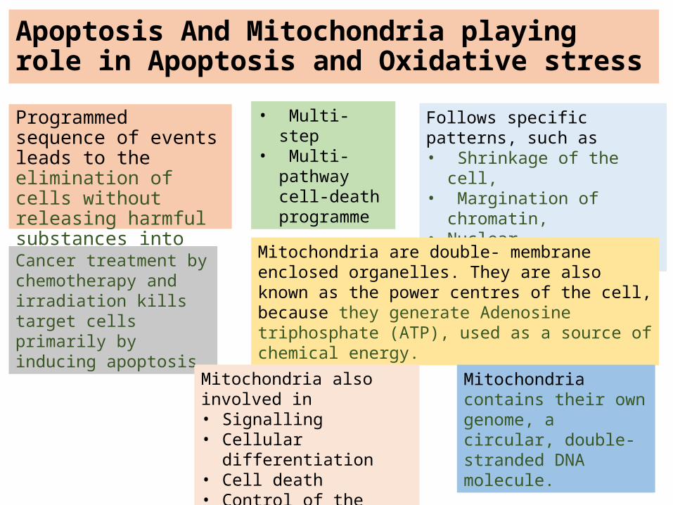
Apoptosis And Mitochondria playing role in Apoptosis and Oxidative stressProgrammed sequence of events leads to the elimination of cells without releasing harmful substances into the surrounding area.
• Multi-step• Multi-pathway
cell-death programme
Follows specific patterns, such as • Shrinkage of the cell,• Margination of chromatin,• Nuclear fragmentation.
Cancer treatment by chemotherapy and irradiation kills target cells primarily by inducing apoptosis.
Mitochondria are double- membrane enclosed organelles. They are also known as the power centres of the cell, because they generate Adenosine triphosphate (ATP), used as a source of chemical energy.
Mitochondria contains their own genome, a circular, double-stranded DNA molecule.
Mitochondria also involved in• Signalling • Cellular differentiation • Cell death • Control of the cell cycle• Cell growth

Pathway of mitochondrial induced apoptosis

IntroductionPlatinum-based drugs remain as the cornerstone of cancer chemotherapy; however, development of multidrug resistance presents a therapeutic challenge.
This study aims at understanding the molecular mechanisms underlying resistance to cisplatin and unravelling surrogate signalling networks that could revert sensitivity to apoptosis stimuli.
They made use of three different sets of cell lines, A549 and H2030 non-small cell lung cancer (NSCLC) and A2780 ovarian cancer cells and their cisplatin-resistant variants
Here they report that cisplatin-resistant cell lines displayed a multidrug-resistant phenotype.
Changes in mitochondrial metabolism and defective mitochondrial signalling were unravelled in the resistant cells.
More interestingly, a marked increase in sensitivity of the resistant cells to death receptor-induced apoptosis, in particular TRAIL (TNF-related apoptosis-inducing ligand)-mediated execution, was observed.

IntroductionA significant increase in the localization of TRAIL death receptor, DR4, to the lipid raft subdomains of plasma membrane was detected in the resistant variants.
Notably, preincubation of cells with TRAIL restored sensitivity of resistant cells to cisplatin.
Furthermore, exposure of cisplatin-resistant cells to TRAIL resulted in upregulation of inducible nitric oxide synthase (iNOS) and increase in nitric oxide (NO) production that triggered the generation of peroxynitrite (ONOO−).
Scavenging ONOO− rescued cells from TRAIL-induced apoptosis, thereby suggesting a critical role of ONOO− in TRAIL-induced execution of cisplatin-resistant cells.
These data provide compelling evidence for employing strategies to trigger death receptor signalling as a second-line treatment for cisplatin-resistant cancers.

RESULTS
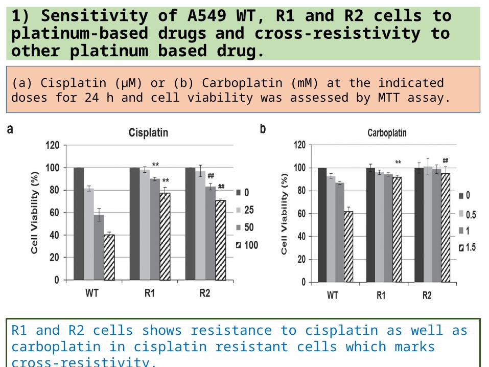
1) Sensitivity of A549 WT, R1 and R2 cells to platinum-based drugs and cross-resistivity to other platinum based drug.(a) Cisplatin (μM) or (b) Carboplatin (mM) at the indicated doses for 24 h and cell viability was assessed by MTT assay.
R1 and R2 cells shows resistance to cisplatin as well as carboplatin in cisplatin resistant cells which marks cross-resistivity.

1) Sensitivity of A549 WT, R1 and R2 cells to different concentrations of cisplatin.
(c) WT, R1 and R2 cells were treated with various concentrations of cisplatinfor 24, 48 and 72 h. Cell viability was determined by MTT assay and expressed as % of untreated control.
The cells R1 and R2 shows significant increase in cell viability than WT cells in different concentrations of cisplatin, hence shows resistance development towards drug.

1) (d) Cells were treated with increasing doses of cisplatin for 24 h and DNAcontent was analyzed by flow cytometry after staining with Propidium iodide (PI), ( y axis: events, x axis: PI linear fluorescence)
Propidium iodide (PI) staining of cisplatin-treated cells revealed higher DNA fragmentation, as indicated by the increased sub-G1 population, in cisplatin-treated WT cells relative to R1 and R2 cells which indicates resistivity of R1 and R2 cells toward Cisplatin.

1) e) WT, R1 and R2 cells were treated with the indicated doses of cisplatin for 24 h and caspase 3, 8 or 9 processing and PARP cleavage was determined by western blotting.
Caspases 8, 9 and 3 were activated in cisplatin-treated WT cells but not in the cisplatin-resistant clones. PARP (poly(ADP-ribose) polymerase), a substrate of caspase 3, was also cleaved in WT but not R1 and R2 cells shows deactivation of apoptotic pathway in cancer resistance cells R1 and R2.

2) Cisplatin-resistant cells exhibit cross-resistance to DNA-damaging agents but not death-receptor signaling. A549 WT and the cisplatin-resistant cells were treated with (a) 5-fluorouracil (μM), (b) etoposide (μM), (c) gemcitabine (mM), (d) TRAIL (ng/ml)

2) Cisplatin-resistant cells exhibit cross-resistance to DNA-damaging agents but not death-receptor signalling.(e) Fas activating antibody (μg/ml) at the indicated doses for 24 h. Cell viability was determined by MTT assay and expressed as % of untreated control.( *,# P ≤ 0.05 and **, ## P ≤ 0.005 compared with WT treated at respective dose).
(f) Western blot analysis of various proteins important in regulating mitochondria-mediated apoptotic signalling. β-Actin was used as a loading control.

2) (h) Respiratory complex 1 and 2 activities were measured in WT and R1 cells using the 96-well enzymatic-based microplate.
2) (g) Oxygen consumption of WT and R1 cells were assessed using a Clark electrode. State 3 respiration was initiated with addition of exogenous 0.2 mM ADP (arrow). The slope of the curve is a measure of the rate of oxygen consumption for a period of 20 min.

2) Cisplatin-resistant cells exhibit cross-resistance to DNA-damaging agents but not death-receptor signalling.(j) Respiratory complex 4 activities were measured in WT and R1 cells using the 96-well enzymatic-based microplate.
ΔmOD indicates change in maximum OD at 340 nm. Data shown are mean ± S.D. of at least three independent experiments.
Mitochondrial respiratory activity was significantly lower in cisplatin resistant cells; mitochondria oxygen consumption and activity of complex I, II and IV were reduced in R1 cells which suggests adaptive response which causes cisplatin resistant cells to evade mitochondrial driven execution.

3) TRAIL induces caspase-dependent apoptosis in cisplatin-resistant cells.
PE- Texas Red lin
(a) Cell cycle analysis – Cells (A549 WT, R1 and R2) were treated with increasing doses of TRAIL for 4 h and DNA content was analyzed by flow cytometry after staining with Propidium iodide (PI), ( y axis: events, x axis: PI linear fluorescence)
Exposure of R1 and R2 cells to TRAIL resulted in a dose-dependent increase in DNA fragmentation.

3) (b) A549 WT and R1 cells were treated with the indicated doses of TRAIL in a time-dependent manner. Western blotting for caspase 3 and caspase 8 processing as well as PARP cleavage was performed using GAPDH as the loading control.Exposure of R1 and R2 cells to TRAIL resulted in a dose-dependent increase in caspase 8 and 3 processing and PARP cleavage.

3) (c) Cisplatin-resistant R1 cells were preincubated with ZVAD-fmk (50 μM) for 1 h followed by 24 h of treatment with 50 ng/ml of TRAIL.
Whole-cell lysates were subjected to western blotting for caspase 3 and caspase 8 processing as well as PARP cleavage.
GAPDH was used as loading control.
3) TRAIL induces caspase-dependent apoptosis in cisplatin-resistant cells.
The pan-caspase inhibitor ZVAD-fmk(carbobenzoxy-valyl-alanyl-aspartyl-[O-methyl]- fluoromethylketone) abrogated TRAIL-induced cell death in R1 cells thus showing the crucial involvement of caspase 8 and 3 in death receptor-induced execution of cisplatin-resistant cells.

4) DR4, but not DR5 causes TRAIL mediated cell death in cisplatin resistance cells.
(a) Western blot analysis for various proteins important in regulating the extrinsic apoptotic pathway. GAPDH was used as loading control.
DR5 (TRAIL-R2) expression was clearly augmented in R1 andR2 cells, whereas a moderate increase in DR4 (TRAIL-R1) level was observed.
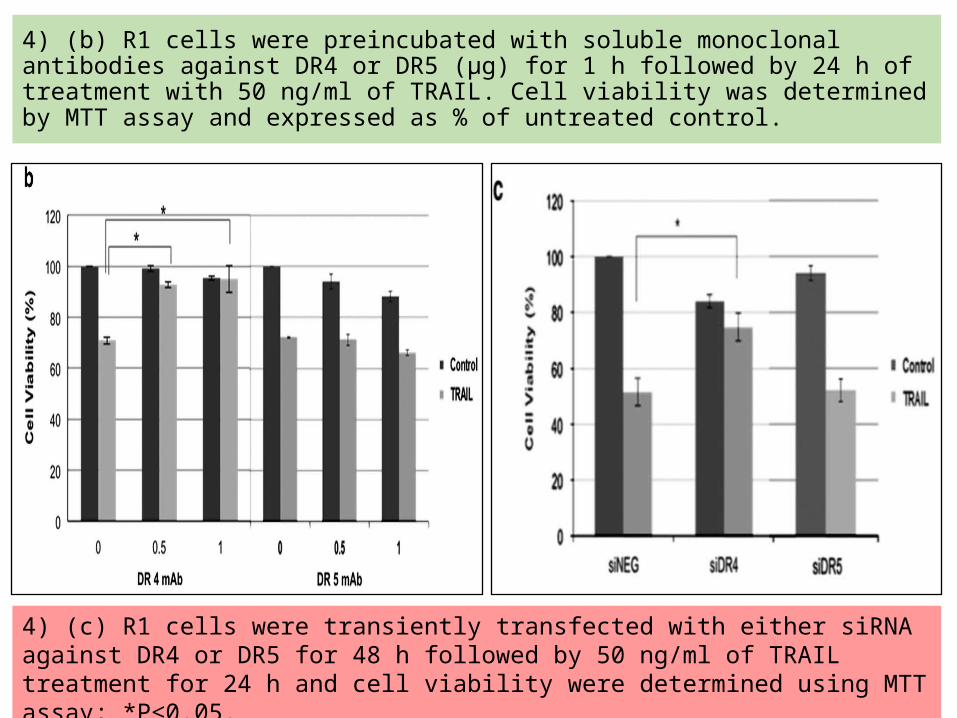
4) (b) R1 cells were preincubated with soluble monoclonal antibodies against DR4 or DR5 (μg) for 1 h followed by 24 h of treatment with 50 ng/ml of TRAIL. Cell viability was determined by MTT assay and expressed as % of untreated control.
4) (c) R1 cells were transiently transfected with either siRNA against DR4 or DR5 for 48 h followed by 50 ng/ml of TRAIL treatment for 24 h and cell viability were determined using MTT assay; *P≤0.05.

(d) R1 cells were preincubated withblocking antibodies against DR4 and DR5 for 1 h followed by 24 h of treatment with 50 ng/ml of TRAIL and lysates were subjected to western blot analysis for the assessment ofcaspase 3 and caspase 8 processing as well as PARP cleavage.
4) DR4, but not DR5 causes TRAIL mediated cell death in cisplatin resistance cells

(e) WT (top) and R1 cells (bottom) were treated with 50 ng/ml TRAIL for 15 min and subjected to discontinuous sucrose density gradients of Triton X-100 cell lysates for separation of lipid raft and non-raft fractions. One to 9 fractions were examined by western blots for the presence of DR4 and DR5 (first two rows). Lipid raft fractions 5 and 6 were identified by western blots. Flotillin and caveolin-1 were used as lipid raft markers.
4) DR4 is redistributed to lipid raft subdomains in R1 cells
Results indicate that DR4 and FADD colocalized with the same fractions as caveolin and flotillin in R1 cells even in the absence of TRAIL.

4) (f) Following TRAIL (50 ng/ml)treatment for 15 min, WT and R1 cells were subjected to discontinuous sucrose density gradients of Triton X-100 cell lysates for separation of lipid raft and non raft fractions.Fractions 4-6 were collected and immunoprecipitated with anti-caveolin-1 antibody. As shown, caveolin-1 and FADD protein expression were detected by immunoblotting.
4) (g) R1 cells were preincubated with the indicated doses of MCD for 1 h followed by 24 h of treatment with 50 ng/ml of TRAIL. Whole-cell lysates were subjected to western blotanalysis for the assessment of caspase 3 and caspase 8 processing as well as PARP cleavage. GAPDH was used as loading control. Data shown are mean ± S.D. of at leastthree independent experiments
These data indicate that DR4 aggregation at the lipid rafts is responsible for the enhanced sensitivity of cisplatin-resistant cells to death receptor signaling.

5) TRAIL-induced cell death in R1 cells involves the generation of reactive nitrogen species. (Data are shown as mean ± S.D. of fold differences of fluorescence from untreated cells for at least three independent experiments)
a) WT and R1 cells were treated with 50 ng/ml of TRAIL for 2 and 4 hours. Cells were subsequently harvested and analyzed by flow cytometry for ROS production using redox-sensitive probe DCFH-DA.
(b) R1 cells were preincubated with 50 μMFeTPPs for 1 h followed by 50 ng/ml of TRAIL for 2 or 4 h. Cells were subsequently harvested and analyzed by flow cytometry after loading with DCFH-DA.
Use of fluorescence probe (DCFH-DA) that primarily detects hydrogen peroxide (H2O2) and peroxynitrite (ONOO−), marked increase in DCF fluorescence in TRAIL-treatedR1 cells, compared with WT cells.

5) TRAIL-induced cell death in R1 cells involves the generation of reactive nitrogen species.
(c) R1 cells were preincubated with 50 μMFeTPPs for 1 h followed by 50 ng/ml of TRAIL for 2 or 4 h and loaded with NO-specific probe DAF.
(d) MitoSox before flow cytomteric analysis. Antimycin A (AA) was used as a positive control for mitochondrial O2.
These data suggests that NO generated upon TRAIL treatment reacts with endogenous mitochondrial O2− to produce ONOO−, as evidenced by the decline in MitoSOX fluorescence in R1 cells.

5) TRAIL-induced cell death in R1 cells involves the generation of reactive nitrogen species. (Data shown are mean ± S.D. of at least three independent experiments).
(e) Western blot analysis of iNOS following treatment of WT and R1 cells with 50 and 100 ng/ml of TRAIL.
(f) R1 cells were preincubated with FeTPPs (50 μM) for 1 h followed by 24 h of treatment with 50 ng/ml of TRAIL. Cell viability was determined by MTT assay and expressed as % of untreated control cells.
The level of inducible nitric oxide synthase (iNOS- one of the most common inducible sources ofintracellular NO) was upregulated in TRAIL-treated R1 cells lending further support to the involvement of NO and subsequently ONOO−.
Scavenging ONOO− by a priori treatment with FeTPPS significantly rescued R1 cells from TRAIL-induced cell death hence it shows the ONOO- is involved in TRAIL induced apoptosis.

5) TRAIL-induced cell death in R1 cells involves the generation of reactive nitrogen species.
(g) Western blot analysis of iNOS in R1 cells, pretreated with 50 μM ZVAD for 1 h followed by 50 ng/ml of TRAIL for 8 h.
(h) R1 cells were preincubated with FeTPPsfor 1 h followed by 24 h of treatment with 50 ng/ml of TRAIL and whole-cell lysates were subjected to western blot analysis for the assessment of caspase 3 and 8 processing as well as PARP cleavage.
ZVAD-fmk completely blocked TRAIL-mediated induction of iNOS and ONOO− production which suggests ZVAD-fmk and FeTPPS could inhibit TRAIL-induced ONOO− production or caspase activation.

6) TRAIL exposure restores apoptosis sensitivity of cisplatin-resistant (CR) cells.
(a) A549 R1 (b) H2030 CR and (c) A2780 CR cells were pretreated with varying doses ofTRAIL for 2 h and followed with the indicated doses of cisplatin for 24 h.Cell viability was assessed by MTT and expressed as % of untreated control cells. Data shown are mean ± S.D. of at least three independent experiments.A combination of low dose of cisplatin and TRAIL was significantly more potent against the three cisplatin-resistant cell lines as compared with the single-agent treatment.

The cell lysates from 6) (d) A549 R1, 6) (e) H2030 CR were collected to assay for caspase 3 and caspase 8 processing as well as PARP cleavage by western blot analysis. GAPDH was used as loading control.

6) TRAIL exposure restores apoptosis sensitivity of cisplatin-resistant (CR) cells.
(f) A2780 CR were collected to assay for caspase 3 and caspase 8 processing as well as PARP cleavage by western blot analysis.
GAPDH was used as loading control.
In agreement with these findings, combinatory treatment of cisplatin andTRAIL markedly enhanced the activities of caspases 8, 9 and 3 in the three cisplatin-resistant cell lines.

7) Schematic diagram of TRAIL sensitivity in WT cancer cells
In the WT cells, TRAIL receptors exist as monomers and upon TRAIL ligation, the receptors oligomerize to form the DISC and subsequently activation of pro-caspase 8.
However, because of over expression of anti-apoptotic proteins such as cFLIP, XIAP and cIAP2, apoptotic execution is impaired.

7) Schematic diagram of TRAIL sensitivity in cisplatin-resistant R1 cancer cells
In R1 cells, TRAIL receptors are redistributed to the lipid raft subdomain, and hence are ‘primed’ for rapid activation.
Upon ligation, the apoptotic signals are transduced promptly that involves downregulation of anti-apoptotic proteins as well as induction of an amplification signal via OONO− formation for efficient death execution.

Summary of Results• Cisplatin-induced apoptosis is blunted in R1 and R2 cells.• Cisplatin-resistant cells exhibit cross-resistance to triggers that
engage the intrinsic death pathway.• Remarkable upregulation of death receptor sensitivity in cisplatin-
resistant cells.• Redistribution of DR4 to the lipid rafts sensitizes cisplatin-resistant
R1 cells to TRAIL-induced apoptosis.• TRAIL-induced cell death in cisplatin-resistant R1 cells involves the
generation of reactive nitrogen species.• TRAIL sensitivity is not exclusive to cisplatin-resistant clones of A549
cell line.• Synergistic effect of TRAIL in combination with cisplatin in the
treatment of cisplatin-resistant cells.

Conclusion• Findings indicates evidence for an amplification of death receptor
signalling in cancer cells rendered resistant to cisplatin (Figure 7). • It involved clustering of death receptors in lipid raft subdomains and
their efficient activation upon exposure to the ligand resulting in downstream caspase activation.• Their results also shows intracellular ONOO− as a critical trigger
downstream of death receptor ligation. • Studies also shows exposure to TRAIL restored sensitivity of resistant
cells to cisplatin.
These data argue in favour of exploiting the enhanced sensitivity to death receptor-mediated apoptosis in the
therapeutic management of tumors that have developed resistance to platinum based drugs.

THANK YOU