activation of the transcription factor eb rescues ... · pdf fileactivation of the...
TRANSCRIPT
bas i c re sea r ch www.k idney - i n t e rna t i ona l . o rg
Activation of the transcription factor EB rescueslysosomal abnormalities in cystinotic kidney cells
Laura R. Rega1, Elena Polishchuk2, Sandro Montefusco2, Gennaro Napolitano2, Giulia Tozzi3,Jinzhong Zhang4, Francesco Bellomo1, Anna Taranta1, Anna Pastore5, Roman Polishchuk2,Fiorella Piemonte3, Diego L. Medina2, Sergio D. Catz4, Andrea Ballabio2 and Francesco Emma11Division of Nephrology and Dialysis, Bambino Gesù Children’s Hospital and Research Institute, Rome, Italy; 2Telethon Institute ofGenetics and Medicine, Pozzuoli (Naples), Italy; 3Unit for Muscular and Neurodegenerative Diseases, Bambino Gesù Children’s Hospitaland Research Institute, Rome, Italy; 4Department of Molecular and Experimental Medicine, The Scripps Research Institute, La Jolla,California, USA; and 5Laboratory of Proteomics and Metabolomics, Bambino Gesù Children’s Hospital and Research Institute, Rome, Italy
Nephropathic cystinosis is a rare autosomal recessivelysosomal storage disease characterized by accumulationof cystine into lysosomes secondary to mutations in thecystine lysosomal transporter, cystinosin. The defectinitially causes proximal tubular dysfunction (Fanconisyndrome) which in time progresses to end-stage renaldisease. Cystinotic patients treated with the cystine-depleting agent, cysteamine, have improved life expectancy,delayed progression to chronic renal failure, but persistenceof Fanconi syndrome. Here, we have investigated therole of the transcription factor EB (TFEB), a master regulatorof the autophagy-lysosomal pathway, in conditionallyimmortalized proximal tubular epithelial cells derived fromthe urine of a healthy volunteer or a cystinotic patient. Lackof cystinosin reduced TFEB expression and induced TFEBnuclear translocation. Stimulation of endogenous TFEBactivity by genistein, or overexpression of exogenousTFEB lowered cystine levels within 24 hours in cystinoticcells. Overexpression of TFEB also stimulated delayedendocytic cargo processing within 24 hours. Rescue ofother abnormalities of the lysosomal compartment wasobserved but required prolonged expression of TFEB. Theseabnormalities could not be corrected with cysteamine. Thus,these data show that the consequences of cystinosindeficiency are not restricted to cystine accumulation andsupport the role of TFEB as a therapeutic target for thetreatment of lysosomal storage diseases, in particular ofcystinosis.Kidney International (2016) 89, 862–873; http://dx.doi.org/10.1016/j.kint.2015.12.045
KEYWORDS: cystinosis; pediatric nephrology; proximal tubule; renal
pathology
ª 2016 International Society of Nephrology
Correspondence: L.R. Rega, Division of Nephrology and Dialysis, BambinoGesù Children’s Hospital, Piazza Sant’Onofrio, 4, 00165 Rome, Italy. E-mail:[email protected]
Received 11 May 2015; revised 22 December 2015; accepted 30December 2015
862
C ystinosis is a rare autosomal recessive lysosomal storagedisease (LSD) with an incidence of 0.5 to 1.0 per100,000 live births. The most severe and frequent form,
affecting w95% of patients, is the infantile nephropathiccystinosis (OMIM 219800).1 All clinical forms of cystinosisare caused by bi-allelic mutations in the CTNS gene, which islocated on chromosome 17p13. More than 100 mutationshave been reported;2 however, the most frequent mutation,found in northern European patients, is a large deletion of57,257 base pairs, involving the first 9 CTNS exons and partof exon 10.3 The CTNS gene encodes the lysosomal cystinetransporter, cystinosin, which is a 367 amino acid lysosomalprotein that contains 7 transmembrane domains.4 Cystinosinis a proton-cystine symporter devoted to the excretion of bothcystine and protons out of the lysosomal lumen.5 Becausecystinosin is ubiquitously expressed, its absence or malfunc-tioning causes accumulation of cystine into the lysosomesthroughout the body. Moreover, cystine is poorly soluble, thusits accumulation leads to the formation of crystals. Kidneysare one of the first organs to be affected in cystinotic patients,which typically present with clinical signs of Fanconi syn-drome, by 4 to 6 months of age. Fanconi syndrome is char-acterized by polyuria and abnormal urinary loss of aminoacids, glucose, low-molecular-weight and intermediate-weight proteins, and other solutes. If untreated, patientsprogress to end-stage renal disease by the age of 10 years.6 Themechanisms linking lysosomal cystine accumulation to celldysfunctions, in particular to the prominent proximal tubulardefect, remain unclear. Recent studies have generated varioushypotheses indicating that proximal tubule damage is asso-ciated with increased sensitivity to apoptosis,7,8 abnormalauthophagy,9,10 mitochondrial dysfunction,11 adenosinetriphosphate depletion,11 increased reactive oxygen speciesproduction,11 endoplasmic reticulum stress,12 endo-lysosomal dysfunctions,13 and cell dedifferentiation.14,15
Cysteamine was introduced in the late 1970s for thetreatment of cystinosis and has been the only therapy avail-able since then.16,17 However, foul odor and gastric side ef-fects of oral cysteamine make adherence extremely difficult.Moreover, cysteamine does not correct all the symptoms ofcystinosis; in particular, it does not have an impact on therenal Fanconi syndrome.1 Even well-treated patients will
Kidney International (2016) 89, 862–873
LR Rega et al.: Role of TFEB in cystinosis ba s i c re sea r ch
eventually progress to envisage renal disease and will requirekidney transplantation. Thus, novel therapeutic approachesare needed for cystinosis.
The recent discovery of a lysosomal gene network, thecoordinated lysosomal expression and regulation network andof its master regulator, transcription factor EB (TFEB), hasprovided new insights in the studies of LSD.18 TFEB activatesthe transcription of genes that encode lysosomal proteinsinvolved in several aspects of cellular clearance, such aslysosomal biogenesis, exocytosis, and autophagy, as well asnonlysosomal proteins involved in the degradation ofautophagy substrates.19 The role of TFEB in promotingcellular clearance has been proven in several diseases such asmultiple sulfatase deficiency,20 Pompe disease,21 and alpha-1-anti-trypsin deficiency.22 Together these findings provideproof of concepts that drugs that stimulate TFEB activity mayrepresent useful therapeutic tools to enhance mobilization ofaberrant storage materials in LSD.
Herein, we have investigated the impact of TFEB activationon 3 cystinotic phenotypes, namely lysosomal cystine accu-mulation, delayed processing of endocytic cargo and aberrantlysosomal compartment morphology. We have observed thatcystinosin-depleted cells have reduced expression of TFEB.Moreover, abnormalities of the lysosomal compartmentinduced by cystinosin deficiency are associated with TFEBnuclear translocation. Both chemical and genetic stimulationof TFEB activity promotes clearance of cystine storages within24 hours. Overexpression of TFEB stimulates also delayedcargo processing. Prolonged increased expression of TFEBcould also rescue other defects of the lysosomal compartmentin cystinotic cells that are not sensitive to cysteamine. Theseresults support the role of TFEB as a therapeutic target for thetreatment of LSDs and provide promising perspectives in thetreatment of cystinosis.
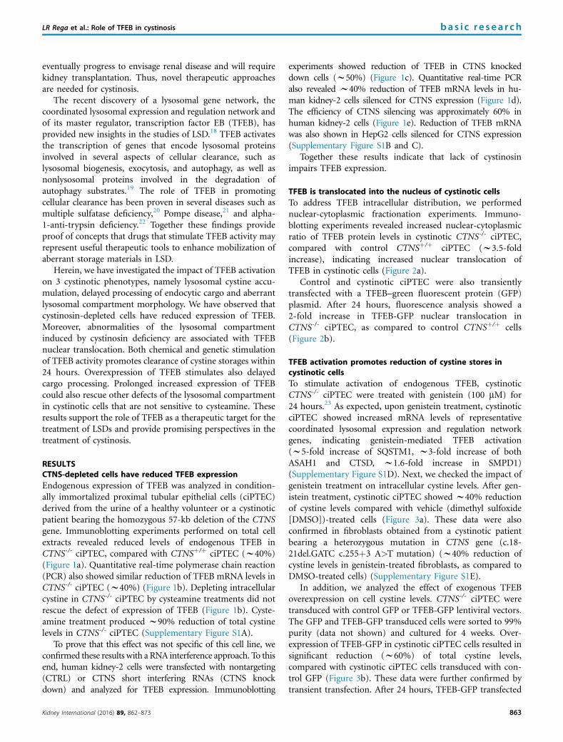
RESULTSCTNS-depleted cells have reduced TFEB expressionEndogenous expression of TFEB was analyzed in condition-ally immortalized proximal tubular epithelial cells (ciPTEC)derived from the urine of a healthy volunteer or a cystinoticpatient bearing the homozygous 57-kb deletion of the CTNSgene. Immunoblotting experiments performed on total cellextracts revealed reduced levels of endogenous TFEB inCTNS-/- ciPTEC, compared with CTNSþ/þ ciPTEC (w40%)(Figure 1a). Quantitative real-time polymerase chain reaction(PCR) also showed similar reduction of TFEB mRNA levels inCTNS-/- ciPTEC (w40%) (Figure 1b). Depleting intracellularcystine in CTNS-/- ciPTEC by cysteamine treatments did notrescue the defect of expression of TFEB (Figure 1b). Cyste-amine treatment produced w90% reduction of total cystinelevels in CTNS-/- ciPTEC (Supplementary Figure S1A).
To prove that this effect was not specific of this cell line, weconfirmed these results with a RNA interference approach. To thisend, human kidney-2 cells were transfected with nontargeting(CTRL) or CTNS short interfering RNAs (CTNS knockdown) and analyzed for TFEB expression. Immunoblotting
Kidney International (2016) 89, 862–873
experiments showed reduction of TFEB in CTNS knockeddown cells (w50%) (Figure 1c). Quantitative real-time PCRalso revealed w40% reduction of TFEB mRNA levels in hu-man kidney-2 cells silenced for CTNS expression (Figure 1d).The efficiency of CTNS silencing was approximately 60% inhuman kidney-2 cells (Figure 1e). Reduction of TFEB mRNAwas also shown in HepG2 cells silenced for CTNS expression(Supplementary Figure S1B and C).
Together these results indicate that lack of cystinosinimpairs TFEB expression.
TFEB is translocated into the nucleus of cystinotic cellsTo address TFEB intracellular distribution, we performednuclear-cytoplasmic fractionation experiments. Immuno-blotting experiments revealed increased nuclear-cytoplasmicratio of TFEB protein levels in cystinotic CTNS-/- ciPTEC,compared with control CTNSþ/þ ciPTEC (w3.5-foldincrease), indicating increased nuclear translocation ofTFEB in cystinotic cells (Figure 2a).
Control and cystinotic ciPTEC were also transientlytransfected with a TFEB–green fluorescent protein (GFP)plasmid. After 24 hours, fluorescence analysis showed a2-fold increase in TFEB-GFP nuclear translocation inCTNS-/- ciPTEC, as compared to control CTNSþ/þ cells(Figure 2b).
TFEB activation promotes reduction of cystine stores incystinotic cellsTo stimulate activation of endogenous TFEB, cystinoticCTNS-/- ciPTEC were treated with genistein (100 mM) for24 hours.23 As expected, upon genistein treatment, cystinoticciPTEC showed increased mRNA levels of representativecoordinated lysosomal expression and regulation networkgenes, indicating genistein-mediated TFEB activation(w5-fold increase of SQSTM1, w3-fold increase of bothASAH1 and CTSD, w1.6-fold increase in SMPD1)(Supplementary Figure S1D). Next, we checked the impact ofgenistein treatment on intracellular cystine levels. After gen-istein treatment, cystinotic ciPTEC showed w40% reductionof cystine levels compared with vehicle (dimethyl sulfoxide[DMSO])-treated cells (Figure 3a). These data were alsoconfirmed in fibroblasts obtained from a cystinotic patientbearing a heterozygous mutation in CTNS gene (c.18-21del.GATC c.255þ3 A>T mutation) (w40% reduction ofcystine levels in genistein-treated fibroblasts, as compared toDMSO-treated cells) (Supplementary Figure S1E).
In addition, we analyzed the effect of exogenous TFEBoverexpression on cell cystine levels. CTNS-/- ciPTEC weretransduced with control GFP or TFEB-GFP lentiviral vectors.The GFP and TFEB-GFP transduced cells were sorted to 99%purity (data not shown) and cultured for 4 weeks. Over-expression of TFEB-GFP in cystinotic ciPTEC cells resulted insignificant reduction (w60%) of total cystine levels,compared with cystinotic ciPTEC cells transduced with con-trol GFP (Figure 3b). These data were further confirmed bytransient transfection. After 24 hours, TFEB-GFP transfected
863
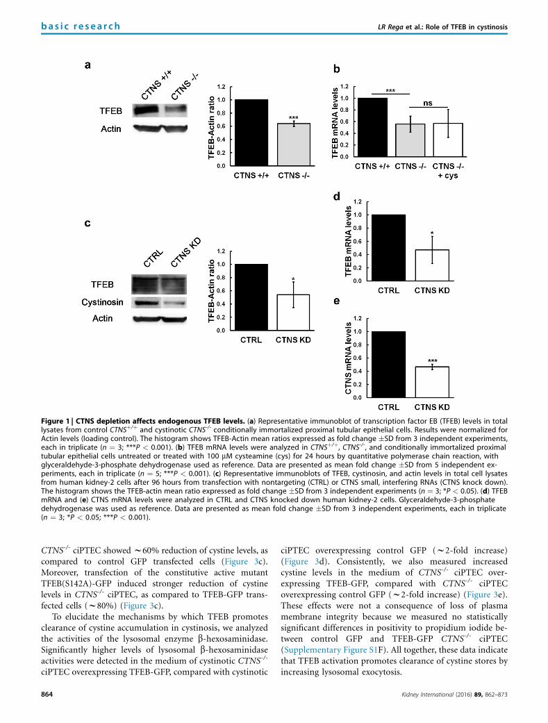
Figure 1 | CTNS depletion affects endogenous TFEB levels. (a) Representative immunoblot of transcription factor EB (TFEB) levels in totallysates from control CTNSþ/þ and cystinotic CTNS-/- conditionally immortalized proximal tubular epithelial cells. Results were normalized forActin levels (loading control). The histogram shows TFEB-Actin mean ratios expressed as fold change �SD from 3 independent experiments,each in triplicate (n ¼ 3; ***P < 0.001). (b) TFEB mRNA levels were analyzed in CTNSþ/þ, CTNS-/-, and conditionally immortalized proximaltubular epithelial cells untreated or treated with 100 mM cysteamine (cys) for 24 hours by quantitative polymerase chain reaction, withglyceraldehyde-3-phosphate dehydrogenase used as reference. Data are presented as mean fold change �SD from 5 independent ex-periments, each in triplicate (n ¼ 5; ***P < 0.001). (c) Representative immunoblots of TFEB, cystinosin, and actin levels in total cell lysatesfrom human kidney-2 cells after 96 hours from transfection with nontargeting (CTRL) or CTNS small, interfering RNAs (CTNS knock down).The histogram shows the TFEB-actin mean ratio expressed as fold change �SD from 3 independent experiments (n ¼ 3; *P < 0.05). (d) TFEBmRNA and (e) CTNS mRNA levels were analyzed in CTRL and CTNS knocked down human kidney-2 cells. Glyceraldehyde-3-phosphatedehydrogenase was used as reference. Data are presented as mean fold change �SD from 3 independent experiments, each in triplicate(n ¼ 3; *P < 0.05; ***P < 0.001).
bas i c re sea r ch LR Rega et al.: Role of TFEB in cystinosis
CTNS-/- ciPTEC showed w60% reduction of cystine levels, ascompared to control GFP transfected cells (Figure 3c).Moreover, transfection of the constitutive active mutantTFEB(S142A)-GFP induced stronger reduction of cystinelevels in CTNS-/- ciPTEC, as compared to TFEB-GFP trans-fected cells (w80%) (Figure 3c).
To elucidate the mechanisms by which TFEB promotesclearance of cystine accumulation in cystinosis, we analyzedthe activities of the lysosomal enzyme b-hexosaminidase.Significantly higher levels of lysosomal b-hexosaminidaseactivities were detected in the medium of cystinotic CTNS-/-
ciPTEC overexpressing TFEB-GFP, compared with cystinotic
864
ciPTEC overexpressing control GFP (w2-fold increase)(Figure 3d). Consistently, we also measured increasedcystine levels in the medium of CTNS-/- ciPTEC over-expressing TFEB-GFP, compared with CTNS-/- ciPTECoverexpressing control GFP (w2-fold increase) (Figure 3e).These effects were not a consequence of loss of plasmamembrane integrity because we measured no statisticallysignificant differences in positivity to propidium iodide be-tween control GFP and TFEB-GFP CTNS-/- ciPTEC(Supplementary Figure S1F). All together, these data indicatethat TFEB activation promotes clearance of cystine stores byincreasing lysosomal exocytosis.
Kidney International (2016) 89, 862–873
Figure 2 | TFEB nuclear translocation is increased in cystinotic cells. (a) Representative immunoblots showing transcription factor EB (TFEB)levels in both cytosolic (C) and nuclear (N) fractions from CTNSþ/þ and CTNS-/- conditionally immortalized proximal tubular epithelial cells.TFEB cytosolic and nuclear levels were normalized for glyceraldehyde-3-phosphate dehydrogenase (GAPDH) and histone H3 levels, respectively.The histogram shows TFEB nucleus-cytoplasm ratios expressed as mean �SD from 3 independent experiments, each in triplicate (n ¼ 3;*P < 0.05). (b) CTNSþ/þ and CTNS-/- conditionally immortalized proximal tubular epithelial cells were transiently transfected with TFEB–greenfluorescent protein (GFP) plasmid (green). Bar ¼ 10 mm. After 24 hours from transient transfection, TFEB nuclear fluorescence intensity wasmeasured by ImageJ software and expressed as percentage of total fluorescence intensity (normalized for the areas). Data are shown asmeans �SD from 5 independent experiments, each in duplicate (n ¼ 5; ***P < 0.001). GFP, green fluorescent protein.
LR Rega et al.: Role of TFEB in cystinosis ba s i c re sea r ch
TFEB activation stimulates cargo processing in cystinotic cellsTo study the impact of TFEB activation on cystinotic endo-cytosis dysfunction, we evaluated the impact of TFEB over-expression on receptor-associated protein (RAP)–glutathione-S-transferase (GST) cargo processing. First, weconfirmed delayed cargo processing in cystinotic cells.13 Tothis end, CTNSþ/þ and CTNS-/- ciPTEC were preincubatedwith RAP-GST ligand and transferred to 37 �C to allowinternalization of the ligand bound to the cell surface. Atindicated time points, cells were fixed and stained for RAP-GST (Figure 4a). In CTNS-/- ciPTEC, RAP-GST remainedvisible for significantly longer time (30 minutes) after inter-nalization, in comparison to control cells (Figure 4a and b).Then, we analyzed RAP-GST cargo processing in CTNS-/-
ciPTEC transfected with TFEB-GFP. After 30 minutes fromcargo internalization, RAP-GST structures showed significantreduced size in TFEB-GFP positive cells, if compared withthose in adjacent nontransfected cells (w50%) (Figure 4c and d).No statistically significant differences in the size of RAP-GSTstructures were observed between untransfected or TFEB-GFPtransfected CTNS-/- ciPTEC after 15 and 45 minutes from theshift to 37 �C (Figure 4c and d). Moreover, no statistically signif-icant differences in RAP-GST processing were highlightedbetween untransfected or control GFP transfected CTNS-/-
ciPTEC (data not shown).
Kidney International (2016) 89, 862–873
These data confirm delayed cargo processing in cystinotic cellsand show that TFEB overexpression is able to rescue this defect.
TFEB overexpression reduces the number and size oflysosomes in cystinotic cellsBased on the findings that TFEB promotes mobilization ofcystine depositions and enhances lysosomal exocytosis incystinotic cells, we wondered whether in these experimentalconditions the structure of the lysosomal compartment wasalso modified. We first checked differences in the number andsize of lysosomes between control and cystinotic cells byimmuno-electron microscopy (EM). To this end, control andcystinotic ciPTEC cells were labeled with anti-LAMP1 anti-body and prepared for ultrastructural analysis. CystinoticciPTEC cells showed increased number of LAMP1 positivestructures, as compared to control cells (the mean number ofLAMP1 positive structures/field was 6 � 0.3, in CTNSþ/þ and10 � 0.9, in CTNS-/-) (Figure 5a, b, and e). Measurements ofthe diameter of these LAMP1 positive structures also revealedincreased size of lysosomes in cystinotic cells (mean diameterof 421.8 � 20.3 nm and 594.6 � 23.4 nm, in control andcystinotic cells, respectively) (Figure 5f). We then analyzedcystinotic ciPTEC transduced with GFP or TFEB-GFP lenti-viruses. After TFEB-GFP overexpression, the number and thesize of LAMP1 positive structures in cystinotic cells was
865
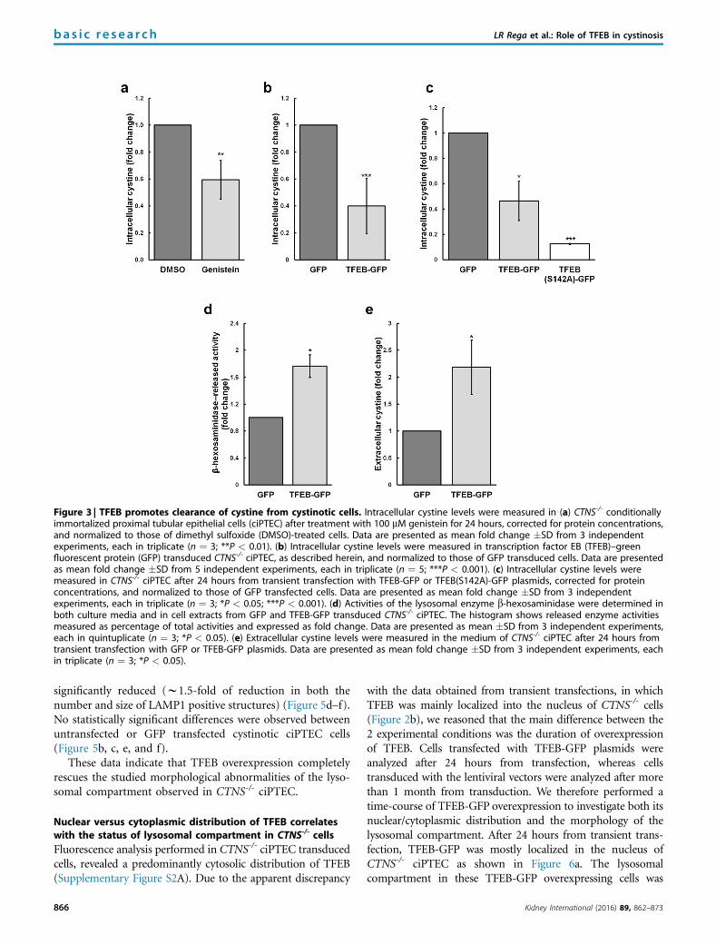
Figure 3 | TFEB promotes clearance of cystine from cystinotic cells. Intracellular cystine levels were measured in (a) CTNS-/- conditionallyimmortalized proximal tubular epithelial cells (ciPTEC) after treatment with 100 mM genistein for 24 hours, corrected for protein concentrations,and normalized to those of dimethyl sulfoxide (DMSO)-treated cells. Data are presented as mean fold change �SD from 3 independentexperiments, each in triplicate (n ¼ 3; **P < 0.01). (b) Intracellular cystine levels were measured in transcription factor EB (TFEB)–greenfluorescent protein (GFP) transduced CTNS-/- ciPTEC, as described herein, and normalized to those of GFP transduced cells. Data are presentedas mean fold change �SD from 5 independent experiments, each in triplicate (n ¼ 5; ***P < 0.001). (c) Intracellular cystine levels weremeasured in CTNS-/- ciPTEC after 24 hours from transient transfection with TFEB-GFP or TFEB(S142A)-GFP plasmids, corrected for proteinconcentrations, and normalized to those of GFP transfected cells. Data are presented as mean fold change �SD from 3 independentexperiments, each in triplicate (n ¼ 3; *P < 0.05; ***P < 0.001). (d) Activities of the lysosomal enzyme b-hexosaminidase were determined inboth culture media and in cell extracts from GFP and TFEB-GFP transduced CTNS-/- ciPTEC. The histogram shows released enzyme activitiesmeasured as percentage of total activities and expressed as fold change. Data are presented as mean �SD from 3 independent experiments,each in quintuplicate (n ¼ 3; *P < 0.05). (e) Extracellular cystine levels were measured in the medium of CTNS-/- ciPTEC after 24 hours fromtransient transfection with GFP or TFEB-GFP plasmids. Data are presented as mean fold change �SD from 3 independent experiments, eachin triplicate (n ¼ 3; *P < 0.05).
bas i c re sea r ch LR Rega et al.: Role of TFEB in cystinosis
significantly reduced (w1.5-fold of reduction in both thenumber and size of LAMP1 positive structures) (Figure 5d–f).No statistically significant differences were observed betweenuntransfected or GFP transfected cystinotic ciPTEC cells(Figure 5b, c, e, and f).
These data indicate that TFEB overexpression completelyrescues the studied morphological abnormalities of the lyso-somal compartment observed in CTNS-/- ciPTEC.
Nuclear versus cytoplasmic distribution of TFEB correlateswith the status of lysosomal compartment in CTNS-/- cellsFluorescence analysis performed in CTNS-/- ciPTEC transducedcells, revealed a predominantly cytosolic distribution of TFEB(Supplementary Figure S2A). Due to the apparent discrepancy
866
with the data obtained from transient transfections, in whichTFEB was mainly localized into the nucleus of CTNS-/- cells(Figure 2b), we reasoned that the main difference between the2 experimental conditions was the duration of overexpressionof TFEB. Cells transfected with TFEB-GFP plasmids wereanalyzed after 24 hours from transfection, whereas cellstransduced with the lentiviral vectors were analyzed after morethan 1 month from transduction. We therefore performed atime-course of TFEB-GFP overexpression to investigate both itsnuclear/cytoplasmic distribution and the morphology of thelysosomal compartment. After 24 hours from transient trans-fection, TFEB-GFP was mostly localized in the nucleus ofCTNS-/- ciPTEC as shown in Figure 6a. The lysosomalcompartment in these TFEB-GFP overexpressing cells was
Kidney International (2016) 89, 862–873
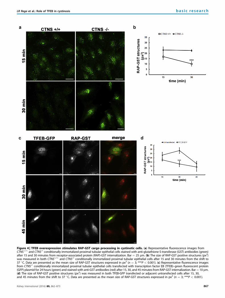
Figure 4 | TFEB overexpression stimulates RAP-GST cargo processing in cystinotic cells. (a) Representative fluorescence images fromCTNSþ/þ and CTNS-/- conditionally immortalized proximal tubular epithelial cells stained with anti-glutathione-S-transferase (GST) antibodies (green)after 15 and 30 minutes from receptor-associated protein (RAP)-GST internalization. Bar ¼ 25 mm. (b) The size of RAP-GST positive structures (px2)was measured in both CTNSþ/þ and CTNS-/- conditionally immortalized proximal tubular epithelial cells after 15 and 30 minutes from the shift to37 �C. Data are presented as the mean size of RAP-GST structures expressed in px2 (n ¼ 3; ***P < 0.001). (c) Representative fluorescence imagesfrom CTNS-/- conditionally immortalized proximal tubular epithelial cells transfected with transcription factor EB (TFEB)–green fluorescent protein(GFP) plasmid for 24 hours (green) and stained with anti-GST antibodies (red) after 15, 30, and 45minutes from RAP-GST internalization. Bar¼ 10 mm.(d) The size of RAP-GST positive structures (px2) was measured in both TFEB-GFP transfected or adjacent untransfected cells after 15, 30,and 45 minutes from the shift to 37 �C. Data are presented as the mean size of RAP-GST structures expressed in px2 (n ¼ 3; ***P < 0.001).
LR Rega et al.: Role of TFEB in cystinosis ba s i c re sea r ch
Kidney International (2016) 89, 862–873 867

Figure 5 | TFEB overexpression reduces both the number and the size of lysosomes in cystinotic cells. (a,b) Representative electronmicrographs from CTNSþ/þ and CTNS-/- conditionally immortalized proximal tubular epithelial cells labeled with anti-LAMP1 antibodies andprepared for immuno-electron microscopy. (c,d) Representative electron micrographs from CTNS-/- conditionally immortalized proximal tubularepithelial cells transduced with lentivirus for control green fluorescent protein (GFP) or transcription factor EB (TFEB)-GFP expression and sortedby fluorescence-activated cell sorter as described in the materials andmethods. LAMP1 positive structures are indicated by arrows. Bar¼ 500 nm.(e) Morphometric analysis was done from electron microscopy images acquired from thin sections using the same magnification. Thenumber of LAMP1 positive structures was counted within 36 mm2
fields of view and expressed as mean � SEM. (f) The size of LAMP1 positivestructures was measured (100 structures for each experimental condition) and expressed as mean �SEM. (***P < 0.001; ns, not statisticallysignificant).
bas i c re sea r ch LR Rega et al.: Role of TFEB in cystinosis
morphologically unaffected when compared with untransfectedCTNS-/- ciPTEC. Indeed, we did not observe any significantdifference in size between lysosomes of untransfected orTFEB-GFP CTNS-/- ciPTEC (Figure 6a, SupplementaryFigure 2B). However, after 1 week (168 hours) from transienttransfection, the morphology of lysosomes in TFEB-GFPCTNS-/- transfected cells changed. TFEB-GFP CTNS-/-
868
ciPTEC showed reduced size of lysosomes, as compared toadjacent untransfected cells and TFEB-GFP distributionbecame predominantly cytosolic (Figure 6a). Conversely, con-trol GFP transient transfection did not affect lysosomalcompartment in cystinotic ciPTEC after 168 hours(Supplementary Figure S2C). These observations wereconfirmed and quantified by high-content imaging. In this
Kidney International (2016) 89, 862–873
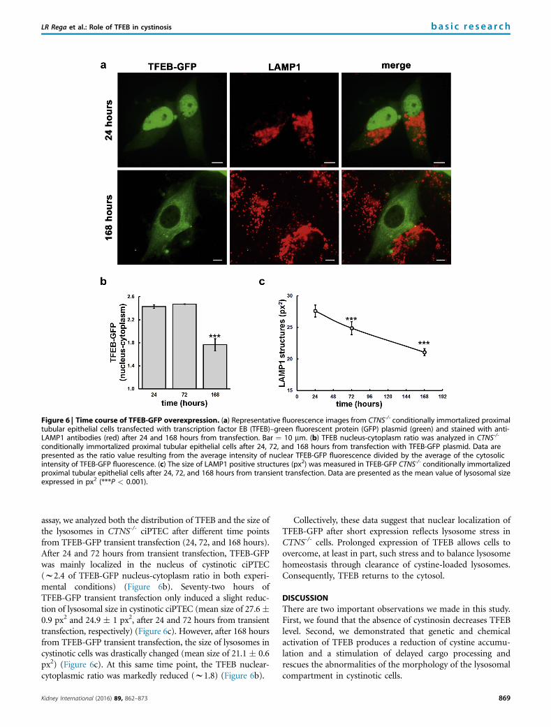
Figure 6 | Time course of TFEB-GFP overexpression. (a) Representative fluorescence images from CTNS-/- conditionally immortalized proximaltubular epithelial cells transfected with transcription factor EB (TFEB)–green fluorescent protein (GFP) plasmid (green) and stained with anti-LAMP1 antibodies (red) after 24 and 168 hours from transfection. Bar ¼ 10 mm. (b) TFEB nucleus-cytoplasm ratio was analyzed in CTNS-/-
conditionally immortalized proximal tubular epithelial cells after 24, 72, and 168 hours from transfection with TFEB-GFP plasmid. Data arepresented as the ratio value resulting from the average intensity of nuclear TFEB-GFP fluorescence divided by the average of the cytosolicintensity of TFEB-GFP fluorescence. (c) The size of LAMP1 positive structures (px2) was measured in TFEB-GFP CTNS-/- conditionally immortalizedproximal tubular epithelial cells after 24, 72, and 168 hours from transient transfection. Data are presented as the mean value of lysosomal sizeexpressed in px2 (***P < 0.001).
LR Rega et al.: Role of TFEB in cystinosis ba s i c re sea r ch
assay, we analyzed both the distribution of TFEB and the size ofthe lysosomes in CTNS-/- ciPTEC after different time pointsfrom TFEB-GFP transient transfection (24, 72, and 168 hours).After 24 and 72 hours from transient transfection, TFEB-GFPwas mainly localized in the nucleus of cystinotic ciPTEC(w2.4 of TFEB-GFP nucleus-cytoplasm ratio in both experi-mental conditions) (Figure 6b). Seventy-two hours ofTFEB-GFP transient transfection only induced a slight reduc-tion of lysosomal size in cystinotic ciPTEC (mean size of 27.6�0.9 px2 and 24.9 � 1 px2, after 24 and 72 hours from transienttransfection, respectively) (Figure 6c). However, after 168 hoursfrom TFEB-GFP transient transfection, the size of lysosomes incystinotic cells was drastically changed (mean size of 21.1 � 0.6px2) (Figure 6c). At this same time point, the TFEB nuclear-cytoplasmic ratio was markedly reduced (w1.8) (Figure 6b).
Kidney International (2016) 89, 862–873
Collectively, these data suggest that nuclear localization ofTFEB-GFP after short expression reflects lysosome stress inCTNS-/- cells. Prolonged expression of TFEB allows cells toovercome, at least in part, such stress and to balance lysosomehomeostasis through clearance of cystine-loaded lysosomes.Consequently, TFEB returns to the cytosol.
DISCUSSIONThere are two important observations we made in this study.First, we found that the absence of cystinosin decreases TFEBlevel. Second, we demonstrated that genetic and chemicalactivation of TFEB produces a reduction of cystine accumu-lation and a stimulation of delayed cargo processing andrescues the abnormalities of the morphology of the lysosomalcompartment in cystinotic cells.
869
bas i c re sea r ch LR Rega et al.: Role of TFEB in cystinosis
TFEB is a master regulator of the coordinated lysosomalexpression and regulation network. TFEB specifically recog-nizes and binds the coordinated lysosomal expression andregulation–box sequence present in the regulatory region ofmany lysosomal genes, leading to activation of their expres-sion.19 A coordinated lysosomal expression and regulation–box has been identified in the promoter of CTNS gene.24
Accordingly we confirmed that overexpression of TFEBstrongly increases expression of CTNS mRNA in different celllines (data not shown). Johnson et al.12 have recently pro-posed that down-regulation of Rab27a expression in cysti-nosis is caused by TFEB dysfunction. This hypothesis wassupported by the finding of 3 possible TFEB-mediated reg-ulatory elements in the promoter regions of the mouse andhuman Rab27a genes.25 Here we found that cystinotic cellshave decreased levels of TFEB, which appears to be related tocystinosin deficiency per se and not to cystine accumulation.Treating cystinotic cells with the cystine-depleting agent,cysteamine, indeed did not rescue this defect, whereasknocking down the expression of CTNS led to significantreduction of TFEB expression and only to a slight accumu-lation of intracellular cystine (compared with the levels ofcystine accumulated in cystinotic cells; data not shown).Napolitano et al.10 also demonstrated that reduction ofcystine by cysteamine treatment did not rescue defectivechaperone-mediated autophagy both in cystinotic mouse fi-broblasts and cystinotic mice. Moreover, a recent paper fromAndrzejewska et al.26 showed that cystinosin is a componentof the mTOR complex 1 and has a direct role in the regulationof the activity of this complex. Indeed, cysteamine treatmentshad no effects on impaired mTOR signaling in CTNS-/- cells.All together, these data strongly support the hypothesis thatcystinosin has additional roles to its cystine transporting ac-tivity and, directly or indirectly, contributes to the modula-tion of TFEB intracellular expression and activity. However,further studies are required to clarify the mechanisms linkingCTNS and TFEB expression.
As mentioned, Andrzejewska et al.26 demonstrated thatmTOR signaling is altered in cystinotic cells. Moreover, lackof cystinosin and accumulation of cystine cause highlysosomal stress that results in abnormal morphology of thelysosomal compartment, characterized by enlarged lyso-somes.13 Both mTOR dysregulation and lysosomal stressinduce TFEB activation and nuclear translocation.27
Consistently, we highlighted TFEB nuclear translocationin cystinotic cells. By analogy of what has been observed inother LSDs, we could demonstrate by both a chemical andgenetic approach, that TFEB stimulation can reduce cystinelevels in cystinotic cells, promoting lysosomal exocytosis.Johnson et al.12 also demonstrated that up-regulation of theexocytic pathway by overexpression of the constitutivelyactive form of Rab27a decreases cystine content in cys-tinotic cells. However, we cannot exclude that other path-ways, such as autophagy, can contribute to TFEB-inducedcellular clearance. Many studies demonstrated that geneticand/or chemical activation of TFEB promotes autophagic
870
clearance of aberrant aggregates.21,22,28 In this context, it isimportant to note that recent advances in the field of cys-tinosis have highlighted that aberrant autophagy is animportant contributor to the pathogenesis of thedisease.10,29
The discovery of compounds, such as genistein23 and2-hydroxypropyl-b-cyclodextrin,30 able to stimulateendogenous TFEB activity has provided an attractive alter-native to gene-transfer therapy in the treatments of diseasesthat potentially can benefit from TFEB activation. Genistein,a natural isoflavone present in soy, has been proposed astherapeutic agent for the LSD, mucopolysaccharidosis, forits ability to inhibit the synthesis and reduce lysosomalstorage of glycosaminoglycans31 and, recently, for its abilityto activate TFEB.23,32 Moreover genistein and soy productshave gained attention for their pleiotropic effects and havebeen suggested to mitigate many conditions such as obesity,metabolic syndrome, and cancer. In this contest, cystinoticpatients could potentially benefit from increased genisteinintake in their diet or from genistein supplementation alsofor its antioxidant and anti-inflammatory properties.33,34
Indeed, cystinosis has been also characterized by enhancedoxidative stress35 and inflammation.36 Because genistein isable to regulate different cellular pathways, we checkedthe impact of this treatment on chaperone-mediated auto-phagy, but no ameliorations of this phenotype were high-lighted in cystinotic cells (Supplementary Figure S3A and B).
Overexpression of exogenous TFEB was able to reducecystine levels after 24 hours from transient transfection,indicating a rapid effect on mobilization of cystine deposits.Moreover, we demonstrated that cystinotic cells over-expressing TFEB show faster processing of endocytic cargo ifcompared with untransfected cells (Figure 4c and d). Thisconsequence of TFEB overexpression opens new insights inthe role of this protein but, most importantly, suggests thatTFEB activation can also improve renal Fanconi syndrome incystinosis. On the contrary, the abnormalities of the lyso-somal compartment were not rescued after 24 hours.Recently, Ivanova et al.13 also demonstrated that treatmentwith the cystine-depleting agent, cysteamine, was not efficientin restoring lysosomal morphology within 24 hours inCTNS-depleted cells. Thus, we wondered whether the com-plete rescue of lysosomal function would require treatmentlonger than 24 hours. After a week from transient trans-fection, the lysosomal compartment in cystinotic cells over-expressing TFEB was significantly modified and becamecomparable to that of control cells. As a result, TFEB nolonger accumulated into the nucleus of cystinotic cells. Theseresults support the hypothesis that depletion of cystine per sedoes not rescue all cystinotic phenotypes.10 TFEB being atranscription factor, it is likely that stimulation and/or sup-pression of other cellular pathways could be responsible forthe rescue of these additional cystinotic cell phenotypes. Thisstrongly suggests that other treatments complementary tocurrent therapies aimed at decreasing lysosomal overload areneeded.
Kidney International (2016) 89, 862–873
LR Rega et al.: Role of TFEB in cystinosis ba s i c re sea r ch
MATERIALS AND METHODSCell culture and treatmentsHuman kidney-2 cells (American Type Culture Collection CRL-2190) were grown in Dulbecco’s modified Eagle’s medium–F12(Invitrogen, Life Technologies, Camarillo, CA) supplemented with5% fetal bovine serum, 100 units/ml penicillin and 100 mg/mlstreptomycin, 10 mg/ml insulin from bovine pancreas, 5.5 mg/mlhuman transferrin, and 5 ng/ml sodium selenite. HepG2 cells(American Type Culture Collection) were cultured in Dulbecco’smodified Eagle’s medium supplemented with 10% fetal bovineserum, 2 mM L-glutamine, 100 units/ml penicillin, and 100 mg/mlstreptomycin. ciPTEC were kindly provided by Dr. Elena N. Lev-tchenko and cultured as described in Wilmer et al.37 Human cys-tinotic fibroblasts were kindly provided by the Cell Lines and DNABank of Patients Affected by Genetic Diseases (Laboratorio diDiagnosi Pre e Postnatale delle Malattie Metaboliche, Istituto G.Gaslini, Italy, partially supported by the Telethon Foundation).Fibroblasts were grown in Dulbecco’s modified Eagle’s mediumsupplemented with 10% fetal bovine serum, 1 mM sodium pyruvate(Invitrogen), 2 mM L-glutamine, 100 units/ml penicillin, and100 mg/ml streptomycin. Cells were grown in a humidified atmo-sphere with 5% CO2 at 37 �C. Unless otherwise specified, all reagentswere purchased from Sigma-Aldrich (St. Louis, MO). After 24 hoursfrom splitting in 24-well plates, cells were incubated with freshnonsupplemented medium containing DMSO at a final concentra-tion of 0.1%, or genistein at a final concentration of 100 mM, 0.1%DMSO for 24 hours.
Plasmid and siRNA transfectionTFEB-GFP plasmid was generated by Dr. Annelies Michiels (ViralVector Core, Leuven, Belgium). ciPTEC cells were transfected withplasmids by Lipofectamine LTX and Plus Reagent (Invitrogen)according to the manufacturer’s instructions. Mutant TFEB(S142A)-GFP was generated by the Q5-Site directed mutagenesis kit (NewEngland, BioLabs Inc., Ipswich, MA) according to the manufac-turer’s instructions. Human kidney-2 and HepG2 cells were trans-fected with siGENOME human CTNS or TFEB siRNA, SMARTpool (Dharmacon, Thermo Scientific, Dublin, Ireland) by Oligo-fectamine Reagent (Invitrogen) according to the manufacturer’sinstructions.
Lentivirus transductionLentivirus expressing GFP and TFEB-GFP were prepared, amplified,and purified by Dr. Annelies Michiels. Lentivirus transduction wasperformed as described in supplementary experimental procedures.
RNA extraction and quantitative real-time PCRTotal RNA was extracted by TRIzol reagent (Ambion, Life Tech-nologies, Foster, CA) and cDNA was synthesized using the Euro-Script RT-PCR kit (EuroClone, Milano, Italy) according to themanufacturer’s instructions. Quantitative PCR assays (shown inFigure 1) were performed using SensiMix II Probe Hi-ROX kit(Bioline, London, UK) and the gene-expression assays for humanTFEB and CTNS (Applied Biosystem, Foster, CA). Quantitative PCRassays from genistein untreated or treated cells (SupplementaryFigure S1D) were performed by SYBR FAST quantitative PCR KitMaster Mix (Kapa Biosystems, Wilmington, MA) with correspond-ing primers listed in Supplementary Table S1. Gene expression datawere determined using the 2-Dct method and normalized usinghuman glyceraldehyde-3-phosphate dehydrogenase.
Kidney International (2016) 89, 862–873
Western blotFor total cell extracts, cells were lysed in radioimmunoprecipitationassay buffer, sonicated, and centrifuged for 10 minutes at 13,000 rpmat 4 �C. Alternatively, nuclear and cytosolic fractions were obtainedas described in Martina and Puertollano.38 Protein concentrationswere measured using the Bio-Rad Protein Assay (Bio-Rad Labora-tories, Inc., Hercules, CA). Proteins were separated by 12% sodiumdodecylsulfate–polyacrylamide gel electrophoresis and immuno-blotted onto a nitrocellulose membrane (Whatman Bioscience Ltd.,Maidstone, UK). The membrane was blocked with 5% bovine serumalbumin in Tris-buffered saline, 0.1% Tween 20 and incubated withprimary antibodies anti-Actin (Ambion), anticystinosin (Abnova,Taipei City, Taiwan), anti-glyceraldehyde-3-phosphate dehydroge-nase, anti-TFEB, or antihistone H3 (Cell Signaling, Danvers, MA),and with horseradish peroxidase secondary antibody conjugate IgG(Santa Cruz Biotechnology, Inc., Santa Cruz, CA). Immunoblotswere developed with LiteAblot EXTEND (EuroClone, Milan, Italy)and acquired with the ChemiDoc XRS System (Bio-Rad).
Measurement of extracellular and intracellular cystine levelsTo measure extracellular cystine levels, cells were cultured in cystine-free medium for 24 hours. Extracellular and intracellular cystinelevels were measured as previously reported.39
b-Hexosaminidase activity assayb-Hexosaminidase activity was measured in both cell extract andmedium as described previously.40
Endocytosis assayRAP-GST processing was assessed as previously described by Ivanovaet al.13 RAP-GST ligand and anti-GST antibodies were kindly pro-vided by Dr. Maria Antonietta De Matteis.
Immuno-electron microscopyCells for pre-embedding immuno-EM were fixed, permeabilized,and labeled as described previously.41 Antihuman LAMP1 (cloneH4A3) antibodies were purchased by Developmental Studies Hy-bridoma Bank (Iowa City, IA). From each sample, thin 65 nm sec-tions were cut using a Leica EM UC7 ultramicrotome (Wetzlar,Germany). EM images were acquired from thin sections using a FEITecnai-12 electron microscope (FEI, Eindhoven, Netherlands)equipped with a VELETTA CCD digital camera (Soft Imaging Sys-tem GmbH, Münster, Germany). Morphometric analysis of numberand size of LAMP1 positive structures was performed using iTEMsoftware (Olympus SYS, Irsee, Germany). Number of lysosomes wascounted using the same magnification within 36 mm2
fields of view.For measurement of the lysosome diameter, the stereology
approach (sequential random sampling methods) was used. Lyso-some diameters were estimated on EM images. EM images weretreated as vertical sections. The diameters were obtained using thepoint sampled intercepts method (see Gundersen et al.42).
Fluorescence assaysUntransfected or transfected cells were grown on glass coverslips,fixed with 4% paraformaldehyde and permeabilized with phosphate-buffered saline containing 0.05% saponin, 0.5% bovine serumalbumin, 50 mM NH4Cl and incubated with anti-LAMP1 antibody(Santa Cruz Biotechnology, Inc., Santa Cruz, CA). Nuclei werestained by Hoechst 33342, trihydrochloride, trihydrate (Invitrogen).Images were acquired on a Nikon Eclipse E600 microscope (NikonInstruments, Melville, NY) equipped with epifluorescence optics and
871
bas i c re sea r ch LR Rega et al.: Role of TFEB in cystinosis
processed with ImageJ software (National Institute of Health,Bethesda, MD).
For the time-course experiments, CTNS-/- ciPTEC were culturedin 12-well plates and transfected with TFEB-GFP plasmid as describedherein. After 8 hours from transient transfection, cells were trypsi-nized and seeded on 96-well plates and fixed after 24, 72, and 168hours from transient transfection. After fixing, cells were per-meabilized and labeled as described. For the analysis, 10 images pereach well of the 96-well plate were acquired by using automated mi-croscopy (Operetta High Content Imaging System; PerkinElmer,Waltham, MA). TFEB localization was analyzed by a dedicatedscript calculating the ratio value resulting from the average intensity ofnuclear TFEB-GFP fluorescence divided by the average of the cytosolicintensity of TFEB-GFP fluorescence. Lysosomal area was analyzed inTFEB-GFP cells by performing LAMP1 staining. All the scripts for theanalysis were developed on Harmony software (PerkinElmer).
DISCLOSURESAll the authors declared no competing interests.
ACKNOWLEDGMENTSThe authors thank Dr. Gianna Di Giovamberardino, Dr. Alessia Palma,and Dr. Ezio Giorda for technical support and Professor OlivierDevuyst and Dr. Alessandro Luciani for helpful discussions. They alsothank Dr. Pierre Courtoy for critical reading of the manuscript. Thesestudies were supported by a grant from the Cystinosis ResearchFoundation, USA.
SUPPLEMENTARY MATERIALSupplementary ResultsFigure S1. (A) Intracellular cystine levels were measured in CTNS-/-
conditionally immortalized proximal tubular epithelial cells, untreatedor treated with 100 mM cysteamine for 24 hours. Data are presentedas mean fold change � SD (n ¼ 5; *** P < 0.001). (B) Transcriptionfactor EB (TFEB) mRNA and (C) CTNS mRNA levels were analyzed inHepG2 cells after 96 hours from transfection with nontargeting (CTRL)or CTNS small, interfering RNAs (CTNS knock down). Glyceraldehyde-3-phosphate dehydrogenase was used as reference. Data are pre-sented as mean fold change � SEM from 3 independent experiments,each in triplicate (n ¼ 3; *P < 0.05; ***P < 0.001). (D) Relative mRNAexpression levels of representative coordinated lysosomal expressionand regulation coordinated lysosomal expression and regulationnetwork genes in CTNS-/- conditionally immortalized proximal tubularepithelial cells treated with genistein (100 mM) for 24 hours. ASAH1,CTSD, SMPD1, and SQSTM1 mRNA expression levels were obtained byquantitative polymerase chain reaction, corrected for the expressionof the housekeeping genes glyceraldehyde-3-phosphate dehydro-genase, and normalized to those of dimethyl sulfoxide (DMSO)-treated cells (black line). Data are reported as mean fold change � SDfrom 3 independent experiments (n ¼ 3; P < 0.05). (E) Intracellularcystine levels were measured in cystinotic fibroblasts after treatmentwith genistein (100 mM) for 24 hours, corrected for protein concen-trations, and normalized to those of DMSO-treated cells. Data arepresented as mean fold change � SD from 3 independent experi-ments, each in triplicate (n ¼ 3; ***P < 0.001). (F) The histogramshows the percentages of green fluorescent protein (GFP) or TFEB-GFP propidium iodide positive CTNS-/- conditionally immortalizedproximal tubular epithelial cells (24 hours of transfection). Data arepresented as mean values � SD from 3 independent experiments,each in triplicate (n ¼ 3; ns ¼ not statistically significant).Figure S2. (A) Representative image of CTNS-/- conditionallyimmortalized proximal tubular epithelial cells after transduction with
872
transcription factor EB (TFEB)–green fluorescent protein (GFP) (green)lentivirus and fluorescence-activated cell sorting. Bar ¼ 100 mm.(B) Representative image of LAMP1 (green) staining in control CTNSþ/þ
and cystinotic CTNS-/- conditionally immortalized proximal tubularepithelial cells. Nuclei are blue. Bar ¼ 10 mm. (C) Representativeimage of LAMP1 (red) staining in CTNS-/- conditionally immortal-ized proximal tubular epithelial cells after 168 hours from transienttransfection with control GFP plasmid (green). Bar ¼ 10 mm.Figure S3. (A) Representative immunoblot showing LAMP2a proteinlevels in CTNSþ/þ and CTNS-/- fibroblasts after treatment withdimethyl sulfoxide or genistein (100 mM) for 24 hours. LAMP2a levelswere normalized for Actin levels. (B) The histogram shows relativeLAMP2a levels expressed as mean fold change � SD from 3independent experiments (n ¼ 3; ns ¼ not statistically significant).Table S1. List of the primer sequences used for quantitative real-timepolymerase chain reaction experiments.Supplementary material is linked to the online version of the paper atwww.kidney-international.org.
REFERENCES1. Emma F, Nesterova G, Langman C, et al. Nephropathic cystinosis: an
international consensus document. Nephrol Dial Transplant.2014;29(suppl 4):iv87–94.
2. Levtchenko E, van den Heuvel L, Emma F, Antignac C. Clinical utility genecard for: cystinosis. Eur J Hum Genet. 2014;22. http://dx.doi.org/10.1038/ejhg.2013.204.
3. Forestier L, Jean G, Attard M, et al. Molecular characterization of CTNSdeletions in nephropathic cystinosis: development of a PCR-baseddetection assay. Am J Hum Genet. 1999;65:353–359.
4. Town M, Jean G, Cherqui S, et al. A novel gene encoding an integralmembrane protein is mutated in nephropathic cystinosis. Nat Genet.1998;18:319–324.
5. Kalatzis V, Cherqui S, Antignac C, Gasnier B. Cystinosin, the proteindefective in cystinosis, is a H(þ)-driven lysosomal cystine transporter.EMBO J. 2001;20:5940–5949.
6. Gahl WA, Thoene JG, Schneider JA. Cystinosis. N Engl J Med. 2002;347:111–121.
7. Park M, Helip-Wooley A, Thoene J. Lysosomal cystine storage augmentsapoptosis in cultured human fibroblasts and renal tubular epithelial cells.J Am Soc Nephrol. 2002;13:2878–2887.
8. Laube GF, Shah V, Stewart VC, et al. Glutathione depletion and increasedapoptosis rate in human cystinotic proximal tubular cells. PediatrNephrol. 2006;21:503–509.
9. Sansanwal P, Sarwal MM. Abnormal mitochondrial autophagy innephropathic cystinosis. Autophagy. 2010;6:971–973.
10. Napolitano G, Johnson JL, He J, et al. Impairment of chaperone-mediated autophagy leads to selective lysosomal degradation defectsin the lysosomal storage disease cystinosis. EMBO Mol Med. 2015;7:158–174.
11. Sansanwal P, Yen B, Gahl WA, et al. Mitochondrial autophagy promotescellular injury in nephropathic cystinosis. J Am Soc Nephrol. 2010;21:272–283.
12. Johnson JL, Napolitano G, Monfregola J, et al. Upregulation of theRab27a-dependent trafficking and secretory mechanisms improveslysosomal transport, alleviates endoplasmic reticulum stress, andreduces lysosome overload in cystinosis. Mol Cell Biol. 2013;33:2950–2962.
13. Ivanova EA, De Leo MG, Van Den Heuvel L, et al. Endo-lysosomaldysfunction in human proximal tubular epithelial cells deficient forlysosomal cystine transporter cystinosin. PLoS One. 2015;10:e0120998.
14. Raggi C, Luciani A, Nevo N, et al. Dedifferentiation and aberrations of theendolysosomal compartment characterize the early stage ofnephropathic cystinosis. Hum Mol Genet. 2014;23:2266–2278.
15. Gaide Chevronnay HP, Janssens V, Van Der Smissen P, et al. Time courseof pathogenic and adaptation mechanisms in cystinotic mouse kidneys.J Am Soc Nephrol. 2014;25:1256–1269.
16. Thoene JG, Oshima RG, Crawhall JC, et al. Cystinosis: intracellular cystinedepletion by aminothiols in vitro and in vivo. J Clin Invest. 1976;58:180–189.
17. Gahl WA, Reed GF, Thoene JG, et al. Cysteamine therapy for childrenwith nephropathic cystinosis. N Engl J Med. 1987;316:971–977.
Kidney International (2016) 89, 862–873
LR Rega et al.: Role of TFEB in cystinosis ba s i c re sea r ch
18. Settembre C, Fraldi A, Medina DL, Ballabio A. Signals from the lysosome:a control centre for cellular clearance and energy metabolism. Nat RevMol Cell Biol. 2013;14:283–296.
19. Sardiello M, Palmieri M, di Ronza A, et al. A gene network regulatinglysosomal biogenesis and function. Science. 2009;325:473–477.
20. Medina DL, Fraldi A, Bouche V, et al. Transcriptional activation oflysosomal exocytosis promotes cellular clearance. Dev Cell. 2011;21:421–430.
21. Spampanato C, Feeney E, Li L, et al. Transcription factor EB (TFEB) is anew therapeutic target for Pompe disease. EMBO Mol Med. 2013;5:691–706.
22. Pastore N, Blomenkamp K, Annunziata F, et al. Gene transfer of masterautophagy regulator TFEB results in clearance of toxic protein andcorrection of hepatic disease in alpha-1-anti-trypsin deficiency. EMBOMol Med. 2013;5:397–412.
23. Moskot M, Montefusco S, Jakobkiewicz-Banecka J, et al. Thephytoestrogen genistein modulates lysosomal metabolism andtranscription factor EB (TFEB) activation. J Biol Chem. 2014;289:17054–17069.
24. Palmieri M, Impey S, Kang H, et al. Characterization of the CLEAR networkreveals an integrated control of cellular clearance pathways. Hum MolGenet. 2011;20:3852–3866.
25. Chiaverini C, Beuret L, Flori E, et al. Microphthalmia-associatedtranscription factor regulates RAB27A gene expression and controlsmelanosome transport. J Biol Chem. 2008;283:12635–12642.
26. Andrzejewska Z, Nevo N, Thomas L, et al. Cystinosin is a component ofthe vacuolar Hþ-ATPase-Ragulator-Rag complex controlling mammaliantarget of rapamycin complex 1 signaling [e-pub ahead of print]. J Am SocNephrol. pii: ASn.2014090937. Accessed 2015.
27. Settembre C, Zoncu R, Medina DL, et al. A lysosome-to-nucleus signallingmechanism senses and regulates the lysosome via mTOR and TFEB.EMBO J. 2012;31:1095–1108.
28. Kilpatrick K, Zeng Y, Hancock T, Segatori L. Genetic and chemicalactivation of TFEB mediates clearance of aggregated a-synuclein. PLoSOne. 2015;10:e0120819.
29. Sansanwal P, Li L, Sarwal MM. Inhibition of intracellular clusterinattenuates cell death in nephropathic cystinosis. J Am Soc Nephrol.2015;26:612–625.
Kidney International (2016) 89, 862–873
30. Song W, Wang F, Lotfi P, et al. 2-Hydroxypropyl-beta-cyclodextrinpromotes transcription factor EB-mediated activation of autophagy:implications for therapy. J Biol Chem. 2014;289:10211–10222.
31. Piotrowska E, Jakobkiewicz-Banecka J, Baranska S, et al. Genistein-mediated inhibition of glycosaminoglycan synthesis as a basis for geneexpression-targeted isoflavone therapy for mucopolysaccharidoses. Eur JHum Genet. 2006;14:846–852.
32. Moskot M, Jakobkiewicz-Banecka J, Kloska A, et al. Modulation ofexpression of genes involved in glycosaminoglycan metabolism andlysosome biogenesis by flavonoids. Sci Rep. 2015;5:9378.
33. Javanbakht MH, Sadria R, Djalali M, et al. Soy protein and genisteinimproves renal antioxidant status in experimental nephrotic syndrome.Nefrologia. 2014;34:483–490 [in English and Spanish].
34. Nagaraju GP, Zafar SF, El-Rayes BF. Pleiotropic effects of genistein inmetabolic, inflammatory, andmalignant diseases.Nutr Rev. 2013;71:562–572.
35. Vaisbich MH, Pache de Faria Guimaraes L, Shimizu MH, Seguro AC.Oxidative stress in cystinosis patients. Nephron Extra. 2011;1:73–77.
36. Prencipe G, Caiello I, Cherqui S, et al. Inflammasome activation by cystinecrystals: implications for the pathogenesis of cystinosis. J Am Soc Nephrol.2014;25:1163–1169.
37. Wilmer MJ, Saleem MA, Masereeuw R, et al. Novel conditionallyimmortalized human proximal tubule cell line expressing functionalinflux and efflux transporters. Cell Tissue Res. 2010;339:449–457.
38. Martina JA, Puertollano R. Rag GTPases mediate amino acid-dependentrecruitment of TFEB and MITF to lysosomes. J Cell Biol. 2013;200:475–491.
39. Pastore A, Massoud R, Motti C, et al. Fully automated assay for totalhomocysteine, cysteine, cysteinylglycine, glutathione, cysteamine, and2-mercaptopropionylglycine in plasma and urine. Clin Chem. 1998;44:825–832.
40. Rodriguez A, Webster P, Ortego J, Andrews NW. Lysosomes behave asCa2þ-regulated exocytic vesicles in fibroblasts and epithelial cells. J CellBiol. 1997;137:93–104.
41. Polishchuk EV, Concilli M, Iacobacci S, et al. Wilson disease protein ATP7Butilizes lysosomal exocytosis to maintain copper homeostasis. Dev Cell.2014;29:686–700.
42. Gundersen HJ, Bendtsen TF, Korbo L, et al. Some new, simple andefficient stereological methods and their use in pathological researchand diagnosis. APMIS. 1988;96:379–394.
873