activationofwnt/ -cateninproteinsignalinginduces ... · activationofwnt/...
TRANSCRIPT

Activation of Wnt/�-Catenin Protein Signaling InducesMitochondria-mediated Apoptosis in HematopoieticProgenitor Cells*□S
Received for publication, January 12, 2012, and in revised form, May 13, 2012 Published, JBC Papers in Press, May 15, 2012, DOI 10.1074/jbc.M112.342089
Ming Ming‡1, Sheng Wang‡1, Wenshu Wu§, Vitalyi Senyuk‡, Michelle M. Le Beau¶, Giuseppina Nucifora‡�,and Zhijian Qian‡�2
From the ‡Department of Medicine and the �Cancer Research Center, University of Illinois, Chicago, Illinois 60621, the ¶Section ofHematology/Oncology and the Comprehensive Cancer Center, University of Chicago, Chicago, Illinois 60637, and the §Children’sHospital Oakland Research Institute, Oakland, California 94609
Background: Wnt/�-catenin signaling plays a strong role in maintaining homeostasis of hematopoietic progenitor cells(HPCs).Results: Activated �-catenin deregulates the survival of HPCs by induction of the mitochondrial apoptotic pathway.Conclusion:Wnt/�-catenin signaling is critical for HPC pool maintenance.Significance:This study clarifies the role of theWnt/�-catenin signaling in HPCs by showing that it has negative impact on thefunction and survival of the cells.
The canonical Wnt/�-catenin signaling is activated duringdevelopment, tumorigenesis, and in adult homeostasis, yet itsrole in maintenance of hematopoietic stem/progenitor cells isnot firmly established. Here, we demonstrate that conditionalexpression of an active form of �-catenin in vivo induces amarked increase in the frequency of apoptosis in hematopoieticstem/progenitor cells (HSCs/HPCs). Activation of Wnt/�-catenin signaling in HPCs in vitro elevates the activity ofcaspases 3 and 9 and leads to a loss of mitochondrial membranepotential (��m), indicating that it induces the intrinsic mito-chondrial apoptotic pathway. In vivo, expression of activated�-catenin in HPCs is associated with down-regulation of Bcl2and expression of Casp3. Bone marrow transplantation assaysreveal that enhanced cell survival by a Bcl2 transgene re-es-tablishes the reconstitution capacity of HSCs/HPCs thatexpress activated �-catenin. In addition, a Bcl2 transgeneprevents exhaustion of these HSCs/HPCs in vivo. Our datasuggest that activation of the Wnt/�-catenin pathway con-tributes to the defective function of HPCs in part by deregu-lating their survival.
The canonical Wnt/�-catenin pathway plays an importantregulatory role in hematopoiesis; however, its involvement inregulation of HSC3 function is controversial. Mice with dele-
tion of �-catenin (1) or �-catenin and �-catenin (2) had noobvious HSC defects. In contrast, other investigators showedthe existence of hematopoietic abnormalities inmicewith dele-tion of �-catenin from fetal life (3). Retroviral expression ofactivated �-catenin enhances the self-renewal of Bcl2 trans-genic HSCs in vitro, and the Wnt signaling inhibitor Axinrepresses HSC proliferation and decreases reconstitutioncapacity of HSCs (4). However, others reported that purifiedWnt protein in vitro (5) or inhibition of GSK3� in vivo (6) pro-motes the self-renewal of HSCs. Baba et al. (7, 8) showed thatconstitutive activation of �-catenin impaired multilineage dif-ferentiation, whereas others found that expression of constitu-tively active �-catenin in hematopoietic cells in vivo leads toloss ofHSC repopulation andmultilineage differentiation block(9, 10).The role of �-catenin in the regulation of cell proliferation
and apoptosis is cell context-dependent. �-Catenin is anenhancer of proliferation and survival in tumor cells (11, 12) butcan also induce apoptosis in a variety of cells. Forced expressionof an activated of �-catenin lacking the NH2 terminus pro-moted proliferation and apoptosis inmouse intestinal epithelialcells (13). However, in fibroblasts, the overexpression of�-catenin induced apoptosis independently of its transactiva-tion function with LEF1 (14). In enzastaurin-treated multiplemyeloma cells, it resulted in cell death-dependent up-regula-tion of JUN and TP73 (15).Taken together, these contradictory reports emphasize the
difficulty of understanding the role of theWnt/�-catenin path-way in HSCs/HPCs. Here, we have revisited this controversyand have examined the effects of activated �-catenin in hema-topoietic progenitor cells in vitro and in vivo. In vitro our stud-ies reveal that activation of the Wnt/�-catenin signalinginduces a mitochondria-dependent apoptotic pathway. In vivowe find that it promotes apoptosis of HPCs by suppressing Bcl2
* This work was supported, in whole or in part, by National Institutes of HealthGrant CA140979 (to Z. Q.). This work was also supported in part by theAmerican Cancer Society Illinois Division (to Z. Q.).
□S This article contains supplemental Table 1 and Figs. S1–S5.1 Both authors contributed equally to this work.2 To whom correspondence should be addressed: Dept. of Medicine and Can-
cer Research Center, University of Illinois, 909 S. Wolcott Ave., COMRB Rm.5051 M/C704, Chicago, IL 60621. Tel.: 312-355-3295; Fax: 312-413-9670;E-mail: [email protected].
3 The abbreviations used are: HSC, hematopoietic stem cell; HPC, hematopoi-etic progenitor cell; BM, bone marrow; 7-AAD, 7-aminoactinomycin D;IMDM, Iscove’s modified Dulbecco’s medium; �-cat, �-catenin; TMRM,tetramethylrhodamine methyl ester perchlorate; pI-pC, polyinosinic-polycyt-
idylic acid; PE, phycoerythrin; EGFP, enhanced green fluorescent protein; LSK,Lin�c-Kit�Sca�; PB, peripheral blood; Apc, adenomatous polyposis coli.
THE JOURNAL OF BIOLOGICAL CHEMISTRY VOL. 287, NO. 27, pp. 22683–22690, June 29, 2012© 2012 by The American Society for Biochemistry and Molecular Biology, Inc. Published in the U.S.A.
JUNE 29, 2012 • VOLUME 287 • NUMBER 27 JOURNAL OF BIOLOGICAL CHEMISTRY 22683
by guest on February 3, 2019http://w
ww
.jbc.org/D
ownloaded from
and inducing Casp3. In addition, we show that an enhancedsurvival signal is required for maintenance and function ofHSC/HPCs with activated �-catenin in vivo. Our studies sug-gest that the negative effect of Wnt/�-catenin signaling inHPCs results from the deregulation of their survival.
MATERIALS AND METHODS
Mice—C57BL/6 and B6.SJL mice were purchased from Tac-onic (Hudson, NY) for breeding in the mouse facility at theUniversity of Illinois at Chicago. The mice (6–12 weeks) wereused for bone marrow (BM) collection. All animal protocolswere approved by the Animal Care and Use Committee of theUniversity of Illinois at Chicago. Vav-Bcl2 transgenicmicewerekindly provided by Dr. Jerry Adams (TheWalter and Eliza HallInstitute of Medical Research, Bundoora, Victoria Australia).�-Cateninflox/flox mice were purchased from The Jackson Lab-oratory (Bar Harbor, ME). Ctnnb1(Ex3)flox/flox mice have beendescribed (16) andwere obtained fromDr. KathleenGoss (Uni-versity of Chicago). Effective deletion of floxed exons wasmon-itored by PCR. The sequence of the primers is listed in supple-mental Table S1.Quantitative Real-time PCR—To evaluate the expression of
candidate genes deregulated in mutant �-catenin mice, HPCswere sorted from Mx1-cre Ctnnb1(Ex3)�/� and control mice(n � 3) 4 days after three doses of pI-pC injection. RNA wasisolated with TRIzol reagent (Invitrogen) and quantified. Rela-tive abundance of specific candidate gene transcripts normal-ized to �-actin was determined by quantitative RT-PCR bySYBRGreen incorporation. The primer sequence for each geneis listed in supplemental Table S1.Retroviral Vectors and BM Infection—The pBabe-�-cat-
S33Y vector encoding an activated formof�-cateninwas kindlyprovided by Dr. Eric Fearon (University of Michigan School ofMedicine, Ann Arbor, MI). The BamHI-EcoRI DNA fragmentencoding �-cat-S33Y was cloned into the MSCV-IRES-EGFP(MIGR1) vector. The Bcl2 cDNA was subcloned into MSCV-EYFP vector. Using Effectene transfection reagent (Qiagen),high titer retrovirus was produced by transient transfection of293T cells with the pCL ecotropic packaging plasmid and theappropriate retroviral vectors (17). The MSCV-Cre-IRES-EYFP and MSCV-puro-Cre vectors were provided by Dr.Jiwang Zhang (Loyola University, Maywood, IL). The BM cellsisolated frommice treated with 5-fluorouracil (150 mg/Kg) for4 days, the lineage-negative BMcells, and the day 14.5 fetal livercells were infected with the appropriate retrovirus by spinocu-lation as described previously (18).Cell Culture—BM cells were collected by flushing murine
femurs and tibias with IMDMcontaining 2% fetal bovine serum(FBS). To isolate lineage-negative BM cells, the cells werestained with biotinylated mouse antibodies specific for lineagemarkers (CD3, B220, IgM, Ter119, Gr-1, and CD19). The Lin�
cells were depleted with streptavidin microbeads and LS col-umns (Miltenyi Biotec, Auburn, CA) following themanufactur-er’s instructions. The BM cells were cultured in IMDM, supple-mented with 10% FBS, penicillin/streptomycin (10,000 units/ml),�-mercaptoethanol (50 �M), 10 ng/ml murine IL-3 and IL-6, and100 ng/ml murine stem cell factor (eBioscience, San Diego CA).Fetal liver cells were isolated at embryonic day 14.5 and grown in
50% IMDM and DMEM containing 20% FBS, 10 ng/ml murineIL-3 and IL-6, and 100 ng/ml murine stem cell factor.Colony-forming Unit Assays—Isolated BM progenitor cells
were infected with MIGR1 or �-catenin retrovirus. The EGFP-positive cells, isolated by flow cytometry, were plated in duplicatein methylcellulose medium (MethoCult M3434, StemCell Tech-nologies) supplemented with IL-3, IL-6, erythropoietin and stemcell factor. The number of colonies was scored 10 days later.Flow Cytometric Analysis—We used single-cell suspensions
of BM and PB. Flow cytometric analysis of HSCs/HPCs hasbeen described previously (19). Briefly, the infected BM cellswere stained with biotinylated antibodies specific for lineagemarkers (CD3, B220, IgM, Ter119, Gr-1, Thy1.1, Il-7R, andCD19) followed by PE-Cy5-conjugated streptavidin, Kit-APC-eFluo780, Sca-PE, CD48-PE-Cy7, and CD150-APC staining.Flow cytometry was performed at the University of Illinois atChicago facility using FACSCalibur or CyAn flow cytometers.Cell cycle analysis with DAPI staining was performed asdescribed previously (20). Cell cycle analysis withHoechst 3342was performed as described (4). For detection of apoptosis, BMcells were cultured overnight at a density of 0.2 � 106/ml. Thenext day, the cells were stained with annexin V (BD Biosci-ences) and 7-AAD or DAPI following the manufacturer’sinstructions. Caspase 3 and caspase 9 staining was performedusing kits from BD Biosciences and BioVision according to themanufacturer’s instructions. All data were analyzed by theFlowJo software (TreeStar).Transplantation Assays—BM cells and day 14.5 fetal liver
cells fromC57BL/6 or BM cells fromVav-Bcl2-transgenicmicewere infected with MIGR1 or �-catenin retrovirus and weretransplanted into lethally irradiated (960 radians) B6.SJLmice by retro-orbital injection. After transplantation, the micewere closelymonitored. FACS analysis of EGFP-positive cells inPB was performed every month after transplantation.Mitochondrial Membrane Potential (��m) Measurement—
Tetramethylrhodamine methyl ester perchlorate (TMRM)(Sigma) was used as an indicator of ��m. Live BM cells werewashed once with PBS and stained with freshly prepared 10 �M
TMRM for 30 min at 37 °C. The cells were then washed twicewith PBS and suspended in PBS for flow cytometric analysis.Western Blot Analysis—Cell extracts were prepared using
lysis buffer containing 1% Triton X-100, 2 mM EDTA, 100 mM
NaCl, 10% glycerol, 50 mM Tris-HCL, and a protease inhibitormixture (Sigma).Mouse anti-�-catenin (BDTransduction Lab-oratories) and mouse anti-tubulin (Millipore, Billerica, MA)were used for Western blot analysis, which was performed asdescribed previously (21).Luciferase Reporter Assay—The luciferase assay was per-
formed with the Dual-GloTM luciferase assay system (Promega).Statistical Analysis—Statistical significance was calculated
using the two-tailed Student’s t test (Excel, Microsoft, Redmond,WA).
RESULTS
Activation of �-Catenin Induces Apoptosis in Vivo—Todetermine whether in vivo activation of �-catenin induces apo-ptosis of HSCs/HPCs, we used the Cre-loxP system to activate�-catenin in hematopoietic stem/progenitor cells in vivo.
Activated �-Catenin Induces Apoptosis
22684 JOURNAL OF BIOLOGICAL CHEMISTRY VOLUME 287 • NUMBER 27 • JUNE 29, 2012
by guest on February 3, 2019http://w
ww
.jbc.org/D
ownloaded from
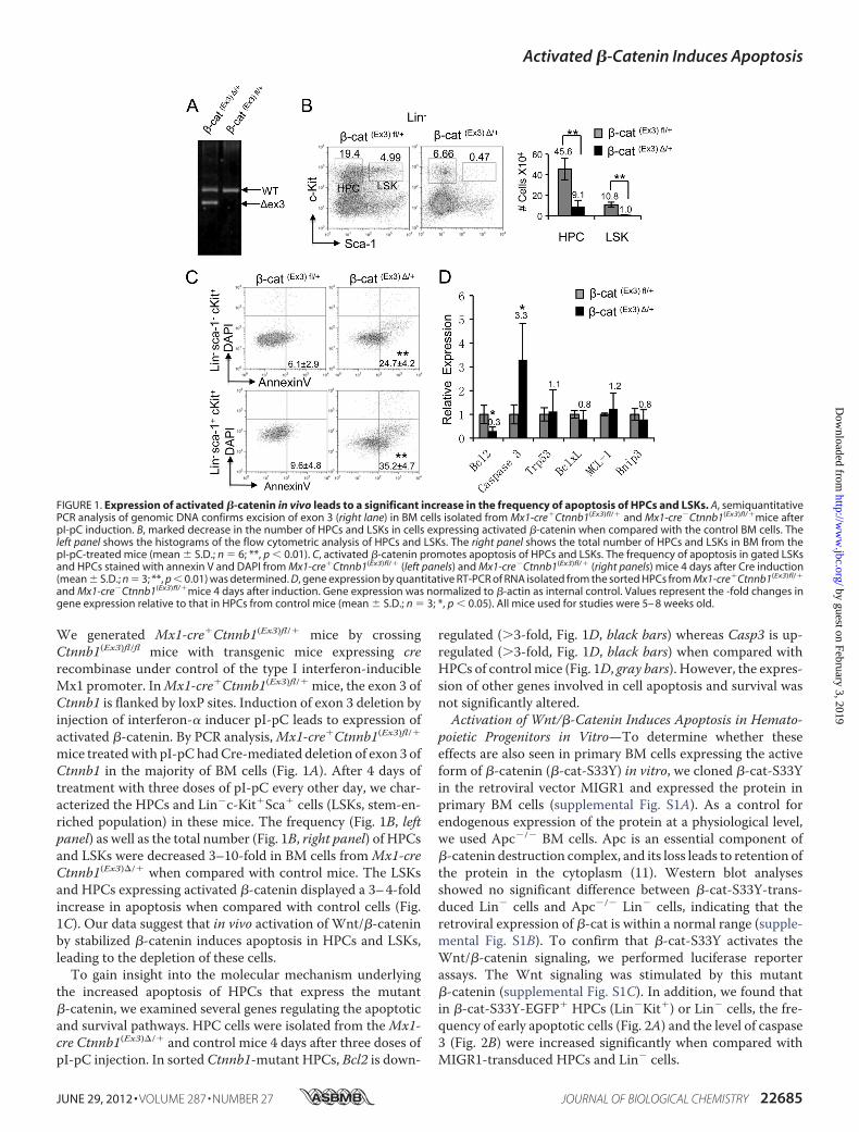
We generated Mx1-cre�Ctnnb1(Ex3)fl/� mice by crossingCtnnb1(Ex3)fl/fl mice with transgenic mice expressing crerecombinase under control of the type I interferon-inducibleMx1 promoter. InMx1-cre�Ctnnb1(Ex3)fl/� mice, the exon 3 ofCtnnb1 is flanked by loxP sites. Induction of exon 3 deletion byinjection of interferon-� inducer pI-pC leads to expression ofactivated �-catenin. By PCR analysis,Mx1-cre�Ctnnb1(Ex3)fl/�mice treatedwith pI-pChadCre-mediated deletion of exon 3 ofCtnnb1 in the majority of BM cells (Fig. 1A). After 4 days oftreatment with three doses of pI-pC every other day, we char-acterized the HPCs and Lin�c-Kit�Sca� cells (LSKs, stem-en-riched population) in these mice. The frequency (Fig. 1B, leftpanel) as well as the total number (Fig. 1B, right panel) of HPCsand LSKs were decreased 3–10-fold in BM cells fromMx1-creCtnnb1(Ex3)�/� when compared with control mice. The LSKsand HPCs expressing activated �-catenin displayed a 3–4-foldincrease in apoptosis when compared with control cells (Fig.1C). Our data suggest that in vivo activation of Wnt/�-cateninby stabilized �-catenin induces apoptosis in HPCs and LSKs,leading to the depletion of these cells.To gain insight into the molecular mechanism underlying
the increased apoptosis of HPCs that express the mutant�-catenin, we examined several genes regulating the apoptoticand survival pathways. HPC cells were isolated from theMx1-cre Ctnnb1(Ex3)�/� and control mice 4 days after three doses ofpI-pC injection. In sorted Ctnnb1-mutant HPCs, Bcl2 is down-
regulated (�3-fold, Fig. 1D, black bars) whereas Casp3 is up-regulated (�3-fold, Fig. 1D, black bars) when compared withHPCs of controlmice (Fig. 1D, gray bars). However, the expres-sion of other genes involved in cell apoptosis and survival wasnot significantly altered.Activation of Wnt/�-Catenin Induces Apoptosis in Hemato-
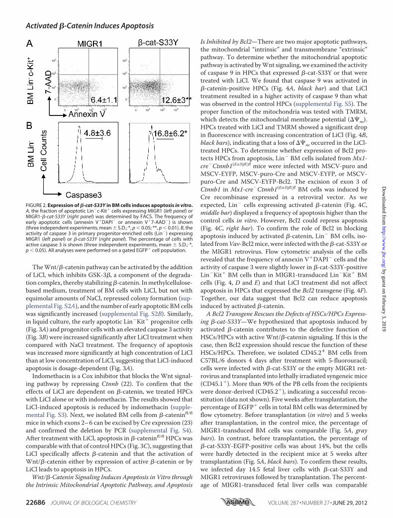
poietic Progenitors in Vitro—To determine whether theseeffects are also seen in primary BM cells expressing the activeform of �-catenin (�-cat-S33Y) in vitro, we cloned �-cat-S33Yin the retroviral vector MIGR1 and expressed the protein inprimary BM cells (supplemental Fig. S1A). As a control forendogenous expression of the protein at a physiological level,we used Apc�/� BM cells. Apc is an essential component of�-catenin destruction complex, and its loss leads to retention ofthe protein in the cytoplasm (11). Western blot analysesshowed no significant difference between �-cat-S33Y-trans-duced Lin� cells and Apc�/� Lin� cells, indicating that theretroviral expression of �-cat is within a normal range (supple-mental Fig. S1B). To confirm that �-cat-S33Y activates theWnt/�-catenin signaling, we performed luciferase reporterassays. The Wnt signaling was stimulated by this mutant�-catenin (supplemental Fig. S1C). In addition, we found thatin �-cat-S33Y-EGFP� HPCs (Lin�Kit�) or Lin� cells, the fre-quency of early apoptotic cells (Fig. 2A) and the level of caspase3 (Fig. 2B) were increased significantly when compared withMIGR1-transduced HPCs and Lin� cells.
FIGURE 1. Expression of activated �-catenin in vivo leads to a significant increase in the frequency of apoptosis of HPCs and LSKs. A, semiquantitativePCR analysis of genomic DNA confirms excision of exon 3 (right lane) in BM cells isolated from Mx1-cre�Ctnnb1(Ex3)fl/� and Mx1-cre�Ctnnb1(Ex3)fl/�mice afterpI-pC induction. B, marked decrease in the number of HPCs and LSKs in cells expressing activated �-catenin when compared with the control BM cells. Theleft panel shows the histograms of the flow cytometric analysis of HPCs and LSKs. The right panel shows the total number of HPCs and LSKs in BM from thepI-pC-treated mice (mean � S.D.; n � 6; **, p 0.01). C, activated �-catenin promotes apoptosis of HPCs and LSKs. The frequency of apoptosis in gated LSKsand HPCs stained with annexin V and DAPI from Mx1-cre�Ctnnb1(Ex3)fl/� (left panels) and Mx1-cre�Ctnnb1(Ex3)fl/� (right panels) mice 4 days after Cre induction(mean � S.D.; n � 3; **, p 0.01) was determined. D, gene expression by quantitative RT-PCR of RNA isolated from the sorted HPCs from Mx1-cre�Ctnnb1(Ex3)fl/�
and Mx1-cre�Ctnnb1(Ex3)fl/�mice 4 days after induction. Gene expression was normalized to �-actin as internal control. Values represent the -fold changes ingene expression relative to that in HPCs from control mice (mean � S.D.; n � 3; *, p 0.05). All mice used for studies were 5– 8 weeks old.
Activated �-Catenin Induces Apoptosis
JUNE 29, 2012 • VOLUME 287 • NUMBER 27 JOURNAL OF BIOLOGICAL CHEMISTRY 22685
by guest on February 3, 2019http://w
ww
.jbc.org/D
ownloaded from
TheWnt/�-catenin pathway can be activated by the additionof LiCl, which inhibits GSK-3�, a component of the degrada-tion complex, thereby stabilizing�-catenin. Inmethylcellulose-based medium, treatment of BM cells with LiCl, but not withequimolar amounts of NaCl, repressed colony formation (sup-plemental Fig. S2A), and the number of early apoptotic BMcellswas significantly increased (supplemental Fig. S2B). Similarly,in liquid culture, the early apoptotic Lin�Kit� progenitor cells(Fig. 3A) and progenitor cells with an elevated caspase 3 activity(Fig. 3B) were increased significantly after LiCl treatment whencompared with NaCl treatment. The frequency of apoptosiswas increased more significantly at high concentration of LiClthan at low concentration of LiCl, suggesting that LiCl-inducedapoptosis is dosage-dependent (Fig. 3A).Indomethacin is a Cox inhibitor that blocks the Wnt signal-
ing pathway by repressing Ctnnb (22). To confirm that theeffects of LiCl are dependent on �-catenin, we treated HPCswith LiCl alone or with indomethacin. The results showed thatLiCl-induced apoptosis is reduced by indomethacin (supple-mental Fig. S3). Next, we isolated BM cells from �-cateninfl/flmice in which exons 2–6 can be excised by Cre expression (23)and confirmed the deletion by PCR (supplemental Fig. S4).After treatment with LiCl, apoptosis in �-cateninfl/fl HPCs wascomparable with that of control HPCs (Fig. 3C), suggesting thatLiCl specifically affects �-catenin and that the activation ofWnt/�-catenin either by expression of active �-catenin or byLiCl leads to apoptosis in HPCs.Wnt/�-Catenin Signaling Induces Apoptosis in Vitro through
the Intrinsic Mitochondrial Apoptotic Pathway, and Apoptosis
Is Inhibited by Bcl2—There are two major apoptotic pathways,the mitochondrial “intrinsic” and transmembrane “extrinsic”pathway. To determine whether the mitochondrial apoptoticpathway is activated byWnt signaling, we examined the activityof caspase 9 in HPCs that expressed �-cat-S33Y or that weretreated with LiCl. We found that caspase 9 was activated in�-catenin-positive HPCs (Fig. 4A, black bar) and that LiCltreatment resulted in a higher activity of caspase 9 than whatwas observed in the control HPCs (supplemental Fig. S5). Theproper function of the mitochondria was tested with TMRM,which detects the mitochondrial membrane potential (��m).HPCs treated with LiCl and TMRM showed a significant dropin fluorescence with increasing concentration of LiCl (Fig. 4B,black bars), indicating that a loss of ��m occurred in the LiCl-treated HPCs. To determine whether expression of Bcl2 pro-tects HPCs from apoptosis, Lin� BM cells isolated from Mx1-cre�Ctnnb1(Ex3)fl/fl mice were infected with MSCV-puro andMSCV-EYFP, MSCV-puro-Cre and MSCV-EYFP, or MSCV-puro-Cre and MSCV-EYFP-Bcl2. The excision of exon 3 ofCtnnb1 in Mx1-cre�Ctnnb1(Ex3)fl/fl BM cells was induced byCre recombinase expressed in a retroviral vector. As weexpected, Lin� cells expressing activated �-catenin (Fig. 4C,middle bar) displayed a frequency of apoptosis higher than thecontrol cells in vitro. However, Bcl2 could repress apoptosis(Fig. 4C, right bar). To confirm the role of Bcl2 in blockingapoptosis induced by activated �-catenin, Lin� BM cells, iso-lated fromVav-Bcl2mice, were infectedwith the�-cat-S33Y orthe MIGR1 retrovirus. Flow cytometric analysis of the cellsrevealed that the frequency of annexin V�DAPI� cells and theactivity of caspase 3 were slightly lower in �-cat-S33Y-positiveLin�Kit� BM cells than in MIGR1-transduced Lin�Kit� BMcells (Fig. 4, D and E) and that LiCl treatment did not affectapoptosis in HPCs that expressed the Bcl2 transgene (Fig. 4F).Together, our data suggest that Bcl2 can reduce apoptosisinduced by activated �-catenin.A Bcl2 Transgene Rescues the Defects of HSCs/HPCs Express-
ing �-cat-S33Y—We hypothesized that apoptosis induced byactivated �-catenin contributes to the defective function ofHSCs/HPCs with active Wnt/�-catenin signaling. If this is thecase, then Bcl2 expression should rescue the function of theseHSCs/HPCs. Therefore, we isolated CD45.2� BM cells fromC57BL/6 donors 4 days after treatment with 5-fluorouracil;cells were infected with �-cat-S33Y or the empty MIGR1 ret-rovirus and transplanted into lethally irradiated syngeneicmice(CD45.1�). More than 90% of the PB cells from the recipientswere donor-derived (CD45.2�), indicating a successful recon-stitution (data not shown). Five weeks after transplantation, thepercentage of EGFP� cells in total BM cells was determined byflow cytometry. Before transplantation (in vitro) and 5 weeksafter transplantation, in the control mice, the percentage ofMIGR1-transduced BM cells was comparable (Fig. 5A, graybars). In contrast, before transplantation, the percentage of�-cat-S33Y-EGFP-positive cells was about 14%, but the cellswere hardly detected in the recipient mice at 5 weeks aftertransplantation (Fig. 5A, black bars). To confirm these results,we infected day 14.5 fetal liver cells with �-cat-S33Y andMIGR1 retroviruses followed by transplantation. The percent-age of MIGR1-transduced fetal liver cells was comparable
FIGURE 2. Expression of �-cat-S33Y in BM cells induces apoptosis in vitro.A, the fraction of apoptotic Lin�c-Kit� cells expressing MIGR1 (left panel) orMIGR1-�-cat-S33Y (right panel) was determined by FACS. The frequency ofearly apoptotic cells (annexin V�DAPI� or annexin V�7-AAD�) is shown(three independent experiments; mean � S.D.; *, p 0.05; **, p 0.01). B, theactivity of caspase 3 in primary progenitor-enriched cells (Lin�) expressingMIGR1 (left panel) or �-cat-S33Y (right panel). The percentage of cells withactive caspase 3 is shown (three independent experiments, mean � S.D.; *,p 0.05). All analyses were performed on a gated EGFP� cell population.
Activated �-Catenin Induces Apoptosis
22686 JOURNAL OF BIOLOGICAL CHEMISTRY VOLUME 287 • NUMBER 27 • JUNE 29, 2012
by guest on February 3, 2019http://w
ww
.jbc.org/D
ownloaded from

before or after transplantation, but again, although the percent-age of �-cat-S33Y-EGFP-transduced fetal liver cells was about27% before transplantation, no EGFP� cells were detected inthe PB of the recipient mice at 5 and 14 weeks after transplan-tation (Fig. 5B, black bars). These data suggest that retroviralexpression of the active form of �-catenin disrupts the recon-stitution capacity of HSCs/HPCs. To determine whether Bcl2rescues the defects in the function of HSCs/HPCs expressingactivated �-catenin, we isolated BM cells from Vav-Bcl2 trans-genic mice (24), infected themwith theMIGR1 vector express-ing either �-cat-S33Y-EGFP or EGFP, and transplanted thecells into lethally irradiated syngeneic recipients. As expected,the percentage of control cells at time of transplantation and at7–8 weeks after transplantation did not decrease (Fig. 5C, graybars). However, in contrast to the previous results, the per-centage of �-cat-S33Y-Bcl2-EGFP� cells was also compara-ble in vitro before transplantation and in total PB and BMcells at 7–8 weeks after transplantation (Fig. 5C, black bars).We characterized the GFP-positive HSC and HPC compart-ments in both groups of recipients. The number of LSK cellsand CD48�CD150� LSK cells was slightly lower in the GFP�
BM cells of Bcl2 transgenic/�-cat-S33Y mice than in thecontrol animals (Fig. 6A), suggesting that the expression ofBcl2 partially rescued the reconstitution capacity of short-term HSCs and HPCs expressing activated �-catenin.Because others reported that in vivo expression of �-cat-S33Y leads to BM failure within 2–3 weeks (9), we monitoreda cohort of chimeric mice expressing both activated�-catenin and Bcl2 transgene. The EGFP� cells in PB werestabilized at 6 months and were almost doubled at 10monthsafter transplantation (Fig. 6B), indicating that the expressionof Bcl2 rescues and may enhance the repopulating capacityof long-term HSCs expressing activated �-catenin. Of note,the apoptosis of Bcl2 transgenic LSKs expressing activated�-catenin was slightly higher than the LSKs expressing con-trol vector, whereas the Bcl2 transgenic HPCs expressingactivated �-catenin or control vector displayed a comparablefrequency of apoptosis (Fig. 6C). These data suggest thatBcl2 can repair the hematopoietic defects of HSCs/HPCsthat express activated �-catenin.
FIGURE 3. �-Catenin mediates the induction of HPC apoptosis after LiCl treatment. A, early apoptotic cells (annexin V�DAPI�) of Lin�c-Kit� BM cellstreated with increasing concentration of NaCl or LiCl (three independent experiments; mean � S.D.; *, p 0.05) for 24 h. Black bars indicate cells treated withNaCl and used as controls; gray bars indicate cells treated with LiCl. B, Lin� BM cells were treated with 15 mM NaCl or 15 mM LiCl for 24 h. The histogram depictsthe mean percentage of cells with active caspase 3 (three independent experiments, mean � S.D.) **, p 0.01. C, the frequency of early apoptotic Lin� Kit� BMcells isolated from �-cateninfl/fl mice with expression of MIGR1 (gray bars) or EYFP-Cre to excise �-catenin (black bars). The BM cells were treated with 10 mM LiClfor 24 h (three independent experiments; mean � S.D.; *, p 0.05).
FIGURE 4. Activation of the Wnt/�-catenin pathway initiates the mito-chondria-dependent apoptotic program, which can be inhibited byBcl2. A, the histogram shows the quantification of active caspase 9 incontrol cells (gray bar) and in cells expressing �-cat-S33Y (black bar). TheLin� kit� BM cells were infected with MIGR1 or �-cat-S33Y retrovirus. Theanalysis was performed on gated EGFP� cells. The results are from threeindependent experiments (mean � S.D.; **, p 0.01). B, the histogramdepicts the mean percentage of TMRM-positive cells in three independentexperiments (mean � S.D.; **, p 0.01). The gated Lin�Il-7R�-kit� BMcells treated with NaCl and LiCl for 18 h and stained with TMRM wereanalyzed by flow cytometry. C, flow cytometric analysis of the early apo-ptotic cells (annexin V�7-AAD�) in Mx1-cre�Ctnnb1(Ex3)fl/fl Lin� BM cells,co-infected with MSCV-puro and MSCV-EYFP (left bar), with MSCV-puro-Cre and MSCV-EYFP (middle bar), or with MSCV-puro-Cre and MSCV-EYFP-Bcl2 (right bar). The EGFP� cells were analyzed after 7 days of selection inmedium containing 1 �g/ml puromycin (mean � S.D.; *, p 0.05). D andE, flow cytometric analysis of early apoptotic cells (D) and of cells withactive caspase 3 (E) isolated from Bcl2 transgenic BM cells and infectedwith MIGR1 or �-cat-S33Y retrovirus. Gated Lin� Kit� GFP� cells wereanalyzed, and the mean of three independent experiments is shown. *,p 0.05. F, flow cytometric analysis of early apoptotic cells (annexin V�7-AAD�) in Bcl2 transgenic BM cells treated with NaCl (left panel) or LiCl(right panel). Gated Lin� Kit� GFP� cells were analyzed. The experimentswere performed in triplicate and repeated three times.
Activated �-Catenin Induces Apoptosis
JUNE 29, 2012 • VOLUME 287 • NUMBER 27 JOURNAL OF BIOLOGICAL CHEMISTRY 22687
by guest on February 3, 2019http://w
ww
.jbc.org/D
ownloaded from

DISCUSSION
Whether �-catenin acts as a proapoptotic factor in hemato-poietic cells has not been conclusively defined. Previous studiesrevealed that expression of activated �-catenin did not alter thefrequency of apoptosis in HSC population (9, 10). However, Liand colleagues (25) showed that activated �-catenin enhances
apoptosis of HSCs in vitro. The goal of this study was to help tosolve, if possible, the existing discrepancies on �-catenin inHSCs/HPCs. Our results include strong data, obtained in vitroand in vivo, which support the apoptotic effect that this proteinexerts on HPCs. In addition, our results show the molecularmechanisms by which this occurs and indicate that regulation
FIGURE 5. Expression of �-cat-S33Y disrupts the reconstitution capacity of HSCs/HPCs, which can be rescued by Bcl2 transgene. A, before transplanta-tion (day 0), the fraction of �-cat-S33Y-EGFP� BM cells (black bar) was 14%. Five weeks later, �-cat-S33Y-EGFP�-positive cells were virtually undetectable(mean � S.D.; n � 5). Control cells (gray bars) did not change significantly. B, the percentage of �-cat-S33Y-EGFP� (black bars) and of control fetal liver (FL) cells(gray bars) before transplantation (Day 0) and at 5 and 14 weeks after transplantation indicates the inability of the cells to be maintained in vivo (mean � S.D.;n � 5). C, the fraction of EGFP�, Bcl2-transgenic/�-cat-S33Y (black bars), or control cells (gray bars) detected by FACS at time of transplantation (day 0) or at 7weeks (PB) or 8 weeks (BM) after transplantation (mean � S.D.; n � 5). Similar results were obtained in two independent experiments.
FIGURE 6. Bcl2 transgene rescues the defects induced by �-catenin in HSCs/HPCs in vivo. A, the top and bottom panels show representative flow cytometricanalyses of LSK and long-term HSCs (LT-HSCs) isolated from BM of the chimeric mice reconstituted with Bcl2 transgenic BM cells expressing EGFP (top) or�-cat-S33Y/EGFP (bottom). On the right side, the bars compare the total number of LSKs (top) or long-term HSCs (bottom) in 105 EGFP� BM cells (mean � S.D.;n � 5). *, p 0.05; ** p 0.01. LSK are defined as Lin�Sca-1�Kit�; long-term HSCs are defined as LSK CD150�CD48�. B, the histogram shows the percentageof EGFP� cells in the PB of chimeric mice reconstituted with Bcl2 transgenic BM cells expressing �-cat-S33Y/EGFP at 2– 6 and 10 months (mean � S.D.; n � 3–5).*, p 0.05. C, the histogram indicates the frequency of early apoptotic cells in LSK (Lin�Kit�Sca�) cells and in HPCs (Lin�Kit�Sca�) in BM of the chimeric micereconstituted with Bcl2 transgenic BM cells expressing EGFP or �-cat-S33Y/EGFP. The analysis was performed on gated EGFP� cells. The mice were analyzed 2months after transplantation (mean � S.D., n � 4). *, p 0.05.
Activated �-Catenin Induces Apoptosis
22688 JOURNAL OF BIOLOGICAL CHEMISTRY VOLUME 287 • NUMBER 27 • JUNE 29, 2012
by guest on February 3, 2019http://w
ww
.jbc.org/D
ownloaded from
of survival by �-catenin is critical for the maintenance of func-tional HSCs/HPCs.By using an Mx1-cre/loxP system to induce the deletion of
exon 3 of Ctnnb1 gene, leading to activated �-catenin in vivo,we showed that a significant decrease in the number of HPCsand LSKs and a markedly increased frequency of apoptosisoccur in these cells. Consistently, Cre-dependent activation of�-catenin in Ctnnb1(Ex3)fl/fl HPCs enhances apoptosis in vitro.The result was confirmed by retroviral expression of activated�-cat-S33Y in HPCs. However, the frequency of apoptosis isinduced by activated�-cateninmore significantly in vivo than itis in vitro. In our opinion, it is likely that the in vivomicroenvi-ronment could contribute to the survival of HPCs. Bcl2 is asurvival factor, and ectopic expression ofBcl2 prevents apopto-sis in HSCs (26). Caspase 3 plays a central role in the executionof cell apoptosis (27). Down-regulation of Bcl2 and inducedexpression ofCasp3 occur inCtnnb1mutant HPCs in vivo. Theexpression of p53 is not altered in the mutant HPCs (Fig. 1D).Thus, increased apoptosis in Ctnnb1 mutant HPCs may be aconsequence of the combined deregulation of Bcl2, Casp3, andperhaps other genes involved in apoptosis and survival ofHPCs,which are still unidentified and which could be regulateddirectly or indirectly by �-catenin. Our in vitro study suggeststhat activation of Wnt/�-catenin signaling activates caspase 3and caspase 9 and reduces themitochondrialmembrane poten-tial, leading to a mitochondria-mediated apoptosis. We previ-ously showed that loss ofApc induces apoptosis in HSCs/HPCs(19). Apc is a multifunction, cell context-dependent protein(28), and it is still unclear whether �-catenin is a critical func-tion mediator of Apc. Our data suggest that �-catenin is likelytomediate, at least in part, the function ofApc in regulating thesurvival of HPCs.Although previous studies suggested that the retroviral
expression of active �-catenin in Bcl2 transgenic HSCsenhanced their self-renewal, there was no indication on thepotential pathways utilized by �-cat-S33Y (4). It was thereforeunclear whether the effects of �-catenin on self-renewaldepended on Bcl2 expression, and the mechanism by whichBcl2 cooperates with �-catenin in maintenance of HSCs/HPCsremained notwell defined. In this study, we show that retroviralexpression of active �-catenin disrupts the reconstitutioncapacity of HSCs/HPCs but that Bcl2 transgenic HSCs/HPCsexpressing activated �-catenin maintain their reconstitutioncapacity. We further show that a Bcl2 transgene inhibits theapoptosis induced by activated �-catenin in vitro and preventsthe exhaustion of HSCs/HPCs in vivo. We believe that takentogether, our studies clarify the existing contradictory data onthe effects of activated �-catenin in HSCs/HPCs not only bysupporting the notion that activation of Wnt/�-catenin nega-tively impacts the function of HSCs/HPCs, but more impor-tantly, by strongly indicating that an enhanced survival signal isrequired for the survival of these cells. It is very likely that reg-ulation of the function ofHSCs byWnt/�-catenin is dosage-de-pendent (29). Our study suggests that the negative impact ofWnt/�-catenin in HSCs/HPCs is due, at least in part, to anincreased apoptosis induced by high dosage of Wnt signaling.Finally, the activation of Wnt/�-catenin signaling and the
overexpression of Bcl2 have been frequently associated with
acutemyeloid leukemia (30–34). Our data suggest thatWnt/�-cateninmay cooperate with survival signaling in promoting theself-renewal or proliferation of leukemia stem/progenitor cellsin some acute myelogenous leukemia patients.
REFERENCES1. Cobas, M.,Wilson, A., Ernst, B., Mancini, S. J., MacDonald, H. R., Kemler,
R., and Radtke, F. (2004) �-Catenin is dispensable for hematopoiesis andlymphopoiesis. J. Exp. Med. 199, 221–229
2. Jeannet, G., Scheller, M., Scarpellino, L., Duboux, S., Gardiol, N., Back, J.,Kuttler, F., Malanchi, I., Birchmeier, W., Leutz, A., Huelsken, J., and Held,W. (2008) Long-term,multilineage hematopoiesis occurs in the combinedabsence of �-catenin and �-catenin. Blood 111, 142–149
3. Zhao, C., Blum, J., Chen,A., Kwon,H. Y., Jung, S.H., Cook, J.M., Lagoo,A.,and Reya, T. (2007) Loss of �-catenin impairs the renewal of normal andCML stem cells in vivo. Cancer Cell 12, 528–541
4. Reya, T., Duncan, A. W., Ailles, L., Domen, J., Scherer, D. C., Willert, K.,Hintz, L., Nusse, R., andWeissman, I. L. (2003) A role forWnt signaling inself-renewal of hematopoietic stem cells. Nature 423, 409–414
5. Willert, K., Brown, J. D., Danenberg, E., Duncan, A. W., Weissman, I. L.,Reya, T., Yates, J. R., 3rd, and Nusse, R. (2003) Wnt proteins are lipid-modified and can act as stem cell growth factors. Nature 423, 448–452
6. Trowbridge, J. J., Xenocostas, A., Moon, R. T., and Bhatia, M. (2006) Gly-cogen synthase kinase-3 is an in vivo regulator of hematopoietic stem cellrepopulation. Nat. Med. 12, 89–98
7. Baba, Y., Garrett, K. P., and Kincade, P. W. (2005) Constitutively active�-catenin confers multilineage differentiation potential on lymphoid andmyeloid progenitors. Immunity 23, 599–609
8. Baba, Y., Yokota, T., Spits, H., Garrett, K. P., Hayashi, S., and Kincade,P. W. (2006) Constitutively active �-catenin promotes expansion of mul-tipotent hematopoietic progenitors in culture. J. Immunol. 177,2294–2303
9. Kirstetter, P., Anderson, K., Porse, B. T., Jacobsen, S. E., and Nerlov, C.(2006) Activation of the canonical Wnt pathway leads to loss of hemato-poietic stem cell repopulation andmultilineage differentiation block.Nat.Immunol. 7, 1048–1056
10. Scheller, M., Huelsken, J., Rosenbauer, F., Taketo, M. M., Birchmeier, W.,Tenen, D. G., and Leutz, A. (2006) Hematopoietic stem cell and multilin-eage defects generated by constitutive �-catenin activation. Nat. Immu-nol. 7, 1037–1047
11. He, T. C., Sparks, A. B., Rago, C., Hermeking, H., Zawel, L., da Costa, L. T.,Morin, P. J., Vogelstein, B., and Kinzler, K. W. (1998) Identification ofc-MYC as a target of the APC pathway. Science 281, 1509–1512
12. Tetsu, O., and McCormick, F. (1999) �-Catenin regulates expression ofcyclin D1 in colon carcinoma cells. Nature 398, 422–426
13. Wong, M. H., Rubinfeld, B., and Gordon, J. I. (1998) Effects of forcedexpression of an NH2-terminal truncated �-catenin on mouse intestinalepithelial homeostasis. J. Cell Biol. 141, 765–777
14. Kim, K., Pang, K. M., Evans, M., and Hay, E. D. (2000) Overexpression of�-catenin induces apoptosis independent of its transactivation functionwith LEF-1 or the involvement of major G1 cell cycle regulators.Mol. Biol.Cell 11, 3509–3523
15. Raab, M. S., Breitkreutz, I., Tonon, G., Zhang, J., Hayden, P. J., Nguyen, T.,Fruehauf, J. H., Lin, B. K., Chauhan, D., Hideshima, T., Munshi, N. C.,Anderson, K. C., and Podar, K. (2009) Targeting PKC: a novel role for�-catenin in ER stress and apoptotic signaling. Blood 113, 1513–1521
16. Harada, N., Tamai, Y., Ishikawa, T., Sauer, B., Takaku, K., Oshima,M., andTaketo, M. M. (1999) Intestinal polyposis in mice with a dominant stablemutation of the �-catenin gene. EMBO J. 18, 5931–5942
17. Qian, Z.,Mao, L., Fernald, A. A., Yu, H., Luo, R., Jiang, Y., Anastasi, J., Valk,P. J., Delwel, R., and Le Beau, M. M. (2009) Enhanced expression of FHL2leads to abnormal myelopoiesis in vivo. Leukemia 23, 1650–1657
18. Lavau, C., Szilvassy, S. J., Slany, R., and Cleary, M. L. (1997) Immortaliza-tion and leukemic transformation of a myelomonocytic precursor by ret-rovirally transduced HRX-ENL. EMBO J. 16, 4226–4237
19. Qian, Z., Chen, L., Fernald, A. A., Williams, B. O., and Le Beau, M. M.(2008) A critical role for Apc in hematopoietic stem and progenitor cell
Activated �-Catenin Induces Apoptosis
JUNE 29, 2012 • VOLUME 287 • NUMBER 27 JOURNAL OF BIOLOGICAL CHEMISTRY 22689
by guest on February 3, 2019http://w
ww
.jbc.org/D
ownloaded from
survival. J. Exp. Med. 205, 2163–217520. Zhang, J., Grindley, J. C., Yin, T., Jayasinghe, S., He, X. C., Ross, J. T., Haug,
J. S., Rupp, D., Porter-Westpfahl, K. S.,Wiedemann, L.M.,Wu, H., and Li,L. (2006) PTEN maintains hematopoietic stem cells and acts in lineagechoice and leukemia prevention. Nature 441, 518–522
21. Qian, Z., Okuhara, D., Abe, M. K., and Rosner, M. R. (1999) Molecularcloning and characterization of a mitogen-activated protein kinase-asso-ciated intracellular chloride channel. J. Biol. Chem. 274, 1621–1627
22. Goessling,W.,North, T. E., Loewer, S., Lord,A.M., Lee, S., Stoick-Cooper,C. L., Weidinger, G., Puder, M., Daley, G. Q., Moon, R. T., and Zon, L. I.(2009) Genetic interaction of PGE2 andWnt signaling regulates develop-mental specification of stem cells and regeneration. Cell 136, 1136–1147
23. Brault, V., Moore, R., Kutsch, S., Ishibashi, M., Rowitch, D. H., McMahon,A. P., Sommer, L., Boussadia, O., and Kemler, R. (2001) Inactivation of the�-catenin gene byWnt1-Cre-mediated deletion results in dramatic brainmalformation and failure of craniofacial development. Development 128,1253–1264
24. Ogilvy, S., Metcalf, D., Print, C. G., Bath, M. L., Harris, A.W., and Adams,J. M. (1999) Constitutive Bcl-2 expression throughout the hematopoieticcompartment affects multiple lineages and enhances progenitor cell sur-vival. Proc. Natl. Acad. Sci. U.S.A. 96, 14943–14948
25. Perry, J.M., He, X. C., Sugimura, R., Grindley, J. C., Haug, J. S., Ding, S., andLi, L. (2011) Cooperation between bothWnt/�-catenin and PTEN/PI3K/Akt signaling promotes primitive hematopoietic stem cell self-renewaland expansion. Genes Dev. 25, 1928–1942
26. Domen, J., andWeissman, I. L. (2000) Hematopoietic stem cells need twosignals to prevent apoptosis; BCL-2 can provide one of these, Kitl/c-Kitsignaling the other. J. Exp. Med. 192, 1707–1718
27. Porter, A. G., and Jänicke, R. U. (1999) Emerging roles of caspase-3 inapoptosis. Cell Death Differ. 6, 99–104
28. Fodde, R., Smits, R., and Clevers, H. (2001) APC, signal transduction, andgenetic instability in colorectal cancer. Nat. Rev. Cancer 1, 55–67
29. Luis, T. C., Naber, B. A., Roozen, P. P., Brugman, M. H., de Haas, E. F.,Ghazvini, M., Fibbe, W. E., van Dongen, J. J., Fodde, R., and Staal, F. J.(2011) Canonical Wnt signaling regulates hematopoiesis in a dosage-de-pendent fashion. Cell Stem Cell 9, 345–356
30. Chung, E. J., Hwang, S. G., Nguyen, P., Lee, S., Kim, J. S., Kim, J. W.,Henkart, P. A., Bottaro, D. P., Soon, L., Bonvini, P., Lee, S. J., Karp, J. E., Oh,H. J., Rubin, J. S., and Trepel, J. B. (2002) Regulation of leukemic celladhesion, proliferation, and survival by �-catenin. Blood 100, 982–990
31. Simon, M., Grandage, V. L., Linch, D. C., and Khwaja, A. (2005) Consti-tutive activation of theWnt/�-catenin signaling pathway in acutemyeloidleukemia. Oncogene 24, 2410–2420
32. Tickenbrock, L., Schwäble, J., Wiedehage, M., Steffen, B., Sargin, B.,Choudhary, C., Brandts, C., Berdel,W. E.,Müller-Tidow, C., and Serve, H.(2005) Flt3 tandem duplication mutations cooperate with Wnt signalingin leukemic signal transduction. Blood 105, 3699–3706
33. Ysebaert, L., Chicanne, G., Demur, C., De Toni, F., Prade-Houdellier, N.,Ruidavets, J. B., Mansat-DeMas, V., Rigal-Huguet, F., Laurent, G., Payras-tre, B., Manenti, S., and Racaud-Sultan, C. (2006) Expression of �-cateninby acute myeloid leukemia cells predicts enhanced clonogenic capacitiesand poor prognosis. Leukemia 20, 1211–1216
34. Del Principe, M. I., Del Poeta, G., Venditti, A., Buccisano, F., Maurillo, L.,Mazzone, C., Bruno, A., Neri, B., Irno Consalvo, M., Lo Coco, F., andAmadori, S. (2005) Apoptosis and immaturity in acute myeloid leukemia.Hematology 10, 25–34
Activated �-Catenin Induces Apoptosis
22690 JOURNAL OF BIOLOGICAL CHEMISTRY VOLUME 287 • NUMBER 27 • JUNE 29, 2012
by guest on February 3, 2019http://w
ww
.jbc.org/D
ownloaded from

Giuseppina Nucifora and Zhijian QianMing Ming, Sheng Wang, Wenshu Wu, Vitalyi Senyuk, Michelle M. Le Beau,
Apoptosis in Hematopoietic Progenitor Cells-Catenin Protein Signaling Induces Mitochondria-mediatedβActivation of Wnt/
doi: 10.1074/jbc.M112.342089 originally published online May 15, 20122012, 287:22683-22690.J. Biol. Chem.
10.1074/jbc.M112.342089Access the most updated version of this article at doi:
Alerts:
When a correction for this article is posted•
When this article is cited•
to choose from all of JBC's e-mail alertsClick here
Supplemental material:
http://www.jbc.org/content/suppl/2012/05/15/M112.342089.DC1
http://www.jbc.org/content/287/27/22683.full.html#ref-list-1
This article cites 34 references, 17 of which can be accessed free at
by guest on February 3, 2019http://w
ww
.jbc.org/D
ownloaded from