active implants and scaffolds for tissue regeneration
TRANSCRIPT
Studies in Mechanobiology, Tissue Engineering and Biomaterials 8
Active Implants and Scaffolds for Tissue Regeneration
vonMeital Zilberman
1. Auflage
Active Implants and Scaffolds for Tissue Regeneration – Zilberman
schnell und portofrei erhältlich bei beck-shop.de DIE FACHBUCHHANDLUNG
Thematische Gliederung:
Medizintechnik, Medizinische Werkstoffe
Springer 2011
Verlag C.H. Beck im Internet:www.beck.de
ISBN 978 3 642 18064 4
Inhaltsverzeichnis: Active Implants and Scaffolds for Tissue Regeneration – Zilberman

Tissue Adhesives as Active Implants
Boaz Mizrahi, Christopher Weldon and Daniel S. Kohane
Abstract Tissue adhesives are substances that hold tissues together, and could bebroadly applicable in medicine and surgery. In appropriate circumstances, suchmaterials could be attractive alternatives to sutures and staples since they can beapplied more quickly, causes less pain and may require less equipment. In addi-tion, there is no risk to the practitioner from sharp instruments (Singer et al., Acad.Emerg. Med. 5(2):94, 1998), and they may obviate the need for suture removal(Coulthard et al., Cochrane Database Syst. Rev. 5:CD004287, 2010). An idealsurgical tissue adhesive should allow rapid adhesion and maintain strong and closeapposition of wound edges for an amount of time sufficient to allow woundhealing. It should not interfere with body’s natural healing mechanisms and shoulddegrade without producing an excessive localized or generalized inflammatoryresponse (Mobley et al., Facial Plast. Surg. Clin. North Am. 10(2):147, 2002). Theclinical and scientific potential of adhesives can be enhanced by a variety offunctionalities that may not be directly related to their function as glues or seal-ants. Here we will review adhesives in general, with an emphasis on enhancements
B. Mizrahi, C. Weldon and D. S. Kohane (&)Department of Anesthesia and Perioperative Medicine, Division of Critical Care,Children’s Hospital Boston, 300 Longwood Avenue, Bader 6, Boston, MA 02115, USAe-mail: [email protected]
B. MizrahiDepartment of Chemical Engineering, Massachusetts Institute of Technology,Cambridge, MA 02139, USA
B. MizrahiOperations Research Center, Massachusetts Institute of Technology,Cambridge, MA 02139, USA
C. WeldonDepartment of Surgery, Children’s Hospital Boston, 300 Longwood Avenue,Fegan 3, Boston, MA 02115, USA
Stud Mechanobiol Tissue Eng Biomater (2011) 8: 39–56 39DOI: 10.1007/8415_2010_48� Springer-Verlag Berlin Heidelberg 2010Published Online: 11 January 2011

that render those otherwise passive materials ‘‘active’’. We note that some gluesalso have intrinsic secondary functionalities that can be direct or indirect conse-quences of their primary function, but that is not the focus of this chapter.(For example, they may augment local hemostasis directly, or by improving tissueapposition, without affecting clotting mechanisms (Reece et al., Am. J. Surg.182(2 Suppl):40S, 2001)).
1 Categories of Adhesives
Tissue adhesives hold tissues together (and therefore are glues) but also can serveas barriers to leakage (and therefore can be sealants) when used for wound closure[74]. Achieving a strong bond (Fig. 1) is dependent upon obtaining close contactbetween the tissue(s) to be bonded and the glue (adhesive strength), and on theintegrity of the glue (cohesive strength of the material) [47]. Consequently thecauses of glue failure [67] can include: (1) adhesive failure—where the materialdetaches from the tissue and (2) cohesive failure—where the adhesive fails withinitself. Even if the glue functions well, the tissue itself may tear; this may occurwhen both adhesive and cohesive forces are too strong.
Tissue adhesives can be divided into three main chemical categories: cyano-acrylates, fibrin sealant, and other cross-linkable polymers. They can be admin-istered for a variety of clinical indications including wound closure [26], fistularepair, including in the bowel, blood vessels and bronchi [70], retinal fixation [30]and others. Cyanoacrylates are the strongest (*68 kPa, [2]) and are widely usedfor wound closure. Fibrin based materials, being weaker (*13 kPa, [2]) areapplied as a sealant in many surgical procedures in conjunction with suturing.Hydrogels, collagen compounds, peptides and polyethylene glycol (PEGs)-basedmaterials are also considered weak (4–17 kPa, [2]) and are therefore used astopical wound dressings or as sealants where mechanical properties are of lessconcern than with internal injuries (where wound dehiscence could be disastrous).
Below we detail a number of compounds used as glues. The list is not intendedto be exhaustive but to provide a framework for the subsequent discussion ofactive glues.
Fig. 1 Types of gluestrength: adhesive strengthbetween the glue and thetissue (black arrow), andcohesive strength withinthe glue (red arrow)
40 B. Mizrahi et al.

1.1 Cyanoacrylates
Cyanoacrylates were first synthesized in 1949 [3] and were first reported as tissueadhesives ten years later [18]. They are produced synthetically by condensationbetween cyanoacetic acid and a suitable alcohol followed by the Knoevenagelreaction [68]. The alcohol group will determine the nature of the final monomer bycontrolling the length of the side chain (e.g. by using methanol or octanol,2-methyl cyanoacrylate or 2-octyl cyanoacrylate will be formed, respectively). Thecyanoacetate oil formed is reacted with paraformaldehyde to form cyanoacrylateoligomers. High vacuum (*0.7 mm Hg) and heat (150–180�C) are applied anddepolymerization is carried out to produce clear and colorless liquids monomers.Usually, further purification by repeated vacuum distillations is utilized to get amedical grade material [33]. Since these monomers are highly reactive, poly-merization inhibitors are added at this stage to prevent the monomers fromhardening (polymerize) while being stored. Although polymerization may occurby one of three mechanisms—anionic (Fig. 2), zwitterionic or free radical—thefirst two mechanisms mentioned are strongly favored in vivo [86] by hydroxide oramine groups presented in the body, ultimately resulting in strong chains holdingthe two tissues’ surfaces together.
Although cyanoacrylates are considered very strong and effective, their use—particularly within tissues—is limited by tissue toxicity, including necrosis,which occurs in the immediate vicinity of the cyanoacrylates. The toxicity ofcyanoacrylate glues has been attributed to several factors including:
Fig. 2 The polymerizationof cyanoacrylate tissue glue
Tissue Adhesives as Active Implants 41

direct toxicity of monomers such as methyl-2-cyanoacrylate [39] or ofbyproducts such as cyanoacetate and formaldehyde [87], insufficient tissuevascularization [2], and the heat from the exothermic nature of the reaction[21]. In addition, it has been postulated that the polymerization of the mono-mers may be initiated by the -NH2 groups of glycosides or amino acids presenton cell surfaces [42] thus damaging membrane lipids [84]. A second concernlimiting the use of cyanoacrylates in tissues stems from the fact that they arehard and brittle, hence they may have insufficient flexibility for the dynamicnature of in vivo conditions [40]. As a result, cyanoacrylates are currentlylimited to external or temporary applications.
In general, the smaller the molecular weight of the side group, the quicker therate of degradation. Accordingly, it has been shown that monomers with highermolecular weights may result in slower production of byproducts with resultantdecreased inflammatory response [38]. This difference may be reflected in the factthat n-2-butyl cyanoacrylate (Histoacryl�), a monomer with a four carbon alkylside chain, was not approved for the US market, while 2-octyl cyanoacrylate(Dermabond�), a monomer with an eight carbon alkyl side chain, was approvedfor use in the United States after it proved to be less toxic than cyanoacrylates withshorter side chains [79].
Cyanoacrylate tissue glues have found multiple uses. They have been used inthe management of corneal perforations, corneal melts and wound leaks [2]. Thecornea glue may also improve visual outcomes by obviating the need for sutures,which are associated with inducing astigmatism. In addition, they may create amore watertight seal, decreasing the risk of infection, thus reducing the chance ofdevastating intraocular infections such as endophthalmitis [5]. In dermatology,cyanoacrylate glues provide a flexible water-resistant coating with improvedcosmetic outcomes. It was also found that patients, in particular children, preferthe concept of being ‘‘glued’’ over sutures and clips [9]. Recently, Dermabond wasfound to be superior for skin closure after repairing congenital cleft lip with orwithout associated palate defects [17]. In mammoplasties, Dermabond� waseffective, safe, and had better cosmetic results than sutures [77]. Operative timesand costs were also decreased, while patient satisfaction increased compared totraditional techniques.
1.2 In Situ Cross-linking Polymers
1.2.1 Fibrin Glue/Sealant
Fibrin tissue glue was first introduced in 1909 as an hemostatic agent, and was firstused as an adhesive material in 1940 [78]. Fibrin based tissue adhesives are com-posed of purified fibrinogen and thrombin, and form a bond via the physiologicalcascade of coagulation (Fig. 3). Some additives such as Factor XIII, fibronectin,
42 B. Mizrahi et al.
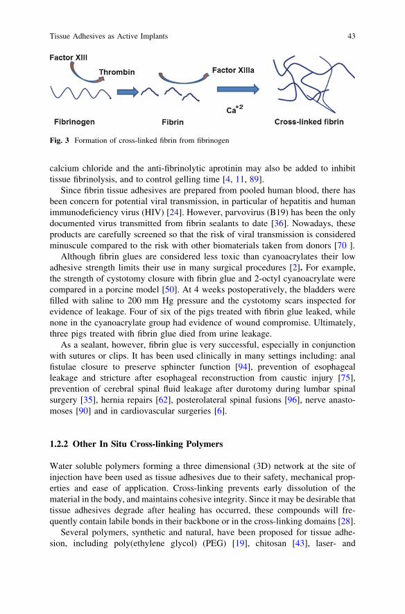
calcium chloride and the anti-fibrinolytic aprotinin may also be added to inhibittissue fibrinolysis, and to control gelling time [4, 11, 89].
Since fibrin tissue adhesives are prepared from pooled human blood, there hasbeen concern for potential viral transmission, in particular of hepatitis and humanimmunodeficiency virus (HIV) [24]. However, parvovirus (B19) has been the onlydocumented virus transmitted from fibrin sealants to date [36]. Nowadays, theseproducts are carefully screened so that the risk of viral transmission is consideredminuscule compared to the risk with other biomaterials taken from donors [70 ].
Although fibrin glues are considered less toxic than cyanoacrylates their lowadhesive strength limits their use in many surgical procedures [2]. For example,the strength of cystotomy closure with fibrin glue and 2-octyl cyanoacrylate werecompared in a porcine model [50]. At 4 weeks postoperatively, the bladders werefilled with saline to 200 mm Hg pressure and the cystotomy scars inspected forevidence of leakage. Four of six of the pigs treated with fibrin glue leaked, whilenone in the cyanoacrylate group had evidence of wound compromise. Ultimately,three pigs treated with fibrin glue died from urine leakage.
As a sealant, however, fibrin glue is very successful, especially in conjunctionwith sutures or clips. It has been used clinically in many settings including: analfistulae closure to preserve sphincter function [94], prevention of esophagealleakage and stricture after esophageal reconstruction from caustic injury [75],prevention of cerebral spinal fluid leakage after durotomy during lumbar spinalsurgery [35], hernia repairs [62], posterolateral spinal fusions [96], nerve anasto-moses [90] and in cardiovascular surgeries [6].
1.2.2 Other In Situ Cross-linking Polymers
Water soluble polymers forming a three dimensional (3D) network at the site ofinjection have been used as tissue adhesives due to their safety, mechanical prop-erties and ease of application. Cross-linking prevents early dissolution of thematerial in the body, and maintains cohesive integrity. Since it may be desirable thattissue adhesives degrade after healing has occurred, these compounds will fre-quently contain labile bonds in their backbone or in the cross-linking domains [28].
Several polymers, synthetic and natural, have been proposed for tissue adhe-sion, including poly(ethylene glycol) (PEG) [19], chitosan [43], laser- and
Fig. 3 Formation of cross-linked fibrin from fibrinogen
Tissue Adhesives as Active Implants 43

non-laser-activated protein solders [1], porcine gelatin, glutaraldehyde mixed withcollagen [49, 53] to name but a few examples. In situ cross-linking polymersystems can form non-self assembling systems (e.g. UV-light/irradiation) or canform spontaneously without the need of external triggers [85]. The bond formedbetween the two polymeric chains (cross-linking) can be covalent or can dependon weaker bonds, such as hydrogen bonds, van der Waals forces, ionic interactionsor a molecule’s side chain interactions [71]. Similarly, adhesive forces between thegel and the tissue can be due to covalent bonds formed between functional groupson one of the polymers (e.g. aldehyde) or by weak van der Waals or hydrogenbonds interactions (e.g. PEG compounds).
BioGlue� (Cryolife, Kennesaw, GA) is a surgical adhesive used in cardiovas-cular surgery, approved by the FDA and the EU as an adjunct in human vascularand pulmonary repair surgery [29]. It is composed of purified bovine serumalbumin (BSA) and glutaraldehyde. The two components are dispensed by adelivery system comprised of a double-chambered syringe and an applicatorapparatus. Once dispensed, the components are mixed within the applicator tip(a mixer) where the cross-linking begins. The glutaraldehyde molecules bond withthe BSA molecules and, upon application to the tissue create an elastic sealindependent of the body’s clotting mechanism [48]. A pilot study [31] aimed todetermine the feasibility of using BioGlue� to achieve hemostasis and to preventurine leakage suggested that this glue provides adequate hemostasis during renalsurgery, and decreased blood loss, transfusion rates and operative times.
Another two-component system [59] is made of aminated star-PEG (a star-shaped poly[ethylene glycol]) and high-molecular-weight dextran-aldehyde(Fig. 4). The two polymers are administered as viscous aqueous solutions that are
Fig. 4 Oxidized dextran(aldehyde is indicated by anarrow) and aminated star-shaped polyethylene glycol
44 B. Mizrahi et al.

delivered through a dual-chambered syringe connected to a single injection needle,thus separating the compounds until the time of administration. Upon mixing,imide bonds are spontaneously formed through a Schiff-base reaction between theamine groups in the PEG molecule and the aldehyde groups of the dextran moi-eties. As a result, a network is formed in seconds. Cohesion strength is created bycross-linking between polymer chains, while adhesion forces are created by thereaction between the aldehyde and the amine groups in tissue. The aldehydes ofthe dextran are in excess of the amine groups in the star-PEG since they areresponsible for both cohesion and adhesion. The adhesive mechanics of this gluevaried with aldehyde content and with tissue type. For example: increasingaldehyde content from 8.8 to 14 and 20% resulted in moduli of 100, 500 and744 kPa, respectively. Likewise, when moduli were measured for different tissuesusing a dextran with 20% aldehyde content, the highest modulus was measured inthe duodenum, followed by the liver, heart and lung (724 ± 86 kPa, 431 ± 15, 296± 60 and 72 ± 7 kPa, respectively). Thus, it was concluded that different tissuesmay require specific surgical sealants when applied.
PEG-based sealants have gained interest in recent years because they areconsidered safe, easy to apply, and very effective in sealing suture lines whencross-linked [19]. In order to provide a tight seal, PEGs can be cross-linked bychemical agents or by visible light. An example is the commercial productFocalSeal� (FocalSeal, Focal, Inc., Lexington, MA) which is composer of an eosinprimer and an aqueous polymeric solution [69]. The primer is applied first, isabsorbed by the tissue, and auto-cross-links. A polymeric solution composed ofPEG is applied and cured by visible light (450–550 nm) [2]. FocalSeal�waseffective in minimally invasive cardiac surgery, where limited exposure and tightquarters make accurate suturing difficult. This product was also found to beeffective for sealing bronchial and parenchymal air leaks [37] and preventingleakage from the cut pancreas (the pancreatic stump) [83].
The preceding systems formed glues spontaneously upon application or mixing.Some, such as chitosan containing azide groups and lactose moieties [63] employ atriggering agent. After ultraviolet light (UV) irradiation, an aqueous solution ofthis material was used to glue two pieces of sliced ham to each other [65]. Thebinding strength of the chitosan hydrogel prepared from 30 to 50 mg/mL solutionswas similar to that of fibrin glue. However, it was more effectively in sealing airleakage from pinholes on isolated small intestine, aorta, and from incisions on theisolated trachea. Neither the tested gel nor its pre-crosslinked solution showed anycytotoxicity in cell culture of human skin fibroblasts, coronary endothelial cells,and smooth muscle cells. In vivo, all mice survived for at least 1 month afterimplantation of 200 lL of photocrosslinked chitosan gel or intraperitonealadministration of the pre-crosslinked solution.
The catechol functionality of L-3,4-dihydroxyphenylalanine (DOPA) is thoughtto be responsible for the ability of marine mussels to form strong bonds with arange of substrates, a property shared by DOPA-coated surfaces [44]. DOPAmoieties have been conjugated to polymers and peptides in efforts to developtissue adhesives [81, 88]. However, the adhesive strength of these biomaterials
Tissue Adhesives as Active Implants 45

have not shown higher adhesive strength than that of fibrin sealant This is believedto be the result of two major factors: (1) low adhesion forces due to intra- and/orintermolecular cross-linking reactions rather than with the surrounding tissue [45],and (2) the soft, flexible nature of hydrogel networks [66] that limits the cohesionforces within the material.
2 Active Tissue Glues
2.1 Tissue Adhesives Releasing Drugs
Tissue adhesives, in addition to being utilized as glues or sealants, can be used asdrug delivery systems. The architecture of the material can be engineered torelease its contents in the desired pattern directly to the target site. Local release bythe glue enables the administration of controlled dosages, reducing the risk ofadverse drug effects. Moreover, patient compliance with these drugs is assured asthe agents are delivered at the time of the procedure and then released without theneed for active participation on the patient’s part. The release and the clinicalbenefits of several drug classes have been investigated utilizing this model,including antibiotics, chemotherapy, analgesics, growth factors, and gene vectorsto name but a few. In some of these applications the adhesives are not used asglues or sealants, but simply as drug delivery (depot) systems.
2.1.1 Antibiotic Impregnated Glues
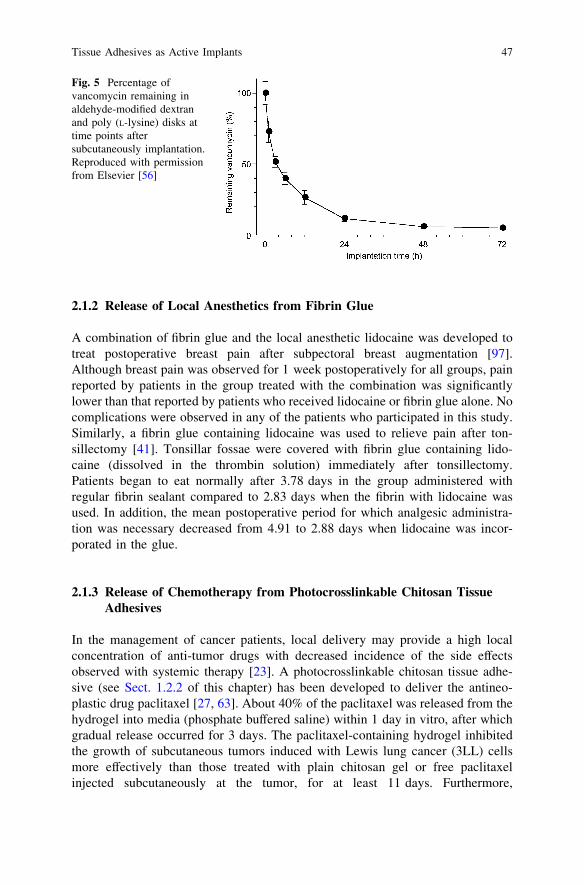
High local antibiotic concentrations at a wound site could prevent local infections(e.g. of surgical wounds) by providing local drug concentrations far in excess ofwhat could be achieved by conventional dosing methods. A variety of formulationshave been developed to achieve that goal. For example, vancomycin, teicoplanin,cephalothin and gentamicin added to the thrombin component of a fibrin glue [51]were released from that matrix for over 96 h in vitro, and exhibited antibacterialactivity against clinical isolates of S. epidermidis. Similarly, amikacin releasedfrom a fibrin sealant/polyurethane mixture implanted subcutaneously in the ante-rior abdominal region of rats [60] was detectable in blood for 24 h, while the samedose given intravenously cleared after only 4 h. Moreover, peak local concen-trations of amikacin in tissue near the glues were 210 times higher than when thedrug was given systemically. A glue composed of aldehyde-modified dextran andpoly (L-lysine) was able to reduce bacterial counts in adjacent subcutaneouslyimplanted Dacron grafts inoculated with methicillin-resistant S. aureus [16, 56].About 95% of the total antibiotic was released over 72 h (Fig. 5), and the localtissue concentration of vancomycin remained above the minimum inhibitoryconcentration throughout this period.
46 B. Mizrahi et al.
2.1.2 Release of Local Anesthetics from Fibrin Glue
A combination of fibrin glue and the local anesthetic lidocaine was developed totreat postoperative breast pain after subpectoral breast augmentation [97].Although breast pain was observed for 1 week postoperatively for all groups, painreported by patients in the group treated with the combination was significantlylower than that reported by patients who received lidocaine or fibrin glue alone. Nocomplications were observed in any of the patients who participated in this study.Similarly, a fibrin glue containing lidocaine was used to relieve pain after ton-sillectomy [41]. Tonsillar fossae were covered with fibrin glue containing lido-caine (dissolved in the thrombin solution) immediately after tonsillectomy.Patients began to eat normally after 3.78 days in the group administered withregular fibrin sealant compared to 2.83 days when the fibrin with lidocaine wasused. In addition, the mean postoperative period for which analgesic administra-tion was necessary decreased from 4.91 to 2.88 days when lidocaine was incor-porated in the glue.
2.1.3 Release of Chemotherapy from Photocrosslinkable Chitosan TissueAdhesives
In the management of cancer patients, local delivery may provide a high localconcentration of anti-tumor drugs with decreased incidence of the side effectsobserved with systemic therapy [23]. A photocrosslinkable chitosan tissue adhe-sive (see Sect. 1.2.2 of this chapter) has been developed to deliver the antineo-plastic drug paclitaxel [27, 63]. About 40% of the paclitaxel was released from thehydrogel into media (phosphate buffered saline) within 1 day in vitro, after whichgradual release occurred for 3 days. The paclitaxel-containing hydrogel inhibitedthe growth of subcutaneous tumors induced with Lewis lung cancer (3LL) cellsmore effectively than those treated with plain chitosan gel or free paclitaxelinjected subcutaneously at the tumor, for at least 11 days. Furthermore,
Fig. 5 Percentage ofvancomycin remaining inaldehyde-modified dextranand poly (L-lysine) disks attime points aftersubcutaneously implantation.Reproduced with permissionfrom Elsevier [56]
Tissue Adhesives as Active Implants 47

the paclitaxel-containing chitosan hydrogel markedly reduced the number ofCD34-positive vessels in subcutaneous 3LL tumors, indicating a strong inhibitionof angiogenesis.
2.1.4 Delivery of Growth Factors and Genetic Material from TissueAdhesives
Although producing a system that releases biomacromolecules from a tissueadhesive can seem relatively simple (e.g. mixing one in the other), the macro-molecules may have complex interactions with the surrounding matrix [80]. Forexample [13], when transforming growth factor beta-1 (TGF-b1) was added tofibrin sealant, release was much slower when fibrinogen concentrations wereincreased, suggesting a binding affinity of TGF-b1 with the fibrinogen. Varying thethrombin concentration though, had a lesser effect.
A matrix to promote wound healing has been developed by incorporatingrecombinant human epidermal growth factor (rhEGF) into a photocross-linkablemixture of glycidyl methacrylated chitooligosaccharide and di-acrylated PluronicF127 [15]. When this hydrogel was administered to dorsal burn wounds in the rat,epidermal differentiation was significantly enhanced compared to plain hydrogel.The in vitro release profiles of rhEGF were dependent on the degradation rates ofthe hydrogels (Fig. 6).
Fibrin sealant has also been used to release nerve growth factor (NGF) into thesite of end-to-end sutured peripheral nerve. Stained sections revealed significantlyincreased regenerated nerve fibers distal to the anastomosis compared to groupsthat received NGF or fibrin sealant alone. Similarly, fibrin sealant containing glia-derived neutropic factor (GDNF) had a greater in vivo effect on neuron growththan did the free factor or the sealant alone [14, 91].
Fibrin sealant has been used to release adenoviral vectors encoding ß-galac-tosidase [7]. Vectors released from fibrin resulted in higher numbers of rabbitcartilage cells expressing ß-galactosidase in vivo than with vector alone.
2.2 Intrinsic Activities of Glues
2.2.1 The Anti-bacterial Properties of Cyanoacrylates
The antimicrobial properties of cyanoacrylate tissue adhesives were first reportedin 1983 [25]. A link has been established between the polymerization process andthe antimicrobial properties, in particular against Gram-positive microorganisms[72], perhaps by action against the bacterial cell wall [22, 76]. Similarly, 2-ethylcyanoacrylate monomers applied onto the surface of bacteria cultures [73] inhibitthe growth of S. aureus and S. pneumoniae (both Gram positive). A possibleexplanation to the higher sensitivity of the Gram-positive bacteria might be the
48 B. Mizrahi et al.

strong electronegative charge on the cyanoacrylate monomer that reacts with thepositively charged carbohydrate capsule of Gram-positive organisms [34]. Whilecyanoacrylates have less effect on Gram negatives, 2-ethyl cyanoacrylatemonomers did kill Escherichia coli [73].
Fig. 6 Release profiles ofrhEGF from a mixture ofglycidyl methacrylatedchitooligosaccharide and di-acrylated Pluronic F127 withphoto-irradiation times of2 min (a), 5 min (b), and10 min (c). The polymericconcentration of all hydrogelswas 20% (w/w). Reproducedwith permission from JohnWiley and Sons [15]
Tissue Adhesives as Active Implants 49

2.2.2 Anti-bacterial Barriers
2-octyl cyanoacrylate films have been shown to be effective barriers to bacteria,fungi, and yeast in vitro [58]. The barrier property of cyanoacrylate bandage wasalso seen in a wound model in swine [52]. S. aureus or Pseudomonas aeruginosawere inoculated on one side of a test bandage placed over a wound. Significantlylower numbers of inoculated bacteria were found among the cyanoacrylate ban-dage group compared with other groups treated with standard or hydrocolloidbandages.
2.2.3 Glues with Wound-Healing and Other Tissue-Active Properties
Photocrosslinkable chitosan is strong, elastic and is considered more effective insealing air leakages than fibrin glue [64]. It can stop bleeding within 30 s ofUV-irradiation and firmly adhere the cut edges of two pieces of skin [32]. It canalso induce wound contraction and accelerate wound closure and healing [10].Histological findings suggest that chitin and chitosan stimulate the migration ofmononuclear and polymorphonuclear cells and accelerate angiogenesis and theformation of connective tissue [55]. Other studies [46, 61] suggest that chitosansposses antibacterial properties, owing to the cationic amines interacting withnegatively charged residues on the bacterial cell surface [95].
Experiments performed in our lab [92] suggest that some caution may beadvisable in using chitosan and UV-cross-linkable chitosan in some contexts.Although in vitro experiments showed neither attractive interactions between thegels and the cells nor a proliferative or marked toxic effect, the same materialapplied in the peritoneal cavity of rabbits caused a granulomatous reaction in allanimals, with resultant adhesion formation (‘‘adhesion’’ in this context meaning anundesirable sticking together of tissues). Although chitosan’s adhesive and otherproinflammatory properties may be beneficial in some biomedical applications,this may not be true in all contexts.
Chitin and chitosan gels have inhibitory effects on tumor angiogenesis andmetastasis [12, 57], and can inhibit tumor cell proliferation by inducing apoptosis[57].
2.2.4 Glues for Islet Cell Immobilization
There is a great need for medical adhesives that effectively function on wet tissuesurfaces with minimal tissue and cell response. A star shape PEG core with DOPAendgroups was suggested as a system for cell immobilization [8]. When aqueoussolutions of this polymer were oxidized with NaIO4, each DOPA endgroupcovalently attached to a neighboring DOPA, forming a 3D hydrogel structure.Donor islet cells were placed into the PEG-DOPA aqueous solution which wasthen oxidized. The encapsulated cells were then implanted in type 1 diabetic mice
50 B. Mizrahi et al.

(Fig. 7). This adhesive material maintained an intact interface with the supportingtissue for up to 1 year. The cells encapsulated within were able to maintainnormoglycemia for over 100 days.
3 Conclusions and Future Directions
Surgical adhesives are attractive alternatives to sutures and staples [20]. Theyallow rapid adhesion and maintain strong and close apposition of wound edges[54]. In some cases, the tissue glues themselves contribute directly to the processof wound healing. A major potential advantage of tissue glues is their ability torelease drugs directly to the wound. In this chapter, we presented the release ofseveral drugs from various classes of tissue adhesives, with emphasis on thechemical and the physical properties of each system.
There is a great variety of adhesives, to which a range of active properties canbe imparted. Further studies will be required to determine whether these newmaterials will translate into the clinical arena. The potential to modify thesematerials has barely been tapped. For example, the incorporation of nanomaterials[93] and/or of components that would allow triggered release of compounds [82]could further enhance their properties.
References
1. Al-qahtani, J.M., McLean, I.W., et al.: Preliminary in vitro study of the histological effects oflow fluence 193-nm excimer laser irradiation of corneal tissue. J. Refract. Surg. 17(2),105–109 (2001)
Fig. 7 Photomicrographof hematoxylin and eosin(H&E)-stained tissueexplants demonstratingstar-PEG-DOPA adhesive-mediated islet cell attachmentto the epididymal fat padsurface. AD. adhesive, ISislet, EF epididymal fattissue. Reproduced withpermission from Elsevier [8]
Tissue Adhesives as Active Implants 51

2. Lauto, A., Mawad, D., et al.: Adhesive biomaterials for tissue reconstruction. J. Chem.Technol. Biotechnol. 83, 464–472 (2008)
3. Ardis, A.: Preparation of monomeric alkyl a-cyanoacrylates. US Patent 2,467,926 (1949)4. Atrah, H.I.: Fibrin glue. BMJ 308(6934), 933–934 (1994)5. Bernard, L., Doyle, J., et al.: A prospective comparison of octyl cyanoacrylate tissue adhesive
(dermabond) and suture for the closure of excisional wounds in children and adolescents.Arch. Dermatol. 137(9), 1177–1180 (2001)
6. Borst, H.G., Haverich, A., et al.: Fibrin adhesive: an important hemostatic adjunct incardiovascular operations. J. Thorac. Cardiovasc. Surg. 84(4), 548–553 (1982)
7. Breen, A., Dockery, P., et al.: Fibrin scaffold promotes adenoviral gene transfer andcontrolled vector delivery. J. Biomed. Mater. Res. A 89(4), 876–884 (2009)
8. Brubaker, C.E., Kissler, H., et al.: Biological performance of mussel-inspired adhesive inextrahepatic islet transplantation. Biomaterials 31(3), 420–427 (2010)
9. Bruns, T.B., Worthington, J.M.: Using tissue adhesive for wound repair: a practical guide todermabond. Am. Fam. Physician 61(5), 1383–1388 (2000)
10. Burkatovskaya, M., Castano, A.P., et al.: Effect of chitosan acetate bandage on woundhealing in infected and noninfected wounds in mice. Wound Repair Regen. 16(3), 425–431(2008)
11. Canonico, S.: The use of human fibrin glue in the surgical operations. Acta. Biomed.74(Suppl 2), 21–25 (2003)
12. Carreno-Gomez, B., Duncan, R.: Evaluation of the biological properties of soluble chitosanand chitosan microspheres. Int. J. Pharm. 48(2), 231–240 (1997)
13. Catelas, I., Dwyer, J.F., et al.: Controlled release of bioactive transforming growth factorbeta-1 from fibrin gels in vitro. Tissue Eng. Part C Methods. 14(2), 119–128 (2008)
14. Cheng, H., Hoffer, B., et al.: The effect of glial cell line-derived neurotrophic factor in fibringlue on developing dopamine neurons. Exp. Brain Res. 104(2), 199–206 (1995)
15. Choi, J.S., Yoo, H.S.: Pluronic/chitosan hydrogels containing epidermal growth factor withwound-adhesive and photo-crosslinkable properties. J. Biomed. Mater. Res. A 95A(2),564–573 (2010)
16. Cirioni, O., Giacometti, A., et al.: Prophylactic efficacy of topical temporin A and RNAIII-inhibiting peptide in a subcutaneous rat pouch model of graft infection attributable tostaphylococci with intermediate resistance to glycopeptides. Circulation 108(6), 767–771(2003)
17. Collin, T.W., Blyth, K., et al.: Cleft lip repair without suture removal. J. Plast. Reconstr.Aesthet. Surg. 62(9), 1161–1165 (2009)
18. Coover, H.W., Joyner, F.B., et al.: Chemistry and performance of cyanoacrylate adhesives.J. Soc. Plast. Eng. 15, 413–417 (1959)
19. Cosgrove, G.R., Delashaw, J.B., et al.: Safety and efficacy of a novel polyethylene glycolhydrogel sealant for watertight dural repair. J. Neurosurg. 106(1), 52–58 (2007)
20. Coulthard, P., Esposito, M., et al.: Tissue adhesives for closure of surgical incisions.Cochrane Database Syst. Rev. 5, CD004287 (2010)
21. DaCruz, D.: Full-thickness skin necrosis of the fingertip after application of superglue.J. Hand Surg. Am. 29(1), 159 (2004). Author reply 159
22. de Almeida Manzano, R.P., Naufal, S.C., et al.: Antibacterial analysis in vitro of ethyl-cyanoacrylate against ocular pathogens. Cornea 25(3), 350–351 (2006)
23. Dhanikula, A.B., Panchagnula, R.: Localized paclitaxel delivery. Int. J. Pharm. 183(2),85–100 (1999)
24. Durham, L.H., Willatt, D.J., et al.: A method for preparation of fibrin glue. J. Laryngol. Otol.101(11), 1182–1186 (1987)
25. Eiferman, R., Snyder, J.: Antibacterial effect of cyanoacrylate glue. Arch. Ophthalmol.101(6), 958–960 (1983)
26. Ghoreishian, M., Gheisari, R., et al.: Tissue adhesive and suturing for closure of the surgicalwound after removal of impacted mandibular third molars: a comparative study. Oral Surg.Oral Med. Oral Pathol. Oral Radiol. Endod. 108(1), e14–e16 (2009)
52 B. Mizrahi et al.

27. Guo, K., Chu, C.C.: Controlled release of paclitaxel from biodegradable unsaturatedpoly(ester amide)s/poly(ethylene glycol) diacrylate hydrogels. J. Biomater. Sci. Polym. Ed.18(5), 489–504 (2007)
28. Hennink, W.E., van Nostrum, C.F.: Novel crosslinking methods to design hydrogels.Adv. Drug Deliv. Rev. 54(1), 13–36 (2002)
29. Herget, G.W., Kassa, M., et al.: Experimental use of an albumin-glutaraldehyde tissueadhesive for sealing pulmonary parenchyma and bronchial anastomoses. Eur. J. Cardiothorac.Surg. 19(1), 4–9 (2001)
30. Hesse, L., Schanze, T., et al.: Implantation of retina stimulation electrodes and recording ofelectrical stimulation responses in the visual cortex of the cat. Graefes Arch. Clin. Exp.Ophthalmol. 238(10), 840–845 (2000)
31. Hidas, G., Kastin, A., et al.: Sutureless nephron-sparing surgery: use of albuminglutaraldehyde tissue adhesive (BioGlue). Urology 67(4), 697–700 (2006). Discussion 700
32. Ishihara, M., Nakanishi, K., et al.: Photocrosslinkable chitosan as a dressing for woundocclusion and accelerator in healing process. Biomaterials 23(3), 833–840 (2002)
33. Jaffe, H., Wade, C.W., et al.: Synthesis and bioevaluation of alkyl 2-cyanoacryloyl glycolatesas potential soft tissue adhesives. J. Biomed. Mater. Res. 20(2), 205–212 (1986)
34. Jang, C.H., Park, H., et al.: Antibacterial effect of octylcyanoacrylate against methicillin-resistant Staphylococcus aureus isolates from patients with chronic suppurative otitis media.In Vivo 22(6), 763–765 (2008)
35. Jankowitz, B.T., Atteberry, D.S., et al.: Effect of fibrin glue on the prevention of persistentcerebral spinal fluid leakage after incidental durotomy during lumbar spinal surgery. Eur.Spine J. 18(8), 1169–1174 (2009)
36. Joch, C., Witzke, G., Groner, A., et al.: Clinical safety of fibrin sealants. Presented at the IXthWorld Conference of Cardio-Thoracic Surgeons, Lisbon, Portugal (1999)
37. Jones, D.R., Stiles, B.M., et al.: Pulmonary segmentectomy: results and complications. Ann.Thorac. Surg. 76(2), 343–348 (2003). Discussion 348–349
38. Kaplan, M., Baysal, K.: In vitro toxicity test of ethyl 2-cyanoacrylate, a tissue adhesive usedin cardiovascular surgery, by fibroblast cell culture method. Heart Surg. Forum 8(3),E169–E172 (2005)
39. Kawamura, S., Hadeishi, H., et al.: Arterial occlusive lesions following wrapping and coatingof unruptured aneurysms. Neurol. Med. Chir. (Tokyo) 38(1), 12–18 (1998). Discussion 18–19
40. Kimura, K.N., Sugiura, K.N.: Adhesive composition. US patent 4321180, Application no. 06/209253, 23 March 1982
41. Kitajiri, S., Tabuchi, K., et al.: Relief of post-tonsillectomy pain by release of lidocaine fromfibrin glue. Laryngoscope 111(4 Pt 1), 642–644 (2001)
42. Kulkarni, R.K., Bartak, D.E., et al.: Initiation of polymerization of alkyl 2-cyanoacrylates inaqueous solutions of glycine and its derivatives. J. Polym. Sci. A1 9(10), 2977–2981 (1971)
43. Lauto, A., Hook, J., et al.: Chitosan adhesive for laser tissue repair: in vitro characterization.Lasers Surg. Med. 36(3), 193–201 (2005)
44. Lee, B.P., Dalsin, J.L., et al.: Synthesis and gelation of DOPA-modified poly(ethylene glycol)hydrogels. Biomacromolecules 3(5), 1038–1047 (2002)
45. Lee, B.P., Huang, K., et al.: Synthesis of 3, 4-dihydroxyphenylalanine (DOPA) containingmonomers and their co-polymerization with PEG-diacrylate to form hydrogels. J. Biomater.Sci. Polym. Ed. 15(4), 449–464 (2004)
46. Lee, D.S., Jeong, S.Y., et al.: Antibacterial activity of aminoderivatized chitosans againstmethicillin-resistant Staphylococcus aureus (MRSA). Bioorg. Med. Chem. 17(20),7108–7112 (2009)
47. Dean, M.J.: Treatise on Adhesion and Adhesives, vol. 7. Marcel Dekker Inc., New York(1991)
48. Manabe, T., Okino, H., et al.: In situ-formed, tissue-adhesive co-gel composed of styrenatedgelatin and styrenated antibody: potential use for local anti-cytokine antibody therapy onsurgically resected tissues. Biomaterials 25(27), 5867–5873 (2004)
Tissue Adhesives as Active Implants 53

49. Marchini, M., Ortolani, F., et al.: Collagen-glutaraldehyde interaction as revealed by theD-banding of negatively stained fibrils and computer-drawn band patterns. Eur. J. Histochem.37(4), 363–373 (1993)
50. Marcovich, R., Williams, A.L., et al.: Comparison of 2-octyl cyanoacrylate adhesive, fibringlue, and suturing for wound closure in the porcine urinary tract. Urology 57(4), 806–810(2001)
51. Marone, P., Monzillo, V., et al.: Antibiotic-impregnated fibrin glue in ocular surgery: in vitroantibacterial activity. Ophthalmologica 213(1), 12–15 (1999)
52. Mertz, P.M., Davis, S.C., et al.: Barrier and antibacterial properties of 2-octyl cyanoacrylate-derived wound treatment films. J. Cutan. Med. Surg. 7(1), 1–6 (2003)
53. Milkes, D.E., Friedland, S., et al.: A novel method to control severe upper GI bleeding frommetastatic cancer with a hemostatic sealant: the CoStasis surgical hemostat. Gastrointest.Endosc. 55(6), 735–740 (2002)
54. Mobley, S.R., Hilinski, J., et al.: Surgical tissue adhesives. Facial Plast. Surg. Clin. NorthAm. 10(2), 147–154 (2002)
55. Mori, T., Okumura, M., et al.: Effects of chitin and its derivatives on the proliferation andcytokine production of fibroblasts in vitro. Biomaterials 18(13), 947–951 (1997)
56. Morishima, M., Marui, A., et al.: Sustained release of vancomycin from a new biodegradableglue to prevent methicillin-resistant Staphylococcus aureus graft infection. InteractCardiovasc. Thorac. Surg. 11(1), 52–55 (2010)
57. Murata, J., Saiki, I., et al.: Inhibitory effect of chitin heparinoids on the lung metastasis ofB16-BL6 melanoma. Jpn. J. Cancer Res. 80(9), 866–872 (1989)
58. Narang, U., Mainwaring, L., et al.: In vitro analysis for microbial barrier properties of 2-octylcyanoacrylate-derived wound treatment films. J. Cutan. Med. Surg. 7(1), 13–19 (2003)
59. Artzi, N., Baker, A.B., Shazly, T., et al.: Aldehyde-amine chemistry enables modulatedbiosealants with tissue-specific adhesion. Adv. Mater. 21, 3399–3403 (2009)
60. Nishimoto, K., Yamamura, K., et al.: Subcutaneous tissue release of amikacin from a fibringlue/polyurethane graft. J. Infect. Chemother. 10(2), 101–104 (2004)
61. No, H.K., Park, N.Y., et al.: Antibacterial activity of chitosans and chitosan oligomers withdifferent molecular weights. Int. J. Food Microbiol. 74(1–2), 65–72 (2002)
62. Novik, B., Hagedorn, S., et al.: Fibrin glue for securing the mesh in laparoscopic totallyextraperitoneal inguinal hernia repair: a study with a 40-month prospective follow-up period.Surg. Endosc. 20(3), 462–467 (2006)
63. Obara, K., Ishihara, M., et al.: Controlled release of paclitaxel from photocrosslinkedchitosan hydrogels and its subsequent effect on subcutaneous tumor growth in mice.J. Control Release 110(1), 79–89 (2005)
64. Ono, K., Ishihara, M., et al.: Experimental evaluation of photocrosslinkable chitosan as abiologic adhesive with surgical applications. Surgery 130(5), 844–850 (2001)
65. Ono, K., Saito, Y., et al.: Photocrosslinkable chitosan as a biological adhesive. J. Biomed.Mater. Res. 49(2), 289–295 (2000)
66. Park, K., Shalaby, W., et al.: Biodegradable hydrogels for drug delivery. TechnomicPublishing Co., Inc., Lancaster (1993)
67. Quinn, J.V.: Tissue Adhesive in Clinical Medicine, BC Decker Inc., Hamilton (2005), p. 2.68. Ramachary, D.B., Anebouselvy, K., et al.: Direct organocatalytic asymmetric heterodomino
reactions: the Knoevenagel/Diels-Alder/epimerization sequence for the highlydiastereoselective synthesis of symmetrical and nonsymmetrical synthons ofbenzoannelated centropolyquinanes. J. Org. Chem. 69(18), 5838–5849 (2004)
69. Ranger, W.R., Halpin, D., et al.: Pneumostasis of experimental air leaks with a newphotopolymerized synthetic tissue sealant. Am. Surg. 63(9), 788–795 (1997)
70. Reece, T.B., Maxey, T.S., et al.: A prospectus on tissue adhesives. Am. J. Surg. 182(2 Suppl),40S–44S (2001)
71. Roldo, M., Hornof, M., et al.: Mucoadhesive thiolated chitosans as platforms for oralcontrolled drug delivery: synthesis and in vitro evaluation. Eur. J. Pharm. Biopharm. 57(1),115–121 (2004)
54 B. Mizrahi et al.

72. Romero, I.L., Malta, J.B., et al.: Antibacterial properties of cyanoacrylate tissue adhesive:Does the polymerization reaction play a role? Indian J. Ophthalmol. 57(5), 341–344 (2009)
73. Romero, I.L., Paiato, T.P., et al.: Different application volumes of ethyl-cyanoacrylate tissueadhesive can change its antibacterial effects against ocular pathogens in vitro. Curr. Eye Res.33(10), 813–818 (2008)
74. Ryou, M., Thompson, C.C.: Tissue adhesives: a review. Tech. Gastrointest. Endosc. 8(1),33–37 (2006)
75. Saldana-Cortes, J.A., Larios-Arceo, F., et al.: Role of fibrin glue in the prevention of cervicalleakage and strictures after esophageal reconstruction of caustic injury. World J. Surg. 33(5),986–993 (2009)
76. Schembri, M.A., Dalsgaard, D., et al.: Capsule shields the function of short bacterialadhesins. J. Bacteriol. 186(5), 1249–1257 (2004)
77. Scott, G.R., Carson, C.L., et al.: Dermabond skin closures for bilateral reductionmammaplasties: a review of 255 consecutive cases. Plast. Reconstr. Surg. 120(6),1460–1465 (2007)
78. Sierra, D.H., Saltz, R.: Surgical Adhesives and Sealants. Technomic Publishing Company,Inc., Lancaster (1996)
79. Singer, A.J., Hollander, J.E., et al.: Prospective, randomized, controlled trial of tissueadhesive (2-octylcyanoacrylate) vs standard wound closure techniques for laceration repair.Stony Brook Octylcyanoacrylate Study Group. Acad. Emerg. Med. 5(2), 94–99 (1998)
80. Spicer, P.P., Mikos, A.G.: ‘‘Fibrin glue as a drug delivery system.’’ J. Control. Release (2010)81. Sun, C.J., Srivastava, A., et al.: Halogenated DOPA in a marine adhesive protein. J. Adhes.
85(2–3), 126–138 (2009)82. Timko, B.P., Dvir, T., et al.: Remotely triggerable drug delivery systems. Adv. Mater. 22(44),
4925–4943 (2010)83. Torchiana, D.F.: Polyethylene glycol based synthetic sealants: potential uses in cardiac
surgery. J. Card. Surg. 18(6), 504–506 (2003)84. Tseng, Y.C., Tabata, Y., et al.: In vitro toxicity test of 2-cyanoacrylate polymers by cell
culture method. J. Biomed. Mater. Res. 24(10), 1355–1367 (1990)85. Van Tomme, S.R., Storm, G., et al.: In situ gelling hydrogels for pharmaceutical and
biomedical applications. Int. J. Pharm. 355(1–2), 1–18 (2008)86. Vauthier, C., Dubernet, C., et al.: Poly(alkylcyanoacrylates) as biodegradable materials for
biomedical applications. Adv. Drug Deliv. Rev. 55(4), 519–548 (2003)87. Vote, B.J., Elder, M.J.: Cyanoacrylate glue for corneal perforations: a description of a
surgical technique and a review of the literature. Clin. Experiment Ophthalmol. 28(6),437–442 (2000)
88. Wang, J., Liu, C., et al.: Co-polypeptides of 3, 4-dihydroxyphenylalanine and L-lysine tomimic marine adhesive protein. Biomaterials 28(23), 3456–3468 (2007)
89. Wang, M.-C., Pins, G.D., et al.: Preparation of fibrin glue: the effects of calcium chloride andsodium chloride. Mater. Sci. Eng. C 3(2), 131–135 (1995)
90. Wieken, K., Angioi-Duprez, K., et al.: Nerve anastomosis with glue: comparative histologicstudy of fibrin and cyanoacrylate glue. J. Reconstr. Microsurg. 19(1), 17–20 (2003)
91. Wood, M.D., Borschel, G.H., et al.: Controlled release of glial-derived neurotrophic factorfrom fibrin matrices containing an affinity-based delivery system. J. Biomed. Mater. Res.A 89(4), 909–918 (2009)
92. Yeo, Y., Burdick, J.A., et al.: Peritoneal application of chitosan and UV-cross-linkablechitosan. J. Biomed. Mater. Res. A 78(4), 668–675 (2006)
93. Yeo, Y., Ito, T., et al.: In situ cross-linkable hyaluronan hydrogels containing polymericnanoparticles for preventing postsurgical adhesions. Ann. Surg. 245(5), 819–824 (2007)
94. Yeung, J.M., Simpson, J.A., et al.: Fibrin glue for the treatment of fistulae in ano—a methodworth sticking to? Colorectal Dis. 12(4), 363–366 (2010)
95. Young, D.H., Kauss, H.: Release of calcium from suspension-cultured glycine max cells bychitosan, other polycations, and polyamines in relation to effects on membrane permeability.Plant Physiol. 73(3), 698–702 (1983)
Tissue Adhesives as Active Implants 55

96. Zarate-Kalfopulos, B., Estrada-Villasenor, E., et al.: Use of fibrin glue in combination withautologous bone graft as bone enhancer in posterolateral spinal fusion. An experimental studyin New Zealand rabbits. Cir. Cir. 75(3), 201–205 (2007)
97. Zhibo, X., Miaobo, Z.: Effect of sustained-release lidocaine on reduction of pain aftersubpectoral breast augmentation. Aesthet. Surg. J. 29(1), 32–34 (2009)
56 B. Mizrahi et al.

http://www.springer.com/978-3-642-18064-4