acute achilles tendon rupture - göteborgs … conclusion: we found no strong evidence to suggest...
TRANSCRIPT
Acute Achilles tendon RuptuRe
evAluAtion of tReAtment And complicAtions
Katarina nilsson helander
department of orthopaedics,institute of clinical sciences at
sahlgrenska Academy, university of Gothenburg Göteborg, sweden
2009

isBn 978-91-628-7720-0
front cover and figures 1, 2, 4 and 10 illustrations by Annette dahlström. layout Krux & co.

to peter
4
ABstRAct
the overall purpose of this thesis was to evaluate the treatment of patients with an acute Achilles tendon rupture with regard to complications, function and patient-reported outcome. moreover, the purpose was to develop and evaluate new outcome measurements.
intRoduction: controversy still remains about whether surgical or non-surgical treatment is the best option to treat patients with Achilles tendon ruptures. there are only a few randomised, controlled studies that compare surgical and non-surgical treatment, when both groups receive early mobilisation. many outcome measurements found in the current literature are non-validated and based on a mixture of assessments and there is a need for patient-reported instruments. in patients with a chronic rupture or a re-rupture of the Achilles tendon, the recommended treatment is surgi-cal. various surgical techniques have been reported in the literature; however, the outcome is rarely evaluated with a sufficiently long follow-up, using appropriate end-points. venous thromboembo-lism (vte) is a major complication and a high incidence of vte has been reported in previous studies of patients treated for an Achilles tendon rupture. the majority of patients with an Achilles tendon rupture have strength deficits and it is therefore desirable to evaluate function with valid, reliable methods, which are sensitive enough to detect possible differences between treatment groups.
mAteRiAl And methods: in Study I, a new patient-reported instrument, the Achilles tendon total Rupture score (AtRs), was developed for measuring outcome, related to symptoms and physi-cal activity after treatment in patients with a total Achilles tendon rupture. in Study II, 97 patients with an acute Achilles tendon rupture were followed for one year. surgical and non-surgical treat-ments were compared; both groups were treated with early mobilisation. the primary end-point was re-rupture. the patients were evaluated using the AtRs, functional tests and clinical examinations. in Study III, 28 patients were evaluated 29 (12-117) months after surgery. A new surgical method to treat a chronic rupture and re-rupture of the Achilles tendon was used and evaluated. the surgical technique involved a single incision, with a free gastrocnemius aponeurosis flap to cover the tendon gap after an end-to-end suture. the patients were evaluated as described in study ii. in Study IV, a new heel-rise work test was evaluated in 78 patients. in Study V, 95 patients from study ii were screened for deep venous thrombosis using colour doppler sonography (cds).
Results: the AtRs was found to be a valid and reliable patient-reported instrument with good responsiveness to measure outcome in terms of symptoms and physical activity in patients with an Achilles tendon rupture. the re-rupture rate was 2 (4%) and 6 (12%) respectively in the surgical and non-surgical group. there were no significant differences when comparing surgically and non-surgically treated Achilles tendon ruptures, in terms of re-ruptures and patient-reported outcome. functional tests indicate a difference between the two groups when evaluated 6 months after initial treatment, with better results in the surgically treated group. this was not, however, seen at 12 months, except in the heel-rise work test. the use of a free gastrocnemius aponeurosis flap to treat a chronic rupture and a re-rupture of the Achilles tendon rendered a good overall subjective and objective outcome in the majority of patients. A heel-rise test that measures both the height of each repetition and the number of repetitions had good validity and a greater ability to detect differences between the injured and uninjured sides than a test that measures only the number of heel-rise rep-etitions. the incidence of asymptomatic and symptomatic deep venous thrombosis was high (34%), however, without any difference between the two groups.

5
conclusion: We found no strong evidence to suggest that surgical treatment is preferable to non-surgical treatment with regard to re-rupture rate and patient-reported scores in patients with an acute Achilles tendon rupture. however, significant differences in favour of surgery were found in muscle function at 6 months. Both groups improved significantly over time and, at the 12-month evaluation, the results were similar except in the heel-rise work test. the functional tests showed that muscle function deficits remained between the injured and uninjured sides after 12 months, regardless of surgical or non-surgical treatment. the use of a free gastrocnemius aponeurosis flap appears to be a useful alternative when treating a chronic rupture and a re-rupture of the Achilles tendon. the new heel-rise work test has good validity and greater ability to detect differences that measuring the number of heel-rises. there was a high incidence of dvt after Achilles tendon rupture and there is a need to evaluate the benefit of thromboprophylactic treatment in the future.
Key WoRds: Achilles tendon rupture, chronic rupture, free flap, augmentation, deep venous thrombosis, movable brace, re-rupture, AtRs, heel-rise work test

6
list of pApeRs
I. The Achilles tendon Total Rupture Score (ATRS): development and validation. nilsson-helander K, thomeé R, Grävare-silbernagel K, thomeé p, faxén e, eriksson Bi, Karlsson J.
Am J Sports Med. 2007;35:421-426.
II. Acute Achilles Tendon Rupture: A Randomized, Controlled Study Comparing Surgical and Non-surgical Treatments Using Validated Outcome Measures.
nilsson-helander K, Grävare silbernagel K, faxén e, thomeé R, olsson n, eriksson Bi, Karlsson J. Manuscript.
III. A new surgical method to treat chronic ruptures and re-ruptures of the Achilles tendon. nilsson-helander K, swärd l, Grävare silbernagel K, thomeé R, eriksson Bi, Karlsson J.
Knee Surg Sports Traumatol Arthrosc. 2008:16:614-620.
IV. A new measurement of heel-rise endurance with the ability to detect functional deficits in patients with Achilles tendon rupture.
Grävare silbernagel K, nilsson-helander K, thomeé R, eriksson Bi, Karlsson J. Manuscript.
V. High incidence of deep venous thrombosis after Achilles tendon rupture – a prospective study. nilsson-helander K, thurin A, Karlsson J, eriksson Bi. Knee Surg Sports Traumatol Arthrosc: 2009 Feb 24 (Epub ahead of print) PMID 19238360.
copyRiGht© 2009 Katarina nilsson helanderthe copyright of the papers belongs to the journal or society which has given permission for reprints in this thesis.
7
contents
ABstRAct ................................................................................................................................... 4
list of pApeRs ........................................................................................................................... 6
contents ................................................................................................................................... 7
ABBReviAtions ........................................................................................................................... 8
definitions ................................................................................................................................ 9
intRoduction ......................................................................................................................... 10
RevieW of the liteRAtuRe ..................................................................................................... 14
pRoBlem AReAs ....................................................................................................................... 21
Aims of the studies .............................................................................................................. 22
suBJects And methods ......................................................................................................... 23
subjects .......................................................................................................................... 23
ethics ............................................................................................................................. 25
methods ....................................................................................................................... 26
statistical methods ........................................................................................................ 34
summARy of pApeRs ............................................................................................................... 36
discussion .............................................................................................................................. 44
methods for evaluating outcome of treatment ................................................................ 44
patient-reported outcome ............................................................................................... 44
evaluation of function ..................................................................................................... 45
treatment of an acute Achilles tendon rupture ................................................................ 47
thrombosis after acute Achilles tendon rupture .............................................................. 51
treatment of re-ruptures ................................................................................................. 52
limitAtions .............................................................................................................................. 54
conclusions ......................................................................................................................... 55
clinicAl RelevAnce ................................................................................................................ 56
futuRe peRspectives .............................................................................................................. 57
summARy in sWedish (svensk sammanfattning) ..................................................................... 58
AcKnoWledGements ............................................................................................................. 60
RefeRences .............................................................................................................................. 62
AppendiX AtRs ......................................................................................................................... 68
pApeR i-v ................................................................................................................................... 71

8
ABBReviAtions
AtR Achilles tendon Rupture
AtRs Achilles tendon total Rupture score
cds colour duplex sonography
cmJ counter movement Jump
drop cmJ drop counter movement Jump
dvt deep venous thrombosis
fAos foot and Ankle outcome score
icc intra-class correlation coefficient
lmWh low molecular-Weight heparin
lsi limb symmetry index
mRi magnetic Resonance imaging
pAs physical Activity scale
Rct Randomised controlled trial
Rom Range of motion
ssc stretch-shortening cycle
us ultrasonography
visA-A questionnaire victorian institute of sports Assessment − Achilles questionnaire
visA-A-s questionnaire the swedish version of the visA-A questionnaire
vte venous thromboembolism
9
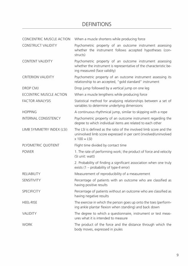
definitions
concentRic muscle Action When a muscle shortens while producing force
constRuct vAlidity psychometric property of an outcome instrument assessing whether the instrument follows accepted hypotheses (con-structs)
content vAlidity psychometric property of an outcome instrument assessing whether the instrument is representative of the characteristic be-ing measured (face validity)
cRiteRion vAlidity psychometric property of an outcome instrument assessing its relationship to an accepted, “gold standard” instrument
dRop cmJ drop jump followed by a vertical jump on one leg
eccentRic muscle Action When a muscle lengthens while producing force
fActoR AnAlysis statistical method for analysing relationships between a set of variables to determine underlying dimensions
hoppinG A continuous rhythmical jump, similar to skipping with a rope
inteRnAl consistency psychometric property of an outcome instrument regarding the degree to which individual items are related to each other
limB symmetRy indeX (lsi) the lsi is defined as the ratio of the involved limb score and the uninvolved limb score expressed in per cent (involved/uninvolved x 100 = lsi)
plyometRic quotient flight time divided by contact time
poWeR 1. the rate of performing work; the product of force and velocity (si unit: watt)
2. probability of finding a significant association when one truly exists (1 – probability of type-ii error)
ReliABility measurement of reproducibility of a measurement
sensitivity percentage of patients with an outcome who are classified as having positive results
specificity percentage of patients without an outcome who are classified as having negative results
heel-Rise the exercise in which the person goes up onto the toes (perform-ing ankle plantar flexion when standing) and back down
vAlidity the degree to which a questionnaire, instrument or test meas-ures what it is intended to measure
WoRK the product of the force and the distance through which the body moves, expressed in joules
10
intRoduction
Achilles tendon rupture is common and studies have reported an increasing incidence [31, 65, 77, 85, 111]. this is thought to be related to a greater interest in recreational sports activities [31, 65, 85]. there is a greater risk of sustaining an Achilles tendon rupture in males than females, with a male/female ratio ranging between 2:1 and 18:1 [90]. persons that acutely rupture their Achilles tendon are frequently physically involved in activities that involve running [31]. the highest incidence of Achilles tendon rupture, related to physical activity, occurs between the age of 30 and 49 years, but there is a second peak in elderly non-athletes [31, 77].
the most common injury mechanism is a sudden, forced ankle dorsiflexion, in many cases during participation in racket sports [4]. the diagnosis of an acute total Achilles tendon rupture is clinical and additional examinations, such as ultrasonography and mRi, are only occasionally needed [47]. the typical history, in a patient without previous symptoms, is the sudden audible “snap” in the calf. the patient often believes he/she has been hit from behind.
the most common rupture type is the so-called subcutaneous rupture, in the mid-substance of the Achilles tendon, i.e. 2-6 cm proximal to the insertion to the calcaneus [39]. the etiology of Achilles tendon ruptures is still not well known and two theories have been discussed; the degenerative and the mechanical [10, 44, 74]. most of the patients have not had any symptoms such as tenderness, stiffness or pain from the Achilles tendon prior to the rupture [40]. some factors, suggested to be linked to Achilles tendon rupture are ageing of the tendon, vascular impairments and lifestyle factors [1, 40]. the total rehabilitation period after an Achilles tendon rupture is long, often up to a year or even longer. due to fear of re-rupture, many patients never return to recreational or competitive sports activity [89].
the first randomised study presented in 1981 by nistor [93] reported better results after non-surgical treatment compared with surgical treatment. since then, several other randomised, controlled stud-ies have been published [11, 12, 15, 43, 46, 48, 68, 76, 80, 82, 89, 100, 112, 119]. the study design varies considerably between these studies, with regard to both the quality of the studies and the way the outcomes are presented. one common problem is the relative lack of power, with regard to the number of subjects. there is also a mix of treatments, such as surgical and non-surgical, as well as different surgical techniques and/or post-operative regimens [48]. immobilisation periods, equinus position or not, weight bearing or not and open or percutaneous procedures are some examples [11, 12, 46, 48, 80, 82, 89, 100, 112, 119]. this mixture makes comparisons difficult and sometimes impossible.
the possible influence on outcome of using an additional functional brace instead of a cast has been discussed [2, 48, 97, 100, 119]. limited information is, however, available on the effects of a functional brace, especially in combination with non-surgical treatment [48, 80, 119]. surgical treat-ment is probably most commonly employed and appears to result in less risk of a re-rupture [6, 48]. the limitations of surgical treatment are a significantly higher risk of infections, adhesions and other wound-related problems, compared with non-surgical treatment [8, 96]. most top-level athletes pre-fer surgical treatment, on individual preferences. taken together, there is still no consensus in terms of the best treatment and the debate is ongoing and seemingly never ending.

11
on the other hand, there is general agreement that surgical treatment is the first choice when a chronic rupture or a re-rupture is treated [9, 26, 55, 67, 72, 118, 122].
Additional ultrasonography or mRi could be of importance for verification of the diagnosis and for the planning of the surgical procedure [73, 86]. the surgical techniques described in the literature differ considerably, although augmentation and tendon transfers dominate [90]. the limited cohorts in all studies and the varying outcome measurements make comparisons difficult or even impossible [67].
it is obvious that there is limited knowledge with regard to the optimal rehabilitation, when consid-ering the fact that most of the patients treated for an Achilles tendon rupture still have functional deficits one year after the injury, regardless of whether the treatment is surgical or non-surgical [84, 89, 93]. until now, no injury-specific, patient-reported scores have been found to evaluate the out-come after the treatment of Achilles tendon rupture.
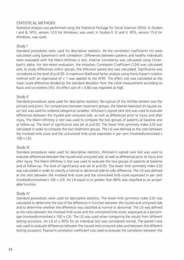
AnAtomythe Achilles tendon is the thickest and strongest tendon in the body [74]. two muscles in the lower leg, gastrocnemius and soleus, contribute to the Achilles tendon. triceps surae is the umbrella term for the gastrocnemius and soleus muscles, the main plantar flexors of the ankle. the two heads of the gastrocnemius muscle, originating from the medial and lateral condyle of the femur respectively, also contribute to knee flexion. the soleus muscle originates from the upper third of the fibula and central third of the tibia. the plantaris muscle, absent in 6%-8%, is a small 5-10 centimetre rudi-mentary muscle which originates from the lateral femoral condyle, by its long tendon inserts medially (together with the Achilles tendon or alone) on the tuber calcaneii or at the plantar aponeurosis [18]. the plantaris muscle contributes to plantar and knee flexion. the tendinous portion of the soleus and gastrocnemius muscles varies in length between 3-11 cm and 11-26 cm respectively [18].
the Achilles tendon spreads out at the insertion to the calcaneus and the narrowest part of the ten-don is approximately 4 cm above the insertion. the Achilles tendon is inserted into the central part of the posterior surface of the calcaneus. the tendon has no true synovial sheet but is instead covered by a pa-ratenon, a thin layer of loose areolar tissue that acts to reduce friction. there is one superficial and one deep bursa at the distal part of the tendon.
the Achilles tendon receives its blood supply from three sites; the mus-culo-tendinous junction, the paratenon and distally at the tendon in-sertion to the bone [1, 95, 105]. the paratenon plays an important role as it is highly vascularised and supplies the tendon with blood through its length. According to carr and co-workers [10], and lagergren and co-workers [57], there are fewer blood vessels at the midsection of the tendon and a larger number at the insertion, but this has been, been questioned [127].
Figure 1. Posterior aspect of the lower leg
12
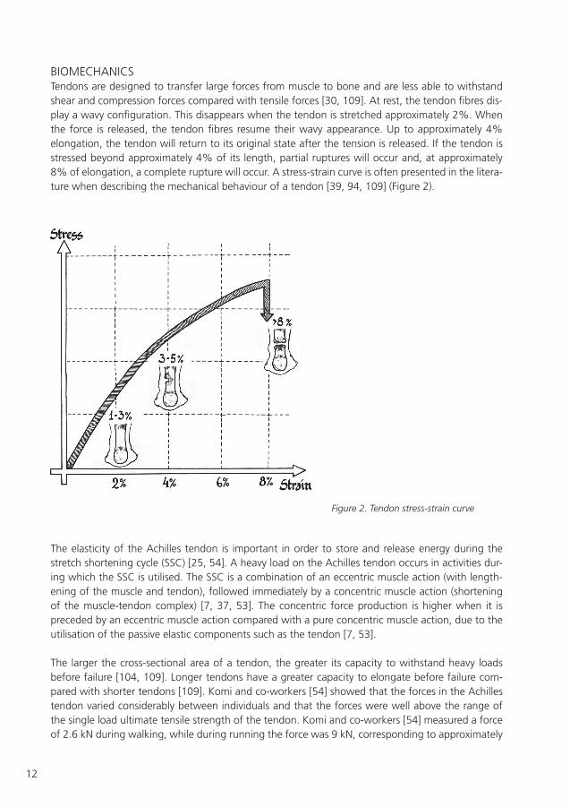
BiomechAnicstendons are designed to transfer large forces from muscle to bone and are less able to withstand shear and compression forces compared with tensile forces [30, 109]. At rest, the tendon fibres dis-play a wavy configuration. this disappears when the tendon is stretched approximately 2%. When the force is released, the tendon fibres resume their wavy appearance. up to approximately 4% elongation, the tendon will return to its original state after the tension is released. if the tendon is stressed beyond approximately 4% of its length, partial ruptures will occur and, at approximately 8% of elongation, a complete rupture will occur. A stress-strain curve is often presented in the litera-ture when describing the mechanical behaviour of a tendon [39, 94, 109] (figure 2).
Figure 2. Tendon stress-strain curve
the elasticity of the Achilles tendon is important in order to store and release energy during the stretch shortening cycle (ssc) [25, 54]. A heavy load on the Achilles tendon occurs in activities dur-ing which the ssc is utilised. the ssc is a combination of an eccentric muscle action (with length-ening of the muscle and tendon), followed immediately by a concentric muscle action (shortening of the muscle-tendon complex) [7, 37, 53]. the concentric force production is higher when it is preceded by an eccentric muscle action compared with a pure concentric muscle action, due to the utilisation of the passive elastic components such as the tendon [7, 53].
the larger the cross-sectional area of a tendon, the greater its capacity to withstand heavy loads before failure [104, 109]. longer tendons have a greater capacity to elongate before failure com-pared with shorter tendons [109]. Komi and co-workers [54] showed that the forces in the Achilles tendon varied considerably between individuals and that the forces were well above the range of the single load ultimate tensile strength of the tendon. Komi and co-workers [54] measured a force of 2.6 kn during walking, while during running the force was 9 kn, corresponding to approximately
13
12 times the body weight of a 75 kg person. cycling produced a force of less than 1 kn. they also noted that the release of force at impact was absent with ball-contact running but was present with heel-contact running.
heAlinG pRocess in tendonsfollowing a tendon injury there are three phases of healing: inflammatory, proliferative, and remod-elling [22, 39, 41, 63]. the acute inflammatory phase lasts for up to one week after injury. during this phase, the inflammatory cells remove the injured tissue, making it possible for phase two, the proliferative phase, to begin. during the proliferative phase type i collagen is produced by the fibrob-lasts to increase tendon strength. After about four weeks more than 50% of the tensile strength of the tissue may be restored. the proliferative phase lasts up to about four weeks in most individu-als. the remodelling phase of healing occurs for up to one and a half years after the original injury. during this phase, the tensile strength, elasticity and structure of the tendon are improved.
in animal studies [41], the healing tendon has been reported to regain about 50% of its tensile strength and 30% of its energy absorption within two weeks after surgery. thermann and co-workers [115] reported that ruptured, non-sutured Achilles tendons in rabbits ruptured at 38% of the force required to rupture the contralateral control side 2 weeks after injury. Research indicates that the tendon requires mechanical loading in order to recover after injury [49, 50]. the optimal amount of loading that would both benefit the healing of the tendon but still not cause a re-rupture is, however, still unknown [21, 34, 49].
14
RevieW of the liteRAtuRe
this literature review focuses on the main purpose of this thesis, i.e. to evaluate treatment in patients with an acute Achilles tendon rupture, particularly in terms of complications, function and symp-toms.
suRGicAl oR non-suRGicAl tReAtmentthe treatment of patients with an acute Achilles tendon rupture can be classified into surgical (open/percutaneous) and non-surgical. post-operative treatment can be divided into cast immobilisation and functional bracing.
in the literature, the number of re-ruptures usually constitutes the main outcome when comparing surgical and non-surgical treatments [48]. According to Bhandari and co-workers [6], only six of 11 eligible randomised, controlled studies were accepted for their meta-analysis. the re-rupture rate was found to be significantly lower with surgical treatment (3.1%), compared with non-surgical treatment (13%). All six trials included in the meta-analysis recommended surgical repair. however, Bhandari and co-workers [6] also pointed out the wide confidence intervals in the six included studies and proposed that a large randomised trial should be carried out before any strong rec-ommendations for a specific treatment could be made. one major problem is the variation in the methodological quality in the studies of the treatment of acute Achilles tendon ruptures. Khan and co-workers [48] presented a meta-analysis, including 12 of 36 eligible randomised trials with a vary-ing level of methodology. the re-rupture rate was estimated at 3.5% and 12.6%, in surgically and non-surgically treated patients, respectively. in a literature review by Ajis and co-workers [2], surgical treatment was recommended, but they pointed out the higher rate of other complications, such as infections, wound problems and adhesions, after surgery. they also reported that percutaneous repair has increased in popularity and, further, that early weight bearing and mobilisation with or without surgical treatment produced the best result, provided that the tendon ends were in contact [2]. Wong and co-workers [125] published a retrospective review of 125 articles. their conclusion was that open repair and early mobilisation produced the best outcome in terms of recovery and re-rupture rate. they also reported promising results after non-surgical treatment, using a functional brace.
Comparison of surgical/open repair/rigid cast and non-surgical treatment/rigid castnistor and co-workers [93] presented a, randomised trial comparing surgical and non-surgical treatment in patients suffering from an acute Achilles tendon rupture. non-surgical treatment was advocated as the treatment of choice, due to the low re-rupture rate and the increased risk of com-plications, such as infections and wound break-down, in surgically treated patients. contrary to this, cetti and co-workers [11] found surgical treatment preferable in their randomised trial.
Comparison of surgical/open repair/movable cast and non-surgical treatment/rigid castmöller and co-workers [89] studied 112 patients with an acute Achilles tendon rupture. they found a statistically significant higher re-rupture rate in the non-surgically treated group, 20.8%, com-pared with 1.7% in the surgically treated group. however, there were no significant differences between the two groups in terms of functional outcome. they recommended surgical treatment as the primary choice. in their study, a movable brace was used in the surgical group, while cast immo-bilisation was used in the non-surgical group. patients who underwent surgery started rehabilitation after 2 weeks using the movable brace, with increasing motion until eight weeks, when the brace was removed. patients who were treated without surgery were immobilised for eight weeks (the

15
same protocol as advocated by nistor [93], i.e. a plaster cast in the equinus position for four weeks and the neutral position for an additional four weeks). in other words, the rehabilitation differed in the two treatment groups, making it difficult to judge whether the surgery was the main beneficial factor or whether the delayed rehabilitation in the non-surgical group was a negative factor.
Comparison of surgical techniques; open repair versus percutaneous repairthe variation in surgical techniques for repairing an acute Achilles tendon rupture is remarkable and it is therefore difficult to determine the method of choice [47]. According to a review by Wong and co-workers [125], the best surgical procedure to repair an acute Achilles tendon rupture is an end-to-end suture. the techniques of ma & Griffith [70] and of delponte and co-workers [19] are the most commonly used percutaneous methods. using a percutaneous technique, the risk of sural nerve damage has been shown [16]. According to cretnik and co-workers [16] the nerve damage can be avoided, however. the risk of nerve injury should be taken into account when percutaneous techniques are compared with open techniques. there is also an increased risk of wound problems when an open technique is used. Wong and co-workers [125] concluded that the simpler procedure appeared to be more favourable. however, this is not supported by randomised, well-powered stud-ies.
lim and co-workers [68] compared open and percutaneous repair in their study and, they were unable to demonstrate any significant differences between the study groups in terms of re-rupture rates. however, other complications, such as infections and other wound problems, appeared to be more common in patients treated with open repair.
taken together, strong scientific evidence is lacking regarding the optimal treatment for acute Achil-les tendon rupture. the outcome appears to be comparable between surgical and non-surgical treat-ments, although the re-rupture rate in absolute numbers is somewhat higher in the non-surgical group in most studies. A movable brace appears to be preferable to a cast regardless of whether or not surgery is used.
Comparison of post-operative regimens; immobilisation versus functional bracein five studies [12, 43, 46, 76, 82] in which all the patients underwent surgical intervention, ran-domisation was performed post-operatively to a rigid cast alone or, alternatively, to a functional brace after a short period in a rigid cast. An overall low re-rupture rate in favour of a functional brace was reported. however, there were no significant differences between the treatment groups in any of the studies in terms of complications other than re-rupture rate. maffulli and co-workers [76] stated that early weight bearing in addition to ankle mobilisation after open repair, compared with cast immobilisation, shortened the rehabilitation period. however, neither strength deficits nor muscle hypotrophy were prevented. Kerkhoffs and co-workers [46] also favoured a functional brace, which gave a shorter rehabilitation period. the brace was deemed especially beneficial in terms of an earlier return to sports.
in a recent meta-analysis [113] including six randomised studies, a comparison between traditional, i.e. immobilisation, and early functional post-operative protocols in patients treated with surgery was performed. the patients’ opinion of their quality of life was shown to be superior in the group using a functional post-operative protocol with early weight bearing, compared with the traditional non-weight-bearing group. patients in the traditional group complained more about scar adhesion and transient sural nerve dysfunction, even though there were no statistically significant differences be-tween the groups. suchak and co-workers [113] therefore recommended that larger, well- powered

16
prospective, randomised studies should be performed to reach more definite conclusions. it is obvi-ous that most treatment studies are hampered by a similar problem, i.e. low or marginal power. most studies include approximately 100 patients, sometimes fewer. taken together, this implies that a large study, comprising around 500 patients, for example, in which the same protocol would be strictly adhered to, is necessary. A multi-centre study therefore appears to be of importance in the future in order realistically to complete such a large study.
the treatment regimens in the studies presented above were not identical, but all of them still con-clude that early mobilisation appears to reduce rehabilitation time and results in a lower re-rupture rate in patients treated surgically. it may therefore be argued that early mobilisation and early weight bearing are probably of major importance in the treatment of an acute Achilles tendon rupture.
Comparison of a functional brace versus a rigid cast in non-surgical treatment As early as 1997, mccomis and co-workers [79] indicated promising results with non-surgical treat-ment and a functional brace and recommended this method as an alternative to surgical treatment. one definite limitation of this study was the lack of a control group.
in their meta-analysis, Khan and co-workers [48] reported a lower re-rupture rate after non-surgical treatment, when early functional mobilisation was emphasised. the results were, however, based on only two studies [97, 100] and, considering the small sample sizes, clinical conclusions should therefore be drawn with caution.
costa and co-workers [15] performed two independent randomised, controlled trials at the same time, comparing immediate weight bearing with traditional plaster cast immobilisation in patients with an acute Achilles tendon rupture, treated either surgically or non-surgically. no differences were reported between patients treated non-surgically with early weight bearing, compared with cast immobilisation. however, the surgically treated group with early weight bearing had a better functional outcome, in terms of walking and stair climbing, compared with those treated with cast immobilisation alone. moreover, the risk of complications was not increased in either surgically or non-surgically treated patients, when immediate weight bearing was allowed. consequently, costa and co-workers [15] advocated immediate weight bearing, irrespective of whether surgery was em-ployed or not, as the treatment for patients with an acute Achilles tendon rupture.
Wallace and co-workers [121] reported on a combined non-surgical cast and brace treatment pro-tocol for an acute Achilles tendon rupture. in their study of 140 patients, there was an overall com-plication rate of 8%, with three complete and five partial re-ruptures, two deep vein thromboses and one temporary drop foot. they strongly recommended their treatment protocol, however, only when patients were supervised by experienced staff.
hufner and co-workers [32] presented long-term results in 168 patients with an Achilles tendon rupture treated non-surgically, using a so-called variostabil functional brace. they reported a low re-rupture rate; 6.4%. A prerequisite in this study was that an ultrasound examination, performed by an orthopaedic surgeon, showed that the gap in the ruptured tendon was less than one cm with the ankle in the neutral position and that the tendon ends were in contact with each other in 20 degrees of plantar flexion. if this was not the case, the patients were recommended surgical treat-ment. to further reduce the risk of re-rupture, a second ultrasonographic examination, performed by an experienced examiner, was recommended after two to five days.

17
Comparison of surgical and non-surgical treatment and a functional bracetwaddle and co-workers [119] compared surgical and non-surgical treatment in a recent study. Both groups received early range of motion exercises using a functional brace and an identical rehabilita-tion protocol. they reported no significant differences in terms of re-rupture rate. surprisingly, there was only one re-rupture in the non-surgical group and two in the surgical group. they suggested that early range of motion was the most important factor, regardless of whether surgical or non-surgical treatment was used [119].
thermann and co-workers [116] presented the results from a two-year functional treatment concept in patients with acute Achilles tendon rupture. these researchers described an ultrasonographic method that was reported to have good reliability. the method was used to determine the separa-tion between the ruptured tendon ends. in a randomised trial, surgical and non-surgical/functional treatment was compared in 50 patients. no significant differences were found between groups in terms of functional results or the course of tendon healing. they also showed that the functional treatment was favourable in terms of a shorter rehabilitation period.
metz and co-workers [80] compared minimally invasive surgery with non-surgical treatment and both groups were allowed immediate full weight bearing. the primary end-point was complications other than re-rupture. A significant difference was found, with more skin-related problems in the non-surgically treated group, which is unusual, and the choice of brace might therefore be discussed. they also stated that the surgical treatment resulted in an earlier return to work. using complications other than re-rupture as the primary end-point is somewhat unusual from a methodological stand-point. this might, however, increase the interest in end-points other than re-rupture alone. it might, however, be of greater importance to evaluate patient-reported outcome and recovery of function as opposed to skin-related complications.
Weber and co-workers [123] presented excellent results in a retrospective study, with a significantly shorter period of pain and a faster return to work in their non-surgically treated group. the non-surgically treated group was treated with an equinus ankle cast and boot allowing full weight bear-ing, compared with a removable brace used in the surgically treated group.
it is difficult to draw strong conclusions as the studies vary considerably in terms of treatment proto-cols, methodology and outcome measurements. one general problem is that, in many or even most of the studies, the sample sizes are too limited and the methods are too insensitive to detect clinically relevant differences between study groups.
Surgical treatment for chronic Achilles tendon ruptureAccording to the literature [72], a chronic rupture refers to an Achilles tendon rupture diagnosed 4-6 weeks after injury. the different terms used for this group of patients in the literature [26, 67] are delayed, neglected and chronic ruptures. for medico-legal reasons, the term “chronic rupture” is preferable. however, there is no universally accepted time limit defining when an acute rupture turns into a chronic rupture. more than 20% of acute Achilles tendon ruptures are supposed to be presented in a delayed manner, depending on the fact that the rupture is unrecognised or misdiag-nosed by the examiner or that the patient waits before seeking medical attention [71, 75].
When treating a chronic rupture, most surgeons agree that surgery is the treatment of choice, unless there are contraindications for surgery or if the patient has low functional demands.

18
even though many different surgical techniques exist, only a few of them have been validated in a strict scientific manner [91]. taken together, there is an obvious lack of evidence-based guidelines for the selection of the optimal surgical technique in patients with a chronic Achilles tendon rupture [67]. the repair of a chronic rupture or a re-rupture is associated with an increased risk of complica-tions [72]. in other words, it is difficult to draw any definite conclusions in terms of the functional outcome of the different surgical techniques presented in the literature, due to the wide variation in study design, post-operative regimens and end-points. the different surgical techniques that are described can be divided into different categories; the v-y technique, local tissue augmentation, turn-down flaps, tendon transfer, free tissue transfer and the use of synthetic materials [72, 81].
several different turn-down flaps have been used in order to bridge the tendon gap. christensen and co-workers [14] were the first to describe this technique in 1931. more than 20 years later, Arner & lindholm [4] used two flaps instead of one, while silfverskiöld [108] used one rotated flap in order to ensure that the smooth tendon surface faced the skin, in order to reduce the risk of skin adhesions. Gerdes and co-workers [29] showed that flap augmentation produced better pull-out strength than end-to-end sutures alone. the difference was 41%.
us and co-workers [120] reported satisfactory results in six patients using a combined technique of v-y plasty, with an additional turn-down flap from the gastrocnemius aponeurosis. they showed that an acceptable restoration of the muscle/tendon complex function was obtained.
maffulli and co-workers [75] presented good results using a free gracilis tendon graft in 21 patients, who had a chronic rupture of the Achilles tendon. their conclusion was that this was a technically demanding yet safe procedure, although it resulted in reduced strength and decreased ankle mo-tion. most people agree that a simple end-to-end suture is not sufficient to treat a chronic rupture or a re-rupture. An open technique is therefore usually recommended for these groups of patients.
ThrombosisWithout thromboprophylaxis, the incidence of deep venous thrombosis is 40-80% and fatal pulmo-nary embolism 1-5% following major orthopaedic surgery [27].
the need for thrombosis prevention has been generally agreed upon when major orthopaedic sur-gery is undertaken and standardised thromboprophylaxis regimens are therefore usually implement-ed. however, in patients treated for so-called minor lower leg injuries, such as an Achilles tendon rupture, no consensus exists with regard to the advantage of thromboprophylaxis. the risk of de-veloping thromboembolism after an acute Achilles tendon rupture has only been demonstrated in a few studies [24]. the results varied from a benefit from thromboprophylaxis to no benefit at all [38, 52, 56, 61, 62] .
the accuracy of a clinical diagnosis of thromboembolism is low and the incidence is therefore un-known. however, lapidus and co-workers [58] verified, from a register of 668 patients treated for an acute Achilles tendon rupture, symptomatic deep venous thrombosis (dvt) in 47 (7%) patients with-in six weeks from injury. venography is considered to be the “gold standard” for verifying a dvt. the method is, however, invasive and demanding for the patient. technical advances and clinical experi-ence have increased the advantage of colour duplex sonography (cds), which is a non-invasive, less expensive and more convenient method for the patient [59]. however, the accuracy of the method has to be studied further before it can be used as the “gold standard” when diagnosing a dvt.
19
Patient-reported outcomeit is important to use reliable, validated outcome measurements when evaluating treatment. patient-reported outcomes have been more frequently used during the last decade, in order to obtain the patients’ own opinion about the result. When evaluating outcome, different functional tests together with the patient’s opinion, as well as complication registration, are necessary to obtain as complete an overall picture of the treatment results as possible [90].
evaluations of the treatment of an Achilles tendon rupture vary considerably between different studies. clinical examination often includes calf muscle circumference, ankle range of motion and tendon width measurements. it has, however, never been shown that any of these factors is of any importance, in terms of function or patient satisfaction.
leppilathi and co-workers [64] designed a scoring method proposed as a standard method to be used to compare outcome in different studies. however, this score has not been validated and moreover constitutes a mixture of subjective and objective measurements. there is therefore an obvious need for a new outcome measurement, based on patient outcome and function and tested for reliability and validity at the same time. A validated patient-reported outcome measurement exists for the evaluation of treatment of Achil-les tendinopathy; i.e. the victorian institute of sports Assessment − Achilles questionnaire (visA-A questionnaire) [98]. A validated patient-reported outcome measurement is also available for foot and ankle injuries; the foot and Ankle outcome score (fAos) [99]. since patients with an Achilles tendon rupture have different functional complaints and symptoms compared with patients with Achilles tendinopathy or ankle injuries, the use of the visA-A and fAos as an outcome measure-ment for this patient group can be questioned. to our knowledge, no validated scores for evaluating treatment in patients with an acute Achilles tendon rupture have previously been presented. Accord-ingly, the Achilles tendon total Rupture score (AtRs) was constructed as a part of this thesis.
Recovery of function the strength deficit of the calf musculature on the injured side, one year after an Achilles tendon rupture, is reported to be approximately 10-30% compared with the uninjured side and it appears that it becomes permanent [47]. moreover, early recovery of plantarflexion torque has not only been shown to indicate the normalisation of the calf musculature function, it can also be due to compensation by the flexor hallucis longus muscle [20].
to evaluate muscle endurance, the most commonly used test is counting the number of consecutive heel rises the subject is able to perform until fatigue sets in on one leg and to compare this with the other leg. A modification of this test was presented by häggmark and co-workers [33] more than 20 years ago. in their test, a light beam was used to ensure that only heel rises over a certain height (in their study 4 cm) were counted. the height of the heel rise may be of importance, since there is a disproportionate weakness in end-range plantar flexion according to mullaney and co-workers [84]. they found that, at angles with the foot in dorsiflexion, there was no strength deficit, but, at 20° and 10° of plantar flexion, there were significant strength deficits. the explanation for this is thought to be due to tendon lengthening that may occur during the healing of the tendon. studies have demonstrated that there is a separation of tendon ends after Achilles tendon repair [42]. interestingly, a study by Kangas and co-workers [42] has shown that early motion resulted in a smaller degree of tendon separation compared with immobilisation following Achilles tendon rupture treated with surgery. the smaller degree of tendon separation also correlated with clinical outcome.

20
the importance of the above-reported changes in plantar flexion muscle forces and deficits in heel-rise heights is the functional implication when it comes to the patients’ ability to walk, run and jump. for the patients, the important aspect following an Achilles tendon rupture is to be able to recover full function and return to previous activities without an increased risk of re-rupture and of developing other overuse injuries. Gait analysis was recently performed by don and co-workers [20] in a study of 49 patients with a surgically repaired Achilles tendon rupture and, at the 24-month evaluation, gait abnormalities were still found. this study also found an eccentric strength deficit in the calf musculature at the 24-month evaluation. further studies are needed to examine the vari-ous aspects of the way lower leg function (with regard to muscular strength, endurance and ability jump) is recovered after Achilles tendon rupture. it would also be interesting to evaluate how lower leg function is affected by the various types of treatment in order to provide some insight into ways of improving treatment.

21
pRoBlem AReAs
the patients’ opinion of their symptoms and physical capacity is an important factor when evalu-ating outcome after the treatment of an Achilles tendon rupture. no validated, reliability-tested patient-reported outcome score is available to study the outcome after treatment of an acute Achil-les tendon rupture. This topic is addressed in Study I.
the possible influence of a functional brace has been discussed in the treatment of an Achilles ten-don rupture. however, limited information is available about the benefits of allowing early range of motion and early weight bearing, especially in combination with non-surgical treatment. This topic is addressed in Study II.
the treatment of a re-rupture is always a “challenge” and the risk of complications is high. however, there is no disagreement that surgery is the best way to treat this group of patients. however, due to a lack of comparative studies, the optimal treatment is still unknown. This topic is addressed in Study III.
When evaluating functional outcome after an acute Achilles tendon rupture, differences between treatment groups are rarely reported. one important explanation is that previously described evalu-ation methods are not tested for reliability and validity and have a low ability to detect small yet clinically relevant differences between treatment groups. This topic is addressed in Study IV.
venous thromboembolism is common after Achilles tendon rupture, but the true incidence is not known. This topic is addressed in Study V.

22
Aims of the studies
Study I to develop and validate a new patient-reported instrument for measuring outcome, related to symptoms and physical activity, after treatment in patients with an acute total Achilles tendon rupture.
Study II the purpose of this study was to compare surgical with non-surgical treatment, using identical mobilization and rehabilitation protocols, for patients with an Achilles tendon rupture, in regards to complications, symptoms and function.
Study III to evaluate the subjective and objective outcomes, following a new surgical method to treat a chronic rupture and a re-rupture of the Achilles tendon.
Study IV to evaluate a heel-rise test that would measure both the height of each heel rise and the number of repetitions and to compare this test with ankle range of motion meas-urements and patient-reported outcome.
Study V to determine the incidence of venous thromboembolism (vte) in patients with Achilles tendon rupture.

23
suBJects And methods
suBJectsA total of 273 patients with an Achilles tendon rupture were included in the five studies in this thesis (table 1). furthermore, 52 healthy subjects (19 females and 33 males) were included in study i and 38 patients with Achilles tendinopathy were included as a reference group in study iv.
Table 1. Summary of patients with an Achilles tendon rupture included in the five studies.
Study IA total of 167 male and female patients (table 2) with a total Achilles tendon rupture were involved in the various steps. the inclusion criteria were:• TotalAchillestendonrupturebasedonaclinicalexamination,
performed by an orthopaedic surgeon.
• 20-70yearsofage.
• AbletoreadandunderstandtheSwedishlanguage.
Table 2. Distribution, mean age and standard deviation (SD) for the patients and healthy subjects in the various steps in Study I (twenty-seven patients were included in more than one step).
Study Patients Male/female Age Comments included (%) mean
i 167 134/33 (20%) 42 27 of the patients were included twice in the various steps
ii 97 79/18 (19%) 41 Allocated from 100 patients. 15 patients also included in study i and 4 patients also included in study iii.
iii 28 22/6 (21%) 46 13 chronic ruptures and 15 reruptures.
iv 78 65/13 (17%) 42 patients from study ii
v 95 79/16 (17%) 41 patients from study ii
Step Description Subjects Age mean ± SD
1 item generation - -
2 item reduction n = 112 21f+91 m 41.9±9.6
3 evaluation of the final AtRs n = 82 19f+ 63 m 43.3±9.7
3 evaluation, healthy subjects n = 52 19f + 36m 41.5±9.3
4 test-retest of the AtRs n = 43 7f + 36 m 41.7±9.9
5 Responsiveness of the AtRs n = 43 8f + 36 m 40.3±8.3
f = females, m = males, AtRs = Achilles tendon total Rupture score

24
Variable Surgical Non-surgical p-value (n=49) (n=48)
Age 40.9 (8.8) 41.2 (9.5) 0.9367 41.0 (24.0; 59.0) 39.0 (23.0; 63.0) n=49 n=48
Gender male 40 (81.6%) 39 (81.3%) female 9 (18.4%) 9 (18.8%) 1.0000
height 178.7 (9.2) 177.7 (8.8) 180.0 (154.0; 197.5) 180.0 (153.0; 192.0) n=47 n=40 0.6362
injured side Right 23 (46.9%) 27 (56.3%) left 26 (53.1%) 21 (43.8%) 0.4753
Work sedentary 15 (30.6%) 15 (31.9%) light but mobile 23 (46.9%) 27 (57.4%) heavy 11 (22.4%) 5 (10.6%) 0.3708
for categorical variables, n (%) is presented. for continuous variables, mean (sd)/median (min; max)/n= is presented. for comparisons between groups, fisher´s exact test was used for dichotomous variables, while the mantel-haenzsel chi-square exact test was used for ordered categorical variables and the mann-Whitney u test was used for continuous variables.
Study IIA total of 100 patients, with an acute Achilles tendon rupture, who sought medical attention at an emergency department at the sahlgrenska university hospital, Göteborg, sweden, were included in this prospective, randomised study, during the period 2004-2007. in all patients, the diagnosis was established, based on medical history and clinical examination (tendon palpation and thompson’s test). patients eligible for the study were randomised to either surgical treatment or non-surgical treatment. computer-generated sealed envelopes were used in the randomisation procedure, which was administered by a co-ordinator. due to diversity in reporting, we do not know how many pa-tients were included from the total number of patients who sought medical attention at the emer-gency wards due to an Achilles tendon rupture.
two patients who were randomised to non-surgical treatment chose surgical treatment. one pa-tient, who had been randomised to surgical treatment, was treated non-surgically as it was not pos-sible to perform the surgery within 72 hours. As a result, 97 patients (79 men and 18 women) were included in the follow-up evaluations (table 3).
patients 16 to 65 years of age with a unilateral Achilles tendon rupture were included in the study if they were randomised and treated within 72 hours from the injury. the exclusion criteria were dia-betes mellitus, former Achilles tendon rupture, other lower leg injuries, immunosuppressive therapy and neurovascular diseases. Table 3. Baseline characteristics of patients included in Study II.

25
Study IIIA total of 28 consecutive patients, all treated during the period 1996-2005 at sahlgrenska univer-sity hospital, Göteborg, sweden (21 male and 7 female), with a mean (sd) age of 46 (10.4) years, ranging from 26-71 years of age, were included in study iii. there were 13 chronic ruptures and 15 re-ruptures. twelve ruptures were on the right side and 16 on the left. three experienced surgeons performed all the surgery.
Study IVseventy-eight patients with an acute Achilles tendon rupture were included in this prospective study. they were all from a cohort of 100 patients with an Achilles tendon rupture, who were included in study ii. for the present study, we added the criterion that all data from both the 6- and 12-month functional evaluations had to be available and we excluded patients who had sustained a re-rupture. As a result, 22 patients from study ii were not included. eight patients had a re-rupture, two patients had a previous rupture on the contralateral side, three patients failed to adhere to the study protocol and one patient developed an ankle contracture and could not be tested. Another four patients did not attend either the 6- or 12-month evaluation and the testing equipment either malfunctioned or did not record the data for another four patients (two during the 6-month evaluation and two during the 12-month evaluation).
of the 78 patients included (65 men and 13 women), 43 received surgical treatment and 35 non-surgical treatment. the mean (standard deviation [sd]) age was 42 (9) years, the mean (sd) height 178 (9) cm and the mean (sd) weight was 85 (13) kg.
As a reference group, patients with chronic painful Achilles tendinopathy from a previous study were included [106].this group consisted of 38 patients (19 men and 19 women). these patients were evaluated using the same methods prior to starting rehabilitation and after one year. in this group, the mean (sd) age was 45 years (8), the mean (sd) height 177 cm (8) and the mean weight 79 kg (12).
Study Vninety-five patients with an acute Achilles tendon rupture from study ii participated in this study.
ethicsAll the subjects received oral and written information about the purpose and procedure of the study and written informed consent was obtained. ethical approval was obtained from the Regional ethi-cal Review Board in Gothenburg, sweden.

26
methods
Study Ithe development of the new instrument, the Achilles tendon total Rupture score (AtRs), consisted of five steps, as described in figure 3.
Figure 3. Achilles tendon Total Rupture Score (ATRS), steps 1-5.
STEP 1 ITEM GENERATION & TEST CONSTRUCTION
literature reviewexpert group
(reduction of similarities)Result = 14 items
STEP 2 ITEM REDUCTION 112 PATIENTS
(test results + verbal & written comments)expert group
(item reduction and item rephrasing)Result = 10 items
STEP 3 EVALUATION OF THE FINAL ATRS
82 patients52 healthy individuals
validitystructure
internal consistency
STEP 4 TEST - RETEST OF THE ATRS
43 patients
STEP 5 RESPONSIVENESS OF THE ATRS
43 patients
27
Study II
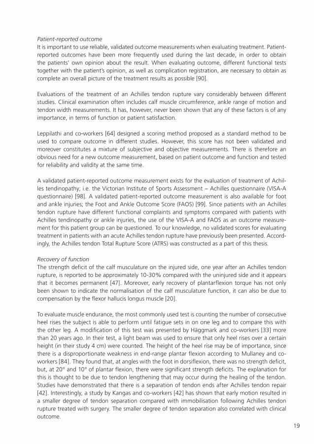
Surgical group:the surgical technique involved the patient being in the prone position under local (19), spinal (26) and general anaesthesia (4). A tourniquet was used for haemostasis in 27% of the cases. After a longitudinal 5-8 cm medial skin and paratenon incision, an end-to-end suture, using a modified Kessler suture technique with 1-0 pds (slow resorbable), was performed (figure 4). post-operatively, the patients were immobilised in a below-the-knee cast in the equinus position. surgery was performed by twenty-eight ortho-paedic surgeons familiar with the technique.
Figure 4. End-to-end suture using modified Kessler technique.
in the surgically treated group, thromboprophylaxis with 500 ml of macrodex® was administered according to a specific protocol. no standard thromboprophylaxis was employed in the non-surgi-cally treated group.
Non-surgical group:After randomisation, treatment was immediately started with a below-the-knee cast in the equinus position.
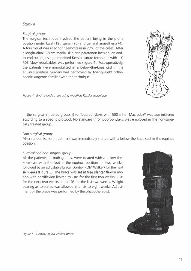
Surgical and non-surgical group:All the patients, in both groups, were treated with a below-the-knee cast with the foot in the equinus position for two weeks, followed by an adjustable brace (donJoy Rom Walker) for the next six weeks (figure 5). the brace was set at free plantar flexion mo-tion with dorsiflexion limited to -30° for the first two weeks, -10° for the next two weeks and +10° for the last two weeks. Weight bearing as tolerated was allowed after six to eight weeks. Adjust-ment of the brace was performed by the physiotherapist.
Figure 5. DonJoy ROM Walker brace.

28
RehabilitationAll patients followed a standardised rehabilitation protocol presented below, supervised by two experienced physiotherapists. (figure 6).
Weeks 8-11 treatment: shoe with a heel lift (1.5 cm), crutches if needed for another 1-3 weeks. exercise programme: visit to a physiotherapist two to three times a week and home
exercises daily. • Exercisebike • Anklerangeofmotion(ROM) • Sittingheel-rise • Standingheel-riseontwolegs • Gaittraining • Balanceexercises • Legpress • Legextensionandlegcurl
Weeks 11-16 treatment: shoe with a heel lift (1.5 cm) until week 16. exercise programme: visit to a physiotherapist two to three times a week and home
exercises daily. • Exercisesasabovewithincreasedweight • Standingheel-riseononeleg • Step • Walkingonmattress
Weeks 16-20 exercise programme: visit to a physiotherapist 2-3 times per week and home exercises. • Exercisesasabovewithincreaseinweightsandintensityastolerated • Slide • Quickreboundingheel-rises From week 18: • Heel-riseonstairs • Sidejumps • Two-leggedjumps
Weeks 20-24 exercise programme: visit to a physiotherapist as needed. • Exercisesasabovewithincreaseinweightsandintensityastolerated • Jog • Sidejumpsforward
Week 24 and onwards exercise programme: continued physiotherapy if needed. • Startgroupexerciseclass(similartoaerobics) • Gradualreturntosports(dependingonthepatient’sability)
Figure 6. Rehabilitation protocol.

29
Follow-upthe patients attended a clinical follow-up examination at the orthopaedic department after 2, 8, 12 weeks and 6 months. this examination was mainly performed by the first author (Knh). screening for deep vein thrombosis (dvt) was performed with color duplex sonography (cds) 8 weeks after treatment was initiated.
evaluation of function, symptoms and physical activity level was performed 6 and 12 months after injury by two experienced independent physical therapists.
Patient-reported outcome and physical activitythe patients’ symptoms and physical activity were assessed using the Achilles tendon total Rupture score (AtRs) (Appendix 1), and a physical activity scale (figure 7) [92, 101]. the AtRs ranges from 0 to 100, and a lower score indicates more symptoms and greater limitation on physical activity. for the physical activity scale a score of 1 equals no physical activity whereas a score of 6 means heavy physical exercise several times per week.
1 hardly any physical activity.
2 mostly sitting, sometimes a walk, easy gardening or similar tasks.
3 light physical exercise around 2-4 hours a week, e.g. walks, fishing, dancing, ordinary gardening, including walks to and from shops.
4 moderate exercise 1-2 hours a week, e.g. jogging, swimming, gymnastics, heavier gardening, home-repairing or easier physical activities more than 4 hours a week.
5 moderate exercise at least 3 hours a week, e.g. tennis, swimming, jogging etc.
6 hard or very hard exercise regularly and several times a week, where the physical exertion is great, e.g. jogging, skiing.
Figure 7. Physical activity scale (Saltin, Grimby, 1968).
Functional evaluationthe musclelab® (ergotest technology, oslo, norway) measurement system was used for the evalua-tions. musclelab® is a data collection unit, to which different kinds of sensors can be connected.
the test battery consisted of two different jump tests, two different strength tests and one muscular endurance test. the test battery has been shown to be reliable and valid for evaluating lower leg function in patients with Achilles tendinopathy and was performed as described by silbernagel et al. [106] the tests have also been used in a recent published study evaluating outcome of patients with a chronic rupture or a re-rupture of the Achilles tendon [91]. the jump tests were a drop counter-movement jump (drop cmJ) and hopping. for the drop cmJ, the patients started by standing on one leg on a 20 cm high wooden box.they were instructed to “fall” down onto the floor and, directly on landing, perform a maximum vertical one-legged jump (figure 9). the maximum jumping height in cm was used for data analysis. hopping is a continuously rhythmical jump similar to skipping rope. the patients performed 25 jumps, the average air flight and floor contact times were documented and the plyometric quotient (flight time/contact time) was used for data analysis.
30
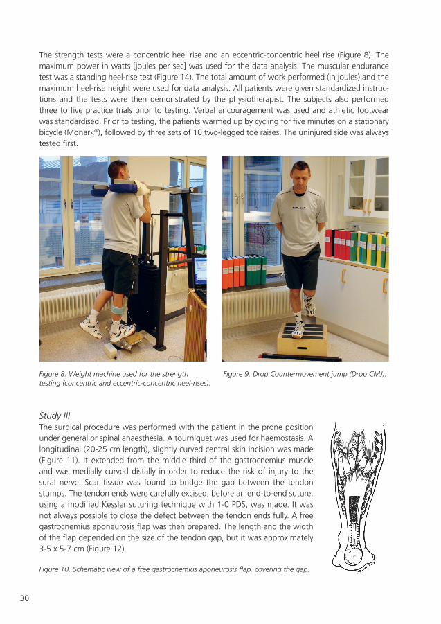
the strength tests were a concentric heel rise and an eccentric-concentric heel rise (figure 8). the maximum power in watts [joules per sec] was used for the data analysis. the muscular endurance test was a standing heel-rise test (figure 14). the total amount of work performed (in joules) and the maximum heel-rise height were used for data analysis. All patients were given standardized instruc-tions and the tests were then demonstrated by the physiotherapist. the subjects also performed three to five practice trials prior to testing. verbal encouragement was used and athletic footwear was standardised. prior to testing, the patients warmed up by cycling for five minutes on a stationary bicycle (monark®), followed by three sets of 10 two-legged toe raises. the uninjured side was always tested first.
Figure 8. Weight machine used for the strength Figure 9. Drop Countermovement jump (Drop CMJ). testing (concentric and eccentric-concentric heel-rises).
Study III the surgical procedure was performed with the patient in the prone position under general or spinal anaesthesia. A tourniquet was used for haemostasis. A longitudinal (20-25 cm length), slightly curved central skin incision was made (figure 11). it extended from the middle third of the gastrocnemius muscle and was medially curved distally in order to reduce the risk of injury to the sural nerve. scar tissue was found to bridge the gap between the tendon stumps. the tendon ends were carefully excised, before an end-to-end suture, using a modified Kessler suturing technique with 1-0 pds, was made. it was not always possible to close the defect between the tendon ends fully. A free gastrocnemius aponeurosis flap was then prepared. the length and the width of the flap depended on the size of the tendon gap, but it was approximately 3-5 x 5-7 cm (figure 12).
Figure 10. Schematic view of a free gastrocnemius aponeurosis flap, covering the gap.

31
the free flap was placed over the rupture and secured with peripheral sutures, using 3-0 pds. the flap covered approximately 75% of the circumference of the tendon and the total tendon gap. the defect in the aponeurosis was then repaired using absorbable side-to-side sutures.
Figure 13. After reconstruction with end-to-end sutures, the free gastrocnemius aponeurosis flap covers the tendon ends. The gap after removal of the gastrocnemius aponeurosis flap is sutured side-to-side.
Figure 11. The planned incision. Figure 12 a, b. The free gastrocnemius aponeurosis flap
32
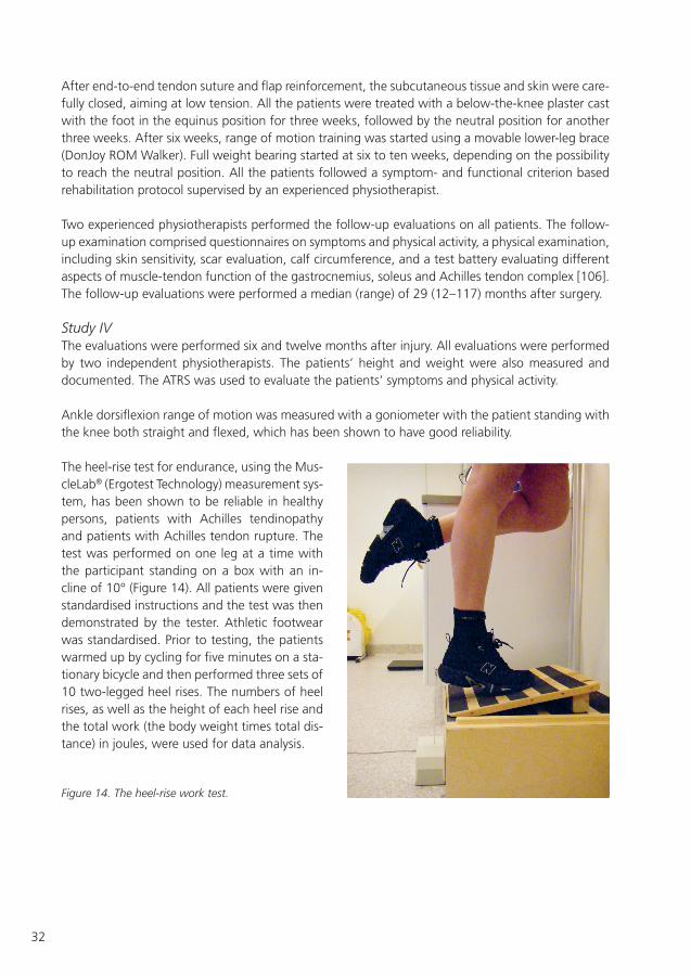
After end-to-end tendon suture and flap reinforcement, the subcutaneous tissue and skin were care-fully closed, aiming at low tension. All the patients were treated with a below-the-knee plaster cast with the foot in the equinus position for three weeks, followed by the neutral position for another three weeks. After six weeks, range of motion training was started using a movable lower-leg brace (donJoy Rom Walker). full weight bearing started at six to ten weeks, depending on the possibility to reach the neutral position. All the patients followed a symptom- and functional criterion based rehabilitation protocol supervised by an experienced physiotherapist.
two experienced physiotherapists performed the follow-up evaluations on all patients. the follow-up examination comprised questionnaires on symptoms and physical activity, a physical examination, including skin sensitivity, scar evaluation, calf circumference, and a test battery evaluating different aspects of muscle-tendon function of the gastrocnemius, soleus and Achilles tendon complex [106]. the follow-up evaluations were performed a median (range) of 29 (12–117) months after surgery.
Study IVthe evaluations were performed six and twelve months after injury. All evaluations were performed by two independent physiotherapists. the patients’ height and weight were also measured and documented. the AtRs was used to evaluate the patients’ symptoms and physical activity.
Ankle dorsiflexion range of motion was measured with a goniometer with the patient standing with the knee both straight and flexed, which has been shown to have good reliability.
the heel-rise test for endurance, using the mus-clelab® (ergotest technology) measurement sys-tem, has been shown to be reliable in healthy persons, patients with Achilles tendinopathy and patients with Achilles tendon rupture. the test was performed on one leg at a time with the participant standing on a box with an in-cline of 10° (figure 14). All patients were given standardised instructions and the test was then demonstrated by the tester. Athletic footwear was standardised. prior to testing, the patients warmed up by cycling for five minutes on a sta-tionary bicycle and then performed three sets of 10 two-legged heel rises. the numbers of heel rises, as well as the height of each heel rise and the total work (the body weight times total dis-tance) in joules, were used for data analysis.
Figure 14. The heel-rise work test.

33
Study Vscreening for deep venous thrombosis was performed using colour duplex sonography (cds), using a sequoia ultrasound machine (siemens Acuson, mountain view, cA, usA) with a linear 6l3 ultra-sound transducer. the cds examination followed a standard protocol, similar to the one described by lapidus and co-workers [59] covering the external iliac, common, deep and superficial femoral, popliteal, gastrocnemius, paired posterior tibial and peroneal veins and the proximal part of the anterior tibial and proximal junctions of the greater and lesser saphenous veins. proximal veins were examined in the supine position with a head-up tilt of about 45°, the popliteal region in the prone position with a similar tilt and the calf veins in the sitting position with the leg extended and the foot resting on the examiner’s lap. vein segments were assessed regarding compressibility and flow. the primary diagnostic criterion was venous incompressibility in the transverse view. colour flow findings were used to clarify the anatomy and as supportive findings, especially in venous segments less easily viewed and compressed, including the distal femoral vein at the adductor canal and the trifurca-tion at the proximal calf. At the time of examination, findings related to these vein segments were recorded on a drawing of the venous system, where the extent of a possible dvt could be clarified. All examinations were also recorded on videotape, in standardised recordings usually lasting 5-10 min/examination. examinations were performed by any of six technicians at the vascular laboratory, all with several years’ experience of venous ultrasonography. two technicians usually collaborated for ergonomic reasons and were able to help each other interpret the images.
After final inclusion, all the video recordings were reviewed by two vascular diagnostic physicians, each with more than ten years’ experience of the method and, at the time of the review, blinded to each other’s diagnosis and to the primary diagnosis at the time of examination. the interpretations were recorded and the reviewer was required to note a decision relating to the presence of proximal dvt (in the popliteal vein or a more proximal vein) and distal dvt (in deep or muscular veins distal to the popliteal vein). in cases of disagreement between these blinded evaluations, adjudication was made by a third person, also with more than 10 years’ first-hand experience of thrombosonography.
in cases of suspected pulmonary embolism, spiral computed tomography or perfusion/ventilation lung scintigraphy was obtained and adjudicated by an experienced physician specialising in the diagnostics of thromboembolism. there was no surveillance of pe in the present study.
the cds examination was performed eight weeks after the trauma and initiation of treat-ment.
the follow-up included clinical examinations after two, eight and 12 weeks and after six months. screening for deep venous thrombosis was performed using colour duplex sonography (cds), a non-invasive method based on ultra-sound (figure 15).
Figure 15. Colour Duplex Sonography (CDS).
34
stAtisticAl methodsstatistical analysis was performed using the statistical package for social sciences (spss). in studies i and iii, spss, version 12.0 for Windows, was used. in studies ii, iv and v, spss, version 15.0 for Windows, was used.
Study Istandard procedures were used for descriptive statistics. All the correlation coefficients (rs) were calculated using spearman’s rank correlation. differences between patients and healthy individuals were evaluated with the mann-Whitney u test. internal consistency was calculated using chron-bach’s alpha. for test-retest evaluation, the intraclass correlation coefficient [124] was calculated and, to study differences within groups, the Wilcoxon paired test was calculated. significance was considered at the level of p<0.05. A maximum likelihood factor analysis using harris Kaiser’s rotation method with an eigenvalue of > 1 was applied to the AtRs. the effect size was calculated as the mean score difference divided by the standard deviation from the initial measurement according to Kazis and co-workers [45]. An effect size of > 0.80 was regarded as high.
Study IIstandard procedures were used for descriptive statistics. Re-rupture of the Achilles tendon was the primary end-point. for comparisons between treatment groups, the mantel-haenzsel chi-square ex-act test was used for ordered categorical variables. Wilcoxon’s signed rank test was used to evaluate differences between the injured and uninjured side, as well as differences prior to injury and after injury. the mann-Whitney u test was used to compare the two groups of patients at baseline and at follow-up. the level of significance was set at p<0.05. the lower limb symmetry index (lsi) was calculated in order to compare the two treatment groups. the lsi was defined as the ratio between the involved limb score and the uninvolved limb score expressed in per cent (involved/uninvolved x 100 = lsi).
Study IIIstandard procedures were used for descriptive statistics. Wilcoxon’s signed rank test was used to evaluate differences between the injured and uninjured side, as well as differences prior to injury and after injury. the mann-Whitney u test was used to evaluate the two groups of patients at baseline and at follow-up. the level of significance was set at p<0.05. the lower limb symmetry index (lsi) was calculated in order to classify a normal or abnormal side-to-side difference. the lsi was defined as the ratio between the involved limb score and the uninvolved limb score expressed in per cent (involved/uninvolved x 100 = lsi). An lsi equal to or greater than 80% was classified as an accept-able function.
Study IVstandard procedures were used for descriptive statistics. the lower limb symmetry index (lsi) was calculated to determine the size of the difference in function between the injured and uninjured side and to determine whether the difference was classified as normal or abnormal. the lsi was defined as the ratio between the involved limb score and the uninvolved limb score, expressed as a percent-age (involved/uninvolved x 100 = lsi). the lsi was used when comparing the results from different testing occasions. An lsi of > 90% for an individual test was considered normal. the paired t-test was used to evaluate differences between the injured and uninjured sides and between the different testing occasions. pearson’s correlation coefficient was used to evaluate the correlation between the

35
heel-rise test and ankle range of motion. since the AtRs presents ordinal data, spearman’s correla-tion coefficient was used to evaluate the correlation between patient-reported symptoms (AtRs) and the heel-rise test and ankle range of motion. the level of significance was set at p<0.05.
Study Vstandard procedures were used for descriptive statistics. the primary end-point of this study was the total incidence of vte in patients treated for Achilles tendon rupture. secondary end-points were distal dvt, proximal dvt and pe. A chi-square test was used in the comparison of vte rates between the surgical and non-surgical groups. the level of significance was set at p<0.05. All the data analy-ses were carried out by the investigators.

36
summARy of pApeRs
Study I: The Achilles tendon Total Rupture Score (ATRS): development and validation
the purpose of this study was to develop and validate a new patient-reported instrument for meas-uring outcome, related to symptoms and physical activity, after treatment in patients with a total Achilles tendon rupture.
intRoduction: the use of patient-reported outcome scores to evaluate functional results and to compare incapacity on an individual level has become more common. outcome measures found in the current literature for patients with an Achilles tendon rupture are non-validated and based on a mixture of assessments of subjective and objective parameters. Robinson et al. [98] have, however, developed a questionnaire as an index of clinical severity in patients with chronic Achilles tendinopa-thy, with a validated swedish version (visA-A-s) [107], while Roos and co-workers [99] have vali-dated a score (fAos) for patients with ankle ligament injuries. the fAos includes 42 questions with five separate subscales; pain, other symptoms, activities of daily living, sport and recreation function, foot- and ankle-related quality of life
there is therefore a need for an easily self-administered, validated instrument with high reliability and high responsiveness, i.e. sensitive to clinically relevant changes over time, which evaluates symp-toms and their effect on physical activity in patients with an Achilles tendon rupture.
methods: the development of this questionnaire comprised five steps.
Step 1 – Item generation and test construction. An expert group, consisting of orthopaedic surgeons and physiotherapists, with several years’ experience of patients with an Achilles tendon rupture had meetings and discussed symptoms and physical activities (so-called items) relevant to the patient group. the face validity of the items was discussed, subjectively judged and 14 relevant items were listed.
Step 2 – Item reduction. one hundred and twelve patients with an Achilles tendon rupture, three months to three years ago, responded to the 14 items. the patients were encouraged to comment freely on the items, reflecting symptoms and physical activities, using an 11-grade likert scale [110]. the scale ranged from 0 = major limitations/symptoms to 10 = no limitations/symptoms. there were associated labels for 0 and 10 but not for the intermediate numbers.
An item analysis was then performed to evaluate each item. in addition, a factor analysis was applied to evaluate the structure of the score.
Based on the results and comments from the patients, four of the items were excluded. the remain-ing 10 items formed the final version of the AtRs.
Step 3 – Evaluation of the final ATRS. the final version of the score was sent out to 115 patients. to test for construct validity and convergent validity, all the subjects were also asked to complete the fAos and the visA-A-s scores. A total of 82 (71%) of the 115 patients completed the final AtRs.
37
total sum scores were evaluated for 79 patients, as three patients were excluded because they an-swered fewer than eight questions. seventy-one (62%) of the 115 patients completed the fAos and 75 (65%) of the 115 patients completed the visA-A-s score.
for construct validity, the patient’s results on the AtRs were compared with the results from the fAos and the visA-A-s, as well as the results from fifty-two healthy individuals. the structure of the AtRs was evaluated with a factor analysis.
Step 4 – Test-retest. test-retest reliability was evaluated for 43 patients who completed the AtRs twice within two weeks.
Step 5 – Responsiveness. As a measure of responsiveness, the effect size was calculated on the results from 43 patients at the 3 to 6-month follow-ups (15 patients) and at the 6 to 12-month follow-ups (28 patients).
Results: the scores for the 10 items in the final version of the AtRs all ranged from 0 to 10. the mean (median) item score ranged from 6.2 (7.0) to 8.7 (10.0).
the total score for the patients ranged from 17 to 100, with a mean (median) of 77 (85) and an sd (interquartile range) of 21.4 (23). A significantly (p < 0.0001) higher total score was found for the healthy subjects, ranging from 94 to 100, with a mean (median) of 99.8 (100) and sd (interquartile range) of 1.1 (0).
the AtRs correlated significantly (p < 0.01) with all the subscales of the fAos (pain rs = 0.60, other symptoms rs = 0.73, Activities of daily living rs = 0.68, sport/Recreation function rs = 0.84, foot- and Ankle-Related quality of life rs = 0.79) and with the visA-A-s questionnaire (rs = 0.78).
the factor analysis produced one factor of importance with an eigenvalue of > 1, indicating that the items used in the final version of the AtRs reflect one dimension, related to symptoms and physical activity.
there was a significantly (p < 0.03) higher score on the second test day (mean 77.2, median 86) compared with the first test day (mean 80.0, median 84). the intraclass correlation coefficient (icc) was 0.98.
the internal consistency of the final 10 items in the AtRs was 0.96 as calculated with cronbach’s alpha.
the effect size for the three- to six-month follow-ups was 2.21, while it was 0.87 for the 6 to 12-month follow-ups.
conclusion: the AtRs is a self-administered instrument with high reliability and validity and good responsiveness for measuring the outcome related to symptoms and physical activity after treatment in patients with a total Achilles tendon rupture.
38
Study II: Acute Achilles tendon rupture – a prospective, randomised, controlled study, comparing surgical and non-surgical treatment, with early range of motion
the purpose of this study was to compare surgical and non-surgical treatment together with a functional brace and an identical treatment protocol in both groups. the primary end-point was the re-rupture rate.
intRoduction: there is no consensus regarding the optimal treatment for patients with an Achil-les tendon rupture. Whether surgical or non-surgical treatment is the best option is still the subject of debate. there are only a few randomised, controlled studies comparing surgical and non-surgical treatment where both groups receive early mobilisation. meta-analyses generally agree that the re-rupture rate is higher in non-surgically treated patients, compared with surgically treated, but the risk varies considerably between studies.
methods: one hundred patients with an acute Achilles tendon rupture were to be included in a randomised trial, comparing surgical and non-surgical treatment with additional early mobilisation using a functional brace. Both groups received identical mobilisation and rehabilitation protocols. of the 100 randomised patients, 97 were included in the analysis (79 men and 18 women). the primary end-point was the re-rupture rate. they were followed for one year. the patients were evaluated with the Achilles tendon total Rupture score (AtRs), functional tests and clinical examination, 6 and 12 months after injury. complications such as wound problems and deep venous thrombosis were also documented.
Results: in the non-surgically treated group, there were six (12%) re-ruptures compared with two (4%) in the surgically treated group (p=0.377). one patient in the surgically treated group suffered a second re-rupture.
there were no significant differences between the two groups at either the 6-month or 12-month evaluations (p=0.870, p=0.441 respectively) when using the patient-reported Achilles tendon total rupture score, but both groups improved significantly (p<0.001) over time.
in the surgical group, the physical activity scale was 3.4 (3.0, 1-6) at the 6-month evaluation and 3.6 (3.0, 1-6) at the 12-month evaluation. the mean (median, range) score for the physical activity scale in the non-surgical group was 3.3 (3.0, 2-6) at the 6-month evaluation and 3.7 (4.0, 2-6) at the 12-month evaluation. there were no significant differences between the two groups at the 6- and 12-month evaluations (p=0.38 and p=0.71 respectively), but both groups experienced a significant reduction in physical activity score at the 6- and 12-month evaluations compared with their pre-injury values.
one patient in the surgically treated group sustained an Achilles tendon contracture and, even though a re-operation was performed, the patient still reports severe symptoms and difficulty with normal gait and physical training.
the results from the cds screening are presented in study iii. two infections occurred in the surgical group, one deep and one superficial. two patients complained of nerve disturbances on the lateral side of the foot. thirteen patients complained about the scar; with cosmetic complaints in 10 cases and three because of reduced ankle function due to scar contracture and pain.
39
When evaluating function, significant differences in terms of concentric strength, heel-rise height and hopping tests were found. the surgical group was significantly better compared with the non-surgical group at the 6-month evaluation (p=0.05, p=0.009, p=0.037). however, there were no significant differences at the 12-month evaluation (p=0.295, p=0.053, p=0.222).
in terms of the heel-rise work test, the surgical group performed significantly better at 6 months and at 12 months (p=0.013, p=0.012) compared with the non-surgical group.
neither the drop cmJ nor the eccentric strength tests resulted in any differences between the two treatment groups at the 6- and 12-month evaluations.
there were significant differences between the injured and the uninjured legs in all tests, at the 6-month and 12-month evaluations, apart from hopping at 12 months, where no difference was found.
the injured side improved significantly over time in both groups.
conclusion: We found no difference in outcomes following acute Achilles tendon rupture in patients treated with or without surgery at one-year follow up. our results support the use of early mobilization regardless of treatment type. larger, high-quality multicenter studies that evaluate the occurrence of complications and patients’ symptoms and function are needed to determine the optimal course of treatment for patients with Achilles tendon rupture.
Study III: A new surgical method to treat chronic rupture and re-rupture of the Achilles tendon
the purpose of this study was to evaluate the subjective and objective outcome following a new surgical treatment for a chronic rupture or re-rupture of the Achilles tendon, using augmentation with a free gastrocnemius aponeurosis flap.
intRoduction: A chronic rupture refers to those situations when there is a delay in establishing the diagnosis – at least four weeks after injury, according to the literature. the recommended treat-ment is surgical for patients suffering from a chronic rupture or re-rupture of the Achilles tendon. various surgical techniques have been reported in the literature. in addition to primary repair, bridg-ing techniques and/or augmentation with aponeurosis flaps, tendon transfers or synthetic grafts have been described; however, the outcome is rarely evaluated with a sufficiently long follow-up, using appropriate end-points.
method: A total of 28 consecutive patients (21 male and 7 female) with a mean age of 46 years were evaluated at a median of 29 (12-117) months after surgery. the surgical technique involved making a single incision and then using a free gastrocnemius aponeurosis flap to cover the tendon gap after an end-to-end suture. the patients were evaluated using the patient-reported Achilles ten-don rupture score (AtRs) and the physical activity scale. functional tests evaluating different aspects of muscle/tendon function of the gastrocnemius/soleus and Achilles tendon complex were used.
Results: the median (range) AtRs was 83 (24–100). twenty-seven of 28 patients returned to work within six months of surgery. seven patients who had a sedentary occupation were able to return within one week of surgery.

40
there was a significant difference in the score on the physical activity scale after the injury compared with prior to the injury (p=0.004). the mean (range) total score for the patients was 4.5 (2–6) before injury and 3.8 (2–6) after injury.
the scar length was 22.6 (17.3–26) cm.
the surgical complications included wound closure complications, which were present in three pa-tients, and deep venous thrombosis, present in two patients. two patients reported disturbed sen-sibility. one patient had a non-palpable tendon, the ultrasonography showed a central defect, but with tendon callus on both sides.
in terms of jump performance, there were no significant differences between the healthy and the injured leg as measured in terms of the drop cmJ and hopping tests.
there were, however, significant differences (p=0.000–0.009) in lower leg strength, in terms of both concentric and eccentric/concentric toe raises and the heel-rise test for endurance on the injured side compared with the healthy side. there was also a significant difference (p<0.001) in maximum heel-rise height between the unin-jured and injured legs.
the percentage of patients who achieved acceptable function (lsi > 80%) for the various tests in the test battery ranged between 41% and 91%.
there were no differences in outcome between patients with a chronic rupture compared with pa-tients with a re-rupture in terms of age or the results of the test battery.
conclusion: A new surgical method using a free gastrocnemius aponeurosis flap appears to be a good alternative to treat re-ruptures of the Achilles tendon.
Study IV: A new measurement of heel-rise endurance with the ability to detect functional deficits in patients with an Achilles tendon rupture
the purpose of this study was to evaluate the validity and ability to detect differences in outcome of a heel-rise test that would measure both the height of each heel rise and the number of repetitions and to compare this test with only measuring the number of repetitions, as well as measures of ankle range of motion and patient-reported outcome.
intRoduction: the main outcome in most research studies evaluating patients with an Achilles tendon rupture is the rate of complications, such as re-rupture and infection. however, since the ma-jority of the patients treated for an Achilles tendon rupture never sustain a re-rupture, the recovery of strength and function are of greater concern, together with patient-reported outcome. in studies evaluating different treatment protocols for an Achilles tendon rupture, there is rarely a difference between treatment groups in recovery of function, despite the fact that the majority of patients with an Achilles tendon rupture have strength deficits. this could indicate that, regardless of treatment, patients will have similar loss of function, or that the evaluation methods are not sensitive enough to detect differences between the injured and uninjured leg.

41
methods: seventy-eight patients (65 men and 13 women) at a mean (sd) age of 42 (9) years with Achilles tendon ruptures were included. forty-three patients received surgical treatment and 35 non-surgical treatment. the patients were evaluated with the new heel-rise test, the Achilles tendon total Rupture score (AtRs) and ankle range of motion measurements, 6 and 12 months after injury. As a reference group, 38 (19 men and 19 women) patients with a mean age of 45 years, with chronic painful Achilles tendinopathy, were included. the lower limb symmetry index (lsi) was calculated to determine the size of the difference in function between the injured and uninjured side and to determine whether the difference was classified as normal or abnormal. the lsi was defined as the ratio between the involved limb score and the uninvolved limb score, expressed as a percentage (involved/uninvolved x 100 = lsi).
Results: the patients (n=78) had a mean (sd) AtRs of 72 (17) points at the 6-month evaluation and 88 (15) points at the 12-month evaluation.
Heel-rise height there was a significant difference in the heel-rise height between the injured and uninjured sides at the 6-month evaluation (10.2 cm vs. 14.1 cm respectively; p<0.001) and at the 12-month evaluation (11.1 cm vs. 13.9 cm respectively; p<0.001). on the injured side, there was a significant increase in height between the 6- and 12-month evaluation (10.2 cm vs. 11.1 cm respectively; p<0.001), but there was no change in heel-rise height on the uninjured side (14.1 cm vs. 13.9 cm respectively; p=0.207) (figure 2). in the reference group (patients with Achilles tendinopathy), there was no significant difference in heel-rise height between the injured and uninjured side at the initial evalu-ation (12.0 cm vs. 12.1 cm respectively; p=0.824) or at the 12-month evaluation (12.4 cm vs. 12.3 cm respectively; p=0.573). nor were there any significant changes in heel-rise height over time on each side.
Heel-rise test – LSI valuesAt the 6-month evaluation, the patients with Achilles tendon rupture had a mean lsi for the heel-rise height of 72%; for work, a mean lsi of 61%; and, for the number of repetitions, a mean lsi of 84%. At the 12-month evaluation, the mean lsi for the heel-rise height was 80%; for work, the mean lsi was 76%; and, for the number of repetitions, the mean lsi was 95%. The ability of the heel-rise test to detect deficits in functionAn lsi of >90% for an individual test was considered normal and an lsi of <90% was abnormal. At the 6-month evaluation, 9% and 6% of the patients were classified as having achieved normal func-tion on the work and height parameter respectively. on the other hand, for the repetition parameter, 38% of the patients were classified as having achieved normal function at the 6-month evaluation. At the 12-month evaluation, the majority (63%) of the patients were classified as having achieved normal function on the repetition parameter, but, for the work and height parameter, only 23% and 22% respectively were classified as having achieved normal function.
Range of motionthe results for range of motion showed a significantly lower ankle dorsiflexion on the injured side compared with the uninjured side at the 6-month (p<0.001) and 12-month (p<0.001) evaluations. When comparing over time, the range of motion did not change on the uninjured side, but it did increase significantly on the injured side.

42
Correlation between heel-rise repetition/work/height and symptoms (ATRS) At the 6-month evaluation, the lsi for heel-rise work and heel-rise height correlated significantly with the AtRs (r=0.314, p=0.005 and r=0.268, p=0.018 respectively), but the heel-rise repetition did not correlate with the AtRs (r=0.140, p=0.222). At the 12-month evaluation, none of the pa-rameters correlated significantly with the AtRs.
Correlation between heel-rise height and ankle range of motionno significant correlations were found between heel-rise height and ankle range of motion at the 6- and 12-month evaluations.
conclusions: in patients with Achilles tendon rupture, a heel-rise test that measures both the height of each repetition and the number of repetitions has good validity and a greater ability to detect differences between the injured and uninjured sides than a test that measures only the number of repetitions. We therefore recommend adding this testing method to measurements of patient-reported outcome when evaluating different treatment protocols in patients with Achilles tendon rupture.
Study V: High incidence of deep venous thrombosis after Achilles tendon rupture – a pro-spective study
the purpose of this study was to assess the incidence of thromboembolism during the first eight weeks after acute Achilles tendon rupture.
intRoduction: deep vein thrombosis (dvt) and pulmonary embolism (pe) are serious and com-mon complications after orthopaedic surgery. many clots resolve spontaneously, but in some pa-tients pe can even complicate asymptomatic distal thrombi. the majority of venous thrombi are asymptomatic and the true incidence is unknown. the risk of developing dvt after acute Achilles tendon rupture has been reported in a few studies. however, more objective diagnostic methods are required, as, after injury and/or surgery to the lower limbs, a clinical diagnosis of dvt is unreliable. cds has improved in recent years due to technical advances; it is non-invasive and more convenient for patients than venography. however, the method requires experience to perform and interpret – a factor of importance when cds is selected for dvt surveillance. there are no unanimously accepted recommendations for thromboprophylaxis in patients with isolated lower limb injury or surgery.
methods: A total of 95/100 patients from study ii, with a median age of 41 (24-63) years, were screened for dvt eight weeks after the initiation of treatment for Achilles tendon rupture. screening for deep venous thrombosis was performed using colour duplex sonography (cds). the criterion for thrombosis was direct for acute thrombosis and it was classified as a distal, proximal and pulmonary embolism (pe). the interpretations were recorded and the reviewer was required to note a decision relating to the presence of proximal dvt (in the popliteal vein or a more proximal vein) and distal dvt (in deep or muscular veins distal to the popliteal vein).
All examinations were reviewed by two vascular diagnostic physicians, each with long experience of the method and, at the time of the review, blinded to each other’s diagnosis and to the primary diag-nosis at the time of examination. After a blinded evaluation, an adjudication in the case of disagree-ment was made by a third experienced person. in the surgically treated group, thromboprophylaxis with 500 ml of macrodex® was given on the day of surgery, while no standard thromboprophylaxis was employed in the non-surgically treated group.

43
Results: We found 32 patients (27 males and 5 females) with a cds-verified dvt, five proximal and 27 distal, and three non-fatal pe. the median (range) age was 43 (30-63) years in patients with dvt.
three of the 32 patients had subjective symptoms of pe including dyspnea; all of them were verified with either ct or ventilation/perfusion scintigraphy. Among the pe patients, two had distal cds-verified thrombosis and the third had a proximal thrombosis. of the 32 patients, ten complained of symptoms when the clinical examination was performed two weeks after the initial treatment was started, at the time when the cast was replaced with the adjustable brace. these patients underwent acute cds confirming dvt in all cases. there were no significant differences (p=0.217) in dvt fre-quency between surgically and non-surgically treated patients. verified vte was treated according to the routine at the local hospital.
conclusion: dvt is common after the treatment of acute Achilles tendon rupture and there is a need to define the possible benefit of thromboprophylaxis.

44
discussion
methods foR evAluAtinG outcome of tReAtmentthere has been a lack of standardised outcome measurements for the evaluating different types of treatment in patients with acute an Achilles tendon rupture. this makes it difficult to compare the results of different research studies [6, 48]. the main outcome has usually been an evaluation of serious complications, such as re-rupture and infections. Although almost all patients have deficits in function one year after treatment [84, 87, 93] most studies do not focus on function as an important end-point. for this reason we decided not only to document complications prospectively, such as re-rupture, wound problems, infection and venous thromboembolism, but also to develop a new instrument for patient-reported outcome in terms of function and symptoms. We also decided to use valid and reliable outcome measurements with good responsiveness, i.e. sensitive to clinically relevant changes, for evaluating lower leg function with regards to muscular strength, endurance and jumping ability. in treatment studies on patients with an Achilles tendon rupture, we suggest that only standardised and validated outcome measurements should be used for the patient’s own opinion on their symp-toms and function, for lower leg muscle strength and endurance, and for physical activity levels. A battery of validated tests, that are able to evaluate these aspects over time, will provide a more complete picture of the patient’s status and progress and increase the opportunity to effectively evaluate different treatments following an Achilles tendon rupture.
pAtient-RepoRted outcometo the best of our knowledge the Achilles tendon total Rupture score (AtRs), developed for this thesis, is the first patient-reported outcome measurement for patients with an Achilles tendon rup-ture. previous scores can be found in the literature, but they are either not validated or constitute a mixture of subjective and objective items. the AtRs can be used to determine the patient’s clinical outcome in terms of symptoms and physical activity after treatment. the instrument can facilitate comparisons between studies and provide a scientific guideline for treatment. it is also easily self- administered and takes approximately five minutes to complete. the AtRs can be used both in research and in clinical practice to evaluate the individual level of capacity and monitor treatment effects. Development of ATRSthe process of developing a patient-reported score comprises several steps, which have to be fol-lowed rigorously. in order to minimise bias, the guidelines drawn up by suk and co-workers [114] were strictly followed. important questions which had to be answered were: does the instrument measure what it is supposed to measure (validity)? does the instrument give the same score when repeated within a short period of time (Reliability)? is the instrument sensitive enough to detect clinically relevant changes over time (Responsiveness)? the AtRs demonstrated good construct and convergent validity with highly significant correlations with other validated instruments [99, 107] and, furthermore, healthy individuals scored significantly higher than patients. Criterion validity could not be demonstrated, since no gold standard was available.

45
Test-retest reproducibility was evaluated after the patients had completed the AtRs twice within 14 days. this resulted in a significantly higher score on the second test day, compared with the first test day. the reliability measured with the intraclass correlation coefficient (ICC) was 0.98. The factor analysis used in the study produced one factor of importance, clearly indicating that the patients considered all 10 questions comprising the AtRs to be on the same construct or dimension, denominated symptoms and physical activity. the AtRs was also found to have high internal consist-ency (cronbach’s alpha = 0.96), reflecting the instrument’s homogeneity, which clearly demonstrates that the instrument measures the same construct or dimension, i.e. how much the patients felt that they were limited by their symptoms during various physical activities. We are aware of the high scores for question four “Are you limited because of pain”, indicating that patients rarely experience pain from their Achilles tendon, especially in the long term. the high score could have resulted in the question being excluded from the AtRs, but it was decided to keep the question in order to be able to discriminate this important complaint. Responsiveness was analysed by calculating the effect size and it was found to be 2.2 for the 3- to 6-month follow-ups and 0.9 for the 6- to 12-month follow-ups. this indicates that the AtRs has good responsiveness, i.e. that the instrument is sensitive to changes over time. to summarise, the AtRs is a valid and reliable outcome score with good responsiveness. it can be used in research as well as in clinical practice; it is self-administered, and only takes a few minutes to complete. Based on our clinical experience with the AtRs, we suggest that a 10-point difference can be used as a clinically relevant change, while being aware that a clinically relevant change is not always equivalent to a statistical significance [17, 117]. the AtRs has also recently been rec-ommended for use in evaluating the outcome of treatment in a publication of current concepts in orthopaedics [90]. evAluAtion of functionevaluation of muscular endurance, as a measurement of functional recovery, are often used in re-search studies [33, 87, 103, 112]. the most commonly used test for evaluating the endurance of the calf musculature is a heel-rise test, where the number of repetitions is documented. in patients with an Achilles tendon rupture, the strength of the calf muscle has been found to be decreased with 10-30% regardless of treatment, when measured approximately one to two years after the injury. this could indicate that patients experience a corresponding loss of function. it is important that the evaluation methods are sensitive enough to detect possible clinically relevant differences in function between different treatment groups. studies have also found a reduction in strength in the end range of plantar flexion and it has been suggested that this can be partly explained by tendon elongation during the healing process [42, 84]. tendon elongation is described as one of the main complications following an Achilles tendon rupture and a reason for surgical revision [64, 126]. We therefore decided to evaluate whether a heel-rise test that measures both the height of each heel rise and the number of repetitions would have a greater ability to detect functional deficits compared with the traditional way of evaluating muscular endurance by only documenting the number of repetitions. A combined test of this kind, which we named the heel-rise work test, might have a greater ability to detect clinically relevant differences between the injured and uninjured sides and thus give researchers a better outcome measurement when comparing dif-ferent treatment protocols.
46
the patients included in the study of the heel rise work test, were from study ii and, as a reference group, patients with Achilles tendinopathy were evaluated. We found that heel-rise height was af-fected by the Achilles tendon rupture, both 6- and 12 months after the injury, indicating that there was still a weakness at the end range of plantar flexion. the heel-rise height measurement thus ap-peared to have good validity for evaluating functional deficits relating to the Achilles tendon rupture, since there was a significant decrease in height on the injured side compared with the uninjured side and the height of the uninjured side did not change over time. moreover, in patients with Achilles tendinopathy, there were no significant differences in heel-rise height between the injured and unin-jured sides at both the initial evaluation and one year later; nor was there any change over time.
Ankle range of motion is commonly described as an indirect measurement when evaluating tendon elongation, expressed as increased active or passive dorsal flexion [64, 82]. We therefore explored whether there was any relationship between ankle range of motion and the heel-rise height meas-ured with the heel rise work test. We found a reduction in dorsiflexion range of motion on the injured side compared with the uninjured side at the 6- and 12-month evaluations. this finding is in contrast with the literature [64, 82], suggesting that there is an increase in range of motion. moreo-ver, we found low, non-significant correlations between heel-rise height and ankle range of motion, indicating that range of motion might not be an appropriate measurement for evaluating tendon elongation. the question of whether the reduction in heel-rise height that was shown in study iv could be due to tendon lengthening or calf muscle weakness needs to be further evaluated. most studies that use the heel-rise test as an outcome evaluate the number of repetitions performed [33, 87, 103, 112]. the results of study iv suggest, however, that measuring both the number of repetitions and the height of each heel-rise (and thereafter calculating the total amount of work per-formed) is of importance when evaluating heel-rise endurance in patients with an Achilles tendon rupture. for example, at the 6-month evaluation, the patients had achieved a mean lsi of 84% on the repetition parameter but only a mean lsi of 61% on the work parameter. At the 12-month eval-uation the mean lsi of the heel-rise repetition parameter was 95%, indicating that the patients had fully recovered function, while the mean lsi on the work parameter was only 76%. this indicates that the function of the gastrocnemius-soleus-Achilles tendon complex had not fully recovered after 12 months. this inability of the one-dimensional heel-rise repetition parameter to detect deficits in function may partially explain why so few studies report any differences in function between various treatment regimens in patients with an Achilles tendon rupture. We therefore recommend using the heel-rise work test along with a patient-reported outcome like the AtRs when evaluating different treatment protocols in patients with an Achilles tendon rupture.
When comparing the heel-rise work test with the AtRs, we found a significant correlation at the 6-month evaluation but not at the 12-month evaluation. this indicates that patients are satisfied with their recovery, as expressed by their own opinion about symptoms and function in the AtRs, even though functional deficits remain.

47
tReAtment of An Acute Achilles tendon RuptuRe the outcome of a randomised controlled trial (Rct) comparing surgical and non-surgical treatment, using a functional brace in both groups is presented in study ii. When this study was started in february 2004, we found no strong evidence was given for the optimal treatment [6, 51, 69, 125]. however, all the studies showed that the re-rupture rate was somewhat higher in the non-surgical groups, but with the drawback of more infections and wound-related problems in the surgical groups [8, 96].
Bhandari and co-workers [6] presented similar results to those reported above in a systematic review and recommended further research with large randomised, controlled trials. Reading the literature, it appears that the re-rupture rate can be reduced using a movable brace instead of a rigid cast. the results of previous studies also indicate a faster recovery when the patient is mobilised in a movable brace instead of a rigid cast [2]. one hundred patients were randomly allocated to the two treatment groups in study ii. due to administrative problems at the hospital, we do not know the total number of patients who were treated for an acute Achilles tendon rupture during this period. the lack of rigorously conducted study designs makes it difficult to compare different studies with one another. important factors for a study design of high quality are correct randomisation combined with valid and reliable outcome measurements with good responsiveness. for the randomisation procedure, we used computer-generated sealed and opaque envelopes, administered by a co-ordinator, to avoid selection bias. furthermore, we used a new validated score and validated functional outcome measurements. two patients who were randomised to non-surgical treatment had to be excluded as they chose surgical treatment. one more patient, randomised to surgery, was a cross-over, as this patient chose non-surgical treatment. these three patients were therefore excluded in the follow-up. the remain-ing 97 (79 men and 18 women) patients were followed for one year. three of the 97 patients did not adhere strictly to the inclusion and treatment criteria, but they were included in the analysis. one of these patients, allocated to the surgical group, was operated on after five days instead of within 72 hours, as stipulated in the study protocol. the other two patients had previously been treated for an acute Achilles tendon rupture on the other leg, which was an exclusion criterion, but this was not discovered until after they had been included. one of these patients was treated surgically and the other non-surgically. the diagnosis of acute Achilles tendon rupture was based on a standardised clinical examination, a positive calf squeeze test, a palpable tendon gap and a typical patient history. According to the lit-erature [90], additional investigations are rarely recommended for an acute Achilles tendon rupture. however, ultrasonography and mRi have been used for other reasons, such as following tendon healing and confirming a late diagnosed Achilles tendon rupture [32, 67, 116]. there is no consen-sus, however, about when and whether ultrasonography is useful as a selection tool (tendon ends in contact) for non-surgical treatment. the surgery in study ii was performed with an open end-to-end repair using a modified Kessler technique, with absorbable pds sutures. various surgical techniques are used to repair an Achilles tendon rupture, but there is a lack of evidence for recommending one technique over another [125]. however, a simple end-to-end suture is commonly used when treating an acute Achilles tendon rupture with open repair [125].
48
All the patients, in both groups, were treated with a below-the-knee cast with the foot in the equinus position for two weeks. thereafter an adjustable brace with increasing range of motion (donJoy Rom Walker) was used for the next 6 weeks. Weight bearing was allowed after 6-8 weeks. Weight-bearing is a debated topic and the literature suggests that mechanical loading is a beneficial factor for tendon healing [5, 49]. on the other hand, the risk of treatment failure might increase if too much or too early weight-bearing is allowed. We might have been too cautious in study ii about mobilising the patients by avoiding weight-bearing as long as 6 weeks [36]. Accordingly, the importance of weight bearing has to be further investigated. A recent study by Ajis and co-workers [2] showed that early weight bearing and early mobilisation were preferable, regardless of surgical or non-surgical treatment. the follow-up time in the present study was one year. this can be regarded as a short follow-up period, as the patients still have deficits in function one year after the injury, thereby limiting the opportunity to draw more definite conclusions. Reading the literature, there is a wide variation in follow-up time [48]. We will therefore continue to evaluate the patients in study ii for an additional period of 2-5 years. Outcome of treatmentRe-ruptureWe found no significant differences between the surgical and non-surgical groups in terms of re-ruptures although there was a difference in absolute numbers. in comparison with a previous study [89], there was a re-rupture rate of 12% in study ii, compared with 20.8% in the previous study for patients treated non-surgically. in the surgically treated patients in study ii, the re-rupture rate was 4%, whereas, in the previous study, it was 1.7%. in study ii, a functional brace was used after 2 weeks, whereas, in the previous study, the patients were immobilised in a cast for 8 weeks. the results strengthen previous recommendations to use a functional brace instead of rigid cast fixation, in agreement with other previous studies using a functional brace for non-surgically treated patients [97, 100, 116, 119]. it is important to realise that, when following a large group of patients with an Achilles tendon rupture, there is a risk that some patients will re-rupture their Achilles tendon due to accidents dur-ing normal activities of daily living, regardless of whether or not they have been treated surgically. there may be many reasons for the accidents, but patients sometimes sustain a re-rupture for no obvious reason, as was the case for three of the patients in the present study. in all, there were six patients who sustained a re-rupture in the non-surgical group and two of them failed to comply with the study protocol. it is, however, unclear whether this was the reason for the re-rupture. one patient, however, sustained a re-rupture when falling. All the patients with a re-rupture underwent successful re-operation. in the surgical group, two re-ruptures occurred. one patient slipped on the floor two weeks after injury and was successfully treated with longer immobilisation with the brace locked. the other patient had a wound infection and sustained a re-rupture two months after a healed infection, without any other explanation. this patient also suffered a second re-rupture, despite re-operation.

49
Patient-reported outcomeWith regard to the self-reported symptoms and physical activity level, we did not find any signifi-cant differences between groups, at the 6-month or 12-month evaluations. Both groups, however, improved the AtRs significantly over time, but neither the surgical nor the non-surgical group had achieved full recovery in terms of the results on the AtRs at the one-year follow-up. in the future, it would be interesting to evaluate whether the patients continue to improve or whether the symp-toms persist in the long term. Both groups also experienced a significant reduction in their physical activity level at the 6- and 12-month evaluations compared with their pre-injury value. this could be explained by either insufficient recovery or the fact that the patients changed their desired physical activity level, or fear of re-injury. Outcome of the heel-rise work test We found a significant difference between the surgical group and the non-surgical group in terms of heel-rise work and heel-rise height at the 6-month evaluation. the surgical group had achieved a greater improvement in lsi on these tests. At the 12-month evaluation, a significant difference between the groups only remained in the heel-rise work test. for both groups, there were significant differences between the injured and uninjured side on both tests at the 6-month and 12-month evaluations, with a deficit ranging from 29-46%.
it is possible to speculate about whether the patients in the non-surgical group needed a longer period for the recovery/healing of the tendon and therefore had greater deficits at the 6-month evaluation. Another aspect could be that the non-surgical group had been more cautious during rehabilitation and had therefore not been able to improve at the same pace. moreover, both groups still had a significant reduction in function, when compared with the uninjured side, one year after injury, which is in accordance with other studies [43, 83, 113, 123]. it is also important to further investigate the implications of the above-reported deficits when it comes to the patients’ lower leg biomechanics and ability to walk, run and jump. A follow-up period of more than one year is recom-mended to determine whether normalisation can be achieved over time.
Other functional outcomesstrength tests for evaluating muscle function in the lower leg have been shown to be reliable and useful [3, 88, 103]. however, the tests that were previously used can be regarded as non-functional, as they are performed in a supine position or sitting position. the equipment that is required is also very expensive and non-portable. We used a battery of tests, in an attempt to evaluate different aspects of lower leg function. the strength tests used in our studies were performed in a standing position using a regular weight machine. using the musclelab system, we were able to measure concentric and eccentric-concentric power, as well as jumping ability with different jumping tests. We found significant differences between the surgical group and non-surgical groups in terms of concentric power and hopping at the 6-month evaluation. the surgical group had achieved a greater improvement in lsi on these tests. no significant differences in these tests were, however, found at the 12-month evaluation. neither the drop cmJ nor the eccentric strength tests resulted in any significant differences between the two treatment groups at the 6- and 12-month evaluations.

50
for both groups, there were significant differences between the injured and uninjured side on all tests at the 6-month evaluation, with a deficit ranging from 10-46%. At the 12-month evalua-tion, there were also significant deficits in all tests, except for hopping, with deficits ranging from 12-32%.
the only test that did not show any difference between the injured and uninjured legs at the 12-month evaluation was hopping. the good recovery in the hopping test, which involves sub- maximum jumping on one foot, might indicate the recovery of one of the major functions, i.e. stor-ing and releasing energy during the so-called stretch shortening cycle (ssc) [53]. this also needs to be further evaluated in the future and it is possible to speculate about whether the results indicate full recovery to previous jumping performance or whether there are other explanations, such as compensatory patterns in the lower leg, which might explain the lack of difference between the injured and uninjured legs. We were also able to demonstrate good recovery in jumping ability in patients treated for a re-rupture in study iii. these results demonstrate the complex function of the lower leg and the importance of using more than one test when evaluating function after acute Achilles tendon rupture.
the fact remains that no normalisation of function was obtained and there was still a significant reduction in function in the injured leg in both groups, when compared with the uninjured leg, one year after the injury. is it possible that the healing and repair mechanism can be accelerated by more aggressive rehabilitation or earlier weight bearing, for example? future studies might give us the answers.
Other complications one patient in the surgically treated group sustained an Achilles tendon contracture and, even though a re-operation was performed, involving the lengthening of the tendon, the patient still reported severe symptoms and difficulties with normal gait and physical activity one year after the initial injury. the patient was healthy before the Achilles tendon injury occurred. We have no expla-nation for this complication. two infections, one deep and one superficial, occurred in the surgical group. the patient with a deep infection was treated with wound dressings and antibiotics and the other patient was given local treatment. two patients, both in the surgical group, complained of nerve disturbances on the lateral side of the foot. thirteen patients complained about the scar, with cosmetic complaints in 10 cases and three be-cause of reduced ankle function due to scar contracture and pain. scar complaints are reported as minor, although they can be very problematic for some patients.

51
thRomBosis AfteR Acute Achilles tendon RuptuRein study iii, a high incidence (34%) of venous thromboembolism (vte) was found in patients with an Achilles tendon rupture, when using cds as a screening procedure eight weeks after the injury. there were 27 distal deep vein thromboses (dvt), five proximal dvts and three of the patients with dvt also developed a non-fatal pulmonary embolism (pe). the majority of the vte patients had no clinical symptoms and there was no significant difference in vte rates between the surgi-cally and non-surgically treated groups. the three patients who sustained pe were, however, all in the non-surgical group. the patients in the surgical group received one or two units of macrodex as prophylaxis against vte, while the non-surgically treated patients were not given any routine thromboprophylaxis. the risk of developing symptomatic vte after an acute Achilles tendon rupture has only been report-ed in a few studies [38, 56, 61, 62]. in an observational study of orthopaedic trauma patients, a 7% incidence of symptomatic dvt was found in patients with an acute Achilles tendon rupture [58]. After major orthopaedic surgery standardised regimens for the prevention of dvt have been imple-mented and there is general agreement regarding about the benefit of vte prevention in the clinical setting [23, 27, 28].
the question of whether thromboprophylaxis is of importance in patients with lower leg injuries, immobilised in a cast or brace, is controversial and the subject of debate. the lack of consensus has resulted in various non-evidence-based regimens with a widespread variation between different hos-pitals. most guidelines advise against routine general prophylaxis in patients with lower leg injuries, due to the lack of sufficient evidence.
in a recent meta-analysis, which included six randomised trials, ettema and co-workers [24] reported the benefit of thromboprophylaxis in patients with lower leg injuries who were immobilised in plas-ter cast or lower leg brace. the rate of vte decreased from 17% to 9.6% using low-molecular-weight heparin (lmWh). lapdius and co-workers [61] reported a high incidence of dvt after an acute Achilles tendon rup-ture, but giving lmWh produced no positive effects compared with placebo. the same authors were unable to show any benefit from thromboprophylaxis in patients with ankle fractures [60]. three of the other four studies in the meta-analysis of lower limb injuries reported a reduced incidence of vte when lmWh was used as thromboprophylaxis [52, 56, 62]. it is extremely difficult to establish a secure diagnosis of deep vein thrombosis by clinical examination alone, especially in patients with trauma, or after surgery or cast immobilisation of the lower limb [78]. in the present study cds was performed as a screening in all patients eight weeks after injury and asymptomatic thrombosis was a common finding. the clinical importance of these thrombi could, however, be discussed, as the majority of clots probably resolve spontaneously, whereas oth-ers could develop into pe.

52
until recently, venography has been used as a so called gold standard to confirm dvt, but technical advances and clinical experience have shown that cds can be an accurate method for detecting asymptomatic dvt [59]. however, there is still no agreement in terms of the accuracy of using cds as a screening procedure. schellong and co-workers [102] demonstrated that cds had a low level of accuracy when per-formed a week after surgery in patients undergoing hip or knee arthroplasty. As mentioned above, one reason for low accuracy levels could be the timing when the examination was performed. lapidus and co-workers [59] demonstrated high sensitivity and specificity when using cds for screening in asymptomatic patients examined six weeks after ankle fracture surgery. cds is also less expensive and more convenient for the patient compared with venography. in study v, symptomatic pe was confirmed by perfusion/ventilation lung scintigraphy or spiral com-puted tomography. the three patients with pe were all in the non-surgically treated group, which might be a coincidence. however, the patients in the surgical group might have had some protection from using macrodex, which has a documented effect against pe. the results emphasise the need for correctly powered, well-designed studies definitively to determine the possible benefit of dvt prophylaxis in patients with an acute Achilles tendon rupture. tReAtment of Re-RuptuRes in study iii, a new surgical technique for treating re-ruptures was evaluated. this surgical method is, however, also suitable for treating a chronic rupture. in order to reach as large a group of patients as possible to evaluate this new surgical method, we felt the need to include both types of injury, i.e. re-ruptures and chronic ruptures, which are often presented separately [35, 66, 90]. the surgical technique was a longitudinal (20-25 cm length) incision. An average scar length of more than 22 cm could be a challenge when minimally invasive surgery is emphasised. the benefits of augmentation using a local flap and only one incision were found to outweigh the disadvantages of a long scar and, as a rule, the patients did not complain about the scar. A free gastrocnemius aponeurosis flap was used to cover the gap after the scar tissue had been excised and an end-to-end suture had been placed. the length and the width of the flap depended on the size of the tendon gap and was approximately 3-5 x 5-7 cm in most cases. the limitations of this method could be the original size of the tendon gap, but an initial gap of up to 6 cm appears to work well. to the best of our knowledge, a free gastrocnemius aponeurosis flap has not been used previously to cover the gap in the same or similar manner as in the present study. however, several turn-down flaps have been used according to the literature. for example, christensen in 1931 [13] and silfver-skiöld in 1941 [108] used turn-down flaps. silfverskiöld chose to rotate the flap to turn the smooth surface to face the skin. the disadvantages of the [108] previous methods are the thickening caused by the turn-down flap. this problem can be avoided using the new technique. maffulli and co-work-ers [75] reported good results using a free gracilis tendon graft to reconstruct the Achilles tendon. the advantage of using that method was that it was safe, albeit demanding. the free gastrocnemius aponeurosis flap technique, as used in study iii, is relatively simple to perform.

53
When evaluating the 28 patients in study iii, using the AtRs and the functional outcome measure-ments, the median AtRs score was 83, which is comparable to the results in study ii, where the patients scored 88 and 86, in the surgical and non-surgical groups respectively, one year after the injury. the results of the test battery showed no differences between injured and uninjured legs in terms of hopping and drop jump. the good recovery during jumping activity might indicate the recovery of the stretch shortening cycle (ssc) [53]. there was significantly less strength and endur-ance in the injured leg compared with the uninjured leg. the maximum heel-rise height was also significantly lower in the uninjured leg. the percentage of patients who achieved acceptable func-tion (lsi>80%) for the various tests ranged between 41% and 91%. All but one of the patients returned to work within 6 months. hyposensibility caused by damage to the sural nerve has previously been described by Khan and co-workers [48] and in study iii two patients complained of nerve disturbance. other complications that were reported included one infection, three wound-closure problems and two dvt:s. it is difficult to compare the results of this study with those of other studies, mainly because of the small number of patients included in most other studies, but also due to the fact that different surgi-cal and outcome measurements have been used. post-operative management after surgery for a chronic rupture or a re-rupture has not previously been investigated in detail [90]. the management used in study iii was a below-the-knee plaster cast with the foot in the equinus position for three weeks, followed by the neutral position for another three weeks. After six weeks, the cast was removed and a movable cast was used. full weight-bearing was started at six to ten weeks, depending on whether it was possible to reach the neutral position. All the patients followed a standardised symptom and functional criterion-based rehabilita-tion protocol supervised by an experienced physiotherapist. the surgical technique presented in study iii was also used to treat the re-ruptures in study ii.

54
limitAtions
Study Iit is important to realize that the AtRs only reflects one aspect of the patients’ opinion of restrictions caused by symptoms during various physical activities. in order to study more complete evaluation of the effects of treatment, other validated instruments must also be used.
in order to be able to use the AtRs for patients early as well as late after injury there was a large range in time after injury in the included patients. Good responsiveness was also achieved for evalu-ation between three and six months, and also between six and 12 months. this is important to consider when using the AtRs for long-term follow-ups. future studies are necessary to assess the responsiveness, i.e. the AtRs ability to detect clinically relevant changes between for example one and five years or even longer after injury.
two weeks was possibly too long time between the tests days in the evaluation of the test-retest reliability, since many patients experienced subjective improvements during this period, reflected in the increased score on the second test day. the reliability measured with the intraclass correlation coefficient (ICC) showed, however, excellent reliability (0.98).
criterion validity could not be established since no “gold standard” score exists for Achilles tendon total rupture.
Study IIthe short follow-up time of one year and the limited sample size limits the possibility to draw defi-nite conclusions.
Study IIIAs the cohort involved in study iii is not uniform, including both patients with a re-rupture and patients with a chronic rupture, conclusions should be drawn with caution. further, the study is ret-rospective and of course prospective randomised controlled studies are desired. however, with the limited number of patients suffering from a chronic rupture or a re-rupture, it would take long time to complete a randomised controlled trial. A multi-center study might be the solution.
Study IVthe heel-rise work test has to be investigated further, in order to explain the importance of using this test to study tendon elongation. likewise the impact of tendon elongation on tendon function is more or less unknown and has to be further investigated.
Study Vit is too early to claim that colour duplex sonography (cds) can replace venography when it comes to the screening of asymptomatic patients in this specific clinical setting, since ultrasonography has only been validated in one previous study on ankle fracture surgery. larger studies are needed to establish the true incidence of dvt and a possible benefit of prophylactic treatment of vte in this group of patients.

55
conclusions
the patient reported questionnaire, Achilles tendon total Rupture score (AtRs) was shown to have a good validity and reliability when evaluating outcome in terms of symptoms and physical function. this also indicates that the patient-reported questionnaire, AtRs, is relevant in research as well as in clinical practice when measuring the outcome, related to symptoms and physical activity, after treat-ment of patients with a total Achilles tendon rupture.
We found no significant differences in terms of re-rupture rate between surgically and non-surgically treated patients with an Achilles tendon rupture.
there were no significant differences between the groups one year after injury, in terms of the pa-tients’ own opinion on their symptoms and function, or in their physical activity level.
functional capacity appeared to be better at 6 months in the surgical group, but at 12 months there were no differences between groups, except for in one of the functional tests.
Both groups improved in function until 12 months, but still had decreased function in the injured leg compared with the non-injured.
We found significant differences between injured and non-injured legs at the 6- and 12-months evaluations, in both the surgically and non surgically-treated patients.
A new surgical method using a free gastrocnemius aponeurosis flap appears to be a good alternative to treat re-ruptures of the Achilles tendon.
the heel-rise test as described in the present study has good validity and a greater ability to detect differences between the injured and uninjured sides than a test that measures only the number of heel-rise repetitions in patients with Achilles tendon rupture. therefore, we recommend this method for comparing the outcomes of different treatment protocols for Achilles tendon rupture.
there is a high incidence of venous thromboembolism (vte) in patients treated for an Achilles ten-don rupture.

56
clinicAl RelevAnce
Study Ithe AtRs is a self-administered instrument with high clinical utility to evaluate limitations in regard to symptoms and physical activity. the score can be used to assess the patient’s symptoms and disability in order to provide a guideline for treatment as well as for monitoring the effect of treatment.
Study IIBoth surgical and non-surgical treatments with early mobilisation using a functional brace are sug-gested to be good alternatives when treating patients with an acute Achilles tendon rupture. each treatment has benefits and drawbacks.however, it appears that a functional brace can replace a rigid cast regardless of surgical or non-surgical treatment in patients who are treated for an acute Achilles tendon rupture.
Study IIIAugmentation with a free gastrocnemius aponeurosis flap appears to be a good alternative to treat a re-rupture of the Achilles tendon.
Study IVthe heel-rise work test appears to be a good alternative, with high ability to detect differences when evaluating functional outcome after Achilles tendon rupture compared to previously used heel-rise test measuring the number of repetitions only.
Study V there is a high incidence of vte in patients treated for Achilles tendon rupture. further investigations are needed to clarify the benefit of thromboprophylaxis in this group of patients.
57
futuRe peRspectives
in the present study, as well as in many other randomized controlled studies comparing surgical and non-surgical treatment, the primary outcome is the re-rupture. this may be questioned and possibly more functional outcome, with for example the AtRs and strength/endurance measures, along with patient satisfaction might be equally of even more important outcome measures.
mechanical stimulation may be important to influence the tendon healing and accordingly further studies, which compare surgical and non-surgical treatments with early or even immediate weight-bearing are necessary. surgical techniques with stronger sutures or local augmentation might be of interest to increase the possibility of early/immediate range of motion training and weight-bearing. it should be born in mind that most older protocols include immobilization and no weight-bearing 4-8 weeks. further studies on tendon healing are necessary.
the question why the Achilles tendon ruptures and re-ruptures is still unanswered.
functional deficits remain at least up to one year after treatment of acute Achilles tendon rupture. A longer follow-up, not only for 2 years but possibly for 5 years or more, might give the answer whether the functional deficit is permanent or not. Also, the question whether a more aggressive rehabilitation could influence the result in a positive direction is still unanswered.
further research is important to clarify the role of thromboprophylactic treatment in this group of patients with a high risk of developing dvt.
the cost of treatment after an acute Achilles tendon rupture is still not investigated. A cost-benefit analysis of surgical versus non-surgical treatments is therefore of interest.
the need for large randomised studies remains. As mentioned earlier, most or even all treatment studies on acute Achilles tendon rupture have a problem with a limited sample size. in the future, larger studies, possibly with around 500 patients, should be considered.

58
svensK sAmmAnfAttninG
det övergripande syftet med denna avhandling är att utvärdera behandlingen av akut hälseneruptur, både avseende komplikationer och slutresultat. syftet har också varit att utveckla nya utvärderings-instrument för att bättre kunna mäta funktion efter behandling.
det är fortfarande kontroversiellt om kirurgisk eller icke-kirurgisk behandling är det bästa alter-nativet för behandling av akut hälseneruptur. vid jämförelse av de båda behandlingsalternativen finns det få studier där tidig rörelseträning i stället för konventionell gipsbehandling har jämförts. läkningen av senan stimuleras av rörelseträning och i de studier som finns publicerade tyder detta på att resultaten kan förbättras, både avseende akuta rupturer och re-rupturer (hälsenan går av på nytt) samt funktionellt slutresultat och patienternas subjektiva uppfattning om behandlingen. det är också känt att skillnaden mellan skadad och frisk sida inte är återställd ett år efter skada.
Kirurgisk behandling av sent upptäckta rupturer och re-rupturer rekommenderas som ett första-handsalternativ, emedan de alternativ som finns varierar, och utvärdering av de kirurgiska meto-derna är bristfälliga.
de utvärderingsinstrument, som används i dag verkar inte vara tillräckligt känsliga för att upptäcka skillnader mellan olika behandlingsprotokoll. det finns därför ett behov att utveckla specifika ut-värderingsinstrument för att upptäcka skillnader mellan olika behandlingsalternativ i samband med hälseneruptur.
djup ventrombos (dvt) är en vanligt förekommande komplikation efter hälseneruptur. de som drab-bas riskerar också att drabbas av lungemboli. diagnosen är inte alltid kliniskt uppenbar och missas lätt.
i Delarbete I har ett nytt frågeformulär utvecklats; Achilles tendon total Rupture score (AtRs) där patienterna själva får uppge vilken besvärsnivå de har efter behandlingen av akut hälseneruptur. metoden för utvecklingen av frågeformuläret innefattar flera steg, dvs. litteraturstudie och framtag-ning av frågor av en expertgrupp med erfarna ortopeder och sjukgymnaster. instrumentet har även testats för valididet och reliabilitet, dvs. att instrumentet mäter det man avser att mäta, men även, att det mäter riktigt, samt om svaret är det samma vid upprepad mätning. frågeformuläret avser att utvärdera patientens besvärsnivå efter behandling av akut hälseneruptur med avseende på symptom och fysisk aktivitet. frågeformuläret består av 10 frågor, där patienten fyller i ifrån 0 (uttalade be-svär), till 10 (inga besvär). om patienten är helt besvärsfri blir resultatet maximalt 100 poäng. Delarbete II. i en prospektiv, randomiserad studie där 100 patienter inkluderats, jämförs två be-handlingsalternativ vid akut hälseneruptur; öppen kirurgi med sutur av senan och icke-kirurgisk be-handling. i båda behandlingsgrupperna följs den akuta fasen av tidig rörelseträning i en funktionell ortos. oavsett vilken behandling, används identiskt rehabiliteringsprotokoll i båda grupperna. primär variabel utgörs av antalet re-rupturer i respektive grupp.
patienternas subjektiva besvärsnivå har utvärderats med AtRs. för att bedöma muskel/senfunktion har funktionella tester använts, med mätning av muskelstyrka, rörlighet, uthållighet och hoppför-måga.

59
Antalet re-rupturer var 2 (4 %) i den kirurgiska och 6 (12 %) i den icke-kirurgiska gruppen. skill-naden är inte statistiskt signifikant. ej heller förelåg någon skillnad avseende subjektiv besvärsnivå vare sig sex eller tolv månader efter skada. patienterna i den kirurgiskt behandlade gruppen klarade funktionstesterna bättre vid undersökning efter sex månader. ett år efter den initiala skadan förelåg inga signifikanta skillnader mellan grupperna förutom i ett test, dvs. totala arbetet som krävs för att utföra en tåhävning. funktionstesterna visade att ett år efter skada finns fortfarande skillnad mellan skadad och frisk sida.
Delstudie III. syftet med studien var att utvärdera en ny kirurgisk metod, för behandling av re-rupturer och kroniska hälsenerupturer. i studien ingick 28 patienter som alla har behandlasts med en ny metod, dvs. primär sutur samt förstärkningsplastik med fritt transplantat från gastrocnemi-us aponeurosen. metoden har utvärderats med funktionella tester, AtRs, fysisk aktivitetsnivå och klinisk undersökning. Resultaten av AtRs är jämförbara med resultaten efter behandling av en akut hälseneruptur.
Delstudie IV. syftet var att utveckla ett tåhävningstest, som kan mäta både tåhävningshöjd och antal repetitioner och jämföra med uthållighet, fotledsrörlighet och patientens subjektiva besvärs-nivå efter behandling av akut hälseneruptur. i studien ingick 77 patienter med akut hälseneruptur. patienterna utvärderades efter sex och tolv månader med ett tåhävningstest, rörelsemätning och AtRs. Resultatet visade att ett utvärderingstest som mäter det totala arbetet (höjd x antal repeti-tioner) vid tåhävning är mer känsligt för att upptäcka skillnader mellan frisk och skadad sida än att enbart mäta antalet repetitioner.
Delstudie V. syftet var att kartlägga incidensen av venös tromboembolism i samband med be-handling av akut hälseneruptur. i studien ingick 95 patienter från delstudie ii. samtliga patienter undersöktes med ultraljud-/färgdopplerundersökning åtta veckor efter påbörjad behandling. trom-bosfrekvensen i studien var 34 %, två tredjedelar hade inga symtom som pekade på en trombos. tre av dessa patienter fick lungemboli.
utifrån avhandlingens resultat är det inte möjligt att dra slutsatsen att kirurgisk behandling är bättre jämfört med icke-kirurgisk behandling avseende risken för re-ruptur. utvecklingen av ett nytt fråge-formulär, som beskriver patientens subjektiva besvärsnivå, AtRs, har visat sig användbart i såväl klinik som i forskning. AtRs visade inga skillnader mellan behandlingsgrupperna.
handläggning av re-rupturer och kroniska hälsenerupturer har utvärderats och med en icke-tidigare beskriven operationsmetod, förstärkning med ett fritt gastrocnemius transplantat, kan dessa patien-ter erbjudas en bra behandling. uthållighetstestet medför att både tåhävningshöjd och antal tåhäv-ningar mäts samtidigt. testet har visat ökad känslighet för att upptäcka skillnader mellan frisk och skadad sida. det föreligger en hög incidens av tromboser efter behandling av akut hälseneruptur.

60
AcKnoWledGements
i would like to express my sincere gratitude to everyone who has made this thesis possible. in par-ticular i wish to thank:
Jon Karlsson, professor and head of the department of orthopaedics, my tutor, clinical mentor and friend, for initiating these studies and for believing in me and for your generous support at any time at the day, professional guidance, patience and never-ending enthusiasm
Bengt I Eriksson, professor at the department of orthopaedics, my co-tutor and friend, for your endless patience in trying to guide me into the world of science, for your enthusiasm and generous help and for your support, not least during the last period
Roland Thomeé, pt, Associate professor at the department of orthopaedics, my co-tutor and friend, for your valuable help throughout the work, for introducing me into the field of science and for your excellent guidance and teaching skills
Karin Grävare Silbernagel, pt, Atc, phd at the department of orthopaedics, co-worker and friend, for your invaluable help, your unfailing energy and spirit which enabled me at last to “just do it”
Leif Swärd, Associate professor at the department of orthopaedics, co-worker and friend, for teaching me surgical skills and for your positive criticism
Tommy Hansson, professor at the department of orthopaedics, for your valuable advice and con-structive criticism
Björn Rydevik, professor at the department of orthopaedics, for your valuable advice and con-structive criticism
Eva Faxén, pt, co-author and friend at sportRehab, for sharing your vast experience and for your commitment to and your never-failing enthusiasm during these years in the rehabilitation and evalu-ation of the patients.
Pia Thomeé, pt, phd and co-worker at sportRehab, for valuable friendly discussions not only about scores
Nicklas Olsson, md and co-worker, at the department of orthopaedics, it is now your turn to continue the development of knowledge relating to the Achilles tendon
Anders Thurin, md and co-worker at the department of clinical physiology, for interpreting the cds results
Margareta Stärnerz, for your support and friendship, helping me in a thousand ways, not only with plasters and braces
Lotta Falkheden Henning, pt, and Annelie Brorsson, pt, at sportRehab, for your never-ending enthusiasm in the rehabilitation and evaluation of the patients, and for your support and friend-ship
Martin Bergqvist, pt, for performing functional tests
Fredrik Einarsson, md, phd, Jonas Isberg, md, phd, and Ann-Charlott Söderpalm, md, colleagues and friends at “ortopeden Östra”, for valuable discussions not only about research. Ann-charlott, you and i have shared many things and you are following hard on my heels. i’m look-ing forward to reading your thesis
Linda Johansson, for your endless patience in all practical matters

61
My colleagues at Kungsbacka hospital: Mats Brittberg, associate professor and colleague, for advice and comments on my research and for always taking the time, Marie-Louise Ivarsson, associate professor, head of surgery, orthopaedics, Radiology and Anaesthesiology at Kungsbacka hospital, for providing me with the opportunity to perform this thesis and for supporting my work, Fredrik Bergström, md, and all other colleagues, for your encouragement and for making it pos-sible for me to take time off work to do research.
All the staff at the operating theatre and outpatient clinic at Kungsbacka hospital, for your sup-port, and Carina Elofsson, for your help with many practical things
All the staff at the orthopaedic theatre, outpatient clinic and patient wards at Östra Hospital, for your continious support, and Pia Gustavsson, for your helpfulness
Gun-Brith Åsell and Helena Axelsson, for administrative help
Peter Kälebo, Associate professor, Stefan Sewall, md, Lars-Åke Goksör, md, at the department of Radiology, for interpreting the ultrasonography for the next study
Gunnel Sandgren, md, and Marie Magnusson, phd, department of clinical physiology, for their help in interpreting the cds results, and the ultrasound technicians, for performing the cds exami-nations
Henry Eriksson, Associate professor at the department of medicine for interpreting the diagnostic results of pulmonary embolism
Lars Nistor, md, phd, department of orthopaedics at skövde hospital, for valuable discussions
Ninni Sernert, pt Associate professor at the department of orthopaedics, nÄl, and the other members of the “Gran canaria research group”, for valuable discussions
Michael Möller, md, phd, at the department of orthopaedics, for valuable discussions
Colleagues at Alingsås Hospital for introducing me to orthopaedic surgery
Jeanette Kliger, for her excellent linguistic revision of this thesis, and Annette Dahlström, for illustrations
Magnus Andersson, Nils-Gunnar Pehrsson and Mattias Molin, for statistical advice
Maria Larsson, pt, phd, followed closed behind me
Amie Wiklund, my sister-in-law and great friend, for the layout of the book and for being patient to “the last minute”
My friends, what would i do without you, for supporting me in thousands of ways
my mother Gun and my late father per- Åke, for your love and for always supporting and believing in me. my sister Birgitta and my nephews for being there for me and my brother Johan and his family for helpfulness
Peter, the meaning of life, my beloved husband, for your endless support, without you this work would have been impossible. your sons Ulf, Pär and their families, for reminding me of things other than research
All colleagues involved in the randomisation of the patients
All the participants in this thesis, for spending time on evaluations
i would also like to thank the FoU/U unit at varberg hospital for helping me to do this research
Financial support was obtained from the Swedish National Centre for Research in Sports (CIF) and the local Research and Development Council of Halland, Sweden.

62
RefeRences
1. Ahmed im, lagopoulos m, mcconnell p, soames RW, sefton GK. Blood supply of the Achilles tendon. J orthop Res. 1998;16(5):591-6.
2. Ajis A, maffulli n. management of acute tendo Achillis ruptures. foot and Ankle surgery. 2007;13:132-35.
3. Alfredson h, pietilä t, Öhberg l, lorentzon R. Achilles tendinosis and calf muscle strength. the effect of short-term immobilization after surgical treatment. Am J sports med. 1998;26(2):166-71.
4. Arner o, lindholm A. subcutaneous rupture of the Achilles tendon; a study of 92 cases. Acta chir scand suppl. 1959;116(supp 239):1-51.
5. Aspenberg p. stimulation of tendon repair: mechanical loading, Gdfs and platelets. A mini-review. int orthop. 2007;31(6):783-9.
6. Bhandari m, Guyatt Gh, siddiqui f, morrow f, Busse J, leighton RK, sprague s, schemitsch eh. treatment of acute Achilles tendon ruptures: a systematic overview and metaanalysis. clin orthop Relat Res. 2002(400):190-200.
7. Bosco c, tarkka i, Komi pv. effect of elastic energy and myoelectrical potentiation of triceps surae during stretch-shortening cycle exercise. int J sports med. 1982;3(3):137-40.
8. Bruggeman nB, turner ns, dahm dl, voll Ae, hoskin tl, Jacofsky dJ, haidukewych GJ. Wound complications after open Achilles tendon repair: an analysis of risk factors. clin orthop Relat Res. 2004(427):63-6.
9. carden dG, noble J, chalmers J, lunn p, ellis J. Rupture of the calcaneal tendon. the early and late management. J Bone Joint surg Br. 1987;69(3):416-20.
10. carr AJ, norris sh. the blood supply of the calcaneal tendon. J Bone Joint surg Br. 1989;71(1):100-1.
11. cetti R, christensen se, ejsted R, Jensen nm, Jorgensen u. operative versus nonoperative treatment of Achilles tendon rupture. A prospective randomized study and review of the literature. Am J sports med. 1993;21(6):791-9.
12. cetti R, henriksen lo, Jacobsen Ks. A new treatment of ruptured Achilles tendons. A prospective randomized study. clin orthop Relat Res. 1994(308):155-65.
13. christensen i. Rupture of the Achilles tendon; analysis of 57 cases. Acta chir scand. 1953;106(1):50-60.
14. christensen R. to tilfaelde af subcutan Achillesseneruptur. dansk Kir sels forh. 1931;75(39).15. costa ml, macmillan K, halliday d, chester R, shepstone l, Robinson Ah, donell st. Randomised
controlled trials of immediate weight-bearing mobilisation for rupture of the tendo Achillis. J Bone Joint surg Br. 2006;88(1):69-77.
16. cretnik A, Kosanovic m, smrkolj v. percutaneous versus open repair of the ruptured Achilles tendon: a comparative study. Am J sports med. 2005;33(9):1369-79.
17. crossley Km, Bennell Kl, cowan sm, Green s. Analysis of outcome measures for persons with patel-lofemoral pain: which are reliable and valid? Arch phys med Rehabil. 2004;85(5):815-22.
18. cummins e, Anson B, carr B, Wright R. the structure of the calcaneal tendon (of Achilles) in relation to orthopedic surgery, With Additional observations on the plantaris muscle. surgery, Gynecology and obstetrics. 1946;83:107-16.
19. delponte p, potier l, de poulpiquet p, Buisson p. [treatment of subcutaneous ruptures of the Achilles tendon by percutaneous tenorraphy]. Rev chir orthop Reparatrice Appar mot. 1992;78(6):404-7.
20. don R, Ranavolo A, cacchio A, serrao m, costabile f, iachelli m, camerota f, frascarelli m, santilli v. Relationship between recovery of calf-muscle biomechanical properties and gait pattern following surgery for achilles tendon rupture. clin Biomech (Bristol, Avon). 2007;22(2):211-20.
21. enwemeka cs. functional loading augments the initial tensile strength and energy absorption capacity of regenerating rabbit Achilles tendons. Am J phys med Rehabil. 1992;71(1):31-8.
22. enwemeka cs. inflammation, cellularity, and fibrillogenesis in regenerating tendon: implications for tendon rehabilitation. phys ther. 1989;69(10):816-25.
63
23. eriksson Bi, Kalebo p, Anthymyr BA, Wadenvik h, tengborn l, Risberg B. prevention of deep-vein thrombosis and pulmonary embolism after total hip replacement. comparison of low-molecular-weight heparin and unfractionated heparin. J Bone Joint surg Am. 1991;73(4):484-93.
24. ettema hB, Kollen BJ, verheyen cc, Buller hR. prevention of venous thromboembolism in patients with immobilization of the lower extremities: a meta-analysis of randomized controlled trials. J thromb haemost. 2008;6(7):1093-8.
25. fukashiro s, Komi pv, Järvinen m, miyashita m. in vivo Achilles tendon loading during jumping in humans. eur J Appl physiol occup physiol. 1995;71(5):453-8.
26. Gabel s, manoli A, 2nd. neglected rupture of the Achilles tendon. foot Ankle int. 1994;15(9):512-7.
27. Geerts Wh, Bergqvist d, pineo Gf, heit JA, samama cm, lassen mR, colwell cW. prevention of venous thromboembolism: American college of chest physicians evidence-Based clinical practice Guidelines (8th edition). chest. 2008;133(6 suppl):381s-453s.
28. Geerts Wh, pineo Gf, heit JA, Bergqvist d, lassen mR, colwell cW, Ray JG. prevention of venous thromboembolism: the seventh Accp conference on Antithrombotic and thrombolytic therapy. chest. 2004;126(3 suppl):338s-400s.
29. Gerdes mh, Brown td, Bell Al, Baker JA, levson m, layer s. A flap augmentation technique for Achilles tendon repair. postoperative strength and functional outcome. clin orthop Relat Res. 1992(280):241-6.
30. Giffin JR, stanish Wd. overuse tendonitis and rehabilitation. can fam physician. 1993;39:1762-9.31. houshian s, tscherning t, Riegels-nielsen p. the epidemiology of Achilles tendon rupture in a danish
county. injury. 1998;29(9):651-4.32. hufner tm, Brandes dB, thermann h, Richter m, Knobloch K, Krettek c. long-term results after
functional nonoperative treatment of achilles tendon rupture. foot Ankle int. 2006;27(3):167-71.33. häggmark t, liedberg h, eriksson e, Wredmark t. calf muscle atrophy and muscle function after
non-operative vs operative treatment of achilles tendon ruptures. orthopedics. 1986;9(2):160-4.34. ingber de. tissue adaptation to mechanical forces in healthy, injured and aging tissues. scand J med
sci sports. 2005;15(4):199-201.35. inglis Ae, scott Wn, sculco tp, patterson Ah. Ruptures of the tendo achillis. An objective assessment
of surgical and non-surgical treatment. J Bone Joint surg Am. 1976;58(7):990-3.36. ingvar J, tagil m, eneroth m. nonoperative treatment of Achilles tendon rupture: 196 consecutive
patients with a 7% re-rupture rate. Acta orthop. 2005;76(4):597-601.37. ishikawa m, Komi pv. effects of different dropping intensities on fascicle and tendinous tissue
behavior during stretch-shortening cycle exercise. J Appl physiol. 2004;96(3):848-52.38. Jorgensen ps, Warming t, hansen K, paltved c, vibeke Berg h, Jensen R, Kirchhoff-Jensen R, Kjaer
l, Kerbouche n, leth-espensen p, narvestad e, Rasmussen sW, sloth c, torholm c, Wille-Jorgensen p. low molecular weight heparin (innohep) as thromboprophylaxis in outpatients with a plaster cast: a venografic controlled study. thromb Res. 2002;105(6):477-80.
39. Józsa lG, Kannus p. human tendons : anatomy, physiology, and pathology. champaign, ill. ; leeds: human Kinetics; 1997.
40. Järvinen tA, Kannus p, paavola m, Järvinen tl, Jozsa l, Järvinen m. Achilles tendon injuries. curr opin Rheumatol. 2001;13(2):150-5.
41. Kader d, saxena A, movin t, maffulli n. Achilles tendinopathy: some aspects of basic science and clinical management. Br J sports med. 2002;36(4):239-49.
42. Kangas J, pajala A, ohtonen p, leppilahti J. Achilles tendon elongation after rupture repair: a rand-omized comparison of 2 postoperative regimens. Am J sports med. 2007;35(1):59-64.
43. Kangas J, pajala A, siira p, hamalainen m, leppilahti J. early functional treatment versus early im-mobilization in tension of the musculotendinous unit after Achilles rupture repair: a prospective, randomized, clinical study. J trauma. 2003;54(6):1171-80; discussion 80-1.
44. Kannus p, Jozsa l. histopathological changes preceding spontaneous rupture of a tendon. A con-trolled study of 891 patients. J Bone Joint surg Am. 1991;73(10):1507-25.
64
45. Kazis le, Anderson JJ, meenan Rf. effect sizes for interpreting changes in health status. med care. 1989;27(3 suppl):s178-89.
46. Kerkhoffs Gm, struijs pA, Raaymakers el, marti RK. functional treatment after surgical repair of acute Achilles tendon rupture: wrap vs walking cast. Arch orthop trauma surg. 2002;122(2):102-5.
47. Khan RJ, fick d, Brammar tJ, crawford J, parker mJ. interventions for treating acute Achilles tendon ruptures. cochrane database syst Rev. 2004(3):cd003674.
48. Khan RJ, fick d, Keogh A, crawford J, Brammar t, parker m. treatment of acute achilles tendon ruptures. A meta-analysis of randomized, controlled trials. J Bone Joint surg Am. 2005;87(10):2202-10.
49. Kjaer m. Role of extracellular matrix in adaptation of tendon and skeletal muscle to mechanical loading. physiol Rev. 2004;84(2):649-98.
50. Kjaer m, langberg h, miller Bf, Boushel R, crameri R, Koskinen s, heinemeier K, olesen Jl, dossing s, hansen m, pedersen sG, Rennie mJ, magnusson p. metabolic activity and collagen turnover in human tendon in response to physical activity. J musculoskelet neuronal interact. 2005;5(1):41-52.
51. Kocher ms, Bishop J, marshall R, Briggs KK, hawkins RJ. operative versus nonoperative man-agement of acute Achilles tendon rupture: expected-value decision analysis. Am J sports med. 2002;30(6):783-90.
52. Kock hJ, schmit-neuerburg Kp, hanke J, Rudofsky G, hirche h. thromboprophylaxis with low-molecular-weight heparin in outpatients with plaster-cast immobilisation of the leg. lancet. 1995;346(8973):459-61.
53. Komi pv. stretch-shortening cycle: a powerful model to study normal and fatigued muscle. J Bio-mech. 2000;33(10):1197-206.
54. Komi pv, fukashiro s, Jarvinen m. Biomechanical loading of Achilles tendon during normal locomo-tion. clin sports med. 1992;11(3):521-31.
55. Kosanovic m. chronic rupture of Achilles tendon: is the percutaneous suture technique. Archives of orthopaedic and trauma surgery. 2008;128(2):211.
56. Kujath p, spannagel u, habscheid W. incidence and prophylaxis of deep venous thrombosis in out-patients with injury of the lower limb. haemostasis. 1993;23 suppl 1:20-6.
57. lagergren c, lindholm A. vascular distribution in the Achilles tendon; an angiographic and micro-angiographic study. Acta chir scand. 1959;116(5-6):491-5.
58. lapidus l. venous thromboembolism and mortality following orthopaedic surgery - A prospective 6 week follow-up of more than 30 000 consecutive patients surgically treated between years 1996 and 2003. 2007.
59. lapidus l, de Bri e, ponzer s, elvin A, noren A, Rosfors s. high sensitivity with color duplex sonog-raphy in thrombosis screening after ankle fracture surgery. J thromb haemost. 2006;4(4):807-12.
60. lapidus lJ, ponzer s, elvin A, levander c, larfars G, Rosfors s, de Bri e. prolonged thromboprophy-laxis with dalteparin during immobilization after ankle fracture surgery: a randomized placebo-controlled, double-blind study. Acta orthop. 2007;78(4):528-35.
61. lapidus lJ, Rosfors s, ponzer s, levander c, elvin A, larfars G, de Bri e. prolonged thromboprophy-laxis with dalteparin after surgical treatment of achilles tendon rupture: a randomized, placebo-controlled study. J orthop trauma. 2007;21(1):52-7.
62. lassen mR, Borris lc, nakov Rl. use of the low-molecular-weight heparin reviparin to prevent deep-vein thrombosis after leg injury requiring immobilization. n engl J med. 2002;347(10):726-30.
63. leadbetter WB. cell-matrix response in tendon injury. clin sports med. 1992;11(3):533-78.64. leppilahti J, forsman K, puranen J, orava s. outcome and prognostic factors of achilles rupture
repair using a new scoring method. clin orthop Relat Res. 1998(346):152-61.65. leppilahti J, puranen J, orava s. incidence of Achilles tendon rupture. Acta orthop scand.
1996;67(3):277-9.66. lerch K, caro W, perlick l, Baer W, schubert t, Grifka J. [unrecognized and untreated rupture of the
Achilles tendon-operative treatment in cases of unstable scar tissue]. orthopade. 2003;32(9):816-23.

65
67. leslie hd, edwards Wh. neglected ruptures of the Achilles tendon. foot Ankle clin. 2005;10(2):357-70.
68. lim J, dalal R, Waseem m. percutaneous vs. open repair of the ruptured Achilles tendon--a prospec-tive randomized controlled study. foot Ankle int. 2001;22(7):559-68.
69. lo iK, Kirkley A, nonweiler B, Kumbhare dA. operative versus nonoperative treatment of acute Achilles tendon ruptures: a quantitative review. clin J sport med. 1997;7(3):207-11.
70. ma GW, Griffith tG. percutaneous repair of acute closed ruptured achilles tendon: a new technique. clin orthop Relat Res. 1977(128):247-55.
71. maffulli n. clinical tests in sports medicine: more on Achilles tendon. Br J sports med. 1996;30(3):250.
72. maffulli n, Ajis A. management of chronic ruptures of the Achilles tendon. J Bone Joint surg Am. 2008;90(6):1348-60.
73. maffulli n, Ajis A, longo uG, denaro v. chronic rupture of tendo Achillis. foot Ankle clin. 2007;12(4):583-96, vi.
74. maffulli n, Almekinders l. the Achilles tendon. new york ; london: springer; 2007.75. maffulli n, leadbetter WB. free gracilis tendon graft in neglected tears of the achilles tendon. clin
J sport med. 2005;15(2):56-61.76. maffulli n, tallon c, Wong J, lim Kp, Bleakney R. early weightbearing and ankle mobilization after
open repair of acute midsubstance tears of the achilles tendon. Am J sports med. 2003;31(5):692-700.
77. maffulli n, Waterston sW, squair J, Reaper J, douglas As. changing incidence of Achilles tendon rupture in scotland: a 15-year study. clin J sport med. 1999;9(3):157-60.
78. magnusson m, eriksson Bi, Kalebo p, sivertsson R. is colour doppler ultrasound a sensitive screening method in diagnosing deep vein thrombosis after hip surgery? thromb haemost. 1996;75(2):242-5.
79. mccomis Gp, nawoczenski dA, dehaven Ke. functional bracing for rupture of the Achilles tendon. clinical results and analysis of ground-reaction forces and temporal data. J Bone Joint surg Am. 1997;79(12):1799-808.
80. metz R, Kerkhoffs Gm, verleisdonk eJ, van der heijden GJ. Acute Achilles tendon rupture: minimally invasive surgery versus non operative treatment, with immediate full weight bearing. design of a randomized controlled trial. Bmc musculoskelet disord. 2007;8:108.
81. miskulin m, miskulin A, Klobucar h, Kuvalja s. neglected rupture of the Achilles tendon treated with peroneus brevis transfer: a functional assessment of 5 cases. J foot Ankle surg. 2005;44(1):49-56.
82. mortensen hm, skov o, Jensen pe. early motion of the ankle after operative treatment of a rupture of the Achilles tendon. A prospective, randomized clinical and radiographic study. J Bone Joint surg Am. 1999;81(7):983-90.
83. movin t, Ryberg A, mcBride dJ, maffulli n. Acute rupture of the achilles tendon. foot Ankle clin. 2005;10(2):331-56.
84. mullaney mJ, mchugh mp, tyler tf, nicholas sJ, lee sJ. Weakness in end-range plantar flexion after Achilles tendon repair. Am J sports med. 2006;34(7):1120-5.
85. möller A, Astron m, Westlin n. increasing incidence of Achilles tendon rupture. Acta orthop scand. 1996;67(5):479-81.
86. möller m, Kalebo p, tidebrant G, movin t, Karlsson J. the ultrasonographic appearance of the ruptured Achilles tendon during healing: a longitudinal evaluation of surgical and nonsurgical treat-ment, with comparisons to mRi appearance. Knee surg sports traumatol Arthrosc. 2002;10(1):49-56.
87. möller m, lind K, movin t, Karlsson J. calf muscle function after Achilles tendon rupture. A pro-spective, randomised study comparing surgical and non-surgical treatment. scand J med sci sports. 2002;12(1):9-16.

66
88. möller m, lind K, styf J, Karlsson J. the reliability of isokinetic testing of the ankle joint and a heel-raise test for endurance. Knee surg sports traumatol Arthrosc. 2005;13(1):60-71.
89. möller m, movin t, Granhed h, lind K, faxen e, Karlsson J. Acute rupture of tendon Achillis. A prospective randomised study of comparison between surgical and non-surgical treatment. J
Bone Joint surg Br. 2001;83(6):843-8.90. niek van dijk c, Karlsson J, maffulli n, thermann h. Achilles tendon RuptuRe current concept.
Guildford: dJo publications; 2008.91. nilsson-helander K, sward l, silbernagel KG, thomee R, eriksson Bi, Karlsson J. A new surgical
method to treat chronic ruptures and reruptures of the Achilles tendon. Knee surg sports traumatol Arthrosc. 2008;16(6):614-20.
92. nilsson-helander K, thomee R, Gravare-silbernagel K, thomee p, faxen e, eriksson Bi, Karlsson J. the Achilles tendon total Rupture score (AtRs): development and validation. Am J sports med. 2007;35(3):421-6.
93. nistor l. surgical and non-surgical treatment of Achilles tendon rupture. A prospective randomized study. J Bone Joint surg Am. 1981;63(3):394-9.
94. o’Brien m. functional anatomy and physiology of tendons. clin sports med. 1992;11(3):505-20.95. o’Brien m. structure and metabolism of tendons. scand J med sci sports. 1997;7(2):55-61.96. pajala A, Kangas J, ohtonen p, leppilahti J. Rerupture and deep infection following treatment of
total Achilles tendon rupture. J Bone Joint surg Am. 2002;84-A(11):2016-21.97. petersen of, nielsen mB, Jensen Kh, solgaard s. [Randomized comparison of cAm walker and
light-weight plaster cast in the treatment of first-time Achilles tendon rupture]. ugeskr laeger. 2002;164(33):3852-5.
98. Robinson Jm, cook Jl, purdam c, visentini pJ, Ross J, maffulli n, taunton Je, Khan Km. the visA-A questionnaire: a valid and reliable index of the clinical severity of Achilles tendinopathy. Br J sports med. 2001;35(5):335-41.
99. Roos em, Brandsson s, Karlsson J. validation of the foot and ankle outcome score for ankle ligament reconstruction. foot Ankle int. 2001;22(10):788-94.
100. saleh m, marshall pd, senior R, macfarlane A. the sheffield splint for controlled early mobilisation after rupture of the calcaneal tendon. A prospective, randomised comparison with plaster treat-ment. J Bone Joint surg Br. 1992;74(2):206-9.
101. saltin B, Grimby G. physiological analysis of middle-aged and old former athletes. comparison with still active athletes of the same ages. circulation. 1968;38(6):1104-15.
102. schellong sm, Beyer J, Kakkar AK, halbritter K, eriksson Bi, turpie AG, misselwitz f, Kalebo p. ultrasound screening for asymptomatic deep vein thrombosis after major orthopaedic surgery: the venus study. J thromb haemost. 2007;5(7):1431-7.
103. schepull t, Kvist J, Andersson c, Aspenberg p. mechanical properties during healing of Achilles tendon ruptures to predict final outcome: a pilot Roentgen stereophotogrammetric analysis in 10 patients. Bmc musculoskelet disord. 2007;8:116.
104. shadwick Re. elastic energy storage in tendons: mechanical differences related to function and age. J Appl physiol. 1990;68(3):1033-40.
105. sharma p, maffulli n. tendon injury and tendinopathy: healing and repair. J Bone Joint surg Am. 2005;87(1):187-202.
106. silbernagel KG, Gustavsson A, thomee R, Karlsson J. evaluation of lower leg function in patients with Achilles tendinopathy. Knee surg sports traumatol Arthrosc. 2006;14(11):1207-17.
107. silbernagel KG, thomee R, Karlsson J. cross-cultural adaptation of the visA-A questionnaire, an index of clinical severity for patients with Achilles tendinopathy, with reliability, validity and structure evaluations. Bmc musculoskelet disord. 2005;6:12.
108. silfverskiöld n. uber die subcutane totale Achillessehnenruptir und deren Behandlung. Acta chiru-rgica scandinavia. 1941;84:393-413.
109. stanish Wd, curwin s, mandell s. tendinitis: its etiology and treatment. new york: oxford university press; 2000.

67
110. streiner d, norman G. health measurement scales, a practical guide to their development and use. oxford, new york: oxford university press inc; 1995.
111. suchak AA, Bostick G, Reid d, Blitz s, Jomha n. the incidence of Achilles tendon ruptures in edmon-ton, canada. foot Ankle int. 2005;26(11):932-6.
112. suchak AA, Bostick Gp, Beaupre lA, durand dc, Jomha nm. the influence of early weight-bearing compared with non-weight-bearing after surgical repair of the Achilles tendon. J Bone Joint surg Am. 2008;90(9):1876-83.
113. suchak AA, spooner c, Reid dc, Jomha nm. postoperative rehabilitation protocols for Achilles tendon ruptures: a meta-analysis. clin orthop Relat Res. 2006;445:216-21.
114. suk m, hanson B, norvell d, helfet d. Ao handbook, musculoskeletal outcome measures and instruments. stuttgart and new york: thieme; 2005.
115. thermann h, frerichs o, Biewener A, Krettek c. healing of the Achilles tendon: an experimental study. foot Ankle int. 2001;22(6):478-83.
116. thermann h, Zwipp h, tscherne h. [functional treatment concept of acute rupture of the Achilles tendon. 2 years results of a prospective randomized study]. unfallchirurg. 1995;98(1):21-32.
117. tubach f, Ravaud p, Baron G, falissard B, logeart i, Bellamy n, Bombardier c, felson d, hochberg m, van der heijde d, dougados m. evaluation of clinically relevant changes in patient reported out-comes in knee and hip osteoarthritis: the minimal clinically important improvement. Ann Rheum dis. 2005;64(1):29-33.
118. turco vJ, spinella AJ. Achilles tendon ruptures-peroneus brevis transfer. foot Ankle. 1987;7(4):253-9.
119. twaddle Bc, poon p. early motion for Achilles tendon ruptures: is surgery important? A randomized, prospective study. Am J sports med. 2007;35(12):2033-8.
120. us AK, Bilgin ss, Aydin t, mergen e. Repair of neglected Achilles tendon ruptures: procedures and functional results. Arch orthop trauma surg. 1997;116(6-7):408-11.
121. Wallace RG, traynor ie, Kernohan WG, eames mh. combined conservative and orthotic manage-ment of acute ruptures of the Achilles tendon. J Bone Joint surg Am. 2004;86-A(6):1198-202.
122. Wapner Kl, hecht pJ, mills Rh, Jr. Reconstruction of neglected Achilles tendon injury. orthop clin north Am. 1995;26(2):249-63.
123. Weber m, niemann m, lanz R, muller t. nonoperative treatment of acute rupture of the achilles tendon: results of a new protocol and comparison with operative treatment. Am J sports med. 2003;31(5):685-91.
124. Weir Jp. quantifying test-retest reliability using the intraclass correlation coefficient and the sem. J strength cond Res. 2005;19(1):231-40.
125. Wong J, Barrass v, maffulli n. quantitative review of operative and nonoperative management of achilles tendon ruptures. Am J sports med. 2002;30(4):565-75.
126. young Js, Kumta sm, maffulli n. Achilles tendon rupture and tendinopathy: management of com-plications. foot Ankle clin. 2005;10(2):371-82.
127. Åström m, Westlin n. Blood flow in the human Achilles tendon assessed by laser doppler flowmetry. J orthop Res. 1994;12(2):246-52.

68
1. Är du begränsad av minskad kraft i vaden/hälsenan/foten?
mycket begränsad
inte alls begränsad
0 1 2 3 4 5 6 7 8 9 10
poÄnG
2. Är du begränsad av att du blir trött i vaden/hälsenan/foten?
mycket begränsad
inte alls begränsad
0 1 2 3 4 5 6 7 8 9 10
poÄnG
3. Är du begränsad av stelhet i vaden/hälsenan/foten?
mycket begränsad
inte alls begränsad
0 1 2 3 4 5 6 7 8 9 10
poÄnG
4. Är du begränsad av smärta i vaden/hälsenan/foten?
mycket begränsad
inte alls begränsad
0 1 2 3 4 5 6 7 8 9 10
poÄnG
5. Är du begränsad i ditt dagliga liv?
mycket begränsad
inte alls begränsad
0 1 2 3 4 5 6 7 8 9 10
poÄnG
AppendiX AtRs
Alla frågor avser hur du upplever eventuella besvär på grund av din skadade hälsena.Markera med ett kryss i den ruta som bäst motsvarar din uppfattning!
LUNDBERGLABORATORIET FÖR ORTOPEDISK FORSKNING. © Katarina Nilsson Helander

69
6. Är du begränsad när du går på ojämnt underlag?
mycket begränsad
inte alls begränsad
0 1 2 3 4 5 6 7 8 9 10
poÄnG
7. Är du begränsad när du går raskt uppför en trappa/backe?
mycket begränsad
inte alls begränsad
0 1 2 3 4 5 6 7 8 9 10
poÄnG
8. Är du begränsad vid aktiviteter som innebär att springa?
mycket begränsad
inte alls begränsad
0 1 2 3 4 5 6 7 8 9 10
poÄnG
9. Är du begränsad vid aktiviteter som innebär att hoppa?
mycket begränsad
inte alls begränsad
0 1 2 3 4 5 6 7 8 9 10
poÄnG
10. Är du begränsad att utföra hårt fysiskt arbete?
mycket begränsad
inte alls begränsad
0 1 2 3 4 5 6 7 8 9 10
poÄnG
LUNDBERGLABORATORIET FÖR ORTOPEDISK FORSKNING. © Katarina Nilsson Helander
ATRS