acute kidney injury jeffrey coughenour, md, facs medical director, surgical critical care
TRANSCRIPT

Acute Kidney Injury
Jeffrey Coughenour, MD, FACSMedical Director, Surgical Critical Care


Acute Kidney Injury
• More than 35 definitions exist in literature• Based on:
– Serum creatinine, urine output, BUN, renal replacement therapy
• RIFLE Criteria proposed in 2002
Acute kidney injury in the intensive care unit: An update and primer for the intensivist
Dennen P Crit Care Med 2010 Jan;38(1):261-75

RIFLE Criteria

AKIN Diagnostic Criteria
• Abrupt onset (within 48 hours) including:– Absolute increase in SCr ≥ 0.3 mg/dL OR– Percentage increase in SCr ≥ 50% OR– Reduction in urine output (< 0.5 mL/kg/hr x6)
• Requires two SCr values within 48 hours• AKIN Stage 1-3 correlates with RIFLE risk,
injury, and failure

Incidence
• Approximately 7% of all hospitalized patients• 65-70% of critically ill patients
– RIFLE Stage F 10-20% of ICU admissions
• AKI requiring RRT: Mortality range 50-70%• Sepsis most common cause

RIFLE criteria for acute kidney injury are associated with hospital mortality in critically
ill patients: a cohort analysis
Hoste EA Crit Care 2006;10(3):R73. Epub 2006 May 12
• 5,383 admissions• 67% of all ICU admissions met AKI criteria• 45% developed AKI after ICU admission• Mortality
– No AKI 5.5%, maximal RIFLE stage increased to 8.8%, 11.4%, 26.3% respectively

Defining the contribution of renal dysfunction to outcome after traumatic injury
Harbrecht BA Am Surg 2007 Aug;73(8):836-40
• 3,968 patients with ISS ≥ 14• 167 (4%) developed SCr > 2• Mortality 2.9% vs. 34.1%• Hospital LOS 10.9 vs. 29.1• Ventilator days 2.4 vs. 12.7

AKI and Mortality
• Independent risk factor• “AKI appears to increase the risk of developing
severe non-renal complications that lead to death”
• Respiratory failure 20.7% vs 57.4%• ICU mortality 14% vs 42.8%• In-hospital mortality 7% vs 34%

Causes of AKI
Top 5• Sepsis• Major surgery• Low cardiac output• Hypovolemia• Medications
Other common causes• Cardiopulmonary
bypass• IAH-ACS• Trauma• Rhabdomyolysis• Obstruction

Risk Factors
• Nephrotoxic medications• Radiographic imaging dye• Gadolinium• Trauma: Age > 60, higher ISS, multiple
transfusions, GCS < 10, PEEP < 6 OR 2.89, PEEP > 6 OR 20.7, hemoperitoneum OR 11.9

Prevention
Primary prevention best, often unpredictable• Contrast-induced nephropathy
– Give fluid, NAC, low volume non-ionic or isoionic contrast agent
• Albumin after large-volume paracentesis or SBP (day 1 and 3) may decrease incidence of AKI

Secondary Prevention
• Recognize underlying risk factors• Maintain renal perfusion• Avoid hyperglycemia• Avoid nephrotoxins


Acute Oliguria
“Lack of urine output in the acutely hypovolemic patient is renal success, not renal failure”

Oliguria
• Urine output less than 400 mL/day• Should be accompanied by:
– Increase in serum Cr ≥ 0.5 mg/dL above baseline– Increase in serum Cr ≥ 50% above baseline– Reduction in creatinine clearance ≥ 50%– Severe renal dysfunction requiring some form of
renal replacement therapy

Prerenal Disorders
• Represents 50% of acute oliguric renal failure• UNa < 20 mEq/L, FENa < 1%
HypovolemiaSevere cardiac dysfunction
Loss of vascular toneRenal vasoconstriction agents (NSAIDs)
Reduction in GFP (ACE-inhibitors)

Intrinsic Renal Disorders
• Impaired glomerular filtration, renal tubular dysfunction, or both
• UNa > 40 mEq/L, FENa > 2%• Described as three entities:
– Acute glomerulonephritis– Acute tubular necrosis (most common)– Acute interstitial nephritis
Acute Tubular Necrosis
• Ischemia and inflammatory cell injury• Slough of tubular epithelial cells into lumen• Obstructed proximal tubule creates back-
pressure decreases filtration• Tubules and adjacent parenchyma involved

Postrenal Disorders
• Obstruction of urinary flow– Collecting system– Ureters– Bladder outlet
• Acute—prerenal values (<20, <1%)• Chronic—renal values (>40, >2%)• Uncommon


Clinical Application
“This too shall pass– just like a kidney stone”

Serum Creatinine
• Standard surrogate measure of GFR• Affected by non-renal factors common in the
ICU (variable secretion, volumes of distribution)
• Late marker of AKI– Rise in SCr = ~ 50% loss of function
Renal Ultrasound
• Confirm number of kidneys• Rule out obstruction• Evaluate degree of chronicity if baseline lab
values are unknown• Measure degree of volume depletion (IVC)

Urine Microscopy
• Urine Microscopy– Examination of sediment, easy, cost-effective
• Abundant tubular epithelial cells (ATN)• White cell casts (interstitial nephritis)• Pigmented casts (myoglobinuria)
If unrevealing, urinary sodium determination may be helpful

Urine Sodium
• In the setting of oliguria, urine sodium below 20 mEq/L usually indicates a prerenal disorder
• Elevated urine sodium can occur when a prerenal disorder is superimposed on intrinsic renal dysfunction (or diuretic therapy)
One of the most reliable parameters to determine difference: FENa

FENa
• FENa < 1% = Prerenal disorder• FENa > 2% = Intrinsic renal disorder

Optimize Central Hemodynamics
• CVP 6-8 mmHg• CO low? Push CVP 10-12 mmHg• Still low? Cardiac contractility measurement• Consider inotropic support agent
– Dopamine 5 mcg/kg/min – Dobutamine 5 mcg/kg/min– Goal CI above 3 L/min/m2
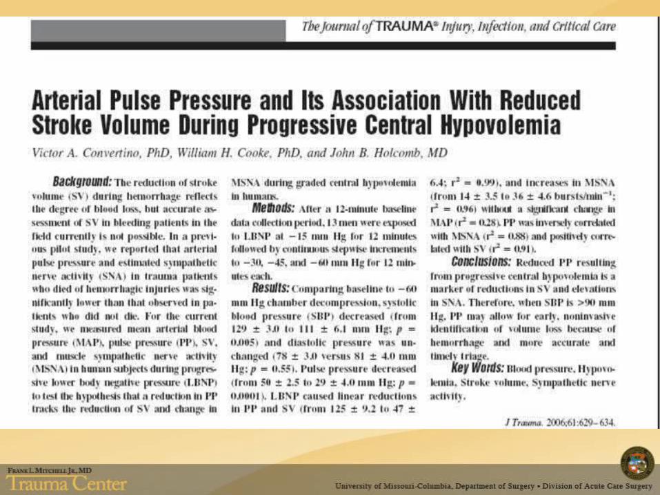

Stroke Volume Variability
Edwards Lifesciences, Irvine, CA
Stroke Volume Variability
• Correlation with “gold standard” of PAC debated
• Requires 100% mechanical ventilation• Interference
– Spontaneous respirations– Arrythmia

Avoid Fluid Overload
• SOAP study subset; 1,120 patients with AKI• Association with positive fluid balance and
increasing mortality• Mean fluid balance differed between survivors
and non-survivors• Patients requiring RRT, increase in fluid
balance 64.6% vs. 44.8% mortalityPayen, Crit Care Med 2008


Maintain Perfusion
• To prevent or mitigate injury, especially with compromised autoregulation
• Volume• Inotropic or vasopressor support• Target MAP ≥ 65 generally accepted

Improving Perfusion
If oliguria persists despite adequate filling pressure and flow…
• No evidence to support low-dose dopamine• Mixed results with fenoldopam

Diuretics in AKI
• Studies conflicting re: affect on mortality• No findings to support
– Shortened duration of AKI– Reduced need for RRT– Improved outcomes
• Furosemide– Less than 10% of bolus dose reaches tubule lumen– Continuous infusion may be preferred method of
delivery, 1-9 mg/hr rates reported

Hyperglycemia
• Decreased incidence of AKI and requirement for RRT with tight glucose control
Tight blood glucose control is renoprotective in critically ill patientsSchetz M, Vanhorebeek I, Wouters PJ, Wilmer A, Van den Berghe G
J Am Soc Nephrol 2008 Mar;19(3):571-8 Epub 2008 Jan 30

Nutrition
• Malnutrition associated with increased mortality
• Prealbumin renally excreted, may falsely elevate in AKI
• AKI patients are hypercatabolic• Consensus recommendation: 20-30
kcal/kg/day and 1.5 gm/kg/day protein

Treatment
“Last week I would've given a kidney to anyone in this office. I would've reached right into my stomach and pulled it out for them. But now, no. I don't have the relationship with these people that I thought I did. I hope they ask, so they can hear me say, "Uh, no, I only give my organs to my
real friends. Go get yourself a monkey kidney.“
--Michael Scott, The Office

Treatment
• CRRT Continuous renal replacement therapy
• SCUF Slow continuous ultrafiltration
• CVVH Continuous venovenous hemofiltration
• CVVHD Continuous venovenous hemodialysis

Classic Indications for RRT
• A—acidosis• E—electrolyte disturbances• I—intoxication • O—overload • U—uremia

Criteria for Initiation
• Non-obstructive oliguria• Severe acidemia• Hyperkalemia• Uremic end-organ involvement• Severe dysnatremia• Hyper- or hyponatremia• Overdose with dialyzable drug

Therapeutic Goal Hemodynamics Preferred Therapy
Fluid removal Stable Intermittent UF
Unstable SCUF
Urea clearance Stable Intermittent HD
Unstable CRRT
Hyperkalemia Stable Intermittent HD
Unstable Intermittent HD
Metabolic acidosis Stable Intermittent HD
Unstable CRRT
Cerebral edema Stable CRRT
Unstable CRRT
Adapted from Continuous Renal Replacement Therapy, John Kellum, Oxford Press 2009

Intermittent vs. Continuous
• Conflicting outcome data• Recent meta-analysis demonstrated no
difference in mortality• What about renal recovery?
– 2 studies—CRRT improved recovery– 4 studies—No difference– No definitive data

Continuous RRT
• Approximates “normal physiology”– Slow correction of metabolic disturbances– Volume removal better tolerated
• Goals– Maintain fluid, electrolyte balance, acid/base,
prevent further renal damage, provide renal support pending recovery

Discontinuation of CRRT
• No consensus in nephrology or critical care literature
• UOP most important predictor of successful discontinuation– Greater than 400 mL/24 hrs without diuretics or >
2300 mL/24 hrs with diuretics, ≥ 80% chance of success
Discontinuation of continuous renal replacement therapy: a post hoc analysis of a prospective multicenter observational studyUchino S Crit Care Med 2009 Sep;37(9):2576-82

Transition—CRRT to IHD
• No data– Hemodynamically stable– No vasopressor support– Need to mobilize patient ?– Need machine for more critically ill patient ?

Summary
• AKI is a common, complex condition• Etiology often multifactorial• Can be functional or structural• “Acute kidney injury” replaces “acute renal
failure”• Small changes in SCr associated with adverse
outcomes– Short and long-term increase in morbidity and
mortality

Summary
• Diagnosis frequently delayed• SCr poor marker of function in critically ill• AKI increases risk of CKD• AKI accelerates progression from CKD to ESRD• Volume overload is associated with worse
outcomes