acute respiratory distress syndrome
DESCRIPTION
historical review of ARDSTRANSCRIPT

ACUTE RESPIRATORY DISTRESS SYNDROMEACUTE RESPIRATORY DISTRESS SYNDROMEUNDERSTANDING PATHOPHYSIOLOGY AND MANAGEMENTUNDERSTANDING PATHOPHYSIOLOGY AND MANAGEMENT
a historical reviewa historical review
UbaidurUbaidur RahamanRahaman
Senior Resident, CCM, SGPGIMSSenior Resident, CCM, SGPGIMS
LucknowLucknow, India, India

idiopathic anasarca of the lungs; pulmonary edema without heart failure
A Treatise on Diseases of the Chest Laennec, 1821
PROBABLY FIRST PUBLISHED SCINTIFIC DISCRIPTION
UbaidurUbaidur RahamanRahaman, S.R., CCM, SGPGIMS, , S.R., CCM, SGPGIMS, LucknowLucknow

What's in a name? That which we call a rose by any other name would smell as sweet.
-William Shakespeare
•Double pneumonia - till mid 1900’s
•Post-traumatic massive pulmonary collapse- WW I
•Traumatic wet lung- WW II
•Shock lung - Korea – early 1950’s
•Da Nang lung / ventilator lung- Vietnam war – late 1960’s
•Capillary leak syndrome
•Acute alveolar syndrome
•Post perfusion lung
•Congestive atelectasis
•Adult hyaline membrane disease
UbaidurUbaidur RahamanRahaman, S.R., CCM, SGPGIMS, , S.R., CCM, SGPGIMS, LucknowLucknow

Acute respiratory distress in adults.Ashbaugh DG, Bigelow DB, Petty TL, Levine BE. 1967, Lancet 2:319-323
Case series of 12 patients presenting with acute respiratory distress, cyanosis refractory to oxygen therapy,
decreased lung compliance and diffuse pulmonary infiltrates on chest x-ray.
AUTOPSYlungs were heavy (average 2110 g), microscopic examination Revealed
areas of alveolar atelectasis, interstitial and alveolar hemorrhage and edema, dilated and congested capillaries
The adult respiratory distress syndrome: clinicalfeatures, factors influencing prognosis and principles of management.
Petty TL, Ashbaugh DG, Chest 1971;60:233–239.
ACUTE RESPIRATORY DISTRESS SYNDROME

lung injury scoring system (LIS)
An expanded definition of the adult respiratory distress syndrome.Murray JF, Matthay MA, Luce JM, Flick MR. Am Rev Respir Dis 1988; 138:720–723.
Addition of objective criteria- PEEP and Compliance
•Oxygenation,•Positive end-expiratory pressure•Respiratory system compliance
•Chest radiograph
•NO LUNG INJURY-------------0•MILD TO MODERATE INJURY------0.1-2.5•SEVERE LUNG INJURY (ARDS)---- >22.5

The American European consensus conference on ARDS: definitions mechanisms, relevantoutcomes and clinical trial coordination.
Bernard GR, Artigas A, Brigham KL, Carlet J, Falke K, Hudson L, Lamy M, Legall JR, Morris A, Spragg R. Am J Respir Crit Care Med.1994;149:818–824.
Acute lung injury ( ALI)ARDS – subset of ALI with severe hypoxaemia
Definition was made broad intentionally to encompass different types of AHRF occurring in variety of settings
Problem•CXR interpretation subjective•P/F affected by level of PEEP
•Requirement of PAC- interpretation- Ppao may be higher in absence of CHF
•Acute onset •CXR- bilateral infiltrates consistent with pulmonary oedema
•Ppaw ≤18 mmHg or absence of clinical evidence of left atrial hypertension,•ALI - PaO2/FIO2 ≤300 ; ARDS PaO2/FIO2 ≤ 200.

Development of a clinical definition for acute respiratory distress syndrome using the delphi technique.
Ferguson ND, Davis AM, Slutsky AS, Stewart TE. J Crit Care. 2005;20:147-154
Delphi criteria
Direct and or indirect factor associated with lung injury
Predisposition6
Cs res syst <50 ml/cmH2O( with patient sedated, VT= 8 ml/kg IBW, PEEP ≥10
Decreased lung compliance
5
No clinical e/o CHF including use of PAC and/or echo
Non cardiogenicorigin
4
Bilateral airspace disease ≥2 quadrants on frontal CXR
CXR3
Rapid onset ≤72 hoursAcute onset2
PaO2/ FiO2 ≥ 200 mmHg with PEEP ≥10Hypoxaemia1
ARDS- presence of criteria 1-4 + one of 5 or 6

Airspace disease- presence of one or more of following-
1. Air bronchogram
2. Acinar shadows- nodular opacities 4-10 mm diam with poor margination
3. Coalescence of acinar shadows
4. Silhouette sign- loss of def of heart border or hemidiaphragm, excluding that caused by lobar collapse
Development of a clinical definition for acute respiratory distress syndrome using the delphi technique.
Ferguson ND, Davis AM, Slutsky AS, Stewart TE. J Crit Care. 2005;20:147-154
UbaidurUbaidur RahamanRahaman, Senior Resident, CCM, SGPGIMS, , Senior Resident, CCM, SGPGIMS, LucknowLucknow, India, India

1970 to mid 1980s1970 to mid 1980s

Acute respiratory distress in adults.Ashbaugh DG, Bigelow DB, Petty TL, Levine BE. 1967, Lancet 2:319-323
Case series of 12 patients presenting with acute respiratory distress, cyanosis refractory to oxygen therapy,
decreased lung compliance and diffuse pulmonary infiltrates on chest x-ray.
AUTOPSYlungs were heavy (average 2110 g), microscopic examination revealed
areas of alveolar atelectasis, interstitial and alveolar hemorrhage and edema, dilated and congested capillaries
ARDS LUNG IS HOMOGENOUSLY HEAVY AND STIFFrequiring high inflation pressure to ventilate
How to treat?
UbaidurUbaidur RahamanRahaman, S.R., CCM, SGPGIMS, , S.R., CCM, SGPGIMS, LucknowLucknow

This Week’s Citation Classic OCTOBER 29, 1979
Ashbaugh D G, Petty T L, Bigelow D B & Harris T M. Continuous positive-pressure breathing
(CPPB) in adult respiratory distress syndrome. J. Thorac. Cardiovas. Surg. 57:31-41, 1969
The first patient in which we observed acute respiratory distress was a 29-year-oldman involved in an automobile accident who, despite being placed on a respirator, went on to develop severe and
progressive respiratory failure and died within 48 hours.
Our failure, in what we felt should have been a salvageable case, stimulated us to look for additional cases.
A few weeks after our first case, a 12-year-old boy was admitted with a severe crushing chest injury. He too, began to follow a
similar downhill course despite a tracheotomy and being placed on our only volume respirator, an Engstrom. Even with large volumes of air and 100% oxygen he was doing poorly.
In desperation it was decided to try adding end expiratory pressure,
which happened to be a feature of that model of Engstrom respirator
Dramatic improvement occurred in the patient’s condition and he eventually went on to make a very good recovery.
Several additional patients were then seen and treated with varying results
UbaidurUbaidur RahamanRahaman, S.R., CCM, SGPGIMS, , S.R., CCM, SGPGIMS, LucknowLucknow

Positive End Expiratory PressurePositive End Expiratory Pressure
The first ENLIGHTENMENT

Acute respiratory distress in adults.Ashbaugh DG, Bigelow DB, Petty TL, Levine BE. 1967, Lancet 2:319-323
Mile stone paper- initially rejected by 3 major US journals
PEEP was applied in five of them (three survived)ZEEP was applied in seven (two survived).
PEEP was described as a “buying time maneuver,” preventing alveolar collapse at end-expiration.
12 patients with ARDS of pulmonary and extra- pulmonary origin, some with fluid overload and shock.

The adult respiratory distress syndrome: Clinical features, factors influencing prognosis and principles of management.
Petty TL, Ashbaugh DG, Chest 1971;60:233–239.
Outcome dependent on1. Degree of original injury
2. Effectiveness of respiratory support3. Prevention of further pulmonary injury
ETIOLOGY•diffuse pulmonary injuries, direct or indirect, of lung parenchyma
•exudation of fluid and loss of surfactant activity •impaired gas exchange and reduced pulmonary compliance
MANAGEMENT PRINCIPLE
Prevent alveolar collapse and maintain oxygenation
• Volume respirator• Oxygen control
• PEEP
Prevent further injury• Oxygen control• Fluid restriction
• Antibiotics for specific infections• Corticosteroid drugs

Ventilation with end expiratory pressure in acute lung disease. Falke KJ, Pontoppidan H, Kumar A, Leith DE, Geffin B, Laver MB. J Clin Invest, 1972, 51:2315-2323
effects of PEEP in 10 patients with severe acute respiratory failurewhen IPPV with Fio2 up to 0.5 failed to maintain PaO2 ≥ 70 torr.
FRC and PaO2 rose linearly with level of PEEP; Pao2 and FRC showed a close correlation.
Lung complianceincreased with lower PEEP- RECRUITMENT,
decreased with higher PEEP– OVERDISTENSIONSurprisingly increase in PaO2 may go along with fall in compliance*
PEEP of 0, 5, 10 and 15 cm H20 were applied for 30-min Gas exchange, lung volumes, compliance, and hemodynamics- studied at each level of PEEP
*recruitment and overdistension of alveoli may take place simultaneously.
The most probable explanation for the effect of PEEP on PaO2 and compliance isrecruitment of gas exchange airspaces and prevention of terminal airway closure.
C.I. fell in some patients and rose in others and there was no correlation with level of PEEP.
HOW PEEP HELPS IN IMPROVING OXYGENATION?

WHAT LEVEL OF PEEP? is their any OPTIMUM PEEP
Optimum end-expiratory airway pressure in patients with acute pulmonary failure.Suter PM, Fairley B, Isenberg. N Engl J Med, 1975; 292:284–289
ventilation within the range of pulmonary pressure/ volume range associated with maximum compliance
negative effect of PEEP on CO is minimum.
•Optimum PEEP- best PaO2 with best oxygen transport ( C.O.)
•Associated with highest compliance of respiratory system compliance
•Recruitment prevails over overdistension.

WHAT LEVEL OF PEEP? is their any OPTIMUM PEEP
SUPER PEEP concept- pressure that maximally reduces the shunt ( ≤20% at PaO2 20 torr)
High level positive end expiratory pressure (PEEP) in acute respiratory insufficiencyKirby RR, Downs JB, Civetta. Chest;1975; 67:156–163
Gas exchange, static pressure volume curve and positive-pressure ventilation at theend of expiration. Study of 16 cases of acute respiratory insufficiency in adults.
Lemaire F, Harf A, Simonneau G. Ann Anesthesiol, 1981, Fr 22:435–441
Minimal PEEP- 2 cmH2O above the LIP on inflation limb of PV curve

SUMMARY
• ARDS lung is homogenously heavy and stiff
• Treat with high tidal volume and pressure to tackle high PaCO2
• Apply high PEEP- ?optimum PEEP to improve oxygenation
• Recognize side effects is barotrauma,• Beware of hemodynamic impairment due to PEEP
UbaidurUbaidur RahamanRahaman, S.R., CCM, SGPGIMS, , S.R., CCM, SGPGIMS, LucknowLucknow

Mid 1980sera of computed tomography
Concept of baby lung

Preservation of Normal Lung Regions in the Adult Respiratory Distress SyndromeAnalysis by Computed Tomography
Richard J. Maunder, W P. Shuman, et sl. JAMA 1986;255:2463-2465)
Despite appearance of diffuse, symmetric involvement by standard CXR
CT images demonstratesparing of substantial portion of lung parenchyma, lack of homogeneity and tendency
toward posterior involvement on CT images

L Gattinoni, A Presenti et al
•22 patients with acute respiratory failure•Lung CT and physiological measurement at 5, 10 and 15 cmH2O PEEP
•Investigated for relationship between morphology and physiology
Increasing PEEP•Progressive clearing of densities and increased mass of normally aerated tissue
(Recruitment)
•Reduction of venous admixture
•Lung densities were concentrated in dependent regions•Average lung weight was increased twofold above normal
•Excess lung weight correlated with mean Pulmonary artery pressure•Venous admixture correlated with non-inflated tissue mass

L Gattinoni, A Presenti et al

ARDS lung is non homogenous with densities concentrated in most dependent regions
Amount of normally aerated tissue at end expiration was about 200-500 gm in severe ARDS: dimension of the lung of a 5-6 years old child
ARDS LUNG IS A BABY LUNG not STIFF LUNG
This baby lung is a healthy anatomical structure, located in the non dependent regions.
Respiratory compliance well correlated with the amount of the normally aerated tissue only
ARDS: the non-homogeneous lung; facts and hypothesisGattinoni L, Pesenti A,Intensive Crit Care Dig. 1987;6:1–4

ARDS lungnon homogenous and BABY
not STIFF
Elasticity of this baby lung is nearly normal
The second ENLIGHTENMENTThe second ENLIGHTENMENT
UbaidurUbaidur RahamanRahaman, S.R., CCM, SGPGIMS, , S.R., CCM, SGPGIMS, LucknowLucknow

•We were ventilating the lung of a healthy child with about 1000ml of VT
• causing more damage then benefit
CAN IT BE PREVENTED &
HOW
UbaidurUbaidur RahamanRahaman, S.R., CCM, SGPGIMS, , S.R., CCM, SGPGIMS, LucknowLucknow

Small healthy aerated tissue in non dependent region with poor perfusion
can making it dependent help?
PRONE Ventilation
UbaidurUbaidur RahamanRahaman, S.R., CCM, SGPGIMS, , S.R., CCM, SGPGIMS, LucknowLucknow

• 13 moderate-severe ARDS patients proned for 2 hours.
• The gas exchange and hemodynamics were evaluated before, during, and after proning• CT was obtained in both the supine and prone positions in two of these patients
• Responder- Pa02 increase ≥ 10 mm Hg after 30 minutes of proning
The prone position in ARDS patients. A clinical study.Langer M, Mascheroni D, Marcolin R, Gattinoni L. Chest, 1988; 94:103–107
CT in prone positiondisappearance of posterobasal densities
andappearance of new densities in the anterior regions
…..continued

Baby lung in not healthy anatomical structure but functional concept
The prone position in ARDS patients. A clinical study.Langer M, Mascheroni D, Marcolin R, Gattinoni L. Chest, 1988; 94:103–107
Red
istr
ibution
of den
sities
aft
er p
ronin
g

Early 90sConcept of sponge lung

•edema fluid is evenly distributed from sternum to vertebra
•Increased lung weight due to accumulated edema raises hydrostatic pressure through out the lung
•Gas in dependent lung regions is squeezed out by heavy lung parenchyma above
Generalized, not regional involvement Densities are in fact due to loss of alveolar gases, not due to increase edema
Vertical gradient of regional lung inflation in adult respiratory distress syndrome.Pelosi P, D’Andrea L, Vitale G. Am J Respir, 1994; Crit Care Med 149:8–13
Superimposed pressure

ARDS LUNG IS SPONGE LUNG
Sponge lung explains
Redistribution of densities in prone positionMechanism of PEEP
The ARDS Lung. New insights from compute tomography,Bone; JAMA, 1993, 269 (16): 2134-2135
ARDS lung is both baby and sponge

• Baby lung is actually small lung open at end expiration
• It may become larger during inspiration due to newly recruited tissue
• Baby lung is not healthy but aerated
• Smaller the baby lung the greater the potential for VILI
SUMMARY
UbaidurUbaidur RahamanRahaman, S.R., CCM, SGPGIMS, , S.R., CCM, SGPGIMS, LucknowLucknow

The third ENLIGHTENMENT
CONCEPT OF
Protective lung ventilation

50 patients LIS ≥ 2.5, mean PaO2/FiO2 = 94
managed with low tidal volume, disregarding hypercapnia
Low mortality associated with low volume pressure limited ventilation with permissive hypercapnia in severe adult respiratory distress syndrome.Hickling KG, Henderson SJ, Jackson R. Intensive Care Med. 1990;16(6):372-7.
hospital mortality was significantly lower than predicted.Only one death was due to respiratory failure, caused by pneumocystis pneumonia.
10 patients had a "ventilator score" greater than 80, which has previously predicted 100% mortality from respiratory failure.
Only 2 died, neither from respiratory failure.

NORMAL OXYGENATION AND VENTILATION ( PaCO2)
ADEQUATE OXYGENATION AND PERMISSIVE HYPERCAPNIA
.The concept of "baby lung".
Gattinoni L, Pesenti A. Intensive Care Med. 2005 Jun;31(6):776-84. Epub 2005 Apr 6
The true ENLIGHTENMENTwas not the use of low tidal volume but the CHANGE OF GOAL

Much has been said about end expiratory pressureMuch has been said about end expiratory pressure
What happens at end inspiration: What happens at end inspiration: concept of recruitmentconcept of recruitment

•During inspiration only part of the lung is recruited
•Opening pressures are widely and normally distributed throughout lung parenchyma
•Some lung regions usually most dependent may require higher opening pressure
•If the Pplat is limited, collapsed tissues with higher opening pressure stayclosed throughout the respiratory cycle.
•At end expiration PEEP, if adequate will keep open only the lung regions already opened by applied Pplat
Applied physiology of intensive care medicine. Pinsky, Mancebo, pg 307
UbaidurUbaidur RahamanRahaman, S.R., CCM, SGPGIMS, , S.R., CCM, SGPGIMS, LucknowLucknow

Recruitment and Derecruitment during Acute Respiratory Failure: A Clinical StudyS Crotti, D Mascheroni, P PelosiI, J J. Marini, L Gattinoni.
Am J Respir Crit Care Med 2001,Vol 164. pp 131–140, 2001
recruitment occurs along entire VP curve of respiratory system, even beyond the UIP
derecruitment is also a continuous process, but is most prevalent over a pressure range (0–10cm H2O) lower than the pressure range over which recruitment occur

Venegas JG, Harris RS, Simon BA. A comprehensive equation for the pulmonary pressure-volume curve. J Appl Physiol 1998;84:389–395.

ARDSNET TRIALSResult of
confluence of basic and clinical research

Experimental pulmonary oedema due to intermittent positive pressure ventilation with high Inflation pressures: protection by positive end expiratory pressure.
Webb HH, Tierney DG. Am Rev Respir Dis 1974; 110: 556
Rats were ventilated with varying level of PIP and PEEP
No alveolar odema10 45 cmH2O
Alveolar and perivascular odema, decreased compliance, hypoxaemia and gross anatomical changes
045 cmH2O
Perivascular odema; no alveolar odema030 cmH2O
No pathological or physiological changes014 cmH2O
PEEPPIP
BAROTRAUMA
BASIC RESEARCH
UbaidurUbaidur RahamanRahaman, S.R., CCM, SGPGIMS, , S.R., CCM, SGPGIMS, LucknowLucknow

Ventilated rats with high Paw with and without chest wall strapped
High inflation pressure pulmonary odema. Respective effects of high airway pressure,high tidal volume and positive end expiratory pressure.
Dreyfuss D, Basset G, Soler P, Saumon G. Am Rev Respir dis, 1988; 137:1159
Rats with strapped chestPaw - very high
VT – modestNo lung damage
Rats without strapped chestPaw - very highVT – very high
Dramatic changes in lung
TRANSPULMONARY PRESSURE- VOLUTRAUMA
PEEP effective in preventing damage in large VT rats
BASIC RESEARCH
UbaidurUbaidur RahamanRahaman, S.R., CCM, SGPGIMS, , S.R., CCM, SGPGIMS, LucknowLucknow

INFERENCE
1. High tidal volume ventilation causes acute lung injury
2. PEEP exerts a protective effect against this injury
3. High tidal volume ventilation can result in distant organ injury
BASIC RESEARCH
UbaidurUbaidur RahamanRahaman, S.R., CCM, SGPGIMS, , S.R., CCM, SGPGIMS, LucknowLucknow

CLINICAL RESEARCHCLINICAL RESEARCH
CT SCAN CORRELATED WITH LUNG MECHANICSCT SCAN CORRELATED WITH LUNG MECHANICS
INFERENCE
1. Lung is non- homogenous
2. Lung is small not stiff
3. Compartment of aerated alveoli ( baby lung) is subject to overdistension
UbaidurUbaidur RahamanRahaman, S.R., CCM, SGPGIMS, , S.R., CCM, SGPGIMS, LucknowLucknow

HYPOTHESIS
End inspiratory lung volume should be limitedto avoid alveolar overdistension (volutrauma)
Sufficient PEEP should be applied to prevent cycles of end expiratory derecruitment following inspiratory recruitment
(avoid biotrauma and atelectotrauma)
UbaidurUbaidur RahamanRahaman, S.R., CCM, SGPGIMS, , S.R., CCM, SGPGIMS, LucknowLucknow

3 3 multicentermulticenter RCTsRCTs
0.7247%ª50%ª1201998(1995-1996)
Stewart et al
0.3937.9%^46.5%^1161998(1994-1996)
Brochard et al
<0.0001
71%*71%**
38%*45%**
531998(1990-1995)
Amato et al
p ValueMortalityHigh VT
MortalityLow VT
No. patient
s
Year published/ enrollment
Authers
*mortality at 28 days
**mortality at hospital discharge
^mortality at 60 days
ªmortality at hospital discharge

Amato et al, 1998, NEJM
Conventional ventilationlowest PEEP for acceptable oxygenation
VT - 12 ml/ kg actual bwPaCO2:35-38 mm Hg
Protective ventilationPEEP above LIP on static P/V curve
VT < 6 ml/ kg actual bwPplat < 20 cmH2O above PEEP
Permissive hypercapnia,
Brochard et al, 1998, AJRCCM
Protective ventilationPEEP – 10 cmH2OPpat – 25 cmH2O
VT - 7 ml/ kg actual bwPaCO2:41-44 mm Hg
Conventional ventilationPEEP – 10 cmH2OPplat – 31 cmH2O
VT - 10 ml/ kg actual bwPaCO2:53-60 mm Hg
Stewart et al, 1998, NEJM
Protective ventilationPEEP – 8 cmH2O
Pplat – 22 cmH2O VT - 7 ml/ kg actual bw
PaCO2:54 mm Hg
Conventional ventilationPEEP – 7-8 cmH2O
Pplat – 26-28 cmH2O VT - 10 ml/ kg actual bw
PaCO2:45 mm Hg
Open lung approach
UbaidurUbaidur RahamanRahaman, S.R., CCM, SGPGIMS, , S.R., CCM, SGPGIMS, LucknowLucknow
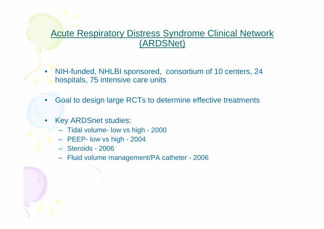
Acute Respiratory Distress Syndrome Clinical Network (ARDSNet)
• NIH-funded, NHLBI sponsored, consortium of 10 centers, 24 hospitals, 75 intensive care units
• Goal to design large RCTs to determine effective treatments
• Key ARDSnet studies:– Tidal volume- low vs high - 2000– PEEP- low vs high - 2004– Steroids - 2006– Fluid volume management/PA catheter - 2006

Ventilation with lower tidal volume as compared to traditional tidal volume for acute lung injury and the acute respiratory distress syndrome- ARMA STUDY
NEJM 2000, 342;18
861 patients
Conventional ventilationPEEP- 5-24 cm H2OPplat ≤ 50 cm H2O
VT – 10-12 ml/ kg IBWPaCO2- 35 mm Hg
Protective ventilationPEEP- 5-24 cm H2OPplat ≤ 30 cm H2OVT – 6-8 ml/ kg IBWPaCO2- 40 mm Hg
Permissive hypercapnia and acidosis
22% relative mortality reduction
9% absolute mortality reduction

Higher versus Lower Positive End-Expiratory Pressures in Patients with theAcute Respiratory Distress Syndrome- ALVEOLI STUDY
NEJM, 2004, 351;4
549 patients
HIGHER PEEP
PEEP- 13±3 cm H2O
Pplat ≤ 26±7 cm H2O
VT – 5.8±1 ml/ kg IBW
PaO2/FiO2- 206±76
Mortality – 27%
LOWER PEEP
PEEP- 8±3 cm H2O
Pplat ≤ 24±6 cm H2O
VT – 6±1 ml/ kg IBW
PaO2/FiO2 - 169±69
Mortality – 24.9%%
similar mortality rate despite significant improvement in PaO2/FiO2
Higher PEEP group had higher Pplat despite lower TV
? Benefit of higher PEEP negated by overdistension

Efficacy and Safety of Corticosteroids for Persistent Acute Respiratory Distress SyndromeNEJM, 2006, 354;16
180 patients
>7days of unresolving ARDSmethylprednisolone 2mg/kg iv stat followed by 0.5 mg/kg q6h for 14 days then
tapering to 0.5 mg/kg q12h for 7 days
•no survival benefit •If given ≥ 2 weeks after onset of ARDS - significantly increased mortality
•improved cardiopulmonary physiology within 3-7 days after their initiation and altered the course of ARDS
• increased number of ventilator-free days, ICU-free days, and shock-free days during the first 28 days

Comparison of Two Fluid-Management Strategies in Acute Lung InjuryN EJM, 2006;354:2564-75
1000 patients
•no significant difference in 60-day mortality
•conservative strategy improved lung function and shortened the duration of mechanical ventilation and intensive care without
increasing non pulmonary-organ failures

Pulmonary-Artery versus Central Venous Catheter to Guide Treatment of Acute Lung InjuryNEJM, 2006,354;21
1000 patients
PAC-guided therapy did not improve survival or organ functionbut
associated with more complications than CVC-guided therapy

I would rather discover a single fact, even a small one, than debate the great issues at length without discovering
anything at all. Galileo Galilei

VENTILATOR SCOREVENTILATOR SCORESmith and GordonSmith and Gordon--19861986
• age• PA-aO2
• mean peak airway pressure

Am. J. Am. J. RespirRespir. . CritCrit. Care Med., . Care Med., VolVol 149, No. 1, 01 1994, 8149, No. 1, 01 1994, 8--1313. . Vertical gradient of regional lung inflation in adult respiratorVertical gradient of regional lung inflation in adult respiratory y
distress syndromedistress syndromeP Pelosi, L P Pelosi, L D'AndreaD'Andrea, G Vitale, A , G Vitale, A PesentiPesenti and L and L GattinoniGattinoni
We obtained chest CT sections in 12 normal subjects (controls) and 17 patients with ARDS to investigate regional lung inflation.
A basal CT section (just above the diaphragm) was obtained in the supine position at ZEEP.
In each CT section the distance from ventral to dorsal surface (hT) was divided into 10 equal intervals, and 10 lung levels from ventral (no. 1) to dorsal (no. 10) were defined.
Knowing the average density and the volume of each level, we computed: (1) the tissue volume; (2) the gas/tissue (g/t) ratio (index of regional inflation); (3) the hydrostatic pressure superimposed on each level (SPL), estimated as density x height.
The total volume of the basal CT section was 49 +/- 2.5 ml x m-2 (mean +/- SE) in control subjects and 43 +/- 2.3 ml x m-2 in patients with ARDS (p = not significant [NS]).
The tissue volume, however, was 16.7 +/- 0.8 ml x m-2 in control subjects and 31.6 +/- 1.7 ml x m-2 in patients with ARDS (p < 0.01).
The g/t ratio in level 1 averaged 4.7 +/- 0.5 in control subjects and 1.2 +/- 0.2 in patients with ARDS (p < 0.01), and this ratio decreased exponentially from level 1 to level 10, both in controls and patients with ARDS. The Kd constant of the exponential decrease was 13.9 +/- 1.3 cm in control subjects and 7.8 +/- 0.8 cm in patients with ARDS (p < 0.01).(ABSTRACT TRUNCATED AT 250 WORDS)
UbaidurUbaidur RahamanRahaman, S.R., CCM, SGPGIMS, , S.R., CCM, SGPGIMS, LucknowLucknow