additive effects of ultraviolet b and crude coal tar on ... · tar on cutaneous carcinogen...
TRANSCRIPT
Additive Effects of Ultraviolet B and Crude Coal Tar on Cutaneous Carcinogen Metabolism: Possible Relevance to the Tumorigenicity of the Goeckerman Regimen
Hasan Mukhtar, Ph.D . , Benjamin J DelTito, Jr., M . S. , Peter M. M at g ouranis, M.D. , Mukul D as, Ph.D ., Parathasarathy Asokan, Ph.D., and David R. Bickers, M .D. Departments of Dermatology, Uni versity Hospitals of Cleveland , Case Western Reserve University, and the Veteran s Administration Med ical Center, ievciand, O hio, U.S.A.
The effect of cutaneous exposure to ultravio let B (UVB) rad iation alo ne, to crude coal tar (CCT) alone, and to the combination of UVB and CCT o n the inducibility of the microsomal cytochrome P-450-dependent carcinogenm etabolizing enzyme aryl hydrocarbon hydroxylase (AHH) and other m o nooxygenases such as 7-ethoxyresorufin O -deethylase (ERD) and 7-ethoxycoumarin O-deethylase (ECD) activities in the skin of neonatal rats was studied. Exposure of the an imals to UVB (400-1600 m]lcm 2
) alone resulted in a dose-dependen t in crease in cutaneous enzym e activities. At a UVB dose of 1200 m]lcm 2 increases in AHH, ECD, and ERD were 194%, 115%, and 244% , respectively. A single topical application of CCT (10 mJ /kg) 24 h before sacrifi ce resulted in significant indu ction of AHH (350%), ECD (921 %), and ERD (796%) activities. Treatment of anim als with the same dose of CCT followed by UVB exposure resulted in additive and /or synergistic effects o n AHH (858%), ECD (1229%), and ERD (1166%) activities in the skin . In contrast, exposure of animals to UVB prior to CCT application had effects no different fro m those of CCT alo ne. Epoxide hydro lase and glutathione S-transferase acti vities in skin from all experimental groups were not different from those of controls. Hig h-
The Goeckerman regimen is widely used for the trea tment of psori as is, a common dermatologic disease of the hum an population. T his modality consists of the appli ca ti on of va rious formul ations containing crude coa l tar (CCT) to skin followed by exposure to ul
traviolet B (UVB) radiation [1 J, both of which are known to be ca rcinogenic for the skin of experimental animals and of humans [2,3]. It is now clea r that patients undergoing repeated Goeckerman therapy are at increased risk for the development of cu-
Manuscript received Deccmber 23, 1985; accepted for publication March 17, 1986.
Supported in part by United States Public Health Service g rants C A-38028, ES-1900, and AM 34368, and funds from the Veterans Administra tion.
Reprint requests to: Hasan Mukhtar, Ph. D., Vcterans Administration Medical Ccntcr , 10701 East Bouleva rd, C lcveland, O hio 44106.
Abbreviations: AH H : aryl hyd roca rbon hydroxylase BP: benzolaJpyrene CCT: crude coa l tar 7-EC: 7-ethoxycoumarin
pressure liquid chromatog raphi c analysis of the m etabolism ofbenzo[a]pyrene (BP) by cutaneous microso m es prepared from animals treated with UVB alone, CCT alo ne, and the combination of UVB and CCT revealed increased formatio n of all the metabolites in each experimental g roup . The larges t increase in m etabo lite formation occurred in animals receiving CCT followed by UVB exposure. The inducibility of trans-7, 8-diol form atio n by UVB alone and CCT alo ne was 203% and 435%, respectively , whereas with CCT followed b y UVB it was 1065% . The differenti al respo nses in AHH activity were found to parallel the capacity of skin microsom al enzym es to enhance the binding of [3H]-BP to DNA. These studies indicate that the sequence of exposure to the components of the Goeckerm an regimen in rodents g rea tly influences m atabolic acti vity in skin. When applied in the sa m e sequence employed in the Goeckerman regim en (CCT followed by UVB exposure) the additive effect upon ca talytic activity essential for cancer initiatio n sugges ts a possible m echanism for the enhancem ent of human skin cancer in individuals exposed to this therapeutic regimen. J Invest Dermatol 87:348-353, 1986
taneous cancer [4] whereas patients receiving intermittent infrequent courses of this modality have little or no enhanced risk of developing skin cancer [5] . Recent epidemiologic studies by Stern et al [6) confirm that psoriatic patients who receive multiple courses of Goeckerman therapy have an increased incidence of skin ca rcinoma compared to age- and sex-matched control populations.
CCT is an exceedingly complex mixture of chemica ls that is ri ch in polycyclic aromati c hydroca rbons (PAHs), including benzol a )pyrene (BP) [7]. The carcinogenic and mutagenic effects
EC D: 7-cthoxycoumarin O -deethylase E H : epoxide hydrolase E RD : 7-ethoxyresorufin O-dcethylase GSH : glutathio ne (reduced) GST: glu tathione S-transferasc HPLC: high-p ressure liquid chro matog raphy (-ic) NA DP H: nicotinamide adenine dinucleotide phosphatc (reduced) 3-0HBP: 3-hydroxybenzola]py rene 7-0HC: 7-hydroxycoum arin PAH: polycyclic aromatic hydroca rbon RF: resorufin
0022-202X/86/S03.50 Copyright © 1986 by The Society for Investigative Dermatology, Inc.
348
VOL. 87. N O.3 SEPTEMBER 1986
of CCT and certain of its co nstituent chemi ca ls in experim ental systems have been welJ documented (3,7-9). Saperstein and Wheeler (10] have demonstrated in vitro mutagenic respo nses to CCT. In subsequent studies Wheeler e t al (11] have shown th e presence of unidentified mutagenic substance(s) in the urine of patients trea ted with topicall y applied 5% CCT. Recent studies by Storer et al [12] have shown th at followin g to pical application of 2% CCT to the ski n of human volunteers certain PAHs including phenanthrene, anthracene, pyrene, and fluoranthene ca n be identified in blood extracts.
The ca rcinogenic PAHs themselves are relatively inert biologically and essentially act as precarcinogens that must first undergo metabolic activation by m ammalian enzy m es which convert them into th eir biologicall y active ultimate ca rcinogeni c metabolites [13-15]. These m etabo lites are hig hl y reactive, unstable moieties that can bind cova lently to cellular RNA, D N A, and proteins, as well as to syntheti c co polymers o f nucleic acids (1 3-15) . It is generall y agreed that the cova lent intera ction of reactive metabo lites of the PAHs with D NA m ay represent an essenti al initial s tep in tumor induction r1 3-15). Stud ies during th e pas t decade h ave identified o ne of the isomeric m etabolites o f BP known as ( + )-7,8, 8a-dih ydroxy-9a-1 Oa-epoxy-7, 8, 9 ,1 O-tetra h yd rob enzo[a]pyrene as the ultim ate ca rcinogeni c species of this PAH (13-15] . The formation of this meta bolite is ca talyzed by 3 successive enzy matic steps: first, hydroxy lation by aryl hydroca rbo n hydroxylase (AHH), a cytochrome P-450-dependent mixed- function oxidase, fo llowed by epoxide hydro lase (E H) and again by AHH [13-15]. T he reactive epoxide interm edi ates are detoxifi ed by conjugation with glutathione, a reactio n ca talyzed by g lutathione (GSH) transferases [13-15] . These enzy m atic pathways for the m etabolic activation and inactivation ofPAHs have been identified in the skin [16,17] .
Studies from our laboratory have shown that topi ca l application of CCT to an imals (18-20) and to hum ans [21] results in the induction o f cutaneous AHH activity. We have recentl y shown that the use of CCT -co ntaining shampoo induces AHH activity and the DNA binding of PAHs in human hair fo llicles [22]. Highly inducible AHH activity appea rs to co rrelate with enhanced risk of tumor susceptibility to PAHs in several inbred mo use s trains [23J. Since each of the m aj or components of the Goeckerm an regimen (CCT and UV B) are known to be ca rcinogenic in mammalian skin w e wondered whether th e combination mi ght have an effect upon carcinogen metabolism. In the study described h ere, w e have attempted to mimic the Goeckerman therapy of psoriasis in the skin of experimenta l anim.als. Our results indicate that skin exposure to the components of th e Goeckerman regimen , in the sequence in which they are used therapeutically, ma y have an additive en hancing effect o n the metabolic activation o f PAH carcinogens that could explain th e ca ncer-causin g effect of this reg imen .
MATERIALS AND METH ODS
Chemicals 3H-Labeled BP-4,5-oxide (sp act 289 m C i/ mmol) and unlabeled BP-4,5-oxide were provided by the Cancer Research Prog ram of the National Cancer Institute, Divisio n of Cancer Cause and Prevention (Bethesda , Mary land). [G-3H]Benzo[a]pyrene (sp act 25 Ci/mmol) and [7, 10-14C]BP (sp act 58.5 mCi / mmol) were purchased from Am.ersham Searle (Chicago , Illinois). Go ld label BP, 7-ethoxycoumarin (7-EC) 7-hydroxycoumarin (7-0HC) , resorufin , and phenol (>99% pure) were obtained from Aldrich C hemica l Co. (Milwaukee, Wisconsin) . 7-Ethoxyreso rufin was a product of Pierce C hemicals . Protease (Type XI) , m-cresol, 8-hydroxyquino line, nicotinamide adenine dinucleotide phosphate (reduced) (NADPH), calf thymus DNA, and ribonuclease A (Type III-A) w ere purchased from Sigma Chemical Co. (St. Louis, Missouri). Standard coal tar solution (USP) was used. All other chemicals w ere obtained from commercial sources in the purest form ava ilable. Radiation Source The UVB radi ation source consisted of a
CARCINOGE N METABO LISM AND GOEC KERMAN THERAPY 349
bank of4 Wes ting house FS-40-T- 12 flu orescent sunl amps which delivered an average of 3.0 ] /cm 2/s o ver the wavelength range 290- 315 nm . Dosimetry of the rad iat io n source was calibrated week ly with an Internati onal Lig ht Spectroradiometer System (IL 700/760/780) containing a PM 270 D-CM 149 detector with a spectral range of 240-81 0 nm.
Animals Sperm-positive pregnant Sprague-Dawley rats of known insemination date w ere o btained from the Holtzm an Rat Farm (Madison, Wisconsin). N eonatal rats bo rn in situ were allowed to suckle until day 4 after birth ; they were then withdrawn from their mothers and used in these experiments. The ad vantages of using neonatal rodents for stud ies o n cutaneous drug and ca rcinogen meta bo li sm have been described previously [17 ,18].
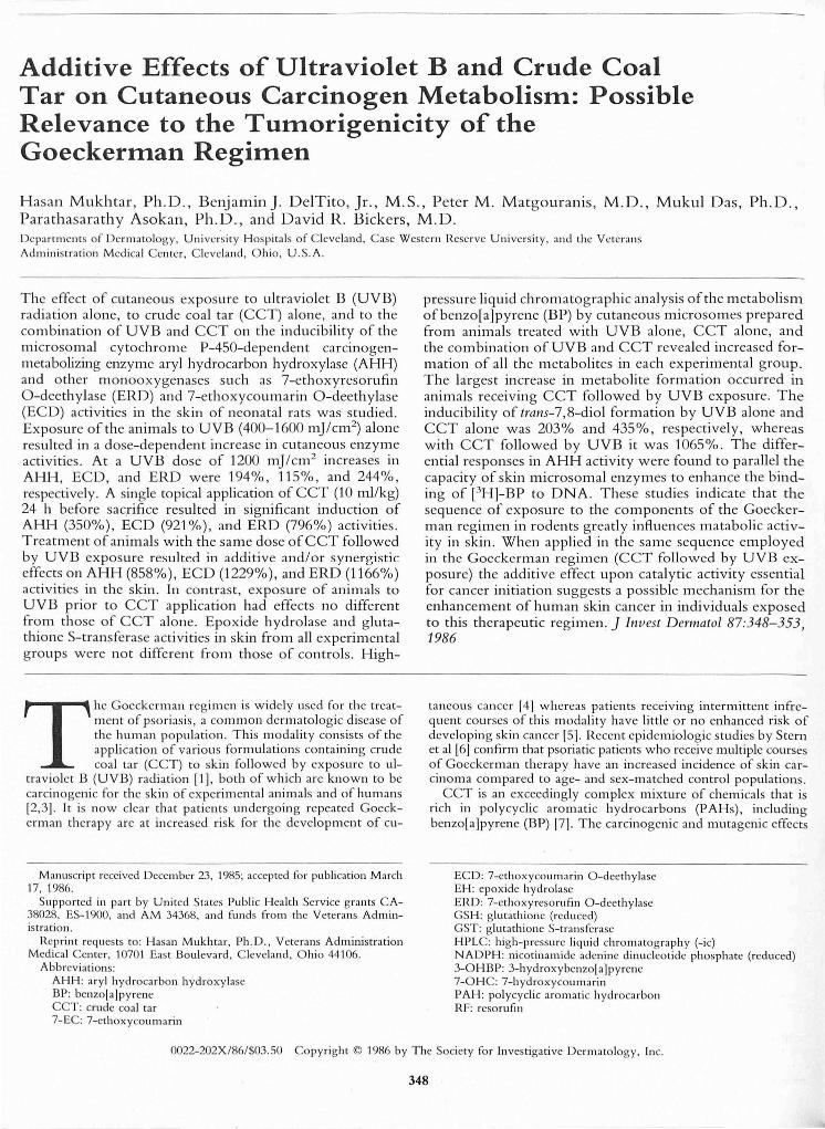
Treatment of Anin'lals In each experim ent 120 neonata l rats were di v ided into 6 g ro ups o f 20 each, and treated in the followin g m ann er: grollp 1 received a sing le to pica l treatment of acetone (10 mllkg) and served as control, grollp 2 received UVB alo ne (1200 m] /cm 2), group 3 received a single topica l appli ca ti on of CCT alone (1 0mllkg), group 4 received UVB followed immedi ately by CCT, group 5 received UVB foll owed by acetone (this gro up was considered necessa ry beca use an im als in group 6 received CCT fo llowed by UVB) , and group 6 received CCT followed by UVB. Fo r th e UVB exposures animals were exposed to the radiation source fo r a period of 4 h to deliver the desired dose. Each treatment was designed in such a way that all animals were killed 24 h after receiving CCT o r aceto ne. T he dose of CCT was selected based o n its m aximum enzym e induction effects r1 8-20]. T he UVB dose selected was based on its own m aximal inducing effects (sec Fig 1) .
Enzyme Assays Ary l hydroca rbo n hydroxy lase activity in cutaneous mi croso mes was determined by a m odification of the method of N ebert and Gelbo in [24], the details of w hi ch have been described ea rlier [1 6, 17]. T he quantitation or pheno lic BP metabolites was based o n comparison with the flu o rescence of a standard so lutio n of 3-hyd roxybenzo laJpyrene (3-0HBP). 7-E thoxycoumarin O-deethylase (ECD) activity was determined acco rding to a slig ht m odifi cati o n of the proced ure of G reenlee and Poland [25], the details of w hich have been described ea rlier [16, 17] . The am o unt of the product formed was calcul ated in comparison with an authentic standard of 7- 0HC. 7-Ethoxyresorufll1 O-deethylase (ERD) acti vity was determined by a m odifi ca tio n of th e method of Po hI and Fo uts [26J, the details of w hich were described ea rlier [27J . Quantitation of the deethylated metabolite w as based on co mpariso n w ith the flu orescence of a standard so lutio n of reso rufin (RF). E poxide hydrol ase acti vity in the microso mal fractions was assayed using BP-4,5-oxide as substrate according to the thin-la yer chro m atographic technique of Jerina et al (28), th e details of which were described previously (1 6). Cytosolic GS H-transferase activity was assayed w ith BP-4,5-oxide as substrate as described previo usly [1 7]. Protein was determined , after precipitation w ith trichl o roaceti c acid , by the procedure of Lowry et al [29] using bovine seru m albumin as a reference standard.
In Vitro BP-DNA-Binding Studies The incubation sys tem for th e in vitro (3H]_BP DNA-binding studies was similar to that described by Hesse et al [30J. After incubatio n , the reaction mixtures were centrifuged at 105,000 g to isolate the mi crosomes, and diges ted with sodium dodecy l sul fa te to extract any traces of pro tein . T he extracti on procedure was similar to that described by Lesca et al (31 J. D etails of th ese procedures were described earlier [32]. Extracted ca lf thymus DN {\ was then dissolved in 5 1111 of 0. 1 M sodium chl o rid e, pH 7.0, and es timated by measuring its absorptio n at 260 nm . The purity of the DNA was assessed by the absorbance ratios of A 260/280 ~ 1.98 and A 260/230 ~ 2.21 (33]. Aliguo ts of extracted DNA were co unted on a Packard TriCarb 460 CD liquid scintill ation spectrometer to determine th e amount of (3H)_BP covalentl y bound to calf th ymus DNA.
350 MUKHTAI< ET AL
-; 2:>0 WI II) III ...
.~ 200 -C III ~ 150 III Q.
o 400 BOO mJ/cm 2
1200 1600
Figure 1. UVB dosimetry for studies on cutaneous monooxygenase activities. Animals were exposed using a bank of 4 Westinghouse FS-40-T-12 flu orescent sunlamps at a distance of9 inches to deliver the dosage indica ted . Following UVB exposure animals were immediately killed and skin microsomal fractions prepared and enzyme activities determined . Opel'l diamonds, ECD; solid diamonds, AHH; solid squares, ERD. Data represent mean of 4 different preparations. For each preparation 20 neonatal rats were pooled.
In Vitro Incubation System for BP Metabolism The incubation mixture in a fin al volume of 1.0 ml contained 0.5 mg skin microsomal protein (prepared from control or treated animals), 0.10 mmol of phosphate buffer, pH 7.4, 3 /Lmol of MgCb and 1. 3 /Lmol of NADPH. The reaction was initiated by the addition of 80 nmol of [' 4C]BP in 40 /LI acetone . The samples were incubated for 30 min in the dark at 3rC in a Dubnoff metabolic shaker. The reaction was terminated by the addition of 1 ml of cold acetone followed by 2 ml of ethyl acetate. The ITlixture was vortexed for 1 min to extract any un reacted BP as well as the metabolites into the organic phase. The organic and aqueous layers were separated by centrifugation at 1500 rpm for 10 min. The radioactivity in the aqueous phase ranged from 0.03-0.4% of the total radioactivity and was proportional to the extent of BP metabolized. The organic phase was dried under a stream of nitrogen and dissolved in 100 /LI of methanol for highpressure liquid chromatographic (HPLC ) analysis as described earlier [32] . All operations were performed under yellow light.
High-Pressure Liquid Chromatographic Analysis of Formation of Metabolites A Waters Associates Model 204 liquid chromatograph, fitted with a Waters Associates C's /LBondapak column (4.6 mm X 25 cm) was used for the analysis of radiolabeled metabolite mixtures of BP. Identification of metabolites was based on reference standards. The column was eluted at
THE JO URNAL OF INVESTIGATIVE DERMATOLOGY
ambient temperature with a 30-min linear gradient of methanol:water:tetrahydrofuran (40:27:3, v /v) to methanol at a solvent flow rate of 0.8 ml/min [34]. The eluates were monitored at 254 nm, fractions of approximately 0.2 ml were collected dropwise, and the radioactivity of each fraction was determined on a Packard Tri-Carb 460 CD liquid scintillation spectrometer.
Statistical Analysis The statistical significance of differences between control and treated animals were evaluated by the Student's i- test. A p value of 0.05 or less was considered as statistically significant.
RESULTS
Effect of Skin Exposure to UVB on Cutaneous Monooxygenase Activities The effect of exposure of animals to varying doses of UYB alone on cutaneous AHH, ECD, and ERD activities is shown in Fig 1. Ultraviolet B exposure alone resulted in dose-dependent increases in all 3 monooxygenase activities. At a UYB dose of 1200 mJ!cm2 increases in ERD, AHH, and ECD activities were 244%, 194%, and 115% as compared with control animals kept in the dark. Since there appears to be a plateau or decline in enzyme activities beyond a UYB dose of1200 mJ/cm2
this was selected for further studies .
Effect of Exposure of Animals to UVB and CCT, Alone and Combined, on Cutaneous Monooxygenase, EH and Glutathione S-Transferase (GST) Activities The effect of exposure of animals to UYB and CCT, alone and combined, on cutaneous AHH, ERD, and ECD activities is shown in Table 1. All 3 monooxygenase activities in skin were found to be induced by the exposure of animals to UVB alone (127-167%) and CCT alone (350-921 %). Cutaneous exposure to UYB followed by treatment with CCT was no more effective than exposure to CCT alone. The treatment of animals with acetone followed by exposure to UYB (group 5) was slightly less effective than UVB exposure alone. Nevertheless, in each case there was statistically significant induction as compared with the enzyme activities in unexposed animals. CCT treatment followed by UYB exposure (group 6) produced maximum inducing effects on enzyme activities . The observed enzyme induction effects by the combination of CCT follow ed by UVB were either additive or synergistic. Cutaneous exposure to UVB alone and treatment with CCT alone resulted in 127% and 350% increases, respectively, in AHH activity. However, in those animals where CCT treatment was followed by UVB exposure, the observed increase was 858% as compared with untreated animals. The increase in AHH activity in this group when compared with those exposed to UVB alone or to UYB followed by acetone was 322% and 412%, respectively . These responses in enzyme activities were quite similar to those observed in animals receiving CCT treatment alone (350%). Similar patterns of effects were observed for ERD and ECD activities . Cutaneous EH and GST activities remained unchanged
Table I. Effect of UYB and CCT, Alone and Combined, on Cutaneous Monooxygenase Activities in Neonatal Rats
AHH Activity ERD Activity ECD Activity
pmol pmol pmol 3-0HBP/min/mg Percent Increase RF/ min /m g Percent Increase 7-HClmin/ mg Percent Increase
Treatment Protein over Contro l Protein over Control Protein over control
Cont ro l 1.75 ± 0.12 1.92 ± 0.14 0.34 ± 0.01 UVB alone 3.97 ± 0.34" 127 4.64 ± 0.27" 142 0.89 ± 0.07" 162 CCT alone 7.85 ± 0.66 350 17.20 ± 2.41b 796 3.47 ± 0.25/' 921 UVB + CCT 8.00 ± 0.64b 358 17.89 ± 2.84/' 832 3.67 ± 0.27b 979 UVB + acetone 3.27 ± 0.21" 87 3.59 ± 0.31" 87 0.82 ± 0.08" 141 CCT + UVB 16.74 ± 1.06' 858 24.30 ± 2.97' 11 66 4.52 ± 0.34' 1229
Data represent mean :t SEM of 4 separate experiments. Enzyme activities in control (acetonc-treatcd) gro up were no different from the untreated group. For treaunent conditions scc Materials alld Methods.
"S tatisti ca ll y significant from control (p < 0.01). 'Statistica lly significant fro m control and from UVB (p < 0.01). 'Statisticall y signifi cant from control. from UVB and from UVB + CCT (p < 0.01).
VOL. 87, NO.3 SEPTEMBER 1986
Table II. Effect of UVB and CCT, Alone and Combined, on Cutaneous Microsomal Enzyme-Mediated Binding of
[3H]BP to Calf Thymus DNA
Treatment
Contro l UVB alone CCT alone UVB + CCT UVB + acetone CCT + UVB
Covalent Binding (pmol [3H]BP/ mg DNA)
3.84 :±: 0.24 6.96 ± 0.61"
10.10 ± 1.1 61'
10.77 :±: 1.12b
5.29 ± 0.26" 12.86 ± 1.44'
Data represent mean ± SEM of 4 separate experim ents. · Statistically significant from control (p < 0.01) .
Percent Increase over Control
81 163 180 38
235
' Statistically sign ificant from control and from UVB (p < 0.0 1). 'Statistically significant from control, from UVB, and fro m UVB + CCT
(p < 0.0 1) .
following exposure of animals to UVB alone, to CCT alone, or to the combination of these 2 modalities (data not shown).
Effect of Exposure of Animals to UVB and CCT, Alone and Combined, on Cutaneous Microsomal Enzyme-Mediated Binding of [3H]BP to DNA Skin microsomes prepared from animals treated with UVB alone and CCT alone caused 81 % and 163% increases, respectively, in the binding of e H]BP to ca lf thymus DNA as compared with microsomes prepared from control animals (Table II) . Microsomes prepared from the skin of animals exposed to UVB followed by treatment with CCT resulted in a 180% increase in macromolecular binding. This increase in the binding activity w as not different from that of CCT trea tment alone. Consistent with the effects observed on monooxygenase activities, treatment of animals with topically applied CCT followed by UVB exposure resulted in the highes t increase (235%) in the binding of[3H]BP to calf thymus DNA.
Effect of Exposure of Animals to UVB and CCT, Alone and Combined, on Cutaneous Microsomal Metabolism of BP In further experiments , the effect of exposure of animals to UVB and CCT, alone and combined, on the pattern of skin microsomal metabolism of BP was assessed. The quantitation of BP metabolites was studied using HPLC analysis. As shown in Table III, skin exposure of animals to UVB alone resulted in an overall 121 % increase in formation of BP metabolites . Treatment of animals with CCT alone resulted in 352% increased formation of BP metabolites. Exposure of animals to UVB followed by CCT treatment had effects no different from that of CCT treatment alone. However, topical application of CCT followed by exposure to UVB resulted in the highest enhancement of BP metabolite formation. The increase in BP metabolite formation by skin microsomes prepared from groups of animals treated with CCT followed by UVB was 834%, 322%, and 373%, respec-
C ARC INOGEN METABOLISM AND GOECKERMAN THERAPY 351
c; ... -c I:) u ... QI ::! I:)
QI 1/1 II QI ... u C -C QI u ... QI c-
1200
1000
800
600
400
200
O
Total Total Phenols ~tabolit ..
Trans-7,8-diol
.uvs o CCT
m lN8 + CCT
liliJ UVS+ Ac.ton.
~ CCT+UVB
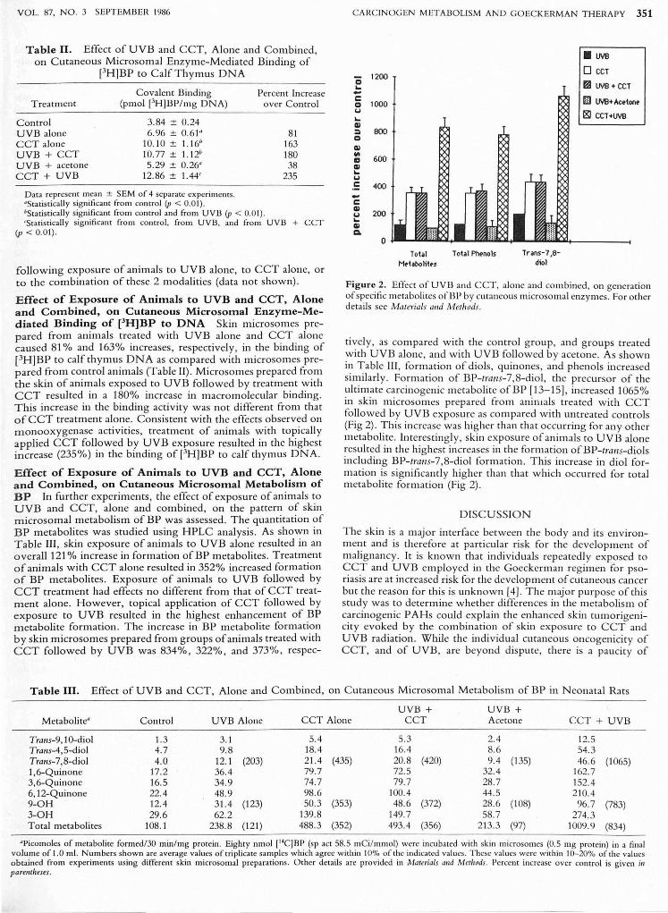
Figure 2. Effect of UVB and CCT, alone and combined, on gcneration of specific mctabolites ofB P by cutaneous microsomal enzymes. For other details see Ma terials alld Meth ods.
ti vel y, as compa red with the control group , and groups treated with UVB alone, and w ith UVB followed by acetone. As shown in Table III , formation of diols, quinones , and phenols increased similarl y . Formation of BP-lralls-7,8-diol, the precursor of the ultimate carcinogenic metabolite ofBP [13-15]. increased 1065% in skin microsomes prepared from animals treated w ith CCT followed by UVB exposure as compared with untreated controls (Fig 2). This increase was higher than that occurrin g for any other metabolite. Interestingly, skin ex posure of animals to UVB alone resulted in the highes t increases in the formation ofBP-lralls-diols including BP-lralls-7,8-diol formation. This increase in diol formation is significantl y higher than that which occurred for total metabolite formation (Fig 2) .
DISCUSSION
The skin is a major interface between th e body and its environment and is therefore at par ti cular risk for the development of malignancy. It is known that individuals repeatedly exp_osed to CCT and UVB employed in the Goeckerman regimen for psori as is are at increased risk for the development of cutaneous cancer but the reason for this is unknown [4). The major purpose of this stud y was to determine whether differences in the metabolism of ca rcinogenic PAHs could explain the enhanced skin tumorigenicity evoked by the combination of skin exposure to CCT and UVB radiation. While the individual cutaneous oncogenicity of CCT, and of UVB , are beyond dispute, there is a paucity of
Table III. Effect of UVB and CCT, Alone and Combined, 011 C utaneous Microsomal Metabolism of BP in N eonatal Rats
UVB + UVB + Metabolite" Control UVB Alone CCT Alone CCT Acetone CCT + UVB
Tra'1S-9,10-diol 1.3 3.1 5.4 5.3 2.4 12.5 Tratls-4,5-diol 4.7 9.8 18.4 16.4 8.6 54.3 Tram-7,8-diol 4.0 12.1 (203) 21.4 (435) 20.8 (420) 9.4 (135) 46.6 (1065) 1,6-Quinonc 17.2 36.4 79.7 72.5 32.4 162.7 3,6-Quinonc 16.5 34.9 74.7 79.7 28.7 152.4 6, 12-Quinone 22.4 48.9 98.6 100.4 44 .5 210.4 9-0H 12.4 31.4 (123) 50.3 (353) 48.6 (372) 28.6 (108) 96.7 (783) 3-0H 29.6 62.2 139.8 149.7 58.7 274.3 Total metabolites 108. 1 238.8 (121) 488.3 (352) 493.4 (356) 213.3 (97) 1009.9 (834)
·Picomoles of metabolite formed/3~ min/mg protein. Eighty nmol I14CJBP (51' act 58.5 mCil l11l1Jol) were incubated w ith skin microsomes (0.5 mg protein) in a final volume of 1.0 ml. Nunlbcrs shown are average values of triplicate samples which agree within 10% o Cthe indicated values. These values w ere w ithin 10-200/0 oCthe val ues obta ined from experiments using different skin microso mal preparations . Other details arc provided in Malel'ials alld Meth ods. Percent increase over co ntro l is given i,1 parfllrheses.
352 MUKHTAR ET AL
knowledge concerning the m anner in w hich these 2 carcinogens interact to evoke cancer in the skin .
C rude coal tar is a complex mix ture o f chemica ls generated by th e destru ctive di still ation of coal. It contains thousands o f chemicals some of which are PAHs, o f which several are known to be ca rcinogenic probabl y because o f their ability to structura.ll y m odify DNA. Similarl y UVB radiation is ca rcinogeni c and this m ay also be fo r the sa me reason. T he effect o f expos ure of human and rodent skin to CCT on cutaneous carcinogen matabo lism has been studied . Bickers and Kappas [21] showed that CCT is capable of inducing human skin AHH in vivo and that th e induction response varies som ewhat am ong individuals, perh aps on a genetic basis. O ur subsequent studies [1 8] have dem onstrated th at CCT applied topically to neonatal rats can induce cutaneous and extracutaneous AHH activity. In addition, topical application of CCT to anim als results in the induction of hepatic EH acti vity and cytochrome P-450 levels [20]. In ano th er study , Mukhtar et al [1 9] evaluated the effects o f topica l appli ca ti on of a number o f defin ed constituent PAHs present in a standard CCT mixture for their ca pacity to m odulate cutaneous and hepati c dru g and carcinogen m etabo lism . T hese studies indica ted th at several PAHs present in coal tar have induction effects on dru g and ca rcinogen metabolism in skin and liver and that th ere are tissue-specifi c differences in res ponses to som e of these hydrocarbons. Since the inducibility o f AHH in target tissue appears to co rrelate w ith tumor inducti on in that tissue, th e observation th at CCT could induce cutaneous AHH is consistent w ith data indica tin g that CCT is carcinogenic for skin.
Excessive sun exposure is also known to be a causa ti ve facto r in human skin cancer as first proposed by Unna in ] 894 [35] . He recognized tha t neoplas m s occurred in the preferentiall y chronicall y sun-exposed skin of sa il o rs. C lass ical studies in experimental animals by Blum defm ed the carcinogenic action spectrum as being almos t exclusively in the UVB ran ge of 290-320 nm [36]. These wavelengths are at the lower, m ore energeti c end of the solar spectrum th at reaches th e surface of the ea rth . Numerous investigators have cl earl y shown that rodents offer an excellent experimental animal model for studies ofUV skin ca rcinogenesis [2]. Furthermore the initiati on-pro motion concept o f chemical carcinogenesis is also applica ble to UV carcinogenesis. Thus, it has been shown that a sing le dose of UV radi ation followed by croton oil promotion produces skin tum ors in hairless mi ce [37] . Although there are some differences in the biologic behavio r o f UV -induced skin tumo rs as compared w ith chemically induced cutaneous lesions, th e initiatio n and promotion concept o f carcinogenesis appea rs to be valid fo r both CCT and UV tumorigenesIs.
Very few studies have been conducted to assess the effects o f UV radiation on drug and ca rcinogen metabolism in skin o r other tissues o f experimental animals. Tredger and C hhabra [38], while stu dyin g dru g metabolism in liver and intestin e, observed circadian variations in enzyme activities . T hey postulated that the light cycle could be related to these circadian changes. Pohl and Fouts [39] found increased ECD acti v ity in the skin of fem ale hairless mice exposed to short-wave UV radiati on (254 nm) and to a sunlamp (280-750 nm) . Goerz et al [40] exposed fem ale hairless N g/ mice to UV radi ation fo r 16 h daily fo r a period of 24 weeks (mean daily doses: UVA = 106 J/c m 2
, UVB = 0.62 J/c m2
), and observed increases in the acitivities o f AHH, aminopyrine N-demethylase, ECD , and in the contents o f the cytochro me P-450 in liver o f exposed animals. H ow ever, these in ves ti gato rs detected no effect o f such exposure on skin enzy me activity.
The manner in w hich chemical agen ts m ay influence UV carcinogenesis is a subj ect o f considerable interes t. Such agents include chemical pro m oters, pho tosensitizers, and ca rcinogens. A number o f substances including CCT and select PAHs present in CCT are bo th pho to toxic and ca rcinogenic. In 1937, Bungeler [41] observed that by injecting tar subcutaneously into w hite mice prior to expos ure to sunlight the yield o f bo th benign and ma-
T HE JO URN AL OF INVESTIGATIV E DERMATOLOGY
lignant tum ors w as g reatl y increased . Urbach [42] demonstrated that CCT enhanced the number o f ea r tumors fro m carefull y m easured mid wa velength(280- 360 nm) exposure.
Several inves tigato rs have exa mined th e effects of combined trea tment o f UV radi ation and chemicals, including tar, on tumo r development in experim ental animals. Findlay [43] painted a sm all area of depilated m ouse skin prio r to exposure to a quartz mercury va por lamp 4 times a week . In these studies skin can cers developed m ore rapidl y in mi ce so trea ted than in mi ce treated w ith ta r alone. Subsequent inves ti ga to rs o btained confli ctin g results from similar studies . E xperim ents in w hich skin painting with BP [44] o r 20-meth ylcholanthrene [45] was followed by exposure to visibl e light o r in w hi ch topical applica tion of BP, dimethylbenzathracene, and 20- m eth ylcholanthrene w as fo ll ow ed by UV radi ation [46] have either fa iled to dem onstrate an in crease in ca rcinogenesis o r have resulted in fewer tumors th an did the chemical ca rcinogen alone. O ther w orkers have repo rted increased [47], decreased [48], or no effect [49,50] from studies o n the combination o f UV radiation and P AH . N one of th ese studies has attempted to mimic Goeckerman therapy in assessing the interacti on of UV radi ation and PAHs on skin can cer fo rmation .
O ur data indicate that bo th UVB alone and CCT alone each substantiall y enhances cutaneous microso m al cytochrome P-450-dependent AHH, ECD , and E RD activities. Of the 3 monooxygcnases studied , ERD is specifi ca lly dependent upon cytochrome P-448. In general, the specificity for cytochrom e P-448 is in the o rder o f ERD > AHH > E C D . It is interestin g to no te from the data in Fig 1 that the degree of observed increases in enzym e acti vity follow ed the specifi city of cytochrom e P-448. It is important to emphasize that this isoenzym_e o f cytochrome P-450 participates in the activation of m any precarcinogens to their ultimate carcinogeni c m etabolite. In addition our data indicate that bo th UVB alone and C CT alone substantially enhance the m etabolism of the carcinogenic P AH, BP, and the binding of m etabolites of BP to epidermal DNA. When CCT was applied to skin previously irradiated with UVB additive effects w ere observed on these param eters in skin . T hese results indicate that exposure of rodent skin to CCT follow ed by UVB radiation additively augm ents the effect of each on ca rcinogen m etabo lism thought essential for tumor induction. Perhaps irradiation of CCTtreated skin yields larger am ounts of reactive m etabolites of PAHs. Pretrea tment with UVB foll owed by CCT does not exhibit additive effects. The explanation fo r this m ajor di fference ma y relate to pho tochemi cal changes produced in skin by UVB which somehow alters the cutaneous response to CCT . It remains to be seen whether grea ter increases in skin AHH and o th er parameters follow ing cutaneous treatment with C CT follow ed by exposure to UVB results in increased carcinogeni city than that occurring with CCT treatment alone . These studies are currently under w ay in our labo rato ry.
T hallks are dll e to Mrs . Sam/ra E 'laIIS for preparillg the ilia/II/script alld to Mr. Dar/iel P. Bik fo r technica l ass istallce.
REFERENCES
1. Goeckerman WH : T reatment of psoriasis: continued observa tions on the use of coa l tar and ultraviolet light. Arch Dermatol 24:446-450, 1931
2. Emm ett EA: Ul traviolet radiation as a cause of skin tumors. CRC Crit Rev Toxicol 2:211 -255 , 1973
3. Henry SA: Occupational cutaneous cancer attributable to certain chemicals in industry. Br Med Bull 4:389-401, 1947
4. Stern RS, Zicrler S, Parrish JA: Skin carcinoma in patients with psorias is treated with topical tar and artifi cial ultraviolet radiation. Lancet 2:732-733, 1980
5. Epstein JH: Risks and benefits of the trea tment of psoriasis. N Engl J Med 300:852-853, 1979

VOL. 87, NO.3 SEPTEMBER 1986
6. Stern RS, Scotto J , Fears TR: Psoriasis and susceptibility to nonmelanoma skin cancer. J Am Acad Dermatol 12:67-73, 1985
7. Repor t on Coal Tar. National Institute for Occupationa l Safety and Health Publica tion, Washington, DC, 1977
8. Rasmussen JE: The crudeness of coal tar. Prog Dermatol 12:23-29, 1978
9. Kipling DM: Soots, tars, and o ils as causes of occupational cancer, C hem ica l Ca rcinogens. Edited by CE Sea rl e. Washington, DC, American Ca ncer Society , 1976, pp 3 15-323
10. Saperstein MD , Wheeler LA: Mutagenicity of coa l tar preparations used in the treat ment of psoriasis. Toxicol Lett 3:325-329, 1979
11. Wheeler LA , Saperstein MD, Lowe NJ: Mutagenici ty of urine fro m pso riat ic patients undergoing treatment w ith coal tar and ultraviolet light. J Invest Dermatol 77:1 8 1- 185 , 1981
12. Storer JS , DeLeon I, Millikan LE, Laseter JL, Griffing C: Human absorption of crude coal tar products. Arch Dcrmatol 120:874-877, 1984
13. Conney AH: Induction of microso mal enzymes by foreign chemi cals and carcinogenes is by polycyclic aromatic hydrocarbons: GH A C lowes Memorial Lecture. Cancer Res 42:4875-4917, 1982
14. Heidelberger C: C hemica l carcinogenesis. Annu Rev Biochem 44:79-121 , 1975
15. Gelboin HV: Benzo(a)pyrene metabo lism, activa tion and carcinogenesis: role of regulat ion of mixed-function oxidases and related enzymes. Physiol Rev 60: 1107-11 66, 1980
16. Bickers DR, Dutta-Choudhury T, Mukhtar H : Epidermis: a site of dru g metabolism in neonatal rat skin . Studies on cytochrome 1'-450 content and mixed function oxidase and epoxide hydrolase activity. Mol Pharmacol 21:239-247, 1982
17. Mukhtar H, Bickers Dn: Drug metabolism in skin : co mparative activity of the mixed function oxidases, epoxide hydrolase, and glutathione-S-transferase in liver and skin of neonatal rat. Drug Metab Dispos 9:311-314,1981
18. Bickers DR, Wroblewski D, Dutta-C houdhury T, Mukhtar H: Induction of neonatal rat sk in and li ve r ary l hydroca rbon hydroxylase by coal tar and its constituents. J Invest Dermatol 78:227- 229, 1982
19. Mukhtar H, Link C M, C herniack E, Kushner DM, Bickers DR: Effect of topical applica tion of defin ed constituents of coal tar on skin and liver aryl hydrocarbon hydroxylase and 7-ethoxycoumarin dee th ylase activities. Toxicol Appl Pharm acol 64:541-549, 1982
20. Mukhtar H , Bickers Dn: Evidence that coal tar is a mixed inducer of microsomal drug metabolizing enzymes. Toxicol Lett 11 :221-227, 1982
21 . Bickers DR, Kappas A: Human skin ary l hydroca rbon hydroxylase: induction by coal tar. J C lin Invest 62:1 061-1 068, 1978
22. M erk HF, Mukhtar H, Bickers Dn: Hum an hair follicle: access ible tissue source for measuring enzyme-mediated DN A-binding of chemical ca rcinogens. C lin Res 32:482, 1984
23. N ebert DW, Goujon FM, GiclenJE: Aryl hydrocarbon hydroxylase induction by polycyclic hydrocarbons- simple autosomal dominant trait in the mouse. Nature 236:107-110, 1972
24. Nebert DW, Gelboin HV: Substrate inducible microsomal aryl hydrocarbon hydroxylase in mammalian cell culture. I. Assay and properties of induced enzyme. J B io i C hem 243:6242-6249, 1968
25. Greenlee WF, Poland A: An improved assay of 7-ethoxycoumarin O-deethylase activity: induction of hepatic enzyme activity in C57B Ll6J and DBA/2J mice by phenobarbital, 3-methylcholanthrene, and 2,3, 7,8-tetrachlorod.ibenzo-p-dioxin . J Pharmacol Exp Ther 205:596-605, 1978
26. Pohl RJ, Fouts JR: A rapid method for assaying the metabo lism of 7-ethoxyresorufin by microsomal subcellular fractions. Anal Biochem 107:150-155, 1980
27. Asokan 1', Das M , Rosenkranz HS, Bickers DR, Mukhtar H : Topically applied nitropyrenes are potent inducers of cutaneous and hepatic monooxygenases. Biochem Biophys Res Co mmun 129: 134-140, 1985
CARCIN OGEN METABO LISM AND GOECKERMA N THERAPY 353
28 . Jerina DM, Dansette PM, Lu A YH, Levin W: Hepatic microsomal epoxlde hyd rase: a sensi ti ve radiometric assay for hydration of arcne oxides of ca rcinogenic aromatic hydrocarbons. Mol Pharmacol 13:342-351, 1977
29. Lowry O H , Rosebrough NJ, Farr AL, Randall RJ: Protein measurement w ith the Folin phenol reagent. J Bioi C hem 193:265- 275 1951 '
30. Hesse S, Jernstrom B, M artinez M , M oldeus P, C hristodouIides L, Ketterer B: Inactiva tion of DN A-binding metabolites of benzo(a)pyrene and benzo(a) pyrene-7,8-dihydrodiol by glutathione and glutathione-S-transferases. Carcinogenesis 3:757-760, 1982
31. Lesca 1', Guenthner TM, Oesch F: M odulation of the covalent binding of aryl hydrocarbon meta bolites to DNA in vitro after trea tment of rats and mice w ith trans-stilbene oxide. Ca rcinogenesis 2: 1 049-1056, 1981
32. Mukhtar H , Del Tiro BJ Jr. Das M , Cherniack EP, C herniack AD, Bickers DR: C lotrimazole, an inhibi tor of epidermal benzo(a)pyrene metabolism and DNA binding and ca rcinogenici ty of the hydrocarbon. Cancer Res 44:4233-4240, 1984
33. Ashurst SW, Cohen GM: T he fo rmation of benzo(a)pyrcne-deoxy ribonucleoside adducts ;/1 vivo and ;/1 vitro. Carcinogenesis 3:267-273, 1982
34. Bickers DR, Mukhtar H , Yang SK: C utaneous metabo lism of benzo(a)pyrene: comparative studies in C57BLl6N and DBA/2N mice and neonatal Sprague-Dawley rats. C hem Bioi Interact 43:263-270, 1983
35. Unna PG: The Histopathology of the Diseases of the Skin. Edinburgh, William FClay, 1896, p 719
36. Blum HF: Carcinogencsis by Ultravio let Light. Princeton, NJ, Princeton Univ Press, 1959
37. Epstein JH, Roth HL: Experim ental ultravio let ligh t carcinogenesis. A study of cro ton o il promoting effects. J In vest Dermatol 50:387-400, 1968
38. Tredger JM ,. C hhabra RS: C ircadian variations in microsomal drugmetabohz111g enzy mc actlvmes 111 rat and rabbit tissues. Xenobiotica 7:481-489, J 977
39. Pohl Rj , Fouts JR: Xcnobiotic metabolism in skin ~f hairless mice exposed to UV-rad iation (U V), Aroclor 1260, or chlo rdane. Pharmacologis t 19:200, 1977
40. Goerz G, Merck H , Boisen K, Tsambaos D , Berger H: Influence of UV -light exposure on hepatic and cutaneous monooxygcnases. Experentia 39:385-386, 1983
41. Bungeler W: Uber den E influss pho tosensibilisierender Substantzen auf die Entstehullg von Hautgeschw ulston. Z Krebsforsch 46:130-132,1937
42. Urbach F: Modification of ultraviolet carcinogenesis by photoactive agents. J In vest Dermato l 32:373-378, 1959
43. Findlay GM: Ultraviolet light and skin cancer. Lancet 2:1 070-1073, 1928
44. Morton JJ , Luce-C lausen EM, Mahoney EB: Visible light and skin tumors induced with benzopyrene in mice. Cancer nes 2:256-260, 1942
45. M ortonJJ, Milder GB , Luce-ClausenEM, Mahoney EB: The effect of visible light on the development in mice of skin tumors and leukemia induced by ca rcinogens. Cancer Res 11 :559-561, 1951
46. Rusch HI' , Kline BE, Ba umann CA: The nonadditive effect of ultraviolet li ght and other carcinogenic procedures. Cancer nes 2: 183-188, 1942
47. MaisinJ , Dejonghe A: Au sujet de !'action de la lumiere et de !'ozone sur certains corps canccrigenes. C R Soc BioI (Paris) 11 7:114-119, 1934
48. Donaiach I, Mottra m JC: O n the effect of light on the incidence of tumors in painted mice. Am J Cancer 39:234-236, 1940
49. Kohn Speyer AC: Effect of ultrav iolet radiation on the incidence of tar cancer in mice. Lancet 1:11 56-11 57, 1929
50. Seelig M G, .Cooper ZK: Light and tar cancer: an experimental study WIth a crmcal revIew of the hteratu ie on light as a carcinogenic factor. Surg Gynecol Obstet 56:752-761, 1933