adenocarcinoma originating from long-segment barrett's
TRANSCRIPT
Kuwayama et al. surg case rep (2020) 6:230 https://doi.org/10.1186/s40792-020-00995-7
CASE REPORT
Adenocarcinoma originating from long-segment Barrett’s esophagus over 15 cm: a series of 3 casesNaoki Kuwayama, Isamu Hoshino*, Hisashi Gunji, Toru Tonooka, Hiroaki Soda, Ryotaro Eto, Nobuhiro Takiguchi and Yoshihiro Nabeya
Abstract
Background: Barrett’s esophagus (BE) is characterized by presence of columnar epithelium in the lower esophageal mucosa, which originally comprises stratified squamous epithelium. Gastroesophageal reflux disease causes BE and BE adenocarcinoma (BEAC); further, the incidence of BEAC is increasing, especially in developed countries. Long-seg-ment BE (LSBE) has a particularly high carcinogenic potential and necessitates treatment, surveillance, and prevention.
Case presentation: Herein, we report three cases of BEAC originating from LSBE larger than 15 cm. All three patients underwent surgery for the diagnosis of BEAC. A 66-year-old man with advanced esophageal cancer underwent neoadjuvant chemotherapy and subsequent subtotal esophagectomy. The postoperative pathological diagnosis was of poorly differentiated adenocarcinoma with lymph node metastasis (pT3 pN3 pM0 pStage III based on the Union for International Cancer Control TNM Classification 8th edition). Two years after the operation, the patient was diagnosed with recurrence around the celiac artery and underwent chemotherapy. An 83-year-old woman with advanced esophageal cancer underwent subtotal esophagectomy. The postoperative pathological diagnosis was of well-differ-entiated adenocarcinoma with supraclavicular lymph node metastasis (pT3 pN3 pM1 pStage IV). Two months after the operation, the patient was diagnosed with recurrence in the neck lymph nodes and underwent chemotherapy; however, she died. A 66-year-old man with early-stage esophageal cancer underwent subtotal esophagectomy. A superficial early cancerous lesion was seen over BE. The postoperative pathological diagnosis was of well-differenti-ated adenocarcinoma without lymph node metastasis (pT1a pN0 pM0 pStage 0). The patient was found to be alive and recurrence-free 3 months after the operation.
Conclusions: BEAC might show good prognosis if detected and treated early. Extremely LSBE is associated with a high incidence of BEAC; therefore, early detection and treatment with close surveillance is essential.
Keywords: Barrett’s esophagus, Adenocarcinoma, Surveillance
© The Author(s) 2020. This article is licensed under a Creative Commons Attribution 4.0 International License, which permits use, sharing, adaptation, distribution and reproduction in any medium or format, as long as you give appropriate credit to the original author(s) and the source, provide a link to the Creative Commons licence, and indicate if changes were made. The images or other third party material in this article are included in the article’s Creative Commons licence, unless indicated otherwise in a credit line to the material. If material is not included in the article’s Creative Commons licence and your intended use is not permitted by statutory regulation or exceeds the permitted use, you will need to obtain permission directly from the copyright holder. To view a copy of this licence, visit http://creat iveco mmons .org/licen ses/by/4.0/.
BackgroundBarrett’s esophagus (BE) is characterized by presence of columnar epithelium in the lower esophageal mucosa, which originally comprises stratified squamous epithe-lium. Importantly, long-segment Barrett’s esophagus (LSBE) has a particularly high carcinogenic potential.
Here, we report three cases of Barrett’s esophageal ade-nocarcinoma (BEAC) originating from LSBE larger than 15 cm.
Case presentationCase 1A 66-year-old man with esophageal obstruction was admitted to a different hospital. He was referred to our institution following diagnosis of BEAC. Endoscopy revealed a squamocolumnar junction 24 cm from the
Open Access
*Correspondence: [email protected] of Gastroenterological Surgery, Chiba Cancer Center, 666-2 Nitona-cho, Chuo-ku, Chiba 260-8717, Japan
Page 2 of 7Kuwayama et al. surg case rep (2020) 6:230
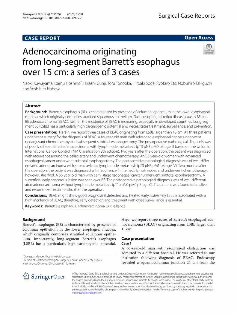
incisor teeth and a type-3 circumferential tumor on the lower esophagus (Fig. 1). Mucosal biopsy revealed adenocarcinoma.
Computed tomography detected an enlarged lymph node but no other distant metastatic sites. Following diagnosis of cT3N1M0, subtotal esophagectomy with lymph node dissection was performed after three courses of S-1 + oxaliplatin therapy.
The postoperative pathological diagnosis was of poorly differentiated adenocarcinoma (T3 70 × 45 mm) with lymph node metastasis (N3) (pT3 pN3 pM0 pStage III; Fig. 1). Two years after the operation, the patient showed recurrence in the region of the celiac artery and is cur-rently undergoing chemotherapy.
Case 2An 83-year-old woman who had undergone upper gas-trointestinal endoscopy for detailed examination of anemia was referred to our hospital with a diagnosis of BEAC.
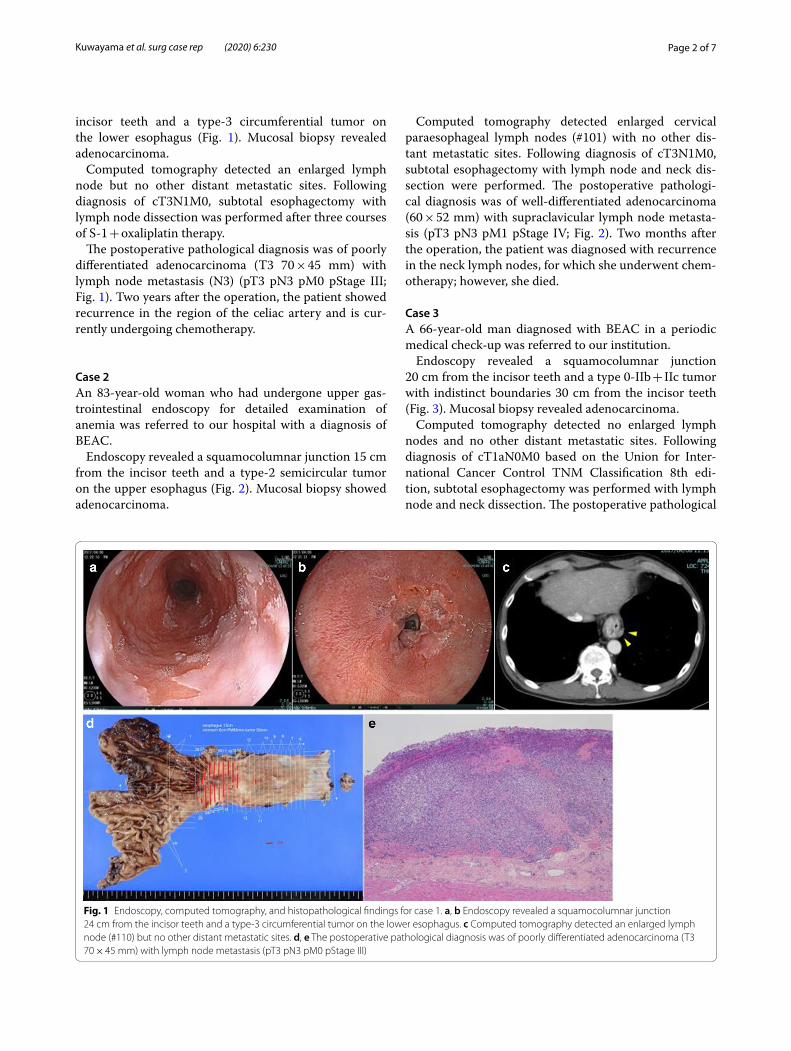
Endoscopy revealed a squamocolumnar junction 15 cm from the incisor teeth and a type-2 semicircular tumor on the upper esophagus (Fig. 2). Mucosal biopsy showed adenocarcinoma.
Computed tomography detected enlarged cervical paraesophageal lymph nodes (#101) with no other dis-tant metastatic sites. Following diagnosis of cT3N1M0, subtotal esophagectomy with lymph node and neck dis-section were performed. The postoperative pathologi-cal diagnosis was of well-differentiated adenocarcinoma (60 × 52 mm) with supraclavicular lymph node metasta-sis (pT3 pN3 pM1 pStage IV; Fig. 2). Two months after the operation, the patient was diagnosed with recurrence in the neck lymph nodes, for which she underwent chem-otherapy; however, she died.
Case 3A 66-year-old man diagnosed with BEAC in a periodic medical check‐up was referred to our institution.
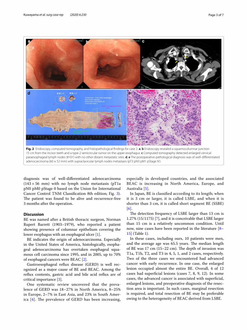
Endoscopy revealed a squamocolumnar junction 20 cm from the incisor teeth and a type 0-IIb + IIc tumor with indistinct boundaries 30 cm from the incisor teeth (Fig. 3). Mucosal biopsy revealed adenocarcinoma.
Computed tomography detected no enlarged lymph nodes and no other distant metastatic sites. Following diagnosis of cT1aN0M0 based on the Union for Inter-national Cancer Control TNM Classification 8th edi-tion, subtotal esophagectomy was performed with lymph node and neck dissection. The postoperative pathological
Fig. 1 Endoscopy, computed tomography, and histopathological findings for case 1. a, b Endoscopy revealed a squamocolumnar junction 24 cm from the incisor teeth and a type-3 circumferential tumor on the lower esophagus. c Computed tomography detected an enlarged lymph node (#110) but no other distant metastatic sites. d, e The postoperative pathological diagnosis was of poorly differentiated adenocarcinoma (T3 70 × 45 mm) with lymph node metastasis (pT3 pN3 pM0 pStage III)
Page 3 of 7Kuwayama et al. surg case rep (2020) 6:230
diagnosis was of well-differentiated adenocarcinoma (143 × 56 mm) with no lymph node metastasis (pT1a pN0 pM0 pStage 0 based on the Union for International Cancer Control TNM Classification 8th edition; Fig. 3). The patient was found to be alive and recurrence-free 3 months after the operation.
DiscussionBE was named after a British thoracic surgeon, Norman Rupert Barrett (1903–1979), who reported a patient showing presence of columnar epithelium covering the lower esophagus with an esophageal ulcer [1].
BE indicates the origin of adenocarcinoma. Especially in the United States of America, histologically, esopha-geal adenocarcinoma has overtaken esophageal squa-mous cell carcinoma since 1995, and in 2005, up to 70% of esophageal cancers were BEAC [2].
Gastroesophageal reflux disease (GERD) is well rec-ognized as a major cause of BE and BEAC. Among the reflux contents, gastric acid and bile acid reflux are of critical importance [3].
One systematic review uncovered that the preva-lence of GERD was 18–27% in North America, 8–25% in Europe, 2–7% in East Asia, and 23% in South Amer-ica [4]. The prevalence of GERD has been increasing,
especially in developed countries, and the associated BEAC is increasing in North America, Europe, and Australia [5].
In Japan, BE is classified according to its length; when it is 3 cm or larger, it is called LSBE, and when it is shorter than 3 cm, it is called short segment BE (SSBE) [6].
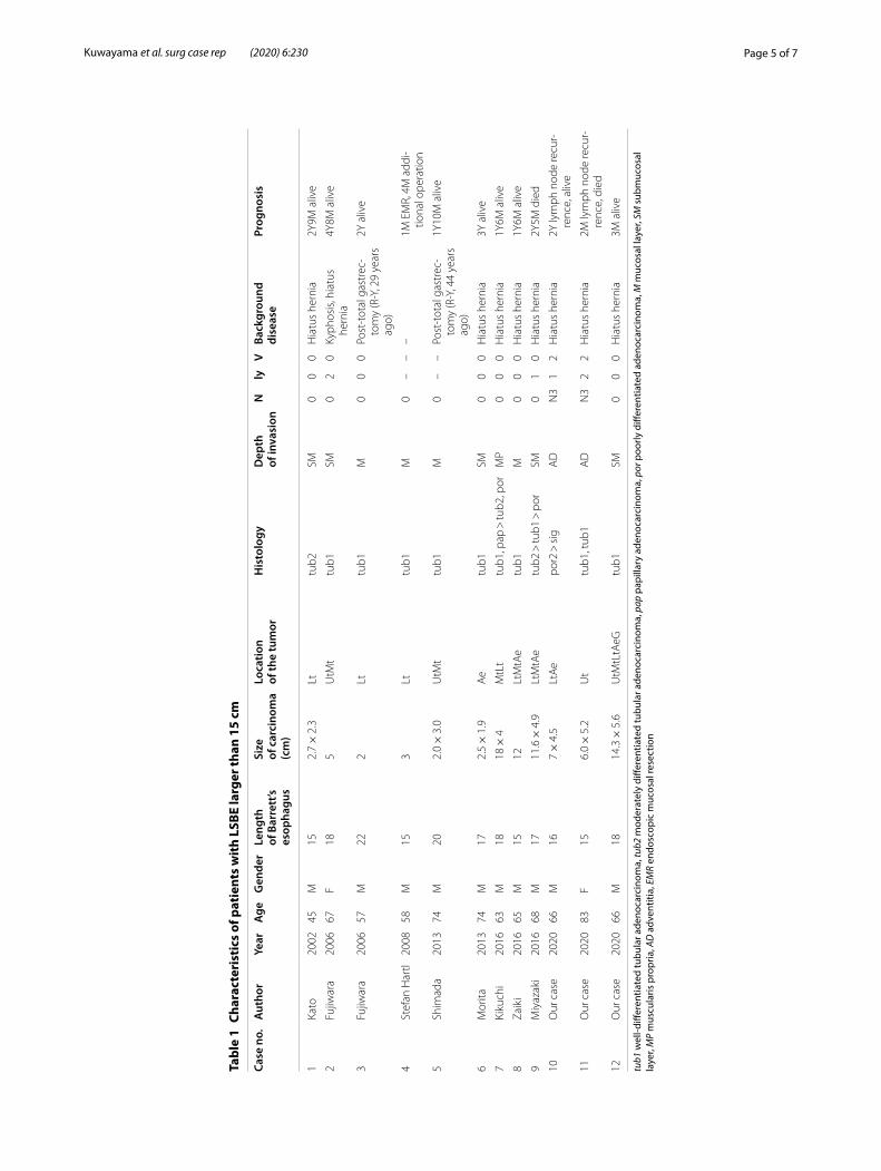
The detection frequency of LSBE larger than 13 cm is 1.27% (15/1175) [7], and it is conceivable that LSBE larger than 15 cm is a relatively uncommon condition. Until now, nine cases have been reported in the literature [8–15] (Table 1).
In these cases, including ours, 10 patients were men, and the average age was 65.5 years. The median length of BE was 17 cm (15–22 cm). The depth of invasion was T1a, T1b, T2, and T3 in 4, 5, 1, and 2 cases, respectively. Two of the three cases we encountered had advanced cancer with early recurrence. In one case, the enlarged lesion occupied almost the entire BE. Overall, 4 of 12 cases had superficial lesions (cases 7, 8, 9, 12). In some cases, the advanced cancer is associated with superficial, enlarged lesions, and preoperative diagnosis of the resec-tion area is important. In such cases, marginal resection is required, and total resection of BE may be preferable owing to the heterogeneity of BEAC derived from LSBE.
Fig. 2 Endoscopy, computed tomography, and histopathological findings for case 2. a, b Endoscopy revealed a squamocolumnar junction 15 cm from the incisor teeth and a type-2 semicircular tumor on the upper esophagus. c Computed tomography detected enlarged cervical paraesophageal lymph nodes (#101) with no other distant metastatic sites. d, e The postoperative pathological diagnosis was of well-differentiated adenocarcinoma (60 × 52 mm) with supraclavicular lymph nodes metastasis (pT3 pN3 pM1 pStage IV)
Page 4 of 7Kuwayama et al. surg case rep (2020) 6:230
The treatment policy for BEAC depends on the depth of invasion and the stage of the disease as in the case of esophageal cancer. We performed surgery after three courses of SOX therapy in case 1. The effectiveness of preoperative chemotherapy for esophageal cancer has been reported, and the National Comprehensive Can-cer Network guideline also recommends preoperative chemotherapy as an option for esophageal adenocar-cinoma with lymph node metastases deeper than T2 [16, 17]. A clinical trial of preoperative SOX therapy for esophageal adenocarcinoma of Siewert type I or II with esophageal invasion larger than 3 cm with lymph node metastasis regardless of T3/T4a or T factor is ongoing in Japan.
Overall, long-term recurrence-free survival is achieved, except in patients with advanced cancer and in case 4, early curative resection may improve the prognosis of BE cancer. Based on these findings, residual BE surveillance and prevention of BEAC are critical.
Heartburn and esophageal reflux were observed in 8 of the 12 cases. Gashi et al. [18] reported that BE shortened the maximum length and total circumference when pro-ton pump inhibitors (PPIs) were administered for 2 years. As such, PPIs may be effective in patients with reflux esophagitis and GERD.
There were two cases of BE after total gastrectomy. In an animal model, Miwa et al. [19] reported that the pres-ence of bile acids and gastric juice was important for the development of BE and adenocarcinoma. It is suggested that bile acid reflux influences the occurrence of BEAC in two cases after total gastrectomy. Shirvani et al. [20] reported that stimulation of the BE mucosa with bile acid increased the expression of cyclooxygenase 2 (COX2); it is considered to suppress apoptosis via prostaglandin E2 by expression of COX2. COX2-selective inhibitors and aspirin may increase apoptosis, suppress growth of esophageal adenocarcinoma, and shorten BE [21–23].
LSBE surveillance is another important consideration for future research. Annual surveillance of all BE cases is financially inefficient, and it is important to iden-tify BE with a high carcinogenic risk and apply an effi-cient method for narrowing the cases that require close surveillance.
In 2014, the British Gastroenterological Society recom-mended surveillance every 2–3 and 3–5 years for LSBE 3 cm or larger and SSBE shorter than 3 cm, respectively [24].
Rajeswari et al. reported that BE length may be a risk for BEAC. The annual risk of BEAC stratified by length is 0.31%/year (3 cm or shorter), 0.97%/year (4 to 6 cm),
Fig. 3 Endoscopy, computed tomography, and histopathological findings for case 3. a, b Endoscopy revealed a squamocolumnar junction 20 cm from the incisor teeth and a type 0-IIb + IIc tumor with indistinct boundaries 30 cm from the incisor teeth. c Computed tomography detected no enlarged lymph nodes and no other distant metastatic sites. d, e The postoperative pathological diagnosis was of well-differentiated adenocarcinoma (143 × 56 mm) with no lymph node metastasis (pT1a pN0 pM0 pStage 0)
Page 5 of 7Kuwayama et al. surg case rep (2020) 6:230
Tabl
e 1
Char
acte
rist
ics
of p
atie
nts
wit
h LS
BE la
rger
than
15
cm
tub1
wel
l-diff
eren
tiate
d tu
bula
r ade
noca
rcin
oma,
tub2
mod
erat
ely
diffe
rent
iate
d tu
bula
r ade
noca
rcin
oma,
pap
pap
illar
y ad
enoc
arci
nom
a, p
or p
oorly
diff
eren
tiate
d ad
enoc
arci
nom
a, M
muc
osal
laye
r, SM
sub
muc
osal
la
yer,
MP
mus
cula
ris p
ropr
ia, A
D a
dven
titia
, EM
R en
dosc
opic
muc
osal
rese
ctio
n
Case
no.
Aut
hor
Year
Age
Gen
der
Leng
th
of B
arre
tt’s
esop
hagu
s
Size
of
car
cino
ma
(cm
)
Loca
tion
of th
e tu
mor
His
tolo
gyD
epth
of
inva
sion
Nly
VBa
ckgr
ound
di
seas
ePr
ogno
sis
1Ka
to20
0245
M15
2.7 ×
2.3
Lttu
b2SM
00
0H
iatu
s he
rnia
2Y9M
aliv
e
2Fu
jiwar
a20
0667
F18
5U
tMt
tub1
SM0
20
Kyph
osis
, hia
tus
hern
ia4Y
8M a
live
3Fu
jiwar
a20
0657
M22
2Lt
tub1
M0
00
Post
-tot
al g
astr
ec-
tom
y (R
-Y, 2
9 ye
ars
ago)
2Y a
live
4St
efan
Har
tl20
0858
M15
3Lt
tub1
M0
––
–1M
EM
R, 4
M a
ddi-
tiona
l ope
ratio
n
5Sh
imad
a20
1374
M20
2.0 ×
3.0
UtM
ttu
b1M
0–
–Po
st-t
otal
gas
trec
-to
my
(R-Y
, 44
year
s ag
o)
1Y10
M a
live
6M
orita
2013
74M
172.
5 ×
1.9
Ae
tub1
SM0
00
Hia
tus
hern
ia3Y
aliv
e
7Ki
kuch
i20
1663
M18
18 ×
4M
tLt
tub1
, pap
> tu
b2, p
orM
P0
00
Hia
tus
hern
ia1Y
6M a
live
8Za
iki
2016
65M
1512
LtM
tAe
tub1
M0
00
Hia
tus
hern
ia1Y
6M a
live
9M
iyaz
aki
2016
68M
1711
.6 ×
4.9
LtM
tAe
tub2
> tu
b1 >
por
SM0
10
Hia
tus
hern
ia2Y
5M d
ied
10O
ur c
ase
2020
66M
167 ×
4.5
LtA
epo
r2 >
sig
AD
N3
12
Hia
tus
hern
ia2Y
lym
ph n
ode
recu
r-re
nce,
aliv
e
11O
ur c
ase
2020
83F
156.
0 ×
5.2
Ut
tub1
, tub
1A
DN
32
2H
iatu
s he
rnia
2M ly
mph
nod
e re
cur-
renc
e, d
ied
12O
ur c
ase
2020
66M
1814
.3 ×
5.6
UtM
tLtA
eGtu
b1SM
00
0H
iatu
s he
rnia
3M a
live
Page 6 of 7Kuwayama et al. surg case rep (2020) 6:230
1.26%/year (7 to 9 cm), 1.64%/year (10 to 12 cm), and 2.41%/year (13 cm or larger). Patients with carcinogen-esis within a year have significantly longer BE length, and a 28% increase in annual risk of BEAC was seen for every 1 cm of BE length increase. Ultra-long BE is a risk factor for carcinogenesis, and for LSBE > 15 cm, short-term follow-up within at least 1 year is consid-ered necessary.
Random biopsy is recommended for surveillance of BEAC in Europe and America; magnifying endoscopy is not recommended. For early lesions, the general strat-egy is to perform radiofrequency ablation (RFA) on the remaining BE after removal by endoscopic mucosal resection.
However, although RFA is an option for LSBE treat-ment and prevention of cancer development, it is not particularly common in Asian countries. Further report-ing and accumulation of therapeutic results is necessary for its dissemination. The patient in case 6 underwent endoscopic submucosal dissection. Narrow-band imag-ing (NBI) allowed accurate recognition of the lesion position and curative resection, and 3-year recurrence-free survival was achieved. Sharma et al. and Curvers et al. [25, 26] reported that biopsy using NBI was useful; however, further work remains to be done as NBI was expected to be efficient for future surveillance.
ConclusionsBEAC might show good prognosis if detected and treated early. Extremely LSBE is associated with a high incidence of BEAC; therefore, early detection and treatment with close surveillance is essential. Further case accumula-tion is warranted for prevention and establishment of surveillance.
AbbreviationsBE: Barrett’s esophagus; BEAC: Barrett’s esophageal adenocarcinoma; COX2: Cyclooxygenase 2; ESD: Endoscopic submucosal dissection; GERD: Gas-troesophageal reflux disease; LSBE: Long-segment Barrett’s esophagus; NBI: Narrow-band imaging; PPI: Proton pump inhibitor; RFA: Radiofrequency ablation.
AcknowledgementsNone.
Authors’ contributionsIH made substantial contributions to the concept and design of the case report. NK, IH, HG, NT, and YN envisioned the study, participated in its design and coordination, and helped draft the manuscript. All authors read and approved the final manuscript.
FundingNone of the authors received any funding for this study.
Availability of data and materialsData sharing is not applicable to this article, as no datasets were generated or analyzed during the study.
Ethics approval and consent to participateNot applicable.
Consent for publicationWritten informed consent was obtained from the patients for the publication of this case report and any accompanying images.
Competing interestsThe authors declare that they have no competing interests.
Received: 29 July 2020 Accepted: 18 September 2020
References 1. Stewart MJ, Hartfall SJ. Chronic peptic ulcer of the oesophagus. J Pathol
Bacteriol. 1929;32:9–14. 2. Lagergren J, Lagergren P. Recent developments in esophageal adenocar-
cinoma. CA Cancer J Clin. 2013;63:232–48. 3. Souza RF, Krishnan K, Spechler SJ. Acid, bile, and CDX: the ABCs of
making Barrett’s metaplasia. Am J Physiol Gastrointest Liver Physiol. 2008;295:G211–G218218.
4. El-Serag HB, Sweet S, Winchester CC, Dent J. Update on the epidemiol-ogy of gastro-oesophageal reflux disease: a systematic review. Gut. 2014;63:871–80.
5. Reid BJ, Li X, Galipeau PC, Vaughan TL. Barrett’s oesophagus and oesophageal adenocarcinoma: time for a new synthesis. Nat Rev Cancer. 2010;10:87–101.
6. Japan Esophageal Society. Japanese classification of esophageal cancer, 11th edition: part I. Esophagus. 2017;14:1–36.
7. Anaparthy R, Gaddam S, Kanakadandi V, Alsop BR, Gupta N, Higbee AD, et al. Association between length of Barrett’s esophagus and risk of high-grade dysplasia or adenocarcinoma in patients without dysplasia. Clin Gastroenterol Hepatol. 2013;11:1430–6.
8. Kato Y, Tsuyuki A, Kikuchi K, Tokuyama J, Fujishiro Y, Ozawa S, et al. A case of an early adenocarcinoma arising in an extremely long Barrett’s esopha-gus. Jpn J Gastroenterol Surg. 2002;35:1783–7.
9. Fujiwara T, Naomoto Y, Yamatsuji T, Shirakawa Y, Noguchi H, Fujiwara T, et al. Three cases of adenocarcinoma arising in extremely long-segment Barrett’s esophagus. Dig Dis Sci. 2006;51:533–8.
10. Hartl S, Siewert JR, Theisen J. Multicentric adenocarcinomas in a long-segment of Barrett’s esophagus. Clin Med Oncol. 2008;2:441–3.
11. Shimada Y, Okumura T, Hojo S, Matsui K, Nagata T, Hayashi S, et al. Adenocarcinoma in long-segment Barrett’s esophagus 44 years after total gastrectomy. J Surg Case Rep. 2013;2013:rjt100.
12. Morita Y, Tanaka S, Toyonaga T, Azuma T. Barrett’s adenocarcinoma in long-segment Barrett’s esophagus successfully detected by narrow-band imaging with magnifying endoscopy. Dig Endosc. 2013;25(Suppl 2):201–5.
13. Kikuchi M, Nakajima M, Takahashi M, Satomura H. Barrett’s adenocarci-noma 18 cm in length arising from long segment Barrett’s esophagus. Nippon Shokaki Geka Gakkai Zasshi. 2016;49:169–76.
14. Zaimoku R, Okamoto K, Kitano Y, Tsukada T. A resected case of broad Barrett’s esophageal adenocarcinoma spread over almost whole area of long-segment Barrett’s esophagus. Nihon Rinsho Geka Gakkai Zasshi. 2016;77:2936–40.
15. ShinichiM, et al. A case of superficial Barrett’s esophageal adenocarci-noma complicated by long segment Barrett esophagus.
16. Sjoquist KM, Burmeister BH, Smithers BM, Zalcberg JR, Simes RJ, Barbour A, et al. Survival after neoadjuvant chemotherapy or chemoradiotherapy for resectable oesophageal carcinoma: an updated meta-analysis. Lancet Oncol. 2011;12:681–92.
17. Ajani JA, D’Amico TA, Bentrem DJ, Chao J, Corvera C, Das P, et al. Esophageal and esophagogastric junction cancers, version 2.2019, NCCN clinical practice guidelines in oncology. J Natl Compr Cancer Netw. 2019;17:855–83.
18. Gashi Z, Bahtiri E, Gashi A, Sherifi F. Proton pump inhibitors diminish Bar-rett’s esophagus length: our experience. Open Access Maced J Med Sci. 2018;6:1041–5.
Page 7 of 7Kuwayama et al. surg case rep (2020) 6:230
19. Miwa K, Sahara H, Segawa M, Kinami S, Sato T, Miyazaki I, et al. Reflux of duodenal or gastro-duodenal contents induces esophageal carcinoma in rats. Int J Cancer. 1996;67:269–74.
20. Shirvani VN, Ouatu-Lascar R, Kaur BS, Omary MB, Triadafilopoulos G. Cyclooxygenase 2 expression in Barrett’s esophagus and adenocarci-noma: ex vivo induction by bile salts and acid exposure. Gastroenterol-ogy. 2000;118:487–96.
21. Souza RF, Shewmake K, Beer DG, Cryer B, Spechler SJ. Selective inhibition of cyclooxygenase-2 suppresses growth and induces apoptosis in human esophageal adenocarcinoma cells. Cancer Res. 2000;60:5767–72.
22. Kaur BS, Khamnehei N, Iravani M, Namburu SS, Lin O, Triadafilopoulos G. Rofecoxib inhibits cyclooxygenase 2 expression and activity and reduces cell proliferation in Barrett’s esophagus. Gastroenterology. 2002;123:60–7.
23. Amano Y, Ishihara S, Kushiyama Y, Yuki T, Takahashi Y, Chinuki D, et al. Barrett’s oesophagus with predominant intestinal metaplasia correlates with superficial cyclo-oxygenase-2 expression, increased prolifera-tion and reduced apoptosis: changes that are partially reversed by non-steroidal anti-inflammatory drugs usage. Aliment Pharmacol Ther. 2004;20:793–802.
24. Fitzgerald RC, di Pietro M, Ragunath K, Ang Y, Kang JY, Watson P, et al. British Society of Gastroenterology guidelines on the diagnosis and management of Barrett’s oesophagus. Gut. 2014;63:7–42.
25. Sharma P, Hawes RH, Bansal A, Gupta N, Curvers W, Rastogi A, et al. Standard endoscopy with random biopsies versus narrow band imaging targeted biopsies in Barrett’s oesophagus: a prospective, international, randomised controlled trial. Gut. 2013;62:15–211.
26. Curvers WL, Bohmer CJ, Mallant-Hent RC, Naber AH, Ponsioen CI, Ragunath K, et al. Mucosal morphology in Barrett’s esophagus: inter-observer agreement and role of narrow band imaging. Endoscopy. 2008;40:799–805.
Publisher’s NoteSpringer Nature remains neutral with regard to jurisdictional claims in pub-lished maps and institutional affiliations.