adenovirus type 12-induced fragility of the human rnu2 locus
TRANSCRIPT
MOLECULAR AND CELLULAR BIOLOGY, Nov. 1995, p. 6246–6255 Vol. 15, No. 110270-7306/95/$04.0010Copyright q 1995, American Society for Microbiology
Adenovirus Type 12-Induced Fragility of the Human RNU2Locus Requires U2 Small Nuclear RNA Transcriptional
Regulatory ElementsARNOLD D. BAILEY, ZENGJI LI, THOMAS PAVELITZ, AND ALAN M. WEINER*
Department of Molecular Biophysics and Biochemistry, Yale University School of Medicine, New Haven, Connecticut 06520-8024
Received 3 May 1995/Returned for modification 12 July 1995/Accepted 26 July 1995
Infection of human cells with oncogenic adenovirus type 12 (Ad12) induces four specific chromosome fragilesites. Remarkably, three of these sites appear to colocalize with tandem arrays of genes encoding small,abundant, ubiquitously expressed structural RNAs—the RNU1 locus encoding U1 small nuclear RNA(snRNA), the RNU2 locus encoding U2 snRNA, and the RN5S locus encoding 5S rRNA. Recently, an artificialtandem array of the natural 5.8-kb U2 repeat unit has been shown to generate a new Ad12-inducible fragile site(Y.-P. Li, R. Tomanin, J. R. Smiley, and S. Bacchetti, Mol. Cell. Biol. 13:6064–6070, 1993), demonstrating thatthe U2 repeat unit alone is sufficient for virally induced fragility. To identify elements within the U2 repeat unitthat are required for virally induced fragility, we generated cell lines containing artificial tandem arrays of theentire 5.8-kb repeat unit, an 834-bp fragment spanning the U2 gene alone, or the same 834-bp fragment fromwhich key U2 transcriptional regulatory elements had been deleted. The U2 snRNA coding regions within eachartificial array were marked by an innocuous single base change (U to C at position 87) so that the relativeexpression of supernumerary and endogenous U2 genes could be monitored by a primer extension assay. Wefind that artificial arrays of both the 5.8- and the 0.8-kb U2 repeat units are fragile but that arrays lackingeither the distal sequence element or both the distal and the proximal sequence elements of the promoter arenot. Surprisingly, variations in repeat copy number and/or transcriptional activity of the artificial arrays do notappear to correlate with the degree of Ad12-inducible fragility. We conclude that U2 transcriptional regulatoryelements are required for virally induced fragility but not necessarily U2 snRNA transcription per se.
A number of cancers (60) as well as several kinds of herita-ble mental retardation known as fragile X syndrome (16, 18)are caused by or associated with chromosome fragile sites.Fragile sites typically appear in metaphase spreads as nonstain-ing or heterochromatic gaps, often accompanied by a disloca-tion in the chromosome axis. Although clinically observed frag-ile sites seldom reflect frank chromosome breakage, fragilesites colocalize with over 60% of recurrent cancer chromo-some breakpoints (59). This suggests that oncogenic rear-rangements and translocations are often caused by recom-bination at fragile sites or by breakage and subsequentchromosome fusion (6, 16, 18, 41, 59, 60). Fragile sites canbe induced by a variety of methods, including abnormal growthconditions, drug or chemical treatment, and viral infection; forthis reason, fragile sites are usually classified according to themethod and efficiency of induction (17, 49). Although there arelikely to be many different causes of chromosome fragility, ithas been shown in at least one case that a fragile site cancorrespond to a region of incompletely condensed chromatin(12). A defect in chromatin condensation could explain whyfragile sites are physically weak and genetically recombino-genic and why certain fragile sites serve as preferential targetsfor viral integration (references 39, 46, and 52, but see alsoreference 57). Fragile sites are also conserved among primates(45), and a majority of the breakpoints observed in the evolu-tion of primate chromosomes fall at or near known fragile sites(33).
Infection of human cells with the nononcogenic adenovirustype 2 or 5 (Ad2 or Ad5) induces generalized chromosomefragility (6), but infection with the oncogenic Ad12 inducesfour, and only four, specific chromosome fragile sites (63).These four Ad12-induced fragile sites have been mapped, inorder of decreasing fragility, to 17q21-22, 1p36, 1q21, and1q42-43 (30, 31, 63). Since these four sites are not preferentialtargets for adenoviral integration (reference 32, but see refer-ence 52), virally inducible fragility must be an intrinsic propertyof the underlying chromosomal regions, perhaps reflectingsome unusual aspect of chromatin structure, gene organiza-tion, transcription, or DNA replication. Remarkably, three ofthe four sites appear to colocalize with large tandem arrays ofgenes encoding small, abundant, ubiquitously expressed struc-tural RNAs—the RNU1 locus encoding U1 small nuclear RNA(snRNA) at 1p36 (23, 35), the RNU2 locus encoding U2snRNA at 17q21-22 (14, 22), and the RN5S locus encoding 5SrRNA at 1q42-43 (47, 48). The fourth fragile site colocalizeswith the PSU1 locus at 1q21 (23), an old and highly divergentfamily of U1 snRNA genes that are no longer expressed asstable U1 snRNA (24) but which may still retain active tran-scriptional regulatory elements.Colocalization of Ad12-inducible fragile sites with active
gene clusters suggested that the gene clusters themselves wererequired, and might be sufficient, for virally induced fragility(22). Subsequent work on the U2 genes has confirmed thisidea. The human U2 genes are organized as a single, essentiallyperfect tandem array containing 5 to 25 copies of a 5,834-bprepeat unit at 17q21-22 (14, 22, 37, 50, 55); the U2 arraytherefore spans 30 to 150 kb. Durnam et al. demonstrated byclassical in situ hybridization with 3H-labeled U2 probes thatthe Ad12-induced fragile site at 17q21-22 was not only near,but occasionally fell within, the RNU2 locus, as evidenced by
* Corresponding author. Mailing address: Department of MolecularBiophysics and Biochemistry, Yale University School of Medicine,P.O. Box 208024, New Haven, CT 06520-8024. Phone: (203) 785-4589.Fax: (203) 785-6404. Electronic mail address: [email protected].
6246

splitting of the U2 hybridization signal (11). This result morestrongly implicated the U2 array itself as the cause of virallyinduced fragility, but it was also possible that unraveling of theU2 array was a consequence rather than the cause of fragility.More recently, Li et al. have generated cell lines containingstably integrated, artificial arrays of the full 5.8-kb U2 repeatunit which exhibit the same Ad12-induced fragility as the nat-ural RNU2 locus (21). This proves that the U2 repeat unitalone is sufficient for virally induced fragility and that fragilitydoes not depend on the immediate flanking sequences of theU2 tandem array or on the chromosomal microenvironmentsurrounding the natural RNU2 locus.The ability of Ad12 to induce specific chromosome fragile
sites may be due solely to the E1 region of the virus; indeed,constitutive fragility can be observed at 17q21-22 in hybridmouse-human cell lines stably transfected with the Ad12 E1region alone (12). The E1 region includes two transcriptionunits: E1A generates three related, multifunctional proteins byalternative 59 splicing, and E1B generates 19,000- and 55,000-molecular-weight proteins (19K and 55K proteins) from a sin-gle mRNA with overlapping reading frames. Virally inducedfragility is unaffected by mutations in the E1B 19K protein (12,41) but is 70% reduced by a mutation in the 55K protein (41).In addition, a hybrid virus (T1227) carrying the first exon ofAd12 E1A in an Ad5 background was unable to induce chro-mosomal damage. Thus, the Ad12 E1B 55K protein may besufficient for virally induced fragility, although a role for one ofthe E1A proteins has not been rigorously excluded.Ad12-induced chromosome fragility requires an interaction
between viral functions and the intrinsic properties of specificchromosomal sites. To understand the causes of virally inducedfragility at the molecular level, we have begun by identifyingthe minimal elements within the U2 repeat unit required forAd12-induced fragility. We have generated cell lines contain-ing tandem arrays of the intact 5.8-kb U2 repeat unit, an834-bp U2 minigene, and U2 minigenes from which key tran-scriptional regulatory elements have been deleted. In eachcase, the U2 snRNA coding regions were marked by a harm-less single base change (U to C at position 87 [U87C]) thatenabled us to monitor the relative expression of supernumer-ary and endogenous U2 genes by a simple primer extensionassay. We found that artificial arrays containing the completeU2 transcription unit are fragile but that arrays lacking theenhancer-like distal sequence element (DSE) (2, 3, 27) are not.The degree of Ad12-inducible fragility did not correlate, how-ever, with variations in repeat copy number and/or transcrip-tional activity. Thus, virally induced fragility requires U2 tran-scriptional regulatory elements but not necessarily U2 snRNAtranscription per se. These results provide the groundwork fora detailed molecular understanding of the chromosomal ele-ments, as well as the viral and host factors, required for thesite-specific chromosomal fragility induced by Ad12. Garganoet al. (13a) have reached very similar conclusions.
MATERIALS AND METHODS
Clone construction. To generate the marked U2 minigene, an 824-bp fragmentextending from the StuI site upstream of the U2 coding region to the HinfI sitejust downstream of the 39-end formation signal (3, 37) was subcloned intoM13mp18. The U87C mutation within the 188-bp U2 coding region was intro-duced by standard techniques (19) and verified by sequence analysis. The markedU2-U87C minigene was excised as an EcoRI-to-BamHI fragment and trans-ferred to pUC18, and a BglII linker (New England Biolabs) was inserted into thefilled-in EcoRI site. This clone was designated mU2, where m indicates a mini-gene. To generate a marked, intact 5.8-kb U2 repeat unit, the HincII-to-ApaLIfragment of mU2 was used to replace the corresponding sequence in the plasmidpTPK18Eco (protocol available upon request). The parent construct, pTPK18,contains a 5.8-kb KpnI fragment spanning the entire U2 repeat unit (37); the
unique XmnI site within the U2 repeat unit was destroyed by insertion of anEcoRI linker to generate pTPK18Eco. The resulting clone was designated iU2,where i indicates an intact gene (Fig. 1A).Transcriptional regulatory elements were deleted from mU2 (Fig. 1A). The
DSE was excised by digestion with HincII and SfuI; the SfuI end was then filledin, and the plasmid was religated to generate mU2DDSE. The DSE and proximalsequence element (PSE) were deleted from HincII to FokI by ligating twofragments of mU2. mU2 was first digested with FokI, and the ends were filled in.The blunt FokI-to-BamHI fragment spanning the marked U2 gene was thenexcised with BamHI and ligated to the BamHI-to-HincII vector fragment ofmU2 spanning sequences upstream of HincII as well as all of pUC18. This clonewas designated mU2DDSEDPSE. The deletions in mU2DDSE and mU2DDSEDPSE were confirmed by sequence analysis. An additional clone, designatedmU21CT, contained the U2 minigene along with the (CT)70 dinucleotide repeatlocated immediately downstream from the U2 transcription unit (Fig. 1A andreference 27). mU21CT was generated by ligating the filled-in RsrII-to-StuIfragment from iU2 into the filled-in XmaI site of pUC18Bgl (a modified pUC18in which the polylinker EcoRI site has been destroyed by insertion of a BglIIlinker).The selection cassette was composed of two parts (Fig. 1A). The neomycin
resistance gene was excised from pMC1NEO (Stratagene) as an XhoI-to-BamHIfragment, filled in, and cloned into the SmaI site of pUC18Bgl. A mouse dihy-drofolate reductase (DHFR) gene carrying a point mutation that renders itresistant to high levels of methotrexate (43) was then cloned into the KpnI siteto generate plasmid E1. To reduce the risk of promoter occlusion or transcrip-tional interference, all four orientations of the two genes (head-to-head, tail-to-tail, and head-to-tail) were made and tested as monomeric plasmids for therelative frequency of Geneticin-resistant colonies. The E1 selection cassette withthe Neor gene upstream of the DHFR gene (Fig. 1A) yielded a 10- to 20-fold-higher frequency of resistance than the other orientations (DHFR gene up-stream of the Neor gene, convergent or divergent transcription units).Cloning and analysis of cell lines. Generation of cell lines that contain large
tandem U2 arrays was not straightforward; large tandem arrays generated by invitro ligation are easily sheared during standard experimental manipulations, andsmaller contaminating arrays are preferentially incorporated during transfection.We therefore conducted numerous pilot experiments as summarized here. Ourgoal was to generate large head-to-tail tandem U2 arrays doped by an occasionalE1 selection cassette; we sought to avoid generating any oligomeric productscontaining the E1 cassette, as these would be preferentially incorporated upontransfection and would therefore require characterization of many more celllines. Tandem arrays were constructed by ligation of fragments with dissimilarbut compatible BamHI and BglII ends; the desired head-to-tail ligations generatehybrid BglII-BamHI restriction sites that are resistant to digestion by eitherenzyme, while undesirable head-to-head and tail-to-tail ligations regenerateBglII and BamHI sites which can be recycled by digestion with both enzymes. Toreduce the risk of damaged ends which act as chain terminators during ligation,BglII-to-BamHI fragments of the U2 constructs and the E1 selection cassette(Fig. 1A) were purified from low-melting-point agarose (13) just before use.Using T4 DNA ligase, we performed a series of ligations in which the totalfragment concentration was varied from 5 to 500 mg/ml and the ratio of U2 to E1fragments was varied from 10:1 to 100:1. The ligation products were character-ized, both before and after digestion with BglII and BamHI, by field inversion gelelectrophoresis (FIGE), ethidium bromide staining, and blotting. Under theFIGE conditions used, monomers through decamers were resolved as ladderbands, while larger multimers ran as a smear. Although we expected oligomericproducts at low DNA concentrations, we were surprised to find significantamounts of oligomeric products (circularized monomers and dimers, etc.) evenat the highest DNA concentrations tested. We were unable to reduce the yield ofoligomeric products, or to generate larger tandem arrays, by simultaneous liga-tion and digestion with BglII plus BamHI or by alternating rounds of ligation anddigestion. This may indicate that most attempts to increase ligation are effectivelyopposed by chain termination due to exonuclease, phosphatase, and/or mechan-ical shear. In fact, ligation at high DNA concentrations generated solutions ofhigh viscosity that were obviously subject to mechanical shear even when handledwith widebore pipette tips. Ligation in the presence of 50 mM spermidine re-duced the viscosity of the ligation reaction mixture and, at least in our hands,generated larger arrays. This is consistent with reports that polyamines condenseDNA, leading to more compact, rigid molecules with reduced viscosity andincreased resistance to shear (5). As expected, a U2-to-E1 ratio of 100:1 reducedincorporation of the selection cassette into oligomeric products, and we alsofound, by varying the temperature from 4 to 378C, that residual monomer wasminimized by ligation at 258C. Finally, we attempted to enrich for larger tandemarrays by preparative FIGE but found that the arrays could not be recoveredfrom agarose without significant shearing.In our final protocol for generating tandem arrays, we used a 100:1 ratio of U2
to E1 and ligated 25 mg of DNA fragments at 500 mg/ml for 3 to 4 h at 258C. Thereaction mixture was then diluted twofold to reduce the viscosity, keeping thespermidine at 50 mM, and digested with a twofold excess of BglII and BamHI toeliminate head-to-head and tail-to-tail junctions. The DNA was then precipitatedwith ethanol, washed with 70% ethanol, and gently resuspended in 250 ml of TEbuffer (10 mM Tris-HCl and 1 mM EDTA, pH 8.0) containing 50 mM spermi-dine.
VOL. 15, 1995 Ad12-INDUCED FRAGILITY OF THE HUMAN RNU2 LOCUS 6247

FIG. 1. (A) Constructs used to generate artificial U2 tandem arrays. All U2 genes contain the U87C mutation (p) to distinguish U2 snRNAs transcribed from theendogenous and artificial arrays (also see panel B). iU2 (for intact) is a full-length 5.8-kb U2 repeat unit (top panel) in which the unique XmnI site has been destroyedby insertion of an EcoRI linker. Artificial arrays of iU2 can be distinguished from endogenous U2 arrays which are not cut by EcoRI (37, 50). Only the diagnostic SmaIand PstI sites (see Fig. 2) are shown below the gene. Also indicated are the location of a solo long terminal repeat (LTR), the CT dinucleotide repeat, and a 59-truncatedL1 sequence (37). mU2 (for mini) is a U2 minigene (middle panel); mU21CT includes the downstream (CT)70 dinucleotide repeat as well. The U2 coding region (openarrow) is preceded by two transcriptional regulatory elements known as the DSE and PSE (open circle and square, respectively) and is followed by the 39-end formationsignal (filled circle). The extent of mU2 and mU21CT (brackets below the map) and the regions deleted from mU2 to generate mU2DDSE and mU2DDSEDPSE(brackets above the map) are indicated. The E1 selection cassette (bottom panel) is also shown. Note that all constructs have dissimilar BglII and BamHI ends to allowhead-to-tail ligation (see text). (B) U2 snRNA sequence and accepted secondary structure (61). The U87C mutation (arrow) was chosen because this position isphylogenetically variable and adjacent to a long single-stranded region, which should facilitate annealing with a primer complementary to U2 positions 89 to 109(bracket).
6248 BAILEY ET AL. MOL. CELL. BIOL.
We compared transfection by Ca2PO4 precipitation and electroporation usingin vitro-ligated tandem arrays prepared as described above. The recipient cellswere the pseudodiploid fibrosarcoma line HT-1080 (gift from S. Bacchetti [21]).Ca2PO4 precipitation and electroporation generated Geneticin-resistant colonieswith comparable efficiencies when the E1 selection cassette was used alone, butCa2PO4 precipitation was roughly twice as efficient as electroporation for trans-fection of large tandem arrays and generates larger arrays on average. Wesuspect that Ca2PO4 precipitation protects large DNA molecules from mechan-ical shear, while small DNA molecules preferentially traverse transient mem-brane pores during electroporation. Ultimately, all cell lines were generated byCa2PO4 precipitation (Gibco/BRL) except cell line mU2 C12 (electroporated ina 0.4-cm-diameter cuvette at 0.2 kV and 960 mF with a Bio-Rad Gene Pulser witha Capacitance Extender). HT-1080 cells were grown in minimal essential me-dium (Gibco/BRL) with 10% fetal bovine serum, the cultures were split 24 hbefore transfection to give 106 cells per 100-mm-diameter plate, and the mediumwas replaced 1 h before transfection. The medium was changed again 18 h aftertransfection, and 24 h later, the cells were divided among 10 to 12 150-mm-diameter plates with 25 ml of media containing 600 ml of a 25-mg/ml stock ofGeneticin (Gibco/BRL). The medium was changed every 3 to 4 days over a 14-to 17-day period. The resulting colonies were collared with a cloning cylinder,trypsinized, and transferred to 48-well microtiter plates. Individual clonal celllines were grown until frozen stocks and genomic DNA plugs could be made.Plugs were made by resuspending trypsinized cells from a 60-mm-diameter
plate in 300 ml of phosphate-buffered saline. To this suspension, 300 ml of 1%Incert Agarose (FMC Corp.) at 408C was added, the suspension was mixed, andsolution was pipetted into the plug mold (Medical Specialties, Baltimore, Md.).Processing of the plugs and FIGE were performed as described previously (37).‘‘Unblots’’ of the FIGE gels are modified genomic blots in which the driedagarose gel is used directly instead of a transfer of the DNA to nitrocellulose ornylon (28).Relative expression of marked U2 genes. Cells were collected by trypsinization
from nearly confluent 100-mm-diameter plates and lysed with Nonidet P-40 asdescribed elsewhere (62), and total RNA was prepared by phenol extraction.Primer extension analysis of total RNA was as described previously (62) exceptthat ddATP was used in place of dATP and extension products were resolved ona 15% sequencing gel. Phosphorimager analysis was performed with a GS-250Molecular Imager (Bio-Rad) and the program Molecular Analyst 2.0.Ad12-induced fragility. Cells were infected with Ad12 (strain Huie) at a
multiplicity of infection of 10 (11, 21). About 17 h after infection, cells weretreated with colcemid at 0.1 mg/ml for 3 h, and metaphase spreads were thenprepared by standard techniques of hypotonic shock and methanol-acetic acidfixation (4).FISH. Natural and artificial U2 loci were labeled for fluorescent in situ hy-
bridization (FISH) by using biotinylated probes. Briefly, slides were denatured ina solution of 70% formamide and 23 SSC (13 SSC is 0.15 M NaCl plus 0.015 Msodium citrate) at 708C for 2 min and then subjected to immediate serial dehy-dration in ice-cold 70, 90, and 100% ethanol. The slides were then air dried atroom temperature. Hybridization was performed in a solution containing 50%formamide, 23 SSC, 10% dextran sulfate, 500 mg of salmon sperm DNA per ml,and 50 mg of biotinylated DNA probe per ml. After 5 min of denaturation at708C, 20 ml of hybridization solution was applied to each slide, and the slideswere then covered with coverslips and sealed with rubber cement. Hybridizationwas performed overnight in a moist chamber at 378C. The coverslips were thenremoved, and the slides were washed twice in a solution of 50% formamide and23 SSC at 428C for 10 min and twice in 0.13 SSC at 378C for 5 min. The washedslides were incubated with 5 mg of fluorescein-conjugated avidin DCS per ml ina solution containing 43 SSC, 3% bovine serum albumin, and 0.1% Tween 20 for30 min. After three washes in a mixture of 43 SSC and 0.1% Tween 20 for 10min, the slides were counterstained with DAPI (4,6-diamidino-2-phenylindole;200 ng/ml) and mounted in a solution containing 90% glycerol, 0.1 M Tris-HCl(pH 8.0), and 2.3% DABCO antifade (1,4-diazabicyclo-2,2,2-octane).Images were collected with a Zeiss Axioskop epifluorescence microscope
equipped with a cooled, charge-coupled device (CCD) camera (Photometrics)and controlled by a computer interface software package (CCD Image Capture).The source images were pseudocolored and merged by using computer software(GeneJoin) available through the Office of Cooperative Research, Yale Univer-sity. The equipment and protocols have been described in detail elsewhere (4).
RESULTS
Construction of artificial U2 arrays. Li et al. previouslydemonstrated that an artificial tandem array of the natural5.8-kb U2 repeat unit will generate a new Ad12-inducible frag-ile site (21). To better understand the causes of Ad12-inducedfragility, we constructed cell lines containing artificial tandemarrays of five different U2 constructs (Fig. 1A). Tandem arrayswere generated by using iU2, an intact 5.8-kb U2 repeat unit inwhich the XmnI site has been destroyed by insertion of anEcoRI linker; mU2, an 824-bp minigene containing all essen-
tial transcriptional regulatory elements, the U2 coding region,and the 39-end formation signal; and mU21CT, an mU2 de-rivative including the downstream (CT)70 dinucleotide tract.We also constructed two mU2 derivatives lacking key tran-scriptional regulatory elements: the DSE is deleted inmU2DDSE, and both the DSE and the PSE are deleted inmU2DDSEDPSE. In each construct, the U2 gene contains apoint mutation from U to C at position 87 in the U2 codingregion (Fig. 1B). Although U2 snRNA is highly conserved insequence and structure from yeasts to humans, position 87 isphylogenetically variable (61) and the U87C mutation is mostunlikely to affect U2 snRNA expression or function. Use of themarked U2-U87C gene allowed us to monitor expression ofthe supernumerary U2 genes in the artificial arrays and toprove that these U2 snRNAs are functional, at least as judgedby immunoprecipitation with anti-Sm antibodies (data notshown).The intact U2 repeat unit (iU2). Geneticin-resistant cell
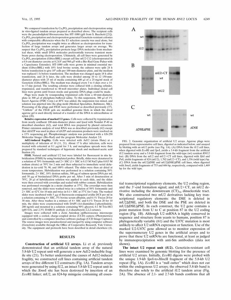
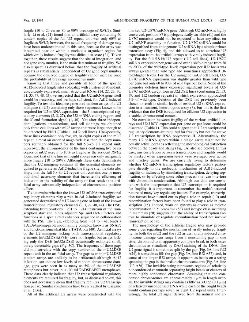
lines were examined by genomic blotting for the presence ofartificial U2 arrays. Initially, EcoRI digests were probed withthe unique 1.9-kb SpeI-to-HincII fragment of the 5.8-kb U2repeat (Fig. 1A). EcoRI is a ‘‘null cutter’’ which does not cutwithin the endogenous U2 arrays; the observed 5.8-kb band istherefore due solely to the artificial iU2 tandem array (Fig.2A). The absence of 2.1- and 2.7-kb bands confirms that all
FIG. 2. Genomic organization of artificial U2 arrays. Agarose plugs wereprepared from representative cell lines, digested as indicated below, and assayedby blotting with an mU2 probe (see Fig. 1A). (A) DNA from the iU2 cell lines,when digested with EcoRI and SpeI, yields a 3.4-kb fragment from the artificialiU2 tandem array and a 5.8-kb fragment from the wild-type (wt) resident RNU2loci. (B) DNA from the mU2 and mU21CT cell lines, digested with SfuI andPstI, yields fragments of 824 (mU2), 1,702 (mU21CT), and 1,356 (wild type) bp.(C) DNA from the mU2DDSE and mU2DDSEDPSE cell lines, when digestedwith SmaI, yields fragments of 753 and 606 bp, respectively, compared with 1,469bp for the wild type.
VOL. 15, 1995 Ad12-INDUCED FRAGILITY OF THE HUMAN RNU2 LOCUS 6249

tandem repeats were head-to-tail, not head-to-head or tail-to-tail. Of 95 Geneticin-resistant cell lines screened, 25 had atleast as many artificial iU2 as endogenous U2 repeats. These25 cell lines were further characterized by digestion withBamHI and BglII followed by FIGE. The gels were then driedand hybridized directly with the labeled probe (28). The num-ber and size of artificial iU2 arrays were found to vary, andeight cell lines containing the largest unique iU2 arrays wereselected for further study. To accurately measure the relativecopy numbers of iU2 and U2 repeats in these eight lines, DNAwas digested with EcoRI and SpeI; iU2 repeats are reduced to3.4-kb fragments, while endogenous arrays yield monomeric5.8-kb fragments (Fig. 2A). The ratio of the artificial to endo-genous bands was determined by phosphorimager analysis, andthe eight cell lines were found to contain 12 to 79 tandemcopies of the 5.8-kb iU2 repeat unit (Table 1, DNA ratio).We soon discovered, to our initial dismay, that all of the
eight iU2 lines were tetraploid, apparently because tetraploidcells had almost overgrown the parental pseudodiploid HT-1080 cells. Tetraploidy is unlikely to be a response to transfec-tion with supernumerary U2 genes, because pseudodiploid iU2lines were also identified (Table 1). As ploidy was not expectedto affect virally induced fragility, we continued to work withthese lines although good metaphase spreads were more dif-ficult to obtain.The minigene construct (mU2). The drug-selected cell lines
were first examined by genomic blotting with SfuI, a ‘‘onecutter’’ for both the endogenous and the mU2 minigene repeatunits. Blots were probed with the mU2 monomer, which de-tects both natural and artificial U2 genes, as well as the many39-truncated U2 retropseudogenes (51). Surprisingly, in threeseparate transfections, we found only seven cell lines that con-tained more than one of the 824-bp mU2 repeat units (1 of 31,4 of 77, and 2 of 127 transfectants). Of these seven cell lines,only three exceeded the minimum array size required for de-tection by FISH, and none contained more than eight tandemrepeats of mU2. The absence of tandem arrays containingmore than eight copies of mU2 contrasts dramatically with theease of obtaining large tandem arrays containing nearly 80copies of the full 5.8-kb iU2 construct (Table 1). We do notknow whether the copy number constraint reflects a problemwith integration or stability of mU2 arrays but, as described ingreater detail below (also see Table 1), the copy number con-straint is relieved when U2 transcription is crippled by deletionof either the DSE (mU2DDSE) or both the DSE and thePSE (mU2DDSEDPSE). Since both U1 snRNA (25) and U2snRNA (3a) are subject to dosage compensation (i.e., snRNAlevels are substantially independent of snRNA gene copy num-ber), one interesting possibility is that large mU2 arrays mightbe unstable if mU2 is able to escape dosage compensation.Attempts to characterize the three mU2 cell lines by diges-
tion with BamHI and BglII failed to give clear results, appar-
TABLE 1. Characteristics of cell lines containing artificial U2 tandem arrays
Cell line or RNACopy no. ofmarked U2genesa
DNA ratiob RNA ratioc RNA/gened RNA/locuse Fragility (%)f
iU2 A11 12 0.26 0.44 1.6 1.7 80–90iU2 A24 25 0.56 0.91 1.6 3.5 10–20iU2 A42 29 0.67 0.58 0.82 2.2 90–100iU2 A36 30 0.68 0.52 0.71 2 80–90iU2 A16 32 0.72 0.91 1.2 3.5 10–20iU2 A37 48 1.1 1.8 1.6 7 90–100iU2 A41 77 1.8 2.5 1.4 9.8 90–100iU2 A40 79 1.8 0.58 0.31 4.4 90–100
mU2 C12 5 0.119 0.155 1.1 0.51 5–10mU2 T38 6 0.128 0.142 0.9 0.46 5–10mU2 L48 8 0.182 0.224 1.1 0.79 10–20
mU2DDSE 33 22 0.98 0.063 0.037 0.072 #5mU2DDSE 50 31 1.4 0.11 0.061 0.17 ;10mU2DDSE 34 53 2.4 0.062 0.015 0.07 ,5mU2DDSE 9 79 3.6 0.029 0.0005 0.0036 ,5mU2DDSE 25 112 5.1 0.039 0.0024 0.024 #5
mU2DDSEDPSE 14 8 0.36 0.027 0.0011 0.008 ,1mU2DDSEDPSE 43 20 0.9 0.026 0 0 ,1mU2DDSEDPSE 5 29 1.3 0.026 0 0 ,1mU2DDSEDPSE 33 57 2.6 0.026 0.0023 0.024 ,1mU2DDSEDPSE 48 78 3.5 0.023 0 0 ,1mU2DDSEDPSE 42 98 4.4 0.033 0.0014 0.012 ,1
HeLa RNA 0.011HT-1080 RNA 0.027
a Calculated from the DNA ratio and the known copy number of endogenous U2 genes (22 for pseudodiploid and 44 for pseudotetraploid HT-1080 cells [36]).b Ratio of marked U2 to endogenous U2 signal as determined by phosphorimager analysis of genomic blots (see Fig. 2). Corrections were made for the extent of
complementarity with the mU2 probe.c Ratio of marked U2 snRNA to wild-type snRNA as determined by phosphorimager analysis of primer extension assays performed on total RNA (see Fig. 4).d RNA ratio divided by the DNA ratio. This represents the relative expression of the marked U2 genes compared with that of the wild type.e RNA ratio divided by the ratio of artificial U2 loci to endogenous RNU2 loci. Assuming that all endogenous loci are equally expressed, this represents the relative
expression per locus of the artificial and endogenous arrays.f Relative frequency of breakage at artificial U2 loci compared with that at endogenous RNU2 loci (see Fig. 3 for scoring of breaks); the range of fragility represents
the variation observed in two separate experiments.
6250 BAILEY ET AL. MOL. CELL. BIOL.
ently because abundant U2 retropseudogenes readily reactwith the minigene probe, but digestion with SfuI and PstI (Fig.1A) resolved monomers of mU2 (824 bp) from endogenous U2genes (1,356 bp) and from the smear of retropseudogenes
above (Fig. 2B). The ratios of mU2 to endogenous U2 geneswere then determined by phosphorimager analysis (Table 1).Minigenes lacking transcriptional regulatory signals (mU2
DDSE and mU2DDSEDPSE). The DSE of the U2 snRNApromoter can be thought of as a specialized enhancer, whilethe PSE functions much like a TATA box (9). We constructedtwo derivatives of the mU2 minigene, one lacking the DSE(mU2DDSE) and the other lacking both the DSE and the PSE(mU2DDSEDPSE). Initially, SmaI digests of DNA from Ge-neticin-resistant cell lines were analyzed by genomic blottingfor the presence of tandem arrays of the mU2DDSE andmU2DDSEDPSE constructs. The mU2 probe should detectSmaI fragments of 1,469 (endogenous U2 genes), 753 (mU2DDSE), and 606 (mU2DDSEDPSE) bp. To our surprise, 32 of 52mU2DDSE cell lines and 35 of 65 mU2DDSEDPSE cell lineshad at least as many artificial U2 repeats as endogenous U2repeats. Thus, large tandem arrays were easily obtained for thefull U2 repeat (iU2) and for the transcriptionally crippledmU2DDSE and mU2DDSEDPSE constructs, but not for themU2 minigene (Table 1). We do not yet understand theseresults, but, as mentioned above, one attractive interpretationis that large mU2 arrays are unstable because the genes some-how escape dosage compensation, perhaps because they lackan additional element that is present in the full 5.8-kb repeatunit. In fact, our preliminary results indicate th1at we can easilyobtain large artificial arrays of the mU21CT construct (Fig. 1).The (CT)70 dinucleotide repeat may therefore stabilize the U2tandem array and/or participate in dosage compensation.On the basis of the SmaI digestion patterns, 25 mU2DDSE
lines and 25 mU2DDSEDPSE lines were further characterizedby digestion with BamHI and BglII; the digests were resolvedby FIGE, and the dried agarose gels were hybridized directlywith the labeled probe (28). Of these, five mU2DDSE and sixmU2DDSEDPSE lines containing a single large array were
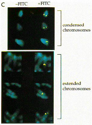
FIG. 3. Ad12-induced fragility of artificial U2 tandem arrays. (A) iU2 series.(B) mU2 series. (C) Cell line mU2DDSE 50. FITC, fluorescein isothiocyanate.
VOL. 15, 1995 Ad12-INDUCED FRAGILITY OF THE HUMAN RNU2 LOCUS 6251
chosen for analysis of U2 expression and fragility. Carefulreanalysis of these mU2DDSE and mU2DDSEDPSE lines bySmaI digestion, genomic blotting, and phosphorimager analy-sis (Fig. 2A) revealed that the mU2DDSE tandem arrays con-tained 22 to 112 copies, while the mU2DDSEDPSE arrayscontained 8 to 98 copies (Table 1).Virally induced fragility. Cells were infected with Ad12 at a
multiplicity of infection of 10 for 18 h and treated with colce-mid for 3 h, and fragility was assayed both by DAPI stainingand by FISH with a U2 probe. We examined at least 50 met-aphases for each iU2 cell line and 100 metaphases for all othercell lines, and we always observed 80 to 90% breakage of theendogenous RNU2 loci (Table 1). We had expected the degreeof fragility to vary from one construct to another, but not themorphology of the break itself; the unexpected variety ofbreaks observed (Fig. 3) may be revealing of the mechanism ofbreakage (see Discussion).For most cell lines containing artificial arrays of the full
5.8-kb U2 repeat (iU2), virally induced breakage of the artifi-cial U2 locus was at least as frequent as (two lines) if not morefrequent than (four lines) that of the endogenous RNU2 loci,although the frequency of breakage was occasionally reducedto 10 to 20% (two lines). A clear gap was usually seen in bothDAPI-stained sister chromatids, usually accompanied by aslight gap in the fluorescent U2 signal (Fig. 3A, lines iU2 A11and iU2 A16). A much larger, nonstaining gap was observed ina few cases, and the fluorescent U2 signal then appeared as aseries of bright beads spanning the gap (Fig. 3A, line iU2 A36).We also observed that the total U2 signal derived from bothnatural and artificial U2 arrays often increased upon Ad12infection, regardless of whether the signal appeared as a singleunsplit bead (Fig. 3A, line iU2 A16) or an entire necklace (Fig.3A, line iU2 A36).For mU2 arrays, virally induced fragility was most often
expressed as a slight gap in the DAPI staining of a single sisterchromatid, accompanied by splitting of the U2 signal. Occa-sionally, a larger gap was observed, accompanied by a disloca-tion in the chromosome axis; in these cases, the U2 signal wasgenerally observed on both sides of the gap (Fig. 3B).For mU2DDSEDPSE arrays, no discernible breaks or gaps
were visible in the DAPI stain, nor any splitting of the U2
signal (data not shown). The same was true for most of themU2DDSE arrays, although a barely detectable gap could beseen in a small percentage of metaphase spreads with relativelyextended chromosomes (Fig. 3C and Table 1). These subtlegaps are unlikely to be artifactual because no such gaps areseen in the mU2DDSEDPSE arrays or in uninfected controls.U2 snRNA expression.We assayed expression of the marked
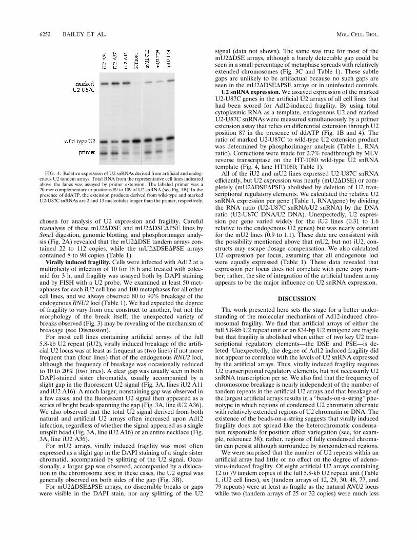
U2-U87C genes in the artificial U2 arrays of all cell lines thathad been scored for Ad12-induced fragility. By using totalcytoplasmic RNA as a template, endogenous U2 and markedU2-U87C snRNAs were measured simultaneously by a primerextension assay that relies on differential extension through U2position 87 in the presence of ddATP (Fig. 1B and 4). Theratio of marked U2-U87C to wild-type U2 extension productwas determined by phosphorimager analysis (Table 1, RNAratio). Corrections were made for 2.7% readthrough by MLVreverse transcriptase on the HT-1080 wild-type U2 snRNAtemplate (Fig. 4, lane HT1080; Table 1).All of the iU2 and mU2 lines expressed U2-U87C snRNA
efficiently, but U2 expression was nearly (mU2DDSE) or com-pletely (mU2DDSEDPSE) abolished by deletion of U2 tran-scriptional regulatory elements. We calculated the relative U2snRNA expression per gene (Table 1, RNA/gene) by dividingthe RNA ratio (U2-U87C snRNA/U2 snRNA) by the DNAratio (U2-U87C DNA/U2 DNA). Unexpectedly, U2 expres-sion per gene varied widely for the iU2 lines (0.31 to 1.6relative to the endogenous U2 genes) but was nearly constantfor the mU2 lines (0.9 to 1.1). These data are consistent withthe possibility mentioned above that mU2, but not iU2, con-structs may escape dosage compensation. We also calculatedU2 expression per locus, assuming that all endogenous lociwere equally expressed (Table 1). These data revealed thatexpression per locus does not correlate with gene copy num-ber; rather, the site of integration of the artificial tandem arrayappears to be the major influence on U2 snRNA expression.
DISCUSSION
The work presented here sets the stage for a better under-standing of the molecular mechanism of Ad12-induced chro-mosomal fragility. We find that artificial arrays of either thefull 5.8-kb U2 repeat unit or an 834-bp U2 minigene are fragilebut that fragility is abolished when either of two key U2 tran-scriptional regulatory elements—the DSE and PSE—is de-leted. Unexpectedly, the degree of Ad12-induced fragility didnot appear to correlate with the levels of U2 snRNA expressedby the artificial arrays. Thus, virally induced fragility requiresU2 transcriptional regulatory elements, but not necessarily U2snRNA transcription per se. We also find that the frequency ofchromosome breakage is nearly independent of the number oftandem repeats in the artificial U2 arrays and that breakage ofthe largest artificial arrays results in a ‘‘beads-on-a-string’’ phe-notype in which regions of condensed U2 chromatin alternatewith relatively extended regions of U2 chromatin or DNA. Theexistence of the beads-on-a-string suggests that virally inducedfragility does not spread like the heterochromatic condensa-tion responsible for position effect variegation (see, for exam-ple, reference 38); rather, regions of fully condensed chroma-tin can persist although surrounded by noncondensed regions.We were surprised that the number of U2 repeats within an
artificial array had little or no effect on the degree of adeno-virus-induced fragility. Of eight artificial U2 arrays containing12 to 79 tandem copies of the full 5.8-kb U2 repeat unit (Table1, iU2 cell lines), six (tandem arrays of 12, 29, 30, 48, 77, and79 repeats) were at least as fragile as the natural RNU2 locuswhile two (tandem arrays of 25 or 32 copies) were much less
FIG. 4. Relative expression of U2 snRNAs derived from artificial and endog-enous U2 tandem arrays. Total RNA from the representative cell lines indicatedabove the lanes was assayed by primer extension. The labeled primer was a20-mer complementary to positions 89 to 109 of U2 snRNA (see Fig. 1B). In thepresence of ddATP, the extension products derived from wild-type and markedU2-U87C snRNAs are 2 and 13 nucleotides longer than the primer, respectively.
6252 BAILEY ET AL. MOL. CELL. BIOL.
fragile (10 to 20 versus 80 to 90% breakage of RNU2). Simi-larly, Li et al. (21) found that an artificial array containing 60tandem copies of the full U2 repeat unit was only 60% asfragile as RNU2; however, the actual frequency of damage mayhave been underestimated in this case, because the array wasintegrated near or within a nucleolus organizer region forwhich virally induced fragility was difficult to score (21). Takentogether, these results suggest that the site of integration, andnot gene copy number, is the main determinant of fragility. Wealso suspect, as discussed below, that the fragility of artificialrepeats is substantially independent of U2 gene copy numberbecause the observed degree of fragility cannot increase oncethe probability of breakage approaches unity.Knowing that three and possibly all four of the specific
Ad12-induced fragile sites colocalize with clusters of abundant,ubiquitously expressed, small structural RNAs (14, 22, 23, 30,31, 35, 47, 48, 63), we suspected that the U2 snRNA gene itselfwould be necessary, and perhaps sufficient, for Ad12-inducedfragility. To test this idea, we generated tandem arrays of a U2minigene (mU2) containing only those sequences known to berequired for U2 snRNA expression—the 59 transcriptional reg-ulatory elements (2, 3, 27), the U2 snRNA coding region, andthe 39-end formation signal (1, 40). Yet after three indepen-dent ligations, transfections, and cell clonings, we obtainedonly three cell lines with mU2 arrays that were large enough tobe detected by FISH (Table 1, mU2 cell lines). Unexpectedly,these lines contained only five, six, or eight copies of the mU2repeat, almost an order of magnitude less than the copy num-bers routinely obtained for the full 5.8-kb U2 repeat unit;moreover, the chromosomes of the lines containing five or sixcopies of mU2 were 5 to 10% as fragile as the resident RNU2locus, and that of the line with eight copies was only marginallymore fragile (10 to 20%). Although these data demonstratethat the U2 minigene contains all the essential elements re-quired to generate an Ad12-inducible fragile site, the data alsoimply that the full 5.8-kb U2 repeat unit contains one or moreadditional accessory elements that increase the efficiency ofinduction or the stability of the array or that render the arti-ficial array substantially independent of chromosome positioneffects.To determine whether the known U2 snRNA transcriptional
regulatory elements play a role in Ad12-induced fragility, wegenerated derivatives of mU2 lacking one or both of the knowntranscriptional regulatory elements (2, 3, 27, 40, 44). The DSE,extending from positions 2231 to 2214 upstream of the tran-scription start site, binds adjacent Sp1 and Oct-1 factors andfunctions as a specialized enhancer sequence in collaborationwith the PSE. The PSE, extending from 261 to 241, binds aTATA-binding-protein-containing complex known as SNAPcand functions somewhat like a TATA box (40). Artificial arraysof the U2 minigene lacking both transcriptional regulatoryelements (mU2DDSEDPSE) were not fragile, but arrays lack-ing only the DSE (mU2DDSE) occasionally exhibited small,barely detectable gaps (Fig. 3C). The frequency of these gapsdid not correlate with the copy number of the mU2DDSErepeat unit in the artificial array. The gaps seen in mU2DDSEtandem arrays are unlikely to be artifactual; although Ad12infection can induce low levels of random chromosome dam-age, gaps were seen in as many as 5% of the mU2DDSEmetaphases but never in .100 mU2DDSEDPSE metaphases.These data clearly indicate that U2 transcriptional regulatoryelements are required for fragility, but, as discussed below, thisdoes not necessarily mean that fragility requires U2 transcrip-tion per se. Similar conclusions have been reached by Garganoet al. (13a).All of the artificial U2 arrays were constructed with the
marked U2-U87C snRNA gene. Although U2 snRNA is highlyconserved, position 87 is phylogenetically variable (61) and theU87C mutation would not be expected to have any effect onU2 snRNP assembly or function. U2-U87C snRNA could bedistinguished from endogenous U2 snRNA by a simple primerextension assay (Fig. 4), and this allowed us to correlate U2expression from the artificial arrays with virally induced fragil-ity. For the full 5.8-kb U2 repeat (iU2 cell lines), U2-U87CsnRNA expression per gene varied over a sixfold range from 30to 200% of the wild-type level; expression per iU2 locus wasalways greater than wild type and sometimes approached 10-fold-higher levels. For the U2 minigene (mU2 cell lines), U2-U87C snRNA expression was slightly greater than wild typeper gene but only 60 to 90% of wild type per locus. None of thepromoter deletion lines expressed significant levels of U2-U87C snRNA except four mU2DDSE lines (containing 22, 53,79, and 112 tandem repeats) in which expression approached5% of wild type. Deletion of the DSE had previously beenshown to result in similar levels of residual U2 snRNA expres-sion in a transient, heterologous assay (3), but this is the firstevidence that the DSE is required for efficient U2 expression ina stable, chromosomal context.No correlation between fragility of the various artificial ar-
rays and U2-U87C expression per gene or per locus could bediscerned (Table 1). This could suggest that U2 transcriptionalregulatory elements are required for fragility but not for activeU2 transcription by RNA polymerase II. Alternatively, themany U2 snRNA genes within a single array might not beequally active, perhaps reflecting the morphological distinctionbetween the beads and string (Fig. 3A; also see below). In thiscase, any correlation between transcription and fragility wouldbe masked when expression levels were averaged over activeand inactive genes. We are currently trying to determinewhether U2 snRNA transcription regulatory factors partici-pate directly in the molecular mechanism of Ad12-inducedfragility or indirectly by stimulating transcription, delaying rep-lication, or by affecting some other process that can interferewith chromatin condensation. Although our data are consis-tent with the interpretation that U2 transcription is requiredfor fragility, it is important to remember the multifunctionalnature of many key regulatory factors. For example, transcrip-tion factors have turned out to play a role in repair (10) andrecombination factors have been found to play a role in tran-scription (15). Indeed, work on systems as diverse as meioticrecombination in S. cerevisiae (58) and isotype class switchingin mammals (20) suggests that the ability of transcription fac-tors to stimulate or regulate recombination need not involvetranscription per se.The morphology of the artificial fragile sites may provide
some clues regarding the mechanism of virally induced fragil-ity. In both the mU2 and the iU2 arrays, virally induced chro-mosome damage can range from a nonstaining gap in onesister chromatid to an apparently complete break in both sisterchromatids as visualized by DAPI staining of the DNA. TheU2 gene signal is sometimes split by the gap (Fig. 3A, line iU2A24), it sometimes fills the gap (Fig. 3A, line iU2 A37), and, insome of the larger iU2 arrays, it appears as beads on a stringspanning the gap in the broken chromosome arm (Fig. 3A, lineiU2 A36). The invisible string represents regions of relativelynoncondensed chromatin separating bright beads or clusters ofmore highly condensed chromatin. Assuming that the con-densed chromosomes are approximately 1 mm in length over-all, the invisible strings may contain as little as 300 bp (0.1 mm)of relatively uncondensed DNA while each of the bright beadswould contain perhaps seven or eight U2 repeat units. Inter-estingly, the total U2 signal derived from the natural and ar-
VOL. 15, 1995 Ad12-INDUCED FRAGILITY OF THE HUMAN RNU2 LOCUS 6253
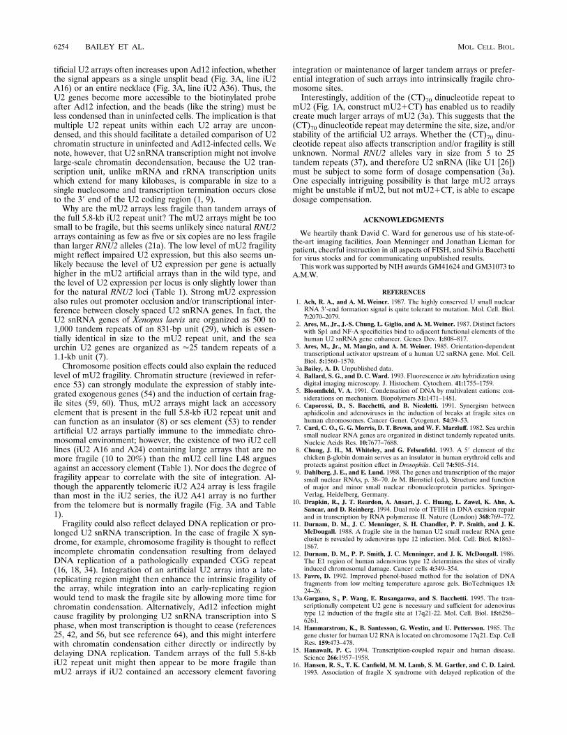
tificial U2 arrays often increases upon Ad12 infection, whetherthe signal appears as a single unsplit bead (Fig. 3A, line iU2A16) or an entire necklace (Fig. 3A, line iU2 A36). Thus, theU2 genes become more accessible to the biotinylated probeafter Ad12 infection, and the beads (like the string) must beless condensed than in uninfected cells. The implication is thatmultiple U2 repeat units within each U2 array are uncon-densed, and this should facilitate a detailed comparison of U2chromatin structure in uninfected and Ad12-infected cells. Wenote, however, that U2 snRNA transcription might not involvelarge-scale chromatin decondensation, because the U2 tran-scription unit, unlike mRNA and rRNA transcription unitswhich extend for many kilobases, is comparable in size to asingle nucleosome and transcription termination occurs closeto the 39 end of the U2 coding region (1, 9).Why are the mU2 arrays less fragile than tandem arrays of
the full 5.8-kb iU2 repeat unit? The mU2 arrays might be toosmall to be fragile, but this seems unlikely since natural RNU2arrays containing as few as five or six copies are no less fragilethan larger RNU2 alleles (21a). The low level of mU2 fragilitymight reflect impaired U2 expression, but this also seems un-likely because the level of U2 expression per gene is actuallyhigher in the mU2 artificial arrays than in the wild type, andthe level of U2 expression per locus is only slightly lower thanfor the natural RNU2 loci (Table 1). Strong mU2 expressionalso rules out promoter occlusion and/or transcriptional inter-ference between closely spaced U2 snRNA genes. In fact, theU2 snRNA genes of Xenopus laevis are organized as 500 to1,000 tandem repeats of an 831-bp unit (29), which is essen-tially identical in size to the mU2 repeat unit, and the seaurchin U2 genes are organized as '25 tandem repeats of a1.1-kb unit (7).Chromosome position effects could also explain the reduced
level of mU2 fragility. Chromatin structure (reviewed in refer-ence 53) can strongly modulate the expression of stably inte-grated exogenous genes (54) and the induction of certain frag-ile sites (59, 60). Thus, mU2 arrays might lack an accessoryelement that is present in the full 5.8-kb iU2 repeat unit andcan function as an insulator (8) or scs element (53) to renderartificial U2 arrays partially immune to the immediate chro-mosomal environment; however, the existence of two iU2 celllines (iU2 A16 and A24) containing large arrays that are nomore fragile (10 to 20%) than the mU2 cell line L48 arguesagainst an accessory element (Table 1). Nor does the degree offragility appear to correlate with the site of integration. Al-though the apparently telomeric iU2 A24 array is less fragilethan most in the iU2 series, the iU2 A41 array is no furtherfrom the telomere but is normally fragile (Fig. 3A and Table1).Fragility could also reflect delayed DNA replication or pro-
longed U2 snRNA transcription. In the case of fragile X syn-drome, for example, chromosome fragility is thought to reflectincomplete chromatin condensation resulting from delayedDNA replication of a pathologically expanded CGG repeat(16, 18, 34). Integration of an artificial U2 array into a late-replicating region might then enhance the intrinsic fragility ofthe array, while integration into an early-replicating regionwould tend to mask the fragile site by allowing more time forchromatin condensation. Alternatively, Ad12 infection mightcause fragility by prolonging U2 snRNA transcription into Sphase, when most transcription is thought to cease (references25, 42, and 56, but see reference 64), and this might interferewith chromatin condensation either directly or indirectly bydelaying DNA replication. Tandem arrays of the full 5.8-kbiU2 repeat unit might then appear to be more fragile thanmU2 arrays if iU2 contained an accessory element favoring
integration or maintenance of larger tandem arrays or prefer-ential integration of such arrays into intrinsically fragile chro-mosome sites.Interestingly, addition of the (CT)70 dinucleotide repeat to
mU2 (Fig. 1A, construct mU21CT) has enabled us to readilycreate much larger arrays of mU2 (3a). This suggests that the(CT)70 dinucleotide repeat may determine the site, size, and/orstability of the artificial U2 arrays. Whether the (CT)70 dinu-cleotide repeat also affects transcription and/or fragility is stillunknown. Normal RNU2 alleles vary in size from 5 to 25tandem repeats (37), and therefore U2 snRNA (like U1 [26])must be subject to some form of dosage compensation (3a).One especially intriguing possibility is that large mU2 arraysmight be unstable if mU2, but not mU21CT, is able to escapedosage compensation.
ACKNOWLEDGMENTS
We heartily thank David C. Ward for generous use of his state-of-the-art imaging facilities, Joan Menninger and Jonathan Lieman forpatient, cheerful instruction in all aspects of FISH, and Silvia Bacchettifor virus stocks and for communicating unpublished results.This work was supported by NIH awards GM41624 and GM31073 to
A.M.W.
REFERENCES
1. Ach, R. A., and A. M. Weiner. 1987. The highly conserved U small nuclearRNA 39-end formation signal is quite tolerant to mutation. Mol. Cell. Biol.7:2070–2079.
2. Ares, M., Jr., J.-S. Chung, L. Giglio, and A. M. Weiner. 1987. Distinct factorswith Sp1 and NF-A specificities bind to adjacent functional elements of thehuman U2 snRNA gene enhancer. Genes Dev. 1:808–817.
3. Ares, M., Jr., M. Mangin, and A. M. Weiner. 1985. Orientation-dependenttranscriptional activator upstream of a human U2 snRNA gene. Mol. Cell.Biol. 5:1560–1570.
3a.Bailey, A. D. Unpublished data.4. Ballard, S. G., and D. C. Ward. 1993. Fluorescence in situ hybridization usingdigital imaging microscopy. J. Histochem. Cytochem. 41:1755–1759.
5. Bloomfield, V. A. 1991. Condensation of DNA by multivalent cations: con-siderations on mechanism. Biopolymers 31:1471–1481.
6. Caporossi, D., S. Bacchetti, and B. Nicoletti. 1991. Synergism betweenaphidicolin and adenoviruses in the induction of breaks at fragile sites onhuman chromosomes. Cancer Genet. Cytogenet. 54:39–53.
7. Card, C. O., G. G. Morris, D. T. Brown, and W. F. Marzluff. 1982. Sea urchinsmall nuclear RNA genes are organized in distinct tandemly repeated units.Nucleic Acids Res. 10:7677–7688.
8. Chung, J. H., M. Whiteley, and G. Felsenfeld. 1993. A 59 element of thechicken b-globin domain serves as an insulator in human erythroid cells andprotects against position effect in Drosophila. Cell 74:505–514.
9. Dahlberg, J. E., and E. Lund. 1988. The genes and transcription of the majorsmall nuclear RNAs, p. 38–70. In M. Birnstiel (ed.), Structure and functionof major and minor small nuclear ribonucleoprotein particles. Springer-Verlag, Heidelberg, Germany.
10. Drapkin, R., J. T. Reardon, A. Ansari, J. C. Huang, L. Zawel, K. Ahn, A.Sancar, and D. Reinberg. 1994. Dual role of TFIIH in DNA excision repairand in transcription by RNA polymerase II. Nature (London) 368:769–772.
11. Durnam, D. M., J. C. Menninger, S. H. Chandler, P. P. Smith, and J. K.McDougall. 1988. A fragile site in the human U2 small nuclear RNA genecluster is revealed by adenovirus type 12 infection. Mol. Cell. Biol. 8:1863–1867.
12. Durnam, D. M., P. P. Smith, J. C. Menninger, and J. K. McDougall. 1986.The E1 region of human adenovirus type 12 determines the sites of virallyinduced chromosomal damage. Cancer cells 4:349–354.
13. Favre, D. 1992. Improved phenol-based method for the isolation of DNAfragments from low melting temperature agarose gels. BioTechniques 13:24–26.
13a.Gargano, S., P. Wang, E. Rusanganwa, and S. Bacchetti. 1995. The tran-scriptionally competent U2 gene is necessary and sufficient for adenovirustype 12 induction of the fragile site at 17q21-22. Mol. Cell. Biol. 15:6256–6261.
14. Hammarstrom, K., B. Santesson, G. Westin, and U. Pettersson. 1985. Thegene cluster for human U2 RNA is located on chromosome 17q21. Exp. CellRes. 159:473–478.
15. Hanawalt, P. C. 1994. Transcription-coupled repair and human disease.Science 266:1957–1958.
16. Hansen, R. S., T. K. Canfield, M. M. Lamb, S. M. Gartler, and C. D. Laird.1993. Association of fragile X syndrome with delayed replication of the
6254 BAILEY ET AL. MOL. CELL. BIOL.
FMR1 gene. Cell 73:1403–1409.17. Harnden, D. G., and H. P. Klinger (ed.). 1985. An international system for
human cytogenetic nomenclature. S. Karger, Basel.18. Knight, S. J. L., et al. 1993. Trinucleotide repeat amplification and hyper-
methylation of a CpG island in FRAXEmental retardation. Cell 74:127–134.19. Kunkel, T. A. 1985. Rapid and efficient site-specific mutagenesis without
phenotypic selection. Proc. Natl. Acad. Sci. USA 82:488–492.20. Leung, H., and N. Maizels. 1994. Regulation and targeting of recombination
in extrachromosomal substrates carrying immunoglobulin switch region se-quences. Mol. Cell. Biol. 14:1450–1458.
21. Li, Y.-P., R. Tomanin, J. R. Smiley, and S. Bacchetti. 1993. Generation of anew adenovirus type 12-inducible fragile site by insertion of an artificial U2locus in the human genome. Mol. Cell. Biol. 13:6064–6070.
21a.Li, Z. Unpublished results.22. Lindgren, V., M. Ares, Jr., A. M. Weiner, and U. Francke. 1985. Human
genes for U2 small nuclear RNA map to a major adenovirus 12 modificationsite on chromosome 17. Nature (London) 314:115–116.
23. Lindgren, V., L. B. Bernstein, A. M. Weiner, and U. Francke. 1985. HumanU1 small nuclear RNA pseudogenes do not map to the site of the U1 genesin 1p36 but are clustered in 1q12-22. Mol. Cell. Biol. 5:2172–2180.
24. Lund, E. 1988. Heterogeneity of human U1 snRNAs. Nucleic Acids Res.16:5813–5826.
25. Lund, E., and J. E. Dahlberg. 1992. Control of 4-8S RNA transcription at themidblastula transition in Xenopus laevis embryos. Genes Dev. 6:1097–1106.
26. Mangin, M., M. Ares, Jr., and A. M. Weiner. 1985. U1 small nuclear RNAgenes are subject to dosage compensation in mouse cells. Science 229:272–275.
27. Mangin, M., M. Ares, Jr., and A. M. Weiner. 1986. Human U2 small nuclearRNA genes contain an upstream enhancer. EMBO J. 5:987–995.
28. Mather, M. W. 1988. Base composition-independent hybridization in driedagarose gels: screening and recovery for cloning of genomic DNA fragments.BioTechniques 6:444–447.
29. Mattaj, I. W., and R. Zeller. 1983. Xenopus laevis snRNA genes: tandemlyrepeated transcription units showing 59 and 39 flanking homology with otherRNA polymerase II transcribed genes. EMBO J. 2:1883–1891.
30. McDougall, J. K. 1970. Effects of adenoviruses on the chromosomes ofnormal human cells and cells trisomic for an E group chromosome. Nature(London) 225:456–458.
31. McDougall, J. K. 1971. Spontaneous and adenovirus type 12 induced chro-mosome aberrations in Fanconi’s anemia fibroblasts. Int. J. Cancer 7:526–534.
32. McDougall, J. K., A. R. Dunn, and K. W. Jones. 1972. In situ hybridizationof adenovirus RNA and DNA. Nature (London) 236:346–348.
33. Miro, R., I. C. Clemente, C. Fuster, and J. Egozcue. 1987. Fragile sites,chromosome evolution and human neoplasia. Hum. Genet. 75:345–349.
34. Nancarrow, J. K., E. Kremer, K. Holman, H. Eyre, N. A. Doggett, D. LePaslier, D. F. Callen, G. R. Sutherland, and R. I. Richards. 1994. Implica-tions of FRA16A structure for the mechanism of chromosomal fragile sitegenesis. Science 264:1938–1941.
35. Naylor, S. L., B. U. Zabel, T. Manser, R. Gesteland, and A. Y. Sakaguchi.1984. Localization of human U1 small nuclear RNA genes to band p36.3 ofchromosome 1 by in situ hybridization. Somatic Cell Mol. Genet. 10:307–313.
36. Paranjape, S. M., R. T. Kamakaka, and J. T. Kadonaga. 1994. Role ofchromatin structure in the regulation of transcription by RNA polymerase II.Annu. Rev. Biochem. 63:265–297.
37. Pavelitz, T., L. Rusche, A. G. Matera, J. M. Scharf, and A. M. Weiner. 1995.Concerted evolution of the tandem array encoding primate U2 snRNAoccurs in situ, without changing the cytological context of the RNU2 locus.EMBO J. 14:169–177.
38. Pirrotta, V., and L. Rastelli. 1994. White gene expression, repressive chro-matin domains and homeotic gene regulation in Drosophila. Bioessays 16:549–556.
39. Romani, M., A. Baldini, E. V. Volpi, I. Casciano, C. Nobile, R. Muresu, andM. Siniscalco. 1994. Concurrent mapping of an adenovirus 5/SV40 integra-tion site and the U1 snRNA cluster (RNU1) within 400 kb of the chromo-some region 1p36.1. Cytogenet. Cell Genet. 67:37–40.
40. Sadowski, C. L., R. W. Henry, S. M. Lobo, and N. Hernandez. 1993. Tar-geting TBP to a non-TATA box cis-regulatory element: a TBP-containingcomplex activates transcription from snRNA promoters through the PSE.Genes Dev. 7:1535–1548.
41. Schramayr, S., D. Caporossi, I. Mak, T. Jelinek, and S. Bacchetti. 1990.Chromosomal damage induced by human adenovirus type 12 requires ex-pression of the E1B 55-kilodalton viral protein. J. Virol. 64:2090–2095.
42. Segil, N., S. B. Roberts, and N. Heintz. 1991. Mitotic phosphorylation of theOct-1 homeodomain and regulation of Oct-1 DNA binding activity. Science254:1814–1816.
43. Simonsen, C. C., and A. D. Levinson. 1983. Isolation and expression of analtered mouse dihydrofolate reductase cDNA. Proc. Natl. Acad. Sci. USA80:2495–2499.
44. Skuzeski, J. M., E. Lund, J. T. Murphy, T. H. Steinberg, R. R. Burgess, andJ. E. Dahlberg. 1984. Synthesis of human U1 RNA. II. Identification of tworegions of the promoter essential for transcription initiation at position 11.J. Biol. Chem. 259:8345–8352.
45. Smeets, D. F. C. M., and F. A. J. M. van de Klundert. 1990. Common fragilesites in man and three closely related primate species. Cytogenet. Cell Genet.53:8–14.
46. Smith, P. P., C. L. Friedman, E. M. Bryant, and J. K. McDougall. 1992. Viralintegration and fragile sites in human papillomavirus-immortalized humankeratinocyte cell lines. Genes Chromosomes Cancer 5:150–157.
47. Sorensen, P. D., B. Lomholt, S. Frederiksen, and N. Tommerup. 1991. Finemapping of human 5S rRNA genes to chromosome 1q42.11-q42.13. Cyto-genet. Cell Genet. 57:26–29.
48. Steffensen, D. M., P. Szabo, and J. K. McDougall. 1976. Adenovirus 12uncoiler regions of human chromosome 1 in relation to 5S rRNA genes. Exp.Cell Res. 100:436–439.
49. Sutherland, G., and F. Hecht. 1985. Oxford monographs on medical genet-ics, vol. 13. Fragile sites on human chromosomes. Oxford University Press,Oxford.
50. van Arsdell, S. W., and A. M. Weiner. 1984. Human genes for U2 smallnuclear RNA are tandemly repeated. Mol. Cell. Biol. 4:492–499.
51. Van Arsdell, S. W., and A. M. Weiner. 1984. Pseudogenes for human U2 donot have a fixed site of 39 truncation. Nucleic Acids Res. 12:1463–1471.
52. van der Drift, P., A. Chan, N. van Roy, G. Laureys, A. Westerveld, F.Speleman, and R. Versteeg. 1994. A multimegabase cluster of snRNA andtRNA genes on chromosome 1p36 harbours an adenovirus/SV40 hybridvirus integration site. Hum. Mol. Genet. 3:2131–2136.
53. Vazquez, J., and P. Schedl. 1994. Sequences required for enhancer blockingactivity of scs are located within two nuclease-hypersensitive regions. EMBOJ. 13:5984–5993.
54. von Knebel Doeberitz, M., T. Bauknecht, D. Bartsch, and H. zur Hausen.1991. Influence of chromosomal integration on glucocorticoid-regulatedtranscription of growth-stimulating papillomavirus genes E6 and E7 in cer-vical carcinoma cells. Proc. Natl. Acad. Sci. USA 88:1411–1415.
55. Westin, G., J. Zabielski, J. Hammarstrøm, H.-J. Monstein, C. Bark, and U.Pettersson. 1984. Clustered genes for human U2 RNA. Proc. Natl. Acad. Sci.USA 81:3811–3815.
56. White, R. J., T. M. Gottlieb, C. S. Downes, and S. P. Jackson. 1995. Mitoticregulation of a TATA-binding-protein-containing complex. Mol. Cell. Biol.15:1983–1992.
57. Withers-Ward, E. S., Y. Kitamura, J. P. Barnes, and J. M. Coffin. 1994.Distribution of targets for avian retrovirus DNA integration in vivo. GenesDev. 8:1473–1487.
58. Wu, T.-C., and M. Lichten. 1993. Meiosis-induced double-strand break sitesdetermined by yeast chromatin structure. Science 263:515–518.
59. Yunis, J. J., and W. R. Hoffman. 1990. Nuclear enzymes, fragile sites, andcancer. J. Gerontol. 44:37–44.
60. Yunis, J. J., and A. L. Soreng. 1984. Constitutive fragile sites and cancer.Science 226:1199–1204.
61. Zavanelli, M. I., J. S. Britton, A. H. Igel, and M. Ares, Jr. 1994. Mutationsin an essential U2 small nuclear RNA structure cause cold-sensitive U2 smallnuclear ribonucleoprotein function by favoring competing alternative U2RNA structures. Mol. Cell. Biol. 14:1689–1697.
62. Zhuang, Y., and A. M. Weiner. 1989. A compensatory base change in humanU2 snRNA can suppress a branch site mutation. Genes Dev. 3:1545–1552.
63. zur Hausen, H. 1967. Induction of specific chromosomal aberrations byadenovirus type 12 in human embryonic kidney cells. J. Virol. 1:1174–1185.
64. Zwerschke, W., H. W. Rottjakob, and H. Kuntzel. 1994. The Saccharomycescerevisiae CDC6 gene is transcribed at late mitosis and encodes an ATP/GTPase controlling S phase initiation. J. Biol. Chem. 269:23351–23356.
VOL. 15, 1995 Ad12-INDUCED FRAGILITY OF THE HUMAN RNU2 LOCUS 6255