advanced digestive endoscopy ercp
TRANSCRIPT

ADVANCED DIGESTIVE ENDOSCOPY: ERCP

ADVANCEDDIGESTIVEENDOSCOPY:ERCP
EDITED BY
PETER B. COTTONAND
JOSEPH LEUNG
© 2005 by Blackwell Publishing LtdBlackwell Publishing, Inc., 350 Main Street, Malden,Massachusetts 02148-5020, USABlackwell Publishing Ltd, 9600 Garsington Road,Oxford OX4 2DQ, UKBlackwell Publishing Asia Pty Ltd, 550 SwanstonStreet, Carlton, Victoria 3053, Australia
The right of the Author to be identified as the Authorof this Work has been asserted in accordance with theCopyright, Designs and Patents Act 1988.
All rights reserved. No part of this publication may be reproduced, stored in a retrieval system, ortransmitted, in any form or by any means, electronic,mechanical, photocopying, recording or otherwise,except as permitted by the UK Copyright, Designs andPatents Act 1988, without the prior permission of thepublisher.
First published 2005
Library of Congress Cataloging-in-Publication Data
Advanced digestive endoscopy: ERCP/edited by PeterCotton and Joseph Leung.
p. ; cm.Includes bibliographical references.ISBN-13: 978-1-4051-2079-1ISBN-10: 1-4051-2079-7
1. Endoscopic retrograde cholangiopancreatography.2. Gastroscopy. [DNLM: 1. Cholangiopancreatography,
Endoscopic Retrograde—methods. 2. DigestiveSystem Diseases. WI 750 A244 2005] I. Title: ERCP.II. Cotton, Peter B. III. Leung, J. W.C.
RC847.5.E53A38 2005616.3′07545—dc22
2005012661
ISBN-13: 978-1-4051-2079-1ISBN-10: 1-4051-2079-7
A catalogue record for this title is available from theBritish Library
Set in 10/131/2 Sabon by Graphicraft Limited, Hong KongPrinted and bound by Replika Press PVT Ltd,Harayana, India
Commissioning Editor: Alison BrownDevelopment Editor: Julie ElliottProduction Controller: Kate Charman
For further information on Blackwell Publishing, visitour website:hppt://www.blackwellpublishing.com
The publisher’s policy is to use permanent paper frommills that operate a sustainable forestry policy, andwhich has been manufactured from pulp processedusing acid-free and elementary chlorine-free practices.Furthermore, the publisher ensures that the text paperand cover board used have met acceptableenvironmental accreditation standards.
Contents
List of Contributors, vii
Preface, ix
1 ERCP OverviewaA 30-Year Perspective, 1Peter B. Cotton
2 ERCP Training, Competence, and Assessment, 9Peter B. Cotton
3 Fundamentals of ERCP, 17Joseph Leung
4 ERCP Communications, Recording, and Reporting, 81Peter B. Cotton
5 Common Bile Duct Stones and Cholangitis, 88Enders K.W. Ng and Sydney Chung
6 The Role of ERCP in Pancreatico-Biliary Malignancies, 120Gulshan Parasher and John G. Lee
7 Management of Postsurgical Bile Leaks and Bile Duct Strictures, 142Jacques J.G.H.M. Bergman
8 Sphincter of Oddi Dysfunction, 165Evan L. Fogel and Stuart Sherman
9 ERCP in Acute Pancreatitis, 199Martin L. Freeman
10 Endoscopy in Chronic Pancreatitis, 239Lee McHenry, Stuart Sherman, and Glen Lehman
v

11 Complications of Pancreatitis, 281Douglas A. Howell
12 ERCP in Children, 309Moises Guelrud
13 ERCP: Risks, Prevention, and Management, 339Peter B. Cotton
Index, 405
CONTENTSvi
vii
List of contributors
BERGMAN, JACQUES J.G.H.M.,Department of Gastroenterology and
Hepatology, Academic Medical Center Amsterdam, Meibergdreef 9, 1105 AZ Amsterdam, The
Netherlands
CHUNG, SYDNEY, Department of Surgery, Prince of Wales Hospital, The Chinese
University of Hong Kong, Shatin, NT, Hong Kong
COTTON, PETER B., Medical University of South Carolina, PO Box 250327, Ste 210
CSB, 96 Jonathan Lucas St, Charleston, SC 29425, USA
FOGEL, EVAN L., Indiana University Medical Center, 550 N. University Drive, Suite
4100, Indianapolis, IN 46202, USA
FREEMAN, MARTIN L., Hennepin County Medical Center, GI Division, 701 Park
Avenue, Minneapolis, MN 55415, USA
GUELRUD, MOISES, New England Medical Center, 750 Washington Street, Booth 213,
Boston, MA 02111, USA
HOWELL, DOUGLAS A., Portland Endoscopy Center, 1200 Congress Street #300,
Portland, ME 04102, USA
LEE, JOHN G., University of California Irvine, Division of Gastroenterology, 101 The City
Drive, Bldg 53, Rm 113, Orange, CA 92817, USA
LEHMAN, GLEN, Indiana University Medical Center, 550 N. University Blvd, Rm 4100,
Indianapolis, IN 46202, USA
LEUNG, JOSEPH, Division of GI UC Davis, 4150 V Street, Ste 3500, PSSB, Sacramento,
CA 95817, USA
MCHENRY, LEE, Indiana University Medical Center, 550 N. University Drive, Suite
4100, Indianapolis, IN 46202, USA

NG, ENDERS K.W., Upper GI Division, Department of Surgery, The Chinese University
of Hong Kong, Hong Kong
PARASHER, GULSHAN, Division of Gastroenterology and Hepatology, University of
New Mexico, Albuquerque NM87131-0001, New Mexico
SHERMAN, STUART, Indiana University Medical Center, 550 N. University Drive,
Suite 4100, Indianapolis, IN 46202, USA
LIST OF CONTRIBUTORSviii

Preface
There was a time, long ago, when endoscopy was a small off-shoot of gastroen-terology, and when most of what budding endoscopists needed to know couldbe covered in a slim book. Thus Practical Gastrointestinal Endoscopy was con-ceived by Christopher Williams and myself over 25 years ago, and had a success-ful run through four editions. The field has expanded enormously over thattime. The number and variety of procedures, and the relevant scientific liter-ature, have proliferated, and there is now a hierarchy within endoscopy. Thereare ‘standard’ procedures which most clinical gastroenterologists master duringtheir training. These constitute routine upper endoscopy and colonoscopy, withtheir common therapeutic aspects, which may be needed at work every day (andsome nights). Then there are recognized ‘advanced’ procedures, such as ERCPand EUS, and the more adventurous therapeutic aspects of upper endoscopy and colonoscopy, such as fundoplication, EMR, and tumor ablation. These arepracticed by only a small percentage of endoscopists, who need more focusedand intensive training. In addition, for a few of the leaders, there is much to belearned in related fields, such as unit design, management, teaching, and qualityimprovement. It is clear that no one person (or two) can speak or write about allof this territory with any authority. Advice and instruction are best given byacknowledged experts in each specific area.
My publishing journey reflects these changes. Thus, the latest (5th) Edition of Practical Gastrointestinal Endoscopy, sub-titled ‘The Fundamentals’, pub-lished in 2003, is devoted solely to the basic facts which all trainees need in theirfirst year or two. It is accompanied by 2 practical CDRoms, one devoted to each‘end’. We removed all of the ‘advanced stuff ’, such as ERCP, teaching methods,and unit management.
We then sought to serve the needs of the established endoscopists, and ofthose learning more advanced aspects, with a new series called ‘AdvancedDigestive Endoscopy’. Reflecting the acceleration of our world, we saw this pri-marily as a virtual ‘ebook’, presented electronically for speed of posting and foreasy updating. This is now evolving on the comprehensive Blackwell Publishingwebsite www.gastrohep.com. It has 5 separate sections:aEndoscopic Practice
ix
and Safety, Upper Endoscopy, Colonoscopy, ERCP, and EUS. I was delighted tobe joined in this endeavor by new partners; Joseph Leung, Joseph Sung, JerryWaye, and Rob Hawes. Between us we have persuaded over 40 distinguishedcolleagues from all over the world to make contributions.
Despite the multiple benefits of electronic publishing, there is still a demandfor print books. Jerry Waye’s book on Colonoscopy, co-edited with Doug Rexand Christopher Williams, is already in print (the ebook version consists of aselection of those chapters).
Here we present the print version of ERCP. I am enormously grateful toJoseph Leung and to the 12 other contributors who have labored long and hardto bring it to fruition. The fact that most of the authors are based in the USAshould not be misinterpreted, for the expertise and methods of ERCP are nowtruly international. The electronic version will continue, and will be updatedevery year or so. We welcome your criticism and suggestions for improvement.
Joseph and I offer our sincere thanks to our families for their support andforbearance, and to our colleagues and trainees who have taught us so much,not least how much we still have to learn.
Peter B Cotton MD FRCP FRCS February 2005Digestive Disease Center, Medical University of South Carolina, Charleston, USA
PREFACEx
CHAPTER 1
ERCP OverviewcA 30-Year Perspective
PETER B. COTTON
Historical background
Endoscopic cannulation of the papilla of Vater was first reported in 1968 [1].However, it was really put on the map shortly afterwards by several Japa-nese groups, working with instrument manufacturers to develop appropriatelong side-viewing instruments [2–5]. The technique (initially called ECPGaendoscopic cholangiopancreatographyain Japan) spread throughout Europe inthe early 1970s [6–13]. Early efforts were much helped by a multinationalworkshop at the European Congress in Paris in 1972, organized by the Olympuscompany. ERCP rapidly became established worldwide as a valuable diagnostictechnique, although doubts were expressed in the USA about its feasibility androle [14], and the potential for serious complications soon became clear [15–18]. ERCP was given a tremendous boost by the development of its therapeuticapplications, notably biliary sphincterotomy in the mid-1970s [19–21] and biliary stenting 5 years later [22,23].
It is difficult for most gastroenterologists today to imagine the diagnosticand therapeutic situation 30 years ago. There were no scans. Biliary obstructionwas diagnosed and treated surgically, with substantial operative mortality. Non-operative documentation of biliary pathology by ERCP was a huge step forward.Likewise, ERCP was an amazing development in pancreatic investigation at atime when the only available test was laparotomy. The ability to ‘see into’ thepancreas, and to collect pure pancreatic juice [24], seemed like a miracle. Weassumed that ERCP would have a dramatic impact on chronic pancreatitis andpancreatic cancer. Sadly, these expectations are not yet realized, but endoscopicmanagement of biliary obstruction was clearly a major clinical advance, espe-cially in the sick and elderly. The period of 15 or so years from the mid-1970sreally constituted a ‘golden age’ for ERCP. Despite significant risks [25], it wasobvious to everyone that ERCP management of duct stones and tumors was easier, cheaper, and safer than available surgical alternatives. Large series werepublished, including some randomized trials [26–31]. Percutaneous transhe-patic cholangiography (PTC) and its drainage applications were also developed
1
during this time, but were used (with the exception of a few units) only whenERCP failed or was not available. The ‘combined procedure’aendoscopic can-nulation over a guidewire placed at PTC [32,33]abecame popular for a while,but was needed less as both endoscopic and interventional techniques improved.
The changing world of pancreatico-biliary medicine
The situation has changed in many ways during the last two decades. ERCP hasevolved significantly, but so have many other relevant techniques.
The impact of scanning radiology
Imaging modalities for the biliary tree and pancreas have proliferated. Highquality ultrasound, computed tomography, endoscopic ultrasonography, andMR scanning (with MRCP) have greatly facilitated the non-invasive evaluationof patients with known and suspected biliary and pancreatic disease. As a result,the proportion of ERCP examinations now performed purely for diagnosticpurposes has diminished significantly. However, it remains a very accurate dia-gnostic tool, and continues to shed important light in selected cases where all ofthe non-invasive tests have been inconclusive.
Extending the indications for therapeutic ERCP
The second major change has been the attempt of ERCP practitioners to extendtheir therapeutic territory from standard biliary procedures into more complexareas such as pancreatitis and suspected sphincter of Oddi dysfunction. Thevalue of ERCP in these contexts remains controversial [34].
Improvements in surgery
The third major change is the substantial and progressive reduction in risk asso-ciated with conventional surgery (due to excellent perioperative and anesthesiacare), and the increasing use of less invasive laparoscopic techniques [35]. It is nolonger correct to assume that ERCP is always safer than surgery. Sadly, seriouscomplications of ERCP (especially pancreatitis and perforation) continue tooccur, especially during speculative procedures performed by inexperiencedpractitioners, often using the needle-knife for lack of standard expertise [36].
Risk reduction
These facts are forcing the ERCP community to search for ways to reduce the risks. Important examples of this preoccupation are the focus on refining
CHAPTER 12
indications [34], prospective studies of predictors of adverse outcomes [37], and attempts to remove stones from the bile duct without sphincterotomy [38],at least in younger patients with relatively small stones and normal sized ducts.
Patient empowerment
Another important driver in this field is the increased participation of patients in decisions about their care. Patients are rightly demanding the data on thepotential benefits, risks, and limitations of ERCP, and the same data about thealternatives. Report cards are one response [39].
Current focus
The focus in the early twenty-first century is on careful evaluation of what ERCPcan offer (in comparison with available alternatives), and on attempts toimprove the overall quality of ERCP practice [40]. Equally important is theincreasing focus on who should be trained, and to what level of expertise. Howmany ERCPists are really needed? (See Chapter 2.)
These issues are important in all clinical contexts, but come into clearestfocus where ERCP is still considered somewhat speculative, e.g. in the manage-ment of chronic pancreatitis and of possible sphincter of Oddi dysfunction [34].
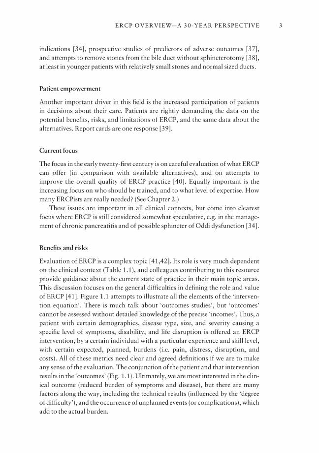
Benefits and risks
Evaluation of ERCP is a complex topic [41,42]. Its role is very much dependenton the clinical context (Table 1.1), and colleagues contributing to this resourceprovide guidance about the current state of practice in their main topic areas.This discussion focuses on the general difficulties in defining the role and valueof ERCP [41]. Figure 1.1 attempts to illustrate all the elements of the ‘interven-tion equation’. There is much talk about ‘outcomes studies’, but ‘outcomes’ cannot be assessed without detailed knowledge of the precise ‘incomes’. Thus, apatient with certain demographics, disease type, size, and severity causing aspecific level of symptoms, disability, and life disruption is offered an ERCPintervention, by a certain individual with a particular experience and skill level,with certain expected, planned, burdens (i.e. pain, distress, disruption, andcosts). All of these metrics need clear and agreed definitions if we are to makeany sense of the evaluation. The conjunction of the patient and that interventionresults in the ‘outcomes’ (Fig. 1.1). Ultimately, we are most interested in the clin-ical outcome (reduced burden of symptoms and disease), but there are many factors along the way, including the technical results (influenced by the ‘degreeof difficulty’), and the occurrence of unplanned events (or complications), whichadd to the actual burden.
ERCP OVERVIEWaA 30-YEAR PERSPECTIVE 3

CHAPTER 14
Biliary• Jaundice• Abnormal LFTs• Suspected/known duct stone
Pancreatitis• Chronic• Acute gallstone related• Idiopathic recurrent• Complicated
Pain• Chronic• Acute intermittent (includes postcholecystectomy)• Early postsurgical
Imaging findings (papilla, pancreas, biliary)
Stent service
Other
Intervention Operator Planned cost
Patient DemographicsIllness burden disease type/stage symptoms life disturbance health care use
Comorbidities (risk)
Difficulty Technicalsuccess
Clinicalsuccess
Value
Actualcosts
Unplannedevents
SatisfactionExpectation
Incomes Outcomes
Fig. 1.1 The intervention process: data elements required.
Table 1.1 Clinical contexts forpossible ERCP use.
Unplanned events
The word ‘complication’ is emotive, raising issues of medical error and legal liability. We prefer to discuss ‘unplanned events’, since they are best describedsimply as deviations from the plan which had been agreed with the patient. The phrase ‘adverse events’ has been used too, but not all unplanned events are
negative. A patient with suspected cancer may be delighted to wake up from aprocedure with an unexpected cure (sphincterotomy and stone removal). Allunplanned events should be documented in a standard format, as an aid toefforts at quality improvement. Some are relatively trivial, such as transienthypotension or self-limited bleeding. At what level of severity do they become‘complications’? An influential consensus conference [43] set the threshold atthe need for hospital admission, and defined levels of severity by the length ofstay, as well as the need for surgery or intensive care. Details of complications,and their avoidance and management, are addressed in Chapter 13.
Clinical success and value
Clinical success may sometimes be relatively obvious, e.g. removal of a stone orrelief of jaundice with a stent. However, in many cases (e.g. chronic pancreatitis,sphincter dysfunction), the judgement can be made only after long periods offollow-up. This greatly complicates evaluation studies in just the clinical cir-cumstances where the knowledge is needed most. Patient satisfaction is anotherimportant parameter. It is determined partly by the clinical results (and how thatcompares with the patient’s expectation), but also by patients’ perception of theprocess (accessibility, courtesy, etc.). The cost (burden) of the intervention isobviously a key consideration. This consists of the planned burden, plus theresult of any unplanned events. The ratio between the clinical impact (benefit)and the burden (cost) determines the ‘value’ of the procedure in that individualpatient (Fig. 1.1). Attempts to provide definitions for all of these metrics areadvancing slowly. Their incorporation in endoscopy reporting databases willallow ongoing useful outcomes evaluations to guide further decisions. If thesame or similar metrics are also used by those performing alternative interven-tions such as surgery, we will obtain a clearer idea of the relative roles of these dif-ferent procedures [44]. In some cases randomization will be necessary to make afinal judgement. However, the issue of ‘operator dependence’ will always exist.A randomized trial of two techniques performed by experts may not be the bestguide to the choice of intervention in everyday community practice.
The future
The trends which we have outlined are likely to continue and to accelerate in thecoming years. Quality is the big issue. That means making sure that we are doingthe right things, and doing them right. It has been clear for a long time (but isonly now becoming generally accepted) that ERCP is a procedure that should beundertaken only by a minority of gastroenterologists. The amount of trainingand continuing dedication in practice needed to attain and maintain high levels
ERCP OVERVIEWaA 30-YEAR PERSPECTIVE 5
of competence, and to improve, means that the procedures should be focused inrelatively few hands. The increasing variety and safety of alternative procedures,and the vigilance of our customers, will drive that agenda. The other imperativeis to pursue the research studies necessary to improve current methods, and toevaluate all of them rigorously. This is best performed in collaboration with col-leagues in surgery and radiology to establish the best methods for approachingpatients with known or suspected biliary and pancreatic disease. The dynamicsbetween specialists will change with time, which is one excellent reason fororganizing care to be patient-focused, rather than in traditional technical silos.Multidisciplinary organizations, like our Digestive Disease Center, attempt toprovide that perspective and a platform for the unbiased research and educationthat aim to improve the quality of service [45].
References
1 McCune WS, Shorb PE, Moscovitz H. Endoscopic cannulation of the ampulla of Vater: a pre-liminary report. Ann Surg 1968; 167: 752–6.
2 Oi I, Takemoto T, Kondo T. Fiberduodenoscope: direct observations of the papilla of Vater.Endoscopy 1969; 1: 101–3.
3 Ogoshi K, Tobita Y, Hara Y. Endoscopic observation of the duodenum and pancreatocholedo-chography using duodenofiberscope under direct vision. Gastrointest Endosc 1970; 12: 83–96.
4 Takagi K, Ideda S, Nakagawa Y, Sakaguchi N, Takahashi T, Kumakura K et al. Retrogradepancreatography and cholangiography by fiber-duodenoscope. Gastroenterology 1970; 59:445–52.
5 Kasugai T, Kuno N, Aoki I, Kizu M, Kobayashi S. Fiberduodenoscopy: analysis of 353 examina-tions. Gastrointest Endosc 1971; 18: 9–16.
6 Classen M, Koch H, Fruhmorgen P, Grabner W, Demling L. Results of retrograde pancreatico-graphy. Acta Gastroenterologica Japonica 1972; 7: 131–6.
7 Cotton PB. Progress report: cannulation of the papilla of Vater by endoscopy and retrogradecholangiopancreatography (ERCP). Gut 1972; 13: 1014–25.
8 Cotton PB, Salmon PR, Blumgart LH, Burwood RJ, Davies GT, Lawrie BW et al. Cannulationof papilla of Vater via fiber-duodenoscope: assessment of retrograde cholangiopancreatographyin 60 patients. Lancet 1972; 1: 53–8.
9 Gulbis A, Cremer M, Engelholm L. La cholangiographie et la wirsungographic endoscopiques.Acta Endoscopica Radiocinematogr 1972; 2: 78–80.
10 Heully F, Gaucher P, Laurent J, Vicari F, Fays J, Bigard M-A, Jenpierre R. La duodenoscopie etla catheterisme de voies biliares et pancreatiques. Nouv Presse Med 1972; 1: 313–18.
11 Safrany L, Tari J, Barna L, Torok I. Endoscopic retrograde cholangiography: experience of 168examinations. Gastrointest Endosc 1973; 19: 163–8.
12 Liguory C, Gouero H, Chavy A, Coffin JC, Huguier M. Endoscopic retrograde cholangiopan-creatography. Br J Surg 1974; 61: 359–62.
13 Cotton PB. ERCP. Gut 1977; 18: 316–41.14 Morrissey JF. To cannulate or not to cannulate [Editorial]. Gastroenterology 1972; 63: 351–2.15 Blackwood WD, Vennes JA, Silvis SE. Post-endoscopy pancreatitis and hyperamylasuria.
Gastrointest Endosc 1973; 20: 56–8.16 Classen M, Demling L. Hazards of endoscopic retrograde cholangio-pancreaticography
(ERCP). Acta Hepatogastroenterol (Stutt) 1975; 22: 1–3.17 Nebel OT, Silvis SE, Rogers G, Sugawa C, Mandelstam P. Complications associated with endo-
scopic retrograde cholangio-pancreatography: results of the 1974 A/S/G/E survey. GastrointestEndosc 1975; 22: 34–6.
CHAPTER 16
18 Bilbao MK, Dotter CT, Lee TG, Katon RM. Complications of endoscopic retrograde cholan-giopancreatography (ERCP): a study of 10 000 cases. Gastroenterology 1976; 70: 314–20.
19 Classen M, Demling L. Endoskopische sphinkterotomie der papilla Vateri und steinextraktionaus dem ductus choledochus. Dtsch Med Wochenschr 1974; 99: 496–7.
20 Kawai K, Akasaka Y, Murakami K, Tada M, Kohill Y, Nakajima M. Endoscopic sphinctero-tomy of the ampulla of Vater. Gastrointest Endosc 1974; 20: 148–51.
21 Cotton PB, Chapman M, Whiteside CG, LeQuesne LP. Duodenoscopic papillotomy and gall-stone removal. Br J Surg 1976; 63: 709–14.
22 Soehendra N, Reijnders-Frederix V. Palliative bile duct drainage: a new endoscopic method ofintroducing a transpapillary drain. Endoscopy 1980; 12: 8–11.
23 Laurence BH, Cotton PB. Decompression of malignant biliary obstruction by duodenoscopeintubation of the bile duct. Br Med J 1980; I: 522–3.
24 Robberrecht P, Cremer M, Vandermers A, Vandermers-Piret M-C, Cotton PB, de Neef P et al.Pancreatic secretion of total protein and three hydrolases collected in healthy subjects via duo-denoscopic cannulation: effects of secretin, pancreozymin and caerulein. Gastroenterology1975; 69: 374–9.
25 Byrne P, Leung JWC, Cotton PB. Retroperitoneal perforation during duodenoscopic sphinc-terotomy. Radiology 1984; 150: 383–4.
26 Vaira D, Ainley C, Williams S, Caines S, Salmon P, Russell C et al. Endoscopic sphincterotomyin 1000 consecutive patients. Lancet 1989; 2: 431–4.
27 Cotton PB. Endoscopic management of bile duct stones (apples and oranges). Gut 1984; 25:587–97.
28 Leung JWC, Emery R, Cotton PB, Russell RCG, Vallon AG, Mason RR. Management of malig-nant obstructive jaundice at The Middlesex Hospital. Br J Surg 1983; 70: 584–6.
29 Cotton PB. Endoscopic methods for relief of malignant obstructive jaundice. World J Surg 1984;8: 854–61.
30 Speer AG, Cotton PB, Russell RCG, Mason RR, Hatfield ARW, Leung JWC et al. Randomizedtrial of endoscopic versus percutaneous stent insertion in malignant obstructive jaundice. Lancet1987; 2: 57–62.
31 Smith AC, Dowsett JF, Russell RCG, Hatfield ARW, Cotton PB. Randomised trial of endo-scopic stenting versus surgical bypass in malignant low bile duct obstruction. Lancet 1994; 344:1655–60.
32 Shorvon PJ, Cotton PB, Mason RR, Siegel HJ, Hatfield ARW. Percutaneous transhepatic assis-tance for duodenoscopic sphincterotomy. Gut 1985; 26: 1373–6.
33 Dowsett JF, Vaira D, Hatfield AR, Cairns SR, Polydorou A, Frost R et al. Endoscopic biliarytherapy using the combined percutaneous and endoscopic technique. Gastroenterology 1989;96: 1180–6.
34 Cohen S, Bacon BR, Berlin JA, Fleischer D, Hecht GA, Loehrer PJ et al. NIH State of the ScienceConference Statement: ERCP for diagnosis and therapy. Gastrointest Endosc 2002; 56: 803–9.
35 Cotton PB, Chung SC, Davis WZ, Gibson RM, Ransohoff DF, Strasberg SM. Issues in cholecys-tectomy and management of duct stones. Am J Gastroenterol 1994; 89: S169–76.
36 Cotton PB. ERCP is most dangerous for people who need it least. Gastrointest Endosc 2001; 54:535–6.
37 Freeman ML, DiSario JA, Nelson DB, Fennerty MB, Lee JG, Bjorkman DJ et al. Risk factors forpost-ERCP pancreatitis: a prospective, multicenter study. Gastrointest Endosc 2001; 54:425–34.
38 Huibregtse K. Endoscopic balloon dilation for removal of bile duct stones: special indicationsonly. Endoscopy 2001; 33 (7): 620–2.
39 Cotton PB. How many times have you done this procedure, Doctor? Am J Gastroenterol 2002;97: 522–3.
40 Quality and Outcome Assessment in Gastrointestinal Endoscopy. Gastrointest Endosc 2000;52: 827–30.
41 Cotton PB. Income and outcome metrics for objective evaluation of ERCP and alternative meth-ods. Gastrointest Endosc 2002; 56 (Suppl. 6): S283–90.
ERCP OVERVIEWaA 30-YEAR PERSPECTIVE 7

42 Cotton PB. Therapeutic gastrointestinal endoscopy: problems in proving efficacy. N Engl J Med1992; 326: 1626–8.
43 Cotton PB, Lehman G, Vennes J, Geenen JE, Russell RCG, Meyers WC et al. Endoscopicsphincterotomy complications and their management: an attempt at consensus. GastrointestEndosc 1991; 37: 383–93.
44 Cotton PB. Randomization is not the (only) answer: a plea for structured objective evaluation ofendoscopic therapy. Endoscopy 2000; 32: 402–5.
45 Cotton PB. Fading boundary between gastroenterology and surgery. J Gastroenterol Hepatol2000; 15: G34–7.
CHAPTER 18
CHAPTER 2
ERCP Training, Competence, and Assessment
PETER B. COTTON
ERCP is challenging, and not for all gastroenterologists
ERCP is the most challenging endoscopic procedure performed regularly by gas-troenterologists. It is often difficult technically, and may fail. Optimal practicerequires considerable manual dexterity, a broad knowledge of pancreatic andbiliary diseases, and familiarity with the many alternative diagnostic and thera-peutic approaches. Furthermore, it carries substantial risks, even in the hands ofexperts [1,2].
ERCP has been seen also as rather glamorous, so that most gastroenterologytrainees have aspired to master the techniques, and to practice them indepen-dently. Many factors make that inappropriate. Firstly, it has become obvious (asdetailed below) that attaining competence takes far more training and experi-ence than previously appreciated. This is time consuming, and also detractsfrom time needed to study other specialist fields of gastroenterology and hepatol-ogy. Secondly, the increasing refinement and availability of imaging techniquessuch as CT scanning, MRCP, and EUS have rendered diagnostic ERCP to be(almost) obsolete [1]. This means that any endoscopist offering ERCP must begeared up to provide therapy for the likely problem. Thirdly, it is now clear thatless experienced practitioners have more failures, and also have more complica-tions. Fourthly, many ERCP endoscopists have been trained (albeit not all verywell) in the last two decades, and very few more are needed each year to main-tain the ranks. Finally, consumer empowerment will be an important driver.Patients are beginning to understand that not all endoscopists are alike, and are seeking out experienced practitioners when they need more aggressive procedures.
All of these facts mandate that only a few people should be trained, and thatthey should be trained well. This is far from a new idea, having been statedclearly and repeatedly over the years by many individuals [3–7] and endoscopyorganizations [8–14]. The problem is that no one has paid attention, as is bru-tally obvious from a recent survey of 69 graduates from US fellowship programs[15]. Most had had some experience of ERCP (range 12–320 cases, median 140).
9
One-third stated that their training was inadequate, yet 91% of them proposedto practice ERCP. This is bad medicine, and embarrassing for our profession[16]. We must ensure that those offering ERCP services are competent to do so.
What is ‘competence’ in ERCP?
There is a wide spectrum of expertise in the performance of ERCP. Competencetraditionally describes the point at which a trainee can practice independently.What are the criteria for independent practice? Sadly, our understanding of the complexity of that issue has been slow to develop, and opinions vary widely[17]. Only now are attempts being made to develop meaningful objective methods of assessment.
Issues of training, competence, and assessment for all aspects of endoscopyhave been well reviewed recently by Cohen [18] and Freeman [19].
The first ASGE guidelines for ERCP relied almost solely on the numbers ofcases experienced during training, and suggested that 100 (including 25 thera-peutic) would be adequate [8]. That guideline attempted to put the onus on thetraining program directors, suggesting that they should not be asked to advise orto arbitrate competency until those ‘threshold’ numbers had been reached. Butthis sensible concept was ignored, and formal assessments were rare events.Even when logbooks became routine, it was difficult to assess what contributionthe trainee had made (or indeed could have made independently).
A study of the learning curve for ERCP at Duke University was a turningpoint in the debate. Even after 180–200 cases, trainees were scarcely performingat an 80% level [20].
The latest guideline from the ASGE in 2002 [21] mentions that 200 proce-dures are not adequate for most trainees to achieve competence, and emphasizesobjective end points (such as an 80% biliary cannulation rate) as better minimalstandards. The Australians have set the highest hurdle so far, i.e. completion of200 procedures, unassisted [22]. The British authorities suggested a 90% hurdlein 1999 [13], but the 2004 version [23] replaced numbers completely in favor of a list of needed skills (without precise goals), stating rather quaintly that‘although trainees must aspire to internationally accepted standards for cannula-tion successaa 90% success rate for uncomplicated cases has been proposedait is unreasonable to demand this level of performance from trainees by the endof their training . . .’.
Whilst these concepts and guidelines are logical and well-meaning, therehave been few attempts so far to document what skill levels are really beingachieved. Nor do we know how performance in the training environment trans-lates into independent practice. It is one thing to complete a procedure in thetraining environment with faculty advice and encouragement, and familiar
CHAPTER 210
assistants and equipment, but quite another to do so unaided in a new unfamil-iar environment, with pressure to succeed. We need to collect meaningful objec-tive data during training, but also in the early phases of practice.
Cognitive competence
The safe and effective practice of ERCP clearly requires far more than technicalskills, as has been well stated repeatedly. Documenting technical competence isdifficult, but proving the acquisition of the necessary cognitive skills may beeven more so [24]. It has been assumed that formal training in Gastroenterologyand Hepatology (e.g. Board certification in the USA) is likely to cover the neces-sary territory [25], but the specifics of pancreaticobiliary medicine have not been assessed formally. Furthermore, the field is in constant flux and requiresongoing study.
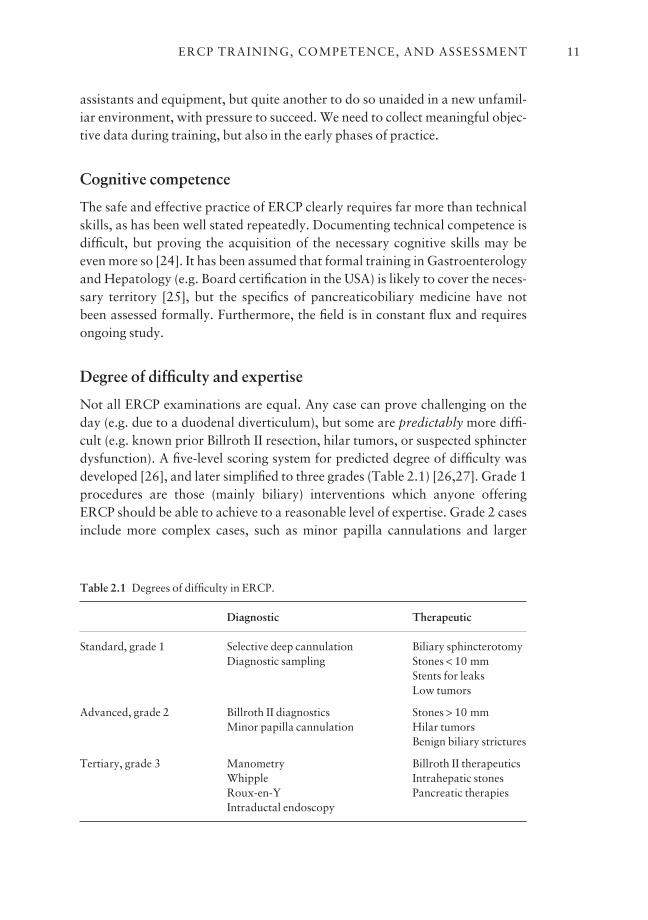
Degree of difficulty and expertise
Not all ERCP examinations are equal. Any case can prove challenging on theday (e.g. due to a duodenal diverticulum), but some are predictably more diffi-cult (e.g. known prior Billroth II resection, hilar tumors, or suspected sphincterdysfunction). A five-level scoring system for predicted degree of difficulty wasdeveloped [26], and later simplified to three grades (Table 2.1) [26,27]. Grade 1procedures are those (mainly biliary) interventions which anyone offeringERCP should be able to achieve to a reasonable level of expertise. Grade 2 casesinclude more complex cases, such as minor papilla cannulations and larger
ERCP TRAINING, COMPETENCE, AND ASSESSMENT 11
Table 2.1 Degrees of difficulty in ERCP.
Diagnostic Therapeutic
Standard, grade 1 Selective deep cannulation Biliary sphincterotomyDiagnostic sampling Stones < 10 mm
Stents for leaksLow tumors
Advanced, grade 2 Billroth II diagnostics Stones > 10 mmMinor papilla cannulation Hilar tumors
Benign biliary strictures
Tertiary, grade 3 Manometry Billroth II therapeuticsWhipple Intrahepatic stonesRoux-en-Y Pancreatic therapiesIntraductal endoscopy

stones. Grade 3 procedures are the most difficult, such as treatments for pan-creatitis and intrahepatic stones, and are performed mainly in tertiary referralcenters.
The above discussion about competence refers primarily to grade 1 proce-dures, which are the ‘garden-variety’ cases that will be encountered in everydaypractice. Endoscopists with more training (e.g. a dedicated fourth year in theUSA), and those who have honed their skills in practice with the aid of com-munity and academic colleagues, will attempt more complex cases. So-calledexperts, working in referral centers, will tackle all comers, but will also havevery high success rates in the easier cases. These concepts of case difficulty andindividual expertise can usefully be combined (Table 2.2).
ERCP training at MUSC
Our trainees select from three levels of training in pancreatico-biliary medicineand ERCP. The simplest is exposure to the service for 2 months, which showsthem approximately 80 cases, and the thinking that goes with them. They learnto use side-viewing endoscopes, but are not expected to perform ERCP. The second level is offered to selected fellows in the GI training program (which lasts3 years). They experience over 300 cases and appear reasonably competent instandard (mainly biliary) procedures when they leave. The third option requiresa dedicated fourth year, with another 300+ cases. These endoscopists have mas-tered standard grade 1 cases, and know enough to attempt some of the morecomplex procedures.
Towards more structured training
Together, all of these issues in training and assessment point to the need for amuch more structured approach, including formalized curricula and enhancededucational resources. The need to be personally involved in so many live casescould be reduced substantially in the future as computer simulators mature andbecome more widely available [18].
CHAPTER 212
Grade of difficulty
Endoscopist 1 2 3
Competent 80–90 – –Proficient 90+ 80+ –Expert 98+ 95+ 90+
Table 2.2 Likely success rates (%) of ERCP, correlating theendoscopist’s level of skill with thegrade of difficulty.
Ongoing competence and re-credentialing
It is logical that endoscopists need a certain ongoing volume of cases to maintaintheir skills, if not to improve them. There have been no studies to provide guide-line figures, but my guess is that it is difficult to remain sharp with less than50–100 cases per year, even if prior experience has been substantial. Few endo-scopists achieve that annual volume in Britain [7], and a survey of US gastroen-terologists in 1987 revealed a median number of only 30 ERCPs per year [28].
There is also the issue of the number of ERCP cases in an individualendoscopy unit or hospital. Continuing experience is needed to maintain thenecessary nursing skills and equipment; my guess would be a minimum of100–200 cases per year. Few hospitals achieve those numbers. A British surveyreported that only 25% of units performed > 200 cases per year in 1997 [7]. Asearch of the National Inpatient sample in the USA revealed that ERCPs weredone in 2629 hospitals. The average number was 49 per year; only 25% of hospitals performed more than 100, and only 5% more than 200 [29].
Hopefully, ongoing privileging (credentialing) in the future will be based on more than numbers alone [21,23]. Outcomes data should be available, and computer simulators are also likely to play an increasing role. The ASGEsuggested in 1995 that intermittent ‘proctoring’ should be considered [21], asensible idea that has been ignored completely.
One promising tool is the endoscopy ‘report card’.
Report cards
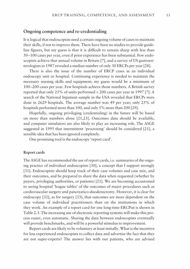
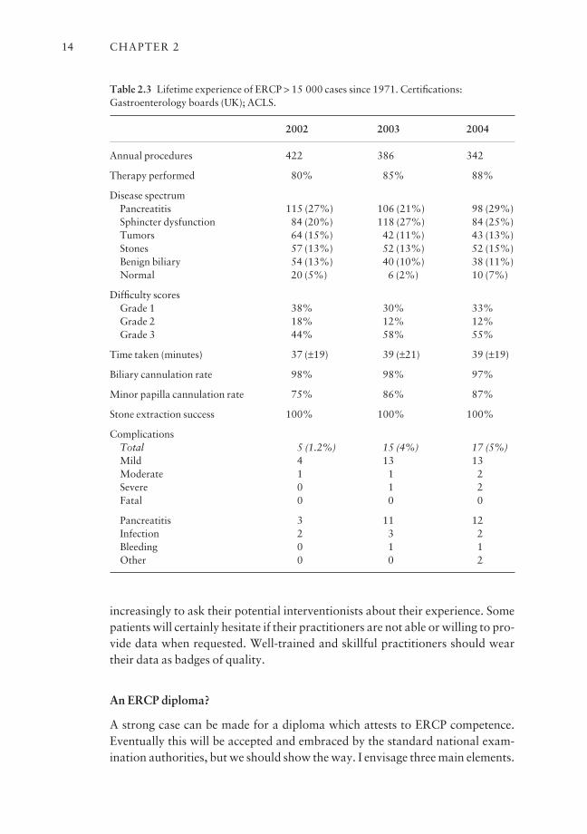
The ASGE has recommended the use of report cards, i.e. summaries of the ongo-ing practice of individual endoscopists [30], a concept that I support strongly[31]. Endoscopists should keep track of their case volumes and case mix, andtheir outcomes, and be prepared to share the data when requested (whether bypayers, privileging authorities, or patients) [21]. We are becoming accustomedto seeing hospital ‘league tables’ of the outcomes of major procedures such ascardiovascular surgery and pancreatico-duodenectomy. However, it is clear forendoscopy [32], as for surgery [33], that outcomes are more dependent on thecase volume of individual practitioners than on the institutions in which they work. An example of a report card for one long-time ERCPist is shown inTable 2.3. The increasing use of electronic reporting systems will make this pro-cess easier, even automatic. Sharing the data between endoscopists eventuallywill provide benchmarks, and will be a powerful stimulus to improvement.
Report cards are likely to be voluntary at least initially. What is the incentivefor less experienced endoscopists to collect data and advertise the fact that theyare not super-experts? The answer lies with our patients, who are advised
ERCP TRAINING, COMPETENCE, AND ASSESSMENT 13
increasingly to ask their potential interventionists about their experience. Somepatients will certainly hesitate if their practitioners are not able or willing to pro-vide data when requested. Well-trained and skillful practitioners should weartheir data as badges of quality.
An ERCP diploma?
A strong case can be made for a diploma which attests to ERCP competence.Eventually this will be accepted and embraced by the standard national exam-ination authorities, but we should show the way. I envisage three main elements.
CHAPTER 214
Table 2.3 Lifetime experience of ERCP > 15 000 cases since 1971. Certifications:Gastroenterology boards (UK); ACLS.
2002 2003 2004
Annual procedures 422 386 342
Therapy performed 80% 85% 88%
Disease spectrumPancreatitis 115 (27%) 106 (21%) 98 (29%)Sphincter dysfunction 84 (20%) 118 (27%) 84 (25%)Tumors 64 (15%) 42 (11%) 43 (13%)Stones 57 (13%) 52 (13%) 52 (15%)Benign biliary 54 (13%) 40 (10%) 38 (11%)Normal 20 (5%) 6 (2%) 10 (7%)
Difficulty scoresGrade 1 38% 30% 33%Grade 2 18% 12% 12%Grade 3 44% 58% 55%
Time taken (minutes) 37 (±19) 39 (±21) 39 (±19)
Biliary cannulation rate 98% 98% 97%
Minor papilla cannulation rate 75% 86% 87%
Stone extraction success 100% 100% 100%
ComplicationsTotal 5 (1.2%) 15 (4%) 17 (5%)Mild 4 13 13Moderate 1 1 2Severe 0 1 2Fatal 0 0 0
Pancreatitis 3 11 12Infection 2 3 2Bleeding 0 1 1Other 0 0 2
1 A written examination covering• a knowledge base of pancreatic and biliary medicine;• safety issues in ERCP practice;• endoscopic and radiological interpretation.
2 Logbook documentation of all cases and achievement of defined thresholdstandards (e.g. cannulation rates, risks, etc.).3 Proctoring of three cases by an outside expert, covering all aspects of thecases, including preparation, consent, performance, and documentation.
This examination would focus on standard grade 1 procedures, and be usedto certify completion of training. It could be applied either at the training unit,or, by default, at the institution at which privileges are sought. A shorter versioncould be used also (along with the report card data and maybe computer simulation testing) for re-credentialing. One could envisage also an analogousdiploma in ‘Advanced ERCP’ for those aspiring to recognition as expert referralresources. These examinations would be voluntary, like the report cards, but the acquisition of a diploma would provide the individual endoscopist with asignificant practice advantage.
Conclusion
ERCP has tremendous potential for benefit, but can cause devastating complica-tions. We must provide the training and credentialing framework to ensure thatit is offered optimally. Structured training and continuing objective assessmentof competence (through collection of real data) will be key elements for futuresuccess.
A diploma of competence in ERCP could become a powerful force forimproving the quality of ERCP services.
References
1 NIH State-of-the-Science Conference Statement. ERCP for diagnosis and therapy, 14–16January 2002. Gastrointest Endosc 2002; 56: 803–9.
2 Cotton PB, Williams CB. (1996). Practical Gastrointestinal Endoscopy, 4th edn. BlackwellScience, Oxford.
3 Sivak MV, Vennes JA, Cotton PB, Geenen JE, Benjamin SB, Lehman GA. Advanced trainingprograms in gastrointestinal endoscopy. Gastrointest Endosc 1993; 39: 462–4.
4 Wicks ACB, Robertson GSM, Veitch PS. Structured training and assessment in ERCP hasbecome essential for the Calman era. Gut 1999; 45: 154–6.
5 Baillie J. ERCP training for the few, not for all. Gut 1999; 45: 9–10.6 Hellier MD, Morris AI. ERCP trainingatime for change. Gut 2000; 47: 459–60.7 Allison MC, Ramanaden DN, Fouweather MG, Davis DKK, Colin-Jones DG. Provision of
ERCP services and training in the United Kingdom. Endoscopy 2002; 32: 693–9.8 American Society for Gastrointestinal Endoscopy. (1986). Guidelines for Advanced Endoscopic
Training. ASGE, Publication no. 1026. ASGE, Manchester, MA. 9 American Society for Gastrointestinal Endoscopy. (1991). Principles of Training in Gastrointest-
inal Endoscopy. ASGE, Manchester, MA.
ERCP TRAINING, COMPETENCE, AND ASSESSMENT 15
10 American Society for Gastrointestinal Endoscopy. Maintaining competency in endoscopicskills. Gastrointest Endosc 1995; 42: 620–1.
11 American Society for Gastrointestinal Endoscopy. Guidelines for credentialing and grantingprivileges for gastrointestinal endoscopy. Gastrointest Endosc 1998; 48: 679–82.
12 American Society for Gastrointestinal Endoscopy. Quality improvement of gastrointestinalendoscopy. Gastrointest Endosc 1999; 49: 842–4.
13 Joint Advisory Group on Gastrointestinal Endoscopy. (1999). Recommendations for Trainingin Gastrointestinal Endoscopy. British Society of Gastroenterology, London (www.bsg.org.uk/training/jag_99.html).
14 American Society for Gastrointestinal Endoscopy. Guidelines for advanced endoscopic training.Gastrointest Endosc 2001; 53: 846–8.
15 Kowalski T, Kanchana T, Pungpapong S. Perceptions of gastroenterology fellows regardingERCP competency and training. Gastrointest Endosc 2003; 58: 345–9.
16 Sivak MV Jr. Trained in ERCP. Gastrointest Endosc 2003; 58: 412–14.17 Waye JD, Bornman PC, Chopita N, Costamagna G, Ganc AJ, Speer T. ERCP training and experi-
ence. Gastrointest Endosc 2002; 56: 607–8.18 Cohen J. (2004). Endoscopic training and credentialing. In: Advanced Endoscopy, e-book/
annual (ed. Cotton, PB) (www.gastrohep.com).19 Freeman ML. Training and competence in gastrointestinal endoscopy. Rev Gastroenterol
Disord 2001; 1: 73–86.20 Jowell PS, Baillie J, Branch S, Affronti J, Browning CL, Bute BP. Quantitative assessment of
procedural competence: a prospective study of training in endoscopic retrograde cholangiopan-creatography. Ann Intern Med 1996; 125: 983–9.
21 American Society for Gastrointestinal Endoscopy. Methods of granting hospital privileges toperform gastrointestinal endoscopy. Gastrointest Endosc 2002; 55: 780–3.
22 Conjoint Committee for Recognition of Training in Gastrointestinal Endoscopy. (1997).Information for Supervisors: Changes to Endoscopic Training. The Conjoint Committee forRecognition of Training in Gastrointestinal Endoscopy, Sydney.
23 Joint Advisory Group on Gastrointestinal Endoscopy. (2004). Guidelines for the Training,Appraisal and Assessment of Trainees in Gastrointestinal Endoscopy, 2004 (www.Thejag.Org.Uk/JAG_ 2004pdf).
24 Wigton RS. Measuring procedural skills. Ann Intern Med 1996; 125: 1003–4.25 The Gastroenterology Leadership Council. Training the gastroenterologist of the future: the
gastroenterology core curriculum. Gastroenterology 1996; 110: 1266–300.26 Schutz SM, Abbott RM. Grading ERCPs by degree of difficulty: a new concept to produce more
meaningful outcome data. Gastrointest Endosc 2000; 51: 535–9.27 Cotton PB. Income and outcome metrics for objective evaluation of ERCP and alternative
methods. Gastrointest Endosc 2002; 56 (Suppl. 2): S283–90.28 Wigton RS, Blank LL, Monsour H, Nicolas JA. Procedural skills of practicing gastroenterolo-
gists: a national survey of 700 members of the American College of Physicians. Ann Intern Med1990; 113: 540–6.
29 Varadarajulu S, Kilgore M, Wilcox CM, Eloubeidi M. Relationship between hospital ERCP volume, length of stay and technical outcomes. Gastrointest Endosc [in press].
30 American Society for Gastrointestinal Endoscopy. Quality assessment of ERCP. GastrointestEndosc 2002; 56: 165–9.
31 Cotton PB. How many times have you done this procedure, Doctor? Am J Gastroenterol 2002;97: 522–3.
32 Petersen BT. ERCP outcomes: defining the operators, experience, and environments.Gastrointest Endosc 2002; 55: 953–8.
33 Birkmeyer JD, Stukel TA, Siewers AE, Goodney PP, Wennberg DE, Lucas FL. Surgeon volumeand operative mortality in the United States. N Engl J Med 2003; 349: 2117–27.
CHAPTER 216
CHAPTER 3
Fundamentals of ERCP
JOSEPH LEUNG
Synopsis
Endoscopic retrograde cholangiopancreatography (ERCP) was first describedin 1968 and we have recently celebrated the 30th anniversary of endoscopicsphincterotomy. This diagnostic and therapeutic modality has impactedsignificantly in the management of patients with many different benign andmalignant pancreatico-biliary problems. A successful ERCP requires the co-ordination and cooperation of a dedicated and committed team of endoscopists,nurses, and assistants, as well as an organized and functioning unit. It takesmany years to learn, and repeated practice, in order to master the skill of ERCPand to do it safely. It is important to understand the indications, contraindica-tions, limitations, and complications of individual procedures when offeringERCP to our patients. Although successful ERCP has replaced surgery as atreatment option for some difficult pancreatico-biliary diseases, we have alsoseen problems and complications arising as a result of endoscopic treatment.Prospective collection of data and selected randomized controlled studies withlong-term follow-up are necessary to evaluate the true value of this technologyin the overall care of our patients.
Introduction
Imaging of the pancreatico-biliary system
Methods for imaging the pancreatic and biliary ductal systems continue toevolve. Correct application of ERCP (and other procedures) requires an up-to-date knowledge of all of these modalities.
ERCP
ERCP is a direct contrast study of the pancreatico-biliary system. It is useful in
17

the diagnosis and treatment of diseases involving the pancreas and bile ducts,such as stones, benign and malignant strictures, and developmental anomalies.
It is superior to indirect cholangiography (oral or IV), especially in cases withobstructive jaundice, which leads to raised intrabiliary pressure and impairedbiliary excretion of contrast.
Moreover, intrahepatic bile duct pathologies can be demonstrated by ERCPusing occlusion cholangiography. Pathology in the gallbladder and cystic ductabnormalities can also be visualized, although ERCP is not the best imagingstudy for gallbladder disease.
ERCP vs. PTC
Comparative investigation of direct cholangiography studies, i.e. ERCP andpercutaneous transhepatic cholangiography (PTC), should take into considera-tion the individual patients and the expertise of the operator; however, ERCP isconsidered less invasive than PTC.
ERCP has the added advantages of allowing duodenoscopy and pancreato-graphy, which are helpful in the diagnosis of ampullary pathology and pancreaticabnormalities. ERCP can be performed in the presence of ascites and/or malig-nancies involving the liver, contraindicating PTC. In addition, bile and pancreaticjuice can be collected for cytological and microbiological examination duringERCP procedures.
MRCP
The development and refinement of magnetic resonance cholangiopancreato-graphy (MRCP) have produced excellent quality pictures of the anatomy of thepancreatico-biliary system. It is non-invasive and can give images comparable to ERCP when performed well. Limitations are few and the diagnostic value is high, and it may replace diagnostic ERCP, especially in the investigation ofjaundice. MRCP, however, lacks therapeutic potential.
EUS
Endoscopic ultrasonography (EUS) allows good visualization of the distal com-mon bile duct (CBD), with an excellent diagnostic accuracy for ductal stones. It provides superb views of the pancreas, and is useful in defining underlyingpancreatic pathology. Fine-needle aspiration cytology further complements thediagnostic capability of EUS in pancreatico-biliary diseases.
CHAPTER 318

Section I: Preparation for ERCP
Room set-up and floor plan (Figs 3.1 and 3.2)
Correct layout of the ERCP room is easier if it is located in a purpose-builtendoscopy suite with in-house fluoroscopy facilities, rather than a shared facil-ity in the radiology department. A purpose-built room with fluoroscopy offersthe advantage of a better floor plan, organization, and ready access to storedaccessories required for the procedures. Daily activities can be better organizedand there is less hassle in moving equipment and endoscopists.
Space
The ERCP room should be large enough to house the endoscopy equipment,monitors, and the fluoroscopy unit. There should be ample room for the endo-scopists and nurse/assistant(s) to manipulate accessories. Additional space isrequired for trainees and interested observers. Space should be available foranesthetic support and resuscitation equipment when needed. Ideally, thereshould be no cables or tubing on the floor that may hinder movement of carts or
FUNDAMENTALS OF ERCP 19
Monitors
X-Ray Endoscopes
Vital signs
VCR
Foot controls
DiathermyFluoro
Scope tower
Storage
Worktop
X-Raymonitor C-arm
N
E
A
Fig. 3.1 Room set-up and floor plan. A, assistant; E, endoscopist; N, nurse.

trolleys. Accessories should be organized and stored to facilitate easy retrievalduring procedures.
Position of monitors and endoscopy cart (Fig. 3.2)
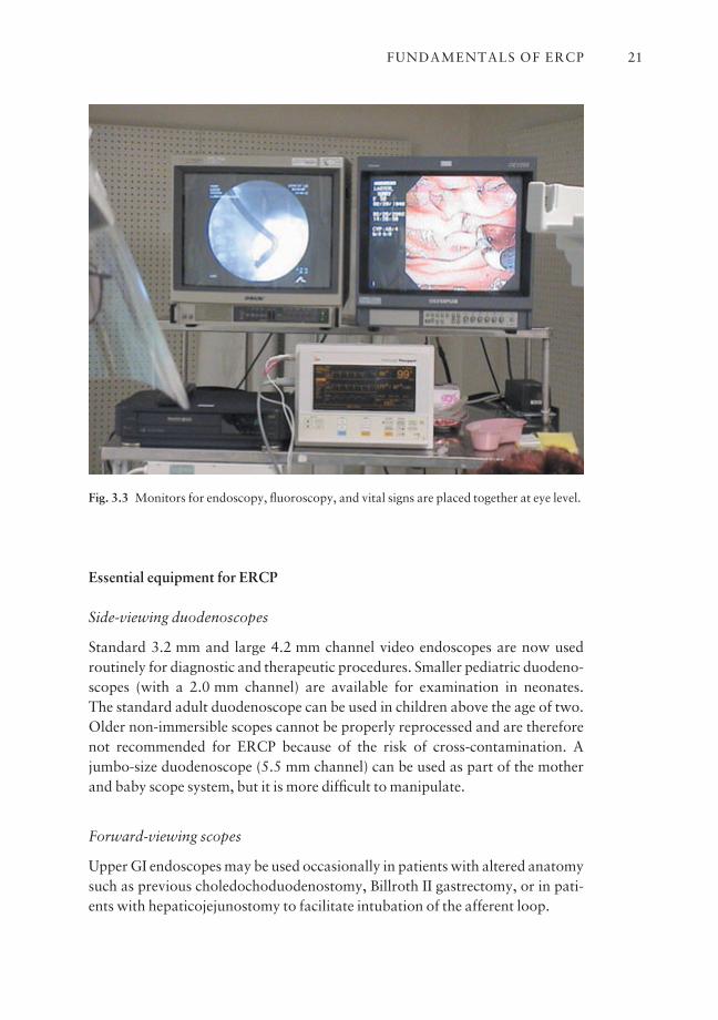
Some units have the endoscopy monitor mounted on the endoscopy cart at thehead of the patient, which means the endoscopist has to turn to the right, awayfrom the patient, in order to observe the endoscopy image. This bodily rotationtends to change the position and orientation of the scope and is best avoided. It is better to have the fluoroscopy and endoscopy monitors placed side by sidefacing the endoscopist, on the opposite side of the X-ray table (Fig. 3.3).
Because of the position of the fluoroscopy machine, the monitors may needto be placed at a 15–20° angle off to the right of the endoscopist for easy observa-tion. The monitors are best ceiling mounted or supported on a stand placed ateye level. The endoscopist should adopt a comfortable position to avoid twistingand turning of the body, which may predispose to scope displacement or strain-ing of the back and neck. The endoscopy tower is usually placed on the rightbehind the endoscopist, with sufficient room left in between for the manipula-tion of accessories.
CHAPTER 320
Fig. 3.2 Space for endoscopist and trainee or assistant. Accessories organized and within easyreach of endoscopist.
Essential equipment for ERCP
Side-viewing duodenoscopes
Standard 3.2 mm and large 4.2 mm channel video endoscopes are now used routinely for diagnostic and therapeutic procedures. Smaller pediatric duodeno-scopes (with a 2.0 mm channel) are available for examination in neonates. The standard adult duodenoscope can be used in children above the age of two.Older non-immersible scopes cannot be properly reprocessed and are thereforenot recommended for ERCP because of the risk of cross-contamination. Ajumbo-size duodenoscope (5.5 mm channel) can be used as part of the motherand baby scope system, but it is more difficult to manipulate.
Forward-viewing scopes
Upper GI endoscopes may be used occasionally in patients with altered anatomysuch as previous choledochoduodenostomy, Billroth II gastrectomy, or in pati-ents with hepaticojejunostomy to facilitate intubation of the afferent loop.
FUNDAMENTALS OF ERCP 21
Fig. 3.3 Monitors for endoscopy, fluoroscopy, and vital signs are placed together at eye level.

Medication
A combination of sedatives and analgesics is used to provide conscious sedationduring the ERCP procedure. Medications drawn up in syringes should be clearlylabeled to avoid making mistakes during drug administration.
Sedatives and analgesics
Standard medications used for IV conscious sedation include demerol (meperi-dine) or fentanyl, and valium (Diazemuls) or midazolam (Versed). The doserequirement is titrated according to the patient’s response. For an average sizedadult, we usually start with 25–50 mg of meperidine or 25–50 µg of fentanyl,and 2.5–5 mg of valium or 1–2 mg of midazolam. Additional injections aregiven during the procedure as needed. IV benadryl 25–50 mg or IV phenerganmay be given to enhance the sedative effects.
Anesthesia
General anesthesia with IV propofol is used increasingly for complex ERCP procedures, especially in anxious patients, those with cardio-pulmonary com-promise, those who use chronic narcotics or excessive alcohol, and others with a history of poor response to standard sedation.
Smooth muscle relaxants
Glucagon (0.25–0.5 mg) or Buscopan (20–40 mg) is given intravenously inincrements to relax the duodenum and to facilitate cannulation.
Reversal agents
Reversal agents including naloxone (Narcan 0.4 mg) and flumazenil (1 mg)should be readily available to reverse the effects of sedation.
Monitoring during conscious sedation
A qualified nurse (or anesthetist) should be assigned to administer medications,and to monitor the patient during the ERCP procedure. This person should haveno other responsibilities. Medications are given in incremental doses based onthe patient’s response and condition in order to avoid oversedation. Vital signsincluding blood pressure, pulse, EKG, and oxygen saturation should be mon-itored continuously.
Supplemental oxygen can be given via a nasal cannula at a flow rate of
CHAPTER 322
2 liters/min; this has been shown to prevent hypoxia. Care must be taken toavoid giving excess oxygen which may lead to respiratory depression in patientswith COPD. Measuring the end-expiration CO2 level using capnography is carried out in some centers.
Contrast agents
The most commonly used contrast media such as conray 280, urografin,hypaque, and renografin contain iodine. Contrast media used for ERCP includeboth hyperosmolar ionic medium and isosmolar, non-ionic medium. Isosmolarnon-ionic contrast agents are more expensive but should be used in patientsallergic to iodine. In addition, it is advisable to give these patients steroid pro-phylaxis and benadryl prior to the procedure to prevent contrast reaction.
Contrast should be drawn up in clearly labeled syringes prior to the proce-dure and be ready for use. It is preferable to have at least two 20 ml syringes filledwith contrast of normal and half normal strength. A 20 ml syringe is used forcontrast injection because it is easy to handle, contains sufficient volume of con-trast, and permits injection by the endoscopist. Normal strength contrast shouldbe used for initial cannulation for better visualization of the pancreatic duct.Half normal strength contrast is used to identify ductal stones in patients withdilated bile ducts.
Syringes for aspiration and irrigation
An empty 20 ml syringe is used to aspirate bile for culture and cytology. Sterilewater is used to flush the catheters prior to insertion of hydrophilic wires orexchanges.
Organization and storage of accessories (Fig. 3.4)
There is a wide range of ERCP accessories. These include cannulas, sphinctero-tomes, guidewires, baskets, balloons, dilators, nasobiliary catheters, stents,biopsy forceps, injection needles, and more complex devices such as mechanicallithotriptors.
The accessories should be categorized and organized, and stored to alloweasy retrieval as well as stock-keeping. A limited supply of commonly used itemsshould be clearly labeled and displayed on shelves like books in a library.
Similar items are best grouped together and more specialized items kept sep-arately. A detailed catalogue list and location of all accessories should be keptfor quick reference. It is helpful to establish a preprocedure ‘game plan’ so thatthe necessary accessories can be retrieved and readied for use.
FUNDAMENTALS OF ERCP 23
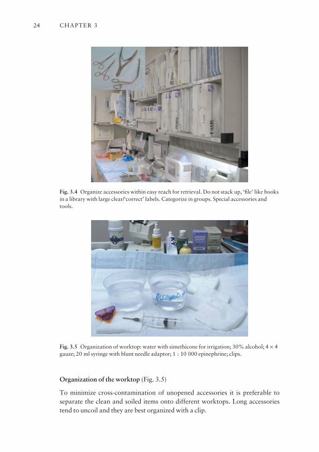
Organization of the worktop (Fig. 3.5)
To minimize cross-contamination of unopened accessories it is preferable toseparate the clean and soiled items onto different worktops. Long accessoriestend to uncoil and they are best organized with a clip.
CHAPTER 324
Fig. 3.4 Organize accessories within easy reach for retrieval. Do not stack up, ‘file’ like booksin a library with large clear/‘correct’ labels. Categorize in groups. Special accessories andtools.
Fig. 3.5 Organization of worktop: water with simethicone for irrigation; 30% alcohol; 4 × 4gauze; 20 ml syringe with blunt needle adaptor; 1 : 10 000 epinephrine; clips.

A small pot of 30% alcohol is useful for cleaning the gloves (finger tips) toremove any sticky contrast or bile. Alcohol also reduces friction at the biopsyvalve and facilitates insertion of accessories. Gauze pads are used for cleaningand wiping. Sterile water with simethicone can be flushed down the channel tosuppress bubbling in the duodenum to improve visualization.
Fluoroscopy for ERCP
ERCP is ideally performed with the help of a radiologist, but more commonlywith the help of a trained radiology technician. Endoscopists who personallyoperate the fluoroscopy unit during the procedure should receive basic flu-oroscopy training and appropriate local licensing.
Fluoroscopy units (Fig. 3.6)
Conventional X-ray machines, as used for barium studies, are adequate forERCP examinations. High-resolution digital fluoroscopy units produce betterpictures but they are also much more expensive. A portable digital C-arm unitcan be used but the resolution may be inferior to the full digital unit. It is pref-erable to use a machine with an under-couch X-ray tube. The X-ray machineshould be capable of taking spot films. Digital units can store the images onto acomputer for subsequent retrieval and review. Hard copies of selected imagescan be printed for reporting and filing. It is essential to know the magnification
FUNDAMENTALS OF ERCP 25
Fig. 3.6 Fluoroscopy machine with under-couch tube. Digital C-arm for designated ERCProom. Full fluoroscopy unit if shared facilities in Radiology. Remote control and foot pedal.Image capture.
factor of the machine for correct interpretation of X-ray findings, and for meas-uring the size of stones and the length of strictures.
A high-resolution monitor is necessary as diagnostic interpretation and therapeutic procedures are often performed in real time under fluoroscopicguidance. The X-ray table should have an electrical remote control for fineadjustment in positioning and preferably be able to tilt in two directions. Apartfrom built-in shielding, additional pieces of lead can be placed over the side andhead end of the table to protect staff from scattered radiation.
KV and mA
These are the settings on the X-ray machine that determine the penetration ofthe X-ray beam and quality of the image generated. Most digital machines canautomatically adjust the setting according to individual patients.
Split screen
The area of interest seen on fluoroscopy can be reduced to allow fine focus on asmaller area. This gives greater detail and reduces the radiation exposure.
Magnified view
A magnified view gives an enhanced image of the area of interest, but it also dou-bles the radiation exposure. It is sometimes necessary for proper localization ofthe tip of a guidewire or accessories during manipulation in the pancreas or forselective ductal cannulation.
Orientation of fluoroscopic images
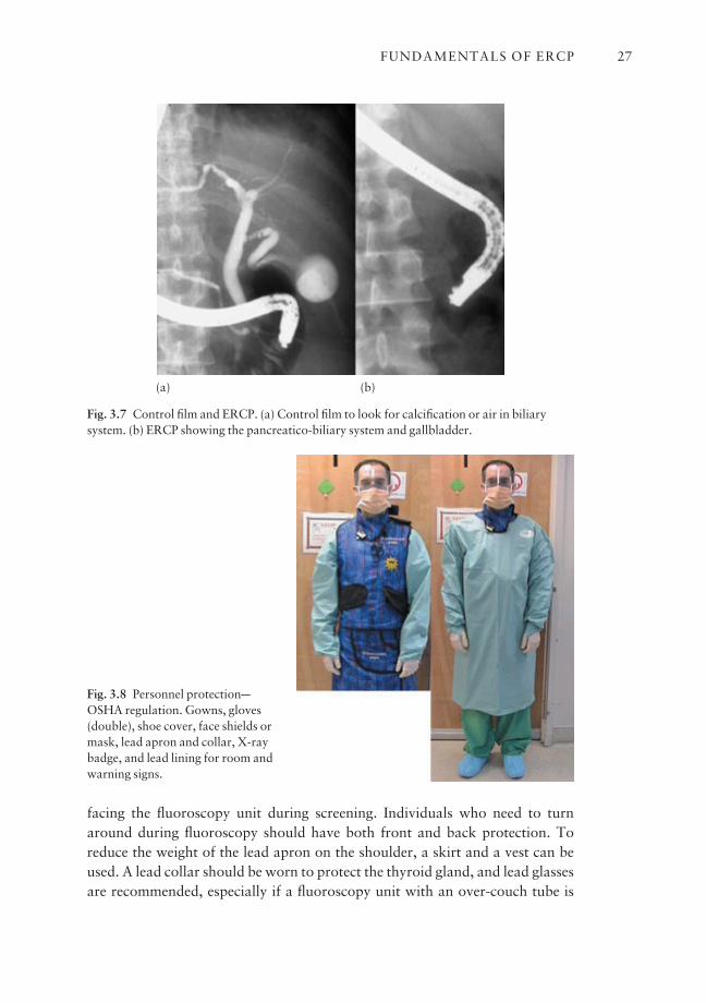
The orientation of the fluoroscopic image on the monitor varies depending onthe individual endoscopist’s personal preference. Some prefer to orientate theimage in the conventional way of viewing X-ray films. Some, however, prefer toorientate the fluoroscopic image according to the anatomical position, i.e. rightside of the screen corresponds to right side of the patient lying in a prone position (Fig. 3.7).
Personnel protection (Fig. 3.8)
Individuals working with or around the fluoroscopy machine should be pro-tected from scattered radiation by using standard lead aprons (lead thickness0.2–0.5 mm). If a one-sided lead apron is used, it is important to keep the apron
CHAPTER 326
facing the fluoroscopy unit during screening. Individuals who need to turnaround during fluoroscopy should have both front and back protection. Toreduce the weight of the lead apron on the shoulder, a skirt and a vest can beused. A lead collar should be worn to protect the thyroid gland, and lead glassesare recommended, especially if a fluoroscopy unit with an over-couch tube is
FUNDAMENTALS OF ERCP 27
Fig. 3.7 Control film and ERCP. (a) Control film to look for calcification or air in biliarysystem. (b) ERCP showing the pancreatico-biliary system and gallbladder.
Fig. 3.8 Personnel protectionaOSHA regulation. Gowns, gloves(double), shoe cover, face shields ormask, lead apron and collar, X-raybadge, and lead lining for room andwarning signs.
(a) (b)
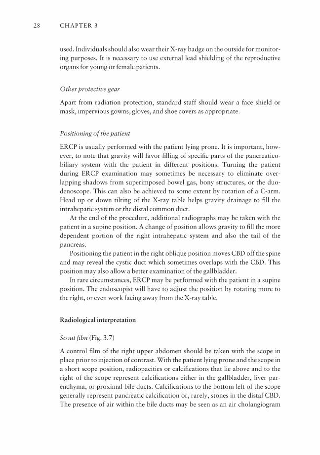
used. Individuals should also wear their X-ray badge on the outside for monitor-ing purposes. It is necessary to use external lead shielding of the reproductiveorgans for young or female patients.
Other protective gear
Apart from radiation protection, standard staff should wear a face shield ormask, impervious gowns, gloves, and shoe covers as appropriate.
Positioning of the patient
ERCP is usually performed with the patient lying prone. It is important, how-ever, to note that gravity will favor filling of specific parts of the pancreatico-biliary system with the patient in different positions. Turning the patient during ERCP examination may sometimes be necessary to eliminate over-lapping shadows from superimposed bowel gas, bony structures, or the duo-denoscope. This can also be achieved to some extent by rotation of a C-arm.Head up or down tilting of the X-ray table helps gravity drainage to fill the intrahepatic system or the distal common duct.
At the end of the procedure, additional radiographs may be taken with thepatient in a supine position. A change of position allows gravity to fill the moredependent portion of the right intrahepatic system and also the tail of the pancreas.
Positioning the patient in the right oblique position moves CBD off the spineand may reveal the cystic duct which sometimes overlaps with the CBD. Thisposition may also allow a better examination of the gallbladder.
In rare circumstances, ERCP may be performed with the patient in a supineposition. The endoscopist will have to adjust the position by rotating more tothe right, or even work facing away from the X-ray table.
Radiological interpretation
Scout film (Fig. 3.7)
A control film of the right upper abdomen should be taken with the scope inplace prior to injection of contrast. With the patient lying prone and the scope ina short scope position, radiopacities or calcifications that lie above and to theright of the scope represent calcifications either in the gallbladder, liver par-enchyma, or proximal bile ducts. Calcifications to the bottom left of the scopegenerally represent pancreatic calcification or, rarely, stones in the distal CBD.The presence of air within the bile ducts may be seen as an air cholangiogram
CHAPTER 328
and suggests a patent communication between the bile duct and the gut, such asa patent stent, a fistula, or a bilioenteric anastomosis.
Contrast studies
Most diagnostic and therapeutic interventions are performed under fluoro-scopic control; however, radiographs or stored images should be taken for documentation. Hard copy radiographs give better resolution compared to thefluoroscopic images and may reveal more detailed information.
If common duct stones are suspected, early filling films should be taken dur-ing injection of contrast. This may demonstrate a ‘meniscus’ sign where thestone is outlined by contrast within the duct. Excess contrast should be avoidedas this tends to mask the small stones in a dilated duct.
With the patient lying prone, the left hepatic system is more dependent andusually fills more quickly than the right side. If the cystic duct is patent, contrastmay preferentially fill the gallbladder. The posterior segments of the right hepatic system are non-dependent in the prone position but may be filled morereadily by turning the patient to a supine position.
Drainage films
Delayed films after removing the duodenoscope are sometimes indicated if thereis a clinical suspicion of a drainage problem, e.g. papillary dysfunction or sten-osis. Drainage films may be taken with the patient in the right lateral position orin the Trendelenburg position.
The normal rate of drainage is affected by many factors and precise normallimits have not been established. Delayed drainage is, however, suspected ifsignificant opacification of the bile duct persists after 45 min, and after 10 minfor the pancreatic duct.
It is necessary to take hard copy spot films to document any therapeuticinterventions. Alternatively, serial digital images are stored and retrieved at theend of the procedure for reporting and filing.
The pancreatogram
Normal anatomy The pancreas is a retroperitoneal organ lying across theabdomen at the level of L1 and L2. Pancreatic calcifications on the control filmsuggest chronic pancreatitis and rarely pancreatic neoplasm. A good qualitypancreatogram should demonstrate the main pancreatic duct up to the tail withadequate filling of the second generation branch ducts. Excess contrast injectionwill result in acinarization or a parenchymogram.
FUNDAMENTALS OF ERCP 29
The pancreatic duct normally has a smooth, slightly wavy course from thepapilla tapering towards the tail. In the head a branch duct is seen draining theuncinate process. In addition, the accessory duct (Santorini’s duct) drainsthrough the minor papilla.
In 5% of cases a prominent branch duct runs parallel to the main pancreaticduct giving the appearance of a bifid pancreas. Several branch ducts join themain pancreatic duct at irregular intervals, usually at right angles to the mainduct. The branch ducts taper and themselves branch off into smaller ducts.
The diameter of the pancreatic duct varies according to the age and size ofthe patient. Elderly patients may have a slightly larger duct. The maximumdiameter of a normal pancreatic duct is 6 mm in the head, 5 mm in the body, and 3 mm in the tail. Care must be taken to correct for magnification, which isusually 30%.
Pathological changes The pancreatic duct may appear normal in mild pancre-atitis. In acute pancreatitis the pancreatic duct may appear slightly irregularwith changes and irregularities of the side branches. Presence of a cyst or pseu-docyst may cause complete obstruction of the pancreatic duct with or withoutcommunication with the duct.
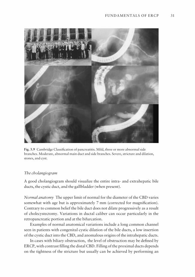
The Cambridge Classification is used to document the severity of chronicpancreatitis (Fig. 3.9) as seen on a pancreatogram:• Mild pancreatitis: a normal main pancreatic duct with three or more abnor-mal side branches.• Moderate pancreatitis: an abnormal main duct with irregularities in three ormore abnormal side branches.• Severe pancreatitis: irregularity with strictures and dilation of the main duct,with filling defects suggestive of stones or filling of cavities or cysts.
There is no direct correlation between the radiological abnormalities and thefunctional loss in chronic pancreatitis because the pancreas has a good func-tional reserve. Leakage of contrast from a transected pancreatic duct with non-filling of the upstream duct is diagnostic of traumatic pancreatitis.
Cancer in the head of the pancreas may cause stricturing of the main pancre-atic duct with uniform dilation of the side branches and the main duct upstreamof the obstruction. In addition, the retropancreatic portion of the CBD may beinvolved, giving rise to the characteristic ‘double duct stricture’ sign. Displace-ment or stretching of the side branches may suggest an underlying tumor in thepancreas.
Congenital anomalies In patients with pancreas divisum, there is non-fusion ofthe dorsal and ventral ducts. The small isolated ventral pancreas drains throughthe main papilla. The dorsal (Santorini’s) duct drains the bulk of the pancreasthrough the minor papilla.
CHAPTER 330
The cholangiogram
A good cholangiogram should visualize the entire intra- and extrahepatic bileducts, the cystic duct, and the gallbladder (when present).
Normal anatomy The upper limit of normal for the diameter of the CBD variessomewhat with age but is approximately 7 mm (corrected for magnification).Contrary to common belief the bile duct does not dilate progressively as a resultof cholecystectomy. Variations in ductal caliber can occur particularly in theretropancreatic portion and at the bifurcation.
Examples of normal anatomical variations include a long common channelseen in patients with congenital cystic dilation of the bile ducts, a low insertionof the cystic duct into the CBD, and anomalous origins of the intrahepatic ducts.
In cases with biliary obstruction, the level of obstruction may be defined byERCP, with contrast filling the distal CBD. Filling of the proximal ducts dependson the tightness of the stricture but usually can be achieved by performing an
FUNDAMENTALS OF ERCP 31
Fig. 3.9 Cambridge Classification of pancreatitis. Mild, three or more abnormal sidebranches. Moderate, abnormal main duct and side branches. Severe, stricture and dilation,stones, and cyst.
occlusion cholangiogram. Contrast is injected under pressure by inflating a balloon below the obstruction to fill the more proximal obstructed system.
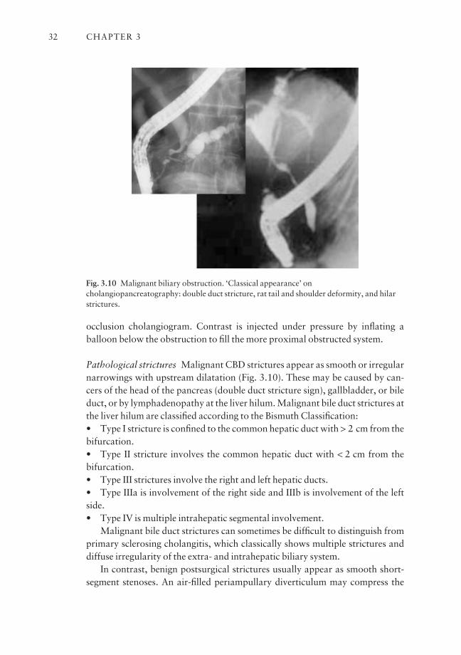
Pathological strictures Malignant CBD strictures appear as smooth or irregularnarrowings with upstream dilatation (Fig. 3.10). These may be caused by can-cers of the head of the pancreas (double duct stricture sign), gallbladder, or bileduct, or by lymphadenopathy at the liver hilum. Malignant bile duct strictures atthe liver hilum are classified according to the Bismuth Classification:• Type I stricture is confined to the common hepatic duct with > 2 cm from thebifurcation.• Type II stricture involves the common hepatic duct with < 2 cm from thebifurcation.• Type III strictures involve the right and left hepatic ducts.• Type IIIa is involvement of the right side and IIIb is involvement of the leftside.• Type IV is multiple intrahepatic segmental involvement.
Malignant bile duct strictures can sometimes be difficult to distinguish fromprimary sclerosing cholangitis, which classically shows multiple strictures anddiffuse irregularity of the extra- and intrahepatic biliary system.
In contrast, benign postsurgical strictures usually appear as smooth short-segment stenoses. An air-filled periampullary diverticulum may compress the
CHAPTER 332
Fig. 3.10 Malignant biliary obstruction. ‘Classical appearance’ oncholangiopancreatography: double duct stricture, rat tail and shoulder deformity, and hilarstrictures.

distal common duct giving rise to a pseudostricture formation. In these cases, thedistal bile duct is seen to ‘open up’ when air is removed from the diverticulum.
Bile duct stones (Fig. 3.11) Stones within the bile duct may be demonstrated initially as a meniscus sign upon contrast injection and subsequently as fillingdefects. They are round or faceted depending upon their origin. It may be neces-sary to change the scope position into a long scope position to expose the mid-/distal CBD, an area otherwise overlapped by the scope. Rarely, parasites suchas Clonorchis sinensis or Ascaris lumbricoides may be seen as unique fillingdefects in the extra- or intrahepatic bile ducts.
FUNDAMENTALS OF ERCP 33
Fig. 3.11 CholangiogramaCBD stones. Common duct stones seen in different size, shape,and number. Stones can form around a migrated surgical clip.

Gallbladder ERCP is not an ideal examination of the gallbladder. If the gallbladder is filled, a delayed film of the gallbladder should be taken after 30–45 min. This allows time for the contrast to mix with bile for better definition of gallstones (Fig. 3.12). Failure to fill the gallbladder despite adequate filling ofthe intrahepatic ducts suggests cystic duct obstruction. Stone impaction in the
CHAPTER 334
Fig. 3.12 ERCP for gallbladder stones. Gallstones may be obvious on cholangiogram. Noteaberrant duct which resembles cystic duct. Always check delayed film of gallbladder for smallstones.
cystic duct may cause edema and compression of the common hepatic duct giving rise to Mirizzi’s syndrome.
Underfilling and delayed drainage With an adequate intrahepatic cholan-giogram, underlying parenchymal liver diseases may be inferred from abnormalappearance of the intrahepatic ducts. Crowding of tortuous intrahepatic ductsmay suggest liver cirrhosis. Stretching of a particular intrahepatic duct may beseen around space-occupying lesions such as abscesses, tumors, or cysts in theliver.
Underfilling of the bile ducts or ‘streaming effect of contrast’ may suggest anapparent narrowing in the distal bile duct. Inadequate filling due to stricture orobstruction may fail to detect intrahepatic pathologies such as stones in patientswith hepatolithiasis. Functional obstruction at the papilla is difficult to diag-nose, but is suspected if there is delayed drainage of contrast (> 45 min).
The clinical diagnosis of papillary stenosis or sphincter of Oddi dysfunctiondepends on the presence of abnormal liver function tests with or without adilated bile duct associated with right upper quadrant abdominal pain. Mano-metric studies are necessary to confirm the diagnosis in patients without obviousduct dilation or liver test abnormalities. Bile leaks and fistulas complicating biliary tract surgery can be readily identified on cholangiography.
Section II: Diagnostic and therapeutic ERCP
Diagnostic ERCP
Scopes
ERCP is performed using side-viewing duodenoscopes with a 2.8, 3.2, or 4.2 mm channel. All of these scopes readily accept a 5 Fr or 6 Fr catheter andaccessories. The larger channel duodenoscopes accept accessories up to 10–11.5 Fr diameter and are used for both diagnostic and therapeutic purposes. Thelarger instrument channel allows aspiration of duodenal contents even with anaccessory in place, and also permits the manipulation of two guidewires oraccessories simultaneously.
Accessories (Fig. 3.13)
The cannula or diagnostic catheter is a 6 or 7 Fr Teflon tube which tapers to a3–5 Fr tip. It is used for injection of contrast into the ductal systems. A variety ofcannulas are available with different tip designs. A commonly used example isthe bullet tip or fluorotip catheter, which has a small metal or radiopaque tip at
FUNDAMENTALS OF ERCP 35
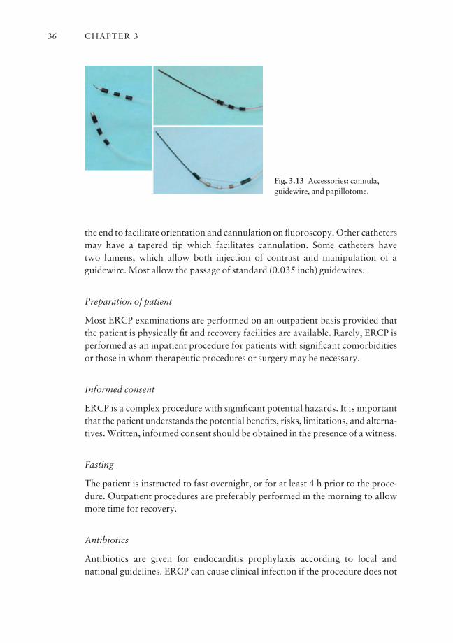
the end to facilitate orientation and cannulation on fluoroscopy. Other cathetersmay have a tapered tip which facilitates cannulation. Some catheters have two lumens, which allow both injection of contrast and manipulation of aguidewire. Most allow the passage of standard (0.035 inch) guidewires.
Preparation of patient
Most ERCP examinations are performed on an outpatient basis provided thatthe patient is physically fit and recovery facilities are available. Rarely, ERCP isperformed as an inpatient procedure for patients with significant comorbiditiesor those in whom therapeutic procedures or surgery may be necessary.
Informed consent
ERCP is a complex procedure with significant potential hazards. It is importantthat the patient understands the potential benefits, risks, limitations, and alterna-tives. Written, informed consent should be obtained in the presence of a witness.
Fasting
The patient is instructed to fast overnight, or for at least 4 h prior to the proce-dure. Outpatient procedures are preferably performed in the morning to allowmore time for recovery.
Antibiotics
Antibiotics are given for endocarditis prophylaxis according to local andnational guidelines. ERCP can cause clinical infection if the procedure does not
CHAPTER 336
Fig. 3.13 Accessories: cannula,guidewire, and papillotome.
relieve the obstruction and if cleaning and disinfection regimens are not ideal.Antibiotics are given prophylactically when difficulty in drainage is anticipated,e.g. in patients with multiple strictures (hilar tumors or sclerosing cholangitis) or pseudocysts. Antibiotics should also be given immediately if obstruction isnot relieved.
ERCP procedure
Intubation and examination of the stomach
When the patient is adequately sedated, a self-retaining mouth guard is placedand the patient is supported in a left lateral/semiprone position. This positionfacilitates intubation and examination of the upper GI tract with the side-viewing duodenoscope.
With the patient in the prone position, slight left rotation of the scope isrequired to correct for the change in axis. Gentle downward tip angulationallows examination of the distal esophagus. Once in the stomach, the gastricjuice is removed by suction to minimize the risk of aspiration. The stomach isinflated slightly to allow an adequate view of the lumen.
The endoscope is slowly advanced with the tip angled downwards looking at the greater curve and distal stomach. With further advancement, the scopewill pass the angular incisura. The cardia can be examined by up angulation andwithdrawal of the scope.
Once past the angular incisura the tip of the scope is further angled down-wards and the pylorus is visualized. The scope is positioned so that the pyloruslies in the center of the field. The tip of the endoscope is then returned to the neutral position as the pylorus disappears from the endoscopic view, the so-called ‘sun-setting sign’.
Gentle pushing will advance the scope into the first part of the duodenum.The scope is angled downwards again and air is insufflated to distend the duode-num. Care must be taken to avoid overinflating the duodenum as this causespatient discomfort and makes the procedure more difficult. Careful examina-tion is performed to rule out any pathologies such as ulcers or duodenitis. The scope is pushed further to the junction of the first and second part of theduodenum.
At this point, the scope is angled to the right and upwards, and by rotatingthe scope to the right and withdrawing slowly, the tip of the scope is advancedinto the second part of the duodenum. This paradoxical movement shortens the scope using the pylorus as a pivot, bringing it into the classical ‘short scopeposition’. The markings on the duodenoscope should indicate 60–65 cm at theincisors.
FUNDAMENTALS OF ERCP 37
With the patient prone, and the scope returned to a neutral position, thepapilla can be easily visualized, in the middle of the second portion of the duode-num. The landmark for identification of the papilla is the junction where thehorizontal folds meet the vertical fold. Duodenal diverticula may cause difficult-ies with cannulation as the papilla may be located on the edge or rarely inside adiverticulum.
Approaching the main papilla
A control film of the right upper abdomen is taken to look for calcification andfor air in the biliary system, prior to injection of contrast.
Cannulation is performed in the short scope position allowing better controlover angulations and tip deflection. In some difficult cases or in attempted minorpapilla cannulation, the long scope approach may be adopted. Excess bubbles inthe duodenum can be removed by injecting a diluted simethicone solution downthe channel. Duodenal contractions may be reduced with the use of antispas-modic medication.
The presence of a periampullary diverticulum does not normally increase thetechnical difficulty of cannulation, unless the papilla is displaced or locatedinside the diverticulum (Fig. 3.14).
The normal papilla appears as a pinkish protruding structure and the sizemay vary. Abnormalities result from previous stone passage, stone impaction,or tumor.
Cannulation of the papilla
Cannulation is best performed in an ‘en face’ position. The cannula should beflushed and primed with contrast to remove any air bubbles prior to insertioninto the duodenoscope. Air injected into the biliary system could mimic stones.Flushing excess contrast in the duodenum should be avoided since hypertoniccontrast stimulates duodenal peristalsis.
A combination of 12 different maneuvers can be used for positioning the tipof the cannula for cannulation. These include up/down and sideways angula-tion, rotation of the endoscope, use of the elevator, and pushing in and pullingback of the scope. Suction collapses the duodenum and pulls the papilla closer tothe endoscope. Air insufflation pushes it away. Most beginners find pancreato-graphy easier to obtain than cholangiography. The pancreatic duct is normallyentered by inserting the cannula in a direction perpendicular to the duodenalwall, in the 1–2 o’clock orientation (Fig. 3.15).
Fine adjustments of the position and axis of the cannula are helpful. Exces-sive pressure in the papilla is best avoided because pushing may distort the
CHAPTER 338

papilla and increase the difficulty with cannulation. Cannulation of the CBD isusually achieved by approaching the papilla from below, in line with the axis ofthe CBD. It may be helpful to lift the roof of the papilla, and to direct the cannulatowards 11 o’clock (Fig. 3.16).
Full strength contrast should be used initially, and is injected under fluoro-scopic control. The pancreatic duct should be filled until the tail and some sidebranches are visualized. Avoid overfilling and acinarization as this increases therisk of post-ERCP pancreatitis. When filling the CBD, start with full strengthcontrast and consider switching over to dilute contrast when stones are visual-ized. If deep cannulation is successful, aspirate bile before injecting contrast toavoid excess contrast masking small stones in a dilated biliary system.
The left hepatic ducts usually fill before the right because they are dependentwith the patient lying prone. The gallbladder is usually filled except in cases withcystic duct obstruction. Multiple spot films are taken during contrast injection.It may be necessary to change the scope position to expose the portion of thecommon duct hidden behind the scope.
FUNDAMENTALS OF ERCP 39
Fig. 3.14 The obscure papilla. Look for bile! Lift the overhanging fold. With priorpapillotomy, biliary orifice is often more cephalad. Note relationship of papilla to duodenaldiverticula. Probing or suction to change shape of diverticulum and axis to reveal the papilla.
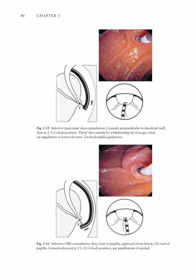
CHAPTER 340
Fig. 3.15 Selective pancreatic duct cannulation. Cannula perpendicular to duodenal wall.Aim at 1–2 o’clock position. ‘Drop’ the cannula by withdrawing tip of scope, relax up angulation or lower elevator. Use hydrophilic guidewire.
Fig. 3.16 Selective CBD cannulation. Stay close to papilla, approach from below, lift roof ofpapilla. Cannula directed at 11–12 o’clock position, use papillotome if needed.
At the end of the procedure the endoscope is withdrawn and air is suctionedfrom the stomach to minimize discomfort. The patient is then turned to a supineposition and more radiographs are taken in different projections (as previouslydescribed).
In patients with a partially filled gallbladder, immediate diagnosis of gall-stones may be difficult due to inadequate mixing of contrast with bile. Delayedfilms of the gallbladder (after about 45 min) may reveal small stones after allow-ing time for the contrast to mix with bile.
Ease and success in cannulation
Success of diagnostic ERCP depends on the experience of the endoscopist andthe presence or absence of pathology. Successful cannulation of both ductal sys-tems is commonly achieved in 85–90% of cases with experts achieving rates ofover 95%. The success rate is lower in patients with previous gastric surgery,e.g. Billroth II gastrectomy.
Minor papilla cannulation
The minor papilla is located proximally and to the right of the main papilla. Itcan be identified as a small protruding structure. It may not be obvious or mayappear as a slightly pinkish nipple between the duodenal folds. When promi-nent, it can sometimes be mistaken for the main papilla; however, it does not havea distinct longitudinal fold and the small opening usually resists cannulation.
Cannulation of the minor papilla is indicated in patients with suspected orproven pancreas divisum and when cannulation of the pancreatic duct fails atthe main papilla. Cannulation of the minor papilla is usually best performed in a long scope position using a 3 mm fine metal tip cannula. Bending the tip of the cannula to form an angle facilitates cannulation.
It is important to identify the correct location of the orifice before anyattempt is made to inject contrast, as trauma from the cannula may result inedema and bleeding and obscure the opening.
If the papilla or orifice is not obvious, it is useful to give secretin by slow IVinfusion and wait 2 min to observe the flow of pancreatic juice. During injection,it is important to monitor the contrast filling by fluoroscopy as the tip of the can-nula is often hidden by the endoscope in the long scope position.
Complications of diagnostic ERCP
The complication rate for diagnostic ERCP is very low in experienced hands. Inaddition to the specific risks related to ERCP, the procedure also carries the risks
FUNDAMENTALS OF ERCP 41
of any endoscopic procedure including those related to sedation and scope perforation.
Respiratory depression and other complications
Adverse drug reactions and respiratory depression due to excess medicationmay occur. This complication is best prevented by giving sedation slowly insmall increments, and by assessing the overall response of the patient. Propermonitoring of blood pressure, pulse, and oxygenation helps to avoid this com-plication. The use of oxygen at 2 liters/min given via a nasal catheter helps toprevent hypoxia. Glucagon may increase the blood sugar level in diabeticpatients and the anticholinergic effect of Buscopan may cause tachyarrhythmia.These unwanted side-effects should be monitored.
Pancreatitis
Pancreatitis is the commonest serious complication of ERCP. The serum amy-lase often increases transiently following pancreatography and may be of littleclinical significance. The incidence of clinical pancreatitis is 0.7–7%. The risk ishigher when the pancreas is overfilled, in patients with sphincter of Oddi dys-function with manometry, and in those with pancreatic manipulation.
Cholangitis
The risk of cholangitis after ERCP is small, but may occur in patients with bileduct obstruction due to stones or stricture, especially when biliary drainage cannot be established. The risk of sepsis is high in patients with acute cholangitiswhen the intraductal pressure is raised by excess injection of contrast. The riskcan be reduced by aspirating bile before injecting contrast.
The most common bacteria causing biliary sepsis include Gram-negativebacteria, i.e. Escherichia coli, Klebsiella, and Enterobacter, and Gram-positiveenterococci. An improperly reprocessed duodenoscope may carry a risk ofcross-infection with other bacteria such as Pseudomonas spp.
Failed cannulation and special situations
What to do with a difficult intubation
Failure to insert the duodenoscope Side-viewing scopes are usually easier topass into the esophagus than standard forward-viewing scopes because of therounded tip. Difficulty may be encountered if the patient is anxious or struggling
CHAPTER 342

due to inadequate sedation. Careful explanation and reassurance prior to theprocedure help to alleviate the patient’s anxiety.
It is sometimes difficult for patients to swallow in the prone position.Supporting the patient in the left lateral position during scope insertion mayhelp to overcome this problem. Check that the scope angulations are appropri-ate and advance the tip of the scope over the tongue and against the posteriorpharyngeal wall; scope insertion is facilitated by asking the patient to swallow.
Do not push if resistance is encountered. It is important to synchronize yourpush with the patient’s swallow. If in doubt, rule out any obstructing factorswith a forward-viewing endoscope. In rare cases, it may be necessary to guidethe scope with the left index finger in the oropharynx.
Lost in the stomach Negotiating the stomach with a side-viewing duode-noscope is sometimes confusing. A side-viewing endoscope can function like aforward-viewing endoscope if the tip is deflected downwards. Orientation iseasier if the patient is in the left lateral (rather than the prone) position.
Rotation of the patient into the prone position changes the axis of the stomach, and the tip of the scope often ends up in the fundus. Air is insufflated todistend the stomach until an adequate view of the lumen is obtained and tolocate the greater and lesser curves.
Downward angulation facilitates examination of the lumen and further pas-sage of the endoscope. If the tip of the scope catches against the mucosa, upwardangulations will lift the tip away. It may be necessary to rotate the scope gentlyto the right to align it with the axis of the stomach.
Passage of the scope is made by a series of up and down tip deflections andpushing movement. Advance the tip until the distal antrum and pyloric openingare seen.
Position the pyloric opening in the center of the endoscopic view and thenreturn the tip of the scope to the neutral position and gently push the scopethrough into the duodenum. It is important to note any changes in the orientationof the pyloric opening while changing the tip position since sideways angulations/rotation may be necessary to compensate for a change in axis.
In a J-shaped stomach secondary to deformity, it may be necessary to deflatethe stomach and even to apply abdominal pressure to assist scope passage. If thepyloric opening is tight or deformed, backing the tip of the scope by downwardtip deflection or, rarely, sideways angulations may help to ‘drive’ the scope intothe duodenum. Again, intubation of the pylorus is much easier in the left lateralposition.
Insufflate a small amount of air to distend the duodenum to identify the junc-tion of the first and second part before advancing the endoscope. Passagethrough a tortuous or deformed duodenum may again require downward tip
FUNDAMENTALS OF ERCP 43

deflection and checking the axis or orientation before upward tip deflectionwhile pushing to advance the scope.
Once the tip of the scope has passed the D1/D2 junction, return the scope toa ‘short scope’ position by up and right angulations of the tip and rotation to theright, while pulling back the scope gently. The patient should now be placed tolie in a prone position. The papilla is normally seen when the scope is returned to the neutral position after this shortening maneuver, with the markings of 65–70 cm at the incisor level in the majority of patients. If examination of thestomach is performed with the patient in a prone position, initial rotation of thescope to the left will compensate for a change in the axis and make the examina-tion easier.
Failure to identify the papilla
Tip of endoscope is too proximal The tip of the scope falls short of the secondpart of the duodenum. This failure to shorten into a ‘short scope’ position is usually due to duodenal deformity caused by existing ulceration or scarring,previous ulcer surgery, or nearby tumor. The malpositioning of the scope is obvious on fluoroscopy. Advance the scope further by pushing gently withdownwards and sideways angulations to negotiate the bends into the third portion of the duodenum before withdrawing the endoscope.
Rotation to the right may be necessary to maintain the scope position andprevent it from slipping back into the stomach. Sometimes cannulation has to beperformed in a distorted and long scope position because of duodenal defor-mity. Care should be taken while pushing the scope through a stenosed duode-num (especially in cases with tumor infiltration) to avoid a perforation.
Tip of scope is too distal The tip of the scope is inserted into the third part of theduodenum. This is sometimes encountered in a very short patient or as a resultof over-energetic pushing of the endoscope. Fluoroscopy is useful for checkingthe position of the scope. In this situation, relax the angulations and withdrawthe scope slowly back into the second part of the duodenum, looking for thelandmarks of the papilla. In a short patient (or child), the marking on the scopemay read 50 or 55 cm and the scope may appear very straight on fluoroscopy. It may be necessary to push in and angle the tip of the scope upwards to gain abetter position for cannulation.
Obscured papilla The papilla usually appears as a prominent structure norm-ally located at the junction where the longitudinal mucosal fold meets the horizontal folds in the second part of the duodenum. In rare cases the papillamay appear as a flat and inconspicuous pinkish area. Excess fluid or bubbles in
CHAPTER 344
the duodenum sometimes obscure the papilla. Examination can be improved bysquirting anti-foam agents, such as simethicone solution, and aspiration. Thepapilla may be obscured by an overhanging duodenal fold. Using the cannula tolift up or push away the covering mucosal fold will expose the papilla.
If the papilla cannot be identified, it is useful to look for the presence of aduodenal diverticulum in the second part of the duodenum. The papilla may lieon the edge, or sometimes within it. Pushing on the edge of the diverticulum maymove the papilla into a more favorable position for cannulation. Excess air inthe duodenum may distend the diverticulum, thus pulling the papilla away.Deflating the duodenum by suction helps to bring the papilla back into the duodenal lumen or into a better axis for cannulation.
In patients with previous sphincter surgery or sphincterotomy, the biliaryorifice is usually separate from the pancreatic orifice, and is found in a morecephalad position. A suprapapillary fistula may drain the bile duct and cannula-tion may fail at the main orifice. It is important to check for a fistulous orificewhich may be hidden by duodenal folds.
What to do if cannulation is difficult
Abnormal papilla Cannulation may be difficult in pathological situations suchas an ampullary tumor or when severe acute pancreatitis results in local edema.Cannulation is still possible if the orifice is seen. For an ampullary tumor, theorifice may not be obvious if the tumor replaces the whole papilla. It is impor-tant to avoid trauma to the tumor with the cannula since this often precipitatesbleeding which makes cannulation more difficult if not impossible. It is worthspending a moment to observe the papilla and to identify the likely openingbefore attempting cannulation. The orifice may be located in the distal or infer-ior aspect of the papilla. Sometimes bile seen draining from the papillary orificehelps with localization. Blindly probing the papilla may create a false passage orresult in intratumor injection of contrast or even a perforation.
Failed common duct cannulation This may result from failure to identify thepapilla or a failure to inject contrast due to poor positioning (access) or orienta-tion (axis). Cannulation is best performed in a short scope position, whichallows better control over the tip of the duodenoscope. Avoid excess body or leftwrist movement since these may affect the scope position. It is useful to insertthe cannula and be ready for cannulation before performing fine adjustment ofthe scope position. Locking the wheel that controls sideways angulations helpsto minimize movement.
Cannulation is best performed with the papilla positioned in the center of theendoscopy field. Proper alignment is achieved by a combination of up/down and
FUNDAMENTALS OF ERCP 45
left /right angulations, rotation of the tip of the scope, and pulling back or push-ing the tip of the scope further into the duodenum. Suction to collapse the duodenum may pull the papilla closer to the scope. These movements, togetherwith lifting the cannula using the elevator, will help to align the papilla for cannulation.
If the cannula is seen to approach the papilla from the side, adjust the right orleft angulation to put the papilla back into a central position. If the pancreaticduct is repeatedly cannulated, the tip of the cannula should be directed upwardstowards the 11–12 o’clock position by advancing the scope further into the second part of the duodenum, so that the tip of the cannula approaches thepapilla from below, and using the elevator to direct the cannula upwards in the axis of the CBD. Use the cannula to lift the roof of the papilla before attempt-ing further insertion.
Putting a curl on the tip of the cannula may facilitate cannulation. In addi-tion, looping the cannula gently in the duodenum may help to align its tip withthe axis of the CBD. Too much pressure on the cannula may impact the tipamongst the folds in the papilla and impede the flow of contrast. Forceful injec-tion of contrast may result in a submucosal injection.
A metal tip cannula (bullet tip) is sometimes better than a standard Tefloncannula. The smooth radiopaque metal tip facilitates cannulation under flu-oroscopy. Injection of a small amount of contrast during attempted cannulationto outline either ductal system will help in correct orientation or alignment. Ifcannulation from below proves difficult because the cannula keeps sliding overthe surface of the papilla, it is useful to first angle the tip of the scope up close tothe papilla and impact the tip of the cannula against the roof of the papillabefore pushing the scope to change its axis. This so-called ‘kissing technique’serves to align the cannula in the orifice of the bile duct before repositioning inorder to achieve deep cannulation.
If cannulation is still unsuccessful, a bowed double or triple lumen sphinc-terotome offers additional upward lift for cannulation of the CBD. Most endoscopists bow the sphincterotome in the duodenum before attempting can-nulation. In this way, there is less control over the tip and cannulation is similarto fishing for the papilla with a ‘hook’. It may be preferable to use the tip of the sphincterotome initially like a standard cannula for cannulation. When achange in axis is desired, the wire is then tightened (this is difficult if the wire isstill within the channel), lifting the tip of the sphincterotome in the axis of thebile duct. In addition, the sphincterotome is gently pushed out while advancingthe tip of the scope further down into the second part of the duodenum.Sometimes sideways angulation is necessary to achieve a correct alignment withthe axis of the bile duct. Frequent injection of small amounts of contrast duringmanipulation helps to guide the sphincterotome.
CHAPTER 346
When conventional methods of deep cannulation fail, a guidewire can beused to cannulate the bile duct. It is helpful to have contrast present in the pan-creatic duct to guide the direction of the guidewire. We prefer to use a 0.025 or0.035 inch hydrophilic-coated guidewire (e.g. Metro tracer wire from WilsonCook). The flexible tip guidewire is inserted through a catheter or a sphinctero-tome and 5 mm of the tip is pushed gently in the direction of the CBD. It isimportant that the endoscopist or an experienced assistant performs the initialgentle probing (or exploration) at the papillary orifice with the guidewire as thefeel and control of the catheter/guidewire are important.
When the tip of the guidewire is advanced without any resistance, the catheteris passed over the guidewire into the ductal system. Passage of the guidewire intothe pancreatic duct can be easily identified on fluoroscopy. When the guidewireand catheter (or sphincterotome) are inserted into the bile duct, the wire is thenremoved and bile is aspirated back into the catheter to confirm the positionbefore contrast is injected to outline the biliary system. The use of tapered tipcannulas and precut sphincterotomy increases the risk of submucosal injectionand perforation, especially when performed by inexperienced endoscopists.
With a displaced papilla, it may sometimes be difficult to get into a correctaxis with the papilla close to the endoscope. A cannula or sphincterotome can bepositioned in the correct axis for cannulation even when the tip of the scope isfurther away from the papilla in a ‘long’ position. With a bulging papilla due toedema or an impacted stone, the orifice of the papilla may be pointing down-wards. It is helpful to advance the tip of the scope further into the duodenumand to approach the papilla from below in a long scope position. Using a bowedsphincterotome passed distal to the papilla and hooking the tip into the orifice isanother way to achieve cannulation. Suction to decompress the duodenum mayalso pull the papilla closer to the endoscope.
Failed pancreatic duct cannulation The most common cause is an improperaxis. The pancreatic duct is best entered by directing the cannula perpendicularto the duodenal wall in the 1 o’clock position. It is sometimes necessary to withdraw the tip of the scope, relaxing the upward angulation together withadjustment of the sideways angulation and lowering the elevator to drop thecannula. Taking a radiograph in cases with an apparent failed cannulation maysometimes reveal a small ventral pancreas.
Pancreas divisum may account for non-visualization of the body and tail ofthe pancreas which can only be demonstrated by injecting contrast through theminor papilla. Obstruction due to carcinoma of the head of the pancreas may bemisinterpreted as a ventral pancreas. Pancreatic stones may obstruct the pancre-atic duct and prevent proper filling. Pancreatic cannulation may be facilitated byusing a flexible tip guidewire.
FUNDAMENTALS OF ERCP 47
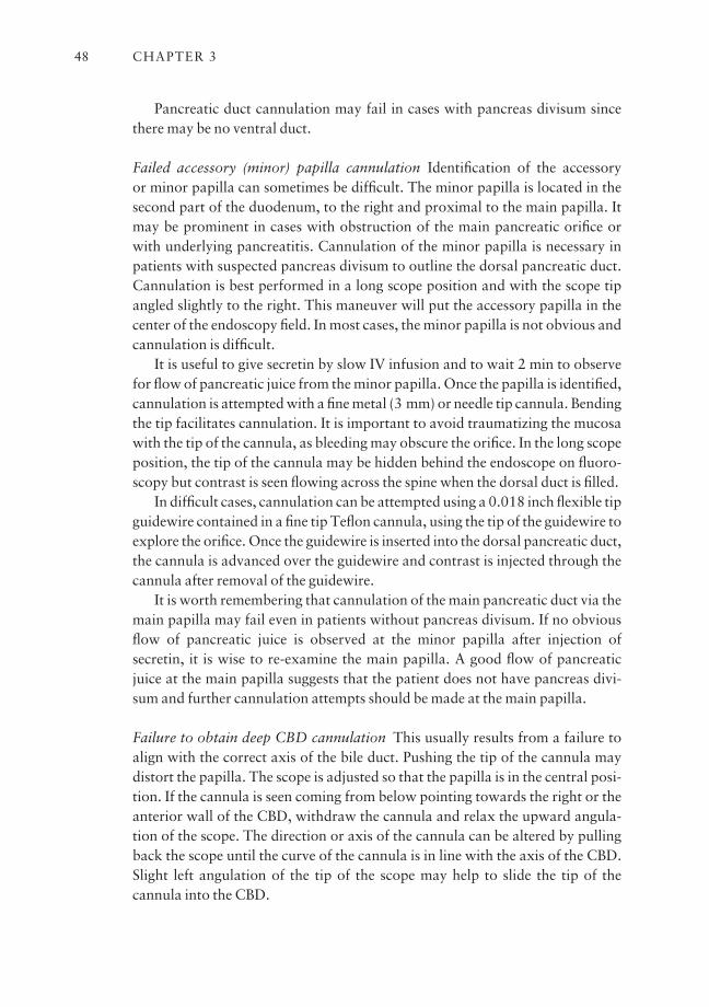
Pancreatic duct cannulation may fail in cases with pancreas divisum sincethere may be no ventral duct.
Failed accessory (minor) papilla cannulation Identification of the accessory or minor papilla can sometimes be difficult. The minor papilla is located in thesecond part of the duodenum, to the right and proximal to the main papilla. Itmay be prominent in cases with obstruction of the main pancreatic orifice orwith underlying pancreatitis. Cannulation of the minor papilla is necessary inpatients with suspected pancreas divisum to outline the dorsal pancreatic duct.Cannulation is best performed in a long scope position and with the scope tipangled slightly to the right. This maneuver will put the accessory papilla in thecenter of the endoscopy field. In most cases, the minor papilla is not obvious andcannulation is difficult.
It is useful to give secretin by slow IV infusion and to wait 2 min to observefor flow of pancreatic juice from the minor papilla. Once the papilla is identified,cannulation is attempted with a fine metal (3 mm) or needle tip cannula. Bendingthe tip facilitates cannulation. It is important to avoid traumatizing the mucosawith the tip of the cannula, as bleeding may obscure the orifice. In the long scopeposition, the tip of the cannula may be hidden behind the endoscope on fluoro-scopy but contrast is seen flowing across the spine when the dorsal duct is filled.
In difficult cases, cannulation can be attempted using a 0.018 inch flexible tipguidewire contained in a fine tip Teflon cannula, using the tip of the guidewire toexplore the orifice. Once the guidewire is inserted into the dorsal pancreatic duct,the cannula is advanced over the guidewire and contrast is injected through thecannula after removal of the guidewire.
It is worth remembering that cannulation of the main pancreatic duct via themain papilla may fail even in patients without pancreas divisum. If no obviousflow of pancreatic juice is observed at the minor papilla after injection ofsecretin, it is wise to re-examine the main papilla. A good flow of pancreaticjuice at the main papilla suggests that the patient does not have pancreas divi-sum and further cannulation attempts should be made at the main papilla.
Failure to obtain deep CBD cannulation This usually results from a failure toalign with the correct axis of the bile duct. Pushing the tip of the cannula maydistort the papilla. The scope is adjusted so that the papilla is in the central posi-tion. If the cannula is seen coming from below pointing towards the right or theanterior wall of the CBD, withdraw the cannula and relax the upward angula-tion of the scope. The direction or axis of the cannula can be altered by pullingback the scope until the curve of the cannula is in line with the axis of the CBD.Slight left angulation of the tip of the scope may help to slide the tip of the cannula into the CBD.
CHAPTER 348
Manipulation is best performed with intermittent injection of contrast tooutline the direction/axis of the CBD on fluoroscopy. Using a cannula with ametal or radiopaque tip will help in correct positioning. Care is taken to avoidrepeated injection or overfilling of the pancreatic duct. If the bile duct axis cannot be defined, it may be necessary to use a sphincterotome as previouslydescribed.
If the bile duct is defined, a guidewire can be used to facilitate deep cannula-tion. The guidewire is inserted initially into the bile duct and the cannula orsphincterotome is advanced over the guidewire. The guidewire is then removedand bile is aspirated back into the syringe before contrast is injected to fill thebile duct. Sometimes, stone impaction at the papilla or tumor involvement mayprevent deep cannulation of the CBD. A stiffer instrument such as a sphinctero-tome can be used to dislodge the impacted stone.
Precut sphincterotomy to assist in CBD cannulation
Precut sphincterotomy can facilitate deep cannulation of the bile duct, and isused when standard cannulation fails in the presence of known bile duct patho-logy (e.g. impacted stone or tumor). Since precutting carries significant hazards,and other safer techniques are available, it should be used only with great caution. There should be a specific indication and a strong need to gain accessinto the bile duct, such as palliation of malignant jaundice. Precut sphinctero-tomy should not be performed for a diagnostic ERCP or as an alternative to agood biliary cannulation technique.
Needle-knife precut technique
Precutting with the needle-knife is performed in two ways, either by insertingthe knife into the papilla and gently moving upwards, or by incising downwardsfrom above the papilla. Prior insertion of a stent into the pancreatic duct protects the pancreatic orifice and may minimize the risk of pancreatitis. Precutneedle-knife sphincterotomy over a stent is also used to perform accessorysphincterotomy for pancreas divisum.
Selective cannulation of the intrahepatic system (IHBD)
In a standard short scope position, the angulation of the scope, curvature of thecannula, and shape of the CBD all favor cannulation of the right hepatic system.Selective cannulation of the right hepatic system is facilitated by the use of a J-tipped guidewire or a straight guidewire contained in a curved catheter, althougha curved cannula may sometimes lodge in the cystic duct.
FUNDAMENTALS OF ERCP 49
Cannulation of the left hepatic system is more difficult, especially if there is stricture of the left hepatic duct. A straight tip catheter or a right angle tipnasobiliary tube can be used to aim the guidewire. Inflating an occlusion balloonin the mid common duct and using it as a fulcrum may help to direct the tip of aguidewire into the respective left and right hepatic ducts.
If the axis of the CBD is straight, the tip of the catheter or nasobiliary tube is positioned in the distal CBD pointing towards the left side, and a straightguidewire is inserted and directed towards the origin of the left hepatic duct.Rotation of the tip of the endoscope to the left may help to deflect the guidewireinto the left hepatic system.
If the axis of the CBD is curved, the guidewire usually ends up in the right hepatic duct. It may be useful to try and direct the tip of the catheter ornasobiliary tube against the wall of the common hepatic duct on the right side,using the common hepatic duct to deflect the tip of the guidewire into the leftsystem. Also, unwinding a looped guidewire gently at the bifurcation maydeflect the tip, thus flipping the guidewire into the left hepatic duct.
If withdrawal of the loop and tip deflection fail, it may be helpful to continuepushing the looped guidewire which may back itself into the left hepatic duct.Once the tip of the guidewire is inside the left system, the guidewire is advancedto gain a more secure position before the catheter or nasobiliary tube is ad-vanced over the guidewire into the left hepatic duct. It is important to rememberthat the distal 3 cm of a guidewire is floppy and advancing a catheter over thisportion of the guidewire may be difficult.
Pushing a stiff catheter may deflect the guidewire and thus the catheter intothe right hepatic system. It is therefore necessary to pass the guidewire furtherinto the desired portion of the intrahepatic system before advancing the catheterover the stiffer portion of the guidewire. Pushing the tip of the scope further intothe duodenum may straighten the axis of the bile duct and increase the chance ofdirecting the guidewire into the left hepatic duct. Selective cannulation can beperformed using wires with a J or curved tip and a torque control to deflect thewire into the respective ductal system.
Cannulation of the papilla in a Billroth II situation (Fig. 3.17)
Previous gastrectomy or gastroenterostomy changes the anatomy of the stom-ach. The approach to the papilla is not through the usual route via the pylorus.Instead the papilla is approached from below via the afferent loop of the gastroenterostomy.
It is worth remembering that the orifice of the afferent loop is usually locatedto the right of the anastomosis. Rotating the scope for a proper orientation, andturning the patient to the supine position, may help facilitate passage of theendoscope.
CHAPTER 350
In difficult cases, intubation of the gastroenterostomy is performed by backingthe scope into the correct loop. Sometimes biopsy forceps may help the passageor advancement of the scope into the afferent loop. Passage of the scope down thesmall intestine is similar to doing a colonoscopy with a side-viewing endoscope.
The presence of bile in the lumen does not always predict the afferent loop. Itis helpful to monitor the passage of the endoscope on fluoroscopy to determinethe direction and position of the scope. It is unlikely that the scope is in the affer-ent loop if the tip is down in the pelvis on fluoroscopy. The length of the afferentloop may vary and affect the success of reaching the papilla.
In situations where difficulty is encountered or the relevant segment is notclearly defined, it is worth taking a biopsy close to the gastroenterostomy wherethe bleeding can serve to identify the jejunal segment that has been explored. Ifintubation with a side-viewing scope fails, it may be necessary to use a forward-viewing colonoscope to examine and intubate the afferent loop. If the papilla issuccessfully identified, it may be useful to place a Savary guidewire through thecolonoscope and leave it in place to guide subsequent intubation with the side-viewing duodenoscope.
The papilla is inverted in the afferent limb and the closed off duodenumappears as a blind stump. Cannulation of the papilla in the inverted position canbe difficult. The pancreatic duct is cannulated more readily than the bile ductwhich comes down in a cephalic and steep axis. A straight cannula gives a betteraxis for cannulation. For CBD cannulation it is helpful to pull back the scope so that the tip is further away from the papilla and cannulation is performed
FUNDAMENTALS OF ERCP 51
Fig. 3.17 Billroth II cannulation. Approach via afferent loop. Straight catheter from adistance to obtain correct axis.
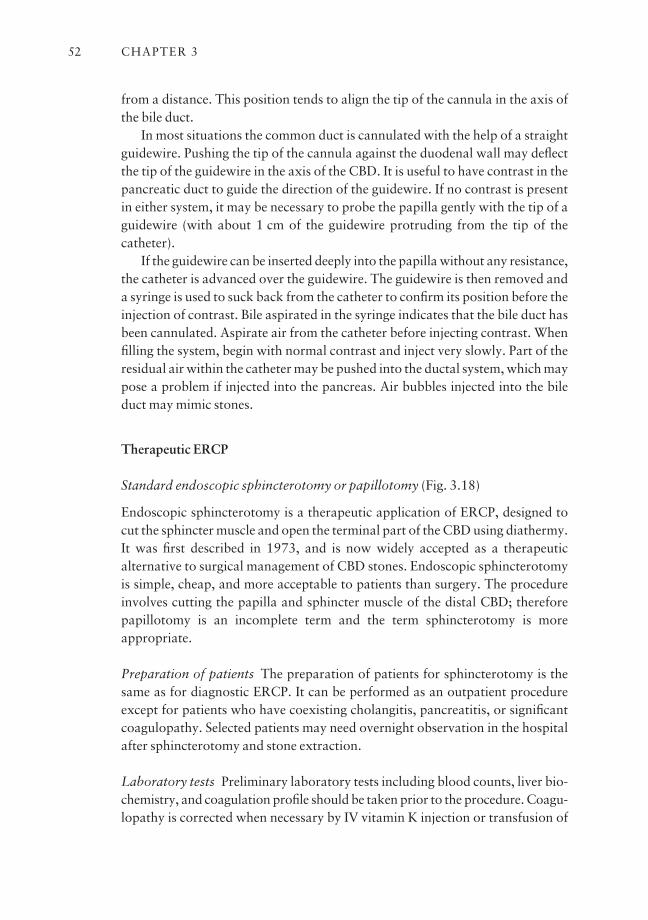
from a distance. This position tends to align the tip of the cannula in the axis ofthe bile duct.
In most situations the common duct is cannulated with the help of a straightguidewire. Pushing the tip of the cannula against the duodenal wall may deflectthe tip of the guidewire in the axis of the CBD. It is useful to have contrast in thepancreatic duct to guide the direction of the guidewire. If no contrast is presentin either system, it may be necessary to probe the papilla gently with the tip of aguidewire (with about 1 cm of the guidewire protruding from the tip of thecatheter).
If the guidewire can be inserted deeply into the papilla without any resistance,the catheter is advanced over the guidewire. The guidewire is then removed anda syringe is used to suck back from the catheter to confirm its position before theinjection of contrast. Bile aspirated in the syringe indicates that the bile duct hasbeen cannulated. Aspirate air from the catheter before injecting contrast. Whenfilling the system, begin with normal contrast and inject very slowly. Part of theresidual air within the catheter may be pushed into the ductal system, which maypose a problem if injected into the pancreas. Air bubbles injected into the bileduct may mimic stones.
Therapeutic ERCP
Standard endoscopic sphincterotomy or papillotomy (Fig. 3.18)
Endoscopic sphincterotomy is a therapeutic application of ERCP, designed tocut the sphincter muscle and open the terminal part of the CBD using diathermy.It was first described in 1973, and is now widely accepted as a therapeutic alternative to surgical management of CBD stones. Endoscopic sphincterotomyis simple, cheap, and more acceptable to patients than surgery. The procedureinvolves cutting the papilla and sphincter muscle of the distal CBD; thereforepapillotomy is an incomplete term and the term sphincterotomy is more appropriate.
Preparation of patients The preparation of patients for sphincterotomy is thesame as for diagnostic ERCP. It can be performed as an outpatient procedureexcept for patients who have coexisting cholangitis, pancreatitis, or significantcoagulopathy. Selected patients may need overnight observation in the hospitalafter sphincterotomy and stone extraction.
Laboratory tests Preliminary laboratory tests including blood counts, liver bio-chemistry, and coagulation profile should be taken prior to the procedure. Coagu-lopathy is corrected when necessary by IV vitamin K injection or transfusion of
CHAPTER 352
fresh frozen plasma. Patients are advised to stop taking aspirin and NSAIDs and anticoagulants are withheld for 5 days prior to elective sphincterotomy toavoid bleeding complications. For patients who require continued anticoagula-tion, for example those with prosthetic heart valves, admission for conversion to intravenous heparin may be required. The procedure is performed after withholding heparin for 4 h. Anticoagulation therapy is restarted after the procedure.
Antibiotics may be given to patients with coexisting cholangitis and thosewith significant biliary stasis.
We prefer to use the larger 4.2 mm channel endoscope for therapeutic pro-cedures because it can accept larger accessories.
The sphincterotome (or papillotome) Sphincterotomes are available in differ-ent designs with some specially designed for altered anatomy following gastricsurgery (e.g. Billroth II). In general, the sphincterotome is a single, double ortriple lumen Teflon catheter containing a continuous wire loop with 2–3 cm ofexposed wire close to the tip. The other end of the wire is insulated and con-nected via an adaptor to the diathermy or electrosurgical unit. The diathermy
FUNDAMENTALS OF ERCP 53
Fig. 3.18 Standard biliary papillotomy. Single lumen papillotome. Double lumenpapillotome over a guidewire. Use blended current, stepwise cut in 11–12 o’clock direction.Avoid excess tension on wire.
unit provides both cutting and coagulation currents, either separately or in combination (blended mode). The power setting on the diathermy machine canbe adjusted. The early single lumen sphincterotome allowed injection of con-trast through a single lumen, but leakage occurred around the side ports for thewire. Double lumen sphincterotomes allow injection of contrast or passage of aguidewire through a separate lumen and can be used for both diagnostic can-nulation and sphincterotomy (Fig. 3.18).
More recent sphincterotomes (e.g. DASH system, Wilson Cook) have a side-arm adaptor that allows contrast injection and insertion of a (0.025 or 0.035inch) guidewire at the same time. The adaptor can be tightened to close an O-ring around the guidewire to prevent spillage of contrast. The O-ring can beloosened to allow free passage of a guidewire through the sphincterotome.Triple lumen sphincterotomes allow both injection of contrast and passage of aguidewire independently.
Most sphincterotome wires tend to deviate to the right when bowed or tight-ened, potentially resulting in a deviated cut with an increased risk of complica-tions (i.e. bleeding, perforation, and pancreatitis). It is often necessary to shapethe wire to ensure that it remains in the 12 o’clock position when bowed to minimize the risk of complications. When a double or triple lumen sphinctero-tome is used, it is helpful to insert a guidewire to stabilize the sphincterotomeand maintain access into the ductal system during sphincterotomy.
A diagnostic ERCP is performed to define the anatomy of the biliary systemand to confirm the presence of stones. Using standard techniques the sphinctero-tome is inserted deeply into the CBD and its position confirmed either by injecting contrast or wiggling the sphincterotome under fluoroscopy. This is toprevent inadvertent cannulation and cutting of the pancreatic duct. The sphinc-terotome is withdrawn until only one-third of the wire lies within the papilla.The wire is then tightened so that it is in contact with the roof of the papilla.Excess tension on the wire should be avoided to prevent an uncontrolled or ‘zipper’ cut. The position of the wire is adjusted and maintained by the elevatorbridge and up/down control of the endoscope.
Electrosurgical unit A blended (cutting and coagulation) current is passed inshort bursts to cut the roof of the papilla in a stepwise manner in the 11–1o’clock direction. The power setting on different diathermy units varies depend-ing on the energy output of individual units, and has to be adjusted accordingly.For the Olympus diathermy (UES series), the power is set at 3–3.5 with ablended current; the setting on a Valley-lab diathermy machine is 3 of cuttingand 6 of coagulation, or a power setting of 30–40 W with a blended I current.The ERBE unit has a unique design that initially coagulates followed by cuttingthe papilla; the sphincterotomy can be performed in a more controlled fashion.
CHAPTER 354
Whitening of the tissue upon passage of current is indicative of the beginningof the cut. If the tissue does not blanch within a few seconds, it is necessary toreduce the length of wire in contact with the papilla. It is important to avoidincreasing the power setting of the diathermy unit without adjusting or reposi-tioning the wire.
Adequacy of sphincterotomy A gush of bile is usually seen flowing from the bileduct when the sphincter is cut. The sphincterotomy is then completed to its fulllength which is usually 1–1.5 cm. The safe length of a sphincterotomy dependson the configuration of the distal CBD and shape of the papilla.
However, it should not go beyond the impression of the common duct on the duodenal wall in order to avoid a perforation. The size of a sphincterotomycan be gauged by pulling a fully tightened (bowed) knife from within the distalbile duct to assess resistance to passage. An alternative method is to size thesphincterotomy by pulling an inflated occlusion balloon through the opening.Any deformity of the balloon would suggest resistance to its passage.
Wire-guided sphincterotomes An advantage of the double or triple lumensphincterotome is that it can be inserted over a guidewire especially in cases withdifficult cannulation. The guidewire also serves to anchor and stabilize thesphincterotome during sphincterotomy. A properly insulated guidewire shouldbe used to prevent the current from jumping between the diathermy wire and the guidewire, leading to an ineffective cut or injury to the liver. Most of the currently available guidewires with hydrophilic coating, such as the JAG wire (Microvasive) or Metro Tracer wire (Wilson Cook), can be used for thispurpose.
Periampullary diverticula and sphincterotomy Diverticula do not increase therisk of sphincterotomy unless the papilla is located on the edge or inside a largediverticulum. Cannulation may be technically more difficult and the risk of per-foration is increased as a result of a deviated cut.
Distorted anatomy A previous Billroth II gastrectomy increases the technicaldifficulty of ERCP and sphincterotomy. Although a forward-viewing scope mayfacilitate entry into the afferent loop, most experts prefer to use a side-viewingduodenoscope because of the additional elevator control. The success of sphinc-terotomy in patients with Billroth II gastrectomy is lower than that for patientswith normal anatomy. Since the approach to the papilla is through the afferentloop, the orientation of the papilla on endoscopy is reversed. Special sphinctero-tomes can be employed or a needle-knife may be used to cut the papilla over abiliary stent.
FUNDAMENTALS OF ERCP 55
Precut sphincterotomy for impacted stone
In general, deep cannulation of the CBD may fail in 5% of patients, but could behigher because of stone impaction at the ampulla. The biliary orifice is often dis-placed more distally because of the bulging papilla. In such cases, a precutsphincterotomy can be performed using a needle-knife which is basically a barewire that protrudes for 4–5 mm at the end of a Teflon catheter. A lower powersetting on the diathermy unit is often sufficient for precut sphincterotomy.
It is relatively safe to cut directly onto the bulging intraduodenal portion ofthe papilla. The needle-knife is either placed right at the orifice and the cut ismade upwards by lifting the knife, or the knife is used to cut down onto thepapilla by dropping the elevator. The risk of pancreatitis is minimal because theimpacted stone pushes the wall of the bile duct away from the pancreatic duct.Once access to the bile duct is achieved, the sphincterotomy can be extendedwith the needle-knife or using a standard sphincterotome. The impacted stonesometimes may pass spontaneously into the duodenum after an adequatesphincterotomy. Fine control of the needle-knife is difficult and carries anincreased risk of bleeding and perforation. It should not be undertaken lightlyby an inexperienced endoscopist or used as an alternative to good ERCP can-nulation techniques.
Indications for sphincterotomy and results
Endoscopic sphincterotomy is useful for the removal of residual or recurrentcommon duct stones in patients with a prior cholecystectomy. The success rateof removing stones ≤ 1 cm in diameter exceeds 95% in expert hands. Patientswith large stones may require special treatment such as mechanical lithotripsy(as discussed in a later section).
In elderly or high-risk patients with the gallbladder in situ, sphincterotomyfor CBD stone obstruction is indicated, especially in those presenting with acutecholangitis. Interval cholecystectomy may be performed but long-term follow-up suggests that cholecystectomy may not be necessary if gallbladder stones areabsent. Even for those with gallbladder stones the majority of patients remainasymptomatic on long-term follow-up. Only about 10% of patients developsubsequent biliary symptoms and require further intervention.
Urgent endoscopic drainage with sphincterotomy and/or insertion of a naso-biliary catheter is effective in reducing the overall mortality of suppurativecholangitis. A prospective randomized controlled study confirmed the benefitsof urgent endoscopic drainage over emergency surgery.
Sphincterotomy and removal of an impacted ampullary stone are beneficial inpatients with severe acute gallstone pancreatitis. A randomized controlled study
CHAPTER 356
demonstrated that urgent ERCP and sphincterotomy resulted in a significantreduction in mortality and complications compared to a control group.
Precut sphincterotomy may be indicated in patients with difficult cannulationto gain access to the bile duct for endoscopic biliary stenting. Sphincterotomyalso facilitates easier exchange of accessories and double stent placement. It isless commonly applied to treat patients with documented papillary stenosis orsphincter of Oddi dysfunction.
Complications of sphincterotomy
The results of sphincterotomy are operator dependent. An endoscopist musthave sufficient skill and experience with ERCP before attempting sphinctero-tomy in order to minimize the risk of complications. Bleeding, pancreatitis, andperforation can have serious consequences.
Postsphincterotomy bleeding Some bleeding may be observed at the time ofsphincterotomy in 2–5% of cases. Clinically significant bleeding is more likely incases with a deviated cut, a large sphincterotomy, and in patients with coexistingcoagulopathy. Active bleeding can be controlled by compressing the sphinctero-tomy with a balloon inflated inside the distal bile duct against the tip of the duodenoscope. Pure coagulation current may be applied to control the bleeding.Injection therapy with 1:10 000 dilution of epinephrine delivered into the apexand side of the sphincterotomy and adjacent tissue using a sclerotherapy needleis also very effective in controlling the bleeding. Injection therapy may give riseto tissue edema and potential biliary stasis. It is therefore necessary sometimes to insert a nasobiliary catheter or a stent to drain the bile duct. There may be a risk of pancreatitis if epinephrine is injected close to the pancreatic orifice.
In rare situations major hemorrhage may result from cutting an aberrantbranch of the retroduodenal artery. The resultant massive bleeding is difficult tocontrol with endoscopy and may require emergency surgery or radiologicalembolization of the bleeding vessel. Surgical treatment for postsphincterotomybleeding is not straightforward since it may be difficult to identify the exactbleeding site and the coagulated tissue does not hold sutures well. The risk ofrebleeding is high in patients with clotting disorders and these should be cor-rected and monitored for up to 7–10 days after the sphincterotomy. Patientsshould continue to withhold aspirin or NSAIDs for another 5 days to preventrecurrent bleeding.
Pancreatitis Pancreatitis may result from inadvertent cutting of or edemaaround the pancreatic orifice. It can also occur from repeated injection of con-trast into the pancreas or excess coagulation during biliary sphincterotomy.
FUNDAMENTALS OF ERCP 57
Post-ERCP pancreatitis can be reduced by ensuring drainage of the pancreaticduct using a temporary 3 Fr stent or a 5 Fr nasopancreatic catheter.
Cholangitis Acute cholangitis is a rare but important early complication following sphincterotomy. This may occur when contrast is injected into anobstructed biliary system but drainage cannot be established. Antibiotics shouldbe given promptly, but the risk is best minimized by ensuring drainage of the biliary system with an indwelling stent or nasobiliary catheter.
Perforation Perforation is a rare complication of sphincterotomy and mayoccur as a result of a deviated cut or excessive cutting of the papilla. Patientscomplain of pain and retroperitoneal free air may be demonstrated on fluoro-scopy. If recognized during ERCP, it may be useful to decompress the bile ductwith a nasobiliary catheter or an indwelling stent to reduce leakage and the riskof retroduodenal abscess formation. If perforation is suspected after the proce-dure, CT scan of the abdomen is the most sensitive test in detecting the presenceof retroduodenal air.
The patient should be kept nil by mouth with nasogastric tube decompres-sion. Intravenous fluids and broad spectrum antibiotics are given to preventinfection. Patients often respond to conservative management and bowel rest,and surgical treatment is usually not necessary. However, early consultation iswise and percutaneous drainage of retroduodenal fluid collection may be neces-sary to prevent abscess formation.
What to do if the sphincterotomy fails to cut
Before the sphincterotomy, it is important to check that the electrosurgical ordiathermy unit is working properly, the patient’s grounding plate is connected,and the correct adaptor is used for the sphincterotome. Poor contact of thegrounding plate can be improved using electroconducting gel or gauze soakedwith normal saline (not sterile water) placed between the patient and thegrounding plate.
If the electrical connections are correct and functional, an apparent failure tocut may be the result of having too much wire in contact with the tissue. With-draw the sphincterotome until only about one-third of the wire is left inside thebile duct. Too little wire in contact with the tissue also produces an ineffectivecut. Too much coagulation current leads to formation of a coagulum adherentto the wire and increases the resistance and difficulty in cutting the papilla. Itmay be necessary to remove the sphincterotome and clean the wire before further cutting or to insert the unbowed sphincterotome into the duct to clearthe coagulum. Poor contact between the wire and the tissue may also result inineffective cutting.
CHAPTER 358
As the sphincterotomy is being performed, it may be necessary to gentlytighten the wire and lift the sphincterotome with the elevator to maintain con-tact with the papilla. Whitening of the tissue indicates the beginning of a cut. Alot of smoke without cutting means that insufficient wire is in contact with thetissue or there is too much coagulation. Gently moving the wire to separate thecut edge of the papilla will facilitate further cutting and ensure proper contactwith the tissue. In patients with a thick papilla due to stone impaction or atumor, it may take some time for the wire to cut through. An impacted stone atthe papilla may prevent adequate tissue contact.
Too much tension on the wire may result in a sudden jump when the sphinc-ter muscle is completely severed. This uncontrolled or ‘zipper’ cut is due toexcess tension on the wire cutting the relatively thin-walled distal bile duct andis associated with an increased risk of bleeding and perforation.
The risk of a half cut
When excess coagulum forms, it may be necessary to remove the sphinctero-tome and to clean the wire. Tissue edema and charring around the sphinctero-tomy site may make subsequent cannulation of the CBD more difficult. There isa potential risk of dissection through a false tissue plane or submucosal injectionof contrast. Using a wire-guided sphincterotome and exchanging over a guide-wire prevent this potential complication. Indeed, inserting a guidewire withinthe bile duct serves also to stabilize the sphincterotome and facilitates exchangesand positioning of the sphincterotome.
What to do with a deviated cut
The risk of bleeding or perforation is increased if the biliary sphincterotomy isperformed outside of the ‘safety zone’, i.e. in the 11–1 o’clock direction. There isa tendency for most sphincterotomy wires to deviate to the right when beingtightened, thus increasing the risk of complications.
It is important to check the wire prior to the sphincterotomy. Some sphinc-terotomes have a stabilizing metal plate or differential catheter thickness thatallows the wire to exit in the 12 o’clock position (at least in theory). If the wirecomes out in a poor direction or orientation, it is necessary to train or shape thewire.
The purpose of training the wire is to ensure that it remains in the centralposition when being tightened. It is performed by turning the tip of the sphinc-terotome by 90° so that the wire is on the left side of the catheter tip, curling thetip of the sphincterotome with the fingers while at the same time tightening thewire. This helps to put a memory on the tip of the sphincterotome which keepsthe wire in the central position when tightened.
FUNDAMENTALS OF ERCP 59
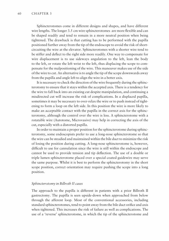
Sphincterotomes come in different designs and shapes, and have differentwire lengths. The longer 3.5 cm wire sphincterotomes are more flexible and canbe shaped readily and tend to remain in a more neutral position when beingtightened. The drawback is that cutting has to be performed with the papillapositioned further away from the tip of the endoscope to avoid the risk of short-circuiting the wire at the elevator. Sphincterotomes with a shorter wire tend tobe stiffer and deflect to the right side more readily. One way to compensate forwire displacement is to use sideways angulation to the left, lean the body to the left, or rotate the left wrist to the left, thus displacing the scope to com-pensate for the malpositioning of the wire. This maneuver makes use of the sideof the wire to cut. An alternative is to angle the tip of the scope downwards awayfrom the papilla and angle left to align the wire in a better axis.
It is necessary to check the direction of the wire frequently during the sphinc-terotomy to ensure that it stays within the accepted axis. There is a tendency forthe wire to fall back into an existing cut despite manipulation, and continuing amisdirected cut will increase the risk of complications. In a displaced papilla,sometimes it may be necessary to over-relax the wire or to push instead of tight-ening to form a loop on the left side. In this position the wire is more likely tomake an acceptable contact with the papilla in the correct axis for the sphinc-terotomy, although the control over the wire is less. A sphincterotome with arotatable wire (Autotome, Microvasive) may help in correcting the axis of thecut, especially with a distorted papilla.
In order to maintain a proper position for the sphincterotome during sphinc-terotomy, some endoscopists prefer to use a long-nose sphincterotome so thatthe wire can be steadied and maintained within the bile duct to minimize the riskof losing the position during cutting. A long-nose sphincterotome is, however,difficult to use for cannulation since the wire is still within the endoscope andcannot be used to provide tension and tip deflection. The use of a double ortriple lumen sphincterotome placed over a special coated guidewire may servethe same purpose. Whilst it is best to perform the sphincterotomy in the shortscope position, correct orientation may require pushing the scope into a longposition.
Sphincterotomy in Billroth II cases
The approach to the papilla is different in patients with a prior Billroth II gastrectomy. The papilla is seen upside-down when approached from belowthrough the afferent loop. Most of the conventional accessories, including standard sphincterotomes, tend to point away from the bile duct orifice and axiswhen tightened. This increases the risk of failure as well as complications. Theuse of a ‘reverse’ sphincterotome, in which the tip of the sphincterotome and
CHAPTER 360
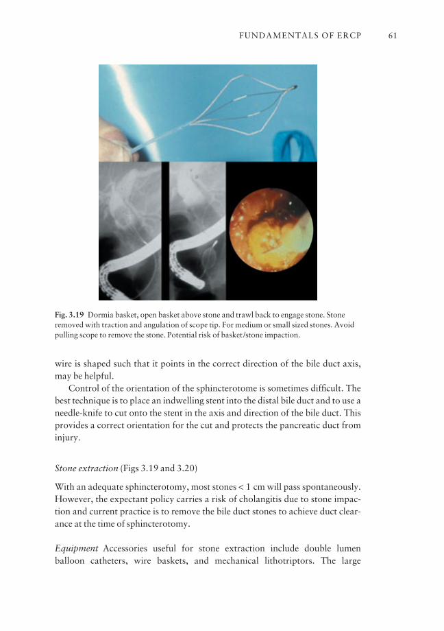
wire is shaped such that it points in the correct direction of the bile duct axis,may be helpful.
Control of the orientation of the sphincterotome is sometimes difficult. Thebest technique is to place an indwelling stent into the distal bile duct and to use aneedle-knife to cut onto the stent in the axis and direction of the bile duct. Thisprovides a correct orientation for the cut and protects the pancreatic duct frominjury.
Stone extraction (Figs 3.19 and 3.20)
With an adequate sphincterotomy, most stones < 1 cm will pass spontaneously.However, the expectant policy carries a risk of cholangitis due to stone impac-tion and current practice is to remove the bile duct stones to achieve duct clear-ance at the time of sphincterotomy.
Equipment Accessories useful for stone extraction include double lumen balloon catheters, wire baskets, and mechanical lithotriptors. The large
FUNDAMENTALS OF ERCP 61
Fig. 3.19 Dormia basket, open basket above stone and trawl back to engage stone. Stoneremoved with traction and angulation of scope tip. For medium or small sized stones. Avoidpulling scope to remove the stone. Potential risk of basket/stone impaction.
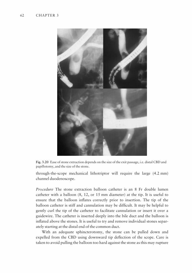
through-the-scope mechanical lithotriptor will require the large (4.2 mm) channel duodenoscope.
Procedure The stone extraction balloon catheter is an 8 Fr double lumencatheter with a balloon (8, 12, or 15 mm diameter) at the tip. It is useful toensure that the balloon inflates correctly prior to insertion. The tip of the balloon catheter is stiff and cannulation may be difficult. It may be helpful togently curl the tip of the catheter to facilitate cannulation or insert it over aguidewire. The catheter is inserted deeply into the bile duct and the balloon isinflated above the stones. It is useful to try and remove individual stones separ-ately starting at the distal end of the common duct.
With an adequate sphincterotomy, the stone can be pulled down andexpelled from the CBD using downward tip deflection of the scope. Care istaken to avoid pulling the balloon too hard against the stone as this may rupture
CHAPTER 362
Fig. 3.20 Ease of stone extraction depends on the size of the exit passage, i.e. distal CBD andpapillotomy, and the size of the stone.
the balloon. As the balloon can be deformed, the balloon may slip past the stonesresulting in stone impaction. Stone extraction is best confirmed by observingstone passage from the sphincterotomy. Alternatively, an occlusion cholangio-gram can be performed to document complete clearance of the bile ducts.
Stones can also be removed using a wire basket. The basket is made of fourwires and shaped such that the wires open like a trap to engage the stones. Thebasket is inserted and opened beyond the stones and withdrawn in a fullyopened position. The basket is moved gently up and down or jiggled around thestone to trap it. When the stone is engaged the basket is closed gently and pulledback to the papilla. The tip of the endoscope is angled up against the papillaryorifice and tension is applied. The stone is extracted by downward tip deflectionand right rotation of the endoscope. If necessary, the maneuver is repeated toremove the stone. A newer design has eight wires on the top (Flower basket,Olympus) which result in a small mesh size between the wires, and is useful fortrapping smaller stones or fragments.
Mechanical lithotripsy
Large > 15 mm, common duct stones are difficult to remove, especially if considerable discrepancy exists between the size of the stone and the diameter ofthe exit passage, i.e. a narrowed distal bile duct, a small sphincterotomy, and inthose who had only balloon sphincteroplasty for stone extraction (Fig. 3.21).Extension of the sphincterotomy is not always possible and may carry an in-creased risk of bleeding and perforation. Lithotripsy facilitates stone extractionand common duct clearance by crushing the stones using strong wire basketsbefore extraction.
There are different designs for lithotripsy basketsaone type requires cuttingthe handle of the basket and removing the endoscope prior to stone fragmenta-tion, e.g. Soehendra lithotriptor (Wilson Cook Medical, Winston Salem, NC).This consists of a 14 Fr metal sheath and a self-locking crank handle. Thelithotriptor can be used with large lithotripsy baskets or standard stone extrac-tion baskets. These are typically four wire hexagonal baskets measuring 2 cm by3 cm or 3 cm by 5 cm in diameter (Fig. 3.22).
Another type is a pre-assembled through-the-scope lithotripsy basket whichcan be inserted through a therapeutic duodenoscope, e.g. BML lithotripsy baskets (Olympus Co, Tokyo, Japan). The BML lithotriptor has three layersaastrong four wire basket, a Teflon catheter, and an overlying metal sheath. Thereusable version requires assembly by inserting the Teflon catheter initiallythrough the metal sheath and then loading the basket retrogradely on to theTeflon catheter. The wires are soldered together on to a shaft which is connectedto the crank handle. Contrast is injected via the Teflon catheter. The opening
FUNDAMENTALS OF ERCP 63

and closing of the basket are controlled with the handle. Stone engagement is performed with the Teflon catheter and basket. The metal sheath is usuallyadvanced over the Teflon catheter up to the level of the stone when lithotripsy isrequired. Traction is applied to the wires by turning the control wheel in orderto crush the stone. As the control does not have a built-in locking mechanism,traction should be applied slowly and continuously to allow time for the wiresto cut through the stone.
The reusable system can be taken apart after lithotripsy for cleaning andsterilization. The disposable version comes with the lithotripsy basket, Tefloncatheter, and metal sheath all built into one. The set-up is designed to break atthe connection between the basket and the crank handle. The basket wires arealso designed to break at the tip to prevent having a broken basket around animpacted stone in the bile duct. The larger lithotripsy baskets, or BML-3Q or201 equivalent, have a slightly thicker metal sheath that goes through a 4.2 mmchannel scope; contrast injection is possible. The smaller basket, or BML-4Q or
CHAPTER 364
Fig. 3.21 Balloon stone extraction. Extraction of small stones in small ducts afterpapillotomy; stone expelled by retracting balloon. Large stones can be removed if axis iscorrect. Risk of stone impaction with inadequate papillotomy.
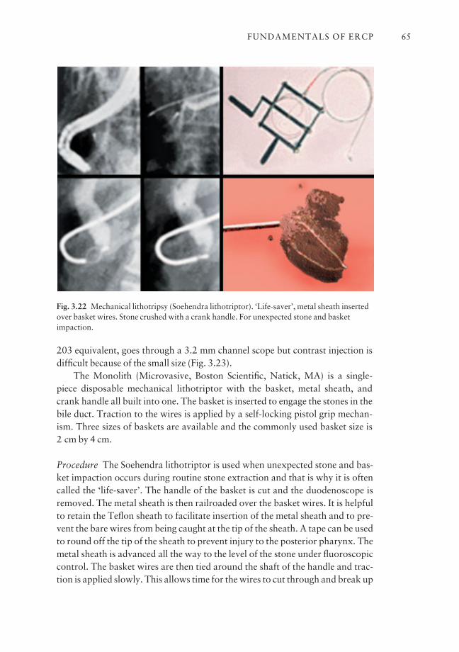
203 equivalent, goes through a 3.2 mm channel scope but contrast injection isdifficult because of the small size (Fig. 3.23).
The Monolith (Microvasive, Boston Scientific, Natick, MA) is a single-piece disposable mechanical lithotriptor with the basket, metal sheath, andcrank handle all built into one. The basket is inserted to engage the stones in thebile duct. Traction to the wires is applied by a self-locking pistol grip mechan-ism. Three sizes of baskets are available and the commonly used basket size is 2 cm by 4 cm.
Procedure The Soehendra lithotriptor is used when unexpected stone and bas-ket impaction occurs during routine stone extraction and that is why it is oftencalled the ‘life-saver’. The handle of the basket is cut and the duodenoscope isremoved. The metal sheath is then railroaded over the basket wires. It is helpfulto retain the Teflon sheath to facilitate insertion of the metal sheath and to pre-vent the bare wires from being caught at the tip of the sheath. A tape can be usedto round off the tip of the sheath to prevent injury to the posterior pharynx. Themetal sheath is advanced all the way to the level of the stone under fluoroscopiccontrol. The basket wires are then tied around the shaft of the handle and trac-tion is applied slowly. This allows time for the wires to cut through and break up
FUNDAMENTALS OF ERCP 65
Fig. 3.22 Mechanical lithotripsy (Soehendra lithotriptor). ‘Life-saver’, metal sheath insertedover basket wires. Stone crushed with a crank handle. For unexpected stone and basketimpaction.
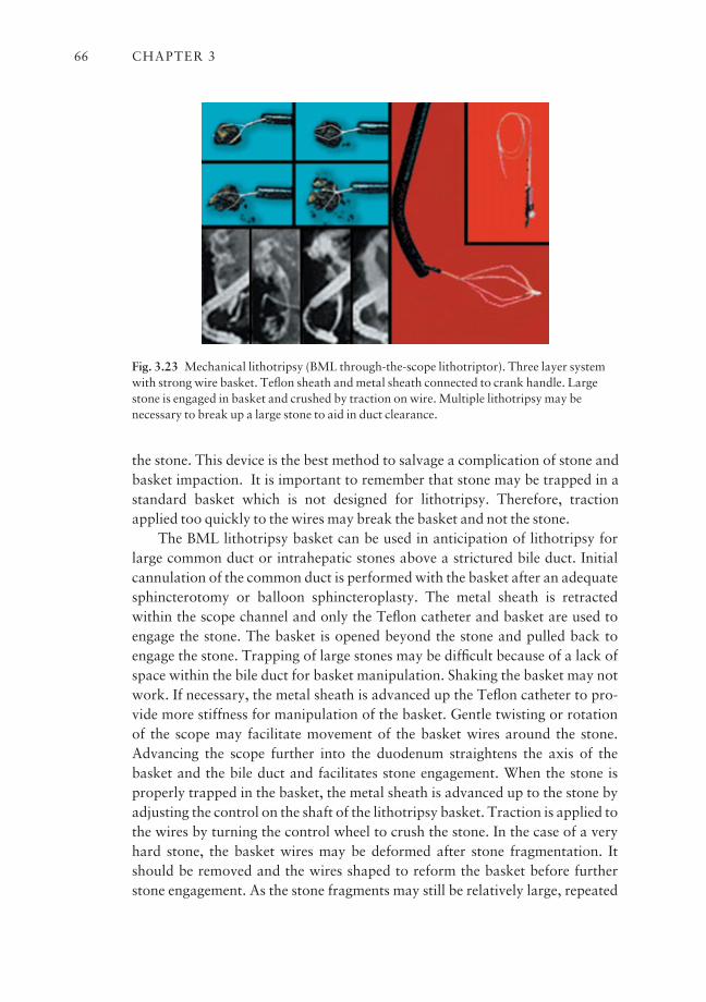
the stone. This device is the best method to salvage a complication of stone andbasket impaction. It is important to remember that stone may be trapped in astandard basket which is not designed for lithotripsy. Therefore, tractionapplied too quickly to the wires may break the basket and not the stone.
The BML lithotripsy basket can be used in anticipation of lithotripsy forlarge common duct or intrahepatic stones above a strictured bile duct. Initialcannulation of the common duct is performed with the basket after an adequatesphincterotomy or balloon sphincteroplasty. The metal sheath is retractedwithin the scope channel and only the Teflon catheter and basket are used toengage the stone. The basket is opened beyond the stone and pulled back toengage the stone. Trapping of large stones may be difficult because of a lack ofspace within the bile duct for basket manipulation. Shaking the basket may notwork. If necessary, the metal sheath is advanced up the Teflon catheter to pro-vide more stiffness for manipulation of the basket. Gentle twisting or rotation of the scope may facilitate movement of the basket wires around the stone.Advancing the scope further into the duodenum straightens the axis of the basket and the bile duct and facilitates stone engagement. When the stone isproperly trapped in the basket, the metal sheath is advanced up to the stone byadjusting the control on the shaft of the lithotripsy basket. Traction is applied tothe wires by turning the control wheel to crush the stone. In the case of a veryhard stone, the basket wires may be deformed after stone fragmentation. Itshould be removed and the wires shaped to reform the basket before furtherstone engagement. As the stone fragments may still be relatively large, repeated
CHAPTER 366
Fig. 3.23 Mechanical lithotripsy (BML through-the-scope lithotriptor). Three layer systemwith strong wire basket. Teflon sheath and metal sheath connected to crank handle. Largestone is engaged in basket and crushed by traction on wire. Multiple lithotripsy may benecessary to break up a large stone to aid in duct clearance.
stone crushing is necessary to facilitate stone extraction and duct clearance. Asdiscussed above, the disposable systems BML-201, 202, 203, and 204 are usedin a similar fashion.
The Monolith lithotriptor is inserted through the duodenoscope, the metalsheath is advanced into the bile duct, and the basket is opened and pulled backto engage the stone. Contrast can be injected to define the position of the stone.Once the stone is engaged, traction is applied to the wires to crush the stone. Asthe basket wires can become deformed, reshaping the wires is necessary beforelithotripsy is repeated. Mechanical problems including failure of proper open-ing of the basket and damage to the scope elevator have been reported.
Results The new lithotripsy baskets are strong and successful mechanicallithotripsy depends mostly on effective trapping of the stone. Lithotripsy mayfail in the presence of stone impaction or if there is insufficient room in the CBDfor manipulation of the basket. Partial fragmentation of a very large stone maybe possible, although the wires may tend to slip around the stone. Repeatedstone crushing is necessary to break up the large fragments. The reported successrate of mechanical lithotripsy for large stones ranges from 85% to 90%, improv-ing the overall common bile duct clearance rate to over 95%.
Complications of lithotripsy
The Soehendra lithotriptor provides effective crushing of the stone in unexpectedcases with stone and basket impaction. However, when a standard basket isused for stone extraction, the basket wires may break in the duodenum resulting in stone and a broken basket impacted in the bile duct. Such cases may requiresurgical common duct exploration to deliver the impacted stone and basket.Special precautions including slow application of traction to the wires may pre-vent this complication.
With the BML system, the baskets are made to break at the connection of theshaft with the handle. When this happens, Olympus has made a special metaloversheath and a crank handle similar to the design of the Soehendra lithotriptor.The standard metal sheath of the lithotriptor basket is replaced by this specialmetal sheath before stone fragmentation is continued. We do not recommendadapting the Soehendra lithotriptor handle to the BML basket for lithotripsy.
Perforation is an uncommon event and rarely occurs as a result of the stiffbasket perforating the bile duct. Excessive force in removing the impacted basket and stone may result in bruising of the pancreatic orifice and a potentialrisk of pancreatitis. Incomplete CBD clearance without adequate drainage mayresult in cholangitis due to retained stone fragments. Forceful extraction of largestone fragments should be avoided to minimize the risk of scope trauma to the
FUNDAMENTALS OF ERCP 67
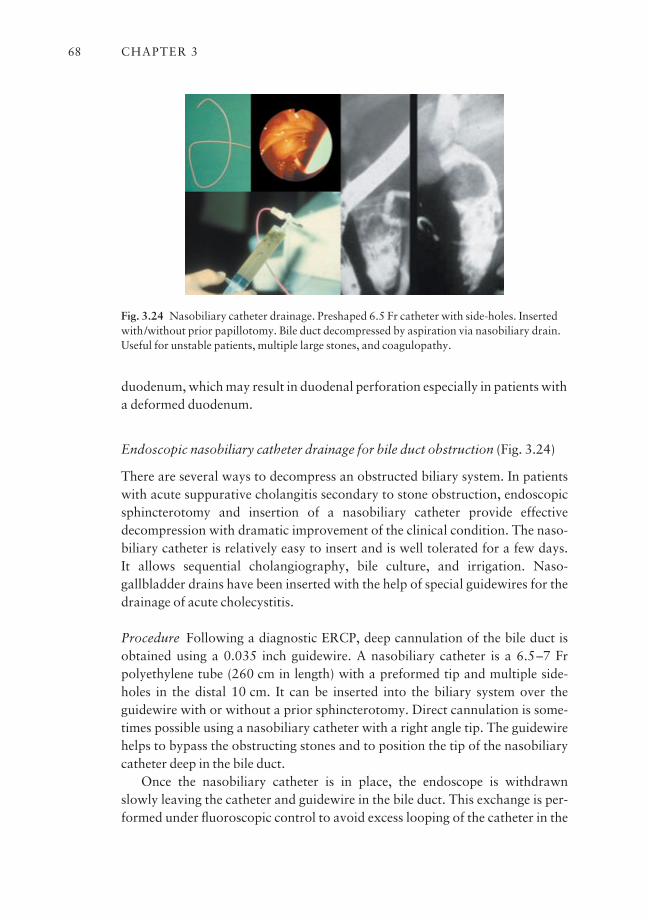
duodenum, which may result in duodenal perforation especially in patients witha deformed duodenum.
Endoscopic nasobiliary catheter drainage for bile duct obstruction (Fig. 3.24)
There are several ways to decompress an obstructed biliary system. In patientswith acute suppurative cholangitis secondary to stone obstruction, endoscopicsphincterotomy and insertion of a nasobiliary catheter provide effective decompression with dramatic improvement of the clinical condition. The naso-biliary catheter is relatively easy to insert and is well tolerated for a few days. It allows sequential cholangiography, bile culture, and irrigation. Naso-gallbladder drains have been inserted with the help of special guidewires for thedrainage of acute cholecystitis.
Procedure Following a diagnostic ERCP, deep cannulation of the bile duct isobtained using a 0.035 inch guidewire. A nasobiliary catheter is a 6.5–7 Frpolyethylene tube (260 cm in length) with a preformed tip and multiple side-holes in the distal 10 cm. It can be inserted into the biliary system over theguidewire with or without a prior sphincterotomy. Direct cannulation is some-times possible using a nasobiliary catheter with a right angle tip. The guidewirehelps to bypass the obstructing stones and to position the tip of the nasobiliarycatheter deep in the bile duct.
Once the nasobiliary catheter is in place, the endoscope is withdrawn slowly leaving the catheter and guidewire in the bile duct. This exchange is per-formed under fluoroscopic control to avoid excess looping of the catheter in the
CHAPTER 368
Fig. 3.24 Nasobiliary catheter drainage. Preshaped 6.5 Fr catheter with side-holes. Insertedwith/without prior papillotomy. Bile duct decompressed by aspiration via nasobiliary drain.Useful for unstable patients, multiple large stones, and coagulopathy.
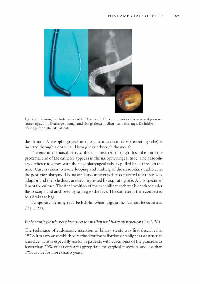
duodenum. A nasopharyngeal or nasogastric suction tube (rerouting tube) isinserted through a nostril and brought out through the mouth.
The end of the nasobiliary catheter is inserted through this tube until theproximal end of the catheter appears in the nasopharyngeal tube. The nasobili-ary catheter together with the nasopharyngeal tube is pulled back through thenose. Care is taken to avoid looping and kinking of the nasobiliary catheter inthe posterior pharynx. The nasobiliary catheter is then connected to a three-wayadaptor and the bile ducts are decompressed by aspirating bile. A bile specimenis sent for culture. The final position of the nasobiliary catheter is checked underfluoroscopy and anchored by taping to the face. The catheter is then connectedto a drainage bag.
Temporary stenting may be helpful when large stones cannot be extracted(Fig. 3.25).
Endoscopic plastic stent insertion for malignant biliary obstruction (Fig. 3.26)
The technique of endoscopic insertion of biliary stents was first described in1979. It is now an established method for the palliation of malignant obstructivejaundice. This is especially useful in patients with carcinoma of the pancreas asfewer than 20% of patients are appropriate for surgical resection, and less than1% survive for more than 5 years.
FUNDAMENTALS OF ERCP 69
Fig. 3.25 Stenting for cholangitis and CBD stones. 10 Fr stent provides drainage and preventsstone impaction. Drainage through and alongside stent. Short-term drainage. Definitivedrainage for high-risk patients.
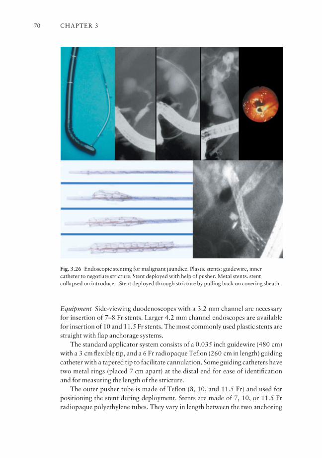
Equipment Side-viewing duodenoscopes with a 3.2 mm channel are necessaryfor insertion of 7–8 Fr stents. Larger 4.2 mm channel endoscopes are availablefor insertion of 10 and 11.5 Fr stents. The most commonly used plastic stents arestraight with flap anchorage systems.
The standard applicator system consists of a 0.035 inch guidewire (480 cm)with a 3 cm flexible tip, and a 6 Fr radiopaque Teflon (260 cm in length) guidingcatheter with a tapered tip to facilitate cannulation. Some guiding catheters havetwo metal rings (placed 7 cm apart) at the distal end for ease of identificationand for measuring the length of the stricture.
The outer pusher tube is made of Teflon (8, 10, and 11.5 Fr) and used forpositioning the stent during deployment. Stents are made of 7, 10, or 11.5 Frradiopaque polyethylene tubes. They vary in length between the two anchoring
CHAPTER 370
Fig. 3.26 Endoscopic stenting for malignant jaundice. Plastic stents: guidewire, innercatheter to negotiate stricture. Stent deployed with help of pusher. Metal stents: stentcollapsed on introducer. Stent deployed through stricture by pulling back on covering sheath.
flaps (5, 7, 9, 10, 12, 15 cm). There is no inner catheter for the 7 Fr stenting system.
Other stents have double pigtails that serve to anchor the stent to preventupward or downward migration. However, the straight stent with side flaps is preferred because it maintains its position with infrequent dislocation. It provides maximal flow and minimizes the risk of blockage compared with thedouble pigtail stents, which have a smaller lumen and side-holes.
Preparation of patient The resectability of the underlying cancer or lesion and the clinical condition of the patient should be carefully assessed prior tostenting. Initial investigations include liver function tests, abdominal ultrasound,and CT scanning to define the nature and level of obstruction. Endoscopic ultra-sound and fine-needle aspiration biopsy are also useful for staging and diagnosisof the underlying cancer. MRCP may be necessary in cases of hilar obstructionto outline the obstructed ductal system.
Coagulation defects are corrected by IV vitamin K1 and/or fresh frozenplasma. Prophylactic antibiotic may be given before the procedure.
Procedure Preparation and sedation of the patient are the same as for standardERCP procedures. A diagnostic ERCP is performed and the level of obstructionis defined. Sphincterotomy is not necessary for placement of a single stent but isuseful to facilitate insertion of multiple stents, and may prevent the complica-tion of pancreatitis following stenting for hilar strictures caused by pressure ofthe stents against the pancreatic orifice.
Initial cannulation and insertion of the guidewire past the stricture can beperformed using standard accessories. It is preferable to use a guidewire with a hydrophilic tip for easy passage through the stricture. Brush cytology can be taken by exchanging the cytology brush over the guidewire. The guidingcatheter is then exchanged over the guidewire to bypass the obstruction. Theguidewire is then removed and additional bile samples can be aspirated for cul-ture and cytology. The length of the stricture is determined on cholangiographywith the help of radiopaque ring markers.
A suitable length stent is chosen so that the proximal flap of the stent liesabout 1 cm above the obstruction. The optimal length of the stent can be deter-mined by measuring the separation between the proximal obstruction and thelevel of the papilla on the radiographs. It can be estimated with reference to the scope diameter or by using the radiopaque markers on the inner catheter.The correct length of the stent is determined by correcting for the magnificationfactor of the fluoroscopy unit. The stent length can also be determined byretracting the guidewire between the two points and measuring the distancetraveled on the outside of the catheter.
FUNDAMENTALS OF ERCP 71
The stricture may be dilated (when particularly tight) prior to stent insertionwith graded dilators or pneumatic balloon dilators (4, 6, 8 mm) inserted overthe guidewire. The stent is loaded onto the catheter or guidewire and then posi-tioned through the obstruction with the help of the pusher tube. The stent isdeployed by removing the inner catheter and guidewire. Bile is seen drainingthrough the stent into the duodenum. The pusher is then removed.
One-step introducer system A modified introducer system combines the innercatheter and pusher into a single system using a Luer lock mechanism. A suit-able length stent is preloaded on to the introducer system, inserted over theguidewire, and positioned through the obstruction. Once the inner catheter is in position, the pusher and inner catheter are unlocked and the stent is pushedand deployed across the obstruction by withdrawing the inner catheter andguidewire.
Bilateral stenting for hilar obstruction
Hilar tumors pose difficult technical problems. Whether it is necessary or desirable to drain all obstructed ducts remains controversial. When one duct isdominant, draining it alone may be sufficient. To drain both the right and lefthepatic systems, it is necessary to insert two guidewires separately. Correctanchorage of the guidewires is necessary because of the potential for dislodge-ment during exchange of accessories. This can be achieved using either a hemo-stat to clip the wire to the biopsy valve or a special anchoring unit that comeswith a particular stenting system.
We recommend performing routine balloon dilation of hilar strictures priorto stent insertion as they are often very tight. It is better to start with the left hepatic system due to the more difficult access and axis with stent insertion.
Once the left stent is in place, another straight stent is introduced into theright side to drain part or all of the right hepatic ducts. Care is taken not to pushthe first stent into the bile duct during insertion of the second stent. In general,successful drainage of the left hepatic system alone may result in improvementof the liver function. The left hepatic duct branches off after 2 cm in contrast tothe right hepatic duct which branches off after 1 cm.
Multiple segment obstruction is more likely to occur on the right side.Successful drainage and recovery of liver function are therefore more difficultdue to the limited volume of liver tissue drained by the individual segmental hepatic ducts. Some endoscopists recommend multiple stents in all cases toachieve complete drainage.
Every attempt should be made to avoid overfilling the intrahepatic system to minimize the risk of sepsis. If endoscopic drainage fails, percutaneous trans-
CHAPTER 372
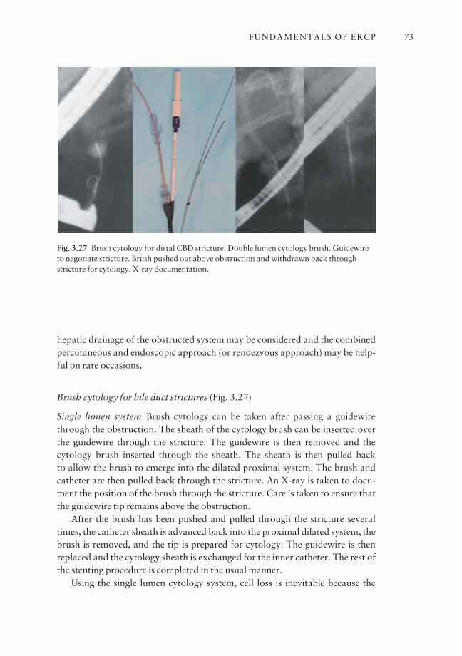
hepatic drainage of the obstructed system may be considered and the combinedpercutaneous and endoscopic approach (or rendezvous approach) may be help-ful on rare occasions.
Brush cytology for bile duct strictures (Fig. 3.27)
Single lumen system Brush cytology can be taken after passing a guidewirethrough the obstruction. The sheath of the cytology brush can be inserted overthe guidewire through the stricture. The guidewire is then removed and thecytology brush inserted through the sheath. The sheath is then pulled back to allow the brush to emerge into the dilated proximal system. The brush andcatheter are then pulled back through the stricture. An X-ray is taken to docu-ment the position of the brush through the stricture. Care is taken to ensure thatthe guidewire tip remains above the obstruction.
After the brush has been pushed and pulled through the stricture severaltimes, the catheter sheath is advanced back into the proximal dilated system, thebrush is removed, and the tip is prepared for cytology. The guidewire is thenreplaced and the cytology sheath is exchanged for the inner catheter. The rest ofthe stenting procedure is completed in the usual manner.
Using the single lumen cytology system, cell loss is inevitable because the
FUNDAMENTALS OF ERCP 73
Fig. 3.27 Brush cytology for distal CBD stricture. Double lumen cytology brush. Guidewireto negotiate stricture. Brush pushed out above obstruction and withdrawn back throughstricture for cytology. X-ray documentation.
brush is pulled back through the whole length of the catheter. It is useful to aspir-ate bile from the catheter to collect any dislodged cells within the catheter toimprove the diagnostic yield.
Double lumen system Double lumen cytology brush systems allow the guide-wire to pass through the central lumen of a tapered tip catheter and the cytologybrush exits from the side lumen near the tip. With the brush in the retracted position, the catheter is inserted over the guidewire through the obstruction.With the help of radiopaque markers, the cytology brush is advanced from thecatheter into the dilated proximal system. The entire apparatus is then movedback and forth through the area of the stricture to obtain samples. An X-ray istaken for documentation.
The brush is then withdrawn into the catheter and the whole set-up isexchanged over the guidewire without having to pull back the brush completely.This set-up ensures access is maintained across the stricture by the guidewire for the ease of subsequent stenting or drainage and also avoids cell loss. Whenthe brush is withdrawn, the tip of the brush is cut off and saved in the cytologysolution. It is useful to remove the stylet and to flush air or water through thechannel of the brush to remove any fluid inside for cytological examination.
Assessment of response to biliary stenting
The clinical course of the patient is a good guide to the function of the stent.With successful drainage, pruritus usually disappears in 1–2 days as jaundicebegins to resolve. Serum bilirubin declines by a mean of 2–3 mg/dl per day. Withdistal bile duct obstruction, bilirubin levels may return to normal after 1–2 weeks.Incomplete or slow recovery of liver function may be related to prolongedobstruction which affects hepatocyte function or may be due to inadequate or incomplete drainage because of multiple segment involvement, as in hilarobstruction. The presence of an air cholangiogram suggests stent patency whichcan also be assessed by an EHIDA scan. Delayed appearance of radioisotope in the biliary system and intestine, with appearance of radioisotope in urine,suggests stent blockage.
Results of biliary stenting The success rates for biliary stenting vary dependingon the level of obstruction. They are high for mid- or distal CBD obstruction and lower for hilar obstruction. The success rates for draining both the right and left hepatic systems are low in patients with bifurcation lesions. Failure of endoscopic stenting may be due to tumor compression and/or distortion of theduodenum, marked displacement of the papilla, or failure of insertion of theguidewire through a very tight stricture.
CHAPTER 374

Complications of stenting
Early complications Early complications of stenting include pancreatitis, bleed-ing if a sphincterotomy is performed, cholangitis in patients with bifurcationtumors, and early stent blockage by blood clots. Guidewire perforation througha soft and necrotic tumor has been reported.
Late complications Late complications are largely due to stent blockage by bacterial biofilm and biliary sludge, resulting in recurrent jaundice and cholan-gitis. Stent dislocation and traumatic ulceration of the duodenum by the distaltip of the stent may occur. Acute cholecystitis secondary to stenting is a rarecomplication.
Recurrent jaundice is a major late complication of endoscopic stenting.Tumor extension may account for a small proportion of cases. The most impor-tant cause is clogging of the lumen of the stent by biliary sludge. Sludge consistslargely of calcium bilirubinate and small amounts of calcium palmitate, choles-terol, mucoprotein, and bacteria. Bacterial infection is important in initiatingsludge formation through adherence and formation of a bacterial biofilm. Thelikely source of bacteria is ascending infection from the duodenum via the stentor descending infection through the portal system. The bacteria are mostly largebowel flora.
Different methods have been tested to prevent stent blockage. Larger lumenstents delay the onset of clogging. Stent exchange at regular intervals also prevents the clinical risk of blockage. Antibacterial plastics and prophylacticantibiotics have not produced any clinically significant benefits. An alternativesolution to the blockage of plastic stents is to use self-expandable metal meshstents.
Self-expandable metal stents
Self-expandable metal stents (SEMS) were introduced as a means of prolongingstent patency. SEMS expand to 1 cm diameter and do not become obstructed bybacterial biofilm. However, metal stent occlusion does occur, but mostly as aresult of tumor/tissue ingrowth or overgrowth.
Stent configurations The commonly used SEMS have an open mesh design.Variations include the Wallstent, the Diamond Stent, the Spiral Z-Stent, the Za-Stent, and the more recently introduced Zilver Stent. SEMS are made of surgical-grade stainless steel or nitinol, a nickel–titanium alloy that provides a high degree of flexibility and is kink resistant. However, nitinol is lessradiopaque than stainless steel and additional radiopaque (gold or platinum)
FUNDAMENTALS OF ERCP 75

markers are put on the stents to improve radiopacity to facilitate proper posi-tioning during deployment. Covered SEMS are now available. One example isthe Wallstent (Microvasive) which has a polymer (Permalune) coating on theinside of the stent except for the proximal and distal 1 cm. This membrane isdesigned to prevent tumor ingrowth and prolong stent patency.
Lengths of stents SEMS usually come in two or three different expandedlengths (e.g. 4.8, 6.8, and 8.0 cm for the Wallstents, 5.7 and 7.5 cm for theSpiral Z-Stents). The Wallstent foreshortens after deployment to about two-thirds of its collapsed length when it is fully expanded. The Spiral Z- and ZilverStents do not shorten after deployment.
Introducer system for SEMS In general, the wire mesh metal stents are col-lapsed and restrained on a 6–6.5 Fr introducer catheter by an 8–8.5 Fr over-lying plastic sheath. Smaller 7/7.5 Fr introducer systems are now available.Sterile water or saline is initially injected to flush the system to minimize frictionbetween the stent and the restraining sheath and to facilitate stent deployment.The whole system is placed over the guidewire and advanced through theobstruction.
With the stent correctly positioned across the stricture, the overlying sheathis pulled back while the handle is held steadily to hold the introducer catheterand guidewire in position. The stent is deployed slowly in a stepwise manner.Stent deployment can be monitored under fluoroscopic control using theradiopaque markers. Adjustment of the stent position may be necessary beforecomplete deployment, especially for stents that foreshorten, e.g. the Wallstent.It is also easier to pull back than to advance a partially deployed stent throughthe stricture or obstruction.
Metal stents are usually placed with the distal tip in the duodenum for distalbile duct obstruction. Due to the limited lengths available, the stent can beplaced completely inside the CBD for proximal or mid-duct strictures. It isimportant to avoid leaving the distal tip of the stent just at the level of the papillaas this can cause discomfort and dysfunction.
Balloon dilation of biliary strictures (Fig. 3.28)
Both malignant and benign bile duct strictures may present with obstructivesymptoms. Patients with dominant extrahepatic strictures complicating pri-mary sclerosing cholangitis (and some with chronic pancreatitis) may respond toballoon dilation of the strictures with or without use of biliary stents. A stenosedcholedochoduodenostomy may be safely and effectively dilated using a pneu-matic balloon dilator. Similarly, balloon sphincteroplasty using a pneumatic
CHAPTER 376
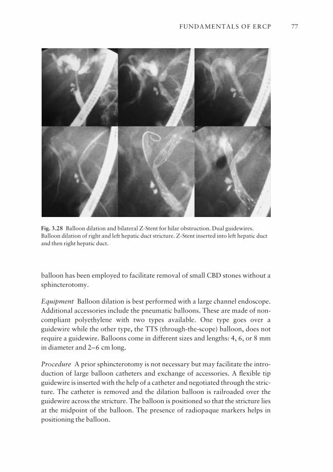
balloon has been employed to facilitate removal of small CBD stones without asphincterotomy.
Equipment Balloon dilation is best performed with a large channel endoscope.Additional accessories include the pneumatic balloons. These are made of non-compliant polyethylene with two types available. One type goes over aguidewire while the other type, the TTS (through-the-scope) balloon, does notrequire a guidewire. Balloons come in different sizes and lengths: 4, 6, or 8 mmin diameter and 2–6 cm long.
Procedure A prior sphincterotomy is not necessary but may facilitate the intro-duction of large balloon catheters and exchange of accessories. A flexible tipguidewire is inserted with the help of a catheter and negotiated through the stric-ture. The catheter is removed and the dilation balloon is railroaded over theguidewire across the stricture. The balloon is positioned so that the stricture liesat the midpoint of the balloon. The presence of radiopaque markers helps inpositioning the balloon.
FUNDAMENTALS OF ERCP 77
Fig. 3.28 Balloon dilation and bilateral Z-Stent for hilar obstruction. Dual guidewires.Balloon dilation of right and left hepatic duct stricture. Z-Stent inserted into left hepatic ductand then right hepatic duct.
The balloon is then inflated with dilute (10%) contrast and the pressureadjusted according to the type of balloon and the manufacturer’s recommenda-tion. The dilation is performed under fluoroscopy and a waist is seen at the midpoint of the balloon upon inflating the balloon. Effective dilation is achievedwhen the waist disappears.
The patient may experience pain during insufflation of the balloon. The balloon is usually kept inflated for 30–60 s and then deflated. It is helpful toreinflate the balloon and note the opening pressure when the waist disappearson the balloon. With successful dilation, the opening pressure should be lowerwith repeat dilation. The balloon is then completely deflated, the guidewireremoved and contrast injected while the balloon catheter is pulled back to assessthe effect of dilation.
Balloon dilation facilitates stent insertion in patients with malignant biliarystrictures. The short-term effects of balloon dilation for benign biliary stricturesare good but long-term follow-up shows some restenosis. Repeat dilation at regu-lar intervals may be necessary to keep the stricture open. Some endoscopistsadvocate the use of temporary stenting (with multiple stents) to keep the stric-ture open and repeat dilation and stent exchange every 3 months for up to ayear. Intrahepatic bile duct stones have been successfully removed followingballoon dilation of intrahepatic strictures.
Endoscopic management of bile leaks
Bile leaks may arise from the cystic duct stump after a cholecystectomy or frominjury to the CBD during surgery. Patients usually present with persistent biledrainage or formation of a biloma. As bile tends to flow in the path of least resistance, an intact papilla maintains a positive intrabiliary pressure and mayperpetuate the leak. Eliminating or bypassing the sphincter mechanism mayreduce the intrabiliary pressure.
Alternatively, an indwelling nasobiliary catheter or stent which bypasses thesphincter may serve to decompress the biliary system and promote healing of theleak. A small leak can be closed off easily by nasobiliary catheter drainage for a few days. Bile leak associated with CBD damage may require placement of anindwelling stent across the leak for up to 4–6 weeks. It is important to check forresidual damage or stricture of the CBD after removal of the stent.
Outstanding issues and future trends
ERCP now plays a very important role in the imaging and therapy of differentpancreatico-biliary problems. Many different technologies are being developedto shorten the time of the procedure by improving access and success with
CHAPTER 378
selective deep cannulation, thus minimizing manipulation within the ductal systems.
ERCP is, however, not without risk and serious complications have beenreported. Acute pancreatitis remains an important complication of this proce-dure and can occur even after a simple diagnostic cannulation. Although we are able to identify individuals who are at increased risk, currently availablemethods are not very effective in preventing this complication. Prophylacticpancreatic stenting to improve drainage is promising but this procedure itselfrequires considerable skill and experience.
MRCP with improved resolution may well replace diagnostic ERCP.However, ERCP will continue to play a role in the management of pancreatico-biliary diseases because of its therapeutic applications. There is a potential con-cern that, with the limited number of cases and the high skill level required of abiliary endoscopist, we may see a significant reduction in the number of trainedendoscopists in the future. We are already seeing a reduction in the number oftraining positions and the expectation of additional (third-tier) training beforean endoscopist becomes qualified to perform these procedures. The question of whether training with simulators may improve the skill of the biliary endo-scopist remains to be addressed.
References1 Cotton PB, Williams CB. (1996). Practical Gastrointestinal Endoscopy, 4th edn. Blackwell
Publishing, Oxford.2 Leung JWC, Ling TK, Chan RC et al. Antibiotics, biliary sepsis, and bile duct stones. Gastro-
intest Endosc 1994; 40: 716–21.3 Sung JJ, Lyon DJ, Suen R et al. Intravenous ciprofloxacin as treatment for patients with acute
suppurative cholangitis: a randomized, controlled clinical trial. J Antimicrob Chemother 1995;35 (6): 855–64.
4 Lee JG, Leung JW. Endoscopic management of common bile duct stones. Gastrointest EndoscClin N Am 1996; 6: 43–55.
5 Leung JWC, Chung SCS, Mok SD, Li AKC. Endoscopic removal of large common bile ductstones in recurrent pyogenic cholangitis. Gastrointest Endosc 1988; 34: 238–41.
6 Chung SC, Leung JW, Leong HT, Li AK. Mechanical lithotripsy of large common bile ductstones using a basket. Br J Surg 1991; 78: 1448–50.
7 Sorbi D, Van Os E, Aberger FJ, Derfus GA, Erickson R, Meier P et al. Clinical application of a new disposable lithotripter: a prospective multicenter study. Gastrointest Endosc 1999; 49:210–3.
8 Chan ACW, Ng EKW, Chung SCS et al. Common bile duct stones become smaller after endo-scopic biliary stenting. Endoscopy 1998; 30: 356–9.
9 Lau JYW, Ip SM, Chung SCS et al. Endoscopic drainage aborts endotoxaemia in acute cholangi-tis. Br J Surg 1996; 83: 181–4.
10 Lai ECS, Mok FPT, Tan ESY et al. Endoscopic biliary drainage for severe acute cholangitis. N Engl J Med 1992; 326: 1582–6.
11 Leung JWC, Chung SCS, Sung JJ et al. Urgent endoscopic drainage for severe acute suppurativecholangitis. Lancet 1989; 1: 1307–9.
12 Sugiyama M, Atomi Y. The benefits of endoscopic nasobiliary drainage without sphincterotomyfor acute cholangitis. Am J Gastroenterol 1998; 93: 2065–8.
FUNDAMENTALS OF ERCP 79

13 Lee DW, Chan AC, Lam YH et al. Biliary decompression by nasobiliary catheter or biliary stentin acute suppurative cholangitis: a prospective randomized trial. Gastrointest Endosc 2002; 56:361–5.
14 Leung JW, Del Favero G, Cotton P. Endoscopic biliary prostheses: a comparison of materials.Gastrointest Endosc 1985; 31: 93–5.
15 Libby E, Leung J. Prevention of biliary stent clogging: a clinical review. Am J Gastroenterol1996; 91: 1301–8.
16 Sung J, Chung SCS. Endoscopic stenting for palliation of malignant biliary obstruction. Dig DisSci 1995; 40: 1167–73.
CHAPTER 380
CHAPTER 4
ERCP Communications, Recording, andReporting
PETER B. COTTON
ERCP in context
There is much more to ERCP than knowing how to perform the procedures. Ata macro level, it is necessary to be able to place the procedure appropriatelywithin the broad spectrum of biliary and pancreatic diseases, and to appreciatethe many ways of approaching them. Achieving this wisdom is the goal of specialist training, but remains an imperative throughout our careers as theworld of medicine changes and as we ourselves help to change it. This evolutionrequires, and is greatly facilitated by, the development of active collaborationbetween all of the interested disciplines, especially gastroenterology, surgery,and radiology, which is the vision behind the Center concept [1].
Teamwork
At the micro, everyday, level, it is essential to realize that ERCP is a team event,requiring careful coordination between the endoscopist and the assistants(nurses and radiology technicians), and any trainees. Teams work better togetherif the goals are clear, and when the efforts of all members are respected. There is potential for confusion when the room is crowded with extra people, such as medical and nursing students, anesthesia staff, interested visitors, and evenequipment vendors. It is wise and polite to make sure that you know everyone’sname (and role) before getting started. Some hospitals have initiated a ‘time out’at the beginning of all operative procedures, like the cockpit drill for pilots thatis mandatory before any take-off or landing. This is intended to double-checkthat we have the correct patient, that key facts (e.g. allergies) have been noted,and that we have a clear plan of action.
It is also important for everyone in the room to maintain focus on the job inhand, keeping irrelevant conversation to a minimum, especially if the patient isunder conscious sedation. The need for appropriate behavior in the endoscopyroom has been well emphasized by one of the leaders of our profession [2].
81
Our experience in watching and performing ERCP around the world hasshown that the teamwork and interpersonal communications essential for thiscollaboration are often threatened by lack of a common lexicon, or consistent‘ERCP speak’. Devices, sites, and actions can be described in many differentways. For example ‘needle out’ can be interpreted as advancing the needle out ofthe catheter, or, just the opposite, i.e. out of the patient. Is the ‘distal pancreas’the head or the tail? Does ‘fluoro further right’ mean to the patient’s right, or toour right? Such confusions can have serious results, and would not be permittedin the cockpit of an aircraft.
ERCP speak
To reduce the potential for confusion, we suggest trying to develop and use astructured lexicon of communication, such as:1 Endoscopist to assistant/nurse
• push/pull wire• push/pull guide catheter• basket open/close• snare open/close• bow/relax sphincterotome• exchanging, push/accept wire• pull everything out• show needle/hide needle• balloon up/balloon down• inject contrast• aspirate• start to deploy (metal stent)
2 Endoscopist to traineeControlling the endoscope
• angle up/down• angle right/left• rotate right/left• bridge up/down• push/pull scope• brake on/off
Controlling devices• push/pull catheter/device
3 Endoscopist to radiology technician• fluoro on/off• take (hard-copy radiograph)• magnify image/mag off
CHAPTER 482
• shutters in/out top and bottom/right/left• flip image right/left• rotate C-arm towards me/away from me• tilt table head up/head down
4 Endoscopist to sedationist/anesthesiologistAll instructions should be equally clear, including dosing.
Confirming commands and feedback
Endoscopists need to know that their requests have been heard and acted upon,at least when this is not obvious visually. For example, we like to be told whenmedicines have been given (e.g. glucagon, Buscopan, or secretin).
Teams need positive educational feedback. Thank everyone when the pro-cedure has been completed, and, if things have not gone completely smoothly,take the opportunity immediately (and politely) to suggest how improvementscan be made.
Recording and reporting
The procedure is not complete until it has been documented appropriately, sothat everyone knows what has been done and why. Inadequate documentationcan result in much uncertainty, and future diagnostic and therapeutic actionsmay be compromised.
Endoscopy reports
There are some published guidelines regarding the content of endoscopy reports[3,4]. Reports should include key details of the patient, referring source(s), indications (including relevant clinical history, labs, and imaging), preparation(including fitness assessment, need for antibiotics, allergy issues, and the processfor patient education and consent), the site and timing of the procedures, thedoctors and staff involved, the sedation/analgesia used and tolerance, instru-ments, extent of the endoscopic survey, cannulation attempts, opacifications,findings on fluoroscopy, adjuvant diagnostic procedures (e.g. biopsy, manome-try), diagnoses made and excluded, treatments attempted and their immediateoutcomes, unplanned events (complications), accessories consumed, total dura-tion and fluoroscopy time, recovery, disposal, patient education, and follow-upplans.
A great deal of work has gone into trying to develop consensus on a commonlexicon for endoscopy. The minimum standard terminology (MST) is the bestknown and studied [5], and is used increasingly in electronic reporting systems.
ERCP COMMUNICATIONS, RECORDING, AND REPORTING 83
These systems drive compliance in reporting by prompting appropriate entries,and may even disallow saving or printing a report until certain mandatory fieldsare completed.
By contrast, there has been no consistency, and no formal recommendations,concerning the number and variety of images that should be recorded duringERCP, either endoscopic or radiological.
Endoscopic image documentation
It would seem logical to document pertinent landmarks (e.g. the papillary area),any lesions or unusual mucosal findings, and the appearances before and aftertherapeutic procedures, e.g. sphincterotomy. These images are now easy to capture, and to annotate, with electronic reporting systems. DICOM technicalstandards are being widely adapted [6–8]. Video-recording onto tape, or digit-ally, provides a much more complete document, but can generate storage andretrieval problems. Most units have many boxes of videotapes that have beenrecorded with enthusiasm, but are either ignored ever after or become a sourcefor frustration when trying to find key sequences for teaching purposes. Thisproblem will be solved eventually with high-capacity digital video storage units,which can be searched by keywords as well as by patient name.
Radiological image documentation
The permanent X-ray images of ERCP found in radiology files (or, increasingly,on CD-ROMs) are often woefully inadequate. Radiologists are rarely involvedduring the actual procedures; image capture is at the whim of the endoscopistand a radiology technician who is often not familiar with ERCP. The usualresult is an inadequate number and variety of images, with only haphazard documentation of the important findings and events. This may lead to errors ofinterpretation at the time, and at subsequent consultations when no other infor-mation is available.
Radiological aspects of ERCP are mentioned in other chapters of this bookin specific contexts. Other books have included some discussion of radiologicalequipment and techniques [3,9,10], but we have been unable to find any generalrecommendations for the number and types of images to be captured. Here wesuggest some minimum standards for radiographic documentation.
Checklist for radiological filming
1 Check that the system has the correct name, date, and timings.2 Take an abdominal scout film with the endoscope in the second part of the
CHAPTER 484
duodenum. This ensures that the field is clear (e.g. of monitoring wires), thepatient position is adequate, and that any unusual densities (e.g. pancreatic orvascular calcification, foreign bodies) are recognized before any contrast con-fuses the view.3 Take films during the filling phase of both biliary and pancreatic systems(when clinically relevant) to detect any small lesions and stones.4 Document complete filling (without overfilling) of all of the relevant ductalsystems. This may require turning the patient (or a C-arm), or moving the endo-scope (particularly to see the mid-part of the bile duct and the region of the pancreatic neck).5 Document any lesion or suspicious area.6 Record all the phases of intraductal procedures to show correct positioningof guidewires, cytology brushes, stents, sphincterotomes, etc.7 Record any possible or definite deviations, such as extraluminal air, intra-vascular contrast, guidewire perforation, acinarization, and submucosal orextravasated contrast.8 Record images prone and supine after removing the endoscope to see howmuch contrast has drained, and to provide a reference for future studies (e.g. ofstent position). The gallbladder is usually best seen with the patient supine withthe head elevated.
Radiographic interpretation
Rarely is there a radiologist in the ERCP room or available quickly nearby, andso most endoscopists have to interpret the fluoroscopy and hard-image findingsin real time to make immediate decisions about the need for further manipula-tions, and for endoscopic therapy.
In most institutions, the captured images are reported later by one of manygeneral radiologists, without reference to the endoscopist, and often even with-out access to the procedure report. This situation is fraught with potential error,with clinical and medico-legal risk. Several studies have now documented thesediscrepancies [11–13].
Reporting errors by endoscopists and radiologists can be reduced by:1 Teaching ERCP trainees about radiological techniques and interpretation.2 Complying with guidelines for capturing images, as suggested above.3 Making sure that the reporting radiologist receives a copy of the completeERCP report.4 Minimizing the number of radiologists involved, and having joint meetingsto discuss interesting cases and discrepancies. Those involved in each institutionshould meet to consider the local situation, and to initiate a process to improvecollaboration and quality control.
ERCP COMMUNICATIONS, RECORDING, AND REPORTING 85
Transmitting the information
The procedure document(s) is of limited value unless it reaches the right people. The primary target is the referring physician, who will put the informa-tion in context and make future care plans. In practice, it is not always easy tofind that target. Patients often reach specialist centers by a roundabout route,which they may not repeat in reverse when they leave. Thus it is very importantto clarify which doctor(s) the patient will see for continuing care, and to ensurethat he/she is on the list of people to receive reports (along with the actual refer-ral source and any primary provider, if different). Speed is a key parameter ofreporting. Phone calls or emails are often very helpful, and the days of snail-mailreporting must be numbered.
What about the patient? It is good medical practice to explain what has beendone to any accompanying person immediately after the procedure, but it issometimes more difficult to ensure that the patient is fully informed, not leastwhen he/she is discharged while still somewhat sleepy in the warm glow ofrecovery. Some endoscopists give patients a copy of the procedure report, but itis perhaps better to provide a simplified version. Newer endoscopy reportingsystems can be programmed to print this out, with the key features and conclu-sions, including the main recommendations, and plans for follow-up.
Most patients like to receive photographic prints of their procedures, andsome are given videos. For ERCP, it is desirable to give patients a CD-ROM ofthe radiographs, since many of them will have several subsequent consultations.
ERCP reporting: conclusion
ERCP procedures, even when indicated and well performed, may ultimately failto help patients if the findings and results are not documented clearly and com-pletely, and do not reach those making subsequent treatment decisions. Endo-scopists should consider how to improve their own reporting practices, and howto help their radiologist colleagues to play a more useful role.
References
1 Cotton PB. Interventional gastroenterology (endoscopy) at the crossroads: a plea for restructur-ing in digestive diseases. Gastroenterology 1994; 107: 294–9.
2 Boyce HW. Behavior in the endoscopy room. Gastrointest Endosc 2001; 53: 133–6.3 Cotton PB, Williams CB. (1996). Practical Gastrointestinal Endoscopy, 4th edn. Blackwell
Scientific, Oxford.4 American Society of Gastrointestinal Endoscopy. (1992). Defining the Endoscopy Report.
American Society of Gastrointestinal Endoscopy, Manchester.5 Korman LY, Delvaux M, Crespi M. The minimal standard terminology in digestive endoscopy:
perspective on a standard endoscopic vocabulary. Gastrointest Endosc 2001; 53: 392–6.
CHAPTER 486
6 Aabakken L. (2004). Digital documentation in endoscopy. In: Endoscopy Practice and Safety(ed. Cotton, PB) (www.gastrohep.com).
7 Fujino MA, Ikeda M. (1996). Electronic image management. In: Gastroenterologic Endoscopy,Vol. 1, 2nd edn (ed. Sivak, MV), pp. 103–14. W. B. Saunders, Philadelphia.
8 Heldwein W, Rosch T. (2002). Reporting terminology and image documentation in endoscopy.In: Gastroenterological Endoscopy (ed. Classen, M, Tytgat, GNJ, Lightdale, CJ), pp. 754–9.Thieme, Stuttgart.
9 Taylor AJ, Bohofoush III, AG. (1997). Interpretation of ERCP with Associated Digital ImagingCorrelation. Lippincott-Raven, Philadelphia.
10 Martin DF, Tweedle D, Haboubi NY. (1998). Clinical Practice of ERCP. Churchill Livingstone,London.
11 Thomas M, Geenen JE, Catalano MF. Importance of real time interpretation (INTERP) ofERCP films over conventional static images: medicolegal implications. Gastrointest Endosc2004; 59: AB183.
12 Khanna N, May G, Cole M, Bass S, Romagnuolo J. Post-ERCP radiology interpretation ofcholangio-pancreatograms appears to be of limited benefit and may be inaccurate. GastrointestEndosc 2004; 59: AB186.
13 Sweeney JT, Shah RJ, Martin SP, Ulrich CD, Somogyi L. The impact of post-procedure inter-pretation by radiologists on patient care: should it be routine or selective? Gastrointest Endosc2003; 58: 549–53.
ERCP COMMUNICATIONS, RECORDING, AND REPORTING 87
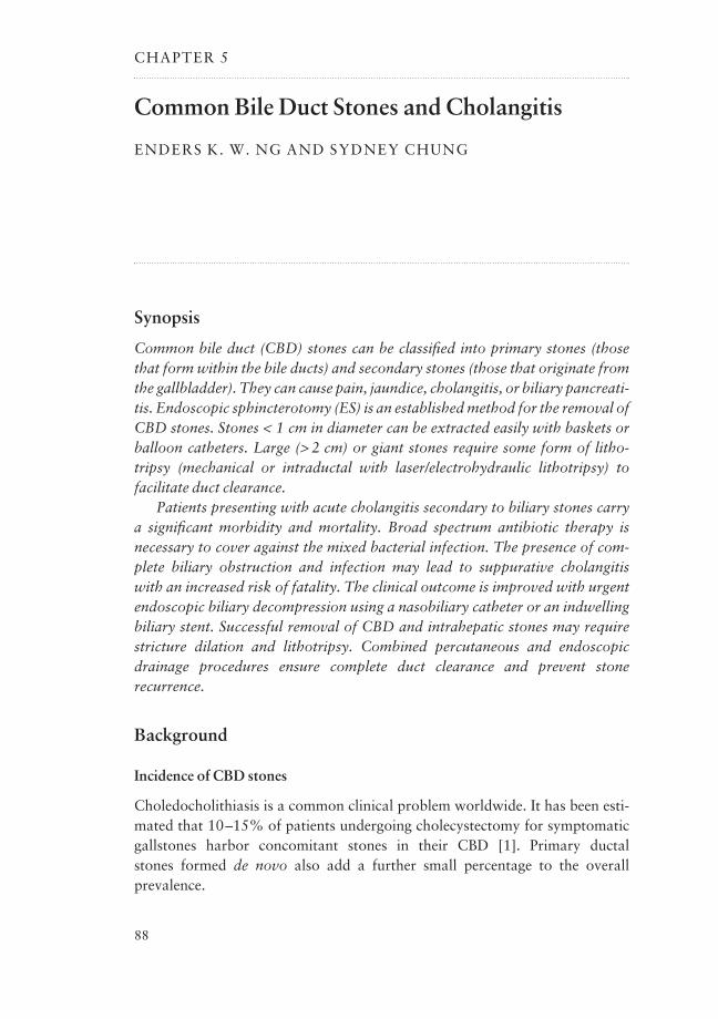
CHAPTER 5
Common Bile Duct Stones and Cholangitis
ENDERS K. W. NG AND SYDNEY CHUNG
Synopsis
Common bile duct (CBD) stones can be classified into primary stones (thosethat form within the bile ducts) and secondary stones (those that originate fromthe gallbladder). They can cause pain, jaundice, cholangitis, or biliary pancreati-tis. Endoscopic sphincterotomy (ES) is an established method for the removal ofCBD stones. Stones < 1 cm in diameter can be extracted easily with baskets orballoon catheters. Large (> 2 cm) or giant stones require some form of litho-tripsy (mechanical or intraductal with laser/electrohydraulic lithotripsy) tofacilitate duct clearance.
Patients presenting with acute cholangitis secondary to biliary stones carry a significant morbidity and mortality. Broad spectrum antibiotic therapy is necessary to cover against the mixed bacterial infection. The presence of com-plete biliary obstruction and infection may lead to suppurative cholangitis with an increased risk of fatality. The clinical outcome is improved with urgentendoscopic biliary decompression using a nasobiliary catheter or an indwellingbiliary stent. Successful removal of CBD and intrahepatic stones may requirestricture dilation and lithotripsy. Combined percutaneous and endoscopicdrainage procedures ensure complete duct clearance and prevent stone recurrence.
Background
Incidence of CBD stones
Choledocholithiasis is a common clinical problem worldwide. It has been esti-mated that 10–15% of patients undergoing cholecystectomy for symptomaticgallstones harbor concomitant stones in their CBD [1]. Primary ductal stones formed de novo also add a further small percentage to the overall prevalence.
88
Traditional management
After the first successful CBD exploration by Courvoisier in 1890, surgicallithotripsy was the treatment of choice for choledocholithiasis for nearly a century [2].
Non-operative approach to CBD stones
The introduction of ES in 1974 [3] and the rapid development of minimal access surgery in the late 1980s have completely revolutionized the approach to CBD stones. As laparoscopic cholecystectomy is the first-line treatment for gallstones nowadays, endoscopic removal of biliary tree calculi has become the most appealing and widely embraced technique for removal ofcholedocholithiasis.
Pathogenesis
Classification of CBD stones
Bile duct stones can be broadly classified into two types according to the site oforigin.
Primary CBD stones
Primary ductal stones are stones that develop de novo in the intrahepatic ductsor common duct. They are far more common in the Asian populations than in the West. The reason for such a geographical difference is enigmatic. Thesestones are often brownish-yellow in color with a soft muddy consistency (Fig. 5.1); biochemically, they consist of calcium bilirubinate mixed with vari-able amounts of cholesterol and calcium salts. While the etiology remains con-jectural, bacterial infections and biliary stasis are considered the two mostimportant causative factors.
Bacteriology of primary CBD stones Gastrointestinal tract microorganismssuch as Escherichia coli, Klebsiella, Proteus, Bacteroides, and Clostridium havebeen isolated from the bile of patients with primary duct stones [4]. In addition,bacterial cytoskeletons are invariably seen in primary duct stones under elec-tronic microscope [5]. These bacteria may have a contributory role by produc-ing enzymes that catalyze deconjugation of bilirubin and lysis of phospholipids,which in turn promote the precipitation of calcium bilirubinate and initiate
COMMON BILE DUCT STONES AND CHOLANGITIS 89
stone formation. Among all the bacteria isolated, Clostridium perfringens hasbeen found to produce the highest beta-glucuronidase enzyme activity, which is34-fold higher than that for E. coli, Corynebacterium spp., Enterococcus spp.,and Klebsiella spp. [6]. On the other hand, the biliary stasis theory is supportedby the fact that intrahepatic ductal strictures and proximal dilation are com-monly seen among patients with primary duct calculi [7]. Nevertheless, whetherthese strictures are the cause or consequence of the intrahepatic ductal calculiremains unresolved.
Secondary CBD stones
Secondary common duct stones are supposed to have originated from the gall-bladder. Conceivably their composition is identical to that of gallstones, whichare mainly yellowish cholesterol or black pigment calculi with a hard and crispyconsistency. It is unclear why gallstones migrate into the common duct in somepatients. In one study the size of the cystic duct has been reported as the singlemost important determinant [8].
Clinical presentations
Asymptomatic biliary stones
A considerable proportion of patients with common or intrahepatic ductal cal-culi are asymptomatic. The stones may be found incidentally during investiga-tion for unrelated abdominal conditions. The presence of coexisting ductal
CHAPTER 590
Fig. 5.1 Typical brown pigmentstone retrieved by ERCP.

stones is sometimes noted by abdominal ultrasound scan when patients arebeing worked up for cholelithiasis.
Symptomatic biliary stones
Obstructive jaundice
Intermittent jaundice is said to be a typical feature of choledocholithiasis, whenthe stone impacts and disimpacts at the papilla or the distal CBD leading to fluctuating jaundice and serum bilirubin levels. Continuous obstruction from stone impaction in the distal common duct may manifest as progres-sive jaundice.
Pain
Dull right upper abdominal pain due to increased biliary tree pressure may alsobe experienced as a result of stone impaction.
Clinical cholangitis
When bacterial infection superimposes in the obstructed biliary system, thepatient presents with the typical Charcot’s triad (fever, pain, and jaundice) ofcholangitis. Nevertheless, cholangitis may not necessarily present with all threefeatures, and the diagnosis should not be dismissed lightly just because thepatient is afebrile or not jaundiced.
Biliary pancreatitis
Small stones may pass spontaneously through the ampulla of Vater. The passageof stones across the papilla may induce a transient rise in the pancreatic ductpressure and trigger intrapancreatic activation of enzymes resulting in acutepancreatitis. Patients with acute pancreatitis typically present with epigastricpain radiating to the back, associated with nausea and vomiting. A serum amy-lase level exceeding 1000 IU/liter is considered to be diagnostic of pancreatitis.
Oriental cholangitis or recurrent pyogenic cholangitis
Patients with primary intrahepatic duct stones may present with recurrentattacks of cholangitis. Characteristically, there are multiple strictures, with stoneformation proximal to the stricture in the dilated portion of one or more seg-ments of the intrahepatic ducts. Jaundice may not be obvious if only segmental
COMMON BILE DUCT STONES AND CHOLANGITIS 91
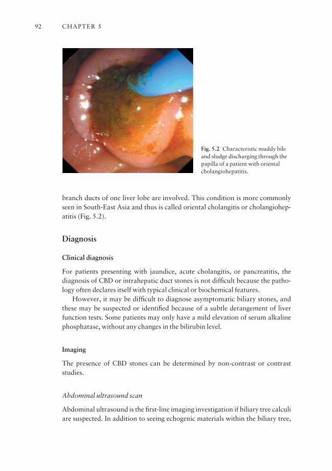
branch ducts of one liver lobe are involved. This condition is more commonlyseen in South-East Asia and thus is called oriental cholangitis or cholangiohep-atitis (Fig. 5.2).
Diagnosis
Clinical diagnosis
For patients presenting with jaundice, acute cholangitis, or pancreatitis, thediagnosis of CBD or intrahepatic duct stones is not difficult because the patho-logy often declares itself with typical clinical or biochemical features.
However, it may be difficult to diagnose asymptomatic biliary stones, andthese may be suspected or identified because of a subtle derangement of liverfunction tests. Some patients may only have a mild elevation of serum alkalinephosphatase, without any changes in the bilirubin level.
Imaging
The presence of CBD stones can be determined by non-contrast or contrast studies.
Abdominal ultrasound scan
Abdominal ultrasound is the first-line imaging investigation if biliary tree calculiare suspected. In addition to seeing echogenic materials within the biliary tree,
CHAPTER 592
Fig. 5.2 Characteristic muddy bileand sludge discharging through thepapilla of a patient with orientalcholangiohepatitis.
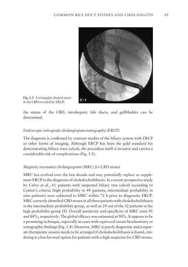
the status of the CBD, intrahepatic bile ducts, and gallbladder can be determined.
Endoscopic retrograde cholangiopancreatography (ERCP)
The diagnosis is confirmed by contrast studies of the biliary system with ERCPor other forms of imaging. Although ERCP has been the gold standard fordemonstrating biliary tract calculi, the procedure itself is invasive and carries aconsiderable risk of complications (Fig. 5.3).
Magnetic resonance cholangiogram (MRC) for CBD stones
MRC has evolved over the last decade and may potentially replace or supple-ment ERCP in the diagnosis of choledocholithiasis. In a recent prospective studyby Calvo et al., 61 patients with suspected biliary tree calculi according toCotton’s criteria (high probability in 49 patients, intermediate probability in nine patients) were subjected to MRC within 72 h prior to diagnostic ERCP.MRC correctly identified CBD stones in all three patients with choledocholithiasisin the intermediate probability group, as well as 29 out of the 32 patients in thehigh probability group [9]. Overall sensitivity and specificity of MRC were 91and 84%, respectively. The global efficacy was estimated at 90%. It appears to be a promising technique, especially in cases with equivocal serum biochemistry orsonographic findings (Fig. 5.4). However, MRC is purely diagnostic and a separ-ate therapeutic session needs to be arranged if choledocholithiasis is found, ren-dering it a less favored option for patients with a high suspicion for CBD stones.
COMMON BILE DUCT STONES AND CHOLANGITIS 93
Fig. 5.3 A triangular shaped stonein the CBD revealed by ERCP.
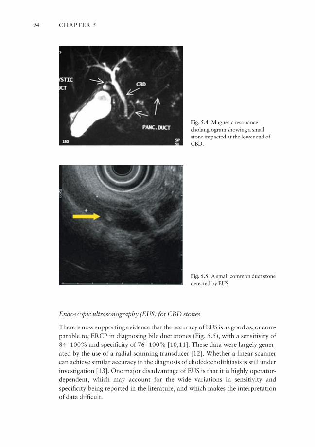
Endoscopic ultrasonography (EUS) for CBD stones
There is now supporting evidence that the accuracy of EUS is as good as, or com-parable to, ERCP in diagnosing bile duct stones (Fig. 5.5), with a sensitivity of84–100% and specificity of 76–100% [10,11]. These data were largely gener-ated by the use of a radial scanning transducer [12]. Whether a linear scannercan achieve similar accuracy in the diagnosis of choledocholithiasis is still underinvestigation [13]. One major disadvantage of EUS is that it is highly operator-dependent, which may account for the wide variations in sensitivity andspecificity being reported in the literature, and which makes the interpretationof data difficult.
CHAPTER 594
Fig. 5.4 Magnetic resonancecholangiogram showing a smallstone impacted at the lower end ofCBD.
Fig. 5.5 A small common duct stonedetected by EUS.

Management for CBD stones
While some may advocate the use of medical treatment such as chemical dissolu-tion for the removal of biliary tree calculi, endoscopic or surgical approachesremain the preferred treatments because they are more effective and reliable.
ERCP, sphincterotomy, and stone extraction
CBD stones < 5 mm in diameter may pass spontaneously or can be removedwithout a sphincterotomy. For stones > 5 mm, ES is the most commonly performed procedure for their retrieval.
Endoscopic sphincterotomy
Choice of endoscopes The preparation, positioning, and sedation of the patientare the same as those for diagnostic ERCP. The choice of duodenoscopes is determined by the anticipated size of the CBD stones. For small stones, wherea complex lithotripsy instrument is unnecessary, a regular duodenoscope with a 2.8 mm channel is adequate. However, when the stone is > 1 cm or there is astrong likelihood that lithotripsy will be needed, a bigger duodenoscope, with a3.2 or 4.2 mm channel, should be used.
Cannulation with sphincterotome Cannulation of the common duct is the same as for diagnostic ERCP. Some patients may have stones impacted at thelower end of the common duct (Fig. 5.6), and the resultant bulging papilla couldrender cannulation more difficult. A cannulating sphincterotome with an
COMMON BILE DUCT STONES AND CHOLANGITIS 95
Fig. 5.6 Two cases with impacted stone at the papilla.
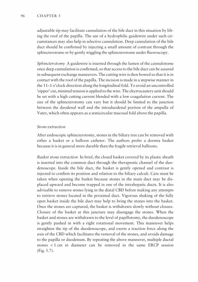
adjustable tip may facilitate cannulation of the bile duct in this situation by lift-ing the roof of the papilla. The use of a hydrophilic guidewire under such cir-cumstances may also help in selective cannulation. Deep cannulation of the bileduct should be confirmed by injecting a small amount of contrast through thesphincterotome or by gently wiggling the sphincterotome under fluoroscopy.
Sphincterotomy A guidewire is inserted through the lumen of the cannulotomeonce deep cannulation is confirmed, so that access to the bile duct can be assuredin subsequent exchange maneuvers. The cutting wire is then bowed so that it is incontact with the roof of the papilla. The incision is made in a stepwise manner inthe 11–1 o’clock direction along the longitudinal fold. To avoid an uncontrolled‘zipper’ cut, minimal tension is applied to the wire. The electrocautery unit shouldbe set with a high cutting current blended with a low coagulation current. Thesize of the sphincterotomy can vary but it should be limited to the junctionbetween the duodenal wall and the intraduodenal portion of the ampulla ofVater, which often appears as a semicircular mucosal fold above the papilla.
Stone extraction
After endoscopic sphincterotomy, stones in the biliary tree can be removed witheither a basket or a balloon catheter. The authors prefer a dormia basketbecause it is in general more durable than the fragile retrieval balloons.
Basket stone extraction In brief, the closed basket covered by its plastic sheathis inserted into the common duct through the therapeutic channel of the duo-denoscope. Inside the bile duct, the basket is gently opened and contrast isinjected to confirm its position and relation to the biliary calculi. Care must betaken when opening the basket because stones in the main duct may be dis-placed upward and become trapped in one of the intrahepatic ducts. It is alsoadvisable to remove stones lying in the distal CBD before making any attemptsto retrieve stones located in the proximal duct. Vigorous shaking of the fullyopen basket inside the bile duct may help to bring the stones into the basket.Once the stones are captured, the basket is withdrawn slowly without closure.Closure of the basket at this juncture may disengage the stones. When the basket and stones are withdrawn to the level of papillotomy, the duodenoscopeis gently pushed in with a right rotational movement. This maneuver helpsstraighten the tip of the duodenoscope, and exerts a traction force along the axis of the CBD which facilitates the removal of the stones, and avoids damageto the papilla or duodenum. By repeating the above maneuver, multiple ductalstones < 1 cm in diameter can be removed in the same ERCP session (Fig. 5.7).
CHAPTER 596

Balloon stone extraction Biliary stones in the CBD of < 1 cm in diameter can beremoved with a balloon catheter. The balloon is deflated and inserted into theCBD through the sphincterotomy, and advanced above the stones. The balloonis gently inflated to the size of the bile duct and pulled back gently, displacing theCBD stone distally. With an adequate sphincterotomy, the stone can be pulledagainst the cut orifice and then expelled by traction on the balloon catheter followed by downward angulation of the tip of the endoscope. The maneuver is repeated and complete clearance of the CBD is confirmed by an occlusioncholangiogram.
Complications
Bleeding, perforation, pancreatitis, and cholangitis are potential complicationsof ES and stone extraction. The reported incidence varies markedly in the litera-ture, but bleeding is generally the most common complication encountered.Previous studies failed to identify predicting factors for these complications.Most of these studies were univariate or bivariate analyses, which generatedinconsistent and often contradictory results [14,15]. Two multicenter studiesbased on multivariate regression models, however, have shed new light on thiscomplex issue.
Acute pancreatitis
In a prospective survey conducted in the United States between 1992 and 1994,acute pancreatitis was found to be significantly more common if the ES was per-formed for suspected dysfunction of the sphincter of Oddi, in young patients, usinga precut technique, after difficult cannulation, or with repeated and excessivepancreatic contrast injections (Fig. 5.8) [16]. Similar findings were reported in
COMMON BILE DUCT STONES AND CHOLANGITIS 97
Fig. 5.7 Left: a wire-guide papillotome locating inside the low CBD; Middle: completion ofendoscopic sphincterotomy (ES); Right: use of dormia basket for retrieval of CBD stone afterES.
an Italian multicenter study, in which acute pancreatitis was independently pre-dicted by young patient age, pancreatic duct opacification, and a non-dilatedCBD [17].
Bleeding
Significant postsphincterotomy bleeding happened more readily if the patienthad associated coagulopathy, had cholangitis, or the procedure was performedby an inexperienced endoscopist. Interestingly, bleeding and cholangitis wereagain associated with small centers with low case volume. These two large-scaleand multivariate studies have two points in common: sphincterotomy com-plications are closely related to indications for the procedure and to the experienceor case volume of individual endoscopists or institutions.
Controversies
Sphincterotomy vs. balloon sphincteroplasty
The reported complication rates of ES ranged from 6 to 10%, with a mortalityresulting from these complications of 0.4–1.2% [18,19]. Although the percent-ages appear to be small, the actual number of patients suffering from these com-plications, as well as the associated prolonged hospitalization, is considerable.Since complications are mostly related to the sphincterotomy, some have advo-cated the use of endoscopic balloon dilatation (EBD) as an alternative procedureprior to stone extraction.
Balloon sphincteroplasty
EBD was first described by Staritz et al. in 1983 [20]. In addition to the lowerrisk of bleeding and perforation, another apparent advantage of EBD is that the
CHAPTER 598
Fig. 5.8 Excessive contrast injectioninto the pancreatic duct mayprecipitate acute pancreatitis.
sphincter of Oddi function can be preserved. This has been demonstrated in anumber of studies involving sphincter of Oddi manometry [21,22]. However,when EBD was first proposed in the early 1980s, it was not widely embraced dueto skepticism concerning its efficacy and the fear of precipitating acute pancre-atitis. The incidence of acute pancreatitis reported then was as high as 25%according to some earlier series in which EBD was performed mainly for sphinc-ter of Oddi dysfunction [23,24].
Balloon sphincteroplasty for CBD stones
More recent studies have revealed that EBD is a safe and effective procedure forpatients with biliary stones [25,26]. In a randomized trial by Bergman et al., 202patients were assigned to either EBD or ES prior to removal of the CBD stones[27]. There was no significant difference in overall duct clearance rate, proce-dure time, early complications, and death associated with the two procedures.The drawback for EBD was that mechanical lithotripsy was more frequentlyrequired in these patients. Besides, a considerable number of patients in the EBDgroup eventually required an ES for ductal clearance.
Sphincterotomy for CBD stones
Although there is concern regarding the safety of ES, a multicenter prospectivedatabase study in the United States based on standard criteria for defining com-plications revealed that morbidities only occurred in 5.8% of the 1921 patientsstudied [28]. Two-thirds of the events were graded as mild, which required lessthan 3 days of hospitalization. In addition, this study disproved the dogma thatcomplications are more likely to occur in young patients with normal sized ducts.Out of the 238 patients aged younger than 60 years, only one developed severecomplications and there were no fatalities.
Long-term complications of sphincterotomy As the short-term outcome of ES is no longer in question, there is an increasing concern about the long-termeffects of sphincterotomy on the biliary system. In a retrospective study byCostamagna et al., 529 patients with successful sphincterotomy and bile ductclearance were evaluated after a follow-up of at least 5 years [29]. Recurrent biliary symptoms or duct stones occurred in only 11.1% of the patients, whilethe remainder were either asymptomatic or died of unrelated causes. A dilatedbile duct > 22 mm was found to be the only independent predictive factor forrecurrence. In another population-based cohort study by Karlson et al., the cancer risk of 992 patients who had undergone sphincterotomy over a medianfollow-up time of 10–11 years was estimated, and was found to be almost thesame as that of the normal population.
COMMON BILE DUCT STONES AND CHOLANGITIS 99

In conclusion, there is no evidence that ES is unsafe in either the short orlong term provided that it is being performed by, or under stringent supervisionof, experienced endoscopists. EBD can be an alternative to ES, but currently itshould be limited to patients with no more than three stones, each < 10 mm indiameter [30].
ERCP vs. laparoscopic common duct exploration for retained CBD stones
In the era of open cholecystectomy, intraoperative cholangiogram was part ofthe operation. If CBD stones were suspected with intraoperative cholan-giogram, exploration of the common duct was performed, and a variety of techniques were used to remove the ductal calculi. The choledochotomy wasclosed around a rubber T-tube, and a check cholangiogram was performedabout 10 days after the operation to rule out any residual ductal stones.However, with the rapid acceptance of laparoscopic cholecystectomy in the lastdecade, when and how these concomitant choledocholithiases should be man-aged is becoming increasingly controversial.
Preoperative ERCP
To perform ERCP for all patients scheduled for laparoscopic cholecystectomy isimpractical and the projected numbers of complications and mortality are alsounacceptable.
Operative removal of CBD stones
An operative laparoscopic cholangiogram can be performed to rule out CBDstones. If stones are found intraoperatively, transcystic duct lithotripsy or explo-ration of the common duct is a feasible option with the laparoscope. Successrates for using such an approach to diagnose and clear the common duct stoneshave been reported to be close to 90% [31,32]. However, these laparoscopicprocedures require a much higher level of skill and expertise, which may not beuniversally available except in some tertiary referral centers.
Factors that predict CBD stones
One possible approach is to identify patients at higher risk of concomitantcholedocholithiasis and send them for preoperative cholangiogram and litho-tripsy prior to laparoscopic cholecystectomy. A number of studies have reportedthat deranged liver function, dilated biliary tree on ultrasound scan, and historyof jaundice or pancreatitis predict the presence of CBD stones [33,34].
CHAPTER 5100
Unfortunately, these studies were unable to generate concrete data that wouldallow accurate prediction of the biliary tree status preoperatively.
MRC for detection of CBD stones
The emergence of magnetic resonance technology in the 1990s opened up newpossibilities for solving this clinical dilemma. Earlier series comparing MRC withconventional ERCP or operative findings have already shown promising dataregarding non-invasive diagnosis of common duct calculi [35,36]. In a recentseries by Laokpessi et al. on a group of 147 patients with clinical and biologicalsigns of choledocholithiasis, MRC was shown to have a sensitivity of 93% and aspecificity of 100% in detecting the ductal calculi [37]. MRC is likely to play animportant role in preoperative diagnosis of concomitant CBD stones in equi-vocal cases, and may allow better planning regarding the mode of stone removalprior to or during laparoscopic cholecystectomy.
Risk scores for prediction of CBD stones
There is as yet no conclusion as to which approach is superior, and the choice ofmanagement method depends mainly on the expertise and support available inindividual centers. One of the latest developments is to categorize patients intohigh-, intermediate-, and low-risk groups for CBD stones, and the managementapproach is dependent upon the risk score of each individual patient [38].
Alternative approaches to CBD stones
Precut sphincterotomy for failed deep cannulation
A needle-knife sphincterotome can be used to incise the lower end of the com-mon duct when guidewire cannulation of the bile duct fails (Fig. 5.9). In a recentprospective study by Binmoeller et al., precut papillotomy was performed on123 out of 327 patients who had an unsuccessful CBD cannulation [39].Selective cannulation was achieved in all cases after the procedure, without asignificant increase in the rate of pancreatitis and bleeding when compared tothose undergoing the conventional pull-type ES.
Complications of precut sphincterotomy
Today, the overall incidence of complications following precut sphincterotomyhas been reported to be 7–11%, which is not much higher than that quoted forconventional sphincterotomy [40,41]. However, it cannot be overemphasized
COMMON BILE DUCT STONES AND CHOLANGITIS 101
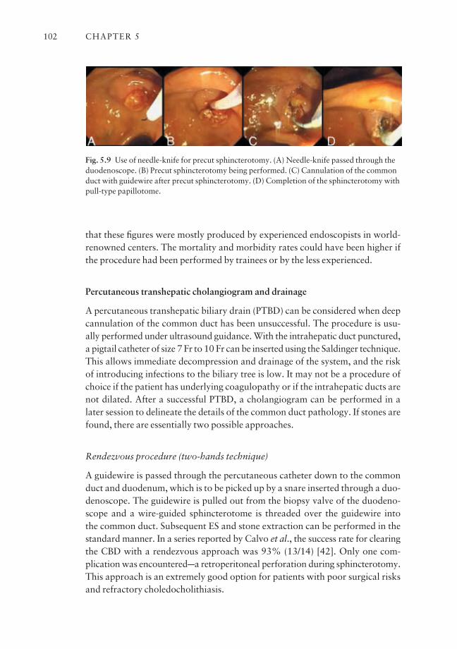
that these figures were mostly produced by experienced endoscopists in world-renowned centers. The mortality and morbidity rates could have been higher ifthe procedure had been performed by trainees or by the less experienced.
Percutaneous transhepatic cholangiogram and drainage
A percutaneous transhepatic biliary drain (PTBD) can be considered when deepcannulation of the common duct has been unsuccessful. The procedure is usu-ally performed under ultrasound guidance. With the intrahepatic duct punctured,a pigtail catheter of size 7 Fr to 10 Fr can be inserted using the Saldinger technique.This allows immediate decompression and drainage of the system, and the riskof introducing infections to the biliary tree is low. It may not be a procedure ofchoice if the patient has underlying coagulopathy or if the intrahepatic ducts arenot dilated. After a successful PTBD, a cholangiogram can be performed in alater session to delineate the details of the common duct pathology. If stones arefound, there are essentially two possible approaches.
Rendezvous procedure (two-hands technique)
A guidewire is passed through the percutaneous catheter down to the commonduct and duodenum, which is to be picked up by a snare inserted through a duo-denoscope. The guidewire is pulled out from the biopsy valve of the duodeno-scope and a wire-guided sphincterotome is threaded over the guidewire into the common duct. Subsequent ES and stone extraction can be performed in thestandard manner. In a series reported by Calvo et al., the success rate for clearingthe CBD with a rendezvous approach was 93% (13/14) [42]. Only one com-plication was encounteredaa retroperitoneal perforation during sphincterotomy.This approach is an extremely good option for patients with poor surgical risksand refractory choledocholithiasis.
CHAPTER 5102
Fig. 5.9 Use of needle-knife for precut sphincterotomy. (A) Needle-knife passed through theduodenoscope. (B) Precut sphincterotomy being performed. (C) Cannulation of the commonduct with guidewire after precut sphincterotomy. (D) Completion of the sphincterotomy withpull-type papillotome.
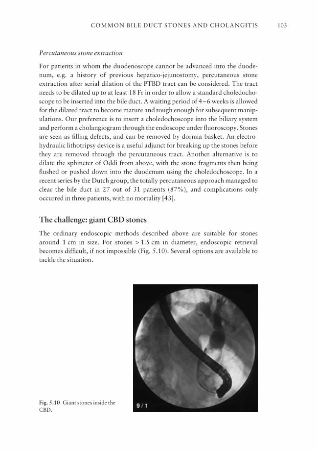
Percutaneous stone extraction
For patients in whom the duodenoscope cannot be advanced into the duode-num, e.g. a history of previous hepatico-jejunostomy, percutaneous stoneextraction after serial dilation of the PTBD tract can be considered. The tractneeds to be dilated up to at least 18 Fr in order to allow a standard choledocho-scope to be inserted into the bile duct. A waiting period of 4–6 weeks is allowedfor the dilated tract to become mature and tough enough for subsequent manip-ulations. Our preference is to insert a choledochoscope into the biliary systemand perform a cholangiogram through the endoscope under fluoroscopy. Stonesare seen as filling defects, and can be removed by dormia basket. An electro-hydraulic lithotripsy device is a useful adjunct for breaking up the stones beforethey are removed through the percutaneous tract. Another alternative is todilate the sphincter of Oddi from above, with the stone fragments then beingflushed or pushed down into the duodenum using the choledochoscope. In arecent series by the Dutch group, the totally percutaneous approach managed toclear the bile duct in 27 out of 31 patients (87%), and complications onlyoccurred in three patients, with no mortality [43].
The challenge: giant CBD stones
The ordinary endoscopic methods described above are suitable for stonesaround 1 cm in size. For stones > 1.5 cm in diameter, endoscopic retrievalbecomes difficult, if not impossible (Fig. 5.10). Several options are available totackle the situation.
COMMON BILE DUCT STONES AND CHOLANGITIS 103
Fig. 5.10 Giant stones inside theCBD.
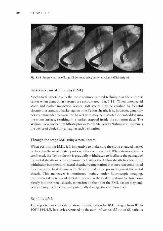
Basket mechanical lithotripsy (BML)
Mechanical lithotripsy is the most commonly used technique in the authors’center when giant biliary stones are encountered (Fig. 5.11). When unexpectedstone and basket impaction occurs, soft stones may be crushed by forceful closure of a standard basket against the Teflon sheath. It is, however, generallynot recommended because the basket wire may be distorted or embedded intothe stone surface, resulting in a basket trapped inside the common duct. TheWilson Cook Soehendra lithotriptor or Percy McGowan ‘fishing reel’ system isthe device of choice for salvaging such a situation.
Through-the-scope BML using a metal sheath
When performing BML, it is imperative to make sure the stone-engaged basketis placed in the most dilated portion of the common duct. When stone capture isconfirmed, the Teflon sheath is gradually withdrawn to facilitate the passage ofthe metal sheath into the common duct. After the Teflon sheath has been fullywithdrawn into the spiral metal sheath, fragmentation of stones is accomplishedby closing the basket wire with the captured stone pressed against the metalsheath. This maneuver is monitored mainly under fluoroscopic imaging.Caution is taken to avoid ductal injury when the basket is about to close com-pletely into the metal sheath, as tension on the tip of the BML basket may sud-denly change its direction and potentially damage the common duct.
Results of BML
The reported success rate of stone fragmentation by BML ranges from 82 to100% [44,45]. In a series reported by the authors’ center, 55 out of 68 patients
CHAPTER 5104
Fig. 5.11 Fragmentation of large CBD stones using basket mechanical lithotriptor.
(81%) had complete CBD clearance after lithotripsy of giant stones with BML[46]. Of the remaining 13 patients, one had the stone crushed with the Soehendralithotriptor, six were successfully managed by electrohydraulic lithotripsythrough a ‘mother and baby’ endoscope, four received an indwelling stent, andtwo patients underwent surgery. The ductal clearance rate was 92% in anothermulticenter study of the efficacy of BML in 116 patients, without any significantincrease in the incidence of pancreatitis or hemorrhage [47]. Thus far the largestseries ever reported was by Schneider et al. on a group of 209 patients, in whichthe overall success rate was 88%, with 79% of the stones > 20 mm in diameter[48]. The reasons for failure in the remaining 12% of patients were either unsuc-cessful insertion of the basket, or failure to engage the stone because of its size,the common duct diameter, or other technical problems.
Mother and baby choledochoscopy and intraductal lithotripsy
Passing a baby endoscope via the channel of a large-sized duodenoscope isanother method for the management of giant CBD stones. Using the babycholangioscope, an electrohydraulic lithotriptor or laser probe can be applied toshatter the stone under direct visualization. This approach used to require astandard mother and baby system, but the recent release of a per-oral choledo-choscope (CHF system, XP20, Olympus) that can go through the 4.2 mm work-ing channel of a regular therapeutic duodenoscope has reduced the cost.Caution should be exercised by the endoscopist controlling the duodenoscopeto avoid excessive elevation of the elevator, which may damage the delicate optical fibers inside the choledochoscope and result in premature failure.
Electrohydraulic lithotripsy (EHL)
EHL was first introduced in the 1950s as a method of fragmenting rocks inmines. It was later adapted for medical use, and Koch et al. in 1977 were the firstto attempt fragmentation of CBD calculi [49]. The mechanism is a crackingforce transmitted by hydraulic pressure waves generated under water by thehigh-voltage sparks discharged across the tip of the EHL probe. Few data areavailable in the literature regarding the use of EHL for biliary tract stones. Thelargest series reported thus far had a success rate of 99% in 65 patients [50].Other smaller series involving less than 10 patients also claimed CBD clearancein all the patients treated [51].
Intraductal laser lithotripsy
The other device that can work through the mother and baby system is laser litho-tripsy. Essentially there are three types of laser available. The Nd:YAG laser is
COMMON BILE DUCT STONES AND CHOLANGITIS 105
no longer in use because of the high risk of ductal injury. The flash lamp pulseddye lasers, on the other hand, can be delivered with thinner fibers and are super-ior to the Nd:YAG lasers for intraductal lithotripsy. The reported success ratesrange from 80 to 94%, and serious complications are uncommon [52,53]. Thelatest development includes the ‘smart dye laser’, which is capable of differenti-ating stone from normal ductal tissue and thus obviates the need for direct visu-alization during the procedure. This new mode of intraductal lithotripsy can beperformed under fluoroscopic guidance without the necessity of a baby cholan-gioscope. In a series of 38 patients, 37 had successful ductal clearance afterbeing treated by an automatic stone recognition laser system [54]. A slightlylower success rate was reported by Hochberger et al. in a group of 50 patients[55]. Major complications were not found in either series.
Stenting and interval endoscopic lithotripsy
For patients whose common duct stones are refractory to endoscopic retrieval, aplastic biliary stent can be inserted as a temporary or permanent measure. It hasbeen shown in several prospective series that elderly patients with difficultstones remained symptom-free after insertion of an endoprosthesis, and thussurgery was avoided. There have also been studies reporting a decrease in thestone size after a period of biliary stenting. In a study by the authors, 46 patientswith large CBD stones received plastic stents [56]. Twenty-eight patients hadrepeat ERCP for stone extraction after a median of 63 days. The size of thestones was significantly reduced, from 11–46 mm (mean 24.9 mm) to 5–46 mm(mean 20.1 mm), and duct clearance was achieved in 25 patients (89%) duringthe repeat procedure. Similar findings have been reported by Maxton et al. andJain et al. [57,58].
Effects of stenting on CBD stones
The exact mechanism causing the change in the size of stones is unclear, butimprovement in the solubility of bile after drainage as well as the mechanicalfriction between stents and calculi are thought to be responsible.
The need for stone extraction after stenting
Temporary drainage and decompression of the biliary system by a plastic stent area valid option for high surgical risk patients whose stones are too big for endo-scopic retrieval at the outset. However, in a prospective randomized trial byChopra et al., significantly more long-term biliary complications were observedin patients treated with endoprosthesis as compared to those with complete
CHAPTER 5106
ductal clearance [59]. Thus it is highly advisable to clear all common duct calculiand reserve stenting as a definitive treatment only for those who are extremelyunfit for other procedures.
Extracorporeal shock-wave lithotripsy (ESWL)
ESWL can be used for giant stones not amenable to regular ERCP or mechanicallithotripsy. The latest models of ESWL are more patient-friendly and the proce-dure can be performed under sedation. ERCP and papillotomy are still requiredin most cases for localization of the stones. A nasobiliary catheter is placed forcholangiography. Alternatively, percutaneous cholangiography can be employedfor stone localization. Even if fragmentation by ESWL is successful, the result-ant stone fragments are often too big to pass out spontaneously and must beremoved by either ERCP or percutaneous cholangioscopy.
Results of ESWL for CBD stones
There are many studies reporting high ductal clearance rates. In a multicenterstudy conducted in Germany, the success rate was 86%, with a mortality rate ofonly 1.8% [60]. Other series also reported similar outcomes, with cholangitisbeing the most frequent complication. The most recent report by Ellis et al. stud-ied 83 patients with retained bile duct stones treated by the third-generationlithotriptor. Complete stone clearance was achieved in 69 (83%) patients.Complications included six cases of cholangitis, and one perinephric hematomawhich resolved spontaneously [61].
Open surgery
For patients with giant or inaccessible CBD stones who have failed all the treat-ment modalities described above, surgical exploration and clearance of the com-mon duct should be contemplated if the general condition of the patient allows.
Intrahepatic duct stones
Primary intrahepatic duct calculi, or hepatolithiasis, is a distinct condition thatis predominantly found in the Far East. It is characterized by the presence ofmultiple strictures and brown or black pigment calculi in the intrahepatic ducts.For unknown reasons, it tends to affect the left lobe of the liver more than theright side. Surgical resection of the affected liver segment and creation ofdrainage are the preferred treatment if the pathology is localized to one side.However, due to the multiple segment involvement and peripheral location of
COMMON BILE DUCT STONES AND CHOLANGITIS 107
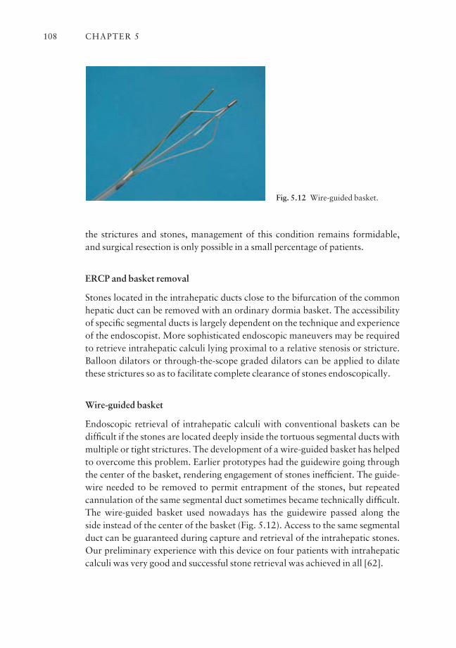
the strictures and stones, management of this condition remains formidable,and surgical resection is only possible in a small percentage of patients.
ERCP and basket removal
Stones located in the intrahepatic ducts close to the bifurcation of the commonhepatic duct can be removed with an ordinary dormia basket. The accessibilityof specific segmental ducts is largely dependent on the technique and experienceof the endoscopist. More sophisticated endoscopic maneuvers may be requiredto retrieve intrahepatic calculi lying proximal to a relative stenosis or stricture.Balloon dilators or through-the-scope graded dilators can be applied to dilatethese strictures so as to facilitate complete clearance of stones endoscopically.
Wire-guided basket
Endoscopic retrieval of intrahepatic calculi with conventional baskets can bedifficult if the stones are located deeply inside the tortuous segmental ducts withmultiple or tight strictures. The development of a wire-guided basket has helpedto overcome this problem. Earlier prototypes had the guidewire going throughthe center of the basket, rendering engagement of stones inefficient. The guide-wire needed to be removed to permit entrapment of the stones, but repeated cannulation of the same segmental duct sometimes became technically difficult.The wire-guided basket used nowadays has the guidewire passed along the side instead of the center of the basket (Fig. 5.12). Access to the same segmentalduct can be guaranteed during capture and retrieval of the intrahepatic stones.Our preliminary experience with this device on four patients with intrahepaticcalculi was very good and successful stone retrieval was achieved in all [62].
CHAPTER 5108
Fig. 5.12 Wire-guided basket.
Percutaneous transhepatic cholangioscopy (PTC)
Although the preferred treatment of primary intrahepatic stones has beenremoval of the stones via resection of the stenotic bile duct and atrophic seg-ments, surgery may be impossible in patients with multisegmental distributionof the stones. PTC lithotripsy is definitely one possible option whereby the intra-hepatic duct stones can be removed from above a stricture using a more straight-forward approach [63].
Results of percutaneous treatment of intrahepatic stones
In a study on 165 patients treated with PTC lithotripsy for intrahepatic ductstones, the success rate of complete stone clearance was 80% [64]. Three majorcauses of incomplete removal of stones were identified: (1) angulation or stric-ture of the intrahepatic ducts; (2) sludgy bile and stones; and (3) a peripherallocation rendering access impossible. After a mean follow-up of 58 months, 43(32.6%) of the 132 patients with initial clearance developed recurrent stones.Other investigators have reported similar results, and it appears that the risk ofstone recurrence tends to increase with time [65]. In a more recent seriesreported by the Korean group, recurrence of intrahepatic stones after percuta-neous cholangioscopic treatment was strongly associated with severe biliarystricture, advanced biliary cirrhosis, and Tsunoda types III and IV hepatolithia-sis [66]. As a whole, PTC lithotripsy is an option for patients whose stones arenot amenable to endoscopic treatment but who, at the same time, are not suitablecandidates for surgical resection due to poor anesthetic risk.
ERCP and sphincterotomy in Billroth II gastrectomy
ERCP is considered to be more difficult in patients with previous Billroth II gastrectomy. The overall complication rates reported in the literature rangefrom 8 to 13% [67,68]. The problems include difficulties in (1) maneuvering the side-view duodenoscope through the afferent loop in a retrograde manner;(2) cannulating the CBD from an inverted position; and (3) carrying out a papillotomy in an upside-down position. Among all the morbidities reported,bowel perforation involving in particular the afferent loop is a considerable andunique complication of ERCP. In one of our recent series, there were 11 per-forations in 185 ERCP procedures for patients with a history of previousBillroth II gastrectomy [69]. Nine perforations occurred when the afferent loop was entered, one occurred after sphincterotomy, and another during can-nulation. The majority of perforations were found near the duodenojejunal
COMMON BILE DUCT STONES AND CHOLANGITIS 109
flexure area. The likely mechanism is that the endoscope loops excessively in thejejunum when it is being inserted across the relatively fixed duodenojejunalflexure. Mucosal tear or perforation happens as a result of overstretching of theproximal jejunum during retrograde advancement of the endoscope.
Precautions and alternatives for Billroth II gastrectomy
We believe that the experience of the endoscopist is the key factor for avoidingperforation in this clinical setting. The passage of the endoscope through theafferent loop must be monitored under fluoroscopy. Excessive looping of theduodenoscope should be avoided. If resistance is encountered, the procedureshould be stopped and alternative methods for gaining access to the biliary system considered. One option is to drain the bile ducts by PTBD and have the calculi fragmented and removed through the PTBD tract in subsequent sessions. This approach is likely to lengthen the course of treatment because thePTBD tract requires serial dilation, to a size of 16 Fr or 18 Fr, before lithotripsyis possible. Another option is to enter the afferent loop with a forward-viewingendoscope instead of a side-viewing duodenoscope.
Side-viewing vs. forward-viewing scope for ERCP in Billroth II gastrectomy
In a comparative study by Kim et al. on the use of these two types of endoscopein patients with previous Billroth II gastrectomies, significantly less bowel per-foration was observed in the group having the forward-viewing endoscope, yet the success rate in cannulating the CBD was comparable [70]. However, dueto the lack of the bridge elevator, maneuverability of various devices is com-promised with the forward-viewing endoscope. Thus we still prefer to use a side-viewing duodenoscope whenever possible because it allows the operator to view the papilla en face. Recently, a special wire-guided B-II papillotome withthe cutting wire on the reverse side has been introduced [71]. Whether it helpsthe safety of sphincterotomy in gastrectomized patients remains to be seen.
Cholangitis
Pathophysiology
In the obstructed biliary tree, stagnant bile is a favorable culture medium forbacteria. Ascending infection in the biliary system is one of the mechanismsleading to acute cholangitis.
CHAPTER 5110
Effect of biliary obstruction on the reticuloendothelial system
It has been shown in both clinical and animal studies that the phagocytic func-tion of the reticuloendothelial cells surrounding the canaliculi is severelyaffected in complete bile duct obstruction. Clearance of the bacteria enteringwith the portal venous blood is therefore compromised, and infection of thestagnant bile inside the obstructed system may take place.
Bacteriology of cholangitis
The infected bile contains a large quantity of bacteria, mostly Gram-negativebacilli, with E. coli, Klebsiella spp., Enterobacter spp., and Pseudomonas, some-times mixed with anaerobes such as C. perfringens and Bacteroides fragilis, orGram-positive enterococci.
Effect of raised intrabiliary pressure and cholangiovenous reflux
The intraductal pressure in the biliary tree may rise in the presence of obstruc-tion. As the terminal ends of bile canaliculi are in direct contact with the hepaticsinusoids, a raised intrabiliary pressure will facilitate cholangiovenous reflux ofinfected materials into the hepatic venous circulation. Under such circumstancesthe patient may develop bacteremia as well as endotoxemia. The septicemiathen triggers a systemic response from the patient’s own immune system, includ-ing a variety of cytokines, complements, and vasodilators. In severe cases thereaction may be overwhelming, with deleterious effects towards other internalorgans, resulting in a phenomenon known as systemic inflammatory responsesyndrome (SIRS), with multiorgan failure, and considerable mortality.
Clinical presentation
Simple cholangitis: Charcot’s triad
Charcot’s triad of acute cholangitis includes acute right upper abdominal pain,fever, and jaundice. Nevertheless, not every patient having cholangitis manifestsall these features. Jaundice can be subtle if the onset is acute or the obstruction isincomplete, yet the patient could be very ill and septic if there is considerableendotoxemia. Frail or old patients may not experience or complain of any paineven if they are suffering from cholangitis. Overall, fever is the most com-mon and consistent symptom, present in more than 90% of patients. Chills and rigor are not as frequent but their presence is suggestive of bacteremia orsepticemia.
COMMON BILE DUCT STONES AND CHOLANGITIS 111

Suppurative cholangitis: Reynold’s pentad
In the presence of profound septicemia, the patient may develop hemodynamicinstability and mental confusion. When added to Charcot’s triad describedabove, hypotension and coma are collectively known as Reynold’s pentad, whichsignifies a severe attack of suppurative cholangitis associated with significantmortality. As the condition can be rapidly fatal, vigorous resuscitation, intensivecare support, and urgent biliary decompression should be provided in order tominimize mortality.
Clinical management
Of patients with clinical cholangitis, 80–90% may respond to conservativetreatment; the remaining 5–10% with suppurative cholangitis will need urgentbiliary decompression.
Initial conservative management
Patients with suspected acute cholangitis should be admitted to hospital for fur-ther management. It is imperative to control the sepsis as early as possible.Aggressive fluid resuscitation and high-dose intravenous broad spectrum anti-biotics are the key initial measures. Close monitoring of the vital signs, includingurine output, is necessary. With such initial treatment, control of sepsis isachieved in 90% of patients. Further intervention and drainage for the obstruct-ing lesion can be performed on a semielective or elective basis in the next avail-able session.
Urgent biliary decompression
Some 5–10% of patients, especially those with Reynold’s pentad, may notrespond to the initial resuscitation. Emergency decompression of the obstructedbiliary system is mandatory. In the case of unstable patients with compromisedhemodynamic status and respiratory function, intensive care monitoring, includ-ing the use of inotropes and/or ventilatory support, should be sought before bili-ary drainage.
Role of ERCP ES with stone extraction is considered to be the procedure of choice in patients with acute cholangitis. It is often performed when the initial treatment was to control the sepsis. However, in patients with severecholangitis not responding to the initial medical therapy, the only possibleoption is decompression and drainage of the infected biliary system. In one of
CHAPTER 5112
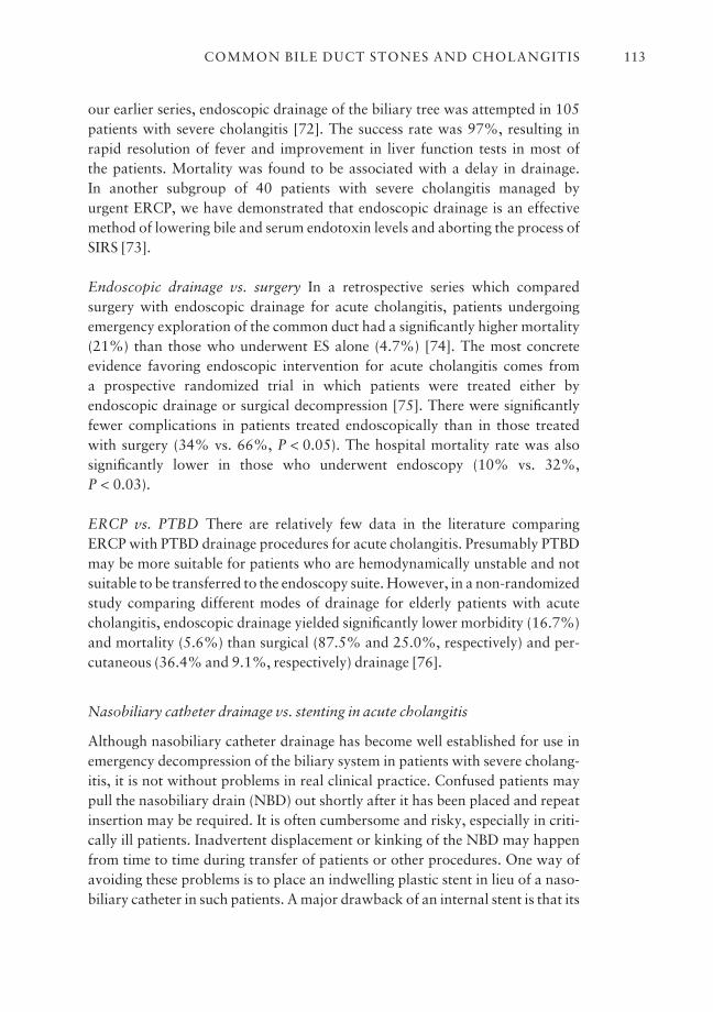
our earlier series, endoscopic drainage of the biliary tree was attempted in 105patients with severe cholangitis [72]. The success rate was 97%, resulting inrapid resolution of fever and improvement in liver function tests in most of the patients. Mortality was found to be associated with a delay in drainage. In another subgroup of 40 patients with severe cholangitis managed by urgent ERCP, we have demonstrated that endoscopic drainage is an effectivemethod of lowering bile and serum endotoxin levels and aborting the process ofSIRS [73].
Endoscopic drainage vs. surgery In a retrospective series which comparedsurgery with endoscopic drainage for acute cholangitis, patients undergoingemergency exploration of the common duct had a significantly higher mortality(21%) than those who underwent ES alone (4.7%) [74]. The most concrete evidence favoring endoscopic intervention for acute cholangitis comes from a prospective randomized trial in which patients were treated either by endoscopic drainage or surgical decompression [75]. There were significantlyfewer complications in patients treated endoscopically than in those treatedwith surgery (34% vs. 66%, P < 0.05). The hospital mortality rate was alsosignificantly lower in those who underwent endoscopy (10% vs. 32%, P < 0.03).
ERCP vs. PTBD There are relatively few data in the literature comparingERCP with PTBD drainage procedures for acute cholangitis. Presumably PTBDmay be more suitable for patients who are hemodynamically unstable and notsuitable to be transferred to the endoscopy suite. However, in a non-randomizedstudy comparing different modes of drainage for elderly patients with acutecholangitis, endoscopic drainage yielded significantly lower morbidity (16.7%)and mortality (5.6%) than surgical (87.5% and 25.0%, respectively) and per-cutaneous (36.4% and 9.1%, respectively) drainage [76].
Nasobiliary catheter drainage vs. stenting in acute cholangitis
Although nasobiliary catheter drainage has become well established for use inemergency decompression of the biliary system in patients with severe cholang-itis, it is not without problems in real clinical practice. Confused patients maypull the nasobiliary drain (NBD) out shortly after it has been placed and repeatinsertion may be required. It is often cumbersome and risky, especially in criti-cally ill patients. Inadvertent displacement or kinking of the NBD may happenfrom time to time during transfer of patients or other procedures. One way ofavoiding these problems is to place an indwelling plastic stent in lieu of a naso-biliary catheter in such patients. A major drawback of an internal stent is that its
COMMON BILE DUCT STONES AND CHOLANGITIS 113
patency and adequacy of drainage cannot be monitored. In a prospective ran-domized trial of 74 patients conducted in the authors’ center, both approacheswere shown to have similar efficacy on initial decompression for the biliary sepsis, with comparable mean procedure times [77]. Nasobiliary catheter dis-placement and kinking happened in five out of the 40 patients assigned to NBD,while stent blockage was found in one patient among the 34 with endoprosthesisplacement. Patients’ tolerance was much better in the stent group, but it had a higher mortality rate than the NBD group (12% vs. 2.5%), although the difference was not statistically significant. Currently, we still prefer NBD fordecompression for patients with cholangitis.
Surgery to prevent recurrent cholangitis
Endoscopic or percutaneous transhepatic lithotripsy may not be feasible insome patients, when cholangitis affects the biliary tree at multiple sites withvarying degrees of severity (Fig. 5.13). Surgery remains the last resort in remov-ing the calculi and preventing stone recurrence in such patients.
Types of operation Although the details of various operative procedures arebeyond the scope of this chapter, surgeons would like to achieve the following
CHAPTER 5114
Fig. 5.13 Recurrent pyogeniccholangitis affecting mainly the right system.
goals: (1) removal of the obstructing stones; (2) improved drainage of the bili-ary system; and (3) resection of non-functioning or atrophic liver segments.Transduodenal sphincteroplasty, supraduodenal choledochoduodenostomy,and end-to-side hepaticojejunostomy are the surgical procedures for improvingbile drainage. Liver resection may be suitable for disease confined to a segmentor to one side of the liver, especially if the affected parenchyma is atrophic withlittle residual function. It is performed not only to eradicate the source of symp-toms and infections, but also to remove the underlying stricture which carries amalignant potential in the future.
Conclusion
Endoscopic treatment is now the first-line management option for choledo-cholithiasis. The success rate has increased with the recent advances in cannula-tion techniques and instrument design. Although endoscopic balloon dilatationhas been proposed as a replacement for sphincterotomy for stone extraction, itis still restricted to selected cases with small calculi. It is advisable to referpatients with giant difficult stones to expert centers where advanced lithotripsytechniques are readily available. With the current endoscopic technology, veryfew patients with choledocholithiasis will require surgery. However, surgicalresection still has a role for those with intractable intrahepatic stones andcholangitis localized to certain segments of the liver.
Outstanding issues and future trends
ERCP remains the gold standard for the diagnosis of CBD stones, although EUShas shown its very high sensitivity in identifying distal CBD stones. Withimprovements in resolution of the MRC technique, this may provide an alternativeand non-invasive method of confirming the diagnosis before intervention.Randomized controlled studies have shown that endoscopic biliary drainage isthe first line of urgent management for patients with suppurative cholangitiswho have failed conservative treatment. Part of the reason for the failure ofantibiotic therapy is the inability of most drugs to penetrate a completelyobstructed biliary system with raised intrabiliary pressure. Drugs that can beexcreted into bile against a pressure gradient would have considerable advan-tages in the management of these sick patients, but they are not a replacement for urgent biliary decompression in sick patients. For urgent biliary drainage, aprior sphincterotomy is not necessary for the placement of a nasobiliary catheteror an indwelling stent. The trick is to aspirate bile to decompress the bile ducts assoon as deep cannulation is achieved. Drainage without sphincterotomy alsoavoids the risk of pancreatitis and postsphincterotomy bleeding. In principle, a
COMMON BILE DUCT STONES AND CHOLANGITIS 115
large 10 Fr stent provides better drainage for the thick infected bile than a 7 or 8 Fr stent. With improvements in scope design, we now have reasonablysized duodenoscopes fitted with larger therapeutic channels. This avoids thedifficult manipulation of large therapeutic scopes in an emergency situation.Recognizing the significance of individual bacteria in biliary stone and sludgeformation may open up a new avenue for the prevention of stone recurrence.Suppression of bacterial enzymatic activities through the use of enzyme block-ers, or down-regulation of the genetic control of enzyme production, may offeran alternative approach to prevention.
References
1 Barbara L, Sama C, Morselli-Labate AM et al. 10-years incidence of gallstone disease: theSirmione study. J Hepatol 1993; 18: S43.
2 Courvoisier OG (1890). Kasuistisch-statistische Beitrage zur Pathologie und Chirurgie derGallenwege, pp. 57–8. Liepzig, FCW Vogel.
3 Kawai K, Akasaka Y, Murakami K. Endoscopic sphincterotomy of the ampulla of Vater.Gastrointest Endosc 1974; 20: 148.
4 Lygidakis NJ. Incidence of bile infection in patients with choledocholithiasis. Am J Gastro-enterol 1982; 77: 12–17.
5 Leung JW, Sung JY, Costerton JW. Bacteriological and electron microscopy examination ofbrown pigment stones. J Clin Microbiol 1989; 27: 915–21.
6 Leung JW, Liu YL, Leung PS, Chan RC, Inciardi JF, Cheng AF. Expression of bacterial beta-gluconidase in human bile: an in-vitro study. Gastrointest Endosc 2001; 54: 346–50.
7 Bernhoft RA, Pellegrini CA, Motson RW et al. Composition and morphologic and clinical fea-tures of common duct stones. Am J Surg 1984; 148: 77–84.
8 Taylor TV, Armstrong CP. Migration of gallstones. Br Med J 1987; 294: 1320–2.9 Calvo MM, Bujanda L, Calderon A et al. Role of magnetic resonance cholangiopancreato-
graphy in patients with suspected choledocholithiasis. Mayo Clin Proc 2002; 77: 422–8.10 Amouyal P, Amouyal G, Levy P et al. Diagnosis of choledocholithiasis by endoscopic ultra-
sonography. Gastroenterology 1994; 106: 1062–7.11 Prat F, Amouyal G, Pelletier V et al. Prospective controlled study of endoscopic ultrasono-
graphy and endoscopic retrograde cholangiography in patients with suspected common bileduct lithiasis. Lancet 1996; 346: 75–9.
12 Canto M, Chak A, Stellato T, Sivak MV Jr. Endoscopic ultrasonography versus cholangio-graphy for the diagnosis of choledocholithiasis. Gastrointest Endosc 1998; 47: 439–48.
13 Kohut M, Nowakowska-Dulawa E, Marek T, Kaczor R, Nowak A. Accuracy of linear endo-scopic ultrasonography in the evaluation of patients with suspected common bile duct stones.Endoscopy 2002; 34: 299–303.
14 Boender J, Nix GA, de Ridder MA. Endoscopic papillotomy for common bile duct stones: factors influencing the complication rate. Endoscopy 1994; 26: 209–16.
15 Chen YK, Foliente RL, Santoro MJ, Walter MH, Collen MJ. Endoscopic sphincterotomy-induced pancreatitis: increased risk associated with nondilated bile ducts and sphincter of Oddidysfunction. Am J Gastroenterol 1994; 89: 327–33.
16 Freeman ML, Nelson DB, Sherman S et al. Complications of endoscopic biliary sphincterotomy.N Engl J Med 1996; 335: 909–18.
17 Loperfido S, Angelini G, Benedetti G et al. Major early complications from diagnostic and thera-peutic ERCP: a prospective multicenter study. Gastrointest Endosc 1998; 48: 1–10.
18 Sherman S, Ruffolo TA, Hawes RH et al. Complications of endoscopic sphincterotomy: aprospective series with emphasis on the increased risk associated with sphincter of Oddi dys-function and nondilated bile ducts. Gastroenterology 1991; 101: 1068–75.
CHAPTER 5116
19 Cotton PB, Lehmann GA, Vennes J et al. Endoscopic sphincterotomy complications and theirmanagement: an attempt at consensus. Gastrointest Endosc 1991; 37: 383–93.
20 Staritz M, Ewe K, Meyer zum Buschenfelde KH. Endoscopic papillary dilatation (EPD) for thetreatment of common duct stones and papillary stenosis. Endoscopy 1983; 15: 197–8.
21 Minami A, Nakatsu T, Uchida N et al. Papillary dilation vs sphincterotomy in endoscopicremoval of bile duct stones: a randomized trial with manometric function. Dig Dis Sci 1994; 40:2250–4.
22 Yasuda I, Tomita E, Enya M, Kato T, Moriwaki H. Can endoscopic papillary balloon dilationreally preserve sphincter of Oddi function? Gut 2001; 49: 686–91.
23 Bader M, Geenen JE, Hogan W et al. Endoscopic balloon dilatation of the sphincter of Oddi inpatients with suspected biliary dyskinesia: results of a prospective randomized trial. Gastro-intest Endosc 1986; 32: 158A.
24 Kozarek RA. Balloon dilatation of the sphincter of Oddi. Endoscopy 1988; 20: 207–10.25 May GR, Cotton PB, Edmunds SE, Chong W. Removal of stones from the bile duct at ERCP
without sphincterotomy. Gastrointest Endosc 1993; 39: 749–54.26 MacMathuna P, White P, Clarke E et al. Endoscopic balloon sphincteroplasty (papillary dila-
tion) for bile duct stones: efficacy, safety and follow-up in 100 patients. Gastrointest Endosc1995; 42: 468–74.
27 Bergman JJ, Rauws EA, Fockens P et al. Randomised trial of endoscopic balloon dilation versusendoscopic sphincterotomy for removal of bile duct stones. Lancet 1997; 349: 1124–9.
28 Cotton PB, Geenen JE, Sherman S et al. Endoscopic sphincterotomy for stones by experts is safe,even in younger patients with normal ducts. Ann Surg 1998; 227: 201–4.
29 Costamagna G, Tringali A, Shah SK, Mutignani M, Zuccala G, Perri V. Long-term follow-up ofpatients after endoscopic sphincterotomy for choledocholithiasis, and risk factors for recur-rence. Endoscopy 2002; 34: 273–9.
30 Bergman JJ, Tytgat GN, Huibregtse K. Endoscopic dilatation of the biliary sphincter forremoval of bile duct stones: an overview of current indications and limitations. Scand JGastroenterol Suppl 1998; 225: 59–65.
31 Stoker ME, Hebert JC, Bothe AJ. Common bile duct exploration in the era of laparoscopicsurgery. Arch Surg 1995; 130: 265.
32 Lezoche E, Paganini AM, Carlei F et al. Laparoscopic treatment of gallbladder and common bileduct stones: a prospective study. World J Surg 1996; 20: 535.
33 Chan AC, Chung SC, Wyman A et al. Selective use of preoperative endoscopic retrogradecholangiopancreatography in laparoscopic cholecystectomy. Gastrointest Endosc 1996; 43:212–15.
34 Santucci L, Natalini G, Sarpi L, Fiorucci S, Solinas A, Morelli A. Selective endoscopic retrogradecholangiography and preoperative bile duct stone removal in patients scheduled for laparo-scopic cholecystectomy: a prospective study. Am J Gastroenterol 1996; 91: 1326–30.
35 Demartines N, Eisner L, Schnabel K, Fried R, Zuber M, Harder F. Evaluation of magnetic resonance cholangiography in the management of bile duct stones. Arch Surg 2000; 135:148–52.
36 Zidi SH, Prat F, Le Guen O et al. Use of magnetic resonance cholangiography in the diagnosis ofcholedocholithiasis: prospective comparison with a reference imaging method. Gut 1999; 44:118–22.
37 Laokpessi A, Bouillet P, Sautereau D et al. Value of magnetic resonance cholangiography in thepreoperative diagnosis of common bile duct stones. Am J Gastroenterol 2001; 96: 2354–9.
38 Liu TH, Consorti ET, Kawashima A et al. Patient evaluation and management with selective useof magnetic resonance cholangiography and endoscopic retrograde cholangiopancreatographybefore laparoscopic cholecystectomy. Ann Surg 2001; 234: 33–40.
39 Binmoeller K, Seifert H, Gerke H et al. Papillary roof incision using the Erlangen-type pre-cutpapillotome to achieve selective bile duct cannulation. Gastrointest Endosc 1996; 44: 689–95.
40 Kasmin F, Cohen D, Batra S et al. Needle-knife sphincterotomy in a tertiary referral center:efficacy and complications. Gastrointest Endosc 1996; 44: 48–53.
41 Gholson C, Favrot D. Needle knife papillotomy in a university referral practice: safety andefficacy of a modified technique. J Clin Gastroenterol 1996; 23: 177–80.
COMMON BILE DUCT STONES AND CHOLANGITIS 117
42 Calvo MM, Bujanda L, Ileras I et al. The rendezvous technique for the treatment of choledo-cholithiasis. Gastrointest Endosc 2001; 54: 511–13.
43 van der Velden JJ, Berger MY, Bonjer HJ, Brakel K, Lameris JS. Percutaneous treatment of bileduct stones in patients treated successfully with endoscopic retrograde procedures. GastrointestEndosc 2000; 51: 418–22.
44 Riemann JF, Seuberth K, Demling L. Mechanical lithotripsy of common bile duct stones.Gastrointest Endosc 1985; 31: 207–10.
45 Higuchi T, Kon Y. Endoscopic mechanical lithotripsy for the treatment of common bile ductstones: experience with the improved double sheath basket catheter. Endoscopy 1987; 19:216–17.
46 Chung SC, Leung JW, Leong HT, Li AKC. Mechanical lithotripsy of large common bile ductstones using a basket. Br J Surg 1991; 78: 1448–50.
47 Shaw MJ, Mackie RD, Moore JP et al. Results of a multicenter trial using a mechanical lithotrip-tor for the treatment of large bile duct stones. Am J Gastroenterol 1993; 88: 730–3.
48 Schneider MU, Matek W, Bauer R et al. Mechanical lithotripsy of bile duct stones in 209patients: effect of technical advances. Endoscopy 1988; 20: 248–53.
49 Koch H, Stolte M, Walz V. Endoscopic lithotripsy in the common bile duct. Endoscopy 1977; 9:95.
50 Binmoeller KF, Bruckner M, Thonke F, Soehendra N. Treatment of difficult bile duct stonesusing mechanical, electrohydraulic and extracorporeal shock wave lithotripsy. Endoscopy1993; 25: 201.
51 Hixson LJ, Fennerty MB, Jaffee PE, Pulju JH, Palley SL. Peroral cholangioscopy with intra-corporeal electrohydraulic lithotripsy for choledocholithiasis. Am J Gastroenterol 1992; 87:296.
52 Cotton PB, Kozarek RA, Schapiro RH et al. Endoscopic laser lithotripsy of large bile ductstones. Gastroenterology 1990; 99: 1128.
53 Prat F, Fritsch J, Choury AD, Frouge C, Marteau V, Etienne JP. Laser lithotripsy of difficult bili-ary stones. Gastrointest Endosc 1994; 40: 290.
54 Neuhaus H, Hoffmann W, Gottlieb K, Classen M. Endoscopic lithotripsy of bile duct stonesusing a new laser with automatic stone recognition. Gastrointest Endosc 1994; 40: 708.
55 Hochberger S, May A, Bayer J, Muhldorfer S, Hahn EG, Ell C. Laser lithotripsy of difficult bileduct stones: results in 50 patients using a rhodamine-6 G dye laser with automatic optical stone-tissue detection system. Gastrointest Endosc 1997; 45: 133A.
56 Chan AC, Ng EK, Chung SC et al. Common bile duct stones become smaller after endoscopicbiliary stenting. Endoscopy 1998; 30: 356–9.
57 Maxton DG, Tweedle DE, Martin DF. Retained common bile duct stones after endoscopicsphincterotomy: temporary and long term treatment with biliary stenting. Gut 1995; 36: 446.
58 Jain SK, Stein R, Bhuva M, Goldberg MJ. Pigtail stents: an alternative in the treatment ofdifficult bile duct stones. Gastrointest Endosc 2000; 52: 490–3.
59 Chopra KB, Peters RA, O’Toole PA et al. Randomised study of endoscopic biliary endoprosthe-sis versus duct clearance for bile duct stones in high-risk patients. Lancet 1996; 21 (348): 791–3.
60 Schreiber F, Stern M. Fragmentation of bile duct stones by extracorporeal shock wavelithotripsy. Gastroenterology 1989; 146: 96.
61 Ellis RD, Jenkins AP, Thompson RP, Ede RJ. Clearance of refractory bile duct stones with extra-corporeal shockwave lithotripsy. Gut 2000; 47: 728–31.
62 Chan AC, Chung SC. New wire-guided basket for intrahepatic stone extraction. GastrointestEndosc 1999; 50: 401–4.
63 Park JH, Choi BI, Han MC, Sung KB, Choo IW, Kim CW. Percutaneous removal of residualintrahepatic stones. Radiology 1987; 163: 619–23.
64 Yeh YH, Huang MH, Yang JC, Mo LR, Lin J, Yueh SK. Percutaneous trans-hepatic cholan-gioscopy and lithotripsy in the treatment of intrahepatic stones: a study with 5 year follow-up.Gastrointest Endosc 1995; 42: 13–18.
65 January YY, Chen MF. Percutaneous trans-hepatic cholangioscopic lithotomy for hepatolithi-asis: long-term results. Gastrointest Endosc 1995; 42: 1–5.
CHAPTER 5118
66 Lee SK, Seo DW, Myung SJ et al. Percutaneous transhepatic cholangioscopic treatment for hepatolithiasis: an evaluation of long-term results and risk factors for recurrence. GastrointestEndosc 2001; 53: 318–23.
67 Cohen SA, Siegel JH, Kasmin FE. Complications of diagnostic and therapeutic ERCP. AbdomImaging 1996; 21: 385–94.
68 Huibregtse K. Complications of endoscopic sphincterotomy and their prevention [Editorial]. N Engl J Med 1996; 335: 961–3.
69 Faylona JM, Qadir A, Chan AC, Lau JY, Chung SC. Small bowel perforations related to endo-scopic retrograde cholangiopancreatography (ERCP) in patients with Billroth II gastrectomy.Endoscopy 1999; 31: 546–9.
70 Kim MH, Lee SK, Lee MH et al. Endoscopic retrograde cholangiopancreatography and needle-knife sphincterotomy in patients with Billroth II gastrectomy: a comparative study of the forward-viewing endoscope and the side-viewing duodenoscope. Endoscopy 1997; 29: 82–5.
71 Wang YG, Binmoeller KF, Seifert H, Maydeo A, Soehendra N. A new guide wire papillotome forpatients with Billroth II gastrectomy. Endoscopy 1996; 28: 254.
72 Leung JW, Chung SC, Sung JJ et al. Urgent endoscopic drainage for acute suppurative cholang-itis. Lancet 1989; 1: 1307–9.
73 Lau JY, Chung SC, Leung JW, Ling TK, Yung MY, Li AK. Endoscopic drainage aborts endotox-aemia in acute cholangitis. Br J Surg 1996; 83: 181–4.
74 Leese T, Neoptolemos JP, Baker AR et al. Management of acute cholangitis and the impact onendoscopic sphincterotomy. Br J Surg 1986; 73: 988–92.
75 Lai EC, Mok FP, Tan ES et al. Endoscopic biliary drainage for severe acute cholangitis. N Engl JMed 1992; 326: 1582–6.
76 Sugiyama M, Atomi Y. Treatment of acute cholangitis due to choledocholithiasis in elderly andyounger patients. Arch Surg 1997; 132: 1129–33.
77 Lee DW, Chan AC, Lam YH et al. Biliary decompression by nasobiliary catheter or biliary stentin acute suppurative cholangitis: a prospective randomized trial. Gastrointest Endosc 2002; 56:361–5.
COMMON BILE DUCT STONES AND CHOLANGITIS 119

CHAPTER 6
The Role of ERCP in Pancreatico-BiliaryMalignancies
GULSHAN PARASHER AND JOHN G. LEE
Synopsis
Approximately 30 000 new cases of pancreatic cancer and 7000 biliary tractcancers are diagnosed annually in the United States [1]. The most commoncause of malignant biliary obstruction is pancreatic adenocarcinoma, followedby cholangiocarcinoma, ampullary neoplasm, and extrinsic compression by meta-static lymphadenopathy in the liver hilum. The role of ERCP in pancreatico-biliary malignancies is to (1) confirm the diagnosis of obstructive jaundice inpatients with suspected pancreatic carcinoma or biliary tumors; (2) obtain tissuefor histopathologic diagnosis; (3) establish the exact site of obstruction, i.e.ampullary, pancreatic, or bile duct; (4) decompress the bile duct; and (5) facil-itate palliative therapy such as intraluminal brachytherapy or intraductal photodynamic therapy. This chapter describes various current and emergingapplications of ERCP in the management of pancreatico-biliary malignancies.
ERCP in diagnosis of pancreatico-biliary malignancies
Radiological diagnosis
Significance of ‘double duct stricture’ sign
The radiographic features of ERCP cannot reliably distinguish between benignand malignant diseases. Although the double duct sign with simultaneous narrowing of the common bile duct and the pancreatic duct has been regardedtraditionally as predictive of pancreatic cancer (Fig. 6.1), recent studies showedthat its specificity is much lower than previously thought, with 15–37% of suchpatients having benign disease on long-term follow-up [2,3]. Stricture length > 14 mm was highly predictive of malignancy in one study [4], while in anotherstudy the pancreatic duct stricture length measured on ERCP correlated withboth size (P < 0.001) and staging (P < 0.002) of the pancreatic cancer [5]. The
120
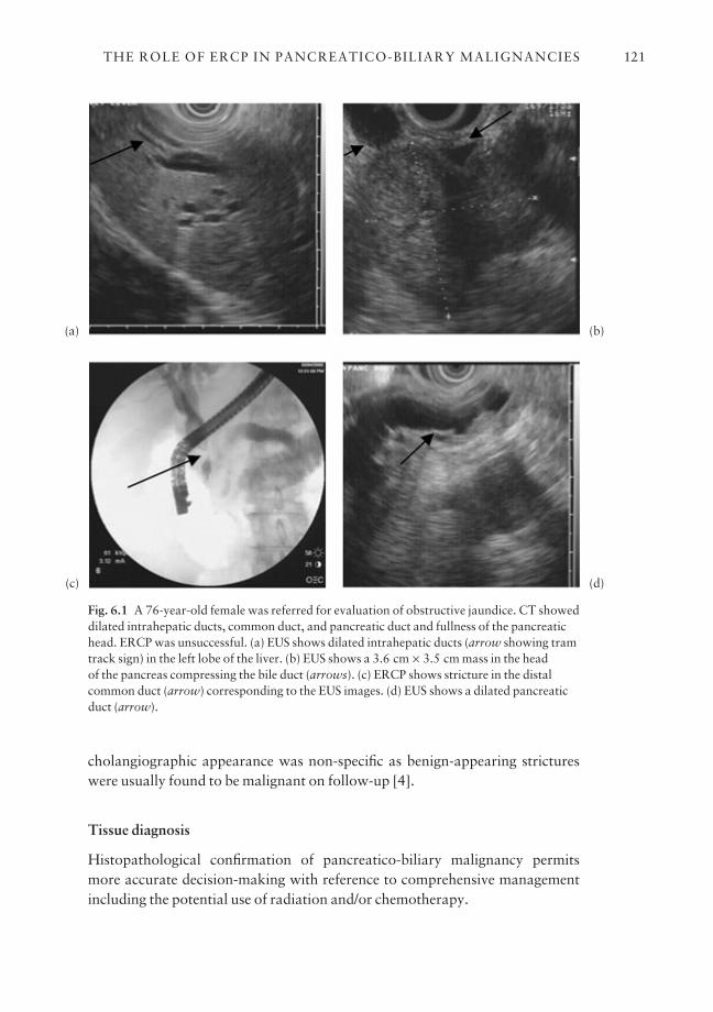
cholangiographic appearance was non-specific as benign-appearing strictureswere usually found to be malignant on follow-up [4].
Tissue diagnosis
Histopathological confirmation of pancreatico-biliary malignancy permitsmore accurate decision-making with reference to comprehensive managementincluding the potential use of radiation and/or chemotherapy.
THE ROLE OF ERCP IN PANCREATICO-BILIARY MALIGNANCIES 121
Fig. 6.1 A 76-year-old female was referred for evaluation of obstructive jaundice. CT showeddilated intrahepatic ducts, common duct, and pancreatic duct and fullness of the pancreatichead. ERCP was unsuccessful. (a) EUS shows dilated intrahepatic ducts (arrow showing tramtrack sign) in the left lobe of the liver. (b) EUS shows a 3.6 cm × 3.5 cm mass in the head of the pancreas compressing the bile duct (arrows). (c) ERCP shows stricture in the distalcommon duct (arrow) corresponding to the EUS images. (d) EUS shows a dilated pancreaticduct (arrow).
(a)
(c)
(b)
(d)
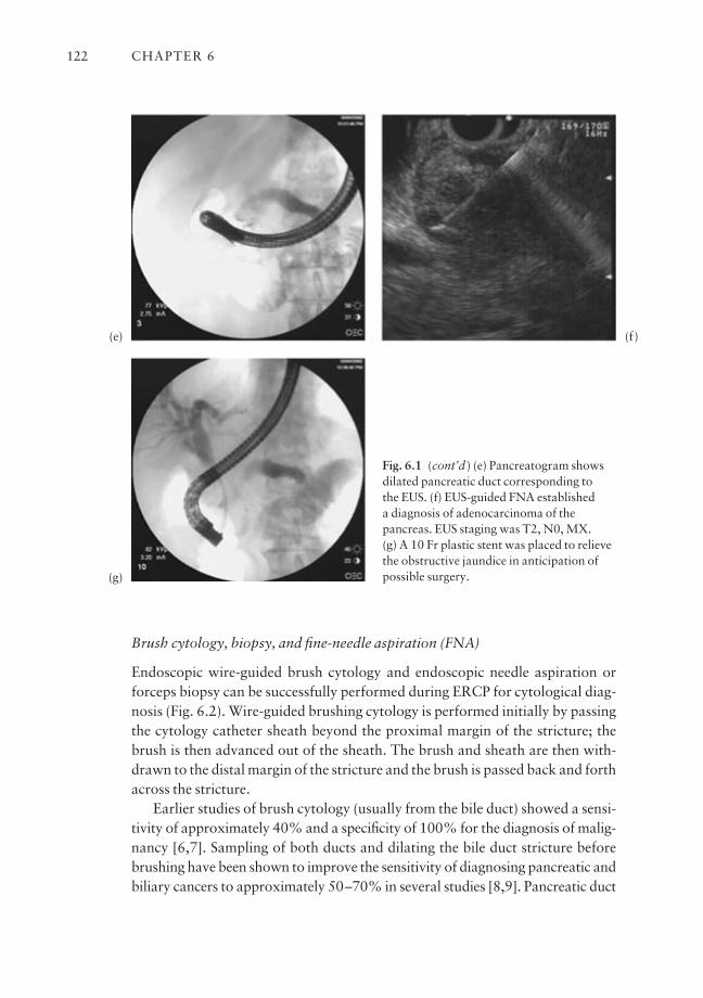
Brush cytology, biopsy, and fine-needle aspiration (FNA)
Endoscopic wire-guided brush cytology and endoscopic needle aspiration orforceps biopsy can be successfully performed during ERCP for cytological diag-nosis (Fig. 6.2). Wire-guided brushing cytology is performed initially by passingthe cytology catheter sheath beyond the proximal margin of the stricture; thebrush is then advanced out of the sheath. The brush and sheath are then with-drawn to the distal margin of the stricture and the brush is passed back and forthacross the stricture.
Earlier studies of brush cytology (usually from the bile duct) showed a sensi-tivity of approximately 40% and a specificity of 100% for the diagnosis of malig-nancy [6,7]. Sampling of both ducts and dilating the bile duct stricture beforebrushing have been shown to improve the sensitivity of diagnosing pancreatic andbiliary cancers to approximately 50–70% in several studies [8,9]. Pancreatic duct
CHAPTER 6122
Fig. 6.1 (cont’d ) (e) Pancreatogram showsdilated pancreatic duct corresponding to the EUS. (f) EUS-guided FNA established a diagnosis of adenocarcinoma of thepancreas. EUS staging was T2, N0, MX. (g) A 10 Fr plastic stent was placed to relievethe obstructive jaundice in anticipation ofpossible surgery.
(e)
(g)
(f)

brushing appears to be safe without an increased risk of pancreatitis in thesestudies.
Finally, combining the results of brush cytology, FNA, and/or forceps biopsyimproves the overall sensitivity of ERCP in diagnosing pancreatic and biliary
THE ROLE OF ERCP IN PANCREATICO-BILIARY MALIGNANCIES 123
Fig. 6.2 A 34-year-old female with a history of inflammatory bowel disease and primarysclerosing cholangitis underwent resection of the common bile duct and hepatic duct forcholangiocarcinoma. The right and left hepatic ducts were anastomosed to the jejunum. Thepatient was referred for asymptomatic elevation of tumor markers. ERCP identified only partof the intrahepatic duct, possibly the left side, and EUS did not show an obvious mass. ERCPwas repeated for cytology and stenting in anticipation of possible photodynamic therapy. (a)ERCP performed using a forward-viewing endoscope shows only one of the openings leadingto the left intrahepatic system. A separate opening to the right intrahepatic system is locatedinferior to this opening, just outside of the visual field. (b) Cholangiography of the left hepaticduct shows changes of sclerosing cholangitis. (c) The right hepatic duct is imaged through aseparate opening and shows changes of sclerosing cholangitis. (d) Brush cytology wasselectively obtained from distal and proximal ducts of the right and left systems to identifylocal recurrence. Unfortunately, all cytological samples were positive for recurrent carcinoma.
(a)
(c)
(b)
(d)

cancers to 70–85%, which is higher than any single method of tissue sampling[10–12]. We recommend performing at least two different types of tissue sam-pling procedure to improve the diagnostic accuracy of ERCP in patients withsuspected pancreatico-biliary cancers.
Tumor markers in bile or pancreatic juice
A number of molecular and genetic markers have been studied alone or in combination in bile or pancreatic juice for the diagnosis of pancreatico-biliarymalignancies (Fig. 6.3). Molecular-based tests may be helpful in diagnosingpancreatic cancer and other biliary malignancy at an early stage when surgicalcure is still possible. The addition of DNA image analysis to routine cytology hasbeen reported to increase the diagnostic sensitivity as compared to results ofcytology alone [13]. Other studies have focused their attention on mutations incodon 12 of the K-ras oncogene, because they are seen in up to 95% of pancreaticadenocarcinoma and in the premalignant conditions of the pancreas [14–16]. Bile
CHAPTER 6124
Fig. 6.2 (cont’d ) (e) Stents were placed intothe right and left system in anticipation ofpossible photodynamic therapy for localrecurrence. (f) Two 7 Fr stents were placedinto the right and left intrahepatic ducts. (g)This endoscopic view clearly shows the twoseparate orifices of the right and left hepaticducts. Photodynamic therapy was notperformed due to widespread disease and the stents were removed several weeks later.
(e)
(g)
(f)
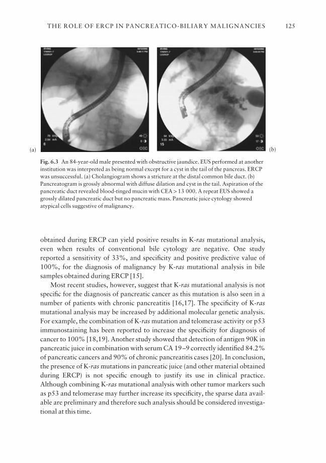
obtained during ERCP can yield positive results in K-ras mutational analysis,even when results of conventional bile cytology are negative. One studyreported a sensitivity of 33%, and specificity and positive predictive value of100%, for the diagnosis of malignancy by K-ras mutational analysis in bile samples obtained during ERCP [15].
Most recent studies, however, suggest that K-ras mutational analysis is notspecific for the diagnosis of pancreatic cancer as this mutation is also seen in anumber of patients with chronic pancreatitis [16,17]. The specificity of K-rasmutational analysis may be increased by additional molecular genetic analysis.For example, the combination of K-ras mutation and telomerase activity or p53immunostaining has been reported to increase the specificity for diagnosis ofcancer to 100% [18,19]. Another study showed that detection of antigen 90K inpancreatic juice in combination with serum CA 19–9 correctly identified 84.2%of pancreatic cancers and 90% of chronic pancreatitis cases [20]. In conclusion,the presence of K-ras mutations in pancreatic juice (and other material obtainedduring ERCP) is not specific enough to justify its use in clinical practice.Although combining K-ras mutational analysis with other tumor markers suchas p53 and telomerase may further increase its specificity, the sparse data avail-able are preliminary and therefore such analysis should be considered investiga-tional at this time.
THE ROLE OF ERCP IN PANCREATICO-BILIARY MALIGNANCIES 125
Fig. 6.3 An 84-year-old male presented with obstructive jaundice. EUS performed at anotherinstitution was interpreted as being normal except for a cyst in the tail of the pancreas. ERCPwas unsuccessful. (a) Cholangiogram shows a stricture at the distal common bile duct. (b)Pancreatogram is grossly abnormal with diffuse dilation and cyst in the tail. Aspiration of thepancreatic duct revealed blood-tinged mucin with CEA > 13 000. A repeat EUS showed agrossly dilated pancreatic duct but no pancreatic mass. Pancreatic juice cytology showedatypical cells suggestive of malignancy.
(a) (b)
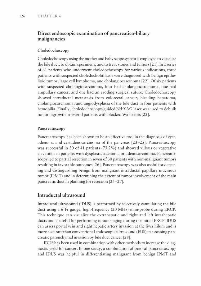
Direct endoscopic examination of pancreatico-biliarymalignancies
Choledochoscopy
Choledochoscopy using the mother and baby scope system is employed to visualizethe bile duct, to obtain specimens, and to treat stones and tumors [21]. In a seriesof 61 patients who underwent choledochoscopy for various indications, threepatients with suspected choledocholithiasis were diagnosed with benign epithe-lioid tumor, large cell lymphoma, and cholangiocarcinoma [22]. Of six patientswith suspected cholangiocarcinoma, four had cholangiocarcinoma, one hadampullary cancer, and one had an eroding surgical suture. Choledochoscopyshowed intraductal metastasis from colorectal cancer, bleeding hepatoma,cholangiocarcinoma, and angiodysplasia of the bile duct in four patients withhemobilia. Finally, choledochoscopy-guided Nd:YAG laser was used to debulktumor ingrowth in several patients with blocked Wallstents [22].
Pancreatoscopy
Pancreatoscopy has been shown to be an effective tool in the diagnosis of cyst-adenoma and cystadenocarcinoma of the pancreas [23–25]. Pancreatoscopywas successful in 30 of 41 patients (73.2%) and showed villous or vegetative elevations in patients with dysplastic adenoma or adenocarcinoma. Pancreato-scopy led to partial resection in seven of 30 patients with non-malignant tumorsresulting in favorable outcomes [26]. Pancreatoscopy was also useful for detect-ing and distinguishing benign from malignant intraductal papillary mucinoustumor (IPMT) and in determining the extent of tumor involvement of the mainpancreatic duct in planning for resection [25–27].
Intraductal ultrasound
Intraductal ultrasound (IDUS) is performed by selectively cannulating the bileduct using a 6 Fr gauge, high-frequency (20 MHz) mini-probe during ERCP.This technique can visualize the extrahepatic and right and left intrahepaticducts and is useful for performing tumor staging during the initial ERCP. IDUScan assess portal vein and right hepatic artery invasion at the liver hilum and ismore accurate than conventional endoscopic ultrasound (EUS) in assessing pan-creatic parenchymal invasion by bile duct cancer [28].
IDUS has been used in combination with other methods to increase the diag-nostic yield for cancer. In one study, a combination of peroral pancreatoscopyand IDUS was helpful in differentiating malignant from benign IPMT and
CHAPTER 6126
resulted in an improvement in postoperative survival [27]. Tamada et al.showed that the presence of sessile tumor, tumor size > 1 cm, and interruptedwall structures was helpful in predicting malignancy in 62 patients with malig-nant biliary strictures and prior negative biopsies [29].
Magnetic resonance cholangiopancreatography
Magnetic resonance cholangiopancreatography (MRCP) is an emerging applica-tion of magnetic resonance imaging (MRI) applied to the pancreatico-biliarytree. MRCP relies on heavily T2–weighted sequences. Fluid-containing struc-tures have a much longer T2 than solid tissue, resulting in higher signal intensity.Stationary fluid in the biliary and pancreatic ducts serves as an intrinsic con-trast medium and the ductal system appears white against a black background,similar to ERCP.
MRCP vs. ERCP
The major advantages of MRCP are that it does not require endoscopy, contrastinjection, or exposure to radiation. MRCP has been reported to distinguishbetween benign and malignant bile duct obstruction, with a sensitivity between50 and 86% and a specificity between 92 and 98% [30–32]. MRCP has beenreported to be similar to ERCP in distinguishing between malignant and benignbiliary obstruction with respect to sensitivity (86% vs. 89%), specificity (82%vs. 94%), and likelihood ratios for positive (4.9 vs. 15.1) and negative (0.2 vs.0.1) tests respectively [32].
In another comparative study, the sensitivity of ERCP for diagnosing pan-creatic cancer was lower (70% vs. 84%) because it missed 11 lesions < 3 cm,most of which were in the head of the pancreas [33]. ERCP was associated withseveral mild cases of pancreatitis, fever, and epigastric pain while MRCP wasfree of complications [33]. MRCP is also helpful in visualizing the main pancre-atic duct in patients with IPMT, especially when ERCP fails because of copiousintraductal mucin [34].
Finally, MRCP can be used to confirm the presence and location of a biliarystricture in a patient with obstructive jaundice before therapeutic ERCP, particu-larly in those with complex hilar lesions, thus minimizing the risk of con-tamination and infection. MRCP-guided endoscopic unilateral stent placementwas associated with lower morbidity and mortality as compared with the stan-dard method of stent insertion in 35 patients with Bismuth types III and IV hilartumors [35].
In conclusion, MRCP is a safe, non-invasive, and accurate, but operator-dependent, technique for imaging the pancreatico-biliary system. MRCP should
THE ROLE OF ERCP IN PANCREATICO-BILIARY MALIGNANCIES 127
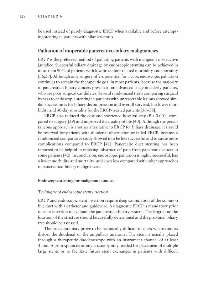
be used instead of purely diagnostic ERCP when available and before attempt-ing stenting in patients with hilar strictures.
Palliation of inoperable pancreatico-biliary malignancies
ERCP is the preferred method of palliating patients with malignant obstructivejaundice. Successful biliary drainage by endoscopic stenting can be achieved inmore than 90% of patients with low procedure-related morbidity and mortality[36,37]. Although only surgery offers potential for a cure, endoscopic palliationcontinues to remain the therapeutic goal in most patients, because the majorityof pancreatico-biliary cancers present at an advanced stage in elderly patients,who are poor surgical candidates. Several randomized trials comparing surgicalbypass to endoscopic stenting in patients with unresectable lesions showed sim-ilar success rates for biliary decompression and overall survival, but lower mor-bidity and 30-day mortality for the ERCP-treated patients [36–38].
ERCP also reduced the cost and shortened hospital stay (P < 0.001) com-pared to surgery [39] and improved the quality of life [40]. Although the percu-taneous approach is another alternative to ERCP for biliary drainage, it shouldbe reserved for patients with duodenal obstruction or failed ERCP, because arandomized comparative study showed it to be less successful and to cause morecomplications compared to ERCP [41]. Pancreatic duct stenting has beenreported to be helpful in relieving ‘obstructive’ pain from pancreatic cancer insome patients [42]. In conclusion, endoscopic palliation is highly successful, hasa lower morbidity and mortality, and costs less compared with other approachesto pancreatico-biliary malignancies.
Endoscopic stenting for malignant jaundice
Technique of endoscopic stent insertion
ERCP and endoscopic stent insertion require deep cannulation of the commonbile duct with a catheter and guidewire. A diagnostic ERCP is mandatory priorto stent insertion to evaluate the pancreatico-biliary system. The length and thelocation of the stricture should be carefully determined and the proximal biliarytree should be assessed.
The procedure may prove to be technically difficult in cases where tumorsdistort the duodenal or the ampullary anatomy. The stent is usually placedthrough a therapeutic duodenoscope with an instrument channel of at least 4 mm. A prior sphincterotomy is usually only needed for placement of multiplelarge stents or to facilitate future stent exchanges in patients with difficult
CHAPTER 6128
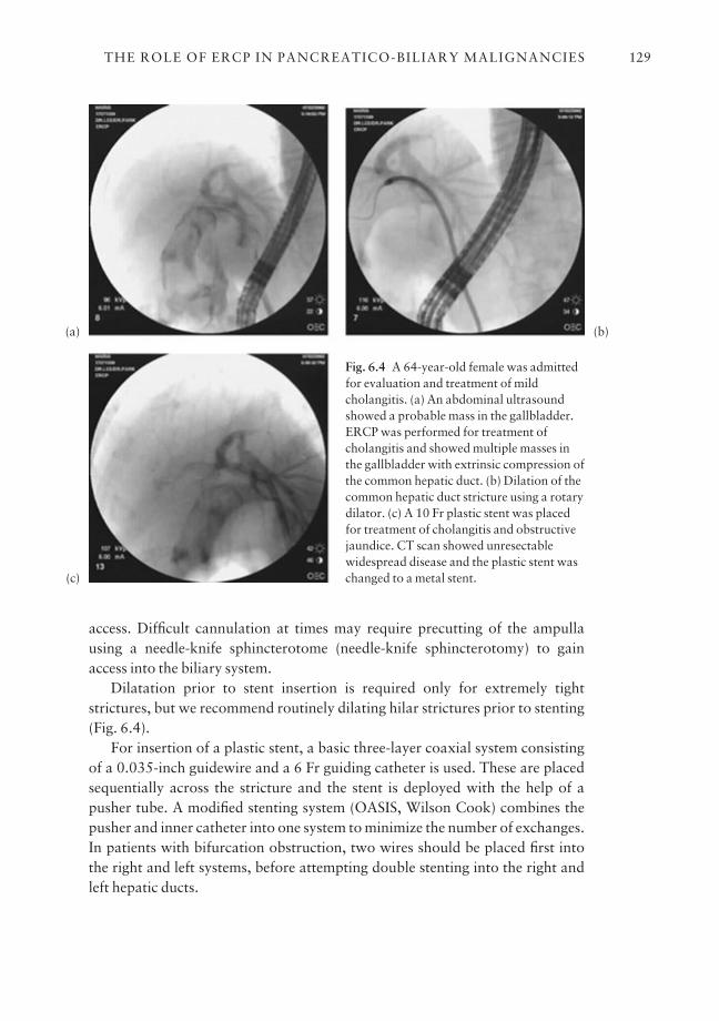
access. Difficult cannulation at times may require precutting of the ampullausing a needle-knife sphincterotome (needle-knife sphincterotomy) to gainaccess into the biliary system.
Dilatation prior to stent insertion is required only for extremely tight strictures, but we recommend routinely dilating hilar strictures prior to stenting(Fig. 6.4).
For insertion of a plastic stent, a basic three-layer coaxial system consistingof a 0.035-inch guidewire and a 6 Fr guiding catheter is used. These are placedsequentially across the stricture and the stent is deployed with the help of apusher tube. A modified stenting system (OASIS, Wilson Cook) combines thepusher and inner catheter into one system to minimize the number of exchanges.In patients with bifurcation obstruction, two wires should be placed first intothe right and left systems, before attempting double stenting into the right andleft hepatic ducts.
THE ROLE OF ERCP IN PANCREATICO-BILIARY MALIGNANCIES 129
Fig. 6.4 A 64-year-old female was admittedfor evaluation and treatment of mildcholangitis. (a) An abdominal ultrasoundshowed a probable mass in the gallbladder.ERCP was performed for treatment ofcholangitis and showed multiple masses inthe gallbladder with extrinsic compression ofthe common hepatic duct. (b) Dilation of thecommon hepatic duct stricture using a rotarydilator. (c) A 10 Fr plastic stent was placedfor treatment of cholangitis and obstructivejaundice. CT scan showed unresectablewidespread disease and the plastic stent waschanged to a metal stent.
(a) (b)
(c)
Types of stent
Plastic stents Plastic stents are mostly made of polyethylene. Other materialsused are polyurethane and Teflon. The mean patency of a plastic stent is approx-imately 2–4 months [43,44]. Important complications associated with plasticstents include stent occlusion, sepsis, stent migration, stent fracture, and, rarely,acute cholecystitis related to occlusion of the cystic duct [44].
The major disadvantage of plastic stents is occlusion from bacterial biofilm,which comprises protein, deconjugated bilirubin, microcolonies of bacteria,and amorphous debris [43]. Stent occlusion leads to recurrence of jaundice or cholangitis, necessitating stent exchanges in 30–60% of patients [43–45].Unfortunately, attempts to improve the patency rates of plastic stents by alter-native stent design and oral administration of bile acids, antibiotics, and aspirinhave not been clinically successful [43–49].
Metal stents The self-expandable metal stent (SEMS) was developed to over-come the short patency of the plastic stent. SEMSs are made of either stainlesssteel alloy monofilaments (Wallstent, Boston Scientific, Natick, MA and SpiralZ-Stent, Wilson Cook, Winston Salem, NC) or nickel titanium alloy (DiamondStent, Boston Scientific, Natick, MA and Za-Stent, Wilson Cook, WinstonSalem, NC).
The comparative efficacy of each design is not well known and their use isguided more by physician preference. SEMSs can be compressed on to a 3-mmdelivery system and expanded to 10 mm after deployment. The larger luminaldiameter of these stents offers a prolonged patency of up to 10–12 months.However, the cost per device is significantly higher than for plastic stents ($1000–1500 vs. $50–100).
SEMSs can also occlude but through different mechanisms, including biliarysludge, dietary fiber, tumor ingrowth or overgrowth, epithelial hyperplasia, or a combination of these. Management of an occluded metal biliary stent includesmechanical dislodgement of the obstructing material, placement of a plasticstent within the metal stent, and placement of a second overlying or overlappingstent to improve drainage. Electrocoagulation or laser therapy to destroy theingrowing tumor has not been effective [44].
Metal vs. plastic stents Metal stents have been compared with plastic stents indifferent studies. In 47 patients with pancreatic cancer with a mean survival of6.2 months from the time of endoscopy, metal stents were shown to have alonger patency than plastic stentsa8.2 months vs. 3.5 months (P < 0.001) [50].
A prospective randomized trial in France evaluated 97 patients with malig-nant strictures of the bile ducts (64% with pancreatic cancer), who were
CHAPTER 6130
randomized to receive either an 11.5 Fr stent to be exchanged on demand orevery 3 months, or a self-expanding metallic wall stent [51]. The mean duration offollow-up was 166 days. Cost effective analysis suggested that metal stents wereadvantageous for patients surviving longer than 6 months, whereas plasticstents were advantageous for patients surviving less than 6 months. This studyshowed initial metal stenting to be the most cost effective approach, providedthat the patient survived for longer than 6 months.
The US Wallstent multicenter randomized trial evaluated the Wallstent com-pared with 10 Fr plastic stents for the palliation of malignant biliary obstruction[52]. Early stent occlusion was reported in 30% of the plastic group and in 0%of the Wallstent group. Sludge occlusion and stent migration were seen in 28%of plastic stents and 6% of Wallstents. The overall complication rate was sig-nificantly lower in the Wallstent group (P < 0.05) for both hilar and distal bili-ary strictures. Wallstents did not offer any survival advantage over the plasticstent but were less expensive because they required fewer repeat ERCPs andstent exchange.
A prospective study from Amsterdam compared Wallstents with plasticstents in distal malignant biliary obstruction and reported a lower occlusion rate(33% vs. 54%), longer stent patency (273 vs. 126 days), and 28% reduction inERCPs per patient in the Wallstent group [53]. These studies show thatWallstents can be deployed successfully in most patients and occlude less fre-quently and less rapidly than the conventional 10 Fr and 11.5 Fr plastic stents.
Logically, therefore, Wallstent use reduces hospitalization and repeatedinterventions leading to a lower cost. In conclusion, the most cost effectiveapproach to palliating malignant obstructive jaundice is to place a SEMS at theinitial ERCP in patients with unresectable cancer who have a life expectancy ofat least 6 months.
Covered and uncovered metal stents Metal stents partially covered with sili-cone or polyurethane membrane have been introduced to overcome the problemof tumor ingrowth and epithelial hyperplasia. Shim et al. compared endoscopic-ally placed polyurethane-covered Z-Stent to non-coated Wallstent or Streckerstent [54]. The median patency of both covered and uncovered stents was com-parable (267 vs. 233 days), but tumor ingrowth was seen in two patients withthe covered stents compared to six in the non-covered stent group. Early andlate complications were the same in both groups [54].
Reported complications associated with covered stents include tumor in-growth or overgrowth, sludge accumulation, stent migration, pancreatitis, andgangrenous cholecystitis [44,54–56]. Finally, covered biliary metal stents havenot uniformly shown a significant advantage in terms of greater patency rates[54,56].
THE ROLE OF ERCP IN PANCREATICO-BILIARY MALIGNANCIES 131
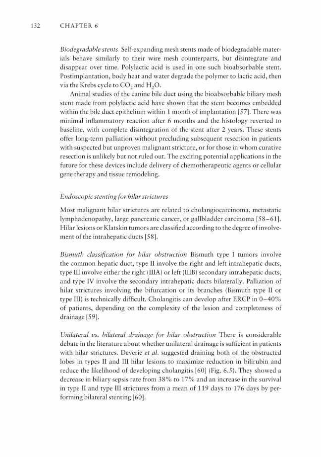
Biodegradable stents Self-expanding mesh stents made of biodegradable mater-ials behave similarly to their wire mesh counterparts, but disintegrate and disappear over time. Polylactic acid is used in one such bioabsorbable stent.Postimplantation, body heat and water degrade the polymer to lactic acid, thenvia the Krebs cycle to CO2 and H2O.
Animal studies of the canine bile duct using the bioabsorbable biliary meshstent made from polylactic acid have shown that the stent becomes embeddedwithin the bile duct epithelium within 1 month of implantation [57]. There wasminimal inflammatory reaction after 6 months and the histology reverted tobaseline, with complete disintegration of the stent after 2 years. These stentsoffer long-term palliation without precluding subsequent resection in patientswith suspected but unproven malignant stricture, or for those in whom curativeresection is unlikely but not ruled out. The exciting potential applications in thefuture for these devices include delivery of chemotherapeutic agents or cellulargene therapy and tissue remodeling.
Endoscopic stenting for hilar strictures
Most malignant hilar strictures are related to cholangiocarcinoma, metastaticlymphadenopathy, large pancreatic cancer, or gallbladder carcinoma [58–61].Hilar lesions or Klatskin tumors are classified according to the degree of involve-ment of the intrahepatic ducts [58].
Bismuth classification for hilar obstruction Bismuth type I tumors involve the common hepatic duct, type II involve the right and left intrahepatic ducts,type III involve either the right (IIIA) or left (IIIB) secondary intrahepatic ducts,and type IV involve the secondary intrahepatic ducts bilaterally. Palliation ofhilar strictures involving the bifurcation or its branches (Bismuth type II or type III) is technically difficult. Cholangitis can develop after ERCP in 0–40% of patients, depending on the complexity of the lesion and completeness ofdrainage [59].
Unilateral vs. bilateral drainage for hilar obstruction There is considerabledebate in the literature about whether unilateral drainage is sufficient in patientswith hilar strictures. Deverie et al. suggested draining both of the obstructedlobes in types II and III hilar lesions to maximize reduction in bilirubin andreduce the likelihood of developing cholangitis [60] (Fig. 6.5). They showed adecrease in biliary sepsis rate from 38% to 17% and an increase in the survivalin type II and type III strictures from a mean of 119 days to 176 days by per-forming bilateral stenting [60].
CHAPTER 6132
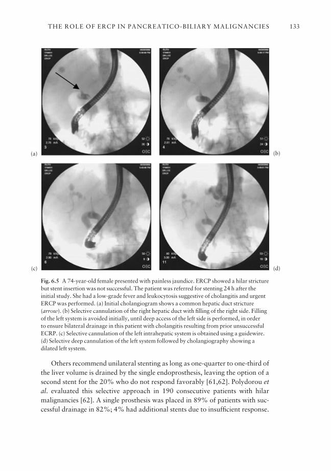
Others recommend unilateral stenting as long as one-quarter to one-third ofthe liver volume is drained by the single endoprosthesis, leaving the option of asecond stent for the 20% who do not respond favorably [61,62]. Polydorou etal. evaluated this selective approach in 190 consecutive patients with hilarmalignancies [62]. A single prosthesis was placed in 89% of patients with suc-cessful drainage in 82%; 4% had additional stents due to insufficient response.
THE ROLE OF ERCP IN PANCREATICO-BILIARY MALIGNANCIES 133
Fig. 6.5 A 74-year-old female presented with painless jaundice. ERCP showed a hilar stricturebut stent insertion was not successful. The patient was referred for stenting 24 h after theinitial study. She had a low-grade fever and leukocytosis suggestive of cholangitis and urgentERCP was performed. (a) Initial cholangiogram shows a common hepatic duct stricture(arrow). (b) Selective cannulation of the right hepatic duct with filling of the right side. Fillingof the left system is avoided initially, until deep access of the left side is performed, in order to ensure bilateral drainage in this patient with cholangitis resulting from prior unsuccessfulECRP. (c) Selective cannulation of the left intrahepatic system is obtained using a guidewire.(d) Selective deep cannulation of the left system followed by cholangiography showing adilated left system.
(a)
(c)
(b)
(d)
Seven per cent required a combined procedure with percutaneous transhepaticaccess. Stenting was technically successful in 93% of type I, 94% of type II, and84% of type III patients, with successful drainage in 91%, 83%, and 73% ofpatients, respectively. Early complications were seen in 7%, 14%, and 31% oftypes I, II, and III patients, and the mortality rates for these groups were 14%,15%, and 32%, respectively [62]. The authors concluded that a single pros-thesis provides good palliation in 80% of patients, whereas a second stent shouldbe reserved for stent failures. A small prospective randomized comparative trialshowed significantly higher technical success and lower complication for patientstreated by unilateral stenting [63].
Another study recommended bilobar drainage in patients in whom both of thelobes were filled during the ERCP, as patients with incomplete drainage had theworst survival among all patients with hilar tumors [64]. All things being equal,it probably makes more sense to drain the left system because the left hepatic
CHAPTER 6134
Fig. 6.5 (cont’d ) (e,f,g) Placement ofguidewires into the left and right systems isfollowed by insertion of two 10 Fr plasticstents into the left and right intrahepaticducts. If possible the left side should bestented first as it is often easier to stent theright system. Although EUS staging mayhave allowed definitive palliation usingmetal stents, EUS was deferred because ofcholangitis.
(e)
(g)
(f)

duct has fewer side branches near the hilum, but this anatomical advantage hasnot been clearly proven to confer any clinical benefit [61].
In our opinion, the most elegant and physiological approach to stenting hilartumor is to first map the lesion using MRCP and document its function using CTprior to determining which duct to stent. With these data in hand, it may be pos-sible to selectively cannulate and stent the desired duct without contaminatingthe other ducts. Both plastic and SEMSs have been used for palliation of hilarmalignancies with varying success and complication rates [61–64].
Metal stents in hilar strictures have the advantage over plastic stents of easeof insertion and drainage of side branches through the stent meshwork. If bothlobes of the liver should be drained, two SEMSs can be placed either endoscopic-ally or percutaneously, most often fashioned into a Y configuration or placedparallel to each other.
Other techniques of endoscopic palliation
Intraductal photodynamic therapy
Photodynamic therapy (PDT) involves intravenous administration of a photo-sensitizing compound, usually a hematoporphyrin derivative that preferentiallyaccumulates within the tumor cells, followed by activation using laser lights.This releases reactive oxygen species leading to tumor necrosis. PDT has beenstudied in cholangiocarcinoma as the cancer cells have been shown to be sensitive to PDT. Photofrin 11 (Porfimer sodium) and 5-aminolevulinic acid (5-ALA) have been studied in humans [65,66]. These drugs are given intra-venously and, 24 to 48 h later, endoscopic or percutaneous transhepatic cholangiography is performed and biliary catheters are advanced through theworking channel of the duodenoscope and placed across the malignant stric-ture. Subsequently, flexible laser fibers are advanced through the biliarycatheters. The tumor is treated sequentially from the proximal to distal margin.Laser light (630 nm) is delivered to activate the Photofrin with a total energy of180 J/cm2.
One study evaluated PDT for cholangiocarcinoma in patients with un-resectable Bismuth types III and IV tumors, who had an inadequate decrease inbilirubin despite adequate biliary stent placement [65]. The patients received upto three monthly treatments. Patients had a significant decrease in serum biliru-bin and improvement in the quality of life, including on the Karnofsky index,WHO index, and biliary obstruction scale, and improved survival. However,another recent study did not show any benefit to intraductal PDT using 5-ALAin patients with unresectable cholangiocarcinoma [66]. An important toxicity associated with PDT is photosensitization, which occurs in 20–40% of
THE ROLE OF ERCP IN PANCREATICO-BILIARY MALIGNANCIES 135

patients [67] despite avoidance of sunlight. Less common side-effects includeinfusion reaction and stricture, and fistula formation in the treated areas[68,69].
Brachytherapy
Brachytherapy involves the intracavitary placement of a radioactive sourcewithin a malignant stricture. 192Ir has been studied in patients with cholangio-carcinoma to improve stent patency and survival. Intraluminal brachytherapycan be accomplished either endoscopically via a previously placed nasobiliarytube or by the percutaneous transhepatic route [70–73].
Radiation therapy is then applied to the area in calculated doses dependingon the various radiation therapy protocols. Patients are hospitalized and giveneither low-dose brachytherapy using 30–45 Gy (3000–4500 rad) over 24–60 hor high-dose brachytherapy as an outpatient. In certain cases radiosensitizingchemotherapeutic agents such as 5-fluorouracil (5-FU) are also administeredsimultaneously [70]. Effective biliary drainage is maintained after treatmentusing plastic or metal stents. Important early complications include cholangitisand duodenal ulcers, and less common long-term complications include biliaryenteric fistula and hematobilia [71,72]. Brachytherapy should be administeredas part of an experimental protocol, because available data are preliminary andbased on the treatment of very few patients, with only questionable benefit insurvival [66,73].
ERCP in the management of ampullary neoplasms
Benign tumors
A number of benign tumors arise at the major papilla, including adenoma,lipoma, leiomyoma, lymphangioma, and hamartoma. Amongst these lesionsadenoma is the most common benign but premalignant tumor. These tumors cancause symptoms of biliary colic, obstructive jaundice, recurrent pancreatitis,and, rarely, gastrointestinal bleeding [74–76]. Ampullary adenoma may be spor-adic or occur as part of familial adenomatous polyposis (FAP) and Gardner’s syn-drome [75]. Ampullary adenoma may contain foci of adenocarcinoma [74–76]and can be excised surgically or endoscopically in many instances.
The surgical options include transduodenal local excision and pancreatico-duodenectomy [74].
Endoscopic treatment involves the combination of snare excision and ther-mal ablation. ERCP should be performed before ampullectomy to identify intra-ductal extension and to rule out other intraductal lesions. Tissue sampling after
CHAPTER 6136
biliary sphincterotomy may increase the diagnostic yield for cancer [74,77].There is accumulating evidence that endoscopic resection, ablation, or both,performed by an experienced endoscopist, is a safe and effective treatment forsporadic or FAP-associated periampullary adenoma [77,78].
Endoscopic snare papillectomy is indicated for tumor size < 4 cm withoutevidence of malignancy, as suggested by endoscopic and histological findings,and in the absence of intraductal extension on ERCP or EUS. Ampullectomy isperformed by snare resection using a blended electrosurgical current either enbloc or in a piecemeal fashion [79].
Some suggest placing pancreatic stents in all patients after snare papillec-tomy; however, others advocate performing stenting only when the pancreaticduct fails to drain after papillectomy [77,78]. In general, extension of the ade-noma into either duct warrants surgical excision, because of the increased likeli-hood of carcinoma and the difficulty of endoscopic excision.
Ampullary carcinoma
The role of endoscopic treatment of ampullary carcinoma is to adequately palli-ate those patients unsuitable for surgery using endoscopic sphincterotomy withstent insertion to relieve obstructive jaundice. In selected patients a large sphinc-terotomy may provide adequate drainage without a stent. Endoscopic palliationcan then be achieved by a combination of snare excision and Nd:YAG laserablation of the tumor tissue. ERCP-assisted ablation of ampullary neoplasmusing ultra high-frequency ultrasound probes may be a promising alternative tothermal ablation in the future [80]. Finally, the application of new imagingmethods during ERCP, such as optical coherence tomography (OCT), may lead toimproved diagnostic accuracy of ampullary neoplasm. One recent study reportedpreliminary experience with this technique in five patients, with OCT identify-ing the characteristic epithelial morphology in two cases of papillary cholangio-carcinoma [81].
Outstanding issues and future trends
The management of pancreatico-biliary malignancies involves a multidisciplin-ary approach combining the expertise of gastroenterologists, radiologists, andsurgeons. ERCP is an important diagnostic and therapeutic modality and playsa crucial role in the management of these patients. Emerging newer diagnosticmodalities are helpful in defining the finite role of ERCP in the management ofpancreatico-biliary malignancies.
At the present time ERCP is an effective, safe, and cost efficient treatment forthe palliation of these tumors. ERCP in combination with EUS and FNA offers
THE ROLE OF ERCP IN PANCREATICO-BILIARY MALIGNANCIES 137

an effective means of tissue sampling. This, coupled with the new molecular technology, may improve the early diagnosis and staging of pancreatico-biliarymalignancies. Although endoscopic stenting is an established palliation formalignant obstructive jaundice, major complications, including blockage ofplastic stents by bacterial biofilm and biliary sludge, still limit its clinical bene-fits. Prolonged palliation of jaundice is achieved by the use of SEMSs but theytoo are limited by tissue and tumor ingrowth. Better innovations in technologyand future studies will further widen the scope of this technique in the manage-ment of pancreatico-biliary malignancies.
References
1 American Cancer Society. Cancer Facts and Figures 2001.2 Menges M, Lerch MM, Zeitz M. The double duct in patients with malignant and benign pancre-
atic lesions. Gastrointest Endosc 2000; 52: 74–7.3 Ralls PW, Halls J, Renner I, Juttner H. Endoscopic retrograde cholangiopancreatography
(ERCP) in pancreatic disease: a reassessment of the specificity of ductal abnormalities in differ-entiating benign from malignant disease. Radiology 1980; 134: 347–52.
4 Bain VG, Abraham N, Jhangri GS et al. Prospective study of biliary strictures to determine thepredictors of malignancy. Can J Gastroenterol 2000; 14: 397–402.
5 Shah SA, Movson J, Ransil BJ, Waxman I. pancreatic duct stricture length at ERCP predictstumor size and pathological stage of pancreatic cancer. Am J Gastroenterol 1997; 92 (6): 964–7.
6 Scudera PL, Koizumi J, Jacobson IM. Brush cytology evaluation of lesions encountered duringERCP. Gastrointest Endosc 1990; 36: 281–4.
7 Ryan ME. Cytologic brushings of ductal lesions during ERCP. Gastrointest Endosc 1991; 37:139–42.
8 McGuire DE, Venu RP, Brown RD, Etzkorn KP, Glaws WR, Abu-Hammour A. Brush cytologyfor pancreatic carcinoma: an analysis of factors influencing results. Gastrointest Endosc 1996;44: 300–4.
9 Vandervoort J, Soetikno RM, Montes H et al. Accuracy and complication rate of brush cytologyfrom bile duct versus pancreatic duct. Gastrointest Endosc 1999; 49: 322–7.
10 Jailwala J, Fogel EL, Sherman S et al. Triple-tissue sampling at ERCP in malignant biliaryobstruction. Gastrointest Endosc 2000; 51: 283–90.
11 Farrell RJ, Jain AK, Brandwein SL, Wang H, Chuttani R, Pleskow D. The combination of stric-ture dilation, endoscopic needle aspiration and biliary brushings significantly improves dia-gnostic yield from malignant bile duct strictures. Gastrointest Endosc 2001; 54: 587–94.
12 Schoefl R, Haefner M, Wrba F et al. Forceps biopsy and brush cytology during endoscopic ret-rograde cholangiopancreatography for the diagnosis of biliary stenoses. Scand J Gastroenterol1997; 32: 363–8.
13 Krishnamurthy S, Katz RL, Shumate A et al. DNA image analysis combined with routine cyto-logy improves diagnostic sensitivity of common bile duct brushings. Cancer 2001; 93: 229–35.
14 Ryan ME, Baldauf MC. Comparison of flow cytometry for DNA content and brush cytology for detection of malignancy in pancreaticobiliary strictures. Gastrointest Endosc 1994; 40:133–9.
15 Lee JG, Leung JW, Cotton PB et al. Diagnostic utility of K-ras mutational analysis on bileobtained by endoscopic retrograde cholangiopancreatography. Gastrointest Endosc 1995; 42:317–20.
16 Pugliese V, Pujic N, Saccomanno S et al. Pancreatic intraductal sampling during ERCP inpatients with chronic pancreatitis and pancreatic cancer: cytologic studies and K-ras-2 codon 12molecular analysis in 47 cases. Gastrointest Endosc 2001; 54: 595–9.
CHAPTER 6138
17 Boadas J, Mora J, Urgell E et al. Clinical usefulness of K-ras gene mutation detection and cyto-logy in pancreatic juice in the diagnosis and screening of pancreatic cancer. Eur J GastroenterolHepatol 2001; 13: 1153–9.
18 Myung SJ, Kim MH, Kim YS et al. Telomerase activity in pure pancreatic juice for the diagnosisof pancreatic cancer may be complimentary to K-ras mutation. Gastrointest Endosc 2000; 51:708–13.
19 Itoi T, Shinohara Y, Takeda K et al. Detection of telomerase activity in biopsy specimens fordiagnosis of biliary tract cancers. Gastrointest Endosc 2000; 52: 380–6.
20 Gentiloni N, Caradonna P, Costamagna G et al. Pancreatic juice 90K and serum CA 19–9 combined determination can discriminate between pancreatic cancer and chronic pancreatitis.Am J Gastroenterol 1995; 90: 1069–72.
21 Neuhaus H. Cholangioscopy. Endoscopy 1992; 24: 125–32.22 Siddique I, Galati J, Ankoma-Sey V et al. The role of choledochoscopy in the diagnosis and
management of biliary tract disease. Gastrointest Endosc 1999; 50: 67–73.23 Tajiri H, Kobayashi M, Ohtsu A, Ryu M, Yoshida S. Peroral pancreatoscopy for the diagnosis of
pancreatic disease. Pancreas 1998; 16: 408–12 .24 Rieman JF, Kohler B. Endoscopy of the pancreatic duct: value of different endoscope types.
Gastrointest Endosc 1993; 39: 367–70.25 Seo DW, Kim MH, Lee SK et al. The value of pancreatoscopy in patients with mucinous ductal
ectasia. Endoscopy 1997; 29: 315–18.26 Yamaguchi T, Hara T, Tsuyuguchi T et al. Peroral pancreatoscopy in the diagnosis of mucin-
producing tumors of the pancreas. Gastrointest Endosc 2000; 52: 67–73.27 Hara T, Yamaguchi T, Ishihara T et al. Diagnosis and patient management of intraductal
papillary mucinous tumor of the pancreas by using peroral pancreatoscopy and intraductalultrasonography. Gastroenterology 2002; 122: 34–43.
28 Tamada K, Ueno N, Ichiyama M et al. Assessment of pancreatic parenchymal invasion by bileduct cancer using intraductal ultrasonography. Endoscopy 1996; 28: 492–6.
29 Tamada K, Tomiyama T, Wada S et al. Endoscopic transpapillary bile duct biopsy with the combination of intraductal ultrasonography in the diagnosis of biliary strictures. Gut 2002; 50:326–31.
30 Hintze RE, Adler A, Veltzke EK et al. Clinical significance of magnetic resonance cholangio-pancreaticography (MRCP) compared to endoscopic retrograde cholangio-pancreaticography(ERCP). Endoscopy 1997; 29: 182–7.
31 Guibaud L, Bret PM, Reinhold C, Atri M, Barkun AN. Bile duct obstruction and choledo-cholithiasis: diagnosis with MR cholangiography. Radiology 1995; 197: 109–15.
32 Arslan A, Geitung JT, Viktil E, Abdelnoor M, Osnes M. Pancreaticobiliary diseases. Com-parisons of 2D single-shot turbo spin-echo MR cholangiopancreatography with endoscopic retrograde cholangiopancreatography. Acta Radiol 2000; 41: 621–6.
33 Adamek HE, Albert J, Breer H, Weitz M, Schilling D, Riemann JF. Pancreatic cancer detectionwith magnetic resonance cholangiopancreatography and endoscopic retrograde cholangiopan-creatography: a prospective controlled study. Lancet 2000; 356: 190–3.
34 Sugiyama M, Atomi Y, Hachiya J. Intraductal papillary tumors of the pancreas: evaluation withmagnetic resonance cholangiopancreatography. Am J Gastroenterol 1998; 93: 156–9.
35 Hintze RE, Abou-Rebyeh H, Adler A, Velzke-Schlieker W, Felix R, Wiedenmann B. Magneticresonance cholangiopancreatography-guided unilateral endoscopic stent placement for Klatskintumors. Gastrointest Endosc 2001; 53: 40–6.
36 Andersen JR, Sorensen SM, Kruse A, Rokkjaer M, Matzen P. Randomized trial of endoprosthe-sis versus operative bypass and malignant obstructive jaundice. Gut 1989; 30: 1132–5.
37 Shepherd HA, Diba A, Ross AP, Arthur M, Ryle G, Collin-Jones D. Endoscopic biliary prosthesisand the palliation of malignant biliary obstruction: a randomized trial. Br J Surg 1988; 75:1166–8.
38 Smith AC, Dowset JF, Russell RCG, Hatfield ARW, Cotton PB. Randomized trial of endoscopicstenting versus surgical bypass in malignant low bile duct obstruction. Lancet 1994; 344: 1655–60.
39 Raikar G, Melin M, Ress A et al. Cost-effective analysis of surgical palliation versus endoscopicstenting in the management of unresectable pancreatic cancer. Ann Surg Oncol 1996; 3: 470–5.
THE ROLE OF ERCP IN PANCREATICO-BILIARY MALIGNANCIES 139
40 Luman W, Cull A, Palmer K. Quality of life in patient’s stented malignant biliary obstruction.Eur J Gastroenterol Hepatol 1997; 9: 481–4.
41 Speer AG, Cotton PB, Russell RCG et al. Randomized trial of endoscopic versus percutaneousstent insertion in malignant obstructive jaundice. Lancet 1987; 2: 57–62.
42 Tham TC, Lichtenstein DR, Vandervoort J et al. Pancreatic duct stents for ‘obstructive type’pain in pancreatic malignancy. Am J Gastroenterol 2000; 95: 956–60.
43 Libby ED, Leung JW. Prevention of biliary stent clogging: a clinical review. Am J Gastroenterol1996; 91: 1301–8.
44 Conio M, Demarquay JF, De Luca L, Marchi S, Dumas R. Endoscopic treatment of pancreatico-biliary malignancies. Crit Rev Oncol Hematol 2001; 37: 127–35.
45 Smit JM, Out MM, Groen AK et al. A placebo-controlled study on the efficacy of aspirin anddoxycycline in preventing clogging of biliary endoprosthesis. Gastrointest Endosc 1989; 35:485–9.
46 Ghosh S, Palmer KR. Prevention of biliary stent occlusion using cyclical antibiotics andursodeoxycholic acid. Gut 1994; 26: 478–82.
47 Catalano MF, Geenen JE, Lehman GA et al. ‘Tannenbaum’ Teflon stents versus traditionalpolyethylene stents for treatment of malignant biliary strictures. Gastrointest Endosc 2002; 55:354–8.
48 Speer AG, Cotton PB, Macrae KD. Endoscopic management of malignant biliary obstruction:stents of 10 French gauge are preferable to stents of 8 French gauge. Gastrointest Endosc 1988;34: 412–17.
49 Pereira-Lima JC, Jakobs R, Maier M, Benz C, Kohler B, Rieman JF. Endoscopic biliary stentingfor the palliation of pancreatic cancer: results, survival predictive factors and comparisons of10-French with 11.5-French gauge stents. Am J Gastroenterol 1996; 91: 2179–84.
50 Born P, Rosch T, Triptrap A et al. Long term results of endoscopic treatment of biliary ductobstruction due to pancreatic disease. Hepatogastroenterology 1998; 45: 833–9.
51 Prat F, Chapat O, Ducot B et al. A randomized trial of endoscopic drainage methods for inoper-able malignant stricture of the common bile duct. Gastrointest Endosc 1998; 47: 1–7.
52 Carr-Locke DL, Ball TJ, Connors PJ et al. Multicenter randomized trial of Wallstent biliaryendoprosthesis versus plastic stents. Gastrointest Endosc 1993; 39: A310.
53 Davids PHP, Groen AK, Rauws EA, Tytgat GN, Huibregtse K. Randomized trial of self-expanding metal stents versus polyethylene stents for distal malignant biliary obstruction. Lancet1992; 340: 1488–92.
54 Shim CS, Lee YH, Cho YD et al. Preliminary results of a new covered biliary metal stent formalignant biliary obstruction. Endoscopy 1998; 30: 345–50.
55 Isayama H, Komatsu Y, Tsujino T et al. Polyurethane-covered metal stent for management ofdistal malignant biliary obstruction. Gastrointest Endosc 2002; 55: 366–70.
56 Hauseggar K, Thurnher S, Bodendorfer G, Zollikofer C. Treatment of malignant biliaryobstruction with polyurethane covered Wallstents. Am J Roentgenol 1998; 170: 403–8.
57 Freeman ML. Bioabsorbable stents for gastrointestinal endoscopy. Tech Gastrointest Endosc2001; 3: 120–5.
58 Bismuth H, Castaign D, Trayner O. Resection or palliation: priority of surgery in the treatmentof hilar cancer. World J Surg 1988; 12: 39–47.
59 Ducreux M, Liguory C, Lefebvre JF et al. Management of malignant hilar biliary obstruction byendoscopy: results and prognostic factors. Dig Dis Sci 1992; 37: 778–83.
60 Deverie J, Baize M, De Touef J, Cremer M. Long-term follow-up of patients with hilar malig-nant stricture treated by endoscopic internal biliary drainage. Gastrointest Endosc 1998; 34:95–101.
61 Polydorou AA, Chisholm EM, Romanos AA et al. A comparison of right versus left hepatic ductendoprosthesis insertion in malignant hilar biliary obstruction. Endoscopy 1989; 21: 266–71.
62 Polydorou AA, Cairns SR, Dowsett JF et al. Palliation of proximal malignant biliary obstructionby endoprosthesis insertion. Gut 1991; 32: 685–9.
63 De Palma G, Galloro G, Siciliano S, Iovino P, Catanzano C. Unilateral versus bilateral endo-scopic hepatic drainage in patients with malignant hilar biliary obstruction. GastrointestEndosc 2001; 53: 547–53.
CHAPTER 6140
64 Chang WH, Kortan P, Haber G. Outcome in patients with bifurcation tumors who undergo unilateral vs. bilateral hepatic duct drainage. Gastrointest Endosc 1996; 43: 376.
65 Rumalla A, Baron TH, Wang KK, Gores GJ, Stadheim LM, de Groen PC. Endoscopic applica-tion of photodynamic therapy for cholangiocarcinoma. Gastrointest Endosc 2001; 53: 500–4.
66 Zoepf T, Jakobs R, Rosenbaum A, Apel D, Arnold JC, Rieman JF. Photodynamic therapy with5-aminolevulinic acid is not effective in bile duct cancer. Gastrointest Endosc 2001; 54: 763–6.
67 Dougherty T, Cooper M, Mang T. Cutaneous phototoxic occurrences in patients receivingphotofrin. Lasers Surg Med 1990; 10: 485–8.
68 Koehler IK. Acute immediate urticaria like reaction to i.v. injected photofrin. Lasers Surg Med1997; 29: 97–8.
69 Luketich J, Westkaemper J, Sommer K. Bronchoesophagopleural fistula after photodynamictherapy for malignant mesothelioma. Ann Thorac Surg 1996; 62: 283–4.
70 Whittington R, Neuberg D, Tester WJ, Benson AB, Haller DG. Protracted intravenousfluorouracil infusion with radiation therapy in the management of localized pancreaticobiliarycarcinoma: a phase 1 Eastern Cooperative Oncology Group Trial. J Clin Oncol 1995; 13: 227–32.
71 Leung J, Kuan R. Intraluminal brachytherapy in the treatment of bile duct carcinomas.Australas Radiol 1997; 41: 151–4.
72 Kuvshinoff B, Armstrong J, Fong Y et al. Palliation of irresectable hilar cholangiocarcinomawith biliary drainage and radiotherapy. Br J Surg 1995; 82: 1522–5.
73 Bowling TE, Galbraith SM, Hatfield AR, Solano J, Spittle MF. A retrospective comparison ofendoscopic stenting alone with stenting and radiotherapy in non-resectable cholangiocar-cinoma. Gut 1996; 39: 852–5.
74 Sivak MV Jr. Clinical and endoscopic aspects of tumor of the major duodenal papilla.Endoscopy 1988; 20: 211–17.
75 Alexander JR, Andrews JM, Buchi KN, Lee RG, Becker JM, Burt RW. High prevalence of ade-nomatous polyps of the duodenal papilla in familial adenomatous polyposis. Dig Dis Sci 1989;34: 167–70.
76 Sakorafas GH, Freiss H, Dervenis CG. Villous tumors of the duodenum: biologic characters andclinical implications. Scand J Gastroenterol 2000; 35: 337–44.
77 Norton ID, Geller A, Petersen BT, Sorbi D, Gostout CJ. Endoscopic surveillance and ablativetherapy for peripapillary adenomas. Am J Gastroenterol 2001; 96: 101–6.
78 Bertoni G, Sassatelli R, Nigrisoli B, Bedogni G. Endoscopic snare polypectomy in patients withfamilial adenomatous polyposis and papillary adenoma. Endoscopy 1997; 29: 685–8.
79 Hirasawa R, Lishi H, Tatsuta M, Ishiguro S. Clinicopathologic features and endoscopic resection of duodenal adenocarcinomas and adenomas with the submucosal saline injectiontechnique. Gastrointest Endosc 1997; 46: 507–13.
80 Prat F, Lafon C, Theilliere JY et al. Destruction of a bile duct carcinoma by intraductal highintensity ultrasound during ERCP. Gastrointest Endosc 2001; 53: 797–800.
81 Poneros JM, Tearney GJ, Shiskov M et al. Optical coherence tomography of the biliary tree during ERCP. Gastrointest Endosc 2001; 54: 595–9.
THE ROLE OF ERCP IN PANCREATICO-BILIARY MALIGNANCIES 141
CHAPTER 7
Management of Postsurgical Bile Leaks and Bile Duct Strictures
JACQUES J. G. H. M. BERGMAN
Synopsis
Although most centers performing laparoscopic cholecystectomy may now bewell beyond the ‘learning-curve’ phase, the incidence of postsurgical bile ductinjuries will probably stay higher than in the era before laparoscopic cholecys-tectomy. In addition, with surgeons embarking on more complex laparoscopicbiliary interventions, such as laparoscopic duct exploration, a further increasein the incidence of surgical bile duct injuries may be expected in the near future.Adequate management of these injuries requires an early postoperative diag-nosis with a low threshold for performing an ERCP.
Patients with a complete ductal transection require an elective surgicalrepair, 6–8 weeks after diagnosis and drainage. Most of the other bile ductinjuries (minor leaks from the cystic stump or peripheral hepatic radicles, majorbile duct leaks, and isolated bile duct strictures) can in general be managedendoscopically.
Patients who require long-term stenting for bile duct strictures should betreated with at least two 10 Fr plastic stents that are electively exchanged every 3 months. If possible, more than two stents should be inserted. There is cur-rently no place for self-expandable metal stents for this indication.
Optimal management of patients with bile duct injuries requires a multi-disciplinary team approach of interventional radiology, therapeutic endoscopy,and reconstructive surgery.
Introduction
The majority of surgical bile duct injuries occur during cholecystectomy, butany surgical procedure involving the liver and/or bile ducts may cause theselesions. Over the last decade laparoscopic cholecystectomy has gained wide-spread acceptance among surgeons and the public and has replaced conventional‘open’ cholecystectomy as the treatment of choice for symptomatic cholecystolith-iasis. Compared with ‘open’ cholecystectomy, laparoscopic cholecystectomy is
142
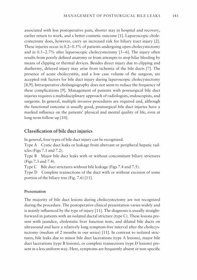
associated with less postoperative pain, shorter stay in hospital and recovery,earlier return to work, and a better cosmetic outcome [1]. Laparoscopic chole-cystectomy does, however, carry an increased risk for biliary tract injury [2].These injuries occur in 0.2–0.5% of patients undergoing open cholecystectomyand in 0.5–2.7% after laparoscopic cholecystectomy [3–6]. The injury oftenresults from poorly defined anatomy or from attempts to stop hilar bleeding bymeans of clipping or thermal devices. Besides direct injury due to clipping anddiathermy, delayed injury may arise from ischemia of the bile ducts [7]. Thepresence of acute cholecystitis, and a low case volume of the surgeon, areaccepted risk factors for bile duct injury during laparoscopic cholecystectomy[8,9]. Intraoperative cholangiography does not seem to reduce the frequency ofthese complications [9]. Management of patients with postsurgical bile ductinjuries requires a multidisciplinary approach of radiologists, endoscopists, andsurgeons. In general, multiple invasive procedures are required and, althoughthe functional outcome is usually good, postsurgical bile duct injuries have amarked influence on the patients’ physical and mental quality of life, even atlong-term follow-up [10].
Classification of bile duct injuries
In general, four types of bile duct injury can be recognized.Type A Cystic duct leaks or leakage from aberrant or peripheral hepatic rad-icles (Figs 7.1 and 7.2).Type B Major bile duct leaks with or without concomitant biliary strictures(Figs 7.3 and 7.4).Type C Bile duct strictures without bile leakage (Figs 7.4 and 7.5).Type D Complete transections of the duct with or without excision of someportion of the biliary tree (Fig. 7.6) [11].
Presentation
The majority of bile duct lesions during cholecystectomy are not recognized during the procedure. The postoperative clinical presentation varies widely andis mainly influenced by the type of injury [11]. The diagnosis is usually straight-forward in patients with an isolated ductal stricture (type C). These lesions pre-sent with jaundice, cholestatic liver function tests, and dilated bile ducts onultrasound and have a relatively long symptom-free interval after the cholecys-tectomy (median of 2 months in our series) [11]. In contrast to isolated stric-tures, bile leaks due to minor bile duct lacerations (type A lesions), major bileduct lacerations (type B lesions), or complete transections (type D lesions) pre-sent in a less uniform way. Here, symptoms are frequently absent or non-specific
MANAGEMENT OF POSTSURGICAL BILE LEAKS 143
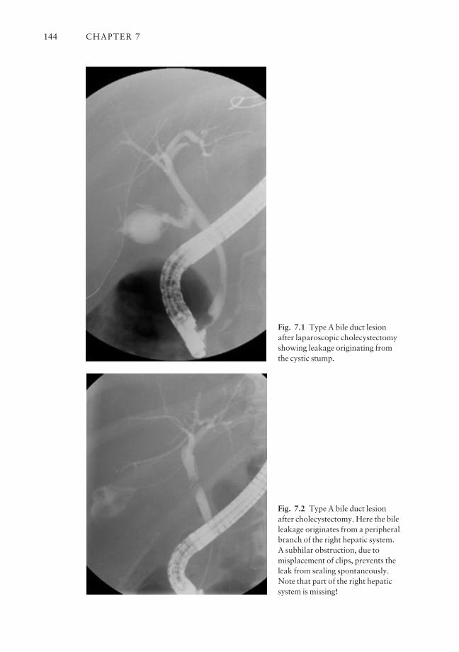
CHAPTER 7144
Fig. 7.1 Type A bile duct lesionafter laparoscopic cholecystectomyshowing leakage originating fromthe cystic stump.
Fig. 7.2 Type A bile duct lesionafter cholecystectomy. Here the bileleakage originates from a peripheralbranch of the right hepatic system. A subhilar obstruction, due tomisplacement of clips, prevents theleak from sealing spontaneously.Note that part of the right hepaticsystem is missing!
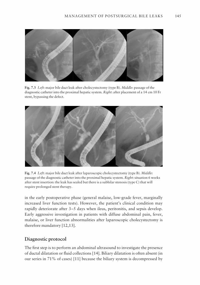
in the early postoperative phase (general malaise, low-grade fever, marginallyincreased liver function tests). However, the patient’s clinical condition mayrapidly deteriorate after 3–5 days when ileus, peritonitis, and sepsis develop.Early aggressive investigation in patients with diffuse abdominal pain, fever,malaise, or liver function abnormalities after laparoscopic cholecystectomy istherefore mandatory [12,13].
Diagnostic protocol
The first step is to perform an abdominal ultrasound to investigate the presenceof ductal dilatation or fluid collections [14]. Biliary dilatation is often absent (inour series in 71% of cases) [11] because the biliary system is decompressed by
MANAGEMENT OF POSTSURGICAL BILE LEAKS 145
Fig. 7.3 Left: major bile duct leak after cholecystectomy (type B). Middle: passage of thediagnostic catheter into the proximal hepatic system. Right: after placement of a 14 cm 10 Frstent, bypassing the defect.
Fig. 7.4 Left: major bile duct leak after laparoscopic cholecystectomy (type B). Middle:passage of the diagnostic catheter into the proximal hepatic system. Right: situation 6 weeksafter stent insertion: the leak has sealed but there is a subhilar stenosis (type C) that willrequire prolonged stent therapy.
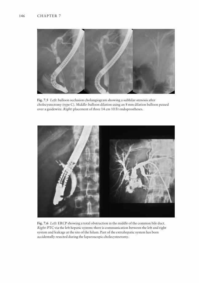
CHAPTER 7146
Fig. 7.5 Left: balloon occlusion cholangiogram showing a subhilar stenosis aftercholecystectomy (type C). Middle: balloon dilation using an 8 mm dilation balloon passedover a guidewire. Right: placement of three 14 cm 10 Fr endoprostheses.
Fig. 7.6 Left: ERCP showing a total obstruction in the middle of the common bile duct.Right: PTC via the left hepatic system: there is communication between the left and rightsystem and leakage at the site of the hilum. Part of the extrahepatic system has beenaccidentally resected during the laparoscopic cholecystectomy.
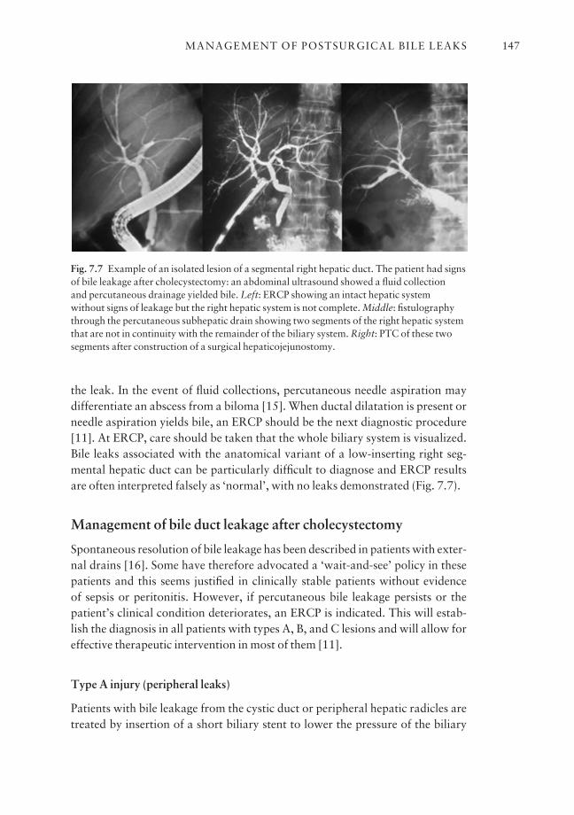
the leak. In the event of fluid collections, percutaneous needle aspiration maydifferentiate an abscess from a biloma [15]. When ductal dilatation is present orneedle aspiration yields bile, an ERCP should be the next diagnostic procedure[11]. At ERCP, care should be taken that the whole biliary system is visualized.Bile leaks associated with the anatomical variant of a low-inserting right seg-mental hepatic duct can be particularly difficult to diagnose and ERCP resultsare often interpreted falsely as ‘normal’, with no leaks demonstrated (Fig. 7.7).
Management of bile duct leakage after cholecystectomy
Spontaneous resolution of bile leakage has been described in patients with exter-nal drains [16]. Some have therefore advocated a ‘wait-and-see’ policy in thesepatients and this seems justified in clinically stable patients without evidence of sepsis or peritonitis. However, if percutaneous bile leakage persists or thepatient’s clinical condition deteriorates, an ERCP is indicated. This will estab-lish the diagnosis in all patients with types A, B, and C lesions and will allow foreffective therapeutic intervention in most of them [11].
Type A injury (peripheral leaks)
Patients with bile leakage from the cystic duct or peripheral hepatic radicles aretreated by insertion of a short biliary stent to lower the pressure of the biliary
MANAGEMENT OF POSTSURGICAL BILE LEAKS 147
Fig. 7.7 Example of an isolated lesion of a segmental right hepatic duct. The patient had signsof bile leakage after cholecystectomy: an abdominal ultrasound showed a fluid collection and percutaneous drainage yielded bile. Left: ERCP showing an intact hepatic system without signs of leakage but the right hepatic system is not complete. Middle: fistulographythrough the percutaneous subhepatic drain showing two segments of the right hepatic systemthat are not in continuity with the remainder of the biliary system. Right: PTC of these twosegments after construction of a surgical hepaticojejunostomy.
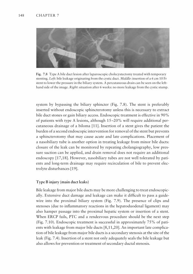
system by bypassing the biliary sphincter (Fig. 7.8). The stent is preferablyinserted without endoscopic sphincterotomy unless this is necessary to extractbile duct stones or gain biliary access. Endoscopic treatment is effective in 90%of patients with type A lesions, although 15–20% will require additional per-cutaneous drainage of a biloma [11]. Insertion of a stent gives the patient theburden of a second endoscopic intervention for removal of the stent but preventsa sphincterotomy that may cause acute and late complications. Placement of a nasobiliary tube is another option in treating leakage from minor bile ducts:closure of the leak can be monitored by repeating cholangiography, low pres-sure suction can be applied, and drain removal does not require an additionalendoscopy [17,18]. However, nasobiliary tubes are not well tolerated by pati-ents and long-term drainage may require recirculation of bile to prevent elec-trolyte disturbances [19].
Type B injury (main duct leaks)
Bile leakage from major bile ducts may be more challenging to treat endoscopic-ally. Extensive duct damage and leakage can make it difficult to pass a guide-wire into the proximal biliary system (Fig. 7.9). The presence of clips andstenoses (due to inflammatory reactions in the hepatoduodenal ligament) mayalso hamper passage into the proximal hepatic system or insertion of a stent. When ERCP fails, PTC and a rendezvous procedure should be the next step (Fig. 7.10). Endoscopic treatment is successful in approximately 75% of pati-ents with leakage from major bile ducts [8,11,20]. An important late complica-tion of bile leakage from major bile ducts is a secondary stenosis at the site of theleak (Fig. 7.4). Insertion of a stent not only adequately seals the bile leakage butalso allows for prevention or treatment of secondary ductal stenosis.
CHAPTER 7148
Fig. 7.8 Type A bile duct lesion after laparoscopic cholecystectomy treated with temporarystenting. Left: bile leakage originating from the cystic duct. Middle: insertion of a 6 cm 10 Frstent to lower the pressure in the biliary system. A percutaneous drain can be seen on the left-hand side of the image. Right: situation after 6 weeks: no more leakage from the cystic stump.
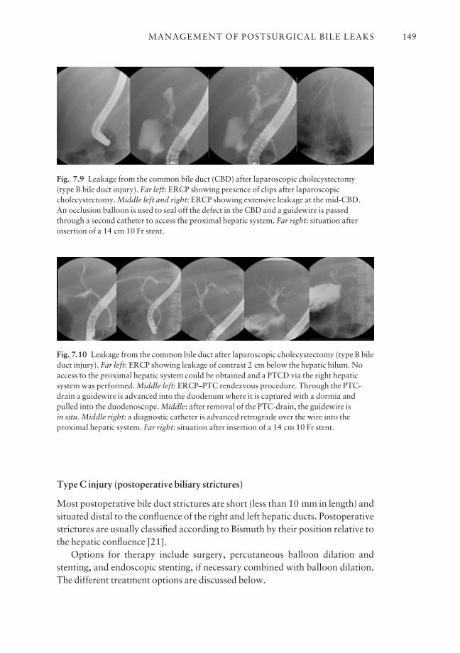
Type C injury (postoperative biliary strictures)
Most postoperative bile duct strictures are short (less than 10 mm in length) andsituated distal to the confluence of the right and left hepatic ducts. Postoperativestrictures are usually classified according to Bismuth by their position relative tothe hepatic confluence [21].
Options for therapy include surgery, percutaneous balloon dilation andstenting, and endoscopic stenting, if necessary combined with balloon dilation.The different treatment options are discussed below.
MANAGEMENT OF POSTSURGICAL BILE LEAKS 149
Fig. 7.9 Leakage from the common bile duct (CBD) after laparoscopic cholecystectomy(type B bile duct injury). Far left: ERCP showing presence of clips after laparoscopiccholecystectomy. Middle left and right: ERCP showing extensive leakage at the mid-CBD. An occlusion balloon is used to seal off the defect in the CBD and a guidewire is passedthrough a second catheter to access the proximal hepatic system. Far right: situation afterinsertion of a 14 cm 10 Fr stent.
Fig. 7.10 Leakage from the common bile duct after laparoscopic cholecystectomy (type B bileduct injury). Far left: ERCP showing leakage of contrast 2 cm below the hepatic hilum. Noaccess to the proximal hepatic system could be obtained and a PTCD via the right hepaticsystem was performed. Middle left: ERCP–PTC rendezvous procedure. Through the PTC-drain a guidewire is advanced into the duodenum where it is captured with a dormia andpulled into the duodenoscope. Middle: after removal of the PTC-drain, the guidewire is in situ. Middle right: a diagnostic catheter is advanced retrograde over the wire into theproximal hepatic system. Far right: situation after insertion of a 14 cm 10 Fr stent.
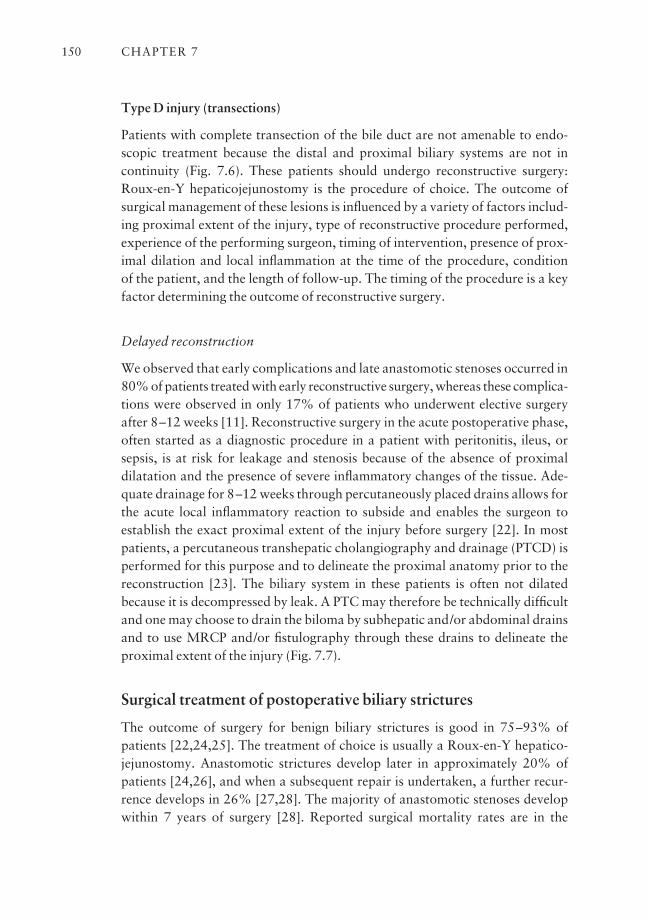
Type D injury (transections)
Patients with complete transection of the bile duct are not amenable to endo-scopic treatment because the distal and proximal biliary systems are not in continuity (Fig. 7.6). These patients should undergo reconstructive surgery:Roux-en-Y hepaticojejunostomy is the procedure of choice. The outcome ofsurgical management of these lesions is influenced by a variety of factors includ-ing proximal extent of the injury, type of reconstructive procedure performed,experience of the performing surgeon, timing of intervention, presence of prox-imal dilation and local inflammation at the time of the procedure, condition of the patient, and the length of follow-up. The timing of the procedure is a keyfactor determining the outcome of reconstructive surgery.
Delayed reconstruction
We observed that early complications and late anastomotic stenoses occurred in80% of patients treated with early reconstructive surgery, whereas these complica-tions were observed in only 17% of patients who underwent elective surgeryafter 8–12 weeks [11]. Reconstructive surgery in the acute postoperative phase,often started as a diagnostic procedure in a patient with peritonitis, ileus, or sepsis, is at risk for leakage and stenosis because of the absence of proximaldilatation and the presence of severe inflammatory changes of the tissue. Ade-quate drainage for 8–12 weeks through percutaneously placed drains allows forthe acute local inflammatory reaction to subside and enables the surgeon toestablish the exact proximal extent of the injury before surgery [22]. In mostpatients, a percutaneous transhepatic cholangiography and drainage (PTCD) isperformed for this purpose and to delineate the proximal anatomy prior to thereconstruction [23]. The biliary system in these patients is often not dilatedbecause it is decompressed by leak. A PTC may therefore be technically difficultand one may choose to drain the biloma by subhepatic and/or abdominal drainsand to use MRCP and/or fistulography through these drains to delineate theproximal extent of the injury (Fig. 7.7).
Surgical treatment of postoperative biliary strictures
The outcome of surgery for benign biliary strictures is good in 75–93% of patients [22,24,25]. The treatment of choice is usually a Roux-en-Y hepatico-jejunostomy. Anastomotic strictures develop later in approximately 20% ofpatients [24,26], and when a subsequent repair is undertaken, a further recur-rence develops in 26% [27,28]. The majority of anastomotic stenoses developwithin 7 years of surgery [28]. Reported surgical mortality rates are in the
CHAPTER 7150

range of 3.2–27%, the higher rate being related to patients with coexist-ing pathology such as portal hypertension [24,25,27]. Factors that are asso-ciated with a favorable outcome include greater distance from the hepaticconfluence, early referral, no previous repair, and the quality of the proximalduct.
It is important to note that there are only a few reports on the surgical treat-ment of patients with postoperative biliary strictures, and that most reportsdescribe surgical treatment of more than a decade ago. Since then, surgeons havebenefited from the improvement in endoscopic and invasive radiographic tech-niques. Nowadays, ultrasound, CT scanning, ERCP, and PTC provide surgeonswith accurate preoperative information and allow for the optimal timing of thereconstructive procedure. Combined with improved surgical techniques, thismay have resulted in an improved outcome of surgical treatment. Lillemoe et al.[23] have reported on a series of 156 patients undergoing surgical reconstruc-tion for postoperative bile duct strictures. Two patients died of unrelated diseasebefore completion of treatment; 12 patients had biliary stents in place at the timeof the report. Of the 142 patients who completed treatment (mean follow-up 58 months), 91% were considered to have a successful outcome without theneed for further interventional procedures.
Percutaneous treatment of postoperative strictures
Percutaneous dilation via transhepatic puncture or T-tube has an associatedmorbidity of less than 7% [29], but the reported success rates vary widely from33 to 100% [29,30]. In general, percutaneous therapy requires several sessionsto obtain a satisfactory outcome. In our hospital, patients undergo a PTC on the first day to obtain a diagnostic cholangiogram and to decompress the biliarysystem. The next day a percutaneous balloon dilation (8–10 mm balloon) is performed and an internal–external PTC-drain is inserted through the anasto-mosis. This drain is left in situ for 6 weeks and then removed after a second balloon dilatation procedure. Misra et al. recently reported their results in 51 patients. The success rate of percutaneous management without the need forsubsequent interventions was 59% [31].
The major concerns with the transhepatic approach are the attendant risksof hemorrhage and bile leakage associated with liver puncture. Additionally,two-thirds of patients may have a non-dilated biliary tract, making ductal punc-ture technically difficult [32]. A further disadvantage is the requirement forlong-term transhepatic intubation. In our unit, the percutaneous approach ismainly reserved for patients with postsurgical anastomotic stenoses (usuallyafter hepaticojejunostomy) and as part of a rendezvous procedure with ERCPafter a failed prior endoscopic approach (Fig. 7.10).
MANAGEMENT OF POSTSURGICAL BILE LEAKS 151
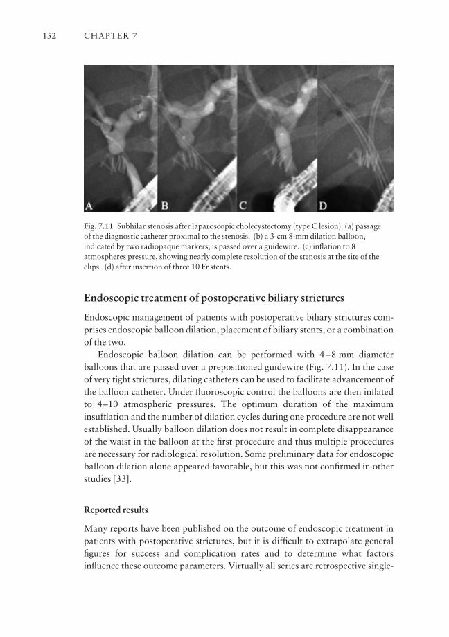
Endoscopic treatment of postoperative biliary strictures
Endoscopic management of patients with postoperative biliary strictures com-prises endoscopic balloon dilation, placement of biliary stents, or a combinationof the two.
Endoscopic balloon dilation can be performed with 4–8 mm diameter balloons that are passed over a prepositioned guidewire (Fig. 7.11). In the caseof very tight strictures, dilating catheters can be used to facilitate advancement ofthe balloon catheter. Under fluoroscopic control the balloons are then inflatedto 4–10 atmospheric pressures. The optimum duration of the maximuminsufflation and the number of dilation cycles during one procedure are not wellestablished. Usually balloon dilation does not result in complete disappearanceof the waist in the balloon at the first procedure and thus multiple proceduresare necessary for radiological resolution. Some preliminary data for endoscopicballoon dilation alone appeared favorable, but this was not confirmed in otherstudies [33].
Reported results
Many reports have been published on the outcome of endoscopic treatment inpatients with postoperative strictures, but it is difficult to extrapolate generalfigures for success and complication rates and to determine what factorsinfluence these outcome parameters. Virtually all series are retrospective single-
CHAPTER 7152
Fig. 7.11 Subhilar stenosis after laparoscopic cholecystectomy (type C lesion). (a) passage of the diagnostic catheter proximal to the stenosis. (b) a 3-cm 8-mm dilation balloon,indicated by two radiopaque markers, is passed over a guidewire. (c) inflation to 8atmospheres pressure, showing nearly complete resolution of the stenosis at the site of theclips. (d) after insertion of three 10 Fr stents.
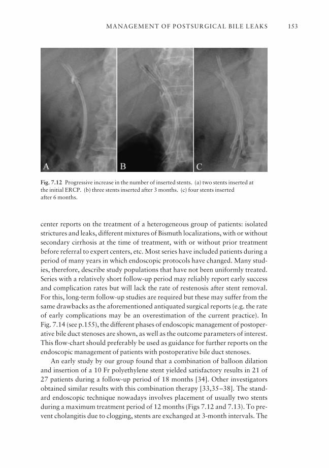
center reports on the treatment of a heterogeneous group of patients: isolated strictures and leaks, different mixtures of Bismuth localizations, with or withoutsecondary cirrhosis at the time of treatment, with or without prior treatmentbefore referral to expert centers, etc. Most series have included patients during aperiod of many years in which endoscopic protocols have changed. Many stud-ies, therefore, describe study populations that have not been uniformly treated.Series with a relatively short follow-up period may reliably report early successand complication rates but will lack the rate of restenosis after stent removal.For this, long-term follow-up studies are required but these may suffer from thesame drawbacks as the aforementioned antiquated surgical reports (e.g. the rateof early complications may be an overestimation of the current practice). In Fig. 7.14 (see p.155), the different phases of endoscopic management of postoper-ative bile duct stenoses are shown, as well as the outcome parameters of interest.This flow-chart should preferably be used as guidance for further reports on theendoscopic management of patients with postoperative bile duct stenoses.
An early study by our group found that a combination of balloon dilationand insertion of a 10 Fr polyethylene stent yielded satisfactory results in 21 of 27 patients during a follow-up period of 18 months [34]. Other investigatorsobtained similar results with this combination therapy [33,35–38]. The stand-ard endoscopic technique nowadays involves placement of usually two stentsduring a maximum treatment period of 12 months (Figs 7.12 and 7.13). To pre-vent cholangitis due to clogging, stents are exchanged at 3-month intervals. The
MANAGEMENT OF POSTSURGICAL BILE LEAKS 153
Fig. 7.12 Progressive increase in the number of inserted stents. (a) two stents inserted at the initial ERCP. (b) three stents inserted after 3 months. (c) four stents inserted after 6 months.
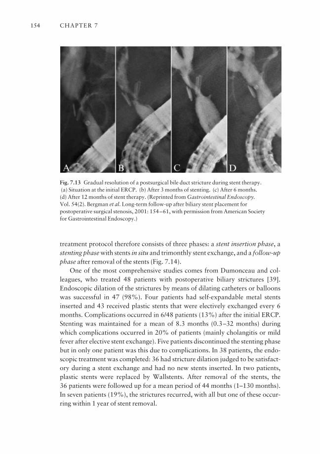
treatment protocol therefore consists of three phases: a stent insertion phase, astenting phase with stents in situ and trimonthly stent exchange, and a follow-upphase after removal of the stents (Fig. 7.14).
One of the most comprehensive studies comes from Dumonceau and col-leagues, who treated 48 patients with postoperative biliary strictures [39].Endoscopic dilation of the strictures by means of dilating catheters or balloonswas successful in 47 (98%). Four patients had self-expandable metal stentsinserted and 43 received plastic stents that were electively exchanged every 6months. Complications occurred in 6/48 patients (13%) after the initial ERCP.Stenting was maintained for a mean of 8.3 months (0.3–32 months) duringwhich complications occurred in 20% of patients (mainly cholangitis or mildfever after elective stent exchange). Five patients discontinued the stenting phasebut in only one patient was this due to complications. In 38 patients, the endo-scopic treatment was completed: 36 had stricture dilation judged to be satisfact-ory during a stent exchange and had no new stents inserted. In two patients,plastic stents were replaced by Wallstents. After removal of the stents, the 36 patients were followed up for a mean period of 44 months (1–130 months).In seven patients (19%), the strictures recurred, with all but one of these occur-ring within 1 year of stent removal.
CHAPTER 7154
Fig. 7.13 Gradual resolution of a postsurgical bile duct stricture during stent therapy. (a) Situation at the initial ERCP. (b) After 3 months of stenting. (c) After 6 months. (d) After 12 months of stent therapy. (Reprinted from Gastrointestinal Endoscopy. Vol. 54(2). Bergman et al. Long-term follow-up after biliary stent placement forpostoperative surgical stenosis, 2001: 154–61, with permission from American Society for Gastrointestinal Endoscopy.)
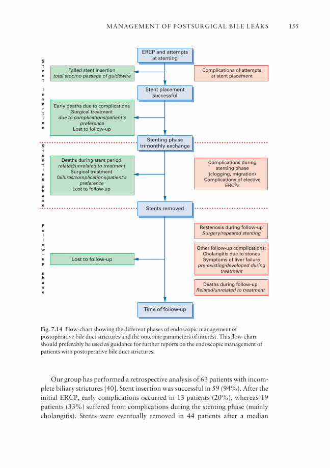
Our group has performed a retrospective analysis of 63 patients with incom-plete biliary strictures [40]. Stent insertion was successful in 59 (94%). After theinitial ERCP, early complications occurred in 13 patients (20%), whereas 19patients (33%) suffered from complications during the stenting phase (mainlycholangitis). Stents were eventually removed in 44 patients after a median
MANAGEMENT OF POSTSURGICAL BILE LEAKS 155
ERCP and attemptsat stenting
Complications of attemptsat stent placement
Failed stent insertiontotal stop/no passage of guidewire
Early deaths due to complicationsSurgical treatment
due to complications/patient'spreference
Lost to follow-up
Deaths during stent periodrelated/unrelated to treatment
Surgical treatmentfailures/complications/patient's
preferenceLost to follow-up
Stent placementsuccessful
Lost to follow-up
Stenting phasetrimonthly exchange
Stents removed
Time of follow-up
Complications duringstenting phase
(clogging, migration)Complications of elective
ERCPs
Restenosis during follow-upSurgery/repeated stenting
Deaths during follow-upRelated/unrelated to treatment
Other follow-up complications:Cholangitis due to stonesSymptoms of liver failure
pre-existing/developed duringtreatment
S
t
e
n
t
I
n
s
e
r
t
i
o
n
S
t
e
n
t
i
n
g
p
h
a
s
e
F
o
l
l
o
w
-
u
p
p
h
a
s
e
Fig. 7.14 Flow-chart showing the different phases of endoscopic management ofpostoperative bile duct strictures and the outcome parameters of interest. This flow-chartshould preferably be used as guidance for further reports on the endoscopic management ofpatients with postoperative bile duct strictures.
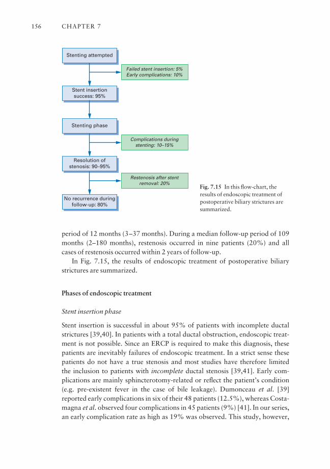
period of 12 months (3–37 months). During a median follow-up period of 109months (2–180 months), restenosis occurred in nine patients (20%) and allcases of restenosis occurred within 2 years of follow-up.
In Fig. 7.15, the results of endoscopic treatment of postoperative biliarystrictures are summarized.
Phases of endoscopic treatment
Stent insertion phase
Stent insertion is successful in about 95% of patients with incomplete ductalstrictures [39,40]. In patients with a total ductal obstruction, endoscopic treat-ment is not possible. Since an ERCP is required to make this diagnosis, thesepatients are inevitably failures of endoscopic treatment. In a strict sense thesepatients do not have a true stenosis and most studies have therefore limited the inclusion to patients with incomplete ductal stenosis [39,41]. Early com-plications are mainly sphincterotomy-related or reflect the patient’s condition(e.g. pre-existent fever in the case of bile leakage). Dumonceau et al. [39]reported early complications in six of their 48 patients (12.5%), whereas Costa-magna et al. observed four complications in 45 patients (9%) [41]. In our series,an early complication rate as high as 19% was observed. This study, however,
CHAPTER 7156
Stenting attempted
Failed stent insertion: 5%Early complications: 10%
Stent insertionsuccess: 95%
Stenting phase
Complications duringstenting: 10–15%
Resolution ofstenosis: 90–95%
No recurrence duringfollow-up: 80%
Restenosis after stentremoval: 20%
Fig. 7.15 In this flow-chart, theresults of endoscopic treatment ofpostoperative biliary strictures aresummarized.
was performed to evaluate the long-term outcome of endoscopic treatment, and patients were treated 10–15 years ago [40]. With the current endoscopicstandards, early complications should not be observed in more than 10% ofpatients.
Stenting phase
Since the stenting phase covers a period of up to 1 year, complications are notuncommon (33% and 20% in the aforementioned studies, respectively).Dumonceau et al. used an interval of 6 months between elective ERCPs and thismay be too long given the average time to stent occlusion reported in other studies [39]. In our retrospective study, complications during the stenting periodmainly occurred in cases in which the patients and/or referring physicians didnot adhere to the treatment protocol [40]. Many years have passed since then, andthe endoscopic treatment of patients with benign biliary strictures has becomemore accepted. Currently, in patients in whom elective trimonthly ERCPs areperformed, complications due to stent dysfunction are mild and occur at a maximum of 10–15% during a stenting period of 1 year. With this regimen,90–95% of patients will have their stenosis adequately dilated within 1 year.
Follow-up phase
Studies have reported a recurrence rate of 20% after removal of the stents[39,40]. In the series of Dumonceau et al., 36 patients were followed for a meanperiod of 44 months after removal of the stents, whereas our group followed 44 patients for a median period of 9.1 years after stent removal. It is importantto note that almost all cases of restenosis occurred relatively early after removalof the stents: only one patient was diagnosed with recurrent stenosis after morethan 6 months and all cases occurred within 2 years of stent removal. This sug-gests that endoscopic treatment of postsurgical stenoses is not associated with ahigh rate of long-term restenosis after stent removal, as suggested by antagonistsof this treatment regimen. This is in contrast to the anastomotic recurrencesafter hepaticojejunostomy, which may occur after many years [28]. It alsoimplies that strict follow-up after stent removal is necessary, especially duringthe first 6–12 months, in order to diagnose restenosis at an early stage.
It is important to note that, apart from recurrence of the initial stenosis,other late complications may also occur after stent removal, e.g. cholangitisfrom bile duct stones or symptoms associated with (pre-existing) liver failure[40,41]. Bile duct stones may develop because of impeded bile flow due to a relative stenosis; however, since all patients initially underwent biliary surgeryfor stone disease, the underlying stone disease may also be held responsible [42].
MANAGEMENT OF POSTSURGICAL BILE LEAKS 157
Postoperative biliary strictures: surgery or endoscopy [43]?
Prospective randomized studies comparing surgical and endoscopic treatmentof postoperative biliary strictures are not available. We have performed a retro-spective study comparing surgical with endoscopic therapy (Table 7.1) [26].Both approaches were found to have a similar long-term success rate, withrecurrences being seen in 17% of patients.
Since there appear to be no clear differences in the primary outcomesbetween surgical and endoscopic management, the choice between the two isdetermined by other factors: the two most important ones are the different char-acteristics of restenosis and patients’ preference.
Recurrent strictures after surgery
The diagnosis of anastomotic stenosis after hepaticojejunostomy may be diffi-cult since most patients already have mild liver function abnormalities anddilatation of the biliary tree on ultrasound is often absent [44]. After Roux-en-Yhepaticojejunostomy, endoscopic approach to the biliary tree is usually not possible. Most physicians are reluctant to perform a PTC in these patientsbecause of the risk of hemorrhage and bile leakage, especially in cases of non-dilated bile ducts [32]. In patients with prior endoscopic treatment of bile ductstrictures, diagnosis of recurrent stenosis is easier, safer, and therefore asso-ciated with less medical delay (which may cause secondary biliary cirrhosis).Furthermore, restenosis after endoscopic treatment occurs relatively early after
CHAPTER 7158
Table 7.1 Retrospective comparison of surgery with endoscopy in the management ofpostoperative biliary strictures.
Surgery Endoscopy
No. of patients 35 66Early complications 26 8Thirty-day mortality 0 1Complications in 1 year 0 27Outcome assessmentb (no. of patients) 35 46a
Excellent 25 33Good 4 5Poor 6 8
aStents were eventually removed in 46 patients in the endoscopy group.bExcellent results were defined as asymptomatic and normal or stable liver function tests;good results as occurrence of only one episode of cholangitis; poor results as recurrentcholangitis or stricture.
stent removal (less than 1–2 years) [40,45], whereas anastomotic recurrencesafter surgery may develop after more than 10 years [28].
Treatment of recurrent stenosis after hepaticojejunostomy involves eitherpercutaneous balloon dilation, often supplemented with internal–external stent-ing, or repeated surgery. Nowadays, most patients with anastomotic stenosisare initially managed by percutaneous treatment, but multiple sessions are oftenrequired and the cumulative morbidity due to bleeding and bile leakage may beas high as 30%. Repeat surgery will eventually be required in 20–30% of thesepatients. Compared to primary hepaticojejunostomy, repeated reconstructivesurgery is associated with a higher complication and failure rate [27,28].
Whereas endoscopic treatment is impossible once a Roux-en-Y loop hasbeen constructed, prior endoscopic treatment does not preclude further surgicaltreatment. In addition, recurrent strictures after prior endoscopic therapy canalso be successfully treated by repeated stenting. Finally, although endoscopictreatment requires multiple ERCPs, many patients and their referring doctorsprefer endoscopic treatment to surgery.
Therefore, we feel that surgery should be reserved for patients with failedendoscopic therapy or for patients who refuse endoscopic therapy.
Metal stents for benign strictures
The use of metal expandable stents in benign biliary disease remains controver-sial. Initially, several groups reported favorable results in the management ofpostoperative strictures. Gianturco–Rösch Z-Stents were placed percutaneouslyin 43 such patients by Coons [46]. All patients had previously undergone anunsuccessful balloon cholangioplasty. The 1-year reocclusion rate was 13%.Maccioni et al. reported long-term patency in 100% of patients with a CBDstricture [47]. Foerster et al., reporting on endoscopic Wallstent placement infour patients, identified no stent-related complications and no cases of occlusion during a follow-up period of 53 weeks [48]. More recent follow-up studies,however, have obtained less satisfactory results. Hausegger et al. reported thatthe patency rate rapidly decreased to 19% at 57 months of follow-up [49].Dumonceau et al. reported a 100% occlusion rate of Wallstents inserted in sixpatients with postoperative strictures within 48 months of follow-up [45].Lopez et al. reported a good clinical result, arbitrarily defined as the need fortwo or fewer invasive interventions, in only five out of 15 patients. The remain-ing 10 patients underwent multiple interventions, including surgery in five witha poor general outcome [50].
Studies in dogs have demonstrated marked bile duct narrowing related toextensive fibrosis after insertion of Wallstents but only minimal changes after
MANAGEMENT OF POSTSURGICAL BILE LEAKS 159
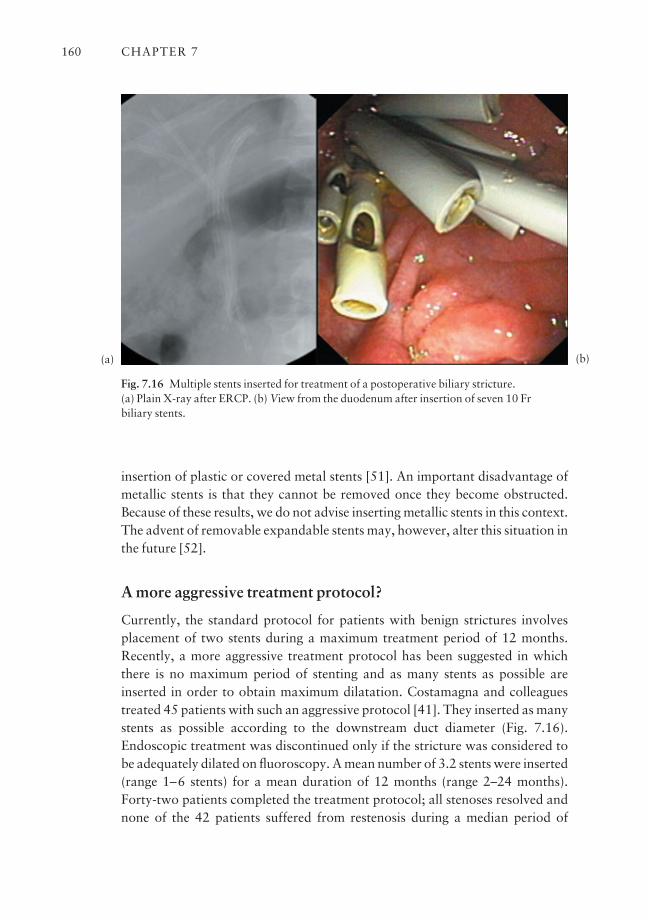
insertion of plastic or covered metal stents [51]. An important disadvantage ofmetallic stents is that they cannot be removed once they become obstructed.Because of these results, we do not advise inserting metallic stents in this context.The advent of removable expandable stents may, however, alter this situation inthe future [52].
A more aggressive treatment protocol?
Currently, the standard protocol for patients with benign strictures involvesplacement of two stents during a maximum treatment period of 12 months.Recently, a more aggressive treatment protocol has been suggested in whichthere is no maximum period of stenting and as many stents as possible areinserted in order to obtain maximum dilatation. Costamagna and colleaguestreated 45 patients with such an aggressive protocol [41]. They inserted as manystents as possible according to the downstream duct diameter (Fig. 7.16).Endoscopic treatment was discontinued only if the stricture was considered tobe adequately dilated on fluoroscopy. A mean number of 3.2 stents were inserted(range 1–6 stents) for a mean duration of 12 months (range 2–24 months).Forty-two patients completed the treatment protocol; all stenoses resolved andnone of the 42 patients suffered from restenosis during a median period of
CHAPTER 7160
Fig. 7.16 Multiple stents inserted for treatment of a postoperative biliary stricture. (a) Plain X-ray after ERCP. (b) View from the duodenum after insertion of seven 10 Fr biliary stents.
(a) (b)
follow-up of 29 months (range 24 months to 11.3 years). These impressiveresults suggest that a more prolonged and aggressive endoscopic approach maybe justified in more difficult cases.
Conclusions
Although most centers performing laparoscopic cholecystectomy may now bewell beyond the ‘learning-curve’ phase, the incidence of postsurgical bile ductinjuries will probably stay higher than in the era before laparoscopic cholecys-tectomy. In addition, with surgeons embarking on more complex laparoscopicbiliary interventions, such as laparoscopic duct exploration, a further increasein the incidence of surgical bile duct injuries may be expected in the near future.Adequate management of these injuries requires an early postoperative diag-nosis with a low threshold for performing an ERCP. Patients with type D lesions(complete ductal transection) require an elective surgical repair, 6–8 weeksafter diagnosis and drainage. Most of the other bile duct injuries (minor leaksfrom the cystic stump or peripheral hepatic radicles, major bile duct leaks, andisolated bile duct strictures) can in general be managed endoscopically.
Patients who require long-term stenting for bile duct strictures should betreated with at least two 10 Fr plastic stents that are electively exchanged every 3 months. If possible, more than two stents should be inserted. There is cur-rently no place for self-expandable metal stents for this indication.
Optimal management of patients with bile duct injuries requires a multidis-ciplinary team approach of interventional radiology, therapeutic endoscopy,and reconstructive surgery.
Outstanding issues and future trends
Most controversies connected to the management of postsurgical bile ductlesions relate to the treatment of biliary stenoses. There are still many questionsconcerning the optimal endoscopic management of these patients. Should stentplacement always be preceded by balloon dilation? How many stents should beinserted? What is the optimal period of stenting? Some groups treat patientsonly for a relatively short time (e.g. 6 months) before deciding on success or fail-ure. Others do not have a maximum period of stenting and attempt to insert asmany stents as possible in order to obtain maximum dilatation.
With such an aggressive endoscopic treatment protocol, however, the ques-tion arises of what are the long-term effects of stenting on the biliary system.Studies using intraductal ultrasonography have shown that stenting inducesprofound thickening and fibrosis of the bile duct wall that occurs as soon as 2 weeks after insertion of a single plastic endoprosthesis [53]. Placing multiple
MANAGEMENT OF POSTSURGICAL BILE LEAKS 161
stents for a period of over 1 year might thus reduce the chances of a successfulsurgical reconstruction if endoscopic treatment should fail. In the near future,the use of biodegradable self-expandable stents that gradually dissolve after 1–2years, or the insertion of covered self-expandable stents that can be removed,might be options.
Which subset of patients will most likely benefit from endoscopic manage-ment? The impression exists that patients in whom the stricture is diagnosed relatively early after surgery have a better prognosis than patients who present along time after surgery, but this has not been substantiated in follow-up studies.Patients with more proximal lesions are more difficult to treat and some expertshave advised primary surgical treatment in patients with hilar strictures [54].Multivariate analysis of large cohorts of patients will be necessary to solve theseissues.
References
1 McMahon AJ, Russell IT, Baxter JN et al. Laparoscopic versus minilaparotomy cholecystec-tomy: a randomised trial [see comments]. Lancet 1994; 343 (8890): 135–8.
2 Gouma DJ, Go PM. Bile duct injury during laparoscopic and conventional cholecystectomy. J Am Coll Surg 1994; 178 (3): 229–33.
3 Glenn F. Iatrogenic injuries to the biliary ductal system. Surg Gynecol Obstet 1978; 146 (3):430–4.
4 Way LW, Bernhoft RA, Thomas MJ. Biliary stricture. Surg Clin North Am 1981; 61 (4):963–72.
5 The Southern Surgeons Club. A prospective analysis of 1518 laparoscopic cholecystectomies. NEng J Med 1991; 324 (16): 1073–8.
6 Deziel DJ, Millikan KW, Economou SG et al. Complications of laparoscopic cholecystectomy: anational survey of 4292 hospitals and an analysis of 77 604 cases. Am J Surg 1993; 165 (1):9–14.
7 Terblanche J, Allison HF, Northover JM. An ischemic basis for biliary strictures. Surgery 1983;94 (1): 52–7.
8 Woods MS, Traverso LW, Kozarek RA et al. Characteristics of biliary tract complications dur-ing laparoscopic cholecystectomy: a multi-institutional study. Am J Surg 1994; 167 (1): 27–33.
9 Schol FP, Go PM, Gouma DJ. Risk factors for bile duct injury in laparoscopic cholecystectomy,analysis of 49 cases [see comments]. Br J Surg 1994; 81 (12): 1786–8.
10 Boerma D, Rauws EA, Keulemans YC et al. Impaired quality of life 5 years after bile duct injuryduring laparoscopic cholecystectomy: a prospective analysis. Ann Surg 2001; 234 (6): 750–7.
11 Bergman JJ, van den Brink GR, Rauws EA et al. Treatment of bile duct lesions after laparoscopiccholecystectomy. Gut 1996; 38 (1): 141–7.
12 Rossi RL, Schirmer WJ, Braasch JW et al. Laparoscopic bile duct injuries: risk factors, recogni-tion, and repair. Arch Surg 1992; 127 (5): 596–601.
13 Davidoff AM, Pappas TN, Murray EA et al. Mechanisms of major biliary injury during laparo-scopic cholecystectomy [see comments]. Ann Surg 1992; 215 (3): 196–202.
14 Ress AM, Sarr MG, Nagorney DM et al. Spectrum and management of major complications oflaparoscopic cholecystectomy. Am J Surg 1993; 165 (6): 655–62.
15 Mueller PR, Ferrucci JTJ, Simeone JF et al. Detection and drainage of bilomas: special consid-erations. AJR Am J Roentgenol 1983; 140 (4): 715–20.
16 Albasini JL, Aledo VS, Dexter SP et al. Bile leakage following laparoscopic cholecystectomy.Surg Endosc 1995; 9 (12): 1274–8.
CHAPTER 7162

17 Chow S, Bosco JJ, Heiss FW et al. Successful treatment of post-cholecystectomy bile leaks usingnasobiliary tube drainage and sphincterotomy. Am J Gastroenterol 1997; 92 (10): 1839–43.
18 Saab S, Martin P, Soliman GY et al. Endoscopic management of biliary leaks after T-tuberemoval in liver transplant recipients: nasobiliary drainage versus biliary stenting. Liver TransplSurg 2000; 6 (5): 627–32.
19 Wills VL, Gibson K, Karihaloot C et al. Complications of biliary T-tubes after choledochotomy.Aust NZ J Surg 2002; 72 (3): 177–80.
20 Traverso LW, Kozarek RA, Ball TJ et al. Endoscopic retrograde cholangiopancreatographyafter laparoscopic cholecystectomy. Am J Surg 1993; 165 (5): 581–6.
21 Bismuth H. (1982) Postoperative strictures of the bile duct. In: The Biliary Tract (ed. Blumgart,LH), pp. 209–18. Churchill Livingstone, Edinburgh.
22 Nealon WH, Urrutia F. Long-term follow-up after bilioenteric anastomosis for benign bile ductstricture. Ann Surg 1996; 223 (6): 639–45.
23 Lillemoe KD, Melton GB, Cameron JL et al. Postoperative bile duct strictures: management andoutcome in the 1990s. Ann Surg 2000; 232 (3): 430–41.
24 Raute M, Podlech P, Jaschke W et al. Management of bile duct injuries and strictures followingcholecystectomy. World J Surg 1993; 17 (4): 553–62.
25 Chapman WC, Halevy A, Blumgart LH et al. Postcholecystectomy bile duct strictures: manage-ment and outcome in 130 patients. Arch Surg 1995; 130 (6): 597–602.
26 Davids PH, Tanka AK, Rauws EA et al. Benign biliary strictures. Surgery or endoscopy? AnnSurg 1993; 217 (3): 237–43.
27 Genest JF, Nanos E, Grundfest-Broniatowski S et al. Benign biliary strictures: an analytic review(1970–84). Surgery 1986; 99 (4): 409–13.
28 Pitt HA, Miyamoto T, Parapatis SK et al. Factors influencing outcome in patients with post-operative biliary strictures. Am J Surg 1982; 144 (1): 14–21.
29 Mueller PR, vanSonnenberg E, Ferrucci JTJ et al. Biliary stricture dilatation: multicenter reviewof clinical management in 73 patients. Radiology 1986; 160 (1): 17–22.
30 Citron SJ, Martin LG. Benign biliary strictures: treatment with percutaneous cholangioplasty.Radiology 1991; 178 (2): 339–41.
31 Misra S, Melton GB, Geschwind JF et al. Percutaneous management of bile duct strictures andinjuries associated with laparoscopic cholecystectomy: a decade of experience. J Am Coll Surg2004; 198 (2): 218–26.
32 Speer AG, Cotton PB, Russell RC et al. Randomised trial of endoscopic versus percutaneousstent insertion in malignant obstructive jaundice. Lancet 1987; 2 (8550): 57–62.
33 Geenen DJ, Geenen JE, Hogan WJ et al. Endoscopic therapy for benign bile duct strictures.Gastrointest Endosc 1989; 35 (5): 367–71.
34 Huibregtse K, Katon RM, Tytgat GN. Endoscopic treatment of postoperative biliary strictures.Endoscopy 1986; 18 (4): 133–7.
35 Berkelhammer C, Kortan P, Haber GB. Endoscopic biliary prostheses as treatment for benignpostoperative bile duct strictures. Gastrointest Endosc 1989; 35 (2): 95–101.
36 Davids PH, Rauws EA, Coene PP et al. Endoscopic stenting for post-operative biliary strictures.Gastrointest Endosc 1992; 38 (1): 12–8.
37 Cunningham JT, Draganov PV, Rawls E et al. Long term outcomes in patients with benign bili-ary stricture treated endoscopically with multiple stents. Gastrointest Endosc 1998; 47 (4):AB112.
38 Weber J, Adamek HE, Riemann JF. Endoscopic stent placement and clip removal for commonbile duct stricture after laparoscopic cholecystectomy. Gastrointest Endosc 1992; 38 (2): 181–2.
39 Dumonceau JM, Deviere J, Delhaye M et al. Plastic and metal stents for postoperative benignbile duct strictures: the best and the worst [see comments]. Gastrointest Endosc 1998; 47 (1):8–17.
40 Bergman JJ, Burgemeister L, Bruno MJ et al. Long-term follow-up after biliary stent placementfor postoperative bile duct stenosis. Gastrointest Endosc 2001; 54 (2): 154–61.
41 Costamagna G, Pandolfi M, Mutignani M et al. Long-term results of endoscopic managementof postoperative bile duct strictures with increasing numbers of stents. Gastrointest Endosc2001; 54 (2): 162–8.
MANAGEMENT OF POSTSURGICAL BILE LEAKS 163
42 Bergman JJ, van der Mey S, Rauws EA et al. Long-term follow-up after endoscopic sphinctero-tomy for bile duct stones in patients younger than 60 years of age [see comments]. GastrointestEndosc 1996; 44 (6): 643–9.
43 De Palma GD, Galloro G, Romano G et al. Long-term follow-up after endoscopic biliary stentplacement for bile duct strictures from laparoscopic cholecystectomy. Hepatogastroenterology2003; 50 (53): 1229–31.
44 Plaisier PW, van der Hul RL, Lameris JS et al. Routine testing of liver function after biliary-enteric anastomosis has no clinical relevance. Hepatogastroenterology 2001; 48 (39): 622–4.
45 Dumonceau JM, Deviere J, Delhaye M et al. Plastic and metal stents for postoperative benignbile duct strictures: the best and the worst. Gastrointest Endosc 1998; 47 (1): 8–17.
46 Coons H. Metallic stents for the treatment of biliary obstruction: a report of 100 cases.Cardiovasc Intervent Radiol 1992; 15 (6): 367–74.
47 Maccioni F, Bezzi M, Gandini R et al. [Metallic stents in benign biliary stenosis: a four-year follow-up]. [Italian]. Radiol Med (Torino) 1993; 86 (3): 294–301.
48 Foerster EC, Hoepffner N, Domschke W. Bridging of benign choledochal stenoses by endo-scopic retrograde implantation of mesh stents. Endoscopy 1991; 23 (3): 133–5.
49 Hausegger KA, Kugler C, Uggowitzer M et al. Benign biliary obstruction: is treatment with theWallstent advisable? Radiology 1996; 200 (2): 437–41.
50 Lopez RR Jr, Cosenza CA, Lois J et al. Long-term results of metallic stents for benign biliarystrictures. Arch Surg 2001; 136 (6): 664–9.
51 Silvis SE, Sievert CEJ, Vennes JA et al. Comparison of covered versus uncovered wire meshstents in the canine biliary tract. Gastrointest Endosc 1994; 40 (1): 17–21.
52 Goldin E, Beyar M, Safra T et al. A new self-expandable and removable metal stent for biliaryobstruction: a preliminary report. Endoscopy 1993; 25 (9): 597–9.
53 Tamada K, Tomiyama T, Ichiyama M et al. Influence of biliary drainage catheter on bile ductwall thickness as measured by intraductal ultrasonography. Gastrointest Endosc 1998; 47 (1):28–32.
54 Draganov P, Hoffman B, Marsh W et al. Long-term outcome in patients with benign biliarystrictures treated endoscopically with multiple stents. Gastrointest Endosc 2002; 55 (6): 680–6.
CHAPTER 7164
CHAPTER 8
Sphincter of Oddi Dysfunction
EVAN L. FOGEL AND STUART SHERMAN
Synopsis
Sphincter of Oddi dysfunction (SOD) refers to a motor abnormality of thesphincter of Oddi, typically resulting in a hypertonic sphincter, and may bemanifested clinically by chronic abdominal pain, pancreatitis, or abnormal liverfunction tests. In this chapter, we discuss the classification systems typically usedin SOD, as well as the epidemiology of this controversial disease. The diagnosticcriteria for SOD and appropriate evaluation of patients are reviewed. Both non-invasive and invasive diagnostic methods are discussed. Sphincter of Oddimanometry (SOM) is the only available method to measure motor activitydirectly, and is considered currently to be the diagnostic gold standard. Indica-tions, performance, and complications of this technique are reviewed. Therapyfor SOD is discussed, using an evidence-based approach.
Introduction
Since its original description by Ruggero Oddi in 1887, the sphincter of Oddi(SO) has been the subject of much study and controversy. Its very existence as a distinct anatomical or physiological entity has been disputed. Hence, it is not surprising that the clinical syndrome of sphincter of Oddi dysfunction (SOD)and its therapy are controversial areas [1]. Nevertheless, SOD is commonlydiagnosed and treated by physicians, most often (but not exclusively) amongstpatients who have residual or recurrent symptoms after cholecystectomy, and in whom more common organic causes have been excluded [2]. This chapterreviews the epidemiology and clinical presentation of SOD, as well as currentlyavailable diagnostic and therapeutic modalities.
Definitions
A distinction is sometimes made between SOD and true sphincter stenosis.
165
Sphincter of Oddi dysfunction
SOD refers to an abnormality of SO contractility. It is a benign, non-calculusobstruction to flow of bile or pancreatic juice through the pancreatico-biliaryjunction, i.e. the SO. SOD may be manifested clinically by ‘pancreatico-biliary’pain, pancreatitis, or abnormal liver function tests. SO dyskinesia refers to amotor abnormality of the SO, which may result in a hypotonic sphincter but,more commonly, causes a hypertonic sphincter.
Sphincter of Oddi stenosis
In contrast, SO stenosis refers to a structural alteration of the sphincter, prob-ably from an inflammatory process, with subsequent fibrosis.
Classification of SOD
Since it is often impossible to distinguish patients with SO dyskinesia from thosewith SO stenosis, the term SOD has been used to incorporate both groups ofpatients. A variety of less accurate termsasuch as papillary stenosis, ampullarystenosis, biliary dyskinesia, and postcholecystectomy syndromeaare listed inthe medical literature to describe this entity. The latter term is somewhat of amisnomer, as SOD may clearly occur with an intact gallbladder.
In an attempt to deal with this confusion, and also to determine the appro-priate utilization of SO manometry (SOM), a biliary clinical classification sys-tem has been developed for patients with suspected SOD (Hogan–Geenen SODclassification system; Table 8.1) based on clinical history, laboratory results,and endoscopic retrograde cholangiopancreatography (ERCP) findings [3]. Apancreatic classification has also been developed, but is less commonly utilized[4] (Table 8.2). Both the biliary and pancreatic classification systems have beenmodified [5], making them more applicable for clinical use, as biliary and pan-creatic drainage times have been abandoned.
Epidemiology
SOD may occur in pediatric or adult patients of any age; however, patients withSOD are typically middle-aged females [6]. Although SOD most commonlyoccurs after cholecystectomy, it may be present with the gallbladder in situ. In a survey on functional gastrointestinal disorders, SOD appeared to have asignificant impact on the quality of life, as it was highly associated with workabsenteeism, disability, and health care use [7].
CHAPTER 8166

SPHINCTER OF ODDI DYSFUNCTION 167
Table 8.1 Hogan–Geenen biliary sphincter of Oddi classification system (post-cholecystectomy) related to the frequency of abnormal sphincter of Oddi manometry andpain relief by biliary sphincterotomy.
Patient group classifications
Biliary Type IPatients with biliary-type pain, abnormal SGOT or alkaline phosphatase > 2 ×normal documented on two or more occasions, delayed drainage of ERCP contrast from the biliary tree > 45 min, and dilated CBD > 12 mm diameter
Biliary Type IIPatients with biliary-type pain but only one or two of the above criteria
Biliary Type IIIPatients with only biliary-type pain and no other abnormalities
Approximatefrequency of abnormalsphinctermanometry
75–95%
55–65%
25–60%
Abnormal
90–95%
85%
55–65%
Probability of pain reliefby sphincterotomy ifmanometry:
Normal
90–95%
35%
< 10%
Manometrybefore sphincterablation
Unnecessary
Highlyrecommended
Mandatory
Table 8.2 Pancreatic sphincter of Oddi classification system.
Patient group classification
Pancreatic Type IPatients with pancreatic-type pain, abnormal amylase or lipase 1.5 × normal on any occasion,delayed drainage of ERCP contrast from the pancreatic duct > 9 min, and dilated PD > 6 mm diameter in the head or 5 mm in the body
Pancreatic Type IIPatients with pancreatic-type pain but only one or two of the above criteria
Pancreatic Type IIIPatients with only pancreatic-type pain and no other abnormalities
SOD in patients with gallbladder disease
The frequency of manometrically documented SOD in patients prior to chole-cystectomy has received limited study. Guelrud and colleagues [8] evaluated121 patients with symptomatic gallstones and a normal common bile ductdiameter (by transcutaneous ultrasound) by SOM prior to cholecystectomy. Anelevated basal sphincter pressure was found in 14 patients (11.6%). SOD wasdiagnosed in 4.1% of patients with a normal serum alkaline phosphatase (4 of96) and in 40% with an elevated serum alkaline phosphatase (10 of 25). Ruffoloand associates evaluated 81 patients with symptoms suggestive of biliary disease,but normal ERCP and no gallbladder stones on transcutaneous ultrasound, byscintigraphic gallbladder ejection fraction and endoscopic SOM [9]. Fifty-threeper cent of patients had SOD and 49% had an abnormal gallbladder ejectionfraction. SOD occurred with a similar frequency in patients with an abnormalgallbladder ejection fraction (50%) and a normal ejection fraction (57%).
SOD after cholecystectomy
The frequency of diagnosing SOD in reported series varies considerably with thepatient selection criteria, the definition of SOD, and the diagnostic toolsemployed. In a British report, SOD was diagnosed in 41 (9%) of 451 consecutivepatients being evaluated for postcholecystectomy pain [10]. Roberts-Thomsonand Toouli evaluated 431 similar patients and found SOD in 47 (11%). In a subpopulation of such patients with a normal ERCP (except dilated ducts in28%) and recurrent pain of more than 3 months’ duration, SOD was diagnosedin 68% [11]. Sherman and colleagues used SOM to evaluate 115 patients withpancreaticobiliary pain with and without liver function test abnormalities [4].Patients with bile duct stones and tumors were excluded from the analysis. Fifty-nine of 115 patients (51%) showed abnormal basal SO pressure greaterthan 40 mmHg. These patients were further categorized by the Hogan–GeenenSOD classification system (Table 8.1). The frequency of abnormal manometryof a single sphincter segment was 86%, 55%, and 28%, for Type I, II, and IIIpatients, respectively. These abnormal manometric frequencies were very similarto those reported by others for Type I and Type II patients [12,13]. In biliaryType III patients, the finding of an abnormal basal sphincter pressure has variedfrom 12% to 55% [14]. As noted, patient selection factors may be one explana-tion for this great variability.
SOD in the biliary or pancreatic sphincter, or both
SOD can involve abnormalities in the biliary sphincter, pancreatic sphincter, or
CHAPTER 8168
both. The true frequency of SOD therefore depends on whether one or bothsphincters are studied. Eversman and colleagues performed manometry of thebiliary and pancreatic sphincter segments in 360 patients with pancreatico-biliary pain and intact sphincters [5]. In this large series, 19% had abnormalpancreatic basal sphincter pressure alone, 11% had abnormal biliary basalsphincter pressure alone, and, in 31%, the basal sphincter pressure was abnormal in both segments (overall frequency of sphincter dysfunction was61%). Among the 214 patients labeled as Type III, 17%, 11%, and 31% hadelevated basal sphincter pressure in the pancreatic sphincter alone, biliarysphincter alone, or both segments, respectively (overall frequency of SOD59%). In the 123 Type II patients, SOD was diagnosed in 65%: 22%, 11%, and 32% had elevated basal sphincter pressure in the pancreatic sphincter only,biliary sphincter only, or both sphincter segments, respectively. Similar findingswere reported by Aymerich and colleagues [15]. In a series of 73 patients with suspected SOD, basal pressures were normal in both segments in 19%,abnormal in both segments in 40%, and abnormal in one segment but normal inthe other in 41%. The negative predictive value of normal biliary basal sphincterpressure in excluding SOD was 0.42; when the pancreatic basal sphincter pressure was normal, the negative predictive value was 0.58. These two studiesclearly suggest that both the bile duct and pancreatic duct must be evaluatedwhen assessing the sphincter by SOM.
SOD and pancreatitis
Dysfunction may occur in the pancreatic duct portion of the SO and cause recur-rent pancreatitis. As noted earlier, a pancreatic SOD classification system hasbeen developed (Table 8.2), but has not been widely utilized [5]. Manometric-ally documented SOD has been reported in 15% to 72% of patients with recur-rent pancreatitis, previously labeled as idiopathic [5,12,16].
Clinical presentation
Abdominal pain is the most common presenting symptom of patients with SOD.The pain is usually epigastric or right upper quadrant, may be disabling, andlasts for 30 min to hours. In some patients the pain is continuous with episodicexacerbations. It may radiate to the back or shoulder and be accompanied bynausea and vomiting. Food or narcotics may precipitate the pain. The pain maybegin several years after a cholecystectomy was performed for a gallbladder dysmotility or stone disease and is similar in character to the pain leading to the cholecystectomy. Alternatively, patients may have continued pain that wasnot relieved by a cholecystectomy. Jaundice, fever, or chills are rarely observed.
SPHINCTER OF ODDI DYSFUNCTION 169
The Rome criteria
Recently, a symposium on functional disorders of the pancreas and biliary treeestablished the Rome II diagnostic criteria [6] for SOD. These include episodesof severe abdominal pain located in the epigastrium and/or right upper quad-rant, and all of the following: (1) symptom episodes lasting 30 min or more withpain-free intervals; (2) symptoms have occurred on one or more occasions in theprevious 12 months; (3) the pain is steady and interrupts daily activities orrequires consultation with a physician; and (4) there is no evidence of structuralabnormalities to explain the symptoms. Physical examination is typically char-acterized only by mild epigastric or right upper quadrant tenderness. The pain is not relieved by trial medications for acid peptic disease or irritable bowel syndrome. Laboratory abnormalities consisting of transient elevation of liverfunction tests, typically during episodes of pain, are present in less than 50% ofpatients. After initial evaluation, patients are commonly categorized accordingto the Hogan–Geenen SOD classification system (Table 8.1). Patients with SODmay present with typical pancreatic pain (epigastric or left upper quadrant radi-ating to the back) and recurrent pancreatitis.
SOD may exist in the presence of an intact gallbladder [17]. As the symp-toms of SOD and gallbladder dysfunction cannot be reliably separated, the diag-nosis of SOD is commonly made after cholecystectomy or less frequently aftergallbladder abnormalities have been excluded [6].
Initial evaluation
The diagnostic approach to suspected SOD may be influenced by the presence of key clinical features. However, the clinical manifestations of functionalabnormalities of the SO may not always be easily distinguishable from thosecaused by organic conditions (e.g. common bile duct stones) or other functionalnon-pancreatico-biliary disorders (e.g. irritable bowel syndrome). Standardevaluation and treatment of other more common upper gastrointestinal conditions,such as peptic ulcer disease and gastroesophageal reflux, should be performedsimultaneously. In the absence of mass lesions, stones, or response to acid sup-pression therapeutic trials, the suspicion for sphincter disease is increased.
Serum chemistries
The evaluation of patients with suspected SOD (i.e. patients with upper abdominal pain with characteristics suggestive of a pancreatico-biliary origin)should be initiated with standard serum liver chemistries, serum amylase, orlipase. The serum enzyme studies should be drawn during bouts of pain, if
CHAPTER 8170
possible. Mild elevations (< 2 × upper limits of normal) are frequent in SOD,whereas greater abnormalities are more suggestive of stones, tumors, and liverparenchymal disease. Although the diagnostic sensitivity and specificity ofabnormal serum liver chemistries are low [18], recent evidence suggests that thepresence of abnormal liver tests in Type II biliary SOD patients may predict afavorable response to endoscopic sphincterotomy [19].
Standard imaging
CT scans and abdominal ultrasounds are usually normal but occasionally adilated bile duct or pancreatic duct may be found (particularly in patients withType I SOD).
Non-invasive diagnostic methods for SOD
Because SOM (considered by most authorities to be the gold standard for diag-nosing SOD) is difficult to perform, invasive, not widely available, and associatedwith a relatively high complication rate, several non-invasive and provocativetests have been designed in an attempt to identify patients with SOD.
Morphine–prostigmin provocative test (Nardi test)
Morphine has been shown to cause SO contraction, as assessed manometrically.Prostigmin (neostigmine), 1 mg subcutaneously, is added as a vigorous cholin-ergic secretory stimulant to morphine (10 mg subcutaneously) to make thischallenge test. The morphine–prostigmin test, historically, had been used exten-sively to diagnose SOD. Reproduction of the patient’s typical pain, associatedwith a fourfold increase in AST, ALT, alkaline phosphatase, amylase, or lipaselevels, constitutes a positive response. The usefulness of this test is limited by its low sensitivity and specificity in predicting the presence of SOD and its poorcorrelation with outcome after sphincter ablation [20]. This test has largely beenreplaced by tests believed to be more sensitive.
Radiographic assessment of extrahepatic bile duct and main pancreatic ductdiameter after secretory stimulation
Ultrasound provocation testing
After a lipid-rich meal or cholecystokinin administration, the gallbladder con-tracts, bile flow from the hepatocytes increases, and the SO relaxes, resulting in bile entry into the duodenum. Similarly, after a lipid-rich meal or secretin
SPHINCTER OF ODDI DYSFUNCTION 171
administration, pancreatic exocrine juice flow is stimulated and the SO relaxes.If the SO is dysfunctional and causes obstruction to flow, the common bile duct or main pancreatic duct may dilate under secretory pressure. This can bemonitored by transcutaneous ultrasonography. Sphincter and terminal ductobstruction from other causes (stones, tumors, strictures) may similarly causeductal dilation and need to be excluded. Pain provocation should also be notedif present. Limited studies comparing these non-invasive tests with SOM or out-come after sphincter ablation [21–26] show only modest correlation. Due tooverlying intestinal gas, the pancreatic duct may not be visualized on standardtranscutaneous ultrasound.
Endoscopic ultrasound monitoring
Despite the superiority of endoscopic ultrasound (EUS) in visualizing the pan-creas, Catalano et al. [27] reported the sensitivity of secretin-stimulated EUS indetecting SOD to be only 57%.
MRCP monitoring
Magnetic resonance cholangiopancreatography (MRCP) can also be performedto non-invasively monitor the pancreatic duct after secretin stimulation. How-ever, recent preliminary data from Devereaux and colleagues [28] revealed thatsecretin-stimulated MRCP demonstrated a diminished, rather than exaggerated,ductal dilation response in 28 patients with SOD.
Quantitative hepatobiliary scintigraphy
Hepatobiliary scintigraphy (HBS) assesses bile flow through the biliary tract.Impairment to bile flow from sphincter disease, tumors, or stones (as well as par-enchymal liver disease) results in i42mpaired radionuclide flow. The precise cri-teria to define a positive (abnormal) study remain controversial, but a duodenalarrival time greater than 20 min and hilum to duodenum time greater than 10min are most widely used [29–31].
Results
Four studies [29,32–34] have shown a correlation between HBS and ERCP withSOM. Taking these four studies as a whole, totaling 105 patients, the overallsensitivity of HBS using SOM as the gold standard was 78% (range 44–100%),specificity 90% (range 80–100%), positive predictive value 92% (range 82–
CHAPTER 8172
100%), and negative predictive value 81% (range 62–100%). However, thesepromising results have not been reproduced by others. Overall, it appears thatpatients with dilated bile ducts and high-grade obstruction are likely to have a positive scintigraphic study. Esber and colleagues [35] found that patientswith lower grade obstruction (Hogan–Geenen classification Types II and III)generally have normal scintigraphy, even if performed after cholecystokininprovocation.
Adding morphine provocation
The value of adding morphine provocation to HBS was recently reported [34].Thirty-four patients with a clinical diagnosis of Type II and Type III SOD underwent scintigraphy with and without morphine and subsequent biliarymanometry. The standard scan did not distinguish between patients with normal and abnormal SOM. However, following provocation with morphine,there were significant differences in the time to maximal activity and the per-centage of excretion at 45 and 60 min. Using a cut-off value of 15% excretion at60 min, the use of morphine during HBS increased the sensitivity and specificityfor SOD detection to 83% and 81%, respectively.
Comparing non-invasive tests
The Milwaukee group recently reported their retrospective review of fatty-mealsonography (FMS) and HBS as potential predictors of SOD [36]. In this study,304 postcholecystectomy patients suspected of having SOD were evaluated bySOM, FMS, and HBS. A diagnosis of SOD was made in 73 patients (24%) byusing SOM as the reference standard. The sensitivity of FMS was 21% and HBS49%, whereas the specificities were 97% and 78%, respectively. FMS, HBS, orboth were abnormal in 90%, 50%, and 44% of patients with Hogan–GeenenSOD Types I, II, and III, respectively. Of the 73 patients who underwent biliarysphincterotomy, 40 had a long-term response. Among these SOD patients,11/13 patients (85%) with an abnormal HBS and FMS had a good long-termresponse. This study suggested that while non-invasive tests are not able to pre-dict an abnormal SOM, they may be of assistance in predicting response tosphincter ablation in SOD patients.
Current status of non-invasive methods
In the absence of more definitive data, we conclude that the use of HBS as a screening tool for SOD should not be recommended for general clinical use.Abnormal results may be found in asymptomatic controls [37]. Furthermore,
SPHINCTER OF ODDI DYSFUNCTION 173
HBS does not address the pancreatic sphincter. The use of HBS and other non-invasive methods should be reserved for situations in which more definitivetesting (manometry) is unsuccessful or unavailable.
Invasive diagnostic methods for SOD
Because of their associated risks, invasive testing with ERCP and manometryshould be reserved for patients with clinically significant or disabling symptoms.In general, invasive assessment of patients for SOD is not recommended unlessdefinitive therapy (sphincter ablation) is planned if abnormal sphincter functionis found.
Cholangiography
Cholangiography is essential to rule out stones, tumors, or other obstructingprocesses of the biliary tree that may cause symptoms identical to those of SOD.Once such lesions are ruled out by a good quality cholangiographic study, ductsthat are dilated or drain slowly suggest obstruction at the level of the sphincter.A variety of methods to obtain a cholangiogram are available. For non-invasiveimaging, magnetic resonance cholangiography (MRC) is most promising, butthe quality varies greatly from center to center. Software development con-tinues and the quality of images continues to evolve. Direct cholangiographycan be obtained by percutaneous methods, intraoperative methods, or moreconventionally at ERCP. Although some controversy exists, extrahepatic ductsthat are greater than 12 mm in diameter (postcholecystectomy), when correctedfor magnification, are considered dilated. Drugs that affect the rate of bile flowand relaxation or contraction of the SO influence drainage of contrast. Suchdrugs must be avoided to obtain accurate drainage times. Since the extrahepaticbile duct angulates from anterior (the hilum) to posterior (the papilla), thepatient must be supine to assess gravitational drainage through the sphincter.Although definitive normal supine drainage times have not been well defined[38], a postcholecystectomy biliary tree that fails to empty all contrast mediumby 45 min is generally considered abnormal.
Endoscopy
Endoscopic evaluation of the papilla and peripapillary area can yield impor-tant information that can influence the diagnosis and treatment of patients withsuspected SOD. Occasionally, ampullary cancer may simulate SOD. The endo-scopist should perform tissue sampling of the papilla (preferably after sphinc-terotomy) in suspicious cases [39].
CHAPTER 8174
Pancreatography
Radiographic features of the pancreatic duct are also important to assess in the patient with suspected SOD. Dilation of the pancreatic duct (> 6 mm in thepancreatic head, and > 5 mm in the body) and delayed contrast drainage time (9 min in the prone position) may give indirect evidence for the presence of SOD.
Intraductal ultrasonography (IDUS)
IDUS makes it possible to assess SO morphology during endoscopy. The sphincter appears as a thin hypoechoic circular structure on IDUS [40]. Limitedstudies thus far reveal no correlation between the basal sphincter pressures (asdetected at SOM) and the thickness of the hypoechoic layer [41]. While IDUSmay provide additional information at the level of the sphincter, it cannot beused as a substitute for SOM.
Sphincter of Oddi manometry
The most definitive development in our understanding of the pressure dynamicsof the SO came with the advent of SOM. SOM is the only available method tomeasure SO motor activity directly. Although SOM can be performed intra-operatively and percutaneously, it is most commonly carried out in the ERCPsetting. SOM is considered by most authorities to be the gold standard for evalu-ating patients for sphincter dysfunction [42,43]. The use of manometry to detectmotility disorders of the SO is similar to its use in other parts of the gastrointest-inal tract. However, performance of SOM is more technically demanding andhazardous, with complication rates (pancreatitis in particular) reported to be as high as 30%. Questions remain as to whether these short-term observations(two 10-min recordings per pull-through) reflect the 24-h pathophysiology ofthe sphincter. Despite some problems, SOM is gaining more widespread clinicalapplication.
SOM: technique and indications
SOM is usually performed at the time of ERCP.
Drug interactions
All drugs that relax (anticholinergics, nitrates, calcium channel blockers,glucagon) or stimulate (narcotics, cholinergic agents) the sphincter should be
SPHINCTER OF ODDI DYSFUNCTION 175
avoided for at least 8–12 h prior to manometry and during the manometric session. Current data indicate that benzodiazepines do not affect the sphincterpressure and therefore are acceptable sedatives for SOM. Meperidine, at a doseof ≤ 1 mg/kg, does not affect the basal sphincter pressure but does alter phasicwave characteristics [44]. Since the basal sphincter pressure is generally the only manometric criterion used to diagnose SOD and determine therapy, it wassuggested that meperidine could be used to facilitate conscious sedation formanometry. Droperidol [45] and propofol [46] are being increasingly utilizedfor SOM, and it appears that these agents also do not affect the basal sphincterpressure. However, further study is required before their routine use in SOM isrecommended. If glucagon must be used to achieve cannulation, an 8–15-minwaiting period is required to restore the sphincter to its basal condition.
Manometry catheters
Five French catheters should be used, since virtually all standards have beenestablished with these catheters. Triple lumen catheters are state of the art andare available from several manufacturers. A variety of catheter types can beused. Catheters with a long intraductal tip may help secure the catheter withinthe bile duct, but such a long nose is commonly a hindrance if pancreaticmanometry is desired. Over-the-wire (monorail) catheters can be passed afterfirst securing one’s position within the duct with a guidewire. Whether thisguidewire influences the basal sphincter pressure is unknown. Some triple lumencatheters will accommodate a 0.018-inch diameter guidewire passed throughthe entire length of the catheter and can be used to facilitate cannulation or maintain position in the duct. However, a recent study in our unit found that stiffer-shafted nitinol core guidewires used for this purpose commonlyincrease the basal sphincter pressure by 50–100%. To avoid such artifacts, such wires need to be avoided or very soft core guidewires must be used.Guidewire-tipped catheters are being evaluated. Aspiration catheters in whichone recording port is sacrificed to permit both end- and side-hole aspiration ofintraductal juice are highly recommended for pancreatic manometry (Fig. 8.1).Most centers prefer to perfuse the catheters at 0.25 ml/channel using a low-compliance pump. Lower perfusion rates will give accurate basal sphincter pres-sures, but will not give accurate phasic wave information. A new water-perfusedsleeve system, similar to that used in the lower esophageal sphincter, awaitsmore definitive trial in the SO [47]. The perfusate is generally distilled water,although physiological saline needs further evaluation. The latter may crystal-lize in the capillary tubing of perfusion pumps and must be flushed out frequently.
CHAPTER 8176
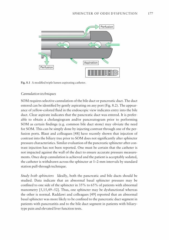
Cannulation techniques
SOM requires selective cannulation of the bile duct or pancreatic duct. The ductentered can be identified by gently aspirating on any port (Fig. 8.2). The appear-ance of yellow-colored fluid in the endoscopic view indicates entry into the bileduct. Clear aspirate indicates that the pancreatic duct was entered. It is prefer-able to obtain a cholangiogram and/or pancreatogram prior to performingSOM as certain findings (e.g. common bile duct stone) may obviate the need for SOM. This can be simply done by injecting contrast through one of the per-fusion ports. Blaut and colleagues [48] have recently shown that injection ofcontrast into the biliary tree prior to SOM does not significantly alter sphincterpressure characteristics. Similar evaluation of the pancreatic sphincter after con-trast injection has not been reported. One must be certain that the catheter is not impacted against the wall of the duct to ensure accurate pressure measure-ments. Once deep cannulation is achieved and the patient is acceptably sedated,the catheter is withdrawn across the sphincter at 1–2-mm intervals by standardstation pull-through technique.
Study both sphincters Ideally, both the pancreatic and bile ducts should bestudied. Data indicate that an abnormal basal sphincter pressure may beconfined to one side of the sphincter in 35% to 65% of patients with abnormalmanometry [5,15,49–52]. Thus, one sphincter may be dysfunctional whereasthe other is normal. Raddawi and colleagues [49] reported that an abnormalbasal sphincter was more likely to be confined to the pancreatic duct segment inpatients with pancreatitis and to the bile duct segment in patients with biliary-type pain and elevated liver function tests.
SPHINCTER OF ODDI DYSFUNCTION 177
Aspiration
Perfusion
Perfusion
Fig. 8.1 A modified triple lumen aspirating catheter.

CHAPTER 8178
Fig. 8.2 The duct entered during sphincter of Oddi manometry can be identified by aspiratingthe catheter. Dark-colored yellow fluid signifies entry into the bile duct, whereas clear fluidindicates pancreatic duct entry.
Abnormalities of the basal sphincter pressure should ideally be observed forat least 30 s in each lead and be seen on two or more separate pull-throughs.From a practical clinical standpoint, we settle for one pull-through (from eachduct) if the readings are clearly normal or abnormal. During standard stationpull-through technique, it is necessary to establish good communication betweenthe endoscopist and the manometrist who is reading the tracing as it rolls off therecorder or appears on the computer screen. This permits optimal positioning ofthe catheter to achieve interpretable tracings. Alternatively, electronic manome-try systems with a television screen can be mounted near the endoscopic imagescreen to permit the endoscopist to view the manometry tracing duringendoscopy. Once the baseline study is done, agents to relax or stimulate thesphincter can be given (e.g. cholecystokinin) and manometric or pain responsemonitored. The value of these provocative maneuvers for everyday use needsfurther study before widespread application is recommended.
Interpretation of manometry tracings
Criteria for the interpretation of an SO tracing are relatively standard; however,they may vary somewhat from center to center. Some areas where there may bedisagreement in interpretation include the required duration of basal SO pres-sure elevation, the number of leads in which basal pressure elevation is required,and the role of averaging pressures from the three (or two in an aspiratingcatheter) recording ports [3]. Our recommended method for reading themanometry tracings is first to define the zero duodenal baseline before and afterthe pull-through. Alternatively, the intraduodenal pressure can be continuouslyrecorded from a separate intraduodenal catheter attached to the endoscope. Thehighest basal pressure (Fig. 8.3) that is sustained for at least 30 s is thenidentified. From the four lowest amplitude points in this zone, the mean of thesereadings is taken as the basal sphincter pressure for that lead for that pull-through. The basal sphincter pressure for all interpretable observations is thenaveraged; this is the final basal sphincter pressure. The amplitude of phasic wavecontractions is measured from the beginning of the slope of the pressure increasefrom the basal pressure to the peak of the contraction wave. Four representativewaves are taken for each lead and the mean pressure determined. The number ofphasic waves per minute and the duration of the phasic waves can also be deter-mined. Most authorities read only the basal sphincter pressure as an indicator of
SPHINCTER OF ODDI DYSFUNCTION 179

pathology of the SO. However, data from Kalloo and colleagues [53] suggestthat the intraductal biliary pressure, which is easier to measure than SO pres-sure, correlates with SO basal pressure. In this study, intrabiliary pressure wassignificantly higher in patients with SOD than those with normal SO pressure(20 vs. 10 mmHg; P < 0.01). This study needs to be confirmed but supports thetheory that increased intrabiliary pressure is a cause of pain in SOD.
Normal values
The best study establishing normal values for SOM was reported by Guelrudand associates [54]. Fifty asymptomatic control patients were evaluated andSOM was repeated on two occasions in 10 subjects. This study established nor-mal values for intraductal pressure, basal sphincter pressure, and phasic waveparameters (Table 8.3). Moreover, the reproducibility of SOM was confirmed.Various authorities interchangeably use 35 mmHg or 40 mmHg as the upperlimits of normal for mean basal SO pressure.
CHAPTER 8180
280
(A)
(B)
240
200
160
120
80
40
0 ab
c
2 mm/sec
mm
Hg
a
Fig. 8.3 (A) An abnormal station pull-through at sphincter of Oddi manometry. The studyhas been abbreviated to fit onto one page. (B) Schematic representation of one lead of theabove tracing. (a) Baseline duodenal 0 reference. (b) Intraductal (pancreatic) pressure of 20 mmHg (abnormal). (c) Basal pancreatic sphincter pressure of 45 mmHg (abnormal).Phasic waves are 155–175 mmHg amplitude and 6 s duration (normal).
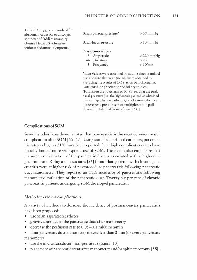
Complications of SOM
Several studies have demonstrated that pancreatitis is the most common majorcomplication after SOM [55–57]. Using standard perfused catheters, pancreat-itis rates as high as 31% have been reported. Such high complication rates haveinitially limited more widespread use of SOM. These data also emphasize thatmanometric evaluation of the pancreatic duct is associated with a high com-plication rate. Rolny and associates [56] found that patients with chronic pan-creatitis were at higher risk of postprocedure pancreatitis following pancreatic duct manometry. They reported an 11% incidence of pancreatitis followingmanometric evaluation of the pancreatic duct. Twenty-six per cent of chronicpancreatitis patients undergoing SOM developed pancreatitis.
Methods to reduce complications
A variety of methods to decrease the incidence of postmanometry pancreatitishave been proposed:• use of an aspiration catheter• gravity drainage of the pancreatic duct after manometry• decrease the perfusion rate to 0.05–0.1 ml/lumen/min• limit pancreatic duct manometry time to less than 2 min (or avoid pancreaticmanometry)• use the microtransducer (non-perfused) system [13]• placement of pancreatic stent after manometry and/or sphincterotomy [58].
SPHINCTER OF ODDI DYSFUNCTION 181
Basal sphincter pressurea > 35 mmHg
Basal ductal pressure > 13 mmHg
Phasic contractions–3 Amplitude > 220 mmHg–4 Duration > 8 s–5 Frequency > 10/min
Note: Values were obtained by adding three standarddeviations to the mean (means were obtained byaveraging the results of 2–3 station pull-throughs).Data combine pancreatic and biliary studies.aBasal pressures determined by: (1) reading the peakbasal pressure (i.e. the highest single lead as obtainedusing a triple lumen catheter); (2) obtaining the meanof these peak pressures from multiple station pull-throughs. [Adapted from reference 54.]
Table 8.3 Suggested standard forabnormal values for endoscopicsphincter of Oddi manometryobtained from 50 volunteerswithout abdominal symptoms.
Aspirating catheter system
In a prospective randomized study, Sherman and colleagues found that the aspirating catheter (this catheter allows for aspiration of the perfused fluid fromend- and side-holes while accurately recording pressure from the two remain-ing side-ports) reduced the frequency of pancreatic duct manometry-inducedpancreatitis from 31% to 4% [55]. The reduction in pancreatitis with the use ofthis catheter in the pancreatic duct, and the very low incidence of pancreatitisafter bile duct manometry, lend support to the notion that increased pancreaticduct hydrostatic pressure is a major cause of this complication. Thus, when thepancreatic duct sphincter is studied by SOM, aspiration of pancreatic juice andthe perfusate is strongly recommended.
Prophylactic stenting
In a prospective randomized trial, Tarnasky and colleagues showed that stent-ing the pancreatic duct decreased post-ERCP pancreatitis from 26% to 6% in agroup of patients with pancreatic sphincter hypertension undergoing biliarysphincterotomy alone [58].
SOM: conclusion
SOM is recommended in patients with idiopathic pancreatitis or unexplaineddisabling pancreatico-biliary pain with or without hepatic enzyme abnormali-ties. An attempt is made to study both sphincters, but clinical decisions can bemade when the first sphincter evaluated is abnormal. An ERCP is usually per-formed (if an adequate study is not available) immediately before SOM toexclude other potential causes for the patient’s symptoms. Indications for theuse of SOM have also been developed according to the Hogan–Geenen SODclassification system (Table 8.1).
Type I patients
In Type I patients, there is a general consensus that a structural disorder of the sphincter (i.e. sphincter stenosis) exists. Although SOM may be useful indocumenting SOD, it is not an essential diagnostic study prior to endoscopic or surgical sphincter ablation. Such patients uniformly benefit from sphincterablation regardless of the SOM results.
Type II patients
Type II patients demonstrate SO motor dysfunction in 50–65% of cases. In this
CHAPTER 8182
group of patients, SOM is highly recommended as the results of the study pre-dict outcome from sphincter ablation.
Type III patients
Type III patients have pancreatico-biliary pain without other objective evidenceof sphincter outflow obstruction. SOM is mandatory to confirm the presence ofSOD. Although not well studied, it appears that the results of SOM may predictoutcome from sphincter ablation in these patients.
Therapy for SOD
The therapeutic approach in patients with SOD is aimed at reducing the resis-tance caused by the SO to the flow of bile and/or pancreatic juice [6]. Historically,emphasis has been placed on definitive intervention, i.e. surgical sphincter-oplasty or endoscopic sphincterotomy. This appears appropriate for patientswith high-grade obstruction (Type I as per Hogan–Geenen criteria). In patientswith lesser degrees of obstruction, the clinician must carefully weigh the risksand benefits before recommending invasive therapy. Most reports indicate thatSOD patients have a complication rate from endoscopic sphincterotomy of atleast twice that of patients with ductal stones [59,60].
Medical therapy
Medical therapy for documented or suspected SOD has received only limitedstudy. As the SO is a smooth muscle structure, it is reasonable to assume thatdrugs that relax smooth muscle might be an effective treatment for SOD.Sublingual nifedipine and nitrates have been shown to reduce the basal sphinc-ter pressures in asymptomatic volunteers and symptomatic patients with SOD[1,61].
Nifedipine
Khuroo and colleagues [62] evaluated the clinical benefit of nifedipine in aplacebo-controlled crossover trial. Twenty-one of 28 patients (75%) withmanometrically documented SOD had a reduction in pain scores, emergencyroom visits, and use of oral analgesics during short-term follow-up. In a similarstudy, Sand and associates [63] found that nine of 12 (75%) Type II SOD (sus-pected; SOM was not performed) patients improved with nifedipine. Althoughmedical therapy may be an attractive initial approach in patients with SOD, several drawbacks exist [1]. First, medication side-effects may be seen in up toone-third of patients. Second, smooth muscle relaxants are unlikely to be of any
SPHINCTER OF ODDI DYSFUNCTION 183
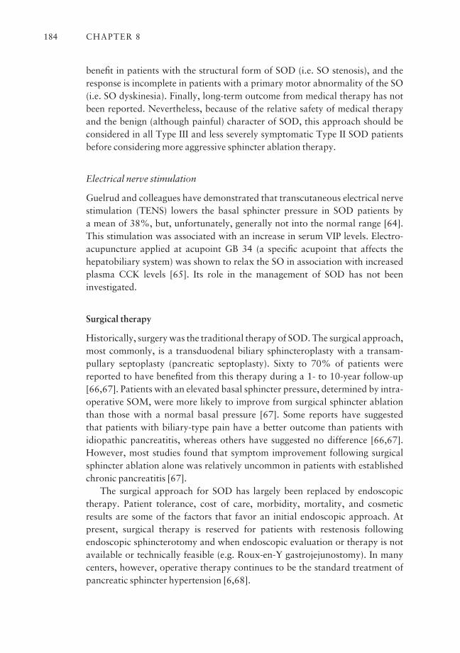
benefit in patients with the structural form of SOD (i.e. SO stenosis), and theresponse is incomplete in patients with a primary motor abnormality of the SO(i.e. SO dyskinesia). Finally, long-term outcome from medical therapy has notbeen reported. Nevertheless, because of the relative safety of medical therapyand the benign (although painful) character of SOD, this approach should beconsidered in all Type III and less severely symptomatic Type II SOD patientsbefore considering more aggressive sphincter ablation therapy.
Electrical nerve stimulation
Guelrud and colleagues have demonstrated that transcutaneous electrical nervestimulation (TENS) lowers the basal sphincter pressure in SOD patients by a mean of 38%, but, unfortunately, generally not into the normal range [64].This stimulation was associated with an increase in serum VIP levels. Electro-acupuncture applied at acupoint GB 34 (a specific acupoint that affects the hepatobiliary system) was shown to relax the SO in association with increasedplasma CCK levels [65]. Its role in the management of SOD has not been investigated.
Surgical therapy
Historically, surgery was the traditional therapy of SOD. The surgical approach,most commonly, is a transduodenal biliary sphincteroplasty with a transam-pullary septoplasty (pancreatic septoplasty). Sixty to 70% of patients werereported to have benefited from this therapy during a 1- to 10-year follow-up[66,67]. Patients with an elevated basal sphincter pressure, determined by intra-operative SOM, were more likely to improve from surgical sphincter ablationthan those with a normal basal pressure [67]. Some reports have suggested that patients with biliary-type pain have a better outcome than patients withidiopathic pancreatitis, whereas others have suggested no difference [66,67].However, most studies found that symptom improvement following surgicalsphincter ablation alone was relatively uncommon in patients with establishedchronic pancreatitis [67].
The surgical approach for SOD has largely been replaced by endoscopictherapy. Patient tolerance, cost of care, morbidity, mortality, and cosmeticresults are some of the factors that favor an initial endoscopic approach. At present, surgical therapy is reserved for patients with restenosis following endoscopic sphincterotomy and when endoscopic evaluation or therapy is notavailable or technically feasible (e.g. Roux-en-Y gastrojejunostomy). In manycenters, however, operative therapy continues to be the standard treatment ofpancreatic sphincter hypertension [6,68].
CHAPTER 8184
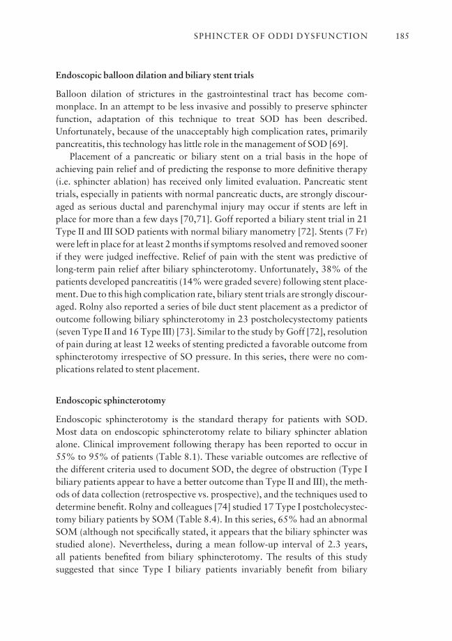
Endoscopic balloon dilation and biliary stent trials
Balloon dilation of strictures in the gastrointestinal tract has become com-monplace. In an attempt to be less invasive and possibly to preserve sphincterfunction, adaptation of this technique to treat SOD has been described.Unfortunately, because of the unacceptably high complication rates, primarilypancreatitis, this technology has little role in the management of SOD [69].
Placement of a pancreatic or biliary stent on a trial basis in the hope ofachieving pain relief and of predicting the response to more definitive therapy(i.e. sphincter ablation) has received only limited evaluation. Pancreatic stenttrials, especially in patients with normal pancreatic ducts, are strongly discour-aged as serious ductal and parenchymal injury may occur if stents are left inplace for more than a few days [70,71]. Goff reported a biliary stent trial in 21Type II and III SOD patients with normal biliary manometry [72]. Stents (7 Fr)were left in place for at least 2 months if symptoms resolved and removed soonerif they were judged ineffective. Relief of pain with the stent was predictive oflong-term pain relief after biliary sphincterotomy. Unfortunately, 38% of thepatients developed pancreatitis (14% were graded severe) following stent place-ment. Due to this high complication rate, biliary stent trials are strongly discour-aged. Rolny also reported a series of bile duct stent placement as a predictor ofoutcome following biliary sphincterotomy in 23 postcholecystectomy patients(seven Type II and 16 Type III) [73]. Similar to the study by Goff [72], resolutionof pain during at least 12 weeks of stenting predicted a favorable outcome fromsphincterotomy irrespective of SO pressure. In this series, there were no com-plications related to stent placement.
Endoscopic sphincterotomy
Endoscopic sphincterotomy is the standard therapy for patients with SOD.Most data on endoscopic sphincterotomy relate to biliary sphincter ablationalone. Clinical improvement following therapy has been reported to occur in55% to 95% of patients (Table 8.1). These variable outcomes are reflective ofthe different criteria used to document SOD, the degree of obstruction (Type Ibiliary patients appear to have a better outcome than Type II and III), the meth-ods of data collection (retrospective vs. prospective), and the techniques used todetermine benefit. Rolny and colleagues [74] studied 17 Type I postcholecystec-tomy biliary patients by SOM (Table 8.4). In this series, 65% had an abnormalSOM (although not specifically stated, it appears that the biliary sphincter wasstudied alone). Nevertheless, during a mean follow-up interval of 2.3 years, all patients benefited from biliary sphincterotomy. The results of this study suggested that since Type I biliary patients invariably benefit from biliary
SPHINCTER OF ODDI DYSFUNCTION 185
sphincterotomy, SOM in this patient group is not only unnecessary, but mayalso be misleading. The results of this study, however, have never been validatedat another center. In contrast, results of several non-randomized controlled tri-als [13,17,75,76] suggest that performance of SOM is highly recommended inbiliary Type II and Type III patients, as clinical benefit is less certain (Table 8.5).
Although most of the studies reporting the efficacy of endoscopic therapy inSOD have been retrospective, three notable randomized trials have been reported.
Randomized controlled trials of endoscopic sphincterotomy for SOD
In a landmark study by Geenen and associates [77], 47 postcholecystectomyType II biliary patients were randomized to biliary sphincterotomy or shamsphincterotomy. SOM was performed in all patients but was not used as a criterion for randomization. During a 4-year follow-up, 95% of patients withan elevated basal sphincter pressure benefited from sphincterotomy. In con-trast, only 30% to 40% of patients with an elevated sphincter pressure treatedby sham sphincterotomy, or with a normal sphincter pressure treated by endoscopic sphincterotomy or sham sphincterotomy, benefited from this therapy. The two important findings of this study were that SOM predicted theoutcome from endoscopic sphincterotomy and that endoscopic sphincterotomyoffered long-term benefit in Type II biliary patients with SOD. Confirmatorydata were seen in a 2-year follow-up study by Toouli et al. [78,79]. In this study,
CHAPTER 8186
Basal sphincter Asymptomatic/of Oddi pressure No. improved after ES/SS
< 40 mmHg 6 (35%) 6 (100%)≥ 40 mmHg 11 (65%) 11 (100%)
15 endoscopic (ES), 2 surgical (SS) sphincterotomies.aFrom reference [74].
Clinical benefit
Reference Type II Type III
Choudhry et al. [17] 10/18 (56%) 9/16 (56%)Botoman et al. [13] 13/19 (68%) 9/16 (56%)Bozkurt et al. [75] 14/19 (78%) 5/5 (100%)Wehrmann et al. [76] 12/20 (60%) 1/13 (8%)
aSix had cholecystectomy.
Table 8.4 Biliary sphincter ablation in Type I SOD (28-monthfollow-up)a.
Table 8.5 Biliary sphincterotomyfor sphincter of Oddi dysfunctiondocumented by sphincter of Oddimanometry: results of four non-randomized controlled trials.
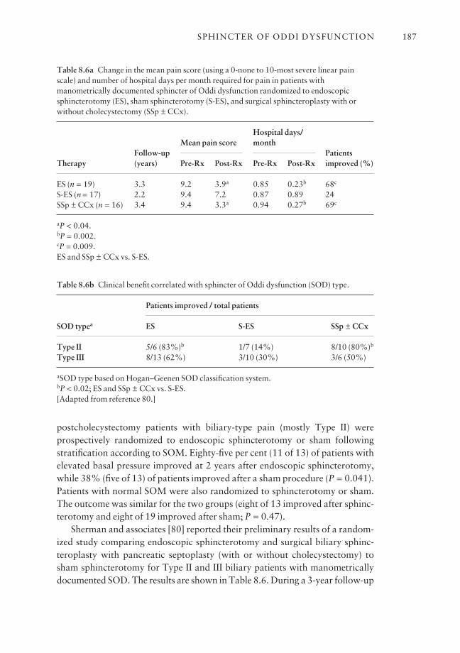
postcholecystectomy patients with biliary-type pain (mostly Type II) wereprospectively randomized to endoscopic sphincterotomy or sham followingstratification according to SOM. Eighty-five per cent (11 of 13) of patients withelevated basal pressure improved at 2 years after endoscopic sphincterotomy,while 38% (five of 13) of patients improved after a sham procedure (P = 0.041).Patients with normal SOM were also randomized to sphincterotomy or sham.The outcome was similar for the two groups (eight of 13 improved after sphinc-terotomy and eight of 19 improved after sham; P = 0.47).
Sherman and associates [80] reported their preliminary results of a random-ized study comparing endoscopic sphincterotomy and surgical biliary sphinc-teroplasty with pancreatic septoplasty (with or without cholecystectomy) tosham sphincterotomy for Type II and III biliary patients with manometricallydocumented SOD. The results are shown in Table 8.6. During a 3-year follow-up
SPHINCTER OF ODDI DYSFUNCTION 187
Table 8.6a Change in the mean pain score (using a 0-none to 10-most severe linear painscale) and number of hospital days per month required for pain in patients withmanometrically documented sphincter of Oddi dysfunction randomized to endoscopicsphincterotomy (ES), sham sphincterotomy (S-ES), and surgical sphincteroplasty with orwithout cholecystectomy (SSp ± CCx).
Hospital days/
Follow-upMean pain score month
PatientsTherapy (years) Pre-Rx Post-Rx Pre-Rx Post-Rx improved (%)
ES (n = 19) 3.3 9.2 3.9a 0.85 0.23b 68c
S-ES (n = 17) 2.2 9.4 7.2 0.87 0.89 24SSp ± CCx (n = 16) 3.4 9.4 3.3a 0.94 0.27b 69c
aP < 0.04.bP = 0.002.cP = 0.009.ES and SSp ± CCx vs. S-ES.
Table 8.6b Clinical benefit correlated with sphincter of Oddi dysfunction (SOD) type.
Patients improved / total patients
SOD typea ES S-ES SSp ± CCx
Type II 5/6 (83%)b 1/7 (14%) 8/10 (80%)b
Type III 8/13 (62%) 3/10 (30%) 3/6 (50%)
aSOD type based on Hogan–Geenen SOD classification system.bP < 0.02; ES and SSp ± CCx vs. S-ES.[Adapted from reference 80.]
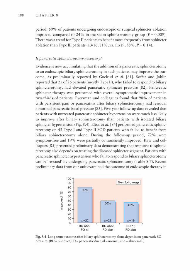
period, 69% of patients undergoing endoscopic or surgical sphincter ablationimproved compared to 24% in the sham sphincterotomy group (P = 0.009).There was a trend for Type II patients to benefit more frequently from sphincterablation than Type III patients (13/16, 81%, vs. 11/19, 58%; P = 0.14).
Is pancreatic sphincterotomy necessary?
Evidence is now accumulating that the addition of a pancreatic sphincterotomyto an endoscopic biliary sphincterotomy in such patients may improve the out-come, as preliminarily reported by Guelrud et al. [81]. Soffer and Johlinreported that 25 of 26 patients (mostly Type II), who failed to respond to biliarysphincterotomy, had elevated pancreatic sphincter pressure [82]. Pancreaticsphincter therapy was performed with overall symptomatic improvement intwo-thirds of patients. Eversman and colleagues found that 90% of patientswith persistent pain or pancreatitis after biliary sphincterotomy had residualabnormal pancreatic basal pressure [83]. Five-year follow-up data revealed thatpatients with untreated pancreatic sphincter hypertension were much less likelyto improve after biliary sphincterotomy than patients with isolated biliarysphincter hypertension (Fig. 8.4). Elton et al. [84] performed pancreatic sphinc-terotomy on 43 Type I and Type II SOD patients who failed to benefit from biliary sphincterotomy alone. During the follow-up period, 72% were symptom-free and 19% were partially or transiently improved. Kaw and col-leagues [85] presented preliminary data demonstrating that response to sphinc-terotomy also depends on treating the diseased sphincter segment. Patients withpancreatic sphincter hypertension who fail to respond to biliary sphincterotomycan be ‘rescued’ by undergoing pancreatic sphincterotomy (Table 8.7). Recentpreliminary data from our unit examined the outcome of endoscopic therapy in
CHAPTER 8188
1009080706050403020100
BD abn;PD nl
BD abn;PD abn
BD nl;PD abn
Imp
rove
d (
%)
5-yr follow-up
50% 46%
n=19
80%
n=23n=22
Fig. 8.4 Long-term outcome after biliary sphincterotomy alone depends on pancreatic SOpressure. (BD = bile duct; PD = pancreatic duct; nl = normal; abn = abnormal.)
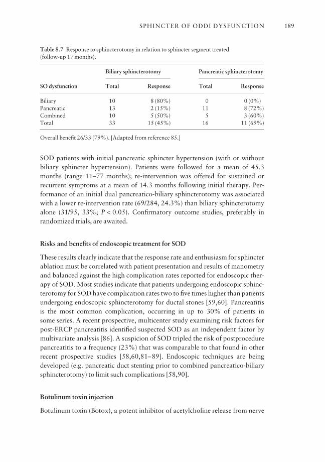
SOD patients with initial pancreatic sphincter hypertension (with or withoutbiliary sphincter hypertension). Patients were followed for a mean of 45.3months (range 11–77 months); re-intervention was offered for sustained orrecurrent symptoms at a mean of 14.3 months following initial therapy. Per-formance of an initial dual pancreatico-biliary sphincterotomy was associatedwith a lower re-intervention rate (69/284, 24.3%) than biliary sphincterotomyalone (31/95, 33%; P < 0.05). Confirmatory outcome studies, preferably in randomized trials, are awaited.
Risks and benefits of endoscopic treatment for SOD
These results clearly indicate that the response rate and enthusiasm for sphincterablation must be correlated with patient presentation and results of manometryand balanced against the high complication rates reported for endoscopic ther-apy of SOD. Most studies indicate that patients undergoing endoscopic sphinc-terotomy for SOD have complication rates two to five times higher than patientsundergoing endoscopic sphincterotomy for ductal stones [59,60]. Pancreatitis is the most common complication, occurring in up to 30% of patients in some series. A recent prospective, multicenter study examining risk factors forpost-ERCP pancreatitis identified suspected SOD as an independent factor bymultivariate analysis [86]. A suspicion of SOD tripled the risk of postprocedurepancreatitis to a frequency (23%) that was comparable to that found in otherrecent prospective studies [58,60,81–89]. Endoscopic techniques are beingdeveloped (e.g. pancreatic duct stenting prior to combined pancreatico-biliarysphincterotomy) to limit such complications [58,90].
Botulinum toxin injection
Botulinum toxin (Botox), a potent inhibitor of acetylcholine release from nerve
SPHINCTER OF ODDI DYSFUNCTION 189
Table 8.7 Response to sphincterotomy in relation to sphincter segment treated (follow-up 17 months).
Biliary sphincterotomy Pancreatic sphincterotomy
SO dysfunction Total Response Total Response
Biliary 10 8 (80%) 0 0 (0%)Pancreatic 13 2 (15%) 11 8 (72%)Combined 10 5 (50%) 5 3 (60%)Total 33 15 (45%) 16 11 (69%)
Overall benefit 26/33 (79%). [Adapted from reference 85.]
endings, has been successfully applied to smooth muscle disorders of the gastrointestinal tract such as achalasia. In a preliminary clinical trial, toxininjection into the SO resulted in a 50% reduction in the basal biliary sphincterpressure and improved bile flow [91]. This reduction in pressure may be accom-panied by symptom improvement in some patients. Although further study iswarranted, Botox may serve as a therapeutic trial for SOD with respondersundergoing permanent sphincter ablation. In a small series [92], 22 postchole-cystectomy Type III patients with manometric evidence of SOD underwentBotox injection into the intraduodenal sphincter segment. Eleven of the 12 pati-ents who responded to botulinum toxin injection later benefited from endo-scopic sphincterotomy, while only two of 10 patients who did not benefit fromBotox injection later responded to sphincter ablation. Such an approach, how-ever, does require two endoscopies to achieve symptom relief. Further studiesare needed before recommending this technique.
SOD in recurrent pancreatitis
Disorders of the pancreatic sphincter may give rise to unexplained (idiopathic)pancreatitis or episodic pain suggestive of a pancreatic origin [68]. SOD hasbeen manometrically documented in 15% to 72% of patients with recurrentpancreatitis, previously labeled as idiopathic [5,14,16,93]. Biliary sphinctero-tomy alone has been reported to prevent further pancreatitis episodes in morethan 50% of such patients. From a scientific, but not practical viewpoint, caremust be taken to separate out subtle biliary pancreatitis [94] that will similarlyrespond to biliary sphincterotomy.
Endoscopic sphincterotomy for SOD in pancreatitis
Lans and colleagues
The value of ERCP, SOM, and sphincter ablation therapy was studied in 51patients with idiopathic pancreatitis [43]. Twenty-four (47.1%) had an elevatedbasal sphincter pressure. Thirty were treated by biliary sphincterotomy (n = 20)or surgical sphincteroplasty with septoplasty (n = 10). Fifteen of 18 patients(83%) with an elevated basal sphincter pressure had long-term benefit (meanfollow-up, 38 months) from sphincter ablation therapy (including 10 of 11 treated by biliary sphincterotomy) in contrast to only four of 12 (33.3%; P < 0.05) with a normal basal sphincter pressure (including four of nine treatedby biliary sphincterotomy).
CHAPTER 8190
Guelrud and colleagues
Guelrud and colleagues [81], by contrast, found that severance of the pancreaticsphincter was necessary to resolve the pancreatitis (Table 8.8). In this series, 69patients with idiopathic pancreatitis due to SOD underwent treatment by stand-ard biliary sphincterotomy (n = 18), biliary sphincterotomy with pancreaticsphincter balloon dilation (n = 24), biliary sphincterotomy followed by pancre-atic sphincterotomy in separate sessions (n = 13), or combined pancreatic andbiliary sphincterotomy in the same session (n = 14). Eighty-one per cent ofpatients undergoing pancreatic and biliary sphincterotomy had resolution oftheir pancreatitis compared to 28% of patients undergoing biliary sphinctero-tomy alone (P < 0.005). These data are consistent with the theory that manysuch patients who benefit from biliary sphincterotomy alone have subtle gall-stone pancreatitis. The results of Guelrud and colleagues [81] also support theanatomic findings of separate biliary and pancreatic sphincters, and the manom-etry findings of residual pancreatic sphincter hypertension in more than 50% ofpersistently symptomatic patients who undergo biliary sphincterotomy alone.
Kaw and Brodmerkel
Kaw and Brodmerkel [95] recently reported that, among patients with idiopathicpancreatitis secondary to SOD, 78% had persistent manometric evidence ofpancreatic sphincter hypertension despite a biliary sphincterotomy.
Toouli and colleagues
Toouli and colleagues [96] also demonstrated the importance of pancreatic andbiliary sphincter ablation in patients with idiopathic pancreatitis. In this series,
SPHINCTER OF ODDI DYSFUNCTION 191
Table 8.8 Pancreatic sphincter dysfunction and recurrent pancreatitis: response to sphinctertherapy.
Patientsimproved/
Treatment total patients
Biliary sphincterotomy alone 5/18 (28%)Biliary sphincterotomy followed by pancreatic sphincter balloon dilation 13/24 (54%)Biliary sphincterotomy plus pancreatic sphincterotomy at later session 10/13 (77%)a
Biliary sphincterotomy and pancreatic sphincterotomy at same session 12/14 (86%)a
aP < 0.005 vs. biliary sphincterotomy alone. [Adapted from reference 81.]
23 of 26 patients (88%) undergoing surgical ablation of both the biliary andpancreatic sphincter were either asymptomatic or had minimal symptoms at amedian follow-up of 24 months (range 9–105 months).
Okolo and colleagues
Okolo and colleagues [97] retrospectively evaluated the long-term results ofendoscopic pancreatic sphincterotomy in 55 patients with manometrically docu-mented or presumed pancreatic sphincter hypertension (presumption based onrecurrent pancreatitis with pancreatic duct dilation and contrast medium drain-age time from the pancreatic duct of greater than 10 min). During a median follow-up of 16 months (range 3–52 months), 34 patients (62%) reportedsignificant pain improvement. Patients with normal pancreatograms were morelikely to respond to therapy than those with pancreatographic evidence ofchronic pancreatitis (73% vs. 58%).
Endoscopic sphincterotomy as a cause of pancreatic sphincter stenosis
Jacob and colleagues [98] postulated that SOD might cause recurrent episodesof pancreatitis, even though SOM was normal, and pancreatic stent placementmight prevent further attacks. In a randomized study, 34 patients with un-explained recurrent pancreatitis and normal pancreatic SOM were treated withpancreatic stents (n = 19; 5–7 Fr gauge, with stents exchanged three times over a 1-year period) or conservative therapy. During a 3-year follow-up, pancreat-itis recurred in 53% of the patients in the control group and only 11% of thestented patients (P < 0.02). This study suggests that SOM may be an imperfecttest, as patients may have SOD but not be detected at the time of SOM.However, long-term studies are needed to evaluate the outcome after removal ofstents, and concern remains regarding stent-induced ductal and parenchymalchanges [70,71,99].
Endoscopic Botox injection
Wehrmann and colleagues [100] recently evaluated the feasibility and effective-ness of botulinum toxin injection in patients with recurrent pancreatitis due topancreatic sphincter hypertension. No side-effects of the injection were noted inany of the 15 treated patients. Twelve patients (80%) remained asymptomaticat 3-month follow-up, but 11 developed a relapse at a follow-up period of 6 ± 2months. These 11 patients underwent pancreatic or combined pancreatico-biliary sphincterotomy with subsequent remission after a median follow-up of 15 months. This study showed that injection of botulinum toxin is safe and may
CHAPTER 8192
be effective short term, but the need for definitive sphincter ablation in themajority of patients limits its clinical use.
SOD in recurrent pancreatitis: conclusion
Currently, establishing the best method of treating residual pancreatic sphincterstenosis (after biliary sphincterotomy) awaits further study. Patients with idio-pathic pancreatitis who fail to respond to biliary sphincterotomy alone shouldhave their pancreatic sphincter re-evaluated and be considered for sphincterablation if residual high pressure is found.
Conclusion
Our knowledge of SOD, and manometric techniques to assist in this diagnosis,is evolving. Successful endoscopic SOM requires good general ERCP skills andcareful attention to the main details listed above. If SOD is suspected in a TypeIII or mild to moderate pain level Type II patient, medical therapy should gener-ally be tried. If medical therapy fails or is bypassed, ERCP and manometric evalu-ation are recommended. The role of less invasive studies remains uncertainowing to undefined sensitivity and specificity. Sphincter ablation is generallywarranted in symptomatic Type I patients and Type II and III patients withabnormal manometry. The symptom relief rate varies from 55% to 95%,depending on the patient presentation and selection. Initial non-respondersrequire thorough pancreatic sphincter and pancreatic parenchymal evaluation.SOD patients have relatively high complication rates after invasive studies ortherapy. Thorough review of the risk–benefit ratio with individual patients ismandatory.
Outstanding issues and future trends
Our hopes for the future in this evolving field are to:• Define the role, if any, of non-invasive imaging studies as a screening test andpredictor of outcome from sphincter ablation • Develop techniques to improve the safety of the procedures used to evaluateand treat patients with pancreatico-biliary pain • Develop a device for longer term SOM such as a 24-h SOM probe • Define predictors of good and poor outcome from therapy to better selectpatients for ‘risky’ interventions • Further investigate the role of pancreatic sphincterotomy for improving outcomes
SPHINCTER OF ODDI DYSFUNCTION 193
• Develop more long-term outcome studies, particularly in Type II and IIIpatients • Explore better medical therapy alternatives for less disabled patients.
References
1 Kalloo AN, Pasricha PJ. Therapy of sphincter of Oddi dysfunction. Gastrointest Endosc ClinN Am 1996; 6: 117–25.
2 Black NA, Thompson E, Sanderson CFB. Symptoms and health status before and six weeksafter open cholecystectomy: a European cohort study. ECHSS Group. European CollaborativeHealth Services Study Group. Gut 1994; 35: 1301–5.
3 Hogan W, Sherman S, Pasricha P, Carr-Locke DL. Sphincter of Oddi manometry. GastrointestEndosc 1997; 45: 342–8.
4 Sherman S, Troiano FP, Hawes RH, O’Connor KW, Lehman GA. Frequency of abnormalsphincter of Oddi manometry compared with the clinical suspicion of sphincter of Oddi dys-function. Am J Gastroenterol 1991; 86: 586–90.
5 Eversman D, Fogel EL, Rusche M, Sherman S, Lehman GA. Frequency of abnormal pancreaticand biliary sphincter manometry compared with clinical suspicion of sphincter of Oddi dys-function. Gastrointest Endosc 1999; 50: 637–41.
6 Corazziari E, Shaffer EA, Hogan W, Sherman S, Toouli J. Functional disorders of the biliarytract and pancreas. Gut 1999; 45 (Suppl 2): 48–54.
7 Anderson TM, Pitt HA, Longmire WP Jr. Experience with sphincteroplasty and sphinctero-tomy in pancreatobiliary surgery. Ann Surg 1985; 201: 399–406.
8 Guelrud M, Mendoza S, Mujica V, Uzcategui A. Sphincter of Oddi (SO) motor function inpatients with symptomatic gallstones. Gastroenterology 1993; 104: A361.
9 Ruffolo TA, Sherman S, Lehman GA, Hawes RH. Gallbladder ejection fraction and its rela-tionship to sphincter of Oddi dysfunction. Dig Dis Sci 1994; 39: 289–92.
10 Neoptolemos JP, Bailey IS, Carr-Locke DL. Sphincter of Oddi dysfunction: results of treatmentby endoscopic sphincterotomy. Br J Surg 1988; 75: 454–9.
11 Roberts-Thomson IC, Toouli J. Is endoscopic sphincterotomy for disabling biliary-type painafter cholecystectomy effective? Gastrointest Endosc 1985; 31: 370–3.
12 Meshkinpoor H, Mollot M. Sphincter of Oddi dysfunction and unexplained abdominal pain:clinical and manometric study. Dig Dis Sci 1992; 37: 257–61.
13 Botoman VA, Kozarek RA, Novell LA, Patterson DJ, Ball TJ, Wechter DG et al. Long term out-come after endoscopic sphincterotomy in patients with biliary colic and suspected sphincter ofOddi dysfunction. Gastrointest Endosc 1994; 40: 165–70.
14 Lehman GA, Sherman S. Sphincter of Oddi dysfunction. Int J Pancreatol 1996; 20: 11–25.15 Aymerich RR, Prakash C, Aliperti G. Sphincter of Oddi manometry: is it necessary to measure
both biliary and pancreatic sphincter pressure? Gastrointest Endosc 2000; 52: 183–6.16 Geenen JE, Nash JA. The role of sphincter of Oddi manometry and biliary microscopy in evalu-
ating idiopathic recurrent pancreatitis. Endoscopy 1998; 30 (9): A237–41.17 Choudhry U, Ruffolo T, Jamidar P, Hawes R, Lehman G. Sphincter of Oddi dysfunction in
patients with intact gallbladder: therapeutic response to endoscopic sphincterotomy. Gastro-intest Endosc 1993; 39: 492–5.
18 Steinberg WM. Sphincter of Oddi dysfunction: a clinical controversy. Gastroenterology 1988;95: 1409–15.
19 Lin OS, Soetikno RM, Young HS. The utility of liver function test abnormalities concomitantwith biliary symptoms in predicting a favorable response to endoscopic sphincterotomy inpatients with presumed sphincter of Oddi dysfunction. Am J Gastroenterol 1998; 93: 1833–6.
20 Steinberg WM, Salvato RF, Toskes PP. The morphine-prostigmin provocative test: is it usefulfor making clinical decisions? Gastroenterology 1980; 78: 728–31.
21 Darweesh RM, Dodds WJ, Hogan WJ, Geenen JE, Collier BD, Shaker R et al. Efficacy of quant-itative hepatobiliary scintigraphy and fatty-meal sonography for evaluating patients with sus-pected partial common duct obstruction. Gastroenterology 1988; 94: 779–86.
CHAPTER 8194
22 Simeone JF, Mueller PR, Ferrucci JT Jr, vanSonnenberg E, Hall DA, Wittenberg J et al.Sonography of the bile ducts after a fatty meal: an aid in detection of obstruction. Radiology1982; 143: 211–15.
23 Troiano F, O’Connor K, Lehman GA, Madura J, Kopecky K, Bogan M. Comparison ofsecretin-stimulated ultrasound and sphincter of Oddi manometry in evaluating sphincter ofOddi dysfunction. Gastrointest Endosc 1989; 35: A166.
24 Warshaw AL, Simeone J, Schapiro RH, Hedberg SE, Mueller PE, Ferrucci JT Jr. Objective evalu-ation of ampullary stenosis with ultrasonography and pancreatic stimulation. Am J Surg 1985;149: 65–72.
25 DiFrancesco V, Brunori MR, Rigo L, Toouli J, Angelini G, Frulloni L et al. Comparison ofultrasound-secretin test and sphincter of Oddi manometry in patients with recurrent acute pan-creatitis. Dig Dis Sci 1999; 44: 336–40.
26 Silverman WB, Johlin FC, Crowe G. Does secretin stimulated ultrasound (SSUS) predictresults: sphincter of Oddi manometry (SOM) basal sphincter pressure (BSP) in patients sus-pected of having sphincter of Oddi dysfunction (SOD)? Gastrointest Endosc 2001; 53: A100.
27 Catalano MF, Lahoti S, Alcocer E, Geenen JE, Hogan WJ. Dynamic imaging of the pancreasusing real-time endoscopic ultrasonography with secretin stimulation. Gastrointest Endosc1998; 48: 580–7.
28 Devereaux BM, Fogel EL, Aisen A, Stockberger S, Ness R, Lehman GA et al. Secretin-stimulated functional MRCP: correlation with sphincter of Oddi manometry. GastrointestEndosc 2000; 51: A197.
29 Sostre S, Kalloo AN, Spiegler EJ, Camargo EE, Wagner HN Jr. A noninvasive test of sphincterof Oddi dysfunction in postcholecystectomy patients: the scintigraphic score. J Nucl Med1992; 33: 1216–22.
30 Kalloo AN, Sostre S, Pasricha PJ. The Hopkins scintigraphic score: a noninvasive, highly accurate screening test for sphincter of Oddi dysfunction. Gastroenterology 1994; 106: A342.
31 Cicala M, Scopinaro F, Corazziari E, Vignoni A, Viscardi A, Habib FI et al. Quantitative cholescintigraphy in the assessment of choledochoduodenal bile flow. Gastroenterology 1991;100: 1106–13.
32 Corazziari E, Cicala M, Habib FI, Scopinaro F, Fiocca F, Pallotta N et al. Hepatoduodenal biletransit in cholecystectomized subjects. Relationship with sphincter of Oddi dysfunction anddiagnostic value. Dig Dis Sci 1994; 39: 1985–93.
33 Peng NJ, Lai KH, Tsay DG, Liu RS, Su KL, Yeh SH. Efficacy of quantitative cholescintigraphyin the diagnosis of sphincter of Oddi dysfunction. Nucl Med Commun 1994; 15: 899–904.
34 Thomas PD, Turner JG, Dobbs BR, Burt MJ, Chapman BA. Use of 99m Tc-DISIDA biliaryscanning with morphine provocation in the detection of elevated sphincter of Oddi basal pres-sure. Gut 2000; 46: 838–41.
35 Esber E, Ruffolo TA, Park H, Siddiqui A, Earle D, Pezzi J et al. Prospective assessment of biliary scintigraphy in patients with suspected sphincter of Oddi dysfunction. GastrointestEndosc 1995; 41: A396.
36 Rosenblatt ML, Catalano MF, Alcocer E, Geenen JE. Comparison of sphincter of Oddimanometry, fatty meal sonography, and hepatobiliary scintigraphy in the diagnosis of sphinc-ter of Oddi dysfunction. Gastrointest Endosc 2001; 54: 697–704.
37 Pineau BC, Knapple WL, Spicer KM, Gordon L, Wallace M, Hennessy WS et al.Cholecystokinin-stimulated mebrofenin (99mTc-Choletec) hepatobiliary scintigraphy in asymp-tomatic postcholecystectomy individuals: assessment of specificity, interobserver reliability,and reproducibility. Am J Gastroenterol 2001; 96: 3106–9.
38 Elta GH, Barnett JL, Ellis JH, Ackermann R, Wahl R. Delayed biliary drainage is common inasymptomatic post-cholecystectomy volunteers. Gastrointest Endosc 1992; 38: 435–9.
39 Ponchon T, Aucia N, Mitchell R, Chavaillon A, Bory R, Hedelius F. Biopsies of the ampullaryregion in patients suspected to have sphincter of Oddi dysfunction. Gastrointest Endosc 1995;42: 296–300.
40 Itoh A, Tsukamoto Y, Naitoh Y, Hirooka Y, Furukawa T, Kato T et al. Intraductal ultra-sonography for the examination of duodenal papillary region. J Ultrasound Med 1994; 13:679–84.
SPHINCTER OF ODDI DYSFUNCTION 195
41 Wehrmann T, Stergiou N, Riphaus A, Lembcke B. Correlation between sphincter of Oddimanometry and intraductal ultrasound morphology in patients with suspected sphincter ofOddi dysfunction. Endoscopy 2001; 33: 773–7.
42 Lehman GA. Endoscopic sphincter of Oddi manometry: a clinical practice and research tool.Gastrointest Endosc 1991; 37: 490–2.
43 Lans JL, Parikh NP, Geenen JE. Application of sphincter of Oddi manometry in routine clinicalinvestigations. Endoscopy 1991; 23: 139–43.
44 Sherman S, Gottlieb K, Uzer MF, Smith MT, Khusro QE, Earle DT et al. Effects of meperidineon the pancreatic and biliary sphincter. Gastrointest Endosc 1996; 44: 239–42.
45 Fogel EL, Sherman S, Bucksot L, Philips SD, Lehman GA. Sphincter of Oddi manometry(SOM): effects of droperidol on the biliary and pancreatic sphincter. Gastrointest Endosc2001; 53: A89.
46 Goff JS. Effect of propofol on human sphincter of Oddi. Dig Dis Sci 1995; 40: 2364–7.47 Craig AG, Omari T, Lingenfelser T, Schloithe AC, Saccone GT, Dent J et al. Development of
a sleeve sensor for measurement of sphincter of Oddi motility. Endoscopy 2001; 33: 651–7.
48 Blaut U, Sherman S, Fogel E, Lehman GA. Influence of cholangiography on biliary sphincter ofOddi manometric parameters. Gastrointest Endosc 2000; 52: 624–9.
49 Raddawi HM, Geenen JE, Hogan WJ, Dodds WJ, Venu RP, Johnson GK. Pressure measure-ments from biliary and pancreatic segments of sphincter of Oddi. Comparison betweenpatients with functional abdominal pain, biliary, or pancreatic disease. Dig Dis Sci 1991; 36:71–4.
50 Rolny P, Ärlebäck A, Funch-Jensen P, Kruse A, Jarnerot G. Clinical significance of manometricassessment of both pancreatic duct and bile duct sphincter in the same patient. Scand JGastroenterol 1989; 24: 751–4.
51 Silverman WB, Ruffolo TA, Sherman S, Hawes RH, Lehman GA. Correlation of basal sphinc-ter pressures measured from both the bile duct and pancreatic duct in patients with suspectedsphincter of Oddi dysfunction. Gastrointest Endosc 1992; 38: 440–3.
52 Chan YK, Evans PR, Dowsett JF, Kellow JE, Badcock CA. Discordance of pressure recordingsfrom biliary and pancreatic duct segments in patients with suspected sphincter of Oddi dys-function. Dig Dis Sci 1997; 42: 1501–6.
53 Kalloo AN, Tietjen TG, Pasricha PJ. Does intrabiliary pressure predict basal sphincter of Oddipressure? A study in patients with and without gallbladders. Gastrointest Endosc 1996; 44:696–9.
54 Guelrud M, Mendoza S, Rossiter G, Villegas MI. Sphincter of Oddi manometry in healthy volunteers. Dig Dis Sci 1990; 35: 38–46.
55 Sherman S, Troiano FP, Hawes RH, Lehman GA. Sphincter of Oddi manometry: decreasedrisk of clinical pancreatitis with the use of a modified aspirating catheter. Gastrointest Endosc1990; 36: 462–6.
56 Rolny P, Anderberg B, Ihse I, Lindstrom E, Olaison G, Arvill A. Pancreatitis after sphincter ofOddi manometry. Gut 1990; 31: 821–4.
57 Maldonado ME, Brady PG, Mamel JJ, Robinson B. Incidence of pancreatitis in patients under-going sphincter of Oddi manometry (SOM). Am J Gastroenterol 1999; 94: 387–90.
58 Tarnasky PR, Palesch YY, Cunningham JT, Mauldin PD, Cotton PB, Hawes RH. Pancreaticstenting prevents pancreatitis after biliary sphincterotomy in patients with sphincter of Oddidysfunction. Gastroenterology 1998; 115: 1518–24.
59 Sherman S, Ruffolo TA, Hawes RH, Lehman GA. Complications of endoscopic sphinctero-tomy. A prospective series with emphasis on the increased risk associated with sphincter ofOddi dysfunction and nondilated bile ducts. Gastroenterology 1991; 101: 1068–75.
60 Freeman ML, Nelson DB, Sherman S, Haber GB, Herman ME, Dorsher PJ et al. Complica-tions of endoscopic biliary sphincterotomy: a prospective, multicenter study. N Engl J Med1996; 335: 909–18.
61 Guelrud M, Mendoza S, Rossiter G, Ramirez L, Barkin J. Effect of nifedipine on sphincter ofOddi motor activity: studies in healthy volunteers and patients with biliary dyskinesia.Gastroenterology 1988; 95: 1050–5.
CHAPTER 8196
62 Khuroo MS, Zargar SA, Yattoo GN. Efficacy of nifedipine therapy in patients with sphincter ofOddi dysfunction: a prospective, double-blind, randomized, placebo-controlled, cross overtrial. Br J Clin Pharmacol 1992; 33: 477–85.
63 Sand J, Nordback I, Koskinen M, Matikainen M, Lindholm TS. Nifedipine for suspected TypeII sphincter of Oddi dyskinesia. Am J Gastroenterol 1993; 88: 530–5.
64 Guelrud M, Rossiter A, Souney P, Mendoza S, Mujica V. The effect of transcutaneous nervestimulation on sphincter of Oddi pressure in patients with biliary dyskinesia. Am J Gastro-enterol 1991; 86: 581–5.
65 Lee SK, Kim MH, Kim HJ, Seo DS, Yoo KS, Joo YH et al. Electroacupuncture may relax thesphincter of Oddi in humans. Gastrointest Endosc 2001; 53: 211–16.
66 Moody FG, Vecchio R, Calabuig R, Runkel N. Transduodenal sphincteroplasty with trans-ampullary septectomy for stenosing papillitis. Am J Surg 1991; 161: 213–18.
67 Sherman S, Hawes RH, Madura J, Lehman GA. Comparison of intraoperative and endoscopicmanometry of the sphincter of Oddi. Surg Gynecol Obstet 1992; 175: 410–18.
68 Chen JW, Saccone GT, Toouli J. Sphincter of Oddi dysfunction and acute pancreatitis. Gut1998; 43: 305–8.
69 Kozarek RA. Balloon dilation of the sphincter of Oddi. Endoscopy 1988; 20: 207–10.70 Kozarek RA. Pancreatic stents can induce ductal changes consistent with chronic pancreatitis.
Gastrointest Endosc 1990; 36: 93–5.71 Fogel EL, Kwon E, Sherman S, Philips SD, Watkins JL, Paige-Ongay B et al. Pancreatic ductal
alterations following small diameter, long length, unflanged pancreatic duct (PD) stent place-ment. Gastrointest Endosc 2001; 53: A86.
72 Goff JS. Common bile duct sphincter of Oddi stenting in patients with suspected sphincter ofOddi dysfunction. Am J Gastroenterol 1995; 90: 586–9.
73 Rolny P. Endoscopic bile duct stent placement as a predictor of outcome following endoscopicsphincterotomy in patients with suspected sphincter of Oddi dysfunction. Eur J GastroenterolHepatol 1997; 9: 467–71.
74 Rolny P, Geenen JE, Hogan WJ. Post-cholecystectomy patients with ‘objective signs’ of partialbile outflow obstruction: clinical characteristics, sphincter of Oddi manometry findings, andresults of therapy. Gastrointest Endosc 1993; 39: 778–81.
75 Bozkurt T, Orth KH, Butsch B, Lux G. Long-term clinical outcome of post-cholecystectomypatients with biliary-type pain: results of manometry, non-invasive techniques and endoscopicsphincterotomy. Eur J Gastroenterol Hepatol 1996; 8: 245–9.
76 Wehrmann T, Wiemer K, Lembcke B, Caspary WF, Juno M. Do patients with sphincter ofOddi dysfunction benefit from endoscopic sphincterotomy? A 5-year prospective trial. Eur JGastroenterol Hepatol 1996; 8: 251–6.
77 Geenen JE, Hogan WJ, Dodds WJ, Toouli J, Venu RP. The efficacy of endoscopic sphinctero-tomy after cholecystectomy in patients with sphincter of Oddi dysfunction. N Engl J Med1989; 320: 82–7.
78 Toouli J, Roberts-Thomson I, Kellow J et al. Prospective randomized trial of endoscopicsphincterotomy for treatment of sphincter of Oddi dysfunction. J Gastroenterol Hepatol 1996;11: A115.
79 Toouli J, Roberts-Thomson IC, Kellow J, Dowsett J, Saccone GTP, Evans P et al. Manometrybased randomized trial of endoscopic sphincterotomy for sphincter of Oddi dysfunction. Gut2000; 46: 98–102.
80 Sherman S, Lehman GA, Jamidar P, Hawes RH, Silverman W, Madura J et al. Efficacy ofendoscopic sphincterotomy and surgical sphincteroplasty for patients with sphincter of Oddi dysfunction (SOD): randomized, controlled study. Gastrointest Endosc 1994; 40: A125.
81 Guelrud M, Plaz J, Mendoza S, Beker B, Rojas O, Rossiter G. Endoscopic treatment in Type IIpancreatic sphincter dysfunction. Gastrointest Endosc 1995; 41: A398.
82 Soffer EE, Johlin FC. Intestinal dysmotility in patients with sphincter of Oddi dysfunction. Areason for failed response to sphincterotomy. Dig Dis Sci 1994; 39: 1942–6.
83 Eversman D, Fogel E, Philips S, Sherman S, Lehman G. Sphincter of Oddi dysfunction (SOD):long-term outcome of biliary sphincterotomy (BES) correlated with abnormal biliary and pan-creatic sphincters. Gastrointest Endosc 1999; 49: A78.
SPHINCTER OF ODDI DYSFUNCTION 197
84 Elton E, Howell DA, Parsons WG, Qaseem T, Hanson BL. Endoscopic pancreatic sphinctero-tomy: indications, outcome, and a safe stentless technique. Gastrointest Endosc 1998; 47: 240–9.
85 Kaw M, Verma R, Brodmerkel GJ. Biliary and/or pancreatic sphincter of Oddi dysfunction(SOD). Response to endoscopic sphincterotomy (ES). Gastrointest Endosc 1996; 43: A384.
86 Freeman ML, DiSario JA, Nelson DB, Fennerty MB, Lee JG, Bjorkman DJ et al. Risk factorsfor post-ERCP pancreatitis: a prospective, multicenter study. Gastrointest Endosc 2001; 54:425–34.
87 Gottlieb K, Sherman S. ERCP- and endoscopic sphincterotomy-induced pancreatitis. Gastro-intest Endosc Clin N Am 1998; 8: 87–114.
88 Tarnasky P, Cunningham T, Cotton P, Hoffman B, Palesch Y, Freeman T et al. Pancreaticsphincter hypertension increases the risk of post-ERCP pancreatitis. Endoscopy 1997; 29:252–7.
89 Sherman S, Lehman G, Freeman ML, Earle D, Watkins J, Barnett T et al. Risk factors for post-ERCP pancreatitis: a prospective multicenter study. Am J Gastroenterol 1997; 92: A1639.
90 Fogel EL, Devereaux BM, Rerknimitr R, Sherman S, Bucksot L, Lehman GA. Does placementof a small diameter, long length, unflanged pancreatic duct stent reduce the incidence of post-ERCP pancreatitis? Gastrointest Endosc 2000; 51: A182.
91 Pasricha PJ, Miskovsky EP, Kalloo AN. Intrasphincteric injection of botulinum toxin for sus-pected sphincter of Oddi dysfunction. Gut 1994; 35: 1319–21.
92 Wehrmann T, Seifert H, Seipp M, Lembcke B, Caspary WF. Endoscopic injection of botulinumtoxin for biliary sphincter of Oddi dysfunction. Endoscopy 1998; 30: 702–7.
93 Kuo WH, Pasricha PJ, Kalloo AN. The role of sphincter of Oddi manometry in the diagnosisand therapy of pancreatic disease. Gastrointest Endosc Clin N Am 1998; 8: 79–85.
94 Ros E, Navarro S, Bru C, Garcia-Puges A, Valderrama R. Occult microlithiasis in ‘idiopathic’acute pancreatitis: prevention of relapses by cholecystectomy or ursodeoxycholic acid therapy.Gastroenterology 1991; 101: 1701–9.
95 Kaw M, Brodmerkel GJ Jr. ERCP, biliary crystal analysis, and sphincter of Oddi manometry inidiopathic recurrent pancreatitis. Gastrointest Endosc 2002; 55: 157–62.
96 Toouli J, Di Francesco V, Saccone G, Kollias J, Schloithe A, Shanks N. Division of the sphincterof Oddi for treatment of dysfunction associated with recurrent pancreatitis. Br J Surg 1996; 83:1205–10.
97 Okolo PI 3rd, Pasricha PJ, Kalloo AN. What are the long-term results of endoscopic pancreaticsphincterotomy? Gastrointest Endosc 2000; 52: 15–19.
98 Jacob L, Geenen JE, Catalano MF, Geenen DJ. Prevention of pancreatitis in patients with idio-pathic recurrent pancreatitis: a prospective nonblinded randomized study using endoscopicstents. Endoscopy 2001; 33: 559–62.
99 Smith MT, Sherman S, Ikenberry SO, Hawes RH, Lehman GA. Alterations in pancreatic ductmorphology following polyethylene pancreatic stent therapy. Gastrointest Endosc 1996; 44:268–75.
100 Wehrmann T, Schmitt TH, Arndt A, Lembcke B, Caspary WF, Seifert H. Endoscopic injectionof botulinum toxin in patients with recurrent acute pancreatitis due to pancreatic sphincter ofOddi dysfunction. Aliment Pharmacol Ther 2000; 14: 1469–77.
CHAPTER 8198
CHAPTER 9
ERCP in Acute Pancreatitis
MARTIN L. FREEMAN
Synopsis
ERCP plays an expanding role in both the diagnosis and therapy of acute andrelapsing pancreatitis of various etiologies. Although initially used in the diag-nosis and treatment of biliary disorders causing pancreatitis, endoscopic inter-ventions are now increasingly directed towards the pancreatic sphincter and ductsas well. In certain settings, such as acute gallstone pancreatitis, the value of ERCPhas been proven in randomized controlled trials. There are also data to supportthe role of ERCP in the treatment of acute relapsing pancreatitis due to variousdisorders, such as pancreas divisum and to a lesser degree sphincter of Oddi dysfunction. Other applications include the use of ERCP to treat smolderingpancreatitis and pancreatic ductal disruptions in the setting of acute and chronicpancreatitis, and most recently in the setting of evolving pancreatic necrosis.Many causes of otherwise unexplained acute recurrent pancreatitis can be foundafter an extensive evaluation and treated by advanced ERCP techniques. Therole of ERCP in acute and especially recurrent pancreatitis should be primarilytherapeutic, with diagnosis first established whenever possible by other tech-niques, including endoscopic ultrasound and MRCP. ERCP for the diagnosisand treatment of severe or acute relapsing pancreatitis is optimally performed ina multidisciplinary context involving primary or critical care, advanced hepato-biliary–pancreatic surgery, and interventional radiology when appropriate.
Introduction
ERCP appeared in the early 1970s and soon evolved as a diagnostic and thera-peutic technique for biliary tract disorders. Biliary therapy including sphinctero-tomy was then applied to biliary causes of acute pancreatitis such as gallstonepancreatitis. Over the last decade, the application of pancreatic diagnostic andtherapeutic techniques has expanded to incorporate a wider range of pancreaticmethods including pancreatic sphincterotomy, stenting, stricture dilation, andstone extraction via the major and minor papillae. These techniques have
199
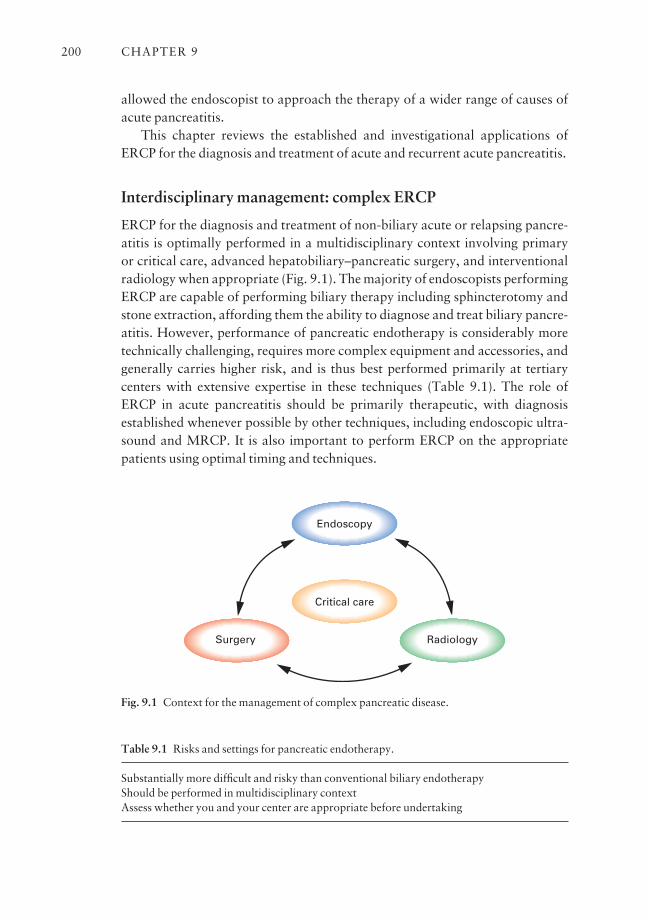
allowed the endoscopist to approach the therapy of a wider range of causes ofacute pancreatitis.
This chapter reviews the established and investigational applications ofERCP for the diagnosis and treatment of acute and recurrent acute pancreatitis.
Interdisciplinary management: complex ERCP
ERCP for the diagnosis and treatment of non-biliary acute or relapsing pancre-atitis is optimally performed in a multidisciplinary context involving primary or critical care, advanced hepatobiliary–pancreatic surgery, and interventionalradiology when appropriate (Fig. 9.1). The majority of endoscopists performingERCP are capable of performing biliary therapy including sphincterotomy andstone extraction, affording them the ability to diagnose and treat biliary pancre-atitis. However, performance of pancreatic endotherapy is considerably moretechnically challenging, requires more complex equipment and accessories, andgenerally carries higher risk, and is thus best performed primarily at tertiary centers with extensive expertise in these techniques (Table 9.1). The role ofERCP in acute pancreatitis should be primarily therapeutic, with diagnosisestablished whenever possible by other techniques, including endoscopic ultra-sound and MRCP. It is also important to perform ERCP on the appropriatepatients using optimal timing and techniques.
CHAPTER 9200
Endoscopy
RadiologySurgery
Critical care
Fig. 9.1 Context for the management of complex pancreatic disease.
Table 9.1 Risks and settings for pancreatic endotherapy.
Substantially more difficult and risky than conventional biliary endotherapyShould be performed in multidisciplinary contextAssess whether you and your center are appropriate before undertaking
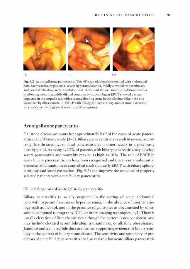
Acute gallstone pancreatitis
Gallstone disease accounts for approximately half of the cases of acute pancre-atitis in the Western world [1–3]. Biliary pancreatitis may result in severe, necrot-izing, life-threatening, or fatal pancreatitis as it often occurs in a previouslyhealthy gland. As many as 25% of patients with biliary pancreatitis may developsevere pancreatitis and mortality may be as high as 10%. The role of ERCP inacute biliary pancreatitis has long been recognized and there is now substantialevidence from randomized controlled trials that early ERCP with biliary sphinc-terotomy and stone extraction (Fig. 9.2) can improve the outcome of properlyselected patients with acute biliary pancreatitis.
Clinical diagnosis of acute gallstone pancreatitis
Biliary pancreatitis is usually suspected in the setting of acute abdominal pain with hyperamylasemia or hyperlipasemia, in the absence of another etio-logy such as alcohol, and in the presence of gallstones as documented by ultra-sound, computed tomography (CT), or other imaging techniques [4,5]. There isusually elevation of liver chemistries although the pattern is not consistent, andmay include elevated serum bilirubin, transaminase, or alkaline phosphatase.Jaundice and a dilated bile duct are further supporting evidence of biliary etio-logy in the context of biliary stone disease. The sensitivity and specificity of pre-dictors of acute biliary pancreatitis are also variable but acute biliary pancreatitis
ERCP IN ACUTE PANCREATITIS 201
Fig. 9.2 Acute gallstone pancreatitis. This 48-year-old female presented with abdominalpain, tachycardia, hypoxemia, severe hyperamylasemia, mildly elevated transaminases, and normal bilirubin, and transabdominal ultrasound showed multiple gallstones with ashadowing stone in a mildly dilated common bile duct. Urgent ERCP showed a stoneimpacted in the ampulla (a), with a second floating stone in the bile duct (likely the onevisualized by ultrasound). (b) ERCP with biliary sphincterotomy and (c) stone extraction was performed with gradual resolution of symptoms.
(a) (b) (c)
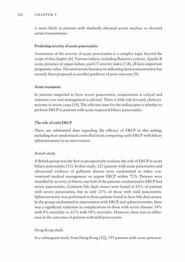
is more likely in patients with markedly elevated serum amylase or elevatedserum transaminase.
Predicting severity of acute pancreatitis
Assessment of the severity of acute pancreatitis is a complex topic beyond thescope of this chapter [6]. Various indices, including Ranson’s criteria, Apache IIscore, presence of organ failure, and CT severity index [7,8], all have importantprognostic value. Elevated serum hematocrit indicating hemoconcentration hasrecently been proposed as another predictor of poor outcome [9].
Acute treatment
In patients suspected to have severe pancreatitis, resuscitation is critical andintensive care unit management is advised. There is little role for early cholecys-tectomy in severe cases [10]. The relevant issue for the endoscopist is whether toperform ERCP in patients with acute suspected biliary pancreatitis.
The role of early ERCP
There are substantial data regarding the efficacy of ERCP in this setting, including four randomized controlled trials comparing early ERCP with biliarysphincterotomy to no intervention.
British study
A British group was the first to prospectively evaluate the role of ERCP in acutebiliary pancreatitis [11]. In that study, 121 patients with acute pancreatitis andultrasound evidence of gallstone disease were randomized to either con-ventional medical management or urgent ERCP within 72 h. Patients werestratified by severity of illness; one-half of the patients randomized to ERCP hadsevere pancreatitis. Common bile duck stones were found in 63% of patientswith severe pancreatitis, but in only 25% of those with mild pancreatitis.Sphincterotomy was performed in those patients found to have bile duct stones.In the group randomized to intervention with ERCP and sphincterotomy, therewas a significant reduction in complications in those with severe disease: 24%with 4% mortality vs. 61% with 18% mortality. However, there was no differ-ence in the outcomes of patients with mild pancreatitis.
Hong Kong study
In a subsequent study from Hong Kong [12], 195 patients with acute pancreat-
CHAPTER 9202
itis were randomized to receive ERCP with sphincterotomy vs. conservativemanagement within 24 h of admission. Stones were found in 65% of thepatients. The major difference in outcome of the group undergoing ERCP was areduction in biliary sepsis (0% after ERCP vs. 14% in the conservative group).There was a tendency towards fewer complications in the ERCP group vs. con-servative management group, especially in those with severe pancreatitis, and a slight trend towards a reduction in mortality. The applicability of this studyhas been questioned as bile duct stones in Asians are more often primary bileduct stones rather than cholesterol stones originating from the gallbladder, thusreflecting different pathophysiology than in Western patients.
Polish study
A randomized controlled trial from Poland has been presented only in abstractform [13]; 280 patients with acute biliary pancreatitis all underwent ERCPwithin 24 h of admission. All patients with bile duct stones were treated with biliary sphincterotomy while the remaining patients without common bile ductstones were randomized to sphincterotomy or conventional treatment. Therewere significant reductions in complications in sphincterotomy-treated patientsvs. conservatively treated patients (17% vs. 36%) and a significant reduction inmortality (2% vs. 13%). The benefits of intervention appeared to apply topatients with all severities of pancreatitis, including those with mild disease.Problems with this study include the fact that it has not been published in a peer-reviewed journal, a lack of true randomization, and the fact that some of thepatients with empty ducts may have had more severe irreversible damage, ormay have had pancreatitis due to etiologies other than stone disease.
German study
The most contentious study is the German multicenter study published in theNew England Journal of Medicine [14], in which 238 patients with suspectedbiliary pancreatitis were randomized to early ERCP within 72 h of presentationor conservative management. Patients with jaundice were excluded. Fifty-eightof 121 patients randomized to the ERCP arm were found to have bile duct stones.In the control arm, 13 of 112 were crossed over to ERCP for apparent bile ductstones. In this study, there was no improvement in outcome from early sphinc-terotomy. Paradoxically, there appeared to be more severe complications, includ-ing respiratory failure, in the early ERCP group, and a numerically increasedmortality. Major criticisms of this study have included the fact that patientsmost likely to benefit from ERCP, i.e. those with jaundice, were excluded fromthe study. Furthermore, many contributing centers enrolled fewer than two pati-ents per year, raising questions about technical proficiency at ERCP.
ERCP IN ACUTE PANCREATITIS 203
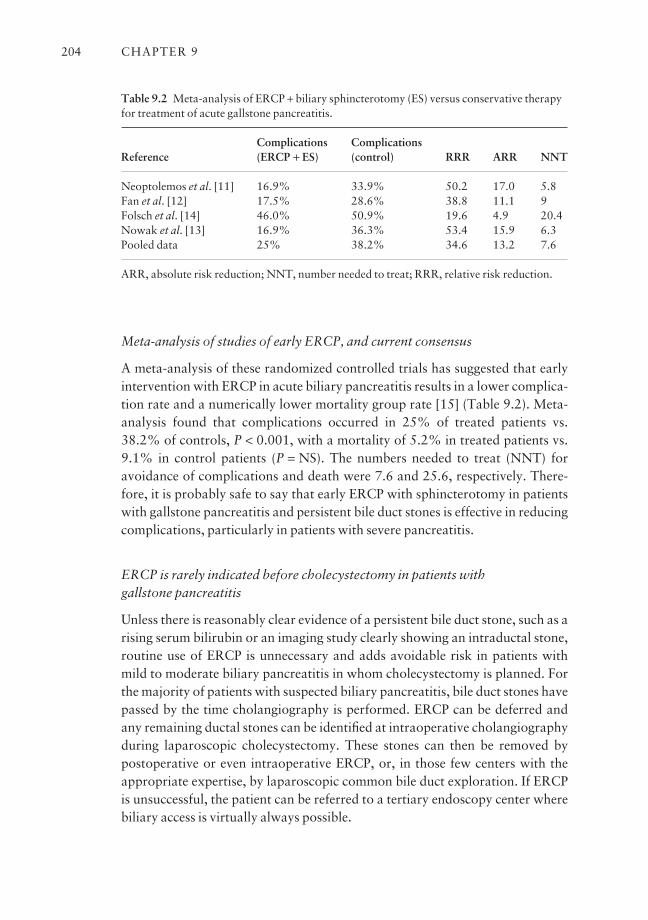
Meta-analysis of studies of early ERCP, and current consensus
A meta-analysis of these randomized controlled trials has suggested that earlyintervention with ERCP in acute biliary pancreatitis results in a lower complica-tion rate and a numerically lower mortality group rate [15] (Table 9.2). Meta-analysis found that complications occurred in 25% of treated patients vs.38.2% of controls, P < 0.001, with a mortality of 5.2% in treated patients vs.9.1% in control patients (P = NS). The numbers needed to treat (NNT) foravoidance of complications and death were 7.6 and 25.6, respectively. There-fore, it is probably safe to say that early ERCP with sphincterotomy in patientswith gallstone pancreatitis and persistent bile duct stones is effective in reducingcomplications, particularly in patients with severe pancreatitis.
ERCP is rarely indicated before cholecystectomy in patients with gallstone pancreatitis
Unless there is reasonably clear evidence of a persistent bile duct stone, such as arising serum bilirubin or an imaging study clearly showing an intraductal stone,routine use of ERCP is unnecessary and adds avoidable risk in patients withmild to moderate biliary pancreatitis in whom cholecystectomy is planned. Forthe majority of patients with suspected biliary pancreatitis, bile duct stones havepassed by the time cholangiography is performed. ERCP can be deferred andany remaining ductal stones can be identified at intraoperative cholangiographyduring laparoscopic cholecystectomy. These stones can then be removed bypostoperative or even intraoperative ERCP, or, in those few centers with theappropriate expertise, by laparoscopic common bile duct exploration. If ERCPis unsuccessful, the patient can be referred to a tertiary endoscopy center wherebiliary access is virtually always possible.
CHAPTER 9204
Table 9.2 Meta-analysis of ERCP + biliary sphincterotomy (ES) versus conservative therapyfor treatment of acute gallstone pancreatitis.
Complications ComplicationsReference (ERCP + ES) (control) RRR ARR NNT
Neoptolemos et al. [11] 16.9% 33.9% 50.2 17.0 5.8Fan et al. [12] 17.5% 28.6% 38.8 11.1 9Folsch et al. [14] 46.0% 50.9% 19.6 4.9 20.4Nowak et al. [13] 16.9% 36.3% 53.4 15.9 6.3Pooled data 25% 38.2% 34.6 13.2 7.6
ARR, absolute risk reduction; NNT, number needed to treat; RRR, relative risk reduction.

Acute pancreatitis postcholecystectomy
ERCP is appropriate in postcholecystectomy patients with suspected biliarypancreatitis, but in many of these patients there is a non-biliary stone eti-ology, such as sphincter of Oddi dysfunction, a setting in which conventionaldiagnostic and therapeutic ERCP techniques can be highly risky [16,17], andprotective measures, such as placement of a pancreatic stent, may be advisable[18,19].
Treatment by biliary sphincterotomy alone?
Empirical biliary sphincterotomy for suspected biliary pancreatitis may beappropriate in certain settings without cholecystectomy, especially in elderlypatients who are not good candidates for surgery due to severe medical comor-bidity [20–24]. Under these circumstances, biliary sphincterotomy is sometimesperformed in the absence of demonstration of a definite bile duct stone or as asemidefinitive treatment in lieu of cholecystectomy. Several studies have sug-gested the effectiveness of endoscopic biliary sphincterotomy in preventing futureepisodes of acute biliary pancreatitis [25]. These uncontrolled case series mostlysuggest a reduction in the frequency of pancreatitis attacks, although recurrentbile duct stones and cholecystitis may be problematic [26]. Caution must beapplied to patients who might have other etiologies. Empirical biliary sphinc-terotomy in patients with recurrent pancreatitis and mildly abnormal enzymesmay in fact be due to sphincter of Oddi dysfunction, especially in women,younger to middle-aged patients, and those who are postcholecystectomy or donot have clearly documented gallstone disease. Empirical biliary sphinctero-tomy and even diagnostic ERCP in this setting may be quite hazardous [16,17]and less likely to be of benefit.
Pancreatic duct disruptions
Acute disruptions of the main pancreatic duct or side branches may occur during acute pancreatitis of various etiologies, such as gallstones or alcohol, ormay be the primary mechanism of pancreatitis in cases such as trauma. Thesedisruptions may result in localized fluid collections, pseudocysts, ascites, or pancreatico-pleural or cutaneous fistulas.
Stenting for duct disruption
ERCP with transpapillary pancreatic duct stenting has been described as aneffective technique to close pancreatic duct disruptions in a variety of settings in
ERCP IN ACUTE PANCREATITIS 205
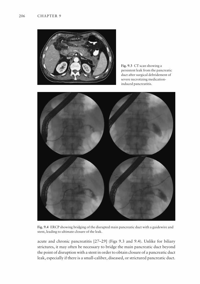
acute and chronic pancreatitis [27–29] (Figs 9.3 and 9.4). Unlike for biliarystrictures, it may often be necessary to bridge the main pancreatic duct beyondthe point of disruption with a stent in order to obtain closure of a pancreatic ductleak, especially if there is a small-caliber, diseased, or strictured pancreatic duct.
CHAPTER 9206
Fig. 9.3 CT scan showing apersistent leak from the pancreaticduct after surgical debridement ofsevere necrotizing medication-induced pancreatitis.
Fig. 9.4 ERCP showing bridging of the disrupted main pancreatic duct with a guidewire andstent, leading to ultimate closure of the leak.
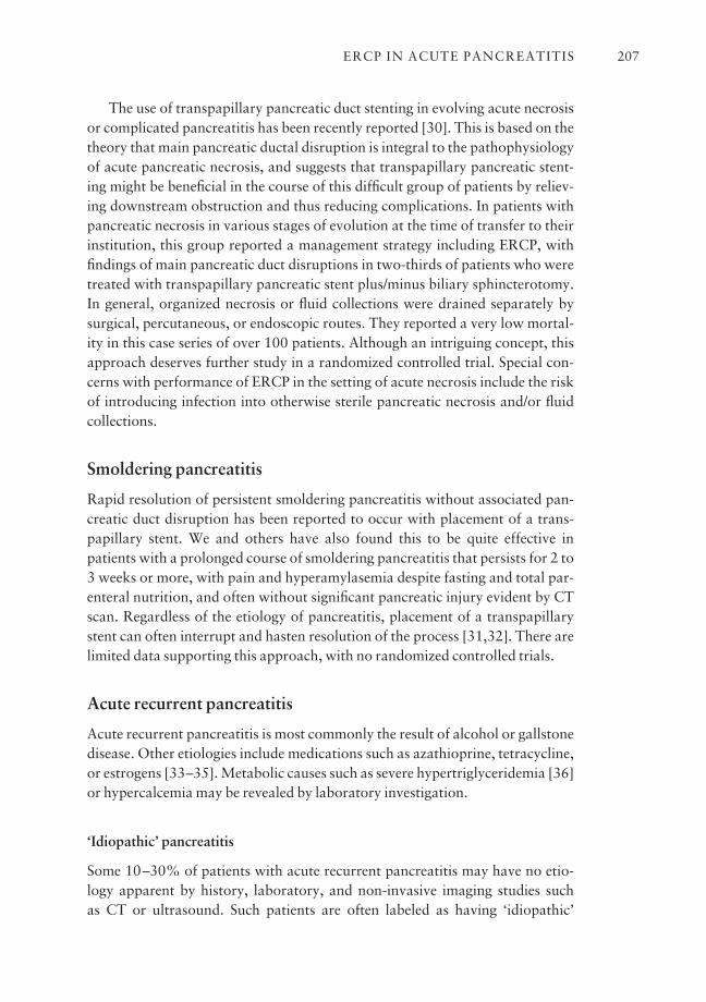
The use of transpapillary pancreatic duct stenting in evolving acute necrosisor complicated pancreatitis has been recently reported [30]. This is based on thetheory that main pancreatic ductal disruption is integral to the pathophysiologyof acute pancreatic necrosis, and suggests that transpapillary pancreatic stent-ing might be beneficial in the course of this difficult group of patients by reliev-ing downstream obstruction and thus reducing complications. In patients withpancreatic necrosis in various stages of evolution at the time of transfer to theirinstitution, this group reported a management strategy including ERCP, withfindings of main pancreatic duct disruptions in two-thirds of patients who weretreated with transpapillary pancreatic stent plus/minus biliary sphincterotomy.In general, organized necrosis or fluid collections were drained separately bysurgical, percutaneous, or endoscopic routes. They reported a very low mortal-ity in this case series of over 100 patients. Although an intriguing concept, thisapproach deserves further study in a randomized controlled trial. Special con-cerns with performance of ERCP in the setting of acute necrosis include the riskof introducing infection into otherwise sterile pancreatic necrosis and/or fluidcollections.
Smoldering pancreatitis
Rapid resolution of persistent smoldering pancreatitis without associated pan-creatic duct disruption has been reported to occur with placement of a trans-papillary stent. We and others have also found this to be quite effective inpatients with a prolonged course of smoldering pancreatitis that persists for 2 to3 weeks or more, with pain and hyperamylasemia despite fasting and total par-enteral nutrition, and often without significant pancreatic injury evident by CTscan. Regardless of the etiology of pancreatitis, placement of a transpapillarystent can often interrupt and hasten resolution of the process [31,32]. There arelimited data supporting this approach, with no randomized controlled trials.
Acute recurrent pancreatitis
Acute recurrent pancreatitis is most commonly the result of alcohol or gallstonedisease. Other etiologies include medications such as azathioprine, tetracycline,or estrogens [33–35]. Metabolic causes such as severe hypertriglyceridemia [36]or hypercalcemia may be revealed by laboratory investigation.
‘Idiopathic’ pancreatitis
Some 10–30% of patients with acute recurrent pancreatitis may have no etio-logy apparent by history, laboratory, and non-invasive imaging studies such as CT or ultrasound. Such patients are often labeled as having ‘idiopathic’
ERCP IN ACUTE PANCREATITIS 207
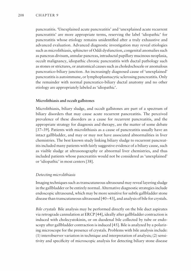
pancreatitis. ‘Unexplained acute pancreatitis’ and ‘unexplained acute recurrentpancreatitis’ are more appropriate terms, reserving the label ‘idiopathic’ forpancreatitis whose etiology remains unidentified after a truly exhaustive andadvanced evaluation. Advanced diagnostic investigation may reveal etiologiessuch as microlithiasis, sphincter of Oddi dysfunction, congenital anomalies suchas pancreas divisum, annular pancreas, intraductal papillary mucinous neoplasia,occult malignancy, idiopathic chronic pancreatitis with ductal pathology suchas stones or strictures, or anatomical causes such as choledochocele or anomalouspancreatico-biliary junction. An increasingly diagnosed cause of ‘unexplained’pancreatitis is autoimmune, or lymphoplasmacytic sclerosing pancreatitis. Onlythe remainder with normal pancreatico-biliary ductal anatomy and no other etiology are appropriately labeled as ‘idiopathic’.
Microlithiasis and occult gallstones
Microlithiasis, biliary sludge, and occult gallstones are part of a spectrum of biliary disorders that may cause acute recurrent pancreatitis. The perceivedprevalence of these disorders as a cause for recurrent pancreatitis, and theappropriate strategy for diagnosis and therapy, are the matter of some debate[37–39]. Patients with microlithiasis as a cause of pancreatitis usually have anintact gallbladder, and may or may not have associated abnormalities in liverchemistries. The best known study linking biliary sludge to recurrent pancreat-itis included many patients with fairly suggestive evidence of a biliary cause, suchas visible sludge at ultrasonography or abnormal liver chemistries, and thusincluded patients whose pancreatitis would not be considered as ‘unexplained’or ‘idiopathic’ in most centers [38].
Detecting microlithiasis
Imaging techniques such as transcutaneous ultrasound may reveal layering sludgein the gallbladder or be entirely normal. Alternative diagnostic strategies includeendoscopic ultrasound, which may be more sensitive for subtle gallbladder stonedisease than transcutaneous ultrasound [40–43], and analysis of bile for crystals.
Bile crystals Bile analysis may be performed directly on the bile duct aspiratesvia retrograde cannulation at ERCP [44], ideally after gallbladder contraction isinduced with cholecystokinin, or on duodenal bile collected by tube or endo-scopy after gallbladder contraction is induced [45]. Bile is analyzed by a polariz-ing microscope for the presence of crystals. Problems with bile analysis include:(1) interobserver variation in technique and interpretation of analysis; (2) sensi-tivity and specificity of microscopic analysis for detecting biliary stone disease
CHAPTER 9208
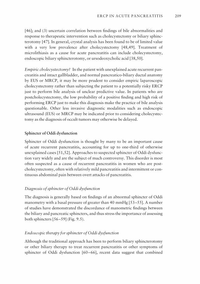
[46]; and (3) uncertain correlation between findings of bile abnormalities andresponse to therapeutic intervention such as cholecystectomy or biliary sphinc-terotomy [47]. In general, crystal analysis has been found to be of limited valuewith a very low prevalence after cholecystectomy [48,49]. Treatment ofmicrolithiasis as a cause for acute pancreatitis can include cholecystectomy,endoscopic biliary sphincterotomy, or ursodeoxycholic acid [38,50].
Empiric cholecystectomy? In the patient with unexplained acute recurrent pan-creatitis and intact gallbladder, and normal pancreatico-biliary ductal anatomyby EUS or MRCP, it may be more prudent to consider empiric laparoscopiccholecystectomy rather than subjecting the patient to a potentially risky ERCPjust to perform bile analysis of unclear predictive value. In patients who arepostcholecystectomy, the low probability of a positive finding and high risk ofperforming ERCP just to make this diagnosis make the practice of bile analysisquestionable. Other less invasive diagnostic modalities such as endoscopicultrasound (EUS) or MRCP may be indicated prior to considering cholecystec-tomy as the diagnosis of occult tumors may otherwise be delayed.
Sphincter of Oddi dysfunction
Sphincter of Oddi dysfunction is thought by many to be an important cause of acute recurrent pancreatitis, accounting for up to one-third of otherwiseunexplained cases [51,52]. Approaches to suspected sphincter of Oddi dysfunc-tion vary widely and are the subject of much controversy. This disorder is mostoften suspected as a cause of recurrent pancreatitis in women who are post-cholecystectomy, often with relatively mild pancreatitis and intermittent or con-tinuous abdominal pain between overt attacks of pancreatitis.
Diagnosis of sphincter of Oddi dysfunction
The diagnosis is generally based on findings of an abnormal sphincter of Oddimanometry with a basal pressure of greater than 40 mmHg [53–55]. A numberof studies have demonstrated the discordance of manometric findings betweenthe biliary and pancreatic sphincters, and thus stress the importance of assessingboth sphincters [56–59] (Fig. 9.5).
Endoscopic therapy for sphincter of Oddi dysfunction
Although the traditional approach has been to perform biliary sphincterotomyor other biliary therapy to treat recurrent pancreatitis or other symptoms ofsphincter of Oddi dysfunction [60–66], recent data suggest that combined
ERCP IN ACUTE PANCREATITIS 209
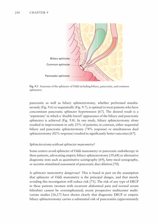
pancreatic as well as biliary sphincterotomy, whether performed simulta-neously (Fig. 9.6) or sequentially (Fig. 9.7), is optimal to treat patients who haveconcomitant pancreatic sphincter hypertension [67]. The desired result is a ‘septotomy’ in which a ‘double-barrel’ appearance of the biliary and pancreaticsphincters is achieved (Fig. 9.8). In one study, biliary sphincterotomy aloneresulted in improvement in only 25% of patients; in contrast, either sequentialbiliary and pancreatic sphincterotomy (78% response) or simultaneous dualsphincterotomy (82% response) resulted in significantly better outcomes [67].
Sphincterotomy without sphincter manometry?
Some centers avoid sphincter of Oddi manometry or pancreatic endotherapy inthese patients, advocating empiric biliary sphincterotomy [39,68] or alternativediagnostic tests such as quantitative scintigraphy [69], fatty-meal sonography,or secretin-stimulated assessment of pancreatic duct dilation [70].
Is sphincter manometry dangerous? This is based in part on the assumptionthat sphincter of Oddi manometry is the principal danger, and that merelyavoiding this investigation will reduce risk [71]. The risk of any type of ERCP in these patients (women with recurrent abdominal pain and normal serumbilirubin) cannot be overemphasized; recent prospective multicenter multi-variate studies [16,17] have shown clearly that diagnostic ERCP or empiric biliary sphincterotomy carries a substantial risk of pancreatitis (approximately
CHAPTER 9210
Common sphincter
Biliary sphincter
Pancreatic sphincter
Fig. 9.5 Anatomy of the sphincter of Oddi including biliary, pancreatic, and commonsphincters.
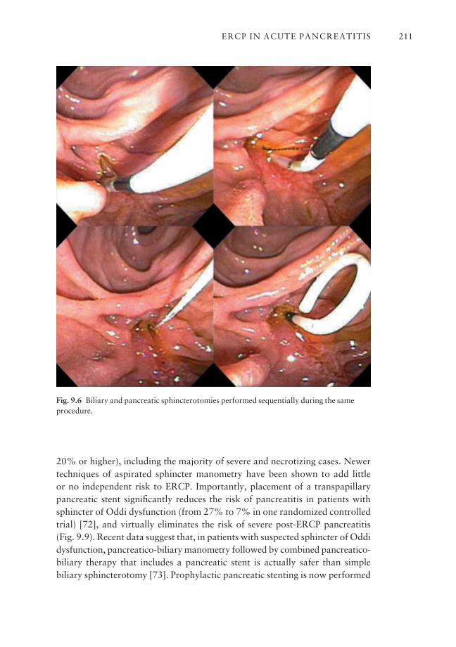
20% or higher), including the majority of severe and necrotizing cases. Newertechniques of aspirated sphincter manometry have been shown to add little or no independent risk to ERCP. Importantly, placement of a transpapillarypancreatic stent significantly reduces the risk of pancreatitis in patients withsphincter of Oddi dysfunction (from 27% to 7% in one randomized controlledtrial) [72], and virtually eliminates the risk of severe post-ERCP pancreatitis(Fig. 9.9). Recent data suggest that, in patients with suspected sphincter of Oddidysfunction, pancreatico-biliary manometry followed by combined pancreatico-biliary therapy that includes a pancreatic stent is actually safer than simple biliary sphincterotomy [73]. Prophylactic pancreatic stenting is now performed
ERCP IN ACUTE PANCREATITIS 211
Fig. 9.6 Biliary and pancreatic sphincterotomies performed sequentially during the sameprocedure.

CHAPTER 9212
Fig. 9.7 Pancreatic manometry and sphincterotomy in a patient unresponsive to previousbiliary sphincterotomy.
Fig. 9.8 Final appearance of pancreatic‘septotomy’ after endoscopic biliary andpancreatic sphincterotomies for sphincterof Oddi dysfunction.

routinely in many centers after pancreatic investigation in these types of patients[18,74,75]. Placement of pancreatic stents can range from technically easy (Fig. 9.10) to very challenging (Fig. 9.11) depending on pancreatic ductalanatomy and endoscopic expertise. Pancreatic stents have potential to causedamage, especially to normal ducts [76–78] (Fig. 9.12), and should be removedwithin 10–14 days from normal ducts. The trend is now to use smaller stents (3 or 4 Fr) compared with traditional larger (5–7 Fr) stents, because they arethought to cause less ductal injury and lower post-ERCP pancreatitis rates [79].
ERCP IN ACUTE PANCREATITIS 213
Cutting wire
Papilloteme
Guidewire
Fig. 9.9 Schematic diagram of pancreatic stent placement to reduce risk of post-ERCPpancreatitis.
Fig. 9.10 Relatively easy pancreatic ductal anatomy for placement of pancreatic stent.
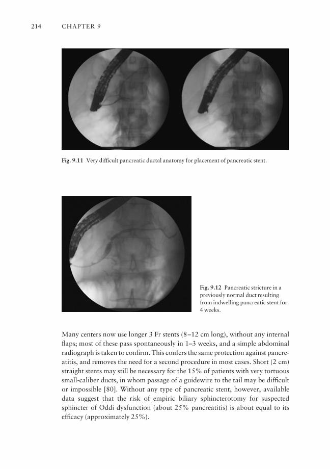
Many centers now use longer 3 Fr stents (8–12 cm long), without any internalflaps; most of these pass spontaneously in 1–3 weeks, and a simple abdominalradiograph is taken to confirm. This confers the same protection against pancre-atitis, and removes the need for a second procedure in most cases. Short (2 cm)straight stents may still be necessary for the 15% of patients with very tortuoussmall-caliber ducts, in whom passage of a guidewire to the tail may be difficultor impossible [80]. Without any type of pancreatic stent, however, availabledata suggest that the risk of empiric biliary sphincterotomy for suspectedsphincter of Oddi dysfunction (about 25% pancreatitis) is about equal to itsefficacy (approximately 25%).
CHAPTER 9214
Fig. 9.11 Very difficult pancreatic ductal anatomy for placement of pancreatic stent.
Fig. 9.12 Pancreatic stricture in apreviously normal duct resultingfrom indwelling pancreatic stent for4 weeks.
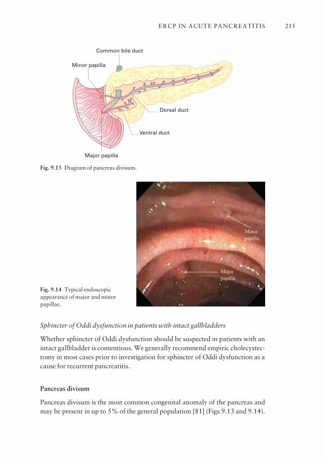
Sphincter of Oddi dysfunction in patients with intact gallbladders
Whether sphincter of Oddi dysfunction should be suspected in patients with anintact gallbladder is contentious. We generally recommend empiric cholecystec-tomy in most cases prior to investigation for sphincter of Oddi dysfunction as acause for recurrent pancreatitis.
Pancreas divisum
Pancreas divisum is the most common congenital anomaly of the pancreas andmay be present in up to 5% of the general population [81] (Figs 9.13 and 9.14).
ERCP IN ACUTE PANCREATITIS 215
Common bile duct
Minor papilla
Major papilla
Dorsal duct
Ventral duct
Fig. 9.13 Diagram of pancreas divisum.
Fig. 9.14 Typical endoscopicappearance of major and minorpapillae.
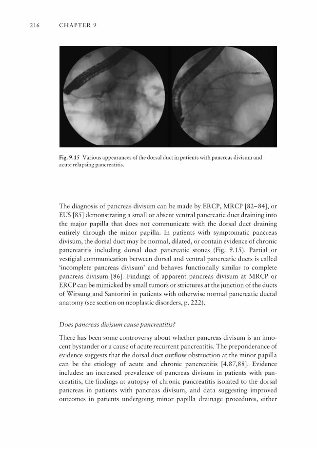
The diagnosis of pancreas divisum can be made by ERCP, MRCP [82–84], orEUS [85] demonstrating a small or absent ventral pancreatic duct draining intothe major papilla that does not communicate with the dorsal duct drainingentirely through the minor papilla. In patients with symptomatic pancreas divisum, the dorsal duct may be normal, dilated, or contain evidence of chronicpancreatitis including dorsal duct pancreatic stones (Fig. 9.15). Partial or vestigial communication between dorsal and ventral pancreatic ducts is called‘incomplete pancreas divisum’ and behaves functionally similar to completepancreas divisum [86]. Findings of apparent pancreas divisum at MRCP orERCP can be mimicked by small tumors or strictures at the junction of the ductsof Wirsung and Santorini in patients with otherwise normal pancreatic ductalanatomy (see section on neoplastic disorders, p. 222).
Does pancreas divisum cause pancreatitis?
There has been some controversy about whether pancreas divisum is an inno-cent bystander or a cause of acute recurrent pancreatitis. The preponderance ofevidence suggests that the dorsal duct outflow obstruction at the minor papillacan be the etiology of acute and chronic pancreatitis [4,87,88]. Evidenceincludes: an increased prevalence of pancreas divisum in patients with pan-creatitis, the findings at autopsy of chronic pancreatitis isolated to the dorsalpancreas in patients with pancreas divisum, and data suggesting improved outcomes in patients undergoing minor papilla drainage procedures, either
CHAPTER 9216
Fig. 9.15 Various appearances of the dorsal duct in patients with pancreas divisum and acute relapsing pancreatitis.
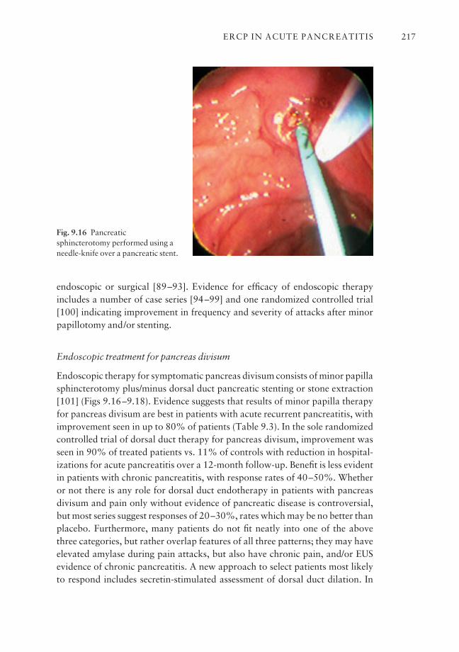
endoscopic or surgical [89–93]. Evidence for efficacy of endoscopic therapyincludes a number of case series [94–99] and one randomized controlled trial[100] indicating improvement in frequency and severity of attacks after minorpapillotomy and/or stenting.
Endoscopic treatment for pancreas divisum
Endoscopic therapy for symptomatic pancreas divisum consists of minor papillasphincterotomy plus/minus dorsal duct pancreatic stenting or stone extraction[101] (Figs 9.16–9.18). Evidence suggests that results of minor papilla therapyfor pancreas divisum are best in patients with acute recurrent pancreatitis, withimprovement seen in up to 80% of patients (Table 9.3). In the sole randomizedcontrolled trial of dorsal duct therapy for pancreas divisum, improvement wasseen in 90% of treated patients vs. 11% of controls with reduction in hospital-izations for acute pancreatitis over a 12-month follow-up. Benefit is less evidentin patients with chronic pancreatitis, with response rates of 40–50%. Whether or not there is any role for dorsal duct endotherapy in patients with pancreasdivisum and pain only without evidence of pancreatic disease is controversial,but most series suggest responses of 20–30%, rates which may be no better thanplacebo. Furthermore, many patients do not fit neatly into one of the abovethree categories, but rather overlap features of all three patterns; they may haveelevated amylase during pain attacks, but also have chronic pain, and/or EUSevidence of chronic pancreatitis. A new approach to select patients most likelyto respond includes secretin-stimulated assessment of dorsal duct dilation. In
ERCP IN ACUTE PANCREATITIS 217
Fig. 9.16 Pancreaticsphincterotomy performed using aneedle-knife over a pancreatic stent.
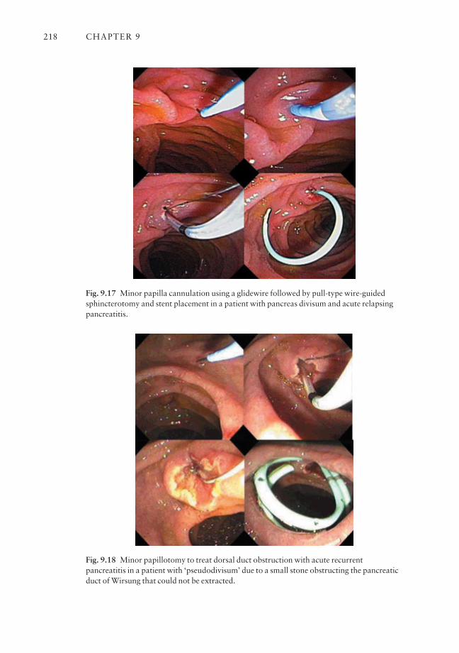
CHAPTER 9218
Fig. 9.17 Minor papilla cannulation using a glidewire followed by pull-type wire-guidedsphincterotomy and stent placement in a patient with pancreas divisum and acute relapsingpancreatitis.
Fig. 9.18 Minor papillotomy to treat dorsal duct obstruction with acute recurrentpancreatitis in a patient with ‘pseudodivisum’ due to a small stone obstructing the pancreaticduct of Wirsung that could not be extracted.
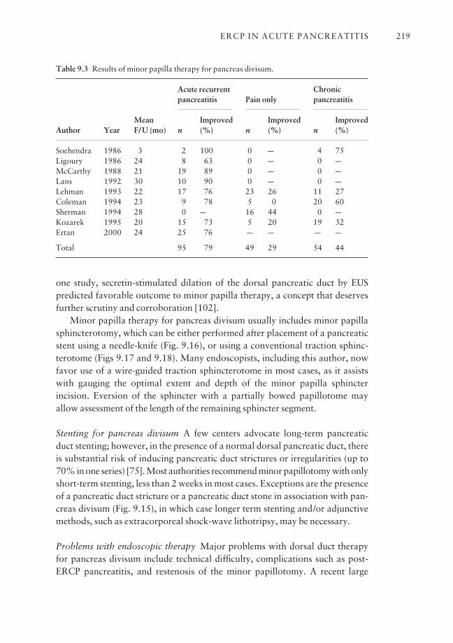
one study, secretin-stimulated dilation of the dorsal pancreatic duct by EUS predicted favorable outcome to minor papilla therapy, a concept that deservesfurther scrutiny and corroboration [102].
Minor papilla therapy for pancreas divisum usually includes minor papillasphincterotomy, which can be either performed after placement of a pancreaticstent using a needle-knife (Fig. 9.16), or using a conventional traction sphinc-terotome (Figs 9.17 and 9.18). Many endoscopists, including this author, nowfavor use of a wire-guided traction sphincterotome in most cases, as it assistswith gauging the optimal extent and depth of the minor papilla sphincter incision. Eversion of the sphincter with a partially bowed papillotome mayallow assessment of the length of the remaining sphincter segment.
Stenting for pancreas divisum A few centers advocate long-term pancreaticduct stenting; however, in the presence of a normal dorsal pancreatic duct, thereis substantial risk of inducing pancreatic duct strictures or irregularities (up to70% in one series) [75]. Most authorities recommend minor papillotomy with onlyshort-term stenting, less than 2 weeks in most cases. Exceptions are the presenceof a pancreatic duct stricture or a pancreatic duct stone in association with pan-creas divisum (Fig. 9.15), in which case longer term stenting and/or adjunctivemethods, such as extracorporeal shock-wave lithotripsy, may be necessary.
Problems with endoscopic therapy Major problems with dorsal duct therapyfor pancreas divisum include technical difficulty, complications such as post-ERCP pancreatitis, and restenosis of the minor papillotomy. A recent large
ERCP IN ACUTE PANCREATITIS 219
Table 9.3 Results of minor papilla therapy for pancreas divisum.
Acute recurrent Chronicpancreatitis Pain only pancreatitis
Mean Improved Improved ImprovedAuthor Year F/U (mo) n (%) n (%) n (%)
Soehendra 1986 3 2 100 0 a 4 75Ligoury 1986 24 8 63 0 a 0 aMcCarthy 1988 21 19 89 0 a 0 aLans 1992 30 10 90 0 a 0 aLehman 1993 22 17 76 23 26 11 27Coleman 1994 23 9 78 5 0 20 60Sherman 1994 28 0 a 16 44 0 aKozarek 1995 20 15 73 5 20 19 32Ertan 2000 24 25 76 a a a a
Total 95 79 49 29 54 44
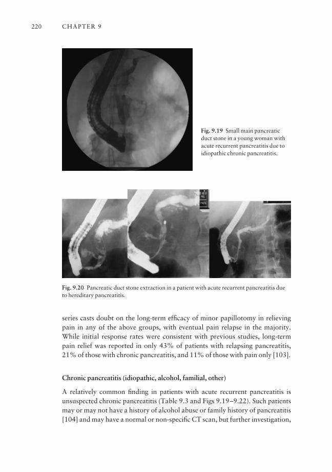
series casts doubt on the long-term efficacy of minor papillotomy in relievingpain in any of the above groups, with eventual pain relapse in the majority.While initial response rates were consistent with previous studies, long-termpain relief was reported in only 43% of patients with relapsing pancreatitis,21% of those with chronic pancreatitis, and 11% of those with pain only [103].
Chronic pancreatitis (idiopathic, alcohol, familial, other)
A relatively common finding in patients with acute recurrent pancreatitis isunsuspected chronic pancreatitis (Table 9.3 and Figs 9.19–9.22). Such patientsmay or may not have a history of alcohol abuse or family history of pancreatitis[104] and may have a normal or non-specific CT scan, but further investigation,
CHAPTER 9220
Fig. 9.19 Small main pancreaticduct stone in a young woman withacute recurrent pancreatitis due toidiopathic chronic pancreatitis.
Fig. 9.20 Pancreatic duct stone extraction in a patient with acute recurrent pancreatitis dueto hereditary pancreatitis.
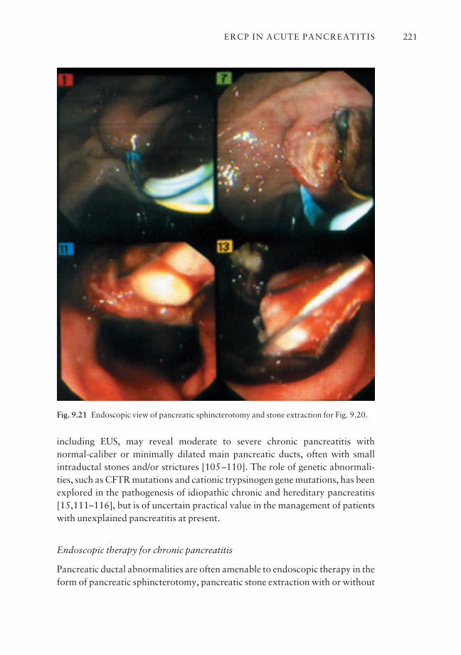
including EUS, may reveal moderate to severe chronic pancreatitis with normal-caliber or minimally dilated main pancreatic ducts, often with smallintraductal stones and/or strictures [105–110]. The role of genetic abnormali-ties, such as CFTR mutations and cationic trypsinogen gene mutations, has beenexplored in the pathogenesis of idiopathic chronic and hereditary pancreatitis[15,111–116], but is of uncertain practical value in the management of patientswith unexplained pancreatitis at present.
Endoscopic therapy for chronic pancreatitis
Pancreatic ductal abnormalities are often amenable to endoscopic therapy in theform of pancreatic sphincterotomy, pancreatic stone extraction with or without
ERCP IN ACUTE PANCREATITIS 221
Fig. 9.21 Endoscopic view of pancreatic sphincterotomy and stone extraction for Fig. 9.20.
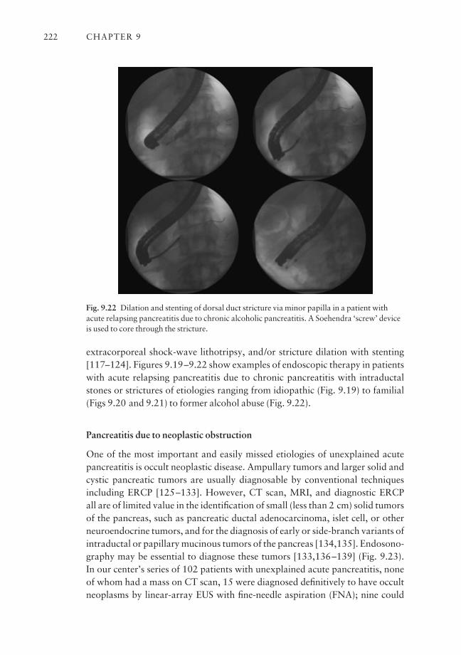
extracorporeal shock-wave lithotripsy, and/or stricture dilation with stenting[117–124]. Figures 9.19–9.22 show examples of endoscopic therapy in patientswith acute relapsing pancreatitis due to chronic pancreatitis with intraductalstones or strictures of etiologies ranging from idiopathic (Fig. 9.19) to familial(Figs 9.20 and 9.21) to former alcohol abuse (Fig. 9.22).
Pancreatitis due to neoplastic obstruction
One of the most important and easily missed etiologies of unexplained acutepancreatitis is occult neoplastic disease. Ampullary tumors and larger solid andcystic pancreatic tumors are usually diagnosable by conventional techniquesincluding ERCP [125–133]. However, CT scan, MRI, and diagnostic ERCP all are of limited value in the identification of small (less than 2 cm) solid tumors of the pancreas, such as pancreatic ductal adenocarcinoma, islet cell, or otherneuroendocrine tumors, and for the diagnosis of early or side-branch variants ofintraductal or papillary mucinous tumors of the pancreas [134,135]. Endosono-graphy may be essential to diagnose these tumors [133,136–139] (Fig. 9.23). In our center’s series of 102 patients with unexplained acute pancreatitis, noneof whom had a mass on CT scan, 15 were diagnosed definitively to have occultneoplasms by linear-array EUS with fine-needle aspiration (FNA); nine could
CHAPTER 9222
Fig. 9.22 Dilation and stenting of dorsal duct stricture via minor papilla in a patient withacute relapsing pancreatitis due to chronic alcoholic pancreatitis. A Soehendra ‘screw’ deviceis used to core through the stricture.
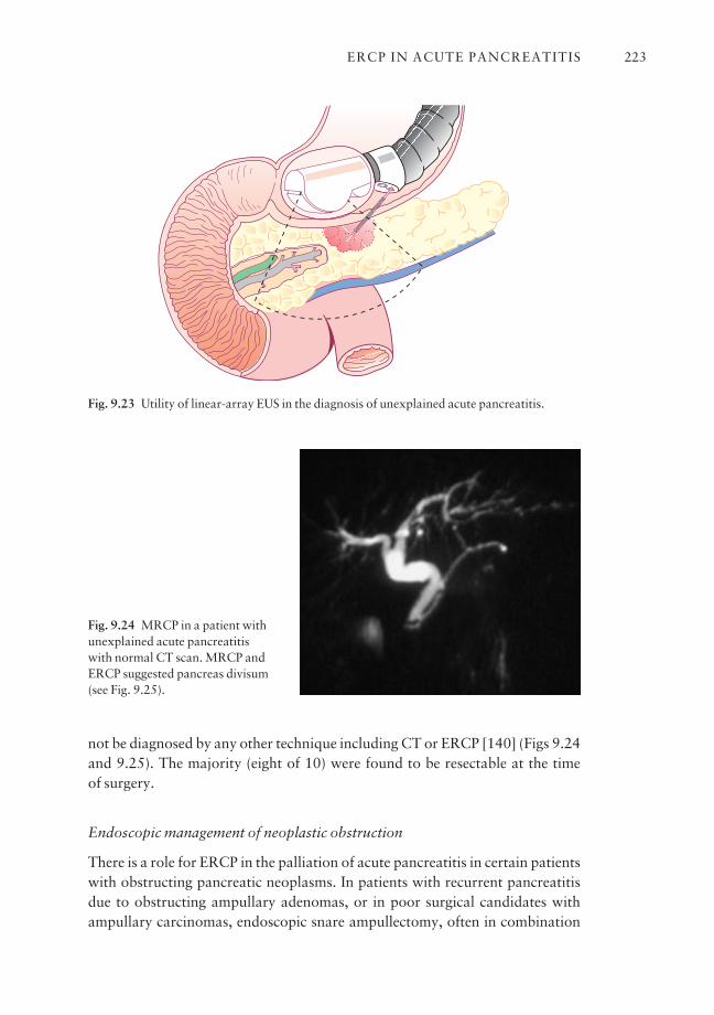
not be diagnosed by any other technique including CT or ERCP [140] (Figs 9.24and 9.25). The majority (eight of 10) were found to be resectable at the time of surgery.
Endoscopic management of neoplastic obstruction
There is a role for ERCP in the palliation of acute pancreatitis in certain patientswith obstructing pancreatic neoplasms. In patients with recurrent pancreatitisdue to obstructing ampullary adenomas, or in poor surgical candidates withampullary carcinomas, endoscopic snare ampullectomy, often in combination
ERCP IN ACUTE PANCREATITIS 223
Fig. 9.23 Utility of linear-array EUS in the diagnosis of unexplained acute pancreatitis.
Fig. 9.24 MRCP in a patient withunexplained acute pancreatitis with normal CT scan. MRCP andERCP suggested pancreas divisum(see Fig. 9.25).

with ablative thermal therapy, such as argon plasma or bipolar coagulation, is areasonable method to achieve palliation or cure of the underlying lesion [141–145]. The technique of ampullectomy increasingly includes pancreatic sphinc-terotomy and placement of a pancreatic stent to reduce the risk of both immediateand relapsing pancreatitis, which can otherwise be substantial [142]. There mayalso be a role for pancreatic sphincterotomy to palliate acute recurrent pancre-atitis by preventing mucin impaction in patients with mucin-secreting tumors(intraductal papillary mucinous tumor, IPMT) who are not surgical candidates.
Stenting for smoldering pancreatitis due to malignancy
Some patients with pancreatic ductal adenocarcinoma or other solid tumorsobstructing the pancreatic duct may present with acute or smoldering pancreat-itis. ERCP in such cases often demonstrates a focal stricture with upstream dila-tion. In such patients, placement of a transpapillary stent through the pancreaticstricture may often interrupt or resolve the pancreatitis, either as a preoperativemaneuver or as definitive palliation [146] (Fig. 9.26). In selected patients withunresectable pancreatic neoplasms and smoldering or acute pancreatitis, self-expanding metallic stents have been placed through pancreatic strictures formore long-term palliation (Fig. 9.27) [147].
Choledochocele
Cystic dilation of the intramural segment of the distal pancreatico-biliary seg-ment is called a choledochocele (type III choledochal cyst). These may range insize from a few millimeters to several centimeters and may herniate in the duo-
CHAPTER 9224
Fig. 9.25 Linear EUS of patient in Fig. 9.24 showing 15 mm mass with FNA positive foradenocarcinoma.
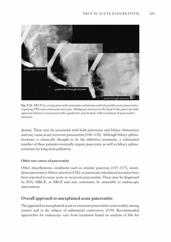
denum. These may be associated with both pancreatic and biliary obstructionand may cause acute recurrent pancreatitis [148–154]. Although biliary sphinc-terotomy is classically thought to be the definitive treatment, a substantial number of these patients eventually require pancreatic as well as biliary sphinc-terotomy for long-term palliation.
Other rare causes of pancreatitis
Other miscellaneous conditions such as annular pancreas [155–157], anom-alous pancreatico-biliary junction [158], or pancreatic intraductal parasites havebeen reported to cause acute or recurrent pancreatitis. These may be diagnosedby EUS, MRCP, or ERCP and may sometimes be amenable to endoscopic intervention.
Overall approach to unexplained acute pancreatitis
The approach to unexplained acute or recurrent pancreatitis varies widely amongcenters and is the subject of substantial controversy [159]. Recommendedapproaches for endoscopy vary from treatment based on analysis of bile for
ERCP IN ACUTE PANCREATITIS 225
Fig. 9.26 ERCP in a young man with metastatic melanoma and intractable acute pancreatitisrequiring TPN and continuous narcotics. Malignant stricture in the head of the pancreas withupstream dilation is traversed with a guidewire and stented, with resolution of pancreatitisand pain.
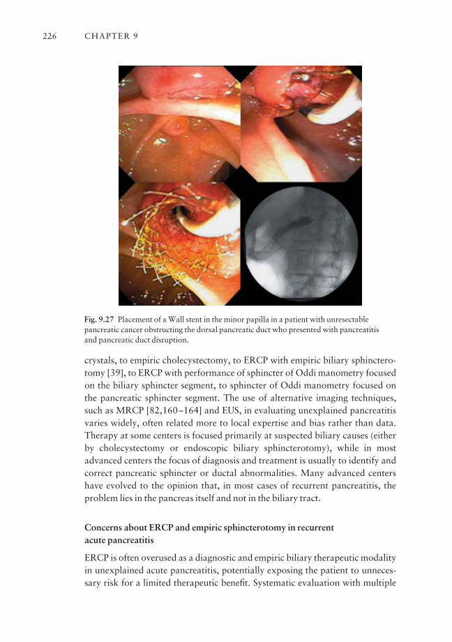
crystals, to empiric cholecystectomy, to ERCP with empiric biliary sphinctero-tomy [39], to ERCP with performance of sphincter of Oddi manometry focusedon the biliary sphincter segment, to sphincter of Oddi manometry focused onthe pancreatic sphincter segment. The use of alternative imaging techniques,such as MRCP [82,160–164] and EUS, in evaluating unexplained pancreatitisvaries widely, often related more to local expertise and bias rather than data.Therapy at some centers is focused primarily at suspected biliary causes (eitherby cholecystectomy or endoscopic biliary sphincterotomy), while in mostadvanced centers the focus of diagnosis and treatment is usually to identify andcorrect pancreatic sphincter or ductal abnormalities. Many advanced centershave evolved to the opinion that, in most cases of recurrent pancreatitis, theproblem lies in the pancreas itself and not in the biliary tract.
Concerns about ERCP and empiric sphincterotomy in recurrent acute pancreatitis
ERCP is often overused as a diagnostic and empiric biliary therapeutic modalityin unexplained acute pancreatitis, potentially exposing the patient to unneces-sary risk for a limited therapeutic benefit. Systematic evaluation with multiple
CHAPTER 9226
Fig. 9.27 Placement of a Wall stent in the minor papilla in a patient with unresectablepancreatic cancer obstructing the dorsal pancreatic duct who presented with pancreatitis and pancreatic duct disruption.
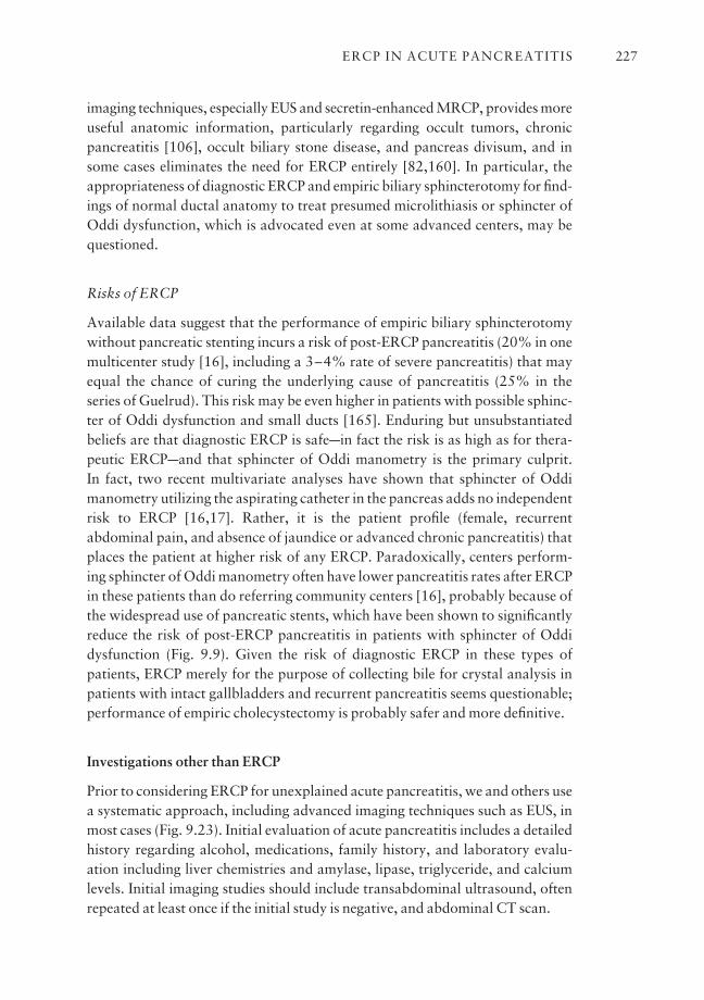
imaging techniques, especially EUS and secretin-enhanced MRCP, provides moreuseful anatomic information, particularly regarding occult tumors, chronicpancreatitis [106], occult biliary stone disease, and pancreas divisum, and insome cases eliminates the need for ERCP entirely [82,160]. In particular, theappropriateness of diagnostic ERCP and empiric biliary sphincterotomy for find-ings of normal ductal anatomy to treat presumed microlithiasis or sphincter ofOddi dysfunction, which is advocated even at some advanced centers, may bequestioned.
Risks of ERCP
Available data suggest that the performance of empiric biliary sphincterotomywithout pancreatic stenting incurs a risk of post-ERCP pancreatitis (20% in onemulticenter study [16], including a 3–4% rate of severe pancreatitis) that mayequal the chance of curing the underlying cause of pancreatitis (25% in theseries of Guelrud). This risk may be even higher in patients with possible sphinc-ter of Oddi dysfunction and small ducts [165]. Enduring but unsubstantiatedbeliefs are that diagnostic ERCP is safeain fact the risk is as high as for thera-peutic ERCPaand that sphincter of Oddi manometry is the primary culprit. In fact, two recent multivariate analyses have shown that sphincter of Oddimanometry utilizing the aspirating catheter in the pancreas adds no independentrisk to ERCP [16,17]. Rather, it is the patient profile (female, recurrent abdominal pain, and absence of jaundice or advanced chronic pancreatitis) thatplaces the patient at higher risk of any ERCP. Paradoxically, centers perform-ing sphincter of Oddi manometry often have lower pancreatitis rates after ERCPin these patients than do referring community centers [16], probably because ofthe widespread use of pancreatic stents, which have been shown to significantlyreduce the risk of post-ERCP pancreatitis in patients with sphincter of Oddi dysfunction (Fig. 9.9). Given the risk of diagnostic ERCP in these types ofpatients, ERCP merely for the purpose of collecting bile for crystal analysis inpatients with intact gallbladders and recurrent pancreatitis seems questionable;performance of empiric cholecystectomy is probably safer and more definitive.
Investigations other than ERCP
Prior to considering ERCP for unexplained acute pancreatitis, we and others usea systematic approach, including advanced imaging techniques such as EUS, inmost cases (Fig. 9.23). Initial evaluation of acute pancreatitis includes a detailedhistory regarding alcohol, medications, family history, and laboratory evalu-ation including liver chemistries and amylase, lipase, triglyceride, and calciumlevels. Initial imaging studies should include transabdominal ultrasound, oftenrepeated at least once if the initial study is negative, and abdominal CT scan.
ERCP IN ACUTE PANCREATITIS 227
MRCP
Magnetic resonance cholangiopancreatography [82,160] is quite useful as it canfairly reliably establish the anatomy of the bile and pancreatic ducts, identifypancreas divisum or pancreatic ductal strictures, diagnose bile duct stones, andimage pancreatic or biliary duct dilation. Secretin administration not onlyimproves imaging of the pancreatic duct, but may also provide functional infor-mation about the presence and severity of outflow obstruction at the level of thesphincter or stricture. The advantages of MRCP include its non-invasivenessand increasingly wide availability. MRCP is contraindicated in patients withpacemakers or cerebral aneurysm clips. The sensitivity and specificity of MRCPfor the detection of small bile duct stones or microlithiasis, for small pancreatictumors, and for pancreas divisum are limited, as MRCP cannot differentiatetrue divisum from ‘pseudodivisum’ due to a small obstructing pancreatic stoneor tumor (Figs 9.24 and 9.25).
EUS
Endoscopic ultrasound is increasingly utilized, and in our opinion is the pro-cedure of choice, for the initial evaluation of unexplained acute pancreatitis[40–43,87,106,108,166] (Fig. 9.23). Linear-array EUS can detect occult biliarytract disease, such as gallstones, biliary sludge, or occult common bile ductstones. It is the method of choice for diagnosing occult pancreatic tumorsincluding neuroendocrine tumors and IPMT as well as small adenocarcinomas,with the advantage of tissue diagnosis via fine-needle aspirate. EUS is probablythe most sensitive test for chronic pancreatitis and can identify intraductal pan-creatic stones that may be missed by CT or even ERCP [105,106,108]. EUS ispotentially accurate in the diagnosis of pancreas divisum and other congenitalanomalies. Finally, EUS is useful for the identification of rare extrapancreaticdisorders that may mimic acute pancreatitis with abdominal pain and mildhyperamylasemia, such as intravascular tumors. EUS carries minimal risk whencompared to ERCP. A completely normal high-quality EUS essentially limits the diagnostic yield of ERCP to sphincter of Oddi dysfunction or microlithiasis.Limitations of EUS are primarily that there are relatively few endoscopiststrained in its use.
Recommended approach to ERCP for acute recurrent pancreatitis
ERCP for acute recurrent pancreatitis at our center, and many others, is reservedfor directed therapy if alternative advanced investigations reveal an anatomiccause, or for sphincter of Oddi manometry if the anatomy appears to be normal.
CHAPTER 9228
If the pancreatico-biliary anatomy is normal by EUS or MRCP, and the gallbladderis intact, we generally recommend empiric cholecystectomy in reasonable sur-gical candidates. In patients with apparently normal pancreatico-biliary anatomywho are postcholecystectomy or who are poor surgical candidates for cholecys-tectomy, we proceed with ERCP with sphincter of Oddi manometry with theprimary goal of assessing for pancreatic sphincter hypertension, as increasing dataand experience suggest that the response to dual sphincterotomy (pancreaticplus biliary) is substantially better than that for biliary sphincterotomy alone.Empiric biliary sphincterotomy may be a reasonable but unproven treatment inpatients with normal sphincter of Oddi manometry who are suspected ofmicrolithiasis. However, we recommend placement of a short-term pancreaticstent to reduce the risk of post-ERCP pancreatitis in this high-risk subgroup ofpatients with normal pancreatico-biliary ductal anatomy and recurrent pancre-atitis (Figs 9.9–9.12).
Because of the risk of ERCP, most authors recommend performance ofERCP only after two attacks of unexplained pancreatitis, unless the first is severe[125,167]. This dilemma can be circumvented by performing EUS after anyunexplained episode of pancreatitis. ERCP is either unnecessary or postponeduntil a second attack occurs in the majority of cases.
Final diagnosis in recurrent acute pancreatitis after extensive investigation
Final diagnoses in series of unexplained acute pancreatitis are highly variable,depending on the patient population, number of patients with intact gallbladders,methods used to evaluate the patients, and thoroughness of evaluation [19,52,168].
Our experience
In most series, less than 20% of patients remain ‘idiopathic’ after extensiveinvestigation. Results of endoscopic and other therapies are also variabledepending on those factors and types of therapy performed. In our series of 102patients with unexplained acute pancreatitis over a 5-year period, only 8% ofpatients remained truly ‘idiopathic’ after extensive investigation. Diagnosis wasmade uniquely by ERCP in 66%, uniquely by EUS in 21%, uniquely by MRCPin 0%, and uniquely at surgery in 6% [140]. Diagnoses were sphincter of Oddidysfunction in 28% (mostly treated with combined pancreatic and biliarysphincterotomy), anatomic causes in 23% (including primarily idiopathicchronic pancreatitis with endoscopically treatable findings such as main pancre-atic duct stone or stricture in the majority), and pancreas divisum in 16%.Occult biliary stone disease was found in only 6%, usually gallbladder diseasediagnosed by EUS and thus amenable to cholecystectomy.
ERCP IN ACUTE PANCREATITIS 229

Occult neoplasms Of great importance was that occult neoplasms were foundin 15% of our patients and included six adenocarcinomas, six IPMT of the pan-creas, one islet cell tumor, and two ampullary tumors. All had CT scans thatwere normal or showed non-specific pancreatic duct dilation, and nine out of 15 of these tumors were diagnosable (i.e. with positive cytology) only by linear-array EUS and not by ERCP. Eight of 10 of these patients who were surgicallyexplored were successfully resected.
Endoscopic treatment and results Endoscopic therapy was performed in 76%of all patients, but consisted of biliary sphincterotomy alone in only 18, with theremaining 59 patients requiring advanced pancreatic endotherapeutic techniquesincluding pancreatic sphincterotomy, pancreatic stenting, pancreateutic stoneextraction with or without extracorporeal shock-wave lithotripsy, endoscopicampullectomy including a pancreatic sphincterotomy and stent, transpapillaryand/or transmural pseudocyst drainage [169–171], etc. Overall long-term out-comes were good with 88% long-term improvement after endoscopic therapy,similar in all major diagnostic groups. The impression from our data was thatEUS is the most appropriate first test for unexplained pancreatitis, and thatendoscopic diagnosis and therapy must be focused primarily on the pancreasitself, with a limited role for purely biliary diagnosis and treatment. The ultimateinference is that unexplained acute pancreatitis is best evaluated and treated atadvanced centers with the capability to perform advanced EUS and pancreaticERCP techniques, as well as complex pancreatico-biliary surgery. Standarddiagnostic and therapeutic ERCP for acute recurrent pancreatitis using con-ventional biliary techniques has limited benefit and significant risk, and shouldprobably be avoided.
Outstanding issues and future trends
There are many issues that demand further clinical investigation, and furthertechniques that need to be developed with respect to ERCP in acute pancreatitis.A substantial body of prospective data exists only for ERCP in acute biliary pan-creatitis. There is much controversy and a paucity of data regarding endoscopictherapy in other settings, such as pancreas divisum, and especially the role ofpancreatic sphincterotomy in the treatment of sphincter of Oddi dysfunction.Future studies will hopefully clarify these murky areas. The importance of pancreatic duct disruption and the need for endoscopic therapy in pancreatic necrosis require further evaluation. The evolution of strategies to evaluate andtreat acute and relapsing pancreatitis will increasingly emphasize diagnosis and the prediction of need for endoscopic therapy using less invasive imagingtechniques such as EUS and MRCP. Secretin-stimulated EUS and MRCP will
CHAPTER 9230
play an increasingly important role in diagnosing the cause of pancreatitis, andin assessing the functional significance of duct obstruction. ERCP will hopefullythen be relegated to a purely therapeutic modality in patients who are highlylikely to benefit from directed endoscopic therapy. Further development of pancreatic stent technology should allow increased safety. Collaboration withlaparoscopic surgery and other disciplines will open up new areas for minimallyinvasive treatment of pancreatic disease. Finally, new techniques to achievesphincter ablation and stricture dilation may borrow from existing techno-logy in minimally invasive surgery and vascular interventional radiology andcardiology.
References
1 Gorelick F. Acute pancreatitis. In: Yamada T, ed. Textbook of Gastroenterology, 2nd edn.Philadelphia: Lippincott 1995, 2064–5.
2 Sakorafas GH, Tsiotou AG. Etiology and pathogenesis of acute pancreatitis: current concepts.J Clin Gastroenterol 2000; 30: 343–56.
3 Steinberg W, Tenner S. Acute pancreatitis. N Engl J Med 1994; 330: 1198–2100.4 Bank S, Indaram A. Causes of acute and recurrent pancreatitis. Clinical considerations and
clues to diagnosis. Gastroenterol Clin North Am 1999; 28: 571–89.5 Frakes JT. Biliary pancreatitis: a review emphasizing appropriate endoscopic intervention.
J Clin Gastroenterol 1999; 28 (2): 97–109.6 Banks PA. Practice guidelines in acute pancreatitis. Am J Gastroenterol 1997; 92: 377–86.7 Balthazar E, Robinson D, Megibow A et al. Acute pancreatitis: value of CT in establishing a
prognosis. Radiology 1990; 174: 331–6.8 Bradley EL III. A clinically based classification system for acute pancreatitis. Arch Surg 1993;
128: 586–90.9 Baillargeon JD, Orav J, Ramagopal V, Tenner SM, Banks PA. Hemoconcentration as an early
risk factor for necrotizing pancreatitis. Am J Gastroenterol 1998; 93: 2130–4.10 Kelly TR, Wagner DS. Gallstone pancreatitis: a prospective randomized trial of the timing of
surgery. Surgery 1988; 104: 600–4.11 Neoptolemos JP, Carr-Locke DL, London NJ, Bailey IA, James D, Fossard DP. Controlled trial
of urgent endoscopic retrograde cholangiopancreatography and endoscopic sphincterotomyversus conservative treatment for acute pancreatitis due to gallstones. Lancet 1988; II:979–83.
12 Fan S-T, Lai ECS, Mok FPT, Lo C-M, Zheng S-S, Wong T. Early treatment of acute biliary pan-creatitis by endoscopic papillotomy. N Engl J Med 1993; 328: 228–32.
13 Nowak A, Sowakowska-Dulawa E, Marek T, Rybicka J. Final results of the prospective, ran-domized, controlled study on endoscopic sphincterotomy versus conventional management inacute biliary pancreatitis [Abstract]. Gastroenterology 1995; 108 (Suppl.): A380.
14 Folsch UR, Nitsche R, Ludtke R, Hilgers RA, Creutzfeldt W. Early ERCP and papillotomycompared with conservative treatment for acute biliary pancreatitis. N Engl J Med 1997; 336:237–42.
15 Choudari CP, Lehman GA, Sherman S. Pancreatitis and cystic fibrosis gene mutations. Gastro-enterol Clin North Am 1999; 28: 543–9.
16 Freeman MF, Nelson DB, Sherman S et al. Complications of endoscopic biliary sphinctero-tomy. N Engl J Med 1996; 335: 909–18.
17 Freeman ML, DiSario JA, Nelson DB et al. Risk factors for post-ERCP pancreatitis: a prospec-tive, multicenter study. Gastrointest Endosc 2001; 54: 425–34.
ERCP IN ACUTE PANCREATITIS 231
18 Sherman S, Lehman GA. Endoscopic therapy of pancreatic disease. Gastroenterologist 1997;5: 262–77.
19 Tarnasky PR, Hawes RH. Endoscopic diagnosis and therapy of unexplained (idiopathic) acutepancreatitis. Gastrointest Endosc Clin North Am 1998; 8: 13–37.
20 Escourrou J, Cordova JA, Lazorthes F et al. Early and late complications after endoscopicsphincterotomy for biliary lithiasis with and without the gallbladder ‘in situ’. Gut 1984; 25:598–602.
21 May GR, Shaffer EH. Should elective endoscopic sphincterotomy replace cholecystectomy forthe treatment of high-risk patients with gallstone pancreatitis? J Clin Gastroenterol 1991; 13:125–8.
22 Siegel JH, Veerappan A, Cohen SA, Kasmin FE. Endoscopic sphincterotomy for biliary pancre-atitis: an alternative to cholecystectomy in high-risk patients. Gastrointest Endosc 1994; 40:573–5.
23 Uomo G, Manes G, Laccetti M, Cavallera A, Rabitti PG. Endoscopic sphincterotomy andrecurrence of acute pancreatitis in gallstone patients considered unfit for surgery. Pancreas1997; 14 (1): 28–31.
24 Welbourn CR, Beckly DE, Eyre-Brook IA. Endoscopic sphincterotomy without cholecystec-tomy for gallstone pancreatitis. Gut 1995; 37: 119–20.
25 Hammarstrom LE, Stridbeck H, Ihse I. Effect of endoscopic sphincterotomy and intervalcholecystectomy on late outcome after gallstone pancreatitis. Br J Surg 1988; 85: 333–6.
26 Hill J, Martin DF, Tweedle DE. Risks of leaving the gallbladder in situ after endoscopic sphinc-terotomy for bile duct stones. Br J Surg 1991; 78: 554–7.
27 Kozarek RA. Endoscopic therapy of complete and partial pancreatic duct disruptions.Gastrointest Endosc Clin N Am 1998; 8: 39–53.
28 Kozarek RA, Ball TJ, Patterson DJ et al. Endoscopic transpapillary therapy for disrupted pancreatic duct and peripancreatic fluid collections. Gastroenterology 1991; 100: 1362–70.
29 Traverso LW, Newman RM, Kozarek RA. Pancreatic ductal disruptions leading to pancreaticfistula, pancreatic ascites or pleural effusions. In: Cameron JL, ed. Current Surgical Therapy,6th edn. St. Louis: Mosby 1988, 510–4.
30 Kozarek RA, Attia FM, Traverso LW et al. Pancreatic duct leak in necrotizing pancreatitis:role of diagnostic and therapeutic ERCP as part of a multidisciplinary approach (Abstract).Gastrointest Endosc 2000, 51: AB138.
31 Jacob L, Geenen JE, Catalano MF, Geenen DJ. Prevention of pancreatitis in patients with idio-pathic recurrent pancreatitis: a prospective nonblinded randomized study using endoscopicstents. Endoscopy 2001; 33: 559–62.
32 KaiKaus RM, Jacob L, Geenen JE et al. ‘Smoldering pancreatitis’: rapid resolution with pancreatic duct stent therapy [Abstract]. Gastrointest Endosc 1995; 41 (4): 425.
33 Tenner SM, Steinberg WM. Drug-induced acute pancreatitis. In: Berger HG, Warshaw AL,Russell RCG, Buchler M, Carr-Locke DL, Neoptolemos JP, Sarr MG, eds. The Pancreas.Oxford: Blackwell Science 1998, 331–42.
34 Runzi M, Layer P. Drug-associated pancreatitis. Pancreas 1996; 13: 100–9.35 Underwood TW, Frye CB. Drug-induced pancreatitis. Clin Pharm 1993; 12: 440–8.36 Toskes PP. Hyperlipidemic pancreatitis. Gastroenterol Clin North Am 1990; 19: 783–91.37 Hernandez CA, Lerch MM. Sphincter stenosis and gallstone migration through the biliary
tract. Lancet 1993; 341: 1371–3.38 Lee SP, Nichols JF, Park HZ. Biliary sludge as a cause of acute pancreatitis. N Engl J Med 1992;
326: 589–93.39 Somogyi L, Martin SP, Venkatesan T, Ulrich CD. Recurrent acute pancreatitis: an algorithmic
approach to identification and elimination of inciting factors. Gastroenterology 2001; 120:708–17.
40 Dahan P, Andant C, Levy P et al. Prospective evaluation of endoscopic ultrasonography andmicroscopic examination of duodenal bile in the diagnosis of cholecystolithiasis in 45 patientswith normal conventional ultrasonography. Gut 1996; 38: 277–81.
CHAPTER 9232

41 Dill JE, Hill S, Callis J et al. Combined endoscopic ultrasound and stimulated biliary drainagein cholecystitis and microlithiasisadiagnosis and outcomes. Endoscopy 1995; 27: 424–7.
42 Liu CL, Lo CM, Chan JKF et al. EUS for detection of occult cholelithiasis in patients with idio-pathic pancreatitis. Gastrointest Endosc 2000; 51: 28–32.
43 Tandon M, Topazian M. Endoscopic ultrasound in idiopathic acute pancreatitis. Am JGastroenterol 2001; 96: 705–9.
44 Geenen JE, Nash JA. The role of sphincter of Oddi manometry and biliary microscopy in evaluating idiopathic recurrent pancreatitis. Endoscopy 1998; 30 (Suppl. 1): 237–41.
45 Neoptolemos JP, Davidson BR, Winder AF, Vallance D. Role of duodenal bile crystal analysisin the investigation of ‘idiopathic’ pancreatitis. Br J Surg 1988; 75: 450–3.
46 Marks JW, Bonorris G. Intermittency of cholesterol crystals in duodenal bile from gallstonepatients. Gastroenterology 1984; 87: 622–7.
47 Rubin M, Pakula R, Konikoff FM. Microstructural analysis of bile: relevance to cholesterolgallstone pathogenesis. Histol Histopathol 2000; 15: 761–70.
48 Kaw M, Brodmerkel GJ. ERCP, biliary crystal analysis, and sphincter of Oddi manometry inidiopathic recurrent pancreatitis. Gastrointest Endosc 2002; 55: 157–62.
49 Quallich LG, Stern MA, Rich M, Chey WD, Barnett JL, Elta GH. Bile duct crystals do not con-tribute to sphincter of Oddi dysfunction. Gastrointest Endosc 2002; 55: 163–6.
50 Ros E, Navarro S, Bru C, Garcia-Puges A, Valderrama R. Occult microlithiasis in ‘idiopathic’acute pancreatitis: prevention of relapses by cholecystectomy or ursodeoxycholic acid therapy.Gastroenterology 1991; 101: 1701–9.
51 Lehman GA, Sherman S. Sphincter of Oddi dysfunction. Int J Pancreatol 1996; 20: 11–25.52 Levy MJ, Geenen JE. Idiopathic acute recurrent pancreatitis. Am J Gastroenterol 2001; 96 (9):
2540–55.53 Geenen JE, Hogan WJ, Dodds WJ et al. The efficacy of endoscopic sphincterotomy after chole-
cystectomy in patients with suspected sphincter of Oddi dysfunction. N Engl J Med 1989; 320:82–7.
54 Hogan WJ, Sherman S, Pasricha P, Carr-Locke D. Sphincter of Oddi manometry. GastrointestEndosc 1997; 45: 342–8.
55 Kuo W-H, Pasricha P, Kalloo A. The role of sphincter of Oddi manometry in the diagnosis andtherapy of pancreatic disease. Gastrointest Endosc Clin North Am 1998; 8: 79–85.
56 Eversman D, Fogel EL, Rusche M, Sherman S, Lehman G. Frequency of abnormal pancreaticand biliary sphincter manometry compared with clinical suspicion of sphincter of Oddi dys-function. Gastrointest Endosc 1999; 50: 637–41.
57 Funch-Jensen P, Kruse A. Manometric activity of the pancreatic duct sphincter in patients withtotal bile duct sphincterotomy for sphincter of Oddi dyskinesia. Scand J Gastroenterol 1987;22: 1067–70.
58 Sherman S, Troiano FP, Hawes RH, O’Connor KW, Lehman GA. Frequency of abnormalsphincter of Oddi manometry compared with the clinical suspicion of sphincter of Oddi dysfunction. Am J Gastroenterol 1991; 86: 586–9.
59 Silverman WB, Ruffolo TA, Sherman S et al. Correlation of basal sphincter pressures measuredfrom both the bile duct and pancreatic duct in patients with suspected sphincter of Oddi dysfunction. Gastrointest Endosc 1992; 38: 440–3.
60 Catalano MF, Sivak MV, Falk GW et al. Idiopathic pancreatitis (IP): diagnostic role of sphinc-ter of Oddi manometry (SOM) and response to endoscopic sphincterotomy (ES). GastrointestEndosc 1993; 39: 310A.
61 Goff JS. Common bile duct sphincter of Oddi stenting in patients with suspected sphincter dysfunction. Am J Gastroenterol 1995; 90: 586–9.
62 Neoptolemos JP, Bailey IS, Carr-Locke DL. Sphincter of Oddi dysfunction: results of treatmentby endoscopic sphincterotomy. Br J Surg 1988; 75: 454–9.
63 Rolny P. Endoscopic bile duct stent placement as a predictor of outcome following endoscopicsphincterotomy in patients with suspected sphincter of Oddi dysfunction. Eur J GastroenterolHepatol 1997; 9: 467–71.
64 Toouli J, Di Francesco V, Saccone G et al. Division of the sphincter of Oddi for treatment ofdysfunction associated with recurrent pancreatitis. Br J Surg 1996; 83: 1205–10.
ERCP IN ACUTE PANCREATITIS 233
65 Toouli J, Roberts-Thomson IC, Dent J, Lee J. Sphincter of Oddi motility disorders in patientswith idiopathic recurrent pancreatitis. Br J Surg 1985; 72: 859–63.
66 Wehrmann T, Seifert H, Seipp M et al. Endoscopic injection of botulinum toxin for biliarysphincter of Oddi dysfunction. Endoscopy 1998; 30: 702–7.
67 Guelrud M, Plaz J, Mendoza S, Beker B, Rojas O, Rossiter G. Endoscopic treatment in Type IIpancreatic sphincter dysfunction (Abstract). Gastrointest Endosc 1995; 52: 398A.
68 Testoni PA, Caporuscio S, Bagnolo F, Lella F. Idiopathic recurrent pancreatitis: long-termresults after ERCP, endoscopic sphincterotomy, or ursodeoxycholic acid treatment. Am JGastroenterol 2000; 95: 1702–7.
69 Kalloo AN, Pasricha PJ. Therapy of sphincter of Oddi dysfunction. Gastrointest Endosc ClinNorth Am 1996; 6: 117–25.
70 Di Francesco V, Brunori MP, Rigo L et al. Comparison of ultrasound-secretin test and sphincter of Oddi manometry in patients with recurrent acute pancreatitis. Dig Dis Sci 1999;44: 336–40.
71 Cotton PB, Lehman G, Vennes J et al. Endoscopic sphincterotomy, complications and theirmanagement: an attempt at consensus. Gastrointest Endosc 1991; 37: 383–93.
72 Tarnasky PR, Palesch YY, Cunningham JT, Mauldin P, Cotton P, Hawes R. Pancreatic stent-ing prevents pancreatitis after biliary sphincterotomy in patients with sphincter of Oddi dysfunction. Gastroenterology 1998; 115: 1518–24.
73 Sherman S, Eversman D, Fogel E, Gottlieb K, Earle D. Sphincter of Oddi dysfunction (SOD):needle-knife pancreaticobiliary sphincterotomy over pancreatic stent (NKOPS) has a lowerpost-procedure pancreatitis rate than pull-type biliary sphincterotomy (BES). GastrointestEndosc 1997; 45: 148A.
74 Shakoor T, Hogan WJ, Geenen JE. Efficacy of nasopancreatic catheter in the prevention ofpost-ERCP pancreatitis: a prospective randomized controlled trial. Gastrointest Endosc 1992;38: 251A.
75 Freeman ML. Role of pancreatic stents in prevention of post-ERCP pancreatitis. JOP 2004; 5:322–7.
76 Kozarek RA. Pancreatic stents can induce ductal changes consistent with chronic pancreatitis.Gastrointest Endosc 1990; 36: 93–5.
77 Siegel JH, Veerappan A. Endoscopic management of pancreatic disorders: potential risks ofpancreatic prosthesis. Endoscopy 1991; 23: 177–80.
78 Smith MT, Sherman S, Ikenberry SO, Hawes RH, Lehman GA. Alternations in pancreatic duc-tal morphology following polyethylene pancreatic stent therapy. Gastrointest Endosc 1996;44: 268–75.
79 Rashdan A, Fogel EL, McHenry L Jr, Sherman S, Temkit M, Lehman GA. Improved stent char-acteristics for prophylaxis of post-ERCP pancreatitis. Clin Gastroenterol Hepatol 2004; 2:322–9.
80 Freeman ML, Overby C, Qi D. Pancreatic stent insertion: consequences of failure and results ofa modified technique to maximize success. Gastrointest Endosc 2004; 59: 8–14.
81 Hill ID, Lebenthal E. Congenital abnormalities of the exocrine pancreas. In: Go VLW, ed. ThePancreas: Biology, Pathobiology and Disease. New York: Raven 1993, 1029–40.
82 Barish MA, Yucel EK, Ferrucci J. Magnetic resonance cholangiopancreatography. N Engl JMed 1999; 341: 258–64.
83 Bret PM, Reinhold C, Taourel P et al. Pancreas divisum: evaluation with MR cholangiopan-creatography. Radiology 1996; 199: 99–103.
84 Lai R, Freeman ML, Cass OW, Mallery S. Accurate diagnosis of pancreas divisum by linear-array endoscopic ultrasonography. Endoscopy 2004; 36: 705–9.
85 Bhutani MS, Hoffman BJ, Hawes RH. Diagnosis of pancreas divisum by endoscopic ultra-sonography. Endoscopy 1999; 31: 167–9.
86 Jacob L, Geenen JE, Catalano MF et al. Clinical presentation and short-term outcome of endo-scopic therapy of patients with symptomatic and incomplete pancreas divisum. GastrointestEndosc 1999; 49: 53–7.
87 Bernard JP, Sahel J, Giovannini M, Sarles H. Pancreas divisum is a probable cause of acute pan-creatitis: a report of 137 cases. Pancreas 1990; 5: 248–54.
CHAPTER 9234
88 Warshaw AL. Pancreas divisum and pancreatitis. In: Beger HG, Warshaw AL, Russel RCG,Buchler M, Carr-Locke DL, Neoptolemos JP, Sarr MG, eds. The Pancreas. Oxford: BlackwellScience 1998, 364–374.
89 Bradley EL III, Stephan RN. Accessory duct sphincteroplasty is preferred for long-term preven-tion of recurrent acute pancreatitis in patients with pancreas divisum. J Am Coll Surg 1996;183: 65–70.
90 Brenner P, Duncombe V, Ham JM. Pancreatitis and pancreas divisum: etiological and surgicalconsiderations. Aust N Z J Surg 1990; 60: 899–903.
91 Keith RG, Shapero TF, Saibil FG. Dorsal duct sphincterotomy is effective long-term treatmentof acute pancreatitis associated with pancreas divisum. Surgery 1989; 106: 660–7.
92 Lehman GA, Sherman S. Diagnosis and therapy of pancreas divisum. Gastrointest Endosc ClinNorth Am 1998; 8: 55–77.
93 Richter JM, Schapiro RH, Mulley AG, Warshaw AL. Association of pancreas divisum andpancreatitis, and its treatment by sphincteroplasty of the accessory ampulla. Gastroenterology1981; 81: 1104–10.
94 Coleman SD, Eisen GM, Troughton AB et al. Endoscopic treatment in pancreas divisum. Am JGastroenterol 1994; 89: 1152–5.
95 Cotton PB. Congenital anomaly of pancreas divisum as a cause of obstructive pain and pancre-atitis. Gut 1980; 21: 105–14.
96 Ertan A. Long-term results after endoscopic pancreatic stent placement without pancreaticpapillotomy in acute recurrent pancreatitis due to pancreas divisum. Gastrointest Endosc2000; 52: 9–14.
97 Lehman GA, Sherman S, Nisi R, Hawes RH. Pancreas divisum: results of minor papilla sphinc-terotomy. Gastrointest Endosc 1993; 39: 1–8.
98 Ligoury C, Lefebvre JF, Canard JM et al. Pancreas divisum: therapeutic results in 12 patients.Gastrointest Endosc 1986; 10: 530S.
99 Soehendra N, Kempeneers I, Nam VC et al. Endoscopic dilation and papillotomy of the access-ory papilla and internal drainage in pancreas divisum. Endoscopy 1986; 18: 129–32.
100 Lans JI, Geenen JE, Johanson JF, Hogan WJ. Endoscopic therapy in patients with pancreasdivisum and acute pancreatitis: a prospective, randomized, controlled clinical trial. Gastro-intest Endosc 1992; 38: 430–4.
101 Kozarek RA, Ball TJ, Patterson DJ et al. Endoscopic approaches to pancreas divisum. Dig DisSci 1995; 40: 1974–81.
102 Catalano MF, Rosenblatt ML, Geenen JE, Hogan WJ. Pancreatic endotherapy of pancreasdivisum; response based on clinical presentation and results of secretin-stimulated endoscopicultrasound (Abstract). Gastrointest Endosc 2001, 53: AB133.
103 Gerke H, Byrne MF, Stiffler HL et al. Outcome of endoscopic minor papillotomy in patientswith symptomatic pancreas divisum. JOP 2004; 5: 122–31.
104 Perrault J. Hereditary pancreatitis: historical perspectives. Med Clin North Am 2000; 84:519–29.
105 Buscail L, Escourrou J, Moreau J et al. Endoscopic ultrasonography in chronic pancreatitis: a comparative prospective study with conventional ultrasonography, computed tomographyand ERCP. Pancreas 1995; 10: 251–7.
106 Catalano MF, Lahoti S, Geenen JE, Hogan WJ. Prospective evaluation of endoscopic ultra-sonography, endoscopic retrograde pancreatography, and secretin test in the diagnosis ofchronic pancreatitis. Gastrointest Endosc 1998; 48: 11–7.
107 Forsmark CE. The diagnosis of chronic pancreatitis. Gastrointest Endosc 2000; 52: 293–8.108 Sahai AV, Zimmerman M, Aabakken L et al. Prospective assessment of the ability of endo-
scopic ultrasound to diagnose, exclude or establish the severity of chronic pancreatitis foundby endoscopic retrograde pancreatography. Gastrointest Endosc 1998; 48: 18–25.
109 Steer ML, Waxman I, Freedman S. Chronic pancreatitis. N Engl J Med 1995; 332: 1482–90.110 Wiersema MJ, Hawes RH, Lehman GA, Kochman ML, Sherman S, Kipecky KK. Prospective
evaluation of endoscopic ultrasonography and endoscopic retrograde cholangiopancreato-graphy in patients with chronic abdominal pain of suspected pancreatic origin. Endoscopy1993; 25: 555–64.
ERCP IN ACUTE PANCREATITIS 235
111 Cohn JA, Bornstein JD, Jowell PS. Cystic fibrosis mutations and genetic predisposition to idio-pathic chronic pancreatitis. Med Clin North Am 2000; 84: 621–31.
112 Cohn JA, Friedman KJ, Noone PG, Knowles MR, Silverman LM, Jowell PS. Relation betweenmutations of the cystic fibrosis gene and idiopathic pancreatitis. N Engl J Med 1998; 339:653–8.
113 Gorry MC, Gabbaizedeh D, Furey W et al. Mutations in the cationic trypsinogen gene areassociated with recurrent acute and chronic pancreatitis. Gastroenterology 1997; 113:1063–8.
114 Sharer N, Schwarz M, Malone G et al. Mutations of the cystic fibrosis gene in patients withchronic pancreatitis. N Engl J Med 1989; 339: 645–52.
115 Whitcomb DC, Gorry MC, Preston RA et al. Hereditary pancreatitis is caused by a mutation inthe cationic trypsinogen gene. Nat Genet 1996; 14: 141–5.
116 Whitcomb DC, Preston RA, Aston CE et al. A gene for hereditary pancreatitis maps to chro-mosome 7q35. Gastroenterology 1996; 110: 1975–80.
117 Binmoeller KF, Jue P, Seifert H et al. Endoscopic pancreatic stent drainage in chronic pan-creatitis and a dominant stricture: long-term results. Endoscopy 1995; 27: 638–44.
118 Cremer M, Deviere J, Delhaye M, Baize M, Vandermeeren A. Stenting in severe chronic pan-creatitis: results of medium-term follow-up in 76 patients. Endoscopy 1991; 23: 171–6.
119 Dumonceau JM, Deviere J, Le Moine O et al. Endoscopic pancreatic drainage in chronic pan-creatitis associated with ductal stones: long-term results. Gastrointest Endosc 1996; 43: 547–55.
120 Kozarek RA, Ball TJ, Patterson DJ et al. Endoscopic approach to pancreatic duct calculi andobstructive pancreatitis. Am J Gastroenterol 1992; 87: 600–3.
121 Kozarek RA, Traverso LW. Endotherapy of chronic pancreatitis. Int J Pancreatol 1996; 19:93–102.
122 Ponchon T, Bory R, Hedelius F et al. Endoscopic stenting for pain relief in chronic pancreatitis:results of a standardized protocol. Gastrointest Endosc 1995; 42: 452–6.
123 Smits ME, Badiga SM, Rauws EAJ, Tytgat GNJ, Huibregtse K. Long-term results of pancreaticstents in chronic pancreatitis. Gastrointest Endosc 1995; 42: 461–7.
124 Smits ME, Rauws EA, Tytgat GNJ et al. Endoscopic treatment of pancreatic stones in patientswith chronic pancreatitis. Gastrointest Endosc 1996; 43: 556–60.
125 Ballinger AB, Barnes E, Alstead EM, Fairclough PD. Is intervention necessary after a firstepisode of acute idiopathic pancreatitis? Gut 1996; 38: 293–5.
126 Procacci C, Biasiutti C, Carbognin G et al. Characterization of cystic tumors of the pancreas:CT accuracy. J Comput Assist Tomogr 1999; 23: 906–12.
127 Sand JA, Hyoty MK, Mattila J et al. Clinical assessment compared with cyst fluid analysis inthe differential diagnosis of cystic lesions in the pancreas. Surgery 1996; 119: 275–80.
128 Sarr MG, Carpenter HA, Prabhakar LP et al. Clinical and pathologic correlation of 84 mucin-ous cystic neoplasms of the pancreas: can one reliably differentiate benign from malignant (or premalignant) neoplasms? Ann Surg 2000; 231: 205–12.
129 Sawada T, Muto T. Familial adenomatous polyposis: should patients undergo surveillance ofthe upper gastrointestinal tract? Endoscopy 1995; 27: 6–11.
130 Siech M, Tripp K, Schmidt-Rohlfing B et al. Cystic tumors of the pancreas: diagnostic accuracy, pathologic observations and surgical consequences. Langenbecks Arch Surg 1998;383: 56–61.
131 Simpson WF, Adams DB, Metcalf JF, Anderson MC. Nonfunctioning pancreatic neuro-endocrine tumors presenting as pancreatitis: report of four cases. Pancreas 1988; 3: 223–31.
132 Vandervoort J, Soetikno RM, Mones H et al. Accuracy and complication rate of brush cyto-logy from bile duct versus pancreatic duct. Gastrointest Endosc 1999; 49: 322–7.
133 Wiersema MJ, Vilmann P, Giovannini M et al. Endosonography-guided fine-needle aspirationbiopsy: diagnostic accuracy and complication assessment. Gastroenterology 1997; 112:1087–95.
134 Adamek HE, Albert J, Breer H, Weitz M, Schilling D, Riemann JF. Pancreatic cancer detectionwith magnetic resonance cholangiopancreatography and endoscopic retrograde cholangio-pancreatography: a prospective controlled study. Lancet 2000; 356: 190–3.
CHAPTER 9236
135 Legmann P, Vignaux O, Dousset B et al. Pancreatic tumors: comparison of dual-phase helicalCT and endoscopic sonography. Am J Roentgenol 1998; 170: 1315–22.
136 Palazzo L, Roseau G, Gayet B et al. Endosonographic ultrasonography in the diagnosis andstaging of pancreatic adenocarcinoma. Endoscopy 1993; 25: 143–50.
137 Rosch T. Staging of pancreatic cancer: analysis of literature results. Gastrointest Endosc ClinNorth Am 1995; 5: 735–9.
138 Rosch T, Braig C, Gain J et al. Staging of pancreatic and ampullary carcinoma by endoscopicultrasonography. Comparison with conventional sonography, computed tomography andangiography. Gastroenterology 1992; 102: 188–99.
139 Rosch T, Lightdale CJ, Botet JF et al. Localization of pancreatic endocrine tumors by endo-scopic ultrasonography. N Engl J Med 1992; 326: 1721–6.
140 Sandozi IK, Freeman ML, Cass OW, Mallery JS. Long term outcome of unexplained acute pan-creatitis (UAP) utilizing advanced endoscopic diagnosis and treatment (Abstract). GastrointestEndosc 2001.
141 Binmoeller KF, Boaventura S, Ramsperger K, Soehendra N. Endoscopic snare excision ofbenign adenomas of the papilla of Vater. Gastrointest Endosc 1993; 39: 127–31.
142 Howell DA, Desilets DJ, Dy RM et al. Endoscopic management of tumors of the major duodenalpapilla: refined techniques to improve outcome and avoid complications. Gastrointest Endosc2001; 54: 202–8.
143 Lambert R, Ponchon T, Chavaillon A, Berger F. Laser treatment of tumors of the papilla ofVater. Endoscopy 1988; 20 (Suppl. 1): 227–31.
144 Ponchon T, Berger F, Chavaillon A et al. Contribution of endoscopy to diagnosis and treat-ment of tumors of the ampulla of Vater. Cancer 1989; 64: 161–7.
145 Robertson JF, Imrie CW. Acute pancreatitis associated with carcinoma of the ampulla ofVater. Br J Surg 1987; 74: 395–7.
146 Tham TC, Lichtenstein DR, Vandervoort J et al. Pancreatic duct stents for ‘obstructive type’pain in pancreatic malignancy. Am J Gastroenterol 2000; 95: 956–60.
147 Keeley SP, Freeman ML. Placement of self-expanding metallic stents in the pancreatic duct fortreatment of obstructive complications of pancreatic cancer. Gastrointest Endosc 2003; 57:756–9.
148 Goldberg PB, Long WB, Oleaga JA, Mackie JA. Choledochocele as a cause of recurrent pan-creatitis. Gastroenterology 1980; 78: 1041–5.
149 Greene FL, Brown JJ, Rubinstein P, Anderson MC. Choledochocele and recurrent pancreatitis.Am J Surg 1985; 149: 306–9.
150 Heikkinen ES, Salminen PM. Congenital choledochal cyst opening into the intraduodenal partof the common bile duct and complicated by cystolithiasis and acute pancreatitis. Acta ChirScand 1984; 150: 183–5.
151 Lopez RR, Pinson CW, Campbell JR et al. Variation in management based on type of chole-dochal cyst. Am J Surg 1991; 161: 612–5.
152 Okada A, Higaki J, Nakamura T et al. Pancreatitis associated with choledochal cyst and otheranomalies in childhood. Br J Surg 1995; 82: 829–32.
153 Taylor RG, Auldish AW. Choledochal cyst presenting as acute pancreatitis. Aust N Z J Surg1985; 55: 611–2.
154 Venu RP, Geenen JE, Hogan W et al. Role of endoscopic retrograde cholangiopancreato-graphy in the diagnosis and treatment of choledochocele. Gastroenterology 1984; 87: 1144–9.
155 Dowsett JF, Rode J, Russell RC. Annular pancreas: a clinical, endoscopic and immunohisto-chemical study. Gut 1989; 30: 130–5.
156 Gress F, Yiengpruksawan A, Sherman S et al. Diagnosis of annular pancreas by endoscopicultrasound. Gastrointest Endosc 1996; 44: 485–9.
157 Lloyd-Jones W, Mountain JC, Warren KW. Annular pancreas in the adult. Ann Surg 1972;176: 163–70.
158 Kochhar R, Nagi B, Chawla S et al. The clinical spectrum of anomalous pancreatobiliary junc-tion. Surg Endosc 1989; 3: 83–6.
ERCP IN ACUTE PANCREATITIS 237
159 Toskes PP. Approach to the patient with acute relapsing pancreatitis. Gastrointest Dis Today1994; 3: 8–15.
160 Barish MA, Soto JA, Yucel EK. Magnetic resonance cholangiopancreatography of the biliaryducts. Techniques, clinical applications and limitations. Top Magn Reson Imaging 1996; 8:302–11.
161 Bret PM, Reinhold C. Magnetic resonance cholangiopancreatography. Endoscopy 1997; 29:472–86.
162 Sica GT, Braver J, Cooney MJ, Miller FH, Chai JL, Adams DF. Comparison of endoscopic retrograde cholangiopancreatography with MR cholangiopancreatography in patients withpancreatitis. Radiology 1999; 210: 605–10.
163 Soto JA, Barish MA, Yucel EK et al. Magnetic resonance cholangiography: comparison withendoscopic retrograde cholangiopancreatography. Gastroenterology 1996; 110: 589–97.
164 Varghese JC, Liddell RP, Farrell MA, Murray FE, Osborne H, Lee MJ. The diagnostic accuracyof magnetic resonance cholangiopancreatography and ultrasound compared with directcholangiography in the detection of choledocholithiasis. Clin Radiol 1999; 54: 604–14.
165 Sherman S, Ruffolo TA, Hawes RH et al. Complications of endoscopic sphincterotomy: a prospective series with emphasis on the increased risk associated with sphincter of Oddi dysfunction and nondilated bile ducts. Gastroenterology 1991; 101: 1068–75.
166 Froussard JL, Sosa-Valencia L, Amouyal G et al. Usefulness of endoscopic ultrasonography inpatients with ‘idiopathic’ acute pancreatitis. Am J Med 2000; 109: 196–200.
167 Gregor JC, Ponich TP, Detsky AS. Should ERCP be routine after an episode of ‘idiopathic’ pancreatitis? A cost-utility analysis. Gastrointest Endosc 1996; 44: 118–23.
168 Venu RP, Geenen JE, Hogan W, Stone J, Johnson GK, Soergel K. Idiopathic recurrent pan-creatitis: an approach to diagnosis and treatment. Dig Dis Sci 1989; 34: 56–60.
169 Binmoeller KF, Seifert H, Walter A et al. Transpapillary and transmural drainage of pancreaticpseudocysts. Gastrointest Endosc 1995; 42: 219–24.
170 Catalano MF, Geenen JE, Schmalz MJ et al. Treatment of pancreatic pseudocysts with ductalcommunication by transpapillary pancreatic duct endoprosthesis. Gastrointest Endosc 1995;42: 214–8.
171 Cremer M, Deviere J, Engelholm L. Endoscopic management of cysts and pseudocysts inchronic pancreatitis: long-term follow-up after 7 years of experience. Gastrointest Endosc1989; 35: 1–9.
CHAPTER 9238
CHAPTER 10
Endoscopy in Chronic Pancreatitis
LEE MCHENRY, STUART SHERMAN, AND GLEN LEHMAN
Synopsis
Chronic pancreatitis is an inflammatory process of the pancreas characterizedpathologically by irreversible destruction of parenchymal and ductal architec-ture. Clinically, pain is the predominant symptom. Pain may be due to elevatedpancreatic ductal or parenchymal pressure, and therapeutic efforts are directedat reducing pancreatic secretion or reducing pancreatic ductal or parenchymalpressure. A variety of interventions are utilized, including pharmacological ther-apy (pancreatic enzymes, octreotide), surgical procedures (resective, decom-pressive, and denervative), and endoscopic techniques.
Endoscopic therapy for chronic pancreatitis has evolved over the past 15 years, with the incorporation of endoscopic techniques previously reservedfor the treatment of biliary tract disorders such as bile duct stones, strictures,and leaks. Endoscopic therapy for chronic pancreatitis is highlighted in thischapter and an extensive literature review accompanies the discussion of a variety of techniques. The endoscopic techniques, safety, and clinical efficacy of the endoscopic management of pancreatic duct strictures are reviewed.Particular attention is paid to the duration of stenting, complications associatedwith stents, and the long-term follow-up of patients undergoing endoscopictherapy. Management of pancreatic duct stones utilizing various stone extrac-tion techniques and the usefulness of incorporating extracorporeal shock-wavelithotripsy into the armamentarium are also discussed. The role of sphincter of Oddi dysfunction in pancreatic disease and the technique of pancreaticsphincterotomy are highlighted. The management of biliary obstruction as acomplication of chronic pancreatitis is also discussed. The results of endoscopicmanagement of pancreatic pseudocysts are briefly reviewed; however, moreexhaustive discussion of the technique appears in a subsequent chapter.Endoscopic therapy of chronic pancreatitis is an expanding area for the interventional endoscopist. The appropriate selection of candidates for variouspancreatic interventions is important to achieve the best results.
239
Chronic pancreatitis
Chronic pancreatitis is an inflammatory process of the pancreas that may resultin chronic, disabling abdominal pain, fat and protein maldigestion, and diabetesmellitus. The histological hallmarks of chronic pancreatitis are irreversibledestruction of the pancreatic parenchyma and ductal architecture associatedwith fibrosis, protein plugs, and ductal calculi [1]. Pain is the predominantsymptom of chronic pancreatitis and its pathogenesis is multifactorial. Pain maybe caused by pancreatic or extrapancreatic processes (Table 10.1) [2,3].Pancreatic duct and parenchymal pressures are generally increased in chronicpancreatitis, whether the main pancreatic duct is dilated or normal in diameter[4]. Such elevated parenchymal and duct pressures contribute to pancreaticischemia, which appears to play a significant role in the pain of chronic pancre-atitis [5,6]. Therapeutic efforts are directed at reducing pancreatic parenchymaland ductal hypertension. Pharmacological agents, endoscopic techniques, andsurgical procedures (resective, drainage, and denervative) have been employedto reduce pain, with variable results. The complexity and multiplicity of thecauses of pain in chronic pancreatitis may well explain the mixed resultsachieved by current methods of therapy.
Treatments for chronic pancreatitis
Most therapeutic efforts in the treatment of chronic pancreatitis are directedtoward the correction of the etiological factors, including relief of obstructionsand control of symptoms.
Medical therapy
Medical therapy consisting of analgesics, dietary alterations, nerve blocks,enzyme supplements, intervals of pancreatic rest, and suppression of pancreatic
CHAPTER 10240
Table 10.1 Abdominal pain in chronic pancreatitis.
Pancreatic causes Extrapancreatic causes
Acute inflammation Common bile duct obstructionIncreased intrapancreatic pressure Descending duodenal obstruction
Ducts Colonic obstructionPseudocysts Duodenal/gastric ulcerParenchyma
Perineural inflammationPancreatic ischemia
secretion (octreotide) is variably effective in relieving pain. Further options oralternatives to medical therapy are sought by patients with uncontrolled, persis-tent pain.
Surgical therapy
Surgical therapy has been the main therapeutic recourse for patients with dis-abling symptoms that fail to improve with standard medical therapy. A surgicaldrainage procedure is usually performed in the setting of a dilated main pancre-atic duct, whereas pancreatic resection and/or denervation are reserved forthose patients with normal or small diameter ducts. Immediate pain relief is seenin 70–90% of patients following surgical drainage procedures. However, painrecurs in 20–50% of patients during long-term follow-up. Surgical drainageprocedures are associated with a morbidity of 20–40%, and a mortality averag-ing 4% [7].
Endoscopic treatment for chronic pancreatitis
Since its inception and initial application in the early 1970s, endoscopic therapyhas revolutionized the approach to a variety of biliary tract disorders. Withinthe past 10 years, similar endoscopic techniques have been applied and adaptedto diseases of the pancreas [8].
Safety issues
These techniques, however, have not been widely utilized because of concernabout prohibitive morbidity and the difficulty in achieving technical success. Itwas not until the relative safety of endoscopic retrograde cholangiopancreato-graphy (ERCP) and endoscopic sphincterotomy in acute gallstone pancreatitiswas recognized that the indications for endoscopic therapy in disorders of thepancreas were expanded [8–10]. Pharmacological agents such as gabexate andinterleukin-10 have shown promise in reducing the incidence and severity ofpancreatitis in patients undergoing therapeutic ERCP and may add furthersafety to endoscopic interventions of the pancreas [11,12].
Indications for endoscopic treatment
Endoscopic therapy is now being applied in the setting of chronic pancreatitisfor patients presenting with pain and/or clinical episodes of acute pancreatitis[13,14]. One of the aims of endoscopic therapy is to alleviate the obstruction toexocrine juice flow. Certain pathological alterations of the pancreatic duct, the
ENDOSCOPY IN CHRONIC PANCREATITIS 241
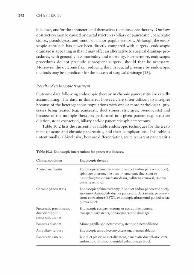
bile duct, and/or the sphincter lend themselves to endoscopic therapy. Outflowobstruction may be caused by ductal strictures (biliary or pancreatic), pancreaticstones, pseudocysts, and minor or major papilla stenosis. Although the endo-scopic approach has never been directly compared with surgery, endoscopicdrainage is appealing in that it may offer an alternative to surgical drainage pro-cedures, with generally less morbidity and mortality. Furthermore, endoscopicprocedures do not preclude subsequent surgery, should that be necessary.Moreover, the outcome from reducing the intraductal pressure by endoscopicmethods may be a predictor for the success of surgical drainage [15].
Results of endoscopic treatment
Outcome data following endoscopic therapy in chronic pancreatitis are rapidlyaccumulating. The data in this area, however, are often difficult to interpretbecause of the heterogeneous populations with one or more pathological pro-cesses being treated (e.g. pancreatic duct stones, strictures, pseudocysts) andbecause of the multiple therapies performed in a given patient (e.g. stricture dilation, stone extraction, biliary and/or pancreatic sphincterotomy).
Table 10.2 lists the currently available endoscopic techniques for the treat-ment of acute and chronic pancreatitis, and their complications. This table is(intentionally) all-inclusive, because differentiating acute recurrent pancreatitis
CHAPTER 10242
Table 10.2 Endoscopic interventions for pancreatic diseases.
Clinical condition
Acute pancreatitis
Chronic pancreatitis
Pancreatic pseudocysts, duct disruption, pancreatic ascites
Pancreas divisum
Ampullary tumors
Pancreatic cancer
Endoscopic therapy
Endoscopic sphincterotomy (bile duct and/or pancreatic duct),sphincter dilation, bile duct or pancreatic duct stent ornasobiliary/nasopancreatic drain, gallstone removal, Ascarisparasite removal
Endoscopic sphincterotomy (bile duct and/or pancreatic duct),stricture dilation, bile duct or pancreatic duct stents, pancreaticstone extraction ± ESWL, endoscopic ultrasound-guided celiacplexus block
Endoscopic cystgastrostomy or cystduodenostomy,transpapillary stents, or nasopancreatic drainage
Minor papilla sphincterotomy, stent, sphincter dilation
Endoscopic ampullectomy, stenting, thermal ablation
Bile duct plastic or metallic stent, pancreatic duct plastic stent,endoscopic ultrasound-guided celiac plexus block
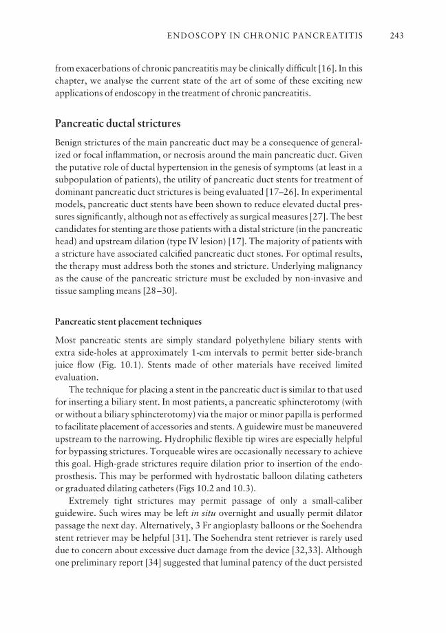
from exacerbations of chronic pancreatitis may be clinically difficult [16]. In thischapter, we analyse the current state of the art of some of these exciting newapplications of endoscopy in the treatment of chronic pancreatitis.
Pancreatic ductal strictures
Benign strictures of the main pancreatic duct may be a consequence of general-ized or focal inflammation, or necrosis around the main pancreatic duct. Giventhe putative role of ductal hypertension in the genesis of symptoms (at least in asubpopulation of patients), the utility of pancreatic duct stents for treatment ofdominant pancreatic duct strictures is being evaluated [17–26]. In experimentalmodels, pancreatic duct stents have been shown to reduce elevated ductal pres-sures significantly, although not as effectively as surgical measures [27]. The bestcandidates for stenting are those patients with a distal stricture (in the pancreatichead) and upstream dilation (type IV lesion) [17]. The majority of patients witha stricture have associated calcified pancreatic duct stones. For optimal results,the therapy must address both the stones and stricture. Underlying malignancyas the cause of the pancreatic stricture must be excluded by non-invasive and tissue sampling means [28–30].
Pancreatic stent placement techniques
Most pancreatic stents are simply standard polyethylene biliary stents withextra side-holes at approximately 1-cm intervals to permit better side-branchjuice flow (Fig. 10.1). Stents made of other materials have received limited evaluation.
The technique for placing a stent in the pancreatic duct is similar to that usedfor inserting a biliary stent. In most patients, a pancreatic sphincterotomy (withor without a biliary sphincterotomy) via the major or minor papilla is performedto facilitate placement of accessories and stents. A guidewire must be maneuveredupstream to the narrowing. Hydrophilic flexible tip wires are especially helpfulfor bypassing strictures. Torqueable wires are occasionally necessary to achievethis goal. High-grade strictures require dilation prior to insertion of the endo-prosthesis. This may be performed with hydrostatic balloon dilating cathetersor graduated dilating catheters (Figs 10.2 and 10.3).
Extremely tight strictures may permit passage of only a small-caliberguidewire. Such wires may be left in situ overnight and usually permit dilatorpassage the next day. Alternatively, 3 Fr angioplasty balloons or the Soehendrastent retriever may be helpful [31]. The Soehendra stent retriever is rarely useddue to concern about excessive duct damage from the device [32,33]. Althoughone preliminary report [34] suggested that luminal patency of the duct persisted
ENDOSCOPY IN CHRONIC PANCREATITIS 243
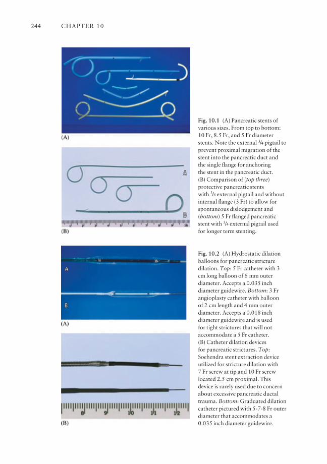
CHAPTER 10244
Fig. 10.1 (A) Pancreatic stents ofvarious sizes. From top to bottom:10 Fr, 8.5 Fr, and 5 Fr diameterstents. Note the external 3/4 pigtail toprevent proximal migration of thestent into the pancreatic duct andthe single flange for anchoring the stent in the pancreatic duct. (B) Comparison of (top three)protective pancreatic stents with 3/4 external pigtail and withoutinternal flange (3 Fr) to allow forspontaneous dislodgement and(bottom) 5 Fr flanged pancreaticstent with 3/4 external pigtail usedfor longer term stenting.
Fig. 10.2 (A) Hydrostatic dilationballoons for pancreatic stricturedilation. Top: 5 Fr catheter with 3cm long balloon of 6 mm outerdiameter. Accepts a 0.035 inchdiameter guidewire. Bottom: 3 Frangioplasty catheter with balloon of 2 cm length and 4 mm outerdiameter. Accepts a 0.018 inchdiameter guidewire and is used for tight strictures that will notaccommodate a 5 Fr catheter. (B) Catheter dilation devices for pancreatic strictures. Top:Soehendra stent extraction deviceutilized for stricture dilation with 7 Fr screw at tip and 10 Fr screwlocated 2.5 cm proximal. Thisdevice is rarely used due to concernabout excessive pancreatic ductaltrauma. Bottom: Graduated dilationcatheter pictured with 5-7-8 Fr outerdiameter that accommodates a0.035 inch diameter guidewire.
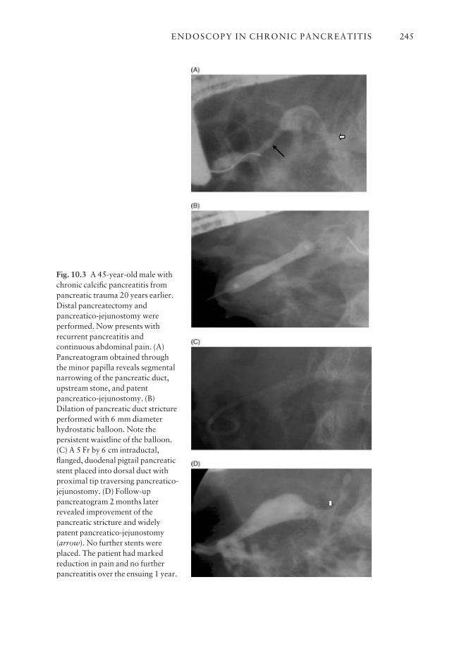
ENDOSCOPY IN CHRONIC PANCREATITIS 245
Fig. 10.3 A 45-year-old male withchronic calcific pancreatitis frompancreatic trauma 20 years earlier.Distal pancreatectomy andpancreatico-jejunostomy wereperformed. Now presents withrecurrent pancreatitis andcontinuous abdominal pain. (A)Pancreatogram obtained throughthe minor papilla reveals segmentalnarrowing of the pancreatic duct,upstream stone, and patentpancreatico-jejunostomy. (B)Dilation of pancreatic duct strictureperformed with 6 mm diameterhydrostatic balloon. Note thepersistent waistline of the balloon.(C) A 5 Fr by 6 cm intraductal,flanged, duodenal pigtail pancreaticstent placed into dorsal duct withproximal tip traversing pancreatico-jejunostomy. (D) Follow-uppancreatogram 2 months laterrevealed improvement of thepancreatic stricture and widelypatent pancreatico-jejunostomy(arrow). No further stents wereplaced. The patient had markedreduction in pain and no furtherpancreatitis over the ensuing 1 year.
at a mean time of 5 months following balloon dilation alone, most authoritieshave observed recurrence of strictures after one-time dilation and thereforeadvocate stenting [15].
As a rule, the diameter of the stent should not exceed the size of the down-stream duct. Therefore, 5, 7, or 8.5 Fr stents are commonly used in smallerducts, whereas 10–11.5 Fr stents or dual side-by-side 5–7 Fr stents may beinserted in patients with severe chronic pancreatitis and a dilated main pancre-atic duct. The tip of the stent in the pancreas must extend upstream to the nar-rowed segment and into a straight portion of the pancreatic duct to avoid stenttip erosion through the duct wall.
For diagnostic trials of pancreatic stenting in patients with nearly daily pain,most stents are left in place for 3–4 weeks. When long-term pancreatic stents areplaced for therapy, stents have remained in place for 3–116 months [17,24].
Stents are known to occlude within the first several weeks [35]; however,clinical improvement may persist for much longer, possibly due to siphoning ofthe pancreatic juice along the stent. At this time, self-expanding metallic stentsplay no role in the management of refractory pancreatic strictures due to thehigh occlusion rate from mucosal hyperplasia [36].
Efficacy of pancreatic duct stenting
The results of pancreatic duct stent placement (usually with ancillary pro-cedures) are detailed in Table 10.3 [17–26]. Successful stent placement wasachieved in 82–100% of patients. Sixty-six per cent of patients with successfulstent placement were reported to benefit from therapy during a mean follow-upto 8–39 months (N.B., many patients still had their stent in place during the follow-up period).
Cremer and colleagues
Cremer and colleagues [17] reported their experience with pancreatic ductstenting in 76 patients with severe chronic pancreatitis (primarily alcoholrelated) complicated by a distal pancreatic duct stricture and upstream dilation.A 10 Fr stent was successfully placed in 75 patients (98.7%) through the major(n = 54) or minor (n = 21) papilla. All patients had undergone biliary and pan-creatic sphincterotomy, stricture dilation, and extracorporeal shock-wave litho-tripsy (ESWL) (most patients) to fragment pancreatic duct stones.
A dramatic decrease or complete relief of pain was initially observed in 94% of patients and was associated with a decrease in the main pancreatic ductdiameter. Clinically, stents were thought to remain patent for a mean time of 12 months (range: 2–38 months). Disappearance of the stricture was observed inonly seven of 64 non-operated patients after 13 months (range: 2–30 months).
CHAPTER 10246
Eleven patients underwent pancreatico-jejunostomy after confirmation ofpain reduction with main pancreatic duct decompression. The remainderrequired repeated stent changes. Fifty-five per cent of non-operated patientsremained symptom-free at a mean follow-up of 3 years [19].
Early complications were related to pancreatic and/or biliary sphinctero-tomy (cholangitis in three patients and hemobilia in 10). Intraductal infectiondue to stent clogging developed in eight patients, and three had their stentmigrate inwardly. Stent therapy was believed by the authors to be an acceptablemedium-term treatment of pain associated with main pancreatic duct stricture.Unfortunately, because the stricture persists in the majority of patients, compli-ance with long-term use of plastic stents (i.e. multiple stent changes are required)would be difficult. As a result, expandable stents (18 Fr diameter, 23 mm long)have been tried in 29 patients [19].
Early follow-up to 6 months was encouraging, because stent clogging didnot occur during this short follow-up interval. However, during longer term follow-up, mucosal hyperplasia (i.e. tissue ingrowth) resulted in stent occlusionin the majority of patients [36]. Because these stents are not removable by
ENDOSCOPY IN CHRONIC PANCREATITIS 247
Table 10.3 Selected series reporting the results of pancreatic duct stenting for dominantstrictures.
Mean No. of patientsNo. of Technical follow-up symptomatically Major
Reference patients successa (months) improved complications Deaths
McCarthy et al. 5 5 14 4 2b 01988 [21]
Grimm et al. 63 55 19 31b 20b 11989 [18]
Cremer et al. 76 75 37 41 12 11991 [17]
Kozarek et al. N/A 17 8 13 3 01989 [20]
Binmoeller et al. 93 84 39 61 6 01995 [23]
Ponchon et al. 28 23 26 12 10 01995 [25]
Smits et al. 51 49 34 40 8 01995 [24]
Total 311c 308 34b 202d (66%) 61b (19%) 2 (1%)
aTechnical success refers to the number of patients successfully stented.bEstimate.cDoes not include the studies from which the number of patients attempted is not available.dPercentage improved refers to the number of patients who benefited (during the follow-up period)of the total number of patients successfully stented.N/A, not available.
endoscopic techniques, their use should be limited, perhaps, to patients in whomresective therapy (during which the stent and head of the pancreas would bothbe removed) is the next step. Evaluation of covered metal stents is in progress.
Ponchon and colleagues
Ponchon and colleagues [25] successfully placed 10 Fr multi-side-hole stentsafter biliary and pancreatic sphincterotomy and balloon dilation of strictures in28 of 33 patients (85%) with a distal pancreatic duct stricture and upstreamdilation. This was a highly selected subgroup, because patients with multiplesites of strictures, pancreatic duct stones, pancreas divisum, common bile ductnarrowing with cholestasis, any duodenal impingement, or the presence of apseudocyst larger than 1 cm were excluded. The stents were exchanged at 2-month intervals for a total stenting duration of 6 months.
Twenty-three patients were observed for at least 1 year after removal of thestent and comprised the basis of the report. During the stenting period, 21 of 23patients (91%) had resolution or reduction in pain, usually within days of stentinsertion, and 17 patients (74%) discontinued analgesic medications. Initialrelief of symptoms correlated with a decreased diameter (2 mm; P < 0.01) of the main pancreatic duct. Twelve patients (52%) had a persistent beneficial outcome for at least 1 year after stent removal. Disappearance of the stenosis on pancreatography at stent removal (P < 0.05) and 1 year later (P < 0.005) andreduction in the pancreatic duct diameter (2 mm) were significantly associatedwith pain relief. Complications of therapy occurred in 10 patients (30%), andincluded mild pancreatitis (resolved within 48 h) in nine and development of acommunicating pseudocyst in one.
Smits and colleagues
Smits and colleagues [24] evaluated the long-term efficacy of pancreatic ductstenting (5 or 7 Fr in nine patients and 10 Fr in 40) in a heterogeneous group of51 patients with pancreatic duct strictures (44 dominant, seven multiple)located in the head (n = 38), body (n = 14), or tail (n = 6), and upstream dila-tion. Associated pancreatic pathology treated at the time of stenting includedpancreatic duct stones (n = 17), pseudocysts (n = 10), common bile duct stric-tures with concomitant cholestasis (n = 12), and pancreas divisum (n = 3).Stents were successfully placed in 49 patients (96%) after pancreatic sphinctero-tomy (n = 31) and stricture dilation (n = 9).
Patients were re-evaluated within 3 months of stent placement and were followed for a median duration of 34 months. Responders underwent stentexchanges (approximately every 3 months) until such time as the stricture
CHAPTER 10248

patency had improved. Clinical benefit was noted in 40 of 49 patients (82%)during the stenting period. In 16 of these 40 patients, the stents were still in situat the time of the report and offered continued clinical improvement over peri-ods ranging from 6 to 116 months. In 22 of the 40 patients, the stents were elec-tively removed. All 22 patients experienced persistent clinical improvement during periods ranging between 6 and 41 months (median: 28.5 months) afterstent removal. There were no demographic factors (age, sex, duration of pancre-atitis, alcohol abuse), ERCP findings (single or multiple strictures, presence of pancreatic duct stones, pseudocyst, or biliary stricture), or additional inter-ventions (stricture dilation, removal of stones, drainage of pseudocyst, stentingof bile duct stricture) that predicted the clinical outcome.
Ashby and Lo
Ashby and Lo [40], from the United States, reported results of pancreatic stenting for strictures that differed from the European experience. Althoughrelief of symptoms was common (86% had significant improvement in theirsymptom score), this was usually not evident until day 7. More disappointingwas the lack of long-term benefit, with recurrence of symptoms within 1 monthof stenting. This study was relatively small (21 successfully stented patients) andincluded five patients with pancreatic cancer. Possible explanations for the lessfavorable results were that sphincterotomy was not performed and strictureswere not dilated routinely before stent placement (to improve pancreatic ductdrainage).
Hereditary and early onset pancreatitis
Pancreatic endotherapy was evaluated in patients with hereditary pancreatitisand idiopathic early onset chronic pancreatitis. In a report by Choudari et al., 27 consecutive patients with hereditary chronic pancreatitis underwent endo-scopic or surgical therapy of the pancreatic duct. Nineteen (70%) underwentendoscopic therapy and eight (30%) underwent surgery as their primary treatment. After a mean follow-up of 32 months, 50% of patients undergoingendoscopic therapy were symptom free, 38% were improved, and 12% wereunchanged with respect to pain. After surgery, 38% were symptom free, 25%were improved, and 37% were unchanged [38]. In a cohort of patients withpainful, early onset idiopathic chronic pancreatitis (aged 16–34 years) and adilated pancreatic duct, 11 patients underwent endoscopic therapy and werefollowed for over 6 years. The median interval between onset of symptoms andendoscopic therapy was 5 years (3–10 years). Pancreatic sphincterotomy andstent insertion provided short-term relief in 11 patients (100%).
ENDOSCOPY IN CHRONIC PANCREATITIS 249
Complications included fever in three patients and cholecystitis in onepatient. Four patients (37%) developed recurrent pain felt to be due to recurrentpancreatic strictures or stones, and underwent further endoscopic therapy [39].These two patient populations of hereditary and early onset idiopathic chronicpancreatitis illustrate the value of endoscopic therapy in affording short-termand medium-term pain relief. Repeat endoscopic therapy is not uncommon.
Predicting the outcome of pancreatic stenting
There are few studies that have been designed to identify subgroups of patientswith chronic pancreatitis who are most likely to benefit from stenting. In a pre-liminary report, 65 chronic pancreatitis patients with duct dilation (> or = to 6 mm), obstruction (usually a stricture with a diameter of 1 mm or less),obstruction and dilation, or no obstruction or dilation underwent pancreaticduct stenting for 3–6 months [37]. The presence of both obstruction and dila-tion was a significant predictor of improvement.
Duration of stenting
The appropriate duration of pancreatic stent placement and the interval fromplacement to change of the pancreatic stent are not known. Two options areavailable [15]: (1) the stent can be left in place until symptoms or complicationsoccur; (2) the stent can be left in place for a predetermined interval (e.g. 3months). If the patient fails to improve, the stent should be removed becauseductal hypertension is unlikely to be the cause of pain. If the patient has bene-fited from stenting, one can remove the stent and follow the patient clinically,continue stenting for a more prolonged period, or perform a surgical drainageprocedure. (This latter option assumes that the results of endoscopic stentingwill predict the surgical outcome.) There are limited data to support any of theseoptions.
In a recent preliminary report, Borel et al. [42] evaluated the effect ofdefinitive pancreatic duct stent placement only exchanged on demand whensymptoms recurred. In 42 patients, a single 10 Fr stent was inserted into themain pancreatic duct following pancreatic sphincterotomy. The patients werefollowed for a median of 33 months with respect to pain reduction, weight gainor loss, and recurrence of symptoms. With recurrence of symptoms, the stentwas exchanged. Of the 42 patients, 72% had pain relief with pancreatic stenting(pain score reduced > 50%) and 69% gained weight. Two-thirds of the patients(n = 28) required only the single pancreatic stent placement and 12 patientsrequired a stent exchange after a median of 15 months. Two patients requiredrepeated stent exchanges for recurrence of pain. Persistence or recurrence of
CHAPTER 10250
pain was significantly associated with the development of cholestasis and con-tinued alcohol abuse. These authors concluded that long-term pancreatic stentingappears to be an effective, and possibly a superior, option compared to tempor-ary stenting [42].
Does response to stenting predict the outcome of surgery?
The question may be posed: in patients with chronic pancreatitis and a dilatedpancreatic duct, will the response to pancreatic stent placement predict theresponse to surgical duct decompression? In a preliminary report of a random-ized controlled trial (n = 8), McHenry and associates evaluated the utility ofshort-term (12 weeks) pancreatic duct stenting to relieve pain and to predict theresponse to surgical decompression in patients with chronic pancreatitis and adilated main pancreatic duct [43]. Four of eight patients benefited from stenting,while no control patient improved. Among five patients who underwent aPuestow procedure following stent therapy, four had pain relief. Improvementwith the pancreatic stent was seen in two of four patients responding to surgery;one patient benefited from the stent but did not improve with surgery. Inanother preliminary series, reported by DuVall and colleagues [44], endoscopictherapy predicted the outcome from surgical decompression in nine of 11patients (82%; positive and negative predictive values were 80% and 83%,respectively) during a 2-year postoperative follow-up interval.
Several institutions have recently reported that symptomatic improvementmay persist after pancreatic stent removal despite stricture persistence [17,23–25]. When summarizing the results of two studies (n = 54) that evaluated theefficacy of pancreatic duct stenting for dominant strictures, 65% of patients hadpersistent symptom improvement after stent removal, although the strictureresolved in only 33% (Table 10.4). Although these data indicate that completestricture resolution is not a prerequisite for symptom improvement, severalother factors may account for this outcome. First, other therapies performed atthe time of stenting (e.g. pancreatic stone removal, pancreatic sphincterotomy)
ENDOSCOPY IN CHRONIC PANCREATITIS 251
Table 10.4 Pancreatic duct stenting for dominant strictures: clinical outcome and strictureresolution after stent removal.
Persistent improvement Median follow-up after StrictureReference after stent removal stent removal (months) resolution
Smits et al. 1995 [24] 23/33 (70%) 29 10/33 (20%)Ponchon et al. 1995 [25] 12/21 (57%) 14 8/21 (38%)
Total 35/54 (65%) 23 18/54 (33%)

may contribute to patient benefit. Second, many of the unresolved strictures hadimproved luminal patency (but without return of lumen diameter to normal).Third, the pain of chronic pancreatitis tends to decrease with time and mayresolve when marked deterioration of pancreatic function occurs [40].
Long-term follow-up
In the largest multicenter trial, Rosch et al. [26] reported on the long-term follow-up of over 1000 patients with chronic pancreatitis undergoing initial endoscopictherapy during the period 1989–95. Some of these patients were previouslyreported with shorter follow-up as noted in Table 10.3.
A total of 1211 patients from eight centers in Europe with pain and obstruc-tive chronic pancreatitis underwent endoscopic therapy including endoscopicpancreatic sphincterotomy, pancreatic stricture dilation, pancreatic stoneremoval, pancreatic stent placement, or a combination of these methods. Over a mean period of 4.9 years (range: 2–12 years), 1118 patients (84%) were fol-lowed for symptomatic improvement and need for pancreatic surgery. Successof endoscopic therapy was defined as a significant reduction or elimination ofpain and reduction in pain medication. Partial success was defined as reductionin pain although further interventions were necessary for pain relief. Failure ofendoscopic therapy was defined as the need for pancreatic decompressive sur-gery or patients that were lost to follow-up.
Over long-term follow-up, 69% of patients were successfully treated withendoscopic therapy and 15% experienced a partial success. Twenty per cent of patients required surgery with a 55% significant reduction in pain. Five percent of patients were lost to follow-up. The patients with the highest frequencyof completed treatment were those with stones alone (76%) as compared to those with strictures alone (57%) and those with strictures and stones (57%)(P < 0.001). Interestingly, the percentage of patients with no or minimal residual pain at follow-up was similar in all groups (strictures alone 84%, stonesalone 84%, and strictures plus stones 87%) (P = 0.677). The authors of thisreport concluded that endoscopic therapy of chronic pancreatitis in experiencedcenters is effective in the majority of patients, and the beneficial response to successful endoscopic therapy in chronic pancreatitis is durable and long-term[26].
Only randomized controlled studies comparing surgical, medical, and endo-scopic techniques will allow us to determine the true long-term efficacy of pancreatic duct stenting for stricture therapy. There remain many unansweredquestions. Which patients are the best candidates? Is proximal pancreatic ductaldilation a prerequisite? Does the response to stenting depend on the etiology ofthe chronic pancreatitis? Finally, as noted, how does endoscopic therapy com-pare with medical and surgical management?
CHAPTER 10252
Complications associated with pancreatic stents
True complication rates are difficult to decipher due to: (1) the simultaneous performance of other procedures (e.g. pancreatic sphincterotomy, stricture dila-tion); (2) the heterogeneous patient populations treated (i.e. patients with acuteor chronic pancreatitis); and (3) the lack of uniform definitions of complicationsand a grading system of their severity [47]. Complications related directly tostent therapy are listed in Table 10.5 [47,49].
Occlusion
The pathogenesis of pancreatic stent occlusion on scanning electron microscopymirrors biliary stent blockage with typical biofilm and microcolonies of bacteriamixed with crystals, similar to biliary sludge. The rate of pancreatic stent occlu-sion appears to be similar to that for biliary stents [35]. We found that 50% ofpancreatic stents (primarily 5–7 Fr) were occluded within 6 weeks of placementand 100% of stents were occluded at more than 9 weeks when carefully evalu-ated by water flow methods. More than 80% of these early occlusions were notassociated with adverse clinical events. In such circumstances, the stent is per-haps serving as a dilator or a wick. Similarly, stents reported to be patent for aslong as 38 months [17] are clinically patent but would presumably be occludedby water flow testing.
Migration
Stent migration may be upstream (i.e. into the duct) or downstream (i.e. into theduodenum). Migration in either direction may be heralded by the return of pain or pancreatitis. Johanson and associates [50] reported inward migration in5.2% of patients and duodenal migration in 7.5%. These events occurred withsingle intraductal and single duodenal stent flanges. Rarely, surgery is needed toremove a proximally migrated stent. Modifications in pancreatic stent designhave greatly reduced the frequency of such occurrences. Dean and associates[51] reported no inward migration in 112 patients stented with a four-barbed
ENDOSCOPY IN CHRONIC PANCREATITIS 253
Occlusion, which may result in pain and/orpancreatitis
Migration into or out of ductDuodenal erosionsPancreatic infectionDuctal perforationDuctal and parenchymal changesStone formation
Table 10.5 Complications directlyrelated to pancreatic duct stents.
(two internal and two external) stent. We have had no inward migration ingreater than 3000 stents with a duodenal pigtail.
Stent-induced duct changes
Although therapeutic benefit has been reported for pancreatic stenting, it is evident that morphological changes of the pancreatic duct directly related to thistherapy occur in the majority of patients. In summarizing the results of sevenpublished series [52–55,57–59], new ductal changes were seen in 54% (range:33–83%) of 297 patients. Limited observations to date indicate a tendency ofthese ductal changes to improve with time following stent change and/orremoval [44,45,47,50,52,53,55,57–59].
The long-term consequences of these stent-induced ductal changes remainuncertain. Moreover, the long-term parenchymal effects have not been studiedin humans. In a pilot study, six mongrel dogs underwent pancreatic duct stent-ing for 2–4 months [49]. Radiographic, gross, and histological abnormalitiesdeveloped in all dogs. The radiographic findings (stenosis in the stented regionwith upstream dilation) were associated with gross evidence of fibrosis, whichincreased proportionally with the length of the stenting period. Histologicalchanges of obstructive pancreatitis were present in most experimental dogs.
Although follow-up after stent removal was short, the atrophy and fibrosisseen were not likely to be reversible. In a recently reported study [59], paren-chymal changes (hypoechoic area around the stent, heterogeneity, and cysticchanges) were seen on endoscopic ultrasound in 17 of 25 patients undergoingshort-term pancreatic duct stenting. Four patients who had parenchymalchanges at stent removal had a follow-up study at a mean time of 16 months.Two patients had (new) changes suggestive of chronic pancreatitis (hetero-geneous echotexture, echogenic foci in the parenchyma, and a thickened hyper-echoic irregular pancreatic duct) in the stented region. While such damage in anormal pancreas may have significant long-term consequences, the outcome inpatients with advanced chronic pancreatitis may be inconsequential.
Brief mini-stents
If brief interval stenting is needed, such as for pancreatic sphincterotomy, wenow commonly use small-diameter stents (3 or 4 Fr) with no intraductal barb[83] (Fig. 10.1). Depending on their length, 80–90% of these stents migrate outof the duct spontaneously. Further studies addressing issues of stent diameter aswell as composition and duration of therapy as they relate to safety and efficacyare needed. Additionally, further evaluation of expandable stents, particularlythe coated models, is awaited.
CHAPTER 10254
Pancreatic ductal stones
Causes of pancreatic ductal stones
Worldwide, alcohol consumption appears to be the most important factor asso-ciated with chronic calcifying pancreatitis. Although the exact mechanism ofintraductal stone formation has not been clearly elucidated, considerable pro-gress in this area has been made [60]. Alcohol appears to be directly toxic to thepancreas and produces a dysregulation of secretion of pancreatic enzymes (in-cluding zymogens), citrate (a potent calcium chelator), lithostathine (pancreaticstone protein), and calcium. These changes favor the formation of a nidus (a pro-tein plug), followed by precipitation of calcium carbonate to form a stone [60,61].
Stones cause obstruction
The rationale for intervention is based on the premise that pancreatic stonesincrease the intraductal pressure (and probably the parenchymal pressure, withresultant pancreatic ischemia) proximal to the obstructed focus. Reports indi-cating that endoscopic (with or without ESWL) or surgical removal of pancreaticcalculi results in improvement of symptoms support this notion [15]. Moreover,stone impaction may cause further trauma to the pancreatic duct, with epithelialdestruction and stricture formation [53,55]. Thus, identification of pancreaticductal stones in a symptomatic patient warrants consideration of removal. Oneor more large stones in the head with upstream asymptomatic parenchymalatrophy probably warrant therapy also.
Endoscopic techniques for stone extraction
Pancreatic sphincterotomy
A major papilla pancreatic sphincterotomy (in patients with normal anatomy,i.e. no pancreas divisum) is usually performed to facilitate access to the ductprior to attempts at stone removal. There are two methods available to cut themajor pancreatic sphincter [63,64]. A standard pull-type sphincterotome (withor without a wire guide) is inserted into the pancreatic duct and orientated alongthe axis of the pancreatic duct (usually in the 12–1 o’clock position). Althoughthe landmarks to determine the length of incision are imprecise, authorities recommend cutting 5–10 mm [63] (Fig. 10.4). The cutting wire should notextend more than 6–7 mm up the duct when applying electrocautery so as toprevent deep ductal injury. Alternatively, a needle-knife can be used to performthe sphincterotomy over a previously placed pancreatic stent [63,64].
ENDOSCOPY IN CHRONIC PANCREATITIS 255
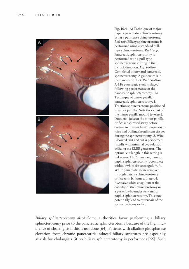
Biliary sphincterotomy also? Some authorities favor performing a biliarysphincterotomy prior to the pancreatic sphincterotomy because of the high inci-d-ence of cholangitis if this is not done [64]. Patients with alkaline phosphataseelevation from chronic pancreatitis-induced biliary strictures are especially at risk for cholangitis (if no biliary sphincterotomy is performed) [65]. Such
CHAPTER 10256
Fig. 10.4 (A) Technique of majorpapilla pancreatic sphincterotomyusing a pull-type sphincterotome.Left top: Biliary sphincterotomy isperformed using a standard pull-type sphincterotome. Right top:Pancreatic sphincterotomy isperformed with a pull-typesphincterotome cutting in the 1o’clock direction. Left bottom:Completed biliary and pancreaticsphincterotomy. A guidewire is inthe pancreatic duct. Right bottom: A 6 Fr pancreatic stent is placedfollowing performance of thepancreatic sphincterotomy. (B)Technique of minor papillapancreatic sphincterotomy. 1.Traction sphincterotome positionedin minor papilla. Note the extent ofthe minor papilla mound (arrows).Duodenal juice at the minor papillaorifice is aspirated away beforecutting to prevent heat dissipation tojuice and boiling the adjacent tissuesduring the sphincterotomy. 2. Wireis bowed taut and cut is performedrapidly with minimal coagulationutilizing the ERBE generator. Theoptimal cut length in this setting isunknown. The 5 mm length minorpapilla sphincterotomy is completewithout white tissue coagulum. 3.White pancreatic stone removedthrough patent sphincterotomyorifice with balloon catheter. 4.Excessive white coagulum at the cut edge of the sphincterotomy in a patient who underwent minorpapilla sphincterotomy. This maypotentially lead to restenosis of thesphincterotomy orifice.
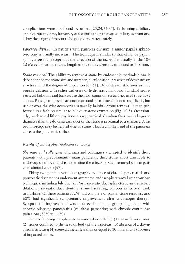
complications were not found by others [23,24,64,65]. Performing a biliarysphincterotomy first, however, can expose the pancreatico-biliary septum andallow the length of the cut to be gauged more accurately.
Pancreas divisum In patients with pancreas divisum, a minor papilla sphinc-terotomy is usually necessary. The technique is similar to that of major papillasphincterotomy, except that the direction of the incision is usually in the 10–12 o’clock position and the length of the sphincterotomy is limited to 4–8 mm.
Stone removal The ability to remove a stone by endoscopic methods alone isdependent on the stone size and number, duct location, presence of downstreamstricture, and the degree of impaction [67,68]. Downstream strictures usuallyrequire dilation with either catheters or hydrostatic balloons. Standard stone-retrieval balloons and baskets are the most common accessories used to removestones. Passage of these instruments around a tortuous duct can be difficult, butuse of over-the-wire accessories is usually helpful. Stone removal is then per-formed in a fashion similar to bile duct stone extraction (Fig. 10.5). Occasion-ally, mechanical lithotripsy is necessary, particularly when the stone is larger indiameter than the downstream duct or the stone is proximal to a stricture. A rattooth forceps may be helpful when a stone is located in the head of the pancreasclose to the pancreatic orifice.
Results of endoscopic treatment for stones
Sherman and colleagues Sherman and colleagues attempted to identify thosepatients with predominantly main pancreatic duct stones most amenable toendoscopic removal and to determine the effects of such removal on the pati-ents’ clinical course [67].
Thirty-two patients with ductographic evidence of chronic pancreatitis andpancreatic duct stones underwent attempted endoscopic removal using varioustechniques, including bile duct and/or pancreatic duct sphincterotomy, stricturedilation, pancreatic duct stenting, stone basketing, balloon extraction, and/or flushing. Of these patients, 72% had complete or partial stone removal, and68% had significant symptomatic improvement after endoscopic therapy.Symptomatic improvement was most evident in the group of patients withchronic relapsing pancreatitis (vs. those presenting with chronic continuouspain alone; 83% vs. 46%).
Factors favoring complete stone removal included: (1) three or fewer stones;(2) stones confined to the head or body of the pancreas; (3) absence of a down-stream stricture; (4) stone diameter less than or equal to 10 mm; and (5) absenceof impacted stones.
ENDOSCOPY IN CHRONIC PANCREATITIS 257

CHAPTER 10258
Fig. 10.5 A 40-year-old female withalcohol-induced chronic pancreatitiscomplicated by pancreatic mainduct stones. (a) Pancreatogramrevealing dilated pancreatic ductwith 5 mm diameter filling defectconsistent with a pancreatic stone. (b) After pancreaticsphincterotomy, a non-wire-guidedstone extraction basket was utilized.The basket is opened fully in thedilated pancreatic duct and the stone is engaged. (c) Basket is slowly closed on the stone. (d) Stone is extracted and follow-uppancreatogram with a ballooncatheter reveals no residual fillingdefects. No further stenting wasperformed.
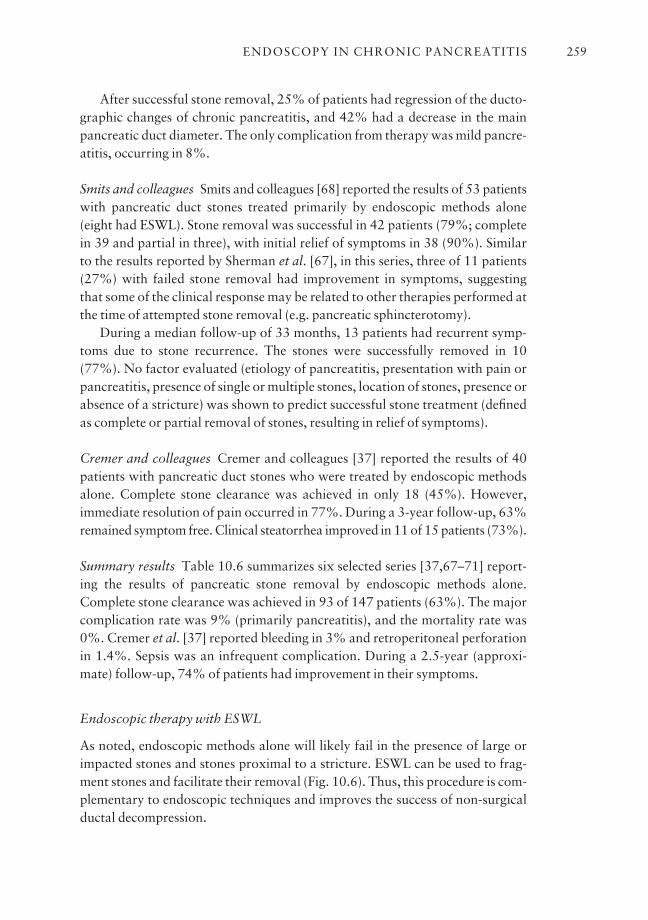
After successful stone removal, 25% of patients had regression of the ducto-graphic changes of chronic pancreatitis, and 42% had a decrease in the mainpancreatic duct diameter. The only complication from therapy was mild pancre-atitis, occurring in 8%.
Smits and colleagues Smits and colleagues [68] reported the results of 53 patientswith pancreatic duct stones treated primarily by endoscopic methods alone (eight had ESWL). Stone removal was successful in 42 patients (79%; completein 39 and partial in three), with initial relief of symptoms in 38 (90%). Similar to the results reported by Sherman et al. [67], in this series, three of 11 patients(27%) with failed stone removal had improvement in symptoms, suggestingthat some of the clinical response may be related to other therapies performed atthe time of attempted stone removal (e.g. pancreatic sphincterotomy).
During a median follow-up of 33 months, 13 patients had recurrent symp-toms due to stone recurrence. The stones were successfully removed in 10(77%). No factor evaluated (etiology of pancreatitis, presentation with pain orpancreatitis, presence of single or multiple stones, location of stones, presence orabsence of a stricture) was shown to predict successful stone treatment (definedas complete or partial removal of stones, resulting in relief of symptoms).
Cremer and colleagues Cremer and colleagues [37] reported the results of 40patients with pancreatic duct stones who were treated by endoscopic methodsalone. Complete stone clearance was achieved in only 18 (45%). However,immediate resolution of pain occurred in 77%. During a 3-year follow-up, 63%remained symptom free. Clinical steatorrhea improved in 11 of 15 patients (73%).
Summary results Table 10.6 summarizes six selected series [37,67–71] report-ing the results of pancreatic stone removal by endoscopic methods alone.Complete stone clearance was achieved in 93 of 147 patients (63%). The majorcomplication rate was 9% (primarily pancreatitis), and the mortality rate was0%. Cremer et al. [37] reported bleeding in 3% and retroperitoneal perforationin 1.4%. Sepsis was an infrequent complication. During a 2.5-year (approxi-mate) follow-up, 74% of patients had improvement in their symptoms.
Endoscopic therapy with ESWL
As noted, endoscopic methods alone will likely fail in the presence of large orimpacted stones and stones proximal to a stricture. ESWL can be used to frag-ment stones and facilitate their removal (Fig. 10.6). Thus, this procedure is com-plementary to endoscopic techniques and improves the success of non-surgicalductal decompression.
ENDOSCOPY IN CHRONIC PANCREATITIS 259
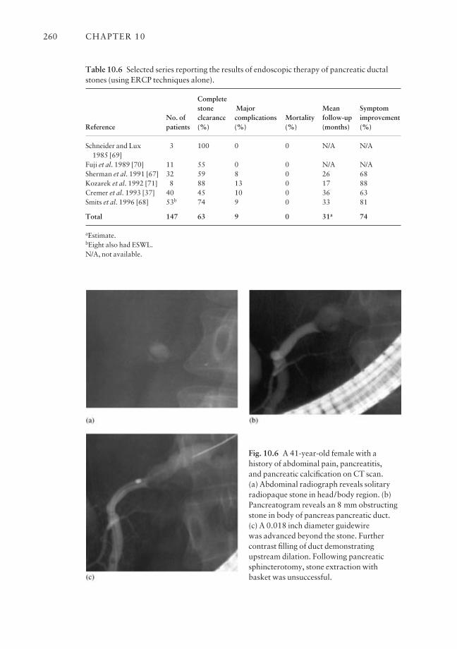
CHAPTER 10260
Table 10.6 Selected series reporting the results of endoscopic therapy of pancreatic ductalstones (using ERCP techniques alone).
Complete stone Major Mean Symptom
No. of clearance complications Mortality follow-up improvementReference patients (%) (%) (%) (months) (%)
Schneider and Lux 3 100 0 0 N/A N/A1985 [69]
Fuji et al. 1989 [70] 11 55 0 0 N/A N/ASherman et al. 1991 [67] 32 59 8 0 26 68Kozarek et al. 1992 [71] 8 88 13 0 17 88Cremer et al. 1993 [37] 40 45 10 0 36 63Smits et al. 1996 [68] 53b 74 9 0 33 81
Total 147 63 9 0 31a 74
aEstimate.bEight also had ESWL.N/A, not available.
Fig. 10.6 A 41-year-old female with ahistory of abdominal pain, pancreatitis, and pancreatic calcification on CT scan. (a) Abdominal radiograph reveals solitaryradiopaque stone in head/body region. (b)Pancreatogram reveals an 8 mm obstructingstone in body of pancreas pancreatic duct.(c) A 0.018 inch diameter guidewire was advanced beyond the stone. Furthercontrast filling of duct demonstratingupstream dilation. Following pancreaticsphincterotomy, stone extraction withbasket was unsuccessful.
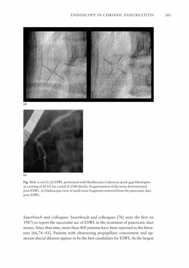
Sauerbruch and colleagues Sauerbruch and colleagues [76] were the first (in1987) to report the successful use of ESWL in the treatment of pancreatic ductstones. Since that time, more than 400 patients have been reported in the litera-ture [66,74–81]. Patients with obstructing prepapillary concrement and up-stream ductal dilation appear to be the best candidates for ESWL. In the largest
ENDOSCOPY IN CHRONIC PANCREATITIS 261
Fig. 10.6 (cont’d ) (d) ESWL performed with Healthronics Lithotron spark-gap lithotriptorat a setting of 26 kV for a total of 2500 shocks. Fragmentation of the stone demonstratedpost-ESWL. (e) Endoscopic view of small stone fragments removed from the pancreatic ductpost-ESWL.
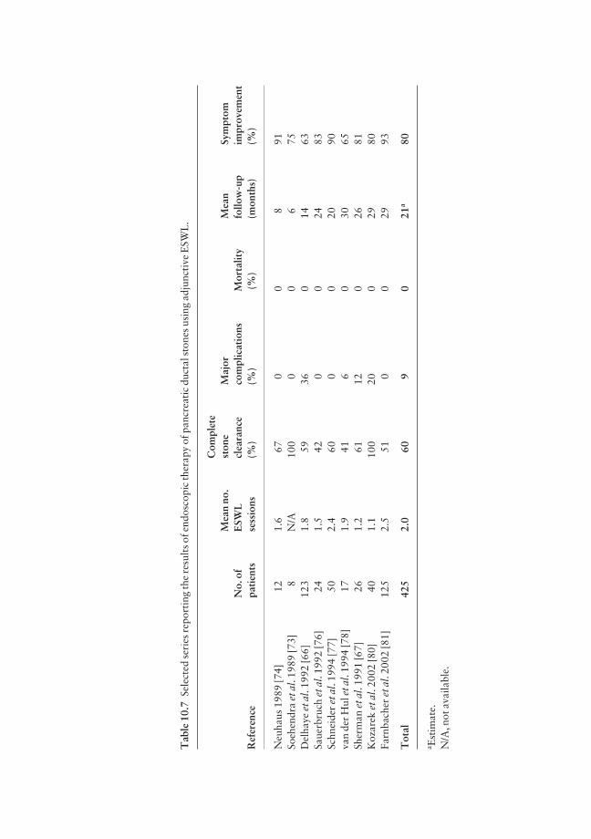
Tab
le 1
0.7
Sele
cted
ser
ies
repo
rtin
g th
e re
sult
s of
end
osco
pic
ther
apy
of p
ancr
eati
c du
ctal
sto
nes
usin
g ad
junc
tive
ESW
L.
Com
plet
eM
ean
no.
ston
eM
ajor
Mea
nSy
mpt
omN
o. o
fE
SWL
clea
ranc
eco
mpl
icat
ions
Mor
talit
yfo
llow
-up
impr
ovem
ent
Ref
eren
cepa
tien
tsse
ssio
ns(%
)(%
)(%
)(m
onth
s)(%
)
Neu
haus
198
9 [7
4]12
1.6
670
08
91So
ehen
dra
et a
l. 19
89 [7
3]8
N/A
100
00
675
Del
haye
et a
l. 19
92 [6
6]12
31.
859
360
1463
Saue
rbru
ch e
t al.
1992
[76]
241.
542
00
2483
Schn
eide
r et
al.
1994
[77]
502.
460
00
2090
van
der
Hul
et a
l. 19
94 [7
8]17
1.9
416
030
65Sh
erm
an e
t al.
1991
[67]
261.
261
120
2681
Koz
arek
et a
l. 20
02 [8
0]40
1.1
100
200
2980
Farn
bach
er e
t al.
2002
[81]
125
2.5
510
029
93
Tot
al42
52.
060
90
21a
80
a Est
imat
e.N
/A, n
ot a
vaila
ble.
reported series, 123 patients with main pancreatic duct stones and proximaldilation were treated with an electromagnetic lithotriptor, usually before pan-creatic duct sphincterotomy [66]. Stones were successfully fragmented in 99%,resulting in a decrease in duct dilation in 90%. The main pancreatic duct wascompletely cleared of all stones in 59%. Eighty-five per cent of patients notedpain improvement during a mean follow-up of 14 months. However, 41% ofpatients had a clinical relapse due to stone migration into the main pancreaticduct, progressive stricture, or stent occlusion.
This same center compared their results of pancreatic stone removal prior tothe availability of ESWL and after the introduction of adjunctive ESWL therapy[37]. Stones were successfully cleared in 18 of 40 patients (45%) by endoscopicmethods alone, compared with 22 of 28 (78.6%) with ESWL. Table 10.7 sum-marizes the results of nine selected series reporting the efficacy and safety ofadjunctive ESWL [66,67,73,74,76–78,80,81]. Complications in these serieswere related primarily to the endoscopic procedure.
Although ultrasound-focused ESWL has been reported to achieve stonefragmentation, such focusing is clearly more difficult. In the series reported bySchneider and associates [77], stone localization was achieved in 17 of 119 ses-sions (14%) when only ultrasonography was used to monitor the position of the stone.
The Brussels group The Brussels group [79] studied 70 pancreatic stone pati-ents who underwent attempts at endoscopic removal, with adjunctive ESWLused in 41 (59%). This was a fairly homogeneous group of patients in that thosewith strictures, previous pancreatic surgery, and failed pancreatic sphinctero-tomy were excluded. The authors evaluated the immediate technical and clinicalresults and reviewed the long-term outcome in patients followed for more than 2 years.
Complete (n = 35) or partial (n = 20) stone removal was achieved in 79%,and was more frequently observed when ESWL was performed (P < 0.005) andin the absence of a non-papillary ductal substenosis or complete main ductobstruction (P < 0.05). Complete stone clearance was most frequently observedwith single stones or stones confined to the head (P < 0.05). In the multivariateanalysis, ESWL was the only independent factor influencing the technical resultsof endoscopic management. In this series, the number of ERCPs performed perpatient was reduced from 3.4 to 2.7 after the introduction of ESWL (P < 0.01).Of the 56 patients with pain on admission, 53 (95%) were pain free (n = 41) orhad a reduction in pain (n = 12).
In both the univariate and multivariate analyses, a significant associationwas found between immediate disappearance of pain and complete or partialmain pancreatic duct clearance. During the first 2 years of follow-up after ther-apy, 25 of 46 (54%) patients were totally pain free, whereas the frequency of
ENDOSCOPY IN CHRONIC PANCREATITIS 263
pain attacks in the remaining 21 was halved. This frequency of recurrent symp-toms (46%) is comparable to that of surgical series [82].
Long-term pain relief was associated with: (1) earlier treatment after diseaseonset (P < 0.005); (2) a low frequency of pain attacks before therapy (P < 0.05);and (3) absence of non-papillary substenosis of the main pancreatic duct (P < 0.05).
Interestingly, outcome was not associated with prior or continued alcoholintake. In the multivariate analysis, pain recurrence was independently asso-ciated with the frequency of pain attacks before therapy, the duration of disease,and the presence of non-papillary substenosis of the main pancreatic duct. It was suggested that such substenosis can induce ductal hypertension by blockingmigration of fragmented stones or by progressing to higher grade stenosis.Twenty per cent underwent subsequent pancreatic surgical procedures. Of theremaining 28 patients, there was statistically significant improvement in meanpain scores, narcotic use, and hospitalizations when comparing intervals beforeand after stone therapy [83].
Kozarek and colleagues Kozarek and colleagues performed a retrospectivereview of the efficacy of ESWL as an adjunct to endoscopic therapy in 40 pati-ents who underwent a total of 46 ESWL sessions (an average of 1.15 sessions/patient). Eighty per cent of patients did not require surgery and had significantpain relief, reduced number of hospitalizations, and reduced narcotic use ascompared to the pre-ESWL period over a mean 2.4-year follow-up [80].
Farnbacher and colleagues Farnbacher and colleagues retrospectively reviewedthe efficacy of pancreatic stone clearance with endoscopic and ESWL therapy.Technical success was achieved in 85% of the 125 patients. The majority of thepatients (111 of 125) required piezoelectric ESWL for stone fragmentation.ESWL was safe, without any serious complications. Middle-aged patients in theearly stages of chronic pancreatitis with stones in a prepapillary location werethe best candidates for successful treatment and required the least number ofESWL treatment sessions [81].
These aforementioned studies reaffirm that ESWL as an adjunct to endo-scopic pancreatic therapy is effective, and the results of the combined modalitymay obviate the need for surgery. The results of endoscopic therapy in conjunc-tion with ESWL for pancreatic stone disease compare favorably to the outcomesin surgically treated patients.
Intraductal lithotripsy
Intraductal lithotripsy via mother–baby scope systems has largely failed due to
CHAPTER 10264

inability to maneuver within the relatively narrow ductal system. Results withfluoroscopy-guided laser lithotripsy were similarly poor [71]. Pancreatoscopy(via a ‘mother–baby’ scope system) can be used to directly visualize laser fibercontact with the stone and fragmentation. Experience is limited to date [70,83].
Medical treatment for stones
Stone dissolution via ductal irrigation (contact dissolution) or oral agent is anattractive endoscopic adjunct for stone removal.
Citrate Sahel and Sarles found that intraduodenal infusion of citrate in dogssignificantly increased the citrate concentration in pancreatic juice [85]. This ledto a non-randomized study of oral citrate in 18 patients with chronic pancre-atitis, 17 of whom had pancreatic duct stones. Seven patients responded duringa mean duration of therapy of 9.5 months, with a mean stone size reduction of21% and an improvement in symptoms [61].
Berger et al. [86] performed nasopancreatic drainage in six patients withmain pancreatic duct stones. The pancreatic duct was perfused with a mixture ofisotonic citrate and saline at 3 ml/min for 4 days. A stone-free state was achievedin all cases.
Pancreatic pain disappeared during the perfusion, and four patientsremained free of pain during the follow-up period (1–12 months). The remain-ing two patients had repeat therapy, which resulted in pain resolution. Pancre-atic exocrine function was evaluated by the Lundh test in five patients beforeand after therapy. An increase of 50–360% was observed in enzyme output inthree patients, while no improvement was noted in the remaining two patients.
Trimethadione Trimethadione, an epileptic agent and a weak organic acid, hasbeen shown in vitro to induce a concentration-dependent increase in calciumsolubility [61]. Noda et al. [87] showed promising results for trimethadione in adog model of pancreatic stones. Unfortunately, the doses used in the dogs, ifextrapolated to humans, could potentially be toxic. At the present time, norapidly effective solvent for human use is available to treat pancreatic stones.Further trials in humans are needed to establish a role for medical therapy(either alone or as an aid to endoscopic measures) in treating patients withsymptomatic pancreatic duct stones.
Overall results for stone treatment
These data suggest that removal of pancreatic duct stones may result in symp-tomatic benefit. Longer follow-up is necessary to determine the stone recurrence
ENDOSCOPY IN CHRONIC PANCREATITIS 265
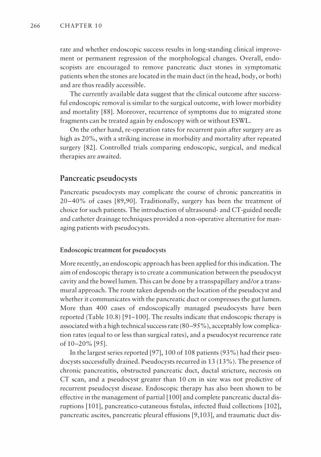
rate and whether endoscopic success results in long-standing clinical improve-ment or permanent regression of the morphological changes. Overall, endo-scopists are encouraged to remove pancreatic duct stones in symptomaticpatients when the stones are located in the main duct (in the head, body, or both)and are thus readily accessible.
The currently available data suggest that the clinical outcome after success-ful endoscopic removal is similar to the surgical outcome, with lower morbidityand mortality [88]. Moreover, recurrence of symptoms due to migrated stonefragments can be treated again by endoscopy with or without ESWL.
On the other hand, re-operation rates for recurrent pain after surgery are ashigh as 20%, with a striking increase in morbidity and mortality after repeatedsurgery [82]. Controlled trials comparing endoscopic, surgical, and medicaltherapies are awaited.
Pancreatic pseudocysts
Pancreatic pseudocysts may complicate the course of chronic pancreatitis in20–40% of cases [89,90]. Traditionally, surgery has been the treatment ofchoice for such patients. The introduction of ultrasound- and CT-guided needleand catheter drainage techniques provided a non-operative alternative for man-aging patients with pseudocysts.
Endoscopic treatment for pseudocysts
More recently, an endoscopic approach has been applied for this indication. Theaim of endoscopic therapy is to create a communication between the pseudocystcavity and the bowel lumen. This can be done by a transpapillary and/or a trans-mural approach. The route taken depends on the location of the pseudocyst andwhether it communicates with the pancreatic duct or compresses the gut lumen.More than 400 cases of endoscopically managed pseudocysts have beenreported (Table 10.8) [91–100]. The results indicate that endoscopic therapy isassociated with a high technical success rate (80–95%), acceptably low complica-tion rates (equal to or less than surgical rates), and a pseudocyst recurrence rateof 10–20% [95].
In the largest series reported [97], 100 of 108 patients (93%) had their pseu-docysts successfully drained. Pseudocysts recurred in 13 (13%). The presence ofchronic pancreatitis, obstructed pancreatic duct, ductal stricture, necrosis onCT scan, and a pseudocyst greater than 10 cm in size was not predictive ofrecurrent pseudocyst disease. Endoscopic therapy has also been shown to beeffective in the management of partial [100] and complete pancreatic ductal dis-ruptions [101], pancreatico-cutaneous fistulas, infected fluid collections [102],pancreatic ascites, pancreatic pleural effusions [9,103], and traumatic duct dis-
CHAPTER 10266
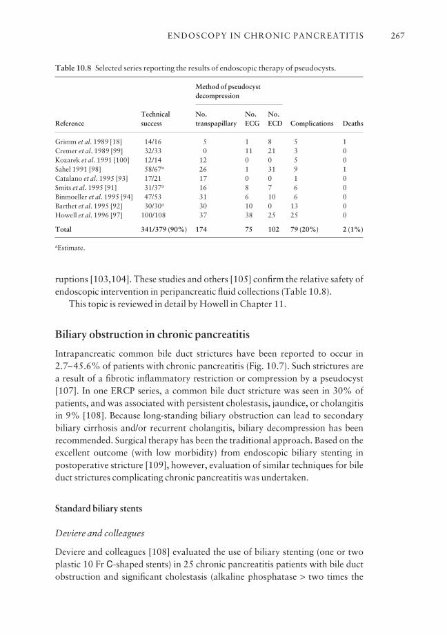
ruptions [103,104]. These studies and others [105] confirm the relative safety ofendoscopic intervention in peripancreatic fluid collections (Table 10.8).
This topic is reviewed in detail by Howell in Chapter 11.
Biliary obstruction in chronic pancreatitis
Intrapancreatic common bile duct strictures have been reported to occur in2.7–45.6% of patients with chronic pancreatitis (Fig. 10.7). Such strictures area result of a fibrotic inflammatory restriction or compression by a pseudocyst[107]. In one ERCP series, a common bile duct stricture was seen in 30% ofpatients, and was associated with persistent cholestasis, jaundice, or cholangitisin 9% [108]. Because long-standing biliary obstruction can lead to secondary biliary cirrhosis and/or recurrent cholangitis, biliary decompression has beenrecommended. Surgical therapy has been the traditional approach. Based on theexcellent outcome (with low morbidity) from endoscopic biliary stenting inpostoperative stricture [109], however, evaluation of similar techniques for bileduct strictures complicating chronic pancreatitis was undertaken.
Standard biliary stents
Deviere and colleagues
Deviere and colleagues [108] evaluated the use of biliary stenting (one or twoplastic 10 Fr C-shaped stents) in 25 chronic pancreatitis patients with bile ductobstruction and significant cholestasis (alkaline phosphatase > two times the
ENDOSCOPY IN CHRONIC PANCREATITIS 267
Table 10.8 Selected series reporting the results of endoscopic therapy of pseudocysts.
Method of pseudocyst decompression
Technical No. No. No. Reference success transpapillary ECG ECD Complications Deaths
Grimm et al. 1989 [18] 14/16 5 1 8 5 1Cremer et al. 1989 [99] 32/33 0 11 21 3 0Kozarek et al. 1991 [100] 12/14 12 0 0 5 0Sahel 1991 [98] 58/67a 26 1 31 9 1Catalano et al. 1995 [93] 17/21 17 0 0 1 0Smits et al. 1995 [91] 31/37a 16 8 7 6 0Binmoeller et al. 1995 [94] 47/53 31 6 10 6 0Barthet et al. 1995 [92] 30/30a 30 10 0 13 0Howell et al. 1996 [97] 100/108 37 38 25 25 0
Total 341/379 (90%) 174 75 102 79 (20%) 2 (1%)
aEstimate.
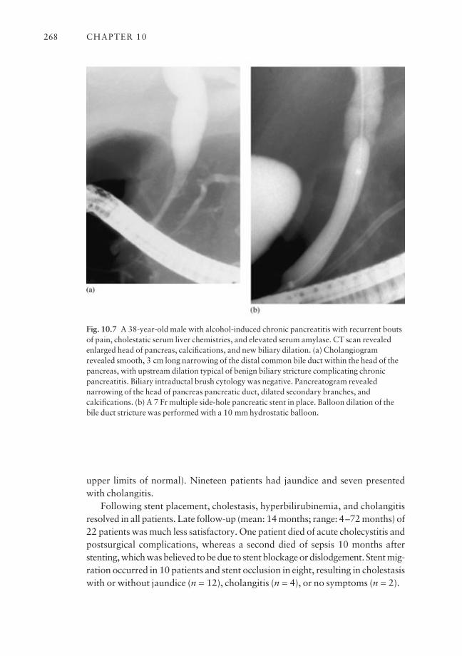
upper limits of normal). Nineteen patients had jaundice and seven presentedwith cholangitis.
Following stent placement, cholestasis, hyperbilirubinemia, and cholangitisresolved in all patients. Late follow-up (mean: 14 months; range: 4–72 months) of22 patients was much less satisfactory. One patient died of acute cholecystitis andpostsurgical complications, whereas a second died of sepsis 10 months afterstenting, which was believed to be due to stent blockage or dislodgement. Stent mig-ration occurred in 10 patients and stent occlusion in eight, resulting in cholestasiswith or without jaundice (n = 12), cholangitis (n = 4), or no symptoms (n = 2).
CHAPTER 10268
Fig. 10.7 A 38-year-old male with alcohol-induced chronic pancreatitis with recurrent boutsof pain, cholestatic serum liver chemistries, and elevated serum amylase. CT scan revealedenlarged head of pancreas, calcifications, and new biliary dilation. (a) Cholangiogramrevealed smooth, 3 cm long narrowing of the distal common bile duct within the head of thepancreas, with upstream dilation typical of benign biliary stricture complicating chronicpancreatitis. Biliary intraductal brush cytology was negative. Pancreatogram revealednarrowing of the head of pancreas pancreatic duct, dilated secondary branches, andcalcifications. (b) A 7 Fr multiple side-hole pancreatic stent in place. Balloon dilation of thebile duct stricture was performed with a 10 mm hydrostatic balloon.
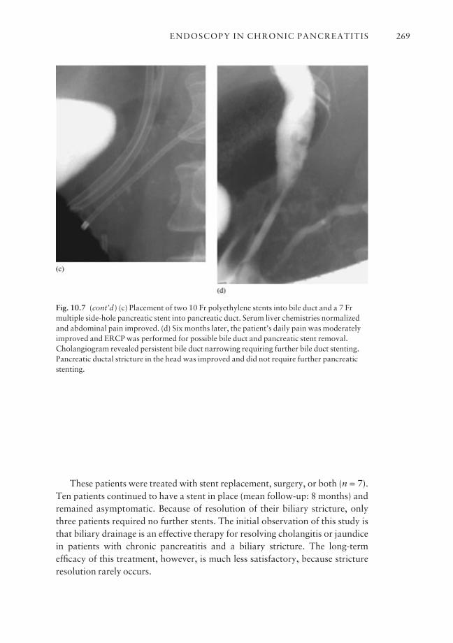
These patients were treated with stent replacement, surgery, or both (n = 7).Ten patients continued to have a stent in place (mean follow-up: 8 months) andremained asymptomatic. Because of resolution of their biliary stricture, onlythree patients required no further stents. The initial observation of this study isthat biliary drainage is an effective therapy for resolving cholangitis or jaundicein patients with chronic pancreatitis and a biliary stricture. The long-termefficacy of this treatment, however, is much less satisfactory, because strictureresolution rarely occurs.
ENDOSCOPY IN CHRONIC PANCREATITIS 269
Fig. 10.7 (cont’d ) (c) Placement of two 10 Fr polyethylene stents into bile duct and a 7 Frmultiple side-hole pancreatic stent into pancreatic duct. Serum liver chemistries normalizedand abdominal pain improved. (d) Six months later, the patient’s daily pain was moderatelyimproved and ERCP was performed for possible bile duct and pancreatic stent removal.Cholangiogram revealed persistent bile duct narrowing requiring further bile duct stenting.Pancreatic ductal stricture in the head was improved and did not require further pancreaticstenting.
The Amsterdam group
The Amsterdam group reported their results of placing 10 Fr biliary stents in 52 chronic pancreatitis patients with cholestasis [15]. Jaundice and cholestasisdisappeared within 2 weeks after stent insertion in all patients. During a medianfollow-up duration of 32 months (range: 3 months to 10 years), 17 patients(33%) had their stent removed without return of cholestasis. Complete resolu-tion of the stricture was seen in 10 of the 17 patients. This suggested that com-plete resolution of the stricture was not necessary for long-term relief ofsymptoms and cholestasis.
Barthet and colleagues
Barthet and colleagues [110] also found that biliary stenting is not a definitivetherapy for chronic pancreatitis patients with a distal common bile duct stric-ture. In their series of 19 patients (mean duration of stenting: 10 months), only two had complete clinical (resolution of symptoms), biological (normalizationof cholestatic liver tests), and radiological (resolution of biliary stricture andupstream dilation) recovery. Six of 10 (60%) possible clinical successes, eight of 19 (42%) possible biological successes, and three of 19 (16%) possible radiological successes were obtained.
Metal stents for biliary obstruction?
Because of the disappointing results with plastic stents and the concern about thehigh morbidity associated with surgically performed biliary drainage proce-dures in alcoholic (frequently debilitated) patients, the group from Brussels evalu-ated the use of uncoated expandable metal stents for this indication [112].
Twenty patients were treated with a 34 mm long metal stent, which becomes10 mm in diameter when fully expanded. The short length of the stent was chosen so that surgical bypass (e.g. choledochoduodenostomy) would still bepossible if necessary. Cholestasis (n = 20), jaundice (n = 7), and cholangitis (n = 3) resolved in all patients. Eighteen patients had no further biliary problemsduring a follow-up period of 33 months (range: 24–42 months). Two patients(10%) developed epithelial hyperplasia within the stent, resulting in recurrentcholestasis in one and jaundice in the other. These patients were treated endo-scopically with standard plastic stents, with one ultimately requiring surgicaldrainage. The authors concluded that this therapy could be an effective alterna-tive to surgical biliary diversion, but longer follow-up and controlled trials arenecessary to confirm these results.
CHAPTER 10270
In a recent abstract report, the Amsterdam group reported the long-term follow-up (mean: 50 months) of a cohort of 13 patients with chronic pancreatitis-induced biliary strictures who had undergone uncovered biliary Wallstent place-ment. Endoscopic Wallstent was successfully placed in all patients between 1994and 1999. Nine patients (69%) were successfully treated and four patients failedWallstent therapy. Of the nine patients treated successfully, four (44%) patientsrequired repeated endoscopic intervention (three with a second Wallstent andone requiring cleaning with a balloon). One patient eventually required surgicalbiliary diversion and three patients are continuing to need endoscopic plasticstents through the Wallstent to maintain biliary patency [136].
Biodegradable stents
A recent exciting development in stent technology, utilizing bioabsorbable poly-l-lactide (PLLA) polymer strands woven into the tubular mesh design similar tothe metallic stent, was reported by Haber et al. [111]. The PLLA stent is uniquein that it undergoes slow hydrolytic degradation and disintegration after 6–18months. In the feasibility study in patients with malignant obstructive jaundice,the endoscopic technique for placement of the bioabsorbable biliary stent wassimilar to present expandable stents and was technically successful in 48 of 50 patients. The unique feature of this stent is that it may obviate the need forfollow-up endoscopy to remove/replace the stent and may potentially be an effec-tive long-term option in benign, chronic pancreatitis-induced biliary strictures.
Stenting for biliary strictures and chronic pancreatitis: conclusion
The aforementioned studies indicate that plastic biliary stents are a useful alter-native to surgery for short-term treatment of chronic pancreatitis-induced com-mon bile duct strictures complicated by cholestasis, jaundice, and cholangitis.This therapy also should be considered for high-risk surgical patients. Becausethe long-term efficacy of this treatment is much less satisfactory, however, oper-ative intervention appears to be a better long-term solution for this problem inaverage-risk patients. More data on the long-term outcome, preferably in con-trolled trials, are necessary before expandable metal stents can be advocated for this indication. Trials of membrane-coated metal stents, bioabsorbablestents, and removable coil spring stents are awaited.
Sphincter of Oddi dysfunction in chronic pancreatitis
Although sphincter of Oddi dysfunction (SOD) is a known cause of acute recur-rent pancreatitis, its role in the pathogenesis of chronic pancreatitis is much lesscertain [113].
ENDOSCOPY IN CHRONIC PANCREATITIS 271
Pathogenesis of SOD in chronic pancreatitis
A direct effect of alcohol on the sphincter of Oddi has been postulated [114]. Instudies performed in humans with T-tubes, it was demonstrated that intragas-tric or intravenous [115] administration of alcohol increased the sphincter tone.
Moreover, Guelrud and colleagues [116] showed that local instillation ofalcohol on the papilla of Vater produced a significant increase in the basal pan-creatic sphincter pressure at sphincter of Oddi manometry in both cholecystec-tomy patients and patients with chronic pancreatitis. The authors postulatedthat the increased motor activity of the sphincter of Oddi may raise the intraduc-tular pancreatic pressure and result in disruption of small pancreatic ductules,and back flow of pancreatic juice into the parenchyma, with subsequent injury.
Other investigators have refuted these findings by showing that intravenousor intragastric administration of alcohol in humans results in a decrease insphincter of Oddi basal pressures at manometry [117].
In a preliminary study, Morita et al. showed that chronic alcohol administra-tion in the Japanese monkey resulted in an increase in sphincter of Oddi mean basalpressure from 9 to 20 mmHg (P < 0.01), while the phasic amplitude decreasedby 75% and the pancreatic ductal secretory rate nearly doubled [118].
Frequency of SOD in chronic pancreatitis
More recent studies using modern manometric techniques have shown a highfrequency of basal sphincter pressure abnormalities, especially the pancreaticsphincter, in patients with established chronic pancreatitis [119]. Results ofother studies using sphincter of Oddi manometry refute these findings and haveshown no difference in the dynamics of the pancreatic sphincter in patients withchronic pancreatitis and controls [120]. Such data suggest that the sphincter, attimes, becomes dysfunctional as part of the overall general scarring process orhas a role in the pathogenesis of chronic pancreatitis.
Surgical sphincter ablation
The surgical literature, although limited, suggests that sphincter ablation ther-apy (both the biliary and pancreatic sphincters) alone for patients with chronicpancreatitis and manometrically documented or suspected SOD benefits 30–60% of patients [121,122]. Bagley and associates [123] reported a surgical seriesof 67 patients with mild to moderate chronic pancreatitis undergoing empiricalbiliary and pancreatic sphincterotomy (n = 33) or sphincteroplasty (n = 34).During a 5-year follow-up, 44% of patients had pain relief. The outcome forpatients with idiopathic chronic pancreatitis was similar to that for patients
CHAPTER 10272
with alcohol-induced chronic pancreatitis. However, 92% (11/12) of patientswho stopped alcohol consumption were clinically improved, compared with12.5% (2/16) of those who continued to drink.
Endoscopic pancreatic sphincterotomy
Because endoscopic pancreatic sphincterotomy has been performed infrequentlyin most institutions, its role in the management of pancreatic sphincter stenosishas not been defined. Kozarek et al. reported resolution of pain and clinicalepisodes of pancreatitis after pancreatic sphincterotomy in six of 10 patients (1-year follow-up) with chronic pancreatitis and suspected or manometrically docu-mented pancreatic SOD [63]. Okolo et al. retrospectively evaluated 55 patientswho had undergone endoscopic pancreatic sphincterotomy over a 4-year period.After a median follow-up of 16 months, 62% of patients reported improvementof pain scores. Patients with pancreatic sphincter dysfunction (n = 15) hadsignificant improvement in pain (73%) compared to patients with pancreato-graphic evidence of chronic pancreatitis (58%) [137]. The utility of endoscopicsphincter ablation as the only therapy in patients with chronic pancreatitisawaits further study, preferably in controlled randomized trials.
Pancreas divisum
Pancreas divisum is the most common congenital variant of pancreatic ductalanatomy, occurring in 7% of autopsy series [124]. Most commonly, in the setting of chronic pancreatitis, minor papilla sphincterotomy is performed toprovide access to the duct to effect stone retrieval or facilitate endoprosthesisplacement [9].
Pancreas divisum: a cause of pancreatitis?
It has been postulated that, in a subpopulation of pancreas divisum patients, theminor papilla orifice appears to be critically small, such that excessively highintrapancreatic dorsal duct pressures occur during active secretion [124]. Thismay result in pancreatic pain or pancreatitis [125]. Although most authoritiesagree that pancreas divisum is a definite cause of acute recurrent pancreatitis, itsrole in the pathogenesis of chronic pancreatitis is much more controversial.Several lines of evidence favor the association of pancreas divisum and pancre-atitis, including: (1) the presence of pancreatographic and histological changes ofchronic pancreatitis isolated to the dorsal pancreas; (2) an increased incidence ofpancreas divisum in patients with idiopathic pancreatitis; and (3) symptomaticbenefit following dorsal duct drainage, endoscopically or surgically [124].
ENDOSCOPY IN CHRONIC PANCREATITIS 273

Minor papilla ablation
Although minor papilla sphincter therapy by endoscopic or surgical techniqueshas been shown to be effective for patients with pancreas divisum and acuterecurrent pancreatitis, the outcome for patients with chronic pancreatitis hasusually been much less satisfactory [21,56,126–132] (Table 10.9). In summariz-ing 54 patients undergoing dorsal duct decompressive therapy by minor papillasphincterotomy and/or dorsal duct stenting, only 44% improved during a meanfollow-up of 22 months.
A recent 4-year follow-up summary from our institution showed a similar62–70% symptom improvement rate for pancreas divisum patients with andwithout dorsal duct chronic pancreatitis changes. These data suggest that methods used to select patients with pancreas divisum and chronic pancreatitiswho are likely to benefit from endoscopic therapy need further investigation. Therole of botulinum toxin use in predicting pain relief warrants further study [133].
Until such methods are identified, minor papilla sphincterotomy (as the onlytherapy) for patients with chronic pancreatitis should preferably be performedin a research setting and restricted to patients who are disabled by pain.
Outstanding issues and future trends
Endoscopic therapy of chronic pancreatitis is an expanding area for the inter-ventional endoscopist. The techniques employed are very similar to the endo-
CHAPTER 10274
Table 10.9 Selected series reporting the results of minor papilla therapy for pancreasdivisum.
Acute recurrent Chronic pancreatitis Pain alone pancreatitis
Meanfollow-up Improved Improved Improved
Reference (months) No. (%) No. (%) No. (%)
Soehendra et al. 1986 [126] 3 2 100 0 a 4 75Ligoury et al. 1986 [127] 24 8 63 0 a 0 aMcCarthy et al. 1988 [21] 21 19 89 0 a 0 aLans et al. 1992 [128] 30 10 90 0 a 0 aLehman et al. 1993 [56] 22 17 76 23 26 11 27Coleman et al. 1994 [129] 23 9 78 5 0 20 60Sherman et al. 1994 [130] 28 0 a 16 44 0 aKozarek et al. 1995 [131] 20 15 73 5 20 19 32
Total 80 80 49 29 54 44
scopic interventions utilized in the biliary tree but tend to be more tedious. Theappropriate selection of candidates for the various pancreatic interventionsappears to be important to obtain optimal results of therapy. The continuedimprovement in resolution of magnetic resonance cholangiopancreatographymay allow for suitable patient selection for endoscopic therapy without the needto perform an initial diagnostic ERCP [134,135].
Over the past decade, multiple series totaling a few thousand patients havedemonstrated the medium-term effectiveness of endoscopic interventions inchronic pancreatitis, rivaling the medium-term outcomes from surgery in thisdisease. ESWL has proven to be indispensable in the management of patientswith pancreatic stones. However, well-designed, long-term controlled studiescomparing endoscopy to surgery in the management of patients with chronicpancreatitis are lacking. Further outcome and cost efficacy studies are awaited.The inexperienced endoscopist should exercise caution in the application ofnewer pancreatic techniques as they are technically demanding and associatedwith a small but significant complication rate.
Acknowledgment
We greatly appreciate the assistance of Joyce Eggleston in the preparation of thischapter.
References
1 Sarles H, Bernard JP, Johnson C. Pathogenesis and epidemiology of chronic pancreatitis. AnnuRev Med 1989; 40: 453–68.
2 Banks PA. Management of pancreatic pain. Pancreas 1991; 6 (Suppl. 1): S52–S59.3 Steer ML, Waxman I, Freedman S. Chronic pancreatitis. N Engl J Med 1995; 332: 1482–90.4 Widdison AL, Alvarez C, Karanjia ND, Reber HA. Experimental evidence of beneficial effects
of ductal decompression in chronic pancreatitis. Endoscopy 1991; 23: 151–4.5 Karanjia ND, Reber HA. The cause and management of the pain of chronic pancreatitis.
Gastrointest Clin North Am 1990; 19: 895–904.6 Lo SK, Lewis MPN, Reber PU et al. In-vivo endoscopic trans-sphincteric measurement of pan-
creatic blood flow (PBF) in humans. Gastrointest Endosc 1996; 43: 409A.7 Malfertheiner P, Buchler M. Indications for endoscopic or surgical therapy in chronic pan-
creatitis. Endoscopy 1991; 23: 185–90.8 Bedford RA, Howerton DH, Geenen JE. The current role of ERCP in the treatment of benign
pancreatic disease. Endoscopy 1994; 26: 113–19.9 Kozarek RA, Traverso LW. Endotherapy of chronic pancreatitis. Int J Pancreatol 1996; 19:
93–102.10 Kaikaus RM, Geenen JE. Current role of ERCP in the management of benign pancreatic dis-
ease. Endoscopy 1996; 28: 131–7.11 Cavallini G, Tittobello A, Frulloni L et al. Gabexate for the prevention of pancreatic damage
related to ERCP. N Engl J Med 1996; 335: 919–23.12 Deviere J, Le Moine O, Van Laethem JL et al. Interleukin-10 reduces the incidence of pan-
creatitis after therapeutic endoscopic retrograde cholangiopancreatography. Gastroenterology2001; 120 (2): 498–505.
ENDOSCOPY IN CHRONIC PANCREATITIS 275
13 Sherman S, Lehman GA. Endoscopic therapy of pancreatic disease. Gastroenterologist 1993;1: 5–17.
14 Kozarek RA. Chronic pancreatitis in 1994: is there a role for endoscopic treatment?Endoscopy 1994; 26: 625–8.
15 Huibregtse K, Smits ME. Endoscopic management of diseases of the pancreas. Am JGastroenterol 1994; 89 (Suppl.): S66–S77.
16 Jacob L, Geenen JE, Catalano MF, Geenen DJ. Prevention of pancreatitis in patients with idio-pathic recurrent pancreatitis: a prospective nonblinded randomized study using endoscopicstents. Endoscopy 2001; 33: 559–62.
17 Cremer M, Deviere J, Delhaye M et al. Stenting in severe chronic pancreatitis: results ofmedium-term follow-up in 76 patients. Endoscopy 1991; 23: 171–6.
18 Grimm H, Meyer WH, Nam VC, Soehendra N. New modalities for treating chronic pancreat-itis. Endoscopy 1989; 21: 70–4.
19 Cremer M, Deviere J, Delhaye M et al. Nonsurgical management of severe chronic pancreat-itis. Scand J Gastroenterol 1990; 25 (Suppl. 175): 77–84.
20 Kozarek RA, Patterson DJ, Ball TJ, Traverso LW. Endoscopic placement of pancreatic stentsand drains in the management of pancreatitis. Ann Surg 1989; 209: 261–6.
21 McCarthy J, Geenen JE, Hogan WJ. Preliminary experience with stent placement in benignpancreatic diseases. Gastrointest Endosc 1988; 34: 16–18.
22 Geenen JE, Rolny P. Endoscopic therapy of acute and chronic pancreatitis. GastrointestEndosc 1991; 37: 377–82.
23 Binmoeller KF, Jue P, Seifert H et al. Endoscopic pancreatic stent drainage in chronic pan-creatitis and a dominant stricture: longterm results. Endoscopy 1995; 27: 638–44.
24 Smits ME, Badiga SM, Rauws EAJ et al. Longterm results of pancreatic stents in chronic pan-creatitis. Gastrointest Endosc 1995; 42: 461–7.
25 Ponchon T, Bory R, Hedelius F et al. Endoscopic stenting for pain relief in chronic pancreatitis:results of a standardized protocol. Gastrointest Endosc 1995; 42: 452–6.
26 Rosch T, Daniel S, Scholz M et al. Endoscopic treatment of chronic pancreatitis: a multicenterstudy of 1000 patients with long-term follow-up. Endoscopy 2002; 34 (10): 765–71.
27 Reber PU, Patel AG, Kusske AM et al. Stenting does not decompress the pancreatic duct as effectively as surgery in experimental chronic pancreatitis. Gastroenterology 1995; 128:386A.
28 Nakaizumi A, Uehara H, Takensaka A et al. Diagnosis of pancreatic cancer by cytology andmeasurement of oncogene and tumor markers in pure pancreatic juice aspirated by endoscopy.Hepatogastroenterology 1999; 46: 31–7.
29 Brandwein SL, Farrell JJ, Centeno BA, Brugge WR. Detection and tumor staging of malignancyin cystic intraductal, and solid tumors of the pancreas by EUS. Gastrointest Endosc 2001; 53:722–7.
30 Lohr M, Muller P, Mora J et al. p53 and K-ras mutations in pancreatic juice samples frompatients with chronic pancreatitis. Gastrointest Endosc 2001; 53: 734–43.
31 Freeman M, Cass OW, Dailey J. Dilation of high-grade pancreatic and biliary ductal strictureswith small-caliber angioplasty balloons. Gastrointest Endosc 2001; 54: 89–92.
32 Van Someren R, Benson M, Glynn M, Ashraf W, Swain P. A novel technique for dilatingdifficult malignant biliary strictures during therapeutic ERCP. Gastrointest Endosc 1996; 43:495–8.
33 Baron T, Morgan D. Dilation of a difficult benign pancreatic duct stricture using the Soehendrastent extractor. Gastrointest Endosc 1997; 46: 178–80.
34 Pasricha PJ, Kalloo AN. Successful endoscopic management of complete obstruction of themain pancreatic duct (MPD) in patients with chronic pancreatitis. Gastrointest Endosc 1993;39: 320A.
35 Ikenberry SO, Sherman S, Hawes RH et al. The occlusion rate of pancreatic stents.Gastrointest Endosc 1994; 40: 611–13.
36 Cremer M, Suge B, Delhaye M et al. Expandable pancreatic metal stents (Wallstent) forchronic pancreatitis: first world series. Gastroenterology 1990; 98: 215A.
37 Cremer M, Deviere J, Delhaye M et al. Endoscopic management of chronic pancreatitis. ActaGastroenterol Belg 1993; 56: 192–200.
CHAPTER 10276

38 Choudari C, Nickl N, Fogel E et al. Hereditary pancreatitis: clinical presentation, ERCPfindings and outcome of endoscopic therapy. Gastrointest Endosc 2002; 56: 66–71.
39 Gabbrielli A, Mutignani M, Pandolfi M et al. Endotherapy of early onset idiopathic chronicpancreatitis: results with long-term follow-up. Gastrointest Endosc 2002; 55: 488–93.
40 Ashby K, Lo SK. The role of pancreatic stenting in obstructive ductal disorders other than pan-creas divisum. Gastrointest Endosc 1996; 42: 306–11.
41 Burdick JS, Geenen JE, Hogan W et al. Pancreatic stent therapy in chronic pancreatitis: whichpatients benefit? Gastrointest Endosc 1993; 39: 309A.
42 Borel I, Saurin J-C, Napoleon B et al. Treatment of chronic pancreatitis using definitive stent-ing of the main pancreatic duct. Gastrointest Endosc 2001; 53 (5): 139A.
43 McHenry L, Gore DC, DeMaria EJ, Zfass AM. Endoscopic treatment of dilated duct chronicpancreatitis with pancreatic stents: preliminary results of a sham controlled, blinded, crossovertrial to predict surgical outcome. Am J Gastroenterol 1993; 88: 1536A.
44 DuVall GA, Scheider DM, Kortan P, Haber GB. Is the outcome of endoscopic therapy ofchronic pancreatitis predictive of surgical success? Gastrointest Endosc 1996; 43: 405A.
45 Ammann RW, Akovbiantz A, Larglader F, Schueler G. Course and outcome of chronic pancre-atitis: longitudinal study of a mixed medical-surgical series of 245 patients. Gastroenterology1984; 86: 820–8.
46 Cotton PB, Lehman G, Vennes J et al. Endoscopic sphincterotomy complications and theirmanagement: an attempt at consensus. Gastrointest Endosc 1991; 37: 383–93.
47 Siegel J, Veerappan A. Endoscopic management of pancreatic disorders: potential risks of pancreatic prostheses. Endoscopy 1991; 23: 177–80.
48 Leung J, Liu Y, Herrera J et al. Bacteriological and scanning electron microscopy (SEM) ana-lyses of pancreatic stents. Gastrointest Endosc 2001; 53 (5): 138A.
49 Sherman S, Alvarez C, Robert M et al. Polyethylene pancreatic stent-induced changes in thenormal dog pancreas. Gastrointest Endosc 1993; 39: 658–64.
50 Johanson JF, Schmalz MJ, Geenen JE. Incidence and risk factors for biliary and pancreaticstent migration. Gastrointest Endosc 1992; 38: 341–6.
51 Dean RS, Geenen JE, Hogan WJ et al. Pancreatic stent modification to prevent stent migrationin patients with benign pancreatic disease. Gastrointest Endosc 1994; 40: 19A.
52 Kozarek RA. Pancreatic stents can induce ductal changes consistent with chronic pancreatitis.Gastrointest Endosc 1990; 36: 93–5.
53 Derfus GA, Geenen JE, Hogan WJ. Effect of endoscopic pancreatic duct stent placement onpancreatic ductal morphology. Gastrointest Endosc 1990; 36: 206A.
54 Rossos PG, Kortan P, Haber GB. Complications associated with pancreatic duct stenting.Gastrointest Endosc 1992; 38: 252A.
55 Burdick JS, Geenen JE, Venu RP et al. Ductal morphological changes due to pancreatic stenttherapy: a randomized controlled study. Am J Gastroenterol 1992; 87: 155A.
56 Lehman GA, Sherman S, Nisi R, Hawes RH. Pancreas divisum: results of minor papilla sphinc-terotomy. Gastrointest Endosc 1993; 39: 1–8.
57 Smith MT, Sherman S, Ikenberry SO et al. Alterations in pancreatic ductal morphology follow-ing polyethylene pancreatic duct stenting. Gastrointest Endosc 1996; 44: 268–75.
58 Eisen G, Coleman S, Troughton A, Cotton PB. Morphological changes in the pancreatic ductafter stent placement for benign pancreatic disease. Gastrointest Endosc 1994; 40: 107A.
59 Sherman S, Hawes RH, Savides TJ et al. Stent-induced pancreatic ductal and parenchymalchanges: correlation of endoscopic ultrasound with ERCP. Gastrointest Endosc 1996; 4:276–82.
60 Deviere J, Delhaye M, Cremer M. Pancreatic duct stones management. Gastrointest EndoscClin N Am 1998; 8: 163–79.
61 Sarles A, Bernard JP. Lithostathine and pancreatic lithogenesis. Viewpoints Dig Dis 1991; 23:7–12.
62 Suda K, Mogaki M, Oyama T, Matsumoto Y. Histopathologic and immunohistochemicalstudies on alcoholic pancreatitis and chronic obstructive pancreatitis: special emphasis on duc-tal obstruction and genesis of pancreatitis. Am J Gastroenterol 1990; 85: 271–6.
63 Kozarek R, Ball TJ, Patterson DJ et al. Endoscopic pancreatic duct sphincterotomy: indica-tions, technique, and analysis of results. Gastrointest Endosc 1994; 40: 592–8.
ENDOSCOPY IN CHRONIC PANCREATITIS 277
64 Esber E, Sherman S, Earle D et al. Complications of major papilla pancreatic sphincterotomy:a review of 106 patients. Gastrointest Endosc 1995; 41: 422A.
65 Kim MH, Myung SJ, Kim YS et al. Routine biliary sphincterotomy may not be indispensablefor endoscopic pancreatic sphincterotomy. Endoscopy 1998; 30: 697–701.
66 Delhaye M, Vandermeeren A, Baize M, Cremer M. Extracorporeal shockwave lithotripsy ofpancreatic calculi. Gastroenterology 1992; 102: 610–20.
67 Sherman S, Lehman GA, Hawes RH et al. Pancreatic ductal stones: frequency of successfulendoscopic removal and improvement in symptoms. Gastrointest Endosc 1991; 37: 511–17.
68 Smits ME, Rauws EA, Tytgat GNJ, Huibregtse K. Endoscopic treatment of pancreatic stonesin patients with chronic pancreatitis. Gastrointest Endosc 1996; 43: 556–60.
69 Schneider MU, Lux G. Floating pancreatic duct concrements in chronic pancreatitis.Endoscopy 1985; 17: 8–10.
70 Fuji T, Amano H, Ohmura R et al. Endoscopic pancreatic sphincterotomy: technique and evalu-ation. Endoscopy 1989; 21: 27–30.
71 Kozarek RA, Ball TJ, Patterson DJ. Endoscopic approach to pancreatic duct calculi andobstructive pancreatitis. Am J Gastroenterol 1992; 87: 600–3.
72 Sauerbruch T, Holl J, Sackman M et al. Disintegration of a pancreatic duct stone with extra-corporeal shockwaves in a patient with chronic pancreatitis. Endoscopy 1987; 19: 207–8.
73 Soehendra N, Grimm H, Meyer HW et al. Extrakorporale stobwellen lithotripsie bei chronis-cher pankreatitis. Dtsch Med Wochenschr 1989; 114: 1402–6.
74 Neuhaus H. Fragmentation of pancreatic stones by extracorporeal shock wave lithotripsy.Endoscopy 1989; 23: 161–5.
75 den Toom R, Nijs HG, van Blankenstein M et al. Extracorporeal shock wave lithotripsy ofpancreatic duct stones. Am J Gastroenterol 1991; 86: 1033–6.
76 Sauerbruch T, Holl J, Sackmann M, Paumgartner G. Extracorporeal lithotripsy of pancreaticstones in patients with chronic pancreatitis and pain: a prospective followup study. Gut 1992;33: 969–72.
77 Schneider HT, May A, Benninger J et al. Piezoelectric shock wave lithotripsy of pancreatic ductstones. Am J Gastroenterol 1994; 89: 2042–8.
78 van der Hul R, Plaiser P, Jeekel J et al. Extracorporeal shockwave lithotripsy of pancreatic ductstones: immediate and longterm results. Endoscopy 1994; 26: 573–8.
79 Dumonceau JE, Deviere J, LeMoine O et al. Endoscopic pancreatic drainage in chronic pancre-atitis associated with ductal stones: longterm results. Gastrointest Endosc 1996; 43: 547–55.
80 Kozarek R, Brandabur J, Ball T et al. Clinical outcomes in patients who undergo extracorpo-real shock wave lithotripsy for chronic pancreatitis. Gastrointest Endosc 2002; 56: 496–500.
81 Farnbacher M, Schoen C, Rabenstein T et al. Pancreatic ductal stones in chronic pancreatitis:criteria for treatment intensity success. Gastrointest Endosc 2002; 56: 501–6.
82 Alvarez C, Widdison AL, Reber HA. New perspectives in the surgical management of chronicpancreatitis. Pancreas 1991; 6 (Suppl. 1): 576–81.
83 Fogel EL, Eversman D, Jamidar P, Sherman S, Lehman GA. Sphincter of Oddi dysfunction:pancreaticobiliary sphincterotomy with pancreatic stent placement has a lower rate of pancre-atitis than biliary sphincterotomy alone. Endoscopy 2002; 34: 325–9.
84 Neuhaus H, Hoffman W, Classen M. Laser lithotripsy of pancreatic and biliary stones via 3.4mm and 3.7 mm miniscopes: first clinical results. Endoscopy 1992; 24: 208–14.
85 Sahel J, Sarles H. (1981) Citrate therapy in chronic calcifying pancreatitis: preliminary results.In: Mitchell, CJ, Keelleheer, J, eds. Pancreatic Disease in Clinical Practice. London: Pitman,346–53.
86 Berger Z, Topa L, Takacs T, Pap A. Nasopancreatic drainage for chronic calcifying pancreat-itis (CCP). Digestion 1992; 52: 70A.
87 Noda A, Shibata T, Ogawa Y et al. Dissolution of pancreatic stones by oral trimethadione in adog experimental model. Gastroenterology 1987; 93: 1002–8.
88 Lehman GA, Sherman S. Pancreatic stones: to treat or not to treat? Gastrointest Endosc 1996;43: 625–6.
89 Grace PA, Williamson RCN. Modern management of pancreatic pseudocysts. Br J Surg 1993;80: 573–81.
CHAPTER 10278
90 Gumaste VV, Pitchumoni CS. Pancreatic pseudocysts. Gastroenterologist 1996; 4: 33–43.91 Smits ME, Rauws EAJ, Tytgat GNJ, Huibregtse K. The efficacy of endoscopic treatment of
pancreatic pseudocysts. Gastrointest Endosc 1995; 42: 202–7.92 Barthet M, Sahel J, BodiouBertel C, Bernard JP. Endoscopic transpapillary drainage of pancre-
atic pseudocysts. Gastrointest Endosc 1995; 42: 208–13.93 Catalano MF, Geenen JE, Schmalz MJ et al. Treatment of pancreatic pseudocysts with ductal
communication by transpapillary pancreatic duct endoprosthesis. Gastrointest Endosc 1995;42: 214–18.
94 Binmoeller KF, Seifert H, Walter A, Soehendra N. Transpapillary and transmural drainage ofpancreatic pseudocysts. Gastrointest Endosc 1995; 42: 219–24.
95 Lehman GA. Endoscopic management of pancreatic pseudocysts continues to evolve. Gastro-intest Endosc 1995; 42: 273–5.
96 Howell DA, Lehman GA, Baron TH et al. Endoscopic treatment of pancreatic pseudocysts: aretrospective multicenter analysis. Gastrointest Endosc 1995; 41: 424A.
97 Howell DA, Lehman GA, Baron TH et al. Recurrent pseudocyst formation in patients man-aged with endoscopic drainage: predrainage features and management. Gastrointest Endosc1996; 43: 407A.
98 Sahel J. Endoscopic drainage of pancreatic cysts. Endoscopy 1991; 23: 181–4.99 Cremer M, Deviere J, Engelholm L. Endoscopic management of cysts and pseudocysts in
chronic pancreatitis: long term followup after 7 years’ experience. Gastrointest Endosc 1989;35: 1–9.
100 Kozarek RA, Ball TJ, Patterson DJ et al. Endoscopic transpapillary therapy for disrupted pancreatic duct and parapancreatic fluid collection. Gastroenterology 1991; 100: 1362–70.
101 Deviere J, Buseo H, Baize M et al. Complete disruption of the main pancreatic duct: endo-scopic management. Gastrointest Endosc 1995; 42: 445–51.
102 Espinel J, Jorquera F, Fernandez-Gundin MJ, Munoz F, Herrera A, Olcoz JL. Endoscopictranspapillary drainage of an infected pancreatic fluid collection in pancreas divisum. Dig DisSci 2000; 45: 237–41.
103 Kozarek RA, Jiranek G, Traverso LW. Endoscopic treatment of pancreatic ascites. Am J Surg1994; 168: 223–8.
104 Kim HS, Lee DK, Kim IW et al. The role of endoscopic retrograde pancreatography in the treat-ment of traumatic pancreatic duct injury. Gastrointest Endosc 2001; 54: 49–55.
105 Costamagna G, Mutignani M, Igrosso M et al. Endoscopic treatment of postsurgical externalpancreatic fistulas. Endoscopy 2001; 33: 317–22.
106 Lau ST, Simchuk EJ, Kozarek RA, Traverso LW. A pancreatic ductal leak should be sought todirect treatment in patients with acute pancreatitis. Am J Surg 2001; 181: 411–15.
107 Frey CF, Suzuki M, Isaji S. Treatment of chronic pancreatitis complicated by obstruction of thecommon bile duct or duodenum. World J Surg 1990; 14: 59–69.
108 Deviere J, Devaere S, Baize M, Cremer M. Endoscopic biliary drainage in chronic pancreatitis.Gastrointest Endosc 1990; 36: 96–100.
109 Davids PHP, Rauws EAJ, Coene PPLO et al. Endoscopic stenting for postoperative biliarystrictures. Gastrointest Endosc 1992; 38: 12–18.
110 Barthet M, Bernard JP, Duval JL et al. Biliary stenting in benign biliary stenosis complicatingchronic calcifying pancreatitis. Endoscopy 1994; 26: 569–72.
111 Haber G, Freeman M, Bedford R et al. A prospective multi-center study of a bioabsorbable bili-ary Wallstent in 50 patients with malignant obstructive jaundice. Am J Gastroenterol 1997; 9:A200.
112 Deviere J, Cremer M, Love J et al. Management of common bile duct strictures caused bychronic pancreatitis with metal mesh self-expandable stents. Gut 1994; 35: 122–6.
113 Kuo W-H, Pasricha P, Kalloo AN. The role of sphincter of Oddi manometry in the diagnosisand therapy of pancreatic disease. Gastrointest Endosc Clin N Am 1998; 8: 79–85.
114 Guelrud M. How good is sphincter of Oddi manometry for chronic pancreatitis? Endoscopy1994; 26: 265–7.
115 Pirola RC, Davis E. Effects of ethyl alcohol on sphincter resistance at the choledochoduodenaljunction in man. Gut 1968; 9: 447–560.
ENDOSCOPY IN CHRONIC PANCREATITIS 279
116 Guelrud M, Mendoza S, Rossiter G et al. Effect of local instillation of alcohol on sphincter ofOddi motor activity: combined ERCP and manometry study. Gastrointest Endosc 1991; 37:428–32.
117 Viceconte G. Effects of ethanol on the sphincter of Oddi: an endoscopic manometry study. Gut1983; 24: 20–7.
118 Morita M, Okazaki K, Yamasaki K et al. Effects of long term administration of ethanol on the papillary sphincter and exocrine pancreas in the monkey. Gastroenterology 1994; 106:309A.
119 Vestergaard H, Krause A, Rokkjaer M et al. Endoscopic manometry of the sphincter of Oddiand the pancreatic and biliary ducts in patients with chronic pancreatitis. Scand J Gastro-enterol 1994; 29: 188–92.
120 Ugljesic M, Bulajic M, Milosavljevic T, Stimec B. Endoscopic manometry of the sphincter ofOddi and pancreatic duct in patients with chronic pancreatitis. Int J Pancreatol 1996; 19:191–5.
121 Sherman S, Hawes RH, Madura JA, Lehman GA. Comparison of intraoperative and endo-scopic manometry of the sphincter of Oddi. Surg Gynecol Obstet 1992; 175: 410–18.
122 Williamson RCN. Pancreatic sphincteroplasty: indications and outcome. Ann R Coll Surg1988; 70: 205–11.
123 Bagley FH, Braasch JW, Taylor RH, Warren KW. Sphincterotomy or sphincteroplasty in thetreatment of pathologically mild chronic pancreatitis. Am J Surg 1981; 141: 418–22.
124 Lehman GA, Sherman S. Pancreas divisum: diagnosis, clinical significance, and managementalternatives. Gastrointest Endosc Clin North Am 1995; 5: 145–70.
125 Cotton PB. Congenital anomaly of pancreas divisum as cause of obstructive pain and pancre-atitis. Gut 1980; 21: 105–14.
126 Soehendra N, Kempeneers I, Nam VC, Grimm H. Endoscopic dilation and papillotomy of theaccessory papilla and internal drainage in pancreas divisum. Endoscopy 1986; 18: 129–32.
127 Ligoury C, Lefebvre JF, Canard JM et al. Le pancreas divisum: etude clinique et therapeutiquechez l’homme: a propos de 87 cas. Gastroenterol Clin Biol 1986; 10: 820–5.
128 Lans JI, Geenen JE, Johanson JF, Hogan WJ. Endoscopic therapy in patients with pancreasdivisum and acute pancreatitis: a prospective, randomized, controlled clinical trial. Gastro-intest Endosc 1992; 38: 430–4.
129 Coleman SD, Eisen GM, Troughton AB, Cotton PB. Endoscopic treatment in pancreas divi-sum. Am J Gastroenterol 1994; 89: 1152–5.
130 Sherman S, Hawes R, Nisi R et al. Randomized controlled trial of minor papilla sphinctero-tomy (MiES) in pancreas divisum (PDiv) patients with pain only. Gastrointest Endosc 1994;40: 125A.
131 Kozarek RA, Ball TJ, Patterson DJ et al. Endoscopic approach to pancreas divisum. Dig Dis Sci1995; 40: 1974–81.
132 Ertan A. Long term results after endoscopic pancreatic stent placement without pancreaticpapillotomy in acute recurrent pancreatitis due to pancreas divisum. Gastrointest Endosc2000; 52: 9–14.
133 Wehrmann T, Schmitt T, Seifert H. Endoscopic botulinum toxin injection into the minorpapilla for treatment of idiopathic recurrent pancreatitis in patients with pancreas divisum.Gastrointest Endosc 1999; 50: 545–8.
134 Farrell RJ, Noonan N, Mahmud N, Morrin MM, Kelleher D, Keeling PWN. Potential impactof magnetic resonance cholangiopancreatography on endoscopic retrograde cholangiopancre-atography workload and complication rate in patients referred because of abdominal pain.Endoscopy 2001; 33: 668–75.
135 Sahel J, Devonshire D, Yeoh KG et al. The decision making value of magnetic resonancecholangiopancreatography in patients seen in a referral center for suspected biliary and pancre-atic disease. Am J Gastroenterol 2001; 96: 2074–80.
136 Van Berkel AM, Van Westerloo D, Cahen D et al. Efficacy of wallstents in benign biliary stric-tures due to chronic pancreatitis. Gastrointest Endosc 2003; 57: AB198.
137 Okolo PI, Pasricha PJ, Kalloo AN. What are the long-term results of endoscopic pancreaticsphincterotomy? Gastrointest Endosc 2000; 52: 15–19.
CHAPTER 10280
CHAPTER 11
Complications of Pancreatitis
DOUGLAS A. HOWELL
Synopsis
Complications of acute and chronic pancreatitis are varied, often complex, and potentially fatal. This chapter attempts to summarize all of these feareddevelopments, address their causation, and review current and potential futureapproaches.
The first section deals with the toxic and metabolic complications. Earlydeath from pancreatitis most frequently follows shock, a poorly understood butdramatic occurrence seen, fortunately, in a small minority. All organ systemsmay be affected, in a fashion that can range from mild to very severe, resulting inrenal failure, respiratory failure, disseminated intravascular coagulation, andsevere gastrointestinal bleeding. Prolonged septic-like systemic inflammatoryresponse syndrome may result in coma and a profound catabolic state untreat-able by total parenteral nutrition.
Recent clinical experience has recognized the major difference in clinicalcourse and treatment when pancreatic necrosis complicates the early phase of acute pancreatitis. Diagnosis, treatment, and predictors of outcome areaddressed.
The chapter closes with a comprehensive review of the main miscellaneouscomplications which are often recognized during the later phase of pancreatitis.Patients who appear to be recovering may experience fistula formation, produc-ing acute pancreatic hydrothorax or enteric fistulas. Percutaneous or surgicaldrainage is a frequent cause of cutaneous fistulas, a particularly difficult anddebilitating complication. Finally, dramatic and potentially fatal vascular com-plication can occur very abruptly, taxing the diagnostic and interventional skillsof the treating team.
Throughout this review, up-to-date reports of successful approaches to all ofthese events are addressed. It is emphasized that since controlled trials of newertreatment modalities have, in general, not been directly compared to traditionalsurgical therapy, a multidisciplinary approach with gastroenterology, interven-tional radiology, and surgery is vital.
281
Toxic and metabolic complications
Few diseases can produce more varied and severe diffuse metabolic complica-tions than acute pancreatitis. Although the organ weighs only 90 g in the adult,when inflamed, the pancreas can produce such profound systemic effects thatrespiratory, renal, and circulatory failure can rapidly ensue, producing multi-organ system death within a few days. In patients with severe acute pancreatitis,the likely sequence is peripancreatic leakage of proteolytic juice, activation ofproinflammatory cytokines, third space loss secondary to retroperitoneal injury,hypoperfusion, and finally, rapid progression to frank pancreatic necrosis.
This sequence is variable and may appear as partial individual organ com-promise or full-blown multiorgan failure. The recognition of these end organcomplications served as the basis for the famous ‘Ranson’s criteria’, which havefor nearly 30 years remained the most popular clinical measure of the severity ofacute pancreatitis (Table 11.1) [1].
A principal drawback to Ranson’s scoring system has been the need toreassess the patient at 48 h before a final score can be calculated. In an effort topredict severity as early as possible to help triage patients to the most appro-priate level of care, several other clinical scoring systems have been advocated[2–4]. The Acute Physiologic and Chronic Health Evaluation scoring system(APACHE II and III) permits a more comprehensive initial assessment of sever-ity. Khan et al. pointed out recently that since the natural history of pancreatitisvaries considerably depending upon underlying etiology, patient responses, andpresence of comorbidity, APACHE scoring, when repeated at 48 h, as in Ranson’scriteria, more accurately predicts outcome [4].
CHAPTER 11282
Table 11.1 Ranson’s criteria. Clinical features significantly related to the severity of anepisode of pancreatitis.
On admission Within first 48 h
1 Age > 55 years 1 Ca < 8 mg %2 Glucose > 200 mg % 2 Pao2 < 65 mmHg3 WBC > 16 000 mm3 3 Base deficit > 4 mEq/liter4 LDL > 700 IU 4 BUN increase > 5 mg %5 SGOT > 250 SFU 5 Hct fall > 10 points
6 Fluid sequestration > 6 liter
Mortality and morbidityLess than three clinical features Three or more clinical featuresMortality 3% Mortality 62%Serious illness 11% Serious illness 33%
After Ranson JH, Rifkind KM, Turner JW. Surg Gynecol Obstet 1976; 143: 209–19.
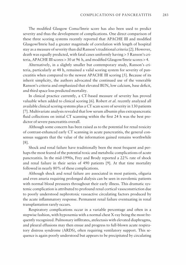
The modified Glasgow Coma/Imrie score has also been used to predictseverity and thus the development of complications. One direct comparison ofthese three scoring systems recently reported that APACHE III and modifiedGlasgow/Imrie had a greater magnitude of correlation with length of hospitalstay as a measure of severity than did Ranson’s traditional criteria [2]. However,death was equally predicted, with fatal cases uniformly having > 5 Ranson’s cri-teria, APACHE III scores > 30 at 96 h, and modified Glasgow/Imrie scores > 4.
Alternatively, in a slightly smaller but contemporary study, Ranson’s cri-teria, particularly at 48 h, remained a valid scoring system for severity of pan-creatitis when compared to the newest APACHE III scoring [5]. Because of itsinherit simplicity, the authors advocated the continued use of the venerableRanson’s criteria and emphasized that elevated BUN, low calcium, base deficit,and third space loss predicted mortality.
In clinical practice currently, a CT-based measure of severity has provedvaluable when added to clinical scoring [6]. Robert et al. recently analyzed allavailable clinical scoring systems plus a CT scan score of severity in 130 patients[7]. Multivariate analysis revealed that low serum albumin plus extrapancreaticfluid collections on initial CT scanning within the first 24 h was the best pre-dictor of severe pancreatitis overall.
Although some concern has been raised as to the potential for renal toxicityof contrast-enhanced early CT scanning in acute pancreatitis, the general con-sensus suggests that the value of the information gained remains worthwhile [8].
Shock and renal failure have traditionally been the most frequent and per-haps the most feared of the potential toxic and metabolic complications of acutepancreatitis. In the mid-1990s, Frey and Brody reported a 22% rate of shockand renal failure in their series of 490 patients [9]. At that time mortality followed in nearly 80% of these complications.
Although shock and renal failure are associated in most patients, oliguriaand even anuria requiring prolonged dialysis can be seen in euvolemic patientswith normal blood pressures throughout their early illness. This dramatic sys-temic complication is attributed to profound renal cortical vasoconstriction dueto poorly understood nephrotoxic vasoactive circulating factors produced bythe acute inflammatory response. Permanent renal failure eventuating in renaltransplantation rarely occurs.
Respiratory complications occur in a variable percentage and often in a stepwise fashion, with hypoxemia with a normal chest X-ray being the most fre-quently recognized. Pulmonary infiltrates, atelectasis with elevated diaphragms,and pleural effusions may then ensue and progress to full-blown acute respira-tory distress syndrome (ARDS), often requiring ventilatory support. This se-quence is again poorly understood but appears to be precipitated by circulating
COMPLICATIONS OF PANCREATITIS 283
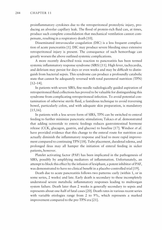
proinflammatory cytokines due to the retroperitoneal proteolytic injury, pro-ducing an alveolar capillary leak. The flood of protein-rich fluid can, at times,produce such complete consolidation that mechanical ventilation cannot com-pensate, resulting in a respiratory death [10].
Disseminated intravascular coagulation (DIC) is a less frequent complica-tion of acute pancreatitis [1]. DIC may produce severe bleeding since extensiveretroperitoneal injury is present. The consequence of such hemorrhage cangreatly worsen the above outlined systemic complications.
A more recently described toxic reaction to pancreatitis has been termed systemic inflammatory response syndrome (SIRS) [11]. High fever, tachycardia,and delirium may persist for days or even weeks and may be difficult to distin-guish from bacterial sepsis. This syndrome can produce a profoundly catabolicstate that cannot be adequately reversed with total parenteral nutrition (TPN)[12–14].
In patients with severe SIRS, fine-needle radiologically guided aspiration ofretroperitoneal fluid collections has proved to be valuable for distinguishing thissyndrome from complicating retroperitoneal infection. To avoid potential con-tamination of otherwise sterile fluid, a fastidious technique to avoid traversingbowel, particularly colon, and with adequate skin preparation, is mandatory[15,16].
In patients with a less severe form of SIRS, TPN can be switched to enteralfeeding to further minimize pancreatic stimulation; Takacs et al. demonstratedthat adding octreotide to enteric feedings reduces gastrointestinal hormonerelease (CCK, glucagon, gastrin, and glucose) to baseline [17]. Windsor et al.have provided evidence that this change to the enteral route for nutrition canactually diminish the inflammatory response and lead to more rapid improve-ment compared to continuing TPN [18]. Tube placement, duodenal edema, andprolonged ileus may all hamper the initiation of enteral feeding in sickerpatients, however.
Platelet activating factor (PAF) has been implicated in the pathogenesis ofSIRS, possibly by amplifying mediators of inflammation. Unfortunately, anattempt to block this effect by the infusion of lexiphant, a potent inhibitor of PAF,was demonstrated to have no clinical benefit in a placebo-controlled trial [19].
Death due to acute pancreatitis follows two patterns: early (within 1, or insome series, 2 weeks) and late. Early death is secondary to these incompletelyunderstood severe metabolic inflammatory responses leading to multiorgan system failure. Death later than 2 weeks is generally secondary to sepsis and represents about one-half of fatal cases [20]. Death rates in various recent serieswith variable etiologies range from 2 to 9%, which represents a markedimprovement compared to the pre-TPN era [21].
CHAPTER 11284
Factors which may be important in the sequence of rapid onset multiorgansystem failure are under active research. Ethridge et al. recently studied theimpact of cyclo-oxygenase (COX) on pancreatitis severity, especially with regardto lung injury [22]. In their mouse model, inhibition of COX-2 or deletion of the COX gene profoundly decreased the severity of pancreatitis and protectedagainst ARDS.
Host response may prove to be a critical determinate of the severity of pan-creatitis and the resulting complications [23,24]. A defect in interleukin-10function has been identified in some patients with severe acute pancreatitis [25].This defect might result in a failure to down-regulate the initial acute inflam-matory reaction once the patients have been maximally stimulated. If confirmed,this finding would help us to understand why these seemingly unpredictablecomplications may be seen even after rather trivial injury, such as after simplecannulation or unremarkable sphincterotomy at ERCP.
In an attempt to prevent post-ERCP pancreatitis, an initial randomized trialof a single bolus of interleukin-10 (8 µg/kg) was given to patients 15 min priorto ERCP and compared to a placebo group [26]. Unfortunately, both groupsexperienced an identical rate of pancreatitis and, furthermore, the severity asmeasured by length of stay was the same. Nevertheless, a full understanding ofthe role of pro- and anti-inflammatory cytokines may be the best hope of pre-venting or at least attenuating the severity of acute pancreatitis.
Pancreatic fluid collections
In the setting of pancreatitis, fluid collections are frequent and have been used asa measure of severity to create a CT-based score as previously discussed [6].When combined with extent of non-perfusion (equivalent to necrosis) anddetection of retroperitoneal complications, CT becomes a vital tool in predict-ing severity [27].
Fluid collections are at first sterile, amylase rich, and unorganized. They areseen early, often within 24 h, and may persist. Unencapsulated and youngerthan 4 weeks from the onset of pancreatitis, they are properly watched expect-antly (Fig. 11.1a). If necrosis is not present, infection is rare. Whether they willpersist, organize, and encapsulate beyond 4 weeks into a true pseudocyst largelydepends on pancreatic ductal anatomy and the presence of necrosis. Normalductal and sphincter anatomy predicts spontaneous resolution, whereas fistula,ductal obstruction, or disruption predict persistence and pseudocyst formation[28]. Finally, necrosis will organize slowly and an associated fluid collection willusually become a complex pseudocyst; this might be better termed an area of‘organizing necrosis’ to distinguish them [29].
COMPLICATIONS OF PANCREATITIS 285
Pseudocysts and abscesses
A pseudocyst of the pancreas is a maturing fluid collection surrounded by granulation tissue which occurs as a consequence of acute or chronic leakage ofpancreatic juice (Fig. 11.1b). In distinction, acute fluid collections may bewholly inflammatory and will frequently resolve. Alternatively, severe ductaldisruption with a consequent large fistula will virtually always result in pseudo-cyst formation [30,31].
Prior to any decision regarding management, a pancreatic pseudocyst mustbe carefully distinguished from other cystic or fluid-filled collections in theretroperitoneum [32–35]. In the absence of a definite attack of pancreatitis,some pseudocysts may be exceedingly difficult to differentiate from true cysticneoplasms [36,37]. Inadvertent endoscopic or surgical drainage of true neo-plasms is ineffective and occasionally disastrous. Be aware that obstructing neo-
CHAPTER 11286
Fig. 11.1 (a) Peripancreatic fluidwith a well-perfused duct predictsprobable resolution. A good case to ‘wait and see’. (b) Pancreaticpseudocyst. Typical well-maturedpseudocysts in the lesser sacelevating and displacing the stomachwhich contains oral contrast. Theviable well-perfused pancreatic tailis seen in the medial aspect of thepseudocysts.
(a)
(b)

plasms, usually malignant, can produce acute or chronic pancreatitis and resultin a pseudocyst. These combination cases can be extremely challenging to diagnose and treat [38]. In most cases, careful clinical history, dynamic CT, andpancreatography are necessary prior to establishing a complete diagnosis. Inatypical cases, endoscopic ultrasound (EUS) adds important diagnostic informa-tion, especially if a small obstructing tumor or a cystic neoplasm is present[39–42].
The evolving pseudocyst requires time to develop a complete encircling wallwhich, of course, lacks an epithelial lining (Fig. 11.1b). By recent consensus, 4 weeks has been chosen as a minimal time from the onset of acute pancreatitisuntil this process is reasonably complete and the collection can be termed apseudocyst [43]. The development of a pseudocyst in chronic pancreatitis isoften more difficult to precisely age. These collections often occur more gradu-ally as a consequence of ductal obstruction by stones or fibrotic strictures andare frequently mature upon discovery. Once a secure diagnosis of a mature ormaturing pseudocyst is established, the clinician must remember that manypseudocysts will still resolve. Early authors emphasized that pseudocysts largerthan 6 cm rarely resolve, but size alone does not always warrant intervention[30]. Etiology remains important since pseudocysts complicating acute pancre-atitis are much more likely to resolve than those due to chronic pancreatitis [44].Clinicians were formerly hesitant to follow pseudocysts for fear of the spon-taneous development of complications, especially infection and hemorrhage.However, several authors have reported a surprisingly low incidence of adverseevents during follow-ups in asymptomatic patients with stable or slowly resolv-ing pseudocysts [45,46].
In general, patients with continued ductal leakage communicating with thepseudocyst will not stabilize and permit resumption of a diet (Fig. 11.2).Clinicians should be suspicious if, after initial improvement with symptom reso-lution on IV therapy (with or without TPN and octreotide), pain returns uponoral feeding. A serious ductal injury or leak is often present in this setting [47].
Unchecked, continued leakage with resulting expansion can produce a seriesof additional complications. Progressive enlargement in the usual retrogastricposition will produce gastric compression resulting in early satiety, nausea,vomiting, pain, and weight loss. Frank outlet obstruction usually occurs at thepylorus or in the duodenal sweep (Fig. 11.3). When the ampullary area and pan-creatic head are involved, obstructive jaundice often occurs.
The accumulating fresh enzyme-rich pancreatic juice from a ductal leak canlead to damage and digestion of additional retroperitoneal structures such asblood vessels, or adjacent hollow organs such as the duodenum or colon, or leadto spontaneous perforation into the peritoneal cavity resulting in pancreaticascites.
COMPLICATIONS OF PANCREATITIS 287

Spontaneous infection of a pseudocyst is a feared late complication. Formerlytermed infected pseudocyst, the preferred term now is pancreatic abscess [43].The source of the infecting organism is often unknown, presumably from trans-migration of nearby colonic bacteria or from transient bacteremia, but may follow line sepsis or fine-needle aspiration. Gas-producing organisms produce
CHAPTER 11288
Fig. 11.2 ERCP pancreatographywith obvious leakage of contrastinto a communicating pseudocystfrom a side branch above a stricture.
Fig. 11.3 Pseudocyst compressingthe duodenum, producing gastricoutlet obstruction.
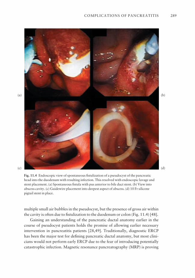
multiple small air bubbles in the pseudocyst, but the presence of gross air withinthe cavity is often due to fistulization to the duodenum or colon (Fig. 11.4) [48].
Gaining an understanding of the pancreatic ductal anatomy earlier in thecourse of pseudocyst patients holds the promise of allowing earlier necessaryintervention in pancreatitis patients [28,49]. Traditionally, diagnostic ERCPhas been the major test for defining pancreatic ductal anatomy, but most clini-cians would not perform early ERCP due to the fear of introducing potentiallycatastrophic infection. Magnetic resonance pancreatography (MRP) is proving
COMPLICATIONS OF PANCREATITIS 289
Fig. 11.4 Endoscopic view of spontaneous fistulization of a pseudocyst of the pancreatichead into the duodenum with resulting infection. This resolved with endoscopic lavage andstent placement. (a) Spontaneous fistula with pus anterior to bile duct stent. (b) View intoabscess cavity. (c) Guidewire placement into deepest aspect of abscess. (d) 10 Fr siliconepigtail stent in place.
(a)
(c)
(b)
(d)
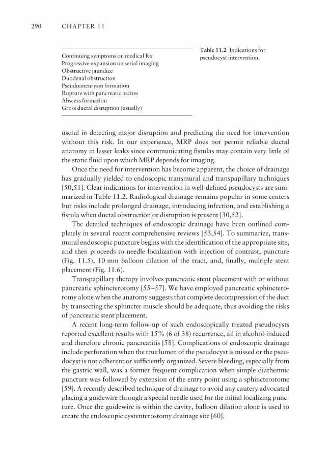
useful in detecting major disruption and predicting the need for interventionwithout this risk. In our experience, MRP does not permit reliable ductalanatomy in lesser leaks since communicating fistulas may contain very little ofthe static fluid upon which MRP depends for imaging.
Once the need for intervention has become apparent, the choice of drainagehas gradually yielded to endoscopic transmural and transpapillary techniques[50,51]. Clear indications for intervention in well-defined pseudocysts are sum-marized in Table 11.2. Radiological drainage remains popular in some centersbut risks include prolonged drainage, introducing infection, and establishing afistula when ductal obstruction or disruption is present [30,52].
The detailed techniques of endoscopic drainage have been outlined com-pletely in several recent comprehensive reviews [53,54]. To summarize, trans-mural endoscopic puncture begins with the identification of the appropriate site,and then proceeds to needle localization with injection of contrast, puncture(Fig. 11.5), 10 mm balloon dilation of the tract, and, finally, multiple stentplacement (Fig. 11.6).
Transpapillary therapy involves pancreatic stent placement with or withoutpancreatic sphincterotomy [55–57]. We have employed pancreatic sphinctero-tomy alone when the anatomy suggests that complete decompression of the ductby transecting the sphincter muscle should be adequate, thus avoiding the risksof pancreatic stent placement.
A recent long-term follow-up of such endoscopically treated pseudocystsreported excellent results with 15% (6 of 38) recurrence, all in alcohol-inducedand therefore chronic pancreatitis [58]. Complications of endoscopic drainageinclude perforation when the true lumen of the pseudocyst is missed or the pseu-docyst is not adherent or sufficiently organized. Severe bleeding, especially fromthe gastric wall, was a former frequent complication when simple diathermicpuncture was followed by extension of the entry point using a sphincterotome[59]. A recently described technique of drainage to avoid any cautery advocatedplacing a guidewire through a special needle used for the initial localizing punc-ture. Once the guidewire is within the cavity, balloon dilation alone is used tocreate the endoscopic cystenterostomy drainage site [60].
CHAPTER 11290
Table 11.2 Indications forpseudocyst intervention.Continuing symptoms on medical Rx
Progressive expansion on serial imagingObstructive jaundiceDuodenal obstructionPseudoaneurysm formationRupture with pancreatic ascitesAbscess formationGross ductal disruption (usually)
Overall, the success rates for endoscopic pseudocyst drainage are summa-rized in Table 11.3 and approach 80%. The complication risk and recurrencerates stated in these studies are included in Table 11.4 and were 12% and 16%,respectively. Finally, recurrences could be retreated endosurgically about 50%of the time, relegating surgery to only eight patients out of the 141 cases selectedand reported.
The role of EUS pseudocyst management remains in evolution (Fig. 11.7).Some authors report rarely needing or using EUS but others consider EUS valuable in selecting appropriate drainage sites, excluding intervening bloodvessels, and determining depth of puncture [53,61]. However, when collected in the diagnostic setting, this information may be difficult to transfer to actualtherapy, which is generally performed with larger channel therapeutic endo-scopes whose angle of view and therefore alignment for puncture is differentfrom EUS [62,63]. Direct EUS therapeutic drainage by placing 7 Fr stents hasbeen reported, but results are unlikely to equal 10 Fr drainage [63]. Finally, thedevelopment of a therapeutic 4.2 channel EUS endoscope has permitted com-plete therapy, including 10 Fr stent placement, in pseudocyst patients [64]. As EUS becomes a more available therapeutic technique, this approach maycompete well with our current endoscopic transmural drainage procedure.
Finally, laparoscopically directed pseudocyst drainage has been reported but
COMPLICATIONS OF PANCREATITIS 291
Fig. 11.5 22-gauge needle puncture before transmural pseudocyst drainage to select anoptimal site for entry. (a) Endoscopic needle localization (ENL) aids by aligning the entry,confirming a shallow depth, checking for integrity by injecting contrast, and aspirating to besure there is no blood. This is the fluoroscopic view during ENL. (b) Artist’s drawing of ENL.
(a) (b)
appears to be more invasive, more costly, and does not show a superior outcome[65].
Pancreatic necrosis
Necrosis of pancreatic tissue complicates acute pancreatitis in a variable percentage of cases, is seen less often in acute exacerbations of chronic pancre-atitis, and accounts for many of the complications and much of the mortality(Fig. 11.8). Etiologies may have an impact on severity with the pancreatitis
CHAPTER 11292
Fig. 11.6 Endoscopic transmural pseudocyst puncture and drainage. (a) Identification ofbulge into the duodenal bulb. (b) Puncture for localization with injection of contrast andaspiration. (c) 10 mm balloon dilation after guidewire placement. (d) Stents in placeatwo 10 Fr silicone pigtails and a 7 Fr nasocystic drain.
(a) (b)
(d)(c)
Tab
le 1
1.3
Met
hods
of d
rain
age
and
succ
ess
rate
s in
rec
ent s
tudi
es o
f end
osco
pic
pseu
docy
st d
rain
age.
Cys
tgas
tros
tom
y C
ystd
uode
nost
omy
Panc
reat
ic d
uct s
tent
All
pati
ents
alon
eal
one
alon
eC
ombi
ned
proc
edur
e
Ref
eren
cePa
tien
tsR
esol
utio
nPa
tien
tsR
esol
utio
nPa
tien
tsR
esol
utio
nPa
tien
tsR
esol
utio
nPa
tien
tsR
esol
utio
n
Bar
thet
et a
l. [5
5]30
23 (7
7%)
00
2016
(80%
)10
7 (7
0%)
Gri
mm
et a
l. [6
2]53
47 (8
9%)
20 to
tal
16 (8
0%)
2927
(93%
)4
4 (1
00%
)b
(sit
e no
t sp
ecifi
ed)a
Cat
alan
o et
al.
[56]
2117
(81%
)0
021
17 (8
1%)
0Sm
its
et a
l. [5
1]37
24 (6
5%)
103
(30%
)7
7 (1
00%
)12
7 (5
8%)
87
(88%
)
Tot
al14
111
1 (7
9%)
Of a
tota
l of 3
7 pa
tien
ts, 2
6.(7
0%).
had_
reso
luti
on82
67
(82%
)22
18 (8
2%)
a Res
olut
ion
refe
rs to
init
ial,
com
plet
e dr
aina
ge o
f a p
seud
ocys
t.b T
hese
res
ults
are
not
exp
licit
ly g
iven
in th
e st
udy,
but
are
infe
rred
.
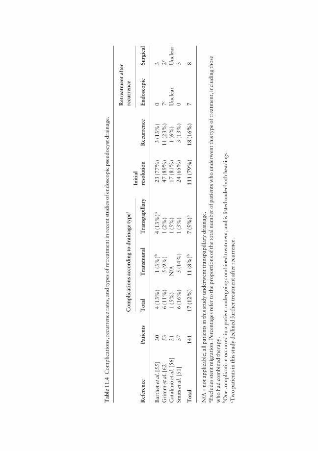
Tab
le 1
1.4
Com
plic
atio
ns, r
ecur
renc
e ra
tes,
and
type
s of
ret
reat
men
t in
rece
nt s
tudi
es o
f end
osco
pic
pseu
docy
st d
rain
age.
Ret
reat
men
t aft
erC
ompl
icat
ions
acc
ordi
ng to
dra
inag
e ty
pea
recu
rren
ceIn
itia
lR
efer
ence
Pati
ents
Tot
alT
rans
mur
alT
rans
papi
llary
reso
luti
onR
ecur
renc
eE
ndos
copi
cSu
rgic
al
Bar
thet
et a
l. [5
5]30
4 (1
3%)
1 (3
%)b
4 (1
3%)b
23 (7
7%)
3 (1
3%)
03
Gri
mm
et a
l. [6
2]53
6 (1
1%)
5 (9
%)
1 (2
%)
47 (8
9%)
11 (2
3%)
7c2c
Cat
alan
o et
al.
[56]
211
(5%
)N
/A1
(5%
)17
(81%
)1
(6%
)U
ncle
arU
ncle
arSm
its
et a
l. [5
1]37
6 (1
6%)
5 (1
4%)
1 (3
%)
24 (6
5%)
3 (1
3%)
03
Tot
al14
117
(12%
)11
(8%
)b7
(5%
)b11
1 (7
9%)
18 (1
6%)
78
N/A
= n
ot a
pplic
able
; all
pati
ents
in th
is s
tudy
und
erw
ent t
rans
papi
llary
dra
inag
e.a E
xclu
des
sten
t mig
rati
on. P
erce
ntag
es r
efer
to th
e pr
opor
tion
s of
the
tota
l num
ber
of p
atie
nts
who
und
erw
ent t
his
type
of t
reat
men
t, in
clud
ing
thos
ew
ho h
ad c
ombi
ned
ther
apy.
b One
com
plic
atio
n oc
curr
ed in
a p
atie
nt u
nder
goin
g co
mbi
ned
trea
tmen
t, a
nd is
list
ed u
nder
bot
h he
adin
gs.
c Tw
o pa
tien
ts in
this
stu
dy d
eclin
ed fu
rthe
r tr
eatm
ent a
fter
rec
urre
nce.
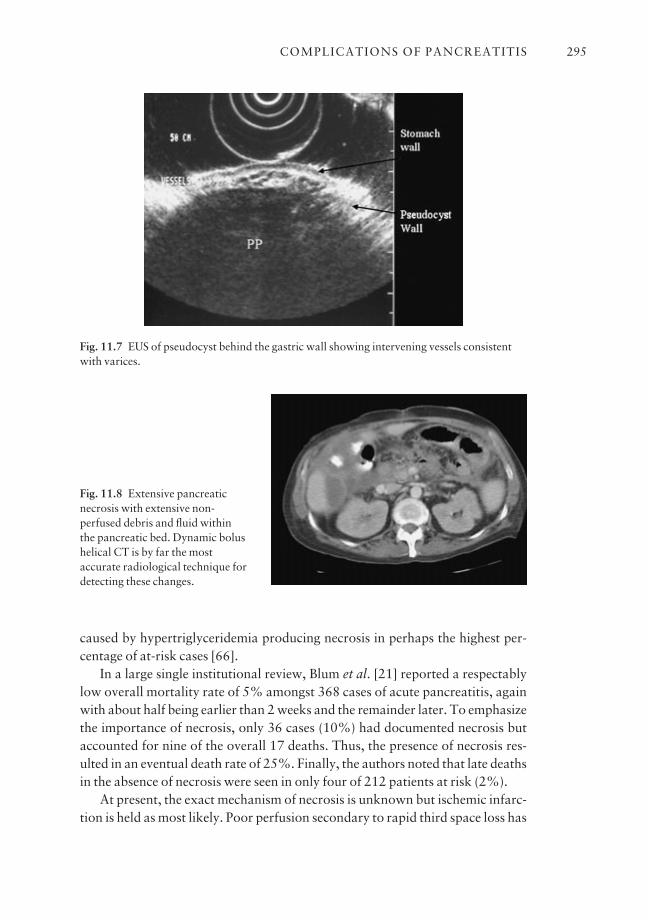
caused by hypertriglyceridemia producing necrosis in perhaps the highest per-centage of at-risk cases [66].
In a large single institutional review, Blum et al. [21] reported a respectablylow overall mortality rate of 5% amongst 368 cases of acute pancreatitis, againwith about half being earlier than 2 weeks and the remainder later. To emphasizethe importance of necrosis, only 36 cases (10%) had documented necrosis butaccounted for nine of the overall 17 deaths. Thus, the presence of necrosis res-ulted in an eventual death rate of 25%. Finally, the authors noted that late deathsin the absence of necrosis were seen in only four of 212 patients at risk (2%).
At present, the exact mechanism of necrosis is unknown but ischemic infarc-tion is held as most likely. Poor perfusion secondary to rapid third space loss has
COMPLICATIONS OF PANCREATITIS 295
Fig. 11.7 EUS of pseudocyst behind the gastric wall showing intervening vessels consistentwith varices.
Fig. 11.8 Extensive pancreaticnecrosis with extensive non-perfused debris and fluid within the pancreatic bed. Dynamic bolushelical CT is by far the mostaccurate radiological technique fordetecting these changes.
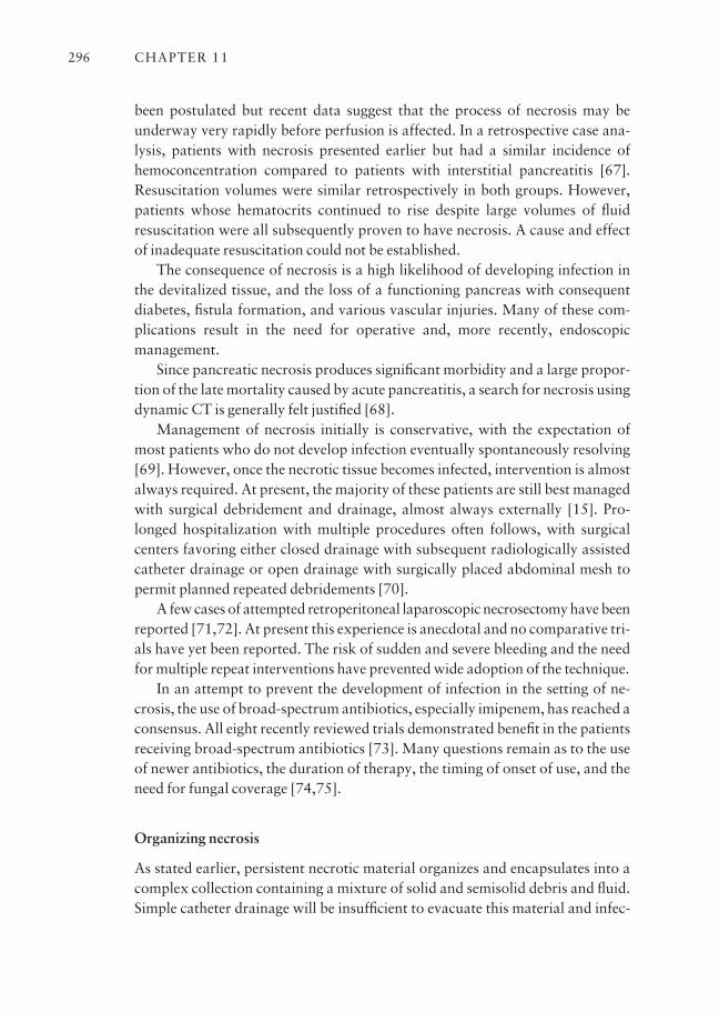
been postulated but recent data suggest that the process of necrosis may beunderway very rapidly before perfusion is affected. In a retrospective case ana-lysis, patients with necrosis presented earlier but had a similar incidence ofhemoconcentration compared to patients with interstitial pancreatitis [67].Resuscitation volumes were similar retrospectively in both groups. However,patients whose hematocrits continued to rise despite large volumes of fluidresuscitation were all subsequently proven to have necrosis. A cause and effectof inadequate resuscitation could not be established.
The consequence of necrosis is a high likelihood of developing infection inthe devitalized tissue, and the loss of a functioning pancreas with consequentdiabetes, fistula formation, and various vascular injuries. Many of these com-plications result in the need for operative and, more recently, endoscopic management.
Since pancreatic necrosis produces significant morbidity and a large propor-tion of the late mortality caused by acute pancreatitis, a search for necrosis usingdynamic CT is generally felt justified [68].
Management of necrosis initially is conservative, with the expectation ofmost patients who do not develop infection eventually spontaneously resolving[69]. However, once the necrotic tissue becomes infected, intervention is almostalways required. At present, the majority of these patients are still best managedwith surgical debridement and drainage, almost always externally [15]. Pro-longed hospitalization with multiple procedures often follows, with surgicalcenters favoring either closed drainage with subsequent radiologically assistedcatheter drainage or open drainage with surgically placed abdominal mesh topermit planned repeated debridements [70].
A few cases of attempted retroperitoneal laparoscopic necrosectomy have beenreported [71,72]. At present this experience is anecdotal and no comparative tri-als have yet been reported. The risk of sudden and severe bleeding and the needfor multiple repeat interventions have prevented wide adoption of the technique.
In an attempt to prevent the development of infection in the setting of ne-crosis, the use of broad-spectrum antibiotics, especially imipenem, has reached aconsensus. All eight recently reviewed trials demonstrated benefit in the patientsreceiving broad-spectrum antibiotics [73]. Many questions remain as to the useof newer antibiotics, the duration of therapy, the timing of onset of use, and theneed for fungal coverage [74,75].
Organizing necrosis
As stated earlier, persistent necrotic material organizes and encapsulates into acomplex collection containing a mixture of solid and semisolid debris and fluid.Simple catheter drainage will be insufficient to evacuate this material and infec-
CHAPTER 11296
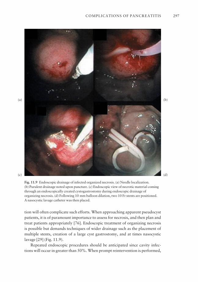
tion will often complicate such efforts. When approaching apparent pseudocystpatients, it is of paramount importance to assess for necrosis, and then plan andtreat patients appropriately [76]. Endoscopic treatment of organizing necrosis is possible but demands techniques of wider drainage such as the placement ofmultiple stents, creation of a large cyst gastrostomy, and at times nasocysticlavage [29] (Fig. 11.9).
Repeated endoscopic procedures should be anticipated since cavity infec-tions will occur in greater than 50%. When prompt reintervention is performed,
COMPLICATIONS OF PANCREATITIS 297
Fig. 11.9 Endoscopic drainage of infected organized necrosis. (a) Needle localization. (b) Purulent drainage noted upon puncture. (c) Endoscopic view of necrotic material comingthrough an endoscopically created cystogastrostomy during endoscopic drainage oforganizing necrosis. (d) Following 10 mm balloon dilation, two 10 Fr stents are positioned. A nasocystic lavage catheter was then placed.
(a)
(c)
(b)
(d)
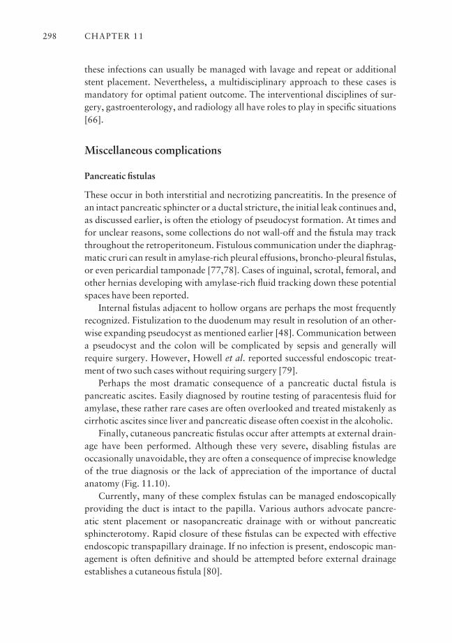
these infections can usually be managed with lavage and repeat or additionalstent placement. Nevertheless, a multidisciplinary approach to these cases ismandatory for optimal patient outcome. The interventional disciplines of sur-gery, gastroenterology, and radiology all have roles to play in specific situations[66].
Miscellaneous complications
Pancreatic fistulas
These occur in both interstitial and necrotizing pancreatitis. In the presence ofan intact pancreatic sphincter or a ductal stricture, the initial leak continues and,as discussed earlier, is often the etiology of pseudocyst formation. At times andfor unclear reasons, some collections do not wall-off and the fistula may trackthroughout the retroperitoneum. Fistulous communication under the diaphrag-matic cruri can result in amylase-rich pleural effusions, broncho-pleural fistulas,or even pericardial tamponade [77,78]. Cases of inguinal, scrotal, femoral, andother hernias developing with amylase-rich fluid tracking down these potentialspaces have been reported.
Internal fistulas adjacent to hollow organs are perhaps the most frequentlyrecognized. Fistulization to the duodenum may result in resolution of an other-wise expanding pseudocyst as mentioned earlier [48]. Communication betweena pseudocyst and the colon will be complicated by sepsis and generally willrequire surgery. However, Howell et al. reported successful endoscopic treat-ment of two such cases without requiring surgery [79].
Perhaps the most dramatic consequence of a pancreatic ductal fistula is pancreatic ascites. Easily diagnosed by routine testing of paracentesis fluid foramylase, these rather rare cases are often overlooked and treated mistakenly ascirrhotic ascites since liver and pancreatic disease often coexist in the alcoholic.
Finally, cutaneous pancreatic fistulas occur after attempts at external drain-age have been performed. Although these very severe, disabling fistulas are occasionally unavoidable, they are often a consequence of imprecise knowledgeof the true diagnosis or the lack of appreciation of the importance of ductalanatomy (Fig. 11.10).
Currently, many of these complex fistulas can be managed endoscopicallyproviding the duct is intact to the papilla. Various authors advocate pancre-atic stent placement or nasopancreatic drainage with or without pancreaticsphincterotomy. Rapid closure of these fistulas can be expected with effectiveendoscopic transpapillary drainage. If no infection is present, endoscopic man-agement is often definitive and should be attempted before external drainageestablishes a cutaneous fistula [80].
CHAPTER 11298
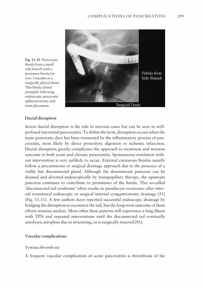
Ductal disruption
Severe ductal disruption is the rule in necrosis cases but can be seen in well-perfused interstitial pancreatitis. To define the term, disruption occurs when themain pancreatic duct has been transected by the inflammatory process of pan-creatitis, most likely by direct proteolytic digestion or ischemic infarction.Ductal disruption greatly complicates the approach to treatment and worsensoutcome in both acute and chronic pancreatitis. Spontaneous resolution with-out intervention is very unlikely to occur. External cutaneous fistulas usuallyfollow a percutaneous or surgical drainage approach due to the presence of aviable but disconnected gland. Although the downstream pancreas can bedrained and diverted endoscopically by transpapillary therapy, the upstreampancreas continues to contribute to persistence of the fistula. This so-called ‘disconnected tail syndrome’ often results in pseudocyst recurrence after inter-nal transmural endoscopic or surgical internal cystgastrostomy drainage [51](Fig. 11.11). A few authors have reported successful endoscopic drainage bybridging the disruption to reconnect the tail, but the long-term outcome of theseefforts remains unclear. More often these patients will experience a long illnesswith TPN and repeated interventions until the disconnected tail eventuallyautolyses, atrophies due to stricturing, or is surgically resected [81].
Vascular complications
Venous thrombosis
A frequent vascular complication of acute pancreatitis is thrombosis of the
COMPLICATIONS OF PANCREATITIS 299
Fig. 11.10 Pancreaticfistula from a small side branch with apersistent fistula forover 3 months to asurgically placed drain.This fistula closedpromptly followingendoscopic pancreaticsphincterotomy andstent placement.
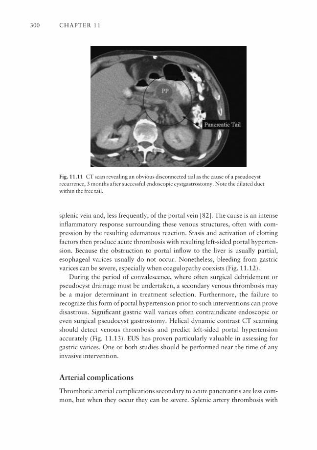
splenic vein and, less frequently, of the portal vein [82]. The cause is an intenseinflammatory response surrounding these venous structures, often with com-pression by the resulting edematous reaction. Stasis and activation of clottingfactors then produce acute thrombosis with resulting left-sided portal hyperten-sion. Because the obstruction to portal inflow to the liver is usually partial,esophageal varices usually do not occur. Nonetheless, bleeding from gastricvarices can be severe, especially when coagulopathy coexists (Fig. 11.12).
During the period of convalescence, where often surgical debridement orpseudocyst drainage must be undertaken, a secondary venous thrombosis maybe a major determinant in treatment selection. Furthermore, the failure to recognize this form of portal hypertension prior to such interventions can provedisastrous. Significant gastric wall varices often contraindicate endoscopic oreven surgical pseudocyst gastrostomy. Helical dynamic contrast CT scanningshould detect venous thrombosis and predict left-sided portal hypertensionaccurately (Fig. 11.13). EUS has proven particularly valuable in assessing forgastric varices. One or both studies should be performed near the time of anyinvasive intervention.
Arterial complications
Thrombotic arterial complications secondary to acute pancreatitis are less com-mon, but when they occur they can be severe. Splenic artery thrombosis with
CHAPTER 11300
Fig. 11.11 CT scan revealing an obvious disconnected tail as the cause of a pseudocystrecurrence, 3 months after successful endoscopic cystgastrostomy. Note the dilated ductwithin the free tail.
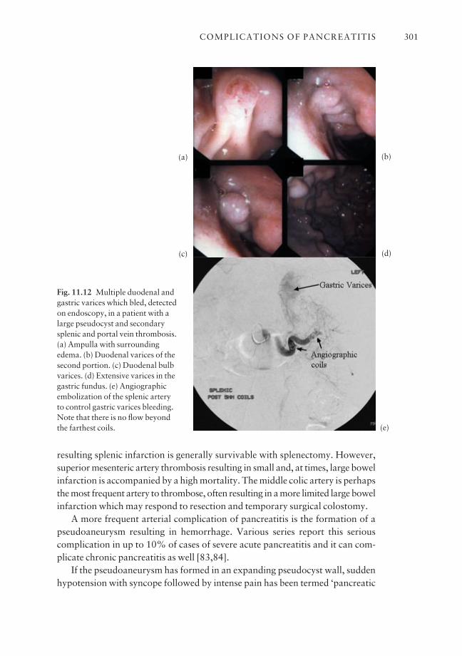
resulting splenic infarction is generally survivable with splenectomy. However,superior mesenteric artery thrombosis resulting in small and, at times, large bowelinfarction is accompanied by a high mortality. The middle colic artery is perhapsthe most frequent artery to thrombose, often resulting in a more limited large bowelinfarction which may respond to resection and temporary surgical colostomy.
A more frequent arterial complication of pancreatitis is the formation of apseudoaneurysm resulting in hemorrhage. Various series report this seriouscomplication in up to 10% of cases of severe acute pancreatitis and it can com-plicate chronic pancreatitis as well [83,84].
If the pseudoaneurysm has formed in an expanding pseudocyst wall, suddenhypotension with syncope followed by intense pain has been termed ‘pancreatic
COMPLICATIONS OF PANCREATITIS 301
Fig. 11.12 Multiple duodenal andgastric varices which bled, detectedon endoscopy, in a patient with alarge pseudocyst and secondarysplenic and portal vein thrombosis.(a) Ampulla with surroundingedema. (b) Duodenal varices of thesecond portion. (c) Duodenal bulbvarices. (d) Extensive varices in thegastric fundus. (e) Angiographicembolization of the splenic artery to control gastric varices bleeding.Note that there is no flow beyondthe farthest coils.
(a)
(c)
(b)
(d)
(e)
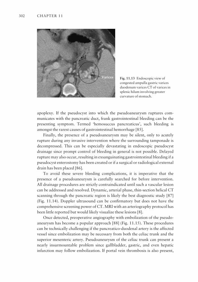
apoplexy. If the pseudocyst into which the pseudoaneurysm ruptures com-municates with the pancreatic duct, frank gastrointestinal bleeding can be thepresenting symptom. Termed ‘hemosuccus pancreaticus’, such bleeding isamongst the rarest causes of gastrointestinal hemorrhage [85].
Finally, the presence of a pseudoaneurysm may be silent, only to acutely rupture during any invasive intervention where the surrounding tamponade isdecompressed. This can be especially devastating in endoscopic pseudocystdrainage since prompt control of bleeding in general is not possible. Delayedrupture may also occur, resulting in exsanguinating gastrointestinal bleeding if apseudocyst enterostomy has been created or if a surgical or radiological externaldrain has been placed [86].
To avoid these severe bleeding complications, it is imperative that the presence of a pseudoaneurysm is carefully searched for before intervention. All drainage procedures are strictly contraindicated until such a vascular lesioncan be addressed and resolved. Dynamic, arterial phase, thin-section helical CTscanning through the pancreatic region is likely the best diagnostic study [87](Fig. 11.14). Doppler ultrasound can be confirmatory but does not have thecomprehensive screening power of CT. MRI with an arteriography protocol hasbeen little reported but would likely visualize these lesions [8].
Once detected, preoperative angiography with embolization of the pseudo-aneurysm has become a popular approach [88] (Fig. 11.15). These procedurescan be technically challenging if the pancreatico-duodenal artery is the affectedvessel since embolization may be necessary from both the celiac trunk and thesuperior mesenteric artery. Pseudoaneurysm of the celiac trunk can present anearly insurmountable problem since gallbladder, gastric, and even hepaticinfarction may follow embolization. If portal vein thrombosis is also present,
CHAPTER 11302
Fig. 11.13 Endoscopic view ofcongested ampulla gastric varicesduodenum varices CT of varices insplenic hilum involving greatercurvature of stomach.

the risk of hepatic infarction increases dramatically. Successful treatment ofhemosuccus pancreaticus radiological embolization at angiography is the pre-ferred approach as well [89].
Once the pseudoaneurysm has been thoroughly embolized and thrombosed,interventions can then be safely carried out [84]. Elton et al. reported success-ful endoscopic pseudoaneurysm/pseudocyst drainage following radiologicalembolization in three such cases [90]. In all three patients, thrombosis followingembolization was documented by repeat dynamic contrast CT or Doppler ultra-sound prior to endoscopic intervention. Successful endoscopic drainage of the obstructing pseudocyst, stent management of strictures, and clearance ofobstructing clots within the pancreatic duct resulted in symptom resolution andavoided surgery in these cases.
COMPLICATIONS OF PANCREATITIS 303
Fig. 11.14 Pseudoaneurysm of the splenic artery complicating chronic pancreatitis within apseudocyst. This has not yet ruptured.
Fig. 11.15 Pseudoaneurysm withina pseudocyst filled with contrast on dynamic bolus helical CT scan.The same pseudoaneurysm onangiography is of the splenic artery.This was successfully embolized.
Finally, massive diffuse retroperitoneal bleeding may be seen in the setting ofnecrotizing pancreatitis, often with coincident coagulopathy. This so-called‘hemorrhagic pancreatitis’ is less often reported since better radiology moreoften identifies a focal arterial source. However, when true diffuse hemorrhagicpancreatitis does occur, mortality rates exceed 35%, even in the modern era [88].
Summary
Complications of pancreatitis vary widely, are of complex etiology, and involvemultiple organ systems. Avoiding these complications remains the basic goal forall treating physicians, but, once present, their expert detection and appropriatemanagement are the key to optimizing patient outcome [91]. Great progress hasbeen made in treating these supremely ill patients but early and specific treat-ments to prevent complications are still lacking. Prolonged hospitalizations,TPN, dialysis, ventilatory support, antibiotic therapy, and radiological, endo-scopic, and surgical treatments all have had a role in reducing mortality to lessthan 10% of afflicted patients. However, much needs to be discovered [92].
Outstanding issues and future trends
The major need in pancreatology remains a full understanding of the patho-physiology of acute pancreatitis that results in the dramatic cascade of eventsoutlined in this chapter. Once the earliest events are identified, specific medicalinterventions, possibly extremely specific pharmacological agents, can be devel-oped that can prevent progression to shock, end organ compromise, necrosis,and the other late complications outlined. More basic research is needed.
Lacking this knowledge, research will continue to look for methods of pre-venting the complications of pancreatitis once severe disease has been estab-lished. A major need is an effective way to prevent progression to necrosis,beyond aggressive fluid resuscitation.
Trends in the future will continue to be innovations in minimally invasivetherapies. Debridement of infected necrosis, intervention prior to infection, andmanagement of ductal disruption resulting in a disconnected tail are all areas ofconsiderable confusion and often subjects of interdisciplinary debate. Thera-peutic, endoscopic, percutaneous laparoscopic debridement, and transgastricendoscopic therapy are the newest players on a seemingly crowded field.
References
1 Ranson JHC, Rifkind KM, Roses DF et al. Prognostic signs and the role of operative manage-ment in acute pancreatitis. Surg Gynecol Obstet 1974; 139: 69–81.
CHAPTER 11304
2 Williams M, Simms HH. Prognostic usefulness of scoring systems in critically ill patients withsevere acute pancreatitis. Crit Care Med 1999; 27: 901–7.
3 Halonen KI, Pettila V, Leppaniemi AK, Kemppainen EA, Puolakkainen PA, Haapiainen RK.Multiple organ dysfunction associated with severe acute pancreatitis. Crit Care Med 2002; 30:1274–9.
4 Khan AA, Parekh D, Cho Y et al. Improved prediction of outcome in patients with severe acutepancreatitis by the APACHE II score at 48 hours after hospital admission compared with theAPACHE II score at admission: acute physiology and chronic health evaluation. Arch Surg2002; 137: 1136–40.
5 Eachempati SR, Hydo LJ, Barie PS. Severity scoring for prognostication in patients with severeacute pancreatitis. Arch Surg 2002; 17: 730–6.
6 Balthazar EJ, Fisher LA. Hemorrhagic complications of pancreatitis: radiologic evaluation withemphasis on CT imaging. Pancreatology 2001; 1: 306–13.
7 Robert JH, Frossard JL, Mermillod B et al. Early prediction of acute pancreatitis: prospectivestudy comparing computed tomography scans, Ranson, Glasgow, acute physiology and chronichealth evaluation II scores, and various serum markers. World J Surg 2002; 26: 612–19.
8 Robinson PJ, Sheridan MB. Pancreatitis: computed tomography and magnetic resonance imag-ing. Eur Radiol 2000; 10: 401–8.
9 Frey CF, Brody GL. Relationship of azotemia and survival in bile pancreatitis in the dog. ArchSurg 1996; 93: 295–300.
10 Salomone T, Tosi P, Di Battista N et al. Impaired alveolar gas exchange in acute pancreatitis.Dig Dis Sci 2002; 47: 2025–8.
11 Hietaranta A, Kemppainen E, Puolakkainen P et al. Extracellular phospholipases A2 in relationto systemic inflammatory response syndrome (SIRS) and systemic complications in severe acutepancreatitis. Pancreas 1999; 18: 385–91.
12 Kingsnorth A. The role of cytokines in the pathogenesis of acute pancreatitis. Am J Surg 1997;40: 1–4.
13 Norman J. The role of cytokines in the pathogenesis of acute pancreatitis. Am J Surg 1998; 175:76–83.
14 Brivet F, Emilie D, Galanaud P et al. Pro- and anti-inflammatory cytokines during acute severepancreatitis: an early and sustained response, although unpredictable of death. Parisian StudyGroup on Acute Pancreatitis. Crit Care Med 1999; 27: 749–55.
15 Hartwig W, Werner J, Uhl W, Buchler MW. Management of infection in acute pancreatitis. J Hepatobiliary Pancreat Surg 2002; 9: 423–8.
16 Banks PA, Gerzof SG, Langevin RE et al. CT-guided aspiration of suspected pancreatic infec-tion: bacteriology and clinical outcome. Int J Pancreatol 1995; 18: 256–70.
17 Takacs T, Hajnal F, Nemeth J et al. Stimulated gastrointestinal hormone release and gallbladdercontraction during continuous jejunal feeding in patients with pancreatic pseudocyst is inhibitedby octreotide. Int J Pancreatol 2000; 28: 215–20.
18 Windsor ACJ, Kanwar S, Li AGK et al. Compared with parenteral nutrition, enteral feedingattenuates the acute phase response and improves disease severity in acute pancreatitis. Gut1998; 42: 431–5.
19 Johnson CD, Imrie CW, McMahon MJ et al. Double blind, randomized, placebo controlledstudy of a platelet activating factor antagonist, lexipafant, in the treatment and prevention oforgan failure in predicted severe acute pancreatitis. Gut 2001; 48: 62–9.
20 Isenmann R, Schwarz M, Rau B, Trautmann M, Schobr W, Beger HG. Characteristics of infec-tion with candida species in patients with necrotizing pancreatitis. World J Surg 2002; 26:372–6.
21 Blum T, Maisonneuve P, Lowenfels AB, Lankisch PG. Fatal outcome in acute pancreatitis: itsoccurrence and early prediction. Pancreatology 2001; 1: 237–41.
22 Ethridge RT, Chung DH, Slogoff M et al. Cyclooxygenase-2 gene disruption attenuates theseverity of acute pancreatitis and pancreatitis-associated lung injury. Gastroenterology 2002;123: 1311–22.
23 Pezzilli R, Billi P, Miniero R, Barakat B. Serum interleukin-10 in human acute pancreatitis. DigDis Sci 1997; 42: 1469–72.
COMPLICATIONS OF PANCREATITIS 305
24 Chen CC, Wang SS, Lu RH, Chang FY, Lee SP. Serum interleukin 10 and interleukin 11 inpatients with acute pancreatitis. Gut 1999; 45: 895–9.
25 Eskdale J, Peat J, Gallagher GE, Imrie CW, McKay CJ. Fine genomic mapping implicates IL-10as a severity gene in acute pancreatitis. Gastroenterology 2001; 120: A24.
26 Dumot JA, Conwell DL, Zuccaro C et al. A randomized, double blind study of interleukin 10 forthe prevention of ERCP-induced pancreatitis. Am J Gastroenterol 2001; 96: 2098–102.
27 Balthazar EJ. Staging of acute pancreatitis. Radiol Clin North Am 2002; 40: 1199–209.28 Lau ST, Simchuk EJ, Kozarek RA, Traverso LW. A pancreatic ductal leak should be sought to
direct treatment in patients with acute pancreatitis. Am J Surg 2001; 181: 411–15.29 Baron TH, Harewood GC, Morgan DE, Yates MR. Outcome differences after endoscopic
drainage of pancreatic necrosis, acute pancreatic pseudocysts, and chronic pancreatic pseudo-cysts. Gastrointest Endosc 2002; 56: 7–17.
30 Bradley EL III, Clements JL Jr, Gonzales AC. The natural history of pancreatic pseudocysts: aunified concept of management. Am J Surg 1979; 137: 135–41.
31 D’Edogo A, Schein M. Pancreatic pseudocysts: a proposed classification and its managementimplications. Br J Surg 1991; 78: 981–4.
32 Warshaw AL, Compton CC, Lewandrowski K et al. Cystic tumors of the pancreas: new clinical,radiologic, and pathogenic observations in 67 patients. Am Surg 1990; 212: 432–45.
33 Levin MF, Vellet AD, Bach DB et al. Peripancreatic fluid collections: vascular structures mas-querading as pseudocysts. Can Assoc Radiol J 1992; 43: 267–72.
34 Sorgman JA, Langevin E, Banks PA. Urinoma masquerading as pancreatic pseudocyst. Int JPancreatol 1992; 11: 195–6.
35 Sperti C, Cappellazzo F, Pasquali C et al. Cystic neoplasms of the pancreas: problems in differ-ential diagnosis. Am Surg 1993; 59: 740–5.
36 Warshaw AL, Rutledge PL. Cystic tumors mistaken for pancreatic pseudocysts. Am Surg 1987;205: 393–8.
37 Hammond N, Miller FH, Sica GT, Gore RM. Imaging of cystic diseases of the pancreas. RadiolClin North Am 2002; 40: 1243–62.
38 Hsieh CH, Tseng JH, Huang SF. Co-existence of a huge pseudocyst and mucinous cystadenoma:report of a case and the value of magnetic resonance imaging for differential diagnosis. Eur JGastroenterol Hepatol 2002; 14: 191–4.
39 Fockens P, Johnson TG, van Dulleman HM et al. Endosonography is a prerequisite before endo-scopic drainage of pancreatic pseudocysts. Gastrointest Endosc 1996; 43: 516 (A).
40 Brugge WR. Role of endoscopic ultrasound in the diagnosis of cystic lesions of the pancreas.Pancreatology 2001; 1 (6): 637–40 [Review].
41 Kloppel G, Kosmahl M. Cystic lesions and neoplasms of the pancreas: the features are becomingclearer. Pancreatology 2001; 1 (6): 648–55 [Review].
42 Sedlack R, Affi A, Vazquez-Sequeiros E, Norton ID, Clain JE, Wiersema MJ. Utility of EUS inthe evaluation of cystic pancreatic lesions. Gastrointest Endosc 2002; 56 (4): 543–7.
43 Banks PA. Practice guidelines in acute pancreatitis. Am J Gastroenterol 1997; 3: 377–86.44 O’Malley VP, Cannon JP, Postie RG. Pancreatic pseudocysts: cause, therapy, and results. Am J
Surg 1985; 150: 680–2.45 Yeo CJ, Bastidas JA, Lynch-Nyhan A et al. The natural history of pancreatic pseudocysts docu-
mented by computed tomography. Surg Gynecol Obstet 1990; 170: 411–17.46 Vitas GJ, Sarr MG. Selected management of pancreatic pseudocysts: operative versus expectant
management. Surgery 1992; 111: 123–30.47 Kozarek RA, Ball TJ, Patterson DJ, Freeny PC, Ryan JA, Traverso LW. Endoscopic transpapil-
lary therapy for disrupted pancreatic duct and peripancreatic fluid collections. Gastroenterology1996; 100 (5 Part 1): 1362–70.
48 Urakami A, Tsunoda T, Hayashi J, Oka Y, Mizuno M. Spontaneous fistulization of a pancreaticpseudocyst into the colon and duodenum. Gastrointest Endosc 2002; 55: 949–51.
49 Nealon W, Walser E. Main pancreatic ductal anatomy can direct choice of modality for treatingpancreatic pseudocysts (surgery versus percutaneous drainage). Ann Surg 2002; 235: 751–8.
50 Grace PA, Williamson RCN. Modern management of pancreatic pseudocysts. Br J Surg 1993;80: 573–81.
CHAPTER 11306

51 Smits ME, Rauws EA, Tytgat GN et al. The efficacy of endoscopic treatment of pancreatic pseu-docysts. Gastrointest Endosc 1995; 42: 202–7.
52 VanSonnenberg E, Wittich GR, Casola G. Complicated pancreatic inflammatory disease: diag-nostic and therapeutic role of interventional radiology. Radiology 1985; 155: 355–40.
53 Howell DA, Elton E, Parsons WG. Endoscopic management of pseudocysts of the pancreas.Gastrointest Endosc Clin N Am 1998; 8 (1): 143–62 [Review].
54 Parsons WG, Howell DA. (1998). Endoscopic management of pancreatic pseudocysts. In:ERCP and its Applications (ed. Jacobson IM), pp. 193–207. Lippincott-Raven, Philadelphia.
55 Barthet M, Sahel J, Bodlou-Bertel C et al. Endoscopic transpapillary drainage of pancreaticpseudocysts. Gastrointest Endosc 1995; 42: 208–13.
56 Catalano MF, Geenen JE, Schmalz MJ. Treatment of pancreatic pseudocysts ductal communica-tion by transpapillary pancreatic duct endoprosthesis. Gastrointest Endosc 1995; 42: 214–18.
57 Mallavarapu R, Habib TH, Elton E, Goldberg MJ. Resolution of mediastinal pancreatic pseu-docysts with transpapillary stent placement. Gastrointest Endosc 2001; 53: 367–70.
58 Sharma SS, Bhargawa N, Govil A. Endoscopic management of pancreatic pseudocyst: a long-term follow-up. Endoscopy 2002; 34: 203–7.
59 Cremer M, Deviere J, Engelholm L. Endoscopic management of cysts and pseudocysts in chronicpancreatitis: long-term follow-up after 7 years of experience. Gastrointest Endosc 1989; 35: 1–9.
60 Monkemuller KE, Baron TH, Morgan DE. Transmural drainage of pancreatic fluid collectionswithout electrocautery using the Seldinger technique. Gastrointest Endosc 1998; 48: 195–200.
61 Chak A. Endosonographic-guided therapy of pancreatic pseudocysts. Gastrointest Endosc2000; 52: S23–S27.
62 Grimm H, Binmoeller KE, Soehendra N. Endosonography-guided drainage of a pancreaticpseudocyst. Gastrointest Endosc 1992; 38: 170–1.
63 Brand B, Penaloza-Ramirez A, Gupta R et al. New mechanical puncture video echoendoscope:one-step transmural drainage of a pseudocyst. Dig Liver Dis 2002; 34: 133–6.
64 Wiersema MJ. Endosonography-guided cystduodenostomy with a therapeutic ultrasound endo-scope. Gastrointest Endosc 1996; 44: 614–17.
65 Trias M, Targarona EM, Balague C et al. Intraluminal stapled laparoscopic cystogastrostomyfor treatment of pancreatic pseudocysts. Br J Surg 1995; 82: 403.
66 Baron TH, Morgan DE. Endoscopic transgastric irrigation tube placement via PEG for debride-ment of organized pancreatic necrosis. Gastrointest Endosc 1999; 50 (4): 574–7.
67 Brown A, Baillargeon JD, Hughes MD, Banks PA. Can fluid resuscitation prevent pancreaticnecrosis in severe acute pancreatitis? Pancreatology 2002; 2: 104–7.
68 Lankisch PG, Struckmann K, Assmus C, Lehnick D, Maisonneuve P, Lowenfels AB. Do we needa computed tomography examination in all patients with acute pancreatitis within 72 h afteradmission to hospital for the detection of pancreatic necrosis? Scand J Gastroenterol 2002; 36:432–6.
69 Ashley SW, Perez A, Pierce EA et al. Necrotizing pancreatitis: contemporary analysis of 99 con-secutive cases. Ann Surg 2001; 234: 572–9.
70 Uhl W, Warshaw A, Imrie C et al. IAP guidelines for the surgical management of acute pancre-atitis. Pancreatology 2002; 2: 565–73.
71 Horvath KD, Kao LS, Wherry KL, Pellegrini CA, Sinanan MN. A technique for laparoscopic-assisted percutaneous drainage of infected pancreatic necrosis and pancreatic abscess. SurgEndosc 2001; 15: 1221–5.
72 Castellanos G, Pinero A, Serrano A, Parrilla P. Infected pancreatic necrosis: translumbarapproach and management with retroperitoneoscopy. Arch Surg 2002; 13: 1060–3.
73 Bassi C. Infections in pancreatic inflammatory disease: clinical trials for antibiotic prophylaxis.Pancreatology 2001; 1: 210–12.
74 Isenmann R, Rau B, Beger HG. Early severe acute pancreatitis: characteristics of a new sub-group. Pancreas 2001; 22: 274–8.
75 Howard TJ, Temple MB. Prophylactic antibiotics alter the bacteriology of infected necrosis insevere acute pancreatitis. J Am Coll Surg 2002; 195: 759–67.
76 Hariri M, Slivka A, Carr-Locke DL et al. Pseudocyst drainage predisposes to infection whenpancreatic necrosis is unrecognized. Am J Gastroenterol 1994; 89: 1781–4.
COMPLICATIONS OF PANCREATITIS 307
77 Mahlke R, Warnecke B, Lankisch PG, Elbrechtz F, Busch C. A sudden coughing up of foul-smelling sputum: a first sign of a pancreaticobronchial fistula, a severe pulmonary complicationin acute pancreatitis. Am J Gastroenterol 2001; 96: 1952–3.
78 Olah A, Jagy AS, Racz I, Gamal ME. Cardiac tamponade as a complication of pseudocyst inchronic pancreatitis. Hepatogastroenterology 2002; 49: 594–6.
79 Howell DA, Dy RM, Gerstein WH, Hanson BL, Biber BP. Infected pancreatic pseudocysts withcolonic fistula formation successfully managed by endoscopic drainage alone: report of twocases. Am J Gastroenterol 2000; 95: 1822–3.
80 Costamagna G, Mutignani M, Ingrosso M et al. Endoscopic treatment of postsurgical externalpancreatic fistulas. Endoscopy 2001; 33: 317–22.
81 Kozarek RA. Endoscopic therapy of complete and partial pancreatic duct disruptions. Gastro-intest Endosc Clin N Am 1998; 8: 39–53 [Review].
82 Isbicki JR, Yekebas EF, Strate T et al. Extrahepatic portal hypertension in chronic pancreatitis.Ann Surg 2002; 236: 82–9.
83 Sawlani V, Phadke RV, Baijal SS et al. Arterial complications of pancreatitis and their radio-logical management. Australas Radiol 1996; 40 (4): 381–6 [Review].
84 Marshall GT, Howell DA, Hansen BL et al. Multidisciplinary approach to pseudoaneurysmscomplicating pancreatic pseudocysts: impact of pretreatment diagnosis. Arch Surg 1996; 131:278–83.
85 Koizumi J, Inoue S, Yonekawa H, Kunieda T. Hemosuccus pancreaticus: diagnosis with CT andMRI and treatment with transcatheter embolization. Abdom Imaging 2002; 27: 77–81.
86 Born LJ, Madura JA, Lehman GA. Endoscopic diagnosis of a pancreatic pseudoaneurysm afterlateral pancreaticojejunostomy. Gastrointest Endosc 1999; 49: 382–3.
87 Balthazar EJ. Acute pancreatitis: assessment of severity with clinical and CT evaluation.Radiology 2002; 223: 603–13.
88 Flati G, Andren-Sandberg A, La Pinta M, Porowska B, Carboni M. Potentially fatal bleeding inacute pancreatitis: pathophysiology, prevention, and treatment. Pancreas 2003; 26: 8–14.
89 Dasgupta R, Davies MJ, Williamson RC, Jackson JE. Haemosuccus pancreaticus: treatment byarterial embolization. Clin Radiol 2002; 57: 1021–7.
90 Elton EDA, Howell SM, Amberson Dykes TA. Combined angiographic and endoscopic man-agement of bleeding pancreatic pseudoaneurysms. Gastrointest Endosc 1997; 46: 544–9.
91 Beger HG, Rau B, Isenmann R. Prevention of severe change in acute pancreatitis: prediction andprevention. J Hepatobiliary Pancreat Surg 2001; 8: 140–7.
92 Bank S, Singh P, Pooran N, Stark B. Evaluation of factors that have reduced mortality fromacute pancreatitis over the past 20 years. J Clin Gastroenterol 2002; 35: 50–60.
CHAPTER 11308

CHAPTER 12
ERCP in Children
MOISES GUELRUD
Synopsis
ERCP has substantially influenced the evaluation and treatment of adult patientswith suspected pancreatic and biliary disease. The first reports of ERCP in infantsand children were chiefly from adult gastroenterologists experienced with suchtechniques. The growth in number and availability of skilled endoscopists hasresulted in more frequent performance of ERCP in children. Moreover, theacquired ability to perform therapeutic endoscopic procedures is also applicableto children and adolescents. Techniques such as endoscopic sphincterotomy,biliary drainage, extraction of common bile duct and pancreatic duct stones, im-plantation of endoprostheses, and drainage of pancreatic pseudocysts are begin-ning to be used in children with an overall success rate similar to that reported foradult patients. In this chapter, the technique, indications, complications, anddiagnostic and therapeutic applications of ERCP in children are defined.
Introduction
Endoscopic retrograde cholangiopancreatography (ERCP) is the most demand-ing endoscopic procedure in children. It is the most sensitive and specific tech-nique in the evaluation and treatment of children with suspected disorders of thepancreas and the biliary tract. The disadvantage is that it is an invasive proce-dure that frequently needs general anesthesia. The use of this technique in chil-dren has been limited. This may be due to the relatively low incidence of diseases,low incidence of clinical suspicion, limited availability of pediatric duodeno-scopes, lack of pediatric gastroenterologists well trained in ERCP due to littleexposure to the procedure, impression that ERCP in children is technicallydifficult to accomplish, difficulty in the effective evaluation of the therapeuticresult, and because the indications and safety of ERCP in children have not beenwell defined. Since the procedure is frequently performed by experienced adultendoscopists, it is important to have a close working collaboration betweenthem and pediatric gastroenterologists.
309
Patient preparation
Sedation for ERCP in children
The preparation and sedation of a child undergoing ERCP are similar to thoseused for upper gastrointestinal endoscopy. Since young children and some ado-lescents are unable to fully cooperate with procedures under conscious sedation,a state of deep sedation from which the patient is not easily aroused is oftenrequired. The endoscopist must choose between conscious sedation and generalanesthesia after considering the pertinent risks and taking into account personalskill and experience, expected complexity of the procedure, and lastly, cost.
Most children can be adequately sedated with a combination of meperi-dine (2–4 mg/kg, maximum 100 mg) and diazepam (0.1–0.3 mg/kg, maximum 15 mg) or midazolam (0.1–0.3 mg/kg, maximum 15 mg). To obtain adequatesedation, children frequently require much higher doses of midazolam on a mil-ligram per kilogram basis than adults. Post-procedure monitoring is the same asfor other endoscopic procedures requiring sedation.
Antibiotic prophylaxis
There are no data to guide antibiotic prophylaxis for ERCP in children. In ourexperience, routine antibiotic prophylaxis is unnecessary in neonates withcholestasis. Prophylactic antibiotics should be used to prevent endocarditis insusceptible patients in the same manner as for upper gastrointestinal endoscopy.Special situations that require a valvular prosthesis, vascular graft material,indwelling catheters, or transplanted organ in an immunosuppressed patientneed individual consideration.
Other medication
Additional medications, which may be useful during ERCP, include glucagonand Buscopan (hyoscine-N-butyl bromide) to reduce duodenal motility, andsecretin to facilitate identification and cannulation of the minor papilla.
Instruments
In neonates and infants younger than 12 months, ERCP is performed with a spe-cial Olympus pediatric duodenoscope PJF [1] (Olympus America Inc., Melville,NY) which has an insertion tube diameter of 7.5 mm, a channel of 2.0 mm, andan elevator. A standard adult duodenoscope (insertion tube diameter approx-imately 11 mm) can be used for older children and adolescents. Therapeutic
CHAPTER 12310
maneuvers, such as placement of endoprostheses and passage of some dilatorsand retrieval baskets, require instruments with a larger (3.2 mm) channel.
Technique
ERCP is performed in a radiology suite. Pediatric endoscopy assistants and spe-cially trained nurses can help reduce pre-procedure anxiety, monitor the clinicalstatus of the patient, and assist in holding and reassuring, administering medica-tion, handling catheters, and injecting contrast material. The heart rate and oxygen saturation must be continuously monitored. Resuscitation medicationsand appropriate equipment should be available. ERCP is performed on anambulatory basis. A recovery area equipped with monitors and specialized pedi-atric nurses familiar with the needs of children is necessary.
The principles of cannulation are those used in adult patients, with the addi-tional limitations of space within the duodenum that depend on age. In younginfants, such as those undergoing investigation for neonatal cholestasis, it isimportant to minimize the procedure time to avoid abdominal overdistensionand respiratory compromise.
Indications
In general, children with suspected biliary and pancreatic disease should undergoMRCP nowadays before considering ERCP (which is more often used for therapy).
Biliary indications
The only indication for ERCP in neonates and young infants is cholestasis.Biliary indications for ERCP in children older than 1 year and in adolescents are:• obstructive jaundice• known or suspected choledocholithiasis• abnormal liver enzymes in children with inflammatory bowel diseases• evaluation of biliary ductal leaks after cholecystectomy or liver transplantation• evaluation of abnormal scans (ultrasound, computerized tomography (CT),or MRCP)• therapeutic ERCP.
Pancreatic indications
Pancreatic indications for ERCP in children are:• non-resolving acute pancreatitis• idiopathic recurrent pancreatitis, chronic pancreatitis
ERCP IN CHILDREN 311
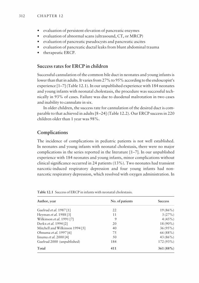
• evaluation of persistent elevation of pancreatic enzymes• evaluation of abnormal scans (ultrasound, CT, or MRCP)• evaluation of pancreatic pseudocysts and pancreatic ascites• evaluation of pancreatic ductal leaks from blunt abdominal trauma• therapeutic ERCP.
Success rates for ERCP in children
Successful cannulation of the common bile duct in neonates and young infants islower than that in adults. It varies from 27% to 95% according to the endoscopist’sexperience [1–7] (Table 12.1). In our unpublished experience with 184 neonatesand young infants with neonatal cholestasis, the procedure was successful tech-nically in 93% of cases. Failure was due to duodenal malrotation in two casesand inability to cannulate in six.
In older children, the success rate for cannulation of the desired duct is com-parable to that achieved in adults [8–24] (Table 12.2). Our ERCP success in 220children older than 1 year was 98%.
Complications
The incidence of complications in pediatric patients is not well established. In neonates and young infants with neonatal cholestasis, there were no majorcomplications in the series reported in the literature [1–7]. In our unpublishedexperience with 184 neonates and young infants, minor complications withoutclinical significance occurred in 24 patients (13%). Two neonates had transientnarcotic-induced respiratory depression and four young infants had non-narcotic respiratory depression, which resolved with oxygen administration. In
CHAPTER 12312
Table 12.1 Success of ERCP in infants with neonatal cholestasis.
Author, year No. of patients Success
Guelrud et al. 1987 [1] 22 19 (86%)Heyman et al. 1988 [3] 11 3 (27%)Wilkinson et al. 1991 [7] 9 4 (45%)Derkx et al. 1994 [2] 20 18 (90%)Mitchell and Wilkinson 1994 [5] 40 36 (95%)Ohnuma et al. 1997 [6] 75 66 (88%)Iinuma et al. 2000 [4] 50 43 (86%)Guelrud 2000 (unpublished) 184 172 (93%)
Total 411 361 (88%)
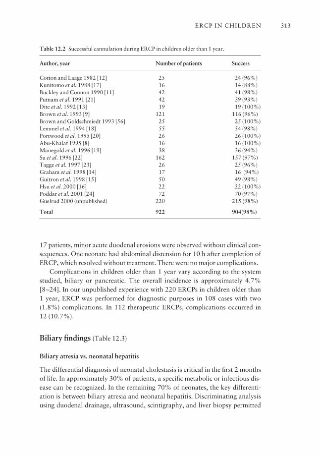
17 patients, minor acute duodenal erosions were observed without clinical con-sequences. One neonate had abdominal distension for 10 h after completion ofERCP, which resolved without treatment. There were no major complications.
Complications in children older than 1 year vary according to the systemstudied, biliary or pancreatic. The overall incidence is approximately 4.7%[8–24]. In our unpublished experience with 220 ERCPs in children older than 1 year, ERCP was performed for diagnostic purposes in 108 cases with two (1.8%) complications. In 112 therapeutic ERCPs, complications occurred in 12 (10.7%).
Biliary findings (Table 12.3)
Biliary atresia vs. neonatal hepatitis
The differential diagnosis of neonatal cholestasis is critical in the first 2 monthsof life. In approximately 30% of patients, a specific metabolic or infectious dis-ease can be recognized. In the remaining 70% of neonates, the key differenti-ation is between biliary atresia and neonatal hepatitis. Discriminating analysisusing duodenal drainage, ultrasound, scintigraphy, and liver biopsy permitted
ERCP IN CHILDREN 313
Table 12.2 Successful cannulation during ERCP in children older than 1 year.
Author, year Number of patients Success
Cotton and Laage 1982 [12] 25 24 (96%)Kunitomo et al. 1988 [17] 16 14 (88%)Buckley and Connon 1990 [11] 42 41 (98%)Putnam et al. 1991 [21] 42 39 (93%)Dite et al. 1992 [13] 19 19 (100%)Brown et al. 1993 [9] 121 116 (96%)Brown and Goldschmiedt 1993 [56] 25 25 (100%)Lemmel et al. 1994 [18] 55 54 (98%)Portwood et al. 1995 [20] 26 26 (100%)Abu-Khalaf 1995 [8] 16 16 (100%)Manegold et al. 1996 [19] 38 36 (94%)Su et al. 1996 [22] 162 157 (97%)Tagge et al. 1997 [23] 26 25 (96%)Graham et al. 1998 [14] 17 16 (94%)Guitron et al. 1998 [15] 50 49 (98%)Hsu et al. 2000 [16] 22 22 (100%)Poddar et al. 2001 [24] 72 70 (97%)Guelrud 2000 (unpublished) 220 215 (98%)
Total 922 904(98%)
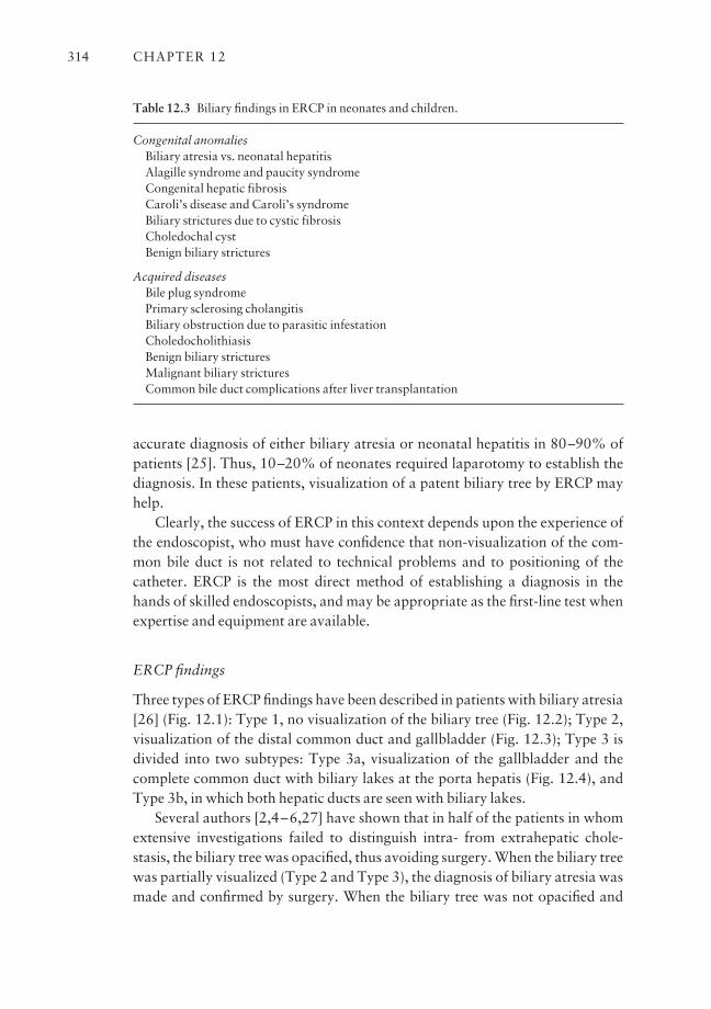
accurate diagnosis of either biliary atresia or neonatal hepatitis in 80–90% ofpatients [25]. Thus, 10–20% of neonates required laparotomy to establish thediagnosis. In these patients, visualization of a patent biliary tree by ERCP mayhelp.
Clearly, the success of ERCP in this context depends upon the experience ofthe endoscopist, who must have confidence that non-visualization of the com-mon bile duct is not related to technical problems and to positioning of thecatheter. ERCP is the most direct method of establishing a diagnosis in thehands of skilled endoscopists, and may be appropriate as the first-line test whenexpertise and equipment are available.
ERCP findings
Three types of ERCP findings have been described in patients with biliary atresia[26] (Fig. 12.1): Type 1, no visualization of the biliary tree (Fig. 12.2); Type 2,visualization of the distal common duct and gallbladder (Fig. 12.3); Type 3 isdivided into two subtypes: Type 3a, visualization of the gallbladder and thecomplete common duct with biliary lakes at the porta hepatis (Fig. 12.4), andType 3b, in which both hepatic ducts are seen with biliary lakes.
Several authors [2,4–6,27] have shown that in half of the patients in whomextensive investigations failed to distinguish intra- from extrahepatic chole-stasis, the biliary tree was opacified, thus avoiding surgery. When the biliary treewas partially visualized (Type 2 and Type 3), the diagnosis of biliary atresia wasmade and confirmed by surgery. When the biliary tree was not opacified and
CHAPTER 12314
Table 12.3 Biliary findings in ERCP in neonates and children.
Congenital anomaliesBiliary atresia vs. neonatal hepatitisAlagille syndrome and paucity syndromeCongenital hepatic fibrosisCaroli’s disease and Caroli’s syndromeBiliary strictures due to cystic fibrosisCholedochal cystBenign biliary strictures
Acquired diseasesBile plug syndromePrimary sclerosing cholangitisBiliary obstruction due to parasitic infestationCholedocholithiasisBenign biliary stricturesMalignant biliary stricturesCommon bile duct complications after liver transplantation
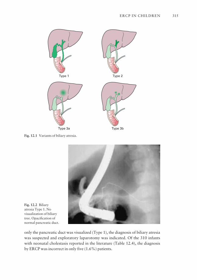
only the pancreatic duct was visualized (Type 1), the diagnosis of biliary atresiawas suspected and exploratory laparotomy was indicated. Of the 310 infantswith neonatal cholestasis reported in the literature (Table 12.4), the diagnosisby ERCP was incorrect in only five (1.6%) patients.
ERCP IN CHILDREN 315
Type 2Type 1
Type 3a Type 3b
Fig. 12.1 Variants of biliary atresia.
Fig. 12.2 Biliary atresia Type 1. Novisualization of biliarytree. Opacification ofnormal pancreatic duct.
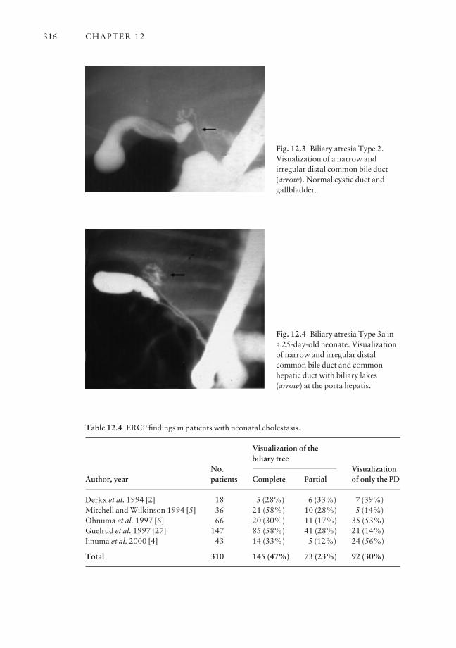
CHAPTER 12316
Fig. 12.3 Biliary atresia Type 2.Visualization of a narrow andirregular distal common bile duct(arrow). Normal cystic duct andgallbladder.
Table 12.4 ERCP findings in patients with neonatal cholestasis.
Visualization of the
No.biliary tree
VisualizationAuthor, year patients Complete Partial of only the PD
Derkx et al. 1994 [2] 18 5 (28%) 6 (33%) 7 (39%)Mitchell and Wilkinson 1994 [5] 36 21 (58%) 10 (28%) 5 (14%)Ohnuma et al. 1997 [6] 66 20 (30%) 11 (17%) 35 (53%)Guelrud et al. 1997 [27] 147 85 (58%) 41 (28%) 21 (14%)Iinuma et al. 2000 [4] 43 14 (33%) 5 (12%) 24 (56%)
Total 310 145 (47%) 73 (23%) 92 (30%)
Fig. 12.4 Biliary atresia Type 3a in a 25-day-old neonate. Visualizationof narrow and irregular distalcommon bile duct and commonhepatic duct with biliary lakes(arrow) at the porta hepatis.

Miscellaneous genetic cholestatic diseases
In Alagille syndrome, the extrahepatic ducts are normal. ERCP shows markedand diffuse narrowing of the intrahepatic duct and reduced arborization[27,28]. Congenital hepatic fibrosis is characterized by disordered terminalinterlobular bile ducts, which form multiple macroscopic and microscopic cysts(Fig. 12.5) that can be demonstrated by ERCP [27]. In Caroli’s disease, there aremultiple segmental cylindrical or saccular dilatations of small biliary radicleswith a normal common bile duct that can be demonstrated by ERCP [27].Diagnosis of these conditions is necessary in order to avoid needless surgery.
Bile plug syndrome
Bile plug syndrome represents a correctable cause of obstruction of the extra-hepatic bile ducts by bile sludge in patients with a normal biliary tract. The diag-nosis is suspected by ultrasonography and confirmed by ERCP, which offerstherapeutic possibility. Improvements of patients after ERCP suggest that simple irrigation with contrast material may be helpful [27].
Choledochal cyst
Choledochal cyst is a congenital malformation of the biliary tract characterizedby saccular dilatation of the biliary tree. Choledochal cyst is primarily a diseaseof children and young adults, and 60% of reported cases are diagnosed beforeage 10 [29]. The diagnosis of this congenital malformation of the biliary tract ismade by abdominal ultrasound, CT, or MRCP. ERCP confirms the diagnosisand helps surgical planning.
ERCP IN CHILDREN 317
Fig. 12.5 Congenital hepaticfibrosis in a 38-day-old infant.Normal extrahepatic ducts.Irregular intrahepatic ducts withmultiple small cysts (arrow).
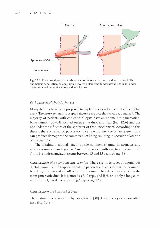
Pathogenesis of choledochal cyst
Many theories have been proposed to explain the development of choledochalcysts. The more generally accepted theory proposes that cysts are acquired. Themajority of patients with choledochal cysts have an anomalous pancreatico-biliary union [30–34] located outside the duodenal wall (Fig. 12.6) and are not under the influence of the sphincter of Oddi mechanism. According to thistheory, there is reflux of pancreatic juice upward into the biliary system that can produce damage to the common duct lining resulting in saccular dilatationof the duct [35].
The maximum normal length of the common channel in neonates andinfants younger than 1 year is 3 mm. It increases with age to a maximum of 5 mm in children and adolescents between 13 and 15 years of age [36].
Classification of anomalous ductal union There are three types of anomalousductal union [37]. If it appears that the pancreatic duct is joining the commonbile duct, it is denoted as P–B type. If the common bile duct appears to join themain pancreatic duct, it is denoted as B–P type, and if there is only a long com-mon channel, it is denoted as Long Y type (Fig. 12.7).
Classification of choledochal cysts
The anatomical classification by Todani et al. [38] of bile duct cysts is most oftenused (Fig. 12.8).
CHAPTER 12318
Normal Anomalous union
Sphincter of Oddi
Duodenal wall
Fig. 12.6 The normal pancreatico-biliary union is located within the duodenal wall. Theanomalous pancreatico-biliary union is located outside the duodenal wall and is not underthe influence of the sphincter of Oddi mechanism.
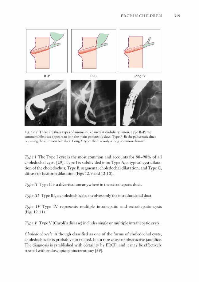
Type I The Type I cyst is the most common and accounts for 80–90% of allcholedochal cysts [29]. Type I is subdivided into: Type A, a typical cyst dilata-tion of the choledochus; Type B, segmental choledochal dilatation; and Type C,diffuse or fusiform dilatation (Figs 12.9 and 12.10).
Type II Type II is a diverticulum anywhere in the extrahepatic duct.
Type III Type III, a choledochocele, involves only the intraduodenal duct.
Type IV Type IV represents multiple intrahepatic and extrahepatic cysts (Fig. 12.11).
Type V Type V (Caroli’s disease) includes single or multiple intrahepatic cysts.
Choledochocele Although classified as one of the forms of choledochal cysts,choledochocele is probably not related. It is a rare cause of obstructive jaundice.The diagnosis is established with certainty by ERCP, and it may be effectivelytreated with endoscopic sphincterotomy [39].
ERCP IN CHILDREN 319
Long 'Y'P–BB–P
Fig. 12.7 There are three types of anomalous pancreatico-biliary union. Type B–P: thecommon bile duct appears to join the main pancreatic duct. Type P–B: the pancreatic duct is joining the common bile duct. Long Y type: there is only a long common channel.
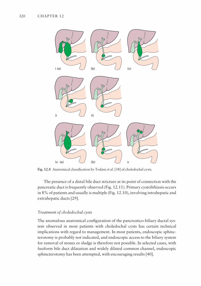
The presence of a distal bile duct stricture at its point of connection with thepancreatic duct is frequently observed (Fig. 12.11). Primary cystolithiasis occursin 8% of patients and usually is multiple (Fig. 12.10), involving intrahepatic andextrahepatic ducts [29].
Treatment of choledochal cysts
The anomalous anatomical configuration of the pancreatico-biliary ductal sys-tem observed in most patients with choledochal cysts has certain technicalimplications with regard to management. In most patients, endoscopic sphinc-terotomy is probably not indicated, and endoscopic access to the biliary systemfor removal of stones or sludge is therefore not possible. In selected cases, withfusiform bile duct dilatation and widely dilated common channel, endoscopicsphincterotomy has been attempted, with encouraging results [40].
CHAPTER 12320
i
ii
(a) (b) (c)
iii
iv (a) (b) v
Fig. 12.8 Anatomical classification by Todani et al. [38] of choledochal cysts.
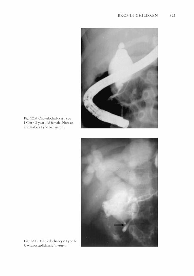
ERCP IN CHILDREN 321
Fig. 12.9 Choledochal cyst Type I-C in a 3-year-old female. Note ananomalous Type B–P union.
Fig. 12.10 Choledochal cyst Type I-C with cystolithiasis (arrow).
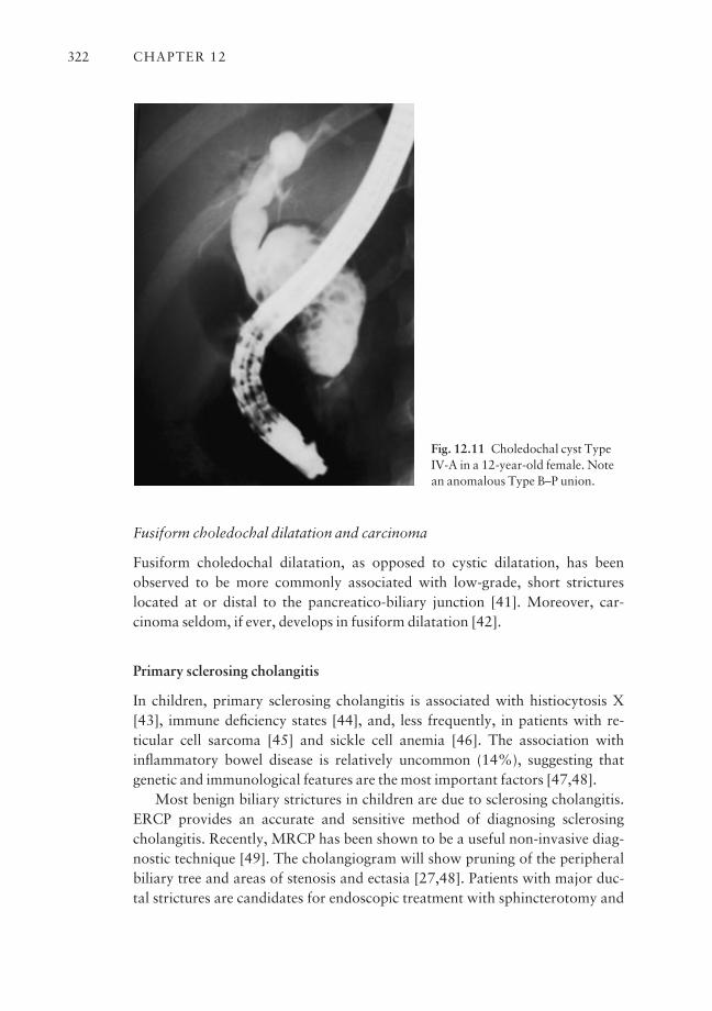
Fusiform choledochal dilatation and carcinoma
Fusiform choledochal dilatation, as opposed to cystic dilatation, has beenobserved to be more commonly associated with low-grade, short strictureslocated at or distal to the pancreatico-biliary junction [41]. Moreover, car-cinoma seldom, if ever, develops in fusiform dilatation [42].
Primary sclerosing cholangitis
In children, primary sclerosing cholangitis is associated with histiocytosis X[43], immune deficiency states [44], and, less frequently, in patients with re-ticular cell sarcoma [45] and sickle cell anemia [46]. The association withinflammatory bowel disease is relatively uncommon (14%), suggesting thatgenetic and immunological features are the most important factors [47,48].
Most benign biliary strictures in children are due to sclerosing cholangitis.ERCP provides an accurate and sensitive method of diagnosing sclerosingcholangitis. Recently, MRCP has been shown to be a useful non-invasive diag-nostic technique [49]. The cholangiogram will show pruning of the peripheralbiliary tree and areas of stenosis and ectasia [27,48]. Patients with major duc-tal strictures are candidates for endoscopic treatment with sphincterotomy and
CHAPTER 12322
Fig. 12.11 Choledochal cyst TypeIV-A in a 12-year-old female. Notean anomalous Type B–P union.
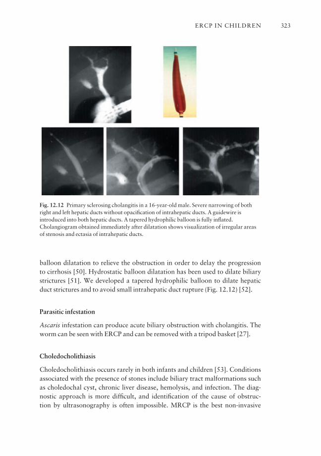
balloon dilatation to relieve the obstruction in order to delay the progression to cirrhosis [50]. Hydrostatic balloon dilatation has been used to dilate biliarystrictures [51]. We developed a tapered hydrophilic balloon to dilate hepaticduct strictures and to avoid small intrahepatic duct rupture (Fig. 12.12) [52].
Parasitic infestation
Ascaris infestation can produce acute biliary obstruction with cholangitis. Theworm can be seen with ERCP and can be removed with a tripod basket [27].
Choledocholithiasis
Choledocholithiasis occurs rarely in both infants and children [53]. Conditionsassociated with the presence of stones include biliary tract malformations suchas choledochal cyst, chronic liver disease, hemolysis, and infection. The diag-nostic approach is more difficult, and identification of the cause of obstruc-tion by ultrasonography is often impossible. MRCP is the best non-invasive
ERCP IN CHILDREN 323
Fig. 12.12 Primary sclerosing cholangitis in a 16-year-old male. Severe narrowing of bothright and left hepatic ducts without opacification of intrahepatic ducts. A guidewire isintroduced into both hepatic ducts. A tapered hydrophilic balloon is fully inflated.Cholangiogram obtained immediately after dilatation shows visualization of irregular areasof stenosis and ectasia of intrahepatic ducts.
technique in demonstrating common bile duct stones and is clearly superior toultrasonography [54].
ERCP for stones
The role and value of ERCP and endoscopic sphincterotomy in children withcholedocholithiasis are not well established. Sphincterotomy with common bileduct stone removal has been successfully performed in young infants [55], andin children and adolescents [11,56–60]. Endoscopic papillary balloon dilata-tion with stone extraction is an alternative technique for stone removal [61].However, pancreatitis can occur in 7% of cases. In children, published experi-ence with this technique is limited [59].
Most infants with asymptomatic gallstones and no factors that would makethem susceptible to stone formation can be managed conservatively [62].However, larger stones are less likely to resolve, whereas smaller stones, sludge,and mucus should be able to pass in response to oral feeding without symptomsor complications. In children, sphincterotomy should be reserved for symp-tomatic patients or those with underlying lithogenic disorders.
A combined endoscopic sphincterotomy with common stone extraction followed by laparoscopic cholecystectomy has been successfully reported inchildren [63]. Although the combined procedure seems to be safe, additionalexperience is awaited so that the true advantages, limitations, and complica-tions of this approach can be placed into clinical perspective.
Biliary strictures and leaks
Primary stricture
Primary stricture of the common hepatic duct has been reported [64]. Hydro-static balloon dilatation may be used in the treatment of dominant common ductstrictures [27].
Malignant strictures
Malignant strictures of the common bile duct are uncommon in children andhave been successfully treated by placement of stents [65,66].
Liver transplantation
In patients whose liver is transplanted, the integrity of the anastomosis can bestudied. ERCP is an alternative to percutaneous transhepatic cholangiography,
CHAPTER 12324
and is the procedure of choice in patients with coagulopathy when the biliarytree must be imaged. When a stricture is found, the area may be dilated and astent may be placed for a limited period of time.
Bile leaks
Bile leaks may be found and can be treated by endoscopic sphincterotomy orwith stent placement [67].
Pancreatic findings (Table 12.5)
Recurrent pancreatitis
ERCP has been found to be useful in the identification of treatable causes in approximately 75% of children with recurrent pancreatitis [10–14,18,21,24,68–70]. Whatever the etiology of pancreatitis, the possibility of an anatomical abnormality amenable to endoscopic therapy or surgery shouldalways be considered.
The timing of performance of an ERCP in children is controversial. In childrenwith idiopathic pancreatitis in whom recovery has occurred with standard
ERCP IN CHILDREN 325
Recurrent pancreatitisCongenital disorders
Biliary anomaliesCholedochal cystAnomalous pancreatico-biliary union
Pancreatic anomaliesPancreas divisumAnnular pancreasShort pancreasCystic dilatation of the pancreatic duct
(pancreatocele)Duodenal anomalies
Duodenal or gastric duplication cystsDuodenal diverticulum
Acquired disordersParasitic infestation: AscarisSphincter of Oddi dysfunctionPancreatic traumaAcquired immunodeficiency syndrome
Chronic pancreatitis
Pseudocysts
Table 12.5 Pancreatic findings inERCP in children.
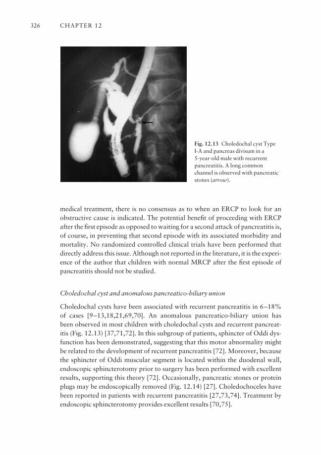
medical treatment, there is no consensus as to when an ERCP to look for anobstructive cause is indicated. The potential benefit of proceeding with ERCPafter the first episode as opposed to waiting for a second attack of pancreatitis is,of course, in preventing that second episode with its associated morbidity andmortality. No randomized controlled clinical trials have been performed thatdirectly address this issue. Although not reported in the literature, it is the experi-ence of the author that children with normal MRCP after the first episode ofpancreatitis should not be studied.
Choledochal cyst and anomalous pancreatico-biliary union
Choledochal cysts have been associated with recurrent pancreatitis in 6–18% of cases [9–13,18,21,69,70]. An anomalous pancreatico-biliary union has been observed in most children with choledochal cysts and recurrent pancreat-itis (Fig. 12.13) [37,71,72]. In this subgroup of patients, sphincter of Oddi dys-function has been demonstrated, suggesting that this motor abnormality mightbe related to the development of recurrent pancreatitis [72]. Moreover, becausethe sphincter of Oddi muscular segment is located within the duodenal wall,endoscopic sphincterotomy prior to surgery has been performed with excellentresults, supporting this theory [72]. Occasionally, pancreatic stones or proteinplugs may be endoscopically removed (Fig. 12.14) [27]. Choledochoceles havebeen reported in patients with recurrent pancreatitis [27,73,74]. Treatment byendoscopic sphincterotomy provides excellent results [70,75].
CHAPTER 12326
Fig. 12.13 Choledochal cyst Type I-A and pancreas divisum in a 5-year-old male with recurrentpancreatitis. A long commonchannel is observed with pancreaticstones (arrow).
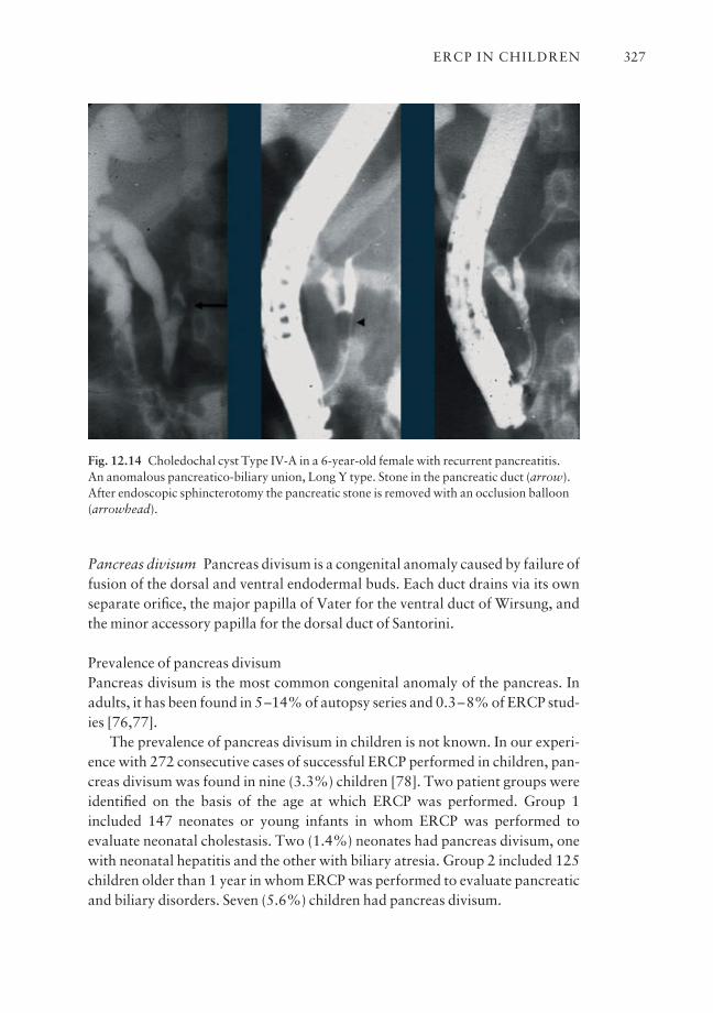
Pancreas divisum Pancreas divisum is a congenital anomaly caused by failure offusion of the dorsal and ventral endodermal buds. Each duct drains via its ownseparate orifice, the major papilla of Vater for the ventral duct of Wirsung, andthe minor accessory papilla for the dorsal duct of Santorini.
Prevalence of pancreas divisumPancreas divisum is the most common congenital anomaly of the pancreas. Inadults, it has been found in 5–14% of autopsy series and 0.3–8% of ERCP stud-ies [76,77].
The prevalence of pancreas divisum in children is not known. In our experi-ence with 272 consecutive cases of successful ERCP performed in children, pan-creas divisum was found in nine (3.3%) children [78]. Two patient groups wereidentified on the basis of the age at which ERCP was performed. Group 1included 147 neonates or young infants in whom ERCP was performed to evaluate neonatal cholestasis. Two (1.4%) neonates had pancreas divisum, onewith neonatal hepatitis and the other with biliary atresia. Group 2 included 125children older than 1 year in whom ERCP was performed to evaluate pancreaticand biliary disorders. Seven (5.6%) children had pancreas divisum.
ERCP IN CHILDREN 327
Fig. 12.14 Choledochal cyst Type IV-A in a 6-year-old female with recurrent pancreatitis. An anomalous pancreatico-biliary union, Long Y type. Stone in the pancreatic duct (arrow).After endoscopic sphincterotomy the pancreatic stone is removed with an occlusion balloon(arrowhead).
Significance of pancreas divisumThe clinical significance of pancreas divisum is controversial. An associationbetween pancreas divisum and pancreatitis has been suggested [76,79–83].However, others [84–86] have considered it to be a coincidental finding. Itappears that the combination of pancreas divisum with accessory papilla stenosiswould lead to a real functional obstruction. In 296 children with recurrent pan-creatitis, pancreas divisum has been found in 10.8% of patients (Table 12.6).
ERCP diagnosis of pancreas divisumERCP is mandatory in the diagnosis of pancreas divisum. Cannulation of themajor papilla shows a short duct of Wirsung (ventral pancreas) that quicklytapers and undergoes arborization (Fig. 12.15). To confirm the diagnosis, it ismost important to cannulate the minor papilla to demonstrate the dorsal pan-creas. Interventional treatment in patients with pancreas divisum is applied tothose whose symptoms are disabling.
Treatment of pancreas divisumSurgical minor papilla sphincteroplasty used to be the treatment of choice, witha 70% improvement [87]. Endoscopic treatment has been utilized to decom-press the dorsal duct by a variety of methods, including endoscopic minorpapilla sphincterotomy with or without insertion of an endoprosthesis.
Endoscopic sphincterotomy of the minor papilla is indicated in patients withdisabling symptoms. It has been attempted in conjunction with temporary stent
CHAPTER 12328
Table 12.6 Frequency of pancreas divisum in children with recurrent pancreatitis.
No. of patients with Author, year No. of patients pancreas divisum
Forbes et al. 1984 [69] 25 4 (16%)Buckley and Connon 1990 [11] 18 1 (5%)Putnam et al. 1991 [21] 12 0Dite et al. 1992 [13] 16 1 (6%)Brown and Goldschmiedt 1994 [10] 9 2 (22%)Lemmel et al. 1994 [18] 29 2 (7%)Guelrud et al. 1994 [70] 50 6 (12%)Portwood et al. 1995 [20] 26 3 (11%)Manegold et al. 1996 [19] 38 3 (8%)Graham et al. 1998 [14] 23 1 (4%)Hsu et al. 2000 [16] 22 6 (27%)Poddar et al. 2001 [24] 28 3 (11%)
Total 296 32 (10.8%)

placement in the dorsal pancreatic duct (Fig. 12.16), and has led to improve-ment in approximately 75% of children [16,18,70]. Overall, these results indi-cate that in certain children with recurrent pain or pancreatitis and pancreas divisum, endoscopic therapy can offer relief or improvement of symptoms.
Other pancreatic congenital anomalies
Annular pancreas has been associated with recurrent pancreatitis in children[70,88–90]. However, the relationship with pancreatitis is unclear. In 14 casesof annular pancreas reported in the English literature, there were five with coexistent pancreas divisum, suggesting that pancreas divisum occurs moreoften in the presence of annular pancreas than in the general population [89].This associ-ation may explain pancreatitis in some patients.
Other pancreatic congenital anomalies found to cause pancreatitis includeshort pancreas [70,91] and cystic dilatation or pancreatocele of the distal pan-creatic duct [60].
ERCP IN CHILDREN 329
Fig. 12.15 Pancreas divisum and chronic pancreatitis in a 12-year-old male. Cannulation ofthe major papilla shows a normal common duct and a small ventral pancreatic duct (arrow).Cannulation of the minor papilla shows a dilated dorsal pancreatic duct (arrowhead ) withdilated primary and secondary branches. After minor papilla sphincterotomy, a 5 Frpancreatic stent (two arrows) without proximal flaps was left in place for 5 days.
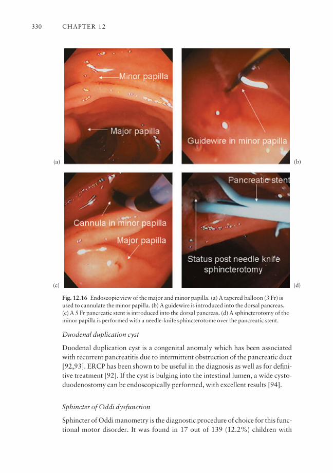
Duodenal duplication cyst
Duodenal duplication cyst is a congenital anomaly which has been associatedwith recurrent pancreatitis due to intermittent obstruction of the pancreatic duct[92,93]. ERCP has been shown to be useful in the diagnosis as well as for defini-tive treatment [92]. If the cyst is bulging into the intestinal lumen, a wide cysto-duodenostomy can be endoscopically performed, with excellent results [94].
Sphincter of Oddi dysfunction
Sphincter of Oddi manometry is the diagnostic procedure of choice for this func-tional motor disorder. It was found in 17 out of 139 (12.2%) children with
CHAPTER 12330
Fig. 12.16 Endoscopic view of the major and minor papilla. (a) A tapered balloon (3 Fr) isused to cannulate the minor papilla. (b) A guidewire is introduced into the dorsal pancreas.(c) A 5 Fr pancreatic stent is introduced into the dorsal pancreas. (d) A sphincterotomy of theminor papilla is performed with a needle-knife sphincterotome over the pancreatic stent.
(a)
(c)
(b)
(d)
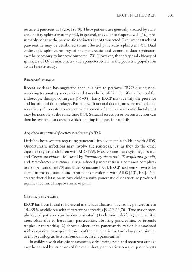
recurrent pancreatitis [9,16,18,70]. These patients are generally treated by stan-dard biliary sphincterotomy and, in general, they do not respond well [16], pre-sumably because the pancreatic sphincter is not transected. Recurrent attacks ofpancreatitis may be attributed to an affected pancreatic sphincter [95]. Dualendoscopic sphincterotomy of the pancreatic and common duct sphincters may be necessary to improve outcome [70]. However, the safety and efficacy ofsphincter of Oddi manometry and sphincterotomy in the pediatric populationawait further study.
Pancreatic trauma
Recent evidence has suggested that it is safe to perform ERCP during non-resolving traumatic pancreatitis and it may be helpful in identifying the need forendoscopic therapy or surgery [96–98]. Early ERCP may identify the presenceand location of duct leakage. Patients with normal ductograms are treated con-servatively. Successful treatment by placement of an intrapancreatic ductal stentmay be possible at the same time [98]. Surgical resection or reconstruction canthen be reserved for cases in which stenting is impossible or fails.
Acquired immunodeficiency syndrome (AIDS)
Little has been written regarding pancreatic involvement in children with AIDS.Opportunistic infections may involve the pancreas, just as they do the otherdigestive organs in children with AIDS [99]. Most common are cytomegalovirusand Cryptosporidium, followed by Pneumocystis carinii, Toxoplasma gondii,and Mycobacterium avium. Drug-induced pancreatitis is a common complica-tion of pentamidine [99] and dideoxyinosine [100]. ERCP has been shown to beuseful in the evaluation and treatment of children with AIDS [101,102]. Pan-creatic duct dilatation in two children with pancreatic duct stricture producedsignificant clinical improvement of pain.
Chronic pancreatitis
ERCP has been found to be useful in the identification of chronic pancreatitis in14–69% of children with recurrent pancreatitis [9–22,69,70]. Two major mor-phological patterns can be demonstrated: (1) chronic calcifying pancreatitis,most often due to hereditary pancreatitis, fibrosing pancreatitis, or juveniletropical pancreatitis; (2) chronic obstructive pancreatitis, which is associatedwith congenital or acquired lesions of the pancreatic duct or biliary tree, similarto those etiological factors found in recurrent pancreatitis.
In children with chronic pancreatitis, debilitating pain and recurrent attacksmay be caused by strictures of the main duct, pancreatic stones, or pseudocysts
ERCP IN CHILDREN 331
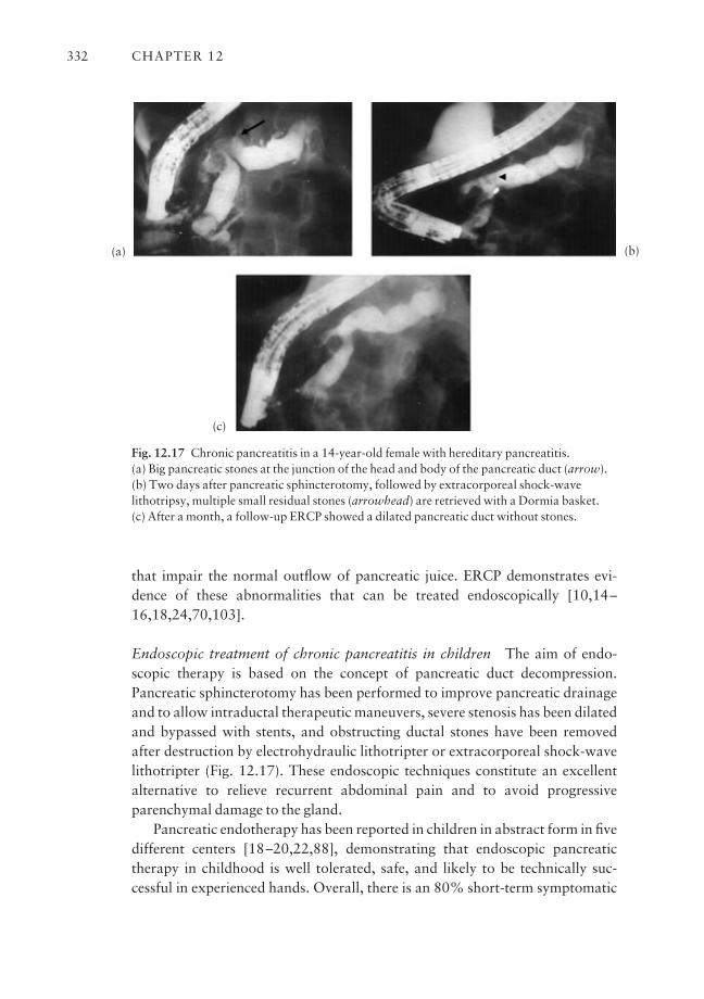
that impair the normal outflow of pancreatic juice. ERCP demonstrates evi-dence of these abnormalities that can be treated endoscopically [10,14–16,18,24,70,103].
Endoscopic treatment of chronic pancreatitis in children The aim of endo-scopic therapy is based on the concept of pancreatic duct decompression.Pancreatic sphincterotomy has been performed to improve pancreatic drainageand to allow intraductal therapeutic maneuvers, severe stenosis has been dilatedand bypassed with stents, and obstructing ductal stones have been removedafter destruction by electrohydraulic lithotripter or extracorporeal shock-wavelithotripter (Fig. 12.17). These endoscopic techniques constitute an excellentalternative to relieve recurrent abdominal pain and to avoid progressiveparenchymal damage to the gland.
Pancreatic endotherapy has been reported in children in abstract form in fivedifferent centers [18–20,22,88], demonstrating that endoscopic pancreatictherapy in childhood is well tolerated, safe, and likely to be technically suc-cessful in experienced hands. Overall, there is an 80% short-term symptomatic
CHAPTER 12332
Fig. 12.17 Chronic pancreatitis in a 14-year-old female with hereditary pancreatitis. (a) Big pancreatic stones at the junction of the head and body of the pancreatic duct (arrow).(b) Two days after pancreatic sphincterotomy, followed by extracorporeal shock-wavelithotripsy, multiple small residual stones (arrowhead) are retrieved with a Dormia basket. (c) After a month, a follow-up ERCP showed a dilated pancreatic duct without stones.
(a)
(c)
(b)
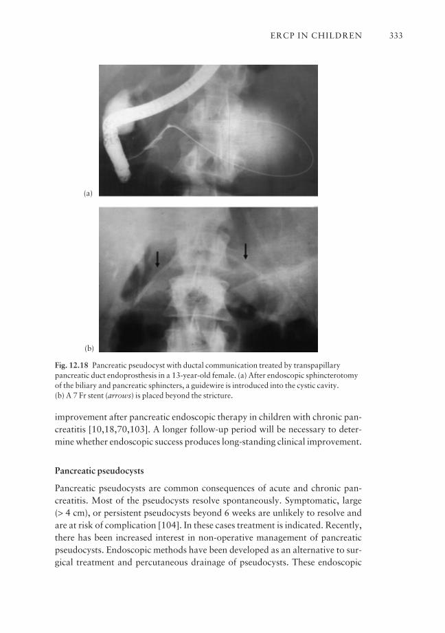
improvement after pancreatic endoscopic therapy in children with chronic pan-creatitis [10,18,70,103]. A longer follow-up period will be necessary to deter-mine whether endoscopic success produces long-standing clinical improvement.
Pancreatic pseudocysts
Pancreatic pseudocysts are common consequences of acute and chronic pan-creatitis. Most of the pseudocysts resolve spontaneously. Symptomatic, large (> 4 cm), or persistent pseudocysts beyond 6 weeks are unlikely to resolve andare at risk of complication [104]. In these cases treatment is indicated. Recently,there has been increased interest in non-operative management of pancreaticpseudocysts. Endoscopic methods have been developed as an alternative to sur-gical treatment and percutaneous drainage of pseudocysts. These endoscopic
ERCP IN CHILDREN 333
Fig. 12.18 Pancreatic pseudocyst with ductal communication treated by transpapillarypancreatic duct endoprosthesis in a 13-year-old female. (a) After endoscopic sphincterotomyof the biliary and pancreatic sphincters, a guidewire is introduced into the cystic cavity. (b) A 7 Fr stent (arrows) is placed beyond the stricture.
(a)
(b)
methods include endoscopic cystogastrostomy, cystoduodenostomy, andtranspapillary drainage (Fig. 12.18). In adults, successful pseudocyst resolutionhas been reported in approximately 80% of cases [105–107]. As with othertherapeutic interventions, pediatric experience is limited to a few case reports[9,18,70,103].
Outstanding issues and future trends
ERCP is an established procedure in children. Even though there is an increasednumber of pediatric gastroenterologists performing ERCP, there is not enoughvolume for them to gain proficiency. This has become more apparent since,nowadays, ERCP is less diagnostic and more therapeutic. It is my belief that, in future, ERCP in children will be performed by highly trained endoscopistsworking in tertiary care facilities, which maintain a high volume of such activity.Further studies should be directed to assess the usefulness of MRCP in the diagnosis of biliopancreatic diseases in children. In general, children with suspected biliary and pancreatic disease should undergo MRCP before ERCP isconsidered, with the latter increasingly being reserved for therapy.
References
1 Guelrud M, Jaen D, Torres P et al. Endoscopic cholangiopancreatography in the infant: evalu-ation of a new prototype pediatric duodenoscope. Gastrointest Endosc 1987; 33: 4–8.
2 Derkx HH, Huibregtse K, Taminiau JA. The role of endoscopic retrograde cholangiopancre-atography in cholestatic infants. Endoscopy 1994; 26: 724–8.
3 Heyman MB, Shapiro HA, Thaler MM. Endoscopic retrograde cholangiography in the diag-nosis of biliary malformations in infants. Gastrointest Endosc 1988; 34: 449–53.
4 Iinuma Y, Narisawa R, Iwafuchi M et al. The role of endoscopic retrograde cholangiopancre-atography in infants with cholestasis. J Pediatr Surg 2000; 35: 545–9.
5 Mitchell SA, Wilkinson ML. The role of ERCP in the diagnosis of neonatal conjugated hyper-bilirubinemia. Gastrointest Endosc 1994; 40: A55.
6 Ohnuma N, Takahashi T, Tanabe M et al. The role of ERCP in biliary atresia. GastrointestEndosc 1997; 45: 365–70.
7 Wilkinson ML, Mieli-Vergani G, Ball C et al. Endoscopic retrograde cholangiopancreato-graphy in infantile cholestasis. Arch Dis Child 1991; 66: 121–3.
8 Abu-Khalaf A. The role of endoscopic retrograde cholangiopancreatography in small childrenand adolescents. Surg Laparosc Endosc 1995; 5: 296–300.
9 Brown CW, Werlin SL, Geenen JE et al. The diagnostic and therapeutic role of endoscopic ret-rograde cholangiopancreatography in children. J Pediatr Gastroenterol Nutr 1993; 17: 19–23.
10 Brown KO, Goldschmiedt M. Endoscopic therapy of biliary and pancreatic disorders in chil-dren. Endoscopy 1994; 26: 719–23.
11 Buckley A, Connon JJ. The role of ERCP in children and adolescents. Gastrointest Endosc1990; 36: 369–72.
12 Cotton PB, Laage NJ. Endoscopic retrograde cholangiopancreatography in children. Arch DisChild 1982; 57: 131–6.
13 Dite P, Vacek E, Stefan H et al. Endoscopic retrograde cholangiopancreatography in child-hood. Hepatogastroenterology 1992; 39: 291–3.
CHAPTER 12334
14 Graham KS, Ingram JD, Steinberg SE et al. ERCP in the management of pediatric pancreatitis.Gastrointest Endosc 1998; 47: 492–5.
15 Guitron A, Adalid R, Barinagarrementeria R et al. Endoscopic cholangiopancreatography(ERCP) in pediatric patients. Rev Gastroenterol Mex 1998; 63: 211–16.
16 Hsu RK, Draganov P, Leung JW et al. Therapeutic ERCP in the management of pancreatitis inchildren. Gastrointest Endosc 2000; 51: 396–400.
17 Kunitomo K, Ming L, Urakami Y et al. Endoscopic retrograde cholangiopancreatography inpediatric surgical biliary diseases. Tokushima J Exp Med 1988; 35: 57–62.
18 Lemmel T, Hawes R, Sherman S et al. Endoscopic evaluation and therapy of recurrent pancre-atitis and pancreaticobiliary pain in the pediatric population. Gastrointest Endosc 1994; 40:A54.
19 Manegold BC, Gottstein T, Pescatore P. Diagnostic and therapeutic ERCP in children under 14years. Gastrointest Endosc 1996; 43: A328.
20 Portwood G, Maniatis A, Jowell PS et al. Diagnostic and therapeutic ERCP in children: safewith a high success rate in experienced hands. Gastrointest Endosc 1995; 41: A342.
21 Putnam PE, Kocoshis SA, Orenstein SR et al. Pediatric endoscopic retrograde cholangio-pancreatography. Am J Gastroenterol 1991; 86: 824–30.
22 Su AY, Hernandez EJ, Brown K et al. Therapeutic endoscopic retrograde cholangiopan-creatography in children. Gastrointest Endosc 1996; 43: A330.
23 Tagge EP, Tarnasky PR, Chandler J et al. Multidisciplinary approach to the treatment of pedi-atric pancreaticobiliary disorders. J Pediatr Surg 1997; 32: 158–64.
24 Poddar U, Thapa BR, Bhasin DK et al. Endoscopic retrograde cholangiopancreatography inthe management of pancreaticobiliary disorders in children. J Gastroenterol Hepatol 2001; 16:927–31.
25 Balistreri WF. Neonatal cholestasis. J Pediatr 1985; 106: 171–84.26 Guelrud M, Jaen D, Mendoza S et al. ERCP in the diagnosis of extrahepatic biliary atresia.
Gastrointest Endosc 1991; 37: 522–6.27 Guelrud M, Carr-Locke D, Fox VL. ERCP in Pediatric Practice. Diagnosis and Treatment.
Oxford: Isis Medical Media Ltd, 1997.28 Morelli A, Pelli MA, Vedovelli A et al. Endoscopic retrograde cholangiopancreatography
study in Alagille’s syndrome: first report. Am J Gastroenterol 1983; 78: 241–4.29 Yamaguchi M. Congenital choledochal cyst: analysis of 1433 patients in the Japanese liter-
ature. Am J Surg 1980; 140: 653–7.30 Guelrud M, Jaen D, Mendoza S et al. Usefulness of endoscopic retrograde cholangiopan-
creatography in diagnosis of choledochal cysts in children. G E N 1989; 43: 9–12.31 Kimura K, Ohto M, Ono T et al. Congenital cystic dilatation of the common bile duct: rela-
tionship to anomalous pancreaticobiliary ductal union. Am J Roentgenol 1977; 928: 571–7.32 Arima E, Akita H. Congenital biliary tract dilatation and anomalous junction of the pancre-
aticobiliary system. J Pediatr Surg 1979; 14: 9–15.33 Oguchi Y, Okada A, Nakamura T et al. Histopathologic studies of congenital dilatation of the
bile duct as related to an anomalous junction of the pancreaticobiliary ductal system: clinicaland experimental studies. Surgery 1988; 103: 168–73.
34 Ikada A, Nakamura T, Higaki J et al. Congenital dilatation of the bile duct in 100 instancesand its relationship with anomalous junction. Surg Gynecol Obstet 1990; 171: 291–8.
35 Babbitt DP. Congenital choledochal cysts: new etiological concept based on anomalous rela-tionship of common bile duct and pancreatic bulb. Ann Radiol 1969; 12: 231–40.
36 Guelrud M, Morera C, Rodriguez M et al. Normal and anomalous pancreaticobiliary union inchildren and adolescents. Gastrointest Endosc 1999; 50: 189–93.
37 Misra SP, Dwivedi M. Pancreaticobiliary ductal union. Gut 1990; 31: 1144–9.38 Todani T, Watanabe Y, Narusue M. Congenital bile duct cyst. Am J Surg 1977; 134: 263–9.39 Venu RP, Geenen JE, Hogan WJ et al. Role of endoscopic retrograde cholangiopancreato-
graphy in the diagnosis and treatment of choledochocele. Gastroenterology 1984; 87: 1144–9.40 Ng WD, Liu K, Wong MK et al. Endoscopic sphincterotomy in young patients with chole-
dochal dilatation and a long common channel: a preliminary report. Br J Surg 1992; 79:550–2.
ERCP IN CHILDREN 335

41 Ito T, Ando M, Nagaya T, Sugito T. Congenital dilatation of the common bile duct in children:the etiologic significance of the narrow segment distal to the dilated common bile duct. ZKinderchir 1984; 30: 40–5.
42 Todani T, Watanabe Y, Fujii T et al. Cylindrical dilatation of the choledochus: a special type ofcongenital bile duct dilatation. Surgery 1985; 98: 964–8.
43 Leblanc A, Hadchouel M, Jehan P et al. Obstructive jaundice in children with histiocytosis X.Gastroenterology 1981; 80: 134–9.
44 DiPalma JA, Strobel CT, Farrow JG. Primary sclerosing cholangitis associated with hyper-immunoglobulin M immunodeficiency (dysgammaglobulinemia). Gastroenterology 1986; 91:464–8.
45 Alpert LI, Jindrak K. Idiopathic retroperitoneal fibrosis and sclerosing cholangitis associatedwith a reticulum cell sarcoma. Gastroenterology 1972; 62: 111–17.
46 Werlin LS, Glicklich M, Jona J et al. Sclerosing cholangitis in childhood. J Pediatr 1980; 96:433–5.
47 Debray D, Pariente D, Urroas E et al. Sclerosing cholangitis in children. J Pediatr 1994; 124:49–56.
48 Classen M, Golze H, Richter HJ et al. Primary sclerosing cholangitis in children. J PediatrGastroenterol Nutr 1987; 6: 197–202.
49 Ferrara C, Valeri G, Salvolini L et al. Magnetic resonance cholangiopancreatography in prim-ary sclerosing cholangitis in children. Pediatr Radiol 2002; 32: 413–17.
50 Stoker J, Lameris JS, Robben SG et al. Primary sclerosing cholangitis in a child treated by non-surgical balloon dilatation and stenting. J Pediatr Gastroenterol Nutr 1993; 17: 303–6.
51 Siegel JH, Guelrud M. Endoscopic cholangiopancreatoplasty: hydrostatic balloon dilation inthe bile duct and pancreas. Gastrointest Endosc 1983; 29: 99–103.
52 Guelrud M, Mendoza S, Guelrud A. A tapered balloon with hydrophilic coating to dilate diffi-cult hilar biliary strictures. Gastrointest Endosc 1995; 41: 246–9.
53 Shaw PJ, Spitz L, Watson JG. Extrahepatic biliary obstruction due to stone. Arch Dis Child1984; 59: 896–7.
54 Arcement CM, Meza MP, Arumania S et al. MRCP in the evaluation of pancreaticobiliary dis-ease in children. Pediatr Radiol 2001; 31: 92–7.
55 Guelrud M, Daoud G, Mendoza S et al. Endoscopic sphincterotomy in a 6-month-old infantwith choledocholithiasis and double gallbladder. Am J Gastroenterol 1994; 89: 1587–9.
56 Brown KO, Goldschmiedt M. Use of ERCP with pancreatic and biliary sphincterotomy fortreatment of familial pancreatitis in a 2 year old pediatric patient. Gastrointest Endosc 1993;39: A309.
57 Guelrud M, Mendoza S, Jaen D et al. ERCP and endoscopic sphincterotomy in infants andchildren with jaundice due to common bile duct stones. Gastrointest Endosc 1992; 38: 450–3.
58 Man DW, Spitz L. Choledocholithiasis in infancy. J Pediatr Surg 1985; 20: 65–8.59 Tarnasky PR, Tagge EP, Hebra A et al. Minimally invasive therapy for choledocholithiasis in
children. Gastrointest Endosc 1998; 47: 189–92.60 Sandoval C, Stringel G, Ozkaynak MF et al. Perioperative management in children with sickle
cell disease undergoing laparoscopic surgery. JSLS 2002; 6: 29–33.61 Komatsu Y, Kawabe T, Toda N et al. Endoscopic papillary balloon dilation for the manage-
ment of common bile duct stones: experience of 226 cases. Endoscopy 1998; 30: 12–17.62 Wesdorp I, Bosman D, de Graaff A et al. Clinical presentations and predisposing factors of
cholelithiasis and sludge in children. Pediatr Gastroenterol Nutr 2000; 31: 411–17.63 Guelrud M, Zambrano V, Jaen D et al. Endoscopic sphincterotomy and laparoscopic cholecys-
tectomy in a jaundiced infant. Gastrointest Endosc 1994; 40: 99–102.64 Chapoy PR, Kendall RS, Fonkalsrud E et al. Congenital stricture of the common hepatic duct:
an unusual case without jaundice. Gastroenterology 1981; 80: 380–3.65 Bickerstaff KI, Britton BJ, Gough MH. Endoscopic palliation of malignant biliary obstruction
in a child. Br J Surg 1989; 76: 1092–3.66 Guelrud M, Mendoza S, Zager A et al. Biliary stenting in an infant with malignant obstructive
jaundice. Gastrointest Endosc 1989; 35: 259–61.
CHAPTER 12336

67 Pfau PR, Kochman ML, Lewis JD et al. Endoscopic management of postoperative biliary com-plications in orthotopic liver transplantation. Gastrointest Endosc 2000; 52: 55–63.
68 Blustein PK, Gaskin K, Filler R et al. Endoscopic retrograde cholangiopancreatography in pan-creatitis in children and adolescents. Pediatrics 1981; 68: 387–93.
69 Forbes A, Leung JW, Cotton PB. Relapsing acute and chronic pancreatitis. Arch Dis Child1984; 59: 927–34.
70 Guelrud M, Mujica C, Jaen D et al. The role of ERCP in the diagnosis and treatment of idio-pathic recurrent pancreatitis in children and adolescents. Gastrointest Endosc 1994; 40:428–36.
71 Mori K, Nagakawa T, Ohta T et al. Pancreatitis and anomalous union of the pancreaticobili-ary ductal system in childhood. J Pediatr Surg 1993; 28: 67–71.
72 Guelrud M, Morera C, Rodriguez M et al. Sphincter of Oddi dysfunction in children withrecurrent pancreatitis and anomalous pancreaticobiliary union: an etiologic concept. Gastro-intest Endosc 1999; 50: 194–9.
73 Greene FL, Brown JJ, Rubinstein P et al. Choledochocele and recurrent pancreatitis. Diagnosisand surgical management. Am J Surg 1985; 149: 306–9.
74 Weisser M, Bennek J, Hormann D. Choledochocele—a rare cause of necrotizing pancreatitis inchildhood. Eur J Pediatr Surg 2000; 10: 258–64.
75 Siegel JH, Harding GT, Chateau F. Endoscopic incision of choledochal cysts (choledochocele).Endoscopy 1981; 13: 200–2.
76 Cotton PB. Congenital anomaly of pancreas divisum as a cause of obstructive pain and pancre-atitis. Gut 1980; 21: 105.
77 Bernard JP, Sahel J, Giovanni M et al. Pancreas divisum is a probable cause of acute pancre-atitis: a report of 137 cases. Pancreas 1990; 5: 248.
78 Guelrud M. The incidence of pancreas divisum in children [letter]. Gastrointest Endosc 1996;43: 83–4.
79 Cotton PB. Pancreas divisum. Curiosity or culprit? Gastroenterology 1985; 89: 1431.80 Gregg JA. Pancreas divisum: its association with pancreatitis. Am J Surg 1977; 1 (34): 539.81 Heiss FN, Shea JA. Association of pancreatitis and various ductal anatomy: dominant drainage
of the duct of Santorini. Am J Gastroenterol 1978; 70: 158.82 Richter JM, Shapiro RH, Mulley AG et al. Association of pancreas divisum and pancreatitis,
and its treatment by sphincterotomy of the accessory ampulla. Gastroenterology 1981; 81:1104.
83 Neblett WW, O’Neill JA. Surgical management of recurrent pancreatitis in children with pan-creas divisum. Ann Surg 2000; 231: 899.
84 Delhaye M, Engelholm L, Cremer M. Pancreas divisum: congenital anatomic variant oranomaly? Contribution of endoscopic retrograde dorsal pancreatography. Gastroenterology1985; 89: 951.
85 Mitchell CJ, Lintott DJ, Ruddell WSJ et al. Clinical relevance of an unfused pancreatic ductsystem. Gut 1979; 20: 1066.
86 Rosch W, Koch H, Schaffner O et al. The clinical significance of pancreas divisum. Gastro-intest Endosc 1976; 22: 206.
87 Warshaw AL, Simeone JF, Schapiro RH et al. Evaluation and treatment of the dominant dorsalduct syndrome (pancreas divisum redefined). Am J Surg 1990; 159: 59.
88 Fox VL, Lichtenstein DR, Carr-Locke DL. Incomplete pancreas divisum in children with recur-rent pancreatitis (Abstract). Gastrointest Endosc 1995; 41: A337.
89 Lehman GA, O’Connor KW. Coexistence of annular pancreas and pancreas divisum – ERCPdiagnosis. Gastrointest Endosc 1985; 31: 25–8.
90 Yogi Y, Shibue T, Hashimoto S. Annular pancreas detected in adults, diagnosed by endoscopicretrograde cholangiopancreatography: report of four cases. Gastroenterol Jpn 1987; 22: 92.
91 Rosenstock F, Achkar E. A ‘short pancreas’. Gastrointest Endosc 1986; 32: 296.92 Holstege A, Barner S, Brambs HJ et al. Relapsing pancreatitis associated with duodenal wall
cysts. Diagnostic approach and treatment. Gastroenterology 1985; 88: 814.93 Lavine JE, Harrison M, Heyman MB. Gastrointestinal duplications causing relapsing pancre-
atitis in children. Gastroenterology 1989; 97: 1556.
ERCP IN CHILDREN 337
94 Johanson JF, Geenen JE, Hogan WJ et al. Endoscopic therapy of a duodenal duplication cyst.Gastrointest Endosc 1992; 38: 60.
95 Guelrud M, Siegel JH. Hypertensive pancreatic duct sphincter as a cause of pancreatitis.Successful treatment with hydrostatic balloon dilatation. Dig Dis Sci 1984; 29: 225–31.
96 Hall RI, Lavelle MI, Venables CW. Use of ERCP to identify the site of traumatic injuries of themain pancreatic duct in children. Br J Surg 1986; 73: 411–2.
97 Rescorla FJ, Plumley DA, Sherman S et al. The efficacy of early ERCP in pediatric pancreatictrauma. J Pediatr Surg 1995; 30: 336–40.
98 Canty TG, Weinman D. Treatment of pancreatic duct disruption in children by an endoscopic-ally placed stent. J Pediatr Surg 2001; 36: 345–8.
99 Miller TL, Winter HS, Luginbuhl LM et al. Pancreatitis in pediatric human immunodeficiencyvirus infection. J Pediatr 1992; 120: 223.
100 Butler KM, Venzon D, Henry N et al. Pancreatitis in human immunodeficiency virus-infectedchildren receiving dideoxyinosine. Pediatrics 1993; 91: 747.
101 Naon H, Shelton M, Thomas D et al. Retrograde-cholangio-pancreatic videoendoscopy(ERCP) findings in pediatric patients with acquired immunodeficiency syndrome (AIDS).Gastrointest Endosc 1995; 41: A430.
102 Yabut B, Werlin SL, Havens P et al. Endoscopic retrograde cholangiopancreatography in chil-dren with HIV infection. J Pediatr Gastroenterol Nutr 1996; 23: 624.
103 Kozarek RA, Christie D, Barclay G. Endoscopic therapy of pancreatitis in the pediatric popula-tion. Gastrointest Endosc 1993; 39: 665.
104 Beebe DS, Bubrick MP, Onstad GR et al. Management of pancreatic pseudocysts. SurgGynecol Obstet 1984; 159: 562–4.
105 Binmoeller KF, Seifert H, Walter A et al. Transpapillary and transmural drainage of pancreaticpseudocysts. Gastrointest Endosc 1995; 42: 219–24.
106 Cremer M, Deviere J, Engelholm L. Endoscopic management of cysts and pseudocysts inchronic pancreatitis: long-term follow-up after 7 years of experience. Gastrointest Endosc1989; 35: 1–9.
107 Smits ME, Rauws EA, Tytgat GN et al. The efficacy of endoscopic treatment of pancreaticpseudocysts. Gastrointest Endosc 1995; 42: 202–7.
CHAPTER 12338

CHAPTER 13
ERCP: Risks, Prevention, and Management
PETER B. COTTON
Synopsis
ERCP is the most risky procedure that endoscopists perform on a regular basis.There is the potential for technical and clinical failure, for misdiagnosis, andsome small risk to staff, but the main interest is in the risk for adverse clinicalevents. A consensus definition of complications and their severity, and a series of careful prospective studies, have clarified the degree of risk in different circumstances, and the relevant risk factors. This process has allowed a clearerpicture to emerge of the risk–benefit ratios in different clinical scenarios, and agreater ability to advise patients about their options. Also, the extensive experi-ence of the last 30 years has permitted authoritative statements on how to minimizethe likelihood of complications, and how to deal with difficult situations whenthey arise.
Introduction
ERCP has become popular worldwide because it can provide significant benefitin many clinical contexts. Sadly, it has also caused considerable harm in a smallnumber of patients. Thus, it is crucial for practitioners and potential patients tounderstand the predictors of benefit and of risk. Defining positive and negativeoutcomes has been a significant challenge [1–4], but much useful informationhas been gathered from increasingly sophisticated outcomes studies over the lasttwo decades.
This chapter concentrates on the risks and risk factors, emphasizes ways toreduce them, and provides guidance about management when adverse eventsoccur.
The risks of ERCP
The concept of ‘risk’ indicates that something can ‘go wrong’, and is thereforebest defined as a deviation from the plan. This assumes that a plan has been
339
clearly formulated. The patient’s perspective and understanding of the plan isenshrined in the process of informed consent. Deviations are best describedgenerically as ‘unplanned events’ [4].
Unplanned events of ERCP are of four types:• risks to staff;• technical failure;• clinical failure;• unplanned adverse eventsacomplications.
Risks for endoscopists and staff
The endoscopy unit is not a dangerous place, but there are a few risks for theERCP endoscopist and staff.
The possibility of transmission of infection exists, but should be entirely pre-ventable with standard precautions (gowns, gloves, and eye protection) andassiduous disinfection protocols.
Certain immunizations are also appropriate. Rarely, staff may become sen-sitive to materials used in the ERCP process, such as glutaraldehyde, or latex gloves.
The risks of radiation are minimized by appropriate education, shielding,and exposure monitoring [5].
Many older endoscopists have neck problems caused by looking downfiberscopes, a situation aggravated by ERCP rooms where the video and X-raymonitors are not side by side. Busy ERCP practitioners sometimes complain alsoof ‘elevator thumb’. A Canadian survey found that more than half of 114 endo-scopists performing ERCP had some attributable musculo-skeletal problem [6].
Technical failure
Not all ERCP procedures are successful technically. It may prove impossible toreach the papilla, to gain access to the duct of interest, or to complete the neces-sary therapeutic maneuvers. The chance of failure depends upon several factors.
Expertise
An important determinant of the chance of success is the level of expertise (ofthe endoscopist and team). There are now good data to show that more activeERCP endoscopists have better results [7], as applies in surgery [8].
Complexity
The risk of technical failure increases with the complexity of the problem. Any
CHAPTER 13340
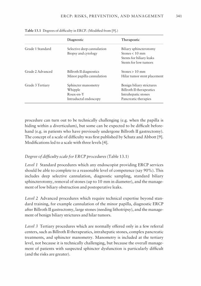
procedure can turn out to be technically challenging (e.g. when the papilla ishiding within a diverticulum), but some can be expected to be difficult before-hand (e.g. in patients who have previously undergone Billroth II gastrectomy).The concept of a scale of difficulty was first published by Schutz and Abbott [9].Modifications led to a scale with three levels [4].
Degree of difficulty scale for ERCP procedures (Table 13.1)
Level 1 Standard procedures which any endoscopist providing ERCP servicesshould be able to complete to a reasonable level of competence (say 90%). Thisincludes deep selective cannulation, diagnostic sampling, standard biliarysphincterotomy, removal of stones (up to 10 mm in diameter), and the manage-ment of low biliary obstruction and postoperative leaks.
Level 2 Advanced procedures which require technical expertise beyond stan-dard training, for example cannulation of the minor papilla, diagnostic ERCPafter Billroth II gastrectomy, large stones (needing lithotripsy), and the manage-ment of benign biliary strictures and hilar tumors.
Level 3 Tertiary procedures which are normally offered only in a few referralcenters, such as Billroth II therapeutics, intrahepatic stones, complex pancreatictreatments, and sphincter manometry. Manometry is included at the tertiarylevel, not because it is technically challenging, but because the overall manage-ment of patients with suspected sphincter dysfunction is particularly difficult(and the risks are greater).
ERCP: RISKS, PREVENTION, AND MANAGEMENT 341
Table 13.1 Degrees of difficulty in ERCP. (Modified from [9].)
Diagnostic Therapeutic
Grade 1 Standard Selective deep cannulation Biliary sphincterotomyBiopsy and cytology Stones < 10 mm
Stents for biliary leaksStents for low tumors
Grade 2 Advanced Billroth II diagnostics Stones > 10 mmMinor papilla cannulation Hilar tumor stent placement
Grade 3 Tertiary Sphincter manometry Benign biliary stricturesWhipple Billroth II therapeuticsRoux-en-Y Intrahepatic stonesIntraductal endoscopy Pancreatic therapies

Defining intent
A confounding issue when trying to assess technical success or failure is howwell the goal of the procedure is, or can be, defined beforehand [1,4]. When theintent is obvious, e.g. to remove a known stone, the resulting outcome isunequivocal. However, ERCP is often used to make or confirm a diagnosis, andthen to perform treatment ‘if appropriate’, so that defining intent, and thus ‘success’ and ‘failure’, may be more subjective. Also, endoscopists have differentthresholds for attempting therapy. Some may back away from a large stone, andcount the case as a success for good judgement rather than as a technical failure.Treatment will not even be considered if the diagnosis is not made (e.g. if can-nulation fails and a stone is missed), but such a case usually will not be countedas a failure of stone treatment [10,11]. Thus, the success literature should beviewed with some skepticism.
Risk consequences of technical failure
There are good data showing that failed procedures carry more complicationsthan successful ones. Failure usually necessitates repeat ERCP, or a percutane-ous or surgical procedure, which brings additional and significant costs and risks[12]. Strictly speaking, on an ‘intention to treat basis’, any complications ofthese subsequent procedures should be attributed to the initial ERCP attempt.
Clinical failure
Clinical success is dependent upon technical success, but the reverse is not neces-sarily true. A procedure may be completed technically in an exemplary fashion,but with no resulting benefit. This would be true certainly when the indication isnot appropriate.
Our aim is to make patients ‘better’, but defining precisely what that meanscan also be a challenge [1,4,13]. In some contexts (e.g. stone extraction, biliarystenting for low tumors), it is reasonable to assume that technical success willalmost guarantee clinical success, at least in the short to medium term. How-ever, some of those patients will have recurrent problems (e.g. new stones andstent occlusion), as detailed later, so that the time frame of measurement is rele-vant to success. It may be helpful to distinguish between initial ‘primary’ failureand ‘secondary’ failure, which means a relapse of the same problem.
It is also difficult to measure the success or failure of interventions in patientswho have intermittent problems such as recurrent pancreatitis or episodes ofpain suspected to be due to sphincter dysfunction. The true outcome in these casescan be measured only after months or years. Furthermore, the clinical response maybe incomplete, with a reduction, but not elimination, of attacks of pancreatitis,
CHAPTER 13342
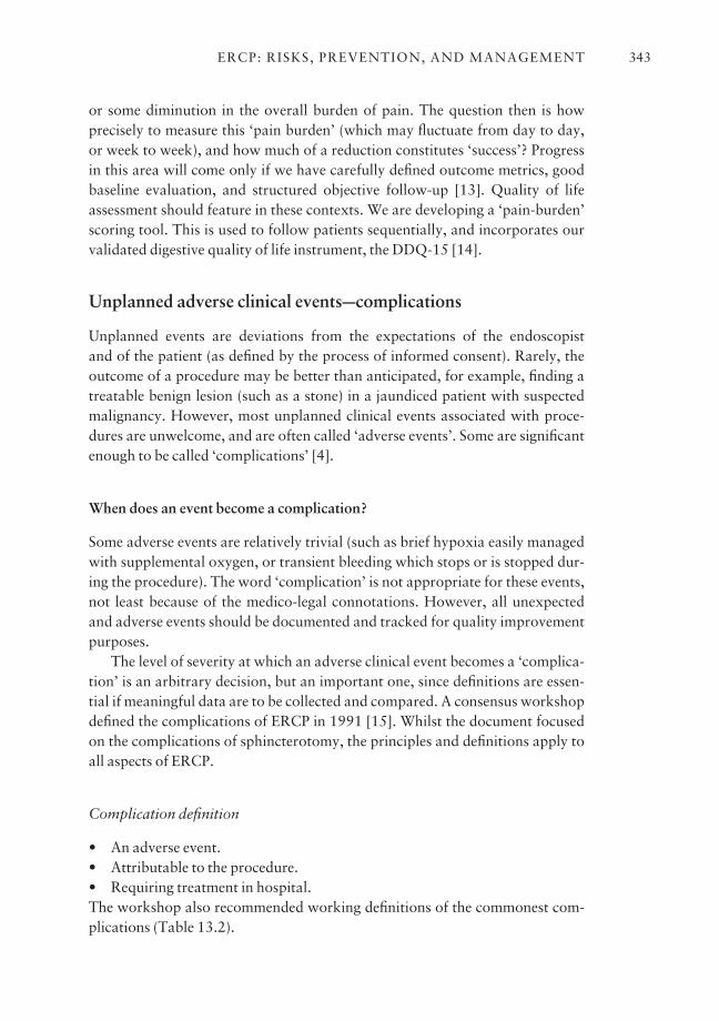
or some diminution in the overall burden of pain. The question then is how precisely to measure this ‘pain burden’ (which may fluctuate from day to day, or week to week), and how much of a reduction constitutes ‘success’? Progressin this area will come only if we have carefully defined outcome metrics, goodbaseline evaluation, and structured objective follow-up [13]. Quality of lifeassessment should feature in these contexts. We are developing a ‘pain-burden’scoring tool. This is used to follow patients sequentially, and incorporates ourvalidated digestive quality of life instrument, the DDQ-15 [14].
Unplanned adverse clinical eventsccomplications
Unplanned events are deviations from the expectations of the endoscopist and of the patient (as defined by the process of informed consent). Rarely, theoutcome of a procedure may be better than anticipated, for example, finding atreatable benign lesion (such as a stone) in a jaundiced patient with suspectedmalignancy. However, most unplanned clinical events associated with proce-dures are unwelcome, and are often called ‘adverse events’. Some are significantenough to be called ‘complications’ [4].
When does an event become a complication?
Some adverse events are relatively trivial (such as brief hypoxia easily managedwith supplemental oxygen, or transient bleeding which stops or is stopped dur-ing the procedure). The word ‘complication’ is not appropriate for these events,not least because of the medico-legal connotations. However, all unexpectedand adverse events should be documented and tracked for quality improvementpurposes.
The level of severity at which an adverse clinical event becomes a ‘complica-tion’ is an arbitrary decision, but an important one, since definitions are essen-tial if meaningful data are to be collected and compared. A consensus workshopdefined the complications of ERCP in 1991 [15]. Whilst the document focusedon the complications of sphincterotomy, the principles and definitions apply toall aspects of ERCP.
Complication definition
• An adverse event.• Attributable to the procedure.• Requiring treatment in hospital.The workshop also recommended working definitions of the commonest com-plications (Table 13.2).
ERCP: RISKS, PREVENTION, AND MANAGEMENT 343
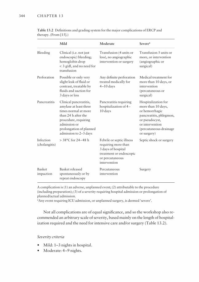
Not all complications are of equal significance, and so the workshop also re-commended an arbitrary scale of severity, based mainly on the length of hospital-ization required and the need for intensive care and/or surgery (Table 13.2).
Severity criteria
• Mild: 1–3 nights in hospital.• Moderate: 4–9 nights.
CHAPTER 13344
Table 13.2 Definitions and grading system for the major complications of ERCP andtherapy. (From [15].)
Bleeding
Perforation
Pancreatitis
Infection (cholangitis)
Basket impaction
A complication is (1) an adverse, unplanned event; (2) attributable to the procedure(including preparation); (3) of a severity requiring hospital admission or prolongation ofplanned/actual admission.aAny event requiring ICU admission, or unplanned surgery, is deemed ‘severe’.
Mild
Clinical (i.e. not justendoscopic) bleeding;hemoglobin drop < 3 g/dl, and no need fortransfusion
Possible or only veryslight leak of fluid orcontrast, treatable byfluids and suction for 3 days or less
Clinical pancreatitis,amylase at least threetimes normal at morethan 24 h after theprocedure, requiringadmission orprolongation of plannedadmission to 2–3 days
> 38°C for 24–48 h
Basket releasedspontaneously or byrepeat endoscopy
Moderate
Transfusion (4 units orless), no angiographicintervention or surgery
Any definite perforationtreated medically for4–10 days
Pancreatitis requiringhospitalization of 4–10 days
Febrile or septic illnessrequiring more than 3 days of hospitaltreatment or endoscopicor percutaneousintervention
Percutaneousintervention
Severea
Transfusion 5 units ormore, or intervention(angiographic orsurgical)
Medical treatment formore than 10 days, orintervention(percutaneous orsurgical)
Hospitalization formore than 10 days, or hemorrhagicpancreatitis, phlegmon,or pseudocyst, or intervention(percutaneous drainageor surgery)
Septic shock or surgery
Surgery
ERCP: RISKS, PREVENTION, AND MANAGEMENT 345
• Severe: 10 nights or more, or surgery, or ICU admission.• Fatal: death attributable to the procedure.
These concepts and definitions have been adopted widely, and have beenused in many subsequent studies of ERCP outcomes. This has helped con-siderably in the attempt to better understand the predictors of good and badoutcomes. If surgeons and interventional radiologists used a similar lexicon, itwould be easier to compare their outcomes outside the context of formal ran-domized trials [13].
Types of adverse clinical event
Unplanned adverse events can be categorized broadly into four groups.• Equipment malfunction.• Medication and sedation issues.• Direct events: those which occur at sites which have been traversed or treated during the endoscopic procedure (e.g. perforation, bleeding, or pancreatitis).• Indirect events: those which occur in other organs (e.g. heart, lungs, and kidneys) as a result of the procedure. Indirect events are more difficult to recog-nize and document because they may not become apparent until several days after the procedure, when the patient has returned home or to other clinicalsupervision.
Timing of events and attribution
Most adverse events are recognized during or shortly after procedures, but somehappen beforehand (e.g. as a result of some aspect of preparation), and some areapparent only later (e.g. delayed bleeding after sphincterotomy).
For adverse events which occur before and during procedures, it is importantto note whether the examination had to be terminated early or could be completed.
The 1991 consensus definition of complications [15] includes the phrase‘attributable to the procedure’. Attribution is not always clear-cut, especiallywhen there is a delay. Is a cardiopulmonary event counted if it occurs a week or two after ERCP, or only if there is some other linking factor (e.g. some important medication was stopped for the procedure)?
To cover this point, the consensus workshop suggested that direct events (asdefined above) are always attributable, even if they do not occur or becomeapparent for several weeks (e.g. delayed bleeding). However, we agreed an arbi-trary time limit of 3 days for indirect events, such as cardiopulmonary problems.
As mentioned above, there is also the issue of how to report the com-
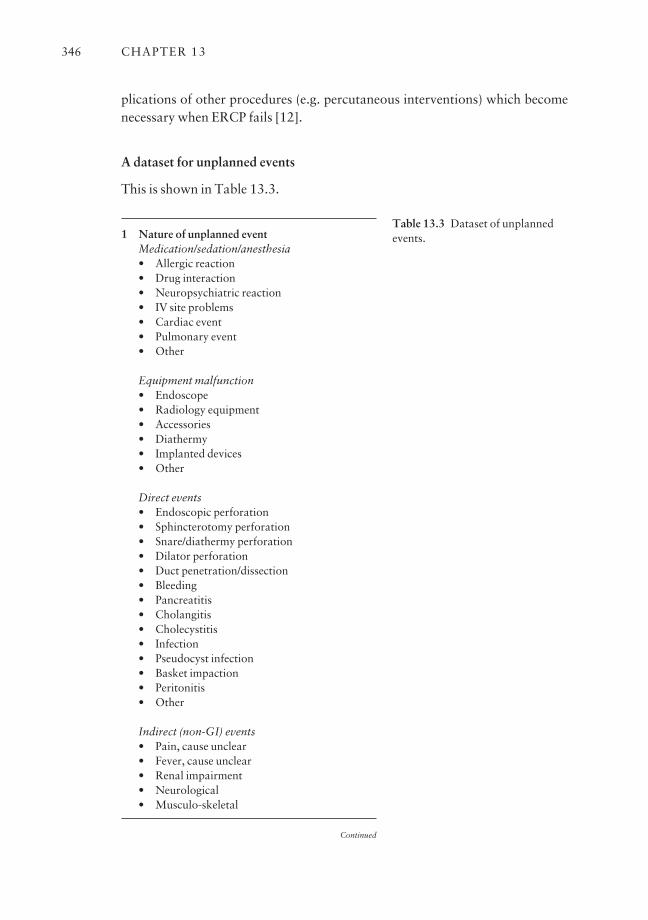
plications of other procedures (e.g. percutaneous interventions) which becomenecessary when ERCP fails [12].
A dataset for unplanned events
This is shown in Table 13.3.
CHAPTER 13346
1 Nature of unplanned eventMedication/sedation/anesthesia• Allergic reaction• Drug interaction• Neuropsychiatric reaction• IV site problems• Cardiac event• Pulmonary event• Other
Equipment malfunction• Endoscope• Radiology equipment• Accessories• Diathermy• Implanted devices• Other
Direct events• Endoscopic perforation• Sphincterotomy perforation• Snare/diathermy perforation• Dilator perforation• Duct penetration/dissection• Bleeding• Pancreatitis• Cholangitis• Cholecystitis• Infection• Pseudocyst infection• Basket impaction• Peritonitis• Other
Indirect (non-GI) events• Pain, cause unclear• Fever, cause unclear• Renal impairment• Neurological• Musculo-skeletal
Continued
Table 13.3 Dataset of unplannedevents.
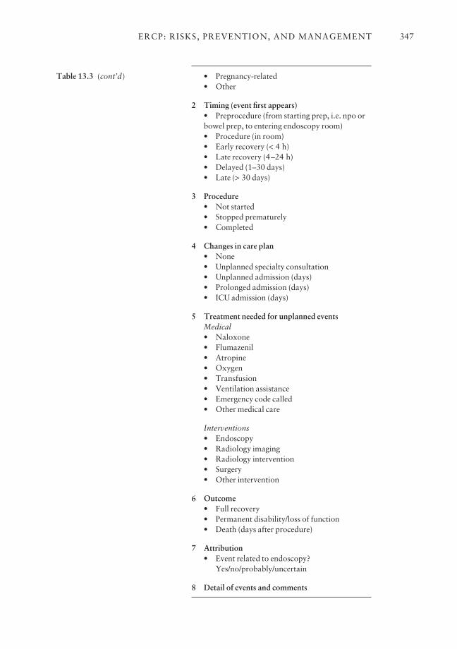
ERCP: RISKS, PREVENTION, AND MANAGEMENT 347
• Pregnancy-related• Other
2 Timing (event first appears)• Preprocedure (from starting prep, i.e. npo orbowel prep, to entering endoscopy room)• Procedure (in room)• Early recovery (< 4 h)• Late recovery (4–24 h)• Delayed (1–30 days)• Late (> 30 days)
3 Procedure• Not started• Stopped prematurely• Completed
4 Changes in care plan• None• Unplanned specialty consultation• Unplanned admission (days)• Prolonged admission (days)• ICU admission (days)
5 Treatment needed for unplanned eventsMedical• Naloxone• Flumazenil• Atropine• Oxygen• Transfusion• Ventilation assistance• Emergency code called• Other medical care
Interventions• Endoscopy• Radiology imaging• Radiology intervention• Surgery• Other intervention
6 Outcome• Full recovery• Permanent disability/loss of function• Death (days after procedure)
7 Attribution• Event related to endoscopy?
Yes/no/probably/uncertain
8 Detail of events and comments
Table 13.3 (cont’d)
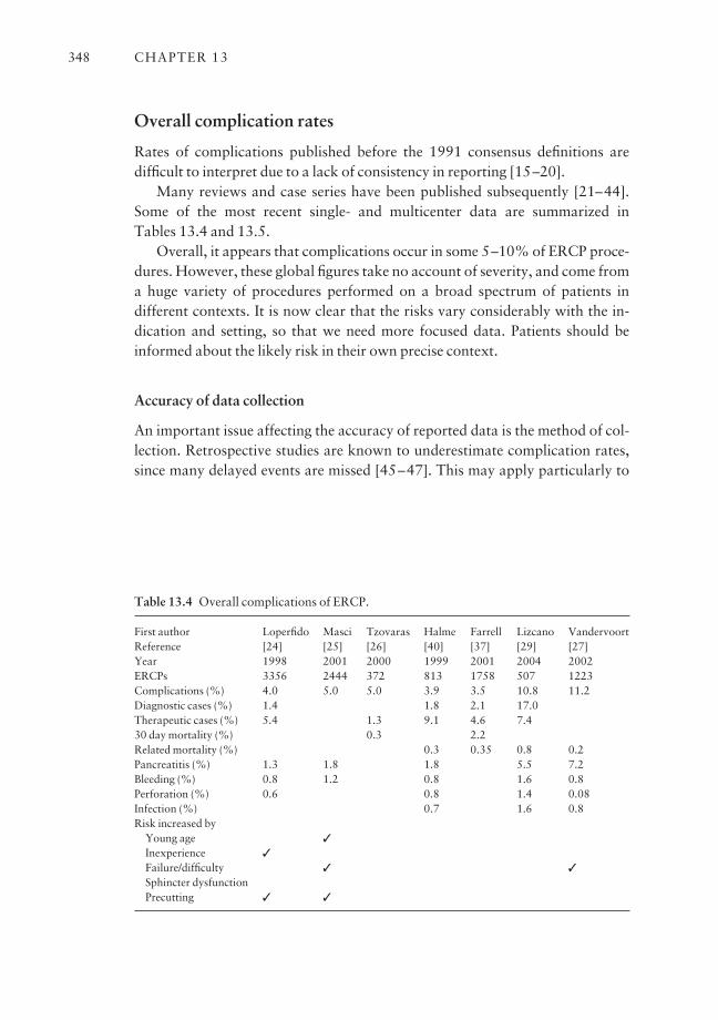
CHAPTER 13348
Table 13.4 Overall complications of ERCP.
First author Loperfido Masci Tzovaras Halme Farrell Lizcano VandervoortReference [24] [25] [26] [40] [37] [29] [27]Year 1998 2001 2000 1999 2001 2004 2002ERCPs 3356 2444 372 813 1758 507 1223Complications (%) 4.0 5.0 5.0 3.9 3.5 10.8 11.2Diagnostic cases (%) 1.4 1.8 2.1 17.0Therapeutic cases (%) 5.4 1.3 9.1 4.6 7.430 day mortality (%) 0.3 2.2Related mortality (%) 0.3 0.35 0.8 0.2Pancreatitis (%) 1.3 1.8 1.8 5.5 7.2Bleeding (%) 0.8 1.2 0.8 1.6 0.8Perforation (%) 0.6 0.8 1.4 0.08Infection (%) 0.7 1.6 0.8Risk increased by
Young age 3Inexperience 3Failure/difficulty 3 3Sphincter dysfunctionPrecutting 3 3
Overall complication rates
Rates of complications published before the 1991 consensus definitions aredifficult to interpret due to a lack of consistency in reporting [15–20].
Many reviews and case series have been published subsequently [21–44].Some of the most recent single- and multicenter data are summarized in Tables 13.4 and 13.5.
Overall, it appears that complications occur in some 5–10% of ERCP proce-dures. However, these global figures take no account of severity, and come from a huge variety of procedures performed on a broad spectrum of patients in different contexts. It is now clear that the risks vary considerably with the in-dication and setting, so that we need more focused data. Patients should beinformed about the likely risk in their own precise context.
Accuracy of data collection
An important issue affecting the accuracy of reported data is the method of col-lection. Retrospective studies are known to underestimate complication rates,since many delayed events are missed [45–47]. This may apply particularly to
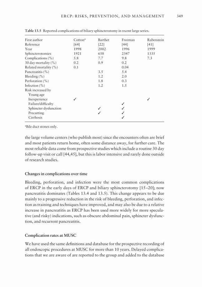
the large volume centers (who publish most) since the encounters often are briefand most patients return home, often some distance away, for further care. Themost reliable data come from prospective studies which include a routine 30 dayfollow-up visit or call [44,45], but this is labor intensive and rarely done outsideof research studies.
Changes in complications over time
Bleeding, perforation, and infection were the most common complications of ERCP in the early days of ERCP and biliary sphincterotomy [15–20]; nowpancreatitis dominates (Tables 13.4 and 13.5). This change appears to be duemainly to a progressive reduction in the risk of bleeding, perforation, and infec-tion as training and techniques have improved, and may also be due to a relativeincrease in pancreatitis as ERCP has been used more widely for more specula-tive (and risky) indications, such as obscure abdominal pain, sphincter dysfunc-tion, and recurrent pancreatitis.
Complication rates at MUSC
We have used the same definitions and database for the prospective recording ofall endoscopic procedures at MUSC for more than 10 years. Delayed complica-tions that we are aware of are reported to the group and added to the database
ERCP: RISKS, PREVENTION, AND MANAGEMENT 349
Table 13.5 Reported complications of biliary sphincterotomy in recent large series.
First author Cottona Barthet Freeman RabensteinReference [64] [22] [44] [41]Year 1998 2002 1996 1999Sphincterotomies 1921 658 2347 1335Complications (%) 5.8 7.7 9.8 7.330 day mortality (%) 0.2 0.9 0.2Related mortality (%) 0.1 0.04Pancreatitis (%) 3.5 5.4Bleeding (%) 1.2 2.0Perforation (%) 1.8 0.3Infection (%) 1.2 1.5Risk increased by
Young ageInexperience 3 3Failure/difficulty 3Sphincter dysfunction 3 3Precutting 3 3Cirrhosis 3
aBile duct stones only.
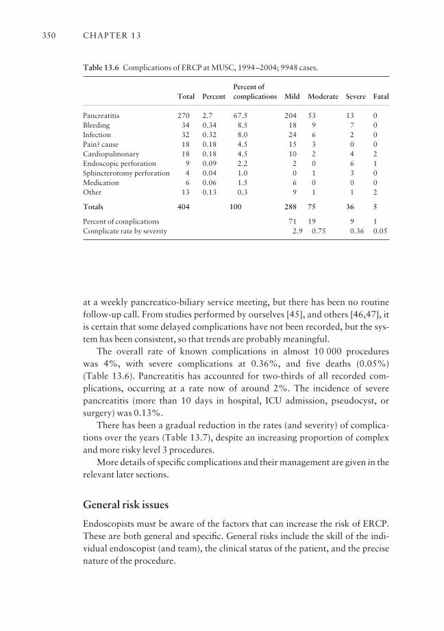
at a weekly pancreatico-biliary service meeting, but there has been no routinefollow-up call. From studies performed by ourselves [45], and others [46,47], itis certain that some delayed complications have not been recorded, but the sys-tem has been consistent, so that trends are probably meaningful.
The overall rate of known complications in almost 10 000 procedures was 4%, with severe complications at 0.36%, and five deaths (0.05%) (Table 13.6). Pancreatitis has accounted for two-thirds of all recorded com-plications, occurring at a rate now of around 2%. The incidence of severe pancreatitis (more than 10 days in hospital, ICU admission, pseudocyst, orsurgery) was 0.13%.
There has been a gradual reduction in the rates (and severity) of complica-tions over the years (Table 13.7), despite an increasing proportion of complexand more risky level 3 procedures.
More details of specific complications and their management are given in therelevant later sections.
General risk issues
Endoscopists must be aware of the factors that can increase the risk of ERCP.These are both general and specific. General risks include the skill of the indi-vidual endoscopist (and team), the clinical status of the patient, and the precisenature of the procedure.
CHAPTER 13350
Table 13.6 Complications of ERCP at MUSC, 1994–2004; 9948 cases.
Percent of Total Percent complications Mild Moderate Severe Fatal
Pancreatitis 270 2.7 67.5 204 53 13 0Bleeding 34 0.34 8.5 18 9 7 0Infection 32 0.32 8.0 24 6 2 0Pain? cause 18 0.18 4.5 15 3 0 0Cardiopulmonary 18 0.18 4.5 10 2 4 2Endoscopic perforation 9 0.09 2.2 2 0 6 1Sphincterotomy perforation 4 0.04 1.0 0 1 3 0Medication 6 0.06 1.5 6 0 0 0Other 13 0.13 0.3 9 1 1 2
Totals 404 100 288 75 36 5
Percent of complications 71 19 9 1Complicate rate by severity 2.9 0.75 0.36 0.05

Details of the specific risks, methods to minimize them, and recommenda-tions for management are given below. Here we document some important general points.
Operator-related issues
There are now significant data showing that more experienced endoscopistsusually have higher success rates and lower rates of complications than thosewho are less active, even when dealing with more complex cases [7,24,28,41–43]. This fact has important implications for training, credentialing, andinformed consent. Lack of experience increases the risk of technical failure.Failures carry risks also of the subsequent needed interventions. In one analysis,failed ERCPs carried three times the complication rate of successful ones (21.5%vs. 7.3%) [12]. The association between inexperience and poor outcomes hasbeen well documented for major surgical procedures [8].
Patient-related issues; clinical status, indications, and comorbidities
Much attention has been paid to analysing the characteristics of patients whichmay affect the risk of performing ERCP [30,48].
Age
Age itself is not a risk factor for ERCP complications [49]. Many studies nowtestify to the safety of performing diagnostic and therapeutic procedures ininfants [50], children [51], and the elderly [52,53].
ERCP: RISKS, PREVENTION, AND MANAGEMENT 351
Table 13.7 ERCP complications at MUSC by year.
Total 1995 1996 1997 1998 1999 2000 2001 2002 2003 2004
Procedures 9948 793 1013 1066 998 1035 1044 1015 1051 983 950Complications 404 50 57 55 40 42 34 37 23 38 28(%) (4.1) (6.3) (5.6) (5.2) (4.0) (4.1) (3.3) (3.7) (2.2) (3.9) (2.9)
Mild (%) 288 (2.9) 34 38 42 25 29 27 23 21 30 19Moderate (%) 75 (0.75) 8 13 7 11 10 7 8 2 4 5Severe (%) 36 (0.36) 8 5 5 4 2 0 5 0 3 4Fatal (%) 5 (0.05) 0 1 1 0 1 0 1 0 1 0
Pancreatitis 270 34 40 36 28 27 23 25 14 25 18(%) (2.7) (4.3) (4.0) (3.5) (2.8) (2.6) (2.2) (2.5) (1.3) (2.5) (1.9)
Illness and associated conditions
Adverse events are more likely to occur in patients who are already severely ill,for example with acute cholangitis [54], and in those with substantial co-morbidities. The most important comorbidities are cardiopulmonary fragility(posing risks for sedation and anesthesia), immunosuppression, and coagu-lopathies (including therapeutic anticoagulation). It would be helpful if therewere an agreed index or score that reflected the degree of risk, but none of thepublished instruments really fit the ERCP context. The American Society forAnesthesiology (ASA) score is often used in surgical practice as a guide to therisk for sedation and anesthesia, but this appears unhelpful in the context ofERCP [55]. This is because the risk is much more dependent on the indicationfor the procedure.
ERCP appears to be safe when needed for management of stones in pregnantpatients [56].
Indication
Fortunately, it is clear that the risks of ERCP are lowest in those patients with the ‘best’ indications, i.e. duct stones, biliary leaks, and low tumors. Conversely,the pioneering studies by Freeman and colleagues have revealed the substantialrisks involved in performing ERCP in patients with obscure abdominal pain(‘suspected sphincter dysfunction’) [36]. This was emphasized strongly by theNIH ‘State-of-the-Science’ Conference on ERCP in 2002 [57]. Sadly, it is truethat ‘ERCP is most dangerous for those who need it least’ [58].
Anatomical factors
In some series, but not in all, the presence of a peripapillary diverticulumappears to be a risk factor [59,60]. With suitable precautions, patients withimplanted pacemakers or defibrillators can be treated safely [61].
A normal-sized bile duct was earlier believed to increase the risk of post-ERCP pancreatitis [62,63], but this is a surrogate for sphincter dysfunction, anddoes not apply to patients with stones [64,65].
Complication-specific risk factors
Patient risk factors for specific complications are detailed below. For example,Billroth II gastrectomy carries an increased risk for afferent loop perforation,and coagulopathies and certain medications increase the risk of bleeding.Equally, patients with hilar tumors and sclerosing cholangitis are at greater risk
CHAPTER 13352
for septic complications because it may prove difficult to provide completedrainage.
Procedure performed
Diagnostic or therapeutic?
Most people assume that therapeutic ERCP is more dangerous than diagnosticprocedures. This is true in several reported series: 5.4% vs. 1.4% [24], 9.1% vs.1.8% [40], and 4.6% vs. 2.1% [37], but not in another small series (7.4% vs.17%) [29] (Table 13.5).
Sedation, cardiopulmonary events, and intubation carry the same riskswhether the procedure is diagnostic or therapeutic. Therapeutic procedures docarry their own specific risks, e.g. bleeding and perforation after sphinctero-tomy, or infection after attempted pseudocyst drainage. These complicationscan be serious, and so it would also seem logical that the likelihood of a severecomplication would be greater after therapeutic procedures. Remarkably, ourown series shows very similar complication rates for diagnostic and therapeuticERCP, and the risk of severe or fatal complications was actually slightly higherfor diagnostic procedures (0.7% vs. 0.3%) (Table 13.8). It is worth noting thatdiagnostic ERCP in some patients may actually be riskier if not followed imme-diately by appropriate therapy (e.g. in a patient with malignant obstructivejaundice or proven sphincter dysfunction). Our ‘diagnostic’ procedures were
ERCP: RISKS, PREVENTION, AND MANAGEMENT 353
Thereapeutic No therapy(%) (%)
Total cases 8136 1812Overall complications 339 (4.20) 68 (3.80)
Mild 242 (3.00) 52 (2.90)Moderate 70 (0.90) 5 (0.30)Severe 23 (0.30) 10 (0.60)Fatal 4 (0.05) 1 (0.05)
Pancreatitis overall 222 (2.70) 48 (2.60)Mild 165 (2.00) 39 (2.20)Moderate 49 (0.60) 4 (0.20)Severe 8 (0.10) 5 (0.30)Fatal 0 (0.00) 0 (0.00)
Perforation 9 (0.11) 4 (0.20)
Bleeding 34 (0.42) 0 (0.00)
Table 13.8 Complications of ERCPat MUSC; therapy vs. no therapy.
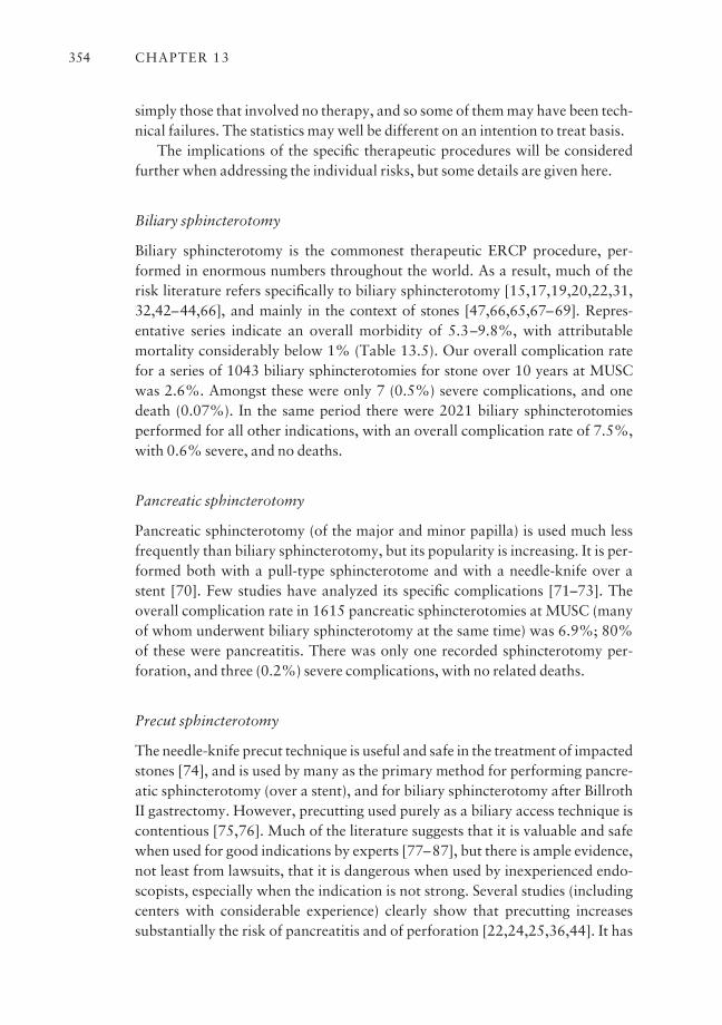
simply those that involved no therapy, and so some of them may have been tech-nical failures. The statistics may well be different on an intention to treat basis.
The implications of the specific therapeutic procedures will be consideredfurther when addressing the individual risks, but some details are given here.
Biliary sphincterotomy
Biliary sphincterotomy is the commonest therapeutic ERCP procedure, per-formed in enormous numbers throughout the world. As a result, much of therisk literature refers specifically to biliary sphincterotomy [15,17,19,20,22,31,32,42–44,66], and mainly in the context of stones [47,66,65,67–69]. Repres-entative series indicate an overall morbidity of 5.3–9.8%, with attributable mortality considerably below 1% (Table 13.5). Our overall complication ratefor a series of 1043 biliary sphincterotomies for stone over 10 years at MUSCwas 2.6%. Amongst these were only 7 (0.5%) severe complications, and onedeath (0.07%). In the same period there were 2021 biliary sphincterotomiesperformed for all other indications, with an overall complication rate of 7.5%,with 0.6% severe, and no deaths.
Pancreatic sphincterotomy
Pancreatic sphincterotomy (of the major and minor papilla) is used much lessfrequently than biliary sphincterotomy, but its popularity is increasing. It is per-formed both with a pull-type sphincterotome and with a needle-knife over astent [70]. Few studies have analyzed its specific complications [71–73]. Theoverall complication rate in 1615 pancreatic sphincterotomies at MUSC (manyof whom underwent biliary sphincterotomy at the same time) was 6.9%; 80%of these were pancreatitis. There was only one recorded sphincterotomy per-foration, and three (0.2%) severe complications, with no related deaths.
Precut sphincterotomy
The needle-knife precut technique is useful and safe in the treatment of impactedstones [74], and is used by many as the primary method for performing pancre-atic sphincterotomy (over a stent), and for biliary sphincterotomy after BillrothII gastrectomy. However, precutting used purely as a biliary access technique iscontentious [75,76]. Much of the literature suggests that it is valuable and safewhen used for good indications by experts [77–87], but there is ample evidence,not least from lawsuits, that it is dangerous when used by inexperienced endo-scopists, especially when the indication is not strong. Several studies (includingcenters with considerable experience) clearly show that precutting increasessubstantially the risk of pancreatitis and of perforation [22,24,25,36,44]. It has
CHAPTER 13354
been suggested that precutting has received a bad reputation only because it maybe used as a last resort, after much other manipulation, and that it may be safewhen used early in the cannulation process. However, it seems a poor alterna-tive to good standard methods.
Variants of the precut technique have been described [88,89], includingusing a standard pull-type sphincterotome in the pancreatic duct. Despite goodexperience with this method reported from one center [90], this seems to becourting disaster.
The data clearly indicate that precut access techniques should be avoided byinexperienced endoscopists, especially when there is little or no evidence for biliary pathology requiring treatment.
Repeat sphincterotomy
Biliary and pancreatic sphincterotomy sometimes need to be repeated for recur-rent stones or stenosis. Whether the second procedure carries increased riskclearly depends on the indication, and on the size of the prior procedure [91,92].One study showed a significant increase in the risk of both bleeding (from 1.7% to 5%) and perforation (from 1% to 8%), but a reduction in pancreatitis (from5.5% to 1%), when comparing repeat biliary sphincterotomies with index cases[93]. These factors are discussed further in the specific risk sections.
Balloon sphincter dilation
As endoscopic stone extraction has become more frequently used in relatively fitand young patients (after the advent of laparoscopic cholecystectomy), there has been increasing interest in trying to further reduce the (albeit small) risks ofsphincterotomy by using balloon dilation of the papilla instead [94,95]. Sincethe main concern about this technique is the risk of provoking pancreatitis, it isdiscussed further in that section.
Endoscopic papillectomy
The increasing familiarity and confidence of endoscopists with polypectomy,mucosal resection, and complex ERCP has led many to perform excision of themajor papilla for treatment of adenomas. The techniques (including temporarypancreatic stenting) are now fairly well established [96–99], but there is con-tinuing concern about the precise indications [100] and the likelihood of recurrence. The immediate risks are bleeding, pancreatitis, and perforation.One large series of 70 cases reported 10 complications (14%), with bleeding infour, pancreatitis in five, and mild perforation in one, with one-third of the ade-nomas recurring after a median follow-up of only 7 months [101].
ERCP: RISKS, PREVENTION, AND MANAGEMENT 355
Stenting
Biliary stenting is widely used for the management of leaks and tumors. It carriesthe same general risks as any ERCP procedure, although the presence of a pancreatic tumor may protect somewhat against pancreatitis. A small biliarysphincterotomy is not necessary in most cases [102], but is wise in hilar tumors,since it may prevent pancreatitis and also facilitates the placement of two stents[103].
Biliary stenting is most risky when it fails, or when drainage is incomplete.This commonly leads to sepsis, and carries the risks of repeat procedures(whether ERCP or percutaneous). The chance of failure, and of complications,is considerably greater with lesions involving the liver hilum. The specific risksof stenting (and pancreatic stenting) are discussed later.
Pseudocyst drainage
Endoscopic drainage of pseudocysts through the stomach wall or duodenumcarries a significant risk of bleeding, perforation, and infection [104].
Reducing the risks of ERCP: general issues
Methods to minimize specific risks are detailed below for each of the main com-plications. However, there are several general strategies which should be under-stood. Clearly, it is helpful to maximize the clinical and technical expertise of the endoscopist and the assisting team, and to follow accepted standards ofpractice. Since complications cannot be avoided completely, it is also manda-tory to ensure that patients and those close to them are fully informed about thekey issues.
The contract with the patient; informed consent
It is always the responsibility of endoscopists to assure themselves that thepotential benefits of the proposed procedure exceed the potential risks, and toconvey that information clearly to their patients [105]. Truly informed consentmeans that the patient really does understand the potential risks and benefits, as well as the possible limitations and any available alternative approaches.That is our contract with the patient. Signing an ‘informed consent form’ is amedico-legal requirement in many institutions, but this is nothing more thanconfirmation of the education process. It is important to ‘tell it as it is’. It is in ournature as physicians to want to reassure nervous patients that ‘things will go justfine’, but that is neither honest nor wise.
CHAPTER 13356
Educational materials
Nothing can replace a detailed discussion between the endoscopist and thepatient (and any accompanying persons), but this process can be enhanced with written, video- or web-based educational materials. Suitable brochures areavailable from national organizations, and on many websites, and can beadapted for local conditions. The document in routine use at MUSC is shown inTable 13.9. The process of informed consent must be clearly documented andwitnessed. For elective procedures, this process should take place preferably atleast a day beforehand to give time for review of the materials and unhurriedreflection. Whatever the details of the education process, patients must be giventhe opportunity to ask questions of their endoscopist (and support staff) againbefore the procedure.
Humanity
It is appropriate also to emphasize the importance of simple courtesies and com-mon humanity in dealing with our customers. What is familiar and routine to the endoscopist and staff may be viewed by patients as a major ordealaespecially by those unfortunate enough to experience a significant adverse event.
Care after ERCP
Admission?
Many patients are kept in hospital under observation overnight after ERCP. The advantage is that nursing staff can ensure adequate fluid intake (mainlyintravenously), and can quickly detect and pay appropriate attention to symp-toms which may herald important complications. However, overnight observa-tion adds costs, and can add other burdens for patients and their families.Several studies have evaluated factors predicting the need for admission[106–110]. Admission is unnecessary in the majority of standard level 1 pro-cedures (simple biliary stones and stents), but seems wise when the risk is pre-dicted to be higher than average (e.g. sphincter dysfunction management), whenthe procedure has been difficult in some way, or when the patient is frail or hasno responsible accompanying person. Staying overnight in a local hotel is anappropriate compromise option for patients who live more than an hour or twoaway. Attempts have been made to use serum tests early in the recovery periodto predict subsequent pancreatitis [111,112], but this has not become standardpractice.
ERCP: RISKS, PREVENTION, AND MANAGEMENT 357
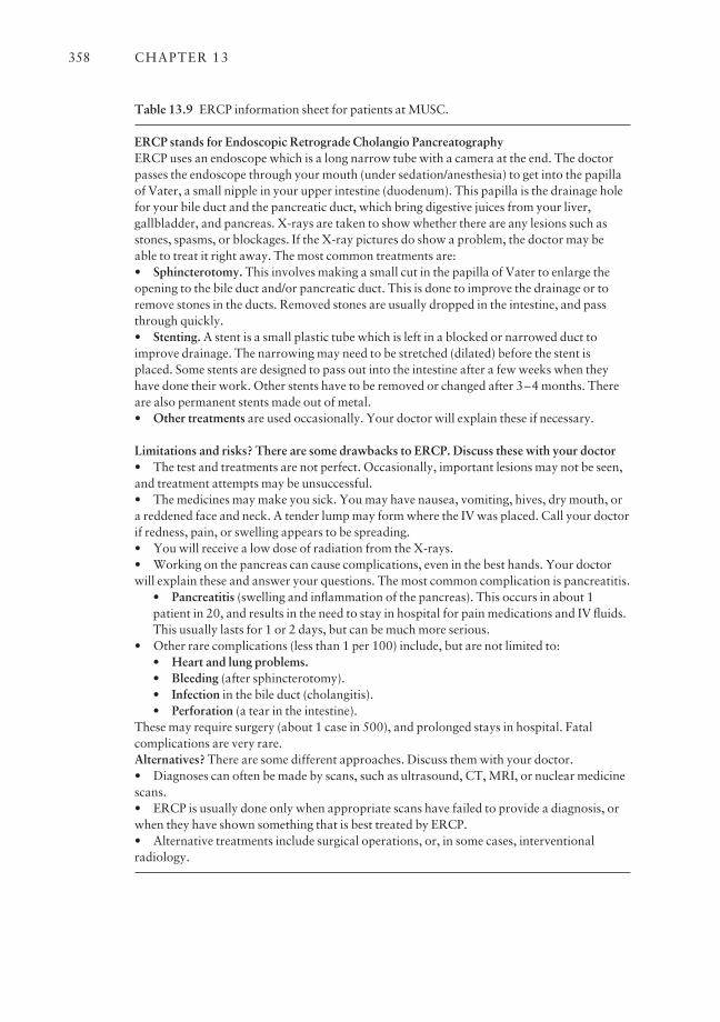
CHAPTER 13358
Table 13.9 ERCP information sheet for patients at MUSC.
ERCP stands for Endoscopic Retrograde Cholangio PancreatographyERCP uses an endoscope which is a long narrow tube with a camera at the end. The doctorpasses the endoscope through your mouth (under sedation/anesthesia) to get into the papillaof Vater, a small nipple in your upper intestine (duodenum). This papilla is the drainage holefor your bile duct and the pancreatic duct, which bring digestive juices from your liver,gallbladder, and pancreas. X-rays are taken to show whether there are any lesions such asstones, spasms, or blockages. If the X-ray pictures do show a problem, the doctor may be able to treat it right away. The most common treatments are:• Sphincterotomy. This involves making a small cut in the papilla of Vater to enlarge theopening to the bile duct and/or pancreatic duct. This is done to improve the drainage or toremove stones in the ducts. Removed stones are usually dropped in the intestine, and passthrough quickly.• Stenting. A stent is a small plastic tube which is left in a blocked or narrowed duct toimprove drainage. The narrowing may need to be stretched (dilated) before the stent isplaced. Some stents are designed to pass out into the intestine after a few weeks when theyhave done their work. Other stents have to be removed or changed after 3–4 months. Thereare also permanent stents made out of metal.• Other treatments are used occasionally. Your doctor will explain these if necessary.
Limitations and risks? There are some drawbacks to ERCP. Discuss these with your doctor• The test and treatments are not perfect. Occasionally, important lesions may not be seen,and treatment attempts may be unsuccessful.• The medicines may make you sick. You may have nausea, vomiting, hives, dry mouth, ora reddened face and neck. A tender lump may form where the IV was placed. Call your doctorif redness, pain, or swelling appears to be spreading.• You will receive a low dose of radiation from the X-rays.• Working on the pancreas can cause complications, even in the best hands. Your doctorwill explain these and answer your questions. The most common complication is pancreatitis.
• Pancreatitis (swelling and inflammation of the pancreas). This occurs in about 1patient in 20, and results in the need to stay in hospital for pain medications and IV fluids.This usually lasts for 1 or 2 days, but can be much more serious.
• Other rare complications (less than 1 per 100) include, but are not limited to:• Heart and lung problems.• Bleeding (after sphincterotomy).• Infection in the bile duct (cholangitis).• Perforation (a tear in the intestine).
These may require surgery (about 1 case in 500), and prolonged stays in hospital. Fatalcomplications are very rare.Alternatives? There are some different approaches. Discuss them with your doctor.• Diagnoses can often be made by scans, such as ultrasound, CT, MRI, or nuclear medicinescans.• ERCP is usually done only when appropriate scans have failed to provide a diagnosis, orwhen they have shown something that is best treated by ERCP.• Alternative treatments include surgical operations, or, in some cases, interventionalradiology.
Early refeeding?
Patients are often keen to catch up on the meals that they have missed as a resultof the procedure, but it has been my practice to recommend taking fluids onlyuntil the next morning, when the main risk of pancreatitis has passed. However,a recent randomized trial suggests that early refeeding is not detrimental [113].
Pancreatitis after ERCP
Pancreatitis is now by far the most common complication of ERCP and sphinc-terotomy (Tables 13.4 and 13.5). Our better understanding of the risk factors inrecent years is largely attributable to the seminal studies anchored by Freeman,who has published several comprehensive reviews [114,115]. This chapterfocuses on the key facts.
Definitions
Serum amylase and lipase levels can be shown to rise in almost every patient ifmeasured within a few hours of ERCP, even sometimes when the pancreaticduct has not been entered or opacified. While this indicates some irritation of thepancreas, it does not constitute clinically relevant pancreatitis. The incidence ofpancreatitis clearly depends greatly on the criteria used for the diagnosis [116].
The consensus workshop suggested this working definition of post-ERCPpancreatitis [15]. ‘Pancreatitis after ERCP is a clinical illness with typical pain,associated with at least a three-fold increase in serum amylase (or lipase) at 24 h,with symptoms impressive enough to require admission to hospital for treat-ment (or extension of an existing or planned admission).’ Severity is graded as mild if hospitalization is needed for less than 3 nights, moderate if 4–9 nights,and severe if more than 10 nights, or if patients require intensive care or surgicaltreatment.
This definition has been widely used, despite some concern about the rele-vance of hospitalization, and the fact that the apparent incidence will varyaccording to the admission policy. It is also sometimes difficult to decide how todeal (statistically as well as clinically) with patients with long-standing pancre-atic pain who linger in hospital after their procedures. We do not count this as acomplication unless the patient is obviously worse afterwards.
Incidence of pancreatitis after ERCP
In addition to the major reviews and studies of ERCP complications [21–44],there is extensive literature specific to the risk of pancreatitis after ERCP
ERCP: RISKS, PREVENTION, AND MANAGEMENT 359

[114–123]. The reported incidence ranges widely, from less than 1% up to ashigh as 40%. Much of this huge variation can be attributed to different defini-tions, incomplete data collection, and differing case mixes. Ranges of 2–9% arerepresentative of more recent prospective series, mostly using consensusdefinitions [114] (Tables 13.4 and 13.5). An innovative population-based studyof 97 810 ERCPs in Canada reported a pancreatitis rate of 2.2%, with greaterrisk in younger patients and in women [124].
The overall pancreatitis rate in our series at MUSC is < 3%, with a gradualreduction over the years, despite an increasing number of cases with suspectedsphincter dysfunction, where the risk is known to be greater (Tables 13.6 and13.7). More than 75% of all these cases of pancreatitis after ERCP were gradedas mild (Table 13.6). Mild complications are disappointing and inconvenient,but are not serious or threatening (medically or legally). Severe pancreatitisoccurred in 13 cases (0.13%). They are devastating for all involved, and fatal-ities occur (one in our series). For these reasons, there is great interest in under-standing the true risk factors, and ways to minimize them.
Risk factors for pancreatitis
Any ERCP procedure can cause pancreatitis, but certain factors are well knownto increase the risk. A listing, adapted from Freeman et al. [115], is shown inTable 13.10. The factors are both patient- and procedure-related.
CHAPTER 13360
Table 13.10 Risk factors for pancreatitis after ERCP. (Adapted from [115].)
Increased risk?
Yes
Maybe
No
Patient-related
Young ageFemaleSuspected SODRecurrent pancreatitisNo chronic pancreatitisPrior post-ERCP pancreatitis
No stoneNormal bilirubinLow-volume endoscopist
Small/normal bile ductPeriampullary diverticulumPancreas divisumContrast allergyPrior failed ERCP
Procedure-related
PancreatographyPancreatic sphincterotomyBalloon dilation of intact sphincterDifficult cannulationPrecut (access) sphincterotomy
Pancreatic acinarizationPancreatic brush cytologyPain during ERCP
Therapeutic versus diagnosticBiliary sphincterotomySphincter manometryIntramural contrast injection
Patient factors increasing the risk [114,115,122,123]
It has become abundantly clear that the risk of developing pancreatitis is greaterin younger patients, and in women as compared to men [114,124], and particu-larly when ERCP is performed for ‘suspected sphincter dysfunction’ in the absence of much objective evidence for biliary or pancreatic pathology. A prior historyof recurrent pancreatitis, or of post-ERCP pancreatitis, also increases the risk inmost studies. Contrary to earlier reports [62,63], a ‘small’ or normal-sized bileduct is not an independent risk factor [114,115].
Procedure factors increasing the risk
Pancreatic manipulation Pancreatitis is more likely to occur with aggressivemanipulation of the pancreatic orifice [114], and with repeated injections ofcontrast [117], sometimes evidenced by acinarization or the appearance of aurogram [125]. The importance of increased pressure in the duct is supported bythe old observation that postprocedure pancreatitis is less likely in patients whohave a patent duct of Santorini. Variations on methods for cannulation havebeen explored [126–130], without any new consensus.
Sphincter manometry For a long time, it was believed that sphincter mano-metry was a potent cause of pancreatitis [131]. However, it is now clear thatmanometry is simply a surrogate for sphincter of Oddi dysfunction (SOD),which is the real culprit [115,133]. Several series show that the apparentincrease disappears if increased sphincter pressure is treated (by pancreaticsphincterotomy or temporary stenting). It has been shown also that the risk isactually higher when suspected SOD (e.g. postcholecystectomy pain) is treatedempirically by sphincterotomy than when manometry is employed [114].
Sphincterotomy Several studies now indicate that standard biliary sphinctero-tomy does not markedly increase the overall risk of pancreatitis (when com-pared with diagnostic ERCP) (Tables 13.4, 13.5 and 13.8). The suggestion thatpure cutting current could reduce the risk of pancreatitis has not been proven inmost of the published studies [134–139].
In the hands of experts (who publish), access precut sphincterotomy appearsto be both useful and safe, at least when used for good (biliary) indications[75–87,140]. However, many series document a significantly increased risk ofpancreatitis when precutting is performed [44,114,115,118,123]. In Freeman et al’s large prospective multicenter analysis, the complication rate was 24.3%after precutting, with 3.6% severe pancreatitis [44].
Pancreatic sphincterotomy is being performed increasingly in referral
ERCP: RISKS, PREVENTION, AND MANAGEMENT 361
centers for many different indications. The pancreatitis rate for 1615 pancreaticsphincterotomies in our unit (performed mainly for sphincter dysfunction andwith temporary stenting) was 5.6%.
Biliary sphincter dilation Balloon dilation of the biliary sphincter has beenadvocated as an alternative to sphincterotomy for removal of duct stones, in thehope of reducing the (small) short- and long-term risks [94,95]. Early case seriesgave encouraging results [94,141,142], but the technique can cause pancreatitis[143]. Many randomized studies have been performed to compare the risk withthat of standard sphincterotomy [144–150]. Some involved older patients,often with dilated ducts and large stones, and showed that the short-term risksof sphincterotomy and of balloon dilation were similar [142,144]. However, theconcept of sphincter preservation is most attractive in younger patients withsmaller stones and relatively normal ducts. A major multicenter US study inthese types of patients (in the context of laparoscopic cholecystectomy) showeda marked increase in the risk of pancreatitis, with two deaths [150]. This has ledto a consensus, at least in the USA, that the balloon technique should be consid-ered now only in special circumstances, such as coagulopathy [151] and maybeBillroth II patients [152,153]. This restrictive recommendation could changewith further progress in preventing pancreatitis, e.g. by combining balloon dila-tion with pharmacological or stenting prophylaxis.
Biliary stenting Temporary stenting of the biliary sphincter has been used as atherapeutic trial in patients with suspected sphincter dysfunction. This tech-nique is a potent cause of pancreatitis [154] and should be avoided.
Some endoscopists routinely perform sphincterotomy before placing biliarystents through strictures, to facilitate subsequent stent exchange and to reducethe risk of pancreatitis caused by irritating the pancreatic orifice. The latter hopehas been documented only in a few patients with hilar tumors [103], wheresphincterotomy is necessary anyway to place more than one stent. Sphinctero-tomy is not necessary or protective in other circumstances [102].
Pancreatic stenting The precise risk of causing immediate pancreatitis by plac-ing pancreatic stents is difficult to measure, since this is done in many ways formany different indications, and often in conjunction with other manipulationssuch as pancreatic sphincterotomy.
Combining patient- and procedure-related factors
Many of these risk factors are additive [114]. For instance, precutting in sus-pected sphincter dysfunction resulted in a complication rate of 35.3%, with no
CHAPTER 13362
fewer than 23.5% overall graded as severe [44]. In another study from the same group concerning post-ERCP pancreatitis, a woman with a normal serumbilirubin, bile duct stone, and easy cannulation had a 5% risk of pancreatitis.This increased to 16% if cannulation proved difficult, and to 42% if no stonewas found (i.e. suspected SOD) [115]. These are the unfortunate patients who are still developing severe pancreatitis after ERCP, and who feature in lawsuits.
Prevention of pancreatitis after ERCP
Avoiding ERCP, especially in high-risk patients
Post-ERCP pancreatitis cannot (currently) be prevented completely, except byavoiding the procedure, which is a good strategy in many cases; sadly, it is notapplicable in retrospect. The availability of sophisticated imaging techniquessuch as MRCP and EUS means that ERCP should be used nowadays almostexclusively for therapy. There is a less than 10% chance of finding objectivepathology by ERCP in a patient with pain, normal or only slightly derangedchemistry, and normal CT/MRCP imaging. Where sphincter dysfunction is suspected, wisdom dictates referral to a center able to perform manometry, andexperienced in methods (e.g. pancreatic stenting) known to reduce the risk ofpancreatitis.
The consensus panel at the 2002 NIH State-of-the-Science Conference onERCP advised strongly against the ‘casual use’ of ERCP in the investigation ofpatients with obscure abdominal pain, stating ‘Diagnostic ERCP has no role inthe assessment of these patients. It is precisely the typical SOD patient profile(young, healthy female) that is at the highest risk for ERCP-induced severe pan-creatitis and even death. Indeed the risk of complication exceeds potentialbenefit in many cases. Therefore, ERCP, if performed, must be coupled withdiagnostic SOM (sphincter of Oddi manometry), possible dual sphincterotomy,and possible pancreatic stent placement. ERCP with SOM and sphincterotomyshould ideally be performed at specific referral centers, and in randomized con-trolled trials that examine the impact and timing of therapeutic maneuvers onclinical outcome’ [57].
When ERCP is indicated, there are several ways to reduce the risk of the pro-cedure [114,118,126,155].
Mechanical factors
Attention to the mechanical factors discussed above can reduce the risk. Gentleintelligent probing for the desired duct with minimal injections of contrast will
ERCP: RISKS, PREVENTION, AND MANAGEMENT 363
help. The endoscopist who personally injects the contrast has better control ofthis important variable. Probing with a guidewire rather than contrast may beprudent, but reduced risk has not been proven. It is important to know when to stop. Failure to complete an ERCP may feel bad, but severe pancreatitis feels much worse, to both endoscopist and patient. Persisting, and using moredangerous approaches like precutting, can be justified only when there is astrong indication for the procedure, i.e. good evidence for biliary (or pancreatic)pathology, and a likelihood of needing endoscopic therapy.
When manometry is performed, it is clearly wise to use an aspirating cathetersystem [156]. In the future, microtransducer technology may be preferable andsafer [157].
The type of current used for sphincterotomy does not appear to be a big factor influencing the pancreatitis rate [134–139], but it is clearly wise to avoidexcessive coagulation near the pancreatic orifice.
Contrast agents
Extensive studies have not shown any consistent benefit for one or other con-trast agent for ERCP [158–163].
Pharmacological prophylaxis
The list of pharmacological agents that have been proposed and tested for pro-phylaxis of post-ERCP pancreatitis is long and varied [114,126,155,164,165].It includes antibiotics [166], heparin [167], corticosteroids [168–173], nifedip-ine [174,175], octreotide and somatostatin derivatives [176–193], trinitrin[194–196], lidocaine spray [197], gabexate [198,199], secretin, cytokine in-hibitors [200–203], and a non-steroidal (rectal diclofenac) [204]. Apart from a12 h infusion of gabexate [198], the study using diclofenac is so far the only oneto show some promise. It deserves further evaluation, not least because of itssimplicity, and the fact that it can be given selectively after ERCP. Preliminarydata on secretin prophylaxis are encouraging [205].
None of the agents tested so far has proven to be sufficiently effective andpracticable to find a place in routine practice, at least in Western countries.Reports suggest that octreotide analogs are widely used for this purpose inJapan.
Pancreatic stenting to prevent pancreatitis
There is now overwhelming evidence that temporary stenting of the pancreaticduct can reduce the risk of pancreatitis after ERCP in high-risk patients, e.g.
CHAPTER 13364
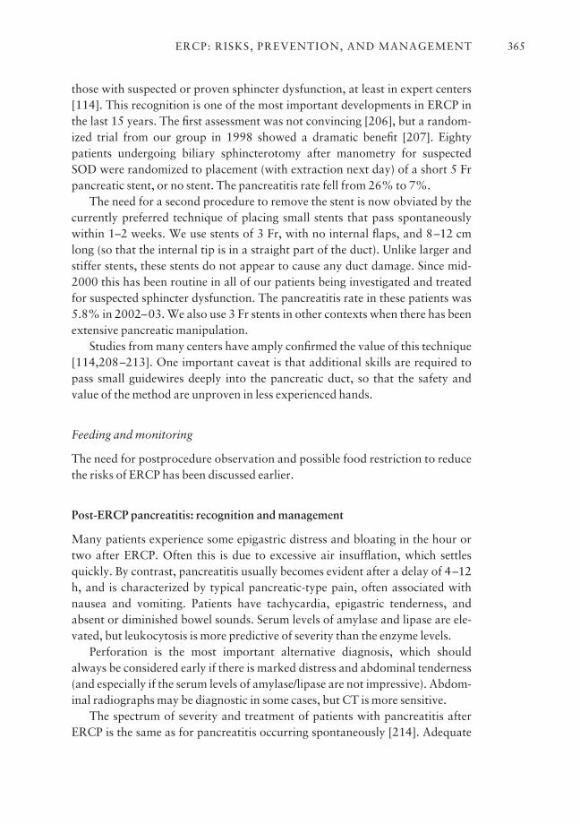
those with suspected or proven sphincter dysfunction, at least in expert centers[114]. This recognition is one of the most important developments in ERCP inthe last 15 years. The first assessment was not convincing [206], but a random-ized trial from our group in 1998 showed a dramatic benefit [207]. Eightypatients undergoing biliary sphincterotomy after manometry for suspectedSOD were randomized to placement (with extraction next day) of a short 5 Frpancreatic stent, or no stent. The pancreatitis rate fell from 26% to 7%.
The need for a second procedure to remove the stent is now obviated by thecurrently preferred technique of placing small stents that pass spontaneouslywithin 1–2 weeks. We use stents of 3 Fr, with no internal flaps, and 8–12 cmlong (so that the internal tip is in a straight part of the duct). Unlike larger andstiffer stents, these stents do not appear to cause any duct damage. Since mid-2000 this has been routine in all of our patients being investigated and treatedfor suspected sphincter dysfunction. The pancreatitis rate in these patients was5.8% in 2002–03. We also use 3 Fr stents in other contexts when there has beenextensive pancreatic manipulation.
Studies from many centers have amply confirmed the value of this technique[114,208–213]. One important caveat is that additional skills are required topass small guidewires deeply into the pancreatic duct, so that the safety andvalue of the method are unproven in less experienced hands.
Feeding and monitoring
The need for postprocedure observation and possible food restriction to reducethe risks of ERCP has been discussed earlier.
Post-ERCP pancreatitis: recognition and management
Many patients experience some epigastric distress and bloating in the hour ortwo after ERCP. Often this is due to excessive air insufflation, which settlesquickly. By contrast, pancreatitis usually becomes evident after a delay of 4–12h, and is characterized by typical pancreatic-type pain, often associated withnausea and vomiting. Patients have tachycardia, epigastric tenderness, andabsent or diminished bowel sounds. Serum levels of amylase and lipase are ele-vated, but leukocytosis is more predictive of severity than the enzyme levels.
Perforation is the most important alternative diagnosis, which shouldalways be considered early if there is marked distress and abdominal tenderness(and especially if the serum levels of amylase/lipase are not impressive). Abdom-inal radiographs may be diagnostic in some cases, but CT is more sensitive.
The spectrum of severity and treatment of patients with pancreatitis afterERCP is the same as for pancreatitis occurring spontaneously [214]. Adequate
ERCP: RISKS, PREVENTION, AND MANAGEMENT 365
analgesia and aggressive fluid replacement are key. Some experts use octreotideanalogs, but proof of benefit is anecdotal. CT scanning is indicated within 24 h if there is suspicion of perforation, and after a few days if clinical progress isslow or if fever develops (Fig. 13.1). Antibiotics are usually not given unless pancreatic infection is proven by percutaneous aspiration. The rare patient whodevelops a pseudocyst or pancreatic necrosis may require percutaneous or endo-scopic drainage, or surgical debridement, and may require transfer to a tertiarycenter.
Post-ERCP pancreatitis: conclusion
Pancreatitis is now the commonest complication of ERCP, and can be devas-tating. It cannot yet be prevented completely, even in expert hands. It is mostlikely to occur when inexperienced endoscopists work on patients with minimalpathology. When the indication is not strong, wise clinicians will exhaust lessinvasive approaches before recommending or performing ERCP, will make surethat patients fully appreciate their individualized risk/benefit balance, and willinclude referral to expert centers in their consent process. Skillful technique, andthe use of small pancreatic stents, will keep the risk of pancreatitis below 5% inmost circumstances, but cannot yet eliminate it.
Perforation
Four different types of perforation have been described resulting from ERCPprocedures [215]. They are:• Perforation of ducts or tumors by guidewires and other instrumentsaperhaps better called ‘penetrations’.• Retroduodenal perforation related to sphincterotomy.
CHAPTER 13366
Fig. 13.1 CT scan of severepancreatitis, taken 1 week afterERCP.

• Endoscopic perforation of the esophagus, stomach, or duodenum (awayfrom the papilla).• Stent-related perforation.These types have different causes and consequences.
Duct and tumor ‘penetrations’
Guidewires and occasionally accessories passed over guidewires (such assphincterotomes, catheters, and dilators) can pass through the wall of the biliaryor pancreatic ductal system (or indeed the raw area of a fresh sphincterotomy)[215,216]. This occurs perhaps most often when attempting to cannulate in apatient with a tumor involving the region of the papilla. These incidents arerarely reported, and so their frequency is unknown. They are more likely tooccur with vigorous probing in difficult cases, especially when there is distortionby tumor or sharp ductal deviation for other reasons. Rigid guidewires may bemore dangerous. Often it is safer to proceed with a ‘flipped’ tip wire, whichtends to find the lumen more easily.
Ducts have also been disrupted occasionally by over-aggressive balloondilatation of biliary (and pancreatic) strictures. The radiographic appearancesmay appear somewhat alarming when contrast is injected.
The risk of this event can be reduced by careful insertion of instrumentswhilst being aware of the potential problem. Recognition is usually straight-forward, and the problem is defused satisfactorily by finding the correct lumen,and by completing the procedure (e.g. by stenting). It is very unusual indeed fora patient to have any adverse consequences.
Sphincterotomy-related perforation
Perforation occurring after sphincterotomy is always retroduodenal. It isdefined by the presence of air (and/or contrast) in the retroperitoneum.
A review of more than 12 000 biliary sphincterotomies performed before1990 showed a sphincterotomy perforation rate of 1.3%; 27% of these patients were operated on, and the overall mortality was 0.2%. Since that time, most publications show a sphincterotomy perforation rate of < 1%[24,25,44,49,62,215,217]. Three studies have reported higher perforationrates: 1.1% [218], 1.8% [22], and 2.2% [40].
Only four perforations have been recorded after 2820 biliary sphinctero-tomies at MUSC over the last 10 years, a rate of 0.14%: three were operated on,and none died.
Routine CT scans in asymptomatic patients after uncomplicated sphinctero-tomy have shown small quantities of periduodenal or retroperitoneal air in up to
ERCP: RISKS, PREVENTION, AND MANAGEMENT 367
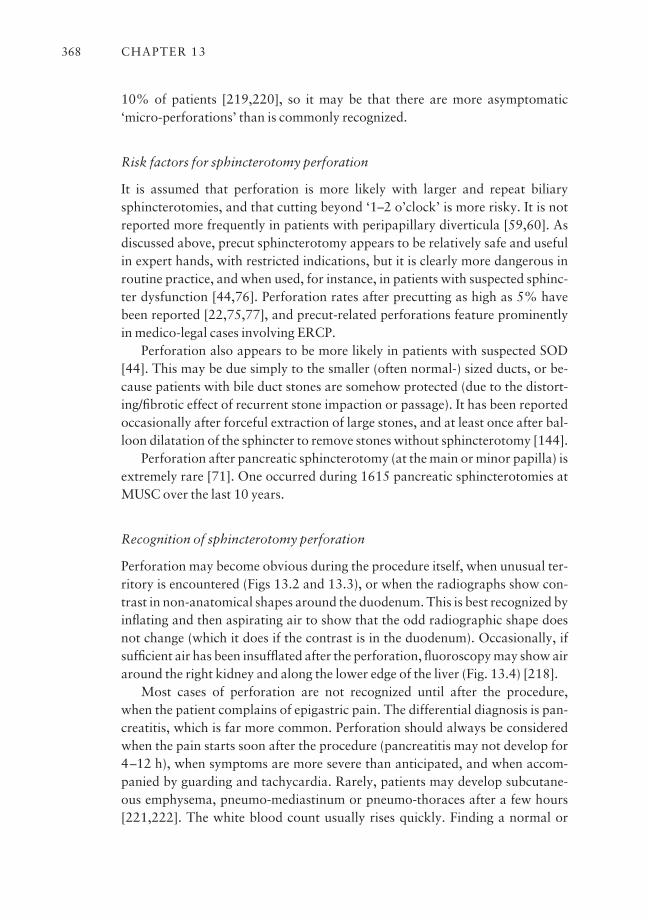
10% of patients [219,220], so it may be that there are more asymptomatic‘micro-perforations’ than is commonly recognized.
Risk factors for sphincterotomy perforation
It is assumed that perforation is more likely with larger and repeat biliarysphincterotomies, and that cutting beyond ‘1–2 o’clock’ is more risky. It is notreported more frequently in patients with peripapillary diverticula [59,60]. Asdiscussed above, precut sphincterotomy appears to be relatively safe and usefulin expert hands, with restricted indications, but it is clearly more dangerous inroutine practice, and when used, for instance, in patients with suspected sphinc-ter dysfunction [44,76]. Perforation rates after precutting as high as 5% havebeen reported [22,75,77], and precut-related perforations feature prominentlyin medico-legal cases involving ERCP.
Perforation also appears to be more likely in patients with suspected SOD[44]. This may be due simply to the smaller (often normal-) sized ducts, or be-cause patients with bile duct stones are somehow protected (due to the distort-ing/fibrotic effect of recurrent stone impaction or passage). It has been reportedoccasionally after forceful extraction of large stones, and at least once after bal-loon dilatation of the sphincter to remove stones without sphincterotomy [144].
Perforation after pancreatic sphincterotomy (at the main or minor papilla) isextremely rare [71]. One occurred during 1615 pancreatic sphincterotomies atMUSC over the last 10 years.
Recognition of sphincterotomy perforation
Perforation may become obvious during the procedure itself, when unusual ter-ritory is encountered (Figs 13.2 and 13.3), or when the radiographs show con-trast in non-anatomical shapes around the duodenum. This is best recognized byinflating and then aspirating air to show that the odd radiographic shape doesnot change (which it does if the contrast is in the duodenum). Occasionally, ifsufficient air has been insufflated after the perforation, fluoroscopy may show airaround the right kidney and along the lower edge of the liver (Fig. 13.4) [218].
Most cases of perforation are not recognized until after the procedure, when the patient complains of epigastric pain. The differential diagnosis is pan-creatitis, which is far more common. Perforation should always be consideredwhen the pain starts soon after the procedure (pancreatitis may not develop for4–12 h), when symptoms are more severe than anticipated, and when accom-panied by guarding and tachycardia. Rarely, patients may develop subcutane-ous emphysema, pneumo-mediastinum or pneumo-thoraces after a few hours[221,222]. The white blood count usually rises quickly. Finding a normal or
CHAPTER 13368
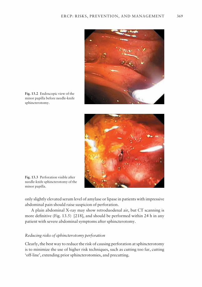
only slightly elevated serum level of amylase or lipase in patients with impressiveabdominal pain should raise suspicion of perforation.
A plain abdominal X-ray may show retroduodenal air, but CT scanning ismore definitive (Fig. 13.5) [218], and should be performed within 24 h in anypatient with severe abdominal symptoms after sphincterotomy.
Reducing risks of sphincterotomy perforation
Clearly, the best way to reduce the risk of causing perforation at sphincterotomyis to minimize the use of higher risk techniques, such as cutting too far, cutting‘off-line’, extending prior sphincterotomies, and precutting.
ERCP: RISKS, PREVENTION, AND MANAGEMENT 369
Fig. 13.2 Endoscopic view of theminor papilla before needle-knifesphincterotomy.
Fig. 13.3 Perforation visible afterneedle-knife sphincterotomy of theminor papilla.
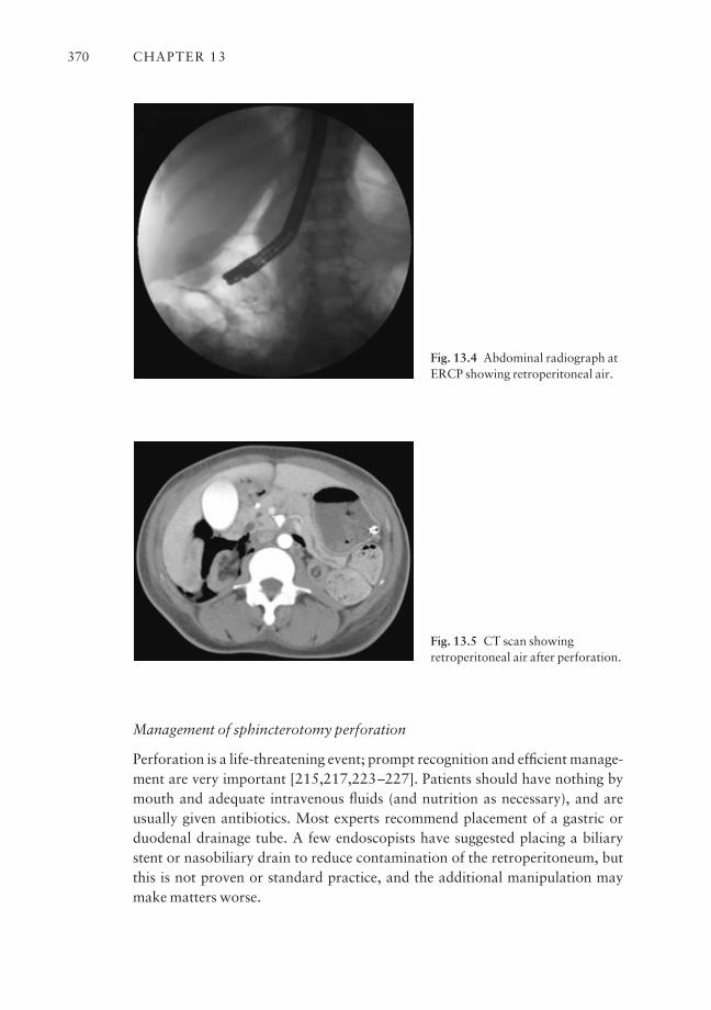
Management of sphincterotomy perforation
Perforation is a life-threatening event; prompt recognition and efficient manage-ment are very important [215,217,223–227]. Patients should have nothing bymouth and adequate intravenous fluids (and nutrition as necessary), and areusually given antibiotics. Most experts recommend placement of a gastric orduodenal drainage tube. A few endoscopists have suggested placing a biliarystent or nasobiliary drain to reduce contamination of the retroperitoneum, butthis is not proven or standard practice, and the additional manipulation maymake matters worse.
CHAPTER 13370
Fig. 13.4 Abdominal radiograph atERCP showing retroperitoneal air.
Fig. 13.5 CT scan showingretroperitoneal air after perforation.
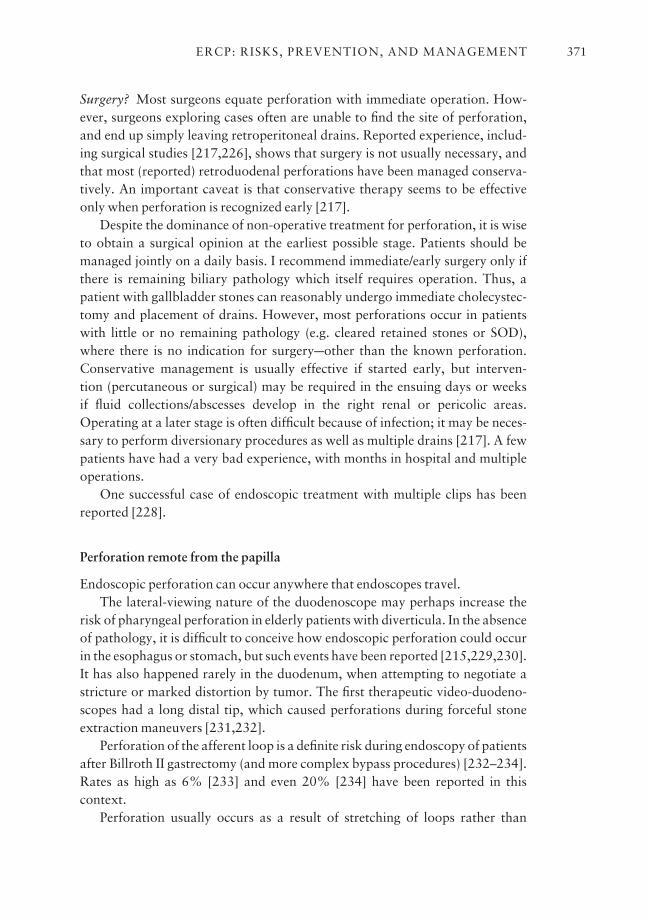
Surgery? Most surgeons equate perforation with immediate operation. How-ever, surgeons exploring cases often are unable to find the site of perforation,and end up simply leaving retroperitoneal drains. Reported experience, includ-ing surgical studies [217,226], shows that surgery is not usually necessary, andthat most (reported) retroduodenal perforations have been managed conserva-tively. An important caveat is that conservative therapy seems to be effectiveonly when perforation is recognized early [217].
Despite the dominance of non-operative treatment for perforation, it is wiseto obtain a surgical opinion at the earliest possible stage. Patients should bemanaged jointly on a daily basis. I recommend immediate/early surgery only ifthere is remaining biliary pathology which itself requires operation. Thus, apatient with gallbladder stones can reasonably undergo immediate cholecystec-tomy and placement of drains. However, most perforations occur in patientswith little or no remaining pathology (e.g. cleared retained stones or SOD),where there is no indication for surgeryaother than the known perforation.Conservative management is usually effective if started early, but interven-tion (percutaneous or surgical) may be required in the ensuing days or weeks if fluid collections/abscesses develop in the right renal or pericolic areas.Operating at a later stage is often difficult because of infection; it may be neces-sary to perform diversionary procedures as well as multiple drains [217]. A fewpatients have had a very bad experience, with months in hospital and multipleoperations.
One successful case of endoscopic treatment with multiple clips has beenreported [228].
Perforation remote from the papilla
Endoscopic perforation can occur anywhere that endoscopes travel.The lateral-viewing nature of the duodenoscope may perhaps increase the
risk of pharyngeal perforation in elderly patients with diverticula. In the absenceof pathology, it is difficult to conceive how endoscopic perforation could occurin the esophagus or stomach, but such events have been reported [215,229,230].It has also happened rarely in the duodenum, when attempting to negotiate astricture or marked distortion by tumor. The first therapeutic video-duodeno-scopes had a long distal tip, which caused perforations during forceful stoneextraction maneuvers [231,232].
Perforation of the afferent loop is a definite risk during endoscopy of patientsafter Billroth II gastrectomy (and more complex bypass procedures) [232–234].Rates as high as 6% [233] and even 20% [234] have been reported in this context.
Perforation usually occurs as a result of stretching of loops rather than
ERCP: RISKS, PREVENTION, AND MANAGEMENT 371
penetration of the endoscopic tip. It can be avoided largely by careful endo-scopic technique, especially in patients who have undergone diversionary pro-cedures or who have known stenosing pathology.
One expert suggests that all Billroth II patients (and more complex diver-sions) should be referred to tertiary centers, because of the added difficulty andsignificant risk [232].
The incidence of endoscopic (as opposed to sphincterotomy) perforations isunknown, but should be extremely low. It is less than 1:1000 in our series, all ofthem in the afferent loop context (Table 13.6).
Recognition and management of endoscopic perforation
Diagnosis of endoscopic perforation is usually obvious, either during the pro-cedure, or because of obvious patient distress and clinical signs in the chest orabdomen. Radiographs show intraperitoneal (or mediastinal) air.
Endoscopic perforation usually requires surgical intervention, and promptsurgical consultation is mandatory. Rare episodes have been treated conservatively.
Stent migration perforation
There have been rare reports of penetration and even perforation of the duode-num, small bowel, and colon by stents which have migrated from the bile duct[235,236]. Almost all of these have been ‘straight’ 10 Fr gauge stents. Those thathave migrated down from the bile duct and penetrated the opposite duodenalwall can sometimes be managed simply by endoscopic extraction. Others haverequired surgical intervention.
Infection after ERCP
Generic infection risks of all endoscopic procedures (e.g. endocarditis, viraltransmission) are discussed elsewhere [237]. Preprocedure antibiotics are recommended for prophylaxis against endocarditis by standard guidelines.
ERCP differs from most other endoscopies in that it risks contaminating territory that is usually sterile. Also, when bile is infected (e.g. in patients withstones or blocked stents), biliary manipulation may disseminate the infectionlocally or systemically.
By consensus, infection is defined as ‘an otherwise unexplained fever ofgreater than 38°C lasting 24–48 h after ERCP’. It is described as moderatelysevere if it requires more than 3 days’ treatment in hospital, or further endo-
CHAPTER 13372
scopic or percutaneous intervention, and as severe if the patient develops septicshock or requires surgery [15].
The reported incidence of clinical infections after ERCP is low, ranging from0.7 to 1.6% in various modern series [22,27,29,40,44,238–243]. However,bacteremia rates of up to 27% have been reported [237,244].
Nosocomial infection
In the early days of ERCP, before the importance of disinfection was recognized,there were several unfortunate outbreaks of nosocomial infections (usually dueto Pseudomonas) [16,18,237,245]. Sadly, Pseudomonas infections are stillbeing described after ERCP [246–249]. Almost all are due to faulty cleaning anddisinfection, and should be preventable.
Cholangitis
Bacteremia and septicemia occur when the bile is infected and drainage is compromised. This can occur after ERCP in patients with stones or strictures ifadequate drainage is not achieved, and when bilary stents become occluded[238–244,250]. The risk of introducing or stirring up infection when the bile isinfected can be minimized by adhering to disinfection protocols, by reducing thebiliary pressure (by aspirating bile before injecting much contrast), and byensuring adequate drainage by removing all obstructing stones or placingappropriate stents. Sepsis is a particular risk after ERCP management of hilartumors and sclerosing cholangitis, where it may prove impossible to providecomplete drainage of all obstructed segments. This is a good reason for obtain-ing detailed anatomical imaging (by CT and/or MRCP) beforehand to assisttherapeutic planning.
Cholecystitis
Cholecystitis, sometimes with odd characteristics [251,252], has occurred soonafter ERCP; presumably this is more likely when there is cystic duct compromiseby stone or tumor (or occasionally after stenting). It is managed by standard per-cutaneous or surgical techniques.
Pancreatic sepsis
This has occurred as part of severe pancreatitis after ERCP [253], and inpatients with pseudocysts [104], due to inadequate disinfection or incompletedrainage.
ERCP: RISKS, PREVENTION, AND MANAGEMENT 373
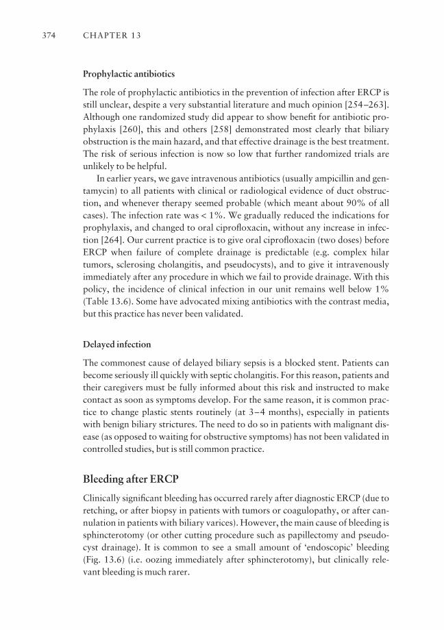
Prophylactic antibiotics
The role of prophylactic antibiotics in the prevention of infection after ERCP isstill unclear, despite a very substantial literature and much opinion [254–263].Although one randomized study did appear to show benefit for antibiotic pro-phylaxis [260], this and others [258] demonstrated most clearly that biliaryobstruction is the main hazard, and that effective drainage is the best treatment.The risk of serious infection is now so low that further randomized trials areunlikely to be helpful.
In earlier years, we gave intravenous antibiotics (usually ampicillin and gen-tamycin) to all patients with clinical or radiological evidence of duct obstruc-tion, and whenever therapy seemed probable (which meant about 90% of allcases). The infection rate was < 1%. We gradually reduced the indications forprophylaxis, and changed to oral ciprofloxacin, without any increase in infec-tion [264]. Our current practice is to give oral ciprofloxacin (two doses) beforeERCP when failure of complete drainage is predictable (e.g. complex hilartumors, sclerosing cholangitis, and pseudocysts), and to give it intravenouslyimmediately after any procedure in which we fail to provide drainage. With thispolicy, the incidence of clinical infection in our unit remains well below 1%(Table 13.6). Some have advocated mixing antibiotics with the contrast media,but this practice has never been validated.
Delayed infection
The commonest cause of delayed biliary sepsis is a blocked stent. Patients canbecome seriously ill quickly with septic cholangitis. For this reason, patients andtheir caregivers must be fully informed about this risk and instructed to makecontact as soon as symptoms develop. For the same reason, it is common prac-tice to change plastic stents routinely (at 3–4 months), especially in patientswith benign biliary strictures. The need to do so in patients with malignant dis-ease (as opposed to waiting for obstructive symptoms) has not been validated incontrolled studies, but is still common practice.
Bleeding after ERCP
Clinically significant bleeding has occurred rarely after diagnostic ERCP (due toretching, or after biopsy in patients with tumors or coagulopathy, or after can-nulation in patients with biliary varices). However, the main cause of bleeding issphincterotomy (or other cutting procedure such as papillectomy and pseudo-cyst drainage). It is common to see a small amount of ‘endoscopic’ bleeding (Fig. 13.6) (i.e. oozing immediately after sphincterotomy), but clinically rele-vant bleeding is much rarer.
CHAPTER 13374
Bleeding can occur immediately, but is often delayed for up to 2 weeks.Whilst significant bleeding is usually manifested by hematemesis and/or
melena, occasional patients can present with biliary pain and cholangitis ifbleeding fills the bile duct.
Definition of bleeding, and incidence
The consensus conference on complications defined bleeding in clinical terms.Even impressive immediate bleeding is not counted as a complication if it can bestopped by endoscopic manipulation during the procedure. The severity of thebleeding is stratified as follows:• mild: clinical (not just endoscopic) evidence of bleeding, with a hemoglobindrop of less than 3 g/dl and no transfusion;• moderate: transfusion (4 units or less), but no angiographic interventionalsurgery;• severe: transfusion of 5 units of more, or intervention (angiographic or surgical);• fatal: death attributable to bleeding.
Memory and some early publications [15,17,19,20,265] indicate that bleed-ing was the commonest complication of sphincterotomy in the first 10–15 yearsafter its introduction, with an average rate of 2.5% in over 20 000 reportedsphincterotomies [15], and a high of 11% [265]. More recent series, using theconsensus definitions, report a lower incidence of 0.8–2% [24,25,44]. Oneseries suggested that the true incidence is higher if hematological parameters arefollowed routinely [266]. More than half the reported episodes of bleeding aredelayed for up to 2 weeks.
The rate of bleeding after biliary sphincterotomy was 0.7%; most were delayed.
ERCP: RISKS, PREVENTION, AND MANAGEMENT 375
Fig. 13.6 Bleeding immediatelyafter starting sphincterotomy.
Risk factors for bleeding, and avoidance
Bleeding is certainly more likely to occur in patients with coagulopathy and/orportal hypertension [267,268], renal failure [268], and apparently also when asphincterotomy is repeated [93]. There is no evidence that the risk is greater inpatients taking aspirin and other agents affecting platelet function [268–270],although it is still common practice to ask patients to discontinue their use.Delayed clinical bleeding may [268] or may not [271] be more common whenthere has been some immediate oozing.
Prevention
Sphincterotomy should always be performed in a controlled manner, withblended current, avoiding the ‘zipper’ cut. Coagulopathies should be correctedwherever possible. Anticoagulants should be discontinued, but the need for tem-porary heparin coverage, and the duration, are controversial. The effect ofnewer antiplatelet agents has yet to be clearly established, but most endoscopistsprefer these to be stopped for 10 days if possible. The type of current used maybe relevant. One study showed that the ERBE generator reduced bleeding visibleat the time of endoscopy, but not the risk of clinically defined bleeding [272].Another study utilizing initial cutting current (to reduce the risk of pancreatitis)did show a slightly increased risk of bleeding [273].
Balloon dilatation of the sphincter can be used instead of sphincterotomy forextracting some stones in patients with irreversible coagulopathy or severe portal hypertension [151,274,275].
Management of sphincterotomy bleeding
Bleeding immediately after sphincterotomy usually stops spontaneously, and(unless there is a pumping vessel) it is usually not necessary to take any dramaticaction. There are varying opinions about management when treatment isneeded [271,276–286]. Some have advocated monopolar cautery [283], localinjection of contrast agent [284], even hemoclips [285]. However, it appearsthat epinephrine injection is the most popular and effective technique[271,286].
My practice (with unimpressive bleeding) is first to spray the site with about10 ml of a dilute (1 : 100 000) solution of epinephrine. This often stops oozingtemporarily, at least enough to see exactly where the bleeding is coming from. Ifbleeding is impressive, or if oozing persists, balloon tamponade is the next step.A retrieval balloon is overinflated in the bile duct, and then pulled down force-fully to compress the bleeding site between the balloon and the endoscope tip
CHAPTER 13376
for 5 min. If that fails, we inject epinephrine (diluted 1 : 10 000) using a stan-dard sclerotherapy needle. Up to 5 ml can be injected in aliquots of 1 ml, takingcare not to compromise the pancreatic orifice. For this reason, it is my practiceto inject just outside the top edges of the sphincterotomy, rather than within it. Ifthere has been much manipulation, it may be wise to place a small protectivepancreatic stent (if possible).
Very rarely, bleeding is profuse, and endoscopic vision is quickly lost. Expertangiographic management can be effective [287]. Surgical oversewing wouldseem logical when all else fails, but re-bleeding may occur [265].
Delayed bleeding
This can occur up to 2 weeks after sphincterotomy, and should be treated likeany other episode of bleeding. It is important to confirm the source of bleeding,since patients occasionally bleed from other lesions.
Complications of stents
Biliary and pancreatic stents can cause problems through local trauma, block-age, and migration. Much depends on their size, nature, and position.Sphincterotomy (when placing biliary stents) appears not to affect the rate ofblockage [102,288], but may reduce the risk of pancreatitis, at least with hilartumors [103].
Blockage of (plastic) biliary stents
This is inevitable after a few months, and can cause serious cholangitis. A host ofingenious attempts to prevent this phenomenon over two decades has so farbeen unavailing (Chapter 3) [289]. It is common practice to reduce the risk byrecommending the routine exchange of biliary stents at about 3 months. This ismandatory in patients with benign strictures. It is perhaps legitimate to awaitevents in patients with malignant disease if they (and their caregivers) are wellinformed about the first symptoms (usually shaking chills), and the need forurgent action. Expandable metal stents usually last much longer, but the con-sequences of blockage are equally serious.
Stent migration
Stents which migrate outwards may cause damage to the duodenum [235] ordistal intestine [236]. Stents which migrate inwards can be difficult to retrieve,especially in the pancreatic duct [290]. Most migrated stents can be teased out of
ERCP: RISKS, PREVENTION, AND MANAGEMENT 377
the papilla with a retrieval balloon, or grasped with foreign body forceps, snare,or basket. Rarely, surgery is needed to rectify these situations.
Duct damage due to stents
The presence of a stent in the bile duct for many months may cause some wallirregularity and thickening. This can be seen radiologically (and can cause diag-nostic difficulty at EUS), but has no clinical relevance. However, stent-inducedduct damage is a serious problem in the pancreas [291–295], especially when theduct initially is normal. Irritation by the tip of the stent (especially at a ductbend), or by internal flaps, often causes wall irregularity, and clinically signifi-cant narrowing. Some early descriptions suggested that most of these lesionsresolved after stent removal, but we have seen many tight fibrotic strictures,which are very difficult to manage. Relatively stiff pancreatic stents of 7 andeven 10 Fr can be used legitimately in some patients with established chronicpancreatitis for the management of stones or strictures. However, when stentingseems indicated in relatively normal ducts, it seems wise to use smaller (3 or 5 Fr)and softer stents, and for only a few weeks [295]. The length of a pancreaticstent should be chosen so that the inner tip is in a straight part of the duct.
Cholecystitis
This has been reported after biliary stenting for malignancy [296–298].
Basket impaction
Baskets may become impacted during attempts to remove large stones from thebile duct [299]. Usually, this situation can be rectified quickly by disengaging thestone, or by crushing it with a ‘rescue’ lithotripsy sleeve (Chapter 3). To preventthis problem, it is wise to use a mechanical lithotripsy system initially whenapproaching stones > 1 cm in diameter. Baskets should be used sparingly andwith great caution in the pancreatic duct. They are effective for the removal ofsoft stones (protein plugs) and mucus, but calcified pancreatic stones are veryresistant to mechanical lithotripsy. There is a risk that the basket will breakinside the duct and remain impacted.
Cardiopulmonary complications and sedation issues
Adverse cardiopulmonary events can occur during any endoscopic procedure[300,301], and myocardial ischemia has been studied specifically during ERCP[302,303].
CHAPTER 13378
Transient hypoxia and cardiac dysrhythmias occur occasionally duringERCP procedures, but are usually recognized and managed appropriately with-out clinical consequences. Very rarely, they may result in severe decompensationduring or after procedures, and are a significant cause of the rare fatalitiesattributable to ERCP.
Risk factors for cardiopulmonary complications include known or unsus-pected premorbid conditions, and problems related to sedation and analgesia.Oversedation can be a serious problem, especially in the elderly and frail, andparticularly if monitoring is inadequate (in a darkened room).
Cardiopulmonary complications can be largely avoided by careful pre-procedure evaluation, appropriate collaboration with anesthesiologists (andcardiologists) when dealing with high-risk patients, formal training of endo-scopists and nurses in sedation and resuscitation, and careful monitoring [304].
Aspiration pneumonia has been described after all types of endoscopic procedures; the incidence is unknown, but it is probably more common thanrecognized, since the onset may be delayed.
Rare complications
Many other untoward events have followed ERCP. These include:• Gallstone ileus after removing large stones [305,306].• Musculo-skeletal injuries (e.g. dislocation of the temporomandibular joint[307] or shoulder, dental trauma).• Opacification of blood vessels. The portal venous system and lymphaticshave been seen [308,309] whilst injecting contrast through tapered tip cathe-ters. The contrast moves rapidly on fluoroscopy. If air is injected as well, theappearances on CT scan are alarming [310], but no sequelae have beenreported.• Antral sinus infection after prolonged nasobiliary drainage.• Renal dysfunction [311] with the use of nephrotoxic medications (such asgentamycin).• Impaction or fracturing of nasobiliary and nasopancreatic drains.• Allergic reactions to iodine-containing contrast agents. Allergic reactionshave happened, even with the very small doses which enter the bloodstream during ERCP. Endoscopy units should have policies in place to deal withpatients who claim to be allergic [312].• Increased cholestasis in patients with sclerosing cholangitis [313].• Splenic injury has been reported several times during ERCP [314–316].• Distant abscesses have occurred in the spleen and kidney [314,317], and nodoubt elsewhere.
ERCP: RISKS, PREVENTION, AND MANAGEMENT 379
• Hemolysis due to G6PD deficiency and hemolytic–uremic syndrome hasbeen reported [318,319].• Dissemination of pancreatic cancer was reported after sphincterotomy[320].• A false aneurysm of a branch of the pancreatico-duodenal artery developedafter needle-knife sphincterotomy [321].
Deaths after ERCP
The literature reporting deaths after ERCP is difficult to analyze as the seriescontain different spectra of patients and procedures, and some do not distin-guish between 30 day mortality and events attributable to the procedure itself.One paper illustrates the difficulty in attributing mortality between concurrentillness, active complications, and complications due to other proceduresrequired after ERCP failure [26]. Data collected for the consensus conference in1991 reported 103 deaths after 7729 sphincterotomies (1.3%). Most subse-quent series report mortality figures of less than 0.5% [24,27,37,44,65,322],with two higher figures of 0.8% [29] and 1% [323].
The causes of death in all of the reported series cover the spectrum of the commonest complications, with approximately equal numbers resulting frompancreatitis, bleeding, perforation, infection, and cardiopulmonary events.Delay in diagnosis of perforation is mentioned as a contributing cause in severalpublications [217,224,324]. Of nine fatalities resulting in claims to insurance inDenmark, seven were attributable to pancreatitis (two of which had undergoneprecutting) [325].
Late complications
There are a number of adverse events attributable to ERCP that may not beapparent for months or even years afterwards.
Diagnostic error
Failure to make the correct diagnosis is an under-reported and greatly under-appreciated complication of ERCP. It can be due to poor technique (both endo-scopic and radiological), as well as incorrect interpretation of adequate images,or both. Bile duct stones are missed with inadequate duct filling, especially in less obvious sites such as the cystic duct stump and the dependent right intra-hepatic duct, or when over-dense contrast is used in a dilated system. Con-versely, air bubbles introduced into the system may be misinterpreted as stones(with the potential serious consequences of an unnecessary sphincterotomy).
CHAPTER 13380
Poor opacification and ignorance of anatomy may lead to missed or erroneousdiagnoses in patients with bile duct injuries. Congenital variations of biliopan-creatic drainage are under-recognized. Early stages of chronic pancreatitis andintraductal mucinous tumors are easily missed with inadequate filling. Pancreasdivisum may be missed when the ventral duct is rudimentary, and the pancreaticpathology unassessed if dorsal cannulation is not achieved.
Few endoscopists have a radiologist on hand to help with fluoroscopy, filmrecording, or the immediate interpretation which is needed to formulate thera-peutic tactics. It is common practice for radiologists to report the available filmsafter the event, and major discrepancies have been noted [326], a fact whichraises complex issues. Providing the reporting radiologist with a detailed copy ofthe endoscopic report is helpful, and allows radiologists to communicate anydifferences of opinion.
Late infection
There is a possibility of transmitting non-bacterial infections at ERCP, with an incubation period long enough to hide the relationship, but there are noproven and reported cases. There is a definite risk of sepsis developing when biliary stents become occluded. Patients present with fevers and shaking chills,and can deteriorate rapidly. Any stented patient (and caregivers) must bewarned about the possibility, and the need for speedy medical contact and res-olution. Patients receiving plastic stents for benign biliary strictures should be advised to undergo a routine stent service at 3–4 months; practice varies with malignant strictures (Chapter 6). Endoscopists placing stents have a con-tinuing responsibility to contact patients with reminders. Occasionally, patientsmay willfully or accidentally avoid the repeat procedure, with considerablepotential for serious complications. The concept of long-term stenting for‘difficult’ stones has been discredited because of the risk of delayed cholangitis[327].
Late effects of sphincterotomy
There has been much interest in the possible long-term adverse consequences ofbiliary sphincterotomy [328–339]. When performed for ‘papillary stenosis’,there is a significant risk of further biliary-type symptoms, whether due torestenosis or an incorrect diagnosis (Chapter 8).
Sphincterotomy leads almost inevitably to bacterial contamination of thebile [340–344], which may be a potent promoter of pigment stone formation.One study showed a significant increase in the incidence of cholangiocarcinomaafter surgical sphincteroplasty [345], but a cohort study in Scandinavia found
ERCP: RISKS, PREVENTION, AND MANAGEMENT 381
no such association after endoscopic sphincterotomy [346]. Many patients havebeen followed for periods of 10 years or more after sphincterotomy for stones[332,334–336,338–340]. The chance of further biliary problems in these stud-ies ranges from 5 to 24%, with an average of about 10% [347]. The Amsterdamstudy had the highest figure (24%) and all but one of the patients had recurrentstones [330]. In other series, some patients had episodes of cholangitis withoutstones, even cholangitis without stenosis of the sphincterotomy [332].
Most of these long-term complications of sphincterotomy are easily man-aged endoscopically, remembering that repeat incisions do carry a slightlygreater risk. A few patients continue to reform stones every 6–12 months despiteapparently adequate drainage, and may need to be scheduled for repeated endo-scopic ‘biliary laundry’ [348].
Sphincterotomy with the gallbladder in place
Most patients having their ducts cleared of stones endoscopically have under-gone cholecystectomy soon afterwards. However, some have not, usuallybecause the risk has been judged to be too great (and especially before the daysof laparoscopic cholecystectomy). Several series have examined the long-termrisks of leaving the gallbladder in place [349–354]. The reported need for chole-cystectomy has ranged from 5 to 33% [337], but most of the follow-up periodsare short. Two trials have addressed this issue recently. Thirty-four patientstreated endoscopically for acute biliary pancreatitis (and without cholecystec-tomy) were followed for a mean of 34 months; only 11.6% developed furtherbiliary complications [354]. However, the Amsterdam group performed a randomized trial of 120 patients with the gallbladder in place after biliarysphincterotomy. No fewer than 47% of those treated expectantly developedfurther biliary symptoms, compared with 2% of those who underwent earlycholecystectomy [353]. The suggestion that non-filling of the gallbladder at the index ERCP (indicating cystic duct obstruction) was a predictor of futuretrouble has not been substantiated [352]. However, it seems clear that the risk isnegligible in patients who have no stones remaining in the gallbladder, which issometimes the case in the context of gallstone pancreatitis [350].
Pancreatic sphincterotomy
The main risk of pancreatic sphincterotomy appears to be restenosis, whichoccurs in at least 20% of reported cases (Chapters 6, 7 and 8).
It is usually treated endoscopically, but strictures that occur beneath thepapilla can be challenging even for surgical repair. Hopefully, better techniques(and new stents) may reduce this risk in the future.
CHAPTER 13382
Stenosis of the pancreatic orifice causing recurrent pancreatitis has beenreported as a late complication of biliary sphincterotomy [355].
Managing adverse events
All ERCP endoscopists experience complications. Each event requires specificskillful recognition and management (as detailed above), but there are severalvery important general guidelines.
Prompt recognition and action
The keys to effective management of all complications are early recognition andprompt focused action. Delay is dangerous both medically and legally. Patientsin pain and distress after procedures should always be examined carefully, andnever simply ‘reassured’ without careful evaluation. If you are not personally oncall on the night after your ERCP procedures, it is helpful to make sure that theperson covering is aware of what you have done. Get appropriate laboratorystudies and radiographs, consult the extensive literature, and do not hesitate toseek advice from other experts in the relevant fields. It is wise to consult an(informed) surgeon early on for anything that might remotely require surgicalintervention. Sometimes it may be appropriate to offer transfer of care of thepatient to a specialty colleague, or to a larger medical center, but, if this happens, try to keep in touch, and to show continuing interest and concern.Apparent abandonment alienates patients and their relatives, and may lead toinitiation of legal action.
Professionalism and communication
Endoscopists often feel devastated when serious complications occur. Your dis-tress is understandable and worthy, and it is important to be sympathetic, but itis equally important to be composed and matter of fact. Excessive apologies maygive an unfortunate impression. Never, never, attempt to cover up the facts.Poor communication is the basis for much unhappiness, and many lawsuits.Remember that the truly informed patient and any accompanying persons have been told already that complications can happen. This is an integral important part of the consent process. So it is appropriate and correct to addresssuspected complications in that spirit. ‘It looks as if we have a perforation here.We discussed that as a remote possibility beforehand, and I am sorry that it has occurred. Here is what I think we should do.’ It is also wise to contact andinform other interested relatives, referring physicians, supervisors, and yourRisk Management advisors.
ERCP: RISKS, PREVENTION, AND MANAGEMENT 383
Documentation
Document what has happened carefully and honestly in real time. Don’t eventhink of adding notes retrospectively. The results of many lawsuits hang on thequality of the documentation, or lack of it.
Learning from lawsuits
Fortunately, most complications do not result in legal action. Despite the factthat ERCP is the most dangerous of the routine endoscopic procedures, there arefar more claims after colonoscopy and upper endoscopy [356]. There are severalreasons why patients (or their survivors) may initiate a claim.
Communication
Communication, or lack of it, is often a major complaint. Too often we hearthat ‘we would never have consented to the procedure if we had known that thismight happen’. Sometimes this is simply because patients don’t want to hear,but often the consent process is quite inadequate. A hurried conversation imme-diately before the procedure is not sufficient. Taking time to provide the infor-mation (face to face and in writing), making sure that it has been understood,and writing down that you have done it, is simply good medical practice [105].
Good communication after an adverse event is equally important. Show thatyou care. Litigants are sometimes simply (and justifiably) angry if they get theimpression that you do not.
Financial concerns
These are also often prominent, even if not stated. Hospital bills and loss ofearnings can be crippling.
Standard of care practice
Once a lawsuit has been filed, the key issue is whether the endoscopist (and others involved) practiced within the ‘standard of care’. This is defined in various ways, but comes down to what reasonable colleagues would do (and isexpressed in court by what expert witnesses opine). The report from the NIHConsensus Conference is a crucial resource [57], and is particularly forceful inrecommending caution when considering ERCP in patients with little or noobjective evidence for pathology (i.e. ‘suspected sphincter dysfunction’).
The key standard of care issues are given below.
CHAPTER 13384
Indications
Was the ERCP procedure really indicated in the first place? The task clearly is tobalance the possible benefits against the potential risks [357]. Although profes-sional societies publish guidelines for the use of ERCP [358], the devil is in thedetails, e.g. how much elevation of liver tests or increased duct size constitutes‘objective evidence of pathology’. In practice, the validity of the decision to proceed will be judged by the severity of the symptoms, by the thoroughness ofprior treatment and investigations, and the process of communication. Were thesymptoms (or other signs of pathology) really that pressing? Had less invasiveapproaches (nowadays including MRCP) been exhausted, or at least consideredand discussed [359]? There are some circumstances (such as postcholecystec-tomy pain with some abnormality of liver tests) which may justify ERCP even ifimaging is negative, but where it may be unwise to strive too hard (e.g. by pro-longed attempts or precutting) when cannulation proves difficult.
For less experienced endoscopists, consideration of alternatives (especiallyfor higher risk procedures) should include possible referral to an expert center.
The procedure
Was there an obvious deviation from customary practice, like placing a 10 Frstent in a normal pancreatic duct, or trying to extract a stone from the bile duct without sphincterotomy (or papillary balloon dilatation)? Did the level ofsuspicion of pathology really justify a precut? Was there radiological evidencefor over-manipulation of the pancreas, over-injection (e.g. acinarization), orinjection into a branch duct? The notes of the procedure nurse may containimportant evidence, like excessive sedation or contrast, or documentation ofpatient distress. Pretty endoscopic photographs may also be incriminating, e.g.if they show sphincterotomy in an unusual direction.
Postprocedure care
Was the patient appropriately monitored, discharged in good condition, andproperly advised? Was action taken promptly when unexpected symptomsdeveloped? Was the endoscopist available to advise? Among the most commonerrors are delay in action (particularly in considering and managing perforation)and inadequate fluid resuscitation in patients with pancreatitis.
Conclusion
After more than 30 years, the risks of ERCP and its therapeutic procedures are
ERCP: RISKS, PREVENTION, AND MANAGEMENT 385
now well documented. Pancreatitis and sedation-related events are the com-monest, but bleeding and perforation still occur. There are a host of rare com-plications. Understanding and managing the main risk factors can keep theseevents to a minimum, but cannot eliminate them. For this reason, making surethat patients understand what they are accepting is of crucial importance.Inexperience and over-confidence are dangerous partners.
Outstanding issues and future trends
The two biggest issues for ERCP at the present time are the quality of practiceand how to minimize or eliminate postprocedure pancreatitis. These are notunrelated, for we know that experts have lower complication rates, even whiledealing with higher risk clientele. Thus, we are forced to focus on how to max-imize expertise.
Many experts for a long time have been advocating that fewer endoscopistsshould be trained in ERCP, so that their skills can be maximized before and afterentering practice. This trend is perhaps evident at long last, driven by severalforces. Firstly, diagnostic ERCP is becoming obsolescent as non-invasive methods (especially MRCP) improve. This means that would-be ERCP practi-tioners can often now see the suspected therapeutic issue beforehand. They mustbe prepared for the challenge, but also have the option of referring problematiccases (e.g. hilar tumors and ‘suspected sphincter dysfunction’). Secondly, theseminal studies of Freeman and colleagues, and a few others, have made endo-scopists (and lawyers) much more aware of certain high-risk behaviors, such ascasual precutting. Thirdly, most gastroenterologists have no shortage of otheractivities (not least screening colonoscopy) to keep them interested and busy.The final driver is the increasing sophistication of our patients, who are learningthat not all interventionists are equalaas is well documented in surgery [8]aandare demanding the data with which to make informed choices [360].
All interventions carry some risks, which are acceptable if the indications are appropriate, i.e. when there are substantial potential benefits. To do a betterjob of predicting benefit will require many more major prospective outcomestudies. We need careful objective and structured cohort studies of ERCP in various clinical contexts, and some randomized studies in comparison withother approaches, such as surgery.
Thus, in the future, we hope that there will be fewer but very well trained andexperienced ERCP practitioners, and that both they and their patients will havea better understanding of the risk/benefit ratio in each case.
CHAPTER 13386
References
1 Cotton PB. Outcomes of endoscopic procedures: struggling towards definitions. GastrointestEndosc 1994; 40: 514–18.
2 Fleischer DE. Better definition of endoscopic complications and other negative outcomes.Gastrointest Endosc 1994; 40 (4): 511–13.
3 Fleischer DE, Van de Mierop F, Eisen GM, Al-Kawas FH, Benjamin SB, Lewis JH et al. A newsystem for defining endoscopic complications emphasizing the measure of importance. Gastro-intest Endosc 1997; 45 (2): 128–33.
4 Cotton PB. Income and outcome metrics for the objective evaluation of ERCP and alternativemethods. Gastrointest Endosc 2002; 56 (6): S283–90.
5 Campbell N, Sparrow K, Fortier M, Ponich T. Practical radiation safety and protection for theendoscopist during ERCP. Gastrointest Endosc 2002; 55 (4): 552–7.
6 O’Sullivan S, Bridge G, Ponich T. Musculoskeletal injuries among ERCP endoscopists inCanada. Can J Gastroenterol 2002; 16 (6): 369–74.
7 Petersen BT. ERCP outcomes: defining the operators, experience, and environments. Gastro-intest Endosc 2002; 55 (7): 953–8.
8 Birkmeyer JD, Stukel TA, Siewers AE, Goodney PP, Wennberg DE, Lucas FL. Surgeon volumeand operative mortality in the United States. N Engl J Med 2003; 349: 2117–27.
9 Schutz SM, Abbott RM. Grading ERCPs by degree of difficulty: a new concept to producemore meaningful outcome data. Gastrointest Endosc 2000; 51 (5): 535–9.
10 Lambert ME, Betts CD, Hill J, Faragher EB, Martin DF, Tweedle DE. Endoscopic sphinctero-tomy: the whole truth. Br J Surg 1991; 78 (4): 473–6.
11 Cotton PB. Endoscopic management of bile duct stones (apples and oranges). Gut 1984; 25:587–97.
12 Perdue DG, Freeman ML, ERCOST Study Group. Failed biliary ERCP: a prospective multi-center study of risk factors, complications and resource utilization. Gastrointest Endosc 2004;59 (5): AB192.
13 Cotton PB. Randomization is not the (only) answer: a plea for structured objective evaluationof endoscopic therapy. Endoscopy 2000; 32 (5): 402–5.
14 Hebert RL, Palesch YY, Tarnasky PR, Aabakken I, Mauldin PD, Cotton PB. DDQ-15 health-related quality of life instrument for patients with digestive disorders. Health ServicesOutcomes Res Methodology 2001; 2: 137–56.
15 Cotton PB, Lehman G, Vennes J, Geenen JE, Russell RC, Meyers WC et al. Endoscopic sphinc-terotomy complications and their management: an attempt at consensus. Gastrointest Endosc1991; 37: 383–93.
16 Bilbao MK, Dotter CT, Lee TG, Katon RM. Complications of endoscopic retrograde cholan-giography (ERCP): a study of 10 000 cases. Gastroenterology 1976; 70: 314–20.
17 Geenen JE, Vennes JA, Silvis SE. Resume of a seminar on endoscopic retrograde sphinctero-tomy (ERS). Gastrointest Endosc 1981; 27: 31–8.
18 Cotton PB, Progress Report ERCP. Gut 1977; 18: 316–41.19 Neuhaus B, Safrany L. Complications of endoscopic sphincterotomy and their treatment.
Endoscopy 1981; 13: 197–9.20 Vaira D, D’Anna L, Ainley C, Dowsett J, Williams S, Baillie J et al. Endoscopic sphincterotomy
in 1000 consecutive patients. Lancet 1989; 2: 431–4.21 American Society for Gastrointestinal Endoscopy. Standards of Practice Committee. Com-
plications of ERCP. Gastrointest Endosc 2003; 57 (6): 633–8.22 Barthet M, Lesavre N, Desjeux A, Gasml M, Berthezene P, Berdah S et al. Complications of
endoscopic sphincterotomy: results from a single tertiary referral center. Endoscopy 2002; 34(12): 991–7.
23 Freeman ML. Adverse outcomes of endoscopic retrograde cholangiopancreatography. RevGastroenterol Disord 2002; 2 (4): 147–67.
ERCP: RISKS, PREVENTION, AND MANAGEMENT 387
24 Loperfido S, Angelini G, Benedetti G, Chilovi F, Costan F, De Berardinis F et al. Major earlycomplications from diagnostic and therapeutic ERCP: a prospective multicenter study. Gastro-intest Endosc 1998; 48: 1–10.
25 Masci E, Toti G, Mariani A, Curioni S, Lomazzi A, Dinelli M et al. Complications of diagnosticand therapeutic ERCP: a prospective multicenter study. Am J Gastroenterol 2001; 96: 417–23.
26 Tzovaras G, Shukla P, Kow L, Mounkley D, Wilson T, Toouli J. What are the risks of diag-nostic and therapeutic endoscopic retrograde cholangiopancreatography? Aust N Z J Surg2000; 70: 778–82.
27 Vandervoort J, Soetikno RM, Tham TC, Wong RC, Ferrari AP Jr, Montes H et al. Risk factorsfor complications after performance of ERCP. Gastrointest Endosc 2002; 56: 652–6.
28 Freeman ML. Adverse outcomes of endoscopic retrograde cholangiopancreatography: avoid-ance and management. Gastrointest Endosc Clin N Am 2003; 13 (4): 775–98.
29 Garcia-Cano Lizcano J, Conzalez Martin JA, Morillas Arino J, Perez Sola A. Complications of endoscopic retrograde cholangiopancreatography: a study in a small ERCP unit. Rev EspEnferm Dig 2004; 96 (3): 155–62.
30 Freeman ML. Understanding risk factors and avoiding complications with endoscopic retro-grade cholangiopancreatography. Curr Gastroenterol Rep 2003; 5 (2): 145–53.
31 Landoni N, Chopita N, Jmelnitzky A. Endoscopic sphincterotomy: its complications and theirfollowup. Acta Gastroenterol Latinoam 1992; 22 (3): 155–9.
32 Sherman S, Ruffolo TA, Hawes RH, Glehman GA. Complications of endoscopic sphinctero-tomy. Gastroenterology 1991; 101: 1068–75.
33 Tanner A. ERCP: present practice in a single region. Eur J Gastroenterol Hepatol 1996; 8:145–8.
34 Mallery JS, Baron TH, Dominitz JA, Goldstein JL, Hirota WK, Jacobson BC et al. Com-plications of ERCP. Gastrointest Endosc 2003; 57: 633–8.
35 Aliperti G. Complications related to diagnostic and therapeutic endoscopic retrograde cholan-giopancreatography. Gastrointest Endosc Clin N Am 1996; 6: 379–40.
36 Freeman ML. Adverse outcomes of ERCP. Gastrointest Endosc 2002; 56 (6): S273–82.37 Farrell RJ, Mahmud N, Noonan N, Kellcher D, Keeling PW. Diagnostic and therapeutic
ERCP: a large single centre’s experience. Ir J Med Sci 2001; 170 (3): 176–80.38 Munoz SR. Towards safer ERCP: selection, experience and prophylaxis. Rev Esp Enferm Dig
(Madrid) 2004; 96 (3): 155–62.39 Misra SP, Dwivedi M. Complications of endoscopic retrograde cholangiopancreatography
and endoscopic sphincterotomy: diagnosis, management and prevention. Natl Med J India2002; 15: 27–31.
40 Halme L, Doepel M, von Numers H, Edgren J, Ahonen J. Complications of diagnostic andtherapeutic ERCP. Ann Chir Gynaecol 1999; 88: 127–31.
41 Rabenstein T, Schneider HT, Nicklas M, Ruppert T, Katalinic A, Hahn EG et al. Impact ofskill and experience of the endoscopist on the outcome of endoscopic sphincterotomy tech-niques. Gastrointest Endosc 1999; 50: 628–36.
42 Davis WZ, Cotton PB, Arias R, Williams D, Onken JE. ERCP and sphincterotomy in the con-text of laparoscopic cholecystectomy: academic and community practice patients and results.Am J Gastroenterol 1997; 92: 597–601.
43 Escourrou J, Delvaux M, Busail L et al. Clinical results of endoscopic sphincterotomy: com-parison of two activity periods in the same endoscopy units. Gastrointest Endosc 1990; 36:205–6.
44 Freeman ML, Nelson DB, Sherman S, Haber GB, Herman ME, Dorsher PJ et al. Complica-tions of endoscopic biliary sphincterotomy. N Engl J Med 1996; 335: 909 –18.
45 Newcomer MK, Jowell PS, Cotton PB. Underestimation of adverse events following ERCP: aprospective 30 day follow up study [Abstract]. Gastrointest Endosc 1995; 41: 408.
46 Zubarik R, Fleischer DE, Mastropietro C, Lopez J et al. Prospective analysis of complications30 days after outpatient colonoscopy. Gastrointest Endosc 1999; 50 (3): 322–8.
47 Zubarik R, Eisen G, Mastropietro C, Lopez J, Carroll J, Benjamin S et al. Prospective analysisof complications 30 days after outpatient upper endoscopy. Am J Gastroenterol 1999; 94 (6):1539–45.
CHAPTER 13388
48 Arenson N, Flamm CR, Bohn RI, Mark DH, Speroff T. Evidence-based assessment: patientprocedure, or operator factors associated with ERCP complications. Gastrointest Endosc2002; 56: s294–s301.
49 Deans GT, Sedman P, Martin DF, Royston CMS, Leow CK, Thomas WEG et al. Are com-plications of endoscopic sphincterotomy age related? Gut 1997; 41: 545–8.
50 Derkx HHF, Huibregtse K, Taminiau JAJM. The role of endoscopic retrograde cholangiopan-creatography in cholestatic infants. Endoscopy 1994; 26: 724–8.
51 Guelrud M, Mujica C, Jaen D, Plaz J, Arias J. The role of ERCP in the diagnosis and treatmentof idiopathic recurrent pancreatitis in children and adolescents. Gastrointest Endosc 1994; 40(4): 428–33.
52 Mitchell RM, O’Connor F, Dickey W. Endoscopic retrograde cholangiopancreatography issafe and effective in patients 90 years of age and older. J Clin Gastroenterol 2003; 36 (1): 72–4.
53 Hui CK, Liu CL, Lai KC, Chan SC, Hu WH, Wong WM et al. Outcome of emergency ERCPfor acute cholangitis in patients 90 years of age and older. Aliment Pharmacol Ther 2004; 19(11): 1153–8.
54 Leung JW, Chung SC, Sung JJ, Banez VP, Li AK. Urgent endoscopic drainage for acute sup-purative cholangitis. Lancet 1989; 1 (8650): 1307–9.
55 Cotton PB, Jowell PS, Baillie J, Leung J, Affronti J, Branch MS et al. Spectrum of complicationsafter diagnostic ERCP and effect of comorbidities. Gastrointest Endosc 1994; 40 (2): P18.
56 Jamidar PA, Beck GJ, Hoffman BJ, Lehman GA, Hawes RH, Agrawal RM et al. Endoscopicretrograde cholangiopancreatography in pregnancy. Am J Gastroenterol 1995; 98 (8):1263–7.
57 Cohen S, Bacon BR, Berlin JA, Fleischer D, Hecht GA, Loehrer PJ Sr et al. National Institutesof Health State-of-the-Science Conference Statement: ERCP for diagnosis and therapy,January 14–16, 2002. Gastrointest Endosc 2002; 56: 803–9.
58 Cotton PB. ERCP is most dangerous for people who need it least. Gastrointest Endosc 2001;54 (4): 535–6.
59 Shemesh E, Klein E, Czerniak A, Coret A, Bat L. Endoscopic sphincterotomy in patients withgallbladder in situ: the influence of periampullary duodenal diverticula. Surgery 1990; 107:163–6.
60 Vaira D, Dowsett JF, Hatfield ARW et al. Is duodenal diverticulum a risk factor for sphinctero-tomy? Gut 1989; 30: 939–42.
61 Veitch A, Fairclough P. Endoscopic diathermy in patients with cardiac pacemakers.Endoscopy 1998; 30 (6): 544–7.
62 Sherman S, Ruffolo TA, Hawes RH, Lehman GA. Complications of endoscopic sphinctero-tomy: a prospective series with emphasis on the increased risk associated with sphincter ofOddi dysfunction and nondilated bile duct. Gastroenterology 1991; 101: 1068–75.
63 Chen YK, Foliente RL, Santoro MJ, Walter MH, Collen MJ. Endoscopic sphincterotomy-induced pancreatitis: increased risk associated with nondilated bile ducts and sphincter ofOddi dysfunction. Am J Gastroenterol 1994; 89 (3): 327–33.
64 Cotton PB, Geenen JE, Sherman S, Cunningham JT, Howell DA, Carr-Locke DL et al. Endo-scopic sphincterotomy for stones by experts is safe, even in younger patients with normalducts. Ann Surg 1998; 227: 201–4.
65 Wilson MS, Tweedle DEF, Martin DF. Common bile duct diameter and complications ofendoscopic sphincterotomy. Br J Surg 1992; 79: 1345–7.
66 Huibregtse K. Complications of endoscopic sphincterotomy and their prevention. N Engl JMed 1996; 335: 961–2.
67 Boender J, Nix GA, de Ridder MA, van Blankenstein M, Schutte HE, Dees J et al. Endoscopicpapillotomy for common bile duct stones: factors influencing the complication rate. Endo-scopy 1994; 26: 209–16.
68 Mehta SN, Pavone E, Barkun JS, Bouchard S, Barkun AN. Predictors of post-ERCP complica-tions in patients with suspected choledocholithiasis. Endoscopy 1998; 30: 457–63.
69 Elfant AB, Bourke MJ, Alhalel R, Kortan PP, Haber GB. A prospective study of the safety ofendoscopic therapy for choledocholithiasis in an outpatient population. Am J Gastroenterol1996; 91 (8): 1499–502.
ERCP: RISKS, PREVENTION, AND MANAGEMENT 389
70 Alsolaiman M, Cotton P, Hawes R, Aliperti G, Carr-Locke DL, Fogel EL et al. Techniques forpancreatic sphincterotomy: lack of expert consensus. Gastrointest Endosc 2004; 59 (5):AB210.
71 Elton E, Howell DA, Parsons WG, Qaseem T, Hanson BL. Endoscopic pancreatic sphinctero-tomy: indications, outcome, and a safe stentless technique. Gastrointest Endosc 1998; 47:240–9.
72 Berkes J, Bernklau S, Halline A, Venu R, Brown R. Minor papillotomy in pancreas divisum: docomplications and restenosis rates differ between use of the needle knife papillotome (NKS) vs.ultratapered traction sphincterotome (UTS)? Gastrointest Endosc 2004; 59 (5): AB207.
73 Delhaye M, Matos C, Deviere J. Endoscopic technique for the management of pancreatitis andits complications. Best Pract Res Clin Gastroenterol 2004; 18 (1): 155–81.
74 Leung JW, Banez VP, Chung SC. Precut (needle knife) papillotomy for impacted common bileduct stone at the ampulla. Am J Gastroenterol 1990; 85: 991–3.
75 Cotton PB. Precut papillotomy: a risky technique for experts only. Gastrointest Endosc 1989;35: 578.
76 Cotton PB. Needleknife precut sphincterotomy: the devil is in the indications. Endoscopy1997; 29: 888.
77 Dowsett JF, Polydorou AA, Vaira D et al. Needle knife papillotomy: how safe and how effec-tive? Gastrointest Endosc 1990; 36 (6): 645–6.
78 Vandervoort J, Carr-Locke DL. Needle-knife access papillotomy: an unfairly maligned tech-nique? Endoscopy 1996; 28: 365–6.
79 Rabenstein T, Ruppert T, Schneider HT, Hahn EG, Ell C. Benefits and risks of needle-knifepapillotomy. Gastrointest Endosc 1997; 46: 207–11.
80 Baillie J. Needle-knife papillotomy revisited [editorial; comment]. Gastrointest Endosc 1997;46: 282.
81 Bruins SW, Schoeman MN, DiSario JA, Wolters F, Tytgat GN, Huibregtse K. Needle-knifesphincterotomy as a precut procedure: a retrospective evaluation of efficacy and complica-tions. Endoscopy 1996; 28: 334–9.
82 Dhir V, Swaroop VS, Mohandas KM, Jagannath P, Desouza LJ. Precut papillotomy using aneedle knife: experience in 100 patients with malignant obstructive jaundice. Indian J Gastro-enterol 1997; 16: 52–3.
83 Foutch PG. A prospective assessment of results for needle-knife papillotomy and standardendoscopic sphincterotomy. Gastrointest Endosc 1995; 41: 25–32.
84 Gholson CF, Favrot D. Needle knife papillotomy in a University referral practice: safety andefficacy of a modified technique. J Clin Gastroenterol 1996; 23: 177–80.
85 Kasmin FE, Cohen D, Batra S, Cohen SA, Siegel JH. Needle-knife sphincterotomy in a tertiaryreferral center: efficacy and complications. Gastrointest Endosc 1996; 44: 48–53.
86 Rollhauser C, Johnson M, Al Kawas FH. Needle-knife papillotomy: a helpful and safe adjunctto endoscopic retrograde cholangiopancreatography in a selected population. Endoscopy1998; 30: 691–6.
87 Harewood GC, Baron TH. An assessment of the learning curve for precut biliary sphinctero-tomy. Am J Gastroenterol 2002; 97: 1708–12.
88 Freeman ML. Precut (access) sphincterotomy: Techniques. Gastrointest Endosc 1999; 1: 40–8.
89 Binmoeller KF, Seifert H, Gerke H, Seitz U, Portis M, Soehendra N. Papillary roof incisionusing the Erlangen-type pre-cut papillotome to achieve selective bile duct cannulation. Gastro-intest Endosc 1996; 44: 689–95.
90 Goff JS. Long-term experience with the transpancreatic sphincter pre-cut approach to biliarysphincterotomy. Gastrointest Endosc 1999; 50: 642–5.
91 Mavrogiannis C, Liatsos C, Papanikolaou IS, Psilopoulos DI, Goulas SS et al. Safety of exten-sion of a previous endoscopic sphincterotomy: a prospective study. Am J Gastroenterol 2003;98 (1): 72–6.
92 Choudari CP, Sherman S, Fogel EL, Phillips S, Kochell A, Flueckiger J et al. Success of ERCP at a referral center after a previously unsuccessful attempt. Gastrointest Endosc 2000; 52 (4):478–83.
CHAPTER 13390
93 Raijman I, Escalante-Glorsky S. Is the complication rate the same for index versus repeat bili-ary sphincterotomy? Gastrointest Endosc 2004; 59 (5): AB193.
94 May GR, Cotton PB, Edmunds EJ, Chong W. Removal of stones from the bile duct at ERCPwithout sphincterotomy. Gastrointest Endosc 1993; 39 (6): 749–54.
95 MacMathuna P. Endoscopic treatment of bile duct stones: should we cut or dilate the sphinc-ter? Am J Gastroenterol 1997; 92 (9): 1411–12.
96 Norton ID, Gostout CJ, Baron TH, Geller A, Petersen BT, Wiersema MJ. Safety and outcomeof endoscopic snare excision of the major duodenal papilla. Gastrointest Endosc 2002; 56:239–43.
97 Desilets DJ, Dy RM, Ku PM, Hanson BL, Elton E, Mattia A et al. Endoscopic management oftumors of the major duodenal papilla: refined techniques to improve outcome and avoid com-plications. Gastrointest Endosc 2001; 54: 202–8.
98 Zadorova Z, Dvofak M, Hajer J. Endoscopic therapy of benign tumors of the papilla of Vater.Endoscopy 2001; 33: 345–7.
99 Catalano MF, Linder JD, Chak A, Sivak MV Jr, Raijman I, Geenen JE et al. Endoscopic man-agement of adenoma of the major duodenal papilla. Gastrointest Endosc 2004; 59: 225–32.
100 Fujita N, Noda Y, Kobayashi G, Kimura K, Ito K. Endoscopic papillectomy: is there room forthis procedure in clinical practice? Digestive Endoscopy 2003; 15: 253–5.
101 Cheng C, Sherman S, Fogel EL, McHenry L, Watkins JL et al. Endoscopic snare papillectomyof ampullary tumors: 10-year review of 55 cases at Indiana University Medical Center.Gastrointest Endosc 2004; 59 (5): AB193.
102 Giorgio PD, Luca LD. Comparison of treatment outcomes between biliary plastic stent place-ments with and without endoscopic sphincterotomy for inoperable malignant common bileduct obstruction. World J Gastroenterol 2004; 10 (8): 1212–14.
103 Tarnasky PR, Cunningham JT, Hawes RH, Hoffman BJ et al. Transpapillary stenting of proximal biliary strictures: does biliary sphincterotomy reduce the risk of post-procedure pan-creatitis? Gastrointest Endosc 1997; 45: 46–51.
104 Baron TH. Endoscopic drainage of pancreatic fluid collections and pancreatic necrosis.Gastrointest Endosc Clin N Am 2003; 13 (4): 743–64.
105 Plumeri PA. Informed consent for upper gastrointestinal endoscopy. Gastroenterol Clin N Am1994; 4 (2): 455–61.
106 Duncan HD, Hodgkinson L, Deakin M, Green JR. The safety of diagnostic and therapeuticERCP as a daycase procedure with a selective admission policy. Eur J Gastroenterol Hepatol1997; 9 (9): 905–8.
107 Ho KY, Montes H, Sossenheimer MJ, Tham TC, Ruymann F, Van Dam J et al. Features thatmay predict hospital admission following outpatient therapeutic ERCP. Gastrointest Endosc1999; 49: 587–92.
108 Freeman ML, Nelson DB, Sherman S, Haber GB, Fennerty MB, DiSario JA et al. Same-day dis-charge after endoscopic biliary sphincterotomy: observations from a prospective multicentercomplication study. Gastrointest Endosc 1999; 49: 580–6.
109 Cvetkovski B, Gerdes H, Kurtz RC. Outpatient therapeutic ERCP with endobiliary stent place-ment for malignant common bile duct obstruction. Gastrointest Endosc 1999; 50: 63–6.
110 Linder JD, Tarnasky P. There are benefits of overnight observation after outpatient ERCP.Gastrointest Endosc 2004; 59 (5): AB208.
111 Testoni PA, Bagnolo F, Caporuscio S, Lella F. Serum amylase measured four hours after endoscopic sphincterotomy is a reliable predictor of postprocedure pancreatitis. Am JGastroenterol 1999; 94: 1235–41.
112 Friedland S, Soetikno RM, Vandervoort J, Montes H, Tham T, Carr-Locke DL. Bedside scor-ing system to predict the risk of developing pancreatitis following ERCP. Endoscopy 2002; 34:483–8.
113 Barthet M, Desjeux A, Gasmi M, Bellon P, Hoi MT, Salducci J et al. Early refeeding after endo-scopic biliary or pancreatic sphincterotomy: a randomized prospective study. Endoscopy2002; 34 (7): 546–50.
114 Freeman ML, Guda NM. Prevention of post-ERCP pancreatitis: a comprehensive review.Gastrointest Endosc 2004; 59 (7): 845–64.
ERCP: RISKS, PREVENTION, AND MANAGEMENT 391
115 Freeman ML, DiSario JA, Nelson DB, Fennerty MB, Lee JG, Bjorkman DJ et al. Risk factorsfor post-ERCP pancreatitis: a prospective, multicenter study. Gastrointest Endosc 2001; 54(4): 535–6.
116 Testoni PA. Why the incidence of post-ERCP pancreatitis varies considerably? Factors affect-ing the diagnosis and the incidence of this complication. JOP 2002; 3 (6): 195–201.
117 Johnson GK, Geenen JE, Johanson JF, Sherman S, Hogan WJ, Cass O et al. Evaluation of post-ERCP pancreatitis: potential causes noted during controlled study of differing contrast media.Gastrointest Endosc 1997; 46 (3): 217–22.
118 Rabenstein T, Hahn EG. Post-ERCP pancreatitis: new momentum. Endoscopy 2002; 34 (4):325–9.
119 Sherman S, Lehman GA. ERCP- and endoscopic sphincterotomy-induced pancreatitis.Pancreas 1991; 6 (3): 350–67.
120 Gottlieb K, Sherman S. ERCP and biliary endoscopic sphincterotomy-induced pancreatitis.Gastrointest Endosc Clin N Am 1998; 8: 87–114.
121 Cotton PB, Baillie J, Leung J, Jowell PS, Affronti J, Branch MS et al. Correlations with post-ERCP pancreatitis [Abstract]. Gastrointest Endosc 1994; 40: P29.
122 Christoforidis E, Goulimaris I, Kanellos I, Tsalis K, Demetriades C, Betsis D. Post-ERCP pan-creatitis and hyperamylasemia: patient-related and operative risk factors. Endoscopy 2002;34: 286–92.
123 Masci E, Mariani A, Curioni S, Testoni PA. Risk factors for pancreatitis following endoscopicretrograde cholangiopancreatography: a meta-analysis. Endoscopy 2003; 35: 830–4.
124 Urbach DR, Rabeneck L. Population-based study of the risk of acute pancreatitis followingERCP. Gastrointest Endosc 2003; 57 (5): AB116.
125 Roszler MH, Campbell WL. Post-ERCP pancreatitis: association with urographic visualiza-tion during ERCP. Radiology 1985; 157: 595–8.
126 Haber GB. Prevention of post ERCP pancreatitis. Gastrointest Endosc 2000; 51: 100–3.127 Cortas GA, Mehta SN, Abraham NS, Barkun AN. Selective cannulation of the common bile
duct: a prospective randomized trial comparing standard catheters with sphincterotomes.Gastrointest Endosc 1999; 50: 775–9.
128 Schwacha H, Allgaier HP, Deibert P, Olschewski M, Allgaier U, Blum HE. A sphincterotome-based technique for selective transpapillary common bile duct cannulation. GastrointestEndosc 2000; 5: 387–91.
129 Laasch HU, Tringali A, Wilbraham L, Marriott A, England RE, Mutignani M et al. Com-parison of standard and steerable catheters for bile duct cannulation in ERCP. Endoscopy2003; 35: 669–74.
130 Maeda S, Hayashi H, Hosokawa O, Dohden K, Hattori M, Morita M et al. Prospective ran-domized pilot trial of selective biliary cannulation using pancreatic guide-wire placement.Endoscopy 2003; 35: 721–4.
131 Maldonado ME, Brady PG, Mamel JJ, Robinson B. Incidence of pancreatitis in patients under-going sphincter of Oddi manometry (SOM). Am J Gastroenterol 1999; 94: 387–90.
132 Singh P, Gurudu SR, Davidoff S, Sivak MV Jr, Indaram A, Kasmin FE et al. Sphincter of Oddimanometry does not predispose to post-ERCP acute pancreatitis. Gastrointest Endosc 2004;59 (4): 499–505.
133 Tarnasky P, Cunningham J, Cotton P, Hoffman B, Palesch Y, Freeman J et al. Pancreaticsphincter hypertension increases the risk of post-ERCP pancreatitis. Endoscopy 1997; 29:252–7.
134 Lee SJ, Song KS, Chung JP, Lee DY, Jeong YS, Ji SW et al. Type of electric currents used forstandard endoscopic sphincterotomy does not determine the type of complications. Korean JGastroenterol 2004; 43 (3): 204–10.
135 MacIntosh D, Love J, Abraham N. Endoscopic sphincterotomy using pure-cut current doesnot reduce the risk of post-ERCP pancreatitis: a prospective randomized trial. GastrointestEndosc 2003; 57: AB189.
136 Elta GH, Barnett JL, Wille RT, Brown KA, Chey WD, Scheiman JM. Pure cut electrocauterycurrent for sphincterotomy causes less post-procedure pancreatitis than blended current.Gastrointest Endosc 1998; 47: 149–53.
CHAPTER 13392
137 Stefanidis G, Karamanolis G, Viazis N, Sgouros S, Papadopoulou E et al. A comparative studyof postendoscopic sphincterotomy complications with various types of electrosurgical currentin patients with choledocholithiasis. Gastrointest Endosc 2003; 57 (2): 192–7.
138 Kohler A, Maier M, Benz C, Martin WR, Farin G, Riemann JF. A new HF current gener-ator with automatically controlled system (Endocut mode) for endoscopic sphincterotomy—preliminary experience. Endoscopy 1998; 30: 351–5.
139 Norton I, Bosco J, Meier P, Baron T, Lange S, Nelson D et al. A randomized trial of endoscopicsphincterotomy using pure cut versus Endocut electrical waveforms [Abstract]. GastrointestEndosc 2002; 55: AB175.
140 Katsinelos P, Mimidis K, Paroutoglou G, Christodoulou K, Pilpilidis I, Katsiba D et al. Needle-knife papillotomy: a safe and effective technique in experienced hands. Hepatogastroentero-logy 2004; 51 (56): 349–52.
141 Komatsu Y, Kawabe T, Toda N, Ohashi M, Isayama M, Tateishi K et al. Endoscopic papillaryballoon dilation for the management of common bile duct stones: experience of 226 cases.Endoscopy 1998; 30: 12–7.
142 MacMathuna PM, White P, Clarke E, Merriman R, Lennon JR, Crowe J. Endoscopic balloonsphincteroplasty (papillary dilation) for bile duct stones: efficacy, safety, and follow-up in 100patients. Gastrointest Endosc 1995; 42: 468–74.
143 Ueno N, Ozawa Y. Pancreatitis induced by endoscopic balloon sphincter dilation and changesin serum amylase levels after the procedure. Gastrointest Endosc 1999; 49: 472–6.
144 Bergman JJ, Rauws EA, Fockens P, van Berkel AM, Bossuyt PM, Tijssen JG et al. Randomisedtrial of endoscopic balloon dilation versus endoscopic sphincterotomy for removal of bile ductstones. Lancet 1997; 349: 1124–9.
145 Fujita N, Maguchi H, Komatsu Y, Yasuda I, Hasebe O, Igarashi Y et al. Endoscopic sphinc-terotomy and endoscopic papillary balloon dilatation for bile duct stones: a prospective randomized controlled multicenter trial. Gastrointest Endosc 2003; 57: 151–5.
146 Minami A, Nakatsu T, Uchida N, Hirabayashi S, Fukuma H, Morshed SA et al. Papillary dila-tion vs. sphincterotomy in endoscopic removal of bile duct stones: a randomized trial withmanometric function. Dig Dis Sci 1995; 40: 2550–4.
147 Ochi Y, Mukawa K, Kiyosawa K, Akamatsu T. Comparing the treatment outcomes of endo-scopic papillary dilation and endoscopic sphincterotomy for removal of bile duct stones. J Gastroenterol Hepatol 1999; 14: 90–6.
148 Arnold JC, Benz C, Martin WR, Adamek HE, Riemann JF. Endoscopic papillary balloon dila-tion vs. sphincterotomy for removal of common bile duct stones: a prospective randomizedpilot study. Endoscopy 2001; 33: 563–7.
149 Vlavianos P, Chopra K, Mandalia S, Anderson M, Thompson J, Westaby D. Endoscopic balloon dilatation versus endoscopic sphincterotomy for the removal of bile duct stones: aprospective randomised trial. Gut 2003; 52: 1165–9.
150 DiSario JA, Freeman ML, Bjorkman DJ, MacMathuna P, Petersen BT, Jaffe PE et al. Endo-scopic balloon dilation compared with sphincterotomy for extraction of bile duct stones.Gastroenterology 2004; 127: 1291–9.
151 Kawabe T, Komatsu Y, Tada M, Toda N, Ohashi M, Shiratori Y et al. Endoscopic papillaryballoon dilation in cirrhotic patients: removal of common bile duct stones without sphinctero-tomy. Endoscopy 1996; 28: 694–8.
152 Prat F, Fritsch J, Choury AD, Meduri B, Pelletier G, Buffet C. Endoscopic sphincteroclasy: auseful therapeutic tool for biliary endoscopy in Billroth II gastrectomy patients. Endoscopy1997; 29: 79–81.
153 Bergman JJGHM, van Berkel A-M, Bruno MJ, Fockens P, Rauws EAJ et al. A randomized trial of endoscopic balloon dilation and endoscopic sphincterotomy for removal of bile ductstones in patients with a prior Billroth II gastrectomy. Gastrointest Endosc 2001; 53 (1):19–26.
154 Goff JS. Common bile duct sphincter of Oddi stenting in patients with suspected sphincter dysfunction. Am J Gastroenterol 1995; 90: 586–9.
155 Freeman ML. Prevention of post-ERCP pancreatitis: pharmacologic solution or patient selec-tion and pancreatic stents. Gastroenterology 2003; 124 (7): 1977–80.
ERCP: RISKS, PREVENTION, AND MANAGEMENT 393
156 Sherman S, Hawes RH, Troiano FP, Lehman GA. Pancreatitis following bile duct sphincter ofOddi manometry: utility of the aspirating catheter. Gastrointest Endosc 1992; 38: 347–50.
157 Wehrmann T, Stergiou N, Schmitt T, Dietrich CF, Seifert H. Reduced risk for pancreatitis afterendoscopic microtransducer manometry of the sphincter of Oddi: a randomized comparisonwith the perfusion manometry technique. Endoscopy 2003; 35: 472–7.
158 Cunliffe WJ, Cobden I, Lavelle MI, Lendrum R, Tait NP, Venables CW. A randomised,prospective study comparing two contrast media in ERCP. Endoscopy 1987; 19: 201–2.
159 Hannigan BF, Keeling PW, Slavin B, Thompson RP. Hyperamylasemia after ERCP with ionicand non-ionic contrast media. Gastrointest Endosc 1985; 31: 109–10.
160 Johnson GK, Geenen JE, Bedford RA, Johanson J, Cass O, Sherman S et al. A comparison ofnonionic versus ionic contrast media: results of a prospective, multicenter study. GastrointestEndosc 1995; 42: 312–16.
161 O’Connor HJ, Ellis WR, Manning AP, Lintott DJ, McMahon MJ, Axon AT. Iopamidol as contrast medium in endoscopic retrograde pancreatography: a prospective randomised com-parison with diatrizoate. Endoscopy 1988; 20: 244–7.
162 Sherman S, Hawes RH, Rathgaber SW, Uzer MF, Smith MT, Khusro QE et al. Post-ERCP pan-creatitis: randomized, prospective study comparing a low- and high-osmolality contrast agent.Gastrointest Endosc 1994; 40 (4): 422–7.
163 Goebel C, Hardt P, Doppl W, Temme H, Hackstein N, Klor HU. Frequency of pancreatitisafter endoscopic retrograde cholangiopancreatography with iopromid or iotrolan: a random-ized trial. Eur Radiol 2000; 10 (4): 677–80.
164 Andriulli A, Caruso N, Quitadamo M, Forlano R, Leandro G, Spirito F et al. Antisecretory vs.antiproteasic drugs in the prevention of post-ERCP pancreatitis: the evidence-based medicinederived from a meta-analysis study. JOP 2003; 4: 41–8.
165 Andriulli A, Leandro G, Niro G, Mangia A, Festa V, Gambassi G et al. Pharmacologic treat-ment can prevent pancreatic injury after ERCP: a meta-analysis. Gastrointest Endosc 2000;51: 1–7.
166 Raty S, Sand J, Pulkkinen M, Matikainen M, Nordback I. Post-ERCP pancreatitis: reductionby routine antibiotics. J Gastrointest Surg 2001; 5: 339–45.
167 Rabenstein T, Roggenbuck S, Framke B, Martus P, Fischer B, Nusko G et al. Complications ofendoscopic sphincterotomy: can heparin prevent acute pancreatitis after ERCP? GastrointestEndosc 2002; 55 (4): 476–83.
168 Weiner GR, Geenen JE, Hogan WJ, Catalano MF. Use of corticosteroids in the prevention ofpost-ERCP pancreatitis. Gastrointest Endosc 1995; 42: 579–83.
169 Dumot JA, Conwell DL, O’Connor JB, Ferguson DR, Vargo JJ, Barnes DS et al. Pretreatmentwith methylprednisolone to prevent ERCP-induced pancreatitis: a randomized, multicenter,placebo-controlled clinical trial. Am J Gastroenterol 1998; 93: 61–5.
170 Sherman S, Blaut U, Watkins JL, Barnett J, Freeman M, Geenen J et al. Does prophylacticsteroid administration reduce the risk and severity of post-ERCP pancreatitis: a randomizedprospective multicenter study. Gastrointest Endosc 2003; 58: 23–9.
171 Manolakopoulos S, Avgerinos A, Vlachogiannakos J, Armonis A, Viazis N, Papadimitriou Net al. Octreotide versus hydrocortisone versus placebo in the prevention of post-ERCP pancre-atitis: a multicenter randomized controlled trial. Gastrointest Endosc 2002; 55: 470–5.
172 Budzynska A, Marek T, Nowak A, Kaczor R, Nowakowska-Dulawa E. A prospective, randomized, placebo-controlled trial of prednisone and allopurinol in the prevention of ERCP-induced pancreatitis. Endoscopy 2001; 33: 766–72.
173 De Palma GD, Catanzano C. Use of corticosteroids in the prevention of post-ERCP pancreat-itis: results of a controlled prospective study. Am J Gastroenterol 1999; 94: 982–5.
174 Prat F, Amaris J, Ducot B, Bocquentin M, Fritsch J, Choury AD et al. Nifedipine for preventionof post-ERCP pancreatitis: a prospective, double-blind randomized study. GastrointestEndosc 2002; 56: 202–8.
175 Sand J, Nordback I. Prospective randomized trial of the effect of nifedipine on pancreatic irrita-tion after endoscopic retrograde cholangiopancreatography. Digestion 1993; 54: 105–11.
176 Binmoeller KF, Harris AG, Dumas R, Grimaldi C, Delmont JP. Does the somatostatin analogoctreotide protect against ERCP induced pancreatitis? Gut 1992; 33: 1129–33.
CHAPTER 13394

177 Arvanitidis D, Anagnostopoulos GK, Giannopoulos D, Pantes A, Agaritsi R, Margantinis G etal. Can somatostatin prevent post-ERCP pancreatitis? Results of a randomized controlled trial.J Gastroenterol Hepatol 2004; 19: 278–82.
178 Bordas JM, Toledo V, Mondelo F, Rodes J. Prevention of pancreatic reactions by bolussomatostatin administration in patients undergoing endoscopic retrograde cholangio-pancreatography and endoscopic sphincterotomy. Horm Res 1988; 29: 106–8.
179 Bordas JM, Toledo-Pimentel V, Llach J, Elena M, Mondelo F, Gines A et al. Effects of bolussomatostatin in preventing pancreatitis after endoscopic pancreatography: results of a ran-domized study. Gastrointest Endosc 1998; 47: 230–4.
180 Guelrud M, Mendoza S, Viera L, Gelrud D. Somatostatin prevents acute pancreatitis after pan-creatic duct sphincter hydrostatic balloon dilation in patients with idiopathic recurrent pancre-atitis. Gastrointest Endosc 1991; 37: 44–7.
181 Persson B, Slezak P, Efendic S, Haggmark A. Can somatostatin prevent injection pancreatitisafter ERCP? Hepatogastroenterology 1992; 39: 259–61.
182 Poon RT, Yeung C, Lo CM, Yuen WK, Liu CL, Fan ST. Prophylactic effect of somatostatin on post-ERCP pancreatitis: a randomized controlled trial. Gastrointest Endosc 1999; 49:593–8.
183 Saari A, Kivilaakso E, Schroeder P. The influence of somatostatin on pancreatic irritation afterpancreatography: an experimental and clinical study. Surg Res Comm 1988; 2: 271–8.
184 Testoni PA, Masci E, Bagnolo F, Tittobello A. Endoscopic papillo-sphincterotomy: preventionof pancreatic reaction by somatostatin. Ital J Gastroenterol 1988; 20: 70–3.
185 Arcidiacono R, Gambitta P, Rossi A, Grosso C, Bini M, Zanasi G. The use of a long-actingsomatostatin analog (octreotide) for prophylaxis of acute pancreatitis after endoscopic sphinc-terotomy. Endoscopy 1994; 26: 715–18.
186 Arvanitidis D, Hatzipanayiotis J, Koutsounopoulos G, Frangou E. The effect of octreotide onthe prevention of acute pancreatitis and hyperamylasemia after diagnostic and therapeuticERCP. Hepatogastroenterology 1998; 45: 248–52.
187 Sternlieb JM, Aronchick CA, Retig JN, Dabezies M, Saunders F, Goosenberg E et al. A multi-center, randomized, controlled trial to evaluate the effect of prophylactic octreotide on ERCP-induced pancreatitis. Am J Gastroenterol 1992; 87: 1561–6.
188 Testoni PA, Lella F, Bagnolo F, Caporuscio S, Cattani L, Colombo E et al. Long-term prophy-lactic administration of octreotide reduces the rise in serum amylase after endoscopic proce-dures on Vater’s papilla. Pancreas 1996; 13: 61–5.
189 Testoni PA, Bagnolo F, Andriulli A, Bernasconi G, Crotta S, Lella F et al. Octreotide 24-h pro-phylaxis in patients at high risk for post-ERCP pancreatitis: results of a multicenter, random-ized, controlled trial. Aliment Pharmacol Ther 2001; 15: 965–72.
190 Tulassay Z, Papp J. The effect of long-acting somatostatin analog on enzyme changes afterendoscopic pancreatography. Gastrointest Endosc 1991; 37: 48–50.
191 Tulassay Z, Dobronte Z, Pronai L, Zagoni T, Juhasz L. Octreotide in the prevention of pancre-atic injury associated with endoscopic cholangiopancreatography. Aliment Pharmacol Ther1998; 12: 1109–12.
192 Testoni PA, Lella F, Bagnolo F, Buizza M, Colombo E. Controlled trial of different dosages ofoctreotide in the prevention of hyperamylasemia induced by endoscopic papillosphinctero-tomy. Ital J Gastroenterol 1994; 26: 431–6.
193 Binmoeller KF, Dumas R, Harris AG, Delmont JP. Effect of somatostatin analog octreotide onhuman sphincter of Oddi. Dig Dis Sci 1992; 37: 773–7.
194 Sudhindran S, Bromwich E, Edwards PR. Prospective randomized double-blind placebo-controlled trial of glyceryl trinitrate in endoscopic retrograde cholangiopancreatography-induced pancreatitis. Br J Surg 2001; 88: 1178–82.
195 Kaffes A, Alrubaie A, Ding S et al. A prospective, randomized, double-blind, placebo-controlled trial of transdermal glyceryl trinitrate in technical success of ERCP and the preven-tion of post-ERCP pancreatitis: preliminary results. Gastrointest Endosc 2003; 57: AB191.
196 Moreto M, Zaballa M, Casado I, Merino O, Rueda M, Ramirez K et al. Transdermal glyceryltrinitrate for prevention of post-ERCP pancreatitis: a randomized double-blind trial. Gastro-intest Endosc 2003; 57: 1–7.
ERCP: RISKS, PREVENTION, AND MANAGEMENT 395
197 Schwartz JJ, Lew RJ, Ahmad NA, Shah JN, Ginsberg GG, Kochman ML et al. The effect oflidocaine sprayed on the major duodenal papilla on the frequency of post-ERCP pancreatitis.Gastrointest Endosc 2004; 59: 179–84.
198 Cavallini G, Tittobello A, Frulloni L, Masci E, Mariana A, Di Francesco V. Gabexate for theprevention of pancreatic damage related to endoscopic retrograde cholangiopancreatography:gabexate in digestive endoscopyaItalian Group. N Engl J Med 1996; 335: 919–23.
199 Masci E, Cavallini G, Mariani A, Frulloni L, Testoni PA, Curioni S et al. Comparison of twodosing regimens of gabexate in the prophylaxis of post-ERCP pancreatitis. Am J Gastroenterol2003; 98: 2182–6.
200 Van Laethem JL, Marchant A, Delvaux A, Goldman M, Robberecht P, Velu T et al. Interleukin10 prevents necrosis in murine experimental acute pancreatitis. Gastroenterology 1995; 108:1917–22.
201 Deviere J, Le Moine O, Van Laethem JL, Eisendrath P, Ghilain A, Severs N et al. Interleukin 10reduces the incidence of pancreatitis after therapeutic endoscopic retrograde cholangiopan-creatography. Gastroenterology 2001; 120: 498–505.
202 Dumot JA, Conwell DL, Zuccaro G Jr, Vargo JJ, Shay SS, Easley KA et al. A randomized, double blind study of interleukin 10 for the prevention of ERCP-induced pancreatitis. Am JGastroenterol 2001; 96: 2098–102.
203 Singh P, Lee T, Davidoff S, Bank S. Efficacy of Interleukin 10 (IL10) in the prevention of post-ERCP pancreatitis: a meta-analysis [Abstract]. Gastrointest Endosc 2002; 55: AB150.
204 Murray B, Carter R, Imrie C, Evans S, O’Suilleabhain C. Diclofenac reduces the incidence ofacute pancreatitis after endoscopic retrograde cholangiopancreatography. Gastroenterology2003; 124: 1786–91.
205 Jowell PS, Branch S, Robuck-Mangum G, Fein S, Purich ED, Stiffler H et al. Synthetic secretinadministered at the start of the procedure significantly reduces the risk of post-ERCP pancreat-itis: a randomized, double-blind, placebo controlled trial (presented at DDW 2003). In press.
206 Smithline A, Silverman W, Rogers D, Nisi R, Wiersema M, Jamidar P et al. Effect of prophy-lactic main pancreatic duct stenting on the incidence of biliary endoscopic sphincterotomy-induced pancreatitis in high-risk patients. Gastrointest Endosc 1993; 39: 652–7.
207 Tarnasky PR, Palesch YY, Cunningham JT, Mauldin PD, Cotton PB, Hawes RH. Pancreaticstenting prevents pancreatitis after biliary sphincterotomy in patients with sphincter of Oddidysfunction. Gastroenterology 1998; 115: 1518–24.
208 Fogel EL, Eversman D, Jamidar P, Sherman S, Lehman GA. Sphincter of Oddi dysfunction:pancreaticobiliary sphincterotomy with pancreatic stent placement has a lower rate of pancre-atitis than biliary sphincterotomy alone. Endoscopy 2002; 34: 280–5.
209 Aizawa T, Ueno N. Stent placement in the pancreatic duct prevents pancreatitis after endo-scopic sphincter dilation for removal of bile duct stones. Gastrointest Endosc 2001; 54:209–13.
210 Fazel A, Quadri A, Catalano MF, Meyerson SM, Geenen JE. Does a pancreatic duct stent pre-vent post-ERCP pancreatitis? A prospective randomized study. Gastrointest Endosc 2003; 57:291–4.
211 Sherman S, Earle DT, Bucksot L, Baute P, Gottlieb K, Lehman G. Does leaving a main pancre-atic duct stent in place reduce the incidence of precut biliary sphincterotomy (ES)-induced pan-creatitis? A final analysis of a randomized prospective study. (Abstract). Gastrointest Endosc1996; 43: A489.
212 Freeman ML, Overby C, Qi D. Pancreatic stent insertion: consequences of failure and results ofa modified technique to maximize success. Gastrointest Endosc 2004; 5: 8–14.
213 Tarnasky PR. Mechanical prevention of post-ERCP pancreatitis by pancreatic stents: results,techniques, and indications. JOP 2003; 4 (1): 58–67.
214 Vege SS, Chari ST, Petersen BT, Baron TH, Jones JR, Munukuti PN et al. Morbidity and mor-tality of ERCP-induced severe acute pancreatitis. Gastrointest Endosc 2004; 49 (5): AB207.
215 Enns R, Eloubeidi MA, Mergener K, Jowell PS, Branch MS, Pappas TM et al. ERCP-relatedperforations: risk factors and management. Endoscopy 2002; 34 (4): 293–8.
216 Jayaprakash B, Wright R. Common bile duct perforation: an unusual complication of ERCP.Gastrointest Endosc 1986; 32: 246–7.
CHAPTER 13396
217 Howard TJ, Tan T, Lehman GA, Sherman S, Madura JA, Fogel E et al. Classification and management of perforations complicating endoscopic sphincterotomy. Surgery 1999; 126 (4):658–65.
218 Zissin R, Shapiro-Feinberg M, Oscadchy A, Pomeraz I, Leichtmann G, Novis B. Retro-peritoneal perforation during endoscopic sphincterotomy: imaging findings. Abdom Imaging2000; 25 (3): 279–82.
219 Genzlinger JL, McPhee MS, Fisher JK, Jacob KM, Helzberg JH. Significance of retroperitonealair after endoscopic retrograde cholangiopancreatography with sphincterotomy. Am J Gastro-enterol 1999; 94 (5): 1267–70.
220 Sezgin O, Tezel A, Sahin B. Limited duodenal pneumatosis during needle-knife sphinctero-tomy. Endoscopy 1999; 31: 554.
221 Savides T, Sherman S, Kadell B et al. Bilateral pneumothoraces and subcutaneous emphysemaafter endoscopic sphincterotomy. Gastrointest Endosc 1993; 39: 814.
222 Ciaccia D, Branch MS, Baillie J. Pneumomediastinum after endoscopic sphincterotomy. Am JGastroenterol 1995; 90: 475–7.
223 Byrne P, Leung JWC, Cotton PB. Retroperitoneal perforation during duodenoscopic sphinc-terotomy. Radiology 1984; 150: 383–4.
224 Martin DF, Tweedle DE. Retroperitoneal perforation during ERCP and endoscopic sphinc-terotomy: causes, clinical features and management. Endoscopy 1990; 22 (4): 174–5.
225 Dunham F, Bourgeois N, Gelin M et al. Retroperitoneal perforations following endoscopicsphincterotomy; clinical course and management. Endoscopy 1982; 14: 92–6.
226 Stapfer M, Selby RR, Stain SC, Katkhouda N, Parekh D et al. Management of duodenal per-foration after endoscopic retrograde cholangiopancreatography and sphincterotomy. AnnSurg 2000; 232 (2): 191–8.
227 Scarlett PY, Falk GL. The management of perforation of the duodenum following endoscopicsphincterotomy: a proposal for selective therapy [Review]. Aust N Z J Surg 1994; 64: 843–6.
228 Baron TH, Gostout CJ, Herman L. Hemoclip repair of a sphincterotomy-induced duodenalperforation. Gastrointest Endosc 2000; 52 (4): 566–8.
229 Tezel A, Sahin T, Kosar Y, Oguz D, Sahin B, Cumhur T. Esophageal perforation due to endo-scopic retrograde cholangiopancreatography. Endoscopy 1998; 30: 52.
230 Lee DW, Chan AC. Visualization of the peritoneum during endoscopic retrograde cholan-giopancreatography. Hong Kong Med J 2001; 7 (4): 445–6.
231 Cotton PB. Take care with the tip of your video duodenoscope. Gastrointest Endosc 1989; 35:582–3.
232 Costamagna G. ERCP and endoscopic sphincterotomy in Billroth II patients: a demandingtechnique for experts only? Ital J Gastroenterol Hepatol 1998; 30: 306–9.
233 Faylona JM, Qadir A, Chan AC, Lau JY, Chung SC. Small-bowel perforations related to endo-scopic retrograde cholangiopancreatography (ERCP) in patients with Billroth II gastrectomy.Endoscopy 1999; 31 (7): 546–9.
234 Kim MH, Lee SK, Lee MH, Myung SJ, Yoo BM, Seo DW et al. Endoscopic retrograde cholan-giopancreatography and needle knife sphincterotomy in patients with Billroth II gastrectomy:a comparative study of the forward-viewing endoscope and the side-viewing duodenoscope.Endoscopy 1997; 29: 82–5.
235 Gould J, Train JS, Dan SL et al. Duodenal perforations as a delayed complication of placementof a biliary endoprosthesis. Radiology 1988; 157: 467–90.
236 Ruffolo TA, Lehman GA, Sherman S et al. Biliary stent migration with colonic diverticularimpaction. Gastrointest Endosc 1991; 38: 81–3.
237 Nelson DB. Infectious disease complications of GI Endoscopy: Part I, endogenous infections.Gastrointest Endosc 2003; 57 (4): 546–56.
238 Benchimol D, Bernard JL, Mouroux J, Dumas R, Elkaim D, Chazal M et al. Infectious com-plications of endoscopic retrograde cholangio-pancreatography managed in a surgical unit. Int Surg 1992; 77: 270–3.
239 Motte S, Deviere J, Dumonceau J-M, Serruys E, Thus J-P, Cremer M. Risk factors for sep-ticemia following endoscopic biliary stenting. Gastroenterology 1991; 101: 1274–81.
ERCP: RISKS, PREVENTION, AND MANAGEMENT 397

240 YuJ-L, Luungh A. Review. Infections associated with biliary drains. Scand J Gastroenterol1996; 31: 625–30.
241 Deviere J, Motte S, Dumonceau JM et al. Septicemia after endoscopic retrograde cholan-giopancreatography. Endoscopy 1990; 22: 72–5.
242 Mollison LC, Desmond PV, Stockman KA et al. A prospective study of septic complications ofendoscopic retrograde cholangiopancreatography. J Gastroenterol Hepatol 1994; 9: 55–9.
243 Novello P, Hagege H, Ducreux M et al. Septicemias after endoscopic retrograde cholangiopan-creatography. Risk factors and antibiotic prophylaxis. Gastroenterol Clin Biol 1993; 17:897–902.
244 Kullman E, Borch K, Lindstrom E, Ansehn S, Ihse I, Anderberg B. Bacteremia following diag-nostic and therapeutic ERCP. Gastrointest Endosc 1992; 38 (4): 444–9.
245 Axon ATR, Cotton PB, Phillips I, Avery SA. Disinfection of gastrointestinal fiber endoscopes.Lancet 1974; 1: 656–8.
246 Katsinelos P, Dimiropoulos S, Katsiba D, Arvaniti M, Tsolkas P et al. Pseudomonas aerugi-nosa liver abscesses after diagnostic endoscopic retrograde cholangiography in two patientswith sphincter of Oddi dysfunction type 2. Surg Endosc 2002; 16 (11): 1638.
247 Bass DH, Oliver S, Bornman PC. Pseudomonas septicaemia after endoscopic retrogradecholangiopancreatographyaan unresolved problem [Review]. S Afr Med J 1990; 77: 509–11.
248 Struelens MJ, Rost F, Deplano A et al. Pseudomonas aeruginosa and enterobacteriaceae bacteremia after biliary endoscopy: an outbreak investigation using DNA macrorestrictionanalysis. Am J Med 1993; 95: 489–98.
249 Allen JI, Allen MO, Olson MM et al. Pseudomonas infection of the biliary system resultingfrom the use of a contaminated endoscope. Gastroenterology 1987; 192: 759–63.
250 Novello P, Hagege H, Buffet C, Fritsch J, Choury A, Etienne JP. Septicemia after endoscopicretrograde cholangiopancreatography. Gastroenterology 1992; 103: 1367.
251 Baker JP, Haber GB, Gray RR, Handy S. Emphysematous cholecystitis complicating endo-scopic retrograde cholangiography. Gastrointest Endosc 1982; 28 (3): 184–6.
252 Alvarez C, Hunt K, Ashley SW, Reber HA. Emphysematous cholecystitis after ERCP. Dig DisSci 1994; 39: 1719–23.
253 Tseng A, Sales DJ, Simonowitz DA, Enker WE. Pancreas abscess: a fatal complication of endo-scopic cholangiopancreatography (ERCP). Endoscopy 1977; 9: 250–3.
254 Finkelstein R, Yassin K, Suissa A, Lavy A, Eidelman S. Failure of cefonicid prophylaxis forinfectious complications related to endoscopic retrograde cholangiopancreatography. ClinInfect Dis 1996; 23: 378–9.
255 Byl B, Deviere J. Antibiotic prophylaxis before endoscopic retrograde cholangiopancreato-graphy. Ann Intern Med 1997; 126 (12): 1001.
256 Sauter G, Grabein B, Huber G, Mannes GA, Ruckdeschel G, Sauerbruch T. Antibiotic prophy-laxis of infectious complications with endoscopic retrograde cholangiopancreatography: arandomized controlled study. Endoscopy 1990; 22: 164–7.
257 Subbani JM, Kibbler C, Dooley JS. Review article: antibiotic prophylaxis for endoscopic retro-grade cholangiopancreatography (ERCP). Aliment Pharmacol Ther 1999; 13 (2): 103–16.
258 Niederau C, Pohlmann U, Lubke H, Thomas L. Prophylactic antibiotic treatment in therapeuticor complicated diagnostic ERCP: results of a randomized controlled clinical study. Gastro-intest Endosc 1994; 40 (5): 533–7.
259 Van den Hazel SJ, Speelman P, Tytgat GNJ, Dankert J, van Leeuwen DJ. Role of antibiotics inthe treatment and prevention of acute and recurrent cholangitis. Clin Infect Dis 1994; 19:279–86.
260 Byl B, Deviere J, Struelens MJ, Roucloux I, De Conick A, Thys J-P et al. Antibiotic pro-phylaxis for infectious complications after therapeutic endoscopic retrograde cholangiopan-creatography: a randomized, double-blind, placebo-controlled study. Clin Infect Dis 1995; 20:1236–40.
261 Alveyn CG, Robertson DAF, Wright R, Lowes JA, Tillotson G. Prevention of sepsis followingendoscopic retrograde cholangiopancreatography. J Hosp Infect 1991; 19 (Suppl. C): 65–70.
262 Van de Meeberg PC, van Berge Henegouwen GP. No routine antibiotic prophylaxis necessaryin endoscopic retrograde cholangiopancreatography. Ned Tijdschr Geneeskd 1997; 141 (9):412–3.
CHAPTER 13398

263 Alveyn CG. Antimicrobial prophylaxis during biliary endoscopic procedures. AntimicrobChemother 1993; 31 (Suppl B): 101–5.
264 Connor P, Hawes RH, Cunningham JT, Cotton PB. Antibiotics before ERCP; a sequentialquality improvement approach. Gastrointest Endosc 2002; 55: AB97.
265 Goodall RJR. Bleeding after endoscopic sphincterotomy. Ann R Coll Surg 1985; 67: 87–8.266 Mellinger JD, Ponsky JL. Bleeding after endoscopic sphincterotomy as an underestimated
entity. Surgery 1991; 172: 465–9.267 Moreira VF, Arribas R, Sanroman AL et al. Choledocholithiasis in cirrhotic patients: is endo-
scopic sphincterotomy the safest choice? Am J Gastroenterol 1991; 86: 1006–9.268 Nelson DB, Freeman ML. Major hemorrhage from endoscopic sphincterotomy: risk factor
analysis. J Clin Gastroenterol 1994; 19 (4): 283–7.269 www.asge.org270 Hussain N, Toubouti Y, Jean-Francois B. Are medications that affect platelet function associ-
ated with bleeding following therapeutic endoscopy: a case–control study. Am J Gastroenterol2003; 98 (9): S220.
271 Wilcox CM, Canakis J, Monkemuller KE, Bondora AW, Geels W. Patterns of bleeding afterendoscopic sphincterotomy, the subsequent risk of bleeding, and the role of epinephrine injec-tion. Am J Gastroenterol 2004; 99: 244–8.
272 Perini RF, Sadurski R, Cotton PB, Patel RS, Hawes RH, Cunningham JT. Post-sphincterotomybleeding after the introduction of a microprocessor-controlled electrocautery. Does the newtechnology make the difference? Gastrointest Endosc 2005; 61: 53–7.
273 Gorelick A, Cannon M, Barnett J, Chey W, Scheiman J, Elta G. First cut, then blend: an electro-cautery technique affecting bleeding at sphincterotomy. Endoscopy 2001; 33: 976–80.
274 Park DH, Kim M-H, Lee SK, Lee SS, Song MH, Choi JS et al. Endoscopic sphincterotomy versus endoscopic papillary balloon dilatation for choledocholithiasis in liver cirrhosis withcoagulopathy. Gastrointest Endosc 2004; 59 (5): AB192.
275 Moneira VF, Morono E, Larraon JL et al. Choledocholithiasis in cirrhotic patients: is endo-scopic sphincterotomy the safest choice? Am J Gastroenterol 1991; 86: 1006–9.
276 Mosca S, Galasso G. Immediate and late bleeding after endoscopic sphincterotomy. Endo-scopy 1999; 31 (3): 278–9.
277 Ooujaoude J, Pelletier G, Fritsch J, Choury A, Lefebvre JF, Roche A et al. Management of clinically relevant bleeding following endoscopic sphincterotomy. Endoscopy 1994; 26:217–21.
278 Matsushita M, Hajiro K, Takakuwa H et al. Effective hemostatic injection above the bleedingsite for uncontrolled bleeding after endoscopic sphincterotomy. Gastrointest Endosc 2000; 51:221–3.
279 Vasconez C, Llach J, Bordas L et al. Injection treatment of hemorrhage induced by endoscopicsphincterotomy. Endoscopy 1998; 1: 37–9.
280 Petersen S, Henke G, Freitag M, Ludwig K. Management of hemorrhage and perforation fol-lowing endoscopic sphincterotomy. Zentralbl Chir 2001; 126 (10): 805–9.
281 Costamagna G. What to do when the papilla bleeds after endoscopic sphincterotomy. Endo-scopy 1998; 30: 40–2.
282 Petersen S, Henke G, Freitag M, Ludwig K. Management of hemorrhage and perforation fol-lowing endoscopic sphincterotomy. Zentralbl Chir 2001; 125 (10): 805–9.
283 Sherman S, Hawes RH, Nisi R, Lehman GA. Endoscopic sphincterotomy-induced hemor-rhage: treatment with multipolar electrocoagulation. Gastrointest Endosc 1992; 38: 123–6.
284 Lo SK, Patel A. Treatment of endoscopic sphincterotomy-induced hemorrhage: injection ofbleeding site with ERCP contrast solution using a minor papilla diagnostic catheter. Gastro-intest Endosc 1993; 39: 346–9.
285 Baron TH, Norton ID, Herman L. Endoscopic hemoclip for post-sphincterotomy bleeding.Gastrointest Endosc 2000; 52: 662.
286 Leung JWC, Chan FK, Sung JJ, Chung S. Endoscopic sphincterotomy-induced hemorrhage: astudy of risk factors and the role of epinephrine injection. Gastrointest Endosc 1995; 52: 550–4.
287 Saeed M, Kadir S, Kaufman SL, Murray RR et al. Bleeding following endoscopic sphinctero-tomy: angiographic management by transcatheter embolization. Gastrointest Endosc 1989;35: 300–3.
ERCP: RISKS, PREVENTION, AND MANAGEMENT 399
288 Margulies C, Siqueira ES, Silverman WB, Lin XS, Martin JA, Rabinovitz M et al. The effect ofendoscopic sphincterotomy on acute and chronic complications of biliary endoprostheses.Gastrointest Endosc 1999; 49: 716–9.
289 Speer AG, Cotton PB, Rode J et al. Biliary stent blockage with bacterial biofilm: a light andelectron microscopy study. Ann Intern Med 1988; 108: 546–33.
290 Johanson JF, Schmalz MJ, Geenen JE. Incidence and risk factors for biliary and pancreaticstent migration. Gastrointest Endosc 1992; 38: 341–6.
291 Kozarek RA. Pancreatic stents can induce ductal changes consistent with chronic pancreatitis.Gastrointest Endosc 1990; 36: 93–5.
292 Sherman S, Hawes RH, Savides TJ, Gress FG, Ikenberry SO, Smith MT et al. Stent-inducedpancreatic ductal and parenchymal changes: correlation of endoscopic ultrasound with ERCP.Gastrointest Endosc 1996; 44: 276–82.
293 Siegel J, Veerappan A. Endoscopic management of pancreatic disorders: potential risks of pan-creatic prostheses. Endoscopy 1991; 23: 177–80.
294 Smith MT, Sherman S, Ikenberry SO, Hawes RH, Lehman GA. Alterations in pancreatic duc-tal morphology following polyethylene pancreatic stent therapy. Gastrointest Endosc 1996;44: 268–75.
295 Rashdan A, Fogel E, McHenry L, Sherman S, Schmidt S, Lazzell L et al. Pancreatic ductalchanges following small diameter long length unflanged pancreatic stent placement [Abstract].Gastrointest Endosc 2003; 57: AB213.
296 Dolan R, Pinkas H, Brady PG. Acute cholecystitis after palliative stenting for malignantobstruction of the biliary tree. Gastrointest Endosc 1993; 39: 447–9.
297 Ainly CC, Williams SJ, Smith AC, Hatfield ARW, Russel RCG, Lees WR. Gallbladder sepsisafter stent insertion for bile duct obstruction: management by percutaneous cholecystostomy.Br J Surg 1991; 78: 961–3.
298 Leung JWC, Chung SCS, Sung YJ, Li MKW. Acute cholecystitis after stenting of the commonbile duct for obstruction secondary to pancreatic cancer. Gastrointest Endosc 1989; 35:109–10.
299 Payne WG, Norman JG, Pinkas H. Endoscopic basket impaction [Review]. Ann Surg 1995; 61:464–7.
300 Lee JF, Leung JWC, Cotton PB. Acute cardiovascular complications of endoscopy: prevalenceand clinical characteristics. Dig Dis 1995; 13 (2): 130–5.
301 Lieberman DA, Wuerker CK, Katon RM. Cardiopulmonary risk of esophagogastroduodeno-scopy: role of endoscope diameter and systemic sedation. Gastroenterology 1985; 88: 468–72.
302 Rosenberg J, Jorgensen LN, Rasmussen V et al. Hypoxaemia and myocardial ischaemia duringand after endoscopic cholangiopancreatography: Call for further studies. Scand J Gastro-enterol 1992; 27: 717–20.
303 Johnston SD, McKenna A, Tham TC. Silent myocardial ischaemia during endoscopic retro-grade cholangiopancreatography. Endoscopy 2003; 35 (12): 1039–42.
304 Freeman ML. Sedation and monitoring for gastrointestinal endoscopy. Gastrointest EndoscClin N Am 1994; 4 (3): 475–98.
305 Despland M, Calvein PA, Mentha G, Rohner A. Gallstone ileus and bowel perforation afterendoscopic sphincterotomy. Am J Gastroenterol 1992; 87: 886–7.
306 Prackup GM, Baborjee B, Piorkowski RJ, Rossan RS. Gallstone ileus following endoscopicsphincterotomy. J Clin Gastroenterol 1990; 12: 230–2.
307 To EW, Pang PC, Lee DW. Temporomandibular joint dislocation during endoscopic retro-grade cholangiopancreatography examination. Endoscopy 2000; 32 (6): S36–7.
308 Blind PJ, Oberg L, Hedberg B. Hepatic portal vein gas following ERCP with sphincterotomy.Eur J Surg 1991; 157: 299–300.
309 Dickey W, Huibregtse K, Rauws EAJ et al. Direct pancreatic lymphangiography during ERCP.Gastrointest Endosc 1995; 41: 528.
310 Herman JB, Levine MS, Long WB. Portal venous gas as a complication of ERCP and endo-scopic sphincterotomy. Am J Gastroenterol 1995; 90: 828–9.
311 Seibert DG, Al-Kawas FH, Graves J, Gaskins RD. Prospective evaluation of renal function fol-lowing ERCP. Endoscopy 1991; 23: 355–6.
CHAPTER 13400
312 Draganov P, Cotton PB. Iodinated contrast sensitivity in ERCP. Am J Gastroenterol 2000; 95(6): 1398–401.
313 Beuers U, Spengler U, Sackmann M, Paumgartner G, Sauerbruch T. Deterioration of choles-tasis after endoscopic retrograde cholangiography in advanced primary sclerosing cholangitis.J Hepatol 1992; 15: 140–3.
314 Furman G, Morgenstern L. Splenic injury and abscess complicating endoscopic retrogradecholangiopancreatography. Surg Endosc 1993; 39: 343–4.
315 Lewis FW, Moloo N, Steigmann GV, Goff JS. Splenic injury complicating therapeutic uppergastrointestinal endoscopy and ERCP. Gastrointest Endosc 1991; 37: 632–3.
316 Tronsden E, Rosseland AR, Moer A, Solheim K. Rupture of the spleen following endoscopicretrograde cholangiopancreatography. Acta Chir Scand 1989; 155: 75–6.
317 Gilad J, David A, Hertzanu Y, Flusser D, Wolak T, Levi I et al. Endoscopic biliary sphinctero-tomy complicated by abscess formation in a simple renal cyst. Endoscopy 1999; 31 (2): S5–6.
318 Katsinelos P, Eugenidis N, Vasilliadis T, Tsoukalas I, Xiarchos P, Triantopoulos I. Hemolysisdue to G-6-PD deficiency induced by endoscopic sphincterotomy. Endoscopy 1998; 30 (6):581.
319 Nguyen NQ, Maddern GJ, Berry DP. Endoscopic retrograde cholangiopancreatography-induced hemolytic uraemic syndrome. Aust N Z J Surg 2000; 70: 235–6.
320 Studley JGN, Sami AR, Williamson RCN. Dissemination of pancreatic carcinoma followingendoscopic sphincterotomy. Endoscopy 1993; 25: 301–2.
321 Al-Jeroudi A, Belli AM, Shorvon PJ. False aneurysm of the pancreaticoduodenal artery com-plicating therapeutic endoscopic retrograde cholangiopancreatography. Br J Radiol 2001; 74(880): 375–7.
322 Male R, Lehman G, Sherman S, Cotton P, Hawes R, Gottlieb K, et al. Severe and fatal com-plications from diagnostic and therapeutic ERCPs. Gastrointest Endosc 1994; 40 (2): P29.
323 Klimczak J, Markert R. Retrospective analysis of post-endoscopic retrograde cholangiopan-creatography mortalities. Pol Merkuriusz Lek 2003; 15 (86): 199–201.
324 Thompson AM, Wright DJ, Murray W, Ritchie GL, Burton HD, Stonebridge PA. Analysis of153 deaths after upper gastrointestinal endoscopy: room for improvement? Surg Endosc 2004;18: 22–5.
325 Trap R, Adamsen S, Hart-Hansen O, Henriksen M. Severe and fatal complications after diagnostic and therapeutic ERCP: a prospective series of claims to insurance covering publichospitals. Endoscopy 1999; 31 (2): 125–30.
326 Khanna N, May G, Cole M, Bass S, Romagnuolo J. Post-ERCP radiology interpretation ofcholangio-pancreatograms appears to be of limited benefit and may be inaccurate. Gastro-intest Endosc 2004; 59 (5): AB186.
327 Cotton PB. Stents for stones: short-term good, long-term uncertain. Gastrointest Endosc 1995;42: 272–3.
328 Prat F. The long term consequences of endoscopic sphincterotomy. Acta Gastro EnterologicaBelgica 2000; LXIII: 395–6.
329 Sheth SG, Howell DA. What are really the true late complications of endoscopic biliary sphinc-terotomy? Am J Gastroenterol 2002; 97 (11): 2699–701.
330 Bergman JJGHM, van der Mey S, Rauws EAJ, Tijssen JGP, Gouma D-J, Tytgat GNJ et al.Long-term follow-up after endoscopic sphincterotomy for bile duct stones in patients youngerthan 60 years of age. Gastrointest Endosc 1996; 44 (6): 643–9.
331 Prat F, Malak NA, Pelletier G, Buffet C, Fritsch J, Choury AD et al. Biliary symptoms and com-plications more than 8 years after endoscopic sphincterotomy for choledocholithiasis. Gastro-enterology 1996; 110: 894–9.
332 Hawes RH, Cotton PB, Vallon AG. Follow-up at 6–11 years after duodenoscopic sphinctero-tomy for stones in patients with prior cholecystectomy. Gastroenterology 1990; 98: 1008–12.
333 Sugiyama M, Atomi Y. Risk factors predictive of late complications after endoscopic sphinc-terotomy for bile duct stones: long-term (more than 10 years) follow-up study. Am J Gastro-enterol 2002; 97 (11): 2763–7.
ERCP: RISKS, PREVENTION, AND MANAGEMENT 401
334 Costamagna G, Tringali A, Shah SK, Mutignani M, Zuccala G, Perri V. Long-term follow-upof patients after endoscopic sphincterotomy for choledocholithiasis, and risk factors for recur-rence. Endoscopy 2002; 34 (4): 273–9.
335 Pereira-Lima JC, Jakobs R, Winter UH, Benz C, Martin WR et al. Long-term results (7–10years) of endoscopic papillotomy for choledocholithiasis: multivariate analysis of prognosticfactors for the recurrence of biliary symptoms. Gastrointest Endosc 1998; 48: 457–64.
336 Tanaka M, Takahata S, Konomi H, Matsunaga H, Yokohata K et al. Long-term consequencesof endoscopic sphincterotomy for bile duct stones. Gastrointest Endosc 1996; 44: 465–9.
337 Frimberger E. Long-term sequelae of endoscopic papillotomy. Endoscopy 1998; 30 (9):A221–7.
338 Kullman E, Borch K, Liedberg G. Long-term follow-up after endoscopic management ofretained and recurrent common duct stones. Acta Chir Scand 1989; 155: 395–9.
339 Park SH, Watkins JL, Fogel EL, Sherman S, Lazzell L, Bucksot L et al. Long-term outcome ofendoscopic dual pancreatobiliary sphincterotomy in patients with manometry-documentedsphincter of Oddi dysfunction and normal pancreatogram. Gastrointest Endosc 2003; 57 (4):483–91.
340 Sugiyama M, Atomi Y. Does endoscopic sphincterotomy cause prolonged pancreatobiliaryreflux? Am J Gastroenterol 1999; 94 (3): 795–8.
341 Cetta F. The possible role of sphincteroplasty and surgical sphincterotomy in the pathogenesisof recurrent common duct brown stones. HPB Surg 1991; 4: 261–70.
342 Sand J, Airo I, Hiltunen K-M, Mattila J, Nordback I. Changes in biliary bacteria after endo-scopic cholangiography and sphincterotomy. Am Surg 1992; 5: 324–8.
343 Bordas JM, Elizalde I, Llach J, Mondelo F, Bataller R, Teres J. Biliary reflux due to sphincter ofOddi ablation: a new pathogenetic explanation for long-term major biliary symptoms afterendoscopic-sphincterotomy. Endoscopy 1996; 28 (7): 642.
344 Johnston GW. Iatrogenic chymobiliaaA disease of the nineties? HPB Surg 1991; 4: 187–90.345 Hakamada K, Sasaki M, Endoh M, Itoh T, Morita T, Konn M. Does sphincteroplasty predis-
pose to bile duct cancer? Surgery 1997; 121: 488–92.346 Karlson BM, Ekbom A, Arvidsson D, Yuen J, Krusemo UB. Population-based study of cancer
risk and relative survival following sphincterotomy for stones in the common bile ducts. Br JSurg 1997; 84: 1235–8.
347 Tham TCK, Carr-Locke DL, Collins JSA. Endoscopic sphincterotomy in the young patient: isthere cause for concern? Gut 1997; 40: 697–700.
348 Geenen DJ, Geenen JE, Jafri FM, Hogan WJ, Catalano MF, Johnson GK et al. The role ofsurveillance endoscopic retrograde cholangiopancreatography in preventing episodic cholan-gitis in patients with recurrent common bile duct stones. Endoscopy 1998; 30: 18–20.
349 Escourrou J, Cordova JA, Lazorthes F, Frexinos J. Early and late complications after endo-scopic sphincterotomy for biliary lithiasis with and without the gallbladder ‘in situ’. Gut 1984;25: 598–602.
350 Tanaka M, Ikeda S, Yoshimoto H et al. The long-term fate of the gallbladder after endoscopicsphincterotomy: complete follow-up study of 122 patients. Am J Surg 1987; 154: 505.
351 Ingoldby CJH, El-Saadi J, Hall RI et al. Late results of endoscopic sphincterotomy for bile ductstones in the elderly patient with gallbladders in situ. Gut 1989; 30: 1129.
352 Hill J, Martin DE, Tweedle DEF. Risks of leaving the gallbladder in situ after endoscopicsphincterotomy for bile duct stones. Br J Surg 1991; 78: 554.
353 Boerma D, Rauws EA, Keulemans YC, Janssen IM, Bolwerk CJ, Timmer R et al. Wait-and-seepolicy or laparoscopic cholecystectomy after endoscopic sphincterotomy for bile-duct stones: arandomized trial. Lancet 2002; 360 (9335): 761–5.
354 Kaw M, Al-Antably Y, Kaw P. Management of gallstone pancreatitis: cholecystectomy orERCP and endoscopic sphincterotomy. Gastrointest Endosc 2002; 56 (1): 61–5.
355 Asbun HJ, Rossi RL, Heiss FW, Shea JA. Acute relapsing pancreatitis as a complication of papillary stenosis after endoscopic sphincterotomy. Gastroenterology 1993; 104: 1814–17.
356 Gerstenberger PD, Plumeri PA. Malpractice claims in gastrointestinal endoscopy: analysis ofan insurance industry data base. Gastrointest Endosc 1993; 39 (2): 132–8.
CHAPTER 13402

357 Cotton PB. Is your sphincterotomy really safeaand necessary? Gastrointest Endosc 1996; 44(6): 752–5.
358 www.asge.org359 Martin DF. Endoscopic retrograde cholangiopancreatography should no longer be used as a
diagnostic test: an independent verdict. Digest Liv Dis 2002; 34: 381–2.360 Cotton PB. How many times have you done this procedure, Doctor? Am J Gastroenterol 2002;
97: 522–3.
ERCP: RISKS, PREVENTION, AND MANAGEMENT 403

abscess, pancreatic 288–9accessories, organization/storage 23, 24accessory duct 30admission after ERCP 357adult respiratory distress syndrome (ARDS)
283–4adverse clinical events 343–5, 345–6, 347–8
action 383documentation 384drug reactions in diagnostic ERCP 42management 383–4patient risks 352recognition 383timing 347types 347
AIDS, children 331Alagille syndrome 317alcohol 25ampullary neoplasms
benign tumors 136–7cancer diagnosis/management 126, 137
ampullectomy 224amylase
paracentesis fluid 298serum levels 91
analgesia 22anesthesia 22antibiotics
diagnostic ERCP 36–7prophylactic for ERCP 374
children 310anti-foam agents 45APACHE scoring, pancreatitis 282–3Ascaris infestation 33, 323ascites, pancreatic 287, 298aspiration 23
balloon catheterbiliary sphincter dilatation 362biliary strictures 76–8, 149, 152–6
recurrent 159common bile duct stones 97, 98–100
double-lumen 61, 62–3duct disruption with dilatation 367intrahepatic bile duct stones 108pancreatic duct strictures 243, 245, 246primary sclerosing cholangitis 323sphincter dilatation 355sphincter of Oddi dysfunction 185
baskets see lithotripsy basketbenzodiazepines, sphincter of Oddi manometry
176bile crystals 208–9bile duct
dilatation 145, 147ERCP 147extrahepatic 171–2guide wire cannulation 47intraductal pressure 180leakage 147–50
children 324–5, 325endoscopic management 78
penetration 367perforation
deviated cut 59lithotripsy 67–8sphincterotomy complication 58
postsurgical injuries 142–62classification 143, 144, 145diagnostic protocol 145, 146, 147management 147–50presentation 143, 144, 145
reconstructive surgery 150stenosis 146, 148
secondary 145, 148stones 33
extraction 61–3intrahepatic 90–4, 107–9removal 61–3, 78
transection 142, 150underfilling 35see also biliary strictures; choledochal cyst;
common bile duct (CBD); stents, bileduct
INDEX
405
Note: page numbers in italics refer to figures, those in bold refer to tables.
bile duct obstruction 31–2, 146Ascaris infestation 323bacterial infection 91chronic pancreatitis 267–71endoscopic nasobiliary catheter drainage
68–9malignant 69–72management 1reticuloendothelial system 111stent insertion 69–72
patient preparation 71procedure 71–2
bile plug syndrome 317biliary atresia
ERCP in children 313–15, 316variants 314, 315
biliary decompression, emergency 112biliary orifice 45biliary pancreatitis 91, 200–5biliary sepsis
delayed 374diagnostic ERCP 42
biliary sludge 208–9biliary sphincter
ablation 186dilatation risks 362sphincter of Oddi dysfunction 168–9
biliary sphincteroplasty, transduodenal with transampullar septoplasty 184
biliary sphincterotomy 186acute pancreatitis 205
recurrent 230choledochocele 225chronic pancreatitis 272empiric 229late effects 381–3pancreatic duct stones 256–7, 258perforation 367–71risks 354sphincter of Oddi dysfunction 214
biliary stasis theory 90biliary strictures 142, 156–7
balloon dilatation 76–8, 152–6benign 322brushing cytology 73–4children 324–5choledochal cyst 320endoscopic treatment 152–60magnetic resonance
cholangiopancreatography 127malignant 78
intraductal ultrasound 127percutaneous treatment 151postoperative 149, 150–1recurrent 158–9stenting phase 156–7stents 142, 152–6
aggressive protocol 160–1complications 377–8insertion phase 156–7
metal 159–60phases 156–7restenosis 157
surgical treatment 150–1recurrent strictures 158–9
bilioenteric anastomosis 29Billroth II gastrectomy
perforation risk 371, 372previous 21
alternatives 110ERCP 109–10papilla cannulation 50–2precautions 110sphincterotomy 55, 60–1, 109–10
risks of ERCP 352biopsy, pancreatico-biliary malignancies 122–4Bismuth Classification of pathological
strictures 32bile duct strictures 149hilar obstruction 132
bleedingcommon bile duct stone removal
complication 98delayed 377management 376–7post-ERCP 374–7risk factors 376
BML lithotripsy baskets 63, 66–7complications 67
botulinum toxin injectionchronic pancreatitis 274sphincter of Oddi dysfunction 189–90, 192–3
bowel perforation 109–10brachytherapy 136brush cytology
bile duct strictures 73–4double-lumen system 74pancreatico-biliary malignancies 122–4
bullet tip catheter 35–6, 46burden of intervention 5buscopan 22
calcifications 28pancreatic 29
Cambridge Classification of pancreatitis 30, 31cannula 35–6
difficult 45–9cannulation
common bile duct stone management 95–6diagnostic ERCP 38, 41, 42–52failed
common bile duct 45–7diagnostic ERCP 42–52
carbon dioxide, end-expiration 23cardiopulmonary complications 378–9care
after ERCP 357, 359continuing 86postprocedure 385standard 384–5
INDEX406
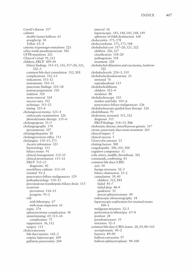
Caroli’s disease 317catheter
double-lumen balloon 61straight tip 50Teflon 63–4
cationic trypsinogen mutations 221celiac trunk pseudoaneurysm 302CFTR mutations 221Charcot’s triad 91, 111children, ERCP 309–34
biliary findings 313–15, 316, 317–20, 321,322–5
common bile duct cannulation 312, 313complications 312–13indications 311–12instruments 310–11pancreatic findings 325–34patient preparation 310sedation 310stone removal 324success rates 312technique 311–12timing 325–6
cholangiocarcinoma 123–4endoscopic examination 126photodynamic therapy 135–6
cholangiogram 31–5cholangiography 174
percutaneous 107cholangiohepatitis 92cholangiovenous reflux 111cholangitis 110–15, 373
Ascaris infestation 323bacteriology 111biliary stones 91clinical management 112–15clinical presentation 111–12ERCP 112–13
diagnostic 42nasobiliary catheter 113–14oriental 91–2pancreatico-biliary malignancies 129pathophysiology 110–11percutaneous transhepatic biliary drain 113recurrent
prevention 114–15pyogenic 91–2
riskwith lithotripsy 67with stone impaction 61
septic 374sphincterotomy complication 58stents/stenting 69, 113–14
complication 75suppurative 56, 112surgery 113
cholecystectomybile duct injuries 142–3empiric laparoscopic 209gallstone pancreatitis 204
interval 56laparoscopic 143, 144, 145, 148, 149sphincter of Oddi dysfunction 168
cholecystitis 373, 378cholecystokinin 171, 173, 184choledochal cyst 317–20, 321, 322
children 326, 327classification 318–20pathogenesis 318treatment 320
choledochal dilatation and carcinoma, fusiform322
choledochocele 224–5, 319choledochoduodenostomy 21
stenosed 76supraduodenal 115
choledocholithiasischildren 323–4incidence 88
choledochoscopy 103mother and baby 105–6pancreatico-biliary malignancies 126
choledochoscopy-guided laser therapy 126cholelithiasis 91cholestasis, neonatal 311, 312
diagnosis 315ERCP findings 314–15, 316
cholestatic disease, miscellaneous genetic 317citrate, pancreatic duct stone treatment 265clinical impact 5clinical success 5Clonorchis sinensis 33clotting factors 300coagulopathy 300, 301, 304cognitive competence 11colic artery, middle, thrombosis 301commands, confirming 83common bile duct (CBD)
axis 50benign strictures 32–3biliary obstruction 31–2cannulation 39, 40
children 312, 313failed 45–7failed deep 48–9guidewire 52precut sphincterotomy 49
endoscopic ultrasonography 18laparoscopic exploration for retained stones
100–1malignant strictures 32–3perforation in lithotripsy 67–8position 28pseudostrictures 33strictures 32–3
common bile duct (CBD) stones 28, 29, 88–110asymptomatic 90–2bacteria 89–90balloon extraction 97balloon sphincteroplasty 98–100
INDEX 407
classification 89–90clinical presentation 90–2complications 97–8diagnosis 92–4extracorporeal shock-wave lithotripsy 107extraction 96–7
after stenting 106–7giant 103–7imaging 92–4incidence 88–9laparoscopic exploration 100–1magnetic resonance cholangiogram 101management 95–7mechanical lithotripsy 63–8non-operative approach 89open surgery 107operative removal 100pathogenesis 89–90percutaneous extraction 103precut sphincterotomy 101–2prediction 100–1
risk scores 101retained 100–1sphincterotomy 56, 98–100stenting 69, 106–7symptomatic 91–2
common hepatic duct, Mirizzi’s syndrome 35communication 82–3, 383, 384competence 10–11
cognitive 11ongoing 13–15
complications of ERCP 343–5, 345–6, 347–8,379–80
cardiopulmonary 378–9changes over time 349late 380–3pancreatitis after 359–66rates 348–50, 351sedation 379
computed tomography (CT), sphincterotomy-related perforation 369
conray 280 23contrast agents 23, 364
diagnostic ERCP 39contrast studies 29cost of intervention 5cyclo-oxygenase (COX), pancreatitis severity
285cystadenocarcinoma 126cystadenoma 126cystic duct size 90cystolithiasis 320, 322
data collection for ERCP 348–9death
pancreatitis 284post-ERCP 380
degree of difficulty 11–12demerol 22diagnostic ERCP 35–52
accessories 35–6bile duct obstruction stent insertion 71biliary system anatomy 54complications 41–2procedure 37–9, 40, 41–2
diagnostic errors 380–1diathermy unit 53–4DICOM technical standards 84difficulty of ERCP 341Digestive Quality of Life instrument (DDQ-15)
343diploma in ERCP competence 14–15disconnected tail syndrome 299, 300disseminated intravascular coagulation (DIC)
284documentation
endoscopic images 84radiological images 84–5
Dormia basket 61double duct stricture sign 120–1drainage films 29droperidol, sphincter of Oddi manometry 176drug interactions, sphincter of Oddi
manometry 175–6duodenal diverticula 38
obscured papilla 45perimampullary 38, 39
sphincterotomy 55duodenoscopes
backing into correct loop 51diagnostic ERCP 35failure to insert 42–3lost in stomach 43–4side-viewing 21, 35
ERCP in Billroth II gastrectomy 110lost in stomach 43–4malignant bile duct obstruction stenting 70sphincterotomy 55
tip position 44duodenum
duplication cyst 330perforation risk with lithotripsy 68ulceration 75
electrohydraulic lithotripsy 105electrosurgical unit 53–4, 54–5endoscopes
forward-viewing 21, 110intubation for diagnostic ERCP 37–8
endoscopic image documentation 84endoscopic papillectomy risks 355endoscopic retrograde
cholangiopancreatography (ERCP)acute pancreatitis 199–231
recurrent 228–9adverse clinical events 343–5, 345–6,
347–8benefits 3bile duct stones 93bleeding after 374–7
INDEX408
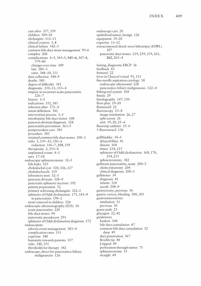
care after 357, 359children 309–34cholangitis 112–13clinical context 3, 4clinical failure 342–3common bile duct stone management 95–6complex 200complications 4–5, 343–5, 345–6, 347–8,
379–80changes over time 349late 380–3rates 348–50, 351
data collection 348–9deaths 380degree of difficulty 341diagnostic 210–11, 353–4empiric in recurrent acute pancreatitis
226–7history 1–2indications 352, 385infection after 372–4intent definition 342intervention process 3, 4intrahepatic bile duct stones 108pancreas divisum diagnosis 328pancreatitis prevention 363–5postprocedure care 385procedure 385retained common bile duct stones 100–1risks 3, 339–43, 350–6
reduction 356–7, 358, 359therapeutic 2, 353–4unplanned events 4–5uses 17–18
endoscopic sphincterotomy 52–5bile leaks 325choledochal cyst 320, 326, 327choledochocele 319laboratory tests 52–3pancreas divisum 328–9pancreatic sphincter necrosis 192patient preparation 52primary sclerosing cholangitis 322–3sphincter of Oddi dysfunction 171, 185–9
in pancreatitis 190–2stone removal in children 324
endoscopic ultrasonography (EUS) 18acute pancreatitis 228bile duct stones 94pancreatic pseudocyst 291sphincter of Oddi dysfunction diagnosis 172
endoscopistsadverse event management 383–4complication rates 351expertise 340humanity towards patients 357risks 340, 351thresholds for therapy 342
endoscopy, direct for pancreatico-biliarymalignancies 126
endoscopy cart 20epithelioid tumor, benign 126equipment 19–20expertise 11–12extracorporeal shock-wave lithotripsy (ESWL)
107pancreatic duct stones 219, 259, 259, 261,
262, 263–4
fasting, diagnostic ERCP 36feedback 83fentanyl 22fever in Charcot’s triad 91, 111fine-needle aspiration cytology 18
endoscopic ultrasound 228pancreatico-biliary malignancies 122–4
fishing reel system 104fistula 29fistulography 147, 150floor plan 19–20flumazenil 22fluoroscopy 25–8
image orientation 26, 27split screen 26unit 19, 20, 25–6
fluorotip catheter 35–65-fluorouracil 136
gallbladder 34–5delayed films 41disease 168intact 214, 215sphincter of Oddi dysfunction 168, 170,
214, 215sphincterotomy 382
gallstone pancreatitis, acute 200–5cholecystectomy 204clinical diagnosis 200–1
gallstones 34diagnosis 41infants 324occult 208–9
gastrectomy, previous 50gastric varices, bleeding 300, 301gastroenterostomy
intubation 51previous 50
gauze pads 25glucagon 22, 42guidewires
baskets 108bile duct cannulation 47common bile duct cannulation 52
deep 49duct penetration 367flexible tip 48J-tipped 49perforation through tumor 75sphincterotome 55straight 49
INDEX 409

hemosuccus pancreaticus 302, 303hepatic artery, intraductal ultrasound 126hepatic duct
common in Mirizzi’s syndrome 35stricture 133
hepatic fibrosis, congenital 317hepaticojejunostomy 21
end-to-side 115recurrent biliary strictures 158–9see also Roux-en-Y-hepaticojejunostomy
hepatitis, neonatal 313–15hepatobiliary scintigraphy 172–4hepatolithiasis see intrahepatic bile duct stoneshilar obstruction
Bismuth classification 132drainage/stenting 132–5strictures 72
hilar tumors, bilateral stenting 72–3histiocytosis X 322Hogan–Geenen sphincter of Oddi dysfunction
classification 166, 167, 170host response, pancreatitis severity 285hypaque 23
immune deficiency 322infection
after ERCP 372–4, 381biliary sepsis 42, 374cholangitis 111delayed 374nosocomial 373opportunist in AIDS in children 331pancreatic necrosis 296, 297–8
information transmission 86informed consent 384
diagnostic ERCP 36patients 356–7
intrabiliary pressurebile leaks 78raised 111
intraductal laser lithotripsy 105–6intraductal papillary mucinous tumor (IPMT)
126intraductal ultrasonography (IDUS), sphincter
of Oddi 175intrahepatic bile duct(s) 35
intraductal ultrasound 126selective cannulation 49–50
intrahepatic bile duct stones 107–9asymptomatic 90–2diagnosis 92–4imaging 92–4percutaneous transhepatic cholangioscopy
109symptomatic 91–2
intubation, diagnostic ERCP 37–8irrigation syringes 23
jaundiceCharcot’s triad 91, 111
malignant and endoscopic stenting 128–35obstructive 121–2
with biliary stones 91recurrent with stenting 75
kissing technique 46K-ras oncogene, mutational analysis 124–5
large cell lymphoma 126laser system
choledochoscopy-guided therapy 126intraductal lithotripsy 105–6tumor ablation after snare excision 137
lawsuits 384–5legal action 384–5lexicon 82–3lifetime experience 14lipid-rich meal 171, 173lithotripsy
electrohydraulic 105interval endoscopic 106–7intraductal 105–6
for pancreatic duct stones 264–5laser 105–6mechanical 63–8
basket 104–5complications 67–8procedure 65–7
lithotripsy basket 61, 63–5common bile duct stone extraction 96, 97giant CBD stones 104–5impaction 378intrahepatic bile duct stones 108mechanical lithotriptor 104–5
lithotriptor, mechanical 61liver
parenchymal disease 35resection 115transplantation in children 324–5
magnetic resonance cholangiography (MRC)bile duct stones 93, 94CBD stones 101sphincter of Oddi dysfunction 174
magnetic resonance cholangiopancreatography(MRCP) 2, 18
acute pancreatitis 223, 228bile duct transection 150children 322, 323–4pancreatico-biliary malignancies 127–8primary sclerosing cholangitis 322sphincter of Oddi dysfunction diagnosis
172stent placement 127
magnetic resonance imaging (MRI) 2magnetic resonance pancreatography (MRP)
289–90magnified view 26malignancy, pancreatico-biliary 120–38meniscus sign 29
INDEX410
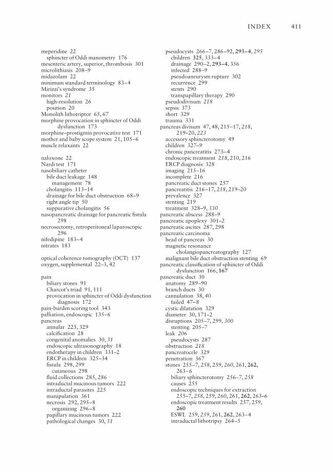
meperidine 22sphincter of Oddi manometry 176
mesenteric artery, superior, thrombosis 301microlithiasis 208–9midazolam 22minimum standard terminology 83–4Mirizzi’s syndrome 35monitors 21
high-resolution 26position 20
Monolith lithotriptor 65, 67morphine provocation in sphincter of Oddi
dysfunction 173morphine–prostigmin provocative test 171mother and baby scope system 21, 105–6muscle relaxants 22
naloxone 22Nardi test 171nasobiliary catheter
bile duct leakage 148management 78
cholangitis 113–14drainage for bile duct obstruction 68–9right angle tip 50suppurative cholangitis 56
nasopancreatic drainage for pancreatic fistula298
necrosectomy, retroperitoneal laparoscopic296
nifedipine 183–4nitrates 183
optical coherence tomography (OCT) 137oxygen, supplemental 22–3, 42
painbiliary stones 91Charcot’s triad 91, 111provocation in sphincter of Oddi dysfunction
diagnosis 172pain-burden scoring tool 343palliation, endoscopic 135–6pancreas
annular 225, 329calcification 28congenital anomalies 30, 31endoscopic ultrasonography 18endotherapy in children 331–2ERCP in children 325–34fistula 298, 299
cutaneous 298fluid collections 285, 286intraductal mucinous tumors 222intraductal parasites 225manipulation 361necrosis 292, 295–8
organizing 296–8papillary mucinous tumors 222pathological changes 30, 31
pseudocysts 266–7, 286–92, 293–4, 295children 325, 333–4drainage 290–2, 293–4, 356infected 288–9pseudoaneurysm rupture 302recurrence 299stents 290transpapillary therapy 290
pseudodivisum 218sepsis 373short 329trauma 331
pancreas divisum 47, 48, 215–17, 218,219–20, 223
accessory sphincterotomy 49children 327–9chronic pancreatitis 273–4endoscopic treatment 218, 210, 216ERCP diagnosis 328imaging 215–16incomplete 216pancreatic duct stones 257pancreatitis 216–17, 218, 219–20prevalence 327stenting 219treatment 328–9, 330
pancreatic abscess 288–9pancreatic apoplexy 301–2pancreatic ascites 287, 298pancreatic carcinoma
head of pancreas 30magnetic resonance
cholangiopancreatography 127malignant bile duct obstruction stenting 69
pancreatic classification of sphincter of Oddidysfunction 166, 167
pancreatic duct 30anatomy 289–90branch ducts 30cannulation 38, 40
failed 47–8cystic dilatation 329diameter 30, 171–2disruptions 205–7, 299, 300
stenting 205–7leak 206
pseudocysts 287obstruction 218pancreatocele 329penetration 367stones 255–7, 258, 259, 260, 261, 262,
263–6biliary sphincterotomy 256–7, 258causes 255endoscopic techniques for extraction
255–7, 258, 259, 260, 261, 262, 263–6endoscopic treatment results 257, 259,
260ESWL 259, 259, 261, 262, 263–4intraductal lithotripsy 264–5
INDEX 411
medical treatment 265obstruction 255pancreatic sphincterotomy 255, 256removal 257, 258treatment results 265–6
strictures 243, 244–5, 246–54recurrence 246
see also stents, pancreatic ductpancreatic juice 287pancreatic orifice 45pancreatic septotomy 210, 212pancreatic sphincter
hypertension 210necrosis and endoscopic sphincterotomy
192sphincter of Oddi dysfunction 168–9
pancreatic sphincterotomy 187, 188, 224chronic pancreatitis 272endoscopic 273late complications 382–3pancreatic duct stones 255, 256pancreatic fistula 298risk factors 361–2risks 354
pancreatico-biliary drainage, congenitalvariations 381
pancreatico-biliary junction, anomalous 225,318, 319, 321
children 326, 327pancreatico-biliary malignancies 120–38
brachytherapy 136choledochoscopy 126diagnosis 120–5direct endoscopic examination 126endoscopic palliation 135–6intraductal photodynamic therapy 135–6intraductal ultrasound 126–7magnetic resonance
cholangiopancreatography 127–8palliation of inoperable 128–36pancreatoscopy 126radiological diagnosis 120–1tissue diagnosis 121–4tumor markers 124–5
pancreatico-biliary medicine 2–5pancreatico-biliary system imaging 17–18pancreatico-duodenal artery 302pancreatitis
after ERCP 359–66incidence 359–60risk factors 360–3
after sphincterotomy 359APACHE scoring 282–3biliary 91, 200–5Cambridge Classification 30, 31complications 281–304
metabolic/toxic 282–5diagnostic ERCP 42, 210–11early onset 249–50hemorrhagic 304
hereditary 249–50idiopathic 207–8interstitial 298necrotizing 298
retroperitoneal bleeding 304neoplastic
obstruction 222–4, 225, 226occult 230
pancreatic endotherapy 249–50post-ERCP 365–6prevention after ERCP 363–5pseudoaneurysm 301–3
embolization 303recurrent 383
children 325–31ERCP in children 331sphincter of Oddi dysfunction 190–3
risk factors after ERCP 360–3severity classification 30, 31smoldering 207, 224
malignant 224sphincter of Oddi dysfunction 169, 209–11,
212, 213–15sphincterotomy complication 57–8stenting complication 75vascular complications 299–304see also gallstone pancreatitis, acute
pancreatitis, acutebiliary sphincterotomy 205common bile duct stone removal
complication 97–8complications 299–300ERCP 199–231, 226–7, 228–9
early 202–5investigations 227–8MRCP 223postcholecystectomy 205pseudocysts 333Ranson’s criteria 282recurrent 207–11, 212, 213–17, 218,
219–25, 226–7diagnosis 229–30endoscopic treatment 230
severity prediction 202treatment 202unexplained 225–30venous thrombosis 299–300
pancreatitis, chronic 220–2alcoholic 222biliary obstruction 267–71children 325, 331endoscopic therapy 221–2, 241–3, 331endoscopy 239–75ERCP in children 331–2intraductal stones 221, 222long-term follow-up 252medical therapy 240–1minor papilla sphincter therapy 274pancreas divisum 273–4pseudocysts 333
INDEX412
sphincter of Oddi dysfunction 271–3surgical therapy 241treatments 240–3
pancreatocele 329pancreatography 29–30, 31, 38
sphincter of Oddi 175pancreatoscopy, pancreatico-biliary
malignancies 126papilla of Vater
abnormal 45bulging 47cannulation 38–9, 40, 41
Billroth II situation 50–2cutting 58–9displaced 47endoscopic cannulation 1failure to identify 44–5functional obstruction 35guidewire 52impacted stone 56inverted 51–2landmark 38minor
cannulation 41, 218failed cannulation 48pancreas divisum 217, 218, 219sphincter therapy 274sphincteroplasty 328–9sphincterotomy 274, 330
obscured 44–5stenosis 35
late complications of sphincterotomy381–2
stone impaction 49tumor 49
papillectomy risks 355papillotome 53–4papillotomy 52–5
minor 219–20patients
communication with 384contract with 356–7early refeeding 359educational materials 357, 358empowerment 3, 5humanity towards 357information for 86, 357, 358informed consent 356–7, 384
diagnostic ERCP 36positioning 28
drainage films 29preparation for diagnostic ERCP 36risk factors for ERCP 361risks of ERCP 351–3satisfaction 5
percutaneous cholangiography 107percutaneous transhepatic biliary drain
CBD stones 102–3cholangitis 113prior Billroth II gastrectomy 110
percutaneous transhepatic cholangiogram anddrainage
bile duct transection 150CBD stones 102–3
percutaneous transhepatic cholangiography1–2, 18
bile duct strictures 151percutaneous transhepatic cholangioscopy
109perforation 365–6
endoscopic 371–2remote from papilla 371–2sphincterotomy-related 367–71
management 370–1recognition 368–9risk reduction 369surgery 371
stent migration 372peripapillary diverticulum 352personnel protection 26–8pharmacological agents, post-ERCP
prophylaxis 364photodynamic therapy, intraductal 135–6photosensitization 135–6platelet activating factor (PAF) 284pneumonia, aspiration 379polylactic acid 132portal vein
intraductal ultrasound 126thrombosis 300, 302–3
professionalism 383propofol 22
sphincter of Oddi manometry 176prostigmin 171pseudoaneurysm, pancreatitis 301–3pseudostrictures, common bile duct 33
radiation, personnel protection 26–8radiological images, documentation 84–5radiological interpretation 28–35radiologists 381radiopaque markers, biliary stricture balloon
dilatation 77radiotherapy, brachytherapy for pancreatico-
biliary malignancies 136Ranson’s criteria for acute pancreatitis 282records 83–6re-credentialing 13–15refeeding, early 359renal failure, pancreatitis 283rendezvous procedure 102renografin 23report cards 13–14reporting 83–6
errors 85speed 86
respiratory depression in diagnostic ERCP 42respiratory system in pancreatitis 283–4reticular cell carcinoma 322Reynold’s pentad 112
INDEX 413
risks of ERCP 350–6anatomical factors 352complication-specific 352–3indications 352operator-related 351patient-related 351–3procedure 353–6reduction 356–7, 358, 359
Rome criteria for sphincter of Oddi dysfunction170
room set-up 19–20Roux-en-Y-hepaticojejunostomy 150
recurrent strictures 158, 159
Santorini’s duct see accessory ductsclerosing cholangitis, primary 322–3
extrahepatic strictures 76risks of ERCP 352–3strictures 32
scout film 28–9secretin
infusion 41, 48MRCP 228sphincter of Oddi dysfunction diagnosis
171–2sedation
complications 379conscious 22–3ERCP in children 310risks 353
sedatives 22sepsis 373shock, pancreatitis 283sickle cell anemia 322simethicone 25, 45smooth muscle relaxants 22snare excision
ampullary neoplasms 136–7laser ablation of tumor 137
snare papillectomy, endoscopic 137Soehendra lithotriptor 63
complications 67giant CBD stones 104, 105
Soehendra stent retriever 243sphincter of Oddi
anatomy 210basal sphincter pressure abnormalities 179,
180dilatation 103endoscopy 174hypertonic 166hypotonic 166, 189intraductal ultrasonography 175pancreatography 175prophylactic stenting 182radiographic assessment 171–2stenosis 165, 166surgical ablation 272–3
sphincter of Oddi dysfunction 35, 165–93,330–1
acute pancreatitis 209–11, 212, 213–15after cholecystectomy 168balloon dilatation 185biliary sphincter 168–9biliary sphincterotomy 214botulinum toxin injection 189–90, 192–3chronic pancreatitis 271–3classification 166, 167, 170clinical presentation 169–70diagnosis 209, 210endoscopic sphincterotomy 171, 185–9
pancreatitis 190–2endoscopic therapy 209–10, 211, 212epidemiology 166evaluation 170–1gallbladder
disease 168intact 215
imaging 171invasive diagnostic methods 174–5laboratory tests 170–1non-invasive diagnostic methods 171–4pancreatic sphincter 168–9pancreatic sphincterotomy 187, 188pancreatitis 169
recurrent 190–3Rome criteria 170stents 185, 210, 212, 213–14surgical therapy 184TENS 184therapy 183
sphincter of Oddi manometry 166, 168, 169,171
aspirating catheter 182cannulation techniques 177, 178, 179catheters 176, 177complications 181–2drug interactions 175–6with ERCP 229normal values 180, 181pancreatic sphincter necrosis 192risks 361sedation 176technique 175–7, 178, 179–83tracing interpretation 179–80
sphincteroplastyballoon 76–7transduodenal 115
sphincterotome 47, 53–4common bile duct stone management 95–6double-lumen 54, 55failed common bile duct cannulation 49guidance 46long-nose 60orientation 61reverse 60–1rotatable wire 60side-arm adaptor 54triple-lumen 55wire length 60
INDEX414
wire-guided 55sphincterotomy
adequacy 55biliary stricture balloon dilatation 77Billroth II cases 60–1bleeding after 57, 98, 375, 376–7common bile duct stones 56, 98–100
management 96complications 57–8, 99–100cutting failure 58–9deviated cut 59–60distorted anatomy 55empiric in recurrent acute pancreatitis
226–7gallbladder in place 382half cut 59indications for 56–7late effects 381–3needle-knife technique 49pancreatitis after 359perforation 367–71
management 370–1recognition 368–9risk reduction 369surgery 371
periampullary diverticula 55precut 49
biliary stenting access 57CBD stones 101–2complications 101–2failed deep cannulation 101–2papilla stone impaction 56risks 354–5
repeat 355risk factors 361–2stone extraction 61–3wire direction 60without sphincter manometry 210–11, 213see also biliary sphincterotomy; endoscopic
sphincterotomy; pancreaticsphincterotomy
splenic artery thrombosis 300–1splenic vein thrombosis 299–300staff
expertise 340humanity towards patients 357risks 340
standard of care practice 384–5stents
balloon dilatation 78, 153–4bilateral for hilar obstruction 72–3bile duct 147–9, 152–6
access 57aggressive protocol 160–1biliary obstruction in chronic pancreatitis
267–71blocked 374complications 75damage 378insertion phase 156–7
malignant obstruction 69–72metal 159–60obstruction 69–72response assessment 74restenosis 157risk factors 362success rate 74
bile leaks 325management 78
biliary strictures 142, 152–6aggressive protocol 160–1complications 377–8phases 156–7restenosis 157
biodegradable 132biliary obstruction in chronic pancreatitis
271blockage 75CBD stones 106–7cholangiocarcinoma 124cholangitis 69, 113–14common bile duct stones 69complications 75, 131, 377–8configuration 75–6dislocation 75endoscopic insertion 128–9hilar obstruction 132–5hilar strictures 132–5introducer system 76length 76magnetic resonance
cholangiopancreatography-guidedplacement 127
malignant jaundice 128–35materials 75metal 130–1
benign biliary strictures 159–60biliary obstruction in chronic pancreatitis
270–1covered 131hilar malignancy 135uncovered 131
migration 377–8perforation 372
occlusion 130, 131one-step introducer system 72pancreas divisum 219, 330pancreatic duct 243, 244–5, 246–54, 331
brief mini-stents 254complications 247–8, 253–4, 377–8damage 378disruptions 205–7duct changes 254duration 250–1efficacy 246–50migration 253–4neoplastic obstruction 224occlusion 253outcome prediction 250pancreatic trauma 331
INDEX 415

pancreatitis prevention 364–5risk factors 362stent placement techniques 243, 244–5,
246strictures 243, 244–5, 246–54surgery outcome prediction 251–2
pancreatic fistula 298pancreatic pseudocysts 290patent 29plastic 129, 130–1
blockage 377hilar malignancy 135
response assessment 74risks 356self-expandable metal 75–6, 130, 131, 135smoldering pancreatitis 207, 224snare papillectomy 137sphincter of Oddi
dysfunction 185, 211, 213–14prophylactic 182
standard for biliary obstruction in chronicpancreatitis 267–70
success rate 74types 130–2
stomach examination, diagnostic ERCP 37–8Strecker stent 131suprapapillary fistula 45surgery
ampullary neoplasms 136cholangitis 113open for CBD stones 107recurrent cholangitis prevention 114–15risk reduction 2
systemic inflammatory response syndrome(SIRS) 284
teamwork 81–2technical failure of ERCP 340–2TENS (transcutaneous electrical nerve
stimulation) 184terminology 82–3
minimum standard 83–4therapeutic ERCP 52–78thermal ablation, ampullary neoplasms
136–7total parenteral nutrition 284
disconnected tail syndrome 299training 9–10, 12
structured 12trimethadione 265tumor markers for pancreatico-biliary
malignancies 124–5
ultrasoundabdominal for bile duct stones 92–3intraductal 126–7see also endoscopic ultrasonography (EUS)
unplanned events 345–6urografin 23
valium 22venous thrombosis, acute pancreatitis
299–300
Wallstents 130, 131worktop organization 24–5
X-ray machines, conventional 25–6settings 26
Z-stent 131
INDEX416