advanced endourologytuleoffice.com/images/editor/file/pdf/book/stone eswl pcn... · 2011-04-25 ·...
TRANSCRIPT


ADVANCED ENDOUROLOGY

CURRENT CLINICAL UROLOGYEric A. Klein, MD, SERIES EDITOR
Advanced Endourology: The Complete Clinical Guide, edited by Stephen Y. Nakadaand Margaret S. Pearle, 2006
Oral Pharmacotherapy for Male Sexual Dysfunction: A Guide to ClinicalManagement, edited by Gregory A. Broderick, 2005
Urological Emergencies, edited by Hunter Wessells and Jack W. McAninch, 2005Management of Prostate Cancer, Second Edition, edited by Eric A. Klein, 2004Essential Urology: A Guide to Clinical Practice, edited by Jeannette M. Potts, 2004Management of Benign Prostatic Hypertrophy, edited by Kevin T. McVary, 2004Laparoscopic Urologic Oncology, edited by Jeffrey A. Cadeddu, 2004Pediatric Urology, edited by John P. Gearhart, 2003Essential Urologic Laparoscopy: The Complete Clinical Guide, edited
by Stephen Y. Nakada, 2003Urologic Prostheses: The Complete Practical Guide to Devices, Their
Implantation, and Patient Follow-Up, edited by Culley C. Carson, III, 2002Male Sexual Function: A Guide to Clinical Management, edited by John J.
Mulcahy, 2001Prostate Cancer Screening, edited by Ian M. Thompson, Martin I. Resnick,
and Eric A. Klein, 2001Bladder Cancer: Current Diagnosis and Treatment, edited by Michael J. Droller, 2001Office Urology: The Clinician’s Guide, edited by Elroy D. Kursh and
James C. Ulchaker, 2001Voiding Dysfunction: Diagnosis and Treatment, edited by Rodney A. Appell, 2000Management of Prostate Cancer, edited by Eric A. Klein, 2000
ADVANCEDENDOUROLOGYTHE COMPLETE CLINICAL GUIDE
Edited by
STEPHEN Y. NAKADA, MDThe University of Wisconsin Medical SchoolMadison, WI
and
MARGARET S. PEARLE, MD, PhDThe University of Texas Southwestern Medical CenterDallas, TX
© 2006 Humana Press Inc.999 Riverview Drive, Suite 208Totowa, New Jersey 07512
humanapress.com
For additional copies, pricing for bulk purchases, and/or information about other Humana titles,contact Humana at the above address or at any of the following numbers: Tel.: 973-256-1699;Fax: 973-256-8341, E-mail: [email protected]; or visit our Website: http://humanapress.com
All rights reserved. No part of this book may be reproduced, stored in a retrieval system, or transmitted in any form orby any means, electronic, mechanical, photocopying, microfilming, recording, or otherwise without written permissionfrom the Publisher.
All articles, comments, opinions, conclusions, or recommendations are those of the author(s), and do not necessarily reflectthe views of the publisher.
Due diligence has been taken by the publishers, editors, and authors of this book to assure the accuracy of the informationpublished and to describe generally accepted practices. The contributors herein have carefully checked to ensure that thedrug selections and dosages set forth in this text are accurate and in accord with the standards accepted at the time ofpublication. Notwithstanding, as new research, changes in government regulations, and knowledge from clinical experi-ence relating to drug therapy and drug reactions constantly occurs, the reader is advised to check the product informationprovided by the manufacturer of each drug for any change in dosages or for additional warnings and contraindications.This is of utmost importance when the recommended drug herein is a new or infrequently used drug. It is the responsibilityof the treating physician to determine dosages and treatment strategies for individual patients. Further it is the responsi-bility of the health care provider to ascertain the Food and Drug Administration status of each drug or device used in theirclinical practice. The publisher, editors, and authors are not responsible for errors or omissions or for any consequencesfrom the application of the information presented in this book and make no warranty, express or implied, with respect tothe contents in this publication.
Production Editor: Amy Thau
Cover Illustration: Cover art provided by Dr. Stephen Y. Nakada, from his intraoperative photo collection.
Cover design by Patricia F. Cleary.
This publication is printed on acid-free paper. ∞ANSI Z39.48-1984 (American National Standards Institute) Permanence of Paper for Printed Library Materials.
Photocopy Authorization Policy:Authorization to photocopy items for internal or personal use, or the internal or personal use of specific clients, is grantedby Humana Press Inc., provided that the base fee of US $30.00 per copy is paid directly to the Copyright Clearance Centerat 222 Rosewood Drive, Danvers, MA 01923. For those organizations that have been granted a photocopy license fromthe CCC, a separate system of payment has been arranged and is acceptable to Humana Press Inc. The fee code for usersof the Transactional Reporting Service is: [1-58829-446-3/06 $30.00].
Printed in the United States of America. 10 9 8 7 6 5 4 3 2 1eISBN:1-59259-954-0Library of Congress Cataloging-in-Publication DataAdvanced endourology : the complete clinical guide / edited by Stephen Y. Nakada and Margaret S. Pearle. p. ; cm. -- (Current clinical urology) Includes bibliographical references and index. ISBN 1-58829-446-3 (alk. paper) 1. Endourology. I. Nakada, Stephen Y. II. Pearle, Margaret Sue. III. Series. [DNLM: 1. Urologic Diseases--surgery. 2. Endoscopy--methods. 3. Urinary Calculi--surgery. WJ 168 A2437 2005] RD572.A36 2005 617.4'60597--dc22
2005010940

Dedication
To our spouses, Deanna and Jack, who remind us that there is more to life than endourology.
v
Preface
vii
Endourology is one of the most important subspecialties in the field of urology becauseof the widespread use of endoscopy for the diagnosis and treatment of a variety of uppergenitourinary tract pathologies. Although most clinical urologists incorporate some basicendourology into their practices, complex upper tract pathology and anatomy requiremore advanced endoscopic skills and instrumentation.
Advanced Endourology: The Complete Clinical Guide is intended as a resource guidefor all aspects of clinical endourology, particularly the more advanced procedures. Thisvolume encompasses endourological applications for upper urinary tract calculi, stric-tures, and urothelial cancer. It will also serve as a comprehensive overview of availableendoscopes and instrumentation.
Advanced Endourology: The Complete Clinical Guide is unique in that most of itsindividual chapters include videos that clearly illustrate critical portions of the techniquesand provide tips and tricks from the experts. Every practicing urologist should have thisbook in his or her library, with the accompanying DVD kept near a DVD player, for quickaccess to detailed procedural instruction and immediate review of the videos.
Stephen Y. Nakada, MD
Margaret S. Pearle, MD, PhD
Contents
ix
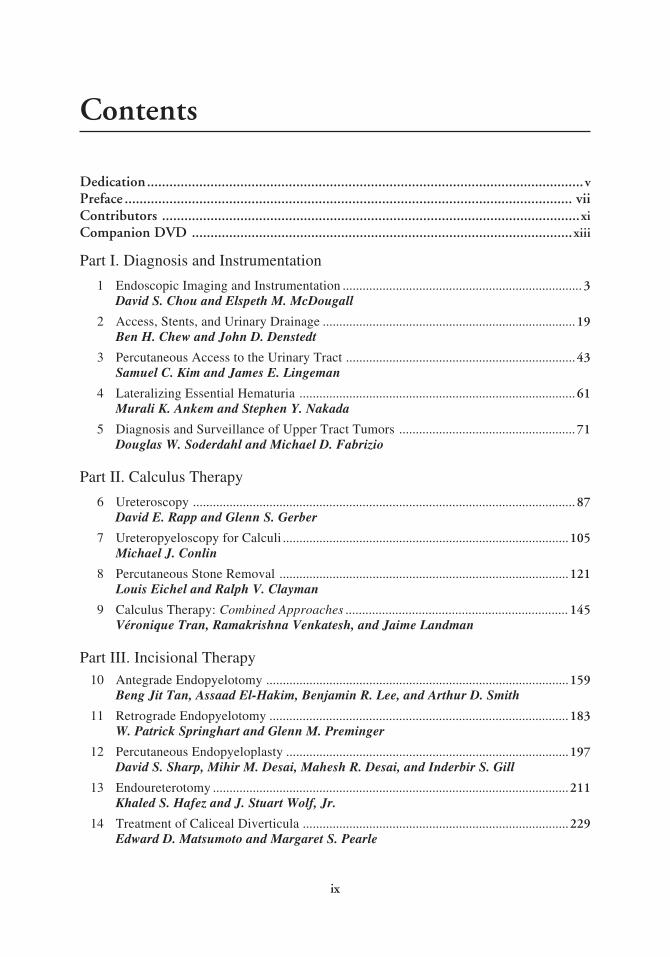
Dedication..................................................................................................................... vPreface ........................................................................................................................ viiContributors ................................................................................................................xiCompanion DVD ......................................................................................................xiii
Part I. Diagnosis and Instrumentation
1 Endoscopic Imaging and Instrumentation ........................................................................ 3David S. Chou and Elspeth M. McDougall
2 Access, Stents, and Urinary Drainage ............................................................................19Ben H. Chew and John D. Denstedt
3 Percutaneous Access to the Urinary Tract .....................................................................43Samuel C. Kim and James E. Lingeman
4 Lateralizing Essential Hematuria ...................................................................................61Murali K. Ankem and Stephen Y. Nakada
5 Diagnosis and Surveillance of Upper Tract Tumors .....................................................71Douglas W. Soderdahl and Michael D. Fabrizio
Part II. Calculus Therapy
6 Ureteroscopy ...................................................................................................................87David E. Rapp and Glenn S. Gerber
7 Ureteropyeloscopy for Calculi ......................................................................................105Michael J. Conlin
8 Percutaneous Stone Removal .......................................................................................121Louis Eichel and Ralph V. Clayman
9 Calculus Therapy: Combined Approaches ...................................................................145Véronique Tran, Ramakrishna Venkatesh, and Jaime Landman
Part III. Incisional Therapy10 Antegrade Endopyelotomy ...........................................................................................159
Beng Jit Tan, Assaad El-Hakim, Benjamin R. Lee, and Arthur D. Smith
11 Retrograde Endopyelotomy ..........................................................................................183W. Patrick Springhart and Glenn M. Preminger
12 Percutaneous Endopyeloplasty .....................................................................................197David S. Sharp, Mihir M. Desai, Mahesh R. Desai, and Inderbir S. Gill
13 Endoureterotomy ...........................................................................................................211Khaled S. Hafez and J. Stuart Wolf, Jr.
14 Treatment of Caliceal Diverticula ................................................................................229Edward D. Matsumoto and Margaret S. Pearle

x Contents
Part IV. Ablative Therapy15 Percutaneous Approach to Upper Urinary Tract Tumors ............................................253
Ioannis M. Varkarakis and Thomas W. Jarrett
16 Ureteroscopic Treatment of Upper Tract Neoplasms ..................................................267Demetrius H. Bagley
Part V. Complications of Endourology17 Complications of Percutaneous Approaches, Including Incisions ..............................283
Brian R. Matlaga, Ojas D. Shah, and Dean G. Assimos
18 Complications of Ureteroscopic Approaches, Including Incisions .............................299Farjaad M. Siddiq and Raymond J. Leveillee
Part VI. Pediatric Minimally Invasive Surgery19 Pediatric Endourology ..................................................................................................323
Christina Kim and Steven G. Docimo
Index .........................................................................................................................351

Contributors
xi
MURALI K. ANKEM, MD • Division of Urology, Department of Surgery, Universityof Wisconsin-Madison Medical School, Madison, WI
DEAN G. ASSIMOS, MD • Department of Urology, Wake Forest University Schoolof Medicine, Winston-Salem, NC
DEMETRIUS H. BAGLEY, MD • Departments of Urology and Radiology, JeffersonMedical College, Thomas Jefferson University, Philadelphia, PA
BEN H. CHEW, MSc, MD, FRCSC • St. Joseph’s Health Centre, University of WesternOntario, London, ON, Canada
DAVID S. CHOU, MD • Island Urology, Honolulu, HIRALPH V. CLAYMAN, MD • Department of Urology, University of California Irvine
Medical Center, Orange, CAMICHAEL J. CONLIN, MD • Division of Urology and Renal Transplantation, Oregon
Health and Sciences University, Portland, ORJOHN D. DENSTEDT, MD, FRCSC • St. Joseph’s Health Centre, University of Western
Ontario, London, ON, CanadaMAHESH R. DESAI, MD • Department of Urology, Muljibhai Patel Urological Hospital,
Nadiad, IndiaMIHIR M. DESAI, MD • Section of Laparoscopic and Minimally Invasive Surgery,
Glickman Urological Institute, Cleveland Clinic Foundation, Cleveland, OHSTEVEN G. DOCIMO, MD • Department of Urology, University of Pittsburgh School
of Medicine, Pittsburgh, PALOUIS EICHEL, MD • Department of Urology, University of California Irvine Medical
Center, Orange, CAMICHAEL D. FABRIZIO, MD • Department of Urology, Eastern Virginia Medical School,
Norfolk, VAGLENN S. GERBER, MD • Section of Urology, Department of Surgery, University
of Chicago Pritzker School of Medicine, Chicago, ILINDERBIR S. GILL, MD • Section of Laparoscopic and Minimally Invasive Surgery,
Glickman Urological Institute, Cleveland Clinic Foundation, Cleveland, OHKHALID S. HAFEZ, MD • Department of Urology, University of Michigan Medical School,
Ann Arbor, MIASSAAD EL-HAKIM, MD • Department of Urology, Long Island Jewish Medical Center,
New Hyde Park, NYTHOMAS W. JARRETT, MD • The James Buchanan Brady Urological Institute, The Johns
Hopkins Medical Institutions, Baltimore, MDCHRISTINA KIM, MD • Pediatric Urology, Department of Urology, University
of Pittsburgh School of Medicine, Pittsburgh, PASAMUEL C. KIM, MD • Indiana Kidney Stone Institute, Methodist Hospital Institute for
Kidney Stone Disease, and Indiana University School of Medicine, Indianapolis, INJAIME LANDMAN, MD • Division of Urology, Washington University School of Medicine,
St. Louis, MO

xii Contributors
BENJAMIN R. LEE, MD • Department of Urology, Long Island Jewish Medical Center,New Hyde Park, NY
RAYMOND J. LEVEILLEE, MD • Department of Urology, University of Miami Schoolof Medicine, Miami, FL
JAMES E. LINGEMAN, MD • Institute for Kidney Stone Disease, Methodist Hospital,and Indiana University School of Medicine, Indianapolis, IN
BRIAN R. MATLAGA, MD, MPH • Department of Urology, Wake Forest University Schoolof Medicine, Winston Salem, NC
EDWARD D. MATSUMOTO, MD, FRCSC • Division of Urology, Department of Surgery,St. Joseph’s Hospital, McMaster University, Hamilton, ON, Canada
ELSPETH M. MCDOUGALL, MD, FRCSC • Department of Urology, University of CaliforniaIrvine Medical Center, Orange, CA
STEPHEN Y. NAKADA, MD • Division of Urology, Department of Surgery, Universityof Wisconsin-Madison Medical School, Madison, WI
MARGARET S. PEARLE, MD, PhD • Department of Urology, The University of TexasSouthwestern Medical Center, Dallas, TX
GLENN M. PREMINGER, MD • Division of Urology, Duke University Medical Center,Durham, NC
DAVID E. RAPP, MD • Section of Urology, Department of Surgery, University of ChicagoPritzker School of Medicine, Chicago, IL
OJAS D. SHAH, MD • Department of Urology, New York University School of Medicine,New York, NY
DAVID S. SHARP, MD • Section of Laparoscopic and Minimally Invasive Surgery,Glickman Urological Institute, Cleveland Clinic Foundation, Cleveland, OH
FARJAARD, M. SIDDIQ, MD • Department of Urology, University of Miami Schoolof Medicine, Miami, FL
ARTHUR D. SMITH, MD • Department of Urology, Long Island Jewish Medical Center,New Hyde Park, NY
DOUGLAS W. SODERDAHL, MD • Department of Urology, Eastern Virginia MedicalSchool, Norfolk, VA
W. PATRICK SPRINGHART, MD • Division of Urology, Duke University Medical Center,Durham, NC
BENG JIT TAN, MD • Department of Urology, Long Island Jewish Medical Center, NewHyde Park, NY
VÉRONIQUE TRAN, MD • Division of Urology, Washington University School of Medicine,St. Louis, MO
IOANNIS M. VARKARAKIS, MD, PhD • The James Buchanan Brady Urological Institute,The Johns Hopkins Medical Institutions, Baltimore, MD
RAMAKRISHNA VENKATESH, MD, FRCSC • Division of Urology, Washington UniversitySchool of Medicine, St. Louis, MO
J. STUART WOLF, JR., MD • Department of Urology, University of Michigan MedicalSchool, Ann Arbor, MI

Companion DVD
xiii
The companion DVD to this volume contains video segments in support of the book,organized in sections corresponding to the book. The DVD can be played in any DVDplayer attached to a NTSC television set. The DVD may also be viewed using anycomputer with a DVD drive and DVD-compatible playback software, such as AppleDVD Player, Windows Media Player 8 or higher (Win XP), PowerDVD, or WinDVD.

I DIAGNOSIS AND INSTRUMENTATION
Endoscopic Imaging and Instrumentation
David S. Chou, MD and Elspeth M. McDougall, MD
CONTENTS
INTRODUCTION
INSTRUMENTATION
CONCLUSION
REFERENCES
1
SUMMARY
With the advancement of materials science and optics, endoscopes have undergonemajor refinements since Bozzini’s lichtleither, leading to the development of the mod-ern endoscopes. This chapter presents the basic physics and characteristics of both rigidand flexible endoscopy. Included is a discussion on video systems and the integratedoperating rooms. The future of cystoscopes, ureteroscopes, and nephroscopes for bothrigid and flexible devices is presented. In addition to presenting the present-day endo-scopes and delineating their features, this chapter includes discussions of the limitingfactors of some of these fragile instruments and future trends to look forward to. It isimportant for the urologist to have a clear understanding of the characteristics of thesehighly technical instruments in order to make appropriate choices when purchasingthese devices, and in understanding the nuances of handling them in their clinical prac-tice. In addition, discussion of care and sterilization has been presented with recentresearch data reported to help in the decision-making process of acquiring these endo-scopes and using them clinically. With the availability of a wide range of rigid, semi-rigid, flexible endoscopes, and specifically designed working instruments, most of theupper urinary tract lesions encountered in urology can be effectively diagnosed andtreated in a minimally invasive approach. Continued refinements may potentiallyimprove the optics, durability, and efficacy of these instruments as technologicaladvances are incorporated into the design of endoscopes and accessory instruments.
Key Words: Endoscope; optics; light source; ureteroscopes; video imaging system;integrated operating room; cystoscopes; nephroscopes; rigid; flexible; semi-rigid;working channel; irrigation channel; deflection; sterilization.
From: Advanced Endourology: The Complete Clinical GuideEdited by: S. Y. Nakada and M. S. Pearle © Humana Press Inc., Totowa, NJ
3

INTRODUCTION
The goal of endoscopy is to access and treat organs, through natural or artificial ori-fices in the body, with a telescope. The gradual evolution toward the modern endo-scopes began with Philipp Bozzini’s construction of the “lichtleiter” in 1806 for directinspection and treatment of the uterus and bladder (1). These early endoscopes werecumbersome and impractical, made of hollow examining tubes with illumination bycandle light directed by a mirror. With the advancement of material science and optics,endoscopes have undergone major refinements since Bozzini’s lichtleither, leading tothe development of the modern endoscopes.
Optics
The first major improvement in optics was made by Nitze in 1877 by using a series ofprecisely aligned thin lenses within a tube (1). The optical image is relayed from the distalend of the scope to the ocular lens where it can be viewed. The next breakthrough in opticsdid not occur until 1960 when Harold Hopkins developed the rod–lens system (Fig. 1) (2).A more durable and smaller diameter scope was made possible by replacing the conven-tional thin lenses with long, contoured glass rods. The rods now served as the transmissionmedium and the thin pockets interspersed between the glass rods acted as lenses. The lightreflecting off an object is detected by the objective lens at the distal tip and the image istransmitted via the rod–lens system back to the ocular lens where it is viewed by the sur-geon’s eye or captured by a camera. The rod–lens system offers better light transmission,reduced image distortion, wider viewing angle, and improved image brightness by ninefold. The size, or the degree of magnification, of the image is dependent on the diameter ofthe lenses, therefore a smaller caliber telescope, such as a ureteroscope, would have a smallerimage than a larger caliber cystoscope. Although the Hopkins lens system provides excel-lent visualization and clarity when the shaft is straight, in straight cystoscopy and nephroscopy,significant deterioration can occur when torque is placed on the scope, as during passagethrough the ureter. The lenses and air spacers may come out of alignment, and up to halfof the image may disappear, leading to a crescent field defect, or a “half-moon” appear-ance. Further stress on the shaft may lead to permanent lens damage or misalignment.Therefore, as demands for ureteroscopes increased, semirigid ureteroscopes or miniscopes
4 Chou and McDougall
Fig. 1. Traditional and Hopkins rod–lens designs.

that incorporate flexible fiberoptics within rigid shafts were designed to circumvent opticalproblems encountered during passage through a tortuous ureter.
Light Source
Throughout this period, the light source also underwent considerable modification.Trouve in 1873 moved the light source from the outside to the inner tip of the endoscopeusing a glowing hot platinum wire (1). This was later replaced by a small incandescentlight bulb. A major step toward modern endoscopy was made in the 1960s with theintroduction of fiberoptic cable that enabled the transmission of light from an outsidesource. Fiberoptic cables provided more illumination with a cool light which made cys-toscopy safer; it also made smaller profile scopes with larger irrigation and workingchannels possible. The fiberoptic cable may be built into the design of the scope, or itmay be attached via a light post to the scope.
INSTRUMENTATION
Early endoscopic procedures were limited by the lack of accessory instruments totreat disease. As the optics of rigid endoscopes underwent continuous refinement, moresophisticated accessory instruments evolved to broaden their therapeutic potentials. Thefirst true endoscopic procedure was performed by Desormeaux in 1853, extracting apapilloma from the urethra through an urethroscope. The usefulness of electrocauterywas demonstrated in 1874 when Bottini performed blind electrosurgery of the prostate.A lever was introduced by Albarran in 1897 allowing the ability to control the electrode.This was improved by Freudenber in 1900 with the addition of an endocope for visual-ization. High-frequency current was introduced by Beer in 1910 which revolutionizedthe field of therapeutic endoscopic procedures. Subsequently, the first resectoscope wasconstructed in 1926 by Stern. It was modified by McCarthy in 1931, with the additionof a lever to move the cutting loop. This basic design is still used today for modernresectosopes. Subsequently, surgeons developed different loops, catheters, and wirebaskets that could be passed through the endoscopes for the treatment of stone disease.Today, these instruments have become increasingly more powerful, with the develop-ment of ultrasonic, pneumatic, electrohydraulic, and laser lithotriptors.
Ureteroscopes
In 1912, Hugh Hampton Young performed the first ureteroscopic procedure using apediatric cystoscope in a 2-month-old child with posterior urethral valve (3). Our modernday concept of enodoscopy of the ureter and renal pelvis was made possible first byMarshall in 1960 with the advent of a 3-mm flexible fiberoscope (4). Similarly, in 1968,Takayasu and Aso developed the first flexible pelviureteroscope with an operatingchannel (5). The first rod–lens ureteroscopy was performed by Lyon to explore the dis-tal ureter with a 11-Fr pediatric cystoscope in 1977 (6). Ureteral orifice dilation was per-formed cystoscopically with Jewett sounds prior to insertion of the scope. The originalureteroscope was made by Richard Wolf Medical Instruments (Vernon Hills, IL) in 1979,modeled after a pediatric cystoscope, and was available with 13-, 14.5-, and 16-Frsheaths (7). The first practical ureteroscope was developed in 1980 and 1981 by EnriquePerez-Castro and the Karl Storz Company (Culver City, CA) (8). However, theseureteroscopes utilized the rod–lens optical system and were limited by their size and thelack of adequate instrumentation for stone fragmentation and removal. They werepurely instruments for diagnosis and not for therapeutic efficacy.
Chapter 1 / Endoscopic Imaging and Instrumentation 5
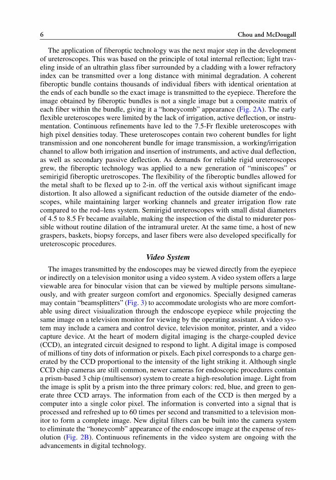
The application of fiberoptic technology was the next major step in the developmentof ureteroscopes. This was based on the principle of total internal reflection; light trav-eling inside of an ultrathin glass fiber surrounded by a cladding with a lower refractoryindex can be transmitted over a long distance with minimal degradation. A coherentfiberoptic bundle contains thousands of individual fibers with identical orientation atthe ends of each bundle so the exact image is transmitted to the eyepiece. Therefore theimage obtained by fiberoptic bundles is not a single image but a composite matrix ofeach fiber within the bundle, giving it a “honeycomb” appearance (Fig. 2A). The earlyflexible ureteroscopes were limited by the lack of irrigation, active deflection, or instru-mentation. Continuous refinements have led to the 7.5-Fr flexible ureteroscopes withhigh pixel densities today. These ureteroscopes contain two coherent bundles for lighttransmission and one noncoherent bundle for image transmission, a working/irrigationchannel to allow both irrigation and insertion of instruments, and active dual deflection,as well as secondary passive deflection. As demands for reliable rigid ureteroscopesgrew, the fiberoptic technology was applied to a new generation of “miniscopes” orsemirigid fiberoptic uretroscopes. The flexibility of the fiberoptic bundles allowed forthe metal shaft to be flexed up to 2-in. off the vertical axis without significant imagedistortion. It also allowed a significant reduction of the outside diameter of the endo-scopes, while maintaining larger working channels and greater irrigation flow ratecompared to the rod–lens system. Semirigid ureteroscopes with small distal diametersof 4.5 to 8.5 Fr became available, making the inspection of the distal to midureter pos-sible without routine dilation of the intramural ureter. At the same time, a host of newgraspers, baskets, biopsy forceps, and laser fibers were also developed specifically forureteroscopic procedures.
Video System
The images transmitted by the endoscopes may be viewed directly from the eyepieceor indirectly on a television monitor using a video system. A video system offers a largeviewable area for binocular vision that can be viewed by multiple persons simultane-ously, and with greater surgeon comfort and ergonomics. Specially designed camerasmay contain “beamsplitters” (Fig. 3) to accommodate urologists who are more comfort-able using direct visiualization through the endoscope eyepiece while projecting thesame image on a television monitor for viewing by the operating assistant. A video sys-tem may include a camera and control device, television monitor, printer, and a videocapture device. At the heart of modern digital imaging is the charge-coupled device(CCD), an integrated circuit designed to respond to light. A digital image is composedof millions of tiny dots of information or pixels. Each pixel corresponds to a charge gen-erated by the CCD proportional to the intensity of the light striking it. Although singleCCD chip cameras are still common, newer cameras for endoscopic procedures containa prism-based 3 chip (multisensor) system to create a high-resolution image. Light fromthe image is split by a prism into the three primary colors: red, blue, and green to gen-erate three CCD arrays. The information from each of the CCD is then merged by acomputer into a single color pixel. The information is converted into a signal that isprocessed and refreshed up to 60 times per second and transmitted to a television mon-itor to form a complete image. New digital filters can be built into the camera systemto eliminate the “honeycomb” appearance of the endoscope image at the expense of res-olution (Fig. 2B). Continuous refinements in the video system are ongoing with theadvancements in digital technology.
6 Chou and McDougall

Integrated Operating Rooms
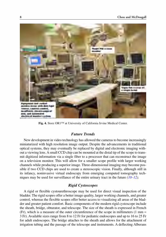
As the equipment for endourology has become more sophisticated, the trend is nowtoward integration of all operating room functions and equipment controls into one cen-tral control unit which may even have touch screen or voice control capabilities, suchas the OR 1™ system by Karl Storz or the Endoalpha™ Centralized OR system byOlympus (Melville, NY). Thus, the management of multiple complex systems can besimplified. Recent studies on surgeon fatigue and discomfort during minimally invasivesurgeries has brought attention to the ergonomics of endoscopic procedures (9). Thesurgeon’s comfort, hand–eye coordination, and visualization can be greatly improvedby using flat-screen, liquid crystal display monitors mounted on booms placed in closerange to the surgeon’s direct line of vision, the surgeon’s hands, and endoscope. Theintegrated operating room provides an efficient and ergonomic work environment forthe entire surgical team. This also provides a multidisciplinary, minimally invasive sur-gical suite. Single flat-screen monitors accommodate laparoscopic surgery, whereas thetriple flat-screen monitors, on a single boom, provide simultaneous endoscopic and flu-oroscopic visualization during endoscopy (Fig. 4).
Chapter 1 / Endoscopic Imaging and Instrumentation 7
Fig. 3. Beamsplitter camera (Karl Storz Inc, Culver City, CA).
Fig. 2. (A) Honeycomb appearance of the modern fiberopotic endoscope image. (B) The use of a dig-ital filter eliminates the honeycomb effect, but also reduces the resolution of the image.
Future Trends
New development in video technology has allowed the cameras to become increasinglyminiaturized with high resolution image output. Despite the advancements in traditionaloptical systems, they may eventually be replaced by digital and electronic imaging with-out a viewing lens. A small CCD chip can be mounted at the distal tip of the scope to trans-mit digitized information via a single fiber to a processor that can reconstruct the imageon a television monitor. This will allow for a smaller scope profile with larger workingchannels while producing a superior image. Three-dimensional imaging may become pos-sible if two CCD chips are used to create a stereoscopic vision. Finally, although still inits infancy, noninvasive virtual endoscopy from emerging computed tomography tech-niques may be used for surveillance of the entire urinary tract in the future (10–12).
Rigid Cystoscopes
A rigid or flexible cystourethroscope may be used for direct visual inspection of thebladder. The rigid scopes offer a better image quality, larger working channels, and greatercontrol, whereas the flexible scopes offer better access to visualizing all areas of the blad-der and greater patient comfort. Basic components of the modern rigid cystoscope includethe sheath, bridge, obturator, and telescope. The size of the sheath is expressed in French(Fr), which is a measure of the outer circumference of the scope in millimeters (1 mm =3 Fr). Available sizes range from 8 to 12 Fr for pediatric endoscopes and up to 16 to 25 Frfor adult endoscopes. The bridge attaches to the sheath and allows for the attachment ofirrigation tubing and the passage of the telescope and instruments. A deflecting Alberans
8 Chou and McDougall
Fig. 4. Storz OR1™ at University of California Irvine Medical Center.

bridge may be used to control deflection of flexible instruments as they pass through thedistal portion of the instrument. The obturator may be inserted into the sheath to create asmooth tip for insertion. Viewing obturators allow the zero degree telescope to be insertedto enable direct visualization for passage of the instrument into and through the urethra.The standard telescopes available are 0 (direct), 12 (operative), 25 or 30 (forward-oblique), 70 (right angle), and 110 to 120° (retrospective). The telescopes contain therod–lens system for image transmission and provide illumination via fiberoptic fibers.
Flexible Cystoscopes
The flexible cystourethroscope can also be used as a percutaneous nephroscope. Thebasic components include fiberoptic bundles, within a flexible shaft, to provide illumina-tion and image transmission to the eyepiece, and a large, 6.4 to 7.5 Fr, channel to accom-modate irrigation and ancillary instruments. The tip of the scope can be deflected in eitherdirection from 180 to 220° with a thumb control. There are a wide variety of long, flexibleinstruments that can be passed through the working channel including grasping forceps,biopsy forceps, lithotripsy and electrocautery probes, and basket entrapping devices. A newdigital cystonephroscope (Fig. 5) made by American Cytoscope Makers, Inc. (ACMI;Southborough, MA) has recently become available and is the first scope to address someof the unique demands of flexible nephroscopy. Besides the improvements in image qual-ity, the digital cystonephroscope is capable of additional flexion perpendicular to the tradi-tional up and down axis of deflection of the flexible cystoscopes. This may facilitate easieraccess of the calyceal system from the percutaneous nephrostomy tract sheath.
Semirigid Ureteroscopes
The newer generation of semirigid ureteroscopes contain fiberoptic bundles larger thanthose in a flexible ureteroscope. Therefore the image is comparable to those derived froma rod–lens system, and the “honeycomb” effect is further reduced by new fiber-packingtechniques and an advanced camera system. A straight working channel for passage of arigid instrument is possible in scopes that take advantage of the flexibility of fiberopticsand have an offset eyepiece. Most of the available semirigid ureteroscopes have round oroval tip designs, but scopes with smooth, triangular tips have recently become available,designed to ease insertion into the ureteral orifice. The shafts of these scopes are taperedsuch that they gradually enlarge from 5 to 8.5 Fr at the distal tip to 7.8 to 14.5 Fr at theproximal shaft. This design increases the proximal strength of the scope while providinga gradual dilation of the ureter as the instrument is advanced. The distal and lower middleureter in men and the renal pelvis in women may be accessed using a 31-cm ureteroscope,whereas a 40-cm ureteroscope may be needed to reach the renal pelvis in male patients.The scopes may be designed with one large channel for both instrumentation and irriga-tion, or with two channels to separate instrumentation and irrigation. A single, straight,large working channel is possible in ureteroscopes with an offset eyepiece. In contrast,two channel scopes allow passage of a working instrument without diminution in the flowof the irrigant fluid. They usually have a 3.4-Fr working channel that can accommodate astandard 3-Fr instrument and a 2.1- to 2.4-Fr irrigation channel. Some of the currentlyavailable semirigid ureteroscopes and their features are listed in Table 1.
Flexible Ureteroscopes
Several state-of-the-art flexible ureteroscopes are available with a small distal diame-ter ranging from 4.9 to 11 Fr, and a relatively large working channel up to 3.6 Fr. These
Chapter 1 / Endoscopic Imaging and Instrumentation 9

scopes all contain imaging and light transmission fiberoptic bundles, a working channel,and a deflecting mechanism. However, each may have variations in dimensions, imagetransmission, working channel size, degrees of active deflection, the deflection mechanism,and tip design depending on the manufacturer. The newer scopes have working lengthsbetween 54 and 70 cm. As in the semirigid scopes, they have tapered shaft designs withthe proximal shaft size between 5.8 and 11 Fr. The smaller tip design has greatly reducedthe need for ureteral dilation and decreased the ureteral complication rate. Some of thecurrently available flexible ureteroscopes and their specifications are listed in Table 2.
Optics
Each scope contains a coherent fiberoptic bundle for image transmission and one ortwo larger noncoherent light transmitting fiberoptic bundles. In general, two sets oflight transmission bundles provide a more even illumination and decreased shadowing.The light cord which carries light from the lightsource to the ureteroscope may beincorporated into the design of the scope, or it may be attached onto a connecting poston the scope. The former uses a continuous bundle from the lightsource to the tip ofthe scope to provide a better illumination and relatively better visibility, whereas thelatter offers the ability to replace the light cord separately if it should become dam-aged. In vitro evaluation of select, available ureteroscopes was undertaken atUniversity of California-Irvine to compare the resolution and distortion of the uretero-scopes using test targets lined with dots of varying diameters at preset distances. Theimages of the test target, viewed through the ureterscopes, were analyzed. Resolutionwas defined as the imaging system’s ability to distinguish object detail, measured inline pairs per millimeter. Distortion was defined as an optical error (aberration) in thelens that causes a difference in magnification of the object at different points in theimage. It is calculated as [(Actual distance–Predicted distance)/Predicted distance] ×100. This was expressed in terms of a percentage. These studies have demonstrated the
10 Chou and McDougall
Fig. 5. Digital Cystonephroscope (ACMI, Southborough, MA).

Tab
le 1
Spec
ific
atio
ns o
f R
igid
and
Sem
irig
id U
rete
rosc
ope
Ang
leE
yepi
ece
Wor
king
Ti
p si
ze
Mid
seg
men
t P
roxi
mal
C
hann
elof
vie
w
Mod
elde
sign
leng
th (
cm)
Tip
shap
e(F
r)si
ze (
Fr)
size
(F
r)N
o. c
hann
els
size
(F
r)(d
egre
es)
AC
MI
MR
-6/ M
R-6
LSt
raig
ht33
/41
Bev
eled
/tria
ngle
6.9
8.3
10.2
23.
4, 2
.35
MR
O-6
33/ M
RO
-642
Off
set
33/4
2B
evel
ed/tr
iang
le6.
98.
310
.22
3.4,
2.3
5M
RO
-733
Off
set
33B
evel
ed/tr
iang
le7.
79.
210
.81
5.4
5M
RO
- 74
2O
ffse
t42
Bev
eled
/tria
ngle
7.7
––10
.81
5.4
5
Oly
mpu
sA
2940
A/ 2
941A
Off
set
43/3
3O
val
6.4
––7.
81
4.2
7A
2942
AO
ffse
t43
Ova
l8.
6––
9.8
16.
67
A29
48A
/ A29
49A
Stra
ight
43/3
3O
val
6.4
––7.
81
4.2
7A
2944
AO
ffse
t43
Tri
angu
lar
7.5
––9.
02
3.4,
2.4
7A
2946
A/ A
2943
ASt
raig
ht33
/43
Tri
angu
lar
7.5
––9.
02
3.6,
2.5
7
Stor
z27
410S
K/ 2
7410
SLSt
raig
ht o
r of
fset
34/4
3T
rian
gula
r7.
59
10.5
23.
6, 2
.50
2743
0K/ 2
7430
LO
ffse
t34
/43
Ova
l8
910
.51
+2
irri
gatio
n5
027
023S
A/ 2
7023
SBSt
raig
ht34
/43
Ova
l10
1213
1 +
2 ir
riga
tion
5.5,
30
2783
0ASt
raig
ht25
Tri
angu
lar
7.5
9.0
10.5
23.
6,2.
50
Wol
f87
02.4
02/ 8
712.
402
Stra
ight
31/4
2.5
Ova
l6
7.5
––1
4.2
087
02.5
33/ 8
702.
534
Off
set
31/ 4
3O
val
67.
511
14.
20
8703
.402
/ 870
7.40
2St
raig
ht o
r of
fset
42.5
/ 31
Ova
l8
9.8
111
5.2
1087
08.5
1St
raig
ht o
r of
fset
31.5
/ 33/
43
Ova
l6.
58.
511
24.
2, 2
.55
8704
.401
/ 871
4.40
1O
ffse
t31
/ 42.
5O
val
8.5
11.5
14.5
16.
210
W87
03.5
34/ 8
703.
533
Off
set
31.5
/ 43
Ova
l8
9.8
141
5.2
1087
21.4
02/ 8
721.
401
Stra
ight
31/4
2.5
Ova
l4.
5––
––1
2.5
0
11

Tab
le 2
Man
ufac
ture
rs’ S
peci
fica
tion
s of
Fle
xibl
e U
rete
rosc
opes
Act
ive
Act
ive
prim
ary
seco
ndar
y W
orki
ngW
orki
ng
Tip
Mid
shaf
t P
roxi
mal
Tip
defl
ecti
on
defl
ecti
onD
efle
ctin
g ch
anne
lA
ngle
M
odel
leng
thdi
amet
erdi
amet
erdi
amet
erde
sign
(deg
rees
)(d
egre
es)
mec
hani
smsi
ze (
Fr)
of v
iew
Com
men
ts
AC
MI
DU
R-8
656.
759.
410
.1B
evel
ed17
0/18
0––
Bot
h3.
69
DU
R-8
E64
6.75
9.4
10.1
Bev
eled
170/
180
0/13
0B
oth
3.6
9A
UR
-765
7.2
7.4
11B
evel
ed17
0/18
0––
Bot
h3.
69
Oly
mpu
sU
RF-
P370
6.9
––8.
4Ta
pere
d18
0/18
0––
Cou
nter
-int
uitiv
e3.
60
Inte
grat
ed
Lig
htco
rd
Stor
z11
274A
AU
707.
48.
58.
9R
ound
120/
170
––In
tuiti
ve3.
60
1127
4AA
707.
48.
58.
9R
ound
170/
120
––C
ount
er-i
ntui
tive
3.6
011
274S
P70
7.4
8.5
8.9
Rou
nd12
0/17
0––
Intu
itive
3.6
0In
tegr
ated
V
ideo
head
1127
4SPU
707.
48.
58.
9R
ound
170/
120
––C
ount
er-i
ntui
tive
3.6
0In
tegr
ated
V
ideo
head
1127
8AU
165
6.7
7.5
8.4
Rou
nd27
0/27
0––
Cou
nter
-int
uitiv
e3.
60
1127
8AU
656.
77.
58.
4R
ound
270/
270
––In
tuiti
ve3.
60
Wol
f73
25.1
72/
70/4
5/20
7.5
7.5
7.5
Tape
red
130/
160
––In
tuiti
ve3.
60
7325
.152
/73
25.1
2273
30.0
72/
70/4
57.
49
9Ta
pere
d13
0/16
0––
Intu
itive
4.5
073
30.0
5273
31.0
0160
7.4
99
Tape
red
130/
160
––In
tuiti
ve3.
60
12
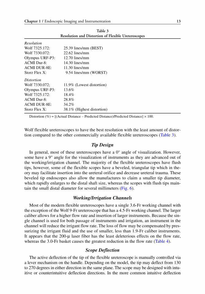
Wolf flexible ureteroscopes to have the best resolution with the least amount of distor-tion compared to the other commercially available flexible ureteroscopes (Table 3).
Tip Design
In general, most of these ureteroscopes have a 0° angle of visualization. However,some have a 9° angle for the visualization of instruments as they are advanced out ofthe working/irrigation channel. The majority of the flexible ureteroscopes have flushtips, however, some of the flexible scopes have a beveled, triangular tip which in the-ory may facilitate insertion into the ureteral orifice and decrease ureteral trauma. Thesebeveled tip endoscopes also allow the manufactures to claim a smaller tip diameter,which rapidly enlarges to the distal shaft size, whereas the scopes with flush tips main-tain the small distal diameter for several millimeters (Fig. 6).
Working/Irrigation Channels
Most of the modern flexible ureteroscopes have a single 3.6-Fr working channel withthe exception of the Wolf 9-Fr ureteroscope that has a 4.5-Fr working channel. The largercaliber allows for a higher flow rate and insertion of larger instruments. Because the sin-gle channel is used for both passage of instruments and irrigation, an instrument in thechannel will reduce the irrigant flow rate. The loss of flow may be compensated by pres-surizing the irrigant fluid and the use of smaller, less than 1.9-Fr caliber instruments.It appears that the 200-μ laser fiber has the least deleterious effects on the flow rate,whereas the 3.0-Fr basket causes the greatest reduction in the flow rate (Table 4).
Scope Deflection
The active deflection of the tip of the flexible ureteroscope is manually controlled viaa lever mechanism on the handle. Depending on the model, the tip may deflect from 130to 270 degrees in either direction in the same plane. The scope may be designed with intu-itive or counterintuitive deflection directions. In the more common intuitive deflection
Chapter 1 / Endoscopic Imaging and Instrumentation 13
Table 3Resolution and Distortion of Flexible Ureteroscopes
ResolutionWolf 7325.172: 25.39 lines/mm (BEST)Wolf 7330.072: 22.62 lines/mm Olympus URF-P3: 12.70 lines/mmACMI Dur-8: 14.30 lines/mmACMI DUR-8E: 11.30 lines/mmStorz Flex X: 9.54 lines/mm (WORST)
DistortionWolf 7330.072; 11.9% (Lowest distortion)Olympus URF-P3: 13.6%Wolf 7325.172: 18.4%ACMI Dur-8: 28.8%ACMI DUR-8E: 34.2%Storz Flex X: 38.1% (Highest distortion)
Distortion (%) = [(Actual Distance – Predicted Distance)/Predicted Distance] × 100.

scopes, the tip deflects in the same direction as the movement of the thumb lever, asopposed to counterintuitive deflection where the tip deflects in the opposite direction tothe movement of the thumb lever. Whereas most of the scopes can be deflected 120 to180° in either direction, the recently introduced “Flex-X” flexible ureteroscope (KarlStorz America Inc, Culver City, CA) can be deflected 270° in either direction (Fig. 7A).Another new ureteroscope, the “DUR-8 Elite” (ACMI Corp, Southborough, MA) incor-porates a more proximal secondary 130° one way deflection in addition to the primary170/180° up and down deflection (Fig. 7B). Besides the active deflection, flexible uretero-scopes also contain a passive deflecting segment; it is a more flexible segment of thescope that is placed several centimeters proximal to the active deflectable segment. This
14 Chou and McDougall
Table 4Irrigation Flow Rate (cc/min) at 100 mmHg and Percent Reduction With Various Instruments
ACMI Dur Stortz Olympus Wolf Wolf 8 Elite Flex-X URF-P3 7325.172 7330.072
Empty 60 56 65.5 70.5 153200-μ laser 33.3 (44.5%) 28.5 (49%) 36 (45%) 37 (47.5%) 110 (28%)400-μ laser 12 (80%) 8.5 (84%) 11 (83%) 11 (84%) 63 (58%)1.9-Fr EHL 17.5 (71%) 13.7 (75%) 18.5 (71.8%) 19 (73%) 81 (47%)
(ACMI)2.2-Fr basket 15.1 (75%) 11.5 (79.5%) 11 (83%) 14 (80.1%) 79 (48.4%)3.0-Fr basket 3.7 (94%) 2.7 (95.1%) 5 (92.3%) 4 (94.3%) 45 (70.5%)2.6-Fr grasping 4.5 (92.5%) 3.1 (94.5%) 5.5 (85.7%) 5 (93%) 53 (65.4%)
forceps(microvasive)
ACMI, Advanced Cytoscope Makers, Inc.; EHL, electrohydraulic lithotripsy.
Fig. 6. Comparison of flexible ureteroscope tip design. (A) Flush tip of Flex-X ureteroscope (KarlStorz Inc, Culver City, CA). (B) Beveled tip of Dur-8 Elite (ACMI, Southborough, MA).

passive deflecting segment, when used in consort with the active deflection, allows thescope to curl upon itself when the tip of the scope is reflected off the medial aspect of therenal pelvis for maneuvers into the lower pole infundibulum. Just as the flow rate is neg-atively impacted, the angle of active and passive deflection can also become severelyrestricted by the presence of instruments in the working channel. This effect on the angleof deflection can also be lessened with newer, smaller, and more malleable instruments.Various techniques have been described to limit the impact of instruments in the work-ing channel, including the use of an unsheathed (bare naked) nitinol basket to reduce itsdiameter (13,14). The degree of loss of deflection caused by the presence of variousinstruments were studied at University of California-Irvine Medical Center; measure-ments of deflection were made by photocopying the ureteroscopes when completelydeflected. In general, the angle of deflection was most impaired by the 365-μ laser fiber,and the least impaired by the 2.2-Fr nitinol basket. The results are shown on Table 5.
Care and Sterilization
Although these modern flexible ureteroscopes are capable of accessing the most dif-ficult areas in the upper urinary tract, they are fragile and require major repair after anaverage of 6 to 15 uses (15). Common reasons for repair are broken fiberopitc fibers,damaged working channel, and poor, or loss of, deflection. Currently, the durability andcost of maintenance is the main limiting factor against incorporation of these delicateinstruments in most general urology practices (16,17).
Rigid and semirigid ureteroscopes are considerably more durable than their flexiblecounterparts because of their outer metal casing. However, proper handling by holdingthese scopes near their eyepieces at the base while supporting the shaft should beemphasized. Cleansing with warm water and a nonabrasive detergent, as well as irriga-tion of the working channels, following each use is important. The rigid and semirigidureteroscopes can be sterilized by gas (ethylene oxide) or by soaking; some may be auto-claved. Similarly, the more fragile flexible ureteroscopes should also be cleaned initiallyby rinsing and irrigating with warm water and a nonabrasive detergent, and then steril-ized by gas or soaking. These delicate scopes are prone to damages from bending or
Chapter 1 / Endoscopic Imaging and Instrumentation 15
Fig. 7. Comparison between the Flex-X and DUR-8 Elite tip deflection mechanism. (A) Flex-X (KarlStorz Inc). (B) Dur-8 Elite, (ACMI).

Tab
le 5
Perc
ent
Loss
of
Def
lect
ion
Wit
h Va
riou
s In
stru
men
ts Oly
mpu
sA
CM
I D
UR
8 E
lite
Stor
z F
LE
X-X
UR
F-P
3W
olf
7325
.172
Wol
f 73
30.0
27
Inst
rum
ent
Dow
nU
pA
ctiv
e se
cond
ary
Act
ive
1+2
Dow
nU
pD
own
Up
Dow
nU
pD
own
Up
200-
μla
ser
fibe
r18
.3%
21.1
%22
.7%
7.1%
9.3%
12.4
%9%
11%
5.4%
11.5
%5.
1%3.
1%36
5-μ
lase
r fi
ber
46.3
%39
.5%
28%
25.9
%26
.8%
31.8
%34
.1%
43.9
%38
.8%
35.9
%33
.3%
28.1
%1.
9-Fr
EH
L9.
1%11
.2%
18.9
%6.
8%8.
5%12
.4%
14.1
%14
.2%
12%
10.7
%19
.2%
10.9
%2.
2-Fr
bas
ket
4.9%
14.5
%16
.7%
6%2.
4%9.
9%12
%9.
7%0.
7%5.
3%13
.5%
4.3%
3-Fr
bas
ket
12.2
%17
.1%
18.2
%7.
9%10
.6%
12.9
%13
.2%
14.8
%17
.4%
6.9%
14.7
%3.
1%2.
6-Fr
gra
sper
22%
20.4
%25
.8%
14.3
%15
.9%
21.9
%17
.4%
14.8
%19
.9%
12.2
%16
.7%
14.1
%
EH
L, e
lect
rohy
drau
lic li
thot
rips
y.
16

trauma to the distal tip or the eyepiece. Therefore, every effort should be made to main-tain them in a straight orientation during cleansing and use. In addition, the flexibleureteroscopes require venting during gas sterilization, either by manually opening avent near the irrigation port near the light post, or some may have an automatic,patented, Autoseal system. Liquid steriliztion may be accomplished by soaking in 2.4%glutaraldehyde (i.e., Cidex, Advanced Sterilization Products, Irvine, CA) or 35% perox-yacetic acid (i.e., Steris, Mentor, OH). Peroxyacetic acid is harsh on flexible endoscopesand has been demonstrated to be associated with higher flexible cystoscope repair costs(18). However, the durability of the flexible ureteroscopes may also be effected by thetechnique and number of personnel involved in the cleaning and maintenance ratherthan the technical demands of the procedure and the endoscopists’ technique (19). Theroutine use of newer ureteroscopic accessories such as ureteral access sheaths, nitinoldevices, and 200-μ holmium laser fibers can decrease the strain on the flexible uretero-scopes and significantly increase the longevity (17).
Rigid and Flexible Nephroscopes
The rigid nephroscopes have undergone little change since the advent of percuta-neous nephrostolithotomy. In general, they provide excellent visualization with arod–lens system and an offset eyepiece to allow passage of large, straight instrumentsfor stone fragmentation, such as the ultrasonic lithotriptor or the lithoclast. Variouslengths are available, ranging from 17.5 to 30 cm, to accommodate a variety of patientbody habitus. Sheaths range from 15 to 27 Fr in size; “mini-nephroscopes” with asmaller, 11-Fr diameter which can be used as a compact cystoscope are also available.A flexible cystoscope may be used as a nephroscope when needed. A new digital cys-tonephroscope by ACMI has been developed and offers additional flexion to the tradi-tional up/down plane to meet the demands of percutaneous nephrostolithotomy (Fig. 5).
CONCLUSION
Since the initial concept of inspecting a body cavity using a light and image trans-mitting system, significant development and advancements have been made in the fieldof urologic endoscopy. With the availability of a wide range of rigid, semirigid, flexi-ble endoscopes, and specifically designed working instruments, most upper urinarytract lesions can be effectively diagnosed and treated in a minimally invasive approach.Continued refinements to these instruments may potentially improve the optics, dura-bility and efficacy of the treatment as technological advances are incorporated into thedesign of the endoscopes and accessory instruments.
REFERENCES1. Reuter MA, Reuter HJ. The development of the cystoscope. J Urol, 1997; 159: 638–640.2. Hopkins HH. Optical principles of the endoscope. In: Endoscopy (Berci, G, ed.), Appleton-Century-
Crofts, New York, 1976, pp. 3–26.3. Young HH, Mckay RW. Congenital valvular obstruction of the prostatic urethra. Surg Gynecol
Obstet, 1929; 48: 509–512.4. Marshal VV. Fiberoptics in urology. J Urol, 1964; 91: 110–113.5. Takagi T, Go T, Takayasu N, Aso Y. A small caliber fiberscope for the visualization of the urinary
tract, biliary tract, and spinal canal. Surgery, 1968; 64: 1033–1036.6. Lyon ES, Kyker JS, Schoenberg, HW. Transurethral ureteroscopy in women: A ready addition to the
urological armamentarium. J Urol, 1978; 119: 35–38.
Chapter 1 / Endoscopic Imaging and Instrumentation 17
7. Lyon ES, Banno JJ, Schoenberg HW. Transurethral ureteroscopy in men using juvenile cystoscopyequipment. J Urol, 1979; 122: 152–155.
8. Perez-Castro EE, Martinez-Piniero JA. Transurethral ureteroscopy- a current urological procedure.Arch Esp Urol, 1980; 33: 445–448.
9. Wolf SJ, Jr., Marcovich R, Gill IS, et al. Survey of Neuromuscular injuries to the patient and surgeonduring urologic lapaoscopic surgery. Urology, 2000; 55: 831–836.
10. Takebayashi S, Hosaka M, Kubota Y, et al. Computerized tomographic ureteroscopy for diagnosingureteral tumors. J Urol, 2000; 163: 42.
11. Fielding JR, Hoyte L, Okon SA, et al. Tumor detection by virtual cystoscopy with color mapping ofbladder wall thickness. J Urol, 2002; 167: 559–562.
12. Takebayashi S, Hosaka M, Takase K, Kubota N, Kishida T, Matsubara S. Computerized tomographynephroscopic images of renal pelvic carcinoma. J Urol, 1999; 162: 315–318.
13. Monga M, Dretler SP, Landman J, Slaton JW, Conradie MC, Clayman RV. Maximizing ureteroscopedeflection: “Play it straight”. Urology, 2002; 60: 902–905.
14. Landman J, Monga M, El-Gabry EA, et al. Bare naked baskets: Ureteroscope deflection and flow char-acteristics with intact and disassembled ureteroscopic nitinol stone baskets. J Urol, 2002; 167:2377–2379.
15. Afane JS, Olweny EO, Bercowsky E, et al. Flexible ureteroscopes: A single center evaluation of thedurability and function of the new endoscopes smaller than 9 Fr. J Urol, 2000; 164: 1164–1168.
16. Landman J, Lee DI, Lee C, Monga M. Evaluation of overall costs of currently available small flexi-ble ureteroscopes. Urology, 2003; 62: 218–222.
17. Pietrow PK, Auge BK, Delvecchio FC, et al. Techniques to maximize flexible ureteroscope longetiv-ity. Urology, 2002; 60: 784–788.
18. Fuselier Jr., HA, Mason C. Liquied sterilization versus high level disinfection in the urologic office.Urology, 1997; 50: 337–340.
19. McDougall EM, Alberts G, Deal KJ, Nagy JM 3rd. Does the cleaning technique influence the dura-bility of the <9 flexible ureteroscope? J Endourol, 2001; 15: 615–618.
18 Chou and McDougall

Access, Stents, and Urinary Drainage
Ben H. Chew, MSc, MD, FRCSC
and John D. Denstedt, MD, FRCSC
CONTENTS
INTRODUCTION
ACCESS TECHNIQUES
STENTING TECHNIQUE
STENT COMFORT, INFECTION, AND ENCRUSTATION:THE ROLE OF NEW BIOMATERIALS AND COATINGS
TIPS AND TRICKS
CONCLUSION
REFERENCES
2
SUMMARY
Ureteral access is necessary in many endourological procedures includingureteroscopy and ureteral stenting. Technologies such as ureteral access sheaths, bal-loon dilators, and coaxial dilators may be helpful in facilitating ureteral access in diffi-cult cases. This chapter describes a stenting technique that relies on fluoroscopicguidance once the initial guidewire is placed and the cystoscope is removed.
Key Words: Ureter; stent; calculi; ureteroscopy; nephrostomy tube; shockwavelithotripsy.
INTRODUCTION
Ureteral stents are a mainstay in the urological armamentarium and are utilized in thetreatment of urolithiasis including postureteroscopy, preshockwave lithotripsy, and to relievesymptomatic renal colic. Routine stenting postureteroscopy and intracorporeal lithotripsy,once the standard of care, have been shown to be unnecessary following uncomplicatedureteroscopy and stone manipulation. Advances such as laser lithotripsy and smaller uretero-scopes have minimized the potential morbidity of ureteroscopy to the point that theindwelling stent has become the most morbid part of the procedure. Ureteral stents maycause considerable side effects ranging from dysuria, urgency and frequency to hematuria
From: Advanced Endourology: The Complete Clinical GuideEdited by: S. Y. Nakada and M. S. Pearle © Humana Press Inc., Totowa, NJ
19

and suprapubic pain. There is an emerging body of literature that routine stenting pos-tureteroscopy is not necessary and that the need for stenting should be determined on a caseby case basis.
Stents are also used to provide urinary drainage in nongenitourinary causes ofureteral obstruction, such as pregnancy and malignant ureteral obstruction. An alterna-tive and effective method of urinary drainage is the percutaneous nephrostomy tubewhich is easily placed in patients with significant hydronephrosis and may be evenmore successful than retrograde ureteral stenting when urinary drainage is required as aresult of obstruction of the distal ureter. Incompressible stents incorporating metal intothe stent material have been used to provide urinary drainage to patients with malignantureteral obstruction. Conversely, biodegradable stents have been developed to provideureteral drainage temporarily following an endourological procedure before degradingand being excreted in the urine, thus obviating the need for cystoscopic stent removal.Other stent advancements will see coatings, new materials, and drugs loaded directlyinto the stent material or coated on the stent surface to improve comfort and reducebiofilm formation, infection, and encrustation.
Access to the ureter is required any time closed endoscopic ureteral procedures are tobe carried out including during ureteral stenting and in association with diagnostic andtherapeutic ureteroscopy for urolithiasis. More detail will be provided in other chaptersregarding procedure specific aspects of ureteroscopy and percutaneous procedures; thischapter will focus on initially gaining retrograde access to the ureter, aspects relatedto ureteral stenting and a comparative analysis of alternative methods of urinary drainage.A brief summary of new stent technologies and biomaterials will also be presented.
Indications to Access the Ureter
Achievement of ureteral access is necessary for performing retrograde endoscopicprocedures such as ureteroscopy, or for placing a ureteral stent. Table 1 lists commonindications for ureteral stent placement.
Stones
Urolithiasis represents one of the more common reasons to insert a ureteral stent.Clinical indications for stenting include patients with intractable pain, those withinfected pyonephrosis, or patients with impaired renal function from obstruction. Inaddition, ureteral stenting is often employed as an adjunct to shockwave lithotripsy orendoscopic procedures in patients requiring surgical stone management.
Ureteral Stones: Retrograde Ureteral Stenting vs Nephrostomy Tube Drainage
Pyonephrosis with an obstructing stone requires urgent decompression using either ret-rograde ureteral stent placement or antegrade percutaneous nephrostomy tube drainage(1). Whether urinary drainage to bypass the obstruction is best accomplished via a ureteralstent or a nephrostomy tube is a subject of debate. The first randomized clinical trial tocompare these two methods in obstructed, infected patients was performed by Pearle et al.(2) in 42 patients with obstructing urolithiasis and pyonephrosis. The time to deferves-cence, length of stay in hospital, pain symptoms, and normalization of leukocytosis didnot differ between these two groups suggesting that urinary decompression by either ret-rograde ureteral stenting or antegrade percutaneous nephrostomy tube insertion are bothequally effective in treating obstructed pyonephrosis. However, patients had significantlyless fluoroscopy exposure (2.6 minutes less) when they were stented in a retrograde fashion.
20 Chew and Denstedt

A similar study was performed by Mokhmalji and colleagues (3), who also found nodifference in relief of the presenting symptoms between patients randomized tonephrostomy tube insertion and ureteral stent placement. Percutaneous nephrostomytube placement was successful in all of the 20 patients randomized to that group, butonly 80% of the 20 patients randomized to retrograde ureteral stent placement were suc-cessfully stented. Although not statistically significant, there was a trend towards animproved quality of life in the nephrostomy tube group when pain, dysuria, frequency,and hematuria were taken into consideration.
From the standpoint of infection and the requirement for urinary decompression, itappears that nephrostomy tube drainage and ureteral stents offer equal drainage of theupper urinary tract. Symptoms of pain and irritation are also similar. Placement of anephrostomy tube or ureteral stent depends on availability of good interventional radi-ologists and the urologist’s access to the cystoscopy suite or operating room. At somehospitals, the radiology suite may be more accessible than the operating room or cys-toscopy suite or vice versa. Subsequent procedures should also be taken into account.For instance, in patients who will require a percutaneous nephrolithotomy, a percuta-neous nephrostomy tube is the preferred intervention and in patients with stonesamenable to shockwave lithotripsy (SWL), a ureteral stent is often preferable. Manyvariables must be taken into account to determine whether a percutaneous nephrostomytube or ureteral stent should be placed in patients with obstructing stones.
Ureteral Stenting Effects on Ureteral Physiology and Stone Passage
Animal studies have demonstrated that ureteral stents decrease the frequency andamplitude of ureteral contraction in animals. In an animal model of ureteral stones caus-ing obstruction, ureteral dilatation was observed proximal to the obstruction in the stented
Chapter 2 / Access, Stents, and Urinary Drainage 21
Table 1Indications for Ureteric Stent Insertion
• Stones—intractable pain, infection, hydronephrosis, acute renal failure, solitary kidney• Postureteroscopy • Pretreatment (pre-SWL)
° Solitary kidney, stone >15 mm in diameter, • Steinstrasse post-SWL• Pyonephrosis (infection)• Stricture (endoureterotomy)• Trauma• Fistula• Ureteropelvic junction obstruction
° To relieve symptoms° Post endopyelotomy/pyeloplasty
• Hydronephrosis/calculi of pregnancy• Post reconstruction
° Renal transplant° Ureteroneocystotomy° Ureteroureterostomy° Cystectomy and urinary diversion
• Extrinsic ureteral obstruction
SWL, shockwave lithotripsy.

group whereas a nephrostomy tube group had no dilatation (4). Stents also impeded spon-taneous stone passage and reduced ureteral contractility when compared to the nephro-stomy group (4). This is controversial, however, as others have shown that ureteral stentsfacilitate spontaneous passage of distal ureteric stones less than 10 mm in diameter in 83%of patients studied (5). The ureter and ureteral orifice are theorized to passively dilate fromthe stent, thus facilitating stone passage. Although stents may affect ureteral peristalsis,the dilation of the ureter and orifice do facilitate spontaneous passage of smaller stones.
Stent Comfort and Quality of Life
There is an increasing awareness that stents impact patients’ quality of life. Stentsmay cause morbidity in up to 80% of patients with symptoms ranging from irritativevoiding symptoms, hematuria, flank pain, suprapubic pain, infection, and stent migra-tion to the “forgotten” encrusted stent (6–8). As a consequence of these concerns, theuse of routine stent placement is being more thoroughly considered on a case-by-casebasis when utilized as an adjunct to SWL or ureteroscopy.
In order to quantify patient morbidity from stents, Joshi et al. (7,8) have developedand validated the first questionnaire of stent symptoms, the Ureteral Stent SymptomQuestionnaire, which consists of 48 items spanning five criteria: pain, voiding symp-toms, work performance, sexual health, and overall general health. Results indicate that76% of stented patients experienced negative symptoms, 70% required analgesic, and42% had to reduce their activity by half (8). This validated tool should become a stan-dard evaluation technique of new stent technologies.
Stones: Stenting as an Adjunct to Shockwave Lithotripsy
Stenting prior to SWL is thought to preclude renal obstruction from stone fragmentsfollowing SWL (9). More recently, the prophylactic efficiency of pre-SWL stenting hasbeen called into question and is now a debated topic where some believe that stents inSWL patients not only lack efficacy to prevent renal obstruction, but may, in fact,impede the passage of stones fragments following SWL (10).
Steinstrasse, or the “street of stone” occurs with an overall rate of 3 to 6% of patientsundergoing SWL (11) and in 13 to 26% of nonstented patients with stone burdens greaterthan 25 mm in diameter (12,13). Placing stents prior to SWL in patients with stones greaterthan 20 to 30 mm in diameter significantly decreased the rate of steinstrasse to 3 to 7%(11,14–16). In patients with stones smaller than 25 mm, the rates of steinstrasse and infec-tion were unaffected by stenting (9,17–20). The reason for this latter finding is likely theresult of a significant risk decrease of steinstrasse in patients with stones less than 20 mm(21). A retrospective review by Madbouly et al. (21) has shown that there are four variablesthat are significantly correlated with an increased risk of steinstrasse: stone size greater than20 mm, stones located in the renal pelvis, a dilated renal pelvis, and shock wave energygreater than 22 kV. The risk of steinstrasse was 3.7 times less in stones smaller than 20 mmcompared with stones greater than that 20 mm. Stone location was also a factor becausedilation of the collecting system would lead to decreased amplitude of each contraction andlower intrapelvic pressures and propulsive power. Stone fragments in the ureter and anondilated renal pelvis are subjected to a higher force and rate of peristalsis which wouldlead to propulsion through the system. The risk of steinstrasse was reduced by two timesfor energies delivered at 18 to 22 kV and reduced by three times at energies of 14 to 18 kV(21). High-energy shock waves have been shown to produce larger stone fragments com-pared with more frequent lower powered shocks which result in finer stone fragments (22).
22 Chew and Denstedt

These studies suggest that ureteral stents should be placed prior to SWL for largestones (>20-mm diameter). Some studies, particularly those treating large stones withSWL, must be considered with caution as percutaneous nephrolithotomy is usually thetreatment of choice in stones greater than 20 or 25 mm. For patients with stones lessthan 20 mm who are to be treated with SWL, there is little evidence that stenting priorto SWL reduces the rate of steinstrasse or infection.
Stones: Stenting Postureteroscopy
The principle of avoiding ureteral obstruction secondary to ureteral edema and stonefragments is the main driving force for routinely leaving a stent post ureteroscopy andhas traditionally been regarded as the standard of care. Technical advances includingminiaturization of ureteroscopes, utilization of the holmium:YAG laser, and softer stonebaskets have made ureteroscopy relatively atraumatic and the main morbidity follow-ing ureteroscopy originates from the use of ureteric stents. Furthermore, stents add tothe cost of patient care and require an additional cystoscopy for removal unless a pullstring is used. Reducing stent use following ureteroscopy should improve patient careand satisfaction (see Table 2).
Hosking (23) was the first to report a large series of nonstented ureteroscopy patientswho had minimal complications. Approximately half of the patients had no discomfortand the majority of those with discomfort described it as mild and easily resolved byoral analgesics. Although this report was a case series and did not have a stented con-trol group, it was the first series to suggest that ureteroscopy did not routinely requirestenting. Denstedt et al. (24) randomized 58 patients to receive either a stent or nostent after ureteroscopy. The results demonstrate that there were no differences inrehospitalization rate, analgesic use or stone free rates. At 1 week, the stented group hadsignificantly more pain and irritative voiding symptoms than the nonstented group.None of these patients underwent ureteral dilation, the holmium:YAG laser was used forintracorporeal lithotripsy, and all stones were less than 2 cm. The holmium:YAG laseris safe and has minimal effects on surrounding tissue which makes it an ideal modalityto preclude the need for a stent postoperatively (25). In addition, a randomized studyusing intracorporeal electrohydraulic lithotripsy also demonstrated that these patientscan be safely left unstented (26). Other randomized studies have found similar resultssuggesting that following uncomplicated ureteroscopy without ureteric dilation, stent-ing is not routinely required (24,26–28). Even in patients who underwent ureteral dila-tion at the time of ureteroscopy, nonstented patients had results and complication ratessimilar to stented patients (23,29). In the series by Hosking and associates (23), ureteraldilation was performed in 88% of patients who suffered minimal complications post-operatively. Borboroglu et al. (29) performed a study in 107 patients, which alsoincluded 83 patients who underwent ureteral balloon dilatation, and found that stentedpatients had more pain and analgesic requirements, but no difference in stone free orrehospitalization rates. These studies provide evidence that stenting after uncompli-cated ureteroscopy is not routinely necessary, but rather should be determined on acase-by-case basis.
Hydronephrosis/Calculi in Pregnancy
Upwards of 90% of women have hydronephrosis by the third trimester of pregnancy(30), but only 0.2–25% will become symptomatic and require medical attention
Chapter 2 / Access, Stents, and Urinary Drainage 23

Tab
le 2
Sum
mar
y of
Ste
nt v
s N
o-St
ent
Tria
ls F
ollo
win
g U
rete
rosc
opy
Sign
ifica
nt
diffe
renc
e in
Sign
ifica
nt d
iffer
ence
Num
ber
of
maj
or c
ompl
icat
ions
?in
min
or c
ompl
icat
ions
?pa
tien
ts
Bal
loon
D
iffer
ence
M
etho
d of
(f
ever
,in
min
or c
ompl
icat
ions
?st
ente
d/no
t di
lati
on o
f in
sto
ne-f
ree
intr
acor
pore
al
reho
spit
aliz
atio
n,(p
ain,
irri
tati
ve v
oidi
ngA
utho
rs (
ref)
Year
sten
ted
uret
er?
rate
s?
lith
otri
psy
obst
ruct
ion)
sym
ptom
s)
Hos
king
et a
l. (2
3)19
990/
93Y
es 8
2/93
N
oE
lect
rohy
drau
licN
/A85
% h
ad n
o pa
in o
r (1
5-Fr
bal
loon
)(3
pts
.) P
neum
atic
pa
in c
ontr
olle
d by
litho
trip
sy (
17 p
ts.)
or
al a
nalg
esic
sB
aske
t ext
ract
ion
(73
pts.
)D
enst
edt e
t al.
(24)
2001
29/2
9N
oN
oH
O:Y
AG
Las
erN
oM
ore
irri
tativ
e sy
mpt
oms
in s
tent
ed p
atie
nts
Che
n et
al.
(26)
2002
30/3
0N
oN
oE
lect
rohy
drau
licN
oN
oH
olle
nbec
k 20
0151
/51
Yes
No
HO
:YA
G L
aser
No
Mor
e pa
in in
ste
nted
et a
l. (2
7)(1
5-Fr
bal
loon
)pa
tient
sB
yrne
et a
l. (2
8)20
0238
/22
No
No
HO
:YA
G L
aser
No
Supr
apub
ic d
isco
mfo
rt
Pneu
mat
icon
PO
D n
o. 1
was
wor
seL
ithot
ript
erin
non
-ste
nted
pat
ient
s,
but b
y PO
D n
o. 6
, was
wor
se in
ste
nted
pat
ient
s.B
orbo
rogl
u 20
0153
/54
Yes
No
HO
:YA
G L
aser
No
Sten
ted
patie
nts
has
et a
l. (2
9)83
/107
Ele
ctro
hydr
aulic
sign
ific
antly
mor
e fl
ank
(15-
or
and
blad
der
pain
, uri
nary
18-F
r ba
lloon
)sy
mpt
oms
and
requ
ired
mor
e na
rcot
ic a
nalg
esia
.N
etto
et a
l. (2
9a)
2001
133/
162
No
No
Bas
ket e
xtra
ctio
nN
oN
oU
ltras
onic
lith
otri
psy
HO
:YA
G, h
olm
ium
: YA
G.
24

Chapter 2 / Access, Stents, and Urinary Drainage 25
(30–32). The vast majority of patients respond to conservative treatment (70–93%) andfew will require ureteral stenting or nephrostomy tube insertion. Indications for stentinginclude: rising creatinine, pyelonephritis (febrile infection), and intractable pain (30–32).Although ultrasonography may be used to confirm the position of the stent during the pro-cedure (33–36), limited fluoroscopy, which most urologists are more familiar with, can beused safely and effectively, especially in the later stages of pregnancy (37,38). Shielding ofthe uterus and brief pulses of fluoroscopy should minimize the risks of radiation, but fluo-roscopy should be avoided during the early stages of pregnancy (37,39).
The incidence of urolithiasis ranges from 1 in 200 to 1 in 2500 pregnancies (40). Themajority of calculi presenting during pregnancy will pass spontaneously with conserva-tive management (41–44). If an obstructing calculus fails spontaneous passage, theoptions are to decompress the kidney and treat the stone after delivery, or to definitivelytreat the stone during pregnancy. Prolonged indwelling stents or nephrostomy tubes maylead to encrustation, biofilm formation, and infection as pregnant women have physio-logic hyperuricosuria and hypercalciuria (45,46); therefore, some studies suggest thatureteral stents should only be placed after 22 weeks gestational age to avoid the need formultiple stent changes (44). In pregnant women less than 22 weeks gestational age, a per-cutaneous nephrostomy tube can be inserted and changed multiple times with relative ease(44,47). If conservative management fails, ureteroscopy and intracorporeal lithotripsy is areasonable treatment option and have proven to be safe in the treatment of urinary calculiin pregnant patients (41,46,48–50). Utilization of intracorporeal methods of lithotripsy,such as the pulsed-dye laser, pneumatic lithotripsy, and the holmium: YAG laser, havebeen shown to be safe with success rates greater than 90% (38,41,44,46,49). Advances inanesthesia and ureteroscopic equipment have made intracorporeal lithotripsy safe andeffective in pregnancy when conservative management fails.
Stenting Postureteropelvic Junction Obstruction Reconstruction
Endopyelotomy for ureteropelvic junction obstruction was initially described byWickham and Kellett in 1983 (51). A standard procedure following endopyelotomy is toleave a tapered 14/7-Fr endopyelotomy stent to traverse and splint the incised uretero-pelvic junction. The size of endopyelotomy stent remains controversial with one studysuggesting that a larger diameter stent (27 Fr) improves results at 2 years (52), whereasother studies in animal models find no difference between 7 and 12–14-Fr stents (53,54).Stent indwelling time is also controversial: in two studies comparing 2 vs 4 weeks ofstenting postendopyelotomy, 2 weeks were shown to have similar results to 4 weeks(55,56). Patency rates were similar between 2 and 4 weeks of stenting (92 vs 90%,respectively) and patients stented for the longer period of time had significantly higherrates of infection (56). There is also further evidence in an animal model that a longerduration of stenting results in more ureteral fibrosis and thus, a higher rate of failure (57).
These studies demonstrate that 2 and 4 weeks of stenting with a 14/7-Fr endopyelo-tomy stent result in the same success rates following endopyelotomy. Likewise, theideal stent diameter is still a matter of debate as sizes from 7- to 27-Fr have been shownto produce equal results.
Malignant Ureteric Obstruction
Malignant extramural compression of the ureter causing hydronephrosis and renal com-promise may be a consequence of many nongenitourinary cancers. When faced with this

situation, the urologist must decide if the patient needs decompression and if so, whether itis urgent and if a stent or antegrade nephrostomy tube should be placed (58). If a stent is tobe placed, what type of stent should it be and how often should it be changed? In the deci-sion algorithm, the patient’s entire clinical picture must be taken into account including theoverall prognosis, symptoms such as flank pain, presence of infection, renal function, andintention for further treatment, such as chemotherapy (59). For instance, a terminally illpatient with bilateral hydronephrosis who is asymptomatic and free of infection may onlysuffer from the addition of urinary drainage tubes (60). The symptomatic patient (infection,flank pain, fluid overload from renal failure) should be diverted. Patients with renal com-promise from obstruction and who are about to undergo chemotherapy (palliative or cura-tive) should have their renal function optimized by urinary drainage.
Park et al. (58) reported on patients who initially had bilateral ureteral stents that failedto lower serum creatinine or relieve ureteral obstruction and subsequently required percu-taneous nephrostomy tube insertion. They suggested that percutaneous nephrostomy tubesare advantageous over ureteral stents in relieving malignant ureteral obstruction and loweringserum creatinine (58). Pappas et al. (61) evaluated 206 patients with malignant ureteralobstruction treated with percutaneous nephrostomy tubes and found that it was a safe andeffective procedure that returned normal renal function to 66% of obstructed patients. Onetheory of why nephrostomy tubes are more efficient at relieving obstruction is that becauseurine drains around a stent rather than through the lumen, extraluminal compression fromcancer prevents ureteral peristalsis and precludes peristent urinary drainage (58,62). Lastly,because stents often cause significant bladder and flank symptoms, nephrostomy tubes mayoffer a better quality of life than stents in cancer patients (60).
The percentage of successful retrograde stent placements is lower than nephrostomytube insertion which is nearly always successful in a dilated system (3,63). With verydistal ureteral obstruction owing to advanced pelvic malignancies, retrograde stentingmay be difficult because of the lack of “purchase” required to advance a guidewire orstent up the ureter (3,63).
Recently, a third method of diversion involving a silicone polytetrafluorethylenecoated tube that connects the renal pelvis to the bladder via a tunneled subcutaneousroute has been described (64–66). Metal, noncollapsible stents have also been attemptedin malignant ureteric obstruction, but the main limiting factors have been blockage ofthe stent with hyperplastic tissue and infection (67–70).
Percutaneous nephrostomy tubes offer easy placement, exchange, and good drainageof the upper urinary tract in this difficult group of patients (71). Improvements in stentmaterials and technology will increase the use of indwelling ureteral stents in managingmalignant ureteral obstruction (72).
ACCESS TECHNIQUES
Ureteral Access: Step 1—The Urethra
Retrograde approaches to the urinary tract begin at the urethra and face the potentialchallenges that are encountered in the lower urinary tract such as meatal stenosis, urethralstricture, false passage, prostatic hyperplasia, and priapism. Good urological principlesguide the management of each situation: meatotomy for stenosis, visual internal urethro-tomy or dilation for strictures, insertion of a safety guidewire to circumvent false pas-sages, use of flexible cystoscopy as an adjunct when an enlarged prostate is encountered,and intracorporal α-agonist injection for priapism.
26 Chew and Denstedt
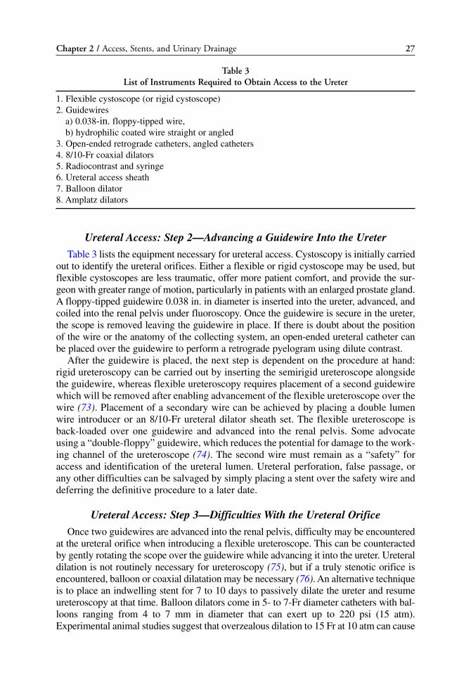
Chapter 2 / Access, Stents, and Urinary Drainage 27
Ureteral Access: Step 2—Advancing a Guidewire Into the Ureter
Table 3 lists the equipment necessary for ureteral access. Cystoscopy is initially carriedout to identify the ureteral orifices. Either a flexible or rigid cystoscope may be used, butflexible cystoscopes are less traumatic, offer more patient comfort, and provide the sur-geon with greater range of motion, particularly in patients with an enlarged prostate gland.A floppy-tipped guidewire 0.038 in. in diameter is inserted into the ureter, advanced, andcoiled into the renal pelvis under fluoroscopy. Once the guidewire is secure in the ureter,the scope is removed leaving the guidewire in place. If there is doubt about the positionof the wire or the anatomy of the collecting system, an open-ended ureteral catheter canbe placed over the guidewire to perform a retrograde pyelogram using dilute contrast.
After the guidewire is placed, the next step is dependent on the procedure at hand:rigid ureteroscopy can be carried out by inserting the semirigid ureteroscope alongsidethe guidewire, whereas flexible ureteroscopy requires placement of a second guidewirewhich will be removed after enabling advancement of the flexible ureteroscope over thewire (73). Placement of a secondary wire can be achieved by placing a double lumenwire introducer or an 8/10-Fr ureteral dilator sheath set. The flexible ureteroscope isback-loaded over one guidewire and advanced into the renal pelvis. Some advocateusing a “double-floppy” guidewire, which reduces the potential for damage to the work-ing channel of the ureteroscope (74). The second wire must remain as a “safety” foraccess and identification of the ureteral lumen. Ureteral perforation, false passage, orany other difficulties can be salvaged by simply placing a stent over the safety wire anddeferring the definitive procedure to a later date.
Ureteral Access: Step 3—Difficulties With the Ureteral Orifice
Once two guidewires are advanced into the renal pelvis, difficulty may be encounteredat the ureteral orifice when introducing a flexible ureteroscope. This can be counteractedby gently rotating the scope over the guidewire while advancing it into the ureter. Ureteraldilation is not routinely necessary for ureteroscopy (75), but if a truly stenotic orifice isencountered, balloon or coaxial dilatation may be necessary (76). An alternative techniqueis to place an indwelling stent for 7 to 10 days to passively dilate the ureter and resumeureteroscopy at that time. Balloon dilators come in 5- to 7-Fr diameter catheters with bal-loons ranging from 4 to 7 mm in diameter that can exert up to 220 psi (15 atm).Experimental animal studies suggest that overzealous dilation to 15 Fr at 10 atm can cause
Table 3List of Instruments Required to Obtain Access to the Ureter
1. Flexible cystoscope (or rigid cystoscope)2. Guidewires
a) 0.038-in. floppy-tipped wire, b) hydrophilic coated wire straight or angled
3. Open-ended retrograde catheters, angled catheters4. 8/10-Fr coaxial dilators5. Radiocontrast and syringe6. Ureteral access sheath 7. Balloon dilator 8. Amplatz dilators

ureteral aperistalsis, vesicoureteric reflux, increased pressure and hydronephrosis proxi-mal to the area of dilation, and diminished ureteral contractility (77,78). Only after 6 to 7weeks of dilatation did the ureteral physiology and histology return to normal in these ani-mals (77–79). The safety and efficacy of balloon dilators in ureteroscopy have been con-firmed in humans and are in routine use (80,81). Sequential polyethylene coaxial dilatorsrange from 6 Fr and up and are more cost effective than balloon dilators (82). Care mustbe taken not to damage the urethra, ureteral orifice, or ureteral lumen. Applying the cor-rect amount of tension to the guidewire while advancing the dilators will reduce the
28 Chew and Denstedt
Fig. 1. Materials required for ureteral stenting. (From top to bottom) Floppy tipped guide wire, Double-J stent (curl magnified), metal-tipped stent pusher (radiopaque metal tip magnified), 8/10 coaxialsheath dilator set.
Fig. 2. Double-J stent. (Curl magnified) Sideholes aid in drainage and the black mark on either endof the stent facilitate visualization of the curl when placing a stent visually through the cystoscope.

Chapter 2 / Access, Stents, and Urinary Drainage 29
likelihood of ureteral or guidewire damage. Shearing forces can damage the ureter orguidewire, which will either prevent advancement of the ureteroscope or damage theworking channel during advancement of a flexible ureteroscope. If resistance is met dur-ing scope advancement over the guidewire, the scope should be removed and the
Fig. 3. Table-top and corresponding radiograph of the 8/10 coaxial dilator set. After advancement ofthe guidewire into the renal pelvis, the cystoscope is removed and the remainder of the procedure isperformed under fluoroscopy. The 8/10 dilator set is advanced over the guidewire. The double arrowscorrespond to the end of the 10-Fr sheath. The single arrow delineates the end of the 8-Fr dilatorwhich is almost in the renal pelvis. The 10-Fr sheath is advanced up to the hub of the patient’s ure-thral meatus and the proximal end reaches the midureter in this case since the patient is female. Inmales, the 10-Fr sheath reaches just above the iliac vessels.

guidewire should be replaced through a ureteral catheter. Although balloon dilators aremore expensive, they are less traumatic to the ureter than coaxial dilators.
Ureteral Access Sheath
Ureteral access sheaths were first developed in the 1970s to aid in difficult access toureters for ureteroscopy (83). The peel-away sheath became used in the 1980s whichrequired sequential rigid dilators and several steps before the ureteroscope could beinserted, but was associated with a high rate of ureteral perforation (15–30%) (84).Today’s access sheaths consist of a two-piece hydrophilic, lubricious outer sheath andinner introducer which is removed after advancement over the guidewire. Sheaths comein various lengths (20–55 cm) and diameters (10–16 Fr) depending on patient size andgender. The access sheath acts as a dilator and a conduit that prevents buckling of the
30 Chew and Denstedt
Fig. 4. The 8-Fr dilator has been removed and the stent is being advanced over the guidewire into the10-Fr sheath. The 10-Fr sheath acts as a conduit to prevent buckling of the stent in the urethra, blad-der, or ureter. Tension must be placed on the guidewire while the stent is advanced.

Chapter 2 / Access, Stents, and Urinary Drainage 31
flexible ureteroscope within the bladder. Operating room times and costs are alsodecreased by use of the access sheath (85).
With these devices, the flexible ureteroscope is not inserted over a guidewire, but isadvanced directly up the lumen of the access sheath. Ureteral access sheaths offer theadvantages of better flow of irrigation, and thus visualization, concomitant intra-operativedrainage of the bladder, and ease of access for repeated removal and reinsertion ofthe flexible ureteroscope (74). This last benefit is particularly useful if basketing ofmultiple stones is desired. At the end of the procedure, the access sheath can facilitatethe insertion of a ureteral stent if necessary (86).
Pressure on the tip of ureteroscopes may be partially responsible for damage to thefibreoptics resulting in costly scope repair. The use of access sheaths has been shown toprevent and delay scope damage by reducing the stress on the tip of the scope duringadvancement, as well as preventing damage to the working channel by obviating theneed for advancement over a guidewire (87). One theoretical complication of accesssheaths is prolonged pressure on the ureteral wall and ischemia resulting in a ureteralstricture. However, this has not been substantiated and the stricture rate is low asdemonstrated in a retrospective review by Delvecchio et al. (87a) where only 1 of 71patients developed a stricture.
Ureteral access sheaths have been shown to be a safe method for obtaining ureteralaccess for ureteroscopy with a low rate of stricture or ureteral perforation. Furthermore,operating room times and such postoperative symptoms as frequency, urgency, dysuria,
Fig. 5. The stent is advanced into the 10-Fr sheath as far as possible and the metal-tipped stent pusheris then advanced over the guidewire to push the stent.

and hematuria were significantly less in patients who were randomized to undergoureteroscopy with a ureteral access sheath. Surgeon frustration is also diminished asmultiple withdrawals and introduction of the scope can be performed easily and opera-tive visualization is improved with the higher flow of irrigation through the sheath.
STENTING TECHNIQUE
The technique of stenting is outlined in Figs. 1 to 11. After a guidewire is placed intothe renal pelvis, the cystoscope is removed and the procedure is visualized using onlyfluoroscopy, a technique used by radiologists. Instead of a single view of the inside of thebladder using cystoscopy, fluoroscopy allows the urologist to monitor both the distal andproximal ends of the guidewire and stent during the procedure. This technique is alsomore comfortable for the patient if they are only under light sedation because the cysto-scope has been removed. An 8/10-Fr dilator is placed over the guidewire into the ureterby first advancing the 8-Fr portion. Once inside the ureter, the 10-Fr sheath is advancedover the 8-Fr portion into the ureter and confirmed by fluoroscopy. The 10-Fr sheath isadvanced so that the hub is at the level of the urethral meatus. The 8-Fr dilator is removed
32 Chew and Denstedt
Fig. 6. As the stent is advanced, it will exit the 10-Fr sheath and enter into the renal pelvis. Pulling theguidewire slightly back will result in the curling of the proximal part of the stent.

Chapter 2 / Access, Stents, and Urinary Drainage 33
leaving the 10-Fr sheath which acts as a conduit for the stent and prevents buckling orcoiling of the stent within the urethra, bladder, or ureter. The stent is advanced throughthe 10-Fr sheath and the pusher is advanced until its radiopaque marker is at the lowerlevel of the pubic symphysis in females, and midway between the upper and lower levelof the symphysis in males. The kidney is viewed on fluoroscopy and the guidewire isslightly retracted until the upper loop is seen to curl in the renal pelvis. With fluo-roscopy on the pubic symphysis, the pusher is held with the radiopaque marker at thecorrect level and the 10-Fr sheath is withdrawn from the urethra. As the guidewire isremoved the lower loop of the stent will curl in the bladder; however, if it remains inthe urethra, it can be advanced by inserting a foley catheter or applying manual supra-pubic pressure to the bladder which will displace the bladder cephalad and pull the stentinto the bladder. This technique of stenting is demonstrated in the companion DVD, thataccompanies this volume and a similar technique utilizing a ureteral access sheath hasalso been described (86).
A potential complication of this technique of ureteral stenting is inadvertentadvancement of the distal end of the stent into the ureter. The best way to avoid this
Fig. 7. Fluoroscopy is used to follow the stent into the renal pelvis. Once within the renal pelvis, theguidewire is withdrawn slightly so that the upper stent curls in the renal pelvis.

34 Chew and Denstedt
complication is to prevent it by ensuring that the radiopaque marker on the stent pusherduring the procedure does not go above the bottom of the pubic symphysis in femalesor above the middle of the symphysis in males. If a stent is advanced too proximal intothe ureter, cystoscopy should be carried out and a grasper should be used to pull thestent into the bladder if the end of the stent is visible in the bladder. If the tether is stillattached to the stent, this can be used to pull the stent back into the bladder. If the stentis well within the ureter, a guidewire should be advanced into the ureter, and a semirigidureteroscope inserted to attempt removal of the stent using a stone basket or graspers.An alternative method is to place a ureteral dilating balloon alongside the stent, partiallyinflate it and deflate it causing the stent to adhere to the deflated balloon. The stent isremoved as the deflated balloon is slowly withdrawn under close fluoroscopic observa-tion taking special care to avoid ureteral avulsion or damage to the ureteral orifice(88,89). These maneuvers are best performed with the patient under neuroleptic or gen-eral anesthesia. There are also reports that stents may migrate distally or even retro-gradely into the kidney (88,90,91).
Fig. 8. Radiographically, the metal-tipped pusher is at the correct level of the symphysis for afemale patient and the 10-Fr sheath has been withdrawn. The metal-tipped pusher is positioned atthe lower border of the pubic symphysis in women and at the level of the mid pubic symphysis inmen. Once the metal-tipped pusher is in the correct position, the 10-Fr sheath is backed-up over theguidewire.

Chapter 2 / Access, Stents, and Urinary Drainage 35
STENT COMFORT, INFECTION, AND ENCRUSTATION: THE ROLE OF NEW BIOMATERIALS AND COATINGS
Ureteral stents may cause considerable morbidity, thus limiting their clinical tolerabil-ity and effectiveness (6). It is only recently that a validated questionnaire to examine themorbidity of stents has been developed and showed that stented patients suffer substantialmorbidity (7,8). Without question, the major obstacles that limit stent use are the fact thatthey are uncomfortable, may cause infection, and provide a surface for crystals to bind andaggregate. The use of new biomaterials and stent technology are reviewed in detailelsewhere and highlight the recent advances in stent technology to improve stent comfortand decrease encrustation and infection rates (92–96).
Risk factors for stent-associated infection include female sex, diabetes, chronic renalfailure, and indwelling stent time greater than 90 days (97). Oral antibiotics are oftenadministered after stent insertion and have been found to prevent or delay both biofilmformation and infection (98). Oral ciprofloxacin has been found to adhere to a ureteralstent at high enough concentrations to inhibit bacterial growth (98). Even 2 to 3 days oforal antibiotics following stent insertion has been shown to delay biofilm formation andurine infection for up to 2 weeks (99).
Fig. 9. The pusher is held in place at the correct level with the radiopaque marker (arrow) at the pubicsymphysis.
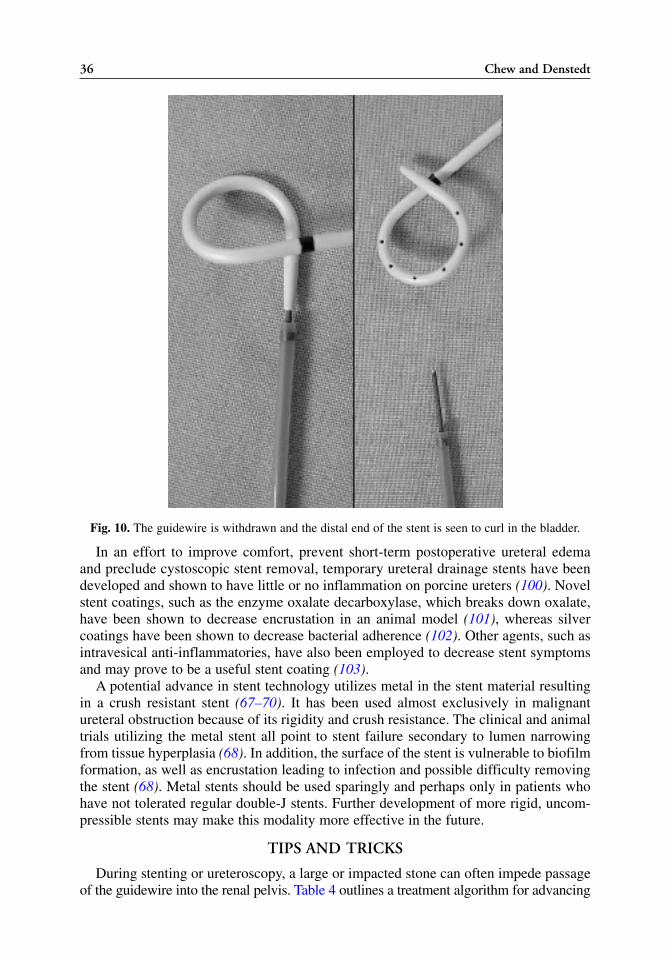
In an effort to improve comfort, prevent short-term postoperative ureteral edemaand preclude cystoscopic stent removal, temporary ureteral drainage stents have beendeveloped and shown to have little or no inflammation on porcine ureters (100). Novelstent coatings, such as the enzyme oxalate decarboxylase, which breaks down oxalate,have been shown to decrease encrustation in an animal model (101), whereas silvercoatings have been shown to decrease bacterial adherence (102). Other agents, such asintravesical anti-inflammatories, have also been employed to decrease stent symptomsand may prove to be a useful stent coating (103).
A potential advance in stent technology utilizes metal in the stent material resultingin a crush resistant stent (67–70). It has been used almost exclusively in malignantureteral obstruction because of its rigidity and crush resistance. The clinical and animaltrials utilizing the metal stent all point to stent failure secondary to lumen narrowingfrom tissue hyperplasia (68). In addition, the surface of the stent is vulnerable to biofilmformation, as well as encrustation leading to infection and possible difficulty removingthe stent (68). Metal stents should be used sparingly and perhaps only in patients whohave not tolerated regular double-J stents. Further development of more rigid, uncom-pressible stents may make this modality more effective in the future.
TIPS AND TRICKS
During stenting or ureteroscopy, a large or impacted stone can often impede passageof the guidewire into the renal pelvis. Table 4 outlines a treatment algorithm for advancing
36 Chew and Denstedt
Fig. 10. The guidewire is withdrawn and the distal end of the stent is seen to curl in the bladder.

Chapter 2 / Access, Stents, and Urinary Drainage 37
Fig. 11. Radiographic appearance as the guidewire is withdrawn and the stent curls in the bladder.
Table 4Algorithm for Passing a Guidewire Past an Obstructing Stone
1. Attempt passage with a hydrophilic guidewire. 2. Attempt passage using a retrograde ureteral catheter, or use to push stone into renal pelvis.3. Pull ureteral catheter back, re-insert guidewire using the catheter to buttress the guidewire
and give it support (Note: careful of ureteral perforation, only soft-tipped guidewires shouldbe used in this situation).
4. Remove guidewire, and perform retrograde pyelogram to push stone back into renal pelvis(ureteral perforation will also be detected at this point, if present).
5. If the stone is very distal, leave the wire at the level of the stone, insert semirigid ureteroscopeand treat the stone with intracorporeal lithotripsy (ensure the safety wire is visible at alltimes). A second guidewire should be placed into the renal pelvis as soon as it is possible.
6. If above fails, ensure there is no extravasation (perforation) by retrograde pyelogram, removeall instruments, and end the procedure. Patient may require a percutaneous nephrostomy tubeif indicated (infection, symptomatic, compromised renal function, solitary kidney).Alternative therapies should be considered (antegrade ureteroscopy, open ureterolithotomy,or a second attempt ureteroscopy in 7 to 10 days).a. There is evidence that percutaneous nephrostomy tubes actually facilitate stone passage (4).
Furthermore, there is also evidence that ureteral stents impede the passage of stones (4) andmay even be ‘jacked’ up to the kidney by a stent during shockwave lithotripsy (90).
38 Chew and Denstedt
a guidewire past an obstructing stone. If a regular floppy tipped guidewire cannot beinserted, a ureteral catheter can be used to exchange it for a hydrophilic guidewire. Thehydrophilic property of these wires reduces friction and allows them to slide betweenthe stone and the ureteral lumen. Hydrophilic guidewires come in both angled andstraight tips; the angled tips are often easier to manipulate around the stone. If this isunsuccessful, the next step is to reinsert the ureteral catheter over the wire and attemptto advance this past the stone. Owing to its blunt tip and greater rigidity, the ureteralcatheter will slide past the stone or push it up into the renal pelvis where it can be easilytreated. The ureteral catheter can also be used to perform a retrograde pyelogram whichmay propel the stone backwards into the renal pelvis. The retrograde pyelogram willalso detect any extravasation of contrast which indicates a ureteral perforation, a poten-tial risk in inflamed ureters with impacted stones.
When faced with difficulty advancing the guidewire past a distal ureteral stone, analternative is to leave the guidewire at the level of the stone, insert the semirigidureteroscope and treat the stone. The guidewire should remain visible at all times andas soon as it is possible, the guidewire should be advanced into the renal pelvisbeyond the obstructing lesion. The last resort is to remove all instruments and aban-don the surgical procedure, particularly if ureteral perforation has occurred. Patientswill usually require urinary drainage via a percutaneous nephrostomy if there is infec-tion, symptomatic pain, compromised renal function, solitary kidney, or ureteral per-foration. Rarely, patients may be treated conservatively and alternative methods, suchas SWL, open ureterolithotomy, or a second attempt at ureteroscopy in 7 to 14 daysmay be considered.
CONCLUSION
Retrograde access to the urinary system is the first step in many endourologic proce-dures and all urologists should be adept at dealing with the nuances of achieving access.Ureteral stents are a vital part of the urological armamentarium and play a role in thetreatment of stones, reconstructive urology, ureteropelvic junction obstruction,hydronephrosis of pregnancy, and ureteral obstruction. Development of novel ureteralstent coatings, new stent materials, and compounds loaded directly into the stent shouldimprove patient comfort and reduce the risks of infection and stent encrustation.
REFERENCES1. Camunez F, Echenagusia A, Prieto ML, Salom P, Herranz F, Hernandez C. Percutaneous nephros-
tomy in pyonephrosis. Urol Radiol 1989; 11(2): 77–81.2. Pearle MS, Pierce HL, Miller GL, et al. Optimal method of urgent decompression of the collecting
system for obstruction and infection due to ureteral calculi. J Urol 1998; 160(4): 1260–1264.3. Mokhmalji H, Braun PM, Martinez Portillo FJ, Siegsmund M, Alken P, Kohrmann KU. Percutaneous
nephrostomy versus ureteral stents for diversion of hydronephrosis caused by stones: a prospective,randomized clinical trial. J Urol 2001; 165(4): 1088–1092.
4. Lennon GM, Thornhill JA, Grainger R, McDermott TE, Butler MR. Double pigtail ureteric stent ver-sus percutaneous nephrostomy: effects on stone transit and ureteric motility. Eur Urol 1997; 31(1):24–29.
5. Leventhal EK, Rozanski TA, Crain TW, Deshon GE, Jr. Indwelling ureteral stents as definitive ther-apy for distal ureteral calculi. J Urol 1995; 153(1): 34–36.
6. Joshi HB, Okeke A, Newns N, Keeley FX, Jr., Timoney AG. Characterization of urinary symptomsin patients with ureteral stents. Urology 2002; 59(4): 511–516.

Chapter 2 / Access, Stents, and Urinary Drainage 39
7. Joshi HB, Stainthorpe A, MacDonagh RP, Keeley FX, Jr., Timoney AG, Barry MJ. Indwellingureteral stents: evaluation of symptoms, quality of life and utility. J Urol 2003; 169(3): 1065–1069.
8. Joshi HB, Newns N, Stainthorpe A, MacDonagh RP, Keeley FX, Jr., Timoney AG. Ureteral stentsymptom questionnaire: development and validation of a multidimensional quality of life measure.J Urol 2003; 169(3): 1060–1064.
9. Pryor JL, Jenkins AD. Use of double-pigtail stents in extracorporeal shock wave lithotripsy. J Urol1990; 143(3): 475–478.
10. Ryan PC, Lennon GM, McLean PA, Fitzpatrick JM. The effects of acute and chronic JJ stent place-ment on upper urinary tract motility and calculus transit. Br J Urol 1994; 74(4): 434–439.
11. Sayed MA, el Taher AM, Aboul-Ella HA, Shaker SE. Steinstrasse after extracorporeal shockwavelithotripsy: aetiology, prevention and management. BJU Int 2001; 88(7): 675–678.
12. Fedullo LM, Pollack HM, Banner MP, Amendola MA, Van Arsdalen KN. The development of stein-strassen after ESWL: frequency, natural history, and radiologic management. AJR Am J Roentgenol1988; 151(6): 1145–1147.
13. Libby JM, Meacham RB, Griffith DP. The role of silicone ureteral stents in extracorporeal shockwave lithotripsy of large renal calculi. J Urol 1988; 139(1): 15–17.
14. Al Awadi KA, Abdul HH, Kehinde EO, Al Tawheed A. Steinstrasse: a comparison of incidence withand without J stenting and the effect of J stenting on subsequent management. BJU Int 1999; 84(6):618–621.
15. Sulaiman MN, Buchholz NP, Clark PB. The role of ureteral stent placement in the prevention ofSteinstrasse. J Endourol 1999; 13(3): 151–155.
16. Sheir KZ, Madbouly K, Elsobky E. Prospective randomized comparative study of the effectivenessand safety of electrohydraulic and electromagnetic extracorporeal shock wave lithotriptors. J Urol2003; 170(2 Pt 1): 389–392.
17. Preminger GM, Kettelhut MC, Elkins SL, Seger J, Fetner CD. Ureteral stenting during extracorpo-real shock wave lithotripsy: help or hindrance? J Urol 1989; 142(1): 32–36.
18. Bierkens AF, Hendrikx AJ, Lemmens WA, Debruyne FM. Extracorporeal shock wave lithotripsy forlarge renal calculi: the role of ureteral stents. A randomized trial. J Urol 1991; 145(4): 699–702.
19. Kirkali Z, Esen AA, Akan G. Place of double-J stents in extracorporeal shock wave lithotripsy. EurUrol 1993; 23(4): 460–462.
20. Chandhoke PS, Barqawi AZ, Wernecke C, Chee-Awai RA. A randomized outcomes trial of ureteralstents for extracorporeal shock wave lithotripsy of solitary kidney or proximal ureteral stones. J Urol2002; 167(5): 1981–1983.
21. Madbouly K, Sheir KZ, Elsobky E, Eraky I, Kenawy M. Risk factors for the formation of a stein-strasse after extracorporeal shock wave lithotripsy: a statistical model. J Urol 2002; 167(3):1239–1242.
22. Eisenberger F, Rassweiler J. Stone Therapy in Urology. Thieme Medical Publisher, New York, 1991: 29.23. Hosking DH, McColm SE, Smith WE. Is stenting following ureteroscopy for removal of distal
ureteral calculi necessary? J Urol 1999; 161(1): 48–50.24. Denstedt JD, Wollin TA, Sofer M, Nott L, Weir M, Honey D’Art. A prospective randomized controlled
trial comparing nonstented versus stented ureteroscopic lithotripsy. J Urol 2001; 165(5): 1419–1422.25. Wollin TA, Denstedt JD. The holmium laser in urology. J Clin Laser Med Surg 1998; 16(1): 13–20.26. Chen YT, Chen J, Wong WY, Yang SS, Hsieh CH, Wang CC. Is ureteral stenting necessary after
uncomplicated ureteroscopic lithotripsy? A prospective, randomized controlled trial. J Urol 2002;167(5): 1977–1980.
27. Hollenbeck BK, Schuster TG, Faerber GJ, Wolf JS, Jr. Routine placement of ureteral stents is unnec-essary after ureteroscopy for urinary calculi. Urology 2001; 57(4): 639–643.
28. Byrne RR, Auge BK, Kourambas J, Munver R, Delvecchio F, Preminger GM. Routine ureteral stent-ing is not necessary after ureteroscopy and ureteropyeloscopy: a randomized trial. J Endourol 2002;16(1): 9–13.
29. Borboroglu PG, Amling CL, Schenkman NS, et al. Ureteral stenting after ureteroscopy for distalureteral calculi: a multi-institutional prospective randomized controlled study assessing pain, out-comes and complications. J Urol 2001; 166(5): 1651–1657.
29a. Netto NR, Jr., Ikonomidis J, Zillo C. Routine ureteral stenting after ureteroscopy for ureteral lithia-sis: is it really necessary? J Urol 2001; 166(4): 1252–1254.

40 Chew and Denstedt
30. Puskar D, Balagovic I, Filipovic A, et al. Symptomatic physiologic hydronephrosis in pregnancy:incidence, complications and treatment. Eur Urol 2001; 39(3): 260–263.
31. Zwergel T, Lindenmeir T, Wullich B. Management of acute hydronephrosis in pregnancy by ureteralstenting. Eur Urol 1996; 29(3): 292–297.
32. Fainaru O, Almog B, Gamzu R, Lessing JB, Kupferminc M. The management of symptomatichydronephrosis in pregnancy. BJOG: 2002; 109(12): 1385–1387.
33. Wolf MC, Hollander JB, Salisz JA, Kearney DJ. A new technique for ureteral stent placement duringpregnancy using endoluminal ultrasound. Surg Gynecol Obstet 1992; 175(6): 575–576.
34. Fabrizio MD, Gray DS, Feld RI, Bagley DH. Placement of ureteral stents in pregnancy using ultra-sound guidance. Tech Urol 1996; 2(3): 121–125.
35. Sahin H, Bircan MK, Yayla M. JJS application in acute symptomatic hydronephrosis in pregnancy.BJOG 1997; 59(2): 141–142.
36. Delakas D, Karyotis I, Loumbakis P, Daskalopoulos G, Kazanis J, Cranidis A. Ureteral drainage bydouble-J-catheters during pregnancy. Clin Exp Obstet Gynecol 2000; 27(3-4): 200–202.
37. Hellawell GO, Cowan NC, Holt SJ, Mutch SJ. A radiation perspective for treating loin pain in preg-nancy by double-pigtail stents. BJU Int 2002; 90(9): 801–808.
38. Watterson JD, Girvan AR, Beiko DT, et al. Ureteroscopy and holmium:YAG laser lithotripsy: anemerging definitive management strategy for symptomatic ureteral calculi in pregnancy. Urology2002; 60(3): 383–387.
39. Roy SC, Sandison GA. Shielding for neutron scattered dose to the fetus in patients treated with 18MV X-ray beams. Med Phys 2000; 27(8): 1800–1803.
40. Gorton E, Whitfield HN. Renal calculi in pregnancy. Br J Urol 1997; 80 (Suppl 1): 4–9.41. Rodriguez PN, Klein AS. Management of urolithiasis during pregnancy. Surg Gynecol Obstet 1988;
166(2): 103–106.42. Hendricks SK, Ross SO, Krieger JN. An algorithm for diagnosis and therapy of management and
complications of urolithiasis during pregnancy. Surg, Gynecol Obstet 1991; 172(1): 49–54.43. Stothers L, Lee LM. Renal colic in pregnancy. J Urol 1992; 148(5): 1383–1387.44. Evans HJ, Wollin TA. The management of urinary calculi in pregnancy. Curr Opin Urol 2001; 11(4):
379–384.45. Goldfarb RA, Neerhut GJ, Lederer E. Management of acute hydronephrosis of pregnancy by ureteral
stenting: risk of stone formation. J Urol 1989; 141(4): 921–922.46. Denstedt JD, Razvi H. Management of urinary calculi during pregnancy. J Urol 1992; 148(3 Pt 2):
1072–1074.47. Kavoussi LR, Albala DM, Basler JW, Apte S, Clayman RV. Percutaneous management of urolithia-
sis during pregnancy. J Urol 1992; 148(3 Pt 2): 1069–1071.48. Razvi HA, Denstedt JD. Endoscopic management of ureteral injury after cesarean section. J Endourol
1994; 8(5): 345–347.49. Lifshitz DA, Lingeman JE. Ureteroscopy as a first-line intervention for ureteral calculi in pregnancy.
J Endourol 2002; 16(1): 19–22.50. Watterson JD, Girvan AR, Beiko DT, et al. Ureteroscopy and holmium:YAG laser lithotripsy: an
emerging definitive management strategy for symptomatic ureteral calculi in pregnancy. Urology2002; 60(3): 383–387.
51. Wickham JE, Kellet MJ. Percutaneous pyelolysis. Eur Urol 1983; 9(2): 122–124.52. Danuser H, Hochreiter WW, Ackermann DK, Studer UE. Influence of stent size on the success of
antegrade endopyelotomy for primary ureteropelvic junction obstruction: results of 2 consecutiveseries. J Urol 2001; 166(3): 902–909.
53. Moon YT, Kerbl K, Pearle MS, et al. Evaluation of optimal stent size after endourologic incision ofureteral strictures. J Endourol 1995; 9(1): 15–22.
54. Anidjar M, Meria P, Cochand-Priollet B, et al. Evaluation of optimal stent size after antegradeendopyelotomy: an experimental study in the porcine model. Eur Urol 1997; 32(2): 245–252.
55. Kumar R, Kapoor R, Mandhani A, Kumar A, Ahlawat R. Optimum duration of splinting after endopy-elotomy. J Endourol 1999; 13(2): 89–92.
56. Mandhani A, Kapoor R, Zaman W, Kumar A, Bhandari M, Gambhir S. Is a 2-weeks duration suffi-cient for stenting in endopyelotomy? J Urol 2003; 169(3): 886–889.
57. Kerbl K, Chandhoke PS, Figenshau RS, Stone AM, Clayman RV. Effect of stent duration on ureteralhealing following endoureterotomy in an animal model. J Urol 1993; 150(4): 1302–1305.

Chapter 2 / Access, Stents, and Urinary Drainage 41
58. Park DS, Park JH, Lee YT. Percutaneous nephrostomy versus indwelling ureteral stents in patientswith bilateral nongenitourinary malignant extrinsic obstruction. J Endourol 2002; 16(3): 153–154.
59. Chye RW, Lickiss JN. Palliative care in bilateral malignant ureteric obstruction. Ann Acad MedSingapore 1994; 23(2): 197–203.
60. Emmert C, Rassler J, Kohler U. Survival and quality of life after percutaneous nephrostomy for malignantureteric obstruction in patients with terminal cervical cancer. Arch Gynecol Obstet 1997; 259(3): 147–151.
61. Pappas P, Stravodimos KG, Mitropoulos D, et al. Role of percutaneous urinary diversion in malig-nant and benign obstructive uropathy. J Endourol 2000; 14(5): 401–405.
62. Kinn AC, Lykkeskov-Andersen H. Impact on ureteral peristalsis in a stented ureter. An experimentalstudy in the pig. Urol Res 2002; 30(4): 213–218.
63. Yossepowitch O, Lifshitz DA, Dekel Y, et al. Predicting the success of retrograde stenting for man-aging ureteral obstruction. J Urol 2001; 166(5): 1746–1749.
64. Jabbour ME, Desgrandchamps F, Angelescu E, Teillac P, Le Duc A. Percutaneous implantation ofsubcutaneous prosthetic ureters: long-term outcome. J Endourol 2001; 15(6): 611–614.
65. Minhas S, Irving HC, Lloyd SN, Eardley I, Browning AJ, Joyce AD. Extra-anatomic stents in uretericobstruction: experience and complications. BJU Int 1999; 84(7): 762–764.
66. Nissenkorn I, Gdor Y. Nephrovesical subcutaneous stent: an alternative to permanent nephrostomy.J Urol 2000; 163(2): 528–530.
67. Barbalias GA, Liatsikos EN, Kalogeropoulou C, Karnabatidis D, Siablis D. Metallic stents in gyne-cologic cancer: an approach to treat extrinsic ureteral obstruction. Eur Urol 2000; 38(1): 35–40.
68. Hekimoglu B, Men S, Pinar A, I et al. Urothelial hyperplasia complicating use of metal stents inmalignant ureteral obstruction. Eur Radiol 1996; 6(5): 675–681.
69. Lopez-Martinez RA, Singireddy S, Lang EK. The use of metallic stents to bypass ureteral stricturessecondary to metastatic prostate cancer: experience with 8 patients. J Urol 1997; 158(1): 50–53.
70. Pauer W, Lugmayr H. Metallic Wallstents: a new therapy for extrinsic ureteral obstruction. J Urol1992; 148(2 Pt 1): 281–284.
71. Little B, Ho KJ, Gawley S, Young M. Use of nephrostomy tubes in ureteric obstruction from incur-able malignancy. Int J Clin Pract 2003; 57(3): 180–181.
72. Gotman I. Characteristics of metals used in implants. J Endourol 1997; 11(6): 383–389.73. Kumar PV, Keeley FX, Timoney AG. Safe flexible ureterorenoscopy with a dual-lumen access
catheter and a safety guidewire. BJU Int 2001; 88(6): 638–639.74. Monga M, Bhayani S, Landman J, Conradie M, Sundaram CP, Clayman RV. Ureteral access for upper
urinary tract disease: the access sheath. J Endourol 2001; 15(8): 831–834.75. Stoller ML, Wolf JS, Jr., Hofmann R, Marc B. Ureteroscopy without routine balloon dilation: an out-
come assessment. J Urol 1992; 147(5): 1238–1242.76. Rodrigues NN, Jr., Caserta LG, Levi D’Ancona CA, Ikari O, Ferreira U, Francisco de Almeida CJ. Is
routine dilation of the ureter necessary for ureteroscopy? Eur Urol 1990; 17(4): 269–272.77. Boddy SA, Nimmon CC, Jones S, et al. Acute ureteric dilatation for ureteroscopy. An experimental
study. Br J Urol 1988; 61(1): 27–31.78. Lennon GM, Fitzpatrick JM. Effects of balloon dilatation on canine ureteric physiology. Eur Urol
1994; 25(3): 248–253.79. Begin LR, Selmy GI, Hassouna MM, Khalaf IM, Elhilali MM. Healing and muscular restoration of
the ureteral wall following balloon-induced rupture: an experimental animal model with light micro-scopic and ultrastructural observations. Experimental & Molecular Pathology 1993; 59(1): 58–70.
80. Ford TF, Parkinson MC, Wickham JE. Clinical and experimental evaluation of ureteric dilatation. BrJ Urol 1984; 56(5): 460–463.
81. Huffman JL, Bagley DH. Balloon dilation of the ureter for ureteroscopy. J Urol 1988; 140(5): 954–956.82. Gaylis F, Bastuba M, Bidair M, Friedel W. Ureteral dilation using a tapered dilator: a cost-effective
approach. J Endourol 2000; 14(5): 447–449.83. Takayasu H, Aso Y. Recent development for pyeloureteroscopy: guide tube method for its introduc-
tion into the ureter. J Urol 1974; 112(2): 176–178.84. Rich M, Lee WJ, Smith AD. Applications of the peel-away introducer sheath. J Urol 1987; 137(3): 452–454.85. Kourambas J, Byrne RR, Preminger GM. Does a ureteral access sheath facilitate ureteroscopy? J Urol
2001; 165(3): 789–793.86. Wu NZ, Auge BK, Preminger GM. Simplified ureteral stent placement with the assistance of a
ureteral access sheath. J Urol 2001; 166(1): 206–208.
42 Chew and Denstedt
87. Pietrow PK, Auge BK, Delvecchio FC, et al. Techniques to maximize flexible ureteroscope longevity.Urology 2002; 60(5): 784–788.
87a. Delvecchio FC, Auge BK, Brizuela RM, et al. Assessment of structure formation with the ureteralaccess sheath. Urology 2003; 61: 518–522.
88. Chin JL, Denstedt JD. Retrieval of proximally migrated ureteral stents. J Urol 1992; 148(4): 1205–1206.89. Ilgit ET, Akpek S, Isik S. Repositioning of a misplaced ureteral stent with a balloon catheter: techni-
cal note. Eur J Radiol 1997; 24(3): 257–259.90. Ko W, Lee W, Jung J, Lee M. Spontaneous proximal double pigtail ureteral stent migration after
shock wave lithotripsy: “jack” phenomenon. J Urol 2001; 166(4): 1387.91. Maheshwari PN, Choudhri S, Parmar VP. Re: Optimal prevention and management of proximal
ureteral stent migration and remigration. J Urol 2002; 168(1): 202.92. Denstedt JD, Wollin TA, Reid G. Biomaterials used in urology: current issues of biocompatibility,
infection, and encrustation. J Endourol 1998; 12(6): 493–500.93. Denstedt JD, Reid G, Sofer M. Advances in ureteral stent technology. World J Urol 2000; 18(4):
237–242.94. Lingeman JE, Preminger GM, Berger Y, et al. Use of a temporary ureteral drainage stent after uncom-
plicated ureteroscopy: results from a phase II clinical trial. J Urol 2003; 169(5): 1682–1688.95. Beiko DT, Knudsen BE, Denstedt JD. Advances in ureteral stent design. J Endourol 2003; 17(4):
195–199.96. Beiko DT, Knudsen BE, Watterson JD, Denstedt JD. Biomaterials in urology. Curr Urol Rep 2003; 4(1):
51–55.97. Kehinde EO, Rotimi VO, Al Awadi KA, et al. Factors predisposing to urinary tract infection after
J ureteral stent insertion. J Urol 2002; 167(3): 1334–1337.98. Reid G, Habash M, Vachon D, Denstedt J, Riddell J, Beheshti M. Oral fluoroquinolone therapy
results in drug adsorption on ureteral stents and prevention of biofilm formation. Int J AntimicrobAgents 2001; 17(4): 317–319.
99. Paick SH, Park HK, Oh SJ, Kim HH. Characteristics of bacterial colonization and urinary tract infec-tion after indwelling of double-J ureteral stent. Urology 2003; 62(2): 214–217.
100. Lingeman JE, Schulsinger DA, Kuo RL. Phase I trial of a temporary ureteral drainage stent. J Endourol2003; 17(3): 169–171.
101. Watterson JD, Cadieux PA, Beiko DT, et al. Oxalate-degrading enzymes from Oxalobacter formi-genes: a novel device coating to reduce urinary tract biomaterial-related encrustation. J Endourol2003; 17(5): 269–274.
102. Multanen M, Talja M, Hallanvuo S, et al. Bacterial adherence to silver nitrate coated poly-L-lacticacid urological stents in vitro. Urol Res 2000; 28(5): 327–331.
103. Beiko DT, Watterson JD, Knudsen BE, et al. A double-blinded prospective randomized controlledtrial assessing the safety and efficacy of intravesical agents for ureteral stent symptoms followingextracorporeal shockwave lithotripsy. J Endourol 2004; 18(8): 723–730.

Percutaneous Access to the Urinary Tract
Samuel C. Kim, MD
and James E. Lingeman, MD, FACS
CONTENTS
INTRODUCTION
INDICATIONS
PATIENT CLINICAL FACTORS
INSTRUMENT STRATEGY
TECHNIQUE
RESULTS
TIPS AND TRICKS
CONCLUSIONS
REFERENCES
3
SUMMARY
Percutaneous access to the kidney is the most important step in percutaneousnephrolithotomy because the site of puncture determines how easily the targeted stonecan be treated. Strategies to maximize the use of rigid instruments and allow for the per-formance of concurrent procedures, such as endopyelotomy, while minimizing patientdiscomfort and pleural morbidity should be considered when planning the site of access.The ability of the urologist to perform percutaneous access allows for flexibility in theprocedure, particularly if multiple accesses are necessary and obviates the need fordependence on a radiologist’s schedule. This chapter reviews the indications for percu-taneous nephrolithotomy and the technique of percutaneous access.
Key Words: Percutaneous access; nephrolithotomy; lithotripsy; nephrolithiasis;kidney; nephroscopy.
INTRODUCTION
Percutaneous access for the sole purpose of removing a renal stone was first per-formed by Fernstrom and Johannson in 1976 (1). Since that time, advances in endoscopy,fluoroscopy, and the development of intracorporeal lithotrites have helped refine thetechnique of percutaneous nephrolithotomy (PCNL) and percutaneous approaches have
From: Advanced Endourology: The Complete Clinical GuideEdited by: S. Y. Nakada and M. S. Pearle © Humana Press Inc., Totowa, NJ
43

obviated the need for open stone surgery. With current endoscopic technology, PCNL canbe used to treat upper tract urinary calculi of any size.
Access to the kidney is the most critical step in PCNL as the site of access determineshow readily the calculus can be treated and which instruments can be utilized. Whenplanning the site for renal puncture, strategies are utilized that maximize the use of rigidinstruments, minimize pleural morbidity, and allow for the performance of adjunctiveprocedures such as endopyelotomy. This chapter reviews the indications and techniqueof percutaneous access.
INDICATIONS
The goal of all surgical stone procedures is to maximize stone removal while mini-mizing morbidity to the patient. Although shockwave lithotripsy (SWL) andureteroscopy (URS) are less invasive options for stone treatment, these approaches arenot capable of treating all stones. PCNL is generally performed for larger stone burdensunable to be managed efficiently by URS or by SWL or if anatomical situations are notamenable to other approaches. Application of PCNL to these clinical scenarios takesadvantage of the high stone-free rates of percutaneous techniques compared with lessinvasive therapies.
A strategy for selecting treatment is Lingeman’s law for the management of nephrolithi-asis (Table 1). Simple stones can be successfully treated with SWL, URS, or PCNL; com-plex stones encompass various conditions best managed with a percutaneous approach(Table 2). The first complex stone situation is a stone burden greater than 2 cm. Both aNational Institutes of Health (NIH) Consensus Conference and an American UrologicalAssociation (AUA) Nephrolithiasis Committee have reviewed the indications for PCNL inpatients with nephrolithiasis (2,3). The NIH recommendation in 1988 for any renal stonegreater than 2 cm was PCNL. This conclusion was made in light of the high retreatmentrates and the need for auxiliary procedures with SWL. The AUA Staghorn Guidelines panelin 1994 recommended PCNL as the optimal treatment for staghorn calculi with adjunctiveSWL as needed. With the availability of improved intracorporeal lithotriptors and the addi-tion of flexible nephroscopy, combination (or “sandwich”) therapy is not currently neces-sary as residual calculi following initial PCNL can be removed at the time of secondaryPCNL (4–6). Although URS has been advocated for noninfectious stones larger than 2 cm,Grasso et al. reported that the 6-months stone-free rate was only 60% and 24% had remain-ing lower pole debris (7). As URS and SWL are unable to clear large stone burdens, themost efficient method of treatment for large and/or complex stone disease is PCNL.
One of the major advantages of PCNL is the independence of stone-free rates fromboth stone burden and composition. Cystine, brushite, and calcium oxalate monohy-drate stones are noted to be resistant to fragmentation with SWL (8). As both cystineand brushite patients are both at a high likelihood of needing multiple interventions, lessinvasive modalities would be best. Stone-free rates for cystine calculi treated with SWLand URS, however, are poor (14.8 and 11.5% respectively), whereas PCNL achieves a67.2% stone-free rate (9). The superiority of PCNL for brushite was also demonstratedby Klee and colleagues (10) who noted that PCNL achieved an excellent stone-free rate(100%), whereas URS and SWL were only 66 and 11% stone-free respectively.Although more invasive, the application of PCNL is often necessary to clear a patient’sstone burden and is justified by better stone-free results.
Although the majority of ureteral calculi are best treated by SWL or URS, large,especially impacted, proximal ureteral stones are best treated percutaneously. SWL is
44 Kim and Lingeman

successful in treating 84% of proximal ureteral stones less than 1 cm and 72% of stonesgreater than 1 cm (11). Impacted stones, however, are more resistant to SWL (11,12)and URS with holmium laser lithotripsy is an effective method for dealing with allureteral stones regardless of location or impaction (12,13). Larger ureteral stones(>2 cm), however, have a favorable median stone-free rate of 86% when treated withPCNL (11). If a dilated proximal ureter is present, stones may be addressed with a rigidnephroscope making stone removal more efficient (14). As PCNL is more invasive, thepercutaneous approach is reserved for complex proximal ureteral calculi-impactedstones that have failed other modalities, dilated renal collecting systems, large stoneburdens, urinary diversions, and distal ureteral strictures (15).
PCNL can be used as salvage therapy for any failures of URS and/or SWL in the kid-ney or proximal ureter. Often patients will opt for less invasive treatments, such asSWL, knowing the risk of multiple treatments and the need for adjunctive procedures.If a patient is not stone-free after multiple SWL sessions or after both SWL and URS,stones in the proximal ureter and/or kidney may be addressed with PCNL.
Lower pole calculi (LPC) represent a unique clinical challenge to the urologist.Although SWL, URS, and PCNL are all options for treatment, PCNL again demon-strates better results. LPC treated with PCNL achieves a 73 to 95% stone-free rate,whereas SWL only reaches 37 to 59% (16–18). PCNL maintains a high stone-free rate(73 to 91%) when LPC greater than 1 cm are treated; SWL success, however, drops to21 to 44% (18). A recent prospective, randomized trial by the Lower Pole II Study groupstudied URS and PCNL for LPC 11–25 mm and stone-free rates using follow-up non-contrast computed tomography (NCCT) were 29.6 and 70.6%, respectively (19). Thepoor results of both SWL and URS suggest that PCNL should be the initial treatmentfor LPC larger than 1 cm.
Chapter 3 / Percutaneous Access 45
Table 1Lingeman’s Law for Management of Nephrolithiasis
If the stone problem is simple, any intervention will be successful.If the stone problem is complex, perform percutaneous nephrolithotomy.
Table 2Complex Stone Problems Appropriate for Percutaneous Nephrolithotomy
1. Stone factorsa. Nonstaghorn calculi >2 cmb. Staghorn calculic. Composition (brushite, cystine, calcium oxalate monohydrate)d. Impacted proximal ureteral calculie. Failure of URS and/or SWL
2. Renal anatomya. Lower pole calculi >1 cmb. Caliceal diverticular calculic. Surgical correction of ureteropelvic junction obstruction and concurrent large
stone burdend. Ectopic renal calculi (horseshoe, pelvic)
URS, ureteroscopy; SWL, shockwave lithotripsy.
Calyceal diverticula are uncommon, congenital entities that can cause flank pain,recurrent urinary tract infections, hematuria, or even damage to surroundingparenchyma (20,21). Traditionally, these diverticular cavities were treated with opensurgical deroofing or marsupialization procedures, diverticulectomy, or partial nephrec-tomy with suture closure of the communicating infundibulum (21–24). SWL can pro-vide symptomatic relief of pain in 36 to 70% of patients; stone-free rates, however, arelow, ranging from 4 to 20% (25,26). Current minimally invasive treatments includeureteroscopy (27–30), laparoscopy (31,32), and percutaneous procedures (25,27,33,34).Of these techniques, the percutaneous approach is the most widely used with excellentstone-free rates (93–100%) and successful obliteration of the diverticular cavity(76–100%) (25,34,35).
Urinary calculi are often associated with ureteropelvic junction obstruction (UPJO).Traditionally, patients with UPJO and concurrent renal calculi have been treated withopen pyeloplasty and pyelolithotomy. Laparoscopic pyeloplasty has recently replacedopen surgical repair as primary therapy as success rates up to 96% have been reportedfor laparoscopy with concurrent pyelolithotomy achieving a stone-free rate of 90% (36).Although laparoscopic stone removal via a pyelotomy is feasible, we find this approachto be cumbersome, time-consuming, and appealing only when a few stones are present.With a larger stone burden, PCNL with antegrade endopyelotomy is a more efficientapproach with an 85% success rate for antegrade endopyelotomy (37). As lodging ofresidual calculi in a pyelotomy incision is a worrisome issue, we believe that PCNL ispreferred over URS for UPJO in the setting of a larger stone burden as it allows for sec-ondary access if any fragments remain on postoperative NCCT.
Horseshoe kidney is the most common congenital renal anomaly (38). Although SWLmonotherapy can achieve up to 55% clearance rates, adjunctive procedures, such as stentplacement, URS, or PCNL, are necessary in up to 69% of patients (39,40). PCNL is avery effective option (75 to 87.5% stone free) for stones in a horseshoe kidney (41,42).
Pelvic kidneys are difficult to treat as the kidney is surrounded by many normalanatomical structures. The sacrum is posterior to the pelvic kidney and bowel loopslocated anteriorly and laterally prevent a direct approach to the kidney. The standardpercutaneous flank approach is not possible without injuring adjacent structures. URSand SWL are feasible but not efficient for treating large stone burdens. To circumventthe anatomical issues, a laparoscopically assisted PCNL technique can be used (43–47).Although other institutions place a nephrostomy tube at the conclusion of the case, weperform laparoscopically assisted PCNL tubeless, by placing a ureteral stent and sutur-ing the nephrotomy closed (46,48).
PATIENT CLINICAL FACTORS
In the setting of urinary tract infections and stones, complete removal of the stoneburden is essential. As SWL depends on the passage of fragments, PCNL or URS maybe more appropriate for the eradication of the stone. This is particularly true of staghornstones as the incidence of sepsis is almost threefold higher in this population as com-pared to the overall sepsis rate for SWL (49). Staghorn calculi are best managed withPCNL as any and all stone burdens can be cleared with the percutaneous approach.
An important contraindication to PCNL is uncorrected coagulopathy. As both SWLand PCNL can cause life-threatening bleeding in this setting, URS with holmium laserlithotripsy is an excellent alternative. Grasso and Chalik (50) demonstrated that URS
46 Kim and Lingeman

can successfully treat urinary calculi without an increase in bleeding complications.Once the coagulopathy is corrected, both SWL and PCNL can be performed safely (51).
INSTRUMENT STRATEGY
The instruments that are used for percutaneous procedures at Methodist Hospital ofIndianapolis, Indiana are listed in Table 3. The instruments we use for PCNL are con-sistent for both adult and pediatric procedures. The equipment listed can be used for anystone burden whether simple or complex because multiple accesses can easily be per-formed. If multiple punctures are necessary, additional wires and 30-Fr sheaths may beneeded; all other equipment can be reused throughout the procedure. Mini-PCNL, asadvocated by some (52,53), requires specialized smaller diameter equipment withreduced working channels that are unable to accommodate current intracorporeallithotrites. These equipment changes are costly and, in our view, unnecessary.
Intraoperatively, we attempt to minimize radiation exposure to both the patient andsurgeon. The radiation dose absorbed by the patient, surgeon, and operating room per-sonnel depends on both direct beam and scatter radiation. Direct radiation exposureoccurs when tissue is placed in the primary X-ray beam. As direct radiation exposure tothe patient correlates with the square of the diameter of the X-ray beam, a 50% reduc-tion in the diameter of the X-ray beam results in a fourfold reduction in radiation expo-sure (54). Currently, our preferred C-arm is the Ziehm Exposcop 7000 (Ziehm Imaging,Nuremberg, Germany) owing to its smaller 6-in. image intensifier and good resolution.The diameter of the radiation beam can be further reduced by coning down on theX-ray tube and using only the fluoroscopic field of view necessary. During our fluoro-scopic triangulation access technique, the C-arm is also tilted away from the axis ofpuncture in order to keep the operator’s hands out of the X-ray beam (Fig. 1).
Chapter 3 / Percutaneous Access 47
Table 3Instruments Used for Percutaneous Nephrolithotomy at Methodist Hospital of Indiana
1. Intra-operative C-arm2. 5-Fr ureteral catheter (consider occlusion balloon catheter if the proximal ureter is dilated)3. 18-gage access needle 4. Wires:
a. 0.035-in. Glidewireb. 0.035-in. Straight removable core wirec. 0.035-in. Amplatz Superstiff wire d. 0.035-in. Removable core J-wire
5. 5-Fr angiographic catheter6. 8-Fr fascial dilator 7. 24-Fr rigid offset nephroscope (longer nephroscope if needed) 8. 8/10 coaxial dilator 9. Balloon dilator with 30-Fr sheath
10. Ultrasonic lithotrite (pneumatic used if necessary)11. Holmium laser with 365 or 200 μ fiber12. 16-Fr flexible nephroscope 13. Nitinol basket (if necessary) 14. 8.5- or 10.2-Fr Cope loop

The correct selection of wires during PCNL is essential. We prefer using a glidewirewith a prefashioned Coude tip on the floppy end as most calyces and even large stones canbe negotiated during initial access. The glidewire has a good combination of a floppy, lubri-cious tip and a stiffer core that safely allows coiling of the wire in the collecting system andeventual passage into the ureter. Once placed into the ureter, we exchange out the glidewirefor an Amplatz superstiff wire, a more secure working wire. The 0.035-in. straight removalcore wire is used as the safety wire and the removal core J-tipped wire is used for brushingoff stones and/or clot.
A complimentary set of rigid nephroscopes aids in PCNL of difficult kidneys. Weprefer to maximize the use of rigid nephroscopy as this expedites the overall proce-dure. For all PCNL, we begin rigid nephroscopy with a 24.5-Fr offset Wolf nephro-scope (Wolf, IL) without the outer sheath and often use this scope for the entire case.This rigid nephroscope is excellent for use in most patients, but may not reach themost distant portions of the collecting system when performing PCNL in obesepatients, horseshoe kidneys, or via an upper pole puncture. A longer offset rigidStorz nephroscope (Karl Storz, Tuttingen, Germany) is used in cases where the Wolfnephroscope is unable to negotiate the entire collecting system. Of note, a longer 11-Fr alligator forceps (Storz, Tuttingen, Germany) must be used in the longer Storzinstrument.
The use of both rigid and flexible nephroscopy allows for the examination of allcalyces and optimizes the chances of achieving a stone free kidney. All attempts aremade to maximize use of the rigid nephroscope and flexible endoscopy is performedwhen the collecting system is thought to be clear of stones. We use flexible nephroscopyduring all PCNL cases; calyces that are not accessible with rigid nephroscopy can beentered. In our practice, we use a 15-Fr flexible Storz nephroscope in tandem with a 15-Fr Pentax instrument (Pentax Imaging Company, Golden, CO). We find that the flexi-ble Storz nephroscope will allow access to all calyces in most kidneys. When we areunable to enter a calyx with the flexible Storz scope, it is our experience that the flexi-ble Pentax instrument will allow entry into the calyx. The Storz and the Pentaxscopes are complimentary as the working channels are 180° apart. If the working chan-nel of one nephroscope does not allow us to basket a stone in a difficult to access calyx,
48 Kim and Lingeman
Fig. 1. Positioning of the C-arm in relation to the surgeon’s hand during percutaneous access. Whenperforming a lower pole puncture (left panel), the C-arm is tilted towards the patient’s head (cepha-lad). When performing an upper pole access (right panel), the C-arm is tilted toward the patient’sfeet (caudad).

for instance, we find that the other endoscope is able to accomplish the task. The Storzendoscope maintains more axial rigidity for entrance especially during secondaryPCNL when not using an Amplatz sheath, whereas the flexibility of the Pentax allowsfor entrance into calyces with difficult anatomy.
TECHNIQUE
Preoperative Preparation
Preoperative imaging of the kidney, ureters, and bladder with plain abdominal films,intravenous pyelogram (IVP) and NCCT can be used to identify the number and loca-tion of stones and detail the anatomy of the collecting system. A NCCT is able to visu-alize renal anatomy in any clinical scenario especially in ectopic kidneys, malrotated orfused kidneys, morbid obesity, or patients with deformities secondary to spinal cordinjury. Retrorenal colons can also be identified on computed tomography scans, as well.As a retrograde pyelogram is performed at the time of surgery, a preoperative IVP isnot perfunctory. An IVP is essential, however, when the diagnosis of a calyceal diver-ticulum or medullary sponge kidney is in question.
As urinary calculi can contain bacteria, preoperative antibiotic treatment is impera-tive. A urine culture is obtained and, if positive, the antibiotic choice is tailored to theorganism’s sensitivity profile. Even if the culture is negative, the stone may harborbacteria, so we empirically treat patients with a 2-week course of fluoroquinolones. Inour opinion, a 2-week antibiotic regimen minimizes the risk of perioperative fever/sep-sis.
Anatomical Considerations
The preferred access site should traverse the kidney at a site least likely to injure therenal vasculature. Because the posterior calyces are usually oriented so that the longaxis points to the avascular Brodel’s area, a posterolateral puncture into a posteriorcalyx would be expected to cross through the avascular plane of the kidney. A poste-rior calyx is also the shortest path to access the renal collecting system. The determi-nation of calyceal orientation and the selection of the optimal calyx of entry are bestdetermined intraoperatively with fluoroscopic guidance.
An infracostal approach should be performed whenever possible. Supracostal accessis associated with a greater risk of pleural injury than an infracostal approach and shouldbe used only when necessary (55,56). The location of the puncture site also must becarefully chosen to avoid injury to neighboring structures. Care should be taken not toplace a puncture too laterally as colon injury can occur. The position of the colon isusually anterior or anterolateral to the lateral most part of the kidney, therefore accessto the kidney should not be performed lateral to the posterior axillary line (57).
Puncture Site Selection
Consideration of stone location and stone burden are the main factors when choos-ing the optimal site for access. The lower pole approach is generally preferred as it isassociated with fewer complications than an upper pole puncture and all puncturesshould be performed to maximize the use of the rigid nephroscope. The angle of thepuncture should be aligned with the infundibular axis of the appropriate calyx to mini-mize the renal trauma that torquing with rigid instruments will cause.
Chapter 3 / Percutaneous Access 49

A supracostal puncture may be indicated when the majority of the stone burden is inthe upper pole calyces, when multiple lower pole calyces contain stone material, inhorseshoe kidneys, or in cases where access to the ureter is vital (i.e., antegrade endopy-elotomy or proximal ureteral stone) (4). A supracostal access may also be used forstones unable to be fully treated by a lower pole puncture. The main advantage of thesupracostal approach is that the line of puncture is aligned with the renal axis and thusallows excellent access to the ureteropelvic junction and proximal ureter (Fig. 2).
Upper pole access can be performed by an infracostal or intercostal approach in addi-tion to a supracostal one (58). For a single stone in an upper pole calyx or an upper polecalyceal diverticulum, an infracostal puncture can be used. If an infracostal approach isnot feasible, an intercostal access between the 11th and 12th ribs can be placed to reachthe upper pole. The risk of pleural injury for this intercostal access is less than that fora supracostal puncture (59).
Access Technique
50 Kim and Lingeman
Fig. 2. Upper pole access can be can be performed by a supracostal, intercostal or infracostal approach.A supracostal approach most closely aligns the puncture with the renal axis and allows access to theproximal ureter and ureteropelvic junction. (Adapted from ref. 58, with permission from Elsevier.)
Renal access at our institution always begins with cystoscopic placement of a ureteralcatheter in dorsal lithotomy. We prefer the lithotomy position as catheter placement israpid and all anatomical conditions, such as urethral or ureteral strictures, can be easilyaddressed in this position as compared to the prone position. A 5- or 6-Fr open-endedureteral catheter is routinely used for retrograde opacification of the collecting system.An occlusion balloon catheter is considered when the proximal ureter is dilated inorder to block the ureteropelvic junction and distend the collecting system for easierpuncture. Additionally, the occlusion balloon can prevent antegrade migration offragments into the ureter when treating large stone burdens, which can often be trouble-some and time-consuming to remove.
In cases where patients have had a urinary diversion, the introduction of contrast intothe collecting system must be achieved using alternate methods. If a small or largebowel conduit is present, a Foley catheter is passed through the stoma and seated withthe balloon inflated. Once the catheter is placed, the patient is positioned prone and con-trast is introduced by gravity and allowed to reflux into the kidneys. If gravity contrastinstillation is not successful, a 22-gage Chiba needle can be introduced directly onto thestone if fluoroscopically visible or into the renal pelvis if the stone is not visualized.Confirmation by urine flow through the needle precedes injection of contrast. Once theChiba needle is in the collecting system, intravenous extension tubing attached to a con-trast filled syringe is placed on the proximal end of the needle to prevent any possibilityof dislodgement during contrast injection. The puncture is then carried out in a routinefashion with opacification of the collecting system via the Chiba needle.
For the puncture, the patient is placed prone with the side to be approached elevatedapprox 30° on a foam wedge. This position brings the posterior calyces into a more ver-tical orientation. The patient is secured with tape and the ureteral catheter is connectedto intravenous extension tubing to facilitate access to the catheter during the percuta-neous procedure. A rolled towel is placed prior to draping in order to direct irrigation intothe drainage bag. For bilateral PCNL, the patient is placed in the straight prone positionand the more symptomatic side or the side with the larger stone burden is addressed first.
A portable C-arm is used for imaging during percutaneous access. During access, theC-arm is angled away from the line of puncture which helps to keep the surgeon’s handswell away from the X-ray beam. For lower pole access, the image intensifier is angledtowards the patient’s head (Fig. 1, left panel) and for an upper pole puncture, the imageintensifier is angled towards the patient’s feet (Fig. 1, right panel). For upper poleaccess, the puncture is performed between the 11th and 12th ribs just lateral to theparaspinal muscles. The puncture is begun at the inferior border of the 11th rib (Fig. 3)as the angle of the puncture will allow the needle to enter the center of the interspacebetween the two ribs (Fig. 3 inset). For lower pole access, the skin puncture is per-formed approx 1 cm inferior and 1 cm medial to the tip of the 12th rib (Fig. 4).
Orientation of the line of puncture is simply a triangulation procedure. The C-arm ismoved back and forth between two positions: one parallel to and one oblique to the lineof puncture. With the C-arm oriented parallel to the line of puncture, adjustments aremade in the mediolateral (or left/right) direction. The C-arm is rotated to the obliqueposition and adjustments are made in the cephalad/caudad (or up/down) orientation ofthe line of puncture taking care not to alter the mediolateral orientation of the needle.Once the proper orientation of the line of puncture has been obtained, respirations aresuspended in full expiration and an 18-gage needle is advanced into the desired calyx.The needle is advanced in the oblique position in order to gage the depth of puncture.
Chapter 3 / Percutaneous Access 51

As the desired calyx is punctured, contrast is instilled through the retrograde catheter toopacify and to distend the collecting system. Gentle aspiration helps to confirm that theappropriate calyx has been entered. A glidewire with a previously fashioned Coude tipis placed into the pelvis. If a supracostal puncture is performed, the wire will often flipdown into the upper ureter with minimal manipulation. If the wire does not pass easilyinto the ureter or a lower pole puncture is used, the wire is left coiled in the renal pelvis.
52 Kim and Lingeman
Fig. 3. For a supracostal upper pole puncture, the point of entry at the skin is at the inferior border ofthe 11th rib and just lateral to the paraspinal muscles. This point of entry will allow the needle to enterthe middle of interspace (solid needle of inset). If the entry point at the skin is at the midpoint betweenthe 11th and 12th rib, the needle will come too close to the 12th rib (dashed needle of inset), and therigid sheath will be difficult to place.
Fig. 4. For a lower pole puncture, a good starting point is 1 cm inferior and 1 cm medial to the tip ofthe 12th rib. The starting point is marked with an X.

An 8-Fr fascial dilator is passed into the calyx followed by a 5-Fr Cobra-tipped angio-graphic catheter to help direct the glidewire into the ureter.
Once the glidewire is positioned into the distal ureter, the 5-Fr angiographic catheteris used to exchange out the glidewire for our working wire of choice, an Amplatz super-stiff wire. The glidewire should never be used as a working or safety wire because itsvery low coefficient of friction could allow it to become easily dislodged. An 8/10-Frcoaxial sheath system is then passed sequentially in order to place our safety wire,usually a 0.035-in. straight removal core wire.
Dilation of the tract is performed with a balloon dilator. In our view, bleeding duringpercutaneous procedures is minimized if tract dilation is accomplished with balloon dila-tors. Balloon dilators have been reported to cause significantly less bleeding than sequen-tial dilators (60). The radial force to spread renal parenchyma produced by balloons is lesstraumatic than the shearing or cutting action of sequential Amplatz dilators or metal tele-scoping dilators. The balloon will dilate virtually any nephrostomy tract, but in the settingof fibrosis from previous renal surgery, the use of sequential dilators (Amplatz or metaldilators) may be helpful. Care should be taken to avoid over advancement of the balloonand sheath combination particularly if the renal collecting system is nondilated or if it isfilled with stone material as when treating staghorn stones. Trauma to the collecting sys-tem from over advancement of dilators or sheaths is one of the most common causes ofundue bleeding during percutaneous procedures. The sheath is advanced only into thecalyx of puncture and rarely is it necessary to pass the sheath all the way into the pelvis.
Stone Removal
Once the tract has been dilated and the Amplatz sheath properly positioned, the stoneburden can be addressed. We prefer to use an ultrasonic lithotrite to both fragment andsuction out stone fragments. A pneumatic or combined pneumatic/ultrasonic lithotriptor isused for particularly hard stones and once fragmented, the stone pieces can then bemanually removed with graspers or ultrasonic lithotripsy.
Flexible nephroscopy is always performed to visualize calyces inaccessible by rigidnephroscopy. Pressurized irrigation fluid is necessary to distend the collecting systemduring flexible nephroscopy. The entire collecting system should be examined and instil-lation of contrast can help with systematic visualization of all calyces. If necessary, aholmium laser can be used for fragmenting and releasing embedded calculi and a remov-able core J-wire can be used for atraumatically clearing calyces of clots and fragments.Small fragments can also be removed with stone baskets through the flexible endoscope.
At the end of the procedure it is our preference to use an 8.5- or 10-Fr Cope loopcatheter. This catheter is preferred because its small diameter maximizes patient comfortwithout compromising drainage or access for secondary procedures (61). The Cope loopis placed directly through the 30-Fr sheath. In cases where the Cope loop does not coilwell in the renal pelvis, a wire can be placed in a calyx and the Cope loop passed overthe wire. Contrast is instilled through the Cope loop to confirm satisfactory positioningof the Cope loop. Although a Cope loop is almost always placed, certain clinical situa-tions necessitate larger nephrostomy tubes (Fig. 5). We place 20-Fr Malecot nephros-tomy catheters when there is gross infection, residual stone burden requiring a secondaryprocedure, or complex renal anatomy that requires secure access. For complex collect-ing systems that required multiple accesses at primary PCNL and will be needed for sec-ondary procedures, a 20-Fr circle loop catheter is most appropriate for drainage.
Chapter 3 / Percutaneous Access 53
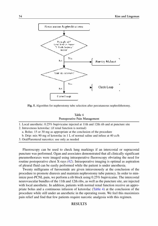
Fluoroscopy can be used to check lung markings if an intercostal or supracostalpuncture was performed. Ogan and associates demonstrated that all clinically significantpneumothoraxes were imaged using intraoperative fluoroscopy obviating the need forroutine postoperative chest X-rays (62). Intraoperative imaging is optimal as aspirationof pleural fluid can be easily performed while the patient is under anesthesia.
Twenty milligrams of furosemide are given intravenously at the conclusion of theprocedure to promote diuresis and maintain nephrostomy tube patency. In order to min-imize post-PCNL pain, we perform a rib block using 0.25% bupivicaine. The intercostalneurovascular bundles of the 11th and 12th ribs, as well as the puncture site, are injectedwith local anesthetic. In addition, patients with normal renal function receive an appro-priate bolus and a continuous infusion of ketorolac (Table 4) at the conclusion of theprocedure while still under an anesthetic in the operating room. We feel this maximizespain relief and find that few patients require narcotic analgesia with this regimen.
RESULTS
54 Kim and Lingeman
Fig. 5. Algorithm for nephrostomy tube selection after percutaneous nephrolithotomy.
Table 4Postoperative Pain Management
1. Local anesthetic: 0.25% bupivicaine injected at 11th and 12th rib and at puncture site2. Intravenous ketorolac: (if renal function is normal)
a. Bolus: 15 or 30 mg as appropriate at the conclusion of the procedureb. Drip: mix 90 mg of ketorolac in 1 L of normal saline and infuse at 40 cc/h
3. Oral/Parenteral narcotics: use only as needed

It is our practice to place patients on 2 weeks of preoperative oral antibiotics inpreparation for PCNL. The selection of the appropriate antibiotic is tailored to theresults of a preoperative urine culture. Our antibiotic of choice is a fluoroquinolonewhen the pre-operative urine culture is sterile. Septic events that were relatively com-mon at the onset of PCNL are now rare at the Methodist Hospital. We believe that 2weeks of antibiotic coverage limits the number of bacteria prior to puncture into the col-lecting system and has minimized the risk of septic events.
Approximately two-thirds of the percutaneous procedures performed at our institu-tion are done via an infracostal, lower pole puncture. Lower pole access avoids themorbidity of pleural injury and the placement of drainage tubes intercostally. With theuse of the flexible nephroscope, we find that all calyces unable to be accessed with rigidnephroscopy can be clearly visualized.
Our treatment technique for calyceal diverticular calculi has evolved over time.Initially, the percutaneous approach included the placement of a ureteral catheter, loca-tion and dilation of the infundibular connection and occasionally supracostal access toupper pole diverticulae. Although stone removal and fulguration methods remain thesame, our current percutaneous technique eliminates the placement of a ureteral catheterand localization of the infundibular communication. All accesses to calyceal diverticulaeare performed infracostally and our last 21 patients have been treated successfully withthis approach with a mean operative time of 58.5 minutes (63).
Examination of PCNL at the Methodist Hospital of Indiana demonstrates the safetyand efficacy of current percutaneous techniques and the ability to place small diameternephrostomy tubes (61). We reviewed 106 consecutive renal units undergoing PCNLfor calculi greater than 2 cm with placement of an 8.5- or 10-Fr Cope loop, a 20-Fr reen-try Malecot catheter, or a 20-Fr circle loop. In our cohort, 134 accesses were created in106 renal units: 35 upper, 7 mid, and 92 lower pole. Sixteen accesses (14 upper, 1 mid, 1lower) were left tubeless with a concomitant nephrostomy tube placed in the lower pole.Of the 111 nephrostomy tubes placed, the majority (85) were Cope loops (76.6%), 19were Malecot catheters (17.1%), and 7 were circle loops (6.3%). 31.1% of the renalunits were stone free with a single procedure and increased to 95.6% with two proce-dures. There were no difficulties with drainage or access for secondary PCNL.Complications included two hydrothoraxes, one arteriovenous fistula, and one ureteralperforation. Three of four renal units requiring transfusions underwent bilateral PCNLand at least one kidney in each of the four patients required multiple accesses. Of note,57.1% of infection stones required a reentry Malecot or circle catheter; only 12% ofnonstruvite stones necessitated one of the larger nephrostomy tubes.
TIPS AND TRICKS
For nephrostomy tract dilation, we routinely use the NephroMax™ high pressurenephrostomy balloon dilator (Boston Scientific, Natick, MA). It is a rapid dilating tech-nique as both dilation and sheath placement is a one-step procedure. We also believe itcauses less bleeding owing to the radial force generated rather than the shearing or cut-ting action of sequential Amplatz or metal telescoping dilators.
Occasionally the standard length (16-cm) 30-Fr rigid sheath that is placed with theballoon dilator is not long enough to reach the collecting system. In these instances, weuse a 20-cm, 30-Fr rigid sheath that will bridge the access tract from the skin to thecalyx of interest.
Chapter 3 / Percutaneous Access 55

Another strategy to lessen patient discomfort is the avoidance of an intercostalnephrostomy tube. In cases where an intercostal puncture is necessary and intraopera-tively the kidney is deemed to be stone free, a separate infracostal lower pole puncturemay be used to place a nephrostomy tube. This minimizes a patient’s discomfort whilemaintaining access to the collecting system for possible secondary procedures.
In obese patients, special consideration must be made when choosing a nephrostomytube. Although a rare occurrence, nephrostomy tubes can become dislodged in obese andspinal cord injury patients. If the choice of a Cope loop was made, consideration should bemade to placing a 5-Fr ureteral catheter to maintain access to the ureteral for possible sec-ondary access. Alternatively, a larger reentry Malecot catheter can be securely positioned.
Patients that have had multiple renal procedures can form a perinephric layer of scartissue. This fibrosis can be difficult to dilate during access. If initial dilation of the tractis difficult with an 8-Fr fascial dilator, a smaller dilator can be used. A dilator as smallas 5-Fr can begin the dilation in 1-Fr increments until the 8-Fr dilator can be placed intothe calyx of puncture. A 4.5-mm fascial incising needle (Cook Urological, Spencer, IN)can also be placed over the working wire to cut through the fibrosis, especially at thefascial level, and facilitate dilation.
The placement of a Superstiff wire into the ureter is the goal of access during percu-taneous procedures. The wire that we use for original access to the ureter is the 0.035-in.glidewire. Once the glidewire is in the ureter, we exchange out the glidewire for a super-stiff wire over a 5-Fr angiographic catheter. If the angiographic catheter does not passeasily over the glidewire, making a “herky-jerky” maneuver in which the angiographiccatheter is abruptly advanced a small amount at a time over the glidewire often allowsthe catheter to pass. The angiographic catheter will eventually slide easily once negoti-ated past the point of difficulty.
In patients with history of urinary tract infections and/or infection stones, a stoneculture is beneficial. Although a positive preoperative urine culture can tailor perioper-ative antibiotic choice, we find by sending a fragment crushed in sterile saline andobtaining a stone culture is helpful for guiding longer term postoperative antibiotictreatment. As our goal is to eradicate the stone burden and all associated bacteria, weare compulsive about removing any fragments visualized on postoperative day-1NCCT. If the stone culture is positive, we place the patient on therapeutic doses of anappropriate antibiotic for 2 weeks then complete a 3-months course of suppressiveantibiotics. A urine culture is obtained at the end of the 3-months course and monthlythereafter for several months to confirm that the patient has been cleared of infection.
CONCLUSIONS
Percutaneous access is best performed by the urologist in the operating room at thetime of stone removal. This is because urologists are most familiar with renal anatomyand with the instruments necessary for nephrolithotomy. The ability to obtain accessavoids dependence on the skill of another person and their schedule. Additionally, flex-ibility in the procedure is allowed, particularly if multiple or additional accesses arenecessary. Finally, it allows the performance of the operation as a single stage, which ispreferable from both the patient’s and surgeon’s perspective.
REFERENCES
56 Kim and Lingeman

1. Fernstrom I, Johansson B. Percutaneous pyelolithotomy: a new extraction technique. Scand J UrolNephrol 1976; 10(3): 257–259.
2. Consensus conference. Prevention and treatment of kidney stones. JAMA 1988; 260(7): 977–981.3. Segura JW, Preminger GM, Assimos DG, et al. Nephrolithiasis clinical guidelines panel summary
report on the management of staghorn calculi. J Urol 1994; 151(6): 1648–1651.4. Lam HS, Lingeman JE, Mosbaugh PG, et al. Evolution of the technique of combination therapy for
staghorn calculi: a decreasing role for extracorporeal shock wave lithotripsy. J Urol 1992; 148(3 Pt2): 1058–1062.
5. Wong C, Leveillee RJ. Single upper pole percutaneous access for treatment of > or = 5 cm complexbranched staghorn calculi: is shock wave lithotripsy necessary? J Endourol 2002; 16(7): 477–481.
6. Preminger GM, Segura JW, Lingeman JE. AUA Staghorn Clinical Guidelines Panel SummaryReport. J Urol 2004, in press.
7. Grasso M, Conlin M, Bagley D. Retrograde ureteropyeloscopic treatment of 2 cm or greater upperurinary tract and minor staghorn calculi. J Urol 1998; 160(2): 346–351.
8. Pittomvils G, Vandeursen H, Wevers M, et al. The influence of internal stone structure upon thefracture behaviour of urinary calculi. Ultrasound Med Biol 1994; 20(8): 803–810.
9. Chow GK, Streem SB. Contemporary urological intervention for cystinuric patients: immediate andlong-term impact and implications. J Urol 1998; 160(2): 341–344; discussion 4–5.
10. Klee LW, Brito CG, Lingeman JE. The clinical implications of brushite calculi. J Urol 1991; 145(4):715–718.
11. Segura JW, Preminger GM, Assimos DG, et al. Ureteral Stones Clinical Guidelines Panel summaryreport on the management of ureteral calculi. The American Urological Association. J Urol 1997;158(5): 1915–1921.
12. Green DF, Lytton B. Early experience with direct vision electrohydraulic lithotripsy of ureteral cal-culi. J Urol 1985; 133(5): 767–770.
13. Mugiya S, Nagata M, Un-No T, Takayama T, Suzuki K, Fujita K. Endoscopic management ofimpacted ureteral stones using a small caliber ureteroscope and a laser lithotriptor. J Urol 2000;164(2): 329–331.
14. Srivastava A, Ahlawat R, Kumar A, Kapoor R, Bhandari M. Management of impacted upper uretericcalculi: results of lithotripsy and percutaneous litholopaxy. Br J Urol 1992; 70(3): 252–257.
15. Preminger GM. Management of ureteral calculi: the debate continues. J Urol 1992; 148(3 Pt 2):1102–1104.
16. Albala DM, Assimos DG, Clayman RV, et al. Lower Pole I: a prospective randomized trial of extra-corporeal shock wave lithotripsy and percutaneous nephrostolithotomy for lower pole nephrolithiasis—initial results. J Urol 2001; 166(6): 2072–2080.
17. Havel D, Saussine C, Fath C, Lang H, Faure F, Jacqmin D. Single stones of the lower pole of the kid-ney: comparative results of extracorporeal shock wave lithotripsy and percutaneous nephrolithotomy.Eur Urol 1998; 33(4): 396–400.
18. Lingeman JE, Siegel YI, Steele B, Nyhuis AW, Woods JR. Management of lower pole nephrolithia-sis: a critical analysis. J Urol 1994; 151(3): 663–667.
19. Kuo RL, Lingeman JE, Leveillee RJ, et al. Lower Pole II: update results from a treatment compari-son of ureteroscopy (URS) vs percutaneous nephrostolithotomy (PCNL) for lower pole stones>10mm in diameter. J Endourol 2003; 17(1): A31.
20. Middleton AW, Jr., Pfister RC. Stone-containing pyelocaliceal diverticulum: embryogenic, anatomic,radiologic and clinical characteristics. J Urol 1974; 111(1): 2–6.
21. Wulfsohn MA. Pyelocaliceal diverticula. J Urol 1980; 123(1): 1–8.22. Williams G, Blandy JP, Tresidder GC. Communicating cysts and diverticula of the renal pelvis. Br J
Urol 1969; 41(2): 163–170.23. Yow RM, Bunts RC. Caliceal diverticulum. J Urol 1955; 73(4): 663–670.24. Prather GC. Calyceal diverticulum. J Urol 1941; 45: 55.25. Jones JA, Lingeman JE, Steidle CP. The roles of extracorporeal shock wave lithotripsy and percuta-
neous nephrostolithotomy in the management of pyelocaliceal diverticula. J Urol 1991; 146(3):724–727.
26. Psihramis KE, Dretler SP. Extracorporeal shock wave lithotripsy for caliceal diverticula calculi.J Urol 1987; 138(4): 707–711.
Chapter 3 / Percutaneous Access 57

27. Auge BK, Munver R, Kourambas J, Newman GE, Preminger GM. Endoscopic management of symp-tomatic caliceal diverticula: a retrospective comparison of percutaneous nephrolithotripsy andureteroscopy. J Endourol 2002; 16(8): 557–563.
28. Grasso M, Lang G, Loisides P, Bagley D, Taylor F. Endoscopic management of the symptomaticcaliceal diverticular calculus. J Urol 1995; 153(6): 1878–1881.
29. Fuchs GJ, Fuchs AM. Flexible endoscopy of the upper urinary tract: a new minimally invasivemethod for diagnosis and treatment. Urologe A 1990; 29(6): 313–320.
30. Batter SJ, Dretler SP. Ureterorenoscopic approach to the symptomatic caliceal diverticulum. J Urol1997; 158(3 Pt 1): 709–713.
31. Ruckle HC, Segura JW. Laparoscopic treatment of a stone-filled caliceal diverticulum: a definitive,minimally invasive therapeutic option. J Urol 1994; 151(1): 122–124.
32. Miller SD, Ng CS, Streem SB, Gill IS. Laparoscopic management of caliceal diverticular calculi.J Urol 2002; 167(3): 1248–1252.
33. Auge BK, Munver R, Kourambas J, Newman GE, Wu NZ, Preminger GM. Neoinfundibulotomy forthe management of symptomatic caliceal diverticula. J Urol 2002; 167(4): 1616–1620.
34. Hulbert JC, Reddy PK, Hunter DW, Castaneda-Zuniga W, Amplatz K, Lange PH. Percutaneous tech-niques for the management of caliceal diverticula containing calculi. J Urol 1986; 135(2): 225–227.
35. Shalhav AL, Soble JJ, Nakada SY, Wolf JS, Jr., McClennan BL, Clayman RV. Long-term outcome ofcaliceal diverticula following percutaneous endosurgical management. J Urol 1998; 160(5): 1635–1639.
36. Jarrett TW, Chan DY, Charambura TC, Fugita O, Kavoussi LR. Laparoscopic pyeloplasty: the first100 cases. J Urol 2002; 167(3): 1253–1256.
37. Goldfischer ER, Jabbour ME, Stravodimos KG, Klima WJ, Smith AD. Techniques of endopyelotomy.Br J Urol 1998; 82(1): 1–7.
38. Jones DJ, Wickham JE, Kellett MJ. Percutaneous nephrolithotomy for calculi in horseshoe kidneys.J Urol 1991; 145(3): 481–483.
39. Locke DR, Newman RC, Steinbock GS, Finlayson B. Extracorporeal shock wave lithotripsy in horse-shoe kidneys. Urology 1990; 35(5): 407–411.
40. Smith JE, VanArsdalen KN, Hanno PM, Pollack HM. Extracorporeal shock wave lithotripsy treatmentof calculi in horseshoe kidneys. J Urol 1989; 142(3): 683–686.
41. Raj GV, Auge BK, Weizer AZ, et al. Percutaneous management of calculi within horseshoe kidneys.J Urol 2003; 170(1): 48–51.
42. Al-Otaibi K, Hosking DH. Percutaneous stone removal in horseshoe kidneys. J Urol 1999; 162(3 Pt 1):674–677.
43. Toth C, Holman E, Pasztor I, Khan AM. Laparoscoipcally controlled and assisted percutaneoustransperitoneal nephrolithotomy in a pelvic dystopic kidney. J Endourol 1993; 7(4): 303–305.
44. Lee CK, Smith AD. Percutaneous transperitoneal approach to the pelvic kidney for endourologicremoval of calculus: three cases with two successes. J Endourol 1992; 6: 133–135.
45. Figge M. Percutaneous transperitoneal nephrolithotomy. Eur Urol 1988; 14(5): 414–416.46. Zafar FS, Lingeman JE. Value of laparoscopy in the management of calculi complicating renal
malformations. J Endourol 1996; 10(4): 379–383.47. Eshghi AM, Roth JS, Smith AD. Percutaneous transperitoneal approach to a pelvic kidney for
endourological removal of staghorn calculus. J Urol 1985; 134(3): 525–527.48. Kim SC, Kuo RL, Paterson RF, Lingeman JE. Laparoscopic assisted percutaneous nephrolithotomy:
best done tubeless? J Urol 2003; 169(4): S79.49. Lam HS, Lingeman JE, Barron M, et al. Staghorn calculi: analysis of treatment results between ini-
tial percutaneous nephrostolithotomy and extracorporeal shock wave lithotripsy monotherapy withreference to surface area. J Urol 1992; 147(5):1219–1925.
50. Grasso M, Chalik Y. Principles and applications of laser lithotripsy: experience with the holmiumlaser lithotrite. J Clin Laser Med Surg 1998; 16(1): 3–7.
51. Streem SB, Yost A. Extracorporeal shock wave lithotripsy in patients with bleeding diatheses. J Urol1990; 144(6): 1347–1348.
52. Jackman SV, Docimo SG, Cadeddu JA, Bishoff JT, Kavoussi LR, Jarrett TW. The “mini-perc” tech-nique: a less invasive alternative to percutaneous nephrolithotomy. World J Urol 1998; 16(6):371–374.
58 Kim and Lingeman

53. Cooper CS, Hedican SP. Mini-percutaneous ultrasonic nephrolithotripsy for pediatric staghorn cal-culi. J Urol 2003; 170(4 Pt 1): 1336–1337.
54. Lingeman JE, Smith LH, Woods JR, Newman DM. Urinary Calculi: ESWL, Endourology, and Med-ical Therapy. Lea and Febiger; Philadelphia; 1989.
55. Forsyth MJ, Fuchs EF. The supracostal approach for percutaneous nephrostolithotomy. J Urol 1987;137(2): 197–198.
56. Fuchs EF, Forsyth MJ. Supracostal approach for percutaneous ultrasonic lithotripsy. Urol Clin NorthAm 1990; 17(1): 99–102.
57. Lang EK. Percutaneous nephrostolithotomy and lithotripsy: a multi-institutional survey of complica-tions. Radiology 1987; 162(1 Pt 1): 25–30.
58. Lingeman JE, Lifshitz DA, Evan AP. Surgical management of urinary lithiasis. In: Campbell’s Urology,7th Ed. (Walsh PC, Vaughn ED, Wein AJ, eds.), WB Saunders, Philadelphia, 1998, pp. 3361–3451.
59. LeRoy AJ, Williams HJ, Jr., Bender CE, Segura JW, Patterson DE, Benson RC. Colon perforation fol-lowing percutaneous nephrostomy and renal calculus removal. Radiology 1985; 155(1): 83–85.
Chapter 3 / Percutaneous Access 59

60. Davidoff R, Bellman GC. Influence of technique of percutaneous tract creation on incidence of renalhemorrhage. J Urol 1997; 157(4): 1229–1231.
61. Kim SC, Kuo RL, Paterson RF, Lingeman JE. Using and choosing a nephrostomy tube after percutaneousnephrolithotomy for large or complex stone disease: a treatment strategy. J Endourol 2005; 19: 348–352.
62. Ogan K, Corwin TS, Smith T, et al. Sensitivity of chest fluoroscopy compared with chest CT andchest radiography for diagnosing hydropneumothorax in association with percutaneous nephros-tolithotomy. Urology 2003; 62(6): 988–992.
63. Kim SC, Kuo RL, Tinmouth WW, Watkins S, Lingeman JE. Percutaneous nephrolithotomy for cal-iceal diverticular calculi: a novel single-stage approach. J Urol 2005; 173: 1194–1198.
60 Kim and Lingeman

Lateralizing Essential Hematuria
Murali K. Ankem, MD
and Stephen Y. Nakada, MD
CONTENTS
INTRODUCTION
DIAGNOSIS
INDICATIONS
TECHNIQUE
RESULTS
TIPS AND TRICKS
CONCLUSION
REFERENCES
4
SUMMARY
Lateralizing essential hematuria is a rare clinical syndrome which poses a diagnosticand therapeutic challenge for the treating urologist. Despite its benign nature in themajority of patients, recurrent macroscopic hematuria can be very frustrating and stress-ful to the patient. Historically, these patients were treated by irrigating the renal collect-ing system with various cauterizing solutions, surgical evaluation of exposed collectingsystem, partial nephrectomy, or even nephrectomy. Recent advances in the endourolog-ical techniques and the development of more sophisticated instruments have enhancedour abilities to diagnose and treat many upper urinary tract lesions. This chapter focuseson the latest developments in the evaluation, technique, and results of fiberopticureteropyeloscopy in the management of lateralizing essential hematuria.
Key Words: Hematuria; essential hematuria; unilateral hematuria; lateralizing hema-turia; diagnosis; flexible ureteropyeloscopy; laser coagulation.
INTRODUCTION
Lateralizing essential hematuria is a rare clinical syndrome, which poses a diagnos-tic and therapeutic challenge to the treating urologist. Despite its benign nature in themajority of patients (1,2), recurrent macroscopic hematuria can be very frustrating and
From: Advanced Endourology: The Complete Clinical GuideEdited by: S. Y. Nakada and M. S. Pearle © Humana Press Inc., Totowa, NJ
61

stressful to the patient. This condition is also known as chronic unilateral hematuria,unilateral essential hematuria, or benign lateralizing hematuria. The complex is definedas gross hematuria localized to one side of urinary tract by cystoscopy. The diagnosis ismade by excluding all common causes of hematuria in the face of normal radiological(computed tomography [CT], ultrasound, intravenous urogram [IVU]) and hematolog-ical studies. Historically, these patients were managed by irrigating the renal collectingsystem with various cauterizing solutions (3,4), surgical evaluation of exposed collect-ing system, partial nephrectomy or even nephrectomy in some patients (5). Recentadvances in endourological techniques and the development of more sophisticatedinstruments have enhanced our abilities to diagnose and treat many upper urinary tractlesions (6,7). This chapter describes the preoperative evaluation, indications, technique,and results of fiberoptic ureteropyeloscopy in the management of lateralizing essentialhematuria.
DIAGNOSIS
Lateralizing essential hematuria often presents as asymptomatic chronic recurrentgross hematuria. Some patients may present with clot colic, clot retention, and even sig-nificant anemia (1,7,8). Lateralizing essential hematuria is common in younger patientsand has no predilection for either sex or side. The work-up should include a thoroughhistory, physical examination, and a cystoscopy at the time of bleeding to lateralize thehematuria to one side of the urinary system and to exclude other common sources ofurinary bleeding. Next, serum studies including complete hemogram, basic renal func-tion studies, coagulation studies, and sickle cell preparation should be performed. Urinefor microscopy, routine culture, and voided cytology (to rule out inflammatory, malig-nant, and nephrological sources of bleeding) should be part of the initial evaluation.Consideration for special urine cultures to rule out atypical fungal and tuberculosis (9)infections should be given. Three early morning voided urine samples should be sentfor Ziehl-Nielsen staining and culture for mycobacterial organisms. In the presence ofpositive Mantoux test and negative mycobacterial cultures from the voided specimens,ureteroscopy and urine from the renal pelvis should be sent for mycobacterial cultures(9). Where appropriate, 24-hour urine collection for albumin and phase-contrastmicroscopy for identification of ghost cells could be useful in differentiating glomeru-lar from the epithelial bleeding.
The IVU typically demonstrates the urinary system in entirety and any areas that arenot clearly seen should be further characterized by retrograde urography. Oblique viewsare very important to avoid superimposition of calices and will aid in methodical exam-ination of calices by ureteropyeloscopy later. If the IVU is equivocal the next logicalstep should be a CT or an ultrasound to rule out renal masses. Patients with intravenouscontrast allergy should be evaluated by ultrasound and bilateral retrograde pyelogramsafter adequate steroid preparation of the patient. CT urography and magnetic resonance(MR) urography are evolving techniques and may eventually replace IVU (10). In cer-tain centers MR urography currently serves as an alternative imaging technique to intra-venous urography and CT urography for children and pregnant women and for patientswith contraindications to iodinated contrast media (10).
There is no uniform consensus regarding the timing and role of arteriography in theevaluation of lateralizing essential hematuria. As spontaneous arteriovenous malforma-tions (AVMs) are rare, and most of the AVMs are secondary to prior renal biopsy, sur-gery, renal trauma, or renal tumors (11), this step could be bypassed in the absence of
62 Ankem and Nakada
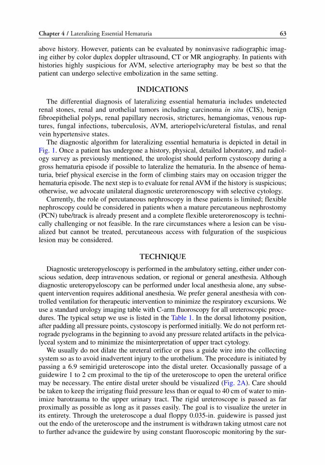
above history. However, patients can be evaluated by noninvasive radiographic imag-ing either by color duplex doppler ultrasound, CT or MR angiography. In patients withhistories highly suspicious for AVM, selective arteriography may be best so that thepatient can undergo selective embolization in the same setting.
INDICATIONS
The differential diagnosis of lateralizing essential hematuria includes undetectedrenal stones, renal and urothelial tumors including carcinoma in situ (CIS), benignfibroepithelial polyps, renal papillary necrosis, strictures, hemangiomas, venous rup-tures, fungal infections, tuberculosis, AVM, arteriopelvic/ureteral fistulas, and renalvein hypertensive states.
The diagnostic algorithm for lateralizing essential hematuria is depicted in detail inFig. 1. Once a patient has undergone a history, physical, detailed laboratory, and radiol-ogy survey as previously mentioned, the urologist should perform cystoscopy during agross hematuria episode if possible to lateralize the hematuria. In the absence of hema-turia, brief physical exercise in the form of climbing stairs may on occasion trigger thehematuria episode. The next step is to evaluate for renal AVM if the history is suspicious;otherwise, we advocate unilateral diagnostic ureterorenoscopy with selective cytology.
Currently, the role of percutaneous nephroscopy in these patients is limited; flexiblenephroscopy could be considered in patients when a mature percutaneous nephrostomy(PCN) tube/track is already present and a complete flexible ureterorenoscopy is techni-cally challenging or not feasible. In the rare circumstances where a lesion can be visu-alized but cannot be treated, percutaneous access with fulguration of the suspiciouslesion may be considered.
TECHNIQUE
Diagnostic ureteropyeloscopy is performed in the ambulatory setting, either under con-scious sedation, deep intravenous sedation, or regional or general anesthesia. Althoughdiagnostic ureteropyeloscopy can be performed under local anesthesia alone, any subse-quent intervention requires additional anesthesia. We prefer general anesthesia with con-trolled ventilation for therapeutic intervention to minimize the respiratory excursions. Weuse a standard urology imaging table with C-arm fluoroscopy for all ureteroscopic proce-dures. The typical setup we use is listed in the Table 1. In the dorsal lithotomy position,after padding all pressure points, cystoscopy is performed initially. We do not perform ret-rograde pyelograms in the beginning to avoid any pressure related artifacts in the pelvica-lyceal system and to minimize the misinterpretation of upper tract cytology.
We usually do not dilate the ureteral orifice or pass a guide wire into the collectingsystem so as to avoid inadvertent injury to the urothelium. The procedure is initiated bypassing a 6.9 semirigid ureteroscope into the distal ureter. Occasionally passage of aguidewire 1 to 2 cm proximal to the tip of the ureteroscope to open the ureteral orificemay be necessary. The entire distal ureter should be visualized (Fig. 2A). Care shouldbe taken to keep the irrigating fluid pressure less than or equal to 40 cm of water to min-imize barotrauma to the upper urinary tract. The rigid ureteroscope is passed as farproximally as possible as long as it passes easily. The goal is to visualize the ureter inits entirety. Through the ureteroscope a dual floppy 0.035-in. guidewire is passed justout the endo of the ureteroscope and the instrument is withdrawn taking utmost care notto further advance the guidewire by using constant fluoroscopic monitoring by the sur-
Chapter 4 / Lateralizing Essential Hematuria 63

geon (Fig. 2B). Next, a small caliber, actively deflecting flexible ureteroscope is passedover the wire under fluoroscopic guidance taking care not to advance the wire beyondthe point of prior visual inspection (Fig. 2C). Then the guide wire is removed and theremaining ureter is examined up to ureteropelvic junction followed by renal pelvic eval-uation. If the visibility is poor, the urine from the pelvis can be emptied via the side portand gentle irrigation can be performed using the 20-mL syringe filled with normalsaline. Selective urinary cytology is obtained at this time.
Systematic evaluation of the superior, middle, and inferior calices is performed (Fig. 2D).Dilute contrast medium is injected to verify inspection of all calices of the renal collect-ing system. Alternatively dilute contrast (30%) can be added to the irrigant to visualizethe collecting system (8). In the event of bleeding, the collecting system is emptied and
64 Ankem and Nakada
Fig. 1. Diagnostic algorithm for lateralizing essential hematuria. (Reproduced from ref. 11a.with permission from AUA Office of Education.)

re-inspection with gentle irrigation will aid in identifying the source of bleeding. Thissystematic examination will prevent confusion owing to bruising of the upper-poleinfundibulum caused by passive deflection of scope against this area when viewing thelower-pole calices (Fig. 2E). Kumon et al. (13) made a point that each calyx should beexamined with and without irrigation, as the irrigation pressure might stop low-pressurebleeding from venous ruptures and hemangiomas. In special circumstances where apatient with an ileal loop has lateralizing essential hematuria, the above protocol shouldbe followed; but entry into the selected ureteral orfice may need guidewire assistance.
Any clear-cut lesion, i.e., small stone, transitional cell carcinoma (TCC) (Fig. 3) orhemangioma, is appropriately treated using the holmium laser or bugbee electrodedepending on the provisional diagnosis after biopsy using biopsy forceps, flat wire bas-ket, or brush (Table 1). If no treatment is rendered and balloon dilation of ureteral orificeis not performed, no ureteral stent is left in situ. If treatment is rendered, balloon dilationperformed, or bleeding encountered, a 6-Fr double-pigtail stent is left for a limited period
Chapter 4 / Lateralizing Essential Hematuria 65
Table 1Instrument List
Equipment list, diagnostic, and therapeutic ureterorenoscopy(University of Wisconsin)
15-Fr flexible cystoscope21-Fr cystoscope with 0°, 30°, and 70° telescopesSemirigid 6.9-Fr ureteroscopeActively deflectable 7.5-and 6.9-Fr flexible ureteroscopeSureseal adaptor0.035-in. dual floppy guide wire10-mL and 20-mL syringesContrast medium 50:50 with saline in graduated cylinder3 Chip video camera and endoscopic towerStirrups, drapesC-arm fluoroscope with still image capability
Available in the room but not open
Bulb tip pressure irrigator0.035-in. Amplatz super stiff guide wire0.035-in. angled glide wireTorque control deviceHolmium laser and goggles200-μ holmium laser fiber2-Fr bugbee electrode3-Fr ureteroscopic biopsy forceps3-Fr ureteroscopic biopsy brush2.4-Fr N-circle stone basket5-Fr ureteral access catheters8/10-Fr Coaxial dilator6-Fr double pigtail ureteral stent, assorted lengthsDual deflecting flexible ureteroscopes
(Reproduced from ref. 11a, with permission from AUA Office of Education.).

of time at the surgeon’s discretion. The patient can be discharged on the same day andusually can be given a short course of oral antibiotics.
The recent introduction of dual actively deflecting ureteroscopes can make visualiza-tion of lower-pole calices much easier and will increase the sensitivity of upper tractinspection. However, these new ureteroscopes are slightly larger than the standard
66 Ankem and Nakada
Fig. 3. Transitional cell carcinoma of superior calyx.
Fig. 2. Diagnostic ureteropyeloscopy is initiated by passing a small caliber (6.9 Fr), semirigid uretero-scope to rule out any distal ureteral pathology (A). Through the ureteroscope, a dual floppy 0.035-in.guidewire is advanced to the uppermost point to which the 6.9-Fr semirigid ureteroscope is advancedunder fluoroscopic control (B) and the short rigid ureteroscope is removed. Next, a small caliber,actively deflectable flexible ureteroscope is passed over the guidewire and into the ureter under fluoro-scopic guidance (C) being cautious not to advance the guidewire beyond the point of prior visual inspec-tion. With the availability of the newer, smaller caliber ureteroscopes, the guidewire can be removed andvisual inspection of the remaining ureter can be performed. Proximal ureteroscopy is performed fol-lowed by examination of the renal pelvis. Selective upper tract urinary cytology is obtained through theureteroscope at this time. Systematic evaluation of the upper, middle, and lower pole calices is per-formed in that order (D). This approach prevents confusion owing to endoscopic bruising of the upperpole infundibulum caused by passive deflection of the shaft of the scope against this area when viewingthe lower pole calices (E). (Reproduced from ref. 11a, with permission from AUA Office of Education.)

actively deflecting flexible ureteroscopes. Currently, we use the DUR-8 Elite ureteroscopeas a second line instrument for upper tract ureteroscopy (Ankem et al., unpublished data).
In patients where diffuse nonspecific changes are seen, Bahnson et al. (4) reportedinstilling 10 mL of 1% silver nitrate into the renal pelvis using ureteral catheters (n = 3).Hematuria stopped after one to two instillations and urine remained clear in 13 monthsof follow-up. Stefanini and colleagues (14) reported usage of oral Epsilon aminocaproicacid at a daily dose of 150 mg/kg in four divided doses given for up to 3 weeks. A totalof nine patients with gross hematuria were given the above dose and the hematuria wascontrolled effectively without any significant complications.
RESULTS
Today, successful examination of entire upper urinary tract using the above-describedapproach is straightforward. In 1998, Tawfiek et al. (6) reported 100% visualization ofipsilateral urothelium in all 23 patients in his series. In 156 published cases of lateralizingessential hematuria, only 8% (13 cases) have been found to have tumors (n = 7) or stones(n = 6) not diagnosed by preoperative imaging (Table 2). Among the patients presentingwith lateralizing essential hematuria, approx 50% will have a discrete vascular lesion (15)that can be fulgurated. The most common treatable vascular abnormality is hemangioma,found in 11 cases in Bagley’s series, seven in Tawfiek’s series, nine in Kumon’s series,and five in Nakada’s series for a total of 32 cases out of 156 (20%) reported in the litera-ture. Hemangiomas are typically seen on the renal papillae and are usually submucosal.They may appear as small, red, or bluish spots, usually on the tip or base of the papilla orthey may be large, bulbous erythematous lesions on the papillary tips (6). Minute venousruptures are considered discrete vascular lesions, which are often located on the bridge oftissue between compound papillae. These vascular lesions appear to be seen equally dis-tributed throughout the kidney (7,8). The etiology and pathogenesis of these venous rup-tures is not clear and they could be related to venous hypertensive states secondary to leftrenal vein obstruction (nut cracker phenomenon) (13,16,17).
Coagulation of discrete lesions when present is effective; Nakada and associates (7)reported resolution of hematuria in the range of 82%. On the other hand, patients withdiffuse lesions or negative examination usually have persistent hematuria followingureterorenoscopy (17% long term resolution) (7). Tawfiek and his associates (6,8) madesimilar observations as well.
Upon reviewing the literature, there is strong evidence that ureteroscopic managementof these lesions has yielded durable results and this pattern suggests a causal relationshipbetween endoscopic fulguration and resolution of bleeding. In only 12% of patients,despite successful fulguration of hemangiomas, recurrence of bleeding occurred.Multifocality may explain these failures in patients who underwent fulguration. Tawfiek(6) reported that 4/5 patients, despite negative examination. maintained clear urine andthey theorize that some minute venous-caliceal communications that were not detectedby endoscopy might have sealed off secondary to the pressure of irrigation and inflam-matory response incited by ureteropyeloscopy alone.
Negative ureteroscopic examinations are disappointing and frustrating both for thepatient and the urologist. In modern series the negative examination rate is in the orderof 23%. Nakada and colleagues (7) found that 67% of patients with negative examina-tions had persistent bleeding. Tawfiek (6) reported 4/8 patients with nonspecific find-ings continued to have hematuria; however, 4/5 patients with negative examination
Chapter 4 / Lateralizing Essential Hematuria 67

Tab
le 2
Publ
ishe
d Se
ries
, Lat
eral
izin
g E
ssen
tial
Hem
atur
ia
Vasc
ular
ab
norm
alit
ies
Neg
ativ
e R
ecur
renc
e Fo
llow
-up
Aut
hor
No.
App
roac
h N
o.D
iscr
ete
Diff
use
Ston
eTu
mor
eval
uati
ons
(%)
(mon
th)
Gitt
es e
t al.
(5)
12N
ephr
osco
py 1
25
70
00
0N
/APa
tters
on e
t al.
(2)
4N
ephr
osco
py 3
Ure
tero
scop
y 4
13
00
00
5 (2
–10)
McM
urtr
y et
al.
(18)
8N
ephr
osco
py 7
Ure
tero
scop
y 3
43
10
025
at 4
mon
th6
(2–4
1)K
avou
ssi e
t al.
(19)
8U
rete
rosc
opy
8N
ot
00
012
at 7
mon
th11
(7–
18)
spec
ifie
dB
agle
y et
al.
(8)
32U
rete
rosc
opy
3214
91
15
16N
/AK
umon
et a
l. (1
3)12
Ure
tero
scop
y 12
91
00
20
10 (
6–21
)K
umon
et a
l. (1
3a)
30U
rete
rosc
opy
3022
11
15
N/A
N/A
Nak
ada
et a
l. (7
)17
Nep
hros
copy
2U
rete
rosc
opy
1510
21
13
4158
(24
–103
)Ta
wfi
ek e
t al.
(6)
23U
rete
rosc
opy
2310
32
35
08
(4–1
8)Y
azak
i et a
l. (2
0)6
Ure
tero
scop
y 6
11
00
5N
/AN
/AH
ara
et a
l. (2
1)4
Ure
tero
scop
y 4
Not
0
13
N/A
N/A
spec
ifie
dTo
tals
156
7630
67
28
(Rep
rodu
ced
from
ref
. 11a
, with
per
mis
sion
fro
m A
UA
Off
ice
of E
duca
tion.
)
68
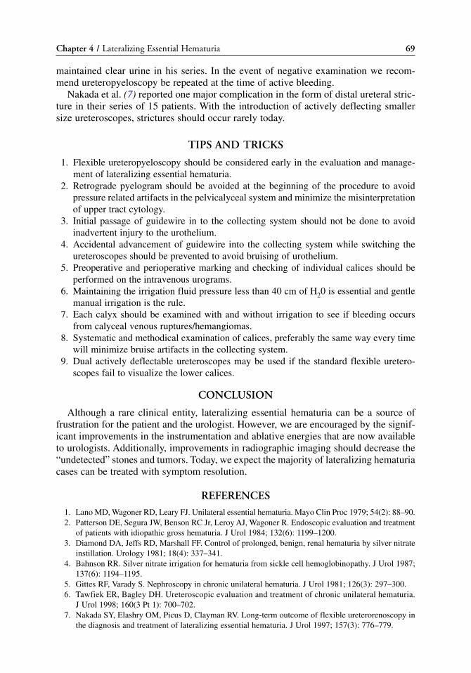
maintained clear urine in his series. In the event of negative examination we recom-mend ureteropyeloscopy be repeated at the time of active bleeding.
Nakada et al. (7) reported one major complication in the form of distal ureteral stric-ture in their series of 15 patients. With the introduction of actively deflecting smallersize ureteroscopes, strictures should occur rarely today.
TIPS AND TRICKS
1. Flexible ureteropyeloscopy should be considered early in the evaluation and manage-ment of lateralizing essential hematuria.
2. Retrograde pyelogram should be avoided at the beginning of the procedure to avoidpressure related artifacts in the pelvicalyceal system and minimize the misinterpretationof upper tract cytology.
3. Initial passage of guidewire in to the collecting system should not be done to avoidinadvertent injury to the urothelium.
4. Accidental advancement of guidewire into the collecting system while switching theureteroscopes should be prevented to avoid bruising of urothelium.
5. Preoperative and perioperative marking and checking of individual calices should beperformed on the intravenous urograms.
6. Maintaining the irrigation fluid pressure less than 40 cm of H20 is essential and gentlemanual irrigation is the rule.
7. Each calyx should be examined with and without irrigation to see if bleeding occursfrom calyceal venous ruptures/hemangiomas.
8. Systematic and methodical examination of calices, preferably the same way every timewill minimize bruise artifacts in the collecting system.
9. Dual actively deflectable ureteroscopes may be used if the standard flexible uretero-scopes fail to visualize the lower calices.
CONCLUSION
Although a rare clinical entity, lateralizing essential hematuria can be a source offrustration for the patient and the urologist. However, we are encouraged by the signif-icant improvements in the instrumentation and ablative energies that are now availableto urologists. Additionally, improvements in radiographic imaging should decrease the“undetected” stones and tumors. Today, we expect the majority of lateralizing hematuriacases can be treated with symptom resolution.
REFERENCES1. Lano MD, Wagoner RD, Leary FJ. Unilateral essential hematuria. Mayo Clin Proc 1979; 54(2): 88–90.2. Patterson DE, Segura JW, Benson RC Jr, Leroy AJ, Wagoner R. Endoscopic evaluation and treatment
of patients with idiopathic gross hematuria. J Urol 1984; 132(6): 1199–1200.3. Diamond DA, Jeffs RD, Marshall FF. Control of prolonged, benign, renal hematuria by silver nitrate
instillation. Urology 1981; 18(4): 337–341. 4. Bahnson RR. Silver nitrate irrigation for hematuria from sickle cell hemoglobinopathy. J Urol 1987;
137(6): 1194–1195.5. Gittes RF, Varady S. Nephroscopy in chronic unilateral hematuria. J Urol 1981; 126(3): 297–300.6. Tawfiek ER, Bagley DH. Ureteroscopic evaluation and treatment of chronic unilateral hematuria.
J Urol 1998; 160(3 Pt 1): 700–702.7. Nakada SY, Elashry OM, Picus D, Clayman RV. Long-term outcome of flexible ureterorenoscopy in
the diagnosis and treatment of lateralizing essential hematuria. J Urol 1997; 157(3): 776–779.
Chapter 4 / Lateralizing Essential Hematuria 69

8. Bagley DH, Allen J. Flexible ureteropyeloscopy in the diagnosis of benign essential hematuria. J Urol1990; 143(3): 549–553.
9. Chan SW, Shalhav AL, Clayman RV. Renal tuberculosis presenting as slateralizing hematuria diagno-sis by ureteronephroscopy and selective upper tract urine culture. J Endourol 1998; 12(4): 363–364.
10. Kawashima A, Glockner JF, King BF Jr. CT urography and MR urography. Radiol Clin North Am2003; 41(5): 945–961.
11. Fogazzi GB, Moriggi M, Fontanella U. Spontaneous renal arteriovenous fistula as a cause of haema-turia. Nephrol Dial Transplant 1997; 12(2): 350–356.
11a. The Diagnosis and Management of Lateralizing Essential Hematuria, Nakada SY: AOA udpate series,Lesson 13, Volume XXII.
12. Kerbl K, Andreoni C, Clayman RV. Lateralizing essential hematuria: tricks in the diagnostic and ther-apeutic ureteroscopy. J Endourol 2000; 14:(suppl 1).
13. Kumon H, Tsugawa M, Matsumura Y, Ohmori H. Endoscopic diagnosis and treatment of chronic uni-lateral hematuria of uncertain etiology. J Urol 1990; 143(3): 554–558.
13a. Kumon H. Endoscopic management of renal hematuria of uncertain etiology. Nishinihon J Urol.1994; 56: 511–515.
14. Stefanini M, English HA, Taylor AE. Safe and effective, prolonged administration of epsilonaminocaproic acid in bleeding from the urinary tract. J Urol 1990; 143(3): 559–561.
15. Clayman RV, McDougall EM, Figenshau RS. Endourology of the upper urinary tract: noncalculusapplications. In: Adult and Pediatric Urology, 3rd Ed. Vol.1., (J. Y. Gillenwater, ed.), Mosby, St.Louis, 1997, pp. 749–827.
16. Rowbotham C, Anson KM. Benign lateralizing haematuria: the impact of upper tract endoscopy. BJUInt 2001; 88(9): 841–849.
17. Chen YM, Wang IK, Ng KK, Huang CC. Nutcracker syndrome: an overlooked cause of hematuria.Chang Gung Med J 2002; 25(10): 700–705.
18. McMurtry JM, Clayman RV, Preminger GM. Endourologic diagnosis and treatment of essentialhematuria. J Endourol. 1987; 1:145–151.
19. Kavoussi LR, Clayman RV, Basler JW. Flexible, actively deflectable fiberoptic ureteronephroscopy.J Urol. 1989; 142: 949.
20. Yazaki T, Kamiyama Y, Tomomasa H, et al. Ureteropyeloscopy in the diagnosis of patients with uppertract hematuria: an initial clinical study. Int J Urol. 1999; 6: 219–225.
21. Hara I, Hara S, Miyake H, et al. Usefulness of ureteropyeloscopy for diagnosis of upper urinary tracttumors. J Endourol. 2001; 15: 601–605.
70 Ankem and Nakada

Diagnosis and Surveillance of Upper Tract Tumors
Douglas W. Soderdahl, MD
and Michael D. Fabrizio, MD, FACS
CONTENTS
INDICATIONS
INSTRUMENT LIST
TECHNIQUE
RESULTS
TIPS AND TRICKS
REFERENCES
5
SUMMARY
Upper tract transitional cell carcinoma is rare and historically has resulted innephroureterectomy. Diagnosis and treatment historically relied upon finding radiographicfilling defects in the collecting system. With the advent of small caliber ureteroscopes, smallworking instruments, and lasers, nephron-sparing procedures are available to these patients.Additionally, these technological advances can be used to rapidly diagnose patients.
The results of endoscopic treatment in transitional cell carcinoma are based on thefact that survival is directly related to the grade and stage of the tumor. Low gradelesions recur less frequently and have less invasive potential than high grade lesions.After endoscopic treatment of upper tract transitional cell carcinoma, low grade lesionsrarely progress and recurrences usually respond to retreatment. This chapter describesthe background surrounding upper tract transitional cell carcinoma. It also outlines thediagnosis and surveillance regimens and report treatment results.
Key Words: Transitional cell carcinoma; ureteroscopy; upper tract tumor.
INDICATIONSIntroduction
Transitional cell carcinoma (TCC) arising in the upper tract urothelium (ureters, renalpelves, and calyces) often presents diagnostic and therapeutic challenges to the urologist.
From: Advanced Endourology: The Complete Clinical GuideEdited by: S. Y. Nakada and M. S. Pearle © Humana Press Inc., Totowa, NJ
71
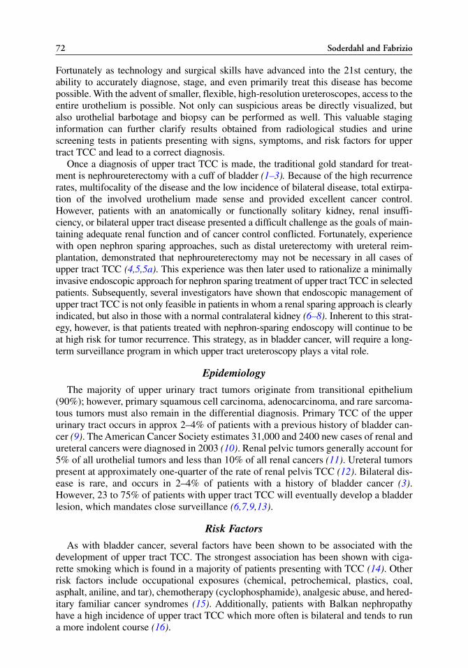
Fortunately as technology and surgical skills have advanced into the 21st century, theability to accurately diagnose, stage, and even primarily treat this disease has becomepossible. With the advent of smaller, flexible, high-resolution ureteroscopes, access to theentire urothelium is possible. Not only can suspicious areas be directly visualized, butalso urothelial barbotage and biopsy can be performed as well. This valuable staginginformation can further clarify results obtained from radiological studies and urinescreening tests in patients presenting with signs, symptoms, and risk factors for uppertract TCC and lead to a correct diagnosis.
Once a diagnosis of upper tract TCC is made, the traditional gold standard for treat-ment is nephroureterectomy with a cuff of bladder (1–3). Because of the high recurrencerates, multifocality of the disease and the low incidence of bilateral disease, total extirpa-tion of the involved urothelium made sense and provided excellent cancer control.However, patients with an anatomically or functionally solitary kidney, renal insuffi-ciency, or bilateral upper tract disease presented a difficult challenge as the goals of main-taining adequate renal function and of cancer control conflicted. Fortunately, experiencewith open nephron sparing approaches, such as distal ureterectomy with ureteral reim-plantation, demonstrated that nephroureterectomy may not be necessary in all cases ofupper tract TCC (4,5,5a). This experience was then later used to rationalize a minimallyinvasive endoscopic approach for nephron sparing treatment of upper tract TCC in selectedpatients. Subsequently, several investigators have shown that endoscopic management ofupper tract TCC is not only feasible in patients in whom a renal sparing approach is clearlyindicated, but also in those with a normal contralateral kidney (6–8). Inherent to this strat-egy, however, is that patients treated with nephron-sparing endoscopy will continue to beat high risk for tumor recurrence. This strategy, as in bladder cancer, will require a long-term surveillance program in which upper tract ureteroscopy plays a vital role.
Epidemiology
The majority of upper urinary tract tumors originate from transitional epithelium(90%); however, primary squamous cell carcinoma, adenocarcinoma, and rare sarcoma-tous tumors must also remain in the differential diagnosis. Primary TCC of the upperurinary tract occurs in approx 2–4% of patients with a previous history of bladder can-cer (9). The American Cancer Society estimates 31,000 and 2400 new cases of renal andureteral cancers were diagnosed in 2003 (10). Renal pelvic tumors generally account for5% of all urothelial tumors and less than 10% of all renal cancers (11). Ureteral tumorspresent at approximately one-quarter of the rate of renal pelvis TCC (12). Bilateral dis-ease is rare, and occurs in 2–4% of patients with a history of bladder cancer (3).However, 23 to 75% of patients with upper tract TCC will eventually develop a bladderlesion, which mandates close surveillance (6,7,9,13).
Risk Factors
As with bladder cancer, several factors have been shown to be associated with thedevelopment of upper tract TCC. The strongest association has been shown with ciga-rette smoking which is found in a majority of patients presenting with TCC (14). Otherrisk factors include occupational exposures (chemical, petrochemical, plastics, coal,asphalt, aniline, and tar), chemotherapy (cyclophosphamide), analgesic abuse, and hered-itary familiar cancer syndromes (15). Additionally, patients with Balkan nephropathyhave a high incidence of upper tract TCC which more often is bilateral and tends to runa more indolent course (16).
72 Soderdahl and Fabrizio

Diagnosis
Most patients with upper tract TCC will initially present with gross or microscopichematuria (2). The presence of vermiform clots suggest an upper tract source.Occasionally, patients will also complain of flank pain which may mimic ureteral colic.This is presumably the result of transient obstruction from either the mass or from clots.Finally, a small percentage of patients may experience no signs or symptoms and thetumor is picked up serendipitously on a diagnostic study done for another reason.
The American Urological Association best practice policy defines microhematuria asthe presence of three or more red blood cells per high power field on two of three uri-nalyses gathered at separate settings (17). The panel also provided a consensus work-upfor patients presenting with hematuria which should include a detailed history and phys-ical examination, imaging of the upper urinary tract, urine studies (microscopic urinal-ysis, culture, cytology), and cystoscopy (18).
The traditional imaging studies for a hematuria work-up are an intravenous pyelo-gram (IVP) with tomograms or retrograde pyelography with renal ultrasonography inpatients with contrast allergy or those with poor upper tract visualization on other stud-ies (19–21). Most commonly upper tract TCC will be identified as a radiolucent fillingdefect or obstructing lesion on an IVP. Occasionally, the involved kidney will be poorlyor nonvisualized owing to obstruction. Computed tomography (CT) scanning, due toimprovements in resolution, capacity for three-dimensional reconstruction and ability todiagnose unrelated, nonurological conditions has recently become an acceptable initialimaging selection (18). It is vital to obtain pre- and postcontrast scans in order to ade-quately characterize masses and to identify radiodense renal calculi (22–24). Magneticresonance urography has been recently described and may have the ability to differen-tiate ureteral tumors from other filling defects and may prove to be superior to IVP inupper tract TCC diagnosis (25,26).
Urinary cytology also plays a role in the work up of hematuria in high-risk individu-als. The sensitivity of urine cytology is dependent on several variables including num-ber of specimens obtained, the stage and grade of the tumor and the experience of thecytopathologist (27–28). Saline barbotage has been shown to improve the diagnosticaccuracy of cytology; however, this requires catheterization or endoscopy (29). Patientswith positive cytology results and no demonstrable pathology in the bladder shouldundergo selective ureteral catheterization for upper tract urinary cytology and salinewashings. This can be performed prior to retrograde pyelography. Several other urinetests (bladder tumor associated antigen, nuclear matrix protein 22, telomerase, etc.) todetect TCC in the bladder have been studied, but their role in upper tract TCC detectionis not clear (18).
Cystoscopy is vital to the work-up of hematuria or in those presenting with radiolog-ical evidence of upper tract TCC owing to the high incidence of concomitant bladderpathology. Bladder lesions can be visually characterized and biopsied, and if performedat the time of bleeding, cystoscopy may also be helpful in localizing the side of uppertract hematuria (30). Additionally, at the time of cystoscopy, specimens can be obtainedfrom the bladder and each ureter for cytological examination.
Once the bladder and urethra have been cleared endoscopically and the suspicion ofupper tract TCC remains owing to abnormal imaging studies or cytological results,ureteroscopy is the next step for diagnosis, staging and possibly treatment. The entireurothelium at risk is carefully inspected, ureteral and renal pelvis washings are col-lected, and suspicious areas biopsied/resected and the base fulgurated.
Chapter 5 / Upper Tract Tumors 73

INSTRUMENT LIST
1. Room equipment:
a. Fluoroscopy table with stirrups.b. Padding for pressure points.c. Fluoroscopy (C-arm, image intensifier) with monitor.d. Lead personal protection aprons/thyroid shields.e. Video camera (3 chip).f. High-resolution monitor.g. High-power light source (300 W Xenon).h. Anesthesia machine and cart.i. Ho:YAG/Nd:Yag laser machine.j. High-power electrocautery generator.k. Pressure bag for irrigation system.l. Irrigation fluids (normal saline, sorbitol, water).
2. Back table stock:Equipment:a. 21-Fr Cystoscope with working channel.b. 30 and 70° rigid lens.c. Light cords.d. Cystoscopy tray and pan filled with sterile water.e. Cups for contrast and normal saline.f. Minor instruments: Kelly clamps (3).g. Semirigid ureteroscope.h. Flexible ureteroscope.
Disposable Items:a. Cystoscopy drapes.b. Sterile towels.c. Irrigation tubing.d. Guidewire (0.038 in. straight, Teflon-coated, floppy-tip).e. Syringes, 60 cc (1), 20 cc (2).f. Surgical lubricant.g. Diluted contrast (50:50 mix with saline).h. Working port seal.i. 5-Fr Open-ended ureteral catheter.
3. Readily available; not opena. Guidewires (hydrophilic, super-stiff, angled).b. Resectoscope set.c. 5-Fr Open-ended ureteral catheter.d. 10-Fr Dual-lumen catheter.e. Ureteral dilation kit.f. Pump irrigation set.g. Torque control device.h Laser fibers (200, 365 μ).i. 3-Fr Biopsy forceps.j. 3-Fr Flat wire basket.k. 2-Fr Fulgurating electrode.
74 Soderdahl and Fabrizio

TECHNIQUE
Procedure Preparation
Ideally, ureteroscopy should be performed by an experienced operative team that isfamiliar with the steps and usual progression of the case, as well as the required instrumen-tation. Communication between anesthesiologist and the urologist is also vital to ensure thepatient is properly sedated throughout the case (especially while operating endoscopes inthe ureter and renal pelvis). The patient’s radiographs should be prominently displayed inthe operating room. These not only remind the team of the affected side, but also they pro-vide a road map of the upper urinary tract during the case. Prior to starting any endoscopiccase, the scopes should be inspected for any damage or nofunction which could compro-mise patient outcome. Back-up scopes should be available for unexpected contingencies.Also, preventive maintenance should be performed on the fluoroscopy unit to ensure highquality images which can be stored and saved are consistently generated.
Patient Preparation
After obtaining informed consent, the patient is brought to the operating room whereanesthesia is induced using a general, regional, or monitored sedation technique. Weprefer general anesthesia in cases in which tumor biopsy and laser fulguration is antic-ipated and a general technique is not contraindicated owing to patient comorbidities. Apreoperative urine culture is obtained, and prior to the procedure, prophylactic antibiotics(first generation cephalosporin) are given. The patient is placed in the dorsal lithotomyposition and care is taken to provide adequate padding for pressure points. Sequentialcompression stockings are applied and the patient is then prepped and draped.
Cystoscopy
Rigid cystoscopy is performed in standard fashion using the 21-Fr cystoscope withthe 30 and 70° telescopes. Barbotage cytology is obtained using a 60 cc syringe and nor-mal saline after thorough examination of the bladder. If the patient presented with grosshematuria, careful attention should be paid to each of the ureteral orifices in order topotentially lateralize the hematuria. We choose not to perform a retrograde pyelogramat the onset of the procedure; instead, contrast is withheld until after cytological speci-mens are retrieved from the ureter and renal pelvis. If any suspicious bladder lesions arenoted at the time of cystoscopy, they are biopsied/resected and fulgurated. The cysto-scope/resectoscope is then removed from the bladder.
Ureteroscopy
The semirigid ureteroscope is advanced through the urethra and into the bladder. Theureteral orifice on the affected side is identified and the scope is placed into the intramu-ral ureter under direct vision. We have found in the majority of cases, using the 6.9-Frscope, that ureteral dilation is unnecessary. Additionally, in order to prevent any possibleureteral trauma prior to initial visualization, we routinely do not pass a guidewire prior toscope placement as we would for nephrolithiasis indications. Many times, simply rotatingthe semirigid scope 90–180° to control the position of the beveled tip, will result in suc-cessful scope passage. In the uncommon case where access cannot be directly obtained inthis manner, we will occasionally use a 0.025-in. hydrophilic glidewire through the work-ing port of the ureteroscope to aid in manipulating the lip of the ureteral orifice to gain
Chapter 5 / Upper Tract Tumors 75

scope access. Finally, in rare cases, we will employ a 12-Fr (4 mm) ureteral dilating bal-loon, which is preferable to continuing multiple attempts at ureteroscope passage andpotentially raising a submucosal flap or creating traumatic edema precluding any access.
The semirigid ureteroscope is used to visually inspect the distal third of the ureter.Irrigation pressure should be regulated such that a clear look at the urothelium is providedwhile limiting potential barotrauma to the upper tract. Any blood or debris that hinders theexamination should be aspirated via the working port of the instrument. In cases wherepressure irrigation is required, we prefer using a pressure bag that provides a constantpressure which can be controlled at the level of the bag as well at the level of the scope.Alternatively, bulb or pump action systems can be used to improve visualization. Afterureteral inspection, ureteral barbotage is performed via the working port of the scopeusing 5–7 cc of normal saline. The most proximal position of the ureteroscope in the ureteris noted on fluoroscopy and then the scope is withdrawn towards the ureterovesical junc-tion with care taken not to completely remove the scope from the ureter. Through theworking channel of the ureteroscope, a 0.038-in. floppy tip guidewire is placed under flu-oroscopic guidance to a point that had been previously visually inspected and not any far-ther. The semirigid ureteroscope is then removed, leaving the wire in place.
A small-caliber, dual actively deflecting ureteroscope is then advanced under fluoro-scopic guidance to the ureteral level that had previously been inspected by the semirigidscope. The scope is then slowly advanced under visual guidance with care taken tomaintain the tip of the instrument in the center of the ureteral lumen. Once the uretero-scope is advanced past the ureteropelvic junction into the renal pelvis, each of thecalyces is systematically inspected beginning with the upper pole and then to the inter-polar and lower pole calyces. It is very helpful to have a contrast study such as an IVPor retrograde available to anticipate the intrarenal calyceal anatomy. It is also veryimportant to visually inspect all of the urothelium prior to the possibility of introducinginadvertent scope trauma from the ureteroscope tip or the shaft of the scope during flex-ion maneuvers to gain lower pole access. With the use of the dual deflecting uretero-scope, lower pole visualization with minimal trauma is facilitated. Once the intrarenalcollecting system is inspected, collect saline barbotage for cytology and then injectdilute contrast via the working port under fluoroscopic guidance to insure that all of thecalyces have been inspected. If no pathology is discovered and ureteral dilation is notrequired, the ureteroscope is then removed and a stent is generally not required. In caseswhere a biopsy is indicated, a floppy tipped wire is placed through the ureteroscope tomaintain access. The scope is then removed and a safety wire is passed using a duallumen catheter prior to reinserting either the flexible or semirigid scope for biopsy andpossible treatment. In cases where treatment is provided, excessive bleeding encoun-tered, or ureteral orifice dilation required, we will generally place a 6- or 7-Frindwelling double-J ureteral stent for 4–10 days.
Ureteroscopic Biopsy
Biopsies of any suspicious lesions are performed with a 2.4-Fr flatwire basket or 3-Frbiopsy forceps which is followed by another saline wash and aspirate. The biopsy tis-sue is not transmitted through the ureteroscopic port, but it removed in the same man-ner as a basketed stone. All samples are then sent to the cytopathologist forinterpretation. Cell blocks are made of the larger tissue samples, and the washes areprocessed with use of the Cytospin technique (9,31). Pathological staging of the pri-mary tumor is performed using the AJCC 1997 staging guidelines (32). (Table 1).
76 Soderdahl and Fabrizio

Some authors have advocated the use of high-resolution endoluminal ultrasound tofurther evaluate the character and depth of involvement of the ureteral wall and peri-ureteral tissues (33). Guarnizo et al. (34), recently reported a technique using the 11.5-Frureteroresectoscope and/or 3-Fr biopsy forceps to obtain samples. This enabled thepathologist to grade the sample based on the histopathological appearance and actuallyevaluate the lamina propria in 67.5% of patients (32). One important consideration,however, is that not all patients are amenable to complete resection via endoscopic tech-niques. Suh et al. (35) reviewed 61 patients presenting with upper tract TCC and foundthat 43% could not be managed by endoscopy alone owing primarily to large tumor size.
Recently, the Neodymium:Yttrium-Aluminum-Garnet (Nd:YAG) and the Holmium:YAG (Ho:YAG) lasers have been used to fulgurate and ablate tumors in the ureter andrenal pelvis following ureteroscopic biopsy (36). Keeley et al. (6), prefer to use theNd:YAG laser to coagulate the tumor prior to ablation with the Ho:YAG. The shallowdepth of penetration with the Ho:YAG (<0.5 mm) allows focused tissue ablation underdirect visualization. In contrast to the Nd:YAG, the Ho:YAG must directly contact thetissue or be in close contact in order to treat the lesion. Optimally, the Nd:YAG, with itsgreater depth of penetration (4–6 mm) can be used to coagulate the tumor which is thenablated with the Ho:YAG. The authors prefer this technique with setting of 1 J and 8 Hzfor the holmium laser and 30 W for the Nd:YAG.
Surveillance Following Endoscopic Treatment
Unlike bladder cancer, where surveillance protocols have been well defined, thereexist only anecdotal strategies for upper tract TCC surveillance. In cases where the uppertract TCC is treated with nephroureterectomy, bladder surveillance follows the same pro-tocol as de novo bladder cancer with periodic cystoscopy and intermittent upper tractstudies. However, with a trend towards nephron sparing techniques for upper tract TCC,urologists are now faced with the question of how to follow these patients clinically. Thedetection of recurrent upper tract TCC can pose a significant challenge and the optimalinterval and choice of the available studies and interventions has not been defined in theliterature (37). Chen et al. compared ureteroscopy to urinalysis, bladder and upper tractcytology/washings, and retrograde pyelography and found these latter methods less validand accurate in detecting upper tract TCC recurrence and concluded that ureteroscopicevaluation with biopsy when indicated is essential in surveillance (29). Other authors rely
Chapter 5 / Upper Tract Tumors 77
Table 1Staging of Primary Tumor for Renal Pelvis and Ureter
Stage
TX Primary tumor cannot be assessed.T0 No evidence of primary tumor.Ta Papillary noninvasive carcinoma.Tis Carcinoma in situ.T1 Tumor invades subepithelial connective tissue.T2 Tumor invades the muscularis.T3 Renal pelvis Tumor invades beyond muscularis into peripelvic fat or the renal
parenchyma.T3 Ureter Tumor invades beyond muscularis into periureteric fat.T4 Tumor invades adjacent organs, or through the kidney into the
perinephric fat.

more heavily on radiographic studies and cytologies to follow the ipsilateral ureter(38,39). Our approach is similar to bladder cancer surveillance, which requires serialendoscopic inspection of at risk urothelium. Patients undergo physical examination, uri-nalysis, voided cytology, cystoscopy, retrograde pyelography, ureteroscopy, and uppertract barbotage cytology at 3 and 6 months. If tumor-free at 6 months, patients return at9 months for office cystoscopy and cytology. Ureteroscopy is repeated at 12 months andthen every 6 months for a total of 3 years. If the patient has no recurrences at 3 years,ureteroscopy is performed yearly. Positive findings during surveillance will prompt fur-ther study and biopsy and treatment of any suspicious lesions. Depending on the stageand grade of the recurrence, patients will be offered nephroureterectomy or will beplaced back into a surveillance protocol starting again at time 0. We have recently addedthe use of a commercially available multitarget fluorescence in situ hybridization(FISH) assay in high-risk patients with multiple recurrences or equivocal cytology whoremain on a surveillance protocol (40,41). Patients with high-grade or advanced-stageddisease, regardless of initial treatment, should undergo a periodic evaluation formetastatic disease with a CT scan, chest X-ray, and liver function tests.
The surveillance protocol for upper tract TCC is labor and resource intensive andrequires a motivated patient for optimal outcomes. Although ureteroscopy often requiresa trip to the operating room and general anesthesia, several authors have described noveltechniques for office-based endoscopic upper tract evaluation (42,43). However, manyurologists are not comfortable with office-based ureteroscopy, and until a more reliablemethod to detect upper tract TCC recurrence is available, periodic ureteroscopy shouldcontinue to play a prominent role in surveillance protocols.
RESULTS
The ideal radiological work-up for a patient presenting with microhematuria is not stan-dardized and may include one or more of the following: IVP, retrograde pyelography, ultra-sonography (US), CT scan, and magnetic resonance imaging. Because upper tract TCC isa relatively uncommon etiology for microhematuria, some investigators have proposed thatrenal ultrasound may serve as an initial screening test in defining renal masses and detect-ing nephrolithiasis or their obstructive sequelae (20,44). Renal ultrasonography has provento be effective in defining renal masses and detecting hydronephrosis; however, it is notsensitive in detecting upper tract TCC (21). IVP provides details of the urothelium, but isnot as effective in evaluating renal masses (23). In order to maximize diagnostic sensitiv-ity, some authors recommend combining both of these modalities in the work-up of hema-turia, whereas others contend that it is not cost-effective. Retrograde pyelography may addadditional detail of the urothelium, but its diagnostic accuracy is dependent on the techni-cal quality of the examination and has a higher yield when read by the urologist at the timeof the study than when later interpreted by a radiologist (6,29). CT scanning provides thebest modality for elucidating the various different etiologies of hematuria and can also pro-vide pertinent information regarding non-urological conditions. It is superior in evaluatingstones than both US and IVP (sensitivity of 94–97% vs 52–59% for IVP and 19% for US)and can provide detailed information for even small renal lesions (sensitivity of 75 vs 40%in US) (45). CT scan urography has been shown to provide adequate opacification of theupper tract collecting system and is useful in the diagnosis of upper tract TCC (46), but isless reliable in providing accurate staging information (47). As the optimal imaging modal-ities for the upper tract have not yet been studied in a controlled, randomized trial, recom-mendations are based on anecdotal and retrospective evidence.
78 Soderdahl and Fabrizio

Voided urinary cytology in the detection of upper and lower urinary tract TCC is gen-erally inadequate with a sensitivity of 10 to 80% and 40 to 76%, respectively (18,38,48).Xia et al. (49) demonstrated that the sensitivity is related to tumor grade with positiveresults in 33% of patients with grade 1 disease, 71% with grade 2, and 100% with grade3 upper tract TCC. The combination of direct visual inspection with cytological analysishas improved diagnostic accuracy. A potential adjunct in evaluating patients with equiv-ocal or negative cytologies is multitarget FISH studies from the ureter. A recent studyevaluating bladder cancer showed an 89 and 60% sensitivity in patients with equivocaland negative cytological results and a 97% specificity at 12 months. Interestingly, 89%of patients with an initial “false-positive” result on the FISH study, developed biopsyproven TCC in the 12-months follow-up period (40). The use of FISH for upper tractTCC has not been adequately studied to provide evidence based recommendations.
One of the potential problems with the endoscopic approach is the ability to ade-quately stage the tumor. Ureteroscopic biopsies of upper tract tumors often do not yielda deep enough sample to allow microscopic determination of invasion (50). However,initial open series confirmed a good correlation with grade and final pathological stage.Three series comparing the grade and final stage noted a nearly 100% correlationbetween grade 1 tumors and superficial (Ta) disease (10,27,36). Similarly, Nielsen andOstri (51), noted that 96% of grade 3–4 tumors had parenchymal invasion.
The ability of ureteroscopic grading to correlate with final pathological stage wasexamined by Keeley et al. (34) They noted a 91% correlation between ureteroscopic gradeand surgical grade. Moreover, 86.6% of grade 1–2 tumors were Ta or T1 lesions. Eight oftwelve patients with grade 3 disease were T2 or T3 lesions. Some recent concerns over theability of ureteroscopy to adequately stage were noted by Guarnizo et al. (33) In theirseries, 10 patients diagnosed with Ta disease ureteroscopically were actually upstagedafter nephroureterectomy to T1 lesions. However, the literature does support the conclu-sion that the ureteroscopic grade gives some prediction of final pathological stage(51,51a). In an attempt to better predict pathological stage, Skolarikos et al. (52) describedusing exfoliated cell cytology in patients with grade 2 upper tract TCC as a adjunct toavoid the possibility of tumor upgrading after nephroureterectomy. This information mayaid in the selection of candidates for nephron sparing endoscopic treatments.
As in bladder cancer, patients with upper tract TCC treated with either ureteroscopic orpercutaneous organ-sparing endoscopic techniques have a high propensity for tumorrecurrence (38,53). Initial studies were limited to case reports. In 1993, Gerber and Lyon(13) reviewed the literature and noted that local recurrence rates were low (13.7%); how-ever, disease progression and disease-specific mortality were difficult to ascertain owingto the limited number of patients. In a more recent review, Tawfiek and Bagley (9)reviewed the treatment results in 205 renal units. The local recurrence rate was 33% forrenal pelvic tumors and 31.2% for ureteral tumors. Of note, bladder recurrences werefound in 43% of patients treated ureteroscopically emphasizing the importance of bothlower- and upper-tract surveillance. Outcomes from several large ureteroscopic and per-cutaneous series are listed in Tables 2 and 3 (6–8,51,41–58). A recent review by Suh et al.(35) found that disease recurrence was significantly influenced by initial tumor location,size, grade, and multifocality, but ultimately did not correlate with the success of a sur-veillance regimen. This led the investigators to propose the consideration of endoscopicmanagement even in cases of high-grade, high-volume, and multifocal upper tract TCC.
Two small studies highlighted the potential concern for tumor dissemination. Tomeraet al. (59), reported on two patients who had local recurrence in the renal fossa after
Chapter 5 / Upper Tract Tumors 79

intraoperative pyeloscopy for indeterminate lesions prior to nephroureterectomy. Lim etal. (60) unexpectedly found tumor in the submucosal vascular and lymphatic spaces ofone patient with a small grade-2 lesion. Two studies looked at this potential problem.Kulp and Bagley (61) noted no tumor in the vascular or lymphatic spaces of 13 patientswho had ureteroscopy followed by nephroureterectomy. Hendin et al. (62) evaluated 96patients undergoing nephroureterectomy, half of whom had diagnostic ureteroscopyprior nephroureterectomy, and found no statistically significant difference in the 5-yearsmetastasis-free survival or overall survival between the two groups.
Finally, complication rates are low in most ureteroscopic series. One to four percent ofpatients experience ureteral perforation from technical errors with endoscopes, guidewires,baskets, and/or lasers. The stricture rates approach 9% and are becoming less commonmost likely as a result of the use of small caliber ureteroscopes as well as the introductionof Nd:YAG and Ho:YAG lasers (63). Ureteral stricture is not always the result of technicalfactors and may represent recurrent disease. This was demonstrated by Daneshmand et al.(46) where 40% of patients with postureteroscopy stricture had recurrent ureteral TCC.
TIPS AND TRICKS
1. When passing the semirigid ureteroscope at the level of the ureteral orifice, rememberto rotate the scope. The tip is beveled. If this fails, simply pass a wire through the chan-nel to elevate the orifice and pass the ureteroscope under direct vision. Remember toonly advance the wire to the level of the distal ureter in order to avoid “scuff” marks inthe ureteral wall.
80 Soderdahl and Fabrizio
Table 2Outcomes Following Ureteroscopic Management of Upper Tract TCC
Disease specific Duration of Series No. of patients Recurrence (%) mortality (%) f/u (mo)
Daneshmand et al. (50) 26 23 (88%) 0 4–106Chen et al. (8) 23 15 (65%) 0 8–103Keeley et al. (6) 38 (41 kidneys) 8 (28%) 0 3–116Elliott et al. (7) 44 17 (38%) 13.5 3–132Martinez-Pineiro et al. (54) 28 8 (29%) 7 2–119
TCC, transitional cell carcinoma.
Table 3Outcomes Following Percutaneous Management of Upper Tract TCC
No. of patients Disease-specific Duration of f/uSeries (renal units) Recurrence (%) mortality (%) (months)
Liatsikos et al. (55) 69 Grade 1 (20%) Grade 1 (100%) 11–168Grade 2 (26%) Grade 2 (96%)Grade 3 (56%) Grade 3 (64%)
Clark et al. (56) 17 (18) 6 (33) 3 (17) 1.7–75Patel et al. (57) 26 6 (23) 2 (7.7) 1–100Jarrett et al. (58) 34 (36) 10 (33) 4 (13) 9–111
TCC, transitional cell carcinoma.
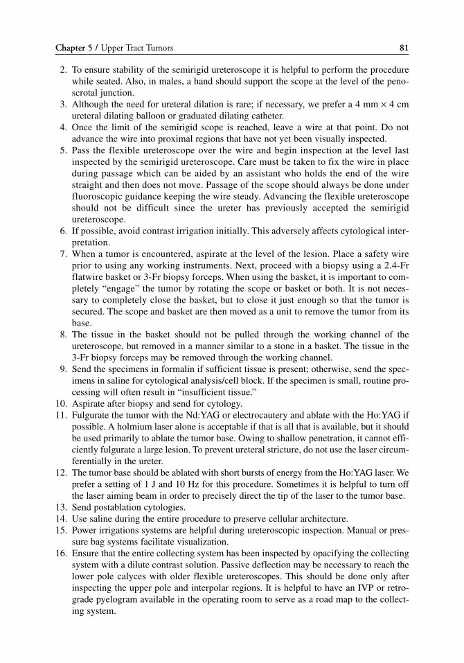
2. To ensure stability of the semirigid ureteroscope it is helpful to perform the procedurewhile seated. Also, in males, a hand should support the scope at the level of the peno-scrotal junction.
3. Although the need for ureteral dilation is rare; if necessary, we prefer a 4 mm × 4 cmureteral dilating balloon or graduated dilating catheter.
4. Once the limit of the semirigid scope is reached, leave a wire at that point. Do notadvance the wire into proximal regions that have not yet been visually inspected.
5. Pass the flexible ureteroscope over the wire and begin inspection at the level lastinspected by the semirigid ureteroscope. Care must be taken to fix the wire in placeduring passage which can be aided by an assistant who holds the end of the wirestraight and then does not move. Passage of the scope should always be done underfluoroscopic guidance keeping the wire steady. Advancing the flexible ureteroscopeshould not be difficult since the ureter has previously accepted the semirigidureteroscope.
6. If possible, avoid contrast irrigation initially. This adversely affects cytological inter-pretation.
7. When a tumor is encountered, aspirate at the level of the lesion. Place a safety wireprior to using any working instruments. Next, proceed with a biopsy using a 2.4-Frflatwire basket or 3-Fr biopsy forceps. When using the basket, it is important to com-pletely “engage” the tumor by rotating the scope or basket or both. It is not neces-sary to completely close the basket, but to close it just enough so that the tumor issecured. The scope and basket are then moved as a unit to remove the tumor from itsbase.
8. The tissue in the basket should not be pulled through the working channel of theureteroscope, but removed in a manner similar to a stone in a basket. The tissue in the3-Fr biopsy forceps may be removed through the working channel.
9. Send the specimens in formalin if sufficient tissue is present; otherwise, send the spec-imens in saline for cytological analysis/cell block. If the specimen is small, routine pro-cessing will often result in “insufficient tissue.”
10. Aspirate after biopsy and send for cytology.11. Fulgurate the tumor with the Nd:YAG or electrocautery and ablate with the Ho:YAG if
possible. A holmium laser alone is acceptable if that is all that is available, but it shouldbe used primarily to ablate the tumor base. Owing to shallow penetration, it cannot effi-ciently fulgurate a large lesion. To prevent ureteral stricture, do not use the laser circum-ferentially in the ureter.
12. The tumor base should be ablated with short bursts of energy from the Ho:YAG laser. Weprefer a setting of 1 J and 10 Hz for this procedure. Sometimes it is helpful to turn offthe laser aiming beam in order to precisely direct the tip of the laser to the tumor base.
13. Send postablation cytologies.14. Use saline during the entire procedure to preserve cellular architecture.15. Power irrigations systems are helpful during ureteroscopic inspection. Manual or pres-
sure bag systems facilitate visualization.16. Ensure that the entire collecting system has been inspected by opacifying the collecting
system with a dilute contrast solution. Passive deflection may be necessary to reach thelower pole calyces with older flexible ureteroscopes. This should be done only afterinspecting the upper pole and interpolar regions. It is helpful to have an IVP or retro-grade pyelogram available in the operating room to serve as a road map to the collect-ing system.
Chapter 5 / Upper Tract Tumors 81
REFERENCES1. Cummings KB. Nephroureterectomy: rationale in the management of transitional cell carcinoma of
the upper urinary tract. Urol Clin North Am 1980; 7(3): 569–578.2. Charbit L, Gendreau MC, Mee S, Cukier J. Tumors of the upper urinary tract: 10 years of experience.
J Urol 1991; 146: 1243–1246.3. Babaian RJ, Johnson DE. Primary carcinoma of the ureter. J Urol 1980; 123(3): 357–359.4. Zungri E, Chechile G, Algaba F, Diaz I, Vila F, Castro C. Treatment of transitional carcinoma of the
ureter: Is controversy justified? Eur Urol 1990; 17: 276–280. 5. Zincke H, Neves RJ. Feasibility of conservative surgery for transitional cell cancer of the upper uri-
nary tract. Urol Clin North Am 1984; 11(4): 717–724.5a. Murphy DM, Zincke H, Furlow WL. Management of high grade transitional cell cancer of the upper
urinary tract. J Urol 1981; 125(1): 25–29.6. Keeley FX Jr, Bibbo M, Bagley DH. Ureteroscopic treatment and surveillance of upper urinary tract
transitional cell carcinoma. J Urol 1997; 157: 1560–1565.7. Elliott DS, Blute ML, Patterson DE, Bergstrahl EJ, Segura JW. Long–term follow-up of endoscopi-
cally treated upper urinary tract transitional cell carcinoma. Urology 1996; 47(6): 819–825.8. Chen GL, Bagley DH. Ureteroscopic management of upper tract transitional cell carcinoma in
patients with normal contralateral kidneys. J Urol 2000; 164(4): 1173–1176.9. Tawfiek ER, Bagley DH. Upper-tract transitional cell carcinoma. Urology 1997; 50(3): 321–329.
10. Jemal A, Murray T, Samuels A, Ghafoor A, Ward E, Thun M. Cancer Statistics 2003. CA Cancer JClin 2003; 53: 5–26.
11. Fraley EE, Lange PH, Hakala TR. Recent studies on the immunobiology and biology of humanurothelial tumors. Urol Clin North Am 1976; 3(1): 31–51.
12. Huben RP, Mounzer AM, Murphy GP. Tumor grade and stage as prognostic variables in upper tracturothelial tumors. Cancer 1988; 62(9): 2016–2020.
13. Gerber GS, Lyon ES. Endourological management of upper tract urothelial tumors. J Urol 1993;150: 2–7.
14. Jenson OM, Knudsen JB, Mclaughlin JK. The Copenhagen case-control study of renal pelvis andureter cancer: role of smoking and occupational exposures. Int J Cancer 1988; 41: 557–561.
15. Morrison AS. Advances in the etiology of urothelial cancer. Urol Clin North Am 1984; 11(4): 557–566.16. Radovanovic Z, Krajinovic S, Hall PW, Petkovic S. Family history of cancer among cases of upper
urothelial tumors in Balkan nephropathy area. J Cancer Res Clin Oncol 1985; 110: 181–183.17. Grossfeld GD, Wolf JS Jr, Litwin MS, et al. Asymptomatic microscopic hematuria in adults: summary
of the AUA best practice policy recommendations. Am Fam Physician 2001; 63(6): 1145–1154.18. Grossfield GD, Litwin MS, Wolf JS Jr, et al. Evaluation of asymptomatic microscopic hematuria in
adults: the American urological association best practice policy-part II: patient evaluation, cytology,voided markers, imaging, cystoscopy, nephrology evaluation, and follow-up. Urology 2001; 57:604–610.
19. Fracchia JA, Motta J, Miller LS, Armenakas NA, Schumann GB, Greenberg RA. Evaluation ofasymptomatic microhematuria. Urology 1995; 46(4): 484–489.
20. Jaffe JS, Ginsberg PC, Gill R, Harkaway RC. A new diagnostic algorithm for the evaluation of micro-scopic hematuria. Urology 2001; 57: 889–894.
21. Grossfeld GD, Carroll PR. Evaluation of microscopic hematuria. Urol Clin North Am 1998; 25(4):661–676.
22. Jamis-Dow CA, Choyke PL, Jenninngs SB, Linehan WM, Thakore KN, Walther MM. Small (<3cm)renal masses: detection with CT versus US and pathologic correlation. Radiology 1996(3); 198:785–788.
23. Warshauer DM, Mc Carthy SM, Street L, et al. Detection of renal masses: sensitivities and specifici-ties of excretory urography/linear tomography, US, and CT. Radiology 1988; 169(2): 363–365.
24. Fielding JR, Steele G, Fox LA, Heller H, Loughlin KR. Spiral computerized tomography in theevaluation of acute flank pain: a replacement for excretory urography. J Urol 1997; 157(6): 2071–2073.
25. Klein LT, Frager D, Subramanium A, Lowe FC. Use of magnetic resonance urography. Urology 1998;52: 602–608.
82 Soderdahl and Fabrizio

26. Jung P, Brauers A, Nolte-Ernsting CA, Jakse G, Gunther RW. Magnetic resonance urographyenhanced by gadolinium and diuretics: a comparison with conventional urography in diagnosing thecause of ureteric obstruction. BJU Int 2000; 86: 960–965.
27. Maier U, Simak R, Neuhold N, The clinical value of urinary cytology: 12 years of experience with615 patients. J Clin Pathol 1995; 48(4): 314–317.
28. Kannan, V. Papillary transitional-cell carcinoma of the upper urinary tract: a cytological review.Diagn Cytopath 1990; 6(3): 204–209.
29. Chen GL, El-Gabry EA, Bagley DH. Surveillance of upper urinary tract transitional cell carci-noma: The role of ureteroscopy, retrograde pyelography, cytology and urinalysis. J Urol 2000; 164(6):1901–1904.
30. Nakada SY. The diagnosis and management of lateralizing essential hematuria. AUA Update Series2003; 22(13): 97–104.
31. Keeley FX, Kulp DA, Bibbo M, McCue PA, Bagley DH. Diagnostic accuracy of ureteroscopic biopsyin upper tract transitional cell carcinoma. J Urol 1997; 157(1): 33–37.
32. Fleming ID, Cooper JS, Henson DE, et al. CC cancer staging manual/American joint committee oncancer.-5th Ed. Philadelphia, Lippincott–Ravens Publishers. 1997: 235–237.
33. Liu JB, Bagley DH, Conlin MJ, Merton DA, Alexander AA, Goldberg BB. Endoluminal sonographicevaluation of ureteral and renal pelvic neoplasms. J Ultrasound Med 1997; 16(8): 515–521.
34. Guarnizo E, Pavlovich CP, Seiba M, Carlson DL, Vaughan ED Jr, Sosa RE. Ureteroscopic biopsy ofupper tract urothelial carcinoma: improved diagnostic accuracy and histopathological considerationsusing a multi-biopsy approach. J Urol 2000; 163(1): 52–55.
35. Suh RS, Faerber GJ, Wolf JS. Predictive factors for applicability and success with Endoscopic treat-ment of upper tract urothelial carcinoma. J Urol 2003; 170: 2209–2216.
36. Bagley DH. Ureteroscopic laser treatment of upper urinary tract tumors. J Clin Laser Med Surg 1998;16(1): 55–59.
37. Canfield SE, Dinney CPN, Droller MJ. Surveillance and management of recurrence for upper tracttransitional cell carcinoma. Urol Clin North Am 2003; 30: 791–802.
38. Jabour ME, Smith AD. Primary percutaneous approach to upper urinary tract transitional cell carci-noma Urol Cl North Am 2000; 27(4): 739–749.
39. Elliott DS, Segura JW, Lightner D, Patterson DE, Blute ML. Is nephroureterectomy necessary in allcases of upper tract transitional cell carcinoma? Long-term results of conservative endourologic man-agement of upper tract transitional cell carcinoma in individuals with a normal contralateral kidney.Urology 2001; 58(2): 174–178.
40. Skacel M, Fahmy M, Brainard JA, et al. Multitarget fluorescence hybridization assay detects transi-tional cell carcinoma in the majority of patients with bladder cancer and atypical or negative urinecytology. J Urol 2003; 169(6): 2102–2105.
41. Sarosdy MF, Schellhammer PF, Bokinsky G, et al. Clinical evaluation of a multi-target fluorescent insitu hybridization assay for detection of bladder cancer. J Urol 2002; 168(5): 1950–1954.
42. Jones JS, Streem SB. Office-based cystoureteroscopy for assessment of the upper urinary tract JEndourol 2002; 16(5): 307–309.
43. Kerbl K, Clayman RV. Incision of the ureterovesical junction for endoscopic surveillance of transi-tional cell cancer of the upper urinary tract. J Urol 1993; 150: 1440–1443.
44. Corwin HL, Silverstein MD. The diagnosis of neoplasia in patients with asymptomatic microscopichematuria: a decision analysis. J Urol 1988; 139(5): 1002–1006.
45. Fielding JR, Silverman SG, Samuel S, Zou KH, Loughlin KR. Unenhanced helical CT of ureteralstones: a replacement for excretory urography in planning treatment. AJR Am J Roentgenol 1998; 171:1051–1053.
46. McNicholas MM, Raptopoulos VD, Schwartz RK, et al. Excretory phase CT urography for opacifi-cation of the urinary collecting system. AJR Am J Roentgenol 1998; 170: 1261–1267.
47. Badalament RA, Bennett WF, Bova JG, et al. Computed tomography of primary transitional cell car-cinoma of the upper urinary tracts. Urology 1992; 40(1): 71–75.
48. Badalament RA, Hermansen DK, Kimmel M, et al. The sensitivity of bladder wash flow cytometry,bladder wash cytology and voided cytology in the detection of bladder carcinoma. Cancer 1987; 60:1423–1427.
Chapter 5 / Upper Tract Tumors 83

49. Xia TL. Cytologic diagnostic value of voided urine in 60 cases of primary transitional carcinoma ofthe renal pelvis and ureter. Chung Hua Wai Ko Tsa Chih 1989; 27(12): 753–755.
50. Daneshmand S, Quek ML, Huffman JL. Endoscopic management of upper urinary tract transitionalcell carcinoma: Long-term experience. Cancer 2003; 98(1): 55–60.
51. Nielsen K, Ostri P. Primary tumors of the renal pelvis: evaluation of clinical and pathological featuresin a consecutive series of 10 years. J Urol 1988; 140(1): 19–21.
51a. Ozsahin M, Zourhair A, Villa S, et al. Prognostic factors in urothelial renal pelvis and ureter tumours:a multicentre rare cancer network study. Eur J Cancer 1999; 35(5): 738–743.
52. Skolarikos A, Griffiths TRL, Powell PH, Thomas DJ, Neal DE, Kelly JD. Cytologic analysis ofureteral washings is informative in patients with grade 2 upper tract TCC considering Endoscopictreatment. Urology 2003; 61(6): 1146–1150.
53. Assimos DG, Hall MC, Martin JH. Ureteroscopic management of patients with upper tract transi-tional cell carcinoma. Urol Cl North Am 2000; 27(4): 751–759.
54. Martinez-Pineiro JA, Matres MJG, Martinez-Pineiro L. Endourological treatment of upper tracturothelial carcinomas: analysis of a series of 59 tumors. J Urol 1996; 156: 377–385.
55. Liatsikos EN, Dinlenc CZ, Kapoor R, Smith AD. Transitional-cell carcinoma of the renal pelvis:ureteroscopic and percutaneous approach. J Endourol 2001; 15(4): 377–383.
56. Clark PE, Streem SB, Geisinger MA. 13-year experience with percutaneous management of uppertract transitional cell carcinoma. J Urol 1999; 161(3): 772–775.
57. Patel A, Soonawalla P, Shepherd SF, Dearnaley DP, Kellett MJ, Woodhous CRJ. Long-term out-come after percutaneous treatment of transitional cell carcinoma of the renal pelvis. J Urol 1996;155(3): 868–874.
58. Jarrett TW, Sweetser PM, Weiss GH, Smith AD. Percutaneous management of transitional cell carci-noma of the renal collecting system: 9-year experience. J Urol 1995; 154(5): 1629–1635.
59. Tomera KM, Leary FJ, and Zinke H. Pyeloscopy in urothelial tumors. J Urol 1982; 127: 1088–1089.60. Lim DJ, Shattuck MC, Cook WA. Pyelovenous lymphatic migration of transitional cell carcinoma
following ureterorenoscopy. J Urol 1993; 149(1): 109–111.61. Kulp DA, Bagley DH. Does flexible ureteropyeloscopy promote local recurrence of transitional cell
carcinoma? J Endourol 1994; 8(2): 111–113.62. Hendin BN, Streem SB, Levin HS, Klein EA, Novick AC. Impact of diagnostic ureteroscopy on long-
term survival in patients with upper tract transitional cell carcinoma. J Urol 1999; 161(3): 783–785.63. Chen GL, Bagley DH. Ureteroscopic surgery for upper tract transitional-cell carcinoma: complica-
tions and management. J Endourol 2001; 15(4): 399–404.
84 Soderdahl and Fabrizio

II CALCULUS THERAPY
Ureteroscopy
David E. Rapp, MD
and Glenn S. Gerber, MD
CONTENTS
INDICATIONS
INSTRUMENTS
TECHNIQUES
TIPS AND DIFFICULT SITUATIONS
REFERENCES
6
SUMMARY
As technological advances continue to arise throughout the field of endourology, theindications and endoscopic management of urinary calculi continue to evolve. An excel-lent working knowledge of the various ureteroscopes and associated endoscopic instru-ments currently available is crucial to successful treatment of urinary calculi. Theseinclude guidewires, dilation devices, rigid ureteroscopes, flexible ureteroscopes, stoneextraction devices, and lithotripsy devices. Much of the success of ureteroscopy alsodepends on a solid technical foundation. These technical aspects of ureteroscopy arereviewed in detail throughout this chapter. Finally, urological surgeons may encountertechnically difficult situations that make ureteroscopy challenging. Such situations andpotential approaches to these situations are discussed.
Key Words: Ureteroscopy; ureter; calculus; stones; lithotripsy.
INDICATIONS
As technological advances continue to arise throughout the field of endourology, theindications for ureteroscopic management of urinary calculi continue to change (1).Ureteroscopes and related instruments also are evolving, allowing for treatment ofstones in all locations within the urinary tract while decreasing the morbidity associatedwith intervention. With this evolution comes the need to define when intervention isrequired and which modilities for intervention are best suited for each clinical scenario.
From: Advanced Endourology: The Complete Clinical GuideEdited by: S. Y. Nakada and M. S. Pearle © Humana Press Inc., Totowa, NJ
87

In general, there is a consensus that small stones may be treated with conservative man-agement. For calculi of 5 mm or less, spontaneous passage is seen in most patients.Spontaneous passage rates of 71 to 98% are observed for such stones in the distal ureter,whereas passage rates of proximal ureteral calculi of small size range from 29 to 98% (2).Spontaneous passage is much less likely with stones greater than 5 mm in size, with ratesfor larger calculi in the proximal and distal ureter of only about 50% or less (2).
For patients with stones 5 mm or less in size, a period of observation is appropriate inthe absence of complicating factors such as infection, renal dysfunction, and poor paincontrol. However, observation for longer than 4–6 weeks is generally not advised owingto concerns regarding effects on renal function. The precise duration of observation inwhich the risk of irreversible parenchymal loss is minimal has not been well defined. Inexperimental cases of complete unilateral obstruction, microscopic changes in the kidneyare seen almost immediately, as changes consistent with injury to the papilla and fornixare observed at 24 hours (3). Papillary necrosis is demonstrated in experimental modelsby 7 days (4). Return of renal function following unilateral obstruction is variable,dependent on factors such as duration and degree of obstruction. Animal models haveshown limited return of renal function beyond 40 days, while human cases have beenreported with some return of renal function following 150 days of obstruction (5,6).Regardless, in cases where spontaneous passage is unlikely or clinical symptoms of renalcolic persist after a period of observation, intervention is indicated.
When surgical intervention is required for the treatment of calculi, both ureteroscopicmanagement and extracorporeal shockwave lithotripsy (SWL) are effective modalities forstone removal. Following SWL, Segura et al. (2) reported overall stone-free rates of 83 and85% for calculi located in the proximal and distal ureter, respectively. In the same review,distal ureteral stones treated via ureteroscopic removal were associated with overall stone-free rates of 90%; however, proximal stones yielded stone-free rates of only 72%. Suchstudies suggest that ureteroscopy has very high success rates in treating distal ureteralstones, whereas stone-free rates for proximal ureteral calculi are generally higher with SWL.However, with continued advances in flexible ureteroscopy and the use of the Holmiumlaser, ureteroscopic treatment of proximal ureteral stones has become more successful (7).
Finally, several clinical scenarios exist in which ureteroscopic management of calculimay be indicated due to technical difficulty associated with SWL. Obese patients mayrepresent a particular challenge to SWL as stone visualization is often poor. In these cases,fluoroscopic imaging may be suboptimal. When stone visualization is achieved, highamounts of radiation exposure are often required. Likewise, distal stones may be difficultto treat utilizing SWL. The bony structures of the pelvis can impede both fluoroscopicvisualization and decrease the shockwave energy transmitted to the calculus. Finally,SWL is contraindicated in pregnant patients and those with uncorrected bleeding diathe-ses. In these cases, ureteroscopic management of ureteral calculi is generally appropriate.
INSTRUMENTS
A thorough working knowledge of the various ureteroscopes and associated endo-scopic instruments currently available is crucial to successful treatment of urinary cal-culi.The following ureteroscopic equipment will be discussed in detail in this section:
1. Guidewires.2. Dilation devices.3. Rigid ureteroscopes.
88 Rapp and Gerber

4. Flexible ureteroscopes.5. Stone extraction devices.6. Lithotripsy devices.
Guidewires
Guidewires are used to gain access to the ureter, which is the initial step in mostureteroscopic surgery. Guidewires differ with respect to size, flexibility, and coating mate-rial. Typically, wire diameter ranges from 0.018 to 0.038 in. Guidewire length ranges from145 to 260 cm. Although individual preferences vary, we generally use a 145-cm, 0.038-inguidewire for standard ureteroscopic procedures. Calibrated guidewires are now availablewhich are marked to help identify length during ureteroscopic cases. Guidewire surfacesare generally coated with polytetrafluoroethylene (PTFE) or a hydrophilic material, bothof which are designed to create a frictionless surface. We have found that hydrophilicguidewires are best used to navigate past tight strictures or impacted stones. However,they may become displaced more easily, thus jeopardizing ureteral access. Therefore, wegenerally prefer to use PTFE-coated wires whenever possible.
Guidewires have various tip designs available to aid in navigation of the ureter whileattempting to minimize ureteral injury. In addition to standard straight-tippedguidewires, various shapes, such as angled tips, are available. These various guidewiretip designs help to navigate tortuous or narrow ureteral segments, as well as bypassimpacted stones. Tip designs also include flexible or fixed tips. Finally, the core guidewire may be flexible, stiff, or super-stiff. Stiff variants may help to prevent kinking dur-ing passage of ureteroscopic instruments and may help straighten tortuous ureters thathave been chronically obstructed.
Ureteral Dilation Devices
With the introduction of small caliber rigid and flexible ureteroscopes, as well as thepowerful and precise stone fragmentation associated with the Holmium laser, the need foractive ureteral dilation during ureteroscopy has decreased. However, there are still selectedcases in which active dilation is useful. For these situations, a variety of dilation systemsare available to aid in passage of ureteroscopic equipment into the ureter. Mechanical dila-tion is the oldest mechanism of ureteral dilation. Mechanical dilation may be accomplishedvia simple ureteral stent insertion during a separate procedure prior to attemptingureteroscopy. Placement of a stent for 3 to 7 days leads to passive ureteral dilation. Morecommonly, active dilation is accomplished during a simultaneous procedure. Dilators witha tapered tip are available for the purpose of dilating the ureteral orifice and distal ureteronly. Conversely, mechanical dilators of progressively increasing calibers are available todilate larger ureteral lengths. Finally, dilation systems including multiple dilators of differ-ent calibers, such as graduated Teflon dilators, allow for progressive ureteral dilation.
Balloon dilating systems are now commonly used in ureteroscopy. These systems areintroduced into the ureter over a guidewire. The balloon is located at the distal aspectof the ureteral catheter and is advanced into position using a radio-opaque marker at thedistal end of the balloon. The balloon is then inflated with contrast media to aid in flu-oroscopic visualization. A pressure gage is used to monitor intraballoon pressure,thereby preventing overinflation, rupture, and ureteral injury. In general, balloon dila-tion is preferred to mechanical dilation because it is more precise and less traumatic tothe ureter.
Chapter 6 / Ureteroscopy 89

Tab
le 1
Com
pari
son
of C
urre
ntly
Ava
ilabl
e R
igid
Ure
tero
scop
esa
Wor
king
Dir
ecti
on o
f M
odel
Cha
nnel
sC
hann
el s
ize
(Fr)
Type
Dia
met
er (
Fr)
leng
th (
cm)
view
(de
gree
s)
Oly
mpu
sA
2940
A/4
1A1
4.2
45°
ocul
ar6.
4/7.
843
(40A
)/33
(41A
)7
2948
A/4
9ASt
raig
ht o
cula
r43
(48A
)/33
(49A
)A
2942
A6.
645
°oc
ular
8.6/
9.8
437
A22
940A
/42A
4.2(
40A
)/6.
6(42
A)
45°
mov
able
6.4/
7.8(
40A
),8.
6/9.
8(42
A)
437
WA
0294
3A/
23.
4 &
2.4
45°
ocul
ar7.
543
744
A/4
6ASt
raig
ht o
cula
r 43
(44A
)/33
(46A
)
Wol
f87
02.5
17/.5
181
4.2
×4.
6St
raig
ht o
cula
r6/
7.5
33(.
517)
/43(
.518
)5
8703
.517
/.518
5.2
×6.
28/
9.8
33(.
517)
/43(
.518
)12
8702
.523
/.524
4.2
×4.
6Pa
ralle
l off
set
6/7.
531
.5(.
523)
/43(
.524
)5
8707
.523
/.524
5.2
×6.
28/
9.8
31.5
(.52
3)/4
3(.5
24)
1287
04.5
23/.5
246.
2×
8.2
8.5/
11.5
31.5
(704
)/42
.5(7
14)
1287
02.5
33/.5
344.
2×
4.7
45°
offs
et
6/7.
5 31
.5(.
533)
/43(
.534
)5
8707
.533
/.534
5.2
×6.
2 8/
9.8
31.5
(.53
3)/4
3(.5
34)
1287
08.5
13/.5
142
4.2
& 2
.5×
3.8
Stra
ight
6.5
/8.5
33(.
513)
/43(
.514
)5
8708
.533
/.534
4.2
& 2
.5×
3.8
45°
offs
et6.
5 /8
.531
.5(.
533)
/43(
.534
)5
8709
.401
/421
4.2
×5
&3.
2×
3.4
45°
offs
et8.
9/9.
831
(401
)/42
.5(4
21)
10
AC
MI
MR
O-7
33/7
421
5.4
45°
ocul
ar7/
9.2
33(7
33)/
42(7
42)
5M
R-6
/6L
23.
4 &
2.3
Stra
ight
ocu
lar
6.9/
8.3
33(6
)/41
(6L
)5
STO
RZ
2743
0K/4
30L
35/
2.4/
2.4
45°
ocul
ar8/
934
(K)/
43(L
)0
2740
1K/4
01L
5.5/
3.0/
2.0
45°
ocul
ar10
/10.
534
(K)/
43(L
)0
411K
/411
L45
°m
ovab
le34
(K)/
43(L
)27
400C
K/4
00C
L2
3.5
& 2
.345
°oc
ular
7.5/
934
(CK
)/43
(CL
)0
2741
0CK
/410
CL
45°
mov
able
34(C
K)/
43(C
L)
a Spe
cifi
catio
ns p
rovi
ded
by m
anuf
actu
rers
.
90
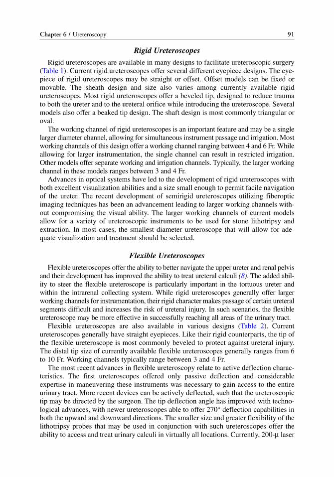
Rigid Ureteroscopes
Rigid ureteroscopes are available in many designs to facilitate ureteroscopic surgery(Table 1). Current rigid ureteroscopes offer several different eyepiece designs. The eye-piece of rigid ureteroscopes may be straight or offset. Offset models can be fixed ormovable. The sheath design and size also varies among currently available rigidureteroscopes. Most rigid ureteroscopes offer a beveled tip, designed to reduce traumato both the ureter and to the ureteral orifice while introducing the ureteroscope. Severalmodels also offer a beaked tip design. The shaft design is most commonly triangular oroval.
The working channel of rigid ureteroscopes is an important feature and may be a singlelarger diameter channel, allowing for simultaneous instrument passage and irrigation. Mostworking channels of this design offer a working channel ranging between 4 and 6 Fr. Whileallowing for larger instrumentation, the single channel can result in restricted irrigation.Other models offer separate working and irrigation channels. Typically, the larger workingchannel in these models ranges between 3 and 4 Fr.
Advances in optical systems have led to the development of rigid ureteroscopes withboth excellent visualization abilities and a size small enough to permit facile navigationof the ureter. The recent development of semirigid ureteroscopes utilizing fiberopticimaging techniques has been an advancement leading to larger working channels with-out compromising the visual ability. The larger working channels of current modelsallow for a variety of ureteroscopic instruments to be used for stone lithotripsy andextraction. In most cases, the smallest diameter ureteroscope that will allow for ade-quate visualization and treatment should be selected.
Flexible Ureteroscopes
Flexible ureteroscopes offer the ability to better navigate the upper ureter and renal pelvisand their development has improved the ability to treat ureteral calculi (8). The added abil-ity to steer the flexible ureteroscope is particularly important in the tortuous ureter andwithin the intrarenal collecting system. While rigid ureteroscopes generally offer largerworking channels for instrumentation, their rigid character makes passage of certain ureteralsegments difficult and increases the risk of ureteral injury. In such scenarios, the flexibleureteroscope may be more effective in successfully reaching all areas of the urinary tract.
Flexible ureteroscopes are also available in various designs (Table 2). Currentureteroscopes generally have straight eyepieces. Like their rigid counterparts, the tip ofthe flexible ureteroscope is most commonly beveled to protect against ureteral injury.The distal tip size of currently available flexible ureteroscopes generally ranges from 6to 10 Fr. Working channels typically range between 3 and 4 Fr.
The most recent advances in flexible ureteroscopy relate to active deflection charac-teristics. The first ureteroscopes offered only passive deflection and considerableexpertise in maneuvering these instruments was necessary to gain access to the entireurinary tract. More recent devices can be actively deflected, such that the ureteroscopictip may be directed by the surgeon. The tip deflection angle has improved with techno-logical advances, with newer ureteroscopes able to offer 270° deflection capabilities inboth the upward and downward directions. The smaller size and greater flexibility of thelithotripsy probes that may be used in conjunction with such ureteroscopes offer theability to access and treat urinary calculi in virtually all locations. Currently, 200-μ laser
Chapter 6 / Ureteroscopy 91

Tab
le 2
Com
pari
sion
of
Cur
rent
ly A
vaila
ble
Flex
ible
Ure
tero
scop
esa
Man
ufac
ture
rO
lym
pus
Ric
hard
Wol
fA
CM
IK
arl
Stor
z
Mod
elU
RF-
P373
25.1
7273
30.0
7273
31.0
01D
UR
-8D
UR
-8 E
lite
1127
4AA
1127
3BD
Flex
-X
Dis
tal t
ip s
ize
(Fr)
6.9
7.5
7.4
7.4
6.75
6.75
7.5
10.5
7.5
Wor
king
leng
th
7070
7060
6564
7054
67.5
Cha
nnel
siz
e (F
r)3.
63.
64.
54
3.6
3.6
3.6
4.5
3.6
Act
ive
defl
ectio
n18
0/18
013
0/16
013
0/16
013
0/16
017
5/18
517
0/18
0 pl
us 1
3017
0/12
018
0/80
270/
270
in d
egre
es (
up/d
own)
(2°)
(2°)
Pass
ive
defl
ectio
n4.
0–7.
06
66
7.5
7.5
N/A
N/A
N/A
Loc
atio
n (c
m f
rom
tip)
Dir
ectio
n of
00
00
12 (
air)
12 (
air)
00
0V
iew
(de
gree
s)
9 (w
ater
)9
(wat
er)
Fiel
d of
vie
w (
degr
ees)
9060
6060
8080
9090
90D
epth
of
fiel
d (m
m)
1.5–
503–
503–
503–
502–
402–
402–
502–
502–
50
a Spe
cifi
catio
ns p
rovi
ded
by m
anuf
actu
rers
.
92

Chapter 6 / Ureteroscopy 93
Fig. 2. Ureteroscopic baskets, left to right: four-wire Segura basket, three-wire helical basket, tiplesshelical basket, tipless non-helical basket.
Fig. 1. Standard three-pronged grasping forceps.
fibers and nitinol based baskets are available which have been shown to cause minimalinfluence on the deflection capabilities of flexible ureteroscopes (9).
Stone Extraction Devices
A variety of ureteroscopic instruments are available to extract calculi within the uri-nary tract. These include endoscopic graspers that are available in two- or three-prongedtypes and may be retracting or nonretracting (Fig. 1). Ureteroscopic baskets offeranother accessory to aid in stone extraction. Basket varieties include helical, double-helical, tipless, and parachute designs (Fig. 2).

A notable technological advance is the development of nitinol instruments. The nitinolbaskets allow for sheathless models, thereby decreasing the diameter of the baskets. Thesmaller diameter and flexibility of these instruments allow for greater flexion capabilitieswhen used in conjuction with flexible ureteroscopes (9). This ability becomes increasinglyimportant when attempting to manage calculi within the proximal ureter and renal calyces.Further, the smaller diameter of these instruments aids in increasing irrigant capabilitiesand visualization when the baskets are in use.
Tipless baskets are a second recent development aiding in stone extraction. Thisdesign is novel from its predecessors in that the distal tip does not extend while deploy-ing the basket. This feature becomes important when trying to entrap calculi lodgedagainst the caliceal wall.
Ex vivo models have demonstrated a higher risk of mucosal and muscle damage whenusing parachute baskets and three-pronged graspers as compared to two-pronged graspers,helical, and Segura baskets (10). Although small differences may be seen amongstureteroscopic instruments, the choice of stone extraction device depends greatly on sur-geon experience and level of comfort with each type of instrument.
Lithotripsy Devices
Several types of intracorporeal lithotripsy devices are available to the ureterosocopicsurgeon. These modalities include pneumatic lithotriptors, electrohydraulic lithotrip-tors, ultrasonic lithotriptors, and laser lithotriptors. A detailed discussion of their use isfound in the Lithotripsy Section.
TECHNIQUES
Ureteral Access
Ureteroscopic surgery begins with obtaining access to the ureter. A thorough knowl-edge of the underlying anatomy is important to achieving successful access to theureters. Following entry past the bladder neck lies the trigone. Within the trigone liesthe interureteric ridge. This crescent-shaped elevation lies at the proximal border of thetrigone. In most patients the interureteric ridge can be easily identified and traced later-ally to identify the ureteral orifices. The ureteral orifices often can be easily identifiedand may have a round, oval, or crescent shape. In some cases, however, identificationof the orifices is difficult. In these cases, methylene blue or indigo carmine may beinjected to aid in identification. If the obstruction is complete, however, passage of dyeand visualization may be limited or delayed.
We prefer to insert a guidewire into the ureteral orifice as the initial step in anyureteroscopic case. A ureteral catheter may be used to direct the guide wire into theureteral orifice. Prior to attempting to pass the guidewire, fluoroscopy should be usedto image the pelvis and surrounding anatomy. Frequently, a radio-opaque stone may bevisualized under fluoroscopy. In these cases, retrograde ureterography should be per-formed carefully to avoid obscuring the stone with contrast. When the calculus is notvisualized using fluoroscopy, retrograde ureterography may be used to assess the loca-tion of the stone. Three areas exist where the ureteral diameter becomes narrowed andare prone to stone obstruction, including the ureteropelvic junction, at the pelvic brimwhere the ureter crosses the iliac vessels, and at the ureterovesical junction.
While advancing the guidewire, care should be taken to avoid ureteral injury, such assubmucosal passage or ureteral perforation. If resistance is encountered during passage,
94 Rapp and Gerber

the guidewire should be carefully manipulated based on the appearance of the retrogradeureterogram. Use of a hydrophilic-coated guidewire or an angled, flexible-tippedguidewire may facilitate passage in these cases. If resistance is encountered in the regionof the calculus care must be taken to avoid retrograde propulsion of the stone during guid-wire insertion. When passage of the guidewire past the calculus is difficult, a ureteralcatheter may be advanced over the guidewire to a point just distal to the calculus. Thecatheter may then provide additional stability while attempting to direct the guidewire pastthe calculus. Further, the ureter is frequently inflamed and edematous at the level of stoneimpaction. This may increase the risk of ureteral perforation. In some cases, passage ofthe ureteroscope to the level of the stone may allow guidewire passage under direct vision.
Following successful passage of a guidewire to the level of the renal collecting sys-tem, we generally prefer to place a second guidewire to serve as a safety wire. This canbe accomplished by advancing a double-lumen catheter over the initial guidewire underendoscopic control or by simply passing a second wire directly into the ureter. Owingto the larger caliber of the double-lumen catheter, care should be taken when introduc-ing the catheter into the ureteral orifice so as not to injure the ureter. If significant resist-ance is met during this step, dilation of the orifice may be necessary not only forintroduction of the catheter, but also for subsequent ureteroscopy. Once the double-lumen access catheter had been successfully advanced into the ureter, a secondguidewire is introduced. We have found the double-lumen catheter to be helpful in gen-tly dilating the distal ureter, as well as helping to assess the caliber and distensibility ofthe ureter. If a hydrophilic wire was used for initial access, it may be exchanged at thispoint for a PTFE-coated guidewire, which may serve more safely as a safety wire andbe easier to work with during the remainder of the procedure.
Dilation
Technological advances have led to the development of small ureteroscopes that stillprovide good visualization. Therefore, there is no need for routine ureteral dilation in allureteroscopic procedures. This trend can be seen in a recent comparison of ureteroscopiccases performed at the Mayo Clinic in 1992 as compared with those of 1998. Balloon dila-tion was performed in 74% of these cases in 1992 as compared with only 59% in 1998 (11).Active dilation of the ureter may lead to ureteral edema and perforation. Although clinicalstudies showed that balloon dilation without subsequent stenting was associated with post-operative pain in only a small percentage of patients, it is generally advisable to place aureteral stent in most cases in which significant ureteral dilation has been performed (12).
In selected cases, ureteral dilation may be necessary to treat a ureteral stricture or toallow the ureteroscope to be advanced through the intramural ureter. In such cases, aureteral balloon should be advanced such that it straddles the narrow area. The balloonshould be inflated with contrast medium under fluoroscopic visualization. This allows thesurgeon to assess the pliability and tightness of the ureter, as well as the length of theureteral narrowing. A characterisitic ureteral “waisting” is seen as the balloon is inflated.The balloon should not be inflated to a pressure greater than recommended by the manu-facturer.
Access Sheath
Although ureteral access sheaths were originally designed to help manage difficultprocedures, recent investigators have recommended their routine use in ureteroscopic
Chapter 6 / Ureteroscopy 95

cases (13,14). In addition to easy re-entry to the ureter, access sheaths have been shownto decrease intrarenal pressure while significantly increasing irrigant flow, and also tominimize ureteral trauma associated with repeated passage of the endoscope duringwithdrawal of stone fragments (15). Owing to these characteristics, access sheaths mayprovide surgeons with better visualization during ureteroscopic stone extraction, as wellas decreasing the risk of complications.
Access sheaths are sized based on both the inner and outer diameter of the sheath.The most common sheaths are generally sized 10/12, 12/14, and 14/16 Fr, with varioussheath lengths being available. Under endoscopic guidance, the access sheath is intro-duced into the ureteral orifice over a standard 0.038-in guidewire. The access sheath isthen advanced to a level just below the ureteral calculus, which is confirmed using flu-oroscopy. When used with flexible ureteroscopy in the kidney, the sheath can be posi-tioned at or just below the ureteropelvic junction. A hydrophilic coat aids in advancingthe sheath. An inner dilator with a tapered distal end provides a mechanical dilatingforce to aid in sheath advancement. Once the access sheath is advanced to the properlevel, the inner dilator is then removed, and the ureteroscope may be inserted into thesheath for subsequent stone removal or fragmentation.
In one series, ureteroscopic stone removal assisted by an access sheath was associ-ated with decreased operative time and costs, and facilitated direct visualization andsimple ureteral re-entry. Further, when ureteral dilation was required, dilation done withthe access sheath was associated with a significantly lower amount of postoperativesymptoms when compared with balloon dilation (14). Evidence suggests that routineuse of an access sheath can be done safely, with no increased incidence of ureteral stric-ture formation (16). Although concern has been raised regarding the risk of ureteralischemia with use of a sheath, it has been shown that the decrease in blood flow is tran-sient and it appears that access catheters are safe for use in ureteroscopy (17).
Choice of Ureteroscope
A wide variety of ureteroscopes now exist for the treatment of urinary calculi.Historically, rigid ureteroscopes were used for treatment of calculi within the distalureter. Rigid ureteroscopes have the advantage of good image transmission and largerworking channels. For this reason they are ideal for calculus extraction using a varietyof ureteroscopic instruments, such as forceps and baskets. The tortuosity of the ureterproximal to the iliac vessels makes rigid ureteroscopy in this region more difficult insome cases. For this reason, flexible ureteroscopes are often used above the level of theiliac vessels, where their flexibility allows for better navigation. Owing to their flexibil-ity, these ureteroscopes are most commonly advanced over a guidewire to direct navi-gation and prevent buckling of the instrument.
Rigid ureteroscopes are also more easily used by a single operator because the instru-ment can be controlled with one hand as instruments are manipulated using the otherhand. In contrast, it often requires both hands to manipulate a flexible ureteroscope.Therefore, a more experienced and better trained assistant may be needed when perform-ing flexible ureteroscopy. Another disadvantage of flexible ureteroscopy is their relativefragility. Landman et al. (18) have found that flexible ureteroscope failure is expectedafter 9 to 25 cases. Repairs are expensive and may lead to annual costs of $10 to 50,000depending on frequency of use.
96 Rapp and Gerber

Lithotripsy
In many cases, the treatment of ureteral calculi will require lithotripsy. Followinglithotripsy, remaining stone fragments can either be extracted utilizing previouslydescribed techniques, or allowed to pass with or without the aid of a ureteral stent.Several types of intracorporeal lithotriptors exist for the management of caluculi,including pneumatic lithotriptors (PLs), electrohydraulic lithotriptors (EHLs), ultra-sonic lithotriptors (ULs), and laser lithotriptors (LLs).
PLs fragment calculi through the transfer of mechanical energy to an inflexibleobject, such as a urinary stone. PLs are effective in fragmenting calculi throughout thelength of the urinary tract. One major advantage of PL lies in their margin of safety,which is significantly better than EHL, UL, or LL (19). The rigidity of the PLs, how-ever, generally prevent them from being used with flexible ureteroscopes.
Ultrasonic lithotripsy is based on the generation of ultrasonic vibrations that are used tofragment urinary calculi. UL is an effective modality in the treatment of stone disease asdemonstrated in many studies (19–21). The major advantage to UL lies in its ability to com-bine stone fragementation with stone removal, which is particularly helpful when treatinglarge kidney stones percutaneously. This is accomplished through an aspiration port as partof the lithotriptor probe. UL probes have the major disadvantage of being inflexible, limit-ing their use to a ureteroscope with a straight working channel. They may also cause signif-icant ureteral injury. In general, UL has been replaced by more effective and safer options.
EHL fragment calculi through energy produced following an underwater dischargeof electrical current. EHL is effective in stone fragmentation and has the advantage offlexible probes, which allow for the treatment of more proximal stones (21). EHL alsomay cause significant ureteral injury and is not commonly used with ureteroscopy as aresult of the development of better lithotripsy devices.
Biri et al. (20) compared PL, UL, and EHL in the treatment of 1121 patients withlower ureteral calculi. In their series, patients treated with PL had the highest stone-freerates, while those treated with EHL had the highest rate of complications. All forms ofintracorporeal lithotripsy were more efficacious in the treatment of calculi than SWL,with stone-free rates ranging from 89 to 96%. The effectiveness of these various intra-corporeal lithotriptors has been confirmed in other studies (21).
Lasers
The use of laser energy in the treatment of urinary calculi disease has dramaticallyimproved the ability to manage stone disease endoscopically. Laser energy was first uti-lized in the treatment of urinary stone disease in the form of a ruby laser (22). Since itsfirst use in lithotripsy, laser technology has been significantly refined and now is animportant modality used in the the treatment of urinary calculi.
The holmium:yttrium-aluminium-garnet (Ho:YAG) laser is currently the most com-monly used laser in the treatment of urinary calculi. Lithotripsy via Ho:YAG laser isachieved via a photothermal mechanism. The Ho:YAG laser emits energy at a wave-length of 2100 nm with a corresponding tissue penetration depth of less than 0.5 mm.Although the Ho:YAG laser can cause ureteral perforation, it is unlikely to cause sig-nificant trauma owing to its limited depth of penetration.
The efficacy of Ho:YAG lithotripsy has been demonstrated in multiple studies(23–25). This efficacy has been proven irrespective of stone type or stone location withinthe ureter or kidney. Stone-free rates of 98,100, and 97% were demonstrated using retro-grade Ho:YAG laser lithotripsy for calculi in the distal, middle, and proximal ureter,
Chapter 6 / Ureteroscopy 97

respectively. In the same series, however, retrograde treatment of renal calculi droppedto 84% (23). This is largely the result of issues related to stone accessibility rather thaneffectiveness of the laser. Prospective series have also suggested efficacy of Ho:YAGlaser lithotripsy regardless of stone burden (24). The mechanism of stone fragmentationaccomplished via Ho:YAG laser is also effective for all stone types, including cystinestones (22).
Ho:YAG laser lithotripsy has also been established as a safe form of intracorporeallithotripsy. Ureteral perforation and stricture are potential complications of laserlithotripsy. Nonetheless, laser-related complications have been shown to occur in lessthan 1% of cases (26,27). Further, laser ureteroscopy has been shown to have no dele-terious effects on renal function (28). Reports have demonstrated cyanide production asa result of Ho:YAG lithotripsy of uric acid calculi. However, the level produced is gen-erally considered clinically insignificant (29).
Other forms of laser lithotripsy have been successfully used to treat urinary calculi.Dye lasers use an organic dye excited by another light source. Although effective stonefree rates using dye laser have been shown in many series, this form of laser lithotripsyis expensive and involves complicated maintenance requirements owing to the need tochange the liquid dye at regular intervals (22,30–32). The use of the alexandrite laser hasalso been evaluated in several series (33–35). In these reports, stone-free rates followinglithotripsy for ureteral calculi ranged from 67 to 95%, with one series citing an overallstone-free rate of 80%. Efficacy was found to be dependent on both stone burden andureteral location (33,34). In their series, Denstedt et al concluded that the alexandritelaser was inferior to other laser systems when used for intracorporeal lithotripsy (35).
The erbium:YAG laser emits light energy at a wavelength of 2940 nm with a depth oftissue penetration of 3 to 6 mm. Recently, the mechanism of lithotripsy with this laser hasbeen evaluated for prospective use in the clinical setting. This evaluation has led authorsto suggest a future use in lithotripsy, stating that Er:YAG lasers may deliver energy in amore efficient and safe manner than Ho:YAG lasers (36).
Regardless of laser chosen for lithotripsy, proper technique must be utilized to min-imize the incidence of laser-related complications. Protective eye wear must be worn bythe surgeon, all operating room personnel, and the patient in all cases utilizing theHo:YAG laser. Prior to intracorporeal use, the laser fiber must be inspected for break-age. Unrecognized fiber breakage can result in laser emission proximal to the fiber endwith surrounding tissue injury or damage to the ureteroscope. Laser energy emitted inthis fashion can also be transmitted through the ureteroscope itself, resulting in tissuedamage. The optical fiber must always be visualized directly on the stone prior to dis-charge of laser energy to prevent injury to tissue structures surrounding the targeted cal-culus. Aggressive irrigation during lithotripsy may aid in visualizing the optical fiberand in keeping the operating field clear of stone debris.
Proximal Stone Migration
The prevention of retrograde stone migration is a technical challenge often encoun-tered during ureteroscopy. In a series by Cheung et al. (24), stone clearance rates for cal-culi in the proximal ureter were found to be higher for larger stones. This finding islikely owing to the fact that larger stones were less likely to migrate during the processof lithotripsy. If retrograde stone migration into the renal collecting system does occur,the technical difficulty of the procedure is often greater as a flexible ureteroscope isoften needed to manage such displaced calculi.
98 Rapp and Gerber

Several techniques exist to help prevent retrograde fragment migration. Althoughexcellent visualization is necessary to both allow for lithotripsy and prevent inadvertentureteral injury, decreasing the irrigant force during stone fragmentation may help pre-vent migration. Positioning the operating table in reverse trendelenberg can also behelpful in some cases. Alternatively, the lithotriptor probe may be positioned at the ante-rior aspect of the calculus and a mild amount of posterior pressure may be applied tohold the stone against the posterior ureteral wall during the lithotripsy. If retrogrademigration is observed during lithotripsy, the stone may be pulled distally in the ureterusing ureteroscopic graspers prior to continuing with lithotripsy.
The choice of lithotriptor is an important consideration in minimizing the risk ofproximal migration. Pneumatic lithotriptors, for example, are accompanied by a highrate of stone propulsion and subsequent migration. Migration rates during PL rangefrom 2 to 17% (37). Stone migration rates during EHL have also been significant, withone series reporting proximal migration rates of 14% (38). In contrast, significant prox-imal migration during Ho:YAG laser lithotripsy appears to be less of a problem (39).
The Dretler stone cone (Medsource, Norwell, MA) offers a new technique to preventretrograde migration. The device functions also as a guidewire and may be passed prox-imally into the renal pelvis. A radio-opaque marker delineates the location of the stonecone. The marker is introduced past the site of the calculus at which point the stone coneis deployed. In one series utilizing the Dretler stone cone, only 6 of 50 patients hadresidual stone fragments and no patients required auxiliary procedures (40).
Finally, ureteral caliber in an important risk factor for stone migration. If ureteralstents have been placed prior to attempting lithotripsy, the dilated ureter may increasethe risk of migration. In this setting, utilization of the previously described techniquesto prevent stone migration is more important. In addition, the ureter proximal to anobstructing stone is often dilated, thus increasing the risk of proximal calculus move-ment. In general, stone migration is best managed by avoiding the problem. However,appropriate use of flexible ureteroscopy usually will allow the surgeon to manage thisproblem effectively and avoid the need for secondary procedures to treat the stone.
Stentless Ureteroscopy
Placement of ureteral stents following ureteroscopic stone extraction has been commonsince the advent of ureteroscopy. Routine ureteral stenting following ureteroscopy hasbeen suggested to decrease postoperative pain and stricture formation (41,42). Reports ofstricture rates following ureteroscopy currently range from 2 to 4% (43). Recent studieshave demonstrated that stents do not need to be routinely inserted following uncompli-cated ureteroscopic stone extraction. Borboroglu et al. (44) demonstrated a comparablestone-free rate when comparing patients undergoing stented vs stentless ureteroscopy forthe treatment of distal ureteral stones. Similar success has been shown using stentlessureterscopy to manage stones in all ureteral locations (44,45). Although proponents ofstented ureterscopic stone extraction cite decreased postoperative pain to be associatedwith stent placement, recent studies have found postoperative pain, narcotic use, and uri-nary symptoms to be higher in stented groups of patients (44,46). Finally, a significantsaving in cost is associated with stentless ureteroscopy (45).
Identifying cases where stentless ureteroscopy can be performed safely continues tobe a challenge. Hollenbeck et al. (47) performed a multivariate analysis to identify fac-tors predictive of postoperative morbidity in patients undergoing stentless ureteroscopy.Risk factors associated with a higher prevalance of postoperative morbidity included
Chapter 6 / Ureteroscopy 99

renal pelvic stone location, history of urolithiasis, and history of urinary tract infections.Further operative variables predictive of postoperative morbidity included bilateralunstented procedures and operative time of greater than 45 minutes with concomitantuse of lithotripsy (47). Stents most commonly should be placed if there is significantureteral edema or inflammation, when ureteral perforation has occurred, or if there aresignificant residual stone fragments.
TIPS AND DIFFICULT SITUATIONS
Difficult Ureteral Access
Patients with urinary diversions are at increased risk of forming kidney stones. In a lit-erature review, Beiko et al. (48) reported that the incidence of stone development forpatients with colonic conduits, ileal conduits, the Kock pouch, ileal ureter, continent cecalreservoirs, the Mitrofanoff procedure, and vesicostomies was 3 to 4, 10 to 12, 16.7, 17, 20,10 to 27 and 33%, respectively. Special considerations are important in the management ofpatients with these various types of diversions who form ureteral and renal calculi.
Endoscopic management of calculi in patients with urinary diversions and large cal-iber stomas can be accomplished using a transstomal approach (49). Because of the riskof injuring the underlying continence mechanism, Patel and Bellman (50) recommendusing a percutaneous endoscopic approach when a small caliber stoma, such as com-monly seen with the Mitrofanoff diversion, is encountered. Standard endoscopic tech-niques may be used when treating stones within urinary diversions; however, care mustbe taken to avoid injury to the diversion itself. In addition, visualization is often limitedby the mucosal folds of the pouch.
Orthotopic neobladder creation has become increasing utilized for diversion fol-lowing radical cystectomy. Management of ureteral stones in patients with orthotopicneobladders poses a challenge to urologists. Ureteral access is often difficult as aresult of the lack of standard anatomical landmarks associated with neobladders.Therefore, localization of the ureteral opening into the neobladder may be very diffi-cult. In addition, angulation of the ureteral entry may also severely limit retrogradeaccess to the ureter. As such, antegrade access is often required for the treatment ofureteral stones.
Nelson et al. (51) have described a small experience involving retrograde ureteralaccess in patients with orthotopic neobladders. In 10 of 13 patients, the ureters and renalpelvis were successfully accessed. In most cases, the authors were able to access theureters under direct visualization using a 0.035-in. directional glidewire. A cystogramwas routinely performed, which often helped to delineate the afferent limb owing toreflux and aid in identification of the ureteral orifices. The ureters were anastomosed toan afferent limb in 11 of the patients in this series, while in the remaining 2 they wereanastomosed to the neobladder dome. Administration of intravenous indigo carminemay also be helpful in identifying the ureter.
Treatment of Calculi Located Within Caliceal Diverticula
The treatment of calculi located within caliceal diverticula continues to remain a chal-lenge for even the advanced ureteroscopic surgeon. Patients with caliceal diverticula oftenpresent with pain and recurrent urinary tract infection, and such diverticula are often asso-ciated with calculi owing to urinary stasis (52). A variety of surgical approaches exist to
100 Rapp and Gerber

treat stones located within caliceal diverticula, including SWL, retrograde ureteroscopy,laparoscopy, percutaneous nephrolithotripsy, and open surgical management.
In their comparison of ureteroscopy and percutaneous lithotripsy for the treatment ofsymptomatic caliceal diverticula, Auge et al. (53) reported only a 19% stone-free ratefollowing ureteroscopic management. In 24% of the patients undergoing ureteroscopicmanagement, it was not possible for Auge et al. to identify the ostium and 41% of thesepatients ultimately underwent percutaneous management. This experience led theauthors to conclude that complex posterior caliceal diverticula should be managed in apercutaneous fashion (53). Percutaneous management of calculi located within anteriorcaliceal diverticula may be especially challenging as percutaneous access and subse-quent passage through the diverticular ostium is difficult to achieve (54). In this situa-tion, a retrograde approach may be indicated.
When ureteroscopy is utilized to treat diverticular calculi, a standard retrogradeapproach is used. A working knowledge of the diverticular anatomy is crucial, as thediverticular neck and ostium are often difficult to locate. A preoperative computedtomography may aid in defining the diverticular anatomy and should be available dur-ing the operative procedure. Once the diverticular neck has been identified, a ureteralcatheter should be used to inject contrast medium within the diverticulum to define therelevant anatomy. A guidewire should then be advanced into the diverticulum. As mostcaliceal diverticula have a narrow or stenotic neck, a short balloon dilating systemshould then be used to achieve mechanical dilation. The diverticulum is then accessedwith a flexible ureteroscope and lithotripsy is performed in standard fashion. Stone frag-ments are then removed using ureteroscopic forceps or a tipless basket.
For larger stone burdens and calculi within long-necked diverticula, Grasso et al.(55) describe a combined antegrade and retrograde approach. In Grasso’s technique,the ostium is indentified and opacified in a standard retrograde approach. Followingopacification of the diverticulum, a percuteanous guidewire was directed into thediverticulum and pulled into the ureter via a ureteroscopic basket. After establishingthrough and through access, percutaneous management of the diverticular calculus wasperformed (55).
Ureteroscopic Management of Lower Pole Calculi
Lower pole calculi are often difficult to manage. Historically, these stones were dif-ficult to reach even with flexible ureteroscopes. With technological advances, uretero-scopes with greater active and secondary flexibility have been developed. Althoughthese new ureteroscopes have made accessing lower pole stones easier, lithotripsy isoften limited by the flexibility of the various laser fibers and other instruments for stoneextraction. Because of these limitations, percutaneous nephrolithotomy (PCNL) orSWL have often been used for the treatment of lower pole calculi.
Although SWL offers a minimally invasive modality for the treatment of thesestones, clearance rates are often low (56). Infundibulopelvic angle and caliceal pelvicheight measurements have both been used to predict success of SWL of lower polestones (57). Though PCNL offers better stone-free rates, this approach is more invasiveand is associated with a higher rate of complications, ranging from 13 to 38% (58).
A technique of calculus displacement has been described, in which lower pole calculiare ureteroscopically repositioned to the renal pelvis or upper calyces where lithotripsycan then be undertaken (59–61). Such a technique may be useful when access to lower
Chapter 6 / Ureteroscopy 101

pole calculi is limited by the flexibility of the laser fiber or other lithotripsy instruments.Repositioning of the calculi within middle or upper pole calyces then allows for success-ful laser lithotripsy. Stone-free rates of 83 to 90% have been reported following uretero-scopic lithotripsy of calculi within the lower pole. In these series, stones were treated insitu or via stone repositioning when in situ lithotripsy was not possible (60,61).
REFERENCES1. Bagley DH. Editorial: ureteroscopy continues to evolve. J Urol 2003; 170: 111.2. Segura JW, Preminger GM, Assimos DG, et al. Ureteral stones clinical guidelines panel summary
report on the management of ureteral calculi. J Urol 1997; 158: 1915–1921. 3. Heyman SN, Fuchs S, Jaffe R, et al. Renal microcirculation and tissue damage during acute ureteral
obstruction in the rat: effect of saline infusion, indomethacin and radiocontrast. Kidney Int 1997; 51:653–663.
4. Nagle RB, Bulger RE. Unilateral obstructive nephropathy in the rabbit. II. Late morphologic changes.Lab Invest 1978; 38: 270–278.
5. Shapiro SR, Bennett AH. Recovery of renal function after prolonged unilateral ureteral obstruction.J Urol 1976; 115: 36–40.
6. Vaughan ED Jr., Gillenwater JY. Recovery following complete chronic unilateral ureteral occlusion:functional, radiographic and pathologic alterations. J Urol 1971; 106: 27–35.
7. Bagley DH. Expanding role of ureteroscopy and laser lithotripsy for treatment of proximal ureteraland intrarenal calculi. Curr Opin Urol 2002; 12: 277–280.
8. Painter DJ, Keeley FX Jr. New concepts in the treatment of ureteral calculi. Curr Opin Urol. 2001;11: 373–378.
9. Michel MS, Knoll T, Ptaschnyk T, Kohrmann KU, Alken P. Flexible ureterorenoscopy for the treat-ment of lower pole calyx stones: influence of different lithotripsy probes and stone extraction toolson scope deflection and irrigation flow. Eur Urol 2002; 41: 312–316.
10. Ptashnyk T, Cueva-Martinez A, Michel MS, Alken P, Kohrmann KU. Comparative investigations on theretrieval capabilities of various baskets and graspers in four ex vivo models. Eur Urol 2002; 41: 406–410.
11. Chow GK, Patterson DE, Blute ML, Segura JW. Ureteroscopy: effect of technology and technique onclinical practice. J Urol 2003; 170: 99–102.
12. Hosking DH, McColm SE, Smith WE. Is stenting following ureteroscopy for removal of distalureteral calculi necessary? J Urol 1999; 161: 48–50.
13. Rich M, Lee WJ, Smith AD. Applications of the peel-away introducer sheath. J Urol 1987; 137:452–454.
14. Kourambas J, Byrne RR, Preminger GM. Does a ureteral access sheath facilitate ureteroscopy? J Urol2001; 165: 789–793.
15. Rehman J, Monga M, Landman J, et al. Characterization of intrapelvic pressure during ureteropy-eloscopy with ureteral access sheaths. Urology 2003; 61: 713–718.
16. Landman J, Venkatesh R, Ragab M, et al. Comparison of intrarenal pressure and irrigant flow duringpercutaneous nephroscopy with an indwelling ureteral catheter, ureteral occlusion balloon, andureteral access sheath. Urology 2002; 60: 584–587.
17. Lallas CD, Auge BK, Raj GV, Santa-Cruz R, Madden JF, Preminger GM. Laser Doppler flowmetricdetermination of ureteral blood flow after ureteral access sheath placement. J Endourol 2002; 16:583–590.
18. Landman J, Lee DI, Lee C, Monga M. Evaluation of overall costs of currently available small flexi-ble ureteroscopes. Urology 2003; 62: 218–222.
19. Piergiovanni M, Desgrandchamps F, Cochand-Priollet B, et al. Ureteral and bladder lesions after bal-listic, ultrasonic, electrohydraulic, or laser lithotripsy. J Endourol 1994; 8: 293–299.
20. Biri H, Kupeli B, Isen K, Sinik Z, Karaoglan U, Bozkirli I. Treatment of lower ureteral stones: extra-corporeal shockwave lithotripsy or intracorporeal lithotripsy? J Endourol 1999; 13: 77–81.
21. Kupeli B, Biri H, Isen K, et al. Treatment of ureteral stones: comparison of extracorporeal shock wavelithotripsy and endourologic alternatives. Eur Urol 1998; 34: 474–479.
22. Manyak MJ, Warner JW. An update on laser use in urology. Contemp Urol 2003; 15: 13–28.
102 Rapp and Gerber

23. Sofer M, Watterson JD, Wollin TA, Nott L, Razvi H, Denstedt JD. Holmium:YAG laser lithotripsyfor upper urinary tract calculi in 598 patients. J Urol 2002; 167: 31–34.
24. Cheung MC, Lee F, Yip SK, Tam PC. Outpatient holmium laser lithotripsy using semirigid uretero-scope. Is the treatment outcome affected by stone load? Eur Urol 2001; 39: 702–708.
25. Kourambas J, Delvecchio FC, Preminger GM. Low-power holmium laser for the management of uri-nary tract calculi, structures, and tumors. J Endourol 2001; 15: 529–532.
26. Teichman JM. Laser lithotripsy. Curr Opin in Urol 2002; 12: 305–309.27. Sun Y, Wang L, Liao G, et al. Pneumatic lithotripsy versus laser lithotripsy in the endoscopic treat-
ment of ureteral calculi. J Endourol 2001; 15: 587–590.28. Lee DI, Bagley DH. Long-term effects of ureteroscopic laser lithotripsy on glomerular filtration rate
in the face of mild to moderate renal insufficiency. J Endourol 2001; 15: 715–717.29. Zagone RL, Waldmann TM, Conlin MJ. Fragmentation of uric acid calculi with the holmium: YAG
laser produces cyanide. Lasers Surg Med 2002; 31: 230–232.30. Yeniyol CO, Ayder AR, Minareci S, Cicek S, Suelozgen T. Comparision of intracorporeal lithotripsy
methods and forceps use for distal ureteral stones: seven years experience. Int Urol Nephrol 2000; 32:235–239.
31. Bierkens AF, Hendrikx AJ, De La Rosette JJ, et al. Treatment of mid- and lower ureteric calculi:extracorporeal shock-wave lithotripsy vs laser ureteroscopy. A comparison of costs, morbidity andeffectiveness. Br J Urol 1998; 81: 31–35.
32. Nutahara K, Kato M, Miyata A, et al. Comparative study of pulsed dye laser and pneumaticlithotripters for transurethral ureterolithotripsy. Int J Urol 2000; 7: 172–175.
33. Pearle MS, Sech SM, Cobb CG, et al. Safety and efficacy of the Alexandrite laser for the treatmentof renal and ureteral calculi. Urology 1998; 51: 33–38.
34. Jung P, Wolff JM, Mattelaer P, Jakse G. Role of lasertripsy in the management of ureteral calculi:experience with alexandrite laser system in 232 patients. J Endourol 1996; 10: 345–348.
35. Denstedt JD, Chun SS, Miller MD, Eberwein PM. Intracorporeal lithotripsy with the Alexandritelaser. Lasers Surg Med 1997; 20: 433–436.
36. Chan KF, Lee H, Teichman JM, et al. Erbium:YAG laser lithotripsy mechanism. J Urol 2002; 168:436–441.
37. Lingeman JE, Lifshitz DA, Evan AE. Surgical management of urinary lithiasis. In: Campbell’sUrology, 8th ed., vol 4, (Walsh PC, Retik AB, Vaughan ED Jr, Wein AJ, eds.), Saunders, Philadelphia,2002; pp. 3361–3451.
38. Yang SS, Hong J. Electrohydraulic lithotripsy of upper ureteral calculi with the semirigid uretero-scope. J Endourol 1996; 10: 27–30.
39. Teichman JM, Rao RD, Rogenes VJ, Harris JM. Ureteroscopic management of ureteral calculi: elec-trohydraulic versus holmium:YAG lithotripsy. J Urol 1997; 158: 1357–1361.
40. Desai MR, Patel SB, Desai MM, et al. The Dretler stone cone: a device to prevent ureteral stonemigration-the initial clinical experience. J Urol 2002; 167: 1985–1988.
41. Harmon WJ, Serchon PD, Blute ML, Patterson DE, Segura JW. Ureteroscopy: current practice andlong-term complications. J Urol 1997; 157: 28–32.
42. Boddy SA, Nimmon CC, Jones S, et al. Acute ureteric dilation for ureteroscopy. An experimentalstudy. Br J Urol 1998; 61: 27–31.
43. Pearle MS, Nadler R, Bercowsky E, et al. Prospective randomized trial comparing shock wavelithotripsy and ureteroscopy for management of distal ureteral calculi. J Urol 2001; 166: 1255–1260.
44. Borboroglu PG, Amling CL, Schenkman NS, et al. Ureteral stenting after ureteroscopy for distalureteral calculi: a multi-institutional prospective randomized controlled study assessing pain, out-comes and complications. J Urol 2001; 166: 1651–1657.
45. Netto NR Jr., Ikonomidis J, Zillo C. Routine ureteral stenting after ureteroscopy for ureteral lithiasis:is it really necessary? J Urol 2001; 166: 1252–1254.
46. Denstedt JD, Wollin TA, Sofer M, Nott L, Weir M, D’A Honey RJ. A prospective randomized con-trolled trial comparing nonstented versus stented ureteroscopic lithotripsy. J Urol 2001; 165:1419–1422.
47. Hollenbeck BK, Schuster TG, Seifman BD, Faerber GJ, Wolf JS. Identifying patients who are suit-able for stentless ureteroscopy following treatment of urolithiasis. J Urol 2003; 170: 103–106.
48. Beiko DT, Razvi H. Stones in urinary diversions: update on medical and surgical issues. Curr Opin
Chapter 6 / Ureteroscopy 103

Urol 2002; 12: 297–303.49. Huffman JL. Endoscopic management of complications of continent urinary diversion. Urol 1992;
39: 145–149.50. Patel H, Bellman GC. Special considerations in the endourologic management of stones in continent
reservoirs. J Endourol 1995; 9: 249–254.51. Nelson CB, Wolf JS, Montie JE, Faerber GJ. Retrograde ureteroscopy in patients with orthotopic
neobladder urinary diversion. J Urol 2003; 170: 107–110. 52. Shalhav AL, Soble JJ, Nakada SY, Wolf JS Jr., McClennan BL, Clayman RV. Long-term outcome of
caliceal diverticula following percutaneous endosurgical management. J Urol 1998; 160: 1635–1639.53. Auge BK, Munver R, Kourambas J, Newman GE, Preminger GM. Endoscopic management of symp-
tomatic caliceal diverticula: a retrospective comparison of percutaneous nephrolithotripsy andureteroscopy. J Endourol 2002; 16: 557–563.
54. Bellman GC, Silverstein JI, Blickensderfer S, Smith AD. Technique and follow-up of percutaneousmanagement of caliceal diverticula. Urology 1993; 42: 21–25.
55. Grasso M, Lang G, Loisides P, Bagley D, Taylor F. Endoscopic management of the symptomatic cal-iceal diverticular calculus. J Urol 1995; 153: 1878–1881.
56. Deliveliotis C, Skolarikos A, Louras G, Kostakopoulos A, Karagiotis E, Tekerlekis P. Extracorporealshock wave lithotripsy for lower pole calculi: our experience. Int J Urol 1999; 6: 337–340.
57. Tuckey J, Devasia A, Murthy L, Ramsden P, Thomas D. Is there a simpler method for predictinglower pole stone clearance after shockwave lithotripsy than measuring infundibulopelvic angle?J Endourol 2000; 14: 475–478.
58. Cass AS. Extracorporeal shockwave lithotripsy or percutaneous nephrolithotomy for lower polenephrolithiasis? J Endourol 1996; 10: 17–20.
59. Auge BK, Dahm P, Wu NZ, Preminger GM. Ureteroscopic management of lower-pole renal calculi:technique of calculus displacement. J Endourol 2001; 15: 835–838.
60. Kourambas J, Delvecchio FC, Munver R, Preminger GM. Nitinol stone retrieval-assisted uretero-scopic management of lower pole renal calculi. Urology 2000; 56: 935–939.
61. Grasso M, Ficazzola M. Retrograde ureteropyeloscopic treatment of lower pole calyceal calculi.J Urol 1999; 162: 1904–1908.
104 Rapp and Gerber

Ureteropyeloscopy for Calculi
Michael J. Conlin, MD, FACS
CONTENTS
INTRODUCTION
HISTORY
INDICATIONS
INSTRUMENT LIST
TECHNIQUE
RESULTS
TIPS AND TRICKS
CONCLUSIONS
REFERENCES
7
SUMMARY
Improved flexible ureteroscopes and the holmium: YAG laser have improved ourability to access and treat intrarenal calculi. Current indications for the treatment ofpatients with renal calculi using flexible ureteroscopy include difficult to visualizestones, body habitus that precludes shockwave lithotripsy (SWL), bleeding diathesis,need for stone-free state, concomitant stricture or infundibular stenosis, and lower polestones. However, flexible ureteroscopy can be used to treat any moderate sized renalcalculi because of equivalent or superior stone-free rates compared with SWL. The useof ureteral access sheaths to facilitate removal of stone fragments, the holmium: YAGlaser to fragment stones or incise an infundibulum or stricture, and nitinol baskets todisplace lower pole stones or retrieve fragments has improved stone-free rates andreduced access failures. Lastly, selective use of post-ureteroscopy ureteral stents canpotentially reduce the morbidity of ureteroscopic treatment of renal caculi.
Key Words: Ureteral access; flexible ureteroscopy; secondary deflection; laserlithotriopsy; stone basket.
INTRODUCTION
Ureteroscopy might become the treatment of choice for most renal calculi. However,there are clearly hurdles to this proposition that are most evident within the area of
From: Advanced Endourology: The Complete Clinical GuideEdited by: S. Y. Nakada and M. S. Pearle © Humana Press Inc., Totowa, NJ
105
flexible ureteroscopy. Continued progress in flexible ureteroscope design and manufactur-ing as well as in our endoscopic techniques have improved our ability to ureteroscopicallytreat intrarenal calculi. Future research and development might allow us to overcomecurrent limitations to successfully treat most, if not all, renal calculi with ureteroscopy.
Currently, flexible ureteroscopy can be used for the primary treatment of intrarenalcalculi and for the treatment of patients who have failed other modalities. This chapterwill describe the instrumentation, technique, and results of ureteroscopy for the treat-ment of renal calculi.
HISTORY
The history of flexible ureteroscopy is closely tied to the development of flexiblefiberoptics. When light travels in a transparent medium such as glass, internal reflec-tion of the light occurs at the interface between that medium and its surroundings. JohnTyndall of London first demonstrated this physical property of internal reflection,which allows bending of light within flexible glass, in 1854 (1). However, the firstpatent for light transmission using flexible glass fibers was not submitted until 1927.Current medical fiberoptic technology is based on this physical property first demon-strated nearly 150 years ago.
Marshall (2) in 1964 and later Takagi et al. (3), and Bush et al. (4) reported the firstflexible ureteroscopic procedures which actually predated the first reports of routinerigid ureteroscopy. These early experimental flexible ureteroscopes could be used forvisualization of the upper urinary tract but had no integrated deflecting mechanism orworking channel. Although they could be used diagnostically, little could be done ther-apeutically with these endoscopes. Because of these limitations, as well as the introduc-tion of shockwave lithotripsy (SWL), flexible ureteroscopy for the treatment of stoneswas not widely utilized until much later.
Ureteroscopic treatment of renal calculi was made possible only with the recent evo-lution in flexible ureteroscopes. Current flexible ureteroscopes allow access to the entireintrarenal collecting system in 94 to 100% of patients (5,6). Likewise an efficient meansof destroying the stone once reached is necessary. Although electrohydraulic lithotripsywas used in the past, the introduction of the holmium:YAG laser for use as an intralu-minal lithotripsy device in the early 1990s greatly improved the precision and effective-ness of ureteroscopic lithotripsy (7–10).
INDICATIONS
The treatment of urolithiasis is the most common indication for ureteroscopy.With improvements in ureteroscopes and working instruments and the advent of theholmium laser, stone-free rates following ureteroscopic treatment of urolithiasisabove the iliac vessels continue to improve, and in some reports, exceed those ofSWL (11,12). The flexible ureteroscope has clearly become the preferred instru-ment for the endoscopic treatment of urolithiasis proximal to the iliac vessels.Although extracorporeal SWL remains a valuable and more widely used initialtreatment option, there are certain clinical situations when ureteroscopy may bepreferred. These situations include: radiolucent or difficult-to-visualize calculi,patients who require assurance of being stone-free (e.g., aircraft pilots) (13), mor-bid obesity, musculoskeletal deformities, bleeding diathesis, concomitant obstruc-tion, poor passage of lower pole fragments, and difficult-to-fragment dense
106 Conlin

compositions of calculi. These conditions can each be dealt with successfully usingflexible ureteroscopy. The advantages of the flexible ureteroscopic treatment ofurolithiasis include:
1. The ability to fragment the calculus under direct vision.2. Treating concomitant upper urinary tract obstruction.3. Removing the fragments of calculi at the time of the procedure.4. Moving lower pole calculi into a more favorable upper pole position.5. Fragmenting all compositions of calculi by the holmium laser (Fig. 1).
Ureteroscopy for stones has been shown to be safe in patients on anticoagulantsand those with uncorrected bleeding diathesis (14). Morbidly obese patients (thosewith a body mass index >28) have been shown to be poor candidates for SWL. Theproblems that might exist are that the patients can exceed the weight limit of manyshockwave lithotriptors, the distance to the stone may exceed the focal length of thelithotriptor, and the stone may be difficult to image owing to the patients size. In con-trast, these morbidly obese patients can be treated successfully with standard uretero-scopic instrumentation (15,16).
INSTRUMENT LIST
Flexible Ureteroscopes
The basic components of flexible ureteroscopes include the optical system, deflec-tion mechanism, and working channel. The optical system consists of the flexiblefiberoptic image and light bundles. These fiberoptic bundles are created from molten
Chapter 7 / Ureteropyeloscopy 107
Fig. 1. Ureteroscopic laser lithotripsy of a calyceal stone.
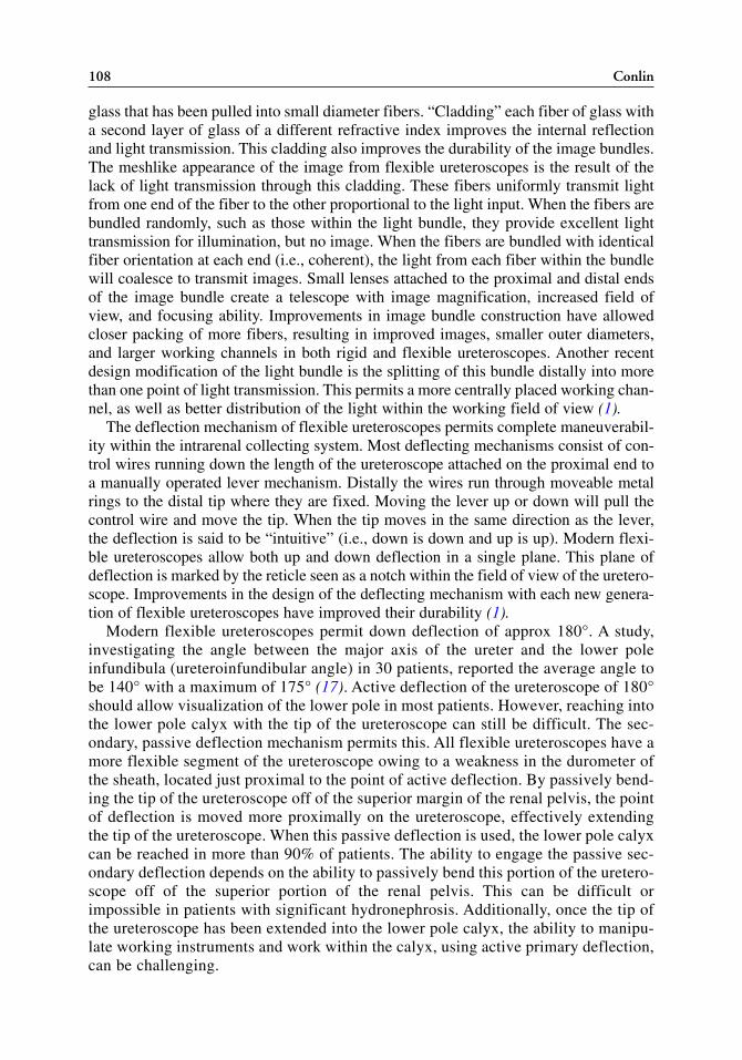
glass that has been pulled into small diameter fibers. “Cladding” each fiber of glass witha second layer of glass of a different refractive index improves the internal reflectionand light transmission. This cladding also improves the durability of the image bundles.The meshlike appearance of the image from flexible ureteroscopes is the result of thelack of light transmission through this cladding. These fibers uniformly transmit lightfrom one end of the fiber to the other proportional to the light input. When the fibers arebundled randomly, such as those within the light bundle, they provide excellent lighttransmission for illumination, but no image. When the fibers are bundled with identicalfiber orientation at each end (i.e., coherent), the light from each fiber within the bundlewill coalesce to transmit images. Small lenses attached to the proximal and distal endsof the image bundle create a telescope with image magnification, increased field ofview, and focusing ability. Improvements in image bundle construction have allowedcloser packing of more fibers, resulting in improved images, smaller outer diameters,and larger working channels in both rigid and flexible ureteroscopes. Another recentdesign modification of the light bundle is the splitting of this bundle distally into morethan one point of light transmission. This permits a more centrally placed working chan-nel, as well as better distribution of the light within the working field of view (1).
The deflection mechanism of flexible ureteroscopes permits complete maneuverabil-ity within the intrarenal collecting system. Most deflecting mechanisms consist of con-trol wires running down the length of the ureteroscope attached on the proximal end toa manually operated lever mechanism. Distally the wires run through moveable metalrings to the distal tip where they are fixed. Moving the lever up or down will pull thecontrol wire and move the tip. When the tip moves in the same direction as the lever,the deflection is said to be “intuitive” (i.e., down is down and up is up). Modern flexi-ble ureteroscopes allow both up and down deflection in a single plane. This plane ofdeflection is marked by the reticle seen as a notch within the field of view of the uretero-scope. Improvements in the design of the deflecting mechanism with each new genera-tion of flexible ureteroscopes have improved their durability (1).
Modern flexible ureteroscopes permit down deflection of approx 180°. A study,investigating the angle between the major axis of the ureter and the lower poleinfundibula (ureteroinfundibular angle) in 30 patients, reported the average angle tobe 140° with a maximum of 175° (17). Active deflection of the ureteroscope of 180°should allow visualization of the lower pole in most patients. However, reaching intothe lower pole calyx with the tip of the ureteroscope can still be difficult. The sec-ondary, passive deflection mechanism permits this. All flexible ureteroscopes have amore flexible segment of the ureteroscope owing to a weakness in the durometer ofthe sheath, located just proximal to the point of active deflection. By passively bend-ing the tip of the ureteroscope off of the superior margin of the renal pelvis, the pointof deflection is moved more proximally on the ureteroscope, effectively extendingthe tip of the ureteroscope. When this passive deflection is used, the lower pole calyxcan be reached in more than 90% of patients. The ability to engage the passive sec-ondary deflection depends on the ability to passively bend this portion of the uretero-scope off of the superior portion of the renal pelvis. This can be difficult orimpossible in patients with significant hydronephrosis. Additionally, once the tip ofthe ureteroscope has been extended into the lower pole calyx, the ability to manipu-late working instruments and work within the calyx, using active primary deflection,can be challenging.
108 Conlin

Two new innovations to the standard deflecting mechanism address this problem.The DUR-8 Elite (Circon ACMI, Stamford, CT) is the first flexible ureteroscope toincorporate active secondary deflection (18). In addition to the active primary deflec-tion (185° down, 175° up) the secondary deflection is now active, 165° down. It is con-trolled with an additional lever opposite the existing primary deflection lever and canbe locked in place. Secondary deflection is not dependant on passive manipulation ofthe scope off of the upper portion of the renal pelvis. The degree of secondary deflec-tion is not dependant upon the position of the scope or how hard you advance the scopebut is controlled with the deflecting lever. Severe hydronephrosis will not preclude theuse of secondary deflection. Locking the secondary deflection in place can simplifymanipulation of the primary deflection within the lower pole calyx (Figs. 2 and 3).
Karl Storz Endoscopy (Tuttlingen, Germany) has introduced “exaggerated deflec-tion” with their Flex-X model flexible ureteroscope (the other new innovation men-tioned in the previous paragraph) (19). This modification of the deflection mechanism
Chapter 7 / Ureteropyeloscopy 109
Fig. 2. Deflection capabilities of the DUR-8 Elite (Circon ACMI, Stamford, CT) ureteroscope.
Fig. 3. (A) Inability to access the lower pole calyx with primary deflection. (B) Successful lower poleaccess using active secondary deflection.

permits active primary deflection to greater than 300°. When approaching the lowerpole calyx, the tip will extend out as it is deflected against the lower pole infundibulum.This improvement of the deflection mechanism results in easier lower pole access andimproved deflection when using working instruments (Fig. 4).
All currently available flexible ureteroscopes have working channels of at least 3.6-Frsize. This allows use of instruments up to 3 Fr, while still permitting adequate irrigation.The specifications of currently available flexible ureteroscopes are detailed in Table 1.
Holmium Laser
The holmium laser has dramatically improved intraluminal lithotripsy and hasbecome the intraluminal lithotripsy energy of choice for most urologists. It has a wave-length of 2100 nm, which is absorbed in 3 mm of water and 0.4 mm of tissue, makingit very safe for use in urology. Fragmentation of calculi is produced by a photothermalreaction with the crystalline matrix of calculi. By not relying on shockwave generationfor stone fragmentation, the photothermal reaction produces stone dust rather than frag-ments, effectively removing a moderate volume of the stone. The flexible quartz fiberscan be used with both rigid and flexible ureteroscopes, and are reusable. These fibersare available in various sizes. The smallest fiber has a diameter of 200 μ, and will limitthe deflection of the ureteroscope less than the larger fibers. The holmium laser willfragment any composition of calculi (20).
Stone Retrieval Devices
Essentially any working instrument 3 Fr or less in size can be used through the flex-ible ureteroscope. These include a variety of stone-graspers and baskets, electrodes, cupbiopsy forceps, and intraluminal lithotripsy devices. Three-pronged stone grasping for-ceps are the safest instruments for removing calculi with the flexible ureteroscope. Theypermit disengagement of calculi that have been found to be too large to be safelyremoved from the ureter. This is critical when performing flexible ureteroscopy becausethere is no second channel to permit fragmentation of an unyielding stone trappedwithin a basket.
110 Conlin
Fig. 4. Exaggerated deflection with the Flex-X (Karl Storz Endoscopy, Tuttlingen, Germany) uretero-

Chapter 7 / Ureteropyeloscopy 111
Table 1Characteristics of the Currently Available Flexible Ureteroscopes
Circon ACMI Olympus Karl Storz Richard Wolf
AUR- DUR- DUR URF- Flex-Characteristics 7 8 8 Elite P3 11274AA X 7330.072 7325.172
Tip diameter (Fr) 7.5 6.75 6.75 6.9 7.5 7.5 6.8Shaft diameter (Fr) 7.5 8.6 8.6 8.4 8.0 8.4 9.0 7.5Working length (cm) 65 65 65 70 70 70 70 70Channel size (Fr) 3.7 3.6 3.6 3.6 3.6 3.6 4.5 3.6Active deflection up
(degrees) 100 175 175 180 120 >300 130 130Active deflection
down (degrees) 160 185 185 100 170 >300 160 160Active secondary
deflection (degrees) 0 0 165 0 0 0 0 0Angle of view
(degrees) 0 12 12 0 6 6 0 0Field of view
(degrees) 80 + 5 80 + 5 80 + 5 90 90 90 65 65Depth of field
(mm) 2–40 2–40 2–40 1–50 2–50 2–50 2–40 2–40Magnification 30× 30× 30× 52× 40× 40× 50× 50×
Stone baskets are available in the usual helical and flatwire designs. Helical basketsare most useful when used in the ureter where they are opened above the stone andpulled down and rotated to engage the stone. The helical design is not particularly use-ful when working within the intrarenal collecting system. Perhaps the most useful bas-ket designed for use with the flexible ureteroscope is the tipless, nickel-titanium(Nitinol) basket. The soft Nitinol wires have memory, and resist kinking, and thereforeopen safely and reliably. These baskets are particularly useful for percutaneous applica-tions, but, because they may permit safer disengagement of larger calculi, can also beused within the intrarenal collecting system through the flexible ureteroscope.
Other newer devices designed to prevent stone migration (such as the Stone Cone,Boston Scientific, Natick, MA) are in general unnecessary while working on stoneswithin the kidney as the stones can be immobilized within a calyx.
Ureteral Access Sheaths
Ureteral access sheaths are available that can facilitate repeated ureteroscopic accessto the intrarenal collecting system. These 12- to 14-Fr sheaths allow repeated passage ofthe ureteroscope without requiring passage of the ureteroscope over a guidewire. The pri-mary disadvantage is related to their size and the (small) potential for ureteral injury (21).Ureteral access sheaths can be useful when multiple fragments of stone require uretero-scopic removal. The majority of stones treated within the kidney will require only a sin-gle passage of the ureteroscope to access and fully fragment the stone. For these cases,an access sheath is usually unnecessary. A new sheath (Aquaguide, Bard Urology,Covington, GA) is available with a built in second channel that permits irrigation of

small fragments of stone out of the kidney during the procedure (Fig. 5). This canimprove visibility and may facilitate treatment of larger stone burdens within the kidney.
Endourological procedures should be performed in an operating room environmentequipped with all instrumentation that may be necessary. The urologist should be pre-pared for any problem encountered. Special guidewires, such as angled hydrophilic,Nitinol core, and extra-stiff wires, should be readily available. Dilation devices includ-ing dilating catheters, high-pressure balloon catheters, and zero-tipped balloon cathetersare standard. Torque-able angled catheters are useful for manipulating around impactedcalculi or tortuous ureters. In addition to back-up flexible ureteroscopes, semirigidureteroscopes should also be available to treat unanticipated pathology below the iliacvessels. The standard supplies necessary for ureteroscopic treatment of renal calculi arelisted in Table 2.
TECHNIQUE
Preoperative patient preparation includes imaging of the stone to be treated and theupper urinary tract to determine the size, location, and associated anatomy. A noncontrasthelical computed tomography scan is the most sensitive study for stones and is usually ade-quate for urolithiasis patients. A urinalysis is performed and if indicated a urine culture.Urinary tract infections should be treated preoperatively and undrained infections behindimpacted ureteral calculi are drained. A routine preoperative antibiotic is given. Anesthesiacan be general, regional, or local with sedation. Fluoroscopy is needed for initial ureteralaccess, monitoring during the ureteroscopy, and during stent placement. Although tablesdesigned for urological endoscopy with fixed fluoroscopy units are available, mobile
112 Conlin
Fig. 5. (A) Two lumens of the Aquaguide (Bard Urology, Covington, GA) ureteral access sheath. (B)Fragments of stone irrigated out of the kidney using the Aquaguide (Bard Urology, Covington, GA) sheath.

C-arm fluoroscopy is preferable. These C-arm fluoroscopy units allow greater mobility,improved image quality, and less radiation exposure for the surgeon because the X-raysource is below the patient rather than above. Modern C-arm fluoroscopy units utilize “lastimage hold” and digital imaging which greatly reduce the amount of radiation exposure forthe patient and surgeon. The urologist should control the fluoroscopy unit with foot pedalcontrol which will facilitate the speed of the case and minimize excessive fluoroscopy time.
Prior to flexible ureteroscopy, cystoscopy of the patient is performed to place a safetyguidewire and fully inspect the bladder. A safety guidewire is critical during ureteroscopyto maintain access and allow placement of a ureteral stent if any problems are encoun-tered. A 0.038-in. diameter flexible tipped Teflon-coated guidewire is usually sufficient.Care must be taken when trying to gain access around an impacted stone as the ureter
Chapter 7 / Ureteropyeloscopy 113
Table 2Supplies for Ureteroscopy for Stones
EndoscopesRigid ureteroscopes
7-Fr or smaller semirigid ureteroscopelarger ureteroscope with straight working channel (optional)
Flexible ureteroscopes7.5 Fr 8.6 Fr or larger Secondary deflection or exaggerated deflection capable ureteroscope
Disposable suppliesGuidewires
0.038-in. angled hydrophilic0.038-in. straight Teflon coated0.038-in. Nitinol core, polyurethane coated0.038-in. extra-stiff
IrrigationPower irrigation (60 cc syringes, extension tubing)High pressure working port seal
Stone retrieval devices (3.0 Fr or smaller) helical baskettipless basketthree pronged grasping forceps
CathetersDual lumen catheter6–12-Fr dilating catheter5-Fr open-ended catheter5-Fr angled tip torque-able catheter
Dilation deviceshigh pressure ureteral dilating balloons (5–7 mm)“zero-tip” ureteral dilating balloon
Ureteral stents5–7 Fr, 20–28 cm, double-pigtail
Intraluminal lithotripsy devicesHolmium laserPneumatic (optional)Electrohydraulic (optional)

can be easily perforated. Manipulation of the guidewire around the stone may requireuse of an angled hydrophilic-coated wire, an angled torque-able catheter, or both. Ahydrophilic wire is generally not secure enough to be used as a safety wire, so it isexchanged for a standard Teflon-coated guidewire. Following placement of the initialguidewire, the bladder is drained to permit accumulation of fluid during ureteroscopy,and minimize buckling of the flexible ureteroscope into the bladder. The cystoscope isremoved and a dual lumen catheter is passed over the initial guidewire. This dual lumencatheter is 10 Fr, which will gently dilate the ureteral orifice and allow placement of asecond 0.038-in. Teflon-coated flexible tipped wire to serve as the working wire. Theflexible ureteroscope is then passed in a monorail fashion over the taut working wire tothe point of the pathology being treated. Dilation of the ureteral orifice with the duallumen catheter is usually sufficient to permit passage of the flexible ureteroscope. If dif-ficulty in passing the flexible ureteroscope through the ureteral orifice is still encoun-tered, a 6- to 12-Fr tapered dilating catheter (Nottingham) or a dilating balloon cathetercan be used to dilate the ureteral orifice. Dilation of the orifice beyond 15 Fr is rarelynecessary for routine ureteroscopy. In most flexible ureteroscopy series, the need forformal dilation is between 8 and 25%, and has decreased with the advent of the smallerdiameter flexible ureteroscopes (22–24).
Irrigation through the ureteroscope can be provided with a pressurized irrigation bag,roller pump, or handheld syringe. Normal saline should be used to prevent accumulationand absorption of hypotonic solution and resultant “transurethral resection syndrome”.
The basic movements of the flexible ureteroscope include deflection, rotation, andadvancing and retracting the ureteroscope. The reticle of the flexible ureteroscopemarks the plane of deflection, and rotation of the ureteroscope is often necessary toalign this plane of deflection in the direction desired. Failure to adequately rotate theureteroscope is a common mistake of the novice ureteroscopist.
When the holmium laser is used, it is important to pass the laser fiber through astraightened flexible ureteroscope to prevent damage to the working channel. Thestraightness of the ureteroscope should be confirmed fluoroscopically. Once the fiber ispassed beyond the tip, the ureteroscope can be deflected appropriately. The most com-monly used sizes of holmium laser fibers include the 365-μ fiber and the 200-μ fiber.When significant deflection of the ureteroscope is needed, the 200-μ fiber is preferred,as it does not limit the deflection of the ureteroscope as much as the larger fibers. Thetip of the fiber must be in contact with the stone during treatment because the holmiumlaser energy is absorbed in 3 mm of water. The holmium laser can damage the uretero-scope, the guidewire, and the ureteral wall. These problems can be avoided by notactivating the laser unless the tip of the fiber is seen in contact with the stone (25). Inaddition, if the helium-neon aiming beam is not seen, the laser should not be activatedas this may be an indication of fiber damage. Firing the holmium laser through a bro-ken fiber can cause significant damage to the ureteroscope.
Complete destruction of the calculus with the holmium laser is usually the goal. Thiscan best be achieved by ablating the stone while keeping the tip of the fiber movingacross the surface of the stone. Alternatively, the stone can be broken into fragmentssmall enough to remove. With this technique, the stone can be broken along its naturalcleavage planes and the fragments removed with stone-grasping forceps. Alternatively,a “drill and core” technique can be used. In this technique the central portion of thestone is slowly ablated, leaving a shell of stone that is then fractured using the holmiumlaser and the fragments removed. If multiple fragments require removal, repeated pas-sage of the flexible ureteroscope can be facilitated with the use of a small ureteral access
114 Conlin
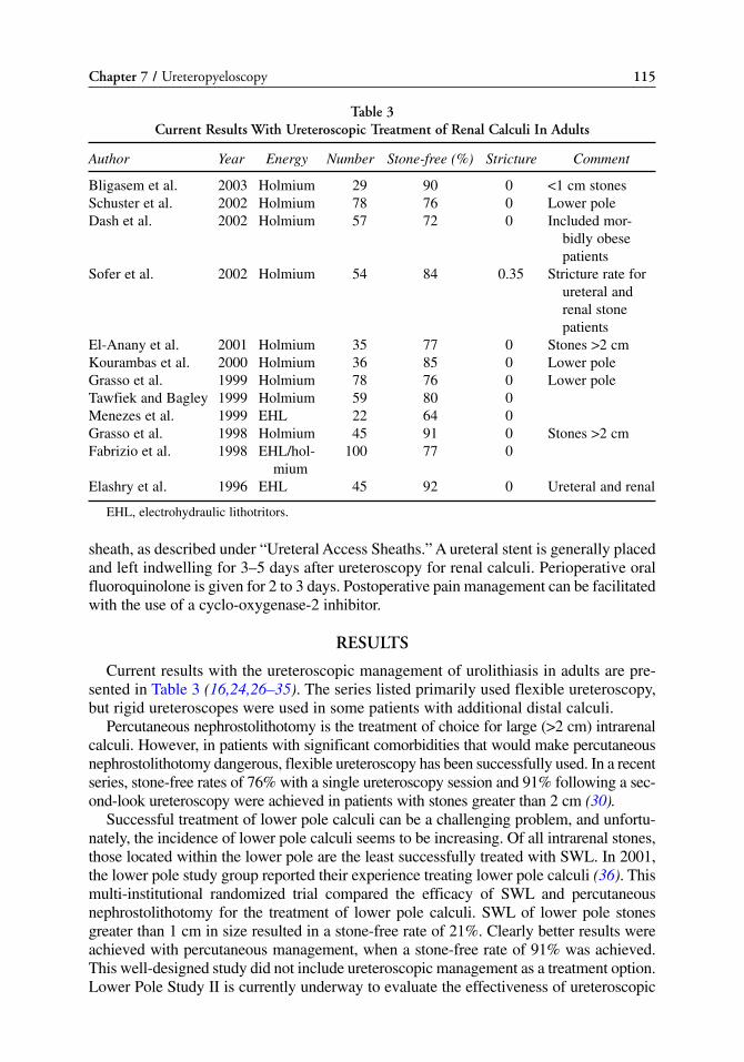
sheath, as described under “Ureteral Access Sheaths.” A ureteral stent is generally placedand left indwelling for 3–5 days after ureteroscopy for renal calculi. Perioperative oralfluoroquinolone is given for 2 to 3 days. Postoperative pain management can be facilitatedwith the use of a cyclo-oxygenase-2 inhibitor.
RESULTS
Current results with the ureteroscopic management of urolithiasis in adults are pre-sented in Table 3 (16,24,26–35). The series listed primarily used flexible ureteroscopy,but rigid ureteroscopes were used in some patients with additional distal calculi.
Percutaneous nephrostolithotomy is the treatment of choice for large (>2 cm) intrarenalcalculi. However, in patients with significant comorbidities that would make percutaneousnephrostolithotomy dangerous, flexible ureteroscopy has been successfully used. In a recentseries, stone-free rates of 76% with a single ureteroscopy session and 91% following a sec-ond-look ureteroscopy were achieved in patients with stones greater than 2 cm (30).
Successful treatment of lower pole calculi can be a challenging problem, and unfortu-nately, the incidence of lower pole calculi seems to be increasing. Of all intrarenal stones,those located within the lower pole are the least successfully treated with SWL. In 2001,the lower pole study group reported their experience treating lower pole calculi (36). Thismulti-institutional randomized trial compared the efficacy of SWL and percutaneousnephrostolithotomy for the treatment of lower pole calculi. SWL of lower pole stonesgreater than 1 cm in size resulted in a stone-free rate of 21%. Clearly better results wereachieved with percutaneous management, when a stone-free rate of 91% was achieved.This well-designed study did not include ureteroscopic management as a treatment option.Lower Pole Study II is currently underway to evaluate the effectiveness of ureteroscopic
Chapter 7 / Ureteropyeloscopy 115
Table 3Current Results With Ureteroscopic Treatment of Renal Calculi In Adults
Author Year Energy Number Stone-free (%) Stricture Comment
Bligasem et al. 2003 Holmium 29 90 0 <1 cm stonesSchuster et al. 2002 Holmium 78 76 0 Lower poleDash et al. 2002 Holmium 57 72 0 Included mor-
bidly obese patients
Sofer et al. 2002 Holmium 54 84 0.35 Stricture rate for ureteral and renal stone patients
El-Anany et al. 2001 Holmium 35 77 0 Stones >2 cmKourambas et al. 2000 Holmium 36 85 0 Lower pole Grasso et al. 1999 Holmium 78 76 0 Lower poleTawfiek and Bagley 1999 Holmium 59 80 0Menezes et al. 1999 EHL 22 64 0Grasso et al. 1998 Holmium 45 91 0 Stones >2 cmFabrizio et al. 1998 EHL/hol- 100 77 0
miumElashry et al. 1996 EHL 45 92 0 Ureteral and renal
EHL, electrohydraulic lithotritors.
management of lower pole calculi. The theoretical advantages of ureteroscopic treatmentof lower pole calculi compared with SWL include the ability to reposition the calculi fromthe lower pole calyx into a more favorable location such as the upper pole. Fragmentationof the calculi in the upper pole would result in potentially easier passage of any residualfragments out of the kidney. Also, a significant volume of the stone is ablated and effec-tively removed with the use of the holmium laser. Reports of ureteroscopic managementof lower pole stones show a good success rate. In a review of prior studies, the averagesuccess rate for ureteroscopic treatment of lower pole calculi was reported as 83% (36a).
Significant complications following flexible ureteroscopy are rare. Urinary tractinfections can be prevented with perioperative antibiotics. Mild gross hematuria is nor-mal, and quickly resolves following stent removal. The most significant complicationfollowing ureteroscopy is ureteral stricture formation. Previous reports of ureteral stric-ture rates following ureteroscopy were unacceptably high. With the advent of smallerflexible ureteroscopes and the decreased need for ureteral dilation, the rates of ureteralstricture have decreased significantly. Several modern ureteroscopy (both rigid and flex-ible) series have reported ureteral stricture rates of 0.5% or lower (24,35,37,38).
The most troubling problem following flexible ureteroscopy is the patient’s discom-fort from the indwelling ureteral stent. After ureteroscopic treatment of renal calculi, astent is usually left for only 3 to 5 days. However, the need for routine ureteral stent-ing following ureteroscopy is controversial. Several reports of successful ureteroscopictreatment of ureteral calculi without ureteral stenting have been published. Patientswere not stented if there were no residual stone fragments, no significant ureteraltrauma, and ureteral dilation was not performed (39–44). Although these reports havehad small numbers of patients, there were no significant complications. A better defini-tion of which patients will require ureteral stenting and improvements in stent designand manufacture, should decrease patient discomfort following flexible ureteroscopy.
TIPS AND TRICKS
Access to the ureter can be challenging in a variety of situations. The first problemthat can be encountered is obtaining access to the ureteral orifice for intubation with theguidewire. Several problems and potential solutions will be presented. Inflammationand distortion of the ureteral orifice from an impacted stone in the intramural tunnel canmake passage of the guidewire into the ureter difficult. Use of an angled-tip hydrophiliccoated guidewire through an angled-tip tourqable catheter will often allow the wire topass alongside the stone. A severely enlarged prostatic median lobe can make it diffi-cult to reach the ureteral orifice with a rigid cystoscope. This can be overcome by ini-tially accessing the ureteral orifice with the flexible cystoscope, placing the guidewire,and then exchanging the guidewire with a super-stiff wire. This will help hold themedian lobe out of the way for passage of the ureteroscope. Severe bladder trabecula-tion can make identification of the ureteral orifice difficult, but can usually be helpedby administering intravenous Indigo Carmine. Scarring or obliteration of the ureteralorifice from prior bladder tumor resection may require resecting scar over the orifice,endoluminal ultrasound inspection of the trigone to identify the location of the ureter,or a percutaneous approach to obtain “above and below” access. Patients with intestinaldiversion of urine can have difficult-to-identify ureterointestinal connections. One of theproblems with ileal conduits is the difficulty distending the conduit for appropriate visi-bility. Placing a 10-mm laparoscopic port into the loose stoma can help prevent egress ofirrigation fluid around the flexible cystoscope and out of the conduit. Distention of the
116 Conlin

conduit, and administration of Indigo Carmine if necessary, will help identify theureteral orifice in patients with intestinal diversion.
The next potential problem encountered is difficulty in passing the guidewire throughthe ureter in the kidney. This can be the result of an impacted ureteral calculus, ureteralstricture, ureteropelvic junction obstruction, or a tortuous “kinked” ureter. The use of anangled hydrophilic guidewire through an angled torque-able catheter will overcomemost of these problems. The wire and the catheter can each be rotated to “screw” it pastthe blockage. If this is unsuccessful, direct ureteroscopic inspection can be used tonegotiate the wire around the point of obstruction.
Once the guidewire has been placed, passage of the flexible ureteroscope is usuallystraightforward, but can be hindered by several problems. It is critical to keep theguidewire (over which the ureteroscope is passed) taut. Any slack in this wire will pre-vent successful passage of the ureteroscope. Rotating the ureteroscope can help pres-ent a more streamlined face of the ureteroscope to the ureteral orifice. Balloon dilationof the orifice or a ureteral stricture is always an option if passage of the ureteroscopeis not possible. If a small renal calculus is being treated with the holmium laser, andremoval of fragments or repositioning of the stone are not necessary, it is reasonableto remove the safety wire to help the passage of the ureteroscope. If anything otherthan simple holmium laser lithotripsy is planned or later necessary, a safety guidewireis required.
If difficulty in passing the flexible ureteroscope is encountered in the absence of anyureteral stricture, the use of a Nitinol core guidewire (Zebra, or Sensor wires,Microvasive Urology, Natick, MA or Roadrunner, Cook Urological, Spencer, IN) maybe helpful. These stiffer, smoother wires enable more efficient transmission of the pushfrom the urologist to the tip of the ureteroscope.
Ureteroscopic access to the lower pole calyx can be challenging for several reasons.With repeated use of the flexible ureteroscope, the primary deflection is often weakenedand incomplete, resulting in the inability to reach an acute angle into the lower pole calyx.Even when the angle into the lower pole calyx can be negotiated, extending the tip of theureteroscope into the calyx may be difficult. The length of the lower pole infundibulum isfrequently longer than the length of the primary deflected segment of the ureteroscope.Grasso et al. (31) reported their experience with ureteroscopic treatment of lower pole cal-culi. They measured the effect of the infundibulopelvic angle, degree of hydronephrosis,and lower pole infundibular length on successful ureteroscopic treatment. The one factorthey found that negatively affected their ability to reach the lower pole calyx was thelower pole infundibular length. In order to extend the tip of the ureteroscope into the lowerpole calyx, current ureteroscope designs rely on a passive secondary deflection mecha-nism as previously described. The use of secondary deflection effectively extends thelength of the deflected segment in order to reach the lower pole calyx. The ability toengage the passive secondary deflection depends on the ability to passively bend this por-tion of the ureteroscope off of the superior portion of the renal pelvis. This is sometimesnot possible with standard flexible ureteroscopes. The latest generation of flexible uretero-scopes, with either active secondary or exaggerated deflection, significantly improve ourability to treat effectively lower pole calculi. Lower pole calculi should be repositionedinto the upper collecting system prior to fragmentation with the holmium laser. Auge etal. (45) should improved stone free rates for lower pole stones repositioned prior to laserlithotripsy compared with those treated in situ in the lower pole (90 vs 83%).
Chapter 7 / Ureteropyeloscopy 117
In order to prevent migration of stones fragments into the lower pole calyx while treat-ing stones in the kidney, the patient can be positioned in Trendelenburg position and rotatedto elevate the effected side. This will encourage fragment accumulation in the renal pelvis.
When working endoscopically within the kidney, respiratory motion can hinder effi-cient laser lithotripsy. Endotracheal intubation of the patient will allow the anesthesiol-ogist to control the rate and depth of the patient’s respiration there by improving laserlithotripsy access to the stone.
CONCLUSIONS
Improvements in flexible ureteroscopes, working instruments, and endoscopic tech-niques, have positively affected our ability to treat intrarenal calculi effectively. Withcontinued advances, the role of ureteroscopy in the treatment of intrarenal calculishould continue to grow. Exciting changes can be anticipated within the next decade,improving our ability to treat more complicated upper urinary tract calculi in a mini-mally invasive fashion.
REFERENCES1. Conlin MJ, Bagley, DH. Flexible ureteroscopes. In: Smith’s Textbook of Endourology (Smith AD,
Badlani GH, Bagley DH, et al. Quality Medical Publishing, St. Louis, 1996, 377–382.2. Marshall VF. Fiberoptics in urology. J Urol 1964; 91: 110.3. Takagi T, Go T, Takayasu H, Aso Y. Fiberoptic pyeloureteroscope. Surgery 1971; 70(5): 661–663.4. Bush IM, Goldberg E, Javadpour N, Chakrobortty H, Morelli F. Ureteroscopy and renoscopy: a pre-
liminary report. Chic Med Sch Q 1970; 30(1): 46–49.5. Abdel-Razzak OM, Bagley DH. Clinical experience with flexible ureteropyeloscopy. J Urol 1992;
148(6): 1788–1792.6. Bagley DH. Intrarenal access with the flexible ureteropyeloscope: effects of active and passive tip
deflection. J Endourol 1993; 7(3): 221–224.7. Johnson DE, Cromeens DM, Price RE. Use of the holmium:YAG laser in urology. Lasers Surg Med
1992; 12(4): 353–363.8. Sayer J, Johnson DE, Price RE, Cromeens DM. Ureteral lithotripsy with the holmium:YAG laser.
Laser Med 1993; 11(2): 61–65.9. Albert P. The holmium:YAG laser: applications in urology. Clin Laser Mon 1993; 11(3): 39–41.
10. Bagley D, Erhard M. Use of the holmium laser in the upper urinary tract. Tech Urol 1995; 1(1): 25–30.11. Grasso M, Loisides P, Beaghler M, Bagley D. The case for primary endoscopic management of upper
urinary tract calculi: I. a critical review of 121 extracorporeal shock-wave lithotripsy failures. Urology1995; 45(3): 363–371.
12. Grasso M, Beaghler M, Loisides P. The case for primary endoscopic management of upper urinary tractcalculi: II. cost and outcome assessment of 112 primary ureteral calculi. Urology 1995; 45(3): 372–376.
13. Zheng W, Beiko DT, Segura JW, Preminger GM, Albala DM, Denstedt JD. Urinary calculi in avia-tion pilots: what is the best therapeutic approach? J Urol 2002; 168(4 Pt 1): 1341–1343.
14. Watterson JD, Girvan AR, Cook AJ, et al. Safety and efficacy of holmium: YAG laser lithotripsy inpatients with bleeding diatheses. J Urol 2002; 168(2): 442–445.
15. Bultitude MF, Tiptaft RC, Dasgupta P, Glass JM. Treatment of urolithiasis in the morbidly obese.Obes Surg 2004; 14(3): 300–304.
16. Dash A, Schuster TG, Hollenbeck BK, Faerber GJ, Wolf JS Jr. Ureteroscopic treatment of renal cal-culi in morbidly obese patients: a stone-matched comparison. Urology 2002; 60(3): 393–397.
17. Bagley DH, Rittenberg, MH. Intrarenal dimensions. Surg Endosc 1987; 1: 119–121.18. Shvarts O, Perry KT, Goff B, Schulam PG. Improved functional deflection with a dual-deflection
flexible ureteroscope. J Endourol 2004; 18(2): 141–144.19. Johnson GB, Grasso M. Exaggerated primary endoscope deflection: initial clinical experience with
prototype flexible ureteroscopes. BJU Int 2004; 93(1): 109–114.
118 Conlin

20. Bagley DH, Erhard M. Use of the holmium laser in the upper urinar tract. Tech Urol 1995; 1(1): 25–30.21. Delvecchio FC, Auge BK, Brizuela RM, et al. Assessment of stricture formation with the ureteral
access sheath. Urology 2003; 61(3): 518–522.22. Elashry OM, Elbahnasy AM, Rao GS, Nakada SY, Clayman RV. Flexible ureteroscopy: Washington
University experience with the 9.3F and 7.5F flexible ureteroscopes [see comments]. J Urol 1997;157(6): 2074–2080.
23. Grasso M, Bagley D. Small diameter, actively deflectable, flexible ureteropyeloscopy. J Urol 1998;160(5): 1648–1653.
24. Tawfiek ER, Bagley DH. Management of upper urinary tract calculi with ureteroscopic techniques.Urology 1999; 53(1): 25–31.
25. Beaghler M, Poon M, Ruckle H, Stewart S, Weil D. Complications employing the holmium:YAGlaser. J Endourol 1998; 12(6): 533–535.
26. Bilgasem S, Pace KT, Dyer S, Honey RJ. Removal of asymptomatic ipsilateral renal stones follow-ing rigid ureteroscopy for ureteral stones. J Endourol 2003; 17(6): 397–400.
27. El-Anany FG, Hammouda HM, Maghraby HA, Elakkad MA. Retrograde ureteropyeloscopicholmium laser lithotripsy for large renal calculi. BJU Int 2001; 88(9): 850–853.
28. Elashry OM, DiMeglio RB, Nakada SY, McDougall EM, Clayman RV. Intracorporeal electrohy-draulic lithotripsy of ureteral and renal calculi using small caliber (1.9F) electrohydraulic lithotripsyprobes. J Urol 1996; 156(5): 1581–1585.
29. Fabrizio MD, Behari A, Bagley DH. Ureteroscopic management of intrarenal calculi. J Urol 1998;159(4): 1139–1143.
30. Grasso M, Conlin M, Bagley D. Retrograde ureteropyeloscopic treatment of 2 cm. or greater upperurinary tract and minor Staghorn calculi. J Urol 1998; 160(2): 346–351.
31. Grasso M, Ficazzola M. Retrograde ureteropyeloscopy for lower pole caliceal calculi. J Urol 1999;162(6): 1904–1908.
32. Kourambas J, Delvecchio FC, Munver R, Preminger GM. Nitinol stone retrieval-assisted uretero-scopic management of lower pole renal calculi. Urology 2000; 56(6): 935–939.
33. Menezes P, Dickinson A, Timoney AG. Flexible ureterorenoscopy for the treatment of refractoryupper urinary tract stones. BJU Int 1999; 84(3): 257–260.
34. Schuster TG, Hollenbeck BK, Faerber GJ, Wolf JS Jr. Ureteroscopic treatment of lower pole calculi:comparison of lithotripsy in situ and after displacement. J Urol 2002; 168(1): 43–45.
35. Sofer M, Watterson JD, Wollin TA, Nott L, Razvi H, Denstedt JD. Holmium:YAG laser lithotripsyfor upper urinary tract calculi in 598 patients. J Urol 2002; 167(1): 31–34.
36. Albala DM, Assimos DG, Clayman RV, et al. Lower pole I: a prospective randomized trial of extra-corporeal shock wave lithotripsy and percutaneous nephrostolithotomy for lower pole nephrolithiasis-initial results. J Urol 2001; 166(6): 2072–2080.
36a. Lingeman JE, Lifshitz DA, Evan AP. Surgical management of urinary lithiasis. In: Campbell’sUrology, 8th Ed., vol 4. (Walsh PC, Retik AB, Vaughn ED, et al., eds). Philadelphia, Saunders, 2002,pp. 3431–3435.
37. Harmon WJ, Sershon PD, Blute ML, Patterson DE, Segura JW. Ureteroscopy: current practice andlong-term complications. J Urol 1997; 157(1): 28–32.
38. Singal RK, Razvi HA, Denstedt JD. Secondary ureteroscopy: results and management strategy at areferral center. J Urol 1998; 159(1): 52–55.
39. Srivastava A, Gupta R, Kumar A, Kapoor R, Mandhani A. Routine stenting after ureteroscopy for distalureteral calculi is unnecessary: results of a randomized controlled trial. J Endourol 2003; 17(10): 871–874.
40. Hollenbeck BK, Schuster TG, Seifman BD, Faerber GJ, Wolf JS, Jr. Identifying patients who are suit-able for stentless ureteroscopy following treatment of urolithiasis. J Urol 2003; 170(1): 103–106.
41. Chen YT, Chen J, Wong WY, Yang SS, Hsieh CH, Wang CC. Is ureteral stenting necessary afteruncomplicated ureteroscopic lithotripsy? A prospective, randomized controlled trial. J Urol 2002;167(5): 1977–1980.
42. Byrne RR, Auge BK, Kourambas J, Munver R, Delvecchio F, Preminger GM. Routine ureteral stentingis not necessary after ureteroscopy and ureteropyeloscopy: a randomized trial. J Endourol 2002; 16(1):9–13.
43. Hollenbeck BK, Schuster TG, Faerber GJ, Wolf JS, Jr. Routine placement of ureteral stents is unnec-essary after ureteroscopy for urinary calculi. Urology 2001; 57(4): 639–643.
Chapter 7 / Ureteropyeloscopy 119

44. Denstedt JD, Wollin TA, Sofer M, Nott L, Weir M, D’A Honey RJ. A prospective randomized controlledtrial comparing nonstented versus stented ureteroscopic lithotripsy. J Urol 2001; 165(5): 1419–1422.
45. Auge BK, Dahm P, Wu NZ, Preminger GM. Ureteroscopic management of lower-pole renal calculi:technique of calculus displacement. J Endourol 2001; 15(8): 835–838.
120 Conlin

Percutaneous Stone Removal
Louis Eichel, MD
and Ralph V. Clayman, MD
CONTENTS
INTRODUCTION
INDICATIONS
SPECIAL CIRCUMSTANCES REQUIRING PERCUTANEOUS STONE
REMOVAL
INSTRUMENT LIST
TECHNIQUE
COMPLICATIONS
TIPS AND TRICKS
CONCLUSION
REFERENCES
8
SUMMARY
In this chapter the method of percutaneous stone removal is reviewed in its entirety.The indications for percutaneous stone removal in the age of shockwave lithotripsy andureteroscopy are carefully reviewed: staghorn stones, obstruction and stones (e.g.,ureteropelvic junction obstruction and calyceal diverticula), renal anomalies (e.g.,horseshoe kidney), stones with difficult lower pole anatomy, and calculi that areextremely hard (i.e., Hounsfield units ≥1000). In addition, the technique is presentedwith the method for percutaneous access and details of intrarenal lithotripsy.Accompanying this section is a detailed account of the instrumentation available. Sometips and tricks for successful stone removal are presented, such as the use of the ureteralaccess sheath to improve renal access and stone clearance, as well as the use of varioushemostatic agents in lieu of placing a percutaneous nephrostomy tube at the terminationof the procedure. Also, a full discussion of complications is provided: prevention,recognition, and treatment.
Key Words: Percutaneous nephrostolithtomy (PCNL); percutaneous stone removal;nephrostomy tube.
From: Advanced Endourology: The Complete Clinical GuideEdited by: S. Y. Nakada and M. S. Pearle © Humana Press Inc., Totowa, NJ
121

INTRODUCTION
Almost 30 years since the birth of percutaneous nephrolithotomy (PCNL), the missionremains the same as it has always been: to simplify minimally invasive surgeries as muchas possible so that both patient and surgeon alike may benefit. Since the original descrip-tion of stone removal through a percutaneous nephrostomy tract by Fernstrom andJohansson in 1976 (1), percutaneous stone removal has evolved into a highly developedprocedure practiced by many urologists. Several new techniques and technologies recentlyhave emerged that simplify the procedure, allow us to take on more challenging cases, andmake it safer and less painful for patients. This chapter is designed to serve as both areview of the basics and a guide to recent cutting edge developments in the field.
INDICATIONS
In the last decade, there has been a 63% increase in the number of ureteroscope cases(URS) being performed, whereas the number of shockwave lithotripsies (SWLs) hasdeclined by roughly 11%. Presumably, these changes reflect the ever-improvingureteroscopic technology becoming available and the relative stagnation/decline ofSWL technology during the same period. Interestingly, at university centers, the per-centage of PCNL cases being performed over the past 10 years has been stable at about28% of all stones treated (2). The reason for this relative stability is because PCNL is usu-ally indicated in situations that are inappropriate for SWL and URS. Hence, the indica-tions for PCNL have remained relatively constant over time.
Choosing the appropriate modality of treatment for a stone depends on two stone fac-tors (size and hardness) and two environmental factors (stone location and the degreeof hydronephrosis). Large meta-analyses have shown that renal stones less than 10 mmcan be treated successfully with SWL as first-line therapy. Stones more than 20 mm insize respond best to PCNL (3). Although treatment of stones more than 2 cm is possi-ble with URS, the stone-free rate ranges from only 47 to 60% and complication ratesranging from 6 to 22% have been reported (4,5). Treatment success rates for stones inthe 11 to 20 mm range are more variable and these cases must be evaluated on an indi-vidual basis. For the 11 to 20 mm “gray zone,” stone location, density, and the presenceof hydronephrosis play a more crucial role.
For the largest stones, staghorn calculi, the percutaneous approach remains the standardtreatment. There is a clear advantage to treating staghorn calculi with PCNL. Historically,it was widely accepted that ancillary procedures (i.e., SWL) would be necessary toachieve complete stone removal. The concept of “sandwich therapy” was originallycoined by Streem in 1987. In fact, prior to 1990, ancillary procedure rates as high as 65%were reported with overall success rates of 83 to 86% (6,7). More recently, with the adventof improved endoscopic equipment and imaging capabilities, there has been a trendtoward using PCNL as monotherapy. Thus, the use of ancillary procedures and “secondlooks” has declined (8). Despite this decline in the use of adjunctive SWL, the overall suc-cess rate of PCNL for staghorn stones has improved ranging from 87 to 95% (8,9). Thisimprovement has largely been the result of the more frequent use of flexible nephroscopyand the advent of holmium laser lithotripsy.
Several authors have addressed the concept of stone composition and relativeresponse to SWL (10,11). Uric acid and calcium oxalate dihydrate stones are relativelysoft. Struvite, apatite, and brushite all have a medium hardness. Cystine, on the otherhand, is extremely hard. Results of SWL for cystine stones are therefore generally poor,
122 Eichel and Clayman

although, success rates of 50 to 75% have been reported for cystine stones less than 15mm treated on a HM-3 SWL (12–14). Cystine stones larger than 15 mm are best treatedwith PCNL; whereas this is the case in general for all other stones that are greater than20 mm in size. Case reports of URS lithotripsy for larger cystine stones exist but, as pre-viously noted, URS for larger stones is fraught with overall poor stone clearance andmultiple procedures (5,15).
Although for years the focus has been on using the computed tomography (CT) scanto predict stone composition in an effort to predict a stone’s response to SWL, lately ofmore importance is stone construction rather than composition. Specifically, ignoringcomposition and focusing on stone durility (i.e., density as determined by Hounsfieldunits [HU], a term coined by Dretler in 1988) may be of greater predictive value (16). Ingeneral, stones that measure less than 500 HU have an 80 to 100% fragmentation rate withSWL. Stones in the 500 to 1000 HU range have roughly a 70% fragmentation rate andstones greater than 1000 HU generally have fragmentation rates of 25% or less with SWLand hence URS or PCNL would be more advantageous, depending on the stone’s size(16,17).
From an environmental standpoint, one anatomic factor of great interest is lower poleanatomy as measured on intravenous pyelography (IVP). Factors such as infundibulopelvicangle (IPA), infundibular length (IL), calyceal pelvic height, and infundibular width (IW)have been found by several authors to affect stone clearance rates. In general, these authorsfound that patients with lower pole stones 17 mm or less who have favorable anatomy(lower pole infundibulo-pelvic angle >70°, IL <3 cm, and IW >5 mm) are excellent candi-dates for SWL whereas patients with less favorable anatomy (LIP angle <40°, IL >3 cm,and IW <5 mm) would be better served by PCNL (18–21). Other authors, however, havenot found lower pole anatomy to be a major factor (22,23). The concept of lower pole meas-urements may be moot, however, because computed tomography (CT) is quickly becom-ing the standard imaging modality for urolithiasis and three-dimensional reconstructions ofthe urinary collecting system are not yet commonly provided. In the final analysis it is gen-erally accepted that for lower pole stones, especially when larger than 1 cm, PCNL is farmore effective than SWL. The reported success rates were 21 vs 95% respectively in oneseries in which patients with lower pole stones were randomized to SWL vs PCNL (24). Inthe same study, the success rates for stones 1 cm or less was 63 vs 100% respectively.
The second environmental factor is hydronephrosis. Two studies address this concerndirectly. Winfield et al. (25) compared PCNL with SWL for treatment of staghorn calculiand found that the success rate of SWL in the presence of high-grade hydronephrosis wasonly 53% compared to 70% without hydronephrosis. Similarly, the ancillary procedurerate was 12 vs 27%, respectively. In the largest series reported to date, Poulakis et al. (21)reviewed 701 cases of SWL for lower pole stones and found that patients with evidenceof either significant hydronephrosis or lack of peristalsis on IVP had a 51% stone clear-ance rate vs 80% for patients with a normally peristaltic, nondilated system.
In sum, PCNL is currently indicated in those patients with stones greater than 2 cm,calculi of all sizes in a markedly hydronephrotic system, lower pole stones greater than1 cm, and for calculi of all sizes with a density exceeding 1000 HU.
Absolute and relative contraindications to PCNL remain few but include:
• Pregnancy (relative).• Bleeding disorders/contraindication to reversal of anticoagulation (absolute).• Severe pulmonary dysfunction (relative).• Severe bodily contortions (relative/absolute).
Chapter 8 / Percutaneous Stone Removal 123

• Untreated infection (absolute).
SPECIAL CIRCUMSTANCES REQUIRING PERCUTANEOUS STONE REMOVAL
Obstruction
Relative urinary obstruction is a direct indication for considering PCNL or rarely, URSas it precludes SWL. This situation is encountered when calculi occur in association with acalyceal diverticulum, ureteropelvic junction obstruction (UPJO), and congenital/iatrogenicrenal/drainage anomalies (e.g., horseshoe, transplant, or pelvic kidney and ileal conduit).
Caliceal Diverticula
Caliceal diverticula are nonsecretory outpouchings of the renal collecting system thatcommunicate with the calyx via a tight neck, usually along the fornix. Up to 50% ofpatients with calyceal diverticula will have stones within them (26,27). The conditionitself does not require treatment. The criteria for treatment are pain and/or infection.
Conventional approaches for the treatment of calyceal diverticula include open sur-gery, laparoscopy, SWL, URS, and PCNL. Historically, excellent results were reportedwith open surgery that entailed ligation of the diverticular neck and marsupialization orwedge excision (28–30). This technique has been abandoned, however, because lessinvasive options now exist. Ramakumar et al. (31) reported a series of 12 patients inwhich a laparoscopic approach to stone removal and marsupialization was used. Sixty-seven percent were salvage procedures after other attempts failed using less invasivetechniques. Overall, the objective success rate (complete stone removal and oblitera-tion) was 92%; however, the subjective success rate (pain relief) was only 75%. At pres-ent, the laparoscopic approach is reserved mainly as salvage therapy or as primarytherapy, in the rare case of a large (i.e., >5 cm) calyceal diverticulum.
Several series have reported treating stones in calyceal diverticula using SWL. Thereis a fundamental argument against this practice, however, because the cause of the prob-lem (a narrow-necked diverticulum) is not addressed. In reviewing five contemporaryseries totaling 95 patients with a follow-up range of 3 to 69 months, pain relief wasachieved in 70 to 86% despite the fact that only 24% were rendered stone free (3,32–34).
URS is currently the least invasive, potentially curative treatment offered for stoneswithin calyceal diverticula; however, to date, results have been mixed. In reviewingthree contemporary series with a combined total of 50 patients, the stone clearance ratewas 58% (35–37). Using a URS approach, success rates are best for upper pole diver-ticula and are dismal when the diverticulum is in the lower pole of the kidney (35).Ideally, the upper pole diverticulum approached with URS should be small (<1.5 cm)and with a minimal stone burden (i.e., <1.5 cm).
The most effective minimally invasive treatment for stones within symptomaticcalyceal diverticula is direct percutaneous stone extraction followed by incision of thediverticular neck and fulguration of the diverticular wall with placement of a nephrostomytube through the dilated neck. This technique provides for both stone extraction and treat-ment of the primary problem. In reviewing nine series with a combined total of 155patients with 3- to 85-month follow-up, the overall stone-free rate was 85% and completeobliteration of the diverticulum was achieved in 73% (5,38–44). Further, support for aPCNL approach can be obtained from a recent analysis of 40 patients with symptomaticcalyceal diverticula in which 22 were treated with PCNL and 18 were treated with URS),
124 Eichel and Clayman

the stone-free rates were 78 vs 19% and the symptom-free rate was 86 vs 35%, respec-tively (45). Thus, the percutaneous approach to symptomatic calyceal diverticula providesthe surest treatment for rendering the patient asymptomatic and stone free. In addition, thistherapy will usually eliminate the underlying cause of stone formation: the diverticulumitself.
Ureteropelvic Junction Obstruction
Patients with primary UPJO have a 16 to 30% chance of concomitant renal stones(26). Stasis and, to a lesser extent, metabolic factors are thought to play a role in stoneformation in the setting of UPJO (46–48). SWL is contraindicated in the setting ofUPJO because of poor distal drainage (49). Occasionally, when the stone sits directly atthe UPJ, it can be difficult to discern whether or not the UPJ is obstructed primarily orsecondarily, from an impacted stone with edema. In this situation, it is best to first treatthe stones percutaneously, leave a nephrostomy tube in place (albeit well away from theUPJ), wait 1 to 2 weeks for resolution of edema, and then assess drainage of the stone-free renal collecting system with a Whitaker test (26).
Open pyelolithotomy and pyeloplasty has long been the gold standard of therapy forUPJO with renal stones. However, with the advent of minimally invasive techniques,several other effective options for treatment have developed. One well-recognized alter-native treatment for simultaneous renal stones and UPJO is to perform percutaneouslithotripsy followed by antegrade percutaneous endopyelotomy. Using these combinedtechniques, patients can be rendered stone free and short-term success rates of 94 to100% can be achieved in terms of UPJ patency and stone-free rate (50,51). Today, theevaluation of this condition would include a renal scan, as well as a spiral CT angiogramwith three-dimensional reconstruction. Patients would then be treated either by PCNLand endopyelotomy or laparoscopic pyelolithotomy and pyeloplasty depending on thedegree of hydronephrosis (grade 1, 2 for PCNL), percent renal function (>25% forPCNL), and absence of a crossing vessel at the UPJ (i.e., favors percutaneousapproach).
Ectopic Kidneys: Horseshoe, Transplant, and Pelvic Kidneys
Horseshoe kidney is a common renal fusion anomaly that occurs in up to 1 out ofevery 400 births. In most cases the lower poles of the kidneys are fused. Normal renalascent is blocked by the inferior mesenteric artery and the inferior longitudinal axis ofthe kidneys is pulled medially. Rotation of the kidneys is also abnormal such that thepelves and ureters lie anterior to the isthmus and the ureters often insert high on thepelvis. This configuration predisposes to renal calculi which occur in 20–61% of casesand potential UPJO which occurs in 15% (52). The unusual location and rotation of thekidney makes management more challenging; however, the options still include SWL,PCNL, retrograde ureteroscopy, and laparoscopy. The main problems with treating thesepatients are that access to the lower pole is difficult with the ureteroscope and the depend-ency of the lower pole along with the anterior insertion of the ureter into the pelvis resultsin poor clearance after SWL. Hence, PCNL constitutes the most definitive therapy.
SWL is a valid treatment option for stones in horseshoe kidneys provided they residein the upper and middle calyces as these calculi can usually be targeted in the supineposition but lower pole stones often require prone positioning because of the medial andanterior deviation of the lower part of the horseshoe kidney (53,54). Traxer (26)recently reviewed the results of seven horseshoe kidney stone series treated with the
Chapter 8 / Percutaneous Stone Removal 125

Dornier HM-3 and 7 series with patients treated with various electromagneticlithotripters and noted a 61.9 and 66.7 % stone-free rate among patients with horseshoekidneys, respectively. They also noted that although the stone-free rates come close tothose achieved with conventional kidneys, the retreatment rates tend to be higher, mostlikely as a result of poor drainage (22.5 and 38.1% for the HM-3 and electromagneticseries, respectively).
PCNL is an effective treatment option for stones in horseshoe kidneys; however, thetechnique for establishment of the percutaneous tract must be modified owing to theabnormal anatomy. Prior to PCNL, in a horseshoe kidney, it is helpful to obtain a CT ofthe abdomen to rule out a retrorenal colon. In these patients, the nephrostomy tract usu-ally lies more medial and thus traverses the psoas muscle. Among series reportingresults of PCNL in horseshoe kidneys, the stone-free rates are 75 to 89% and ancillaryprocedure rates are 10 to 70% (55–57). It is also important to remember that PCNL isalso appropriate for the simultaneous treatment of UPJO and renal lithiasis even in horse-shoe kidneys, as lithotripsy and antegrade endopyelotomy can be performed at the samesetting, even in these patients (56). Ureteroscopy has been reported anecdotally as suc-cessful for upper and middle calyceal stones, but is less effective for lower pole stonesin horseshoe kidneys (26). In general, the highest stone-free rates are achieved withPCNL.
The incidence of renal stone formation in renal transplant recipients is roughly the sameas in the population at large, estimated as approx 0.4 to 1% (58,59) . Anatomically, trans-planted kidneys lie within the pelvis overlying the iliac vessels. They are shielded poste-riorly by the sacrum and iliac bones and may have overlying bowel anteriorly. Thesefactors complicate the use of SWL and PCNL as treatment modalities. The ureteral pathmay be tortuous and, most commonly, the ureter inserts into the bladder at the dome; oftenthe presence of renal calculi is associated with a distal ureteral stricture. Retrograde instru-mentation of the ureter can therefore be difficult. There may also be scar tissue encasingthe kidney, which can make percutaneous access and endoscopic manipulation difficult.Because of these potential variations, each individual case must be evaluated with regardto stone size, location, the presence of infection, and the individual renal and ureteralanatomy based on both the operative report and a comprehensive radiological evaluation.
Because of the relative rarity of stones in a transplanted kidney, series reported in theliterature are small. SWL has been used successfully for the treatment of small stones intransplanted kidneys. The pelvic location of the kidney necessitates modified positioning:patients, if supine, must be positioned such that the shock waves pass just below thesacrosciatic notch (57) or they can be turned into the prone position (54,60). Furthermore,it is recommended that only stones 1 cm or less, be treated with SWL because the risk ofsteinstrasse has much more severe ramifications in this patient population. Retrogradeureteroscopy in the transplanted kidney has been reported for stone treatment, retrieval ofmigrated stents, and evaluation of funguria and abnormal cytology (61). PCNL is a viablemodality of treatment for larger stones in transplant kidneys. The anterior position of thetransplanted kidney allows for excellent visualization of the stone and localization fornephrostomy puncture. This puncture is best done under combined ultrasound and fluoro-scopic guidance inorder to avoid any possibility of bowel injury. Cases of PCNL for trans-planted kidneys in the literature are sporadic but indicate successful results (62–64).
The anatomy of the pelvic kidney has been well described by Dretler et al. (65).Pelvic kidneys are found in a frequency of 1 of every 1000 autopsies and have a 3:2male to female ratio. Right and left ectopias have an equal frequency; 15% of ectopic
126 Eichel and Clayman

kidneys are crossed. In general, pelvic kidneys tend to be smaller and malrotated suchthat the collecting system faces anteriorly. The pyelocaliceal system may be entirelyextrarenal and appear to be dilated because in these kidneys the collecting system ismore easily distended with urine. There may be multiple renal vessels that arise fromthe aorta and common or internal iliac vessels. SWL has also been used for varioussize stones in ectopic pelvic kidneys. In one series 11 of 12 stones were cleared at3 months (66).
As with the transplanted kidney, the same issues (potential difficulty accessing thelower pole or negotiating a malrotated kidney) apply to the ectopic pelvic kidney (26).Only sporadic cases of successful ureteroscopic stone treatment in ectopic pelvic kid-neys exist in the literature (61,67). In contrast, PCNL for the pelvic kidney is extremelydifficult owing to overlying bowel. However, the nephrostomy access has been reportedin one case to have been successfully obtained using laparoscopic guidance, this wasthe first report of laparoscopy being applied to kidney surgery (68). More recently,laparoscopy has been used successfully to remove stones in the pelvic kidney althoughthe indications for this are limited (69).
Ileal Conduits
The ileal urinary conduit, popularized by Bricker (70), has been the standard solutionto urinary diversion for the last 50 years. Rates of upper tract stone formation in patientswith ileal conduit urinary diversion range from 4.8 to 20% (71–75). SWL, URS, andPCNL alone and in combination can be used to treat upper tract calculi in patients withileal loop diversions. When opacification of the collecting system is required for SWLor PCNL, a loopogram can be performed on the treatment table. However, becausethese are usually infection stones, it is essential that the patient receives appropriateantibiotic coverage and has a sterile urine culture, prior to proceeding with stone manip-ulation or even a loopogram. The choice of treatment depends on stone size, location,and the specific anatomic situation that exists. In patients who require treatment, expe-rienced centers (76) have found that renal stones less than 2 cm, in the absence ofobstruction, can be treated with SWL with an initial expected success rate of 76%. If asecondary SWL is used to clear residual stones the success rate rises to 92%. PCNL canbe used for larger stones with a success rate approaching 100%.
INSTRUMENT LIST
Item Manufacturer Order no.
PreparationNephrostomy drape AllegianceFluoroscopy compatible OR tableSplit leg positioners Steris/Amsco Corp.
AccessStraight 0.035-in. Nitinol guidewireCurved 0.035-in. Nitinol guidewireFloppy tip (e.g., Bentson) (PTFE) 0.035-in. guidewire Amplatz super-stiff 0.035-in. guidewire Exchange guide wire (260 cm) 0.035-in. 8/10-Fr coaxial ureteral dilatation
system and safety wire introducer sheath Boston Scientific: Microvasive 260-120
Chapter 8 / Percutaneous Stone Removal 127

Ureteral Access Sheath (various sizes/lengths)
SureSeal II adapter Applied Medical SEAL-00512-Fr Foley Catheter18-G, 5-cm-long, 15 F fascial incising needle Cook Urological 090070
INSTRUMENT LIST (Continued )
Item Manufacturer Order no.
18-G, 15-cm-long disposable trocar needle Cook Urological DTN-18-15.05.5-Fr, 40-cm Kumpe Catheter Cook Urological 023540Amplatz Renal Dilator Set Cook Urological 7500010-mm Nephromax balloon dilator set Boston Scientific: Microvasive 210-117Leveen syringe Boston Scientific: Microvasive 210-110
EndoscopesFlexible cystoscope / nephroscopeRigid NephroscopeFlexible Ureteroscope
Stone RemovalAssorted stone graspers and baskets for rigid
and flexible nephroscopesUltrasonic LithotriptorPneumatic LithotriptorLithoclast LithotriptorLithoclast UltraHolmium Laser
Exit EquipmentAssorted indwelling ureteral stents10-Fr Cope loop nephrostomy tube Cook Urological 085000Kaye Tamponade Catheter 18 Fr Cook Urological 086514FloSeal (Hemostatic Gelatin Matrix) Baxter7.5-Fr, 11.5-mm ureteral occlusion Boston Scientific: Microvasive OB/11.7/7/100
balloon catheter
TECHNIQUE
Anatomy
The kidneys lie in the retroperitoneum on the quadratus lumborum and psoas muscles,with the superior pole of the kidney resting on the posterior portion of the diaphragm. Assuch, given the thinning out of these muscles superiorly, the upper pole of each kidney liescloser to the back than the lower pole of the kidney. Each kidney has a thin walled fibrouscapsule that is intimately adherent to the parenchyma, which in turn is surrounded byperirenal fat. The perirenal fat is contained by Gerota’s fascia, which in turn is surroundedby another layer of fat (i.e., the pararenal fat).
Deep to this, the underlying pleura attaches to the 11th rib which must be consideredwhen a superior pole percutaneous approach is planned, especially on the left where the kid-
128 Eichel and Clayman

Chapter 8 / Percutaneous Stone Removal 129
Fig. 1. Sagittal view of the structures surrounding the kidney. (Reprinted from ref. 76a, with permission.)
ney lies higher in the retroperitoneum (Fig. 1). Thus, a supracostal approach may result inhydrothorax or, rarely, pneumothorax in 8 to 12% of patients (77–79).
A retrorenal colon can be seen on either side in 1 to 10% of percutaneous cases depend-ing on patient positioning; it is more common when the patient is in the prone position.However, this condition is usually limited to patients with a markedly redundant colon(e.g., after jejunal ileal bypass) or patients with a horseshoe kidney (80). Usually theretrorenal colon covers only the lateral most portion of the upper pole of the kidney, thusexplaining the rarity with which it is harmed during PCNL.
For anatomic purposes, the kidney can be divided into anterior and posterior seg-ments. The plane of division for these segments rests 30 to 50° posterior to the frontalplane of division for the body as a whole owing to the rotation of the renal axis anteri-orly by the psoas major muscle (Fig. 2) (81). The psoas muscle also defines the axis ofthe kidneys in the longitudinal plane so that the upper pole is medial and posterior whilethe lower pole is more lateral and anterior (80). As such, the distance from skin to col-lecting system is shortest at the upper pole and greatest at the lower pole of the kidney.
The decision to gain upper pole or lower pole access is crucial. Upper pole accessoffers several distinct advantages, but, as mentioned previously, has a slightly highercomplication rate because of the risk of hydropneumothorax when access is supracostal(79). Upper pole access offers the shortest distance to the collecting system, a direct lineto the UPJ for guidewire passage, the most direct access to the renal pelvis, and excel-lent access to the lower and middle calyces. In an effort to characterize renal vascularanatomy more specifically in the context of percutaneous renal surgery, Sampaio andcoworkers (82) performed three-dimensional endocasts of renal collecting systems,arteries, and veins in fresh cadavers. They also studied the extent of vascular injuries sus-tained from percutaneous punctures of the renal collecting system at various locations(83). They discovered that there is a high likelihood of a significant vascular injury if the
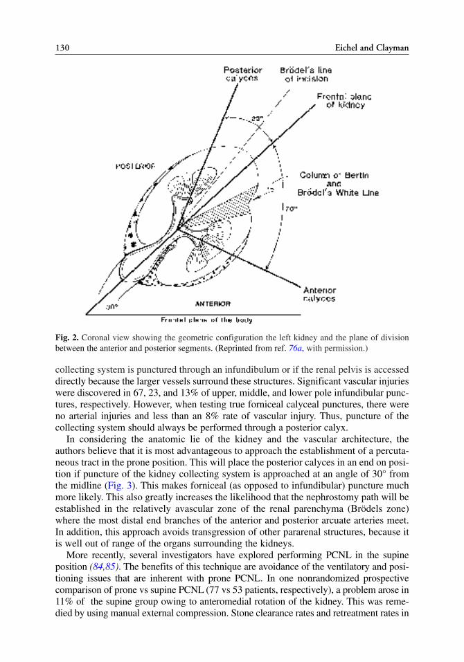
collecting system is punctured through an infundibulum or if the renal pelvis is accesseddirectly because the larger vessels surround these structures. Significant vascular injurieswere discovered in 67, 23, and 13% of upper, middle, and lower pole infundibular punc-tures, respectively. However, when testing true forniceal calyceal punctures, there wereno arterial injuries and less than an 8% rate of vascular injury. Thus, puncture of thecollecting system should always be performed through a posterior calyx.
In considering the anatomic lie of the kidney and the vascular architecture, theauthors believe that it is most advantageous to approach the establishment of a percuta-neous tract in the prone position. This will place the posterior calyces in an end on posi-tion if puncture of the kidney collecting system is approached at an angle of 30° fromthe midline (Fig. 3). This makes forniceal (as opposed to infundibular) puncture muchmore likely. This also greatly increases the likelihood that the nephrostomy path will beestablished in the relatively avascular zone of the renal parenchyma (Brödels zone)where the most distal end branches of the anterior and posterior arcuate arteries meet.In addition, this approach avoids transgression of other pararenal structures, because itis well out of range of the organs surrounding the kidneys.
More recently, several investigators have explored performing PCNL in the supineposition (84,85). The benefits of this technique are avoidance of the ventilatory and posi-tioning issues that are inherent with prone PCNL. In one nonrandomized prospectivecomparison of prone vs supine PCNL (77 vs 53 patients, respectively), a problem arose in11% of the supine group owing to anteromedial rotation of the kidney. This was reme-died by using manual external compression. Stone clearance rates and retreatment rates in
130 Eichel and Clayman
Fig. 2. Coronal view showing the geometric configuration the left kidney and the plane of divisionbetween the anterior and posterior segments. (Reprinted from ref. 76a, with permission.)

Chapter 8 / Percutaneous Stone Removal 131
Fig. 3. (A) Puncturing the collecting system at a 30° angle from the midline vertical plane. A fornicealpuncture is demonstrated. (B) The position of the surgeon in relation to the prone patient and fluoro-scopic equipment during puncture. (Reprinted from ref. 76a, with permission.)
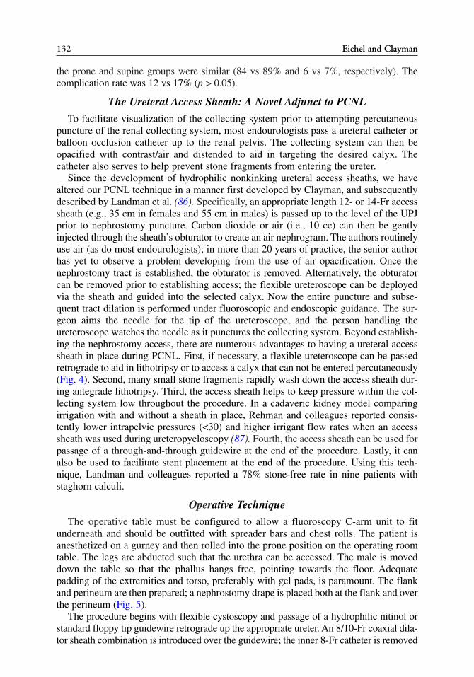
the prone and supine groups were similar (84 vs 89% and 6 vs 7%, respectively). Thecomplication rate was 12 vs 17% (p > 0.05).
The Ureteral Access Sheath: A Novel Adjunct to PCNL
To facilitate visualization of the collecting system prior to attempting percutaneouspuncture of the renal collecting system, most endourologists pass a ureteral catheter orballoon occlusion catheter up to the renal pelvis. The collecting system can then beopacified with contrast/air and distended to aid in targeting the desired calyx. Thecatheter also serves to help prevent stone fragments from entering the ureter.
Since the development of hydrophilic nonkinking ureteral access sheaths, we havealtered our PCNL technique in a manner first developed by Clayman, and subsequentlydescribed by Landman et al. (86). Specifically, an appropriate length 12- or 14-Fr accesssheath (e.g., 35 cm in females and 55 cm in males) is passed up to the level of the UPJprior to nephrostomy puncture. Carbon dioxide or air (i.e., 10 cc) can then be gentlyinjected through the sheath’s obturator to create an air nephrogram. The authors routinelyuse air (as do most endourologists); in more than 20 years of practice, the senior authorhas yet to observe a problem developing from the use of air opacification. Once thenephrostomy tract is established, the obturator is removed. Alternatively, the obturatorcan be removed prior to establishing access; the flexible ureteroscope can be deployedvia the sheath and guided into the selected calyx. Now the entire puncture and subse-quent tract dilation is performed under fluoroscopic and endoscopic guidance. The sur-geon aims the needle for the tip of the ureteroscope, and the person handling theureteroscope watches the needle as it punctures the collecting system. Beyond establish-ing the nephrostomy access, there are numerous advantages to having a ureteral accesssheath in place during PCNL. First, if necessary, a flexible ureteroscope can be passedretrograde to aid in lithotripsy or to access a calyx that can not be entered percutaneously(Fig. 4). Second, many small stone fragments rapidly wash down the access sheath dur-ing antegrade lithotripsy. Third, the access sheath helps to keep pressure within the col-lecting system low throughout the procedure. In a cadaveric kidney model comparingirrigation with and without a sheath in place, Rehman and colleagues reported consis-tently lower intrapelvic pressures (<30) and higher irrigant flow rates when an accesssheath was used during ureteropyeloscopy (87). Fourth, the access sheath can be used forpassage of a through-and-through guidewire at the end of the procedure. Lastly, it canalso be used to facilitate stent placement at the end of the procedure. Using this tech-nique, Landman and colleagues reported a 78% stone-free rate in nine patients withstaghorn calculi.
Operative Technique
The operative table must be configured to allow a fluoroscopy C-arm unit to fitunderneath and should be outfitted with spreader bars and chest rolls. The patient isanesthetized on a gurney and then rolled into the prone position on the operating roomtable. The legs are abducted such that the urethra can be accessed. The male is moveddown the table so that the phallus hangs free, pointing towards the floor. Adequatepadding of the extremities and torso, preferably with gel pads, is paramount. The flankand perineum are then prepared; a nephrostomy drape is placed both at the flank and overthe perineum (Fig. 5).
The procedure begins with flexible cystoscopy and passage of a hydrophilic nitinol orstandard floppy tip guidewire retrograde up the appropriate ureter. An 8/10-Fr coaxial dila-tor sheath combination is introduced over the guidewire; the inner 8-Fr catheter is removed
132 Eichel and Clayman

leaving the 10-Fr sheath and original guidewire in the ureter. Through the 10-Fr sheath, anAmplatz super stiff guidewire is passed; if a nitinol guidewire was passed initially, it is nowexchanged for a standard floppy tip guidewire. Now the 10-Fr sheath is removed leavingthe two guidewires in place. A 12-Fr ureteral access catheter (length = 35 cm for womenand 55 cm for men) is then passed over the Amplatz guidewire. The floppy tip guidewireis maintained as a safety wire; a Kelly or tonsil clamp is used to secure the safety guidewirewhere it exits the urethra, to the perineal drapes. The tip of the 12-Fr access sheath shouldbe positioned at the UPJ. A Foley catheter is inserted along side the 12-Fr sheath in orderto drain the bladder. If the ureter is particularly capacious, then a 14-Fr sheath can be used.
Targeting of the desired calyx is accomplished by injecting 10 cc of air into the renalcollecting system via the obturator of the access sheath. Because the patient is in the prone
Chapter 8 / Percutaneous Stone Removal 133
Fig. 4. (A) Radiograph demonstrating a calyx that is inaccessible from the established upper poleaccess (marked by a *). (B,C) The collecting system is opacified using radiographic contrast. (D) Aureteroscope can then be guided into the desired calyx.
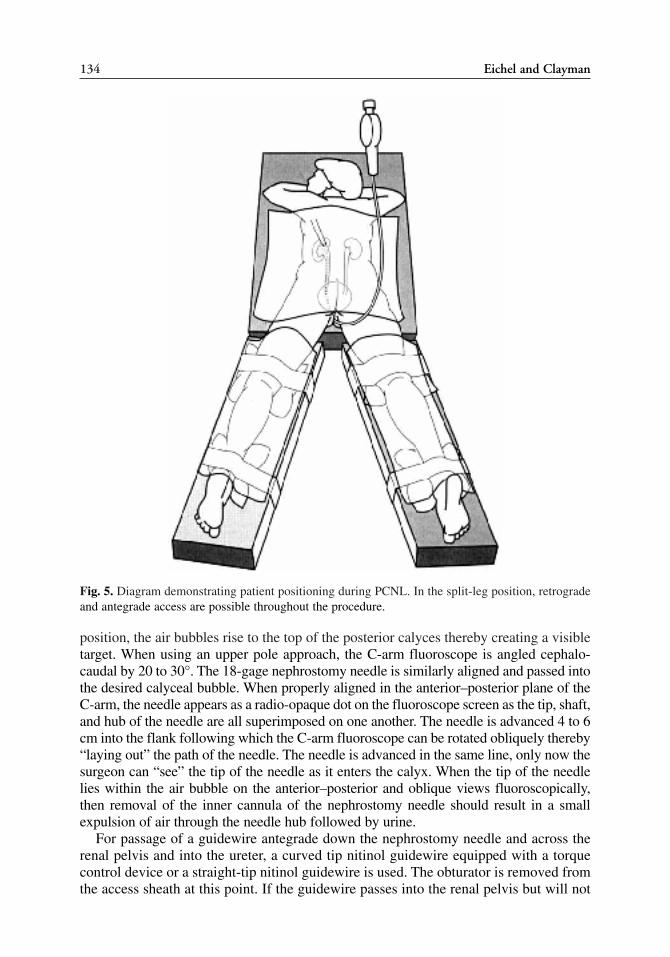
position, the air bubbles rise to the top of the posterior calyces thereby creating a visibletarget. When using an upper pole approach, the C-arm fluoroscope is angled cephalo-caudal by 20 to 30°. The 18-gage nephrostomy needle is similarly aligned and passed intothe desired calyceal bubble. When properly aligned in the anterior–posterior plane of theC-arm, the needle appears as a radio-opaque dot on the fluoroscope screen as the tip, shaft,and hub of the needle are all superimposed on one another. The needle is advanced 4 to 6cm into the flank following which the C-arm fluoroscope can be rotated obliquely thereby“laying out” the path of the needle. The needle is advanced in the same line, only now thesurgeon can “see” the tip of the needle as it enters the calyx. When the tip of the needlelies within the air bubble on the anterior–posterior and oblique views fluoroscopically,then removal of the inner cannula of the nephrostomy needle should result in a smallexpulsion of air through the needle hub followed by urine.
For passage of a guidewire antegrade down the nephrostomy needle and across therenal pelvis and into the ureter, a curved tip nitinol guidewire equipped with a torquecontrol device or a straight-tip nitinol guidewire is used. The obturator is removed fromthe access sheath at this point. If the guidewire passes into the renal pelvis but will not
134 Eichel and Clayman
Fig. 5. Diagram demonstrating patient positioning during PCNL. In the split-leg position, retrogradeand antegrade access are possible throughout the procedure.
pass down into the ureter or into the access sheath, then a 5-Fr Kumpe catheter can bepassed over the guidewire to provide greater maneuverability.
After a guidewire is passed into the ureter, the tract can be dilated. If the guidewirepasses down and out the ureteral access sheath, its urethral end is clamped to maintainthrough-and-through access. Over the guidewire, a fascial incising needle is passed; thiswill incise the fascia to 5 mm. Next, an 8/10-Fr coaxial dilator sheath is passed percuta-neously. The 8-Fr catheter is removed and an Amplatz super-stiff guidewire is placed downthe ureter or into the access sheath. If the 8/10-Fr sheath does not pass easily over the niti-nol guidewire, then a 5-Fr open-ended angiographic catheter can be passed; the nitinolguidewire can then be exchanged for an Amplatz type super-stiff guidewire followingwhich the 5-Fr angiographic catheter is removed. Following this, the 8/10-Fr system ispassed, and a second guidewire—usually a floppy-tip, 0.035-in. type—is passed into thesystem. The floppy-tip, guidewire is sutured to the flank with a 0-silk; it thus becomesanother “safety” guidewire. The Amplatz super-stiff guidewire thus serves as the “work-ing” guidewire over which all other catheters and dilators will be passed. If access is lostduring the case, then the suture is removed from the “safety” guidewire and another “work-ing” guidewire is placed; the “safety” guidewire is then sutured again to the skin.
Next a 30-Fr high-pressure (i.e., rated to 15–20 atm) dilating balloon catheter is passedover the “working” guidewire (i.e., Amplatz super-stiff guidewire) until the tip of the bal-loon rests within the calyx but proximal to the infundibulum of the calyx. Use of a dilat-ing balloon system has been found to have a 10% transfusion rate vs 25% for use of theAmplatz sequential dilator system (88). The balloon is then inflated and the 30-Frnephrostomy sheath is passed over the balloon until it too resides within the calyx proxi-mal to the infundibulum. The balloon is then deflated and removed.
Rigid and flexible nephroscopy as well as URS can be performed depending on thestone size and location. For stones that are 1 cm or less, direct removal using stone grasp-ing forceps is a very efficient technique. Grasping forceps or a rigid nitinol basketretriever, as designed by Denstedt, can be used for this purpose. For larger stones, a vari-ety of lithotripters can be used including ultrasonic, pneumatic, combined pneumatic andultrasonic (e.g., Lithoclast Ultra), Holmium:YAG laser, or rarely, if ever today, the electro-hydraulic lithotripter. In our experience, modes of energy that can be applied simultane-ously with suction removal of the fragments are the most efficient means of reducingstone burden (i.e., ultrasonic or Lithoclast Ultra). However, the pneumatic orHolmium:YAG laser may be required for particularly hard stones; the former is very effi-cient at rapidly fragmenting large, hard calculi, whereas the latter is very effective when-ever flexible endoscopy is needed. The electrohydraulic lithotriptor is rarely used; its onlyindication is when loss of flexible endoscope deflection precludes stone access with theholmium laser.
Difficult to reachcalycescanbereachedusingflexiblenephroscopyorantegradeor retrograde(i.e., via the access sheath) flexible ureteroscopy (Fig. 4). Prior to exit, a full rigid and flexiblenephroscopic and fluoroscopic exam is essential to ensure complete stone removal. If visibility ispoor, it isbest toplacea large-bore (i.e., 20-,22-,or24-Fr)nephroureteral tubeandplanfora“sec-ond look” based on the results of a postoperative day-one CT scan without contrast.
Traditionally, following successful PCNL with a stone-free endoscopic and fluoro-scopic status, our preference was to leave a 10-Fr Cope loop nephrostomy tube in therenal collecting system. We have now modified our technique to a “tubeless” approach.This approach, originally championed by Wickham in the mid-1980s never became
Chapter 8 / Percutaneous Stone Removal 135

popular in the United States until resurrected and modified by Bellman and colleaguesmore than a decade later (89–91).
Presently, among endoscopic and fluoroscopic stone-free patients our routine for exitingthe nephrostomy tract begins by pulling the nephrostomy sheath back to the junction of renalparenchyma and the collecting system. A guidewire—usually a 260-cm exchange guidewireor a standard 140-cm guidewire in the male and female patient, respectively—is passedthrough the access sheath; a rigid nephroscope is used to grasp and pull the floppy end ofthe guidewire out through the nephrostomy tract, thereby creating a “through-and-through”guidewire access. Next, a 7-Fr, 11.5-mm balloon occlusion catheter is passed retrogradeover the “through-and-through” ureteral access guidewire until the tip of the balloon lies atthe edge of the nephrostomy sheath (Fig. 6) (92). The passage of the occlusion ballooncatheter to this point is monitored with the nephroscope; the balloon is inflated at the entrypoint of the nephrostomy tract into the calyx. A hemostat is placed on the guidewire whereit exits the occlusion balloon catheter at the perineal end. The endoscope is withdrawn; now,the long laparoscopic hemostatic gelatin matrix applicator (FloSeal, Baxter HealthcareCorp., Deerfield, IL) is passed into the 30-Fr sheath until the surgeon feels it touching theexpanded occlusion balloon. The 30-Fr sheath is withdrawn 1 cm and now the entire tract isfilled with FloSeal as the surgeon injects and simultaneously withdraws the applicator withone hand while also retracting the 30-Fr Amplatz sheath with the other hand (Fig. 7). Bykeeping the “through-and-through” guidewire taut, the occlusion balloon is maintained in itsposition thereby precluding any of the gelatin matrix from entering the collecting system.Also, once the 30-Fr sheath and applicator are removed, the surgeon can then apply gentletamponade for up to 10 minutes to the incision in the flank, thereby sandwiching the hemo-static material between a gauze sponge applied to the skin surface and the occlusion balloon(92). The access sheath is pulled back to the midureter and then cut at the urethral end; thesolitary “through-and-through” guidewire is pulled retrograde under fluoroscopic control
136 Eichel and Clayman
Fig. 6. Fluoroscopic image showing an Amplatz nephrostomy sheath pulled back to the junction ofthe renal collecting system and parenchyma. The ureteral occlusion balloon is inflated at the tip of thesheath to prevent injection of FloSeal into the renal collecting system.

until its tip is seen in the collecting system. The Foley catheter is removed so it will not inter-fere with stent placement. An internal ureteral stent can then be passed retrograde followingwhich the access catheter is removed followed by removal of the guidewire and then thepusher. For this purpose, a 36-cm “tail stent” (7-Fr proximal to 3-Fr distal) (MicrovasiveBoston Scientific Corp, Natick, MA) is selected, because these stents have been shown pre-viously to cause less pain and irritable bladder symptoms postoperatively (93). The tail isrepositioned from the urethra (in the male) or perineum (in the female) into the bladder(endoscopically in males or with a Kelly clamp in females). A Foley catheter is then passed.The skin is closed with a 4-O absorbable suture placed in a subcuticular fashion.Alternatively, when a second look procedure is planned, a nephroureteral access tube canbe fashioned by passing a 70-cm single pigtail catheter through the middle of a 22-FrCouncill tip catheter such that the pigtail catheter rests within the bladder and the Councilltip catheter lies in the renal collecting system. One cubic centimeter of saline is placed intothe balloon of the Councill catheter to secure it within the renal collecting system. A plas-tic side arm adapter fits into the butt end of the Councill catheter; it then can be in turn tight-ened around the shaft of the 7-Fr pigtail catheter. The Councill catheter is sewn to the skin withtwo 0-silk sutures. (Fig. 8) This system provides large bore nephrostomy access to the kid-ney and fail-safe and secure access to the ureter.
COMPLICATIONS
Hemorrhage from the nephrostomy tract is the most common major complication asso-ciated with PCNL. The incidence of significant hemorrhage requiring transfusion in mod-
Chapter 8 / Percutaneous Stone Removal 137
Fig. 7. Demonstration of technique for injecting a percutaneous tract with hemostatic gelatin matrix.
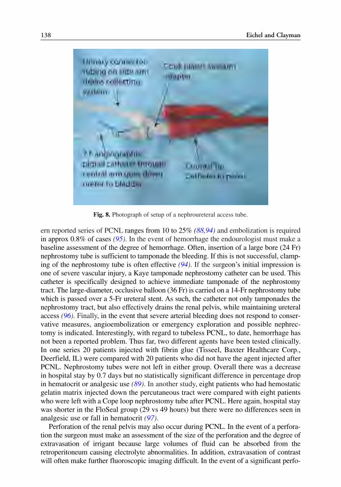
ern reported series of PCNL ranges from 10 to 25% (88,94) and embolization is requiredin approx 0.8% of cases (95). In the event of hemorrhage the endourologist must make abaseline assessment of the degree of hemorrhage. Often, insertion of a large bore (24 Fr)nephrostomy tube is sufficient to tamponade the bleeding. If this is not successful, clamp-ing of the nephrostomy tube is often effective (94). If the surgeon’s initial impression isone of severe vascular injury, a Kaye tamponade nephrostomy catheter can be used. Thiscatheter is specifically designed to achieve immediate tamponade of the nephrostomytract. The large-diameter, occlusive balloon (36 Fr) is carried on a 14-Fr nephrostomy tubewhich is passed over a 5-Fr ureteral stent. As such, the catheter not only tamponades thenephrostomy tract, but also effectively drains the renal pelvis, while maintaining ureteralaccess (96). Finally, in the event that severe arterial bleeding does not respond to conser-vative measures, angioembolization or emergency exploration and possible nephrec-tomy is indicated. Interestingly, with regard to tubeless PCNL, to date, hemorrhage hasnot been a reported problem. Thus far, two different agents have been tested clinically.In one series 20 patients injected with fibrin glue (Tisseel, Baxter Healthcare Corp.,Deerfield, IL) were compared with 20 patients who did not have the agent injected afterPCNL. Nephrostomy tubes were not left in either group. Overall there was a decreasein hospital stay by 0.7 days but no statistically significant difference in percentage dropin hematocrit or analgesic use (89). In another study, eight patients who had hemostaticgelatin matrix injected down the percutaneous tract were compared with eight patientswho were left with a Cope loop nephrostomy tube after PCNL. Here again, hospital staywas shorter in the FloSeal group (29 vs 49 hours) but there were no differences seen inanalgesic use or fall in hematocrit (97).
Perforation of the renal pelvis may also occur during PCNL. In the event of a perfora-tion the surgeon must make an assessment of the size of the perforation and the degree ofextravasation of irrigant because large volumes of fluid can be absorbed from theretroperitoneum causing electrolyte abnormalities. In addition, extravasation of contrastwill often make further fluoroscopic imaging difficult. In the event of a significant perfo-
138 Eichel and Clayman
Fig. 8. Photograph of setup of a nephroureteral access tube.

ration it is best to abort the procedure and place a large-bore nephroureteral catheter com-bination (vide supra). A second-look procedure should be postponed until confirmation ofno extravasation on a nephrostogram. Following a significant perforation, it is also possi-ble that stone fragments may be extruded into the perinephric space. Attempts at retrievalof these fragments are rarely successful and usually only enlarge the perforation and fillthe retroperitoneum with irrigant. Further, these fragments are of little clinical significanceunless the stone was infected. Even under the last circumstance, subsequent infection andabscess formation is too rare to justify extensive retroperitoneal manipulation.
Colonic injury during percutaneous stone removal is fortunately a rare event, occur-ing in less than 1% of procedures. A retrorenal colon occurs in approx 0.6% of patients,but is more common in patients with horseshoe kidney and other ectopias (98). Acutesigns of perforation include intraoperative diarrhea, hematochezia, sepsis, or passageof gas and feces from the nephrostomy tube. More often, a transcolonic injury is appar-ent on a postoperative CT or nephrostogram. If the patient is asymptomatic, the prob-lem can be managed conservatively with placement of an internal ureteral stent plus aFoley catheter and pulling back of the nephrostomy tube into the colon, therebydecompressing both the urinary tract and the colon through separate tubes. If, however,the patient exhibits signs of sepsis or if the injury is transperitoneal, open repair is rec-ommended.
As mentioned earlier in this chapter, because of the close relationship of the pleura andunderlying kidney, a supracostal approach may result in a hydrothorax, or rarely a pneu-mothorax in 8 to 12% of patients (77,78). Traditionally, an upright chest radiograph follow-ing PCNL was recommended to detect this complication. However, more recent work byOgan and colleagues (99) have shown that intraoperative fluoroscopy in combination witha postoperative day-one CT scan is a more effective means of detecting hydropneumotho-rax. If detected on intraoperative fluoroscopy, a percutaneous drain, such as a 10-Fr Copeloop, can be placed fluoroscopically at the end of the PCNL procedure. The drain shouldbe attached to chest tube suction. If a hydropneumothorax is only first detected on the post-operative day one CT then the surgeon must make an assessment of the patient’s symptomsto determine if drainage is necessary.
TIPS AND TRICKS
Starting off with the patient in the split leg prone position ensures that the surgeonwill have the maximum number of treatment options during PCNL. It takes only a fewcases to become adept at prone flexible cystoscopy and, with use of the access sheath,prone flexible ureteroscopy. After mastering this skill, the surgeon not only can obtainendoscopically monitored upper tract access prior to PCNL, but also can use this accessto drain the collecting system and evacuate stone fragments during the case by position-ing the ureteral access sheath at the level of the UPJ. With the inflow from the nephro-scope flowing directly down the 12-Fr access sheath, fragments up to 3 mm in size canbe flushed antegrade out of the patient. Also, having an access sheath positioned at theUPJ gives the surgeon the option of passing a flexible ureteroscope retrograde to thecollecting system to aid in lithotripsy or in accessing a calyx that is not accessible viathe nephrostomy tract. In this situation, the flexible ureteroscope can be used to eitherfragment the stone in situ or to basket the stone and deliver it into the renal pelvis whereit can then be retrieved with the rigid nephroscope.
Chapter 8 / Percutaneous Stone Removal 139

Another key point that greatly increases the number of options available to the sur-geon is the establishment of a “through-and-through” guidewire using a 260-cm long0.035-in. guidewire. With this guidewire in place, the surgeon can pass any lengthcatheter antegrade or retrograde during the case. It also gives the surgeon the ability to“pull” a catheter up or down the ureter as desired by placing a clamp onto the guide wirebehind the catheter after the catheter has been completely loaded on the guidewire; thesurgeon can then pull the catheter into the desired position. This guidewire is also idealfor placing an occlusion balloon catheter for subsequent use with FloSeal or for placingan indwelling stent (vide supra).
Tips to Note
1. When positioning a male patient prone with legs on spreader bars, be sure the phalluslies free of the table.
2. 10/12-Fr or 12/14-Fr ureteral access sheaths should be placed with the end of the sheathat the UPJ (i.e., obturator tip would lie in renal pelvis or upper pole infundibulum).
3. Consider entry into an upper pole posterior calyx whenever possible as this provides themost direct path to the ureter and the shortest path into the kidney.
4. Use of retrograde flexible ureteroscopy via the access sheath, allows access to stones ina remote upper pole calyx (juxtaposed to the nephrostomy tract).
5. “Through-and-through” access can be established using a 260-cm exchange guidewire.6. FloSeal in association with an occlusion balloon catheter can be used to achieve hemo-
stasis in the percutaneous tract.
CONCLUSION
Percutaneous stone removal has progressed significantly over the past 29 years. Theadvent of improved guide wires (e.g., nitinol), tract dilators (e.g., balloon), lithotriptors(e.g., pneumatic and Holmium), and hemostatic agents has made this procedure moreeffective and safer. Today, PCNL is capable of rendering more than 90% of patientsstone free regardless of stone burden or location.
REFERENCES1. Fernstrom I, Johansson B. Percutaneous pyelolithotomy. A new extraction technique. Scand J Urol
Nephrol 1976; 10: 257–259.2. Kerbl K, Rehman J, Landman J, Lee D, Sundaram C, Clayman RV. Current management of urolithi-
asis: progress or regress? J Endourol 2002; 16: 281–288.3. Lingeman JE, Coury TA, Newman DM, et al. Comparison of results and morbidity of percutaneous
nephrostolithotomy and extracorporeal shock wave lithotripsy. J Urol 1987; 138: 485–490.4. Robert M, Drianno N, Marotta J, Delbos O, Guiter J, Grasset D. The value of retrograde
ureterorenoscopy in the treatment of bulky kidney calculi. Prog Urol 1997; 7: 35–41.5. Grasso M, Conlin M, Bagley D. Retrograde ureteropyeloscopic treatment of 2 cm. or greater upper
urinary tract and minor staghorn calculi. J Urol 1998; 160: 346–351.6. Lee WJ, Snyder JA, Smith AD. Staghorn calculi: endourologic management in 120 patients.
Radiology 1987; 165: 85–88.7. Lam HS, Lingeman JE, Mosbaugh PG, et al. Evolution of the technique of combination therapy for
staghorn calculi: a decreasing role for extracorporeal shock wave lithotripsy. J Urol 1992; 148:1058–1062.
8. Lingeman JE, Siegel YI, Steele B, Nyhuis AW, Woods JR. Management of lower pole nephrolithia-sis: a critical analysis. J Urol 1994; 151: 663–667.
140 Eichel and Clayman

9. Wong C, Leveillee RJ. Single upper-pole percutaneous access for treatment of > or = 5-cm complexbranched staghorn calculi: is shockwave lithotripsy necessary? J Endourol 2002; 16: 477–481.
10. Zhong P, Chuong CJ, Goolsby RD, Preminger GM. Microhardness measurements of renal calculi:regional differences and effects of microstructure. J Biomed Mater Res 1992; 26: 1117–1130.
11. Williams JC, Jr., Saw KC, Paterson RF, Hatt EK, McAteer JA, Lingeman JE. Variability of renalstone fragility in shock wave lithotripsy. Urology 2003; 61: 1092–1096.
12. Knoll LD, Segura JW, Patterson DE, Leroy AJ, Smith LH. Long-term followup in patients with cys-tine urinary calculi treated by percutaneous ultrasonic lithotripsy. J Urol 1988; 140: 246–248.
13. Katz G, Landau EH, Lancovsky Z, Pode D, Shapira A, Pfau A. Extracorporeal shock wave lithotripsyfor cystine urolithiasis. Harefuah 1991; 120: 182–184.
14. Kachel TA, Vijan SR, Dretler SP. Endourological experience with cystine calculi and a treatmentalgorithm. J Urol 1991; 145: 25–28.
15. Rudnick DM, Bennett PM, Dretler SP. Retrograde renoscopic fragmentation of moderate–size(1.5–3.0-cm) renal cystine stones. J Endourol 1999; 13: 483–485.
16. Dretler SP, Spencer BA. CT and stone fragility. J Endourol 2001; 15: 31–36.17. Joseph P, Mandal AK, Singh SK, Mandal P, Sankhwar SN, Sharma SK. Computerized tomography
attenuation value of renal calculus: can it predict successful fragmentation of the calculus by extra-corporeal shock wave lithotripsy? A preliminary study. J Urol 2002; 167: 31968–1971.
18. Sampaio FJ, D’Anunciacao AL, Silva EC. Comparative follow-up of patients with acute and obtuseinfundibulum-pelvic angle submitted to extracorporeal shockwave lithotripsy for lower calicealstones: preliminary report and proposed study design. J Endourol 1997; 11: 157–161.
19. Keeley FX, Moussa SA, Smith G, Tolley DA. Clearance of lower-pole stones following shock wavelithotripsy: effect of the infundibulopelvic angle. Eur Urol 1999; 36: 371–375.
20. Elbahnasy AM, Clayman RV, Shalhav AL, et al. Lower-pole caliceal stone clearance after shockwavelithotripsy, percutaneous nephrolithotomy, and flexible ureteroscopy: impact of radiographic spatialanatomy. J Endourol 1998; 12: 113–119.
21. Poulakis V, Dahm P, Witzsch U, de Vries R, Remplik J, Becht E. Prediction of lower pole stone clear-ance after shock wave lithotripsy using an artificial neural network. J Urol 2003; 169: 1250–1256.
22. Moody JA, Williams JC, and Lingeman JE. Lower pole renal anatomy: effects on stone clearanceafter shock wave lithotripsy in a randomized population. J Urol 1999; 161: 378.
23. Madbouly K, Sheir KZ, Elsobky E. Impact of lower pole renal anatomy on stone clearance aftershock wave lithotripsy: fact or fiction? J Urol 2001; 165: 1415–1418.
24. Albala DM, Assimos DG, Clayman RV, et al. Lower pole I: a prospective randomized trial of extra-corporeal shock wave lithotripsy and percutaneous nephrostolithotomy for lower pole nephrolithia-sis-initial results. J Urol 2001; 166: 2072–2080.
25. Winfield HN, Clayman RV, Chaussy CG, Weyman PJ, Fuchs GJ, Lupu AN. Monotherapy of staghornrenal calculi: a comparative study between percutaneous nephrolithotomy and extracorporeal shockwave lithotripsy. J Urol 1988; 139: 895–899.
26. Pearle MS and Traxer O. Renal urolithiasis: therapy for special circumstances part II. AUA UpdateSeries, 2000; 20: 314.
27. Yow RM, Bunts RC. Caliceal diverticulum. J Urol 1955; 73: 663–670.28. Devine CJ, Jr., Guzman JA, Devine PC, Poutasse EF. Calyceal diverticulum. J Urol 1969; 101: 8–11.29. Abeshouse BS, Abeshouse GA. Calyceal Diverticulum: A Report of Sixteen Cases and Review of the
Literature. Urol Int 1963; 15: 329–357.30. Williams DI, Kenawi MM. The prognosis of pelviureteric obstruction in childhood: a review of 190
cases. Eur Urol 1976; 2: 57–63.31. Ramakumar SJW. Laparoscopic surgery for renal urolithiasis: pyelolithotomy, caliceal diverticulec-
tomy, and treatment of stones in a pelvic kidney. J Endourol 2000; 14: 829.32. Psihramis DE, Dretler SP. Extracorporeal shock wave lithotripsy of caliceal diverticular calculi.
J Urol 1987; 138: 707–711.33. Hendrikx AJ, Bierkens AF, Bos R, Oosterhof GO, Debruyne FM. Treatment of stones in caliceal
diverticula: extracorporeal shock wave lithotripsy versus percutaneous nephrolitholapaxy. Br J Urol1992; 70: 478–482.
34. Streem SB, Yost A. Treatment of caliceal diverticular calculi with extracorporeal shock wavelithotripsy: patient selection and extended followup. J Urol 1992; 148: 1043–1046.
Chapter 8 / Percutaneous Stone Removal 141

35. Batter SJ, Dretler SP. Ureterorenoscopic approach to the symptomatic caliceal diverticulum. J Urol1997; 158: 709–713.
36. Chong TW, Bui MH, Fuchs GJ. Calyceal diverticula. Ureteroscopic management. Urol Clin NorthAm 2000; 27: 647–654.
37. Grasso M, Lang G, Loisides P, Bagley D, Taylor F. Endoscopic management of the symptomatic cal-iceal diverticular calculus. J Urol 1995; 153: 1878–1881.
38. Hulbert JC, Reddy PK, Hunter DW, Castaneda-Zuniga W, Amplatz K, Lange PH. Percutaneous tech-niques for the management of caliceal diverticula containing calculi. J Urol 1986; 135: 225–227.
39. Jones JA, Lingeman JE, Steidle CP. The roles of extracorporeal shock wave lithotripsy and percuta-neous nephrostolithotomy in the management of pyelocaliceal diverticulua. J Urol 1991; 146: 724–727.
40. Ellis JH, Patterson SK, Sonda LP, Platt JF, Sheffner SE, Woolsey EJ. Stones and infection in renal cal-iceal diverticula: treatment with percutaneous procedures. AJR Am J Roentgenol 1991; 156: 995–1000.
41. Hendrikx AJ, Bierkens AF, Bos R, Oosterhof GO, Debruyne FM. Treatment of stones in calicealdiverticuli: Extracorporeal shock wave lithotripsy versus percutaneous nephrolitholapaxy. Br J Urol1992; 70: 478–482.
42. Bellman GC, Silverstein JI, Blickensderfer S, Smith AD. Technique and follow-up of percutaneousmanagement of caliceal diverticula. Urology 1993; 42: 21–25.
43. Harewood LM, Agarwal D, Lindsay S, Vaughan MG, Cleeve LK, Webb DR. Extraperitoneal laparo-scopic caliceal diverticulectomy. J Endourol 1996; 10: 425–430.
44. Shalhav AL, Soble JJ, Nakada SY, Wolf JS, Jr., McClennan BL, Clayman RV. Long-term outcome ofcaliceal diverticula following percutaneous endosurgical management. J Urol 1998; 160: 1635–1639.
45. Auge BK, Munver R, Kourambas J, Newman GE, Preminger GM. Endoscopic management of symp-tomatic caliceal diverticula: a retrospective comparison of percutaneous nephrolithotripsy andureteroscopy. J Endourol 2002; 16: 557–563.
46. Matin SF, Streem SB. Metabolic risk factors in patients with ureteropelvic junction obstruction andrenal calculi. J Urol 2000; 163: 1676–1678.
47. Husmann DA, Milliner DS, and Segura JW. Ureteropelvic junction obstruction with concurrent renalpelvic calculi in the pediatric patient: a long-term follow-up. J Urol 1996; 156: 741–743.
48. Bernardo NO, Liatsikos EN, Dinlenc CZ, Kapoor R, Fogarty JD, and Smith AD. Stone recurrenceafter endopyelotomy. Urol 2000; 56: 378–381.
49. Streem SB. Stone extraction. In: Smith’s Textbook of Endourology. Vol. 1, (Smith AD, Badlani GH,Bagley DH, et al.,eds.), Quality Medical Publishing, St. Louis, MO, 1996, pp.: 239–263.
50. Shalhav AL, Giusti G, Elbahnasy AM, et al. Adult endopyelotomy: impact of etiology and antegradeversus retrograde approach on outcome. J Urol 1998; 160: 685–689.
51. Hoenig DM, Hemal AK, Shalhav AL, Clayman RV. Percutaneous nephrostolithotomy, endopyelo-tomy and nephropexy in a single session. J Urol 1998; 160: 826–827.
52. Pitts WR Jr., Muecke EC. Horseshoe kidneys: a 40-year experience. J Urol 1975; 113: 743–746.53. Vandeursen H, Baert L. Electromagnetic extracorporeal shock wave lithotripsy for calculi in horse-
shoe kidneys. J Urol 1992; 148: 1120–1122.54. Jenkins AD, Gillenwater JY. Extracorporeal shock wave lithotripsy in the prone position: treatment
of stones in the distal ureter or anomalous kidney. J Urol 1988; 139: 911–915.55. Janetschek G, Kunzel KH. Percutaneous nephrolithotomy in horseshoe kidneys: applied anatomy and
clinical experience. Br J Urol 1988; 62: 117–122.56. Lingeman JE, Chee SK. Percutaneous operative procedures in horseshoe kidneys. J Urol 1999; 161: 371.57. Jones DJ, Wickham JEA, Kellett MJ. Percutaneous nephrolithotomy for calculi in horseshoe kidneys.
J Urol 1991; 145: 481–483.58. Rhee BK, Bretan PN, Stoller ML. Urolithiasis in renal and combined pancreas/renal transplant recip-
ients. J Urol 1999; 161: 1458–1462.59. Hayes JM, Streem SB, Graneto D, Steinmuller DR, and Novick AC. Urolithiasis after renal transplan-
tation. Trans Proc 1989; 21: 1960–1961.60. Wheatley M, Ohl DA, Sonda LP, Wang SC, and Konnak JW. Treatment of renal transplant stone by
extracorporeal shock wave lithotripsy in the prone position. Urology 1991; 37: 57.61. Del Pizzo JJ, Jacobs SC, and Sklar GN. Ureteroscopic evaluation in renal transplant recipients.
J Endourol 1998; 12: 135–138.62. Eshghi M, Smith AD. Endourologic approach to a transplant kidney. Urology 1986; 18: 504–507.
142 Eichel and Clayman

63. Hulbert JC, Reddy P, Young AT, et al. The percutaneous removal of calculi from transplanted kidneys.J Urol 1985; 134: 324–326.
64. Francesca F, Felipetto R, Mosca F, Boggi U, Rizzo G, Puccini R. Percutaneous nephrolithotomy oftransplanted kidney. J Endourol 2002; 16: 225–227.
65. Dretler SP, Olsson C, Pfister RC. The anatomic, radiologic and clinical characteristics of the pelvickidney: an analysis of 86 cases. J Urol 1971; 105: 623–627.
66. Rigatti P, Montorsi F, Guazzoni G, di Girolamo V, et al. Multimodal therapy for stones in pelvic kid-neys. Urol Int 1991; 46: 29–34.
67. Fabrizio MD, Behari, A., and Bagley, D.H. Ureteroscopic management of intrarenal calculi. J Urol1998; 159: 1139–1143.
68. Eshghi AM, Roth JS, Smith AD. Percutaneous transperitoneal approach to a pelvic kidney forendourological removal of staghorn calculus. J Urol 1985; 134: 525–527.
69. Ramakumar S, Segura JW. Laparoscopic surgery for renal urolithiasis: pyelolithotomy, caliceal diver-ticulectomy, and treatment of stones in a pelvic kidney. J Endourol 2000; 14: 829–832.
70. Bricker EM. Symposium on clinical surgery; bladder substitution after pelvic evisceration. Surg ClinN Amer 1950; 30: 1511.
71. Dretler SP. The pathogenesis of urinary tract calculi occurring after ileal conduit diversion: i. clinicalstudy. ii. conduit study. iii. prevention. J Urol 1973; 109: 204–209.
72. Turk TM, Koleski FC, Albala DM. Incidence of urolithiasis in cystectomy patients after intestinalconduit or continent urinary diversion. World J Urol 1999; 17: 305–307.
73. Sullivan JW, Grabstald H. Whitmore Jr. WF Complication of ureteroileal conduit with radical cystec-tomy: review of 336 cases. J Urol 1980; 124: 797–801.
74. Schmidt JD, Hawtrey CE, Flocks RH. Culp DA. Complications, results and problems of ileal conduitdiversion. J Urol 1973; 109: 210–216.
75. McDougal WS, Kock MO. Impaired growth and development and urinary intestinal interposition.Trans Amer Ass Genitourinary Surg 1991; 105: 3.
76. Cohen TD, Streem SB, Lammert G. Long-term incidence and risks for recurrent stones followingcontemporary management of upper tract calculi in patients with a urinary diversion. J Urol 1996;155: 62–65.
76a. Clayman RV, Castaneda-Zuniga WR. In: Techniques in Endourology: A Guide to the PercutaneousRemoval of Renal and Ureteral Calculi, Heritage Press, Dallas, 1984.
77. Golijanin D, Katz R, Verstandig A, Sasson T, Landau EH, Meretyk S. The supracostal percutaneousnephrostomy for treatment of staghorn and complex kidney stones. J Endourol 1998; 12: 403–405.
78. Picus D, Weyman PJ, Clayman RV, McClennan BL. Intercostal-space nephrostomy for percutaneousstone removal. AJR Am J Roentgenol 1986; 147: 393–397.
79. Munver R, Delvecchio FC, Newman GE, Preminger GM. Critical analysis of supracostal access forpercutaneous renal surgery. J Urol 2001; 166: 1242–1246.
80. Sampaio FJB. Surgical anatomy of the kidney. In: Smith’s Textbook of Endourology. (Smith AD,Badlani GH, Bagley DH, et al., eds.), Quality Medical Publishing, St. Louis, MO, 1996: pp.153–184.
81. Kaye KW. Renal anatomy: Endourological considerations. In: (Clayman RV, W. Castañeda-Zuñiga,eds.) Techniques in Endourology: A Guide To The Percutaneous Removal of Renal and UreteralCalculi. Heritage Press, Dallas, 1984: pp. 55–71.
82. Sampaio FJB, Aragao AHM. Anatomical relationship between the renal venous arrangement and thekidney collection system. J Urol 1990; 144: 1089–1093.
83. Sampaio FJB, Aragao AHM. Inferior pole collecting system anatomy: its probable role in extracor-poreal shock wave lithotripsy. J Urol 1992; 147: 322–324.
84. Valdivia-Uria JG, Valle-Gerhold J, Lopez-Lopez JA, et al. Technique and complications of percuta-neous nephroscopy: experience with 557 patients in the supine position. J Urol 1998; 160:1975–1978.
85. Shoma AM, Eraky I, El-Kenawy MR, El-Kappany HA. Percutaneous nephrolithotomy in the supineposition: technical aspects and functional outcome compared with the prone technique. Urology2002; 60: 388–392.
86. Landman J, Venkatesh R, Lee DI, et al. Combined percutaneous and retrograde approach to staghorncalculi with application of the ureteral access sheath to facilitate percutaneous nephrolithotomy. J Urol2003; 169: 64–67.
Chapter 8 / Percutaneous Stone Removal 143
87. Rehman J, Monga M, Landman J, et al. Characterization of intrapelvic pressure during ureteropy-eloscopy with ureteral access sheaths. Urology 2003; 61: 713–718.
88. Davidoff R, Bellman GC. Influence of technique of percutaneous tract creation on incidence of renalhemorrhage. J Urol 1997; 157: 1229–1231.
89. Mikhail AA, Kaptein JS, Bellman GC. Use of fibrin glue in percutaneous nephrolithotomy. Urology2003; 61: 910–914.
90. Feng MI, Tamaddon K, Mikhail A, Kaptein JS, Bellman GC. Prospective randomized study of vari-ous techniques of percutaneous nephrolithotomy. Urology 2001; 58: 345–350.
91. Yew J, Bellman G. Modified “tubeless” percutaneous nephrolithotomy using a tail-stent. Urology2003; 62: 346–349.
92. Lee DI, Uribe C, Eichel L, et al. Sealing percutaneous nephrolithotomy tracts with gelatin matrixhemostatic sealant: initial clinical use. J Urol 2004; 171: 575–578.
93. Dunn MD, Portis AJ, Kahn SA, et al. Clinical effectiveness of new stent design: randomized single-blind comparison of tail and double-pigtail stents. J Endourol 2000; 14: 195–202.
94. Gallucci M, Fortunato P, Schettini M, Vincenzoni A. Management of hemorrhage after percutaneousrenal surgery. J Endourol 1998; 12: 509–512.
95. Dunnick NR, Carson CC, Braun SD, et al. Complications of percutaneous nephrostolithotomy.Radiology 1985; 157: 51–55.
96. Kaye KW, Clayman RV. Tamponade nephrostomy catheter for percutaneous nephrostolithotomy.Urology 1986; 27: 441–445.
97. Eichel L SL, Lee D, McDougall EM, Clayman RV. Early Experience With Tubeless PCNL UsingHemostatic Gelatin Matirx: A Comparison To The Standard Technique. J Urol 2004; 171: A396.
98. Hadar H, Gadoth N. Positional relations of colon and kidney determined by perirenal fat. AJR Am JRoentgenol 1984; 143: 773–776.
99. Ogan K, Corwin TS, Smith T, et al. Sensitivity of chest fluoroscopy compared with chest CT andchest radiography for diagnosing hydropneumothorax in association with percutaneous nephros-tolithotomy. Urology 2003; 62: 988–992.
144 Eichel and Clayman

Calculus Therapy Combined Approaches
Véronique Tran, MD, Ramakrishna Venkatesh, MD,FRCSC, and Jaime Landman, MD
CONTENTS
INTRODUCTION
INDICATIONS
INSTRUMENT LIST
TECHNIQUE
RESULTS
TIPS AND TRICKS
REFERENCES
9
SUMMARY
Advances in ureteroscopic tecnique and technology have increased the scope of uro-logical pathology that can be treated in a retrograde manner. “Combined approaches” thatincorporate traditional antegrade access and retrograde access offer the urologist increasedflexibility in the management of complex urinary tract pathology. In the following chap-ter, we review the indications, technique, and results associated with the combinedapproach to urolithiasis.
Key Words: Kidney; kidney calculi; ureter; nephrostomy; percutaneous; surgicalinstruments.
INTRODUCTION
Surgical stone management has evolved tremendously over the last two decades. Withthe advent of shockwave lithotripsy (SWL) in the 1980s, this extracorporeal modality rap-idly became the primary treatment of choice for 95% of stone cases. At this time, an endo-scopic approach was used less than 5% of the time. Over the past two decades, however,advances in flexible endoscopes, irrigation systems, lasers, and laser fibers (in addition tothe introduction of small caliber nitinol instrumentation) have increased the utility of theendoscopic approach for stone disease. Although SWL remains the procedure of choice
From: Advanced Endourology: The Complete Clinical GuideEdited by: S. Y. Nakada and M. S. Pearle © Humana Press Inc., Totowa, NJ
145

for the majority of stone cases (75%), there remains an important role for endoscopy instone disease. Currently, the endoscopic (antegrade or retrograde) approach is employedas the primary treatment strategy in approx 25% of stone cases that require intervention.
The endoscopic approach for the management of nephrolithiasis has continued toexpand over the last two decades with the development of percutaneous nephrolithotomy(PCNL). In the early conception of PCNL, treatment of large calculi with PCNL was fol-lowed by SWL, and then a second-look PCNL. This type of “sandwich therapy” becamethe standard of care. Refinements in percutaneous surgical techniques have made PCNLwith or without a second-look nephroscopy as successful as “sandwich therapy.”
As experience and comfort with the antegrade and retrograde approaches to the uri-nary tract have expanded, a combined approach to endoscopy has evolved. Applicationof this combined antegrade and retrograde approach allows for unification of the uri-nary tract. The advantages of both the antegrade and retrograde approaches can be com-bined for safe and efficient stone ablation in all parts of the upper urinary tract. Thefollowing chapter will summarize the indications, techniques, and results of the com-bined antegrade and retrograde approach to urolithiasis.
INDICATIONS
Percutaneous Nephrolithotomy
The authors currently utilize a combined approach involving simultaneous percuta-neous antegrade and retrograde access to stone disease that requires PNCL. Stone char-acteristics that should be considered when deciding if PCNL is indicated include stoneburden (size and number), composition, and location. Larger stones are usually treatedwith PCNL. Staghorn calculi and nonstaghorn stones greater than 30 mm in size, regard-less of other stone-related factors, are best managed with PCNL as first-line treatment.
Depending on stone size and renal anatomy, many lower pole calculi require PCNL.Specifically, Elbahnasy et al. (1) identified anatomic characteristics that are associatedwith poor fragment clearance: infundibulopelvic angle less than 70°, infundibular lengthgreater than 3 cm, and an infundibular width less than 5 mm. Stones located in excludedcalyces or associated with ureteropelvic junction (UPJ) obstruction or other anatomicallychallenging situations are likewise candidates for PCNL. Stone composition is alsoimportant. Stones resistant to SWL and lithotrites, such those composed of as cystine,brushite, and calcium oxalate monohydrate, are often candidates for PCNL. Final con-sideration of whether to utilize PCNL for stone ablation is based on patient characteris-tics, such as body habitus, infection, poorly controlled hypertension, and renal failure.
The introduction of the contemporary ureteral access sheath has expanded the utilityof the retrograde approach to stone disease. Initially, we applied the combined ante-grade and retrograde approach for PCNL only to partial or complete staghorn calculi.Prior to application of the combined approach, we used an occlusion balloon to preventmigration of stone fragments down the ureter. At first, the ureteral access sheath wasused in place of the occlusion balloon to allow passage of stone fragments from the uri-nary tract instead of being trapped in the renal collecting system where they have to bemanually removed (Fig. 1). It quickly became evident that there were multiple advan-tages to applying the access sheath in this setting.
The ureteral access sheath has improved upon standard PCNL in a variety of ways. First,the access sheath allows passage of stone fragments out of the urinary tract. Second, theureteral access sheath permits passage of a ureteroscope into the upper and middle calyces
146 Tran et al.

of the kidney to directly fragment and/or remove stones remote from the nephrostomytract; the ureteroscope can be readily passed in and out of the renal collecting system in anexpeditious and atraumatic manner to complement the percutaneous access and improveaccess to the entire collecting system. As such, larger stones can be completely ablatedusing a single lower pole access. Placement of the access sheath therefore decreases theneed for additional percutaneous access and obviates the need for upper pole access,thereby eliminating the risk of pleural transgression. Finally, we have demonstrated that useof the ureteral access sheath improves endoscopic visualization by increasing irrigant flowwhile minimizing pressure within the collecting system (2,3). Currently, we utilize thecombined approach for the vast majority of our PCNL procedures.
Renal and Ureteral Stones Associated With Urinary Diversions
The combined approach can also enhance management of ureteral calculi in patientswith urinary diversions. Frequently, in patients with ileal conduits or neobladders, ret-rograde access to the ureters is challenging, as the ureteral orifices may be difficult toidentify. Initial percutaneous access allows negotiation of a wire down the ureter intothe neobladder or conduit, thereby providing through-and-through access. Once theguidewire is retrieved from the conduit or neobladder, a retrograde approach can be uti-lized. If required, additional access can also be achieved in an antegrade fashion.
Large Ureteral Stones
Large ureteral stones are uncommon, but can be very challenging. Although open andlaparoscopic ureterolithotomy have been performed in this setting (4,5), the laparoscopic
Chapter 9 / Calculus Therapy 147
Fig. 1. Stone fragment passing down access sheath properly positioned at ureteropelvic junction asseen from antegrade access.
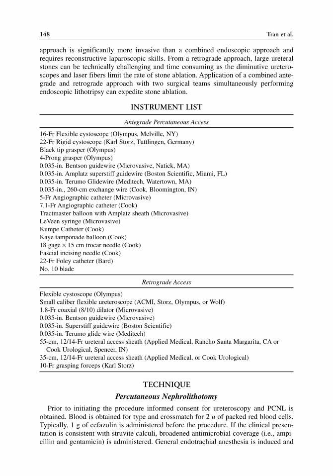
approach is significantly more invasive than a combined endoscopic approach andrequires reconstructive laparoscopic skills. From a retrograde approach, large ureteralstones can be technically challenging and time consuming as the diminutive uretero-scopes and laser fibers limit the rate of stone ablation. Application of a combined ante-grade and retrograde approach with two surgical teams simultaneously performingendoscopic lithotripsy can expedite stone ablation.
INSTRUMENT LIST
Antegrade Percutaneous Access
16-Fr Flexible cystoscope (Olympus, Melville, NY)22-Fr Rigid cystoscope (Karl Storz, Tuttlingen, Germany)Black tip grasper (Olympus)4-Prong grasper (Olympus)0.035-in. Bentson guidewire (Microvasive, Natick, MA)0.035-in. Amplatz superstiff guidewire (Boston Scientific, Miami, FL)0.035-in. Terumo Glidewire (Meditech, Watertown, MA)0.035-in., 260-cm exchange wire (Cook, Bloomington, IN)5-Fr Angiographic catheter (Microvasive)7.1-Fr Angiographic catheter (Cook)Tractmaster balloon with Amplatz sheath (Microvasive)LeVeen syringe (Microvasive)Kumpe Catheter (Cook)Kaye tamponade balloon (Cook)18 gage × 15 cm trocar needle (Cook)Fascial incising needle (Cook)22-Fr Foley catheter (Bard)No. 10 blade
Retrograde Access
Flexible cystoscope (Olympus)Small caliber flexible ureteroscope (ACMI, Storz, Olympus, or Wolf)1.8-Fr coaxial (8/10) dilator (Microvasive)0.035-in. Bentson guidewire (Microvasive)0.035-in. Superstiff guidewire (Boston Scientific)0.035-in. Terumo glide wire (Meditech)55-cm, 12/14-Fr ureteral access sheath (Applied Medical, Rancho Santa Margarita, CA or
Cook Urological, Spencer, IN)35-cm, 12/14-Fr ureteral access sheath (Applied Medical, or Cook Urological)10-Fr grasping forceps (Karl Storz)
TECHNIQUE
Percutaneous Nephrolithotomy
Prior to initiating the procedure informed consent for ureteroscopy and PCNL isobtained. Blood is obtained for type and crossmatch for 2 u of packed red blood cells.Typically, 1 g of cefazolin is administered before the procedure. If the clinical presen-tation is consistent with struvite calculi, broadened antimicrobial coverage (i.e., ampi-cillin and gentamicin) is administered. General endotrachial anesthesia is induced and
148 Tran et al.

the patient is placed prone on the operative table with the legs on spreader bars (Fig. 2).The patient is secured with padded straps, chest rolls are placed, and the upper andlower extremities are padded to prevent pressure injuries. The flank and perineum arethen prepared and draped in the standard manner.
Flexible prone cystoscopy is performed. Although it is initially challenging, pronecystoscopy rapidly becomes familiar to the urologist and the need for repositioning iseliminated. The ipsilateral ureteral orifice is identified and a Bentson guidewire ispassed into the renal collecting system under fluoroscopic guidance. The flexible cys-toscope is removed. A 1.8-Fr coaxial dilation and safety wire introducer sheath, com-monly referred to as an 8/10-Fr dilator, is passed over the guidewire to the midureter.The 8/10-Fr dilator is used to gently dilate the ureteral orifice and to place an Amplatzsuper-stiff guidewire into the renal collecting system. With the Amplatz super-stiff wirein place, a 12/14-Fr ureteral access sheath is advanced to the UPJ under fluoroscopicguidance and the guidewire is removed. In males, a 55-cm access sheath is usuallyrequired, whereas in females, a 35-cm access sheath is usually sufficient to reach theureteropelvic junction. The renal anatomy can then be well delineated fluoroscopicallyby injecting 5 to 10 mL of air through the ureteral access sheath to produce an air pyel-ogram (Fig. 3). The position of the external component of the ureteral access sheathshould be noted at obturator removal. If the ureteral access sheath is caudad to the UPJ,the obturator and guidewire can be reinserted and the sheath can be gently advanced.
Once proper position for lower pole access has been determined fluoroscopically,renal access is achieved using a disposable 18 gage × 15 cm trocar needle. We routinelyuse only lower pole access in the majority of procedures to minimize pulmonary com-plications. While gaining percutaneous access, normal saline irrigation is infused at lowpressure via the access sheath. Retrograde irrigation facilitates needle and guidewireaccess by dilating the target collecting system. The irrigation also prevents decompression
Chapter 9 / Calculus Therapy 149
Fig. 2. Patient in prone split-leg position for combined retrograde and antegrade access with all pressurepoints padded and pneumatic compression boots in place. The phallus is positioned off the end of thetable to facilitate retrograde access.

of the renal collecting system after the needle has been placed. Irrigation is continuedwhile a guidewire is advanced into the collecting system to the ureteropelvic junction.Through a 5.5-Fr (40 cm) Kumpe access catheter, a Glidewire™ can usually be negoti-ated into and through the ureteral access sheath, providing through-and-through access.This maneuver is simplified by briefly pulling the distal end of the ureteral accesssheath into the proximal ureter. In this manner the UPJ serves as a funnel, guiding thewire into the access sheath. After the wire is advanced into the access sheath it easilypasses out of the urethra, where it is anchored with a hemostat clamp. The ureteralaccess sheath is then repositioned to the UPJ. The whole procedure can be completedwith this single through-and-through wire in position.
150 Tran et al.
Fig. 3. (A) Fluoroscopic image of air pyelogram produced by injecting through the ureteral accesssheath. (B) Air pyelogram surrounding a faintly radiopaque staghorn calculus in a different patient.

A dilating balloon is advanced antegrade over the guidewire into the desired calyx andinflated to dilate the nephrostomy tract. Care should be taken to prevent dilation of theassociated infundibulum. A 30-Fr working sheath is passed over the balloon into the calyxunder fluoroscopic guidance and the balloon is removed. Rigid nephroscopy is then per-formed. The stone is ablated with the ultrasonic probe, which allows the simultaneousfragmentation and aspiration of stone fragments. Alternatively electrohydraulic, pneu-matic, or holmium laser lithotripsy may be performed. A flexible cystoscope is often usedto access upper and mid pole calices via the lower pole access tract. Using the combinedantegrade and retrograde technique complete staghorn calculi may be ablated using a sin-gle lower pole access. Our operative staff has noted an increased irrigant requirement forPCNL performed with the ureteral access sheath. As such, we irrigate only with warmedirrigation fluid to prevent systemic hypothermia. If two light sources are available, simul-taneous retrograde ureteroscopic laser or electrohydraulic stone ablation via the ureteralaccess sheath may expedite renal stone ablation. In addition, retrograde ureteroscopy canbe used to fragment and grasp upper pole calculi. The ureteroscope is then used to pres-ent these stones to the rigid nephroscope for extraction or expedited fragmentation.
Before terminating the procedure, all renal calices are inspected with a flexible cys-toscope through the nephrostomy tract. Calices that are not adequately examined withthe flexible cystoscope may be accessed by retrograde ureteroscopy via the ureteralaccess sheath. Renal or ureteral drainage via a stent or nephrostomy tube is left accord-ing to surgeon preference. Postoperative chest X-ray is not required because lower polerenal access is not associated with pleural injury.
Stone protocol spiral computed tomography (CT) is performed on the first postoper-ative day. If residual stone fragments are identified, second-look PCNL is performed thenext day in the standard manner. If residual stones are located in a position that may bedifficult to access through the percutaneous tract (for example, a parallel calyx), thepatient may be placed in the split-leg position and ureteroscopic access from below mayagain be used to facilitate stone extraction.
Follow-up evaluation includes spiral stone protocol CT or plain abdominal X-ray withCT 1 month after the procedure. Plain X-ray is performed only when the stones wereknown to be clearly visible on plain X-ray of the kidneys, ureters, and bladder. Patientswith small residual fragments undergo repeat radiographic evaluation 3 months postoper-atively.
Renal and Ureteral Stones Associated With Urinary Diversions
Preoperative measures including consent, availability of blood, and perioperativeantibiotics are performed as described in the Subheading entitled “PercutaneousNephrolithotomy” under “Techniques.” After induction of general anesthesia the patientis positioned prone as previously described and then prepared and draped. The prone posi-tion is modified with bolsters such that adequate access to the urostomy can be achieved.Occasionally, retrograde access to the ureters can be achieved by inspection of the urinarydiversion with a flexible cystoscope after the intravenous administration of indigo carmineor methylene blue. However, attempts to identify the ureteral orifices are frequently frus-trating, and retrograde access cannot be reliably achieved. If preoperative imaging demon-strates that the affected kidney manifests hydronephrosis, we typically gain renal access inthe operating room under C-arm fluoroscopic guidance. If the renal collecting system isdecompressed, renal access is significantly more challenging and access is gained preoper-atively in a radiology suite owing to the availability of superior imaging devices. After per-
Chapter 9 / Calculus Therapy 151

cutaneous renal access has been gained, a guidewire is negotiated past the stone, down theureter, and into the urinary diversion for through-and-through access. Use of a Glidewirewith a torquing device is helpful to negotiate the wire down the ureter.
With through-and-through access, retrograde endoscopy is easily performed. Anaccess sheath may be gently passed in a retrograde manner. Care should be taken toavoid damage to the ureteroenteric anastomosis. If resistance is encountered, attemptsto pass the access sheath should be aborted. If the stone burden is limited, retrogradeaccess may suffice for expeditious stone clearance. However, if the stone burden is sig-nificant, the combined antegrade and retrograde approach can be very useful. If there isa large renal stone burden, dilation up to 30-Fr and placement of a large sheath asdescribed in the Subheading entitled “Percutaneous Nephrolithotomy” underTechniques” may be performed. However, if only limited access to the kidney andureter are required, we frequently advance an access sheath in an antegrade manner intothe renal collecting system or ureter (Fig. 4). The access sheath generally passes easilyand atraumatically antegrade into the renal collecting system after skin incision.Additionally, passage of an antegrade access sheath has not been associated with signif-icant risk of hemorrhage. If antegrade and retrograde access sheaths are used, two sur-gical teams may work simultaneously to ablate renal and ureteral stones.
If the surgeon is confident that complete stone clearance has been achieved, a 7.1-Fr,single pigtail catheter is passed retrograde into the renal pelvis with the distal end pro-truding out of the stoma. If we suspect residual calculi that may require second-lookflexible nephroscopy, a nephroureteral stent is deployed in an antegrade fashion.
Follow-up on the first postoperative day includes a spiral CT scan to evaluate forresidual stone fragments. If the stones are radiopaque, follow-up imaging is performedwith plain X-ray films as per the surgeon’s preference.
Large Ureteral Stones
152 Tran et al.
Fig. 4. Placement of access sheath in the ureter and percutaneous nephrostomy tract.

After appropriate preoperative and perioperative measures are taken as previouslydescribed, general endotracheal anesthesia induced. The patient is positioned into theprone split-leg position as previously indicated. Initial access is gained in a retrogrademanner preferably using a 0.035-in. Terumo guidewire to bypass the ureteral calculusunder fluoroscopic guidance. After the Glidewire has been passed, the 8/10 dilator or a5-Fr ureteral catheter is used to exchanged the Glidewire for a more secure wire(Bentson or Amplatz super-stiff) into the renal collecting system. A ureteral accesssheath is deployed to the level of the ureteral stone. Great care is taken not to trauma-tize the ureter by deploying the access sheath at the level of the stone. As these casesroutinely require significant time in the ureter, it is imperative that the endoscopic fieldremains free of blood and debris as long as possible.
Antegrade access is then achieved as described earlier. Although we typically gainaccess to the lower pole for PCNL procedures, access for ureteral procedures for largecalculi is gained through a middle or upper pole calyx depending on the individual renalanatomy. Access through the lower pole creates an acute angle for passage of the accesssheath and instruments into the ureter and may limit the ability to access the largeureteral stone. Stone ablation can be performed using both the antegrade and retrograderoutes individually or simultaneously. We prefer to use both access routes simultane-ously with application of two endoscopic towers and two holmium lasers. However, iftwo lasers are not available, simultaneous ablation with the holmium laser and electro-hydraulic lithotripsy has also been performed with success.
RESULTS
Percutaneous Nephrolithotomy
Our combined approach was applied initially to staghorn or large renal calculi. In thispopulation, our initial experience involved nine patients (7). In this series, the meanpatient age was 46 years and mean body mass index was 31.4. In the study group therewere six complete and three partial staghorn calculi. Mean stone size was 6.2 cm (range5.5–8 cm) in greatest diameter. The primary stone composition was struvite in fivecases, calcium oxalate monohydrate in two and uric acid in two.
Table 1 lists individual stone characteristics, operative parameters, hospital stay, andcomplications. Mean operative time for the primary procedure was 3.1 hours. Meanestimated blood loss was 290 mL and patients required an average of 33.2 mg of MSO4equivalents. Mean hospital stay was 3.2 days. There were no major and four minor (44%)complications. No patient required transfusion. One patient experienced venous bleedingfrom the percutaneous nephrostomy tract and required that a tamponade balloon beinflated intraoperatively for 20 minutes to control the bleeding. This patient experiencedintercostal muscle injury during transport and required additional parenteral analgesicsthat extended the hospital stay for 1 day.
Another patient, who required significant analgesic medication postoperatively, haddecreased oxygen saturation on pulse oximetry. Complete evaluation, including arterialblood gases, electrocardiography, chest CT angiography, and pulmonary function tests,revealed only atelectasis as the etiology of desaturation. Despite a 4-mm residual fragment,this patient did not undergo a second look procedure owing to the pulmonary complication.Two additional minor complications included transient paresthesia on the ipsilateral lateralthigh that resolved with observation within 1 month and an asymptomatic 4-cm perirenalhematoma that was not associated with a significant decrease in hematocrit.
Chapter 9 / Calculus Therapy 153
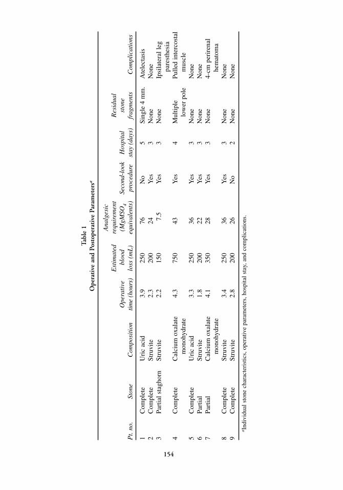
Tab
le 1
Ope
rati
ve a
nd P
osto
pera
tive
Par
amet
ersa
Ana
lges
icE
stim
ated
requ
irem
ent
Res
idua
lO
pera
tive
bloo
d (M
gMSO
4Se
cond
-loo
kH
ospi
tal
ston
eP
t. no
.St
one
Com
posi
tion
time
(hou
rs)
loss
(m
L)
equi
vale
nts)
proc
edur
est
ay (
days
)fr
agm
ents
Com
plic
atio
ns
1C
ompl
ete
Uri
c ac
id3.
925
076
No
5Si
ngle
4 m
m.
Ate
lect
asis
2C
ompl
ete
Stru
vite
2.3
200
24Y
es3
Non
eN
one
3Pa
rtia
l sta
ghor
nSt
ruvi
te2.
215
07.
5Y
es3
Non
eIp
sila
tera
l leg
pare
sthe
sia
4C
ompl
ete
Cal
cium
oxa
late
4.
375
043
Yes
4M
ultip
le
Pulle
d in
terc
osta
lm
onoh
ydra
telo
wer
pol
em
uscl
e5
Com
plet
eU
ric
acid
3.3
250
36Y
es3
Non
eN
one
6Pa
rtia
lSt
ruvi
te1.
820
022
Yes
3N
one
Non
e7
Part
ial
Cal
cium
oxa
late
4.
135
028
Yes
3N
one
4-cm
per
iren
alm
onoh
ydra
tehe
mat
oma
8C
ompl
ete
Stru
vite
3.4
250
36Y
es3
Non
eN
one
9C
ompl
ete
Stru
vite
2.8
200
26N
o2
Non
eN
one
a Ind
ivid
ual s
tone
cha
ract
eris
tics,
ope
rativ
e pa
ram
eter
s, h
ospi
tal s
tay,
and
com
plic
atio
ns.
154

Chapter 9 / Calculus Therapy 155
Complete stone clearance was achieved in seven of the nine patients (78%). A second-look procedure was not done in one case because of pulmonary complications. Thispatient had a 4-mm uric acid residual fragment and underwent oral alkalinization, whichresulted in stone resolution at the 3-months follow-up evaluation. Another patient hadmultiple small fragments in the lower pole at 1 month of follow-up. This patient subse-quently required ureteroscopic stone extraction for stone clearance.
Subsequently, as a result of the success and efficiency of the combined approach forvery large calculi, we now apply the combined approach for all patients undergoingPCNL. Presently, we have performed 64 PCNL procedures using the combined technique,including 26 procedures for staghorn or partial staghorn calculi. Only five (7.8%) proce-dures required more than one percutaneous access for stone ablation. Each remaining casewas completed with a single lower pole access. Three patients (4.6%) required bloodtransfusion. With CT scan performed at 3 months, 89% (57/64) of patients were stone-free.
Little published data is available regarding the combined antegrade and retrogradetechnique for patients with urinary diversions or large ureteral stones. However, thecombined approach has been applied successfully by the authors and others for man-agement of these challenging patients.
TIPS AND TRICKS
1. The use of an Amplatz super-stiff wire facilitated placement of the ureteral accesssheath in either an antegrade or retrograde manner.
2. An air pyelogram is typically performed to delineate the renal anatomy. Instillation ofair instead rather than contrast material provides an excellent pyelogram and avoids theuse of contrast, which can mask stones. Combination of air and contrast simultaneouslycan also help delineate posterior calyces for puncture as air will rise posteriorly whenthe patient is in the prone position.
3. Use of retrograde irrigation is helpful in dilating the collecting system and keeping thecollecting system dilated while gaining percutaneous access to the kidney.
4. When applying the combined approach for PCNL there is high fluid turnover.Application of warmed irrigant will help prevent patient hypothermia.
5. One milligram of intravenous glucagon and gentle retrograde pressure on the accesssheath may be used to overcome muscle spasm encountered during passage of an accesssheath. Never force the access sheath as ureteral injury may result.
REFERENCES1. Elbahnasy AM, Clayman RV, Shalhav AL, et al. Lower-pole caliceal stone clearance after shockwave
lithotripsy, percutaneous nephrolithotomy, and flexible ureteroscopy: impact of radiographic spatialanatomy. J Endourol 1998; 12: 113–119.
2. Landman J, Venkatesh R, Ragab M, et al. Comparison of intrarenal pressure and irrigant flow duringpercutaneous nephroscopy with an indwelling ureteral catheter, ureteral occlusion balloon, andureteral access sheath. Urology 2002; 60: 584–587.
3. Rehman J, Monga M, Landman J, et al. Characterization of intrapelvic pressures during ureteropy-eloscopy with the ureteral access sheath. Urology 2003; 61: 713–718.
4. Keeley FX, Gialas I, Pillai M, Chrisofos M, Tolley DA. Laparoscopic ureterolithotomy: the Edinburghexperience. BJU Int 1999; 84: 765–769.
5. Feyaerts A, Rietbergen J, Navarra S, Vallancien G, Guillonneau B. Laparoscopic ureterolithotomy forureteral calculi. Eur Urol 2001; 40: 609–613.
6. Delvecchio FC, Kuo RL, Iselin CE, Webster GD, Preminger GM. Combined antegrade and retrogradeendoscopic approach for the management of urinary diversion-associated pathology. J Endourol2000; 14: 251–256.
7. Landman J, Venkatesh R, Lee DI, et al. Combined percutaneous and retrograde approach to staghorncalculi with application of the ureteral access sheath to facilitate percutaneous nephrolithotomy. J

III INCISIONAL THERAPY

Antegrade Endopyelotomy
Beng Jit Tan, MD, PhD, Assaad El-Hakim, MD,Benjamin R. Lee, MD, and Arthur D. Smith, MD
CONTENTS
INTRODUCTION
INDICATIONS
PROGNOSTIC FACTORS
INSTRUMENTS AND EQUIPMENT
TECHNIQUE
RESULTS
COMPLICATIONS
CONCLUSION
REFERENCES
10
SUMMARY
The popularity of minimally invasive surgical techniques, such as endopyelotomy, hasincreased markedly among urologists in recent years. Antegrade endopyelotomy hasbecome a well-established alternative to open operative pyeloplasty for management ofboth primary and secondary ureteropelvic junction obstruction (UPJO). Endopyelotomyresults in significantly less morbidity, and should this technique fail, subsequent openpyeloplasty is still technically feasible. Although several variations of the technique havebeen described, the goal in all cases is to develop a full thickness incision though theobstructing proximal ureter that extends out to the peripyeloureteral fat and heals over aninternal stent. Antegrade endopyelotomy is particularly valuable in the setting of uppertract stones that can then be managed simultaneously. Contraindications to antegradeendoscopic incision of the UPJ include a long stricture, sepsis, a large redundant renalpelvis, poor renal function, and the presence of crossing lower vessel as the cause of theUPJO. The role of percutaneous endopyelotomy in children remains undefined. Althoughsuccessful results have been reported in infants, the relative morbidity and long-termsuccess of open pyeloplasty in this age group are excellent, thus limiting the relativeadvantage of an endoscopic approach. However, there may be a role for endopyelotomyin older children and in those patients with secondary obstruction who have failed open
From: Advanced Endourology: The Complete Clinical GuideEdited by: S. Y. Nakada and M. S. Pearle © Humana Press Inc., Totowa, NJ
159
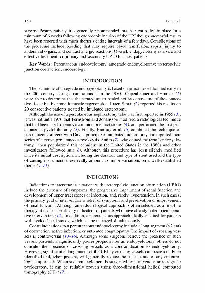
surgery. Postoperatively, it is generally recommended that the stent be left in place for aminimum of 6 weeks following endoscopic incision of the UPJ though successful resultshave been reported with much shorter stenting intervals of a few days. Complications ofthe procedure include bleeding that may require blood transfusion, sepsis, injury toabdominal organs, and contrast allergic reactions. Overall, endopyelotomy is a safe andeffective treatment for primary and secondary UPJO for most patients.
Key Words: Percutaneous endopyelotomy; antegrade endopyelotomy; ureteropelvicjunction obstruction; endourology.
INTRODUCTION
The technique of antegrade endopyelotomy is based on principles elaborated early inthe 20th century. Using a canine model in the 1950s, Oppenheimer and Hinman (1)were able to determine that the stented ureter healed not by contracture of the connec-tive tissue but by smooth muscle regeneration. Later, Smart (2) reported his results on20 consecutive patients treated by intubated ureterotomy.
Although the use of a percutaneous nephrostomy tube was first reported in 1955 (3),it was not until 1976 that Fernström and Johansson modified a radiological techniquethat had been used to remove common bile duct stones (4), and performed the first per-cutaneous pyelolithotomy (5). Finally, Ramsay et al. (6) combined the technique ofpercutaneous surgery with Davis’ principle of intubated ureterotomy and reported theirseries of elective percutaneous pyelolysis. Smith (7), who coined the term “endopyelo-tomy,” then popularized this technique in the United States in the 1980s and otherinvestigators followed suit (8). Although this procedure has been slightly modifiedsince its initial description, including the duration and type of stent used and the typeof cutting instrument, these really amount to minor variations on a well-establishedtheme (9–11).
INDICATIONS
Indications to intervene in a patient with ureteropelvic junction obstruction (UPJO)include the presence of symptoms, the progressive impairment of renal function, thedevelopment of upper tract stones or infection, and, rarely, hypertension. In such cases,the primary goal of intervention is relief of symptoms and preservation or improvementof renal function. Although an endourological approach is often selected as a first-linetherapy, it is also specifically indicated for patients who have already failed open opera-tive intervention (12). In addition, a percutaneous approach ideally is suited for patientswith pyelocaliceal stones, which can be managed simultaneously.
Contraindications to a percutaneous endopyelotomy include a long segment (>2 cm)of obstruction, active infection, or untreated coagulopathy. The impact of crossing ves-sels is controversial (13–16). Although some surgeons believe the presence of suchvessels portends a significantly poorer prognosis for an endopyelotomy, others do notconsider the presence of crossing vessels as a contraindication to endopyelotomy.However, significant entanglement of the UPJ by crossing vessels can occasionally beidentified and, when present, will generally reduce the success rate of any endouro-logical approach. When such entanglement is suggested by intravenous or retrogradepyelography, it can be reliably proven using three-dimensional helical computedtomography (CT) (17).
160 Tan et al.

PROGNOSTIC FACTORS
Length of Stricture
Long avascular strictures, total obliteration of the UPJ, and severe periureteral fibro-sis are clear contraindications to endourological procedures. Overall results have, ingeneral, been unsatisfactory (18–20). Patients with these features should be treated byopen repair or laparoscopic surgery whenever possible.
Degree of Hydronephrosis
Initial experience has shown that a large size of the renal pelvis had a negative influ-ence on the results of endopyelotomy (21). Antegrade endopyelotomy may be favoredin cases of massive hydronephrosis for technical reasons: the UPJ may be more easilyidentified, incised, and drained antegrade when the UPJ is massively hydronephrotic.Whereas endourological procedures can address intrinsic factors of obstruction, theycannot correct extrinsic factors or reduce the size of a massively dilated renal pelvis.The degree of hydronephrosis was of statistical significance when combined with thepresence or absence of crossing vessels. The final success rate decreased from an over-all success rate of 81 to 85 % for those with moderate hydronephrosis to 50 to 54%when high-grade hydronephrosis was present (14,22,23).
Renal Function
Renal function is a significant prognostic factor. A high risk of failure has been reportedwhen the function of the affected kidney is greatly impaired (13,14,21,24,25).Preoperative high-grade hydronephrosis, as well as poor renal function, are significantcauses of failure (14). Unfortunately, the isolated impact of this factor is difficult to assessbecause no prospective data have been collected. In most series, preoperative function ofthe involved kidney has been systematically assessed only recently or in selected cases,and its influence cannot be dissociated from that of the degree of hydronephrosis.Nevertheless, Gupta et al. (14) reported that of the 205 patients with known renal functionwho underwent endopyelotomy at a single institution, success rates for endopyelotomywere 92, 80, and 54% for good renal function (differential renal function greater than40%), moderate renal function (differential renal function between 25 and 40%), and poorrenal function (differential renal function less than 25%), respectively. Nephrectomy is thetreatment of choice if the differential renal function falls below 15%.
Primary vs Secondary UPJO
Both primary and secondary cases of UPJO can be successfully treated endoscopi-cally. Cumulative success rates for endopyelotomy of 62 to 94% rate for primaryobstruction and 67 to 100% for secondary obstruction have been achieved (14,26–34).Although laparoscopic pyeloplasty has yielded higher success rates of 92 to 98% for pri-mary UPJO and 83 to 89% for secondary obstruction (35–37), it is associated with otherproblems.
Age and Sex of the Patient and the Side of Obstruction
Currently, the role of endopyelotomy remains unclear in children age 6 or less, andthere is only a limited reported series in the pediatric population (19,38–44). Manyyounger children have long proximal ureteral strictures, especially in the proximal one-
Chapter 10 / Antegrade Endopyelotomy 161

third of the ureter. In addition, their ureters are of small caliber requiring small-sizedstents. Both these indications make endopyelotomy challenging in young children. Inaddition, narrow lumen stents in young children can easily be obstructed. In these chil-dren, open pyeloplasty is still the preferred procedure owing to its consistently superiorresults, especially in primary cases. In secondary cases of UPJO, such as failures of openpyeloplasty, endopyelotomy can be safe and effective. With further refinements andminiaturization of equipment, it ultimately might become a preferred option (38,41,43).
In elderly patients, endopyelotomy offers results comparable with those in the adultgroup (29,45). Not surprisingly, neither the sex of the patient nor the side of the obstruc-tion influences the outcome (14).
Crossing Vessels
The significance of vessels crossing the UPJ remains a matter of debate and their rolein the pathogenesis of the obstruction, as well as their influence on the results of vari-ous endourological procedures, is controversial (13–15,18,22–23,46). The presence ofcrossing vessels does not necessarily cause UPJO. However, significant entanglementof the UPJ by crossing vessels can occasionally be identified and, recently, spiral CT(47,48) and endoluminal ultrasonography (49,50) have been used to identify crossingvessels. In cases when the crossing vessel is the primary cause of UPJO, it has beenfound to reduce the success rate of endopyelotomy. In such cases, laparoscopic or opendismembered pyeloplasty have yielded a success rate of 97% (50,51). An extensive his-torical review addressing these issues has been published recently (23).
INSTRUMENTS AND EQUIPMENT
The recommended equipment and instruments for antegrade endopyelotomy includethe following (See Fig. 1):
Nephroscope, 0° lens with 26-Fr sheath.Cystoscopes, 30 and 70° lenses.Urethrotome or transurethral reaction resectoscope.Amplatz dilator set (8–30-Fr dilators and sheaths) or balloon dilation set.0.038-in. J-tip guidewire.0.038-in. stiff torque wire.18-Gage access needle.6-Fr open ended ureteral catheter.14/7-Fr endopyelotomy stent.Fluoroscopic X-ray.Nephroscope graspers.Endopyelotomy knife (cold knife, hook-knife, half moon, or straight).Hypaque, diluted 30%.
TECHNIQUE
Patient Selection and Preparation
The preoperative evaluation should include a search for any concomitant medical con-dition impacting on the risk of anesthesia. As in any elective operative procedure, a com-plete history and physical examination should be performed. The patient should be agood candidate for general or regional anesthesia. Standard blood studies should include
162 Tan et al.
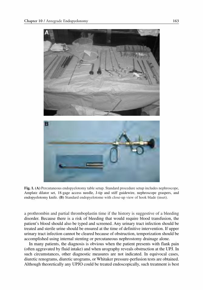
a prothrombin and partial thromboplastin time if the history is suggestive of a bleedingdisorder. Because there is a risk of bleeding that would require blood transfusion, thepatient’s blood should also be typed and screened. Any urinary tract infection should betreated and sterile urine should be ensured at the time of definitive intervention. If upperurinary tract infection cannot be cleared because of obstruction, temporization should beaccomplished using internal stenting or percutaneous nephrostomy drainage alone.
In many patients, the diagnosis is obvious when the patient presents with flank pain(often aggravated by fluid intake) and when urography reveals obstruction at the UPJ. Insuch circumstances, other diagnostic measures are not indicated. In equivocal cases,diuretic renograms, diuretic urograms, or Whitaker pressure-perfusion tests are obtained.Although theoretically any UPJO could be treated endoscopically, such treatment is best
Chapter 10 / Antegrade Endopyelotomy 163
Fig. 1. (A) Percutaneous endopyelotomy table setup. Standard procedure setup includes nephroscope,Amplatz dilator set, 18-gage access needle, J-tip and stiff guidewire, nephroscope graspers, andendopyelotomy knife. (B) Standard endopyelotome with close-up view of hook blade (inset).

when the UPJ is relatively dependent and there are no gross anatomic abnormalities. Thepatient’s anatomy should be such that access is practical and safe.
When counseling a patient, the surgeon should outline the open operative procedureand other endourological treatment options, each of which has its own advantages anddisadvantages, so that the patient can provide informed consent. The patient should becounseled regarding the risks and benefits of the procedure, including the fact that thesuccess rate of any endourological approach, such as percutaneous endopyelotomy, willbe less than that of standard open operative intervention. In advising patients, it is impor-tant to consider factors such as age, sex, associated pathology, e.g., stones in the renalpelvis, previous renal surgery, the availability of equipment and resources, whether ornot a nephrostomy tube is already in position, the surgeon’s experience, and cost (52).
Technique, Helpful Tips, and Tricks
Percutaneous access to the urinary tract has been described in detail in Chapter 3.Therefore, only the important steps and helpful tips for achieving percutaneous accesswill be highlighted in this section.
PATIENT POSITIONING
All patients undergoing any endopyelotomy procedure are given a general anesthetic.Complete evaluation of the renal collecting system is imperative prior to definitive per-cutaneous puncture for access tract creation. The patient is placed in a lithotomy posi-tion. Cystoscopy and retrograde urography is performed to delineate the ureteralanatomy distal to the obstruction, the exact location, degree, and, sometimes, length ofthe obstruction. A 6-Fr ureteral catheter is left in place to allow retrograde injection ofcontrast. A 16-Fr Foley catheter is inserted and attached to a drainage bag concurrentwith the 6-Fr catheter.
The patient is then positioned prone to allow access to the posterior flank. All bonyprominences are padded and protected. The chest and abdomen are elevated on tworolls that extend from the shoulder to the hip to reduce resistance to breathing (Fig. 2).
TECHNIQUE FOR CREATING ACCESS TRACTS
The percutaneous nephrostomy is created by way of an upper or middle calyx withthe aid of C-arm fluoroscopy. Thorough evaluation of the renal collecting systemanatomy is essential before definitive percutaneous puncture for access tract creation. Aposterolateral transparenchymal puncture minimizes the chance of injury to the majorrenal vessels. The ideal percutaneous access tract is located in the posterior axillary line,via the posterior calyx because major vessels surrounding the pelvis can be avoided(Fig. 3). The ideal tract courses through the posterior lateral flank, in a direct line withthe middle of the renal pelvis, through the renal parenchyma, into the tip of a postero-lateral calyx, and into the renal pelvis. The transparenchymal portion of the tract stabi-lizes the nephrostomy tube and provides a seal around the tube, preventing urineextravasation into the perinephric space. Generally, a midposterior or superolateral calixis chosen, although occasionally, an inferolateral calix may be used (53). A target mid-pole calyx or upper pole calyx is optimal over a lower pole initial access in order toallow subsequent instrument maneuverability without tearing renal parenchyma.
The skin insertion site may be placed more lateral in obese patients in whom the lateralsegment of the colon is displaced ventrally. In patients with abnormal abdominal anatomy,such as following jejunal–ilial bypass or horseshoe kidneys, the position of the kidney rel-
164 Tan et al.

ative to the colon may be distorted. These patients are at higher risk for a transcolonic nee-dle placement. For horseshoe kidneys, the access tract is virtually perpendicular to the planeof the back, in a paraspinous, medial position. One must take into consideration as well thata more medial insertion site is uncomfortable for the patient who invariably lies supine afterthe procedure, and can lead to compression and kinking of the external portion of thenephrostomy tube into the back. A subcostal approach lowers the risk of pneumothorax.Puncture of anterior calices is required only if access to the posterior calyces is not possi-ble. Access from an anterior calices to the renal pelvis is very difficult because of the inher-ent difficulty in directing a wire backwards. Direct puncture of the renal pelvis should alsobe avoided, owing to the significant risk of injury to the posterior branch of the renal artery.
C-arm fluoroscopy is used to help localize the target calyx, using a triangulation methodof two different views. A Seldinger-technique based access is employed to gain access intothe renal collecting system. Details of obtaining percutaneous access to the urinary tracthave been described in detail in Chapter 3. Dilation of the tract is performed usingAmplatz dilators or balloon dilation. Normal saline is the irrigant of choice because it pre-vents hyponatremia that can result from intravascular absorption when hyposmotic solu-tions are used. Care is taken to avoid inadvertent over advancement of instruments or theaccess sheath, as this will likely cause a laceration of a renal infundibulum or renal pelvis.
Once the UPJ has been bridged with a 0.038-in. super-stiff guidewire (the guidewire is inposition from the flank through the urethral meatus), an 8-Fr Teflon catheter is advanced
Chapter 10 / Antegrade Endopyelotomy 165
Fig. 2. Correct patient positioning for endopyelotomy. All bony prominences are padded and protected.The chest and abdomen are elevated on two rolls that extend from the shoulder to the hip to reduceresistance to breathing. Great care is taken to pad all joints. Additionally, bolsters can be used underthe chest to elevate the upper torso.

across the UPJ and into the ureter. Under fluoroscopic guidance, an 18-Fr dilator is advanceddown the UPJ over the 8-Fr catheter in order to dilate the UPJ to allow introduction of theincising device. Alternatively, an 12-Fr ureteral dilating balloon should be used for thisrather than a larger 15- to 18-Fr balloon, as larger balloons may tear the UPJ.
ENDOPYELOTOMY TECHNIQUES
Several devices have been used to incise the UPJ including laser incision, coldknives, and semilunar or hook knives.
An urethrotome with a hook-shaped cold knife (Karl Storz Endoscopy-America) or ahookknife is passed through a rigid nephroscope (Olympus Medical Systems, OlympusAmerica) over the guidewire (Fig. 4). Because of the anatomic findings described bySampaio (15,53–55) regarding the location of crossing vessels, the incision is now madeposterolaterally. The posterolateral position is determined by orienting the knife with the
166 Tan et al.
Fig. 3. Access tract placement. (A) Tract 1 is the preferred route. Tract 2 (through the superior calyx)is also possible, although it may result in chest complications. Tract 3 (through an inferior calyx) isnot an adequate route for this procedure because it will not provide good visibility of the ureteropelvicjunction (UPJ). (B) Kidney in supine orientation. To achieve a smooth curve to the UPJ, the approachshould be posterolateral (solid line) and not directly posterior (dotted line).

posterolateral position of the patient’s flank while referencing the position of the kidney onintravenous pyelography or CT. This is usually easy to perform and is complicated onlywhen kidneys are malrotated. However, in cases when the ureter inserts into the renal pelvison the anterior or posterior wall, the incision should instead marsupialize the proximal ureterinto the renal pelvis such that an anterior or posterior incision may be required. When suchincisions are made under direct vision, any crossing vessel can be directly visualized andavoided. When the patient has undergone previous UPJ repair, the previous surgical recordshould be consulted because the position of the UPJ relative to the vascular supply of thekidney could have changed. The knife should slide through the UPJ yielding a clean cut.The incision should be extended down the ureter for approx 1 cm beyond the area ofobstruction and should be continued laterally up into the renal pelvis for 1 to 2 cm. The inci-sion is performed under direct vision so that the operator can identify the exact location ofthe cut and identify and avoid extrapelvic structures such as aberrant or polar vessels. Incontrast to blind techniques in which the location of a repeat cut (if necessary) cannot beexactly determined, the antegrade endopyelotomy incision can be lengthened with preci-sion.
The UPJO is incised full thickness in a posterolateral position until the periureteral fator wispy retroperitoneal tissue is visible and the UPJ appears wide open (Fig. 5). In patientsundergoing reoperation, previous scarring can make determining the proper depth of theincision difficult. Direct vision permits avoidance of crossing a vessel at this point. Theincision must be extensive enough that one is able to look into the proximal ureter (Fig. 6).
A holmium laser can be used in a similar manner to incise the UPJ. The 365-mm fiberis introduced through the 8-Fr ureteral catheter placed in the nephroscope and, underdirect vision, the UPJ is incised (Fig. 7). Another alternative is cutting the UPJ with scis-sors. A small full-thickness puncture is made into the renal pelvis in a posterolateral posi-tion, as far as 1 cm away from the UPJ, with the scissors in the closed position. Then, thescissors are opened under constant visual control until the peripelvic space is reached (Fig.
Chapter 10 / Antegrade Endopyelotomy 167
Fig. 4. Guidewire runs from the flank through the urethral meatus and bridging the ureteropelvic junc-tion (UPJ). Photo showing a hook-shaped cold knife approaching the UPJ to enter the ureter.

168 Tan et al.
Fig. 6. Full thickness incision has been carried out through the muscle into the surrounding peri-urethral fat as seen in the inferior area of the picture. The incision should be extensive enough thatone is able to look into the proximal ureter with the guidewire inside. The periuretral space should beseen through the incision.
Fig. 7. (Opposite Page) Technique of incision with laser: (A) Using the guidewire as a marker, theLaser fiber approaches the ureteropelvic junction (UPJ) posterolaterally. (B) The 365-mm fiber isintroduced through a conventional stent pusher placed in the nephroscope. Under direct vision, theUPJ is incised in the posterolateral position, showing the anteromedial surface of the ureter with theguidewire inside. (C) The fiber reaches the peripelvic space, permitting observation and coagulationof bleeding vessels. The external face of the ureteral wall is exposed.
Fig. 5. Hook-shaped cold knife previously introduced in the proximal ureter is withdrawn in a retro-grade direction, cutting the ureteropelvic junction.
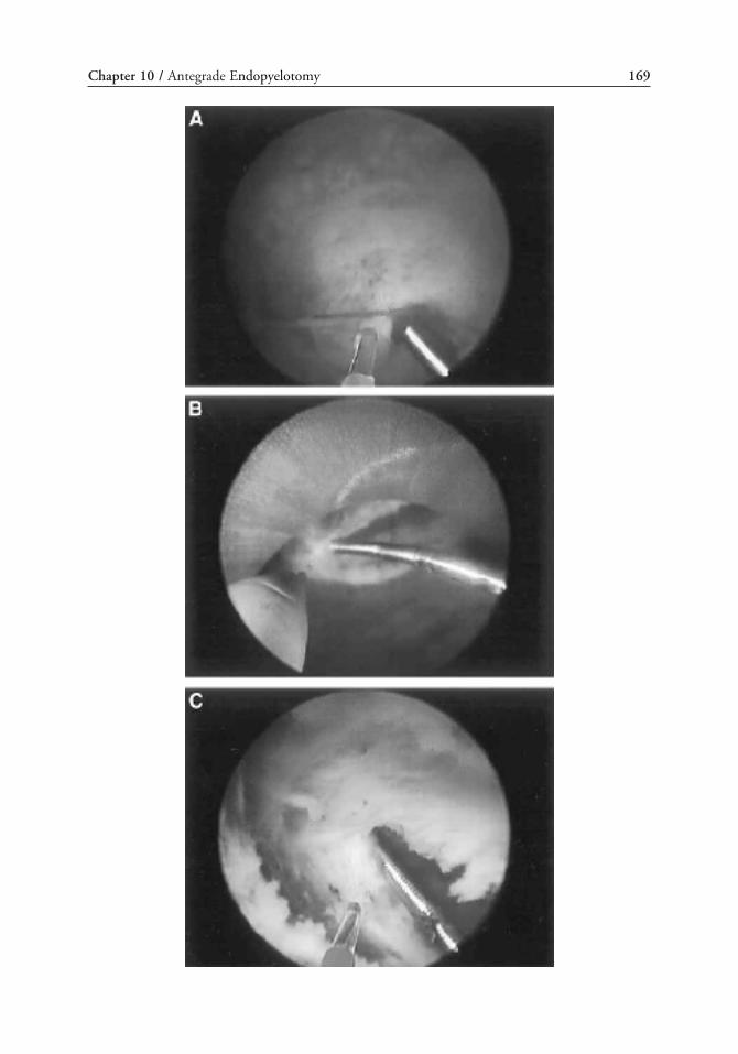
Chapter 10 / Antegrade Endopyelotomy 169
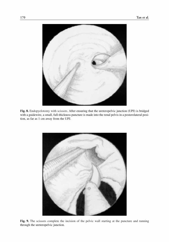
170 Tan et al.
Fig. 8. Endopyelotomy with scissors. After ensuring that the ureteropelvic junction (UPJ) is bridgedwith a guidewire, a small, full-thickness puncture is made into the renal pelvis in a posterolateral posi-tion, as far as 1 cm away from the UPJ.
Fig. 9. The scissors complete the incision of the pelvic wall starting at the puncture and runningthrough the ureteropelvic junction.
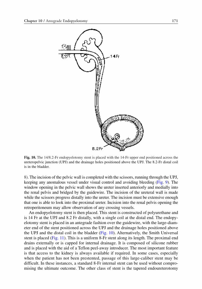
8). The incision of the pelvic wall is completed with the scissors, running through the UPJ,keeping any anomalous vessel under visual control and avoiding bleeding (Fig. 9). Thewindow opening in the pelvic wall shows the ureter inserted anteriorly and medially intothe renal pelvis and bridged by the guidewire. The incision of the ureteral wall is madewhile the scissors progress distally into the ureter. The incision must be extensive enoughthat one is able to look into the proximal ureter. Incision into the renal pelvis opening theretroperitoneum may allow observation of any crossing vessels.
An endopyelotomy stent is then placed. This stent is constructed of polyurethane andis 14 Fr at the UPJ and 8.2 Fr distally, with a single coil at the distal end. The endopy-elotomy stent is placed in an antegrade fashion over the guidewire, with the large-diam-eter end of the stent positioned across the UPJ and the drainage holes positioned abovethe UPJ and the distal coil in the bladder (Fig. 10). Alternatively, the Smith Universalstent is placed (Fig. 11). This is a uniform 8-Fr stent along its length. The proximal enddrains externally or is capped for internal drainage. It is composed of silicone rubberand is placed with the aid of a Teflon peel-away introducer. The most important featureis that access to the kidney is always available if required. In some cases, especiallywhen the patient has not been prestented, passage of this large-caliber stent may bedifficult. In these instances, a standard 8-Fr internal stent can be used without compro-mising the ultimate outcome. The other class of stent is the tapered endoureterotomy
Chapter 10 / Antegrade Endopyelotomy 171
Fig. 10. The 14/8.2-Fr endopyelotomy stent is placed with the 14-Fr upper end positioned across theureteropelvic junction (UPJ) and the drainage holes positioned above the UPJ. The 8.2-Fr distal coilis in the bladder.

stent, which requires a Council catheter placement as a nephrostomy tube for 48 hours.Although this stent has been used safely in many patients, a major concern has alwaysbeen that the stent is inaccessible if it becomes obstructed after the nephrostomy tube isremoved and that the proximal coil can migrate outside the endopyelotomy site and intothe retroperitoneum. Once proper positioning of the stent is determined fluoroscopi-cally, any remaining safety wires are withdrawn. A Foley catheter is placed andnephrostomy tube secured to the skin to prevent inadvertent dislodgement.
All patients undergoing endopyelotomy should have a stent placed. The type of stentdoes not influence the outcome (14). The size of the stent, (29,32,56) the length of time itshould remain in place (1,57,58), and the type of stent material are issues that remain unan-swered. In a nonrandomized, retrospective study, Danuser and et al. (32) reported superior
172 Tan et al.
Fig. 11. The Smith Universal stent is a uniform 8 Fr along its length. The proximal end drains exter-nally or is capped for internal drainage. The distal end was originally passed to the level of the pelvicbrim, but ureteral stricture formation necessitated passage into the bladder. It is composed of siliconerubber and is placed with the aid of a Teflon peel-away introducer.

short-term (83 vs 94%, respectively) and long-term (71 and 93%, respectively) success ratesin patients in whom a modified 27/14/8.2-Fr nephroureteral stent was placed compared topatients who had the standard, 14/8.2-Fr graduated. Similarly, Hwang and co-workers(56), in an earlier study, reported they had better overall success rates with the 14/7endopyelotomy stent compared to the 6-Fr internal ureteral stent (93.3 and 84%, respec-tively). However, the difference in success rate was not statistically significant.Accommodating these large diameter stents often require placing a double-J stent to dilatethe ureter for several days in order to decrease the difficulty of inserting the larger endopy-elotomy stent down the ureter or, as Danuser and co-workers have done, making a 3 to 5cm incision that extended well into the normal renal pelvis and ureter. Some feel that a stentserves as a mold around which healing takes place and that the largest possible diameterstent is preferable to induce formation of a sufficient caliber lumen but without causingischemia. Others believe that the stent acts as a scaffold to orient regenerating muscle fibersand, therefore, a smaller caliber stent is just as effective. Several retrospective, nonrandom-ized series have demonstrated comparable success rates using standard size stents. Stent sizehas been considered an insignificant factor based on two animal studies that showed no sig-nificant difference in success using 7- or 14-Fr and 7- or 12-Fr stents, respectively (10,59) aswell as in clinical studies (14,27,29). Thus, only a prospective, randomized study comparinglarge diameter stents to standard caliber stents, using universally accepted criteria for obstruc-tion, will answer the question of optimal stent size after endopyelotomy definitively.
Most urologists leave the stent in place for 6 weeks based on initial animal studiesshowing 90% regeneration of smooth muscle and muscular continuity within 6 to 7 weeksof stenting (1,57,60,61). The conclusion was that stenting is important until the epitheliumregrowth pattern is established. However, using a porcine model, Kerbl et al. (11) showedthat stenting beyond 1 week resulted in lower healing scores and inflammation associatedwith a chronic indwelling stent is a source of late fibrosis. Several clinical studies havereported that stents can be removed earlier without untoward sequelae (58,63,63). Forexample, in a prospective, randomized trial, Mandhani et al. (58) found that a 2-weeksduration of stenting is sufficient duration to allow functional restoration across the UPJafter endopyelotomy. They suggested that a stent is important as long as epitheliumregrowth occurs. However, after this point a stent induces greater inflammation and fibro-sis. In addition, short-stent duration helps decrease morbidity including pain, bladder irri-tation, and urinary tract infection. However more prospective, randomized studiescomparing duration of stenting are needed to definitively answer the question of optimalstent duration after endopyelotomy.
In a modification to the classical endopyelotomy approach as described above, theureteral stent is placed prior to antegrade endopyelotomy. Again, percutaneous access isestablished. A wire traverses the UPJ percutaneously and passes antegrade across theureterovesical junction into the bladder. It is advantageous to pass a stent before perform-ing the actual endopyelotomy incision. The purpose of this action is twofold. First, hav-ing the stent in place at the outset of the procedure obviates concern about avulsing theUPJ during placement of a stent after the UPJ has already been incised. Second, place-ment of the stent before the incision serves to better define the UPJ, allowing a more pre-cise incision. The UPJ and proximal ureter can often be seen to bulge into the renal pelvis,so that the subsequent endopyelotomy is equivalent to a ureteral meatotomy at theureterovesical junction. With the stent in place, an acorn tip Bugbee electrode or, alterna-tively, a Collins knife on a 24-Fr resectoscope is used to marsupialize the proximal ureterinto the renal pelvis. In the setting of a high insertion, the incision can often be extended
Chapter 10 / Antegrade Endopyelotomy 173

all the way to the dependent portion of the renal pelvis under direct vision, bridging thegap between the lateral wall of the ureter and the medial wall of the pelvis across the peri-ureteral and peripelvic fat. When a cautery incision is performed, the stent will insulatethe remainder of the ureter from thermal injury; however, care must be taken not to have anoninsulated safety wire in place because it can act to transmit the current if touched by anactive electrode. Once the incision is complete, the stent is already in place, and the proce-dure is essentially complete. Nephrostomy tube drainage is instituted for 24 to 48 hours.
Percutaneous management is ideal when the UPJO is associated with upper tract stonedisease because the stones can be managed concomitantly. In such cases, percutaneousaccess is established with a wire across the UPJ. The stone should be removed before theendopyelotomy so that stone fragments do not migrate into the peripyeloureteral tissue.
POSTOPERATIVE CARE
Although “tubeless” percutaneous endourological procedures have been reported(64–70), most endourologists prefer to leave a nephrostomy tube indwelling for 24 to48 hours, at which time a nephrostogram is performed to ensure proper positioning ofa patent stent. Nephrostogram is performed either through the endopyelotomy stent orthe Council catheter used as a nephrostomy tube (Fig. 12). If the endopyelotomy stentis in a good position and no extravasation is evident, the stent is capped, and the patient
174 Tan et al.
Fig. 12. Nephrostography performed 48 hours after endopyelotomy demonstrating that the mucosalbridge is already healed and no extravastion of urine is evident.

is discharged from the hospital. The duration of stenting is 6 weeks on the basis of thestudy of Oppenheimer and Hinman (1), which showed that a 6-week period is neces-sary for regeneration of the muscular wall of the ureter. An excretory urogram isobtained 1 month after stent removal. A successful result means that the patient is symp-tom free and has improved urographic findings.
Most patients can return to average levels of activity after 5 to 7 days. Patients returnin 6 weeks for cystoscopic stent removal. Strenuous activity should be avoided for 8 to10 days following the procedure. The optimal time for postendopyelotomy stenting hasnot been determined, although 4 to 6 weeks seems adequate. It is advisable to give dailyprophylactic antibiotics while the stent is indwelling. Once the stent is removed, the patientis seen 1 month later for clinical follow-up and radiographic evaluation. This follow-upgenerally includes an intravenous pyelogram with or without a diuretic renogram. If thepatient remains asymptomatic and the degree of calicectasis is diminished from preoper-ative studies, or if the T1/2 of a diuretic renogram is in a nonobstructing range, re-evalua-tion is performed at 6- and then 12-month intervals for at least 2 years.
Persistent obstruction is unusual in the early postoperative period because of theinternal stent. Occasionally, the stent can be obstructed from blood clots, and continuednephrostomy drainage for a few extra days will almost always allow the problem toresolve spontaneously with lysis of any clots. In rare instances when stent obstructionpersists, the stent can be changed over a wire in an antegrade or retrograde fashion, tak-ing care not to lose access across the UPJ.
RESULTS
The immediate and long-term results of percutaneous endopyelotomy are well estab-lished. Clearly, percutaneous endopyelotomy compares favorably with open operativepyeloplasty in terms of postoperative pain, the length of hospital stay, and the return toprehospitalization activities (8,71–73). Although these outcomes are important, the goalof any intervention for UPJO should be relief of obstruction as determined by the relief(if not improvement) of symptoms and stabilization, in ipsilateral renal function. Whatactually constitutes a good result is controversial. Although some clinicians suggest thatobjective measures, such as renograms, are necessary, others maintain that an improvedurogram and relief of symptoms are sufficient.
Antegrade endopyelotomy is a proven technique performed worldwide by a multitudeof surgeons with a good success rate ranging between 57 and 100%, depending on thesize of the series, length of follow-up, experience of the surgeon and whether or not theUPJ was a primary or secondary obstruction (6,12,14,22,25,26,29,74). The mean successrate is 81%. Currently, success rates approaching 85 to 90% are reported at experiencedcenters, with little difference in outcome noted in patients undergoing the procedure forprimary vs secondary UPJO (13,18,29,73). Although one would expect that the treatmentof failed previous repairs would be less successful, the literature suggests that the resultsin these patients are approximately equal to those in primary UPJO.
The main advantages of this technique are a small incision, minimal morbidity, goodpostoperative drainage of the kidney, and that the surgeon is able to visualize the obstruc-tion to avoid incising a crossing vessel (16,19,34,75). Of all the techniques of endopyelo-tomy, the antegrade approach has by far the largest series with the longest follow-up.Although the results for antegrade endopyelotomy are not as impressive as those from openpyeloplasty, antegrade endopyelotomy results in significantly less morbidity, and should this
Chapter 10 / Antegrade Endopyelotomy 175

technique fail, subsequent open or laparoscopic pyeloplasty is still technically feasible(18,25).
One of the major criticisms of antegrade endopyelotomy is the inability to move acrossing vessel. There has been a recent resurgence in the debate of the role of thecrossing vessel in UPJO with the increased popularity of endopyelotomy(13–15,46,54,55). However, the mere presence of a crossing vessel close to the UPJdoes not mean that it is the primary cause of the UPJO. Sampaio and Favorito (54,55)showed that 71% of kidneys will have a vessel (artery or vein) crossing within 1.5 cmof the UPJ; most (91%) are located anterior to the UPJ and the rest are posterior. Rarelyare these vessels located posterolateral to the UPJ; and hence the UPJ is usually cutposterolaterally (54). Most of these vessels are not aberrant arteries but rather arisefrom the aorta or main renal artery and can supply up to half the renal parenchyma.
When crossing vessels are the primary cause of the UPJO, they have a statisticallysignificant negative influence on the outcome of endoureteropyelotomy (28,73). In aprospective study, Van Cangh et al. (22) demonstrated that the presence of a crossingvessel on intra-arterial digital subtraction angiography predicts a lower success rate ofpercutaneous antegrade endopyelotomy. In 26 of 67 (39%) patients who underwentendoureteropyelotomy, vessels were demonstrated in close contact with the site of theobstruction. The degree of hydronephrosis was also a negative factor but was of lessersignificance. The influence of the combination of both factors on final outcome washighly significant, with a 95% success rate attained when there was no crossing vesseland only a moderate degree of hydronephrosis compared with a 39% success rate whena crossing vessel was associated with high-grade hydronephrosis (odds ratio, 28.29;95% confidence interval, 24.91 to 31.66; p < 0.001).
Van Cangh and co-workers (23) subsequently reported on the preoperative vascularanatomy in 86 patients with a follow-up extending more than 12 years (mean, 6.5 years).The importance of the previously mentioned prognostic factors was confirmed. Significantsize of crossing vessels was demonstrated in 15 of 18 (83%) patients undergoing second-ary open pyeloplasty for endopyelotomy failure; concomitant high-grade hydronephrosiswas present in 13 instances(23). Figenshau and colleagues (38) reported a similar experi-ence with percutaneous endopyelotomy in children and Lim and Walker (76) identifiedcrossing vessels in two of three recurrent UPJO after pyeloplasty in children.
On the other hand, Gupta et al. (14,46) reported an overall 85% success rate for ante-grade endopyelotomy in 401 patients, the largest series reported to date. A crossing ves-sel was found in only 13 of 54 patients during open exploration and, thus, could onlypossibly be the cause of failed antegrade endopyelotomy in 4% of patients. The most com-mon finding at exploration was severe fibrosis of the UPJ, suggesting an intrinsic defect.
If failure occurs, a salvage open or laparoscopic procedure is not compromised by theendopyelotomy. The reported incidence of late failures or recurrences is diverse. Someinvestigators have found that failures occur early, and that late failures or recurrencesare distinctly uncommon (14,18). Gupta and co-workers (14) report that 92% of failuresoccurred in the first year after surgery. Kletscher and co-workers (29) reported that allfailures occurred in the first 2 months. However, a higher incidence of late failures orrecurrences was reported by Van Cangh et al. (23). Seven of the eighteen failuresoccurred after 1 year; and one occurred 6 years postoperatively. In 15 of the 18 failurestreated by open pyeloplasty, a crossing vessel was found. Clearly, long-term success canbe achieved in the presence of crossing vessels. In those instances, the operation suc-ceeds in correcting both the intrinsic and the extrinsic factors of obstruction. The func-
176 Tan et al.

tional patency of the UPJ is re-established and the crossing vessels become somewhatfixed in a silent nonobstructing position. In recurrences, perhaps either or both outcomesare insufficient, or the quality of the hypotonic renal pelvic musculature is inadequateand thereby irreparable. Even with limited diuresis, the renal pelvis balloon out and pro-trudes through the vascular window, making recurrence inevitable. Yet another cause offailure is the formation of postendopyelotomy adhesions between the vessels and theUPJ, resulting in a fixed extrinsic compression of the UPJ (14).
Although the importance of crossing vessel is controversial, there is general agree-ment that marked hydronephrosis impacts on the failure rate. Gupta and co-workers(14) reviewed the records of 401 antegrade endopyelotomies performed over a 12-yearperiod. Fifty-four of sixty failures were explored. Severe extrinsic fibrosis was the mostcommon finding as a cause of failure. Failure was strongly correlated with markedhydronephrosis and poor initial renal function but not with crossing vessels.
Other disadvantages of antegrade endopyelotomy include the need for a nephros-tomy tube, the risk of bleeding with the possibility of emergency embolization, a 2- to4-day hospitalization, a reduction in volume of the renal pelvis dependent on tone, andonly a 50% success rate for repeat endopyelotomy. However, the overall decreased mor-bidity coupled with a high success rate that has been duplicated in many large series ofpatients at many institutions around the world makes this procedure an excellent choicein most adult patients. To decrease the duration of hospitalization after antegradeendopyelotomy, Bellman et al. (64) placed a 14/7-Fr internal endopyelotomy stent inthe ureter and left a Councill catheter draining externally from the renal pelvis, whichwas subsequently removed 2 to 3 hours after surgery when it was clear that the patientwas not hemorrhaging. The mean duration of hospitalization in this group was 0.6 day,truly making this an outpatient procedure.
The role of endopyelotomy in cases of horseshoe kidney was first described byNakamura et al. (77). They reported successful results in three patients, including one inwhom open primary pyeloplasty and isthmus division failed. In addition, two patients hadassociated renal calculi that were extracted at endopyelotomy. Bellman and Yamaguchi (78)advocated more posterior, medial, and inferior access to the horseshoe kidney, posterolat-eral incision, and the use of longer instruments. Jabbour et al. (79) reported their experiencein four patients with horseshoe kidney who underwent percutaneous antegrade endopyelo-tomy in the standard fashion. At 2 months endopyelotomy failed in one of the four patientsand subsequently ileal interposition was performed. The other three patients remain symp-tom-free to this date. Similar successful results were reported by Koikawa et al. (80).
When percutaneous endopyelotomy fails, several options exist, including retrogradeendopyelotomy, repeat percutaneous endopyelotomy, or laparoscopic or open operativeintervention. Although a repeat endopyelotomy can be offered, the results in this settingwill be compromised when compared with a primary procedure. Open operative inter-vention or laparoscopic pyeloplasty should be offered to any patient who has failed anendourological approach. In this setting, the results of standard intervention are notcompromised and should exceed 95% (14,81).
COMPLICATIONS
The complications associated with percutaneous endopyelotomy are analogous to thecomplications associated with percutaneous nephrolithotomy (82–85).
Chapter 10 / Antegrade Endopyelotomy 177

Hemorrhage
Hemorrhage is a risk of any percutaneous procedure, including antegrade endopyelo-tomy. Bleeding may result from traumatized renal parenchyma or injury to a perinephricvessel. However, because the renal parenchyma in patients with UPJO is generally thinnerthan that in patients with normal kidneys, and because the collecting system is dilated, thisrisk seems to be less than in the general population of patients with stones undergoing per-cutaneous manipulation. Treatment of hemorrhage in this setting should be conservative tostart and include immediate tamponade with a large nephrostomy tube placement, bed rest,hydration, and transfusion as necessary. Transfusion rates in antegrade endopyelotomyrange between 2 and 23% (39,71,73,82,84). The nephrostomy tube should not be irrigatedacutely; rather, it is preferable to allow the pyelocaliceal system to tamponade the bleed-ing. When bleeding continues despite these conservative measures, the next step is selec-tive angiographic embolization. This procedure is almost uniformly successful andobviates the need for open operative exploration, which may lead to nephrectomy.
Infection
Infection is a risk of any urinary tract manipulation, including percutaneous endopyelo-tomy. All attempts should be made to sterilize the urinary tract before percutaneousendopyelotomy. The patient is treated with a second-generation cephalosporin 30 minutesprior to the procedure and two doses after the procedure. Consideration can also be givento the use of prophylactic antibiotics while the endopyelotomy stent is indwelling for 1month following the procedure, especially in women who are more prone to bacteriuria.
Vascular Complications
Vessels crossing the UPJ can be a potential source of serious complications (83,85).Vascular complications of endoureteropyelotomy can be significant and remain animportant concern to urologists (23,71). Careful visual inspection of the operative siteto direct the incision away from pulsating vessels is strongly advocated with directvisual endourological approaches and is indeed a recognized advantage of endoscopicvs fluoroscopic techniques (18).
Reported vascular complications have been reported (23). Malden and co-workers(83) described an arteriovenous fistula complicating antegrade endopyelotomy. Brooksand colleagues (71) reported the need to transfuse 3 of 13 (23%) of their patients under-going antegrade endopyelotomy, while Capolicchio and co-workers (39) reported hav-ing to transfuse 1 of their 9 pediatric patients (11%) who underwent percutaneousendopyelotomy as a result of bleeding from the nephrostomy tract.
Some investigators believe that such data are of sufficient importance to justify pre-operative documentation of crossing vessels and the selection of an alternative approachwhen they are found (especially when they are associated with high-grade hydronephro-sis) (55,86,87). By following these guidelines, hemorrhagic complications have all butdisappeared, and success rates have dramatically increased (86–89). Quillin and co-workers (90) reported the absence of failures in patients without crossing vessels docu-mented by spiral CT angiography. As the risk of long-term recurrence increases whencrossing vessels are present, the documentation of crossing vessels preoperatively hasthe additional benefit of improving postoperative follow-up planning.
Perforation of the Collecting System or Adjacent Organs
178 Tan et al.
Perforation of the collecting system is managed by nephrostomy drainage. Provided thatthe collecting system is adequately drained, the urothelium seals within 24 to 48 hours.Adjacent organ injury may occur to the liver, spleen, duodenum, colon, and adjacentstructures. Colonic injury is the most common of these and occurs when the nephrostomyis passed through a redundant portion of the colon before entering the kidney.Management of colonic injury includes repositioning the nephrostomy catheter backuntil it is in the colon and placing an external retrograde ureteral catheter to drain therenal pelvis. The patient needs to be observed closely, and if clinical peritonitis devel-ops, the patient should be returned to the operating room for colostomy. Pleural injuryis usually associated with a supracostal approach, resulting in a hydrothorax and/orpneumothorax. Pleurotomy is managed with simple thoracentesis, but rarely, it mayrequire chest-tube placement and drainage for 48 hours.
Contrast Reactions
In patients with known contrast allergy, preoperative steroids should be administered(e.g., prednisone). If a contrast reaction develops intraoperatively, the procedure shouldbe terminated immediately, and antihistamines, steroids, H1 and H2 blockers, plus epi-nephrine (if needed) are administered to the patient.
CONCLUSION
Clearly, during the past 20 years, the development of new techniques and instrumenta-tion has made open pyeloplasty, as a first-line treatment for ureteropelvic obstruction,obsolete in most adult patients. Antegrade endopyelotomy has become the procedure ofchoice for patients with UPJO. Overall success rates of 85% can be expected when theprocedure is used in a broad spectrum of patients. Contraindications include an uncor-rected bleeding diathesis, untreated infection, and any anatomic abnormality precludingsafe percutaneous access. Although endoureteropyelotomy represents a significantadvance in the management of UPJO, it should not be offered indiscriminately. The goalis to characterize prognostic factors fully in an attempt to achieve better case selection.The length of the stricture, the level of renal function, and the grade of hydronephrosis aregenerally accepted prognostic factors. Additionally, crossing vessels may have a role and,in association with the grade of hydronephrosis, are major predictive factors of outcome.
REFERENCES1. Oppenheimer R, Hinman F. Ureteral regeneration: contracture vs. hyperplasia of smooth muscle.
J Urol 1955; 74: 476–484.2. Smart W. An evaluation of intubation ureterotomy with a description of surgical technique. J Urol
1961; 85: 512–524.3. Goodwin W, Casey W, Woolf W. Percutaneous trocar (needle) nephrostomy in hydronephrosis.
JAMA 1955; 157: 891–894.4. Burhenne J. Nonoperative retained biliary stone exraction. A new roentgenologic technique. Am
J Roentgen Radium Ther Nucl Med 1973; 2: 388.5. Fernstrom I, Johansson B. Percutaneous pyelolithotomy. Scand J Urol Nephrol 1976; 10: 257–259.6. Ramsay J, Miller R, Kellett M, Blackford H, Wickham J, Whitfield H. Percutaneous pyelolysis: indi-
cations, complications and results. Br J Urol 1984; 56: 586–588.7. Badlani G, Eshghi M, Smith AD. Percutaneous surgery for ureteropelvic junction obstruction
(endopyelotomy): Technique and early results. J Urol 1986; 135: 26–28.8. Karlin G, Badlani G, Smith. A endopyelotomy versus open pyeloplasty; comparison in 88 patients.
J Urol 1988; 140: 476–478.
Chapter 10 / Antegrade Endopyelotomy 179

9. Pearle M. Use of ureteral stents after endopyelotomy. J Endourol 1996; 10: 169–176.10. Moon Y, Kerbl K, Pearle M, et al. Evaluation of optimal stent size after endourologic incision of
ureteral strictures. J Endourol 1995; 9: 15–22.11. Kerbl K, Chandhoke P, Figenshau R, Stone A, Clayman R. Effect of stent duration on ureteral heal-
ing following endoureterotomy in an animal model. J Urol 1993; 150: 1302–1305.12. Jabbour ME, Goldfischer ER, Klima WJ, Stravodimos KG. Smith AD. Endopyelotomy after failed
pyeloplasty: The long-term results. J Urol 1998; 160: 690–692.13. Danuser H, Ackermann DK, Bohlen D, Studer UE. Endopyelotomy for primary ureteropelvic junc-
tion obstruction: Risk factors determine the success rate. J Urol 1998; 159: 56–61.14. Gupta M, Tuncay OL, Smith AD: Open surgical exploration after failed endopyelotomy: A 12-year
perspective. J Urol 1997; 157: 1613–1619.15. Sampaio FJB: Vascular anatomy at the ureteropelvic junction. Urol Clin North Am 1998; 25: 251–258.16. Van Cangh PJ, Jorion JL, Wese FX, et al. Endoureteropyelotomy: Percutaneous treatment of uretero-
pelvic junction obstruction: J Urol 1989; 141: 1317–1321.17. Kumon H, Tsugawa M, Hashimoto H, et al. Impact of three-dimensional helical computerized
tomography on selection of operative methods for ureteropelvic junction obstruction. J Urol 1997;158: 1696–1700.
18. Motola JA, Badlani GH, Smith AD. Results of 212 consecutive endopyelotomies: An 8-year followup. J Urol 1993; 149: 453–456.
19. Netto NR, Ikari O, Esteves SC, et al. Antegrade endopyelotomy for pelviureteric junction obstructionin children. Br J Urol 1996; 78: 607–612.
20. Preminger GM, Clayman RV, Nakada SY, et al. A multicenter clinical trial investigating the use of afluoroscopically controlled cutting balloon catheter for the management of ureteral and ureteropelvicjunction obstruction. J Urol 1997; 157: 1625–1629.
21. Lam JS, Cooper KL, Greene TD, et al. Impact of hydronephrosis and renal function on treatment out-come: antegrade versus retrograde endopyelotomy. Urology 2003; 61: 1107–1112.
22. Van Cangh PJ, Wilmart JF, Opsomer RJ, et al. Long-term results and late recurrence afterendoureteropyelotomy: A critical analysis of prognostic factors. J Urol 1994; 151: 934–937.
23. Van Cangh PJ, Nesa S, Galeon M, et al. Vessels around the ureteropelvic junction: Significance andimaging by conventional radiology. J Endourol 1996; 10: 111–119.
24. Kavoussi LR, Albala DM, Clayman R. Outcome of secondary open surgical procedure in patientswho failed primary endopyelotomy. Br J Urol 1993; 72: 157–160.
25. Kapoor R, Zaman W, Kumar A, Srivastava A. Endopyelotomy in poorly functioning kidney: is itworthwhile J Endourol 2001; 15: 725–728.
26. Cuzin B, Abbar M, Dawahra M, et al. 100 Endopyelotomies percutanees: Techniques, indication,resultats. Prog Urol 1992; 2: 559–569.
27. Meretyk I, Meretyk S, Clayman RV. Endopyelotomy: Comparison of ureteroscopic retrograde andantegrade percutaneous techniques. J Urol 1992; 148: 775–783.
28. Perez LM, Friedman RM, Carson CC. Endoureteropyelotomy in adults. Urology 1992; 39: 71–76. 29. Kletscher BA, Segura JW, LeRoy AJ, et al. Percutaneous antegrade endoscopic pyelotomy: Review
of 50 consecutive cases. J Urol 1995; 153: 701–709.30. Combe M, Gelet A, Abdelrahim AF, et al. Ureteropelvic invagination procedure for endopyelotomy
(Gelet technique): Results of 51 consecutive cases. J Endourol 1996; 10: 153–157.31. Gill HS, Liao JC. Pelviureteric junction obstruction treated with Acucise retrograde endopyelotomy.
Br J Urol 1998; 82: 8–1132. Danuser H, Hochreiter WW, Ackerman DK, et al. Influence of stent size on the success of antegrade
endopyelotomy for primary ureteropelvic junction obstruction: results of 2 consecutive series. J Urol2001; 166: 902–909.
33. Conlin MJ, Bagley DH. Ureteroscopic endopyelotomy at a single setting. J Urol 1998; 159: 727–731.34. Van Cangh PJ. Nesa S. Tombal B. The role of endourology in ureteropelvic junction obstruction.
Current Urology Reports 2001; 2: 149–153.35. Sundaram CP, Grubb RL 3rd, Rehman J, et al. Laparoscopic pyeloplasty for secondary ureteropelvic
junction obstruction. J Urol 2003; 169: 2037–2040.36. Siqueira TM Jr, Nadu A, Kuo RL, et al. Laparoscopic treatment for ureteropelvic junction obstruc-
180 Tan et al.

tion. Urology 2002; 60: 973–978.37. Jarrett TW, Chan DY, Charambura TC, et al. Laparoscopic pyeloplasty: the first 100 cases. J Urol
2002; 167: 1253–1256.38. Figenshau RS, Clayman RV. Endourologic options for management of ureteropelvic junction obstruc-
tion in the pediatric patient. Urol Clin N Am 1998; 25: 199–209.39. Capolicchio G, Homsy YL, Houle AM, et al. Long-term results of percutaneous endopyelotomy in
the treatment of children with failed open pyeloplasty. J Urol 1997; 158: 1534–1537.40. Tan HL, Najmaldin A, Webb DR. Endopyelotomy for pelvi-ureteric junction obstruction in children.
Eur Urol 1993; 24: 84–88.41. Nicholls G, Hrouda D, Kellett MJ. Duffy PG. Endopyelotomy in the symptomatic older child. BJU
International 2001; 87: 525–527.42. Schenkman EM, Tarry WF. Comparison of percutaneous endopyelotomy with open pyeloplasty for
pediatric ureteropelvic junction obstruction. J Urol 1998; 159: 1013–1015.43. Austin JC, Hawtrey CE. Re: Long-term results of percutaneous endopyelotomy in the treatment of
children with failed open pyeloplasty. J Urol 1998; 159: 2101–2102.44. Rodrigues Netto N Jr, Ikari O, Esteves SC. D’Ancona CA. Antegrade endopyelotomy for pelvi-
ureteric junction obstruction in children. BJU 1996; 78: 607–612.45. Horgan JD, Maidenberg MJ, Smith AD. Endopyelotomy in the elderly. J Urol 1993; 150: 1107–1109.46. Gupta M, Smith AD. Crossing vessels at the ureteropelvic junction: do they influence endopyelotomy
outcome? J Endourol 1996; 10: 183–187.47. Mitsumori A, Yasui K, Akaki S, et al. Evaluation of crossing vessels in patients with ureteropelvic
junction obstruction by means of helical CT. Radiographics 2000; 20: 1383–1393.48. Nakada SY, Wolf JS Jr, Brink JA, et al. Retrospective analysis of the effect of crossing vessels on suc-
cessful retrograde endopyelotomy outcomes using spiral computerized tomography angiography. J Urol1998; 159: 62–65.
49. Lee DI, Bagley DH, Liu JB. Experience with endoluminal ultrasonography in the urinary tract. J Endourol 2001; 15: 67–74.
50. Parkin J, Evans S, Kumar PV, et al. Endoluminal ultrasonography before retrograde endopyelotomy:can the results match laparoscopic pyeloplasty? BJU International 2003; 91: 389–391.
51. Turk IA, Davis JW, Winkelmann B, et al. Laparoscopic dismembered pyeloplasty—the method ofchoice in the presence of an enlarged renal pelvis and crossing vessels. European Urology 2002;42: 268–275.
52. Gerber G, Lyon E. Endopyelotomy: patient selection, results and complications. Urology 1994; 43: 2–10.53. Sampaio FJB. Percutaneous antegrade endopyelotomy: choosing a calyx to access the UPJ.
Contemporary Urology 1996; Nov: 13–21.54. Sampaio F. The dilemma of the crossing vessel at the ureteropelvic junction: precise anatomic study.
J Endourol 1996; 10: 411–415.55. Sampaio F, Favorito L. Ureteropelvic junction stenosis: vascular anatomical background for endopy-
elotomy. J Urol 1993; 150: 1787–1791.56. Hwang TK, Yoon JY, Ahn JH, Park YH. Percutaneous endoscopic management of upper ureteral
stricture size of stent J Urol 1996; 155: 882–884.57. Davis DM, Strong G, Drake WM. Intubated ureterotomy: Experimental work and clinical results. J Urol
1948; 59: 851–862.58. Mandhani A, Kapoor R, Zaman W, et al. Is a two-week duration sufficient for stenting in endopyelo-
tomy? J Urology 2003; 169: 886–889.59. Anidjar M, Meria P, Cochand-Priollet B, et al. Evaluation of optimal stent size after antegrade
endopyelotomy: an experimental study in the porcine model. Eur Urol 1997; 32: 245–252.60. Lapides, J. and Caffrey, E. L.: Observation on healing of ureteral muscle: relationship to intubated
ureterotomy. J Urol 1955; 73: 47–51.61. McDonald JH, Calams JA. Experimental ureteral stricture: ureteral regrowth following ureterotomy
with and without intubation. J Urol 1960; 84: 52–56.62. Abdel Hakimn AM. Endopyelotomy for ureteropelvic junction obstruction: is long-term stenting
mandatory? J Endourol 1987; 1: 265–26863. Kuenkel M, and Korth K. Endopyelotomy: results after long- term follow-up of 135 patients. J Endourol
Chapter 10 / Antegrade Endopyelotomy 181

1990; 4: 109–116.64. Bellman GC, Davidoff R, Candela J, et al. Tubeless percutaneous renal surgery. J Urol 1997; 157:
1578–1582.65. Yew J, Bellman G. Modified “tubeless” percutaneous nephrolithotomy using a tail-stent. Urology
2003; 62: 346–349.66. Kim SC, Kuo RL, Lingeman JE. Percutaneous nephrolithotomy: an update. Current Opinion in
Urology 2003; 13: 235–241.67. Limb J, Bellman GC. Tubeless percutaneous renal surgery: review of first 112 patients. Urology
2002; 59: 527–531.68. Lojanapiwat B, Soonthornphan S, Wudhikarn S. Tubeless percutaneous nephrolithotomy in selected
patients. J Endourol 2001; 15: 711–713.69. Weld KJ, Wake RW. Simultaneous bilateral tubeless percutaneous nephrolithotomy. Urology 2000;
56: 1057.70. Delnay KM, Wake RW. Safety and efficacy of tubeless percutaneous nephrostolithotomy. World
J Urol 1998; 16: 375–377.71. Brooks JD, Kavoussi LR, Preminger GM, Schuessler WW, Moore RG. Comparison of open and
endourologic approaches to the obstructed ureteropelvic junction. Urology 1995; 46: 791–795.72. Gettman MT, Lotan Y, Roerhborn CG, Cadeddu JA, Pearle MS. Cost-effective treatment for uretero-
pelvic junction obstruction: a decision tree analysis. J Urol 2003; 169: 228–232. 73. Cassis A, Brannen G, Bush W, Correa R, Chambers M. Endopyelotomy: Review of results and
complications. J Urol 1991; 146: 1492–1495.74. Pardalidis NP, Papatsoris AG, Kosmaoglou EV. Endoscopic and laparoscopic treatment of uretero-
pelvic junction obstruction. J Urol 2003; 168: 1937–1940.75. Bernardo NO, Smith AD. Percutaneous endopyelotomy. Urology 2000; 56: 322–327.76. Lim DJ, Walker RD. Management of the failed pyeloplasty. J Urol 1996; 156: 738–740.77. Nakamura K, Baba S, and Tazaki H. Endopyelotomy in horseshoe kidney. J Endourol 1994; 8: 203–206.78. Bellman GC, and Yamaguchi R. Special considerations in endopyelotomy in a horseshoe kidney.
Urology 1996; 47: 582–585.79. Jabbour ME, Goldfischer ER, Stravodimos KG, Klima WJ, Smith AD. Endopyelotomy for horseshoe
and ectopic kidneys. J Urol 1998; 160: 694–697.80. Koikawa Y, Naito S, Uozumi J, et al. Percutaneous endopyelotomy for ureteropelvic junction obstruc-
tion in a horseshoe kidney. Scand J Urol 1996; 30: 145–147.81. Motola JA, Fried R, Badlani GH, Smith AD. Failed endopyelotomy: Implications for future surgery
on the ureteropelvic junction. J Urol 1993; 150: 821–823.82. Bellman GC. Complications of endopyelotomy. J Endourol 1996; 10: 177–181.83. Malden ES, Picus D, Clayman RV. Arteriovenous fistula complicating endopyelotomy. J Urol 1992;
148: 1520–1523.84. Weiss JN, Badlani GH, Smith AD. Complications of endopyelotomy. Urol Clin North Am 1988; 15:
449–451.85. Badlani G, Karlin G, Smith AD. Complications of endopyelotomy: Analysis in a series of 64 patients.
J Urol 1988; 140: 473–475.86. Keely FX, Bagley DH, Kulp-Hughe D, Gomella LG. Laparoscopic division of crossing vessels at the
ureteropelvic junction. J Endourol 1996; 10: 163–168.87. Wolf JS, Siegel CL, Brink JA, Clayman RV. Imaging for ureteropelvic junction obstruction in adults.
J Endourol 1996; 10: 93–104.88. Bagley DH, Liu JB, Goldberg BB, Grasso M. Endopyelotomy: Importance of crossing vessels
demonstrated by endoluminal ultrasonography. J Endourol 1995; 9: 465–467.89. Bagley DH, Conlin MJ, Liu JB. Device for intraluminal incision guided by endoluminal ultrasonog-
raphy. J Endourol 1996; 10: 421–423. 90. Quillin SP, Brink JA, Heiken JP, Siegel CL. McClennan BL, Clayman RV. Helical (spiral) CT angiog-
raphy for identification of crossing vessels at the ureteropelvic junction. AJR Am J Roentgenol 1996;166: 1125–1130.
182 Tan et al.

Retrograde Endopyelotomy
W. Patrick Springhart, MD
and Glenn M. Preminger, MD
CONTENTS
INTRODUCTION
INDICATIONS
INSTRUMENT LIST
TECHNIQUE
RESULTS
TIPS AND TRICKS
REFERENCES
11
SUMMARY
Evolving technology has lead to attempts to decrease morbidity associated with opencorrection of ureteropelvic junction obstruction but without compromising success.Several different methods of treatment have been devised, including the fluoroscopi-cally guided acucise endopyelotomy device, and direct vision retrograde ureteroscopicapproaches with multiple cutting devices. This chapter provides insight into the retro-grade applications currently available.
Key Words: Endopyelotomy; Acucise; laser; ureteropelvic junction; holmium:YAG.
INTRODUCTION
In 1990, the first retrograde endopyelotomy was described with ureteroscopic incisionof the ureteropelvic junction (UPJ). At the time, only rigid ureteroscopes greater than 9 Frin diameter were available. As a result, concomitant ureteral trauma was a significant pos-sibility, resulting in subsequent iatrogenic ureteral stricture formation in 21% of patientsin one series (1). Subsequently, some authors suggested that prestenting patients for 1 to2 weeks prior to endopyelotomy would facilitate ureteroscopic access and lessen thechance of ureteral stricture (2,3).
Evolutions in endourological equipment have allowed significant advances in endouro-logical techniques, specifically in the performance of ureteroscopic endopyelotomy.
From: Advanced Endourology: The Complete Clinical GuideEdited by: S. Y. Nakada and M. S. Pearle © Humana Press Inc., Totowa, NJ
183

Specifically, retrograde management of the UPJ obstruction (UPJO) was facilitated greatyby the introduction of small caliber, flexible ureteroscopes and the holmium laser, alongwith development of the cutting balloon electrode.
INDICATIONS
Patients with a UPJO may present either with and/or without symptoms. The diagnosismay be made as an incidental finding during the work-up of an unrelated problem or withevidence of progressive deterioration of ipsilateral renal function. Nausea and vomiting aretypically present and, occasionally, one may see intermittent abdominal pain associatedwith increased fluid consumption or mild diuretic use (alcohol, caffeine, etc). Indicationsfor treatment are based primarily on whether or not the patient is symptomatic and/or if pro-gressive deterioration of renal function is noted. Location, length, degree of obstruction,and ipsilateral renal function are all factors that should be addressed prior to intervention,as these components have significant predictive impact on outcomes after intervention(4–6). Hence, it is our routine to perform an intravenous or retrograde pyelogram to eval-uate location, length, and degree of obstruction. Moreover, a 99mTm-DTPA renal scan withlasix washout is used to evaluate split renal function and to potentially provoke the diag-nosis if clinical symptoms and radiographic studies are equivocal. In addition, the renalscan is used to monitor improvements or progression of obstruction following intervention.
INSTRUMENT LIST
22- to 25-Fr Cystoscope sheath and 0- to 30°-angle lens.6-Fr Open-ended ureteral stent.Combination floppy tipped/stiff shaft guidewire.Flexible or semirigid ureteroscope.Ureteral access sheath (if using flexible ureteroscope).Real-time fluoroscopy.Holmium:YAG laser.200- or 365-μm holmium laser fibers.Ureteral dilating balloon.Acucise balloon catheter (when indicated).7- or 8-Fr ureteral stent.16-Fr Foley catheter.
TECHNIQUE
Approaches to endopyelotomy vary from those that undergo asynchronous balloondilatation prior to incision by cold knife, laser, or electrocautery to retrograde balloonendopyelotomy or Acucise technique, which combines synchronous balloon dilatationwith electroincision of the UPJO.
General Technique
The approach to retrograde endopyelotomy should be a standard, disciplined proce-dure. Each patient is prepared preoperatively by having explained to him or her the pro-cedure in detail outlining the goals, outcomes, potential complications, limitations ofthe treatment, and expectations during the postoperative convalescence. Preoperativeantibiotics are given for prophylaxis of typical uropathogens. After induction of generalanesthesia, patients are placed in a dorsolithotomy position.
184 Springhart and Preminger
Balloon dilatation of the UPJ alone has a high failure rate, often requiring repeatintervention. Therefore, our initial treatment modality combines ureteral dilatation (fordelineation of the UPJO) followed by immediate retrograde endopyelotomy.
Our standard instrument layout includes: 30 and 70° direction of view rigid cysto-scopic lenses, 17- and 22.5-Fr sheaths, a ureteral access sheath, a combination floppytipped-stiff shaft guidewire, a pressure seal, contrast media, and flexible or semirigidureteroscopes when indicated.
An open ended ureteral catheter is introduced into the ipsilateral ureteral orifice anda retrograde pyelogram is performed using a 1:1 mixture of normal saline and contrastmedia. The site of ureteropelvic obstruction is confirmed and marked on the fluoro-scopic image screen using a marking pen. For procedures involving direct visionendopyelotomy, a 0.038-in. “combination” guidewire is advanced into the collectingsystem under intermittent fluoroscopic guidance. The characteristics of this wire allowa relatively atraumatic leading tip, stiff body, and a hydrophilic rear tip to allow easypassage of a ureteroscope, if needed. The open-ended ureteral catheter is then retractedand the cystoscope disassembled, carefully leaving the guidewire in place. Moistenedsurgical towels are then placed over the guidewire to secure it in place.
An 18-Fr ureteral dilating balloon is advanced across the area of interest. The radio-opaque markers incorporated into the design of the dilating balloon allow precise placementof this device across the stenotic UPJ. The balloon is slowly insufflated, noting the charac-teristic “waisting” caused by the UPJO, and continues to be inflated until the “waisting” hasdisappeared. The balloon-dilating catheter is removed, again with care to maintain accessto the collecting system with the safety guidewire at all times. The remainder of the proce-dure depends on the modality of UPJ incision (cold knife, electrocautery, or laser endopy-elotomy) or whether a direct vision or fluoroscopically guided UPJ incision is performed.
Owing to the differences in anatomy between each gender, a semirigid ureteroscopeis preferred for the initial attempt of direct vision endopyelotomy in females because ofits ease in controlling a precise UPJ incision. Semirigid ureteroscopy is generally per-formed without having to dilate the distal ureter. The scope is gently advanced up to thelevel of interest with intermittent fluoroscopy to confirm the level of access.
If, however, attempts to reach the UPJ are difficult with the semirigid ureteroscopeor the procedure is being performed in a male patient, a flexible ureteroscope may beused. Prior to placement of the flexible ureteroscope, a 12/14-Fr or larger ureteral accesssheath is placed to facilitate passage of the flexible ureteroscope (7). The access sheathis advanced over the safety guide wire under fluoroscopic visualization. If the accesssheath does not easily advance past the ureterovesical junction, the inner stylet of theaccess sheath may be used to sequentially dilate the ureter followed by replacement ofthe stylet into the outer sheath, with a second attempt at access sheath placement. Ifresistance is still encountered, the narrow portion of the ureter can be dilated with aureteral-dilating balloon. Once the access sheath is in proper position, the guidewire issecured and placed under a moistened surgical towel to prevent inadvertent removal.
Pressurized normal saline irrigation allows adequate visualization during uretero-scopic endopyelotomy. We prefer an automated pressure device that heats the saline,while maintaining pressure at 200 mm of mercury pressure.
The flexible ureteroscope, if used, is advanced through the access sheath alongside thesafety guidewire and up the ureter, while keeping the lumen in the center of the visual fieldat all times. Importantly, with either the semirigid or flexible ureteroscope, the mucosalsurface should be seen moving in the opposite direction, indicating that the ureteroscope
Chapter 11 / Retrograde Endopyelotomy 185

is not stuck within the ureter. If blood clots obscure the lumen of the ureter or collectingsystem, it is best to pass the ureteroscope along the wall of the ureter and attempt to bypassthe clot. Special care must be taken to avoid firing the laser within the ureteroscope, aslaser injury is the most common cause of flexible ureteroscope damage (8). Once theUPJO is visualized ureteroscopically, the area is inspected for pulsations, which may betransmitted by a crossing vessel. The modality of incising the UPJ is then chosen.
Laser Endopyelotomy
Only the holmium: YAG allows the versatility of stone fragmentation, while creating anequally efficacious incision when compared with other urological lasers. Our preference touse the holmium: YAG laser, rather than other cutting devices, is based on its ease of use,precise incision, and compatibility with flexible and semirigid ureteroscopes, all the whilecreating a hemostatic incision of the UPJ (9,10). A 200- or 365-μm fiber is placed into theureteroscope, extending approx 0.5 to 1 cm from the tip of the scope. The power settingsare set at 1 J at 15 Hz or 15 W (10–12). The laser fiber is advanced 1 cm proximal to theoriginal area of “waisting” and its position is confirmed fluoroscopically. Incision of theUPJ is far easier to perform as the ureteroscope is withdrawn. Therefore, the ureteroscopewith laser fiber is advanced across the UPJO, into the dilated renal pelvis. The fiber is thendirected to the lateral aspect of UPJ (13,14). The ureteroscope and laser are withdrawn inunison at a slow rate to ensure a through-and-through incision to approx 1 cm distal to theinitial radiographic “waisting.” This process may be repeated several times along the ini-tial axis of incision to ensure a controlled incision to the depth of the perinephric fat. Ifbleeding is encountered the laser is defocused away from the site of bleeding, fired, and theureter reinspected for hemostasis. Once an adequate incision is accomplished, the laserfiber is withdrawn and contrast solution is injected through the working port of the uretero-scope confirming extravasation outside the UPJ. Following removal of the ureteroscope, astandard 7- or 8-Fr internal ureteral stent is placed through the access sheath, across theUPJ, into the renal pelvis. The access sheath is carefully removed leaving the stent in thecorrect position. All steps involved in placing the stent are performed under intermittentfluoroscopic guidance. A Foley catheter is placed to prevent reflux of urine across theendopyelotomy incision, and left indwelling for 3 days.
Cold-Knife Endopyelotomy
Cold-knife endopyelotomy mimics the cold-knife urethrotomy, in that the area inquestion is incised under direct visualization, but through a semirigid ureteroscope.Knife blades come in similar shapes as those found within urethrotomy sets:straight,hook, or half-moon. Other configurations include those that may be cannulated over theindwelling guidewire. However, owing to limited access to the male UPJ with a semi-rigid ureteroscope, cold-knife endopyelotomy is currently infrequently performed in aretrograde manner. Moreover, there is an higher incidence of bleeding when using acold knife to make the UPJ incision (15,16).
Electrocautery
Electrocautery incision of the UPJ is performed with small 2- to 3-Fr cutting electrodes(16,17). After routine access is gained to the renal pelvis, the indwelling safety guidewireis covered by a 5-Fr open-ended ureteral stent, which maintains access while avoidingpotential arching of current between the electrode and metallic guidewire. Incision of theUPJO is performed in a similar manner to laser endopyelotomy. After orienting the scope
186 Springhart and Preminger

laterally, a full thickness incision into periureteral fat, 1 cm proximal and distal to theobstructing segment is created while withdrawing the ureteroscope. Retrograde pyelogra-phy confirming a complete UPJ incision and internal stent placement are then performed.
Acucise
The Acucise cutting balloon catheter incorporates radio-opaque markers surroundinga low-pressure balloon associated with a monopolar-electrocautery cutting wire (18).The application of the Acucise is similar to that of ureteroscopic endopyelotomy in thatthe UPJ is dilated and incised. In contrast, Acucise endopyelotomy is performed underfluoroscopic guidance rather than direct visualization (7,19). Dilation and incision areperformed simultaneously. The Acucise cutting catheter has undergone several modifi-cations, now resulting in a 5-Fr circumference as well as an electrically active cuttingsurface 2.8 cm in length and 150 μm in diameter.
Use of the Acucise catheter does not require the use of a corresponding ureteroscope.Although the Acucise may be used through a cystoscope, we prefer placement of a 35-cm,12/14-Fr or larger, ureteral access sheath positioned several centimeters below the levelof obstruction. As with direct vision endopyelotomy, a lateral incision of the UPJ is per-formed. Proper positioning of the Acucise balloon catheter prior to retrograde placementup the wire, avoids spiraling of the catheter (often caused by drag created by the undi-lated ureter). Improper positioning of the wire can result in renal vascular injury.
When performing Acucise endopyelotomy, it is important that only one workingguidewire is used. Additional safety guidewires or guidewires positioned adjacent to theendopyelotomy cutting balloon wire may cause arching of current, potentially damag-ing adjacent tissue.
Intermittent fluoroscopy confirms correct placement of the Acucise balloon’s radio-opaque markers across the predetermined marked site on the fluoroscopic monitor. Onemust confirm that the cutting wire is lateral to the indwelling working guidewire. At nopoint should the cutting wire cross the working guidewire as this would indicate spiral-ing of the catheter. Once aligned, the Acucise balloon is slowly insufflated with a 50/50dilution of normal saline and radiographic contrast. The distinctive “waisting” is notedon intermittent fluoroscopy, but in contrast to ureteroscopically assisted techniques, thewaist is not fully expanded until electrocautery is applied simultaneously. Seventy-fiveto one hundred watts of pure cutting current are applied. Under continuous fluoroscopy,the balloon is inflated with the contrast solution to its full capacity of 2.5 mL while syn-chronous activation of the electrocautery device is performed for 3 to 5 seconds. Thewaist should disappear as the balloon is fully inflated.
The balloon is desufflated and a retrograde pyelogram is performed through theAcucise catheter to confirm extravasation of contrast through the incised UPJ. If noextravasation is seen, the Acucise catheter can be withdrawn in an antegrade fashionwhereupon an additional pyelogram is performed. If no extravasation is noted the cathetershould be withdrawn and assessed for correct delivery of current to the cutting wire.
If successful endopyelotomy has occurred, the balloon should be maximally re-inflated for 10 minutes to tamponade the incised tissue. The balloon catheter should thenbe deflated and removed followed by placement of a 7- or 8-Fr internal ureteral stent.The stent is advanced under intermittent fluoroscopic guidance and confirmed in therenal pelvis. A Foley catheter is placed to prevent reflux of urine across the endopyelo-tomy incision. The patient is instructed to remove the catheter after 3 days. Outpatientflexible cystoscopy is performed after 6 weeks to remove the indwelling ureteral stent.
Chapter 11 / Retrograde Endopyelotomy 187

RESULTS
Currently, dismembered pyeloplasty is the gold standard approach to primary UPJOin children and secondary UPJO in adults. Success rates continue to exceed 95%, yet,the open approach involves longer duration of stay, increased morbidity and anincreased need for analgesia (20).
In an attempt to decrease patient morbidity, endourological techniques wereemployed in the treatment of UPJO. Balloon dilatation of a ureteral stricture was orig-inally introduced in the 1980s. However, owing to lower-reported success rates and anincrease in the number of attempts required to achieve a desired outcome, balloondilatation has fallen from favor, as the simple application of dilating the diseased seg-ment does not allow for regeneration of salubrious tissue (21–23). Hence, most agreethat endourological management by simple dilatation has been supplanted by the needfor concomitant incision of the diseased segment (4,24,25).
Wickham et al. (26) described the original description of endopyelotomy as an antegradetechnique in 1983. However, in an attempt, to decrease the morbidity inherent with percu-taneous access along with the longer hospital stay, retrograde access to perform the endopy-elotomy was attempted. Unfortunately stricture rates of 21% were reported at the time when10.8-Fr or larger ureteroscopes were used (27). The re-evaluation after progress in uretero-scope design resulting in much smaller scopes has improved the iatrogenic ureteral stricturerate to less than 1% (28). Furthermore, it has been shown that retrograde vs antegradeendopyelotomy is a more cost-effective procedure (4). In terms of success with retrogradeendopyelotomy, use of a laser or electrocautery devices appear to be equivalent and dependon availability and the surgeon’s preference (29–31). Prior to endourological intervention,it is important to recognize those factors that have a significant predictive value on out-comes after intervention. These factors include: the presence of crossing vessels, prior openor endourological intervention, length and degree of obstruction, and ipsilateral renal func-tion.
Crossing Vessels
Postmortem studies of normal kidneys demonstrated that vessels that cross the UPJare rarely located laterally (13,14,32). Hence, a directly lateral incision is preferred forendopyelotomy. However, the risk of postoperative hemorrhage is still a potential prob-lem and patients should be aware of this complication preoperatively. In those patientswith UPJO, crossing vessels have been noted in 53 and 79% when evaluated using endo-luminal ultrasound and computed tomography, respectfully (33,34). Furthermore, sev-eral studies have shown that crossing vessels are a deterrent to successful outcomes afterendopyelotomy noted by markedly decreased success rates when present (28,35–37).
Therefore, if an open or laparoscopic approach is chosen, crossing vessels can bedirectly visualized, followed by dismembered pyeloplasty and potential translocation ofthe UPJ, if needed. However, if an endoscopic approach is elected, preoperative imag-ing may be performed so that more accurate preoperative counseling can be providedfor a more realistic expectation of postoperative outcomes.
Preoperative Hydronephrosis or Renal Function
Studies have demonstrated that poor preoperative ipsilateral renal function and severehydronephrosis, either alone or in combination, are poor prognostic factors for success(9,38–40). Etiological factors thought to contribute to poor surgical outcomes are absoluteurine production, decreased mitogenic factors, and decreased production of growth fac-
188 Springhart and Preminger

tors (41). Potentially, the decreased production of urine may not adequately keep theincised ureter dilated enough and may result in contraction of the wound leading to fail-ure of the intervention. Thus, while evaluating and counseling patients preoperatively, itbehooves the physician to be aware of these prognostic factors when explaining realisticexpectations of success.
Endopyelotomy After Prior Intervention
Open pyeloplasty continues to be the gold standard treatment for UPJO, with successrates that range from 90 to 95% (42,43). The cause of failure in the 5 to 10% of patientsundergoing open procedures seems to be from postoperative fibrosis as a result of urinaryextravasation, suture material, or ischemia from ureteral stenting. Repeat open pyeloplastyhas been laden with substantial complications and may even require nephrectomy. Yet,recent studies suggest that an endopyelotomy may provide a reasonable success rate forpatients who have failed open pyeloplasty (44).
Although success rates average 82% with primary retrograde endopyelotomy (Tables1 and 2) and no significant difference is noted in primary vs secondary endopyelotomy,the success rate falls significantly to less than 50% for those who undergo repeatendopyelotomy (45). Thus, endourological intervention after initial endourological fail-ure has a reduced success rate (44,46). In contrast, a failed endopyelotomy does notappear to adversely affect the success rate of subsequent open pyeloplasty (39).
Preoperative Stent Placement
Originally, retrograde ureteroscopic endopyelotomy was performed with large, rigidureteroscopes, which resulted in an increased rate of postoperative iatrogenic strictures(1). Therefore, many investigators recommended preoperative stenting to allow for pas-sive dilation of the ureter, prior to endopyelotomy (2,3,47). Currently, advances in uretero-scope design and the introduction of the holmium laser allow for equally efficaciousoutcomes during retrograde endopyelotomy without stent placement, albeit at the expenseof increased operative times (7,28,31,48). Thus, we currently do not routinely stent ourpatients preoperatively unless they have already been stented by a referring physician.
Postoperative Stent Size
Endopyelotomy is based on the Davis intubated ureterotomy, where a full-thicknessincision is made along the length of the stenotic ureteral segment, followed by pro-longed stenting to allow for ureteral re-epithelialization. Histological evaluations notedcomplete muscular regeneration after 6 weeks of stenting (49).
Current debate surrounds proper postprocedural stent sizes as they have been reportedto range from 5 to 16 Fr (50). Discrepancies exist between those investigators who favorthe use of large ureteral stents vs those who note they are more difficult to place and mayjeopardize blood flow to the ureteral segment (51–55). Because secondary UPJO is usu-ally the result of an ischemic segment and primary UPJO is mainly the result of a derange-ment of muscle fibers, it would seem logical that one would prefer to stent the ischemicstrictures with the largest possible stent because vascular compromise is no longer anissue, and with primary UPJO, placement of a less-restrictive stent may avoid any vascu-lar compromise to the healing segment. However, recent reports have suggested that stent-ing with a 7- or 8-Fr ureteral stent produces similar results to those with the 7/14-Frendopyelotomy stent (56–58). In addition, it remains uncertain whether a certain stent sizeis more beneficial for either primary or secondary UPJO. Recently, a porcine model was
Chapter 11 / Retrograde Endopyelotomy 189
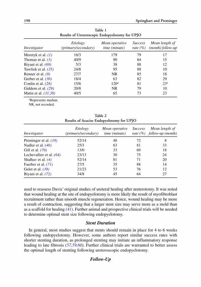
190 Springhart and Preminger
Table 1Results of Ureteroscopic Endopyelotomy for UPJO
Etiology Mean operative Success Mean length ofInvestigator (primary/secondary) time (minute) rate (%) (month) follow-up
Meretyk et al. (1) 16/3 179 79 17Thomas et al. (3) 40/9 90 84 15Biyani et al. (69) 5/3 38 88 12Tawfiek et al. (35) 24/8 95 88 10Renner et al. (9) 27/7 NR 85 18Gerber et al. (30) 18/4 63 82 29Conlin et al. (28) 15/6 120a 81 23a
Giddens et al. (29) 20/8 NR 79 10Matin et al. (31,38) 40/5 65 73 23
aRepresents median.NR, not recorded.
used to reassess Davis’ original studies of ureteral healing after ureterotomy. It was notedthat wound healing at the site of endopyelotomy is more likely the result of myofibroblastrecruitment rather than smooth muscle regeneration. Hence, wound healing may be morea result of contraction, suggesting that a larger stent size may serve more as a mold thanas a scaffold for healing (41). Further animal and prospective clinical trials will be neededto determine optimal stent size following endopyelotomy.
Stent Duration
In general, most studies suggest that stents should remain in place for 4 to 6 weeksfollowing endopyelotomy. However, some authors report similar success rates withshorter stenting duration, as prolonged stenting may initiate an inflammatory responseleading to late fibrosis (57,59,60). Further clinical trials are warranted to better assessthe optimal length of stenting following ureteroscopic endopyelotomy.
Follow-Up
Table 2Results of Acucise Endopyelotomy for UPJO
Etiology Mean operative Success Mean length of Investigator (primary/secondary) time (minute) rate (%) follow-up (month)
Preminger et al. (18) 52/14 48 72 8Nadler et al. (46) 25/3 63 81 33Gill et al. (70) 13/0 33 69 18Lechevallier et al. (64) 23/13 30 75 24Shalhav et al. (4) 52/14 81 71 20Faerber et al. (71) 27/5 35 88 14Gelet et al. (38) 21/23 53 76 12Biyani et al. (72) 34/8 45 64 27

Durability of endopyelotomy has recently been scrutinized, as most contemporarystudies average follow-up is only 17 months (Tables 1 and 2). Prior studies have shownthat most failures occur within 1 year of intervention (39,61). As recent studies havelooked at long-term results of endopyelotomy, the durability of all endourological inter-vention may not be as good as initially believed. Long-term assessment of the postop-erative durability in those who underwent endourological intervention for UPJOdemonstrated that the majority of failures occurred within 12 months but that a signifi-cant number of failures occurred thereafter. Using statistical methods with their data thisrecent retrospective review determined that no further failures were expected after36 months and recommend 3 years minimum follow-up following endopyelotomy (62).Of note, there does not appear to be a difference in the failure rates between patientswith primary or secondary UPJO treated with endopyelotomy (63,64). Based on thisinformation, we perform routine follow-up on our patients using either intravenouspyelogram or nuclear renal scans every 6 months for 5 years.
Comparison of Approaches
In an attempt to duplicate success rates and at the same time decrease perioperativemorbidity, endourological techniques using antegrade, retrograde, and Acucise endopy-elotomy techniques were devised. On average, success rates range from 73 to 88% and64 to 88% for retrograde ureteroscopic and Acucise endopyelotomy with a mean fol-low-up of 17 and 20 months, respectively (19,31,48,65).
However, success rates have not compared with open reconstructive or laparoscopictechniques. In the early 1990s the first laparoscopic pyeloplasty was performed asanother attempt to decrease perioperative morbidity associated with the open technique(66,67). In several studies comparing laparoscopic pyeloplasty to open and endourolog-ical intervention, laparoscopic techniques had equivalent success rates to open surgeryand superior outcomes when compared with endourological techniques. Laparoscopicpyeloplasty has also demonstrated equivalent hospital stay and analgesic use whencompared with endourological techniques and were significantly less than thosepatients who underwent an open surgical approach (68). Along with recent familiariza-tion of intracorporeal suturing techniques and with the further developments of roboticlaparoscopic procedures, some now consider laparoscopic pyeloplasty as the primarytechnique for initial intervention of UPJO with endourological techniques reserved forthose who fail the laparoscopic approach.
In conclusion, retrograde endopyelotomy provides a safe, less invasive, and cost-effective means to treatment of primary or secondary UPJO (65). Although endourolog-ical techniques initially appeared promising, the gold standard of therapy continues tobe a formal repair via laparoscopic or open pyeloplasty. If patients do decide to undergoendourological management, there appears to be no difference in the success rates ofsubsequent pyeloplasty. Furthermore, in the minority of cases that fail primary plasticprocedures, endopyelotomy continues to play a successful role in those cases refractoryto open or laparoscopic repair. Further prospective trials evaluating optimal size andduration of ureteral stent placement must be performed.
TIPS AND TRICKS
General Tips
1. After performing retrograde pyelography, the site of the UPJO is confirmed and marked
Chapter 11 / Retrograde Endopyelotomy 191

on the fluoroscopic image screen using a marking pen. This maneuver will aid in iden-tifying the area of interest as the endoscope is advance proximally.
2. The radio-opaque markers incorporated into the design of the dilating balloon allowprecise placement of this device across the stenotic UPJ indicated by the marked areaon the saved fluoroscopic image.
3. Semirigid ureteroscopy is preferred for the initial attempt of direct vision endopyelo-tomy. However, anatomical constraints in most men and some women may not allowpassage of the semirigid device.
4. Pressurized irrigation from an automated pressure device will allow better visualizationduring ureteroscopic endopyelotomy.
5. While performing ureteroscopy, if blood clots obscure the lumen of the ureter or collecting sys-tem, it is best to pass the ureteroscope along the wall of the ureter and attempt to bypass the clot.
6. Once the UPJO is visualized ureteroscopically, the area is inspected for pulsations,which may be transmitted by a crossing vessel.
Tips for Laser Endopyelotomy
1. Incision of the UPJ is easier to perform as the ureteroscope is withdrawn from the renalpelvis, across the UPJ. The optimal holmium laser settings to incise the UPJ are either1 J at 15 Hz or 1 J at 20 Hz.
2. The ureteroscope and laser are withdrawn in unison, at a slow rate during endopyelo-tomy, to ensure a through-and-through incision to approx 1 cm distal to the initial radi-ographic “waisting” of the ureteral dilating balloon. Although repeat incisions may beperformed to insure a full-thickness incision, a single pass of the ureteroscope and laserwill always result in the “cleanest” UPJ incision.
3. If bleeding is encountered at the incision site, the laser may be “defocused,” by movingthe fiber 2 to 3 mm away from the site of bleeding, fired, and the ureter reinspected forhemostasis.
Electrocautery Tips
1. Cover the indwelling safety guidewire with a 5-Fr open-ended ureteral stent to avoidpotential arching of current between the electrohydraulic lithotriptor electrode andmetallic guidewire.
Acucise Tips
1. Place a 35-cm, 12/14-Fr or larger ureteral access sheath several centimeters below thelevel of obstruction. This helps decrease the potential drag that the Acucise device mayencounter during retrograde placement into the ureter.
2. Only use one working guidewire. Additional guidewires positioned adjacent to theendopyelotomy-cutting balloon wire may cause arching of current, potentially damag-ing adjacent tissue.
3. At no point should the cutting wire cross the working guidewire on the fluoroscopicimage, as this would indicate spiraling of the catheter.
4. If no extravasation is noted, the catheter should be withdrawn and assessed for correctdelivery of current to the cutting wire.
REFERENCES1. Meretyk I, Meretyk S, Clayman RV. Endopyelotomy: comparison of ureteroscopic retrograde and
antegrade percutaneous techniques. J Urol 1992; 148: 775–783.
192 Springhart and Preminger
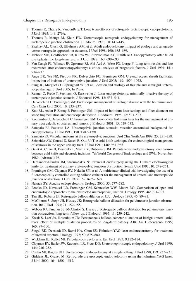
2. Thomas R, Cherry R, VandenBerg T. Long term efficacy of retrograde ureteroscopic endopyelotomy.J Urol 1993; 149: 276A.
3. Thomas R, Monga M, Klein EW. Ureteroscopic retrograde endopyelotomy for management ofureteropelvic junction obstruction. J Endourol 1996; 10: 141–145.
4. Shalhav AL, Giusti G, Elbahnasy AM, et al. Adult endopyelotomy: impact of etiology and antegradeversus retrograde approach on outcome. J Urol 1998; 160: 685–689.
5. Jabbour ME, Goldfischer ER, Klima WJ, Stravodimos KG, Smith AD. Endopyelotomy after failedpyeloplasty: the long-term results. J Urol 1998; 160: 690–693.
6. Van Cangh PJ, Wilmart JF, Opsomer RJ, Abi-Aad A, Wese FX, Lorge F: Long-term results and laterecurrence after endoureteropyelotomy: a critical analysis of prognostic factors. J Urol 1994; 151:934–937.
7. Auge BK, Wu NZ, Pietrow PK, Delvecchio FC, Preminger GM: Ureteral access sheath facilitatesinspection of incision of ureteropelvic junction. J Urol 2003; 169: 1070–1073.
8. Sung JC, Marguet CG, Springhart WP, et al. Location and etiology of flexible and semirigid uretero-scope damage. J Urol 2005; In Press.
9. Renner C, Frede T, Seemann O, Rassweiler J: Laser endopyelotomy: minimally invasive therapy ofureteropelvic junction stenosis. J Endourol 1998; 12: 537–544.
10. Delvecchio FC, Preminger GM: Endoscopic management of urologic disease with the holmium laser.Curr Opin Urol 2000; 10: 233–237.
11. Kuo RL, Aslan P, Zhong P, Preminger GM: Impact of holmium laser settings and fiber diameter onstone fragmentation and endoscope deflection. J Endourol 1998; 12: 523–527.
12. Kourambas J, Delvecchio FC, Preminger GM: Low-power holmium laser for the management of uri-nary tract calculi, structures, and tumors. J Endourol 2001; 15: 529–532.
13. Sampaio FJ, Favorito LA: Ureteropelvic junction stenosis: vascular anatomical background forendopyelotomy. J Urol 1993; 150 :1787–1791.
14. Sampaio FJ: Vascular anatomy at the ureteropelvic junction. Urol Clin North Am 1998; 25: 251–258.15. Schneider AW, Conrad S, Busch R, Otto U: The cold-knife technique for endourological management
of stenoses in the upper urinary tract. J Urol 1991; 146: 961–965.16. Gelet A, Cuzin B, Dessouki T, Martin X, Dubernard JM. Percutaneous endopyelotomy: comparison
between cold knife and electrode incisions. 7th World Congress of Endourology and SWL. November1989; (Abstract) P6.
17. Hernandez-Graulau JM, Stroumbakis N: Intrarenal endosurgery using the Hulbert electrosurgicalknife for treatment of primary ureteropelvic junction obstruction. Semin Urol 1992; 10: 248–251.
18. Preminger GM, Clayman RV, Nakada SY, et al. A multicenter clinical trial investigating the use of afluoroscopically controlled cutting balloon catheter for the management of ureteral and ureteropelvicjunction obstruction. J Urol 1997; 157:1625–1629.
19. Nakada SY: Acucise endopyelotomy. Urology 2000; 55: 277–282.20. Brooks JD, Kavoussi LR, Preminger GM, Schuessler WW, Moore RG: Comparison of open and
endourologic approaches to the obstructed ureteropelvic junction. Urology 1995; 46: 791–795.21. Tan HL, Roberts JP: Retrograde balloon dilation or UPJ. Urology 1995; 46: 89–91.22. McClinton S, Steyn JH, Hussey JK: Retrograde balloon dilatation for pelviureteric junction obstruc-
tion. Bri J Urol 1993; 71: 152–155.23. Webber RJ, Pandian SS, McClinton S, Hussey J: Retrograde balloon dilatation for pelviureteric junc-
tion obstruction: long-term follow-up. J Endourol 1997; 11: 239–242.24. Kwak S, Leef JA, Rosenblum JD: Percutaneous balloon catheter dilatation of benign ureteral stric-
tures: effect of multiple dilatation procedures on long-term patency. AJR. Am J Roentgenol 1995;165: 97–100.
25. Singal RK, Denstedt JD, Razvi HA, Chun SS: Holmium:YAG laser endoureterotomy for treatmentof ureteral stricture. Urology 1997; 50: 875–880.
26. Wickham JE, Kellet MJ. Percutaneous pyelolysis. Eur Urol 1983; 9:122–124.27. Clayman RV, Basler JW, Kavoussi LR, Picus DD: Ureteronephroscopic endopyelotomy. J Urol 1990;
144: 246–252.28. Conlin MJ, Bagley DH: Ureteroscopic endopyelotomy at a single setting. J Urol 1998; 159: 727–731.29. Giddens JL, Grasso M: Retrograde ureteroscopic endopyelotomy using the holmium:YAG laser.
J Urol 2000; 164: 1509–1512.
Chapter 11 / Retrograde Endopyelotomy 193

30. Gerber GS, Kim JC: Ureteroscopic endopyelotomy in the treatment of patients with ureteropelvicjunction obstruction. Urology 2000; 55: 198–203.
31. Matin SF, Yost A, Streem SB: Ureteroscopic laser endopyelotomy: a single-center experience.J Endourol 2003; 17: 401–404.
32. Sampaio FJ: The dilemma of the crossing vessel at the ureteropelvic junction: precise anatomic study.J Endourol 1996; 10: 411–415.
33. Bagley DH, Liu JB, Grasso M, Goldberg BB: Endoluminal sonography in evaluation of theobstructed ureteropelvic junction. J Endourol 1994; 8: 287–292.
34. Nakada SY, Wolf JS, Jr, Brink JA, et al. Retrospective analysis of the effect of crossing vessels onsuccessful retrograde endopyelotomy outcomes using spiral computerized tomography angiography.J Urol 1998; 159: 62–65.
35. Tawfiek ER, Liu JB, Bagley DH: Ureteroscopic treatment of ureteropelvic junction obstruction.J Urol 1998; 160: 1643–1647.
36. Van Cangh PJ, Nesa S, Galeon M, et al. Vessels around the ureteropelvic junction: significance andimaging by conventional radiology. J Endourol 1996; 10: 111–119.
37. Van Cangh PJ, Nesa S: Endopyelotomy. Prognostic factors and patient selection. [Review] [55 refs].Urol Clin North Am 1998; 25: 281–288.
38. Gelet A, Combe M, Ramackers JM, et al. Endopyelotomy with the Acucise cutting balloon device.Early clinical experience. Eur Urol 1997; 31: 389–393.
39. Gupta M, Tuncay OL, Smith AD: Open surgical exploration after failed endopyelotomy: a 12-yearPerspective. J Urol 1997; 157: 1613–1619.
40. Niemczyk P, Krisch EB, Zeiger L, Marmar JL: Use of diuretic renogram in evaluation of patientsbefore and after endopyelotomy. Urology 1999; 53: 271–275.
41. Andreoni CR, Lin HK, Olweny E, et al. Comprehensive evaluation of ureteral healing after electro-surgical endopyelotomy in a porcine model: original report and review of the literature. J Urol 2004;171: 859–869.
42. Bauer JJ, Bishoff JT, Moore RG, Chen RN, Iverson AJ, Kavoussi LR: Laparoscopic versus openpyeloplasty: assessment of objective and subjective outcome. J Urol 1999; 162: 692–695.
43. Soulie M, Thoulouzan M, Seguin P, et al. Retroperitoneal laparoscopic versus open pyeloplasty witha minimal incision: comparison of two surgical approaches. Urology. 2001; 57: 443–447.
44. Ng CS, Yost AJ, Streem SB: Management of failed primary intervention for ureteropelvic junctionobstruction: 12-year, single-center experience. Urology 2003; 61: 291–296.
45. Badlani G, Eshghi M, Smith AD: Percutaneous surgery for ureteropelvic junction obstruction(endopyelotomy): technique and early results. J Urol 1986; 135: 26–28.
46. Nadler RB, Rao GS, Pearle MS, Nakada SY, Clayman RV: Acucise endopyelotomy: assessment oflong-term durability. J Urol 1996; 156: 1094–1098.
47. Thomas R, Cherry R: Ureteroscopic endopyelotomy for the management of ureteropelvic juctionobstruction. J Endourol 1990; 4: S141.
48. Conlin MJ: Results of selective management of ureteropelvic junction obstruction. J Endourol 2002;16: 233–236.
49. Davis DM: Intubated ureterotomy: A new operation for ureteral and ureteropelvic stricture. Surg,Gynecol Obstet. 1943; 76: 513–517.
50. Meretyk S, Albala DM, Clayman RV, Denstedt JD, Kavoussi LR: Endoureterotomy for treatment ofureteral strictures. J Urol 1992; 147: 1502–1506.
51. Siegel JF, Smith AD: The ideal ureteral stent for antegrade and retrograde endopyelotomy: whatwould it be like? J Endourol 1993; 7: 151–154.
52. Hwang TK, Yoon JY, Ahn JH, Park YH: Percutaneous endoscopic management of upper ureteralstricture size of stent. J Urol 1996; 155: 882–884.
53. Mahoney SA, Koletsky S, Persky L: Approximation and dilatation: the mode of healing of an unin-tubated ureterostomy. J Urol 1962; 88: 197–202.
54. Ramsay JWA, Payne SR, Gosling PT, Whitfield HN, Wickham JEA, Levison DA: The effects of dou-ble J stenting on unobstructed ureters. Br J Urol 1985; 57: 630–634.
55. Weaver RG: Ureteral regeneration: experimental and clinical. J Urol 1957; 77: 164–172.56. Kletscher BA, Segura JW, LeRoy AJ, Patterson DE: Percutaneous antegrade endoscopic pyelotomy:
review of 50 consecutive cases. J Urol 1995; 153: 701–703.
194 Springhart and Preminger

57. Kerbl K, Chandhoke PS, Figenshau RS, Stone AM, Clayman RV: Effect of stent duration on ureteralhealing following endoureterotomy in an animal model. J Urol 1993; 150: 1302–1305.
58. Moon YT, Kerbl K, Pearle MS, et al. Evaluation of optimal stent size after endourologic incision ofureteral strictures. J Endourol 1995; 9: 15–22.
59. Abdel-Hakim AMRM: Endopyelotomy for ureteropelvic junction obstruction: Is long-term stentingmandatory? J Endourol 1987; 1: 265–268.
60. Kuenkel M, Korth K: Endopyelotomy: Long-term follow up of 143 patients. J Endourol 1990; 4:109–116.
61. Cohen TD, Gross MB, Preminger GM: Long-term follow-up of Acucise incision of ureteropelvicjunction obstruction and ureteral strictures. Urology 1996; 47: 317–323.
62. Albani JM, Yost AJ, Streem SB: Ureteropelvic junction obstruction: determining durability ofendourological intervention. J Urol 2004; 171: 579–582.
63. Hoenig DM, Shalhav AL, Elbahnasy AM, McDougall EM, Smith D, Clayman RV: Impact of etiol-ogy of secondary ureteropelvic junction obstruction on outcome of endopyelotomy. J Endourol 1998;12: 131–133.
64. Lechevallier E, Eghazarian C, Ortega JC, Andre M, Gelsi E, Coulange C: Retrograde Acuciseendopyelotomy: long-term results. J Endourol 1999; 13: 575–580.
65. Gettman MT, Lotan Y, Roerhborn CG, Cadeddu JA, Pearle MS: Cost-effective treatment for uretero-pelvic junction obstruction: a decision tree analysis. J Urol 2003; 169: 228–232.
66. Schuessler WW, Grune MT, Tecuanhuey LV, Preminger GM: Laparoscopic dismembered pyelo-plasty. J Urol 1993; 150: 1795–1799.
67. Kavoussi LR, Peters CA: Laparoscopic pyeloplasty. J Urol 1993; 150: 1891–1894.68. Baldwin DD, Dunbar JA, Wells N, McDougall EM: Single-center comparison of laparoscopic pyelo-
plasty, acucise endopyelotomy, and open pyeloplasty. J Endourol 2003; 17: 155–160.69. Biyani CS, Cornford PA, Powell CS: Retrograde endoureteropyelotomy with the holmium:YAG
laser. Initial experience. Eur Urol 1997; 32: 471–474.70. Gill HS, Liao JC: Pelvi-ureteric junction obstruction treated with Acucise retrograde endopyelotomy.
British J Urol 1998; 82: 8–11.71. Faerber GJ, Richardson TD, Farah N, Ohl DA: Retrograde treatment of ureteropelvic junction
obstruction using the ureteral cutting balloon catheter. J Urol 1997; 157: 454–458.72. Biyani CS, Minhas S, el Cast J, Almond DJ, Cooksey G, Hetherington JW: The role of acucise
endopyelotomy in the treatment of ureteropelvic junction obstruction. Eur Urol 2002; 41:305–310.
Chapter 11 / Retrograde Endopyelotomy 195

Percutaneous Endopyeloplasty
David S. Sharp, MD, Mihir M. Desai, MD, Mahesh R.Desai, MS, FRCS, and Inderbir S. Gill, MD, Mch
CONTENTS
INTRODUCTION
INDICATIONS
INSTRUMENT LIST
TECHNIQUE
RESULTS
TIPS AND TRICKS
REFERENCES
12
SUMMARY
Percutaneous endopyeloplasty is a minimally invasive endourological approach tothe treatment of ureteropelvic juction obstruction (UPJO). This proceedure incorpo-rates endoscopic suturing through a nephroscope to provide a mucosa-to-mucosaclosure of a traditional endopyelotomy incision. Closing the logitudinal endopyelo-tomy incision in a Heineke-Mikulicz fashion opens the UPJ while limiting urinaryextravasation that may decrease the success rate of endopyelotomy alone.Experimental and clinical studies have supported the safety and efficacy of percuta-neous endopyeloplasty. Comparatively, in similar patients, percutaneous endopyelo-plasty requires a shorter operative time than laparoscopic dismembered pyeloplastyand is relatively simple to perform. Functional results appeal very promising at 1–2years. Further experience and clinical trials will help determine where this procedurefits into the treatment algorithm for UPJO. We offer percutaneous endopyeloplastyas a primary treatment modality for many patients with UPJO.
Key Words: Ureteropelvic junction; UPJ; UPJ obstruction; UPJO; pyeloplasty;endopyeloplasty; percutaneous surgery; percutaneous endopyeloplasty; endourology.
INTRODUCTION
Percutaneous endopyeloplasty is a recently described endourological techniquedesigned in an attempt to retain the advantages of a minimally invasive endourologi-
From: Advanced Endourology: The Complete Clinical GuideEdited by: S. Y. Nakada and M. S. Pearle © Humana Press Inc., Totowa, NJ
197

cal approach to the treatment of ureteropelvic junction obstruction (UPJO) while alsoimproving results. The introduction of traditional endopyelotomy (either antegrade orretrograde) has revolutionized the treatment paradigms for UPJO. It has largelyreplaced open pyeloplasty as the initial treatment of choice for most adults with pri-mary UPJO. Similarly, with the advent of laparoscopic dismembered pyeloplasty, theindications for traditional open pyeloplasty are diminishing. Despite the significantlyreduced morbidity compared to open pyeloplasty, endopyelotomy (even in a favor-able group of patients with a short UPJO, good renal function, only mild to moder-ate hydronephrosis, and absence of a crossing vessel) is associated with a 10 to 15%failure rate (1–3). The success rates of laparoscopic dismembered pyeloplasty mayexceed 95%; however, this technique employs advanced intracorporeal suturingskills, which may limit its applicability to a minority of practicing urologists (4).
Oshinsky et al. (5) initially explored the concept of intrarenal suturing through thenephroscope. A single absorbable suture was placed after standard endopyelotomyeither through the nephroscope using a J-shaped needle and a conventional laparoscopicneedle driver (N = 5), or through a separate retroperitoneal tract. However, no furtherreports of this technique were forthcoming, probably owing to the technical difficultyand unreliability because of suture cut through.
In 2000, we developed a novel technique of percutaneous endopyeloplasty using amodified laparoscopic suturing device (Sew-Right SR•5, LSI Solutions, Victor, NY)in a porcine survival model (6). Percutaneous endopyeloplasty consists of horizontalsuturing of a conventional vertical endopyelotomy incision performed through a soli-tary percutaneous renal tract via a nephroscope, essentially creating a Fengerplasty.Our detailed animal study demonstrated that endopyeloplasty is technically straight-forward and reproducible, heals by primary intention full-thickness healing, and cre-ates a wider caliber UPJ as compared to conventional endopyelotomy. Also, incontrast to endopyelotomy, the meticulously sutured endopyeloplasty was associatedwith absent or minimal contrast extravasation on antegrade contrast study performedintraoperatively immediately after suturing. Given the known detrimental effect ofurinary extravasation on ureteral healing (7), these findings suggest that endopyelo-plasty may provide an optimal environment for full-thickness primary intention heal-ing of the reconstructed UPJ. Thus, our animal experiment provided evidence for apossible functional advantage of endopyeloplasty over endopyelotomy in the surgicaltreatment of UPJO.
In this chapter, we will focus on our technique of percutaneous endopyeloplasty. Wewill discuss our indications and contraindications, the instruments used, the techniqueitself, our laboratory and clinical results, and some “tips and tricks” to performing theprocedure. Additionally, we will discuss our preliminary laboratory work with percuta-neous dismembered pyeloplasty, where an Anderson-Hynes type dismembered pyelo-plasty is performed via a transrenal tract. We believe these procedures hold promise inthe treatment of UPJO, and may continue to evolve along with the changing algorithmsin the treatment of this common urological disorder.
INDICATIONS
Essentially, any patient that is a candidate for primary antegrade or retrogradeendopyelotomy is a candidate for percutaneous endopyeloplasty. The indications to
198 Sharp et al.

intervene for any patient with UPJO include the presence of symptoms, progressive oroverall impairment of renal function, development of upper tract stones or infection, or,rarely, causal hypertension (8). Percutaneous endopyeloplasty has been utilized as theprimary therapy in an increasing number of patients. To date, in part because of theextrarenal dissection needed to appropriately mobilize the tissues prior to suture place-ment, percutaneous endopyeloplasty is not advocated for use as a secondary treatmentmodality. Percutaneous stone removal during the same setting is advocated for percuta-neous endopyelotomy. Five of fifty patients to date have undergone percutaneous stoneat the same time for small calyceal or renal pelvic stones. As a result of concerns overtissue dissection and healing, large or infectious calculi have been excluded from theendopyeloplasty to this point.
Contraindications to a percutaneous endopyeloplasty include active infection, a longsegment of stricture/obstruction (generally greater than 1–2 cm), and uncorrected bleedingdisorders. The issue of a crossing vessel and its relationship to the etiology of UPJOremains controversial. We consider the presence of a crossing vessel to be a contraindica-tion at this time. In one patient who had negative preoperative imaging for crossing ves-sels, a pulsating vessel was identified upon periureteral dissection after completing theendopyelotomy incision. The significance of this vessel is uncertain, but the procedure wascompleted uneventfully and the patient has done well with more than 6 months follow-up.Another early patient did have a crossing vessel on preoperative imaging and still desireda percutaneous endopyeloplasty. Unfortunately, this patient failed and had persistent UPJO.We advocate routine dedicated helical computed tomography of the kidneys with three-dimensional rendering to detect the presence of a crossing vessel. In patients noted to havea crossing vessel, currently laparoscopic or open dismembered pyeloplasty is offered.
INSTRUMENT LIST
Standard setup and equipment utilized for percutaneous renal access and nephroscopyare used. This equipment includes: access needles, a dilator set for the percutaneous renaltract, nephroscopy sheath, light source, video equipment, fluoroscopy table and unit, andirrigation sets. In addition, the following supplies are needed:
1. Nephroscope: A 26-Fr Storz Nephroscope is used. (Karl Storz, Germany). The size ofthe working channel on this scope permits the passage of the suturing device.
2. Suturing Device: Sew-Right® SR•5™ Suturing device (LSI Solutions® Rochester,NY). The Sew-Right SR•5 is a 5-mm laparoscopic suturing instrument used for plac-ing interrupted sutures (Fig. 1). The original laparoscopic version of the suturingdevice was reduced in outer diameter and lengthened to enable its use through thenephroscope.
3. Sutures: Specialized sutures with attached ferrules are necessary to load the suturingdevice (LSI Solutions). We have utilized 2.0 polyglysorb suture with good results. Thedevelopment of finer suture for the device is underway and 3.0 suture is now availablecommercially.
4. Knot pushers: Standard knot pushers, either with closed loop or open loop, used forextra-corporeal laparoscopic suture tying are utilized.
5. Endopyelotomy device: We use a bugbee electrode on cutting current to initiate theendopyelotomy incision. Other devices depending on surgeon preference could be uti-lized including cold knife, holmium laser, or laparoscopic hook dissector.
Chapter 12 / Percutaneous Endopyeloplasty 199
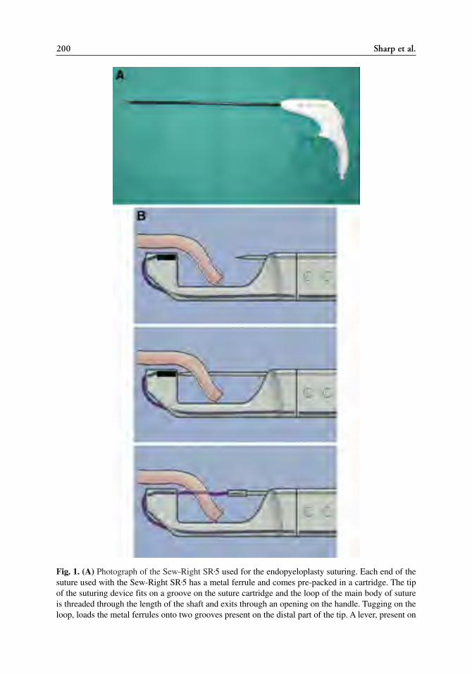
200 Sharp et al.
Fig. 1. (A) Photograph of the Sew-Right SR•5 used for the endopyeloplasty suturing. Each end of thesuture used with the Sew-Right SR•5 has a metal ferrule and comes pre-packed in a cartridge. The tipof the suturing device fits on a groove on the suture cartridge and the loop of the main body of sutureis threaded through the length of the shaft and exits through an opening on the handle. Tugging on theloop, loads the metal ferrules onto two grooves present on the distal part of the tip. A lever, present on

6. Laparoscopic Endoshears: Both 3- and 5-mm Endoshears are useful (USSC, Norwalk,CT). A 5-mm Endoshear with curved blades is used to undermine and dissect out theendopyelotomy mucosal edges prior to suturing. The 3-mm MicroEndoshear hasstraight blades and can be maneuvered to reach areas that the 5-mm device is too bulkyto reach. The 3-mm MicroEndoshear is also useful for completing the endopyelotomyincision.
TECHNIQUE
Step 1: Retrograde Ureteral Access
Retrograde ureteral access is obtained cystoscopically by placing a 6-Fr open-endedureteral catheter into the pelvicalyceal system.
Step 2: Percutaneous Renal Access
Percutaneous renal access is obtained in a standard fashion through an upper or mid-pole calyx, which provides direct access to the UPJ (Fig. 2). A 30-Fr Amplatz sheath ispositioned within the renal pelvis.
Step 3: Conventional Endopyelotomy
A conventional, laterally placed, full-thickness endopyelotomy incision is madeusing 3-mm MicroEndoshears and a bugbee electrode on cutting current (Fig. 3). Thebugbee electrode is used to make the initial perforation in the mucosa, and then theincision is completed with the 3-mm MicroEndoshears that have a linear cutting pro-file. The incision is made across the stricture segment and extends for approx 1 cminto the normal ureter distally and normal pelvis proximally. Care is taken to ensurea clean and sharp cut and to avoid ragged edges to facilitate subsequent endopyelo-plasty suturing.
Step 4: Mobilizing the Distal Ureteral Lip
A crucial step in preparation for endopyeloplasty suturing is the mobilization of thedistal ureteral lip (Fig. 4). The periureteral fibroareolar tissue is carefully dissectedaway from the incised ureteral margin and the adjacent unincised ureter. This maneuveris performed carefully under vision using a 5-mm laparoscopic Endoshears. The 3 mmMicroEndoshears enables even more precise dissection. Care is taken not to excessivelythin out the ureteral wall during this step. The entire dissection is performed “cold,”without cautery. Only specific spot coagulation of bleeding points is carried out asrequired. Occasionally one can encounter a significant-sized vessel, which can be gently
Chapter 12 / Percutaneous Endopyeloplasty 201
Fig. 1. (Continued) the proximal end of the suturing device, switches between the two needles presentat the proximal end of the tip. (B) The instrument is positioned such that the edge of the tissue to besutured is brought into the trough on the instrument tip (top). Squeezing the handle pushes the needle,which pierces the tissue and locks into the metal ferrule (middle). When the handle is released, the nee-dle retracts into its resting position thereby pulling one end of the suture through the tissue (bottom).The lever is switched to activate the other needle and the suture is passed through the other tissue edgeto be sutured.

dissected away from the ureteral wall. This critical step serves three important purposes.First, it provides space for the suturing device while placing the distal bites. Second, itdefines the distal ureteral lip enabling precise full-thickness suturing. Third, it releasestension on the horizontal suture line.
202 Sharp et al.
Fig. 2. Percutaneous renal access is obtained through a suitable upper or middle calyx.
Fig. 3. A laterally oriented full-thickness endopyelotomy incision is made using a Bugbee electrodeand cutting current.
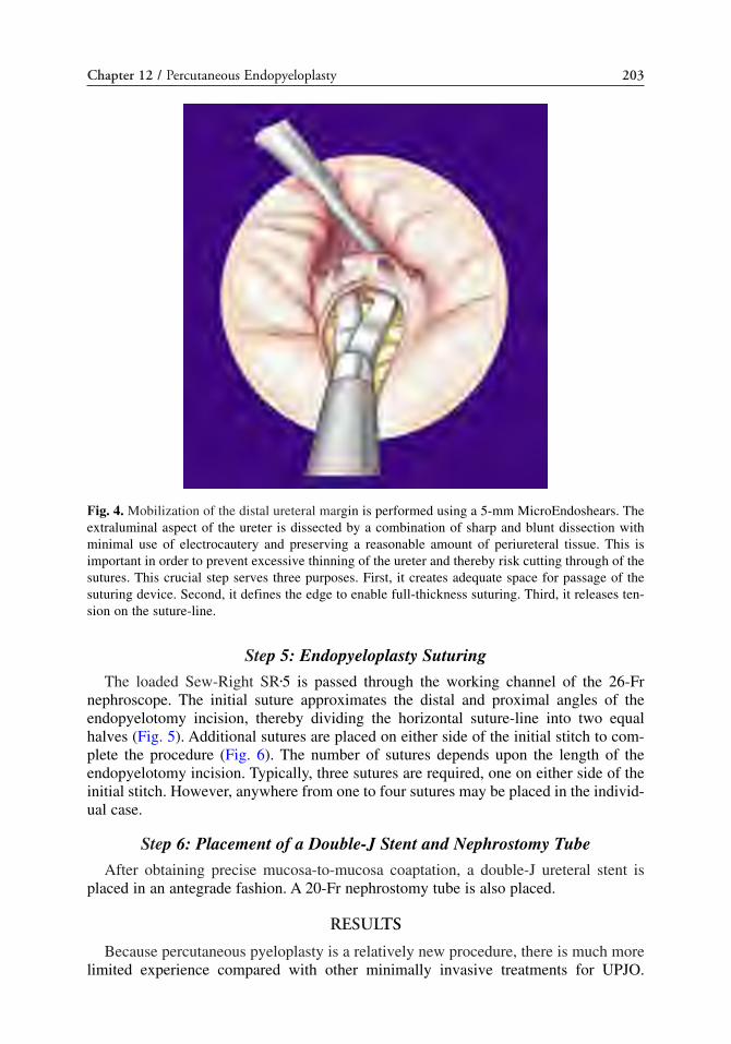
Step 5: Endopyeloplasty Suturing
The loaded Sew-Right SR•5 is passed through the working channel of the 26-Frnephroscope. The initial suture approximates the distal and proximal angles of theendopyelotomy incision, thereby dividing the horizontal suture-line into two equalhalves (Fig. 5). Additional sutures are placed on either side of the initial stitch to com-plete the procedure (Fig. 6). The number of sutures depends upon the length of theendopyelotomy incision. Typically, three sutures are required, one on either side of theinitial stitch. However, anywhere from one to four sutures may be placed in the individ-ual case.
Step 6: Placement of a Double-J Stent and Nephrostomy Tube
After obtaining precise mucosa-to-mucosa coaptation, a double-J ureteral stent isplaced in an antegrade fashion. A 20-Fr nephrostomy tube is also placed.
RESULTS
Because percutaneous pyeloplasty is a relatively new procedure, there is much morelimited experience compared with other minimally invasive treatments for UPJO.
Chapter 12 / Percutaneous Endopyeloplasty 203
Fig. 4. Mobilization of the distal ureteral margin is performed using a 5-mm MicroEndoshears. Theextraluminal aspect of the ureter is dissected by a combination of sharp and blunt dissection withminimal use of electrocautery and preserving a reasonable amount of periureteral tissue. This isimportant in order to prevent excessive thinning of the ureter and thereby risk cutting through of thesutures. This crucial step serves three purposes. First, it creates adequate space for passage of thesuturing device. Second, it defines the edge to enable full-thickness suturing. Third, it releases ten-sion on the suture-line.

204 Sharp et al.
Fig. 5. Placement of the first suture during an endopyeloplasty. The initial suture approximates theproximal and distal angle of the endopyelotomy incision, thereby dividing the incision into two equalhalves. (A) First needle-pass through distal angle. (B) Second ferrule grasped with opposite needlethrough mucosa of proximal angle. (C) Resultant interrupted stitch is tied down with extracoporealknot pusher.
Despite its novelty, the success of the technique is well-supported in the preliminarystudies performed in both experimental and clinical settings. The encouraging data forthe technique is outlined herein.
Experimental Data
Our initial studies used to develop the technique were performed in a porcine model.In 2002, we reported on the feasibility and efficacy of percutaneous endopyeloplastyin a chronic porcine bilateral UPJO model and compared outcome data with conven-tional endopyelotomy and laparoscopic pyeloplasty (6). This study used multiple out-come measures to prove the efficacy of the procedure and critically assess thetechnique. Partial UPJO was successfully created in 20 kidneys (11 pigs) by laparo-scopic ligation of the upper ureter over a 5-Fr ureteral catheter. Subsequently, afterinterval development of hydronephrosis over a period of 4 to 6 weeks, animals
Chapter 12 / Percutaneous Endopyeloplasty 205
Fig. 6. (A) Diagrammatic and (B) nephroscopic view of a completed endopyeloplasty. Typically, twoadditional sutures are placed on either side of the initial suture providing precise mucosa-to-mucosaapproximation.
received either percutaneous endopyeloplasty (n = 10), conventional percutaneousendopyelotomy (n = 5) or laparoscopic pyeloplasty (n = 5). Percutaneous endopyelo-plasty was technically successful in all 10 kidneys with a mean total operative time of

81.4 minutes (51–117 minutes). The mean time to complete the suturing step of the pro-cedure was 29.4 minutes (20–64 minutes). The solitary complication was a lower poleinfundibular stenosis. Over a mean follow-up of 7.7 weeks, all renal units showed reliefof obstruction as evidenced by regression of hydronephrosis, improvement in T1/2 andglomercular filtration rate on renogram and a low intrapelvic pressure on Whitaker test.At autopsy, the endopyeloplasty site showed a fine, well-healed transverse scar with noevidence of residual suture on the mucosal surface. Mean caliber of the UPJ followingendopyeloplasty (13.8 ± 2.2 Fr) was significantly greater (p = 0.01) than that followingconventional endopyelotomy (7.5 ± 1.9 Fr). Intraoperative extravasation upon comple-tion of endopyeloplasty was absent (n = 6) or mild (n = 4) as compared to significantextravasation in all five kidneys following conventional endopyelotomy. Thus, ourdetailed animal study demonstrated that endopyeloplasty is technically straightforwardand reproducible, heals by primary intention full-thickness healing, and creates a widercaliber UPJ as compared to conventional endopyelotomy. Also, in contrast to endopy-elotomy, the meticulously sutured endopyeloplasty was associated with absent or min-imal contrast extravasation on antegrade contrast study performed intraoperativelyimmediately after suturing. As previously mentioned, it is felt that decreasing or elimi-nating urinary extravasation may improve uretereal healing and thus improve functionalresults over endopyelotomy. The technique is optimized to allow for full-thickness pri-mary intention healing of the reconstructed UPJ.
Clinical Data
Since our initial clinical report (9), we have now performed percutaneous endopyelo-plasty in more than 50 patients with primary UPJO. Data on the first 32 patients wasrecently compiled (10). Inclusion criteria for this cohort included short (<1 cm) segmentstenosis, absence of crossing vessel on preoperative imaging, and absence of prior sur-gery on the UPJ. Mean age for the group was 27.5 years. Hydronephrosis was mild in 3,moderate in 19, and severe in 10 patients. High insertion of the UPJ was present in 11patients. The criteria for success was strictly defined, importantly including both symp-tomatic and radiographic improvement. Success was defined as the absence of pain andimprovement of drainage on intravenous pyelogram and/or diuretic renal scan.Percutaneous endopyeloplasty was technically successful in all 32 patients. Mean oper-ative time was 106.3 minutes and mean time for suturing was 29.7 minutes.Endopyeloplasty involved placement of 1 suture in 2 cases, 2 sutures in 3 cases, 3 suturesin 22 cases, 4 sutures in 4 cases, and 5 sutures in 1 case. Intraoperative complica-tions included irrigation fluid extravasation (n = 1), and suture cut-through (n = 1).Postoperative complications included pyrexia requiring prolonged double-J ureteral stent-ing (n = 3). Follow-up was greater than 2 years in 9, 1–2 years in 7, greater than 6 monthsin 5, and less than 6 months in 11 patients (Mean 14.1 months; range, 1–27 months). All32 patients remain symptom free and show improved drainage on intravenous pyelogram(IVP) (Fig. 7) and/or diuretic renal scan (mean postop T1/2 9.6 minutes, mean improve-ment in renal function 12%). This data confirms that percutaneous pyeloplasty is feasi-ble, well-tolerated, and efficacious.
To evaluate percutaneous endopyeloplasty vs other minimally-invasive techniquesfor the correction of UPJO, a comparative study was then performed. We reported ourintermediate term (1 year) data on endopyeloplasty and retrospectively compared itwith endopyelotomy and laparoscopic pyeloplasty in 44 patients with primary UPJO(11). At two institutions, 44 consecutive, nonrandomized patients with primary UPJO
206 Sharp et al.

Chapter 12 / Percutaneous Endopyeloplasty 207
Fig. 7. (A) Preoperative intravenous pyelogram (IVP) shows right ureteropelvic junction (UPJ) obstruc-tion. (B) Followup IVP performed 1 year after endopyeloplasty. On the right side, there is significantreduction in hydronephrosis, funneling and wider caliber of the UPJ, and prompt drainage of contrast.

underwent either percutaneous endopyeloplasty (n = 15; group I), percutaneousendopyelotomy (n = 15; group II), or laparoscopic dismembered pyeloplasty (n = 14;group III). Study inclusion criteria were short segment (<1 cm) stenosis, no prior sur-gery for UPJO, and no crossing vessel in group I. Mean age was 30.3 vs 38.6 vs 38.9years, and duration of symptoms was 5.5 vs 6 vs 6.6 months in group I, II, and III,respectively. Postoperative success was evaluated on symptoms, intravenous urogram(IVU), and/or diuretic renogram. Mean operative time was 119 minutes in group I, 52minutes in group II, and 243 minutes in group III (p < 0.001). Complications occurredin three patients in group I (fever two, fluid extravasation one), two patients in group II(bleeding one, urinoma one), and no patients in group III. Duration of double-J stentplacement was 2, 4 and 6 weeks in groups I, II, and III, respectively. Resolution ofsymptoms and unobstructed drainage on IVP and/or diuretic renogram was noted in 100and 100% of patients in group I (mean follow-up 11.6 months), 93 and 88% of patientsin group II (mean follow-up 31.4 months), and 93 and 100% in group III (mean follow-up 20 months).
Though retrospective in nature, again this report supports percutaneous endopyelo-plasty as efficacious and comparible to endopyelotomy and laparoscopic pyeloplasty.There is shorter operative time in percutaneous endopyeloplasty compared to laparo-scopic dismembered pyeloplasty and complications were not clinically significantamong the three groups. Though follow-up time was shorter in the percutaneous endopy-eloplasty group, success rates are at least as good if not better than the other treatmentmodalities. There is no reason to speculate that durability of the success will be signif-icantly different for percutaneous endopyeloplasty than the durability observed withboth endopyelotomy and laparoscopic pyeloplasty, though this remains to be defini-tively proven. A prospective, multicenter trial of the technique would be useful to notonly test this durability, but also ensure reproducibility among other institutions.
Future Directions
We recently demonstrated the initial technical feasibility of dismembered percuta-neous endopyeloplasty in the animal model (12); which may further increase the roleof percutaneous intrarenal reconstructive surgery. With this technique, an Anderson-Hynes type completely dismembered pyeloplasty is performed via a single transrenaltract utilizing similar instrumentation as the percutaneous (Fengerplasty) endopyelo-plasty described in this chapter. The procedure utilized small hook electrocautery fordissection in addition to 3-mm MicroEndoshears and evolved during its developmentto be performed under intrarenal CO2 insufflation. In five pigs with unilateral UPJO cre-ated 3–6 weeks prior, percutaneous dismembered endopyeloplasty was performed.Percutaneous transrenal access to the UPJ was obtained, and the UPJ was completely dis-membered in all cases from within the renal pelvis through the solitary percutaneoustract. The dismembered proximal ureter was circumferentially mobilized, and in two ani-mals, the UPJ segment was completely excised and removed. A spatulated end-to-endendopyeloplasty anastomosis (Anderson-Hynes) was created transrenally with 5 to 10interrupted sutures using the Sew-Right device (Fig. 8). The technique was developedin three acute pigs. Subsequently, two survival pigs were sacrificed at 2 and 5 weeks,respectively. All cases were successfully dismembered and a precisely suturedmucosa-to-mucosa anastomosis was created. Intraoperative bleeding was negligibleand operative time ranged from 3 to 5 hours, with the majority of time dedicated totransrenal retroperitoneal dissection of the scarred, fibrotic UPJ. CO2 insufflation was
208 Sharp et al.
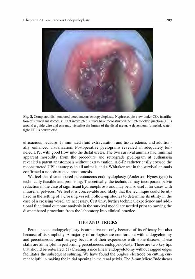
efficacious because it minimized fluid extravasation and tissue edema, and addition-ally, enhanced visualization. Postoperative pyelograms revealed an adequately fun-neled UPJ, with good flow into the distal ureter. The two survival animals had minimalapparent morbidity from the procedure and retrograde pyelogram at euthanasiarevealed a patent anastomosis without extravasation. A 6-Fr catheter easily crossed thereconstructed UPJ at autopsy in all animals and a Whitaker test in the survival animalsconfirmed a nonobstructed anastomosis.
We feel that dismembered percutaneous endopyeloplasty (Anderson-Hynes type) istechnically feasible and promising. Theoretically, the technique may incorporate pelvicreduction in the case of significant hydronephrosis and may be also useful for cases withintrarenal pelvices. We feel it is conceivable and likely that the technique could be uti-lized in the setting of a crossing vessel. Follow-up studies to determine its utility in thecase of a crossing vessel are necessary. Certainly, further technical experience and addi-tional functional outcome analysis in the survival model are needed prior to moving thedismembered procedure from the laboratory into clinical practice.
TIPS AND TRICKS
Percutaneous endopyeloplasty is attractive not only because of its efficacy but alsobecause of its simplicity. A majority of urologists are comfortable with endopyelotomyand percutaneous renal surgery because of their experience with stone disease. Theseskills are all helpful in performing percutaneous endopyeloplasty. There are two key tipsthat should be reiterated: (1) Creating a nice linear endopyelotomy without ragged edgesfacilitates the subsequent suturing. We have found the bugbee electrode on cutting cur-rent helpful in making the initial opening in the renal pelvis. The 3-mm MicroEndoshears
Chapter 12 / Percutaneous Endopyeloplasty 209
Fig. 8. Completed dismembered percutaneous endopyeloplasty. Nephroscopic view under CO2 insuffla-tion of sutured anastomosis. Eight interrupted sutures have reconstructed the ureteropelvic junction (UPJ)around a guide wire and one may visualize the lumen of the distal ureter. A dependent, funneled, water-tight UPJ is constructed.
may then be utilized to complete the endopyelotomy until healthy ureteral tissue isreached and opened for an additional centimeter. (2) Adequate mobilization of the distalureteral lip is critical. We have utilized both the 3- and 5-mm MicroEndoshears for thisstep. This step is important to allow for adequate space for precise full-thickness sutur-ing and releases tension on the horizontal suture line.
REFERENCES1. Motola JA, Badlani GH, Smith AD. Results of 212 consecutive endopyelotomies: an 8 year follow-
up. J Urol 1993; 149: 453–456. 2. Van Cangh PJ, Wilmart JF, Opsomer RJ, et al. Long term results and late recurrence after
endoreteropyelotomy: a critical analysis of prognostic factors. J Urol 1994; 151: 934–937.3. Gupta M, Tuncay OL, Smith AD. Open surgical exploration after failed endopyelotomy: a 12 year
perspective. J Urol 1997; 157: 1613–1618.4. Bauer JJ, Bischoff JT, Moore RG, Chen RN, Iverson AJ, Kavoussi LR. Laparoscopic versus open
pyeloplasty: assessment of objective and subjective outcome. J Urol 1999; 162: 692–695.5. Oshinsky GS, Jarrett TW, Smith AD. New technique of managing ureteropelvic junction obstruction:
percutaneous endoscopic pyeloplasty. J Endourol 1996; 10: 147–151.6. Desai MM, Gill IS, Carvalhal EF, et al. Percutaneous endopyeloplasty: a novel technique. J Endourol
2002; 16: 431–443.7. Andreoni C, Lin HK, Olweny E, Ovrby A, Landman J, Clayman RV. Evaluation of healing after elec-
trosurgical endopyelotomy in a porcine model. J Endourol 2000; Supplement, Abstract BS4-5, A13.8. Streem SB, Franke JJ, Smith JA. Management of Upper Urinary Tract Obstruction In: Campbell’s
Urology, 8th Ed., (Walsh PC, et al. eds.) Saunder’s, Philadelphia, 2002, pp. 463–512.9. Gill IS, Desai MM, Kaouk JH, Wani K, Desai MR. Percutaneous endopyeloplasty: description of a
new technique. J Urol 2002; 168: 2097–2102.10. Desai MM, Desai MR, Gill IS. Percutaneous endopyeloplasty: current clinical status. BJU Int 2005;
95:106–109.11. Desai MM, Desai MR, Gill IS. Endopyeloplasty versus endopyelotomy versus laparoscopic pyelo-
plasty for primary ureteropelvic junction obstruction. Urology 2004; 64:16–21.12. Sharp DS, Desai MM, Molina WR, et al. Dismembered percutaneous endopyeloplasty: a new proce-
dure. J Endourol 2005; 65:473–478.
210 Sharp et al.

Endoureterotomy
Khaled S. Hafez, MD and J. Stuart Wolf, Jr., MD
CONTENTS
INTRODUCTION
INDICATIONS
SURGICAL TECHNIQUES
INSTRUMENT LIST
TYPES OF CUTTING DEVICES
ENDOURETEROTOMY RESULTS
TIPS AND TRICKS
ENDOURETEROTOMY IN SPECIAL CIRCUMSTANCES
CONCLUSION
REFERENCES
13
SUMMARY
Although not as widely reported as the endoscopic treatment of ureteropelvic junctionobstruction, endoureteromy for ureteral obstruction at other sites is nonetheless an effec-tive minimally invasive procedure. Most ureteral strictures other than at the ureteropelvicjunction are acquired, and are very often iatrogenic. Endoureterotomy for distal and upperureteral strictures less than 2 cm and not associated with radiation or other ischemic injuryis highly successful and results in minimal morbidity. Strictures longer than 2 cm, thoseassociated with radiation or ischemic injury, and some in a middle ureteral location maybe managed more appropriately by open reconstruction because of the increased failurerate of endoureterotomy in these patients. Importantly, failure to establish patency withendoureterotomy does not preclude a successful open surgical reconstruction.
Key Words: Endoureterotomy; ureteral obstruction; ureteral stricture; urinary tractobstruction; laser; balloon dilation catheter.
INTRODUCTION
Treatment of ureteral stricture has changed dramatically with the widespread use ofupper urinary tract endoscopy. This minimally invasive technology has greatly improvedoutcomes and the quality of life of many patients. Strictures of the upper urinary tract are
From: Advanced Endourology: The Complete Clinical GuideEdited by: S. Y. Nakada and M. S. Pearle © Humana Press Inc., Totowa, NJ
211

either congenital or acquired. Congenital ureteral strictures are most commonly locatedat the ureteropelvic junction (UPJ). Aside from primary UPJ obstruction (UPJO), mostother ureteral strictures are acquired and typically iatrogenic (1). The most common eti-ology of iatrogenic ureteral stricture disease is injury during endoscopic, open, or laparo-scopic surgical procedures. The advent of ureteroscopic manipulation has led to anincreased incidence of ureteral strictures (2,3). A 1 to 11% incidence of stricture forma-tion has been reported after upper tract endoscopy (2–8). In addition, gynecologic proce-dures, most commonly radical hysterectomy, carry a higher risk of ureteral injury.Ureteral injury has also been described during various general and vascular surgical pro-cedures. Ureteroileal strictures arising after urinary diversion and post-kidney transplantrepresent special subsets. Noniatrogenic acquired causes of ureteral stricture includethose that develop after spontaneous passage of calculi and after chronic inflammatoryureteral involvement as in tuberculosis and schistosomiasis (9–12).
INDICATIONS
Benign Ureteral Strictures
Benign ureteral strictures are either ischemic or nonischemic in nature. A stricture result-ing from surgical injury or radiation therapy is ischemic. Ischemic strictures heal withfibrosis and scar formation, and have diminished success rates after endoureterotomy(1,13). Alternatively, a stricture is nonischemic if it is the result of stone passage or congen-ital abnormality (1). Postendoscopy ureteral strictures may be either ischemic or nonis-chemic depending on the mechanism of injury (i.e., mechanical or thermal trauma) (14,15).
Malignant Ureteral Strictures
Ureteral strictures owing to the recurrence of a primary malignancy or as the resultof extrinsic mechanical compression (usually from a malignancy) are best managedwith formal resection and anastomosis, or catheter bypass (indwelling ureteral stents orpercutaneous nephrostomy tubes).
SURGICAL TECHNIQUES
The success rate of endourological techniques in the management of ureteral stric-tures is generally less than that of formal resection and anastomosis. Nonetheless, theseminimally invasive approaches are preferred given their decreased morbidity, reducedoperative time, shorter hospitalization, and decreased cost as compared to formal recon-struction. Importantly, successful open operative repair is not precluded by failure of aminimally invasive endourologic technique.
Endoureterotomy for upper urinary tract strictures can be performed in an antegradefashion using percutaneous access, an exclusively retrograde method, or by means of acombined antegrade and retrograde approach. The combined technique is usuallyreserved for complex strictures, as is the case of ureteroenteric strictures in patients withurinary diversion.
General Technique of Endoureterotomy
Endoureterotomy begins with the passage of a guidewire across the stenotic segment.Guidewire placement can be accomplished cystoscopically or percutaneously with thepatient in the prone position. A balloon dilation catheter is guided over the wire across thestricture. With only partial inflation of the balloon, the stricture creates a “waist” in the
212 Hafez and Wolf

balloon that characterizes the stricture and helps to formulate the proper approach. If thestricture is particularly tight, then complete balloon dilation of the stricture (i.e., full ratherthan partial inflation of the balloon) may be needed to provide adequate access to the dis-tal end of the stricture. At the University of Michigan, we routinely dilate strictures in thismanner prior to endoscopic incision, as it greatly improves access and visualization inmost strictures. After the balloon is removed, a flexible or semirigid endoscope isadvanced beyond the previously dilated stenotic segment to the level of the normalureteral mucosa. A full-thickness incision is performed, into the retroperitoneal fat,extending approx 1 cm above and below the lesion. For extreme proximal or distal stric-tures, carrying the incision all the way into the renal pelvis or bladder marsupializes thesite into the larger cavity. A variety of cutting devices can be used to create the incision.
The site of the incision varies according to the location of the stricture: proximal andmidureteral strictures are incised laterally, distal strictures (i.e., below the iliac vessels)are incised medially, and strictures over the iliac vessels are incised anteriorly. After theinitial incision, the dilating balloon should be placed over the guidewire and slowlyinflated. The balloon should expand with minimal pressure and without any evidence ofresidual stricture. If a ureteral narrowing or waisting of the balloon is noted, the stenoticsite should be re-incised under direct vision. Typically an 18- or 24-Fr balloon is used,but some urologists use up to a 30-Fr balloon. Following incision, a ureteral stent isplaced to facilitate regeneration of urothelial and muscle layers with an adequate caliberlumen. After placement of the ureteral stent and according to the route employed foraccess, a Foley catheter and/or a nephrostomy tube is left in place for 2 to 3 days to pre-vent urinary extravasation through the incision.
INSTRUMENT LIST
1. Endoscopes: cystoscopes, ureteroscopes, nephroscopes (rigid and flexible).2. Access wires.3. Access catheters.4. Balloon catheter (18–24 Fr).5. Cutting devices: cold knife, electrocautery, lasers, Acucise endoureterotomy.6. Stents and catheters.
Antegrade Endoureterotomy
A percutaneous endoureterotomy is typically the recommend treatment for proximalor midureteral strictures when there is co-existent pathology in the kidney (e.g., renalcalculi). Antegrade endoureterotomy should employ an upper or mid-calyceal approachto provide straight access to the ureter. The incision in the ureteral stricture is full thick-ness until retroperitoneal fat is seen. The extent of the incision should be 1 cm beyondthe area of the stricture on either side, including extension of the cephalic portion of theincision into the renal pelvis for extreme proximal strictures.
Midureteral strictures may be approached antegrade or retrograde. In cases ofmidureteral stricture using the antegrade approach, a flexible ureteroscope is recom-mended to minimize trauma to the intervening ureter. The flexible ureteroscope necessi-tates the use of a 2- or 3-Fr electrosurgical probe or a laser fiber. Using visual orientationalong with fluoroscopy in two planes, a full-thickness incision is made laterally in theureter above the iliac crossing, anteriorly in the ureter overlying the iliac vessels, oranteromedially in the ureter below the iliac vessels. Retroperitoneal or periureteral fat
Chapter 13 / Endoureterotomy 213

should be exposed by the incision, which extends 1 cm proximal and 1 cm distal to theureteral stricture. Tissue injury from use of electrosurgical devices with a greater than400-μm tip is similar to that of a cold-knife incision (16).
Retrograde Endoureterotomy
The decision of whether to use a retrograde rather than an antegrade endoureterotomyapproach to manage a ureteral stricture depends on the individual case and the surgeon’spreference. Decreased morbidity, hospitalization, and ease of access to the upper urinarytract, make the retrograde approach attractive for many patients with ureteral strictures.
The retrograde approach is easiest for distal ureteral strictures. Usually these stric-tures occur at the ureteral orifice, in the intramural portion, or just at or slightly abovethe ureterovesical junction. If the orifice or intramural ureter is involved, these stricturesare incised cystoscopically such that the lower limb of the incision extends through theureteral orifice to open the site into the larger cavity (17). A right-angle electrocauteryattachment is placed through the resectoscope sheath. Using a 50-W pure cut, the sur-geon begins the incision at the 12 o’clock position of the ureteral orifice and extends itcephalad through the ureteral orifice, ureteral tunnel, and for a distance of 1 cm cepha-lad to the area of stricturing. The incision is performed over an inflated ureteral dilatingballoon placed in the intramural ureter, with care taken not to puncture the balloon withelectrocautery. For more proximal strictures, a ureteroscope can be used (Fig. 1).
The Acucise cutting balloon device has also been used in the management of proximaland distal ureteral strictures (18). Under fluoroscopic control, the balloon catheter is posi-tioned in the strictured region of the ureter. In the proximal ureter above the iliac vessels,the cutting wire is oriented posterolateral. Below the iliac vessels the cutting wire isdirected anteromedially to avoid the branches of the internal iliac artery and vein. Forstrictures lying directly over the iliac vessels, direct ureteroscopic visualization would bea more appropriate approach.
Antegrade/Retrograde Endoureterotomy
For complex ureteral strictures, such as those located at ureteroenteric anastomosisor occurring in the ureter of a transplanted kidney, an approach with both antegrade andretrograde control affords optimal access to and control of the stenotic site. Through-and-through access facilitates identification and manipulation of the stenotic area,which is often difficult when approached from only one direction.
Complete ureteral obstruction is a great technical challenge. When this situationoccurs, the extent of the stricture can be estimated through a combined antegradenephrostogram and retrograde ureterogram. A well-described technique for these casesis the “cut-to-the-light” procedure performed using a small electrocautery probethrough one endoscope with the light turned off, cutting towards the light at the tip ofanother endoscope on the other side of the stricture. A short (<1 cm) occlusion can beapproached effectively in this manner. Bagley (19,20) has reported successful recanal-ization of complete ureteral obstructions up to 5 cm in length. However, in general,ureteral strictures longer than 2 cm are more successfully managed with an open surgi-cal procedure. Alternative endoscopic techniques for complete ureteral occlusion havebeen described, including a “cut-to-the-light” procedure that uses a laser (with railroad-ing of a catheter over the laser fiber) and entrapment snares to pull a wire through theureter.
214 Hafez and Wolf

Chapter 13 / Endoureterotomy 215
Fig. 1. (A) Intravenous urogram demonstrating high-grade proximal right ureteral stricture (whitearrowhead). (B) Prior to retrograde ureteroscopic incision, the stricture is defined with low-pressureinflation of a ureteral dilating balloon (white arrowheads indicate stricture). (C) Ureteroscope proxi-mal to stricture following incision laterally. (D) 10-mm balloon fully inflated at the stricture site. (E)Contrast material injected through the distal balloon port after deflating the balloon reveals thedesired wide extravasation (white arrowhead). (Reprinted from ref. 17a, with permission.)

216 Hafez and Wolf
Fig. 1. (Continued)
TYPES OF CUTTING DEVICES
Cold Knife
A rigid ureteroscope is employed when making a cold-knife incision to relieveureteral strictures. Knife blades come in variety of configurations: straight, half-moon,and hook shape. Owing to the large size of the endoscopic instrument needed to performthe incision, their use is limited to the distal ureter where a retrograde approach employsa semirigid ureteroscope, or to the proximal ureter, where an antegrade approachesemploys a nephroscope. For additional control under direct endoscopic vision, knifeblades can be mounted over a guidewire running through a resectoscope (20).

Electrocautery
The electroincision technique for ureteral strictures usually uses 2- to 3-Fr electrodes,which are available in various configurations. Electroincision is favored over the cold-knifeincision because the lower caliber probes can be placed through smaller semirigid and flexi-ble ureteroscopes, therefore enabling the incision to be made anywhere along the ureter (20).
Lasers
The small and flexible laser fibers provide the main advantage this modality for ureteralstrictures. Currently available lasers include the Nd:YAG (neodynium:yttrium-aluminum-garnet), diode lasers with contact fibers, the KTP Nd:YAG (potassium-titanyl-phosphateNd:YAG), and the Ho: YAG (holmium:YAG). The Ho:YAG laser provides the finest inci-sions and the least peripheral damage. Of these lasers, only the Ho:YAG laser also per-mits stone fragmentation, and as such it is the most versatile for endourological purposes.The preferred power setting for performing endoureterotomy is 10 W (21,22).
Acucise Endoureterotomy
The Acucise endoureterotomy incorporates both a monopolar electrocautery cuttingwire and a low-pressure balloon (18,23). The catheter is passed to the strictured segmentover a working guidewire either in a retrograde or antegrade fashion (24). The balloonis utilized to define the area of stenosis and to carry the cutting wire. The electricallyactive surface on the cutting wire is 2.8 cm in length and 150 μm in diameter. The cut-ting wire should be activated for 5 seconds at 75 W. The catheter body has radio-opaquemarkers that help locate the balloon and cutting wire during positioning. Fluoroscopymust be employed to visualize the orientation of the cutting wire within the ureter.
ENDOURETEROTOMY RESULTS
Endoureterotomy success rates vary between 55 and 85% for benign ureteral strictures(17–25) (Table 1). Comparisons among the studies are difficult because of the different
Chapter 13 / Endoureterotomy 217
Fig. 1. (Continued)

cutting modalities, variable length of follow-up, etiology, stricture location, and length,as well as variable duration of stenting and size of stents used.
Endoureterotomy for middle and distal ureteral stricture disease appears to havegood clinical results with success rates ranging from 66 to 88%. The overall success ratefor 156 patients undergoing endoureterotomy is 78%, which appears to be better thanthe overall success rate of 67% noted for balloon dilation (1,18,24–28).
The largest reported series of endoureterotomy consisted of 38 benign ureteral and30 benign ureteroenteric strictures in renal units with greater than 25% of residual renalfunction (1). Various cutting modalities were employed. Median follow-up for benignureteral strictures was 28.4 months, and all failures occurred within 11 months of theprocedure, with a 3-years success rate of 80% (1). Preminger et al. (26) reported theresults of a multicenter trial involving the use of the Acucise balloon catheter for themanagement of 40 ureteral and 9 ureteroenteric strictures. Patients were followed foran average of 8.7 months (range 1.2 to 17 months). Acucise incision of the distal ureter
218 Hafez and Wolf
Table 1Endoureterotomy for Benign Ureteral Strictures (Review of Literature)
References Location % Success Follow-up months
Lopatkin et al. (68) Proximal (3) 67% 22 Middle (0) N/ADistal (4) 100%Total (7) 86%
Eshghi et al. (69) Total (20) 88% N/ASchneider et al. (28) Proximal (0) N/A 15
Middle (0) N/ADistal (12) 83%Total (12) 83%
Chandhoke et al. (18) Proximal & middle (3) 67% 4Distal (5) 80%Total (8) 75%
Cohen et al. (24) Proximal (3) 67% 29Middle (0) N/ADistal (5) 80%Total (8) 75%
Preminger et al. (26) Total (40) 71% 9 Wolf et al. (1) Proximal (4) 75% 28
Middle (5) 100%Distal (29) 78%Total (38) 82%
Singal et al. (21) Proximal & middle (2) 50% 11Distal (10) 70%Total (12) 67%
Total Proximal (10) 70% 4–29Proximal & middle (5) 60%Middle (7) 86%Distal (65) 78%Total (145) 78%

had a success rate of 58%, while incision of the proximal and midureter had successrates of 50% each. The overall success rate for Acucise incision of benign ureteralstrictures was 55%.
TIPS AND TRICKS
Several studies have suggested that better outcomes are achieved when endouretero-tomy is applied to strictures in the terminal portions of the ureter (i.e., distal or proxi-mal), in nonischemic strictures and in short strictures. Ipsilateral renal function hasalso been identified as an important predictor of outcome (13,14,25,29–32).
Stricture Location
Endoureterotomy for proximal and distal ureteral strictures has a greater success ratethan that for midureteral strictures (25,31). Opening the stricture widely by marsupial-ization into a larger cavity such as the renal pelvis or bladder may account for the dif-ference. Other authors have suggested that distal reflux after stricture marsupializationinto the bladder could provide additional distention of the incised ureter that could con-tribute to higher success rates (25).
Smith (30) showed that in a series of 28 patients with ureteral stricture disease, all 4patients with a midureteral stricture failed balloon dilation. Similarly, Meretyk et al.(25) noted a 25% success rate for endourologic incision of midureteral strictures com-pared with an 80% success rate for distal and proximal ureteral strictures.
Stricture Type
The causes of ureteral strictures have also had a significant impact on the success ofa procedure. The most common cause (23%) of ureteral stricture in one series was post-operative fibrosis following open pelvic surgery or ureteroscopic procedures. These rel-atively nonischemic strictures respond better to endoscopic treatments than do poorlyvascularized strictures (25,33,34).
Ureteral strictures secondary to radiation therapy, or resulting from extraluminalmalignancies causing periureteral compression, respond poorly to endoureterotomy(33). In contrast, patients with a concomitant ureteral calculus and an apparent ureteralstricture usually have resolution of the stricture following removal of the obstructionand alleviation of the inflammatory response.
Stricture Length
Longer strictures tend to be associated with poorer success rates. Netto et al. (33) andChang et al. (34) concluded that strictures longer than 1 cm rarely respond well to bal-loon dilation. Meretyk et al. (25) found that the best results following endoureterotomywere in those patients with strictures less than 2 cm. Schneider et al. (28) reported thatthe longest stricture they treated by cold-knife incision was 2.5 cm in length, and thispatient re-obstructed 24 hours after removal of the ureteral stent. It is most appropriateto apply endosurgical management only to strictures less than 2 cm in length.
Stricture Duration
The duration of a ureteral stricture before treatment has no significant effect on thesuccess rate of the therapy. When the factors of stricture length, location, and type are
Chapter 13 / Endoureterotomy 219

controlled, the duration of the stricture does not alter the outcome. Successful endosurgicaltherapy has been reported in strictures ranging from 8 weeks to 18 months duration (33).
Renal Function
Renal units contributing less than 25% of overall renal function are more likely tofail endoureterotomy. This may be the result of inadequate urine flow through the ureter,which may contribute to preventing the incised stricture from re-stenosing. Moreover,a poorly functioning kidney produces less epidermal growth factors. Production of var-ious growth factors appears to correlate well with glomerular filtration rate, irrespectiveof the cause of decreased renal function. Poor stricture healing may be caused by lackof sufficient mitogenic stimulation (35).
Stent Size
Stents from 5 to 16 Fr in size have been utilized following endoureterotomy (25). It is notclear whether the stent should act as a mold, around which the ureter reforms, or as a scaf-fold that guides ureteral healing. Some like to use the a large stent, whereas others argue thatlarge stents might compromise vascularization of the ureteral segment (36–40). A retrospec-tive report suggests that benign ureteral strictures benefited from the use of a 12 Fr or largerstent (1), but other studies of endopyelotomy suggest that smaller stents (6–8 Fr) provideequal results, as compared to patients who received a 7/14-Fr endopyelotomy stent (41–43).
Duration of Stenting
The rationale of placing stents following ureteral dilation or incision is to promoteureteral healing, prevent extravasation of urine, and avoid restricturing. Most authorsagree that stents are useful for successful healing following endoureterotomy. Althoughthere have been experimental studies of “stentless” endoureterotomy, this has not beenapplied clinically. Excessively long duration of indwelling stents can cause inflammationthat may prevent adequate healing or promote the formation of hyperplastic muscle orscar tissue. In one animal study, there was no difference in ureteral healing among inci-sions managed with a 1-, 3-, or 6-weeks period of stenting (44). The duration of stenting,as long as it is not less than 1 week or more than 6 week, likely is not a major factor.
Choice of Cutting Modality
Cold-knife incision of ureteral strictures is as effective as electrosurgery and Ho:YAGlaser (18,21,26,28). Figenshau et al. (45) investigated the acute tissue changes thatoccur in the pig ureter following balloon dilation, cutting balloon, and endoscopic inci-sion with a cold knife, Nd:YAG laser, or electrocautery (250- and 660-μm electro-cautery probes). There was no significant difference in the degree of tissue injuryamong the various cutting modalities except for the larger 660-μm electrosurgicalprobe. Unlike a ureteral incision balloon, dilation resulted in injury to the lamina pro-pria but did not appear to split the muscularis and adventitial layers (45,46). TheHo:YAG laser is currently the cutting modality of choice for many urologists since itprovides a well-controlled, hemostatic incision (21,47).
Adjunctive Steroids
Triamcinolone at a dose of 120 to 200 mg (3–5 mL at 40 mg/mL) has been endoscop-ically injected with a 3-Fr Greenwald needle into the incised stricture bed in selected
220 Hafez and Wolf
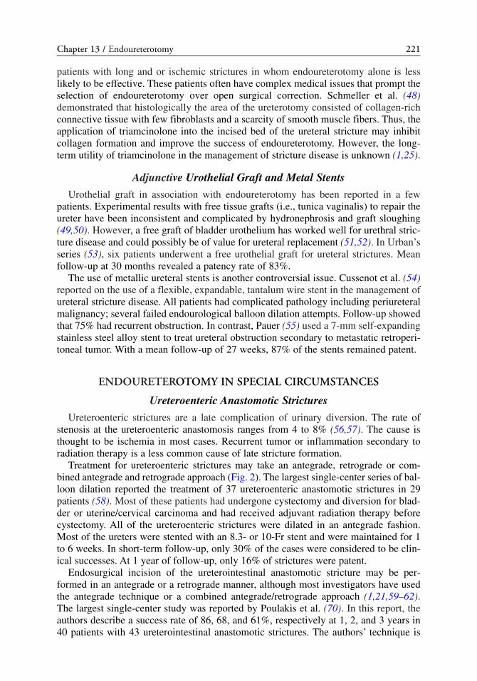
patients with long and or ischemic strictures in whom endoureterotomy alone is lesslikely to be effective. These patients often have complex medical issues that prompt theselection of endoureterotomy over open surgical correction. Schmeller et al. (48)demonstrated that histologically the area of the ureterotomy consisted of collagen-richconnective tissue with few fibroblasts and a scarcity of smooth muscle fibers. Thus, theapplication of triamcinolone into the incised bed of the ureteral stricture may inhibitcollagen formation and improve the success of endoureterotomy. However, the long-term utility of triamcinolone in the management of stricture disease is unknown (1,25).
Adjunctive Urothelial Graft and Metal Stents
Urothelial graft in association with endoureterotomy has been reported in a fewpatients. Experimental results with free tissue grafts (i.e., tunica vaginalis) to repair theureter have been inconsistent and complicated by hydronephrosis and graft sloughing(49,50). However, a free graft of bladder urothelium has worked well for urethral stric-ture disease and could possibly be of value for ureteral replacement (51,52). In Urban’sseries (53), six patients underwent a free urothelial graft for ureteral strictures. Meanfollow-up at 30 months revealed a patency rate of 83%.
The use of metallic ureteral stents is another controversial issue. Cussenot et al. (54)reported on the use of a flexible, expandable, tantalum wire stent in the management ofureteral stricture disease. All patients had complicated pathology including periureteralmalignancy; several failed endourological balloon dilation attempts. Follow-up showedthat 75% had recurrent obstruction. In contrast, Pauer (55) used a 7-mm self-expandingstainless steel alloy stent to treat ureteral obstruction secondary to metastatic retroperi-toneal tumor. With a mean follow-up of 27 weeks, 87% of the stents remained patent.
ENDOURETEROTOMY IN SPECIAL CIRCUMSTANCES
Ureteroenteric Anastomotic Strictures
Ureteroenteric strictures are a late complication of urinary diversion. The rate ofstenosis at the ureteroenteric anastomosis ranges from 4 to 8% (56,57). The cause isthought to be ischemia in most cases. Recurrent tumor or inflammation secondary toradiation therapy is a less common cause of late stricture formation.
Treatment for ureteroenteric strictures may take an antegrade, retrograde or com-bined antegrade and retrograde approach (Fig. 2). The largest single-center series of bal-loon dilation reported the treatment of 37 ureteroenteric anastomotic strictures in 29patients (58). Most of these patients had undergone cystectomy and diversion for blad-der or uterine/cervical carcinoma and had received adjuvant radiation therapy beforecystectomy. All of the ureteroenteric strictures were dilated in an antegrade fashion.Most of the ureters were stented with an 8.3- or 10-Fr stent and were maintained for 1to 6 weeks. In short-term follow-up, only 30% of the cases were considered to be clin-ical successes. At 1 year of follow-up, only 16% of strictures were patent.
Endosurgical incision of the ureterointestinal anastomotic stricture may be per-formed in an antegrade or a retrograde manner, although most investigators have usedthe antegrade technique or a combined antegrade/retrograde approach (1,21,59–62).The largest single-center study was reported by Poulakis et al. (70). In this report, theauthors describe a success rate of 86, 68, and 61%, respectively at 1, 2, and 3 years in40 patients with 43 ureterointestinal anastomotic strictures. The authors’ technique is
Chapter 13 / Endoureterotomy 221

notable for the use of a cold-knife, which may minimize thermal damage to the sur-rounding tissue. In addition, the authors used multiple shallow cuts rather than a singledeep incision. This technique differs from the more commonly performed single inci-sion through the entire scar depth. The excellent results of these authors may suggestthat this type of incision may be a better way to manage ureteroenteric strictures. Theseauthors found that many factors (renal function, length of stricture, interval to stricture
222 Hafez and Wolf
Fig. 2. (A) Endoscopic view from the ileal conduit aspect of a ureteroenteric anastomotic stricture ata wire that has been placed through the stricture using an antegrade ureteroscope (white arrowheadindicates light from antegrade ureteroscope). (B) A ureteral dilating balloon has been placed over thewire and a resectoscope with Colling’s knife (white arrowhead) is used to incise the stricture over theballoon. (C) Appearance of stricture after incision, which was marsupialized ureter proximal to thestricture into the ileal conduit. (Reprinted from ref. 17a.).

formation, degree of stricture, etc.) were cumulative in their impact on their results.Wolf et al. (1) reported their series of 30 ureterointestinal anastomotic strictures in 25patients. The success rates of endoureterotomy for ureteroenteric strictures at 1, 2, and3 years were 72, 51, and 32%, respectively. There was an improved outcome for rightrather than left strictures (68 vs 17% 3-years success rates, respectively) and for stric-tures treated less than 24 months after the etiological insult. More favorable outcomewas noted with the use of 12 Fr or larger stents and stenting longer than 4 weeks. Thethree most important factors in the minimally invasive management of ureteroentericstrictures probably are renal function, stricture length, and completeness of the stricture.
Ureteral Strictures Postrenal Transplantation
Following renal transplant, the frequency of a ureteral complication is closely asso-ciated with the type of reimplant performed at the time of transplantation. WithLeadbetter–Politano reimplantation, urinary tract complications occur in 5 to 11% ofpatients (63). However, with the adaptation of extravesical ureteroneocystostomy, theincidence of urological complications has fallen to below 4% (63). However, up to two-thirds of these urological complications are still owing to ureteral obstruction. Theobstruction may be either intrinsic (i.e., ureteral stricture) or extrinsic (i.e., perirenalfluid collection, such as lymphocele, urinoma, abscess, or hematoma). The developmentof strictures usually occurs early in the postoperative course, but in some cases forma-tion may occur as late as 5 years postoperatively (63–65).
Endoureterotomy has been successfully used in posttransplant ureteric strictures.Conrad et al. (66) used cold-knife endoureterotomy to treat 11 transplant patients withureteral strictures; all but two of the strictures were in the distal ureter. An indwelling14-Fr stent was placed for a period of 4 to 6 weeks. Success was achieved in 82% ofpatients with a mean follow-up of 28 months. Using the Acucise balloon device, Youssefet al. (67) reported a high rate of success in postrenal transplant ureteral strictures. Overall,it would appear that an endourological approach with balloon dilation or endoureterotomyis a reasonable first step when dealing with posttransplant ureteral strictures.
Chapter 13 / Endoureterotomy 223
Fig. 2. (Continued)

CONCLUSION
Endoureterotomy is the procedure of choice for the initial management of benignureteral strictures. Endoureterotomy for benign ureteral strictures has consistently reportedhigher success rates than endoureterotomy for ureteroenteric strictures, and in the former,failures appear within the first year. In addition, repeat endoureterotomy has a better like-lihood of success if radiological improvement was noted after the initial procedure. In con-trast, endoscopic or fluoroscopic incision of ureteroenteric strictures appears to do lesswell, with failures continuing for the first three years. Repeat incisions are more likely tofail and open repair or chronic long-term stenting is often a better management if open sur-gery is to be avoided (68).
The success of endosurgical treatment depends to a large extent on the characteris-tics of the stricture: cause, length, and location (69). Unfortunately, rarely do studyreports subdivide the patient groups according to their stricture characteristics. This fac-tor, when combined with the inconsistencies of the technique of incision and the vari-ability in posttreatment stent size and duration of stenting, results in a significantamount of clinical confusion such that cumulative data on the endosurgical manage-ment of ureteral stricture can only be judged in a broad manner.
In conclusion, endourological management of ureteral strictures has not acquired thesame degree of acceptance as endourological management of UPJO. Overall, the endo-surgical management of distal and upper ureteral strictures less than 2 cm and not asso-ciated with radiation or other ischemic injury is highly successful and results in minimalmorbidity. Also, failure to establish patency does not preclude a subsequent open recon-structive repair. Strictures longer than 2 cm and those associated with radiation orischemic injury or a middle ureteral location may be managed more appropriately byopen reconstruction because of the high failure rate associated with this group ofpatients treated endosurgically. Further clinical studies are necessary to determine thelong-term feasibility and success of adjuvant therapy, such as triamcinolone injectionand free urothelial grafting.
REFERENCES1. Wolf JS Jr, Elashry OM, Clayman RV. Long-term results of endoureterotomy for benign ureteral and
ureteroenteric strictures. J Urol 1997; 158: 759–764.2. Weinberg IJ, Ansong K, Smith AD. Complications of ureteroscopy in relation to experience: Report
of survey and author experience. J Urol 1987; 137: 384–385.3. Assimos DG, Patterson LC, Taylor CL. Changing incidence and etiology of iatrogenic ureteral
injuries. J Urol 1994; 152: 2240–2246.4. Kramolowsky EV. Ureteral perforation during ureterorenoscopy: Treatment and management. J Urol
1987; 138: 36–38.5. Chang R, Marshall FF. Management of ureteroscopic injuries. J Urol 1987; 137: 1132–1135.6. Blute ML, Segura JW, Patterson DE. Ureteroscopy. J Urol 1988; 139: 510–512.7. Lytton B, Weiss RM, Green DF. Complications of ureteral endoscopy. J Urol 1987; 137: 649–653.8. Selzman AA, Spirnak JP. Iatrogenic ureteral injuries: A 20-year experience in treating 165 injuries.
J Urol 1996; 155: 878–881.9. Ghoneim MO, Nabeeh A, El-Kappany H. Endourologic treatment of ureteral strictures. J Endourol
1988; 2: 263–270. 10. Sabha M, Nilsson T, Bahar RH, et al. Endoscopic balloon dilation of bilharzial ureteric strictures.
J Endourol 1988; 2: 23–26. 11. Ward AS, Karanjia ND, Russell AJ. Ureteral obstruction following aortobifemoral bypass:
Management by endoscopic balloon dilation. J Urol 1992; 147: 120–122.
224 Hafez and Wolf

12. Ramanathan LT, Kumar A, Kapoor R, et al. Relief of urinary tract obstruction in tuberculosis toimprove renal function. Analysis of predictive factors. Br J Urol 1998; 81: 199–205.
13. Thomas R: Choosing the ideal candidate for endoscopic endoureterotomy. J Urol 1993; 149: 314A.14. Kramolowsky EV, Tucker RD, Nelson CM. Management of benign ureteral structures: Open surgi-
cal repair or endoscopic dilation? J Urol 1989; 141: 285–286.15. Motola JA, Smith AD. Complications of ureteroscopy: Prevention and treatment. AUA Update Series
1992; 11: 162–168.16. Dierks SM, Clayman RV, Kavoussi LR, et al. Intraureteral surgery: appropriate cutting modality.
J Endourol 1990; 4: S114.17. Yamada S, Ono Y, Ohshima S, et al. Transurethral ureteroscopic ureterotomy assisted by a prior bal-
loon dilation for relieving ureteral strictures. J Urol 1995; 153: 1418–1421.17a. Hafez KS, Wolf JS, Jr. Update on minimally invasive management of ureteral strictures. J Endourol
2003; 17: 453–464.18. Chandhoke PS, Clayman RV, Stone AM, et al. Endopyelolomy and endoureterotomy with the acucise
ureteral cutting balloon device: Preliminary experience. J Endourol 1993; 7: 45–51.19. Bagley DH. Endoscopic ureteroureterostomy. J Urol 1990; 143: 234A.20. Conlin MJ, Bagley DH. Incisional treatment of ureteral strictures. In: Smith’s Textbook of Endourology,
(Smith AD, Badlani GH, Bagley DH, et al., eds.) Quality Medical Publishing, St. Louis, MO, 1996, 497–510.21. Singal RK, Denstedt JD, Razvi HA, et al. Holmium:YAG laser endoureterotomy for treatment of
ureteral stricture. Urol 1997; 50: 875–880.22. Kourambas J, Delvecchio FC, Preminger GM. Low power holmium laser for the management of uri-
nary tract calculi, strictures and tumors. J Endourol 2001; 15: 529–532.23. Aronson WJ, Barbaric ZL, Fain JS, et al. Fluoroscopically guided incision of ureteral strictures in
pigs with the cautery-wire balloon catheter: phase I study. J Urol 1993; 149: 1178–1181.24. Cohen TD, Gross MB, Preminger GM: Long-term follow-up of acucise incision of ureteropelvic
junction obstruction and ureteral strictures. Urol 1996; 47: 317–323.25. Meretyk S, Albala DM, Clayman RV, et al. Endoureterotomy for treatment of ureteral strictures.
J Urol 1992; 147: 1502–1506.26. Preminger GM, Clayman RV, Nakada SY, et al. A multicenter clinical trial investigating the use of a
fluoroscopically controlled cutting balloon catheter for the management of ureteral and ureteropelvicjunction obstruction. J Urol 1997; 157: 1625–1629.
27. Delvecchio FC, Preminger GM. Endourologic management of upper urinary tract strictures. AUAUpdate Series, 2000; 14–22, lesson 32.
28. Schneider AW, Conrad S, Busch R, et al. The cold-knife technique for endourological managementof stenosis in the upper urinary tract. J Urol 1991; 146: 961–965.
29. Johnson CD, Oke EJ, Dunnick NR, et al. Percutaneous balloon dilation of ureteral strictures. AJR1987; 148: 181–184.
30. Smith AD. Management of iatrogenic ureteral strictures after urological procedures. J Urol 1988;140: 1372–1374.
31. Fuchs GJ, Patel A, Gutierrez J, et al. Endourological treatment of ureteral strictures: Experience of 2 referral centers. J Urol 1996; 155: 474A.
32. Beckmann CF, Roth RA, Bihrle W. Dilation of benign ureteral strictures. Radiol 1989; 172: 437–441.33. Netto NR Jr, Ferreira U, Lemos GC, et al. Endourological management of ureteral strictures. J Urol
1990; 144: 631–634.34. Chang R, Marshall FF, Mitchell S. Percutaneous management of benign ureteral strictures and fistu-
las. J Urol 1987; 137: 1126–1131.35. Jabbour ME, Goldfischer ER Anderson AE, et al: Endopyelotomy failure is associated with reduced
transforming growth factor-beta. J Urol 1998; 160: 1991–1994.36. Siegel JF, Smith AD. The ideal ureteral stent for antegrade and retrograde endopyelotomy: What
would it be like? J Endourol 1993; 7: 151–154.37. Mahoney SA, Koletslry S, Persky L. Approximation and dilatation: The mode of healing of an unin-
tubated ureterostomy. J Urol 1962; 88: 197–202.38. Ramsay WA, Payne SR, Gosling PT, et al. The effects of double J stenting on unobstructed ureters.
Br J Urol 1985; 57: 630–634.39. Weaver RG. Ureteral regeneration: Experimental and clinical. J Urol 1957; 77: 164.
Chapter 13 / Endoureterotomy 225

40. Hwang TK, Yoon JY, Alm JH, et al. Percutaneous endoscopic management of upper ureteral stricturesize of stent. J Urol 1996; 155: 882–884.
41. Kletscher BA, Segura JW, LeRoy AI, et al. Percutaneous antegrade endoscopic pyelotomy: Reviewof 50 consecutive cases. J Urol 1995; 153: 701–703.
42. Moon YT, Kerbl K, Pearle MS, et al. Evaluation of optimal stent size after endourologic incision ofureteral strictures. J Endourol 1995; 9: 15–22.
43. Pearle MS. Use of ureteral stents after endopyelotomy. J Endourol 1996; 10: 169–176.44. Kerbl K, Chandhoke PS, Figenshau RS, et al. Effect of stent duration on ureteral healing following
endoureterotomy in an animal model. J Urol 1993; 150: 1302–1305.45. Figenshau RS, Clayman RV, Wick MR, et al. Acute histologic changes associated with endouretero-
tomy in the normal pig ureter. J Endourol 1991; 5: 357–360.46. Gelet A, Cuzin B, Dessouki T, et al. Percutaneous endopyelotomy: Comparison between cold knife and
electrode incisions. J Endourol 1989; 3: 6.47. Kuo RL, Asian P, Fitzgerald KB, et al. Use of ureteroscopy and holmium:YAG laser in patients with
bleeding diatheses. Urol 1998; 2: 609–613.48. Schmeller N, Leitl F, Arnholdt H. Histology of ureter after unsuccessful endoscopic intubated inci-
sion. J Urol 1992; 147: 450–452.49. Gardiner RA, Weedon D, Sing J, et al. Replacement of ureteric segments by intubated neoureterotomies
(modified Davis technique) using autologous bladder and omentum in dogs. Br J Urol 1984; 56: 354–358.50. Hovnanian AP, Javadpour H, Gruhn JG. Reconstruction of the ureter by free autologous bladder
mucosa graft. J Urol 1965; 93: 455–457.51. Hendren WH, Reda EF. Bladder mucosa graft for construction of male urethra. J Pediatr Surg 1986;
21: 189–192.52. Greenberg R, Coleman JW, Quiguyan CC, et al. Bladder mucosal grafts: experimental use as a
ureteral substitute and observation of certain physical properties. J Urol 1983; 129: 634–636.53. Urban DA, Kerbl K, Clayman RV, et al. Endo-ureteroplasty with a free urothelial graft. J Urol 1994;
152: 910–915.54. Cussenot O, Bassi S, Desgrandchamps F, et al. Outcomes of non-self-expandable metal prosthesis in
strictured human ureter: suggestions for future developments. J Endourol 1993; 7: 250–259.55. Pauer W, Lugmayr H. Metallic wall stems: a new therapy for extrinsic ureteral obstruction. J Urol
1992; 148: 281–284.56. Guiliani L, Giberti C, Martovana G, et al. Results of radical cystectomy for primary bladder cancer:
a retrospective study of more than two thousand cases. Urology 1985; 26: 243–248.57. Schmidt JD, Hawtrey CE, Flocks RH, et al. Complications, results and problems of ileal diversion.
J Urol 1973; 109: 210–216.58. Shapiro MJ, Banner MP, Amendola MA, et al. Balloon catheter dilation of ureteroenteric strictures:
long-term results. Radiology 1988; 168: 385–387.59. Ahmadzadeh M. Use of a prototype 3-Fr needle electrode with flexible ureteroscopy of antegrade
management of stenosed ureteroileal anastomosis. Urol lnt 1992; 49: 215–217.60. Cornud F, Mendelsberg M, Chretian Y, et al. Fluoroscopically guided percutaneous transrenal elec-
troincision of ureterointestinal anastomotic strictures. J Urol 1992; 147: 578–581.61. Germinale F, Bottino P, Caviglia C, et al. Endourologic treatment of ureterointestinal strictures.
J Endourol 1992; 6: 439–441.62. Lin DW, Bush WH, Mayo ME. Endourological treatment of ureteroenteric strictures: efficacy of
Acucise endoureterotomy. J Urol 1999; 162: 696–698.63. Thiounn N, Benoit G, Osphal C, et al. Urological complications in renal transplantation. Prog Urol
1991; 1: 531–538.64. Kashi SH, Lodge JPA, Giles GR, et al. Ureteric complications of renal transplantation. Br J Urol
1992; 70: 139–143.65. Kinnaert P, Hall M, Janssen F, et al. Ureteral stenosis after kidney transplantation: true incidence and
long-term follow-up after surgical correction. J Urol 1985; 133: 17–20.66. Conrad S, Schneider AW, Tenschert W, et al. Endourological cold-knife incision for ureteral stenosis
after renal transplantation. J Urol 1994; 105: 906–909.
226 Hafez and Wolf

67. Youssef NI, Jindal R, Babayan RI, et al. The acucise catheter: a new endourological method for cor-recting transplant ureteric stenosis. Transplantation 1994; 57: 1398–1400.
68. Lopatkin NA, Martov AG, Gushchin BL. An endourologic approach to complete ureteropelvic andureteral strictures. J Endourol 2000; 14: 721–726.
69. Eshghi M. Endoscopic incisions of the urinary tract. AUA Update Series, 1989; vol 8:Lesson 37.70. Poulakis V, Witzsch U, De Vries R, et al. Cold-knife endoureterotomy for nonmalignant ureteroin-
testinal anastomotic strictures. Urology 2003; 61(3): 512–517.
Chapter 13 / Endoureterotomy 227

Treatment of Caliceal Diverticula
Edward D. Matsumoto, MD, FRCSC,and Margaret S. Pearle, MD, PhD
CONTENTS
INTRODUCTION
INDICATIONS AND PREPARATION FOR SURGICAL INTERVENTION
RETROGRADE URETEROSCOPIC APPROACH
PERCUTANEOUS APPROACH
HYDROCALYX
RESULTS
TIPS AND TRICKS
CONCLUSIONS
REFERENCES
14
SUMMARY
Caliceal diverticula represent a therapeutic challenge for the endourologist. Treatmentoptions for symptomatic, stone-bearing, caliceal diverticula include shockwave lithotripsy(SWL), ureteroscopy, percutaneous nephrostolithotomy/ablation, and laparoscopicunroofing. A surprising number of patients achieve symptomatic relief despite poor stone-free rates with SWL, although this approach is best reserved for patients with a small stoneburden in a diverticulum with a widely patent neck. Ureteroscopic management (consist-ing of laser endoinfundibulotomy or balloon dilation of the infundibular neck and frag-mentation/removal of the stones) is a good option for relatively small, upper or middlecaliceal diverticula with stones less than 15 mm in size. The percutaneous approach isassociated with the highest stone-, symptom-, and diverticulum-free rates and is the opti-mal treatment for all but anteriorly located diverticula. Lastly, laparoscopy is reserved forlarge, anteriorly positioned diverticula or diverticula that fail endourological management.
Key Words: Caliceal diverticulum; hydrocalyx; renal calculus; endoinfundibulo-tomy; infundibular stenosis.
INTRODUCTION
Caliceal diverticula are eventrations of the upper collecting system that are lined bytransitional epithelium and communicate with the collecting system via narrow
From: Advanced Endourology: The Complete Clinical GuideEdited by: S. Y. Nakada and M. S. Pearle © Humana Press Inc., Totowa, NJ
229

infundibula (1). The radiographic incidence of caliceal diverticula is 0.2 to 0.5% (2,3).In 25 to 50% of cases, stones are identified within the diverticulum (4,5).
Although caliceal diverticula are often asymptomatic, they may also be associatedwith flank pain, urinary tract infection or hematuria. Asymptomatic patients requireno treatment; however, intervention may be necessary in symptomatic patients.
230 Matsumoto and Pearle
Fig. 1. (A) Scout film demonstrating cluster of stones. (B) Intravenous pyelogram demonstratingstones within upper pole caliceal diverticulum.

Historically, symptomatic caliceal diverticula were managed surgically by (1) obliter-ating the diverticular cavity and enlarging or closing the infundibulum; (2) excising thethin overlying parenchyma and marsupializing the diverticulum; or (3) partial nephrec-tomy (1,2,6). Currently, minimally-invasive alternatives are available for managementof symptomatic caliceal diverticula with or without stones, including shockwavelithotripsy (SWL), ureteroscopy, percutaneous nephrostolithotomy, or laparoscopicablation. Likewise, hydrocalices associated with stenotic infundibula can be treated bythe same endoscopic approaches, while leaving the secretory epithelium intact. Theobjective of this chapter is to review the endourological approaches to treatment of cal-iceal diverticula.
INDICATIONS AND PREPARATION FOR SURGICAL INTERVENTION
Indications for the treatment of caliceal diverticula include flank pain, hematuria, orurinary tract infections that cannot be attributed to other causes. Microscopic or grosshematuria is most commonly associated with stone-bearing caliceal diverticula.
All patients should undergo preoperative imaging with a computed tomography (CT)scan and intravenous urogram to determine the size and location of the diverticula andstones, to assess the overlying renal parenchyma and the diverticular neck and to delin-eate the relational anatomy of the diverticula. Careful inspection of intravenous uro-gram images may allow identification of the calyx with which the diverticulum isassociated; retrograde pyelography may further delineate the diverticular neck (Fig. 1).
Active urinary tract infections should be treated in advance of the procedure and theselection of preoperative antibiotics should take into account organisms cultured fromprevious urine specimens.
RETROGRADE URETEROSCOPIC APPROACH
Instrument List
1. Flexible or rigid cystoscope.2. Flexible ureteroscope.3. 0.035-in. Bentson guidewire.4. 0.038-in. double flexible tip, extra-stiff guidewire.5. 5-Fr open-ended angiographic catheter.6. 8-Fr cone-tip catheter.7. 8/10-Fr coaxial dilator or 10-Fr dual lumen catheter.8. Single action syringe-assist pressure irrigating device or pressurized irrigation bags.9. Holmium:YAG laser and 200-μm laser fiber.
10. 7-Fr double pigtail ureteral stent.11. Optional:
a. 0.035-in. angled and/or straight hydrophilic guidewire.b. 12/14-Fr ureteral access sheath.c. 4-mm, 4-cm dilating balloon mounted on a 3-Fr shaft for passage through the uretero-
scope (Passport™ Balloon on a Wire, Boston Scientific, Natick, MA) or 4-mm, 4-cmhigh pressure dilating balloon (7-Fr shaft).
Indications for Retrograde Approach
Patients with upper- or midpole caliceal diverticula with or without stones 15 mm orless in size are amenable to a retrograde ureteroscopic approach.
Chapter 14 / Caliceal Diverticula 231

Technique
Broad-spectrum or culture-specific antibiotics are administered preoperatively. Afterinduction of general anesthesia, the patient is placed in the dorsal lithotomy positionthen prepared and draped. Rigid or flexible cystoscopy is performed to inspect the bladder
232 Matsumoto and Pearle
Fig. 2. Ureteroscopic view of diverticular ostium with stone evident.
Fig. 3. Guidewire advanced into diverticulum.

and a retrograde pyelogram is performed via a 8-Fr cone tip catheter to delineate thecaliceal anatomy and identify the diverticulum. A 0.035-in. Bentson guidewire is passedcystoscopically into the appropriate ureteral orifice and coiled in the collecting systemusing fluoroscopic guidance. After removal of the cystoscope, a second 0.038-in. doubleflexible tip, extra-stiff guidewire is introduced via an 8/10-Fr coaxial dilating system ora 10-Fr duel lumen catheter. The flexible ureteroscope is passed over the extra-stiffguidewire into the collecting system. Use of a ureteral access sheath is optional and maybe placed at the discretion of the surgeon at this point in the operation.
Using pressurized saline irrigation as needed, the collecting system is inspected inorder to identify the ostium of the diverticulum, which appears as a thin membrane orpinhole opening in the fornix or papilla (Fig. 2). If the ostium is not readily apparent,injection of dilute contrast through the ureteroscope may opacify the diverticulum flu-oroscopically and allow identification of the calyx with which the diverticulum is asso-ciated. The “blue spritz” test has also been described, whereby methylene blue-stainedsaline is instilled through the flexible ureteroscope into the collecting system, then aspi-rated back out through the working channel (7). A stream of blue dye effluxing from theostium of the diverticulum directs attention to the diverticular neck. A guidewire ispassed through the ureteroscope into the ostium and then coiled within the diverticulumto confirm proper position (Fig. 3).
The infundibulum is then dilated or incised. Dilation can be performed using a 4-mm,4-cm dilating balloon mounted on a 3-F catheter that can be passed directly through theureteroscope into the diverticulum; alternatively, the ureteroscope is removed and a 4-mm, 4-cm dilating balloon (7-Fr shaft) is passed over the guidewire under fluoro-scopic guidance until the radio-opaque marks straddle the diverticular neck. The bal-loon is dilated under fluoroscopic guidance until a waist, representing the narrowedinfundibulum, is no longer identified. If the infundibular neck is short, incision can becarried out under direct vision using a 200-μ Holmium:YAG laser fiber or a 2- or 3-Frelectrosurgical probe (Fig. 4). Optimal settings for incision are 1 J at 10–15 Hz for theHolmium:YAG laser and 50 W pure cutting current for an electrosurgical probe. Theguidewire should be removed from the diverticulum before incision to avoid lasing orelectrifying the guidewire. The infundibulum is incised by way of several shallow,radial cuts to avoid bleeding from a single deep incision. Dilation of the diverticularneck after incision is optional, but assures separation of the edges of the incised tissue.
Once access to the diverticulum is established, the diverticulum is entered and thestone is removed intact or fragmented with the laser. With the ureteroscope in the diver-ticulum, pressurized irrigation should be used judiciously to avoid perforating the thinparenchyma overlying the diverticula. A 3-Fr pronged-wire grasper or tipless nitinolbasket is used to remove small stones or fragments. Larger stones require fragmen-tation into pieces small enough to pass through the diverticular opening. SimultaneousSWL has been described to accomplish stone fragmentation after ureteroscopic treat-ment of the diverticular neck (7,8). Finally, gentle irrigation through the ureteroscopeassures clearance of all fragments from the diverticulum. Although ablation of thediverticular wall is desirable, it is often impractical from a ureteroscopic approachbecause of the large surface area that needs to be ablated by the small laser fiber orelectrosurgical probe.
A double pigtail ureteral stent is placed with the proximal coil optimally posi-tioned within the diverticulum. However, for small diverticula with insufficientroom to accommodate the coil, the stent is simply placed with the proximal coil in
Chapter 14 / Caliceal Diverticula 233

the collecting system. The stent may be removed in 7 days if it is placed in the diver-ticulum and potentially sooner if it is not. Antibiotic coverage is continued until stentremoval.
PERCUTANEOUS APPROACH
Instrument List
1. Radiolucent endourology table, with adequate clearance for a C-arm, fitted with split-leg spreader bars.
2. Portable C-arm and fluoroscope.3. 0.035-in., 260-cm Exchange Bentson guidewire.4. 7-Fr, 11.5-mm Occlusion balloon catheter.5. Touhy-Borst adapter (side arm fitting) (Cook, Inc., Spencer, IN).6. 60% Iodinated contrast, diluted 50:50 with normal saline.7. 20-Fr Councill catheter.8. 22- or 18-gage, 15-cm Chiba needle.9. 0.018-in. Platinum-tip Cope Mandril guidewire (Cook, Inc., Spencer, IN).
10. Jeffrey introducer (Cook, Inc., Spencer IN).11. 0.035-in. Bentson guidewire.12. 0.035-in. Angled hydrophilic guidewire.13. 30-Fr Nephrostomy tract dilating balloon with 30-Fr Amplatz working sheath.14. 7.1-Fr Single pigtail catheter.15. 22-Fr Councill catheter.16. 8-mm, 4-cm Dilating balloon.
234 Matsumoto and Pearle
Fig. 4. Holmium:YAG laser infundibulotomy.

17. 24-Fr Rigid nephroscope.18. Resectoscope fitted with a rollerball electrode.19. Flexible nephroscope.20. Ultrasonic lithotriptor.21. Optional:
a. Flexible ureteroscope.b. Holmium:YAG laser.
Indications for Percutaneous Approach
Peripherally located caliceal diverticula containing large stones (>15 mm cumulativesize) are best treated from a percutaneous approach. Direct percutaneous access is optimalfor posterior diverticula, whereas anterior caliceal diverticula are most safely accessedindirectly to avoid traversing a large segment of renal parenchyma and risking renalhemorrhage.
Technique
Broad-spectrum parenteral antibiotics are administered preoperatively and intermit-tent compression devices are applied to the patient’s lower extremities prior to induc-tion of anesthesia. After intubation, the patient is positioned prone, with legs secured tospreader bars and bolsters placed lengthwise from shoulders to iliac spine to facilitatechest excursion. In female patients, the breasts are placed inside the rolls. A foam facecushion prevents pressure on the face and eyes. The arms are flexed at the elbows andplaced at an angle less than 90° with the shoulders to prevent brachial plexus injury. Theknees are padded and flexed and hyperextension of the hips should be avoided.
The perineum and the ipsilateral flank are prepared and sterilely draped. A 0.035-in.,260-cm exchange Bentson guidewire is introduced into the collecting system via flexi-ble cystoscopy. A 7-Fr, 11.5-mm occlusion balloon catheter passed through a 20-FrCouncill catheter fitted with a Touhy-Borst adapter is passed over the guidewire untilthe tip of the occlusion balloon catheter is visualized fluoroscopically within the renalpelvis. The Councill catheter is passed into the bladder and the balloon is inflated with10 cc of sterile water. The occlusion balloon is inflated with 1 cc of dilute contrast andwithdrawn until it is resting at the ureteropelvic junction. The occlusion balloon issecured to the Councill catheter using the Touhy-Borst adapter.
DIRECT APPROACH
A stone-bearing caliceal diverticulum is targeted directly, or the diverticulum isopacified with dilute contrast via the occlusion balloon catheter. Under fluoroscopicguidance, a 22-gage Chiba needle is used to directly puncture the diverticulum (Fig. 5).Proper position of the needle within the diverticulum is confirmed by aspirating fluidand then gently injecting contrast to opacify the diverticulum. A 0.018-in. platinum-tipped Cope Mandril guidewire (Cook Urological, Spencer, IN) is introduced throughthe Chiba needle and coiled within the diverticulum (Fig. 6). Although an attempt ismade to negotiate the guidewire through the diverticular neck into the collecting sys-tem, in most cases the diverticular neck is unable to be cannulated fluoroscopically pre-venting access to the collecting system. An introducer set (Jeffery sheath, Cook, Inc.,Spencer, IN) is passed over the 0.018-in. guidewire into the diverticulum and the innerdilator and stiffener are removed leaving the outer sheath in place, through which a
Chapter 14 / Caliceal Diverticula 235

standard 0.035-in. Bentson or Terumo guidewire is introduced. A moveable coreguidewire provides variable flexibility or rigidity as needed and may be preferred to astandard Bentson guidewire. Alternatively, an 18-gage Chiba needle may be used topuncture the diverticulum, and a standard size guidewire used directly. The percutaneoustract is dilated using a 30-Fr nephrostomy tract dilating balloon with a back-loaded 30-Fr Amplatz working sheath. Care is taken not to perforate the diverticulum as the
236 Matsumoto and Pearle
Fig. 6. Guidewire coiled within caliceal diverticulum.
Fig. 5. Percutaneous puncture directly into caliceal diverticulum.

Chapter 14 / Caliceal Diverticula 237
balloon is passed over the guidewire. Because the dilating balloon has a long tapered tipand the guidewire is typically coiled within the diverticulum, the working sheath is oftenleft short of the diverticulum, necessitating careful maneuvering of the nephroscope intothe diverticulum with subsequent further passage of the sheath into the diverticular cav-ity.
When the neck of the diverticulum cannot be identified or intubated with aguidewire, creation of a neoinfundibulum may provide secure access to the collectingsystem and allow internal drainage of the diverticulum (9–11). In this case, an 18-gageChiba needle is used to directly puncture the diverticulum through and through until theneedle enters the collecting system. A guidewire can then be directed down the ureterinto the bladder, thereby securing access. The neoinfundibulum is then dilated afterremoval of the diverticular stones.
For caliceal diverticula that are too small to allow secure percutaneous access but forwhich percutaneous ablation is desirable (small diverticulum with no stone), a com-bined retrograde/antegrade approach may be advantageous. With the patient positionedprone as for a percutaneous approach, a flexible ureteroscope is advanced into the col-lecting system and an attempt is made to identify the ostium of the diverticulum. If theostium can be seen, the diverticular neck is incision or dilated as previously describedand the diverticulum is entered. Percutaneous puncture directly into the diverticulum isperformed under both fluoroscopic and endoscopic guidance and the guidewire ispassed antegrade through the Chiba needle and retrieved ureteroscopically to obtainsecure through and through access. The nephrostomy tract can then be safely dilatedand the diverticulum entered and fulgurated from an antegrade approach as previouslydescribed. In this case, ureteroscopic assistance facilitates safe access to the collectingsystem, and greatly facilitates treatment of the diverticulum.
Small stones (≤1 cm) can be removed from the diverticulum with either a grasper orbasket. Larger stones (>1 cm) require fragmentation using ultrasonic, pneumatic, orelectrohydraulic lithotripsy (Fig. 7). After clearance of stones from the diverticulum, anattempt is made to identify the diverticular neck, which often appears as a pinpointostium. Injection of air or indigo-carmine stained saline via the occlusion balloon forcesa stream of bubbles (Fig. 8) or blue dye (Fig. 9) into the diverticulum, thereby facilitat-ing identification of the diverticular neck. A guidewire can then be passed through thediverticular neck into the collecting system and ideally down the ureter. The wall of thediverticulum is ablated using a resectoscope fitted with a rollerball electrode (Fig. 10).The guidewire leading into the collecting system should be covered with an angio-graphic catheter during electrocoagulation to prevent electrifying the guidewire andpotentially injuring the collecting system or ureter. The diverticular neck is then dilatedwith an 8-mm, 4-cm high-pressure dilating balloon or incised with the holmium:YAG(Ho:YAG) laser in shallow, radially oriented incisions. The diverticulum is fulguratedprior to dilation or incision of the diverticular neck to prevent loss of orientation andinadvertent fulguration of the collecting system.
Passage of a 7.1-Fr single pigtail catheter over the guidewire into the bladder providesa secure tract over which a large bore nephrostomy tube (22-Fr Councill catheter) isadvanced into the collecting system, traversing the dilated/incised diverticular neck.Drainage holes should be cut proximal to the balloon, taking care not to incise the balloonport, to allow drainage of the diverticulum. If the diverticular neck cannot be identified,the balloon catheter alone is passed over the guidewire into the ablated diverticulum toserve as a drain. Placement of a large nephrostomy tube without active fulguration has
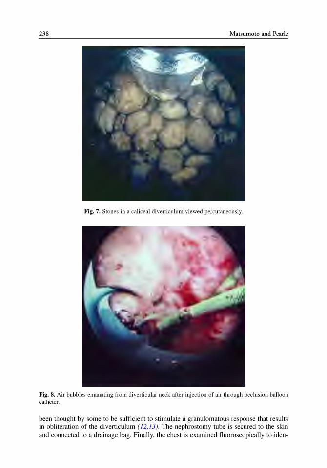
238 Matsumoto and Pearle
Fig. 7. Stones in a caliceal diverticulum viewed percutaneously.
Fig. 8. Air bubbles emanating from diverticular neck after injection of air through occlusion ballooncatheter.
been thought by some to be sufficient to stimulate a granulomatous response that resultsin obliteration of the diverticulum (12,13). The nephrostomy tube is secured to the skinand connected to a drainage bag. Finally, the chest is examined fluoroscopically to iden-
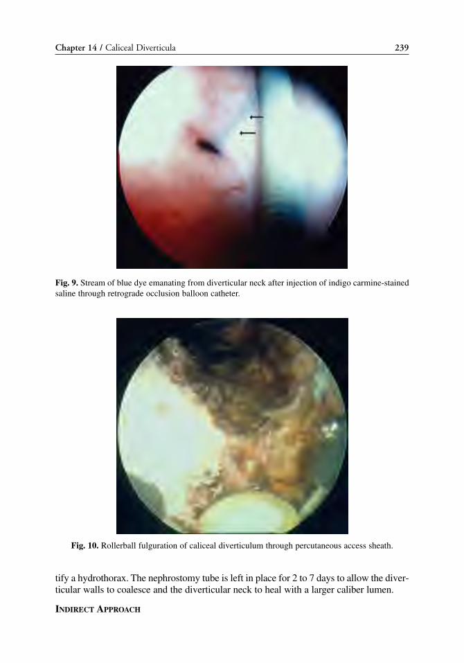
tify a hydrothorax. The nephrostomy tube is left in place for 2 to 7 days to allow the diver-ticular walls to coalesce and the diverticular neck to heal with a larger caliber lumen.
INDIRECT APPROACH
Chapter 14 / Caliceal Diverticula 239
Fig. 9. Stream of blue dye emanating from diverticular neck after injection of indigo carmine-stainedsaline through retrograde occlusion balloon catheter.
Fig. 10. Rollerball fulguration of caliceal diverticulum through percutaneous access sheath.

For the anteriorly located diverticulum, or a diverticulum that lies above the 11th rib,an indirect percutaneous approach is safer than a direct approach, which risks bleedingor injury to the pleura. With an indirect approach, percutaneous access is obtained via aposterior calyx remote from the diverticulum and the diverticulum is accessed indirectlywith a flexible or rigid nephroscope. Preoperative preparation and positioning are iden-tical to the direct percutaneous approach. Based on preoperative contrast studies, a pos-terior calyx likely to provide easy access to the diverticulum is identified and puncturedas described in the direct approach. For a diverticulum lying above the 11th rib, a con-venient posterior lower pole calyx is chosen; for an anterior diverticulum, a posteriorcalyx either in the same polar region or opposite is used. Once percutaneous access isobtained, a rigid or flexible nephroscope is used to inspect the collecting system andidentify the ostium of the diverticulum, similar to a ureteroscopic approach (Fig. 11).A 0.035-in. Bentson guidewire is passed through the ostium and coiled in the divertic-ulum. The nephroscope is removed and an 8-mm, 4-cm high pressure dilating balloonis passed over the guidewire under fluoroscopic guidance until the radio-opaque marksstraddle the diverticular neck, and the infundibulum is dilated until the waist disappears.Alternatively, endoinfundibulotomy can be performed using either a 2- or 3-Fr electro-surgical probe or the Ho:YAG laser as described previously. If an electrosurgical probeis used, a 5-Fr angiographic catheter should be passed over the guidewire to preventcautery transmission and inadvertent urothelial injury.
Once the neck of the diverticulum is dilated/incised, the rigid or flexible nephroscopeis advanced into the diverticulum and the stones are fragmented and/or removed usingultrasonic, pneumatic, electrohydraulic, or laser lithotripsy (Fig. 12). After stone removal,the urothelial lining of the diverticulum is ablated using electrocautery via a 5-FrBugbee electrode (with a flexible nephroscope) or via a resectoscope fitted with arollerball electrode (with a rigid nephroscope). Irrigation pressure should be minimized
240 Matsumoto and Pearle
Fig. 11. Ostium of caliceal diverticulum viewed via indirect percutaneous approach.

while working in the diverticulum to avoid perforation of the diverticulum, particularlywhen the overlying parenchyma is thin.
A large bore nephrostomy tube as previously described is placed into the diverticu-lum and secured to the skin. Extra drainage holes proximal to the balloon are necessaryto provide adequate drainage of the collecting system. Alternatively, an 8 to 12 Fr lock-ing loop nephrostomy tube is positioned with the loop in the diverticulum. Thenephroscopy tube is left in place for 2 to 7 days.
HYDROCALYX
A hydrocalyx associated with a stenotic infundibulum can be approached in a man-ner similar to a calyceal diverticulum. However, because the hydrocalyx is associatedwith secretory epithelium, only the stenotic infundibulum should be addressed and thecalyx should not be fulgurated. It can be difficult to distinguish a hydrocalyx from a cal-iceal diverticula radiographically. However, inspection of a hydrocalyx will reveal arenal papilla whereas inspection of a caliceal diverticular cavity will not.
RESULTS
Outcomes for endourological modalities (ureteroscopy and percutaneous approaches)for the treatment of caliceal diverticula are reviewed in this section. However, for thepurposes of comparison, a brief review of outcomes for SWL and laparoscopicapproaches are included as well in order to guide treatment selection.
Shockwave Lithotripsy
Chapter 14 / Caliceal Diverticula 241
Fig. 12. Basket extraction of a stone from a caliceal diverticulum approached indirectly.

242 Matsumoto and Pearle
SWL has been used for treatment of stones in caliceal diverticula with limited suc-cess. The mean stone-free rate for SWL from published series is only 21% (range 13 to58%); however, relief of symptoms surprisingly has been achieved in 68% of patients(range 56 to 86%) (14–19). In the most successful series reported, Streem and Yost (19)selected patients with caliceal diverticula associated with short, radiographically patentdiverticular necks and stones less than 15 mm in diameter. A stone-free state wasachieved in 58% of 19 stone-bearing diverticula; furthermore, in 86% of 14 sympto-matic patients, treatment resulted in resolution of symptoms postoperatively.
In general, SWL is indicated only for patients with small stones in caliceal divertic-ula with a short, radiographically patient diverticular neck. Endoscopic approaches,both ureteroscopic and percutaneous, allow for treatment of both the stone and divertic-ulum, and as such, are preferred treatment over SWL.
Ureteroscopic Approach
In 1989, Fuchs and David (8) ureteroscopically treated 15 patients with stone-bear-ing caliceal diverticula, additionally applying simultaneous SWL to fragment the stonesafter dilating or incising diverticular neck. A stone-free state was achieved in 73% ofpatients and 87% of patients were rendered symptom free. Only one patient (7%)required salvage percutaneous nephrolithotomy. Although in most cases, the diverticulawas reduced in size, only one patient demonstrated complete obliteration of the diver-ticulum on follow-up imaging studies.
Expanding their earlier series (previous paragraph), Pang and associates reported on36 patients with symptomatic stone-bearing caliceal diverticula in whom ureteroscopicmanagement was initially attempted (8,20). In four patients with lower pole calicealdiverticula, percutaneous treatment was ultimately required. Among the remaining 32patients, 75% were rendered stone free with combined SWL/ureteroscopy and 87.5% ofpatients were rendered free of symptoms. Although four patients with residual stoneshad resolution of their symptoms at early follow-up, two subsequently became sympto-matic and were successfully retreated ureteroscopically.
Grasso and associates (21) treated four patients with five caliceal diverticula via anendoscopic approach. Although ureteroscopy was attempted in all patients, a com-pletely ureteroscopic approach was utilized in only two cases: the remaining three casesrequired a combined retrograde ureteroscopic/percutaneous approach. Only one of thetwo patients treated completely ureteroscopically was rendered stone free. In contrast,the three patients treated with a combined approach were successfully rendered free ofboth stones and symptoms.
Batter and Dretler (22) treated 26 patients with caliceal diverticula, with or withoutstones, via a ureteroscopic approach, including 18 patients who failed previous SWLtreatment. The diverticula were successfully entered in 18 cases (69%) and successfullycleared of stones in 83% of the 18 cases. The three patients with residual stones requiredfurther SWL treatment. Among the patients in whom the diverticula were successfullyentered and the stones cleared, all were symptom free at a mean follow-up of45 months. Complications included pain, sepsis, and bleeding, although no patientrequired a blood transfusion.
In the largest series to date, Chong and co-workers (23) treated 96 caliceal diverticula,including 10 lower pole diverticula, using a ureteroscopic approach. The diverticular neckwas dilated with a balloon or incised with a Bugbee electrode or Ho:YAG laser; a combi-nation of Ho:YAG or electrohydraulic lithotripsy was used to fragment the stones. The

Chapter 14 / Caliceal Diverticula 243
diverticular neck was successfully identified in 95% of cases, with the four failures occur-ring in lower pole diverticula. An immediate stone-free rate of 90% was achieved and, at8-year follow-up, only 8% of patients had recurrence of symptoms or stones.
Auge and co-workers (24) reviewed their series of 17 patients with symptomaticstone-bearing caliceal diverticula treated ureteroscopically. In four patients, the ostiumof the diverticulum was unable to be identified, necessitating a percutaneous approach.Three other patients initially treated with ureteroscopy later underwent a percutaneousprocedure because of retained stones and inadequate symptom relief. Among tenpatients managed completely ureteroscopically, three patients were rendered stone freeand six were symptom free. No complications were reported.
Overall, the retrograde ureteroscopic approach for treatment of caliceal diverticulahas been successful in achieving a stone-free state in 78% of patients and a symptom-free state 79% of patients (Table 1). However, in 11% of cases, a secondary procedurewas necessary. The complication rate was 9%, although most complications wereminor, such as bleeding not requiring transfusion and infection. Caliceal diverticulaassociated with upper or middle calyces were more successfully accessed ureteroscop-ically (96% of 79 cases) than diverticula associated with lower calyces, which were suc-cessfully entered in only 23% of 13 of cases.
Percutaneous Approach
Hulbert and associates (25) used direct (n = 15) and indirect (n = 3) percutaneousaccess to manage 18 stone-bearing caliceal diverticula in one of the earliest publishedseries on percutaneous treatment of caliceal diverticula. No attempt was made to fulgu-rate the diverticular cavity as the authors felt that the trauma to the wall of the divertic-ulum caused by the dilation process was sufficient to promote obliteration. All patientswere rendered stone free and no complications were reported. Among the 15 patientsavailable for follow-up, the diverticulum was successfully obliterated in 12 patients (alldirect access); in the 3 patients who underwent an indirect approach, the diverticulapersisted on follow-up imaging.
Eshghi and colleagues (13) also reported on 14 patients with caliceal diverticula/hydrocalyces managed percutaneously. A direct approach was used to access the divertic-ula/hydrocalyx in 11 cases and an indirect approach in the remaining 3. The diverticularneck was managed in one of three ways: radial cold knife incision at one or two sitesunder direct vision (n = 8); dilatation to 16 to 18 Fr with fascial dilators or a dilating bal-loon (n = 4) or by direct-vision dissection of the infundibulum using two-prong graspingforceps (n = 2). No attempt was made to fulgurate the diverticulum. A Malecot drain wasleft in the diverticulum from 3 to 14 days. At 12-months follow-up, all 14 patients werestone free and 85.7% of patients demonstrated a decrease in the size of thediverticulum/hydrocalyx. However, in no case was the diverticulum completely obliter-ated.
Hedelin and colleagues (26) percutaneously treated 13 patients with caliceal diver-ticula percutaneously (direct approach in seven cases, indirect in six cases) and reporteda 69% stone-free and symptom-free rate despite complete obliteration of the diverticu-lum in only one case. No major complications occurred, and a single patient developeda new 1-mm stone in the residual diverticulum during follow-up.
Ellis and co-workers (27) reviewed their series of 12 patients with caliceal divertic-ula, among whom 10 patients had stones and 2 had associated infections. Among the 10patients with stones, 9 were treated with direct percutaneous puncture and dilation of

244
Tab
le 1
Ret
rogr
ade
Ure
tero
scop
ic M
anag
emen
t of
Cal
icea
l Div
erti
culu
m
Man
agem
ent
Succ
essf
ul
Obl
iter
atio
nA
djun
ctof
div
erti
cula
ren
try
into
St
one
Sym
ptom
ofFo
llow
-up
Stud
yN
Loc
atio
npr
oced
ures
neck
dive
rtic
ulum
free
free
dive
rtic
ulum
Com
plic
atio
ns(m
o)
Fuch
s 15
UP
6 SW
LD
ilatio
n10
0%
73%
87
%
7%
—et
al.
(8)
MP
7 (1
5/15
)(1
1/15
)(1
3/15
)(1
/15)
LP
2
Pang
32
UP/
MP
SWL
——
75%
87
.5%
—
——
et a
l. (2
0)32
(24/
32)
(28/
32)
Gra
sso
2U
P1
—
Dila
tion
or10
0%
50%
0%
et a
l. (2
1)M
P1
Ho:
YA
G in
cisi
on(2
/2)
(1/2
)
Bat
ter
26U
P15
—
Dila
tion
or
69%
83
%
100%
15
%
45et
al.
(22)
MP
4 el
ectr
oinc
isio
n(1
8/26
)(1
5/18
)(1
8/18
)(4
/26)
(15-
84)
LP
7
Cho
ng
96N
RSW
LD
ilatio
n or
96
%
90%
—
——
et a
l. (2
3)in
cisi
on(9
2/96
)(8
6/96
)
Aug
e 17
UP1
2 —
Dila
tion
or
76%
18
%
35%
18
%
0%
1.4
et a
l. (2
4)M
P2
Ho:
YA
G in
cisi
on(1
3/17
)(3
/17)
(6/1
7)(3
/17)
(0/1
7)(1
-2)
LP
3
Ove
rall
191
—
——
88%
78
%
79%
13
%
9%
—(1
40/1
59)
(142
/184
)(6
5/82
)(4
/32)
(4/4
3)
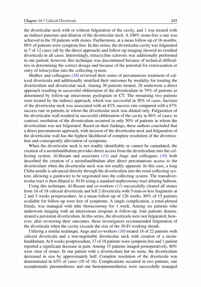
Chapter 14 / Caliceal Diverticula 245
the diverticular neck with or without fulguration of the cavity, and 1 was treated withan indirect puncture and dilation of the diverticular neck. A 100% stone-free a rate wasachieved in the 10 patients with stones. Furthermore, at a mean follow-up of 16 months,88% of patients were symptom free. In this series, the diverticular cavity was fulguratedin 7 of 12 cases (all by the direct approach) and follow-up imaging showed no residualdiverticula in all cases. Interestingly, tetracycline sclerosis was additionally performedin one patient; however, this technique was discontinued because of technical difficul-ties in determining the correct dosage and because of the potential for extravasation orentry of tetracycline into the collecting system.
Shalhav and colleagues (28) reviewed their series of percutaneous treatment of cal-iceal diverticula and additionally stratified their outcomes by modality for treating thediverticulum and diverticular neck. Among 30 patients treated, 28 underwent a directapproach resulting in successful obliteration of the diverticulum in 79% of patients asdetermined by follow-up intravenous pyelogram or CT. The remaining two patientswere treated by the indirect approach, which was successful in 50% of cases. Incisionof the diverticular neck was associated with an 83% success rate compared with a 67%success rate in patients in whom the diverticular neck was dilated only. Fulguration ofthe diverticular wall resulted in successful obliteration of the cavity in 86% of cases; incontrast, resolution of the diverticulum occurred in only 50% of patients in whom thediverticulum was not fulgurated. Based on their findings, these authors concluded thata direct percutaneous approach, with incision of the diverticular neck and fulguration ofthe diverticular wall has the highest likelihood of complete resolution of the diverticu-lum and consequently alleviation of symptoms.
When the diverticular neck is not readily identifiable or cannot be cannulated, thecreation of a neoinfundibulum provides direct access from the diverticulum into the col-lecting system. Al-Basam and associates (11) and Auge and colleagues (10) bothdescribed the creation of a neoinfundibulum after direct percutaneous access to thediverticulum when the diverticular neck was not readily apparent. In this case, a longChiba needle is advanced directly through the diverticulum into the renal collecting sys-tem, allowing a guidewire to be negotiated into the collecting system. The transdiver-ticular tract is then dilated to 30-Fr using a standard nephrostomy tract dilating balloon.
Using this technique, Al-Basam and co-workers (11) successfully cleared all stonesfrom 16 of 18 caliceal diverticula and left 2 diverticula with 5-mm-or-less fragments at2 and 5 weeks postprocedure. At a mean follow-up of 126 weeks, 80% of 15 patientsavailable for follow-up were free of symptoms. A single complication, a renal-pleuralfistula, was managed with tube thoracostomy for 1 week. Among six patients whounderwent imaging with an intravenous urogram at follow-up, four patients demon-strated a persistent diverticulum. In this series, the diverticula were not fulgurated; how-ever, after reviewing their outcomes, these investigators recommended fulguration ofthe diverticula when the cavity exceeds the size of the 30-Fr working sheath.
Utilizing a similar technique, Auge and co-workers (10) treated 18 of 22 patients withcaliceal diverticula and a non-negotiable diverticular neck with creation of a neoin-fundibulum. At 6 weeks postprocedure, 17 of 18 patients were symptom free and 1 patientreported a significant decrease in pain. Among 15 patients imaged postoperatively, 80%were clear of stones. In one patient with a diverticulum but no stone, the diverticulumdecreased in size by approximately half. Complete resolution of the diverticula wasdemonstrated in 63% of cases (10 of 16). Complications occurred in two patients; oneasymptomatic pneumothorax and one hemopneumothorax were successfully managed

Tab
le 2
Perc
utan
eous
Nep
hros
tolit
hoto
my
App
roac
h of
Cal
icea
l Div
erti
culu
m
Ful
gura
tion
Man
agem
ent
Obl
iter
atio
nof
of d
iver
ticul
arSy
mpt
omof
Foll
ow-u
pSt
udy
NA
ppro
ach
dive
rtic
ulum
neck
Ston
e fr
eefr
eedi
vert
icul
umC
ompl
icat
ions
(mon
ths)
Esh
gi e
t al.
(13)
14D
irec
t 11
No
abla
tion
Inci
sion
8
100%
(14
/14)
—0%
(0/
14)
0% (
0/14
)12
Indi
rect
3D
ilata
tion
4 D
isse
ctio
n 2
Hul
bert
et a
l. (2
5)18
Dir
ect 1
5 A
blat
ion
1D
ilata
tion
1880
% (
12/1
5)—
80%
(12
/15)
6% (
1/18
)4–
14In
dire
ct 3
Hed
elin
et a
l. (2
6)13
Dir
ect 7
N
o ab
latio
nD
ilata
tion
869
% (
9/13
)69
% (
9/13
)8%
(1/
13)
0% (
0/13
)24
(6–
48)
Indi
rect
6E
llis
et a
l. (2
7)12
Dir
ect 1
1 A
blat
ion
7 D
ilata
tion
910
0% (
10/1
0)88
% (
7/8)
75%
(6/
8)33
% (
4/12
)33
% (
4/12
)In
dire
ct 1
Scle
rosi
s 1
Jone
s et
al.
(17)
24D
irec
t 22a
Abl
atio
n 17
Dila
tatio
n 22
96%
(23
/24)
100%
(24
/24)
10
0% (
17/1
7)29
% (
7/24
)16
(4–
72)
Indi
rect
2L
ang
et a
l. (3
4)10
Dir
ect 1
0N
o ab
latio
nN
eoin
fund
i-10
0% (
10/1
0)—
20%
(2/
10)
10%
(1/
10)
35 (
6–60
)bu
lum
10
Hen
drik
x et
al.
(18)
13D
irec
t 12
A
blat
ion
3D
ilata
tion
977
%(1
0/13
) 77
% (
10/1
3)—
31%
(4/
13)
24–8
4In
dire
ct 1
Bel
lman
et a
l. (3
5)20
Dir
ect 2
0A
blat
ion
20D
ilata
tion
2095
% (
18/1
9)10
0% (
20/2
0)
80%
(16
/20)
25%
(5/
20)
18 (
3–36
)Sh
alha
v et
al.
(28)
30D
irec
t 28
Abl
atio
n 22
bIn
cisi
on 1
2 93
% (
21/2
3)85
% (
23/2
7)76
% (
16/2
1)20
% (
6/30
)6
(3–3
6)In
dire
ct 2
Dila
tatio
n 9
Don
nella
n et
al.
(36)
21D
irec
t 20
No
abla
tion
Inci
sion
7
95%
(20
/21)
86%
(18
/21)
29%
(6/
20)
10%
(2/
21)
42 (
12–8
8)In
dire
ct 1
Dila
tatio
n 13
Mon
ga e
t al.
(37)
14D
irec
t 14
Abl
atio
n 14
Inci
sion
10
0% (
11/1
1)10
0% (
14/1
4)10
0% (
14/1
4)7%
(1/
14)
74 (
6–14
4)A
l-B
asam
et a
l. (1
1)18
Dir
ect 1
8N
o ab
latio
nN
eoin
fund
i-89
% (
16/1
8)80
% (
12/1
5)33
% (
2/6)
6% (
1/18
)38
(8–
74)
bulu
m 1
8A
uge
et a
l. (1
0)18
Dir
ect 1
8N
o ab
latio
nN
eoin
fund
i-80
% (
12/1
5)94
% (
17/1
8)64
% (
10/1
6)11
% (
2/18
)32
(3–
82)
bulu
m 1
8L
andr
y et
al.
(38)
31D
irec
t 31
Abl
atio
n 24
Inci
sion
31
84%
(26
/31)
88%
(27
/31)
64%
(20
/31)
13
% (
4/31
)(1
.5–3
)O
vera
ll25
6—
——
89%
(21
2/23
7)89
% (
181/
204)
60%
(12
2/20
5)15
% (
38/2
56)
24.6
(18
–96)
a Inc
lude
s 10
pat
ient
s E
SWL
+PC
NL
.b o
nly
incl
udes
pat
ient
s w
ith f
ollo
w-u
p.
246
Chapter 14 / Caliceal Diverticula 247
with tube thoracostomy. The average hospital stay was 2.8 days, and all patients were dis-charged home with a 22-Fr nephrostomy tube that was removed 1 week later.
In published series, overall stone-free and symptom-free rates of 89 and 89%, respec-tively, have been achieved (Table 2). Furthermore, in 60% of cases, the diverticula weresuccessfully obliterated. For caliceal diverticula approached directly the overall successrate (obliteration of diverticulum) was 63% (88/139) vs indirectly which was 9% (2/22).Complication rates are slightly higher with a percutaneous approach (15%) comparedwith a retrograde ureteroscopic approach (9%) (Tables 1 and 2).
Laparoscopic Approach
A total of 13 cases of laparoscopic unroofing of caliceal diverticula have beenreported in the literature, including a transperitoneal approach in 1 case and a retroperitonealapproach in 12 cases (29–33). Operative times ranged from 60 to 215 minutes, and theoverall complication rate was 7.7%, with the only reported complication consisting ofbleeding requiring transfusion. Radiographic studies demonstrated obliteration of thediverticula in all 13 cases and all patients were rendered symptom free at a mean follow-up of 6 months. The laparoscopic approach is generally reserved for large diverticula (>5cm) or anteriorly located diverticula with thin overlying parenchyma.
TIPS AND TRICKS
Ureteroscopic Approach
• Contrast injected through the ureteroscope helps to identify the calyx with which thediverticulum is associated.
• Passage of a guidewire into the diverticula confirms proper identification of the ostiumand opens up the diverticular neck sufficiently to position a laser fiber for incision.
• Dilation of the diverticular neck after incision separates the incised edges and facilitatesclearance of stones fragments from the diverticulum.
Percutaneous Approach
• After percutaneous puncture, a moveable core guidewire or J-wire coiled within thediverticulum provides moderately secure guidewire access over which a dilating bal-loon can be passed.
• Injection of indigo carmine-stained saline or air via the occlusion balloon catheter facil-itates identification of the diverticular neck, which can then be cannulated with aguidewire.
• Fulguration of the diverticulum prior to dilation or incision of the diverticular neck pre-vents inadvertent fulguration of the collecting system urothelium.
• The transdiverticular approach is an alternative method of treating the diverticulum thatprovides secure access into the collecting system.
CONCLUSIONS
Therapeutic options for treatment of symptomatic caliceal diverticula include SWL,ureteroscopy, percutaneous nephrostolithotomy/ablation, and laparoscopic unroofing.SWL is associated with the lowest stone-free rates, although in a surprising number ofpatients symptoms resolve despite persistence of the diverticulum and stone fragments.
248 Matsumoto and Pearle
SWL therapy is best reserved for patients with a small stone burden (≤15 mm) in a mid-or upper pole caliceal diverticulum with a radiographically patient diverticular neck.
Retrograde ureteroscopy should be considered for upper- and midcaliceal diverticulaassociated with a stone burden of 15 mm or less, when the infundibulopelvic angle isfavorable for ureteroscopic access (7). Likewise, anteriorly located diverticula with amodest stone burden are effectively accessed via a ureteroscopic approach, which issafer than a percutaneous approach and less invasive than a laparoscopic approach.Although the diverticulum is rarely successfully obliterated, high stone-free rates can beachieved and improved drainage of the diverticulum attained.
A percutaneous approach offers the best chance of achieving a stone-free andsymptom-free state and is the only endoscopic option in which the diverticulum can bedirectly treated. A direct percutaneous approach is preferable to the indirect approach, andif direct access cannot be safely performed, a ureteroscopic approach should be consid-ered if the diverticulum and/or stone burden is small. An indirect approach is still favoredover ureteroscopy for a large stone burden or a large diverticulum (>2 cm), particularly ifa rigid nephroscope can be used to access the diverticulum and fulgurate the urothelium.
Management of the diverticular neck is controversial. Whether it is necessary orpreferable to dilate or incise the neck vs simply fulgurate the diverticulum has not beenfirmly established. Likewise, if treatment of the diverticular neck proves superior tosimple fulguration, the advantage of one modality over another (incision vs dilation)will remain to be proven. Until further information is available, it is advisable to dilateor incise the diverticular neck if the ostium can be identified after fulguration of thediverticulum. If the diverticular neck cannot be identified, fulguration of the urotheliallining may be sufficient.
For large diverticula (>5 cm) or for anteriorly located diverticula with thin overlyingparenchyma and large stones, laparoscopic unroofing offers the most expeditious treat-ment, resulting in successful stone removal and resolution of the diverticula.
REFERENCES1. Wulfsohn MA. Pyelocaliceal diverticula. J Urol 1980; 123: 1–8.2. Middleton AW Jr., Pfister RC. Stone-containing pyelocaliceal diverticulum: embryogenic, anatomic,
radiologic and clinical characteristics. J Urol 1974; 111: 2–6.3. Timmons JW Jr., Malek RS, Hattery RR, Deweerd JH. Caliceal diverticulum. J Urol 1975; 114: 6–9.4. Williams G, Blandy JP, Tresidder GC. Communicating cysts and diverticula of the renal pelvis. Br J
Urol 1969; 41: 163–170.5. Yow RM, Bunts RC. Calyceal diverticulum. J Urol 1955; 73: 663–670.6. Devine CJ Jr., Guzman JA, Devine PC, Poutasse EF. Calyceal diverticulum. J Urol 1969; 101: 8–11.7. Chong TW, Bui MH, Fuchs GJ. Calyceal diverticula. Ureteroscopic management. Urol Clin North
Am 2000; 27: 647–654.8. Fuchs GJ, David RD. Flexible ureterorenoscopy, dilatation of narrow caliceal neck and ESWL: A new
minimally invasive approach to stones in caliceal diverticulua. J Endourol 1989; 3: 255–263.9. Bennett JD, Brown TC, Kozak RI, Sales J, Denstedt JD. Transdiverticular percutaneous nephrostomy
for caliceal diverticular stenosis. J Urol 1992; 6: 55–57.10. Auge BK, Munver R, Kourambas J, Newman GE, Wu NZ, Preminger GM. Neoinfundibulotomy for
the management of symptomatic caliceal diverticula. J Urol 2002; 167: 1616–1620.11. Al-Basam S, Bennett JD, Layton ZA, Denstedt JD, Razvi H. Treatment of caliceal diverticular stones:
transdiverticular percutaneous nephrolithotomy with creation of a neoinfundibulum. J Vasc IntervRadiol 2000; 11: 885–889.
12. Hulbert JC, Reddy PK, Hunter DW, Castaneda-Zuniga W, Amplatz K, Lange PH. Percutaneous tech-niques for the management of caliceal diverticula containing calculi. J Urol 1986; 135: 225–227.

13. Eshghi M, Tuong W, Fernandez F, Addonizio JC. Percutaneous (Endo) Infundibulotomy. J Endourol1987; 1: (2)107–113.
14. Wilbert DM, Jenny E, Stoeckle M, Ridemiller H, Jacobi G. Calyceal diverticar stones: is ESWLworthwhile? J Urol 1986; 135: 183A
15. Psihramis KE, Dretler SP. Extracorporeal shock wave lithotripsy of caliceal diverticula calculi. J Urol1987; 138: 707–711.
16. Ritchie AW, Parr NJ, Moussa SA, Tolley DA. Lithotripsy for calculi in caliceal diverticula? Br J Urol1990; 66: 6–8.
17. Jones JA, Lingeman JE, Steidle CP. The roles of extracorporeal shock wave lithotripsy and percuta-neous nephrostolithotomy in the management of pyelocaliceal diverticula. J Urol 1991; 146: 724–727.
18. Hendrikx AJ, Bierkens AF, Bos R, Oosterhof GO, Debruyne FM. Treatment of stones in calicealdiverticula: extracorporeal shock wave lithotripsy versus percutaneous nephrolitholapaxy. Br J Urol1992; 70: 478–482.
19. Streem SB, Yost A. Treatment of caliceal diverticular calculi with extracorporeal shock wavelithotripsy: patient selection and extended followup. J Urol 1992; 148: 1043–1046.
20. Pang K, David RD, Fuchs GJ. Treatment of stones in caliceal diverticuli using retrograde endoscopicapproach: Critical assessment after 2 years. J Endourol 1992; 6(suppl): S80.
21. Grasso M, Lang G, Loisides P, Bagley D, Taylor F. Endoscopic management of the symptomatic cal-iceal diverticular calculus. J Urol 1995; 153: 1878–1881.
22. Batter SJ, Dretler SP. Ureterorenoscopic approach to the symptomatic caliceal diverticulum. J Urol1997; 158: 709–713.
23. Chong TW, Bui MH, Fuchs GJ. Calyceal diverticula. Ureteroscopic management. Urol Clin NorthAm 2000; 27: 647–654.
24. Auge BK, Munver R, Kourambas J, Newman GE, Preminger GM. Endoscopic management of symp-tomatic caliceal diverticula: a retrospective comparison of percutaneous nephrolithotripsy andureteroscopy. J Endourol 2002; 16: 557–563.
25. Hulbert JC, Hernandez-Graulau JM, Hunter DW, Castaneda-Zuniga W. Current concepts in the man-agement of pyelocaliceal diverticula. J Endourol 1988; 2: 11–17.
26. Hedelin H, Geterud K, Grenabo L, Henriksson C, Pettersson S, Zachrisson BF. Percutaneous surgeryfor stones in pyelocaliceal diverticula. Br J Urol 1988; 62: 206–208.
27. Ellis JH, Patterson SK, Sonda LP, Platt JF, Sheffner SE, Woolsey EJ. Stones and infection in renalcaliceal diverticula: treatment with percutaneous procedures. AJR Am J Roentgenol 1991; 156:995–1000.
28. Shalhav AL, Soble JJ, Nakada SY, Wolf JS Jr, McClennan BL, Clayman RV. Long-term outcome ofcaliceal diverticula following percutaneous endosurgical management. J Urol 1998; 160: 1635–1639.
29. Ruckle HC, Segura JW. Laparoscopic treatment of a stone-filled, caliceal diverticulum: a definitive,minimally invasive therapeutic option. J Urol 1994; 151: 122–124.
30. Hoznek A, Herard A, Ogiez N, Amsellem D, Chopin DK, Abbou CC. Symptomatic caliceal divertic-ula treated with extraperitoneal laparoscopic marsupialization fulguration and gelatin resorcinolformaldehyde glue obliteration. J Urol 1998; 160: 352–355.
31. Harewood LM, Agarwal D, Lindsay S, Vaughan MG, Cleeve LK, Webb DR. Extraperitoneal laparo-scopic caliceal diverticulectomy. J Endourol 1996; 10: 425–430.
32. Curran MJ, Little AF, Bouyounes B, Nieh PT, Bihrle W 3rd. Retroperitoneoscopic technique for treat-ing symptomatic caliceal diverticula. J Endourol 1999; 13: 723–725.
33. Miller SD, Ng CS, Streem SB, Gill IS. Laparoscopic management of caliceal diverticular calculi. J Urol 2002; 167: 1248–1252.
34. Lang EK. Percutaneous infundibuloplasty: management of calyceal diverticula and infundibularstenosis. Radiology 1991; 181: 871–877.
35. Bellman GC, Silverstein JI, Blickensderfer S, Smith AD. Technique and follow-up of percutaneousmanagement of caliceal diverticula. Urology 1993; 42: 21–25.
36. Donnellan SM, Harewood LM, Webb DR. Percutaneous management of caliceal diverticular calculi:technique and outcome. J Endourol 1999; 13: 83-88.
37. Monga M, Smith R, Ferral H, Thomas R. Percutaneous ablation of caliceal diverticulum: long-termfollowup. J Urol 2000; 163: 28–32.
Chapter 14 / Caliceal Diverticula 249

IV ABLATIVE THERAPY

SUMMARY
The “gold standard” for treatment of upper tract urothelial carcinoma is nephrourterec-tomy. In some cases, however, organ-sparing endoscopic therapy via percutaneous orureteroscopic approaches should be considered. The percutaneous approach is generallyused for cases of larger volume renal disease and those not accessible by ureteroscopictechniques. This chapter will review the indications, technique, complications, and resultsof this technique.
Key Words: TCC; percutaneous; management.
NATURAL HISTORY OF UPPER URINARY TRACTTRANSITIONAL CELL CARCINOMAS AND INDICATIONS
FOR CONSERVATIVE THERAPY
Upper urinary tract (UUT) urothelial tumors are rare accounting for 1 to 2% of allgenitourinary tumors (1). The vast majority are transitional cell carcinomas (TCC)(90%), whereas only 10% are squamous cell carcinomas and 1% adenocarcinomas (2).
From: Advanced Endourology: The Complete Clinical GuideEdited by: S. Y. Nakada and M. S. Pearle © Humana Press Inc., Totowa, NJ
253
Percutaneous Approach to Upper Urinary Tract Tumors
Ioannis M. Varkarakis MD, PhD
and Thomas W. Jarrett, MD
CONTENTS
NATURAL HISTORY OF UPPER URINARY TRACT TRANSITIONAL CELL
CARCINOMAS AND INDICATIONS FOR CONSERVATIVE THERAPY
INSTRUMENTATION
TECHNIQUE
RESULTS
TIPS AND TRICKS
CONCLUSION
REFERENCES
15

Urothelial tumors of the renal pelvis are three to four times more frequent than thoselocated in the ureter (3).
The incidence of UUT TCC increases with age in both genders and appears most fre-quently during the sixth and seventh decades of life. Males present with this diseasethree times more frequently than women (4).
Like that of the bladder, upper TCC most likely represents a field change disease withmultiple recurrences in both time and space. This polychronotopism is generally confinedto the ipsilateral renal unit or to the bladder. Although recurrence at an additional site in thegenitourinary system will occur in 30 to 50% of patients (5), recurrence in the controlateralrenal unit will develop only in 1 to 5.8% (6,7). This natural history makes nephroureterec-tomy with resection of a bladder cuff safe and effective for therapy of an UUT TCC. Ratesof local or ipsilateral recurrence after nephron preserving surgery are high and thereforeconservative management with renal sparing procedures has been implemented only whenpreservation of renal function is necessary. This includes patients with an anatomically orfunctionally solitary kidney, those with bilateral disease, and patients who refuse or areunable to tolerate open surgery because of medical comorbidities.
Recent advances in endoscopic technology with the development of better optics,actively deflecting telescopes, and adjunctive instrumentation made it possible for usto diagnose and stage more accurately patients with upper urinary TCC. In fact directvisualization of the tumor allows obtaining a tumor biopsy and selective urine cytol-ogy. Tumor grading in this setting is very accurate and is 90% in agreement with thegrade of the final pathological specimen. Unfortunately ureteroscopic biopsy is unre-liable in determining stage (8). However several studies have suggested a good corre-lation between the grade of TCC and the stage of the tumor (9,10) and high accuracyof the Computed tomography (CT) scan in detecting evidence of tumor extendingbeyond the wall of the ureter or renal pelvis (11). Therefore the combination of lowgrade on biopsy and absence of frank extension outside of the urinary tract by CT scanstrongly suggests the disease is superficial (12,13). These criteria create a new subsetof patients with small and low-grade tumors that can potentially be managed by endo-scopic management only, even in the presence of a healthy controlateral kidney (14).Although the cancer-related risks are greater for any treatment short of the gold stan-dard nephroureterectomy, some patients are better treated by parenchymal sparing sur-gery, provided they know the risks and are committed to vigilant follow-up.
Both ureteroscopic and percutaneous tumor resection are possible and are used inselected centers for up to 15% of patients with upper urinary TCC (15).
The percutaneous access is preferred for larger tumors located proximally in the renalpelvis and or upper ureter. The main advantage of the percutaneous approach is the abil-ity to remove a larger tumor volume from any portion of the collecting system owing tothe use of instruments with larger working channels, which allows better visualizationand faster resection. Deeper biopsies can be obtained when compared to those takenwith ureteroscopy, whereas the percutaneous approach may avoid the limitationsencountered even by flexible ureteroscopy, especially in complicated caliceal systemsor areas difficult to access, such as the lower pole calyx or the UUT of patients with uri-nary diversion. Access to any renal unit is possible irrespective of any prior operativeintervention such as urinary diversion. With a percutaneous approach, the establishednephrostomy tract can be maintained for immediate postoperative nephroscopy andadministration of topical adjuvant therapy.
The main disadvantage with the antegrade access is the increased morbidity comparedto ureteroscopy. Nephrostomy tube placement has inherent risks and therefore requires
254 Varkarakis and Jarrett

inpatient admission. In addition, loss of urothelial integrity and exposure of nonurothelialsurfaces to tumor cells carries the risk for tumor seeding along the nephrostomy tract (16).
INSTRUMENTATION
Imaging Guidance
• C-arm configuration fluoroscopy equipment (preferred) or• Real-time diagnostic ultrasonography or• CT fluoroscopy
Access Equipment
• 18 or 21-gage needle for puncture of the collecting system• Guidewires: 0.018-, 0.035-, 0.038-in.; stiff or soft bodied; straight or angled tipped; soft
or hard tipped; Teflon or hydrophilic coated• Conversion catheter• Fascia incising needle
Dilatation and Maintenance of the Nephrostomy Tract
• Progressive fascial dilators• Amplatz renal dilator sets• Metal coaxial dilators• High-pressure balloon systems
Tumor Resection/Ablation
• Working Amplatz or plastic sheath • Rigid or flexible nephroscope• Resectoscope• Ho:YAG or Nd:YAG laser• Foley-type urinary drainage catheters• Self-locking pigtail nephrostomy catheters
TECHNIQUE
Patient Preparation
Preoperative evaluation should include a coagulation profile with negative urine cul-tures. We favor a single stage approach for access, tract dilatation, and tumor resection.
Patient Positioning and Stent Placement
Anesthesia induction is usually performed in the operating room but on the patient’spreoperative stretcher. After the patient is intubated and ready from an anesthesia endpoint the patient is flipped into the prone position on the operative table. Use of two gel-foam rolls placed under the patient allows the flank area of interest to be presented in abetter manner. After patient positioning is complete, cystoscopy is performed with thepatient in a prone position and an open-ended ureteral catheter is placed in the renalpelvis. The use of the flexible cystoscope allows the surgeon to evaluate the bladder andcatheterize the ureter even after the patient has been moved in the prone position.Alternatively anesthesia induction on the operative table and stent placement with thepatient in the supine position can be performed but we have found that changingposition is less cumbersome when the patient is simply rolled off the preoperative
Chapter 15 / Percutaneous Management of UUT TCC 255

stretcher. The ureteral stent is available for retrograde injection at all times during theoperation. Finally the patient is prepped and draped in the standard sterile fashion.
Establishment of the Nephrostomy Tract
Contrast medium is injected through the ureteral catheter to define the caliceal anatomyand tumor location. A percutaneous nephrostomy tract is established through the desiredcalyx. In some cases additional percutaneous accesses are required to resect all tumor com-pletely. Access under the 12th rib is usually preferred. Supracostal approaches may be usedif necessary but at the risk of pleural injury. Tumors in peripheral calyces are bestapproached with direct puncture distal to the tumor (Fig. 1). Disease in the renal pelvis andupper ureter is best approached through an upper or middle pole (Fig. 1) access to allowscope maneuvering through the collecting system and down the ureteropelvic junction.After a needle is passed through the desirable calyx and a guidewire is manipulated prefer-ably down the ureter, the tract is then dilated using either sequential (Amplatz) or balloondilatation so as to accommodate a 30-Fr sheath. Access to the desired calyx and correctpositioning of the nephrostomy tract is crucial to the success of the procedure and should bedone by the urologist or by the radiologist after direct consultation with the operating surgeon.
After 30-Fr is reached, the nephroscope is inserted and the ureteral catheter isgrasped, brought out of the tract, and exchanged for a stiff guidewire, thus providing
256 Varkarakis and Jarrett
Fig. 1. Nephrostomy tract puncture site. Position of the nephrostomy is important for successful per-cutaneous resection of transitonal cell carcinomas of the renal collecting system and upper ureter.Tumors located in peripheral calyces (A–C) are best approached by direct puncture. Tumors locatedin the renal pelvis and upper ureter (D,E) are best approached by puncture to an upper or middlecalyx. (Reprinted from ref. 17 with permission from Elsevier.)

both antegrade and retrograde control. This is the second safety guidewire and helpsmaintain access should the original wire be inadvertently removed.
Biopsy and Definitive Therapy
Through the 30-Fr inner diameter nephrostomy sheath, which is used to maintain alow-pressure system, the collecting system is evaluated thoroughly using rigid andflexible endoscopes when necessary (Fig. 2). Any suspicion of upper ureteral involvement
Chapter 15 / Percutaneous Management of UUT TCC 257
Fig. 2. Technique for percutaneous removal of transitional cell carcinomas of the renal collecting sys-tem. (A) The tumor is removed to its base with the big biopsy forceps and its base after being biop-sied in fulgurated. (B) Alternatively the tumor may be resected using the standard resectoscope. (C)For tumors smaller in size laser ablation can be used. (D) Laser fibers through flexible scopes can beused to reach tumors located even in the most difficult of positions. (Reprinted from ref. 17, withpermission from Elsevier.)

warrants antegrade ureteroscopy. After identification, cold-cup biopsies of the tumor (ifnot already sampled) and surrounding mucosa are performed to evaluate the extent ofthe disease and to rule out the possibility of carcinoma in situ. If the tumor is smallenough the cold-cup biopsy forceps can be used also for complete tumor removal. Insuch a case, the bulk of the tumor is grasped using forceps (Fig. 2) and removed inpiecemeal fashion until the base is reached. A separate biopsy of the base is performedfor staging purposes and the base is cauterized using a Bugbee electrode and cautery.Low-grade papillary lesions on a thin stalk are easily treated in this manner with mini-mal bleeding. Alternatively a cutting loop (Fig. 2) from a standard resectoscope is usedto remove the tumor to its base. Because the relatively small capacity of the renal pelvis,the specimen must be removed after each loop and irrigation drained in order to keepvisualization optimal and prevent migration of specimen. Once again, the base shouldbe resected and sent separately for staging purposes. This approach is more effective forlarger broad based-tumors for which simple debulking to a stalk is not possible. In addi-tion, when resecting care must be taken not to go too deep, because the pelvicalycealsystem lacks a thick muscle layer and, therefore, perforation with parenchymal and vas-cular injury is always possible.
Alternatively a Holmium:YAG or Ho:YAG or Neodymium:YAG (Nd:YAG) laser (at set-tings of 25–30 and 15–20 W respectively for three exposures) can be used to ablate thetumor after an adequate cold-cup biopsy has been obtained (Fig. 2). Some authors (8) pre-fer using a combination of both. The Nd:YAG laser can be used to coagulate the major vol-ume of tumor because it can penetrate to several millimeters, while the coagulated tissue canthen be removed with the Ho:YAG laser taking advantage of its more shallow penetrationwhich makes its use more controllable (19). The ability to control the energy settings and thefact that the thermal effect on adjacent tissues declines with distance makes laser energy safeespecially for the thin pelvic/ureteral wall. In fact, the lower risk of perforation and theabsence of bleeding, decrease the risk of stricture and tumor cell extravasation as well.
Other means of tumor resection have been used as well but in fewer patients. Nakadaet al (20) reported the use of electrovaporization using high levels of pure cut energy forablation of relatively large tumors. Electrovaporization was found to be effective, safe,fast, and simple in use, but cold-cup biopsies were necessary before the treatment inorder to establish a diagnosis.
At the end of the procedure, a 24-Fr nephrostomy tube is always left in place. This accesscan be used for second look follow-up nephroscopy to ensure complete tumor removal.
Second-Look Nephroscopy
Follow up nephroscopy is performed 4 to 14 days later to allow for adequate healing(Fig. 3). The tumor site is identified and any residual tumor is removed. If no tumor isidentified the base should be biopsied and treated using cautery or the Nd:YAG laser(15–20 W and 3-second exposures) because of its very superficial effect. The nephro-stomy tube can be removed several days later if all tumors have been resected. Someauthors advocate a third look with random biopsies before the nephrostomy tube isfinally removed (20). If the patient is being considered for adjuvant topical therapythen a small 8-Fr nephrostomy tube is left to provide access for instillations. Thenephrostomy tube is removed after the patient successfully tolerates clamping of thetube for several hours. A nephrostomogram can be performed to rule out extravasationbut in our institution is not routinely performed.
258 Varkarakis and Jarrett

Adjuvant Installation Therapy
Adjuvant therapy has been employed after percutaneous resection of urothelialtumors in an effort to decrease recurrences in these patients. Both immunotherapeuticand chemotherapeutic agents have been used.
Bacillus Calmette-Guerin (BCG) is usually administered in six weekly installationsthrough the nephrostomy tube (Table 1), with the first dose given a week after the sec-ond look nephroscopy. Negative urine cultures, cessation of hematuria, and confirma-tion of nonobstructive flow are necessary before administration of any agent. For thisprocedure the patient is usually hospitalized overnight and placed on intravenous antibi-otics. Fifty cubic centimetres of 1 × 10⁸ colony-forming units of BCG (or 1 amp dilutedin 50 cc normal saline) are administered through the nephrostomy tube. Then the tubeis clamped for 1 hour. Care is taken so that installation pressures never rise above 25cm of H2O. The tube is unclamped, the patient voids and is discharged from the hospi-tal the following day. The third look procedure in this setting is delayed until 2 weeksafter completion of the BCG therapy.
Chapter 15 / Percutaneous Management of UUT TCC 259
Fig. 3. Algorithm for management of upper urinary tract urothelial tumors after initial managementwith percutaneous resection.

Alternatively the agent may be administered by bladder installation in theTrendelenberg position after double-J insertion or by a simple bladder catheter if thereis vesicoureteral reflux (22). However the extent of urothelial exposure is variable andtherefore the effectiveness questionable.
Adriamycin (23) and Mitomycin (20,24) (20 mg in 50 mL of water) are thechemotherapeutic agents most frequently used for adjuvant therapy. The same precau-tions and routes of administration required for BCG intracavitary instillation areemployed for these agents as well.
Surveillance Protocol
Surveillance for recurrence is essential with endoscopic treatment of the UUT.Patients are evaluated every 3 months for 1 year, every 6 months for 4 years, and thenyearly. Follow-up visits include always history, physical examination, and urine cytologystudies. Cystoscopy is also necessary because of the high incidence of recurrent bladdertumors in these patients. Evaluation of the entire UUT can be done at 3- to 6-month inter-vals; either by excretory urography or by retrograde ureteropyelography. Nevertheless,radiographic studies present with high rates of false negative results (25,26) and, there-fore, many investigators prefer ureteroscopy because direct inspection has proven to bemore sensitive in detecting tumor recurrence (18). Because ureteroscopy is invasive, itcan be obtained yearly or when clinically indicated. Higher-grade lesions need to be fol-lowed more carefully and should include evaluation for metastatic disease with imagingof chest, abdomen, and pelvis. If the pathological results at any time reveal deeply inva-sive and/or high-grade carcinoma, or if there is a question of unresectable tumor,nephroureterectomy, if medically allowed, is performed.
RESULTS
Tumor Control
Conservative management of UUT urothelial tumors requires lifelong vigilant followup for recurrence in the site of primary resection or elsewhere in the UUT. Recurrencein the bladder is seen in 30 to 50% (5,7) and requires cystoscopic surveillance as well.
Local control can be achieved with the percutaneous approach but long-term resultsvary from study to study owing to the diversity of patient characteristics, follow-up pat-terns, and the small number of patients used in these cohorts. Comparative studies areof limited applicability owing to selection bias because some studies treat all patients,while other treats only patients with bad prognosis and a history of highly-recurrent dis-ease. When comparing results to the gold standard nephroureterectomy, one must keepin mind the possibility of understaging of conservatively treated patients since patho-logic staging is not available in all cases.
What has been invariantly shown is that tumor grade was the strongest prognosticindicator of recurrence and cancer related deaths. Recurrence rates increased withincreasing grade and the only cancer related deaths were in patients with grade III dis-ease. In a study by Jarrett et al. (21) grade I, II, and III tumors recurred in 18, 33, and50% respectively. Similarly time of recurrence depends from tumor grade as well, withhigh-grade tumors recurring faster than low grade ones.
Grade I tumors have generally good prognosis. Recurrence rates after percutaneous man-agement are relatively low and comparable to those presented with nephroureterectomy.
260 Varkarakis and Jarrett

Excellent results for grade I tumors with recurrence rates of 0 to 29% and a disease spe-cific survival of 100% have been reported by many authors (18,21,27–30).
Grade II tumors present with variable behavior and, although progression of gradeII tumors does not exceed 11% according to the national bladder cancer group, ipsilat-eral recurrence (31) is frequent after open conservative surgery. This is the reason whypercutaneous management of theses tumors is still the center of controversy. Localrecurrence rates for grade II tumors after percutaneous management vary from 6 to40%, whereas disease-specific survival range from 8 to 100%. Stage for this subset oftumors can further distinguish prognosis. Ta grade II tumors progress in 6 to 20% ofcases whereas similar grade T1 tumors progress in 21 to 40% (13,21,27,28,32–36).Similarly effected is disease specific survival, with Ta and T1 grade II tumors havinga 100 and 80% rates, respectively. With Ta tumors all recurrences are superficial andeasily treatable and only 5% progressed to invasive and metastatic disease. Previoushistory of TCC in the UUT or bladder is also an important factor and might accountfor higher recurrence rates (37).
Grade III tumors have bad prognosis invariably of the approach followed and recur-rence is high as 56% with 5-years survival rates near only 60% (36). These patients arebest served with nephroureterectomy unless medically contraindicated.
Tumor stage overall is not as reliable in predicting outcome since disease progressionand death from metastasis was seen also in a patient with T1 grade III disease (21).
Existing data show that survival can be directly related to DNA ploidy status (38) aswell, but this is not used in everyday practice.
Resection modality does not influence success of conservative management.Nd:YAG laser success rates are comparable to those obtained after electro-resection(21,33,39), although some authors had higher recurrences when lasers were used andtherefore are less enthusiastic for this option (35).
We conclude that percutaneous management is acceptable in patients with low-gradedisease regardless the status of the contralateral kidney, provided the patient is committedto life long endoscopic follow-up. Patients with grade III disease do poorly regardless andshould probably undergo nephroureterectomy in order to maximize cancer therapy. Thelargest area of controversy is for percutaneous management of patients with grade II dis-ease and a normal contralateral kidney. Some studies present acceptable results for theconservative treatment of noninvasive grade II disease while others do not.
Complications
The percutaneous approach is more invasive than ureteroscopy and several compli-cations may occur. In most cases they are similar to those induced during percutaneoussurgery for benign reasons.
Bleeding was the most commonly reported complication in a series of 34 patientswith upper TCC treated percutaneously (21). Of the 34 patients, 52.9% required trans-fusion, 11.7% needed further embolization, and 5.8% finally underwent salvagenephroureterectomy after interventional radiology failed. Blood loss was seen to bedirectly related to tumor grade. This seems reasonable since higher grade tumors areusually of higher stage and require more extensive resection.
Usually the percutaneous approach is used for relatively large tumors making deeperresection often necessary. Deeper resection carries the risk of perforation and vascularinjury, which can usually be managed conservatively by leaving the nephrostomy tube in
Chapter 15 / Percutaneous Management of UUT TCC 261

place. However with perforation the risk for potential extraluminal seeding, particularlyalongside the nephrostomy tract theoretically exists. Tomera et al (40) first reported localrecurrence after open pyeloscopy for filling defects of unclear etiology despite the factthat nephroureterectomy was performed immediately after the diagnosis of TCC wasmade. The fact that recurrence occurred even for low-grade tumors suggested thatpyeloscopy led to seeding of the tumor. Although several more reports of nephrostomytract infiltration with high-grade tumors exist (16,40), there was no tract seeding reportedin large contemporary series (21,30,34,42) implying that this event is possible but veryrare. Some authors use sterile water as the irrigant for its cytolytic effect (43) in an attemptto prevent seeding of the tract. Alternatively irradiation of the access tract with an iridiumwire or a commercial high dose rate radiation delivery system has been used (44,45) withgood results and acceptable morbidity. A potential but rare complication is the develop-ment of a urinary cutaneous fistula that can lead even to a nephroureterectomy (29).
Complications such as hemothorax and hydrothorax have been reported. Avoidingintercostal punctures by using the triangulation or renal displacement technique allowus to avoid entering in the pleural space (21,30). If these complications occur they canbe managed as with any percutaneous procedure.
Stricture formation is a long-term complication of percutaneous management. The risk isprobably greater for extensive tumors particularly if they are located near the ureteropelvicjunction or the entire circumference of the ureter. The overall incidence of stricture forma-tion with percutaneous management is lower compared to the ureteroscopic approach.
Injury of adjacent solid or hollow viscera can occur but are very rare. For example, onlyone case of colonic perforation exists in the literature and was managed conservatively (34).
A case report (46) of intracavitary explosion during tumor fulguration is rather anextreme complication, which can be prevented by avoiding the introduction of air intothe collecting system.
Water absorption and dilutional hyponatremia can occur and are treated as usuallywith furosemide and saline infusion.
Renal function preservation is the ultimate goal of endoscopic management of upperTCC. Tumor resection itself seems not to impair renal function. From 33 patients only2 (6%) had significant deterioration of renal function leading eventually to end renalstage disease within 16 months of therapy (21).
Overall complications increase in number and severity with higher-tumor grade. Thisfinding is likely the result of the more extensive pathology and treatments necessary toeradicate the tumor. However when compared to open or laparoscopic procedures; over-all endoscopic management was superior in terms of morbidity (16).
Adjuvant Therapy
Intravesical agent administration has been successfully used for the treatment ofsuperficial TCC of the bladder and therefore some investigators believe that theseagents may play a role in the treatment of UUT TCC as well. Several retrospective stud-ies have been published, but are unable to define the exact value of adjuvant topicaltherapy because they are not prospective and long-term follow-up is lacking.Comparisons are difficult because dosing, administration route, and patient populationsvary significantly among the various studies. Unlike intravesical instillation, deliverymodalities for intracavitary infusion are unable to achieve uniform distribution and ade-quate dwell time, which would enable a complete clinical response. Thiotepa (30,33),
262 Varkarakis and Jarrett

Mitomycin (18,35, 47), and BCG (21,48,49) have all been used and delivery is feasibleand safe.
BCG administration reduced recurrence rates in several studies both for low-(50) andhigh-grade (35) tumors. Recurrence rates range from 12.5 to 40% with better responsesreported for lower-grade tumors. Even without previous tumor resection, negative con-version of previously positive urinary cytologies has been reported in 63% of patientsafter BCG therapy for presumed cis of the renal pelvis (48). On the other hand severalother studies were not able to demonstrate a survival advantage (18,21,33).
Mitomycin C used for high-risk disease has achieved a 42% disease-free status (18),but not many studies exist.
While the efficacy of topical chemotherapy or immunotherapy is still unproven com-plications are rare provided urine is sterile with no hematuria and antegrade infusion isperformed under low-pressure in a nonextravasating system.
Short-term fever is a common adverse effect and usually subsides with broad-spectrum antibiotics. BCG sepsis and persistent fever (5.1%) are rare complications butrequire prompt and prolonged anti-tuberculous therapy. A death for BCG sepsis hasbeen reported (21). BCG instillations seem not to deteriorate renal function althoughanecdotal exacerbation of chronic renal insufficiency attributed to BCG has beenreported. Discontinuation of therapy brought the creatinine to pretreatment levels.Granulomatous involvement of the kidney in the absence of systemic signs of BCGinfection was the most common event seen in a paper by Bellman et al. (51) but itsimplication is unknown.
Toxic agranulocytosis owing to absorption of extravasated Mitomycin C has beenreported (35).
TIPS AND TRICKS
1. Percutaneous access tract is directed towards the demonstrated filling defect. 2. Dilation of the percutaneous tract is always done under fluoroscopic guidance. 3. Introduction and maintenance of a reserve safety guidewire within the working tract
safeguards our access.4. Clear visualization at all times during tumor resection is essential. If at some point of
the procedure clear visualization cannot be achieved it is better to place a nephrostomytube and stage the procedure for another time.
5. The likelihood of access tract seeding can be lowered by:
a. Single-stage percutaneous access, tract dilatation, and tumor resection should be pre-ferred.
b. Use of a large 30-Fr working sheath provides good visualization and lowers intrarenalpelvic pressures.
c. Maintenance of a low intrarenal pelvic pressure can also be obtained by maintainingthe irrigation solution less than 40 cm above the level of the patient.
d. Use of sterile water as the irrigant for its cytolitic effect.
CONCLUSION
Percutaneous resection can be a feasible and safe alternative to nephroureterectomy.Acceptable candidates include those with low grade and stage disease, those at risk forrenal failure with removal of the entire renal unit, and those with major medical comor-bidities and contraindications to a major surgical procedure. More recently, well-informed
Chapter 15 / Percutaneous Management of UUT TCC 263

patients with low grade and stage disease have been considered for conservative manage-ment providing they are committed to lifelong follow-up. Ureteroscopic therapy shouldbe considered for those patients with small volume disease, whereas percutaneous management should be considered for those with high-volume disease.
REFERENCES1. Silverberg E. Cancer statistics 1984. CA Cancer J Clin 1984; 34: 7–23.2. McDonald MW, Zincke H. Urothelial tumors of the UUT. In: Genitourinary Cancer Management. 1st
ed. (deKernion JR, Paulson DF, eds.) Lea & Febiger, Philadelphia, 1987, pp.1–39.3. Murphy DM, Zincke H, Furlow WL. Primary grade I transitional cell carcinoma of the renal pelvis
and ureter. J Urol 1980; 123: 629–633.4. Bennington JL, Beckwith JB. Tumors of the kidney, renal pelvis, and ureter. In: Atlas of Tumor
Pathology, 2nd series, fasc 12.: Armed Forces Institute of Pathology, Washington, DC, 1975.5. Oldbring J, Glifberg I, Mikulowski P, Hellsten S. Carcinoma of the renal pelvis and ureter following blad-
der carcinoma: frequency, risk factors, and clinicopathological findings. J Urol 1989; 141: 1311–1313.6. Charbit L, Gendreau MC, Mee S, Cukier J. Tumors of the UUT: 10 years experience. J Urol 1991;
146: 1243–1246.7. Kang CH, Yu TJ, Hsieh HH, et al. The development of bladder tumors and controlateral UUT tumors
after primary transitional cell carcinoma of the UUT. Cancer 2003; 98:1620–1626.8. Daneshmand S, Quek ML, Huffman JL. Endoscopic management of UUT transitional cell carci-
noma: long-term experience. Cancer 2003; 98: 55–60.9. Chesko SB, Gray GF, McCarron JP. Urothelial neoplasia of the UUT. In: Pathology Annual, part 2,
Sommers SC, Rosen PP eds: Appleton Century-Crofts, New York, 1981, pp.127–153.10. Heney NM, Nocks BN, Daly JJ, Blitzer PH, Parkhurst EC. Prognostic factors in carcinoma of the
ureter. J Urol 1981; 125: 632–636.11. Badalament RA, Bennett WF, Bova JG, et al. Computed tomography for detection and staging of
transitional cell carcinoma of UUTs. Urology 1992; 40:71–75.12. Gerber GS, Lyon ES. Endourological management of upper tract urothelial tumors. J Urol
1993;150:2–7.13. Tawfiek ER, Bagley DH. Upper tract transitional cell carcinoma. Urology 1997; 50: 321–329.14. Huffman, JL. Management of upper transitional cell carcinomas. In: Genitourinary oncology, 2nd ed.,
(Volgelzang NJ, Scardino PT, Shipley WU, Coffey DS, eds.) Lippincott Williams & Wilkins,Philadelphia, 2000, pp.367–383.
15. Murphy DP, Gill IS, Streem SB. Evolving management of upper tract transitional cell carcinoma at atertiary care center. J Endourol 2002; 16: 483–487.
16. Huang A, Low RK, White RD. Nephrostomy tract tumor seeding following percutaneous manipula-tion of a urethral carcinoma. J Urol 1995; 153: 1041–1042.
17. Management of urothelial tumors of the renal pelvis and ureter. In: Campbell’s Urology, 8th ed.,(Sagalowsky AI and Jamett TW, eds), Elsevier, Philadelphia, 2002, pp. 2866–2867.
18. Keeley FX, Bibbo M, Bagley DH: Ureteroscopic treatment and surveillance of upper tract transitionalcell carcinoma. J Urol 1997; 157: 1560–1565.
19. Mills LW, Leniado ME, Patel A. The role of endoscopy in the management of patients with upper uri-nary transitional cell carcinoma. BJU Int 2001; 87: 150–162.
20. Nakada SY, Clayman RV. Percutaneous electrovaporization of upper tract transitional cell carcinomain patients with functionally solitary kidneys. Urology 1995; 46: 751–755.
21. Jarrett TW, Sweetser PM, Weiss GH, Smith AD. Percutaneous management of transitional cell carci-noma of the renal collecting system: 9-year experience. J Urol 1995; 154: 1629–1635.
22. Jabbour ME, Smith AD. Primary percutaneous approach to UUT transitional cell carcinoma. UrolClin North Am 2000; 27: 739–750.
23. See WA. Continuous antegrade infusion of adriamycin as adjuvant therapy for upper tract urothelialmalignancies. Urology 2000; 56: 216–222.
24. Blute ML. Treatment of UUT transitional cell carcinoma. In: Textbook of Endourology, (Smith AD,Badlani GH, Bagley DH, et al., eds) Quality Medical Publishing, St. Louis, 1996, pp.352–365.
264 Varkarakis and Jarrett

25. Grossman HB, Schwartz SL, Konnak JW. Ureteroscopic treatment of urothelial carcinoma of theureter and renal pelvis. J Urol 1992; 148: 275–277.
26. Bagley DH. Ureteroscopic treatment of ureteral and renal pelvic tumors: extended follow-up. J Urol1993; 149: 492A.
27. Fulgsig S, Kraup T. Percutaneous nephroscopic resection of renal pelvic tumors. Scand J UrolNephrol 1995; 172: 15–19.
28. Plancke HRF, Strijbos WEM, Delaere KPJ. Percutaneous treatment of urothelial tumors of the renalpelvis. Br J Urol 1995; 75: 736–739.
29. Lee BR, Jabbour ME, Marshall FF, Smith AD, Jarrett TW. Thirteen-year survival comparison of per-cutaneous and open nephroureterectomy approaches for management of transitional cell carcinomaof renal collecting system: equivalent outcomes. J Endourol 1999; 13: 289–294.
30. Patel A, Soonawalla P, Shepherd SF, Dearnaley DP, Kellett MJ, Woodhouse CR. Long tern outcome afterpercutaneous treatment of transitional cell carcinoma of the renal pelvis. J Urol 1996; 155: 868–874.
31. Murphy DM, Zincke H, Furlow WL. Management of high-grade transitional cell cancer of the UUT.J Urol 1981; 125: 25–29.
32. Heney NM. Natural history of superficial bladder cancer. Prognostic features and long-term diseasecourse. Urol Clin North Am 1992; 19: 429–433.
33. Elliott DS, Blute ML, Patterson DE, Bergstralh EJ, Segura JW. Long term follow up of endoscopi-cally treated UUT transitional cell carcinoma. Urology 1996; 47: 819–825.
34. Clark PE, Streem SB, Geisinger MA. Thirteen year experience with percutaneous treatment of tran-sitional cell carcinoma. J Urol 1999; 161: 772–775.
35. Martinez-Pineiro JA, Matres MJG, Mertinez-Pineiro L. Endourological treatment of upper tracturothelial carcinomas: Analysis of a series of 39 tumors. J Urol 1996; 156: 377–385.
36. Jabbour ME, Desgrandchamps F, Cazin S, Teillac P, le Duc A, Smith AD. Percutaneous managementof grade II UUT transitional cell carcinoma: the long term outcome. J Urol 2000; 163: 1105–1107.
37. Vasvada SP, Streem SB, Novick AC. Definitive tumor resection and percutaneous resection and per-cutaneous Bacille Calmette-Guerin for management of renal pelvic transitional cell carcinoma in soli-tary kidneys. Urology 1995; 45: 381–386.
38. Blute ML, Tsushima K, Farrow GM, Thermeau TM, Lieber MM. Transitional cell carcinoma of therenal pelvis: nuclear DNA ploidy studied by flow cytometry. J Urol 1988; 140: 944–949.
39. Schoenberg MP, Van Arsdalen KN, Wein AJ. The management of transitional cell carcinoma in soli-tary renal units. J Urol 1991; 146: 700–702.
40. Tomera KM, Leary FJ, Zincke H. Pyeloscopy in urothelial tumors. J Urol 1982; 127: 1088–1089.41. Slywotzky C, Maya M. Needle tract seeding of transitional cell carcinoma following fine-needle aspi-
ration of a renal mass. Abdom Imaging1994; 19: 174–176.42. Deligne E, Colombel M, Badet L, et al. Conservative management of UUT tumors. Eur Urol 2002;
42: 43–48.43. Carson III CC. Endoscopic treatment of upper and lower urinary tract lesions using lasers. Sem Urol
1991; 9: 185–191.44. Shepherd SF, Patel A, Bidmead AM, Kellett MJ, Woddhouse CR, Dearnaley DP. Nephrostomy tract
brachytherapy following percutaneous resection of transitional cell carcinoma of the renal pelvis.Clin Oncol 1995; 7: 385–387.
45. Nolan RL, Nickel JC, Froud PI. Percutaneous endourologic approach for transitional cell carcinomaof the renal pelvis. Urol Radiol 1988; 9: 217–219.
46. Andrews PE, Segura JW. Renal pelvic explosion during conservative management of upper tracturothelial cancer. J Urol 1991; 146: 407–408.
47. Eastham JA, Huffman JL. Technique of Mitomycin C instillation in the treatment of UUT urothelialtumors. J Urol 1993; 150: 324–325.
48. Studer UE, Casanova G, Kraft R, Zingg EJ. Percutaneous bacillus Calmette-Guerin perfusion of theUUT for carcinoma in situ. J Urol 1989; 142: 975–977.
49. Sharpe JR, Duffy G, Chin JL. Intrarenal BCG therapy for UUT carcinoma in situ. J Urol 1993; 149:457–459.
50. Orihuela E, Smith AD. Percutaneous treatment of transitional cell carcinoma of the UUT. Urol ClinNorth Am 1988; 15: 425–431.
Chapter 15 / Percutaneous Management of UUT TCC 265

51. Bellman GC, Sweetser P, Smith AD. Complications of intracavitary bacillus Calmette-Guerin afterpercutaneous resection of upper tract transitional cell carcinoma. J Urol 1994; 151: 13–15.

Ureteroscopic Treatment of Upper Tract Neoplasms
Demetrius H. Bagley, MD
CONTENTS
INTRODUCTION
INDICATIONS
INSTRUMENTS
TECHNIQUES
TUMOR TREATMENT
RESULTS OF TREATMENT
SURVEILLANCE
COMPLICATIONS
TIPS AND TRICKS FOR THE URETEROSCOPIC MANAGEMENT
OF UPPER TRACT NEOPLASM
REFERENCES
16
SUMMARY
Ureteroscopy is an esential step in the diagnosis and treatment of upper tract neo-plasms. Most filling defects in the upper tract require endoscopic visualization andbiopsy to direct treatment. Ureteroscopic treatment has had excellent success and a highrecurrence rate and persistence of good renal function. Endoscopic surveillance is nec-essary to detect recurrences when they are small. Ureteroscopic tumor treatmentincludes mechanical removal, fulguration, and laser coagulation and resection. Large,extensive, and high-grade neoplasms may not be amenable to ureteroscopic treatmentand can require percutaneous treatment or nephroureterectomy.
Key Words: Ureteroscopy; neoplasms; ureter; intrarenal neoplasms; lasers; biopsy;endoscopy.
INTRODUCTION
The ureteroscopic treatment of upper tract neoplasms has become possible only withthe development of adequate endoscopes and instruments for tissue sampling anddestruction. Initial ureteroscopic procedures for the diagnosis of upper tract neoplasms
From: Advanced Endourology: The Complete Clinical GuideEdited by: S. Y. Nakada and M. S. Pearle © Humana Press Inc., Totowa, NJ
267

expanded into ablative procedures for the actual treatment of the neoplasms (1). Theconcept of endoscopic treatment of transitional cell carcinoma (TCC) of the urotheliumof the upper tract has a ready audience among urologists whose standard treatment ofmany bladder tumors has been endoscopic.
There are several limitations to the techniques of ureteroscopic treatment of upper tracttumors. With the instruments available, it is difficult to obtain adequate samples for accu-rate pathological evaluation and staging. The approach to diagnosis must be altered toinclude cytopathological techniques. Similarly, there are limitations to an extensive widetreatment of the lesion, particularly in the ureter where there is little room to avoid dam-age to the normal tissue. Endoscopic surveillance, which plays a crucial role in the man-agement of bladder tumors is also an essential component in testing upper tract lesions.
As success has been observed with endoscopic treatment, the indications haveexpanded to include potentially more patients. Not only are patients with a solitary low-grade lesion in a solitary kidney eligible but also those with larger lesions, a normal con-tralateral kidney and even high grade lesions may be considered.
As experience grows with endoscopic techniques, we can expect to see more suc-cessful techniques and procedures and also to see refinement of patient selection to opti-mize treatment. As with bladder tumors, we expect to see the application and refinementof adjuvant therapy.
INDICATIONS
The ureteroscopic treatment of upper urinary tract neoplasms has expanded as theappropriate instruments have become available and experience with the techniques andthe results have accumulated. In general, the aim of these procedures is to treat for cureor to manage upper tract neoplasms while preserving renal function. Thus, there are rel-ative indications in different patients (see Table 1).
The strongest indication for minimally invasive endoscopic therapy is in patients sus-pected to have or proven to have a benign lesion. For example, large (>2 cm), smoothmasses with a solitary base and no obstruction may be suspicious for a large fibroep-ithelial polyp. These lesions can be thoroughly biopsied and often removed at the base.
The ideal patient for ureteroscopic treatment of an upper tract neoplasm is a poor surgi-cal risk with a solitary kidney or compromised renal function with a relatively small low-grade TCC. Initial experience and series with these patients demonstrated the feasibility ofsuch treatment and also the development of treatment plans.
Other indications are patients with a larger tumor which cannot be resected in a sin-gle setting, but with compromised renal function or some other relative contraindicationto surgical therapy which would render the patient anephric. Thus, an older patient with
268 Bagley
Table 1Indications for Endoscopic Treatment of Upper Tract Neoplasms
RelativeMild renal insufficiency (including diabetes)Patient’s preference
StrongSolitary kidney Compromised contralateral kidneyMedical condition precluding nephroureterectomy

a solitary kidney and a larger (2 to 3 cm) low-grade tumor may be treated satisfactorilywith a staged ureteroscopic resection.
It is also an acceptable alternative to treat some patients with normal kidney function andtwo kidneys. Thus, smaller low-grade tumors may be appropriate for planned initial endo-scopic treatment. The patient should be well informed and should understand that surveil-lance is necessary and that other more extensive treatment may be necessary in the future.
High-grade TCC in general are not amenable to endoscopic treatment. Althoughthere have been some reports of successful ureteroscopic management of small highergrade tumors, there is a high rate of recurrence with a significant risk of distant spread.
Some upper tract neoplasms cannot possibly be treated endoscopically. These includetumors that are too large, too extensive, or endoscopically inaccessible. Some of thelesions inappropriate for ureteroscopic treatment may be amenable to a percutaneousapproach, although most will require extirpative therapy (see Contraindications below).
ContraindicationsLarge tumorHigh-grade TCCInaccessible lesion
INSTRUMENTS
Appropriately sized ureteral endoscopes are the first requirement for diagnostic ortherapeutic intervention for upper tract neoplasms. Biopsy devices are essential to beable to sample the neoplasm for pathologic evaluation. Other instruments for ablationor coagulation are needed in order to treat the lesion.
Access to the upper tract for tumor, diagnosis, treatment, and surveillance mustinclude the entire upper urinary tract from the ureteral orifice to each calyx. A combi-nation of rigid and flexible ureteroscopes can most reliably provide this access. Thesmallest size available with a working channel greater than 3 Fr should be available.
Small rigid endoscopes, not greater than 7.5 Fr, can reach the distal ureter reliably ina vast majority of patients (2,3). This instrument can be directed through the orifice withdirect visual control in most patients. They can also be used as a mechanical dilator insome patients with a relatively small diameter ureteral orifice and intramural ureter.Any additional dilation of the orifice is rarely needed. It is usually easy to pass thisinstrument through the distal ureter to the level of the iliac vessels, but it then maybecome more difficult to progress proximally, especially in males. The preferred endo-scopes have a channel greater than 3 Fr to accommodate working devices and wires.A larger channel or second channel provides a lumen for irrigation with a workinginstrument in place.
Each endoscope consists of the housing or mechanical or skeletal componentsincluding the channels, the illuminating system and the optical mechanism. In small,diameter rigid endoscopes, the size itself confers a certain fragility to the instrument andalso results in some bending in use. Therefore, these have been called semirigid endo-scopes. Glass or plastic optical fibers are used to direct illumination from an externalsource through the ureteroscope. The imaging optical system is also routinely fiberop-tic. A very acceptable image with good resolution can be achieved with these systemsand there is no loss of image with deflection of the endoscope. The earlier, larger diam-eter ureteroscopes had a rod lens system which lost a portion of the visual field with theresultant change in the apparent image from a disk to an oval or “cat’s eye” appearance.
Chapter 16 / Treatment of Upper Tract Neoplasms 269

Flexible ureteroscopes with active deflection can be passed throughout the upper uri-nary tract. Active deflection in at least one direction of a full 175° is necessary to reach alllower infundibula. In the series measured from radiographs, the average angle of deflec-tion to reach the lower infundibulum was 140° and the maximum in that series was 175°.Extension of the tip is necessary to reach into the depth of the lower infundibulum to beable to access a lower pole calyx (4,5). This was first achieved by use of secondary deflec-tion which was a more flexible portion of the shaft of the endoscope located proximal tothe active deflection. The point of deflection could then be passed from the primary to sec-ondary deflecting point to pass the tip of the endoscope into the lower infundibulum.
Two new flexible ureteroscopes have been introduced which achieve the same goal withan active mechanism (6,7). One design uses an active primary and secondary deflectingsegment and the other uses extended deflection which can achieve more than 200° ofdeflection in each direction with a single deflecting segment. Thus, both endoscopes canreach the lower pole without the instability incurred with the passively deflecting segment.
In general, the smallest diameter flexible ureteroscope available will be easier toplace in more patients with minimal trauma. For example, it is much less likely that itwill be necessary to dilate the ureter to place a 7.5-Fr instrument than is necessary fora 9-Fr device. Therefore, in order to minimize ureteral trauma, the smallest endoscopeavailable should be used, especially for the initial diagnostic procedure.
Several devices are available for sampling tissue through the ureteroscope. Baskets,graspers, and forceps have all been used with some success. In a study of a yield of diag-nostic samples with different instruments, the flatwire or Segura basket was shown to bethe most successful in retrieving diagnostic tissue (8). The largest sample from a papillarytumor can generally be retrieved with these devices. Other baskets and graspers can beused if deflection of the tip of the flexible endoscope is limited by the stainless steelflatwire basket. Deflection is effected less with a 2.4-Fr device than with the 3-Fr designs.
Cup forceps can take a fragment of tissue approx 1 mm in diameter. However, thesedevices limit the deflection of all of the flexible ureteroscopes.
Brushes have been used for the cystoscopic biopsy of upper tract tumors. However,when used under direct vision, it can be seen that they are relatively ineffective inobtaining samples of tissue. They can be more effective in sampling flat lesions, espe-cially in areas that cannot be reached with the biopsy forceps.
The most effective resection and ablation devices are the Holmium:YAG (Ho:YAG)and the Neodymium:YAG (Nd:YAG) lasers and 2- and 3-Fr electrode (9,10). Theholmium laser can both coagulate and ablate tissue. Lower energy levels tend to havemore coagulative effect while the higher energies are more ablative. In comparison, theNd:YAG laser can only coagulate tissue. It penetrates more deeply through water, salineor tissue and thus can coagulate more deeply. Small electrodes of 2- or 3-Fr can also beused for coagulation of neoplasms ureteroscopically. A disadvantage for electrocoagu-lation is the need to use a nonconductive irrigant such as water or glycine.
Ureteral resectoscopes have been available. However, these are a larger diameterrigid design which are therefore usually limited to the distal ureter. These are more dif-ficult to use and are presently not an important device or technique.
TECHNIQUES
Many techniques are involved in treating upper tract tumors ureteroscopically. Thereare several different steps and instruments (discussed previously) that can be employed.
270 Bagley

Biopsy of the lesion is important to provide tissue for a pathological diagnosis andpossibly for the grading of transitional cell carcinoma. Occasionally with smallertumors, the biopsy procedure may remove the major volume of the lesion. The choiceof instrument and the technique used depend on the location and configuration of thetumor. In each case, however, we always obtain a fluid sample from the site of thetumor: either a wash or subsequent aspiration of the saline with a ureteral lesion. Whenthe tumor is in a larger space, such as the renal pelvis, then just an aspiration of the fluidpresent can give adequate volume. Aspiration is accomplished most efficiently by usinga 50- to 60-mL syringe attached directly to the working channel of the ureteroscope.Care must be taken to keep the tip of the ureteroscope off the mucosa because it willobstruct the channel and cause bleeding. The irrigating fluid in the operative field is alsoaspirated initially and at other times during the procedure. The field is cleared by aspi-rating the fluid during and after resection of any tumors, especially in the renal pelvis.We also obtain a sample by aspiration after biopsy and after laser resection. All aspiratesamples are taken fresh to the cytopathology laboratory.
The tumor is then biopsied by one of several techniques. If there are multiple tumorspresent, then each is biopsied separately to confirm the diagnosis. The most common typeof lesion encountered endoscopically for treatment is a relatively small papillary tumor.The best technique for sampling these lesions is to use a flatwire basket. The basket isplaced under direct endoscopic vision and opened at the level of the tumor. The widestopening of the basket should be at the tumor. It is then manipulated and pressed toward thetumor to allow the fronds to enter the basket which is then closed, keeping it centered onthe tumor. It is closed snugly but not completely. If it were closed completely, then the tis-sue would be sheared from the basket and lost. If the basket is closed snugly, it will holdthe tumor which is then pulled from the stalk or base. In this way, a relatively larger sam-ple of as much as 3 to 4 mm can be removed. The entire unit of tumor basket and uretero-scope are then removed from the urinary tract. If there is only a very small sample of tissuewithin the basket, it can be removed through the channel of the ureteroscope without lossof sample. If the sample cannot be avulsed from the tumor easily, then the basket shouldbe opened and a smaller sample grasped or another technique used.
There are many advantages of this basket technique. The major benefit is that it pro-vides a larger sample than other techniques. It also can be used throughout the upperurinary tract because the small baskets do not severely limit the deflection of the flexi-ble ureteroscope. Thus, it can be used in the mid- and lower calyces. This technique canalso be effective in removing surprisingly large volumes of tumor tissue.
The major limitation of the basket biopsy technique is that it cannot be used effec-tively for very small papillary lesions or for nodular or flat lesions. Other baskets havealso been used to sample tissue. However, we have found that they are not as effectiveas the flatwire basket where the tissue can be caught between the wires. A helical bas-ket or a nitinol tipless style is less effective in collecting and holding tissue.
Cup biopsy forceps can be used to sample any type of tumor. It is quite effective forsessile or flat lesions, but it can also be used for papillary lesions. The device should beplaced through the ureteroscope into the endoscopic field and then opened to be appliedto the lesion to be sampled. With the larger sessile lesions, multiple samples, includingthose from the depths of the tumor, should be obtained. With smaller papillary lesions,the forceps is applied to the base and occasionally the entire lesion can be removed. Ifthe tissue is located entirely within the cup, the forceps is removed through the channelof the ureteroscope. However, if there is tissue protruding outside the cup, then the
Chapter 16 / Treatment of Upper Tract Neoplasms 271

entire unit of tumor, forceps and ureteroscope should be removed to avoid losing anytissue as the forceps is drawn into the channel.
The major limitation of these biopsy forceps is the stiffness. They all are approx1 mm in diameter and made at least in part of metal. Therefore, they severely limit thedeflection of all flexible ureteroscopes. They usually cannot be used to sample tissue inthe lower pole.
Brushing of upper tract tumors was initially reported as a very effective technique forobtaining samples when used cystoscopically from the level of the bladder. However,when used ureteroscopically under direct vision, it can be seen that the brush tends tomove the tumor. It does not sample the tumor as effectively as the other instruments.However, it can be effective in releasing cells from the surface of the urothelium or flatneoplasms. The entire brush should be submitted as a sample and a post-brushing wash-ing and aspirate also obtained.
Possibly, the most important step in the biopsy process is the handling of the specimens.All of the samples should be handled as cytopathology specimens (11). The tissue samplescan be placed in saline and the aspirates in individually labeled containers and immediatelytaken to the cytology laboratory. The appropriate preservative is added to samples whenthere is any delay in transport to the pathology department. As the biopsy specimens areprocessed, any macroscopic sample is prepared as a cell block and then read as a histologyspecimen while the others are processed and read as a pure cytological preparation.
TUMOR TREATMENT
Mechanical removal of small tumors can be a very effective technique in treatment.As demonstrated by the biopsy procedures, significant volumes of tumor can be removedin the biopsy process. For example, a 6- to 8-mm tumor may be almost completelyremoved with a single-basket biopsy. A small stump or stalk of tumor is left. This is thenremoved further with a cup forceps or electrocoagulated or ablated with a laser.
Some papillary lesions can be removed with a cup forceps. If the tumor has a smallbase, it may be encompassed in the biopsy forceps. Other tiny single papillary lesionscan be fully encompassed with the biopsy forceps and multiple tumors can be removedby repetitive biopsy.
Small lesions can be treated effectively with electrocoagulation. A biopsy may notbe necessary if the diagnosis is known. In that circumstance, small defined neoplasmscan be treated by electrocoagulation alone. Other lesions that have been biopsiedleaving only a small stalk are similarly fulgurated effectively. The most frequentlyused device is a 2- or 3-Fr round tip electrode. The device is passed through theureteroscope and then, under direct vision, applied to the tumor as it is activated. Theshortest effective exposure should be used to avoid deeper coagulation of the normalureter or renal pelvis. It is important to use a nonconductive irrigant, such as water orglycine, during the electrosurgical procedure. Use a small volume in order to minimizethe risk of absorption. A defined amount of fluid such as that contained within a 30- or50-mL syringe, is used as the irrigant during the electrosurgical activity. A much smalleramount of this irrigant is actually used to clear the field for visualization for fulguration.
An earlier technique for tumor removal is ureteroscopic resection with a uretero-scopic resectoscope. This device, which is similar to a long pediatric resectoscope, isrigid and placed into the distal ureter after adequate dilation of the ureterovesical junc-tion. The loop is then extended and withdrawn while activated to coagulate and resect
272 Bagley
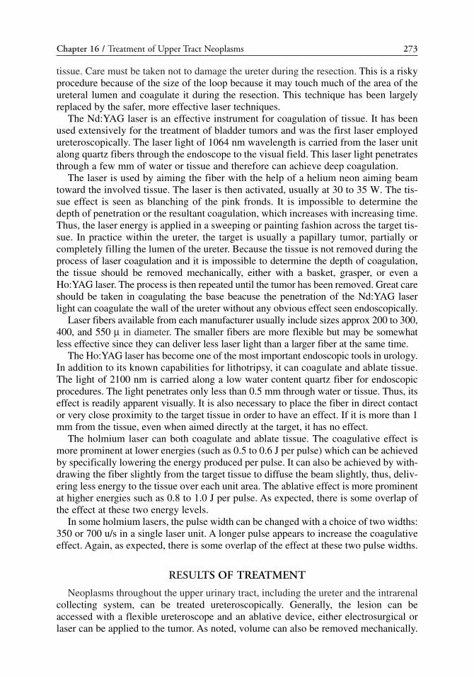
tissue. Care must be taken not to damage the ureter during the resection. This is a riskyprocedure because of the size of the loop because it may touch much of the area of theureteral lumen and coagulate it during the resection. This technique has been largelyreplaced by the safer, more effective laser techniques.
The Nd:YAG laser is an effective instrument for coagulation of tissue. It has beenused extensively for the treatment of bladder tumors and was the first laser employedureteroscopically. The laser light of 1064 nm wavelength is carried from the laser unitalong quartz fibers through the endoscope to the visual field. This laser light penetratesthrough a few mm of water or tissue and therefore can achieve deep coagulation.
The laser is used by aiming the fiber with the help of a helium neon aiming beamtoward the involved tissue. The laser is then activated, usually at 30 to 35 W. The tis-sue effect is seen as blanching of the pink fronds. It is impossible to determine thedepth of penetration or the resultant coagulation, which increases with increasing time.Thus, the laser energy is applied in a sweeping or painting fashion across the target tis-sue. In practice within the ureter, the target is usually a papillary tumor, partially orcompletely filling the lumen of the ureter. Because the tissue is not removed during theprocess of laser coagulation and it is impossible to determine the depth of coagulation,the tissue should be removed mechanically, either with a basket, grasper, or even aHo:YAG laser. The process is then repeated until the tumor has been removed. Great careshould be taken in coagulating the base beacuse the penetration of the Nd:YAG laserlight can coagulate the wall of the ureter without any obvious effect seen endoscopically.
Laser fibers available from each manufacturer usually include sizes approx 200 to 300,400, and 550 μ in diameter. The smaller fibers are more flexible but may be somewhatless effective since they can deliver less laser light than a larger fiber at the same time.
The Ho:YAG laser has become one of the most important endoscopic tools in urology.In addition to its known capabilities for lithotripsy, it can coagulate and ablate tissue.The light of 2100 nm is carried along a low water content quartz fiber for endoscopicprocedures. The light penetrates only less than 0.5 mm through water or tissue. Thus, itseffect is readily apparent visually. It is also necessary to place the fiber in direct contactor very close proximity to the target tissue in order to have an effect. If it is more than 1mm from the tissue, even when aimed directly at the target, it has no effect.
The holmium laser can both coagulate and ablate tissue. The coagulative effect ismore prominent at lower energies (such as 0.5 to 0.6 J per pulse) which can be achievedby specifically lowering the energy produced per pulse. It can also be achieved by with-drawing the fiber slightly from the target tissue to diffuse the beam slightly, thus, deliv-ering less energy to the tissue over each unit area. The ablative effect is more prominentat higher energies such as 0.8 to 1.0 J per pulse. As expected, there is some overlap ofthe effect at these two energy levels.
In some holmium lasers, the pulse width can be changed with a choice of two widths:350 or 700 u/s in a single laser unit. A longer pulse appears to increase the coagulativeeffect. Again, as expected, there is some overlap of the effect at these two pulse widths.
RESULTS OF TREATMENT
Neoplasms throughout the upper urinary tract, including the ureter and the intrarenalcollecting system, can be treated ureteroscopically. Generally, the lesion can beaccessed with a flexible ureteroscope and an ablative device, either electrosurgical orlaser can be applied to the tumor. As noted, volume can also be removed mechanically.
Chapter 16 / Treatment of Upper Tract Neoplasms 273

There is a good basis for the endoscopic treatment of upper tract tumors by comparisonwith bladder tumors. Thus, general treatment is possible with certain limitations andwith frequent recurrences.
Several series have demonstrated the value of ureteroscopic treatment of upper tractneoplasms. The initial studies were in those patients with a strong indication for renalpreservation. These included patients with a solitary kidney or compromised contralat-eral kidney and those with medical indications, contraindicating or prohibitingnephroureterectomy. More recently, these techniques have been applied to patients withtwo functioning kidneys. This has been feasible because of the recognition of the suc-cess achieved in the earlier groups with compromised renal function. It is also based onrecognition of the risk of developing a neoplasm in the contralateral kidney.
The ureteroscopic treatment of upper tract tumors represents a rational extensionof techniques based on the instruments available. Nearly any area in the upper col-lecting system is accessible to ureteroscopes and ablative instruments necessary forthe treatment of tumors. There is a strong basis for the endoscopic treatment of tran-sitional cell carcinoma as a result of the long extensive experience with the endo-scopic treatment of bladder tumors. Early series and the wider application of thesetechniques have demonstrated the general feasibility of treating upper tract tumorsureteroscopically. Although there are frequent recurrences, there is also strong evi-dence for long-term survival with maintenance of renal function. There has also beenconsiderable evidence developed suggesting in which tumors there may be benefitsfrom endoscopic treatment and in which tumors endoscopic treatments should beavoided. Nevertheless, many questions remain.
Some patients have tumors or other medical factors which can form a strong indica-tion for the ureteroscopic treatment of the upper tract neoplasm. Patients with a solitary,small low-grade lesion tend to do quite well without recurrences and with maintenanceof renal function after ureteroscopic treatment. Those patients with a solitary kidney orcompromised contralateral kidney or overall renal risks provide an even stronger indica-tion for endoscopic treatment. In contrast, there are other patients who may have tumorsthat are less likely to respond well to endoscopic resection. These include large tumorsand higher grade lesions as indicated by cytology positive for malignant cells. Lesionsthat cover the circumference of the ureter or infundibulum tend to develop strictures aftertreatment. These may require alternative treatment or staged endoscopic removal.
The results of endoscopic treatment have been seen only in relatively small groupsof heterogeneous patients. These also include a wide range of treatment techniques withrigid and flexible ureteroscopes and with tumor removal accomplished with electrosur-gical instruments and/or lasers (either Ho: or Nd:YAG). There is a high overall recur-rence rate after ureteroscopic treatment of upper tract lesions of approx 35% with awide range in therapeutic series. The risks of bladder tumors developing after treatmenthave been approx 36 to 39%.
Several parameters seem to affect the ability to render the kidney tumor free. Forexample, there is evidence that low-grade tumors are amenable to ureteroscopicresection with resultant better tumor-free state than higher grade tumors (40 vs 76%)(12). Others have seen a similar pattern with a more favorable outcome for a low-grade neoplasm (12–28). However, there are definitely some high-grade tumorswhich can be treated ureteroscopically. Suh et al. (29) have documented successfultreatment with survival of patients with high grade upper tract neoplasms treatedureteroscopically.
274 Bagley

Similarly, a negative effect on successful outcome was seen with a larger tumor(greater than 1.5 cm in diameter). Smaller tumors could be treated successfully in 91%of patients with 25% recurrence rate compared to 36 and 50% in the tumors larger than1.5 cm. Multifocal disease was less frequently resected completely than solitarylesions (50 vs 19%). The effect of location, whether in the ureter or in the kidney, hasvariably been reported as significant or insignificant in terms of recurrences. A previ-ous review demonstrated a similar recurrence rate for renal pelvic tumor (33%) andureteral tumor (31%) (30).
Long-term follow-up of patients treated ureteroscopically has indicated good survivaldespite the associated relatively high local recurrence rate. The 5-years survival rate was100% for grade I tumors but decreased to 80% for grade II tumors and 60% for grade IIItumors (22). The common association with upper tract tumors has been well establishedand has been the basis for continued cystoscopic follow-up of patients afternephroureterectomy for upper tract neoplasms. The occurrence of new bladder tumorsafter nephroureterectomy has been as high as 36% in reported surgical series. It has beenapprox 50% in patients with a history of previous bladder cancer. Thus, the rate of 36 to39% seen after ureteroscopic treatment of upper tract tumors is not far from the knownrange seen with surgical treatment. Again, the patients treated ureteroscopically representa heterogeneous group, including those both with and without previous bladder cancers.
There are definitely some patients with upper tract neoplasms that cannot be treatedadequately ureteroscopically. These include large tumors that cannot be treated ade-quately even with staged procedures. Those patients may then require a percutaneousapproach for resection of the entire tumor or surgical therapy, either with partial or totalnephroureterectomy. An even more common limiting factor is a widely extensive orinaccessible tumor. A low-grade neoplasm carpeting the entire collecting system withcircumferential extension into each infundibulum cannot be treated adequately uretero-scopically. There may also be locations within the kidney, especially in the lower poleor the medial portion of the upper pole, which may be visualized endoscopically butcannot be reached directly with an ablative device.
SURVEILLANCE
Surveillance is essential after ureteroscopic treatment of upper tract neoplasms.Ureteroscopy is the most sensitive technique available for the diagnosis of recurrentlesions. It also offers the opportunity for treatment during the same procedure. Previousstudies have shown that retrograde pyelography is inadequate to demonstrate recur-rences reliably. In one earlier series, as many as 75% of recurrences found by uretero-scopic inspection were missed endoscopically (12). Even extensive TCC of the uppertract confirmed by ureteroscopy and nephroureterectomy may not be seen on intra-venous urography (16). Comparison of other techniques for the detection of recurrenceshad shown the low detection rate of voided urinary cytology or urinalyses (31).However, there was relatively high specificity of either abnormal cytology or micro-scopic hematuria. The newer immunologic diagnostic studies searching for tumor asso-ciated antigens have not been studied adequately with upper tract neoplasms.
Endoscopy has been employed extensively in the surveillance for upper tract recur-rences. Our surveillance program has been outlined previously (32). Initially, at 3months after ureteroscopic resection ureteroscopy is repeated to define the tumor statusor complete resection of any persistent or recurrent tumor. Ureteroscopy is continued
Chapter 16 / Treatment of Upper Tract Neoplasms 275

every 3 months only until the upper tract is clear. In continued surveillance, cystoscopyis performed every 3 months and ureteroscopy of the entire tract is performed every 6months for the first 2 years. At 2 to 5 years, cystoscopy and ureteroscopy are continuedat 6-month intervals. With any recurrence, the endoscopic cycle is restarted. The con-tralateral collecting system is studied radiographically (usually with retrograde pyelog-raphy) at yearly intervals.
The role of adjuvant therapy has not been confirmed experimentally. There hasbeen experience in small series with topical adjuvant therapy using bacillusCalmete-Guerin (BCG), Mitomycin C, Thiotepa, Fluorouracil and Interferon α-2B (33–36). There is some evidence of a positive effect from BCG or MitomycinC. Mitomycin C has been delivered through a ureteral catheter in the early postop-erative period. BCG treatment has been delayed until the urothelial damage fromresection has healed.
COMPLICATIONS
Complications can occur with any endoscopic procedure, and there appear to besome complications related to the specific endoscopic procedure. For example, ureteralperforation and ureteral stricture appear to be much more common after ureteroscopictreatment of neoplasms than with stone treatment. This may be explained by the obser-vation that the ablative technique is being applied to the wall of the ureter itself. For cal-culi, the interventional technique is limited to the stone rather than the wall of the ureter.In a comparison of tumor and stone treatment, a combined series of rate of stricture for-mation after ureteroscopic treatment of upper tract tumors has been 13% (37), whereasit is a rare occurrence after stone therapy at 0.5% (38).
Perforation of the ureter or renal pelvis is less common occurring in 0 to 10% ofpatients. These perforations generally have been treated by drainage with an indwellingstent or a percutaneous nephrostomy.
The other potential complication is the dissemination of the tumor either throughextraluminal spillage or throughout the urothelium. The initial concern regarding localrecurrence was reported after intraoperative pyeloscopy during surgical exploration ofthe kidney for renal pelvic lesions. Two of 18 patients developed local recurrences inthe renal fossa after exploration for low-grade lesions (39). There was also a single casereport of tumor cells, in unusual lymphatic and vascular locations, with nephroureterec-tomy after ureteropyeloscopy (40). Thus, the question of the safety of even uretero-scopic biopsy was raised. However, in a series of 13 patients who were treated withnephroureterectomy after ureteropyeloscopy, there was no evidence of extraluminaltumor (41). An excellent study examined 96 patients, half of whom had ureteroscopicbiopsy before nephroureterectomy and half without ureteroscopy (42). There was nodifference in the recurrence rate or the disease specific survival between the two groups.Thus, the conclusion from that study was that ureteroscopic biopsy of upper tract neo-plasms is safe. In our patients treated with nephroureterectomy after unsuccessful endo-scopic treatments, there has been no evidence of unusual spread beyond the urothelium.
There has been no documented pattern of increased urothelial implantation or disper-sion of tumor after ureteroscopic treatment. Possibly, the best indication of this lack oftumor dissemination is the similar rates of bladder tumor occurrence after upper tracttumor treatment by ureteroscopy or by nephroureterectomy. However, we have noted ananecdotal case in which there were multiple urothelial recurrences throughout the ureter
276 Bagley
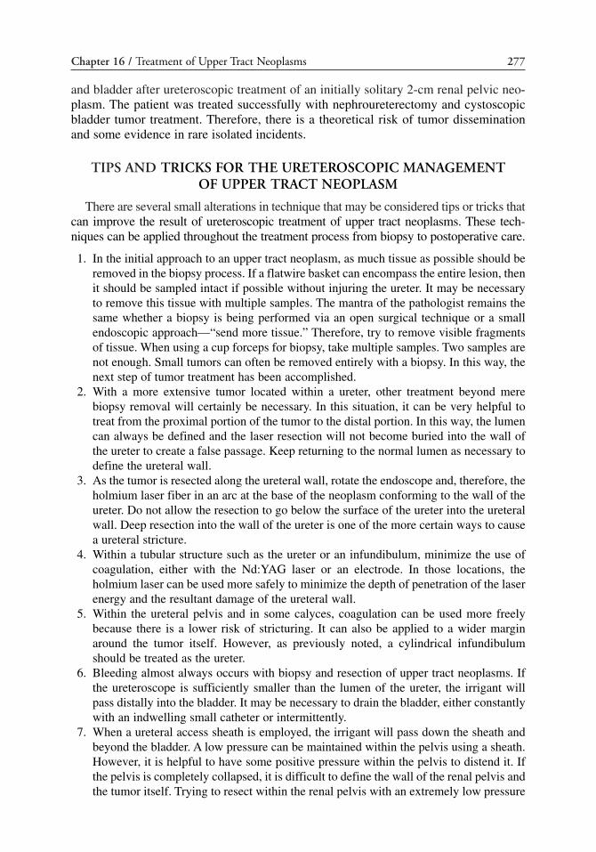
and bladder after ureteroscopic treatment of an initially solitary 2-cm renal pelvic neo-plasm. The patient was treated successfully with nephroureterectomy and cystoscopicbladder tumor treatment. Therefore, there is a theoretical risk of tumor disseminationand some evidence in rare isolated incidents.
TIPS AND TRICKS FOR THE URETEROSCOPIC MANAGEMENT OF UPPER TRACT NEOPLASM
There are several small alterations in technique that may be considered tips or tricks thatcan improve the result of ureteroscopic treatment of upper tract neoplasms. These tech-niques can be applied throughout the treatment process from biopsy to postoperative care.
1. In the initial approach to an upper tract neoplasm, as much tissue as possible should beremoved in the biopsy process. If a flatwire basket can encompass the entire lesion, thenit should be sampled intact if possible without injuring the ureter. It may be necessaryto remove this tissue with multiple samples. The mantra of the pathologist remains thesame whether a biopsy is being performed via an open surgical technique or a smallendoscopic approach––“send more tissue.” Therefore, try to remove visible fragmentsof tissue. When using a cup forceps for biopsy, take multiple samples. Two samples arenot enough. Small tumors can often be removed entirely with a biopsy. In this way, thenext step of tumor treatment has been accomplished.
2. With a more extensive tumor located within a ureter, other treatment beyond merebiopsy removal will certainly be necessary. In this situation, it can be very helpful totreat from the proximal portion of the tumor to the distal portion. In this way, the lumencan always be defined and the laser resection will not become buried into the wall ofthe ureter to create a false passage. Keep returning to the normal lumen as necessary todefine the ureteral wall.
3. As the tumor is resected along the ureteral wall, rotate the endoscope and, therefore, theholmium laser fiber in an arc at the base of the neoplasm conforming to the wall of theureter. Do not allow the resection to go below the surface of the ureter into the ureteralwall. Deep resection into the wall of the ureter is one of the more certain ways to causea ureteral stricture.
4. Within a tubular structure such as the ureter or an infundibulum, minimize the use ofcoagulation, either with the Nd:YAG laser or an electrode. In those locations, theholmium laser can be used more safely to minimize the depth of penetration of the laserenergy and the resultant damage of the ureteral wall.
5. Within the ureteral pelvis and in some calyces, coagulation can be used more freelybecause there is a lower risk of stricturing. It can also be applied to a wider marginaround the tumor itself. However, as previously noted, a cylindrical infundibulumshould be treated as the ureter.
6. Bleeding almost always occurs with biopsy and resection of upper tract neoplasms. Ifthe ureteroscope is sufficiently smaller than the lumen of the ureter, the irrigant willpass distally into the bladder. It may be necessary to drain the bladder, either constantlywith an indwelling small catheter or intermittently.
7. When a ureteral access sheath is employed, the irrigant will pass down the sheath andbeyond the bladder. A low pressure can be maintained within the pelvis using a sheath.However, it is helpful to have some positive pressure within the pelvis to distend it. Ifthe pelvis is completely collapsed, it is difficult to define the wall of the renal pelvis andthe tumor itself. Trying to resect within the renal pelvis with an extremely low pressure
Chapter 16 / Treatment of Upper Tract Neoplasms 277

in the pelvis which causes it to collapse completely is like trying to resect within thebladder when it is fully collapsed.
8. The kidney and the upper ureter move with respiration. It may be necessary to slow therespiratory movement to stabilize the kidney or at least provide predictable movementduring which resection can be accomplished. With the slow predictable movement, theurologist can anticipate the position of the target tissue as it passes and treat in thatperiod. The anesthesiologist can usually slow the respiratory rate, often with the sup-pressant effect of narcotics.
REFERENCES1. Huffman JL, Bagley DH, Lyon ES, Morse MJ, Herr HW, Whitmore Jr. WF: Endoscopic diagnosis
and treatment of upper tract urothelial tumors: a preliminary report. Cancer 1985; 55: 1422–1428.2. Dretler SP, Cho G: Semirigid ureteroscopy: a new genre. J Urol 1989; 141(6): 1314–1316. 3. Abdel-Razzak OM, Bagley DH: The 6.9F semi-rigid ureteroscope in clinical use. Urology 1993;
41(1): 45–48.4. Bagley DH, Rittenberg MH: Intrarenal dimensions: guidelines for flexible ureteropyeloscopes. Surg
Endosc 1987; 1: 119–121.5. Bagley DH: Ureteroscopic stone retrieval: rigid versus flexible endoscopes. Sem Urol 1994; 12:
32–38.6. Lobik L, Lopez-Pujals A, Leveillee RJ. Variables affecting deflection of a new third-generation flex-
ible ureteropyeloscope (DUR-8 Elite). J Endourol 2003; 17: 733–736.7. Johnson GB, Grasso M: Exaggerated primary endoscope deflection initial clinical experience with
prototype flexible ureteroscopes. BJU Int 2004; 93: 109–114.8. Abdel-Razzak OM, Ehya H, Cubler-Goodman A, Bagley DH: Ureteroscopic biopsy in the upper uri-
nary tract. Urology 1994; 44(3): 451–457.9. Bagley DH: Ureteroscopic laser treatment of upper urinary tract tumors. J Clin Laser Med Surg 1998;
16 (1): 55–59.10. Razvi HA, Chun SS, Denstedt JD, Sales JL: Soft-tissue applications of the holmium:YAG laser in
urology. J Endourol 1995; 9: 387–390. 11. Tawfiek ER, Bibbo M, Bagley DH: Ureteroscopic biopsy: technique and specimen preparation.
Urology 1997; 50: 117–119.12. Keeley FX Jr, Bibbo M, Bagley DH. Ureteroscopic treatment and surveillance of upper urinary tract
transitional cell carcinoma. J Urol 1997; 157: 1560–1565. 13. Schmeller NT, Hofstetter AG. Laser treatment of ureteral tumors. J Urol 1989; 141: 840–843. 14. Blute ML, Segura JW, Patterson DE: Impact of endourology on diagnosis and management of upper
urinary tract urothelial cancer. J Urol 1989; 141: 1298–1301.15. Johnson DE. Treatment of distal ureteral tumors using endoscopic argon photoirradiation. Lasers
Surg Med 1992; 12: 490–493.16. Grossman HB, Schwartz SL, Konnak JW. Ureteroscopic diagnosis and treatment of urothelial carci-
noma of the ureter and renal pelvis. J Urol 1992; 148: 275–277.17. Tasca A, Zattoni F, Garbeglio A, et al. Endourologic treatment of transitional cell carcinoma of the
upper urinary tract. J Endourol 1992; 6: 253–256.18. Kaufman RP Jr., Carson CC 3rd. Ureteroscopic management of transitional cell carcinoma of the
ureter using the neodymium:YAG laser. Lasers Surg Med 1993; 13: 628.19. Gaboardi E, Bozzola A, Dotti E, et al. Conservative treatment of upper urinary tract tumors with
Nd:YAG laser. J Endourol 1994; 8: 37–41.20. Bales GT, Lyon ES, Gerber GS. Conservative management of transitional cell carcinoma of the
kidney and ureter. Diagn Ther Endosc 1995; 1: 121–123. 21. Engelmyer EI, Belis JA. Long-term ureteroscopic management of low-grade transitional cell carci-
noma of the upper urinary tract. Tech Urol 1996; 2: 113–116. 22. Elliott DS, Blute ML, Patterson DE, et al. Long term follow-up of endoscopically treated upper uri-
nary tract transitional cell carcinoma. Urology 1996; 47: 819–825. 23. Martinez-Pineiro JA, Garcia Matres MJ, Martinez-Pineiro L. Endourological treatment of upper tract
278 Bagley

urothelial carcinomas: analysis of a series of 59 tumors. J Urol 1996; 156: 377–385. 24. Patel A, Fuchs GJ. New techniques for the administration of topical adjuvant therapy after endoscopic
ablation of upper urinary tract transitional cell carcinoma. J Urol 1998; 159: 71–75. 25. Grasso M, Fraiman M, Levine M. Ureteroscopic diagnosis and treatmentn of upper urinary tract
urothelial malignancies. Urology 1999; 54: 240–246.26. Chen GL, Bagley DH. Ureteroscopic management of upper tract transitional cell carcinoma in
patients with normal contralateral kidneys. J Urol 2000; 154: 1173–1176. 27. Elliott DS, Segura JW, Lightner D, et al. Is nephroureterectomy necessary in all cases of upper tract tran-
sitional cell carcinoma? Long-term results of conservative endourologic management of upper tract tran-sitional cell carcinoma in individuals with a normal contralateral kidney. Urology 2001; 58: 174–178.
28. Matsuoka K, Lida S, Tomiyasu K, Inoue M, Noda S. Transurethral endoscopic treatment of upper uri-nary tract tumors using a holmium:YAG laser. Lasers Surg Med 2003; 32(5): 336–340.
29. Suh RS, Faerber GJ, Wolf JS Jr. Predictive factors for applicability and success with endoscopic treat-ment of upper tract urothelial carcinoma. J Urol 2003; 170(6 Pt 1): 2209–2216.
30. Tawfiek, ER, Bagley DH. Upper tract transitional cell carcinoma. Urology 1997; 50(3): 321–329. 31. Chen GL, El-Gabry EA, Bagley DH. Surveillance of upper urinary tract transitional cell carcinoma: the
role of ureteroscopy, retrograde pyelography, cytology and urinalysis. J Urol 2000; 165: 1901–1904. 32. Lee D, Trabulsi E, McGinnis D, Strup S, Gomella LG, Bagley DH. Totally endoscopic management
of upper tract transitional cell carcinoma. J Endourol 2002; 16(1): 37–41.33. Sharpe JR, Duffy G, Chin JL. Intrarenal bacillus Calmette-Guerin therapy for upper urinary tract car-
cinoma in situ. J Urol 1993; 149: 457–460. 34. Eastham JA, Huffman JL. Technique of mitomycin C instillation in the treatment of upper
urinary tract urothelial tumors. J Urol 1993; 150: 324–325. 35. Patel A, Fuchs GJ. New techniques for the administration of topical adjuvant therapy after endoscopic
ablation of upper urinary tract transitional cell carcinoma. J Urol 1998; 159: 71–75. 36. Keeley FX, Bibbo M, Bagley DH. Adjuvant mitomycin C following endoscopic treatment of upper
tract transitional cell carcinoma. J Urol 1997; 158: 2074–2077.37. Chen GL, Bagley DH. Ureteroscopic surgery for upper tract transitional cell carcinoma: Complications
and management. J Endourol 2001; 15: 399–404.38. Chow GK, Patterson RF, Blute ML, Segura JW. Ureteroscopy: effect of technology and technique on
clinical practice. J Urol 2003; 170: 99–102. 39. Tomera KM, Leary FJ, Zincke H. Pyeloscopy in urothelial tumors. J Urol 1982; 127: 1088–1089. 40. Lim DJ, Shattuck MC, Cook WA. Pyelovenous lymphatic migration of transitional cell carcinoma
following flexible ureteroscopy. J Urol 1993; 149: 109.41. Kulp DA, Bagley DH. Does flexible ureteropyeloscopy promote local recurrence of transitional cell
carcinoma? J Endourol 1994; 8: 111–113. 42. Hendin BN, Streem SB, Levin HS, et al. Impact of diagnostic ureteroscopy on long-term survivalin
patients with upper tract transitional cell carcinoma. J Urol 1999; 161: 783–785.
Chapter 16 / Treatment of Upper Tract Neoplasms 279

V COMPLICATIONS OF ENDOUROLOGY

Complications of PercutaneousApproaches, Including Incisions
Brian R. Matlaga, MD, MPH,Ojas D. Shah, MD, and Dean G. Assimos, MD
CONTENTS
INTRODUCTION
INJURY TO THE RENAL UNIT
INJURY RESULTING FROM ENERGY SOURCES
INJURY TO PERINEPHRIC STRUCTURES
MEDICAL COMPLICATIONS
CONCLUSIONS
REFERENCES
17
SUMMARY
Percutaneous endoscopic renal surgery is usually a safe and effective treatment forpatients with complex renal calculi, tumors of the collecting system, and ureteropelvicjunction obstruction. However, a unique set of complications can occur with this surgi-cal approach that may involve the targeted kidney and surrounding structures. Methodsof preventing these events, diagnosing their occurrence, and management strategies arereviewed in this chapter.
Key Words: Hemorrhage; sepsis; hydrothorax; pneumothorax; ureteral stricture; colonicinjury; ureteral avulsion.
INTRODUCTION
A variety of complications can occur with percutaneous endorenal surgery. The tar-geted renal unit and perirenal structures may be injured and patients may develop sys-temic complications such as sepsis. These complications, measures to prevent theiroccurrence, and management methods are reviewed in this chapter.
From: Advanced Endourology: The Complete Clinical GuideEdited by: S. Y. Nakada and M. S. Pearle © Humana Press Inc., Totowa, NJ
283
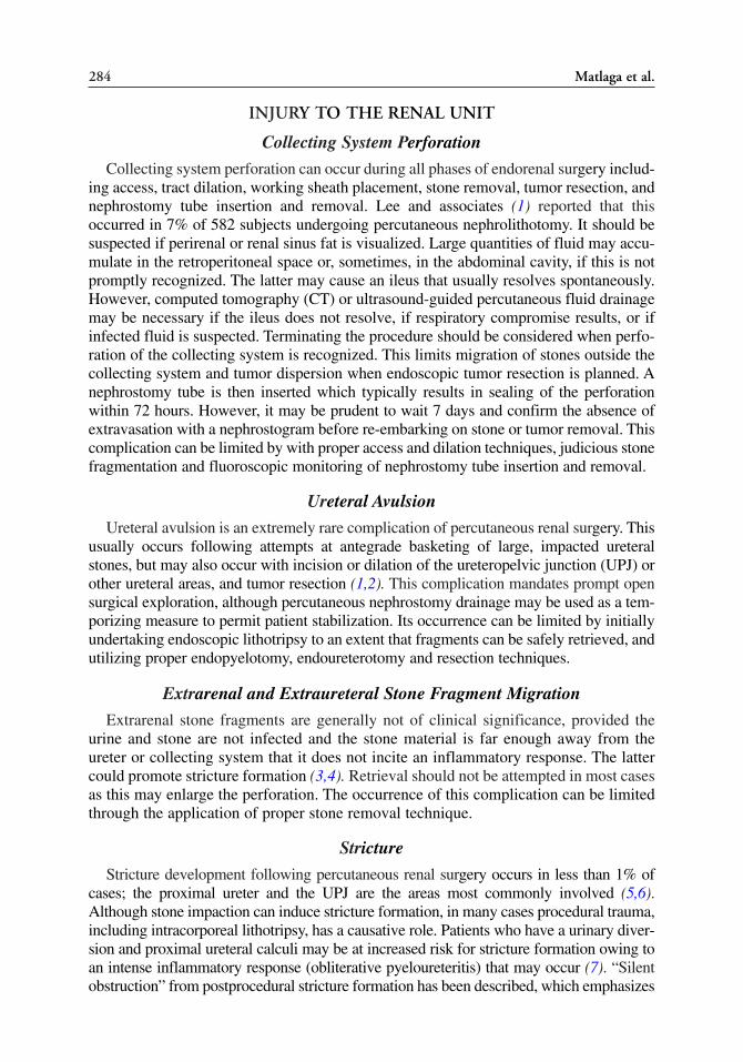
INJURY TO THE RENAL UNIT
Collecting System Perforation
Collecting system perforation can occur during all phases of endorenal surgery includ-ing access, tract dilation, working sheath placement, stone removal, tumor resection, andnephrostomy tube insertion and removal. Lee and associates (1) reported that thisoccurred in 7% of 582 subjects undergoing percutaneous nephrolithotomy. It should besuspected if perirenal or renal sinus fat is visualized. Large quantities of fluid may accu-mulate in the retroperitoneal space or, sometimes, in the abdominal cavity, if this is notpromptly recognized. The latter may cause an ileus that usually resolves spontaneously.However, computed tomography (CT) or ultrasound-guided percutaneous fluid drainagemay be necessary if the ileus does not resolve, if respiratory compromise results, or ifinfected fluid is suspected. Terminating the procedure should be considered when perfo-ration of the collecting system is recognized. This limits migration of stones outside thecollecting system and tumor dispersion when endoscopic tumor resection is planned. Anephrostomy tube is then inserted which typically results in sealing of the perforationwithin 72 hours. However, it may be prudent to wait 7 days and confirm the absence ofextravasation with a nephrostogram before re-embarking on stone or tumor removal. Thiscomplication can be limited by with proper access and dilation techniques, judicious stonefragmentation and fluoroscopic monitoring of nephrostomy tube insertion and removal.
Ureteral Avulsion
Ureteral avulsion is an extremely rare complication of percutaneous renal surgery. Thisusually occurs following attempts at antegrade basketing of large, impacted ureteralstones, but may also occur with incision or dilation of the ureteropelvic junction (UPJ) orother ureteral areas, and tumor resection (1,2). This complication mandates prompt opensurgical exploration, although percutaneous nephrostomy drainage may be used as a tem-porizing measure to permit patient stabilization. Its occurrence can be limited by initiallyundertaking endoscopic lithotripsy to an extent that fragments can be safely retrieved, andutilizing proper endopyelotomy, endoureterotomy and resection techniques.
Extrarenal and Extraureteral Stone Fragment Migration
Extrarenal stone fragments are generally not of clinical significance, provided theurine and stone are not infected and the stone material is far enough away from theureter or collecting system that it does not incite an inflammatory response. The lattercould promote stricture formation (3,4). Retrieval should not be attempted in most casesas this may enlarge the perforation. The occurrence of this complication can be limitedthrough the application of proper stone removal technique.
Stricture
Stricture development following percutaneous renal surgery occurs in less than 1% ofcases; the proximal ureter and the UPJ are the areas most commonly involved (5,6).Although stone impaction can induce stricture formation, in many cases procedural trauma,including intracorporeal lithotripsy, has a causative role. Patients who have a urinary diver-sion and proximal ureteral calculi may be at increased risk for stricture formation owing toan intense inflammatory response (obliterative pyeloureteritis) that may occur (7). “Silentobstruction” from postprocedural stricture formation has been described, which emphasizes
284 Matlaga et al.

the necessity of routine postoperative imaging in patients to assess for this occurrence (8).The majority of patients who develop ureteral strictures can be managed successfully uti-lizing endourologic techniques providing the stricture is less than 1.5 cm in length and in anonradiated field. Open surgical or laparoscopic reconstruction may be required in patientswith more extensive strictures or in those who fail an endoscopic approach. Stricture for-mation can be limited by employing proper percutaneous and endoscopic techniques.
Infundibular Stenosis
Infundibular stenosis is a rare complication of percutaneous renal surgery (6,9,10).Parsons et al. (11) describe a 2% incidence of infundibular stenosis following percuta-neous nephrolithotomy (PCNL). This probably occurs as a result of mechanical or thermalinjury of the infundibulum which could induce ischemic and inflammatory changes.Prolonged operative time, a large stone burden requiring multiple procedures, andextended postoperative nephrostomy tube drainage were reported to be independent riskfactors for this occurrence by Parsons and colleagues. This complication usually mani-fests within 1 year of the percutaneous procedure. Initial management of these patientsshould be endourological, with open surgery reserved for those failing this therapy.Close observation is a reasonable consideration for those who are asymptomatic provid-ing there is no functional deterioration of the involved kidney. Proper surgical techniqueis the best method of limiting the occurrence of this complication.
Retained Foreign Bodies
Any instrument employed in percutaneous renal surgery has the potential to be retainedin the collecting system. The retained foreign body should be removed as it may be a nidusfor infection, stone formation, or granulomatous reaction. Removal of the foreign bodycan be facilitated with fluoroscopic guidance if it is radio-opaque. The item usually canbe extracted with rigid or flexible nephroscopy using grasping devices or a basket. A ret-rograde ureteroscopic extraction may be considered if the retained object is identified afterthe nephrostomy tube has been removed. This complication can be limited with carefulmanipulation of laser fibers, other lithotripsy instruments, baskets, grasping devices,guidewires, and tubes to avoid shearing them off into the collecting system. These devicesshould be inspected periodically during and at the end of the case to monitor for thisoccurrence. The tips of nephrostomy tubes, such as Malecot catheters, may becomeentrapped in the renal collecting system because of ingrowth of fibrous or inflammatorytissue. Removal of the restrictive tissue through another nephrostomy tract is recom-mended for managing patients with this problem (12). This will generally free up the tubeand facilitate its removal. Limiting the dwell time of catheters will limit this occurrence.
Tumor Seeding
Tumor seeding and dissemination are potential complications of percutaneous resec-tion of transitional cell carcinoma of the renal collecting system. These risks may beminimized by appropriate working sheath positioning, low pressure irrigation, avoidingperforation during resection or ablation, and optimal postoperative drainage. Only a fewreports of tract seeding have been reported with this procedure (12–15). This occurredin patients with locally advanced high-grade tumors; clearly cases where local and sys-temic failure would be expected. However this has not been reported in properly-selected cases; subjects with low-grade, noninvasive tumors (13–15).
Chapter 17 / Complications of Percutaneous Approaches 285

Nephrocutaneous Fistula
The development of a nephrocutaneous fistula following percutaneous renal surgeryis a rare complication (16). This is usually caused by distal obstruction owing to ureteraledema, an obstructing stone, blood clot, or stricture. This usually resolves with removalof the stone or ureteral stent placement.
Intraoperative Hemorrhage
Patients undergoing percutaneous renal surgery may develop high volume intraopera-tive bleeding and require blood transfusion. While this occurrence is reported to be lessthan 5% in a number of contemporary series, others have noted that transfusion may berequired in approximately one-third of patients (5,6,17,18). Surgical experience, technique,preoperative anemia, and advanced patient age are factors that may influence transfusionrequirements. Several technical steps may reduce this occurrence. The collecting systemshould be accessed through a posterior calyx along the direction of the infundibulum,thereby avoiding blood vessels that are adjacent to the infundibulum (19). Additionally, theposterior calyx that provides the most direct access to the targeted stone should be chosen.Once created, the tract should only be dilated up to the peripheral aspect of the collectingsystem. If dilation proceeds too far medially, perforation of the renal pelvis can occur, withconcomitant laceration of the hilar vessels. Stoller and colleagues have reported that renalpelvic perforation is a risk factor for excessive blood loss during PCNL (17). The methodof dilation has been reported to influence bleeding by some investigators. Davidoff andBellman reported that balloon dilation, as compared to coaxial dilation, of the tract is asso-ciated with a significantly lower risk of bleeding. However, others have not demonstratedthis association (20,21). Working sheaths are utilized in the majority of initial percutaneousinterventions and they must be kept in the collecting system to limit parenchymal bleeding.Excessive torquing of rigid instruments may injure renal parenchyma and vasculature and,thus, should be avoided. Alternatively, flexible nephroscopy or placement of an additionalnephrostomy track should be performed if access to the targeted pathology is not easilyobtained with a rigid nephroscope. The validity of this philosophy is supported by the studyof Lam and associates (20) who reported that multiple access tracts and flexible endoscopydecreased transfusion requirements in patients with staghorn calculi undergoing PCNL.
There is typically some degree of ongoing blood loss with these procedures. However,if this is to the point that visibility is limited, certain measures should be taken.Intraoperative hemorrhage may simply be because of the end of the working sheath beingin the parenchyma and not the collecting system. If this is the case, repositioning the sheathwill usually rectify this problem. However, if this maneuver does not significantly attenu-ate bleeding, certain measures should be undertaken. Inflation of a 30-Fr dilating balloonin the tract for 10 to 20 minutes with subsequent placement of a large nephrostomy (24–28Fr) will usually control hemorrhage. The nephrostomy tube should be clamped if bleedingpersists. This allows blood to clot and hopefully tamponade the injured vessel(s). The tubeis unclamped 2 to 3 hours later and left open if there is no evidence of significant bleeding.If this is unsuccessful, a specialized nephrostomy tamponade catheter may be inserted(Kaye, Cook Urological, Spencer, IN) (22). The peripheral balloon is inflated in the tractwhile urine drains through the inner core of this device, and it is typically left inflated for2 to 4 days. The majority of bleeding is from peripheral vessels along the nephrostomytract and the aforementioned measures are usually successful. Significant bleeding from themain renal vein can also be controlled using a tamponade approach. Gupta and colleagues(23) reported successful management of this complication in four patients by inflating a
286 Matlaga et al.

Councill balloon catheter adjacent to the point of venous injury. When these measures areunsuccessful or the patient is hemodynamically unstable, renal arteriography should beundertaken with the intent to proceed with embolization.
Significant hemorrhage can develop when the ureteropelvic junction is incised duringendopyelotomy. This can be avoided by carefully aligning the incision to avoid crossing ves-sels. Preoperative computed tomographic or magnetic resonance angiography is recom-mended for secondary UPJ obstruction (UPJO) and cases involving ectopic kidneys asvascular anatomy is not highly predictable. Others advocate endoluminal ultrasonography forthis purpose (24). When significant hemorrhage arises from the incised UPJ, a 24-Fr dilatingballoon should be placed across this area and inflated for 10 minutes. The balloon is thendeflated and if bleeding persists or the patient is hemodynamically unstable, the balloon isreinflated and angiographic embolization undertaken if possible. Open surgical explorationwith vascular repair or nephrectomy may be needed if these measures are unsuccessful.
Postoperative Hemorrhage
Significant hemorrhage may occur at any time following surgery. Fortunately, seri-ous postoperative bleeding requiring intervention other than tamponade occurs rarely(21,25). If postoperative bleeding occurs with the nephrostomy tube in place, the previ-ously discussed measures should be undertaken. When the tube is out and there is highvolume bleeding from the tract, initial digital tamponade is recommended with subse-quent placement of a tamponade catheter under fluoroscopic guidance. Managementthen includes bedrest and blood transfusions as necessary. If bleeding persists, transfu-sion-dependent anemia develops, or the patient becomes hemodynamically unstable,renal arteriography with superselective embolization may be performed.
The most common causes of postoperative bleeding are laceration of segmental renalvessels, and development of an arteriovenous fistula or pseudoaneurysm (5,6,18,26).Kessaris and colleagues (25) reported that 17 of 2200 patients (0.8%) who underwentpercutaneous renal procedures over a 10-year period required angiography andembolization for uncontrolled significant bleeding. Twenty-four percent of thesepatients presented in the immediate postoperative period (<24 hours), 41% in the earlypostoperative period (2 to 7 days), and 35% March in the late postoperative period (>7days). These patients require selective or superselective angiographic embolization andresults are generally excellent. Kessaris and colleagues (25) reported success in 15 of 17such cases and Patterson and associates (26) in 7 of 7 cases treated with selective or super-selective embolization. The nephrostomy catheter must sometimes be removed so that thebleeding site may be localized. Open surgical exploration with vascular repair, partial ortotal nephrectomy may be necessary if the aforementioned measures are unsuccessful.
Perinephric hemorrhage should be suspected if the patient has a decreasing hemoglobinlevel in the face of clear urine draining from the bladder and nephrostomy tube. This situa-tion may develop following difficult access, or malpositioning of the working sheath outsidethe renal parenchyma. As well, sandwich therapy with shockwave lithotripsy (SWL) andsubsequent second look PCNL is another potential risk factor, as subcapsular or perinephrichemorrhage from SWL may be exacerbated by further tract and collecting system manipu-lation. The patient should be evaluated with a CT scan if this is suspected (Fig.1).
INJURY RESULTING FROM ENERGY SOURCES
Technological advances in the design of lithotripsy and ablative energy sources havefacilitated percutaneous renal surgery. However, the potential for energy-related complica-
Chapter 17 / Complications of Percutaneous Approaches 287

tions is real, and intracorporeal damage from these sources can range from minor to exten-sive. The surgeon should be familiar with each energy source and its potential dangers priorto its use.
Ultrasonic lithotripsy is a commonly used energy source for PCNL (2). If excessivepressure is applied to this device, collecting system or ureteral perforation may occur.If the probe becomes clogged with debris, the device may overheat and thermal injurycan occur.
Electrohydraulic lithotripsy (EHL) is less frequently used for PCNL owing to theavailability of newer devices with better safety profiles (27). The most common com-plications associated with EHL are perforation of the collecting system and bleeding,which are managed as previously discussed.
The holmium:YAG laser is frequently used to fragment stones, incise strictures, andablate upper tract tumors. Although the holmium laser has a high safety profile, hemor-rhage, perforation of the collecting system, and thermal injury may still occur (28).Such occurrences are minimized with careful technique and utilization of appropriateenergy settings. Heat generated from laser lithotripsy can interact with hydrogen gasand generate an explosion potentially resulting in collecting system perforation andhemorrhage (29).
Pneumatic lithotriptors and hybrid devices (pneumatic/ultrasound) are also used dur-ing PCNL (30–32). There is a small risk of perforation and hemorrhage with their uti-lization, which can be reduced with appropriate precautions.
Electrical generators may be employed during percutaneous renal surgery to facili-tate electrocautery or tumor resection. The patient must be properly grounded to preventthermal burns. Only nonconductive materials should be in contact with the collectingsystem and ureter to prevent current dispersal and possible thermal injury. The risk ofhemorrhage is minimized by maintaining proper orientation with respect to adjacentvascular structures. Sterile glycine is used as an irrigant in this setting so there is apotential for fluid absorption syndrome. This is limited by reducing irrigant pressureand resection time.
288 Matlaga et al.
Fig. 1. Right perinephric hematoma.

INJURY TO PERINEPHRIC STRUCTURES
Lung and Pleura
The lung and pleura are the perinephric structures at greatest risk for injury duringpercutaneous renal surgery (5,6). Most injuries to these structures occur when a supra-costal approach is employed (33,34). Hopper and Yakes (35) performed CT imagingduring maximal inspiration and expiration of 43 randomly selected patients and pre-dicted that in full expiration the pleura and lung would be traversed 86% of the time onthe right and 79% of the time on the left with an 11th rib approach, and 29% of the timeon the right and 14% of the time on the left with a 12th rib approach. Reported experi-ences suggest that clinically significant injuries occur with less frequency, as pneumoth-orax has been reported in 0 to 4% and hydrothorax in 0 to 8% of individuals subjectedto supracostal access (36,37). Munver et al. (33) recently compared complications asso-ciated with supracostal access to those occurring with an infracostal approach.Approximately 33% of their upper pole access cases required a supracostal approachand, of the supracostal approaches 73.5% of the tracts were above the 12th rib and26.5% were above the 11th rib. The overall complication rate for supracostal access was16.3% (supra-11th, 34.6%; supra-12th, 9.7%); 87% of the intrathoracic complicationsoccurred with supracostal access; hemothorax/hydrothorax in 4% of supracostal accesstracts, nephropleural fistula in 2%, and pneumothorax in 1%. A working sheathshould be utilized with a supracostal approach, as this provides a barrier to the influxof fluid and air into the pleural cavity if the parietal pleura is violated. Routine intra-operative chest fluoroscopy is recommended at the termination of PRS to evaluate forobvious hydrothorax or pneumothorax (38). Routine postoperative chest radiographyis not necessary when fluoroscopy is normal and the patient has no signs of pulmonarycompromise (39).
Patients found to have a small-volume pneumothorax or hydrothorax may beobserved, providing there are no signs of pulmonary compromise (Fig. 2). Aspiration ofthe pneumothorax or tube thoracostomy may be required for larger pneumo/hydrotho-races or in cases of patient instability. A nephropleural fistula should be suspected ifdrainage persists after tube thoracostomy. This will usually resolve after placement of aninternalized ureteral stent (40).
The intercostal vessels may be lacerated during a supracostal approach and hemoth-orax may result. Tube thoracostomy or thoracotomy may be required if this occurs.
Colon
Colonic perforation is a rare complication of percutaneous renal surgery, reported inless than 1% of cases (1,2,41–44). This low incidence is likely the result of the colonrarely being retrorenal. Hadar and Gadoth (45) and Sherman and associates (46) havereported that the colon is retrorenal in approx 0.6% of the general population.Individuals at higher risk for colon injury are those with horseshoe kidney and otherforms of renal fusion and ectopia and those with colonic distention owing to jejunoilealbypass, partial jejunoileal bypass, neurological impairment, and “institutional” bowel.Preoperative CT is recommended in high-risk groups to assess for retrorenal colon(Fig. 3). CT-guided access should be considered if this exists as the window of entryinto the collecting system may be quite small (47).
Chapter 17 / Complications of Percutaneous Approaches 289

Prompt recognition of a colonic perforation is critical to limit serious infectioussequelae (Fig. 4). Passage of gas or feculent material through the nephrostomy tract,intraoperative diarrhea or hematochezia, and peritonitis are signs of a possible colonicperforation.
The majority of patient with colonic injuries can be managed without open surgicalintervention if the penetration is retroperitoneal and the patient does not have
290 Matlaga et al.
Fig. 2. Computed tomography scan demonstrating right hydrothorax.
Fig. 3. Computed tomography scan demonstrating retrorenal colon.

peritonitis or sepsis (48). An indwelling ureteral stent should be inserted and thenephrostomy tube should be pulled back into the colon (Fig. 5). Broad-spectrumantibiotic therapy is administered. The patient is placed on a low-residue diet. Sevento ten days following the injury a contrast study is performed through the colostomytube and the tube is removed if there is no evidence of a nephrocolic fistula (41,43).Open surgical management is required in patients with transperitoneal perforation,peritonitis, or sepsis.
Small Intestine
The second and third portions of the duodenum are adjacent to the right kidney andmay be rarely injured during percutaneous renal surgery (49). This can occur when therenal pelvis is perforated during dilation, placement of the working sheath, or stone ortumor removal. This complication can be avoided with careful fluoroscopic monitoringduring access, tract dilation, working sheath placement, and proper endoscopic manip-ulations. The diagnosis should be suspected if intestinal mucosa or contents are visual-ized, or if communication with the small bowel is demonstrated on a nephrostogram. Inthe face of a large perforation or patient instability, open surgical repair is required.However, for patients with small injuries and no signs of peritonitis or sepsis, nonoper-ative management may be attempted. For this group, antibiotics are administered andbowel rest is achieved with nasogastric suction and parenteral hyperalimentation. Thenephrostomy tube should be positioned correctly to assure adequate drainage. Anephrostogram and upper gastrointestinal X-ray study are performed 10 to 14 daysfollowing injury to assess for closure of the fistula.
Liver and Spleen
Splenic injury is uncommon in percutaneous renal surgery, likely owing to theorgan’s cephalad position (50,51). Hopper and Yakes (35) also performed a study of therelationship between the kidney, spleen, and lower ribs, and noted that the spleen should
Chapter 17 / Complications of Percutaneous Approaches 291
Fig. 4. Retrorenal left colon traversed by nephrostomy tube.

not be traversed if an 11th-12th rib supracostal approach to the collecting system isundertaken during expiration. However, there is a 13% risk if access is performed dur-ing inspiration, and the risk increases to 33% if a 10th-11th rib approach is utilized. Therisk is also increased in patients with splenomegaly; cross-sectional imaging should beperformed in these cases to assist with preoperative planning and possibly to facilitatenephrostomy tube placement. Splenic injury can cause significant internal bleeding andin some cases hypovolemic shock. The diagnosis is established with ultrasonography orCT. Although some patients with splenic laceration can be managed nonoperatively,most will require splenectomy (5,6,51).
In the aforementioned study, Hopper and Yakes (35) also examined the likelihood ofliver injury during percutaneous access, and reported that the risk of injuring the liverduring an 11th-12th rib intercostal approach was minimal and would occur in only 14%of patients if a 10th-11th rib route was taken during inspiration. Hepatomegaly doesplace the patient at increased risk for this complication; such patients should be evalu-ated with a preoperative CT scan. For these patients, CT-guided access may help pre-vent this injury. If liver injury is diagnosed postoperatively, the nephrostomy tubeshould be left in place at least 7 to 10 days to allow for tract maturation. The tube canthen be carefully removed, but, if high-volume bleeding occurs, it should be reinserted.Retrograde placement of an internalized ureteral stent at the time of nephrostomy tuberemoval may prevent development of a renobiliary fistula.
Lymphatic
Perforation of the collecting system during percutaneous renal surgery may disruptadjacent renal lymphatics, leading to chyluria (52). Management of this complicationconsists of optimizing urinary drainage and administering total parenteral hyperalimen-tation until the chyluria resolves. Somatostatin administration may also be effective in
292 Matlaga et al.
Fig. 5. Nephrostomy tube withdrawn into the ascending colon.
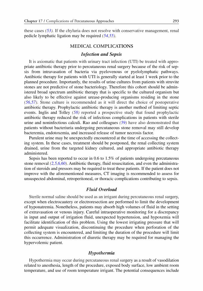
these cases (53). If the chyluria does not resolve with conservative management, renalpedicle lymphatic ligation may be required (54,55).
MEDICAL COMPLICATIONS
Infection and Sepsis
It is axiomatic that patients with urinary tract infection (UTI) be treated with appro-priate antibiotic therapy prior to percutaneous renal surgery because of the risk of sep-sis from intravasation of bacteria via pyelovenous or pyelolymphatic pathways.Antibiotic therapy for patients with UTI is generally started at least 1 week prior to theplanned procedure. Importantly, the results of urine cultures from patients with struvitestones are not predictive of stone bacteriology. Therefore this cohort should be admin-istered broad spectrum antibiotic therapy that is specific to the cultured organism butalso likely to be effective against urease-producing organisms residing in the stone(56,57). Stone culture is recommended as it will direct the choice of postoperativeantibiotic therapy. Prophylactic antibiotic therapy is another method of limiting septicevents. Inglis and Tolley (58) reported a prospective study that found prophylacticantibiotic therapy reduced the risk of infectious complications in patients with sterileurine and noninfectious calculi. Rao and colleagues (59) have also demonstrated thatpatients without bacteriuria undergoing percutaneous stone removal may still developbacteremia, endotoxemia, and increased release of tumor necrosis factor.
Purulent urine may be unexpectedly encountered at the time of accessing the collect-ing system. In these cases, treatment should be postponed, the renal collecting systemdrained, urine from the targeted kidney cultured, and appropriate antibiotic therapyadministered.
Sepsis has been reported to occur in 0.6 to 1.5% of patients undergoing percutaneousstone removal (2,5,6,60). Antibiotic therapy, fluid resuscitation, and even the administra-tion of steroids and pressors may be required to treat these patients. If the patient does notimprove with the aforementioned measures, CT imaging is recommended to assess forunsuspected abdominal, retroperitoneal, or thoracic complications contributing to sepsis.
Fluid Overload
Sterile normal saline should be used as an irrigant during percutaneous renal surgery,except when electrocautery or electroresection are performed to limit the developmentof hyponatremia. Nonetheless, patients may absorb high volumes of fluid in the settingof extravasation or venous injury. Careful intraoperative monitoring for a discrepancyin input and output of irrigation fluid, unexpected hypertension, and hypoxemia willfacilitate identification of this problem. Using the lowest irrigating pressure that willpermit adequate visualization, discontinuing the procedure when perforation of thecollecting system is encountered, and limiting the duration of the procedure will limitthis occurrence. Administration of diuretic therapy may be required for managing thehypervolemic patient.
Hypothermia
Hypothermia may occur during percutaneous renal surgery as a result of vasodilationrelated to anesthesia, length of the procedure, exposed body surface, low ambient roomtemperature, and use of room temperature irrigant. The potential consequences include
Chapter 17 / Complications of Percutaneous Approaches 293
impaired platelet function, altered enzymatic drug clearance, and postoperative shiver-ing causing up to a 400% increase in oxygen consumption (61). The latter problemplaces patients with compromised cardiac reserve at risk for myocardial ischemia andcardiac arrhythmia. The use of warmed irrigating fluid and proper coverage of patients(blankets and heat-preserving surgical drapes) can attenuate hypothermia.
Positioning Related Injuries
It is essential that a patient is properly positioned to prevent a unique set of compli-cations. Brachial plexus damage, shoulder dislocation, other forms of peripheral nerveinjury, and cutaneous trauma can occur if this is not performed properly. Prompt neuro-logic evaluation should be undertaken if neuropraxia is suspected. These injuries usu-ally resolve over time and physical therapy can hasten recovery.
Air Embolism
Air embolism may occur after injection of air or carbon dioxide into the collecting sys-tem or if there is reversal of airflow in an ultrasonic lithotripter (62). Patients typicallymanifest hypoxemia, cardiac instability, and, in extreme cases, circulatory arrest. Amachinery-type murmur may also be heard on cardiac auscultation. Management consistsof placing the patient in a left lateral decubitus position with the head and thorax tilteddownward. A central venous access line is placed through which the air is aspirated.
Deep Vein Thrombus/Pulmonary Embolism
One to three percent of patients undergoing percutaneous renal surgery will developclinically apparent deep veinous thrombosis (2,63). The utilization of thromboembolicdisease prevention stockings, sequential compression devices, and early postoperativeambulation minimize this risk. If postoperative deep venous thrombophlebitis, treatmentgoals are to prevent extension of the thrombus or embolic events (64). Anticoagulationtherapy is initially undertaken, but placement of an inferior vena caval filter may berequired if hemorrhagic complications develop. If the patient has a mature nephrostomytract, anticoagulation is usually well tolerated.
Mortality
Postoperative death is extremely rare and has been reported in 0.1 to 0.3% of patientsundergoing percutaneous renal surgery (1,2). The majority of deaths are because myocar-dial infarction or pulmonary embolism and occurred in high-risk patients. Therefore,careful preoperative medical evaluation, patient preparation, and postoperative cardiacmonitoring should be considered for patients with significant cardiopulmonary disease.
Loss of Renal Function
Patients undergoing uncomplicated percutaneous renal surgery suffer minimal renaldamage. Lechevallier et al. (65) evaluated patients with single photon emission CT priorto and following PCNL and found that small scars usually involving less than 4% ofrenal cortical mass developed in the treated area. Ekelund et al. (66), having evaluatedpatients with pre- and postoperative intravenous pyelography, nuclear renography, andCT, reported maintenance of renal function and only the development of small, discreteparenchymal scars at the tract site. Urivetsky et al. (67) evaluated patients with urinaryenzyme studies before and after PCNL and reported no change in enzyme activity.
294 Matlaga et al.

Some patients treated for staghorn calculi may be at long-term risk for future renalfunctional deterioration. Teichman et al. (68) have reported that 25% of these patientsdevelop renal functional deterioration. This is most likely the result of nonproceduralrelated factors. This is supported by the findings of this group who found that solitarykidney, development of recurrent calculi, hypertension, urinary diversion, and neuro-genic bladder were risk factors for renal functional deterioration.
Renal loss owing to a percutaneous renal procedure is unusual. Acute renal loss isusually the result of uncontrollable hemorrhage. This has been reported to occur in only0.1 to 0.3% of cases. A meta-analysis of the literature on percutaneous removal ofstaghorn calculi indicated that the long-term risk of renal loss is 1.6% (69).
CONCLUSIONS
It is inevitable that complications will periodically occur during and after percuta-neous endorenal surgery. Patients need to be informed about the risks of developingthese complications during preoperative counseling. Proper patient selection and prepa-ration, meticulous operative technique, and fastidious postoperative care help preventthe occurrence and lessen the magnitude of complications. Prompt diagnosis of thecomplication and institution of appropriate measures to rectify the problem will alsolimit its impact.
REFERENCES1. Lee WJ, Smith AD, Cubelli V, et al. Complications of percutaneous nephrolithotomy. AJR Am J
Roentgenol 1987; 148: 177–180.2. Segura JW, Patterson DE, LeRoy AJ, et al. Percutaneous removal of kidney stones: review of 1,000
cases. J Urol 1985; 134: 1077–1081.3. Verstandig AG, Banner MP, Van Arsdalen KN, Pollack HM. Upper urinary tract calculi: extrusion into
perinephric and periureteric tissues during percutaneous management. Radiology 1986; 158: 215–218.4. Evans CP, Stoller ML. The fate of the iatrogenic retroperitoneal stone. J Urol 1993; 150: 827–829.5. Roth RA, Beckmann CF. Complications of extracorporeal shock-wave lithotripsy and percutaneous
nephrolithotomy. Urol Clin North Am 1988; 15: 155–166.6. Lang EK. Percutaneous nephrostolithotomy and lithotripsy: a multi-institutional survey of complica-
tions. Radiology 1987; 162: 25–30.7. Zagoria RJ, Assimos DG, Yap MA, Dyer RB. Obliterative pyeloureteritis: a complication of stone
disease in patients with urinary conduit diversion. J Urol 1993; 150: 961–965.8. Meretyk S, Albala DM, Clayman RV, Denstedt JD, Kavoussi LR. Endoureterotomy for treatment of
ureteral strictures. J Urol 1992; 147: 1502–1506.9. Weir MJ, Honey JD. Complete infundibular obliteration following percutaneous nephrolithotomy.
J Urol 1999; 161: 1274–1275.10. Buchholz NP. Double infundibular obliteration with abscess formation after percutaneous
nephrolithotomy. Urol Int 2001; 66: 46–48.11. Parsons JK, Jarrett TW, Lancini V, Kavoussi LR. Infundibular stenosis after percutaneous
nephrolithotomy. J Urol 2002; 167: 35–38.12. Koolpe HA, Lord B. Eccentric nephroscopy for the incarcerated nephrostomy. Urol Radiol 1990; 12:
96–98.13. Smith AD, Orihuela E, Crowley AR. Percutaneous management of renal pelvic tumors: a treatment
option in selected cases. J Urol 1987; 137: 852–856.14. Streem SB, Pontes EJ. Percutaneous management of upper tract transitional cell carcinoma. J Urol
1986; 135: 773–775.15. Patel A, Soonawalla P, Shepherd SF, Dearnaley DP, Kellett MJ, Woodhouse CR. Long-term outcome
after percutaneous treatment of transitional cell carcinoma of the renal pelvis. J Urol 1996; 155:868–874.
16. White EC, Smith AD. Percutaneous stone extraction from 200 patients. J Urol 1984; 132: 437–438.
Chapter 17 / Complications of Percutaneous Approaches 295

17. Stoller ML, Wolf JS, Jr., St Lezin MA. Estimated blood loss and transfusion rates associated with per-cutaneous nephrolithotomy. J Urol 1994; 152: 1977–1981.
18. Clayman RV, Surya V, Hunter D, et al. Renal vascular complications associated with the percutaneousremoval of renal calculi. J Urol 1984; 132: 228–230.
19. Mahaffey KG, Bolton DM, Stoller ML. Urologist directed percutaneous nephrostomy tube place-ment. J Urol 1994; 152: 1973–1976.
20. Lam HS, Lingeman JE, Mosbaugh PG, et al. Evolution of the technique of combination therapy forstaghorn calculi: a decreasing role for extracorporeal shock wave lithotripsy. J Urol 1992; 148:1058–1062.
21. Davidoff R, Bellman GC. Influence of technique of percutaneous tract creation on incidence of renalhemorrhage. J Urol 1997; 157: 1229–1231.
22. Kaye KW, Clayman RV. Tamponade nephrostomy catheter for percutaneous nephrostolithotomy.Urology 1986; 27: 441–445.
23. Gupta M, Bellman GC, Smith AD. Massive hemorrhage from renal vein injury during percutaneousrenal surgery: endourological management. J Urol 1997; 157: 795–797.
24. Giddens JL, Grasso M. Retrograde ureteroscopic endopyelotomy using the holmium:YAG laser.J Urol 2000; 164: 1509–1512.
25. Kessaris DN, Bellman GC, Pardalidis NP, Smith AG. Management of hemorrhage after percutaneousrenal surgery. J Urol 1995; 153: 604–608.
26. Patterson DE, Segura JW, LeRoy AJ, Benson RC, Jr., May G. The etiology and treatment of delayedbleeding following percutaneous lithotripsy. J Urol 1985; 133: 447–451.
27. Denstedt JD, Clayman RV. Electrohydraulic lithotripsy of renal and ureteral calculi. J Urol 1990; 143:13–17.
28. Grasso M. Experience with the holmium laser as an endoscopic lithotrite. Urology 1996; 48:199–206.
29. Andrews PE, Segura JW. Renal pelvic explosion during conservative management of upper tracturothelial cancer. J Urol 1991; 146: 407–408.
30. Denstedt JD, Eberwein PM, Singh RR. The Swiss Lithoclast: a new device for intracorporeallithotripsy. J Urol 1992; 148: 1088–1090.
31. Granata M, Costanzo V, Condorelli S, Pisciotta F, Matera M, Costantino G. [Percutaneous lithotripsy:how to make it safer. Personal experience]. Minerva Urol Nefrol 2002; 54: 173–177.
32. Kostakopoulos A, Stavropoulos NJ, Picramenos D, Kyriazis P, Deliveliotis C. The Swiss lithoclast:an ideal intracorporeal lithotripter. Urol Int 1995; 55: 19–20.
33. Munver R, Delvecchio FC, Newman GE, Preminger GM. Critical analysis of supracostal access forpercutaneous renal surgery. J Urol 2001; 166: 1242–1246.
34. Gupta R, Kumar A, Kapoor R, Srivastava A, Mandhani A. Prospective evaluation of safety and effi-cacy of the supracostal approach for percutaneous nephrolithotomy. BJU Int 2002; 90: 809–813.
35. Hopper KD, Yakes WF. The posterior intercostal approach for percutaneous renal procedures: risk ofpuncturing the lung, spleen, and liver as determined by CT. AJR Am J Roentgenol 1990; 154: 115–117.
36. Forsyth MJ, Fuchs EF. The supracostal approach for percutaneous nephrostolithotomy. J Urol 1987;137:197–198.
37. Picus D, Weyman PJ, Clayman RV, McClennan BL. Intercostal-space nephrostomy for percutaneousstone removal. AJR Am J Roentgenol 1986; 147: 393–397.
38. Ogan K, Corwin TS, Smith T, et al. Sensitivity of chest fluoroscopy compared with chest CT andchest radiography for diagnosing hydropneumothorax in association with percutaneous nephros-tolithotomy. Urology 2003; 62: 988–992.
39. Pearle MS, Corwin TS, Mullican MA. Comparison of intraoperative chest fluoroscopy, chest radiog-raphy, and chest CT for detecting hydropneumothorax after percutaneous nephrostolithotomy(PCNL). J Endourol 2000; 14: A86.
40. Redorta JP, Gassol JM, Vilaseca AP, Dalmau MR, Robles JM, Wahad AA. Renopleural fistula afterpercutaneous nephrostolithotomy. Urol Int 1988; 43: 104.
41. Appel R, Musmanno MC, Knight JG. Nephrocolic fistula complicating percutaneous nephrostolitho-tomy. J Urol 1988; 140: 1007–1008.
42. Morse RM, Spirnak JP, Resnick MI. Iatrogenic colon and rectal injuries associated with urologicalintervention: report of 14 patients. J Urol 1988; 140: 101–103.
296 Matlaga et al.

43. Neustein P, Barbaric ZL, Kaufman JJ. Nephrocolic fistula: a complication of percutaneous nephros-tolithotomy. J Urol 1986; 135: 571–573.
44. Vallancien G, Capdeville R, Veillon B, Charton M, Brisset JM. Colonic perforation during percuta-neous nephrolithotomy. J Urol 1985; 134: 1185–1187.
45. Hadar H, Gadoth N. Positional relations of colon and kidney determined by perirenal fat. AJR Am JRoentgenol 1984; 143: 773–776.
46. Sherman JL, Hopper KD, Greene AJ, Johns TT. The retrorenal colon on computed tomography: a nor-mal variant. J Comput Assist Tomogr 1985; 9: 339–341.
47. Matlaga BR, Shah OD, Zagoria RJ, Dyer RB, Streem SB, Assimos DG. Computerized tomographyguided access for percutaneous nephrostolithotomy. J Urol 2003; 170: 45–47.
48. Gerspach JM, Bellman GC, Stoller ML, Fugelso P. Conservative management of colon injury follow-ing percutaneous renal surgery. Urology 1997; 49: 831–836.
49. Culkin DJ, Wheeler JS, Jr., Canning JR. Nephro-duodenal fistula: a complication of percutaneousnephrolithotomy. J Urol 1985; 134: 528–530.
50. Goldberg SD, Gray RR, St Louis EL, Mahoney J, Jewett MA, Keresteci AG. Nonoperative manage-ment of complications of percutaneous renal nephrostomy. Can J Surg 1989; 32: 192–195.
51. Kondas J, Szentgyorgyi E, Vaczi L, Kiss A. Splenic injury: a rare complication of percutaneousnephrolithotomy. Int Urol Nephrol 1994; 26: 399–404.
52. Thrasher JB, Snyder JA. Post-nephrolithotomy chyluria. J Urol 1990; 143: 578–579.53. Campieri C, Raimondi C, Dalmastri V, et al. Posttraumatic chyluria due to lymphorenal fistula
regressed after somatostatin therapy. Nephron 1996; 72: 705–707.54. Hemal AK, Gupta NP. Retroperitoneoscopic lymphatic management of intractable chyluria. J Urol
2002; 167: 2473–2476.55. Zhang X, Ye ZQ, Chen Z, et al. Comparison of open surgery versus retroperitoneoscopic approach to
chyluria. J Urol 2003; 169: 991–993.56. Fowler JE, Jr. Bacteriology of branched renal calculi and accompanying urinary tract infection. J Urol
1984; 131: 213–215.57. Ohkawa M, Tokunaga S, Nakashima T, Yamaguchi K, Orito M, Hisazumi H. Composition of urinary
calculi related to urinary tract infection. J Urol 1992; 148: 995–997.58. Inglis JA, Tolley DA. Antibiotic prophylaxis at the time of percutaneous stone surgery. J Endourol
1988; 2: 59.59. Rao PN, Dube DA, Weightman NC, Oppenheim BA, Morris J. Prediction of septicemia following
endourological manipulation for stones in the upper urinary tract. J Urol 1991; 146: 955–960.60. Moskowitz GW, Lee WJ, Pochaczevsky R. Diagnosis and management of complications of percuta-
neous nephrolithotomy. Crit Rev Diagn Imaging 1989; 29: 1–12.61. Roberts S, Bolton DM, Stoller ML. Hypothermia associated with percutaneous nephrolithotomy.
Urology 1994; 44: 832–835.62. Cadeddu JA, Arrindell D, Moore RG. Near fatal air embolism during percutaneous nephrostomy
placement. J Urol 1997; 158: 1519.63. Lee WJ, Smith AD, Cubelli V, Vernace FM. Percutaneous nephrolithotomy: analysis of 500 consec-
utive cases. Urol Radiol 1986; 8: 61–66.64. Patel A, Fuchs GJ. Air travel and thromboembolic complications after percutaneous nephrolithotomy
for staghorn stone. J Endourol 1998; 12: 51–53.65. Lechevallier E, Siles S, Ortega JC, Coulange C. Comparison by SPECT of renal scars after extracor-
poreal shock wave lithotripsy and percuaneous nephrolithotomy. J Endourol 1993; 7: 465.66. Ekelund L, Lindstedt E, Lundquist SB, Sundin T, White T. Studies on renal damage from percuta-
neous nephrolitholapaxy. J Urol 1986; 135: 682–685.67. Urivetsky M, Motola J, King L, Smith AD. Impact of percutaneous renal stone removal on renal func-
tion: assessment by urinary lysozyme activity. Urology 1989; 33: 305–308.68. Teichman JM, Long RD, Hulbert JC. Long-term renal fate and prognosis after staghorn calculus man-
agement. J Urol 1995; 153: 1403–1407.69. Segura JW, Preminger GM, Assimos DG, et al. Nephrolithiasis Clinical Guidelines Panel summary
report on the management of staghorn calculi. J Urol 1994; 151: 1648–1651.
Chapter 17 / Complications of Percutaneous Approaches 297

Complications of UreteroscopicApproaches, Including Incisions
Farjaad M. Siddiq, MD
and Raymond J. Leveillee, MD
CONTENTS
INTRODUCTION
PREPARATION FOR URETEROSCOPY
URETERAL ACCESS
DILATION OF THE URETERAL ORIFICE: IS IT NECESSARY?COMPLICATIONS OF URETEROSCOPY
URETERAL STENTS: ARE THEY NECESSARY?POSTOPERATIVE IMAGING
COMPLICATIONS OF URETEROSCOPIC MANAGEMENT OF UPPER
TRACT TCCCOMPLICATIONS OF URETEROSCOPIC INCISIONS
CONCLUSION
REFERENCES
18
SUMMARY
Ureteroscopy has progressed from cystoscopic examination of a dilated ureter in achild in 1929 and the initial use of rigid ureteroscopes in the 1980s, to its current stateof small caliber semirigid and flexible instruments. In this chapter the authors reviewcomplications of ureteroscopy including those associated with incisional techniqueswhere one would anticipate a higher incidence of complications. They review the his-tory and development of modern ureteroscopes, focusing on engineering advances.Clinical points made include proper patient selection and preparation; proper use ofdilators, wires, and ureteral access sheaths; and the incidence, identification, and man-agement of complications associated with ureterorenoscopy (both intraopertively andpostoperatively).
Key Words: Ureteroscopy; calculi; urinary stones; stricture or ureter; urothelial carcinoma; surgical complications.
From: Advanced Endourology: The Complete Clinical GuideEdited by: S. Y. Nakada and M. S. Pearle © Humana Press Inc., Totowa, NJ
299

INTRODUCTION
Ureteroscopy has progressed from cystoscopic examination of a dilated ureter in achild with posterior urethral valves by Young and McKay (1) in 1929 and the initial useof a rigid ureteroscope by Perez-Castro Ellendt and Martinez-Pineiro (2,3) in the early1980s, to its current state of small caliber semirigid and flexible instruments. In addition,there has been a concurrent improvement in endoscopic lithotrites and other ureteralaccess and interventional accessories. This has facilitated retrograde endoscopic diagno-sis as well as therapeutic intervention for a multitude of upper tract disease processes.Indeed, while the most common application of ureteroscopy remains the management ofurinary calculi, treatment of strictures, upper tract transitional cell carcinoma (TCC),and essential hematuria has become commonplace.
Although ureteroscopy has come to be considered a relatively benign procedure withan acceptably low complication rate, major and minor complications do occur. This chap-ter provides a summary of various complications associated with ureteroscopic access,treatment of calculi, TCC, and strictures, as well as historical perspectives on the trendsin complications of ureteroscopy since its inception. In addition, technical hints to preventcomplications and manage them once they occur will be offered in a timeline extendingfrom patient selection and preoperative preparation to long-term postoperative care.
PREPARATION FOR URETEROSCOPY
Sepsis, active urinary tract infection, and untreated bleeding diatheses are absolutecontraindications to ureteroscopy. All patients should have sterile preoperative urinecultures. In addition, broad spectrum prophylactic antibiotics covering common geni-tourinary pathogens should be administered. In most instances, the use of sufficientanesthesia is critical. Although general anesthesia is preferred, use of local anestheticwith sedation may be adequate in some situations. For therapeutic procedures, we pre-fer a general anesthetic as a sudden move by a lightly sedated patient can be cata-strophic with the ureteroscope in situ. Although regional anesthesia (i.e., spinal) may besafely utilized for distal ureteroscopic procedures, it may not provide adequate analgesiaduring ureterorenoscopy where renal distention may occur.
URETERAL ACCESS
Access failure prohibits ureteroscopy and is a significant complication. The initial steponce the patient is anesthetized and positioned is cystoscopy followed by a retrogradepyelogram to “roadmap” the collecting system. Care must be taken at this seeminglysimple initial step as trauma to the ureteral orifice or intramural mucosa may make sub-sequent ureteroscopy difficult. Ureteral orifices may be difficult to visualize, much lesscanulate, in men with large intravesical prostates. Even if the ureter can be accessed anda wire placed, “J-hooking” of the distal ureter may preclude the insertion of a uretero-scope. Further, the angulation between the bladder neck and the ureteral orifice may betoo severe to negotiate with a semirigid ureteroscope. In this situation, attempting tostraighten the ureter with a super-stiff wire or using a flexible ureteroscope may be help-ful. Women with large cystoceles offer a similar challenge. These ureters, nevertheless,can be canulated if the bladder is lifted transvaginally. Previous pelvic or retroperitonealsurgery or radiation therapy can fix the ureter in the deep pelvis. This increases the riskof perforation significantly, particularly when a rigid instrument is used. Orthopedicabnormalities and contractures, which limit hip mobility and contralateral lower extremity
300 Siddiq and Leveillee

hyperflexion, may hinder the introduction of a ureteroscope. Previously reimplantedureters, ectopic ureters, and duplicated ureters may be difficult to access as well.
If the ureteral orifice can be identified and accessed, a floppy tipped guidewireshould be placed in the renal pelvis under cystoscopic and fluoroscopic guidance. Ifresistance is encountered, other options should be sought. The wire should never beforced. On occasion, if a mucosal flap has been raised, the guidewire may need to beplaced under vision through a ureteroscope. Ureteral stricture, ureteral tortuosity, or animpacted stone may hinder insertion of a wire or ureteroscope. A hydrophilic AngledGlidewire (Terumo, Japan; Microvasive, Boston Scientific Corporation, Watertown,MA) is relatively atraumatic and may safely negotiate a strictured or tortuous ureter.Once wire access is obtained, the stricture can be dilated and the wire exchanged witha super-stiff wire through a ureteral catheter to straighten a tortuous ureter. Impactedstones may be negotiated by injection of lidocaine jelly into the ureter (4). However,systemic absorption of the lidocaine must be considered. An Angled Glidewire may behelpful. This can be exchanged with a stiffer and more secure wire prior to lithotripsy.The exchange may be facilitated by an angled hydrophilic Glide Catheter (5).Nevertheless, if contrast does not bypass the obstruction, it is unlikely that a wire will
Chapter 18 / Complications of Ureteroscopic Approaches 301
Fig. 1. (A) Scout film demonstrates two large distal ureteral calculi, known to be present for severalmonths. (B) Retrograde ureterography demonstrated failure of contrast to bypass the obstructing calculi.(C) The Glidewire coiled at the level of the stones and could not be passed proximally. (D)Ureteroscopy was performed up to the level of the stone, but the Glidewire could not be placed. Inorder to avoid ureteral trauma, no further attempts at retrograde manipulation and ureteroscopy were

302 Siddiq and Leveillee
Fig. 1. (Continued)
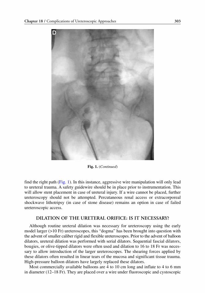
find the right path (Fig. 1). In this instance, aggressive wire manipulation will only leadto ureteral trauma. A safety guidewire should be in place prior to instrumentation. Thiswill allow stent placement in case of ureteral injury. If a wire cannot be placed, furtherureteroscopy should not be attempted. Percutaneous renal access or extracorporealshockwave lithotripsy (in case of stone disease) remains an option in case of failedureteroscopic access.
DILATION OF THE URETERAL ORIFICE: IS IT NECESSARY?
Although routine ureteral dilation was necessary for ureteroscopy using the earlymodel larger (>10 Fr) ureteroscopes, this “dogma” has been brought into question withthe advent of smaller caliber rigid and flexible ureteroscopes. Prior to the advent of balloondilators, ureteral dilation was performed with serial dilators. Sequential fascial dilators,bougies, or olive-tipped dilators were often used and dilation to 16 to 18 Fr was neces-sary to allow introduction of the larger ureteroscopes. The shearing forces applied bythese dilators often resulted in linear tears of the mucosa and significant tissue trauma.High-pressure balloon dilators have largely replaced these dilators.
Most commercially available balloons are 4 to 10 cm long and inflate to 4 to 6 mmin diameter (12–18 Fr). They are placed over a wire under fluoroscopic and cystoscopic
Chapter 18 / Complications of Ureteroscopic Approaches 303
Fig. 1. (Continued)
guidance, taking care to remain distal to the stone if one is present. If the balloon isdeployed alongside the stone, the stone may become impacted or may be extrudedthrough a perforation. Further, the balloon may be ruptured by a sharp stone. The bal-loon can also rupture with overinflation or if the inflation is performed too rapidly. Thiscan be severely traumatic given the pressure (18 atm/~265psi) under which contrast willexit through a “pinhole” resulting in bleeding, mucosal tearing, and extravasation.Mucosal injury is less likely with slow radial balloon dilation until the “waist” is elim-inated, never exceeding manufacturer specified tolerances. Garvin and Clayman (6)demonstrated no strictures and a 20% incidence of transient vesicoureteral reflux from bal-loon dilation of the ureteral orifice to 24 Fr at 6 weeks. Nevertheless, balloon dilation togreater than 18 Fr is rarely necessary given the current diameter of ureteroscopes (<10 Fr).
Ureteral access sheaths may also be used to dilate the ureteral orifice. These sheathsalso allow ureteral access under vision through the lumen of the sheath and are particu-larly advantageous during procedures involving ureteral biopsy or treatment of a largestone where repeat ureteral insertion of the ureteroscope may be necessary. Earlier ureteralaccess systems required multiple steps and sequential passage of rigid dilators with a peelaway outer sheath prior to ureteroscope insertion (7,8). However, the process was riskywith a 31% perforation rate in one study (9). In contrast, the current ureteral access sheathis a flexible and hydrophilic single step two-piece system available in a variety of lengthsand diameters. Kourambas et al. (10) evaluated the newer sheaths in a prospective ran-domized study. Operative time and costs were lower in patients who underwent accesssheath dilation. Stents were required in 43% of patients undergoing access sheath dilationand 100% of patients undergoing balloon dilation. Although use of the access sheath min-imized ureteral trauma, ureteral dilation was successful in 71% of patients in comparisonwith a 100% success rate for the balloon dilator. They reported no intraoperative compli-cations from the newer sheaths with no strictures at 3 months postoperatively. Anotherstudy evaluated ureteral blood flow in a porcine model after access sheath insertion (11).A transient decrease in blood flow was noted with restoration of blood flow to near-base-line during the course of the study. This effect was most pronounced with the larger diam-eter sheaths. Histologically, there was no evidence of ischemic damage. Hence, theauthors concluded that the ureteral access sheath was safe but that care must be taken inselecting an appropriate size sheath for each individual case.
Stoller et al. (12) found ureteral dilation necessary in 16% of their population usingsemirigid ureteroscopes ranging from 9.5 to 12.5 Fr and concluded that routine dilation ofthe ureteral orifice was not necessary. Netto et al. (13) came to the same conclusion, fail-ing to access 1 ureter out of 73 without dilation using semirigid ureteroscopes. Kourambaset al. (10) noted that 24% of their patients required dilation using 6.5-Fr semirigid and 7.5-Fr flexible ureteroscopes. Indeed, although ureteral dilation was once considered an essen-tial step in ureteroscopy, the authors find it infrequently necessary in current clinicalpractice and, therefore, routinely access the ureter atraumatically without ureteral dilation.
COMPLICATIONS OF URETEROSCOPY
The incidence of major and minor complications has decreased significantly since theinception of ureteroscopy. The decline in the incidence of ureteral injury has been partic-ularly significant (Table 1). This trend may be counterintuitive given the large number ofinterventional procedures ureteroscopy currently facilitates. Nevertheless, with theadvent of improved imaging, smaller diameter endoscopes, and increasing surgeon expe-rience, the incidence of complications has actually decreased while the success rate has
304 Siddiq and Leveillee

increased. In 1986, Carter et al. (14) reported a 67% success rate for ureteroscopicureterolithotomy with a 15% open ureterolithotomy rate secondary to ureteroscopic fail-ure. No lithotrite was used for in situ stone fragmentation. The main complication wasfailure to access the ureter or reach the stone in 15% of patients. Major ureteric injuryrequiring open repair occurred in 2.4% of patients with additional perforations managedconservatively in 3.2%. Flam et al. (4) subsequently reported an overall success rate of78% for ureteroscopic stone removal with ureteral injury occurring in 4% of patients.They found that larger (>8 mm) stones located in the proximal ureter were the most dif-ficult to treat resulting in the poorest success rate and highest complication rate. In addi-tion, surgeon experience had a positive effect on outcomes as evidenced by later patientsfaring better. In a similar study, Blute et al. (15) reported a 5.2% ureteral injury rate.
These studies uniformly employed rigid endoscopes larger than 10 Fr. Ureteral dila-tion to 18 Fr, using a balloon or serial dilators, was standard practice. Earlier studiesdescribed stone removal without fragmentation. Technology has advanced significantlysince these earlier reports. The advent of small caliber semirigid and flexible uretero-scopes as well as improvements in accessories has allowed the endourologist to performcomplex stone manipulation in the upper urinary tract with greater success and minimalmorbidity. The success of the technology is also evident in the rapid proliferation ofindications for ureteroscopy with calculus extraction as the indication for ureteroscopyin 67% of patients in current series and 84% of patients in early series (16).
Harmon et al. (16) reported on the impact of technological advancement and surgeonexperience on ureteroscopic outcomes by comparing a contemporary cohort of patientswith a cohort previously reported by Blute et al. (15) from the same institution. Theoverall ureteroscopic success rate increased from 86 to 96%. Failure in the earlier expe-rience was largely owing to the inability to access the ureter or approach the stone (54%of cases). In contrast, failure in the newer cohort was largely owing to the impassableureteral strictures (63%). The overall complication rate decreased from 20 to 12% andthe rate of significant ureteral injury decreased from 5.2 to 1.5%. Stricture formationdecreased from 1.4 to 0.5%. Grasso and Bagley (17) reported an overall complicationrate of 11.2% with a stricture rate of 0.5% and no significant ureteral injuries, usingsmall diameter actively deflectable flexible ureteroscopes.
Although the overall complication rate has declined, one cannot ignore the possibil-ity of major or minor complications that may occur. Indeed, although appropriate pre-cautions may decrease the frequency and seriousness of complications, accidents willhappen. Complications can be divided into perioperative and postoperative. Each cate-gory can be further subdivided into major and minor complications and early and late
Chapter 18 / Complications of Ureteroscopic Approaches 305
Table 1Trends in Ureteroscopic Ureteral Injury
Major Ureteral Injury (%)
Authors No. of Procedures Unspecified Perforation Avulsion Stricture(%)
Carter et al. (14) 125 4.0 — 4.0Flam et al. (4) 180 4.0 — — 1.0Blute et al. (15) 346 4.6 0.6 1.4Harmon et al. (16) 209 1.0 0.0 0.5Grasso et al. (17) 560 0.0 0.0 0.5

306 Siddiq and Leveillee
Table 2Classification of Complications
Perioperative complications Postoperative complications
Major Early• Avulsion • Colic• Perforation • Infection• Intussusception • Bleeding• Equipment failure Late
Minor • Ureteral necrosis• Bleeding • Stricture• Mucosal tear • Reflux• Proximal calculus migration• Extrusion of calculi• Thermal injury
Fig. 2. Mechanism of ureteral avulsion. (A) Passage of stone basket proximal to large calculus.(B) Stone is engaged and retrograde extraction attempted. Note proximal thinning of ureteral wall(small arrow) prior to avulsion. (C) Complete avulsion with persistent pulling.

complications, respectively (Table 2). Some of the more common injuries and suggestionson how to treat and avoid them will be discussed.
Perioperative Complications
MAJOR
Avulsion. Avulsion is a major intraoperative complication usually resulting fromblind forceful manipulation. The most common scenario is forceful extraction of a largeproximal ureteral calculus that has been engaged in a basket (Fig. 2). The most commonsite of ureteral avulsion is the proximal third of the ureter (18). The ureter is thinnest atthis level with the least well-defined muscular layers and a greater percentage of fibro-connective tissue, placing this portion of the ureter at greatest risk of avulsion (19). Theincidence of this catastrophic injury ranges from 0.5% in the earlier series (20) to 0%in the more recent series (17,21).
If a ureteral avulsion is recognized, the procedure must be stopped and percutaneousdrainage should be obtained. A safety wire will not prevent an avulsion and, if the injuryis severe, a stent offers inadequate drainage. Primary repair may be feasible if a small por-tion of the ureter is injured. However, if the defect is large or if the ureter is devascular-ized at the site of avulsion and significant debridement is required, more complexreconstruction including ileal interposition and renal autotransplatation may be required.If the kidney functions poorly, nephrectomy may be an option. Transureteroureterostomyis contraindicated in patients with nephrolithiasis or upper tract TCC. The best therapy forthis complication is prevention. Clearly, stones that have failed to pass or are larger thanthe diameter of the ureteroscope or the nondilated ureter will prove difficult to removewithout lithotripsy. These stones should be fragmented prior to attempts at retrieval. Thetime and money savings hoped for by taking a “short cut” are miniscule compared to thecost of trauma to the patient. If a situation occurs where the stone is entrapped in a basketand cannot be removed, the basket should be disassembled and the stone fragmented afterthe ureteroscope is reinserted alongside the wire allowing removal of the smaller stoneand basket. Removing the basket from the ureteroscope may not be necessary in uretero-scopes with two channels. The basket should never be forcefully pulled (Fig. 3).
Perforation. The incidence of ureteral perforation ranges from 0% in the more recentflexible ureteroscopy series (17) to as high as 17% in earlier series (22). Although major
Chapter 18 / Complications of Ureteroscopic Approaches 307
Fig. 3. The golden rule: Don’t pull too hard!

perforation may occur during forceful manipulation of the ureteroscope, it more commonlyresults from an endoscopie lihotrite. Santa-Cruz et al. (23) studied the perforation potentialof four lithotripters ex vivo. The electrohydraulic lithotripter (EHL) and the holmium:YAG(Ho:YAG) laser could easily perforate the ureter while the coumarin pulsed-dye laser andthe pneumatic impactor had a significant margin of safety. Nevertheless, the Ho:YAG laserwas unable to perforate the ureter at a distance of greater than 2 mm, providing a marginof safety if the laser is directed away from the ureteral mucosa. Therefore, the electrohy-draulic lithotripter offers the smallest margin of safety (23,24). In an analysis of predictivefactors of ureteroscopic complications, Schuster et al. (21) reported perforations in 15 of322 patients undergoing ureteroscopy (4.7%). The type of ureteroscope used (flexible orsemirigid) was not contributory. There was, however, a significant association of ureteralperforation with operative time, correlating with the difficulty of the procedure. Eightureteral perforations occurred while extracting impacted stones. An additional six perfora-tions occurred while trying to gain access past an impacted stone.
If a perforation is noted intraoperatively, the presence of a preplaced safety guidewirecannot be overemphasized. The procedure should be stopped and the ureter stented inanticipation of a staged procedure. Persistence may convert a minor perforation to a majorperforation with significant extravasation or complete avulsion. If water or another hypo-tonic solution is being used, the irrigation should be changed to saline. Fluid is readilyabsorbed from the retroperitoneum. Extravasation of hypotonic fluids may result in severesymptomatic hyponatremia. Most ureteral perforations will heal spontaneously with a dou-ble-J stent in place and appropriate antibiotic coverage (4). Rarely, placement of percuta-neous nephrostomy to drain the kidney or a percutaneous abdominal drain to evacuate aurinoma may be necessary. Radiological evaluation of the affected ureter is critical 4 to 6wk after the stent is removed to screen for ureteral stricture formation and obstruction.
Intussusception. Ureteral intussusception is a rare complication involving avulsionof the ureteral mucosa (Fig. 4). It can occur in both an antegrade and retrograde direc-tion. Intussusception can occur with forceful extraction of large ureteral calculi. It hasalso been reported in association with ureteral polypoid TCC (25). Park et al. (26)described retrograde intussusception caused by retrograde pyelography. Another casereport described retrograde intussusception of the ureter before ureteroscopy duringdilation of the ureter with a 12-Fr peel-away sheath (27). The injury went unrecognized.Ureteroscopy and stent placement was possible through the intussuscepted segment.The only suspicious finding was that the ureteral lumen appeared pale white and nar-row. Obstruction developed immediately once the stent was removed, requiring uretero-neocystostomy. Prevention of this injury requires a careful and gentle technique. Asafety guidewire should always be present prior to any ureteral manipulation.Instruments should be advanced without force and under vision where possible.
Equipment Failure. The current state of ureteroscopy is entirely dependent on com-plex and costly equipment. Poor vision or a malfunctioning accessory instrument canresult in patient injury. Instrument breakage in the patient has been reported (4). Andersonet al. (28) recently reported a fractured flexible ureteroscope with locked deflection in siturequiring cutting the ureteroscope and open extraction. Postoperative evaluation revealedthat a deflection ring in the distal portion of the scope had failed causing opposing controlcables to slide to the same side and locking the ureteroscope in a kinked position. Theauthors recommended checking all instruments before introducing them into patients.
Nevertheless, poor handling and damage to the instrumentation caused by surgeonerror can also be considered a complication. Firing an EHL probe close to the uretero-
308 Siddiq and Leveillee

scope tip can crack the lens as easily as it can fragment a stone. Similarly, firing a laserfiber in close proximity or within a ureteroscope can result in costly damage. The flex-ible ureteroscopes are fragile and most susceptible to damage. Sharp angulations of thescope during storage and cleaning may damage the scope. These instruments shouldnever be passed through a cystoscope sheath as kinking at the sheath inlet will damagethe ureteroscope. Forceful attempts at passage of an instrument through a flexedureteroscope will also damage the working channel. In fact, we advocate straighteningthe scope prior to advancement of an accessory instrument. As with prevention of com-plications to patients, prevention of damage to equipment requires a gentle touch.
MINOR
Bleeding. Minor bleeding secondary to ureteral trauma is a frequent finding duringureteroscopy. It is seldom severe and transfusion is required only rarely (20). Morecommonly, it obscures vision. The incidence of bleeding has ranged from 3.1 to 0%(15–17,29). Minor bleeding is usually self limiting. Nevertheless, severe bleeding may
Chapter 18 / Complications of Ureteroscopic Approaches 309
Fig. 4. Ureteral intussusception can occur with advancement of the ureteroscope if excessive force isapplied or if the ureter is narrower than the scope.

occur requiring embolization or open repair. This is more likely when incisions of thecollecting system are undertaken.
Mucosal Tear. Minor mucosal injury to the ureter is common, especially duringureteral dilation or insertion of the ureteroscope (Fig. 5). Mucosal trauma can also occurduring stone manipulation. These injuries are self limiting and will usually heal within48 to 72 hours with conservative management and drainage with a double-J stent. Asmall mucosal abrasion can be converted to a submucosal tunnel with inappropriatepassage of a ureteroscope, wire, or catheter, causing devascularization of the ureter.This may result in postoperative stricture formation or, rarely, ureteral necrosis (22).
Proximal Calculus Migration. Proximal calculus migration is no longer considereda treatment failure given the widespread availability of extracorporeal shockwavelithotripsy and flexible ureteroscopy. Nevertheless, proximal migration of a calculusdoes prolong anesthesia and operative time and may submit the patient to a secondprocedure. Placing the patient in a reverse Trendelenburg position and minimizing theirrigant pressure can limit proximal stone migration. Various devices ranging from theDretler laser basket to the Lithocatch and Parachute baskets to the Passport balloon(Microvasive, Boston Scientific Corp.) have been used to prevent proximal stonemigration (30). These devices were suboptimal for a variety of reasons ranging frominadequate ureteral occlusion to limiting intraluminal space as all required an additionalsafety wire. The Stone Cone (Microvasive, Boston Scientific Corp.) is the newestadvance. It prevents stone migration and promotes sweeping multiple stone fragmentsin a single pass, while substituting for the ureteral guidewire (30,31).
Extrusion of Calculi. The reported incidence of retroperitoneal extrusion of a calculusthrough a ureteral perforation is between 0.5 and 2.3% (4,20,32–36). The concern is thatparaureteral calculi hinder ureteral healing and increase the risk of stricture formation.There is additional apprehension that extrusion of infected or struvite calculi may result
310 Siddiq and Leveillee
Fig. 5. Mucosal tear noted after balloon dilation in the mid-ureter to facilitate insertion of a ureteroscope.

in periureteral abscess formation (33,36). Several studies, however, have shown that therisks of stricture and periureteral abscess formation are negligible. Evans and Stoller(35) reported a 1.2% incidence of iatrogenic retroperitoneal stones in 400 patientsundergoing ureteroscopy. No abscess formation or infectious complications were noted.One patient developed a ureteropelvic junction (UPJ) stricture requiring open repair, butthe stricture formation was felt to be a result of aggressive ureteroscopic manipulationin an attempt to retrieve the stone. Chang and Marshall (34) also documented anincreased severity of ureteral injury with persistent attempts to remove the stone.Kriegmair and Schmeller (37) provided long-term follow-up of 15 patients with parau-reteral calculi. One stricture was noted, which was easily treated endoscopically. Noabscesses were noted. More recently, Lopez-Alcina et al. (38) reported a 1.1% incidenceof calculus extrusion in 1047 patients. At a mean follow-up of 18 mo, no infection or sec-ondary ureteral strictures were found.
Owing to the low incidence of secondary complications from extrusion of calculiand risk of worsening injury, persistent ureteroscopic manipulation to retrieve anextruded stone is not recommended. We suspect that the real risk factor for strictureformation is the perforation itself and the associated extravasation rather than theextruded calculus. Stent drainage along with a short course of perioperative antibioticsis recommended by the above authors. Follow-up imaging should be obtained to con-firm adequate renal drainage (Fig. 6). The infection stone may be followed with a com-puted tomography (CT) scan to rule out abscess (38). However, given the extremelylow risk of abscess formation, imaging should be guided by clinical suspicion. Patientsshould be educated about persistent calculi to avoid future mismanagement by otherphysicians (35).
Thermal Injury. Ureteroscopic treatment of stones is often performed with lithotriteswhich can cause thermal injury to the ureter, resulting in delayed fibrosis and strictureformation. The ultrasonic lithotripter achieves fairly high temperatures and can causethermal ureteral injury if not allowed to cool periodically (22). It is seldom used sincethe advent of EHL and Ho:YAG. EHL also generates enough heat to cause thermal orblast injury to the ureteral wall (23,24). In recent years, lasers have become the mostcommonly used lithotrites. The coumarin pulsed-dye laser is not absorbed by hemoglo-bin or soft tissues and is not associated with a risk of thermal injury. The Ho:YAG lasernot only fragments, but also vaporizes stone to fine dust. It can also cause thermal injuryand perforation, but there is a margin of safety if the laser is kept at a distance greaterthan 2 mm from the ureteral wall (23). In addition, adequate irrigation is critical to allowcooling and prevent thermal injury. Recently, a pulsed solid-state laser system that hasbeen used for biliary and salivary lithiasis has been applied to urinary calculi. TheFrequency-Doubled Double-Pulse Neodymium:YAG (FREDDY) laser (World ofMedicine, Berlin Germany) lacks clinical data regarding efficacy and safety in treat-ment of urinary calculi. However, it has been shown to be an efficient lithotripter invitro with only superficial tissue injury (39,40).
Postoperative Complications
EARLY
Colic. Postoperative colic following a ureteroscopic procedure is common and maybe a result of transient obstruction from ureteral edema, clots, or stone fragments. Theincidence ranges from 3.5 to 9.0% (16,17,29). This can be averted with placement of a
Chapter 18 / Complications of Ureteroscopic Approaches 311

312 Siddiq and Leveillee
Fig. 6. (A) Extruded calculus. Kidneys, ureter, and bladder after ureteroscopy complicated by perfo-ration and placement of a percutaneous nephroureterostomy tube. (B) Antegrade nephrostogramdemonstrates no obstruction several weeks after the injury. Antegrade ureteroscopy confirmed that thecalculus was not in the ureteral lumen with minimal impression in the mucosa. No further managementwas undertaken.
stent, which has its own attendant morbidities. Colic often lasts less than 24 to 28 hoursand can be controlled with oral analgesics. Flam et al. (4) reported a 1.1% incidence ofpostoperative admission for control of flank pain. The necessity of stent placement pos-tureteroscopy has recently been questioned and is addressed below.
Infection. Symptoms of postoperative infection range from a low-grade fever topyelonephritis with bacteremia and sepsis. Significant infection in the presence of ster-ile preoperative urine and use of preoperative antibiotics is rare, occurring in 0.3 to1.3% of patients in a large series (14,15,19). Nevertheless, low-grade fever is muchmore common, occurring in as many as 6.9% of patients in one series (29). A clinicalhistory of urinary tract infection (UTI) or infectious calculi increases the risk of postop-erative infection considerably (18). If purulent drainage is noted during any portion ofupper tract evaluation, the collecting system should be drained and further manipulationhalted until infection has resolved. Ureteroscopy in a febrile patient with an obstructingstone is contraindicated and can result in life-threatening urosepsis.
LATE
Ureteral Necrosis. Ureteral necrosis is the postoperative outcome of a severe intra-operative injury involving intussuception, severe thermal injury, or mucosal dissection.Lytton (41) reported loss of the entire ureter attributed to partial perforation of the ureterfollowed by devasculariztion secondary to submucosal irrigation. Kaufman (42) reporteddistal ureteral necrosis in a patient after ureteral injury and extravasation during stoneextraction. This is a rare but major complication and prompt diagnosis is dependent onclinical suspicion.
Stricture. Ureteral stricture formation after ureteroscopy is a major late complicationthat subjects patients to additional procedures or, if left untreated, puts them at risk forrenal loss. Although ureteral stricture rates as high as 37% after instrumentation havebeen reported in one series (43), most series document stricture rates less than 2% withuncomplicated ureteroscopy (15–17,29). There has been a trend toward a decrease inthe incidence of strictures. Blute et al. (15) noted a 1.4% stricture rate in 1988, whereasHarmon et al. (16) noted a 0.5% stricture rate in 1997 at the same institution.
In contrast, Roberts et al. (44) noted a 24% incidence of ureteral strictures in 21patients undergoing ureteroscopy for impacted stones. Four of the five patients whodeveloped postoperative stricture had suffered ureteral perforation during ureteroscopy.Therefore, the authors concluded that ureteral perforation was a significant risk factorfor stricture formation in patients with impacted stones. Mugiya et al. (45) demonstrateda stricture in 17% of patients undergoing ureteroscopic management of impacted stones.They found a correlation between the duration of impaction and the incidence of stric-tures. Histological studies have revealed chronic inflammation, fibrosis, and urothelialhypertrophy at the site of the impaction (44). Dretler and Young (46) found granuloma-tous inflammation around residual embedded stone fragments at the site of strictures inpatients who underwent stone fragmentation prior to extraction. They suggested thatresidual stone fragments and the resultant inflammation were an etiological factor instricture formation.
The inflammation in progress around impacted stones may predispose to perforationin addition to stricture formation. Contradictory evidence exists regarding the role ofperforation in stricture formation. Kramolowsky (43) reported strictures in 37% ofpatients who suffered a perforation. In contrast, Stackl and Marberger (32) found nocorrelation between perforation and stricture formation with no late complications at a
Chapter 18 / Complications of Ureteroscopic Approaches 313

minimum of 16-months follow-up. Lytton (41) had similar outcomes with no strictureformation in 12 patients managed conservatively after perforation.
Therefore, the etiology of stricture formation is likely multifactorial. Impactedstones, ureteral perforation and extravasation, mechanical trauma from the ureteroscopeor an accessory instrument such as a lithotrite, ureteral ischemia, and thermal injurymay all play a role in stricture formation (24). The common thread here is urothelialtrauma. In addition, inflammation is a key step towards stricture formation (44,47).
The management of ureteral strictures is dependent on length and duration of thestricture as well as the degree of periureteral fibrosis. Short annular strictures respondwell to endoscopic management with balloon dilation or incision. Longer strictures andthose associated with significant periureteral fibrosis are best managed with open orlaparoscopic reconstruction. The type of procedure performed is variable and dependson the location and length of the ureteral stricture. If the ipsilateral renal function ispoor, nephrectomy remains an option in lieu of reconstruction.
Reflux. Vesicoureteral reflux (VUR) has been described as a late complication ofureteroscopy. However, VUR in uninfected adults is of little clinical significance (22).Garvin and Clayman (6) demonstrated a 20% incidence of VUR after ureteral dilationto 24 Fr. In 1999, Richter et al. (48) found a 10% incidence of VUR after ureteral dila-tion to 13.5 Fr. These authors followed their patients prospectively; the patients demon-strated complete resolution of reflux at 2 weeks postoperatively. Further evaluation orscreening for this complication is unnecessary given the low incidence of reflux afterureteroscopy and the minimal pathologic effect of VUR on a fully developed adult kid-ney.
URETERAL STENTS: ARE THEY NECESSARY?
Routine placement of ureteral stents in all patients after ureteroscopy has been consid-ered essential (24). The evidence to support this practice is sparse and based on animal mod-els demonstrating ureteral injury and obstruction after ureteral dilation (49,50). Thepresumption is that placement of a stent serves to protect against postoperative complica-tions including ureteral obstruction from edema, clot or stone fragments, as well as strictureformation (16).However, ureteral stents are not without their own morbidity. Complicationsassociated with stents include bladder and flank pain, irritative voiding symptoms, infec-tion, stent migration, encrustation, and retention in patients lost to follow-up. Several stud-ies report a 10 to 85% incidence of stent related symptoms (51–53), whereas others note a94 to 100% rate of symptom resolution after stent removal (51,54,55). In addition, stentsadd to the operative time and cost. Cystoscopy is required to remove the stent, unless a pullstring is left. Given the relatively atraumatic state of current ureteroscopic equipment andtechnique, elimination of stents in ureteroscopy patients has become a viable option.
There is mounting evidence supporting the selective use of ureteral stents. Hosking etal. (56) reviewed 93 consecutive unstented patients, 88% of whom also underwentureteral dilation. The majority of patients had no postoperative pain or required minimaloral analgesics. Rane et al. (57) treated distal stones using semirigid ureteroscopes with-out ureteral dilation with similar results and minimal postoperative flank pain. In addition,four randomized trials have demonstrated similar complication rates between stented andunstented patients, with the stented groups experiencing significantly greater irritative andpainful symptoms (58–61). Stented patients also had longer operative times and higheroverall cost (59). No strictures were noted on postoperative imaging in unstented patients.
314 Siddiq and Leveillee

Although three trials exluded patients undergoing ureteral dilation, 57% of patients with-out stents in one study underwent intraoperative distal ureteral dilation (60). Thesepatients did as well as the unstented patients who did not undergo ureteral dilation. Re-admission rates among the unstented patients were 3 to 7% in the different studies withshort-term stent placement required in two patients in a single study (60).
Recently, Hollenbeck et al. (62) retrospectively evaluated 219 patients undergoingstentless ureteroscopy to identify factors associated with postoperative complications inthis group. Renal pelvic stones, lithotripsy, bilateral procedures, operative time 45 min-utes or greater, recent/recurrent UTIs, diabetes mellitus, and history of urolithiasis wereassociated with postoperative morbidity in this unstented cohort. The authors suggestthat these findings may serve as guidelines for selecting patients who may undergostentless ureteroscopy.
The current standard of care regarding stenting after ureteroscopy is evolving. Clearly,a large number of patients undergoing uncomplicated diagnostic and/or therapeuticureteroscopy may safely remain unstented. Nevertheless, common sense and clinicaljudgment should prevail. Patients undergoing complicated ureteroscopy with mucosaltrauma, perforation, impacted stones, or solitary kidneys should be stented. We continueto stent patients undergoing ureteral dilation and bilateral ureteroscopy.
POSTOPERATIVE IMAGING
Postoperative imaging is another evolving issue in endourology. The rationalebehind routine surveillance postoperative radiographic imaging has been detection ofsilent obstruction or stricture formation that may have an adverse effect on renal func-tion. However, the advent of small caliber urteroscopes, safer lithotripters, and otheraccessories has resulted in low postoperative complication and high success rates. Thishas led some to suggest that routine postoperative imaging may not be necessary afterureteroscopy (63,64). In a retrospective review of 189 patients undergoingureteroscopy, Karod et al. (64) found that although all 10% of patients with radi-ographically confirmed obstruction had flank pain, none of the asymptomatic patientswere obstructed on radiologic follow-up at a median of 60 days. Stricture was thesource obstruction in 0.8%. They concluded that routine postoperative imaging to ruleout obstruction is not necessary in asymptomatic patients, as obstructed patients willpresent with flank pain.
However, in another retrospective study (65), 30 of 241 patients (12.3%) devel-oped postoperative obstruction at a mean follow-up of 5.4 months. Although 23 ofthese 30 patients (76.7%) had pain, 7 patients (23.3%) with obstruction were asymp-tomatic. Therefore, silent obstruction developed in 2.9% of the total populationundergoing ureteroscopy. One patient with asymptomatic obstruction receivedchronic hemodialysis for renal failure. Although patients with pain afterureteroscopy were more likely to have obstruction than those without pain, noparameters reliably predicted silent obstruction. The authors felt the possibility ofmissing 2.9% of patients with silent obstruction posed a significant risk and recom-mended routine imaging of the collecting system within 3 months of ureteroscopy.The use of pain to predict obstruction in another study had a negative and positivepredictive value of 83 and 75%, respectively (63).
There is likely a cohort of postureteroscopy patients who do not require routine imag-ing. Absence of symptoms, nevertheless, is not an adequate criterion to omit imaging as
Chapter 18 / Complications of Ureteroscopic Approaches 315

the morbidity of undiagnosed obstruction is significant. Other variables may exist thatcould suggest obstruction, or lack thereof. However, these are lacking to date. In themeantime, the authors continue to image patients postureteroscopy. Depending on theclinical situation, a renal ultrasound, intravenous pyelography, CT, or a nuclear scanmay be adequate.
COMPLICATIONS OF URETEROSCOPIC MANAGEMENTOF UPPER TRACT TCC
Ureteroscopic management of upper tract TCC has become routine with the availabilityof small caliber semirigid and flexible ureteroscopes allowing tumor visualization, biopsy,resection, or ablation. Whereas nephroureterectomy remains the standard for patients withhigh-grade TCC and most patients with low-grade TCC, endoscopic tumor therapy is anoption in patients with solitary kidneys, renal insufficiency, bilateral TCC, or significantmedical comorbidities (66). More recently, attention has turned to renal preservation inpatients with normal contralateral kidneys and small volume focal low-grade upper tractTCC. Recent studies have shown that this is a safe therapeutic modality in this population,which traditionally would have undergone a nephroureterectomy (67,68). Life-long fre-quent endoscopic and radiological surveillance is required. Complications of ureteroscopicmanagement of upper tract TCC include loss of access, equipment failure, and bleedingand are similar to complications of ureteroscopy in general, which have already been dis-cussed. Perforation and stricture formation, however, deserve additional mention.
Perforation is a rare occurrence. Grasso and Bagley (17) reported no perforations in101 patients undergoing treatment of upper tract TCC. However, Elliott et al. (67) andMartinez-Pineiro et al. (69) reported two and four perforations, respectively. Perforationcan occur with the guidewire or the endoscope. In addition, the ureter may be perforatedwhen a grasper or basket is used to biopsy the tumor. The basket and tumor sampleshould be withdrawn under vision to prevent ureteral avulsion. Injury is more likelywhen tissue other than the tumor is grasped or the amount of tissue grasped is large. Theureter can also be perforated with electrocautery (the Ho:YAG and theneodymium:YAG [Nd:YAG] laser) if the power settings are too high, the ablation toodeep, or if the instrument is misdirected onto the ureteral wall. Most ureteral perfora-tions can be managed conservatively with a stent (69). Thus the presence of a safetywire is critical. Once a perforation is diagnosed intraoperatively, discontinuation of theprocedure should be considered to prevent tumor seeding. Nevertheless, to our knowl-edge, retroperitoneal seeding after perforation has not been reported. Larger perfora-tions may require percutaneous drainage or even nephroureterectomy (66).
The incidence of ureteral strictures after treatment of upper tract TCC has rangedfrom 5 to 25% (70). Chen and Bagley (66) reviewed 139 patients in five series andfound an overall stricture incidence of 8.6%. This is in stark contrast to the 0.5% stric-ture rates reported by Harmon et al. (16) and Grasso and Bagley (17) in series with largestone populations. The higher stricture rate after treatment of upper tract TCC comparedwith stones is largely because of the different targets during therapy. Although stonesabsorb most of the energy during laser lithotripsy, treatment of TCC requires that thelaser, resectoscope, or fulgerating probe be directed at the urothelium. Patients under-going circumferential or deep ablation are at higher risk of scarring and stricture forma-tion. The Nd:YAG laser may result in less stricture formation than electrocautery (71).However, the Nd:YAG laser penetrates to a depth of 5 to 6 mm and may be more damag-
316 Siddiq and Leveillee

ing than the Ho:YAG laser, which penetrates only to 0.5 mm. Keeley et al. (72) reportedtwo strictures in 19 patients using fulgeration and/or the Nd:YAG laser and none in 22patients using the Ho:YAG laser. Directing the tip of the laser fiber only onto the visi-ble tumor can minimize the risk of stricture formation. If a stricture develops, manage-ment is similar to strictures resulting from other causes. However, the strictured areashould be biopsied to rule out a malignant stricture.
Incomplete tumor resection is a significant complication of endoscopic managementof upper tract TCC and is more common with large tumors (>1.5 cm) and tumors locatedin the lower pole of the kidney. Although significant bleeding is rare, clot and debris canobscure the margins of larger tumors. If the tumor is low grade, a staged approach maybe used with retreatment at a later date. However, nephroureterectomy should be seri-ously considered in patients whose tumors are too large or multifocal to adequatelyablate endoscopically even if low grade, the tumor is inaccessible, or if it is high grade.
COMPLICATIONS OF URETEROSCOPIC INCISIONS
Ureteroscopic incisions can be used to treat a variety of upper tract obstructiveprocesses including ureteral stricture, UPJ obstruction, calyceal diverticulum, andinfundibular stenosis. Complications attendant to ureteroscopy, in general, may occurwith upper tract incisions as well. Incising the upper tract, however, involves purposefulperforation with healing over a stent. Several studies have addressed ureteroscopicendopyelotomy (73–75). The complication rates are low with no intraoperative or imme-diate postoperative complications reported in a recent series by Giddens and Grasso (76).
In addition to stent-related complications and urinoma formation, the major risk ofureteroscopic incisions is intraoperative hemorrhage. An incision at the UPJ may inad-vertently cut through a crossing vessel resulting in immediate bleeding. Similarly, inci-sion of a stricture over the iliac vessels or injury to an intrarenal artery during incisionof an infundibulum may result in severe bleeding. The key to management is preven-tion of vascular injury. Knowledge of renal vascular anatomy is imperative. All inci-sions at the UPJ should be directed laterally. Posterior incisions should be avoided atthe iliacs. Bagley et al. (77) have championed the use of intraluminal ultrasonographyto define vascular anatomy and direct incisions away from them. If hemorrhage isencountered or the patient complains of excessive postoperative pain, emergent imag-ing should be performed. Persistent bleeding with a continued transfusion demand or anunstable patient necessitates emergent arteriography and selective embolization. In rareinstances, a nephrectomy may be necessary (78).
CONCLUSION
Clearly, the current state of ureteroscopy has provided an efficacious minimally inva-sive alternative for a number of upper tract disease processes with minimal morbidity.The progress in patient outcomes and advancement in technology is undeniable.Ureteroscopy, however, continues to evolve with advent of larger working channels insmall caliber scopes, as well as smaller and more efficient accessory instruments.Dependability and longevity of the equipment remains to be addressed and representsthe next frontier in instrumentation. The ureteroscope, in the end however, is only assafe as its operator. Proper patient selection, knowledge of potential complications, thecapabilities and limitations of the instruments, and common sense and sound judgmentwill result in improved outcomes and reduced morbidity.
Chapter 18 / Complications of Ureteroscopic Approaches 317

REFERENCES1. Young HH, McKay RW. Congenital valvular obstruction of the prostatic urethra. Surg Gynecol
Obstet 1929; 48: 509–512.2. Perez-Castro Ellendt E, Martinez-Pinero JA. Transurethral ureteroscopy: a current urological proce-
dure. Arch Esp Urol 1980; 33: 445–460.3. Perez-Castro Ellendt E, Martinez-Pineiro JA. Ureteral and renal endoscopy: a new approach. Eur
Urol 1982; 8: 117–120.4. Flam TA, Malone MJ, Roth RA. Complications of ureteroscopy. Urol Clin North Am 1988; 15:
167–181.5. Leveillee RJ, Bird V. A new tool to aid the urologist in the placement of stents for impacted ureteral
stones or strictures: the glide catheter. Urology 2000; 55: 944–946.6. Garvin TJ, Clayman RV. Balloon dilation of the distal ureter to 24Fr: an effective method of uretero-
scopic stone retrieval. J Urol 1991; 146: 742–745.7. Newman RC, Hunter PT, Hawkins IF, et al. A general ureteral dilator-sheathing system. Urology
1985; 25: 287–288.8. Rich M, Lee WJ, Smith AD. Applications of the peel-away introducer sheath. J Urol 1987; 137:
452–454.9. Newman RC. Ureteral access system: Techniques and results. Urol Clin North Am 1988; 15: 323–328.
10. Kourambas J, Byrne RR, Preminger GM. Does a ureteral access sheath facilitate ureteroscopy? J Urol2001; 165: 789–793.
11. Lallas CD, Auge BK, Raj GV, et al. Laser doppler flowmetric determination of ureteral blood flowafter ureteral access sheath placement. J Endourol 2002; 16: 583–590.
12. Stoller ML, Wolf Jr. JS, Hofmann R, et al. Ureteroscopy without routine balloon dilation: An outcomeassessment. J Urol 1992; 147: 1238–1242.
13. Netto Jr. NR, Lemos GS, D’Ancona CAL, et al. Is routine dilation of the ureter necessary forureteroscopy? Eur Urol 1990; 17: 269–272.
14. Carter SStC, Cox R, Wickham JEA. Complications associated with ureteroscopy. Br J Urol 1986; 58:625–628.
15. Blute ML, Segura JW, Patterson DE. Ureteroscopy. J Urol 1988; 139: 510–512.16. Harmon WJ, Sershon PD, Blute ML, et al. Ureteroscopy: current practice and long-term complica-
tions. J Urol 1997; 157: 28–32.17. Grasso M, Bagley D. Small diameter, actively deflectable, flexible ureteropyeloscopy. J Urol 1998;
160: 1648–1654.18. Grasso M. Complications of ureteropyeloscopy. In: Textbook of Endourology, (Sosa RE, Albala DM,
Jenkins AD, Perlmutter AP, eds.), WB Saunders, Philadelphia, 1997, pp. 75–84.19. Sosa RE, Bagley DH, Huffman JL. Complications of ureteroscopy. In: Ureteroscopy, (Huffman JL,
Bagley DH, Lyon ES, eds.), WB Saunders, Philadelphia, 1988, pp. 159–168.20. Weinberg JJ, Ansong K, Smith AD. Complications of ureteroscopy in relation to experience: report
of survey and author experience. J Urol 1987; 137: 384–385.21. Schuster TG, Hollenbeck BK, Faerber GJ, et al. Complications of ureteroscopy: analysis of predic-
tive factors. J Urol 2001; 166: 538–540.22. Leveillee RJ, Hulbert JC. Complications. In: Smith’s Textbook of Endourology, (Smith AD, Badlani
GH, Bagley DH, et al., eds.), Quality Medical Publishing, St. Louis, 1996, pp. 513–525.23. Santa-Cruz RW, Leveillee RJ, Krongrad A. Ex vivo comparison of four lithotripters commonly used
in the ureter: What does it take to perforate? J Endourol 1998; 12: 417–422.24. Motola JA, Smith AD. Complications of Ureteroscopy: prevention and treatment. AUA Update Series
1992; 11: 161–168.25. Compton JS, Drummond M. Intussusception of the ureter by a polypoid transitional cell carcinoma.
Br J Urol 1986; 58: 725–726.26. Park J, Siegel C, Moll M, et al. Retrograde ureteral intussusception. J Urol 1994; 151: 997–998.27. Bernhard PH, Reddy PK. Retrograde ureteral intussusception: a rare complication. J Endourol 1996;
10: 349–351.28. Anderson JK, Lavers A, Hulbert JC, et al. The fractured flexible ureteroscope with locked deflection.
J Urol 2004; 171: 335.
318 Siddiq and Leveillee

29. Abdel-Razzak OM, Bagley DH. Clinical experience with flexible ureteropyeloscopy. J Urol 1992;148: 1788–1792.
30. Dretler SP. The stone cone: a new generation of basketry. J Urol 2001; 165: 1593–1596.31. Desai MR, Patel SB, Desai MM, et al. The Dretler stone cone: a device to prevent ureteral stone
migration – the initial clinical experience. J Urol 2002; 167: 1985–1988.32. Stackl W, Marberger M. Late complications of the management of ureteral calculi with the
ureterorenoscope. J Urol 1986; 136: 386–389.33. Moretti KL, Miller RA, Kellet MJ, Wickham JE. Extrusion of calculi from upper urinary tract into
perinephric and periuretereric tissues during endourologic stone surgery. Urology 1991; 38: 447–449.34. Chang R, Marshall FF. Management of ureteroscopic injuries. J Urol 1987; 137: 1132–1135.35. Evans CP, Stoller ML. The fate of the iatrogenic retroperitoneal stone. J Urol 1993; 150: 827–829.36. Lytton B, Weiss RM, Green DF. Complications of ureteral endoscopy. J Urol 1987; 137: 649–653.37. Kriegmair M, Schmeller N. Paraureteral calculi caused by ureteroscopic perforation. Urology 1995;
45: 578–580.38. Lopez-Alcina E, Broseta E, Oliver F, et al. Paraureteral extrusion of calculi after endoscopic pulsed-
dye laser lithotripsy. J Endourol 1998; 12: 517–521.39. Zorcher T, Hochberger J, Schrott KM. In vitro study concerning the efficiency of the frequency-
doubled double pulse Neodymium:YAG laser (FREDDY) for lithotripsy of calculi in the urinarytract. Lasers Surg Med 1999; 25: 38–42.
40. Delvecchio FC, Auge BK, Brizuela RM, et al. In vitro analysis of stone fragmentation ability of theFREDDY laser. J Endourol 2003; 17: 177–179.
41. Lytton B. Complications of ureteroscopy. Semin Urol 1986; 4: 183–190.42. Kaufman JJ. Ureteral injury from ureteroscopic stone manipulation. Urology 1984; 23: 267–269.43. Kramolowsky EV. Ureteral perforation during ureteroscopy: treatment and management. J Urol 1987;
138: 36–38.44. Roberts WW, Cadeddu JA, Micali S, et al. Ureteral stricture formation after removal of impacted cal-
culi. J Urol 1998; 159: 723–726.45. Mugiya S, Ito T, Maruyama S, et al. Endoscopic features of impacted stones. J Urol 2004; 171: 89–91.46. Dretler SP, Young RH. Stone granuloma: a cause of ureteral stricture. J Urol 1993; 150: 1800–1802.47. O’Sullivan DC, Lemberger RJ, Bishop MC, et al. Ureteric stricture formation following ureteric
instrumentation in patients with a nephrostomy drain in place. Br J Urol 1994; 74: 165–169.48. Richter S, Moshe S, Lobik L, et al. Early postureteroscopy vesicureteral reflux – A temporary and
infrequent complication: prospective study. J Endourol 1999; 13: 365–366.49. Boddy SA, Nimmon CC, Jones S, et al. Acute ureteric dilation for ureteroscopy. An experimental
study. Br J Urol 1988; 61: 27–31.50. Lindell O, Makinen J, Salo J, et al. Intramural ureteric injury due to acute dilation. Ann Chir
Gynaecol 1991; 80: 402–404.51. Pollard SG, Macfarlane R. Symptoms arising from double-J ureteral stents. J Urol 1988; 139: 37–38.52. Bregg K, Riehle Jr. RA. Morbidity associated with indwelling internal ureteral stents after shockwave
lithotripsy. J Urol 1989; 141: 510–212.53. McDougall EM, Denstedt JD, Clayman RV. Comparison of patient acceptance of polyurethane vs. sil-
icone indwelling ureteral stents. J Endourol 1990; 4: 79–91.54. Tawfiek ER, Bagley DH. Management of upper urinary tract calculi with ureteroscopic techniques.
Urology 1999; 53: 25–31.55. Pryor JL, Langley MJ, Jenkins AD. Comparison of symptom characteristics of indwelling ureteral
catheters. J Urol 1991; 145: 719–722.56. Hosking DH, McColm SE, Smith WE. Is stenting following ureteroscopy for removal of distal
ureteral calculi necessary? J Urol 1999; 161: 48–50.57. Rane A, Cahill D, Larner T, et al. To stent or not to stent? That is the question. J Endourol 2000; 14:
479–781.58. Denstedt JD, Wollin TA, Sofer M, et al. A prospective randomized controlled trial comparing non-
stented vs. stented ureteroscopic lithotripsy. J Urol 2001; 165: 1419–1422.59. Netto Jr. NR, Ikonomidis J, Zillo C. Routine ureteral stenting after ureteroscopy for ureteral lithiasis:
Is it really necessary? J Urol 2001; 166: 1252–1254.
Chapter 18 / Complications of Ureteroscopic Approaches 319

60. Borborglu PG, Amling CL, Schenkman NS, et al. Ureteral stenting after ureteroscopy for distalureteral calculi: a multi-institutional prospective randomized controlled study assessing pain, out-comes and complications. J Urol 2001; 166: 1651–1657.
61. Chen YT, Chen J, Wong WY, et al. Is ureteral stenting necessary after uncomplicated ureteroscopiclithotripsy? A prospective, randomized controlled trial. J Urol 2002; 167: 1977–1980.
62. Hollenbeck BK, Schuster TG, Seifman BD, et al. Identifying patients who are suitable for stentlessureteroscopy following treatment of urolithiasis. J Urol 2003; 170: 103–106.
63. Bugg Jr. CE, El-Galley R, Kenney PJ, Burns JR. Follow-up functional radiographic studies are notmandatory for all patients after ureteroscopy. Urology 2002; 59: 662–667.
64. Karod JW, Danella J, Mowad JJ. Routine radiographic surveillance for obstruction is not required inasymptomatic patients after ureteroscopy. J Endourol 1999; 13: 433–436.
65. Weizer AZ, Auge BK, Silverstein AD. Routine postoperative imaging is important after ureteroscopicstone manipulation. J Urol 2002; 168: 46–50.
66. Chen GL, Bagley DH. Ureteroscopic surgery for upper tract transitional-cell carcinoma:Complications and management. J Endourol 2001; 15: 399–404.
67. Elliott DS, Blute ML, Patterson DE, et al. Long-term follow-up of endoscopically treated upperurinary tract transitional cell carcinoma. Urology 1996; 47: 819–825.
68. Chen GL, Bagley DH. Ureteroscopic management of upper tract transitional cell carcinoma inpatients with normal contralateral kidneys. J Urol 2000; 164: 1173–1176.
69. Martinez-Pineiro JA, Matres Garcia MJ, Martinez-Pineiro L. Endourological treatment of upper tracturothelial carcinomas: analysis of a series of 59 tumors. J Urol 1996; 156: 377–385.
70. Tawfiek E, Bagley DH. Upper tract transitional cell carcinoma. Urology 1997; 50: 321–329.71. Schmeller NT, Hofstetter AG. Laser treatment of urothelial tumors. J Urol 1989; 141: 840–843.72. Keeley FX, Bibbo M, Bagley DH. Ureteroscopic treatment and surveillance of upper urinary tract
transitional cell carcinoma. J Urol 1997; 157: 1560–1565. 73. Inglis JA, Tolley DA. Ureteroscopic pyelolysis for pelviureteric junction obstruction. Br J Urology,
1986; 58: 250–252.74. Meretyk I, Meretyk S, Clayman RV. Endopyelotomy: comparison of ureteroscopic retrograde and
antegrade percutaneous techniques. J Urol 1992; 148: 775–782.75. Conlin MJ, Bagley DH. Ureteroscopic endopyelotomy at a single setting. J Urol 1998; 159: 727–731.76. Giddens JL, Grasso M. Retrograde ureteroscopic endopyelotomy using the Holmium:YAG laser.
J Urol 2000; 164: 1509–1512.77. Bagley DH, Liu J, Goldberg B, Grasso M. Endopyelotomy: the importance of crossing vessels
demonstrated by endoluminal ultrasound. J Endourol 1995; 9: 465–467.78. Santiago L, Bellman GC, Murphy J, Tan L. Small bowel and splenic injury during percutaneous renal
surgery. J Urol 1998; 159: 2071–2072.
320 Siddiq and Leveillee

VI PEDIATRIC MINIMALLY INVASIVE
SURGERY

Pediatric Endourology
Christina Kim, MD and Steven G. Docimo, MD
CONTENTS
INTRODUCTION
OPERATIVE LAPAROSCOPY
LAPAROSCOPIC ORCHIDOPEXY
LAPAROSCOPIC NEPHRECTOMY
LAPAROSCOPIC PYELOPLASTY
UROLITHIASIS
LAPAROSCOPIC BLADDER RECONSTRUCTION
VESICOURETERAL REFLUX
CONCLUSION
REFERENCES
19
SUMMARY
Minimally invasive surgery is gaining popularity in both adult and pediatric urology.There are multiple applications with varied outcomes comparable to open surgery. Thegoal is to decrease postoperative discomfort, minimize hospital stays, and improve cosme-sis. It is imperative to gain more information about the long-term outcomes so we candecide the best applications.
Key Words: Pyeloplasty; orchidopexy; nephrectomy; bladder reconstruction; urolithi-asis; laparoscopy; nephrolithotomy; pediatric.
INTRODUCTION
Pediatric endourology is a changing and evolving field. The optimal use of minimallyinvasive surgery is being explored in both adult and pediatric urology. The indications forthese interventions have been better defined in adults with advantages of shorter hospital-izations, quicker return to normal activity, and less visible scarring. Although these advan-tages are less obvious in the pediatric population, minimally invasive techniques arebecoming increasingly important. This chapter summarizes current indications, technique,outcomes, and controversies of commonly used pediatric endourologic techniques. These
From: Advanced Endourology: The Complete Clinical GuideEdited by: S. Y. Nakada and M. S. Pearle © Humana Press Inc., Totowa, NJ
323

include laparoscopic orchidopexy, laparoscopic partial and total nephrectomy, laparo-scopic pyeloplasty, minimally invasive lower tract reconstruction, and stone manipula-tion.
OPERATIVE LAPAROSCOPY
Pediatric urologists have greatly expanded the use of laparoscopy. Initially it was usedas a diagnostic tool for the nonpalpable testes (1). Now it can facilitate a one or two stageFowler-Stephens orchidopexy. It has an increasing role for nephrectomy and pyeloplasty.Some emerging uses include ureteral reimplantation and bladder reconstruction.
The first step in laparoscopy is access into the peritoneum or retroperitoneum. In chil-dren, many prefer an open technique to place the initial trocar. A radially dilating trocarintroduced via a small umbilical incision induces less trauma to the muscles and maynot require external fixation. It has been associated with minimal risk of complicationsand gas leakage (2). Another method is using a Veress needle to achieve pneumoperi-toneum with either blind or visually-assisted initial trocar placement (3,4). A variationof this is the 2-mm port with an in situ Veress needle introducer (5). The complicationrate of open access is 1.2 to 3.8% vs 2.6 to 7.8% for Veress needle technique. For bothtechniques, the rate it is related to operator experience (6,7). It is important for the sur-geon to be comfortable with various approaches.
Most of the initial attempts at laparoscopy in pediatric urology have been performedvia a transperitoneal approach. There is increased comfort with this approach becauseit is used for many general surgical cases. In addition, there is a larger working spaceand surgical landmarks are easier to identify (8–10). When compared to a retroperi-toneal approach, some have suggested an increased morbidity from future adhesion for-mation and injury to intraabdominal organs. These concerns have not been borne out inclinical studies (11,12).
Because the retroperitoneal approach is common in many open urological operations,there is a growing number of practitioners that prefer this approach. Some advantages ofretroperitoneal access are shorter distance to the kidney and less manipulation of otherorgans. Some disadvantages are decreased working space and less obvious anatomic land-marks (9). This can make suturing and knot tying more difficult. In addition, if bleedingoccurs, it can be more difficult to see and to control. One of the most common complica-tions with retroperitoneal access is violating the peritoneum (24–30%) (13,14). The gasinsufflation will leak and decrease the size of the working space. The lateral peritonealreflection is the most vulnerable. One way to avoid this is freeing the peritoneum off theiliac fossa before placing accessory trocars. If a tear is created, it can be dealt with in dif-ferent ways. Often the tear will be closed with a laparoscopic suture. Another option isplacing a Veress needle into the peritoneum to vent the leak during the case. If the tearoccurs late in the case, it may be possible to continue with no direct intervention (14).There are times when the working space can not be maintained. Therefore, the retroperi-toneal surgeon should be able to convert to transperitoneal dissection if necessary (15).
Once access is obtained, the surgeon has many ways to manipulate the tissue. Handinstruments vary in size from 2, 3, 5, 10, and 12 mm. Most equipment that is availablefor open surgery is also available for laparoscopy. The 2-mm instruments available haveless rigidity and fewer functional variations. The 2-mm suction aspirator has low flowthat is inadequate to aspirate blood for a clean surgical field (5). Ten and twelve mil-limeter ports are rarely needed in pediatrics. They are primarily used if a vascular sta-pler is needed for a larger vessel, such as the renal vein.
324 Kim and Docimo
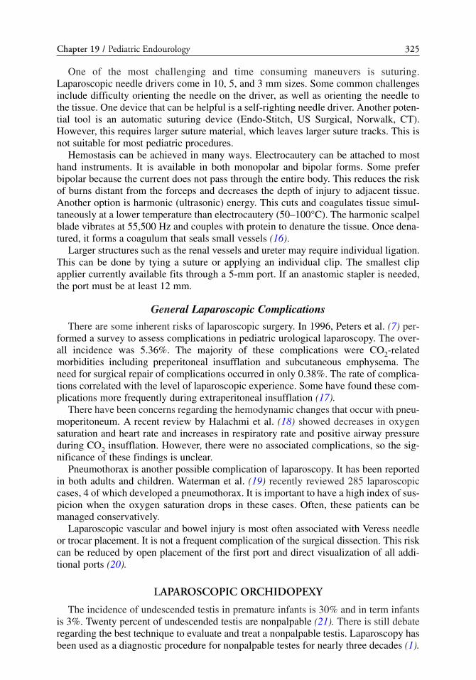
One of the most challenging and time consuming maneuvers is suturing.Laparoscopic needle drivers come in 10, 5, and 3 mm sizes. Some common challengesinclude difficulty orienting the needle on the driver, as well as orienting the needle tothe tissue. One device that can be helpful is a self-righting needle driver. Another poten-tial tool is an automatic suturing device (Endo-Stitch, US Surgical, Norwalk, CT).However, this requires larger suture material, which leaves larger suture tracks. This isnot suitable for most pediatric procedures.
Hemostasis can be achieved in many ways. Electrocautery can be attached to mosthand instruments. It is available in both monopolar and bipolar forms. Some preferbipolar because the current does not pass through the entire body. This reduces the riskof burns distant from the forceps and decreases the depth of injury to adjacent tissue.Another option is harmonic (ultrasonic) energy. This cuts and coagulates tissue simul-taneously at a lower temperature than electrocautery (50–100°C). The harmonic scalpelblade vibrates at 55,500 Hz and couples with protein to denature the tissue. Once dena-tured, it forms a coagulum that seals small vessels (16).
Larger structures such as the renal vessels and ureter may require individual ligation.This can be done by tying a suture or applying an individual clip. The smallest clipapplier currently available fits through a 5-mm port. If an anastomic stapler is needed,the port must be at least 12 mm.
General Laparoscopic Complications
There are some inherent risks of laparoscopic surgery. In 1996, Peters et al. (7) per-formed a survey to assess complications in pediatric urological laparoscopy. The over-all incidence was 5.36%. The majority of these complications were CO2-relatedmorbidities including preperitoneal insufflation and subcutaneous emphysema. Theneed for surgical repair of complications occurred in only 0.38%. The rate of complica-tions correlated with the level of laparoscopic experience. Some have found these com-plications more frequently during extraperitoneal insufflation (17).
There have been concerns regarding the hemodynamic changes that occur with pneu-moperitoneum. A recent review by Halachmi et al. (18) showed decreases in oxygensaturation and heart rate and increases in respiratory rate and positive airway pressureduring CO2 insufflation. However, there were no associated complications, so the sig-nificance of these findings is unclear.
Pneumothorax is another possible complication of laparoscopy. It has been reportedin both adults and children. Waterman et al. (19) recently reviewed 285 laparoscopiccases, 4 of which developed a pneumothorax. It is important to have a high index of sus-picion when the oxygen saturation drops in these cases. Often, these patients can bemanaged conservatively.
Laparoscopic vascular and bowel injury is most often associated with Veress needleor trocar placement. It is not a frequent complication of the surgical dissection. This riskcan be reduced by open placement of the first port and direct visualization of all addi-tional ports (20).
LAPAROSCOPIC ORCHIDOPEXY
The incidence of undescended testis in premature infants is 30% and in term infantsis 3%. Twenty percent of undescended testis are nonpalpable (21). There is still debateregarding the best technique to evaluate and treat a nonpalpable testis. Laparoscopy hasbeen used as a diagnostic procedure for nonpalpable testes for nearly three decades (1).
Chapter 19 / Pediatric Endourology 325
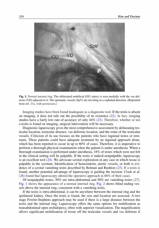
Imaging studies have been found inadequate as a diagnostic tool. If the testis is absenton imaging, it does not rule out the possibility of its existence (22). In fact, imagingstudies have a fairly low rate of accuracy of only 44% (23). Therefore, whether or nota testis is found on imaging, surgical intervention will be necessary.
Diagnostic laparoscopy gives the most comprehensive assessment by delineating tes-ticular location, testicular absence, vas deferens location, and the route of the testicularvessels. Criticism of its use focuses on the patients who have inguinal testes or rem-nants. These patients could have adequate treatment by an inguinal approach alone,which has been reported to occur in up to 60% of cases. Therefore, it is imperative toperform a thorough physical examination when the patient is under anesthesia. When athorough examination is performed under anesthesia, 18% of testes which were not feltin the clinical setting will be palpable. If the testis is indeed nonpalpable, laparoscopyis an excellent tool (24). We advocate scrotal exploration in any case in which tissue ispalpable in the scrotum. Identification of hemosiderin, atretic vessels, or both is evi-dence of a scrotal vanishing testis described by Belman and Rushton (25). If a testis isfound, another potential advantage of laparoscopy is guiding the incision. Cisek et al.(26) found that laparoscopy altered the operative approach in 66% of their cases.
Of nonpalpable testes, 50% are intra-abdominal and 50% are vanishing testes (27).Fig. 1 shows the appearance of a normal internal ring. Fig. 2 shows blind ending ves-sels above the internal ring, consistent with a vanishing testis.
If the testis is intra-abdominal, it can be anywhere between the internal ring and theipsilateral kidney. Once the testis is found, the size and location are assessed. A twostage Fowler-Stephens approach may be used if there is a large distance between thetestis and the internal ring. Laparoscopy offers the same options for mobilization astransabdominal open orchidopexy, often with superior visualization. The magnificationallows significant mobilization of tissue off the testicular vessels and vas deferens if
326 Kim and Docimo
Fig. 1. Normal internal ring. The obliterated umbilical (OU) artery is seen medially with the vas def-erens (VD) adjacent to it. The spermatic vessels (SpV) are traveling in a cephalad direction. (Reprintedfrom ref. 27a, with permission.)

indicated. If a Fowler-Stephens approach is used, laparoscopy gives excellent exposureof the peritoneum that must be spared with the vas deferens.
When orchidopexy is performed laparoscopically, three ports are placed: one in theumbilicus and two just below the umbilicus along the anterior axillary line (Fig. 3).To mobilize the testis, a peritoneal incision is made lateral to the spermatic vesselsand towards the internal ring. The peritoneum between the vas deferens and the sper-matic vessels is spared. This preserves the collateral blood supply in case the sper-matic vessels either spasm or need to be cut to get additional length (Fig. 4). Ifneeded, the testicular vessels can be followed proximally to the level of the renalhilum. If the vessels need to be divided, a 5-mm laparoscopic clip applier is usuallysufficient in size.
Once there is adequate mobilization, the testis can be delivered into the scrotum. Onemethod is to pass a dilating trocar from the scrotum into the abdomen. First, a subdar-tos pouch is created in the ipsilateral scrotum. Then, a 2-mm laparoscopic grasper ispassed through the internal ring, over the pubis, and out a scrotal incision. If additionallength is needed, the trocar can be passed medial to the epigastric vessels or medial tothe medial umbilical ligament, taking care to avoid the bladder. From the scrotum, a2-mm radially dilating trocar sheath is passed over the grasper. The grasper is removedfrom the sheath, and a 5- or 10-mm dilating trocar is used to create an adequately sizedneocanal. A laparoscopic Allis clamp is passed from below to guide the testis into thescrotum. The testis is then secured in the subdartos pouch. If a radially dilating systemis not available or adequate, dilation can be achieved with Amplatz dilators over thegrasper (28).
Results of Laparoscopic Orchidopexy
Chapter 19 / Pediatric Endourology 327
Fig. 2. Blind ending vessels are seen with the short dark arrows. The curved arrow points to the vas def-erens. The open arrowhead shows the empty internal ring. (Reprinted from ref. 27a, with permission.)
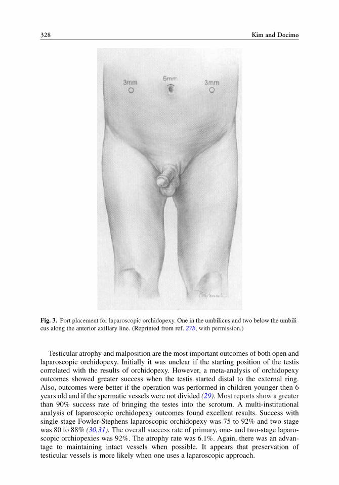
Testicular atrophy and malposition are the most important outcomes of both open andlaparoscopic orchidopexy. Initially it was unclear if the starting position of the testiscorrelated with the results of orchidopexy. However, a meta-analysis of orchidopexyoutcomes showed greater success when the testis started distal to the external ring.Also, outcomes were better if the operation was performed in children younger then 6years old and if the spermatic vessels were not divided (29). Most reports show a greaterthan 90% success rate of bringing the testes into the scrotum. A multi-institutionalanalysis of laparoscopic orchidopexy outcomes found excellent results. Success withsingle stage Fowler-Stephens laparoscopic orchidopexy was 75 to 92% and two stagewas 80 to 88% (30,31). The overall success rate of primary, one- and two-stage laparo-scopic orchiopexies was 92%. The atrophy rate was 6.1%. Again, there was an advan-tage to maintaining intact vessels when possible. It appears that preservation oftesticular vessels is more likely when one uses a laparoscopic approach.
328 Kim and Docimo
Fig. 3. Port placement for laparoscopic orchidopexy. One in the umbilicus and two below the umbili-cus along the anterior axillary line. (Reprinted from ref. 27b, with permission.)

These results challenge the results of open orchidopexy. Success may be related tomagnification, less manipulation of the testicular tissue, and a wider peritoneal window.
LAPAROSCOPIC NEPHRECTOMY
Laparoscopic renal surgery is gaining popularity in both adults and pediatrics. Somewell-documented advantages of laparoscopy are improved cosmesis, shorter hospitalstays, quicker return to work, and less postoperative pain (32–35). Initially, there wasskepticism about the usefulness of this surgery in children. But studies of laparoscopicpartial and total nephrectomy have been consistent with the adult data. This approach isfeasible with low morbidity and favorable outcomes (36,37).
There are no definite contraindications for a laparoscopic renal approach in children.However, most reserve this approach for benign renal disease, e.g., multicystic kidney,nonfunctioning and hydronephrotic kidney, and pretransplant removal. If malignancy ispresent, most will choose an open approach (14).
Laparoscopic nephrectomy can be done by a transperitoneal or retroperitonealapproach. The first cases were performed transperitoneally. When using this approach,the initial port is placed at the umbilicus. A second port is placed in the lower quadrantalong the midclavicular line between the umbilicus and the pubis. The third port isplaced in the midline or midclavicular line above the umbilicus, depending on the sizeof the patient, avoiding the falciform ligament (Fig. 5). In infants and young children,three midline trocars can be used (38). If an additional port is needed for retraction, itis placed in the anterior axillary line along the superior edge of the kidney.
Chapter 19 / Pediatric Endourology 329
Fig. 4. The peritoneum between the vas deferens and spermatic vessels is outlined and left intact.(Reprinted from ref. 27b, with permission.)

Although the transperitoneal approach is commonly used, the retroperitonealapproach is gaining popularity. Retroperitoneal access can be achieved in either theprone or lateral position. Some find the prone position advantageous because theabdominal contents fall away from the operative field. This helps not only with trocarplacement, but also with kidney dissection (39) (Fig. 6). When approaching the patientprone, the initial access is on the lateral edge of the paraspinal muscle. A subcostal inci-
330 Kim and Docimo
Fig. 5. Placement of ports for a transperitoneal pyeloplasty. Another option in smaller children is plac-ing all three ports in the midline. (Reprinted from ref. 37a, with permission.)
Fig. 6. (A) Access with the patient prone. Notice how the kidney falls away from the laparoscopicequipment. (B) The patient in a lateral position and the kidney is displaced medially. (Reprinted fromref. 37a, with permission.)

sion is made and dissection is between the latissimus and oblique muscles. Gerota’s fas-cia is identified and opened. Finger or balloon dilation will open this space. After thespace is developed, a camera can be inserted and the surrounding fascia secured with abox-stitch 3.0 absorbable suture (9). This will not only create a tight seal for pneu-moperitoneum, but also be used for ultimate closure of that site. Additional ports areplaced above the iliac crest and lateral to the paraspinal muscle and medial to the peri-toneum (Fig. 7).
When the patient is positioned prone, it may be difficult to emergently convert to anopen incision. Some prefer to put the patient in a lateral position. If access is performed inthe lateral position, it is started off the 12th rib with a muscle-splitting incision. Additionalports can be placed above the iliac crest or posteriorly and inferiorly. Compared to theprone retroperitoneal approach, lateral positioning provides a larger working space andallows more extensive ureteral dissection. However, the kidney often falls medially andonto the renal pedicle. Constant lateral traction is needed visualize the pedicle (40).
When approached retroperitoneally, the kidney lies in a more vertical orientation(41). Regardless of the access used, the dissection is similar. It is important to remem-ber that dysplastic kidneys often have aberrant blood supply. It is helpful to keep thedissection close to the renal parenchyma to ensure ligation of the vessels at the appro-priate level. The hilum can be approached either anteriorly or posteriorly. It may behelpful to mobilize the kidney inferiorly and follow the ureter up to the hilum. Once thehilum is exposed, the renal vessels can usually be controlled with individual clips. It israre that a vascular stapler is needed in children. If the renal vein is too large to clip, itcan be ligated once with an intracorporeal tie, followed by clips on either side (14).
If operating on a cystic kidney, it is easier to leave the cysts intact until late in the dis-section. This allows for easy identification of the planes between the cyst wall and adjacentorgans. If the size of the cyst inhibits progress, the cyst can be decompressed by a laparo-scopic instrument or by inserting a spinal needle through the body wall. After decompres-sion, the surgeon can grasp the cyst wall to provide traction for easier dissection.
Once the specimen is free, it can often be removed through the 5-mm port incision.The incision may need blunt enlargement to accommodate the specimen. If necessary,
Chapter 19 / Pediatric Endourology 331
Fig. 7. Diagram on the left shows port placement for a prone retroperitoneal approach. Diagrams onthe right (A,B) show port placement for flank positioning. (Reprinted from ref. 37a, with permission.)

one can morcelate the tissue in a bag so that the port site does not have to be signifi-cantly enlarged. Because most pediatric nephrectomies are for benign disease, there islittle need to maintain the surgical margin for pathological examination.
Laparoscopic Partial Nephrectomy
A common indication for partial nephrectomy in children is to remove the nonfunc-tioning segment of a duplicated system. It is usually easy to distinguish the nonfunc-tional segment because it will be cystic or hydronephrotic. If it is difficult to distinguish,intraoperative ultrasound can verify composition (37).
Different landmarks can be used to identify the ureter. Once the white line of Toldtis incised, the ureter is found under the peritoneum at the level where it crosses the com-mon iliac vessels. Another useful location is where it crosses the medial umbilical lig-ament. The ureter of interest is often dilated and easy to identify. If necessary, theduplicated ureters can be followed to the renal pelvis for definite identification.
Once identified, the affected ureter is divided. If there is no associated reflux, the dis-tal ureter can be left open. Otherwise, it is followed to its bladder insertion and ligatedwith endocorporeal knots or absorbable clips. Ligating the ureter before cutting itallows the upper ureter to remain dilated, which assists in dissection of the upper pole.The upper ureter can be used as a handle for cephalad dissection. It is important to avoidinjury of the periureteral tissue around the remaining healthy ureter. The dissectedureter is freed from behind the renal vessels and passed cephalad to facilitate dissectionof the upper pole and its vasculature (42).
The biggest concern with a partial nephrectomy is protecting the blood supply to theremnant parenchyma and ureter. There should be minimal mobilization of the functionalpole and the hilum near the remnant vessels. In addition, vessels should be ligated as closeto the abnormal renal unit as possible. Some find the dissection easier with a retroperi-toneal approach because of its direct exposure. In addition, the lower pole does not requireany separation from the peritoneum which can help protect the lower vasculature.
Once the vessels are ligated, the renal units can be separated using electrocautery orharmonic energy (36). To minimize bleeding, it is common to leave a small rim of thenonfunctioning segment on the remaining renal tissue.
After laparoscopic nephrectomy, most pediatric patients are started on a diet that isadvanced as tolerated. The younger children will often have good pain control within afew hours and go home the same day. Some mild abdominal pain for 24 hours is morecommon in the older children. This may be related to retained carbon dioxide (43).
Outcome After Laparoscopic Nephrectomy
Most reports of pediatric laparoscopic nephrectomies have shown a low rate of com-plications (36,37,43). The complications of greatest interest include vascular and vis-ceral injuries.
Excessive bleeding during the operation can originate from tears of the renal vein, itscontributing vessels, or accessory or polar renal arteries. Any blood in the field cancover and obscure visualization of the tissues. In addition, the blood will absorb lightand further limit the view. The rate of open conversion because of bleeding is low, buthas been reported (14).
332 Kim and Docimo

Another theoretical concern is postoperative bleeding. This usually presents with achange in vital signs. If the patient has left the hospital when this complication occurs,the key is a high level of suspicion for bleeding if the child is not doing well.
Similarly, bowel injury can present late if it was unrecognized in the operating room.This is a complication of both transperitoneal and retroperitoneal nephrectomies. Thesebowel injuries may result from trauma during access or electrocautery during the case.Most commonly, the patient will have fever and peritoneal irritation, although bowelinjury can present subacutely after laparoscopic procedures (44–46).
The choice between a transperitoneal vs retroperitoneal approach is essentially basedon the surgeon’s preference. Meraney et al. (47) reviewed 404 retroperitoneal renal andadrenal cases. They had 1.7% vascular and 0.25% bowel injuries. The complication ratecorrelated with prior major abdominal surgery and the experience of the surgeon. Studiescomparing the two techniques show similar rates for operative time, blood loss, and hos-pital stay (48). There has not been persuasive data to show an increased morbidity asso-ciated with either approach.
LAPAROSCOPIC PYELOPLASTY
Laparoscopic pyeloplasty was first described in both the adult and pediatric popula-tions in 1993 (49,50). So far, the outcomes of laparoscopic repair are comparable to theresults of open repair (8). The first series was performed transperitoneally (51), butsome are using a retroperitoneal approach.
There are no strict criteria that guide when to use minimally invasive procedures inchildren. Some prefer open pyeloplasty for all children under 6 months old. Otherschoose a cut off age of 18 months old. An ideal candidate is an older child with anextrarenal pelvis. There are multiple challenges in the smaller child. Placing a stent hasmore risk of traumatizing either the infant’s delicate ureterovesical junction or the pos-terior anastomosis. The small diameter of the ureter makes the anastomosis technicallydifficult, especially within the confines of a small working space.
If the laparoscopic approach is chosen, the first step is positioning the patient.Placing an intravenous line in the ipsilateral hand allows easy access for the anesthesi-ologist because that arm is draped over the patient, usually on an elevated arm board.Some prefer a preoperative stent placement and others find it cumbersome during theanastomosis. We use a preoperatively placed guidewire that is prepped and draped inthe surgical field. The wire allows visualization of the ureteropelvic junction (UPJ)without decompressing the renal pelvis. Once the UPJ is divided, the wire can be usedto manipulate the ureter nontraumatically. Before completing the anastamosis, a stent isplaced over the wire.
Positioning of the patient is similar to other laparoscopic renal surgery. When oper-ating transperitoneally, the patient is positioned in a 45° flank position. The patientshould be close to the edge of the table to maximize the mobility of the instruments. Thepatient is secured to the table at the level of the thorax and the iliac crest. This enablesthe surgeon to change the position of the patient from supine to full flank. An axillarygel pad, rather than an axillary roll is placed to prevent excessive pressure on the latis-simus dorsi muscle.
The first trocar is placed (usually by an open technique) at the umbilicus. A secondtrocar is placed in the midline superior to the umbilicus and another trocar is placed inthe midclavicular line ipsilateral to the affected kidney halfway between the umbilicus
Chapter 19 / Pediatric Endourology 333
and the pubis. An insufflation pressure of 10 mmHg is used for small patients weighingless than 10 kg and a pressure of 10 to 15 mmHg is used for heavier patients. The whiteline of Toldt is incised to free the ipsilateral colon and expose the retroperitoneum. Thegonadal vessels will run close to the UPJ and need to be reflected medially.
An alternative to mobilizing the colon on the left side is a transmesenteric approach(52): instead of incising the line of Toldt, a small window through the mesentery isopened over the UPJ. The location of the UPJ can be revealed by moving a preopera-tively placed guidewire. The transmesenteric approach is less appealing on the rightside because of the close proximity of the duodenum and great vessels.
When approaching the kidney retroperitoneally, the initial access is 1 cm from thelower edge of the 12th rib. Using blunt dissection, Gerota’s fascia is opened under directvision. Once the first trocar is placed, gas insufflation will create a larger working space.A second trocar is placed posteriorly near the costovertebral angle and a third port isplaced along the anterior axillary line, 1 cm above the iliac crest (53).
There are two approaches to division of the UPJ from an anterior approach, depend-ing on the presence of crossing vessels. When vessels are encountered, the ureter is dis-sected posteriorly until the UPJ is seen behind the vessels. The ureter can be dividedjust below the UPJ. At that point, attention is turned to the anterior pelvis. As this isfreed, the UPJ can be brought anterior to the vessels, where the anastomosis can be per-formed. The vessels themselves require no manipulation.
Once the UPJ is divided, the ureter is spatulated laterally and the pelvis is spatulatedmedially. Usually there is no to need to reduce or excise the renal pelvis. When thereare no crossing vessels, the approach is straightforward. The UPJ can be divided on thepelvis side, leaving a tag of pelvis on the ureter for manipulation (51).
A stay suture is placed in the anterior renal pelvis and then passed through the lateralabdominal wall. This stabilizes the pelvis for anastomosis and, especially in the case ofa transmesenteric approach, improves exposure and access to the UPJ (52). After spat-ulating the ureter, the anastomosis is started with interrupted sutures at the dependentpart of the renal pelvis. Fine absorbable suture is used, e.g., 5.0 or 6.0 polyglactic acidsuture. A small (Ethicon, Somerville, NJ), such as a TF, half-circle needle, is preferable.This can pass through a 3.5- or 5-mm port. When passing the needle, it is important notto blunt the tip. Optimal ergonomics would suggest alignment of the needle driver withthe line of suturing and to have the second instrument 30° offset from the first.
The remainder of the anastomosis may be done with interrupted or running sutures.Once the posterior wall is completed, the ureteral stent is positioned. As stated before, thestent may be placed preoperatively and the anastomosis sewn around the stent. Anotheroption is placing a wire preoperatively and then guiding a stent retrograde or antegradeafter the posterior wall is closed. Also, the stent can be introduced by an angiocatheter andwire. In most cases, it is not necessary to leave a perinephric drain. The stent is left in placefor 2 to 4 weeks. Patients can be managed with or without a foley catheter for one night(53).
An alternative to stenting is placing a percutaneous nephrostomy tube. This can beplaced under direct visualization and used to confirm postoperative success beforeremoval. Nephroureteral tubes can also be used but must be removed before assessingthe anastomosis.
Some prefer robotic assistance to expedite the suturing. The automatic suturingdevice (Endo-Stitch, US Surgical) is not suitable for use in children. The availablesutures for this and other devices are too large, and the needle for the Endo-Stitch is per-
334 Kim and Docimo

pendicular to the attached suture. In addition, there is concern for possible stone forma-tion not only from larger sutures, but also from the titanium clips that can accompanythese devices (54,55).
Outcomes of Laparoscopic Pyeloplasty
The success for laparoscopic pyeloplasty in adults has been better than 95%, which iscomparable to open repair (56–58). The benefits include improved cosmesis and quickerconvalescence. El-Ghoneimi et al. (53) showed that school-age children returned to fullschool activities within 1 weeks postoperatively. They also saw a quicker return to painlessactivity (53).
Complication rates of laparoscopic repair are 10 to 13% (59). Potential complicationsof laparoscopic pyeloplasty are identical to those of the open procedure. These includeurine leakage and postoperative obstruction. The complication rate has been similarwith both approaches. The biggest challenge for most is intracorporeal suturing. Withsmaller and sturdier needle drivers, experienced laparoscopists are improving theiroperative times while maintaining the key surgical principles.
Postoperative success has been defined both as improved hydronephrosis andimproved drainage on renal scan. The long-term success rate of laparoscopic pyelo-plasty in children is still unknown. Most series have excellent results, but only have data6 to 12 months postoperatively (53,60,61).
UROLITHIASIS
Although there is a low incidence of urolithiasis in children, it often poses a thera-peutic dilemma. When looking at all patients with urinary stone disease, only 1.3 to2.0% of them are pediatric patients (62). As with adults, the management in childrenmust take into account multiple factors. This includes stone size, stone location, stonecomposition, and urinary tract anatomy. Metabolic abnormalities are found in 53 to80% of pediatric patients with stones (63). Calcium-related abnormalities are the mostcommon type found (64).
Presentation of symptomatic stones can be extremely variable in children. The mostcommon symptom is abdominal pain (53–75%). Another common symptom is hema-turia (14–33%). Classic renal colic is only found 7% of the time (65).
The radiographic work up in children is similar to adults. Plain abdominal films caneasily miss both radiolucent and small radiopaque stones. Renal ultrasound may miss anonobstructing ureteral stone. Helical computed tomography scan of the abdomen andpelvis is a reliable method when there is a high suspicion of stones. Since children donot tend to have phleboliths, there is little confusion in differentiating distal stones fromphleboliths (66).
Etiology and stone composition are variable based on geography. The majority ofpatients in Europe have infectious stones, whereas in the United States, it is more com-mon for children to have calcium oxalate stones and metabolic abnormalities. Obtaininga 24-hour urine specimen can be challenging in this population; therefore, one can usespot urine calcium, creatinine, and uric acid levels as a substitute.
Once a stone is identified, it may require treatment secondary to obstruction, infec-tion, or hematuria. The American Urological Association has provided guidelines fortreatment of ureteral stones in adults. There is no correlating guideline for children. VanSavage et al. (67) looked at their pediatric stone population and found the majority of
Chapter 19 / Pediatric Endourology 335
patients with stones less than 3 mm in size would spontaneously pass. For adults, thereis a 98% spontaneous passage rate for stone less than 5 mm. The biggest factor for thelower rate in children is probably the smaller diameter of the ureter. In addition, thereis a lower threshold for pain and an increased need for attention which may hasten inter-vention.
There is no absolute stone size that children will pass. The decision to intervene sur-gically is similar to adults: associated fever, intractable pain, intractable vomiting, orrenal compromise. If mild symptoms are present, many will choose expectant manage-ment for stones less than 4 mm in size (68).
Extracorporal Shockwave Lithotripsy
Shockwave lithotripsy (SWL) is often used for renal and ureteral stones in adults. Itsrole in pediatrics was initially for renal stones only. One problem is patient positioning: itcan be challenging to keep a small child in the same position without sedation or a gen-eral anesthetic. Some have used a modified infant car seat to help with positioning (69).
A concern with distal ureteral SWL is the potential for ovarian damage from theshock waves. McCullough et al. looked at the function of rat ovaries after SWL. Whencomparing the control and treated rats, he found no difference in the number of preg-nancies, spontaneous abortions, or fetal weights. The short-term complications forhuman patients are negligible, but the bigger concern is long term effects on the ovaries.Studies have not found an increased risk of renal scar, renal function, linear growth,hypertension, or renal disease in the long term (71,72).
Most pediatric patients do not require ureteral stents with SWL unless the stone bur-den is large enough for the development of steinstrasse. When treating staghorn calculiwith SWL, preoperative stenting has been associated with fewer complications; how-ever, its presence may compromise stone passage.
With regard to stone size, there is no defined cut off for SWL in children. In adults, mostwill choose percutaneous nephrostolithotomy (PCNL) if the stone burden is greater than2 cm. A similar size cut off is generally utilized in children. However, some advocate SWLas monotherapy for staghorn calculi as well. It is felt that shockwave fragmentation is moreeffective in pediatrics because of the overall stone burden is smaller and the stones are oftensoft. In addition, a smaller body volume allows better transmission of the shockwaves.Children seem to have a greater capacity than adults to pass fragments after lithotripsy.
Success rates of SWL range from 84 to 97%, with some patients requiring more thanone treatment (73,74). Pediatric staghorn calculi treated with SWL monotherapy have asuccess rate of 70 to 80% after multiple procedures (75–78). As expected, success islower with large stones, lower pole stones, and stones in kidneys with abnormal anatomy.In addition, cystine and calcium monohydrate stones are more resistant to SWL.
Morbidity after SWL is minimal. Gross hematuria is common for a few days.Pulmonary contusions with hemoptysis may occur if shockwaves traverse pulmonarytissue. Pulmonary dysplasia has not been a long-term complication of SWL, but shield-ing of the lung with foam is recommended in smaller children (79).
Ureteroscopy
Many centers choose ureteroscopy as the first line of treatment for not only distalureteral stones, but also for proximal stones.
Previously, the problem with ureteroscopy was the need to dilate the ureteral orifice.The biggest concern with this is the possibility of creating reflux, although when this
336 Kim and Docimo

occurs, it appears to be transient (80). There is also a risk of distal ureteral stricture,which has been reported to occur more commonly in children than in adults (81). Ourpreference is to prestent when necessary, allowing the ureters of even small children toeasily accommodate modern, small endoscopes.
One limiting factor associated with the smaller endoscopes is a small working channel.Placing an instrument through the port decreases the level of irrigation and visibility.These issues must be considered before choosing the best intervention for each patient.Often, a 220 μ laser fiber is the best option for stone manipulation through a flexibleureteroscope, although laser lithotripsy may not be available in all pediatric settings. Inaddition, a range of ureteroscopes are available for purchase, but it is impractical formost centers to carry all types.
A versatile rigid scope is the 6.9-Fr cystourethroscope, which has either 2- and 3.5-Frworking channels, or a single 5-Fr channel. This ureteroscope easily accesses the distalureter in small and larger children. The 15-cm length endoscopy works well for mostsituations. The flexible ureteroscopes are also available in 6.9 Fr and allow easy accessto the upper ureter and renal calyces. The degree of deflexion of the ureteroscope is atleast 180° in most models and active secondary deflection is also available in newergeneration ureteroscopes.
If repeated entry is anticipated, one may elect to use an access sheath. The 9.5-Fr accesssheath allows easy entry of the ureteroscope into the ureter while allowing antegrade flowaround the scope, thereby preventing excess pressure within the collecting system.
For manipulating stone fragments, baskets and grasping devices are available assmall as 1.7 Fr. However, these can be difficult to manipulate and frustrating to use. Assuch, the 3-Fr instruments are more commonly employed.
The technique of ureteroscopy should be strictly followed with the underlying themeof maintaining ureteral access. Whether or not a stent is present, the procedure is startedby placing a guidewire into the renal collecting system through a cystoscope. If it is dif-ficult to bypass a ureteral or UPJ calculus, a hydrophilic coated flexible wire can beused or the guidewire can be manipulated from within a ureteral catheter. Once the wireis in place, the cystoscope is removed. A 10-Fr dual-lumen catheter, or an 8/10-Fr dilat-ing system, is used to place a second safety wire that is coiled and either secured to thedrapes, sutured to the patient, or both. This step is vital because loss of ureteral integrityand/or primary access can occur in even the most straightforward cases. The uretero-scope can be advanced between the wires, or over one of the wires, depending on thecircumstances. The irrigating fluid should be warmed to prevent hypothermia in smallchildren. Water should never be used as an irrigant because of the risk of hyponatremiaand its consequences should extravasation occur (82,83).
When the stone is visualized, it can either be removed intact or fragmented. Onlysmall stones should be basketed in children because of the risk of ureteral avulsion.Fragmentation can be carried out using a variety of lithotripsy devices, but for flexibleureteroscopy, only laser or electrohydraulic lithotripsy (EHL) are options owing to theflexibility of the probes. If available, the Holmium laser is an ideal instrument for thispurpose because it has excellent energy transfer and less likelihood of ureteral or renalpelvic injury than EHL. With either energy source, it is important that the interface ofprobe/fiber and stone be visible prior to the application of energy.
The use of postoperative stents depends on the degree of manipulation that wasrequired and whether future procedures are anticipated. Leaving a stent often requires
Chapter 19 / Pediatric Endourology 337
an additional anesthetic to perform cystoscopy for stent removal. If the stent is neededfor a short period of time, it may be left attached to a string for bedside removal (84).
After the stent is removed, the patient’s upper tract must be observed for postopera-tive hydronephrosis. This is done by ultrasound within 4 weeks. If hydronephrosis issignificant or the child has symptoms of obstruction, an intravenous pyelogram or renalscan must be considered to evaluate the ureteral anatomy and degree of obstruction.
There is limited data regarding outcomes for pediatric ureteroscopy. The few reportsdo show good success rates (87–100%) with minimal complications. The rate ofureteral stricture is probably higher than in adults, and can probably be best avoided byprestenting and avoiding of aggressive ureteral dilation (81).
Percutaneous Nephrolithotomy
The first pediatric series of PCNL was described in 1985 (85). PCNL has an estab-lished role both when used alone and as an adjunct to other treatments. Although renalcalculi are uncommon in children, when they do occur, they are often associated witha metabolic or anatomic abnormality leading to a high risk of recurrence. As such, pro-cedures that render the patient free of stones in as few procedures as possible arepreferable (86).
The trend in adults is to utilize PCNL for a stone burden greater than 2 cm. Stoneburden alone does not constitute an absolute indication in children. PCNL is indicatedin patients who fail SWL or ureteroscopic therapy and is preferable in patients withanatomic abnormalities that inhibit their ability to drain urine and clear stones, i.e., UPJobstruction, infundibular stenosis, and calyceal diverticulum. An advantage of PCNL isthe ability to simultaneously treat stones and the associated anatomic abnormality.PCNL is a minimally invasive procedure that can be used to avoid multiple surgical pro-cedures in a patient with recurrent stone disease.
Initially, PCNL was always performed using percutaneous access similar to the adultpopulation. Intuitively, this large size is a concern for infants because a 24-Fr tract in chil-dren is comparable to a 72-Fr tract in an adult (87). The mini-perc technique, described in1998, described renal access through an 11-Fr peel-away sheath which could accommo-date a 7.5-Fr rigid or a 9.5-Fr flexible scope (Fig. 8). Possible advantages are smaller inci-sions, decreased pain, shorter hospital stays, and a smaller number of lost nephrons (88).
The initial access into the collecting system is performed either by a radiologist priorto the stone procedure or simultaneously in the operating room with C-arm guidance.Once access is obtained, some prefer to wait a 24–48 hours before enlarging the tractand manipulating the stone. Others will proceed the same day of initial access.
Technique is important to avoid loss of access and to avoid injury to the collecting sys-tem. Once access to the collecting system is obtained, a guidewire is introduced. For aminiperc procedure, a small 4-mm fascial incising needle is passed through the fascia(Pediatric Percutaneous Access Set, Cook Urological, Spencer, IN). This is followed bya dual-lumen 10-Fr hydrophilic coated catheter. Ideally the wire has been routed throughthe UPJ and down the ureter prior to this maneuver. If not, care must be exercised toavoid perforating the renal pelvis by advancing too far. A second safety wire is intro-duced and sutured to the skin. The 11-Fr peel-away sheath is then introduced and the tro-car removed. A “nipple” is present to secure the trocar to the sheath. It is vital that thisnipple not be removed prior to initiating the procedure to insure free egress of fluid at alltimes (89). We prefer to use a 6.9-Fr offset cystoscope with a 5-Fr working channel for
338 Kim and Docimo

stone manipulation, though flexible scopes are also useful. Stones are fragmented usingthe modalities previously described.
At the end of the procedure, the patient may be left tubeless or with a 6- or 8-Frnephroureteral stent that can be passed over a wire through the sheath, after which thesheath is peeled away around the stent.
OUTCOMES OF PCNL
PCNL in children is associated with a stone-free rate of 67 to 100% (85,90–92). Themost concerning complications are bleeding and hypothermia. In adults, the biggest riskfactors for bleeding are preoperative anemia and total blood loss. As in adults, it is rarefor children to require blood transfusions after PCNL.
Young patients can quickly become hypothermic. When evaluating PCNL in adults,Roberts et al. found that the majority of patients dropped their core body temperature.In their series, hypothermia was associated with longer operative times and longerinduction periods, although the irrigant in this study was room temperature. Al-Shammari et al. (90) looked at a pediatric population and found that operative time ofmore than 150 minutes correlated with hypothermia. Some safeguards againsthypothermia include a warm operating room, warmed irrigation solution, and briefanesthetic inductions. Our irrigant of choice is warmed normal saline that not only min-imizes hypothermia, but also avoids hyponatremia in the event that extravasation doesoccur.
LAPAROSCOPIC BLADDER RECONSTRUCTION
Chapter 19 / Pediatric Endourology 339
Fig. 8. The “mini-perc” access kit contains two 0.035-in. guidewires, a fascial incising needle, a duallumen dilator, and a 11-Fr peel away sheath (Cook Urological, Spencer, IN).

The use of laparoscopy in bladder reconstruction has recently been explored, with thefirst laparoscopic bladder augmentation reported in 1995 (93). This late introduction ispartly the result of the complexity of lower urinary tract reconstruction.
Most laparoscopic bladder reconstruction falls into one of three categories: laparo-scopic autoaugmentation, laparoscopic enterocystoplasty, and laparoscopic-assistedreconstruction.
Incorporating laparoscopy is appealing because of the improved cosmetic result anddecreased morbidity. For instance, patients with myelodysplasia frequently need a largemidline incision to access bowel because the cecum can be malrotated or abnormally fixed.
Contraindications to laparoscopic reconstructive procedures include severe inflam-matory processes of the bladder or bowel, short gut syndrome, uncontrolled coagu-lopathies, inability to perform intermittent catheterization, and high risk ofintra-abdominal adhesions (e.g., history of radiation therapy, multiple open abdominalsurgeries). Presence of a ventriculo-peritoneal shunt and prior surgery are not con-traindications.
Laparoscopic autoaugmentation has been reported by using both preperitoneal andtransperitoneal access. These reports involved small groups of patients but did show anincrease in bladder size with improved continence (94,95). Most long term series ofbladder autoaugmentation have been somewhat disappointing in terms of long-termbladder compliance.
Pure laparoscopic enterocystoplasty has also been reported in a small number ofpatients. However, one disadvantage was long surgical time, which ranged from 5.3 to9 hours. Long-term follow-up on these patients is still pending (96,97).
The most beneficial aspect of laparoscopy in bladder reconstruction appears to beassisting the mobilization of the cecum, the gastric segment, or the appendix. One candivide the reconstructive procedure into two parts: mobilization of tissue for augmenta-tion and/or stoma creation and actual reconstruction of the bladder. Laparoscopy canaccomplish the first half, leaving the final half to be completed through a smallPfannenstiel incision. Often, the lower incision can incorporate multiple laparoscopicport sites. If ileum or sigmoid is being used for the augmentation and no continentstoma is needed, then the entire operation can be done through a lower abdominal inci-sion and laparoscopy is not warranted.
The goal is to maximize the strengths of both laparoscopic and open techniques tominimize patient morbidity. By using laparoscopy to perform tasks that would requireupper abdominal access, the patient is spared a larger incision. And by performing themore complex tasks through a small open incision, the patient is spared the longer oper-ative times seen with purely laparoscopic reconstruction.
Technique for Laparoscopic-Assisted Bladder Reconstruction
The day prior to surgery, the patient completes a mechanical and antibiotic bowelpreparation.
The patient is positioned in low lithotomy to perform cystoscopy to assure that thereare no signs of cystitis. The patient is then repositioned supine and well padded to pre-vent pressure ulceration. In addition, the patient is secured to the bed by taping acrossthe chest and legs, thereby allowing for safe lateral and deep Trendelenberg rotation.
If a concealed umbilical stoma is planned, a U-shaped incision is made (98). An infe-rior flap is raised and the umbilical fascia incised sharply. Once the peritoneum isopened, a radially dilating 10- or 12-mm trocar is placed. The remaining ports are posi-
340 Kim and Docimo

tioned based on the procedure planned. If a Malone antegrade continence enema isplanned, the second port should be placed at the proposed stoma site. This can be a 3-or 5-mm port. A third 2- or 3-mm port is placed in the midepigastrium (Fig. 9).
To access the appendix, the white line of Toldt is incised to mobilize the right colon.Once the appendix is isolated on its pedicle, it can be divided laparoscopically with an
Chapter 19 / Pediatric Endourology 341
Fig. 9. Port placement for laparoscopic assisted bladder reconstruction. The most inferior port isplaced along the planned Pfannenstiel incision. (Reprinted from ref. 98a, with permission.)
Fig. 10. Appearance after laparoscopic assisted bladder reconstruction with continent stoma.(Reprinted from ref. 99, with permission.)

Endo-GIA stapler or divided after bringing it through the open incision. There is usuallylittle or no dissection needed to mobilize small bowel or sigmoid colon. Occasionally, thelateral peritoneal reflection is incised to give extra length to the sigmoid.
Once the bowel is harvested, a 4 to 5 cm Pfannenstiel incision is made for theenterovesical anastomosis, stomal creation, and any bladder neck manipulation.
To complete the catheterizable stoma, the dorsal aspect is spatulated and sewn to theU flap, leaving a widely patent but well-concealed stoma (Fig. 10).
Outcomes of Laparoscopic-Assisted Bladder Reconstruction
Laparoscopic assisted bladder reconstruction is technically challenging and has onlybeen employed at a few centers. The initial surgical outcomes are comparable to a com-pletely open approach. It has been shown that these patients are quicker to tolerate aregular diet and have shorter hospital stays. This is accomplished without an increase inoperative morbidity or operative times.
Chung et al. (99) reported the largest series (comprised of 30 patients) of laparoscopicassisted bladder reconstruction to date. There were no intraoperative complications,there was one open conversion, and the mean hospital stay was 6 days.
At 32 months, 95% were continent and could easily catheterize. Stomal stenosisoccurred in 5.1% and stomal revision was required in 7.7%. Postoperative complica-tions occurred in 16%. This included ileus, partial bowel obstruction, traumatic bladderperforation, wound infection, and deep venous thrombosis (99).
It has been shown that scars on the torso affect body image (100). The patients whorequire bladder reconstruction often have other anomalies. Therefore, any steps to min-imize the degree of external scarring are desirable. In addition, the likelihood ofintraperitoneal adhesions is likely decreased by using this approach, conferring a poten-tially lifelong benefit to the patient.
VESICOURETERAL REFLUX
Vesicoureteral reflux (VUR) is a common problem that affects 1% of children. Thegoal of diagnosing and treating VUR is to prevent long-term complications such aspyelonephritis, hypertension, and renal insufficiency (101). Options for treatmentinclude long term antibiotic prophylaxis, subureteral injections, and laparoscopic andopen reimplantation. Although open reimplantation provides success rates of 95 to 98%with low morbidity, the search continues for less invasive methods (102,103).
Laparoscopic reimplantation has been performed both intravesically and extrav-esically. In 1993, Atala et al. (104) reported their experience performing laparo-scopic transperitoneal extravesical (Lich-Gregoir) antireflux plasty in a pig model.In 1995, Lakshmanan et al. (105,106) reported a human series of 71 patients under-going extravesical ureteral reimplantation. Their series showed low postoperativemorbidity with a quick return to full physical activities. This approach was used forboth unilateral and bilateral reimplantations. The initial follow-up was comparableto open reimplantation.
In an effort to simplify the approach, the same repair was performed via an extraperi-toneal approach (107). Later that year, another small series confirmed the feasibility ofshowed this technique. Follow-up at 2 months showed no reflux in any patient. Somefind this approach easier and less invasive than the transperitoneal approach. It is theo-
342 Kim and Docimo

rized that limiting the dissection and greater preservation of vascularity around the blad-der neck will decrease the risk of postoperative voiding dysfunction.
Intravesical endoscopic ureteral reimplantation was first described in a pig model.Gill et al. (108) reported their experience performing transvesical laparoscopic reim-plantation with a Cohen technique in a small series of three patients all over the age of10. Olsen et al. (109) described a similar technique in a larger series. The Davinci robotmay provide easier access for submucosal tunneling and intravesical suturing of theanastomosis, as suggested in reports in a pig model.
Although these series are promising, there are still concerns. The laparoscopicapproach is not advisable in ureters with ureteroceles or megaureters that require taper-ing. In addition, the small pelvis of children under 4 years old is often a limiting factor.Moreover, there is a risk of voiding dysfunction when reimplanting bilaterally (110).However, despite the feasibility of these approaches, the procedure is still technicallychallenging and has a steep learning curve. One of the reasons that these techniqueshave not become more popular is that ureteral reimplantation can be performedthrough a cosmetically acceptable small Pfannenstiel incision requiring only anovernight hospital stay and no drainage tubes and has a success rate of more than 95%(111,112).
Subureteral Injection Therapy
An alternative to open reimplantation is subureteral injection therapy. This conceptwas introduced in the 1980s by O’Donnell and Puri (113). The ideal injection substanceis biocompatible, nonallergenic, nonmigratory, and durable. Various substances havebeen tried such as collagen, autologous substances, polyteterflourourethrane (Teflon),polydimethy-siloxane (Macroplastique), and dextranomer/Hyalouronic acid (Deflux).
Polytetraflourourethane (Teflon) has particles that varied in size from 5 to 100 μ andwas taken off the US market because the smaller particles could be injected into capil-laries and embolize to distal sites. Collagen is not durable because it is biodegradableand contractile (114).
Atala et al. (115) first reported injection of autologous chondrocytes in an animalmodel. Later, Caldamone and Diamond (116) reported this technique in a clinical trial.First, posterior auricular cartilage is harvested from the patient. After an enzymaticdigestion, the chondrocytes are grown in culture over 6 weeks. Finally, the patient isbrought back for endoscopic implantation at the ureterovesical junction.
In 2001, the FDA approved Dx/HA copolymer (Deflux,Q-Med Scandinavia) as anacceptable material for injection therapy to treat VUR. Deflux consists of micros-pheres that range from 80 to 120 μ in diameter. This larger size prevents migration ofmaterial. It consists of crosslinked dextran polysaccharide molecules, so there are nofreely circulating dextran molecules, thereby avoiding allergic or immunogenic reac-tions. Once implanted, Deflux does not contract because of ingrowth of fibroblastsand collagen.
TECHNIQUE OF INJECTION THERAPY
The technique of injection is similar to that previously described for the subureteraltransurethral injection prodecure. The patient is placed under general anesthesia andpositioned in lithotomy. A 10-Fr cystoscope with an offset lens is used to guide a 3.5-Fr needle containing 0.5 to 1.2 mL of material. The needle is injected into the submu-
Chapter 19 / Pediatric Endourology 343
cosa at the 6 o’clock position, producing a bulge with a nipple appearance. The orificeshould appear slit-like on top of the mound (113,117).
RESULTS OF INJECTION THERAPY
Some significant concerns with endoscopic treatment of VUR include long-term out-comes and overall success rates. Either results are suboptimal compared to open repairor long term results are lacking. Collagen has been shown to be suboptimal because ofits tendency to disappear leading to recurrence of reflux. Caldamone and Diamond(116) reported 65% success at 1-years follow-up of autologous chondrocytes injectiontherapy. A series using Macroplastique demonstrated success in more than 80% ofpatients, with the longest follow up in these series being 94 months (118).
Puri et al. (117) reviewed 11- and 17-years follow-up of Teflon injection therapy andfound a 5% recurrence rate at 17 years. They noted a 76% success rate with the firstinjection, and a need for repeat injection in 20% of patients.
Outcomes of Deflux injection are still evolving. Although high success has beenreported, the data must be viewed carefully. Kirsch et al. (114) reported a 72% successrate at 3 months in 130 patients evaluated by voiding cystourethrography. Puri et al.(117) recently reported 3-months and 1-year follow-up of 113 patients after Defluxinjection. Although success rate was reportedly 99% after one to three injections, only9.7% completed follow up at 1-year.
The true test of injection therapy is how it compares to open reimplantation surgery.It is still unknown if long-term outcomes will be comparable. Currently, strict guidelineson when to operate for vesicoureteral reflux are lacking, as is the type of interventionindicated. It remains a challenge to improve on the more than 95% success of open reim-plantation. With a small incision and often overnight hospital stay, the morbidity of openintervention is already impressively low.
CONCLUSION
Minimally invasive surgery in children is an exciting and evolving field. Indicationshave progressed from diagnostic to reconstructive therapy. It is a challenge to find thebest application for these techniques given the excellent outcomes of open urologic sur-gery in children. However, we must continue to explore new ways to provide ourpatients with the best outcome and the least morbidity.
A few indications are well accepted, such as laparoscopy for intra-abdominal testis.The initial results of laparoscopic nephrectomy and pyeloplasty are encouraging withoutcomes comparable to open surgery. These cases are becoming more common at cen-ters across the world. The most technically demanding cases such as laparoscopic blad-der reconstruction and reimplantation are still limited in application, but they may gainpopularity with the use of robotic assistance.
Urolithiasis is a rare (but still significant) problem in pediatrics. Since the early1980s, open stone surgery in adults has been replaced by less invasive methods such asPCNL, SWL, and ureteroscopy. This transition has been slower in children primarilybecause of technical limitations. As endoscopic equipment continues to improve withregard to better optics and instrumentation for smaller scopes, the applications in pedi-atrics will likewise grow.
REFERENCES
344 Kim and Docimo

1. Cortesi N, Ferrari P, Zambarda E, Manenti A, Baldini A, Morano FP. Diagnosis of bilateral abdomi-nal cryptorchidism by laparoscopy. Endoscopy 1976; 8(1): 33–134.
2. Cuellar DC, Kavoussi PK, Baker LA, Docimo SG. Open laparoscopic access using a radially dilat-ing trocar: experience and indications in 50 consecutive cases. J Endourol 2000; 14(9): 755–756.
3. Bonjer HJ, Hazebroek EJ, Kazemier G, Giuffrida MC, Meijer WS, Lange JF. Open versus closedestablishment of pneumoperitoneum in laparoscopic surgery. Br J Surg 1997; 84(5): 599–602.
4. Thomas MA, Rha KH, Ong AM, et al. Optical access trocar injuries in urological laparoscopic sur-gery. J Urol 2003; 170(1): 61–63.
5. Gill IS. Needlescopic urology: current status. Urol Clin North Am 2001; 28(1): 71–83.6. Yerdel MA, Karayalcin K, Koyuncu A, et al. Direct trocar insertion versus Veress needle insertion in
laparoscopic cholecystectomy. Am J Surg 1999 ; 177(3) :247–249.7. Peters CA. Complications in pediatric urological laparoscopy: results of a survey. J Urol 1996; 155(3):
1070–1073.8. Chen RN, Moore RG, Kavoussi LR. Laparoscopic pyeloplasty. Indications, technique, and long-term
outcome. Urol Clin North Am 1998; 25(2): 323–1330.9. Gill IS, Clayman RV, Albala DM, et al. Retroperitoneal and pelvic extraperitoneal laparoscopy: an
international perspective. Urology 1998; 52(4): 566–571.10. Jarett T, Fabrizio MD, Lamont DJ et al. Laparoscopic pyeloplasty: a five year experience. J Urol,
suppl 1999; 161(24): 79.11. Diamond DA, Price HM, McDougall EM, Bloom DA. Retroperitoneal laparoscopic nephrectomy in
children. J Urol 1995; 153(6): 1966–1968.12. Moore RG, Partin AW, Adams JB, Kavoussi LR. Adhesion formation after transperitoneal nephrec-
tomy: laparoscopic v open approach. J Endourol 1995; 9(3): 277–280.13. Valla JS, Colomb F, Son S, Michelini E, Steyaert H, Guilloneau B. Mini-invasive surgery of the
retroperitoneal space in children. Ann Urol (Paris) 1999; 33(5): 328–332.14. El-Ghoneimi A, Valla JS, Steyaert H, Aigrain Y. Laparoscopic renal surgery via a retroperitoneal
approach in children. J Urol 1998; 160(3 Pt 2): 1138–1141.15. Hsu TH, Gill IS. Bilateral laparoscopic adrenalectomy: retroperitoneal and transperitoneal
approaches. Urology 2002; 59(2): 184–549.16. Lee SJ, Park KH. Ultrasonic energy in endoscopic surgery. Yonsei Med J 1999; 40(6): 545–549.17. Villani A, Bozza P. Anaesthesiological Aspects in Children; 2002.18. Halachmi S, El-Ghoneimi A, Bissonnette B, et al. Hemodynamic and respiratory effect of pediatric
urological laparoscopic surgery: a retrospective study. J Urol 2003; 170(4 Pt 2): 1651–1654. 19. Waterman BJ, Robinson BC, Snow BW, Cartwright PC, Hamilton BD, Grasso M. Pneumothorax in
pediatric patients after urological laparoscopic surgery: experience with 4 patients. J Urol 2004;171(3): 1256–1258.
20. Yeung C. Complications of Laparoscopic and Retroperitoneoscopic Renal Surgery in Infants andChildren. Pediatric Endosurgery and Innovative Techniques 2003; 7(1): 19–25.
21. Kanemoto K, Hayashi Y, Kojima Y, Tozawa K, Mogami T, Kohri K. The management of nonpalpa-ble testis with combined groin exploration and subsequent transinguinal laparoscopy. J Urol 2002;167(2 Pt 1): 674–646.
22. Elder JS. Ultrasonography is unnecessary in evaluating boys with nonpalpable testis. Pediatrics 2002;110: 748.
23. Hrebinko RL, Bellinger MF. The limited role of imaging techniques in managing children with unde-scended testes. J Urol 1993; 150(2 Pt 1): 458–460.
24. Moore RG, Peters CA, Bauer SB, Mandell J, Retik AB. Laparoscopic evaluation of the nonpalpabletests: a prospective assessment of accuracy. J Urol 1994; 151(3): 728–731.
25. Belman AB, Rushton HG. Is the vanished testis always a scrotal event? BJU Int 2001; 87(6): 480–483.26. Cisek LJ, Peters CA, Atala A, Bauer SB, Diamond DA, Retik AB. Current findings in diagnostic
laparoscopic evaluation of the nonpalpable testis. J Urol 1998; 160(3 Pt 2): 1145–1149.27. Van Savage J. Avoidance of Inguinal Incision in Laparoscopically confirmed vanishing testis syn-
drome. J Urol 2001; 166(4): 1421–1424.27a. Docimo SG, Peters CA. Pediatric endourology and laparoscopy. In: Campbell’s Urology, 8th edition,
vol. 3 (Walsh PL, Retik AB, Vaugnn ED Jr., Weim AJ, eds.). Philadelphia, WB Saunders, 2002,
Chapter 19 / Pediatric Endourology 345

pp. 2564–2592.27b. Mathews R, Docimo SG. Laparoscopy for the management of the undescended testis. In: Atlas of the
Urologic Clinics of North America, 2000.28. Ferrer FA, Cadeddu JA, Schulam P, Matthews R, Docimo SG. Orchiopexy using 2 mm. laparoscopic
instruments: 2 techniques for delivering the testis into the scrotum. J Urol 2000; 164(1): 160–161.29. Docimo SG. The results of surgical therapy for cryptorchidism: a literature review and analysis.
J Urol 1995; 154(3): 1148–1152.30. Baker LA, Docimo SG, Surer I, et al. A multi-institutional analysis of laparoscopic orchidopexy. BJU
Int 2001; 87(6): 484–489.31. Lindgren BW, Franco I, Blick S, et al. Laparoscopic Fowler-Stephens orchiopexy for the high abdom-
inal testis. J Urol 1999; 162(3 Pt 2): 990–993.32. Wilson BG, Deans GT, Kelly J, McCrory D. Laparoscopic nephrectomy: initial experience and cost
implications. Br J Urol 1995; 75(3): 276–280.33. Keeley FX, Tolley DA. A review of our first 100 cases of laparoscopic nephrectomy: defining risk
factors for complications. Br J Urol 1998; 82(5): 615–618.34. Gill IS, Kavoussi LR, Clayman R.V, Ehrlich RM, Evans R, Fuchs G. Complications of laparoscopic
nephrectomy in 185 patients: a multi-institutional review. J Urol 1995; 154: 479.35. Dunn MD, Portis AJ, Shalhav AL, et al. Laparoscopic versus open radical nephrectomy: a 9-year
experience. J Urol 2000; 164(4): 1153–1159.36. Janetschek G, Seibold J, Radmayr C, Bartsch G. Laparoscopic heminephroureterectomy in pediatric
patients. J Urol 1997; 158(5): 1928–1930.37. Yao D, Poppas DP. A clinical series of laparoscopic nephrectomy, nephroureterectomy and hem-
inephroureterectomy in the pediatric population. J Urol 2000; 163(5): 1531–1535.37a. Peters CA. Laparoendoscopic renal surgery in children. J Endoural 2000; 14: 841–848.38. Zuniga ZV, Ellis D, Moritz ML, Docimo SG. Bilateral laparoscopic transperitoneal nephrectomy with
early peritoneal dialysis in an infant with the nephrotic syndrome. J Urol 2003; 170(5): 1962.39. Borer JG, Cisek LJ, Atala A, Diamond DA, Retik AB, Peters CA. Pediatric retroperitoneoscopic
nephrectomy using 2 mm. instrumentation. J Urol 1999; 162(5): 1725–1729.40. Borzi PA, Yeung CK. Selective approach for transperitoneal and extraperitoneal endoscopic nephrec-
tomy in children. J Urol 2004; 171(2 Pt 1): 814–816.41. Urbanowicz W, Wieczorek M, Sulislawski J. Retroperitoneoscopic nephrectomy in the prone position
in children (point of technique). Eur Urol 2002; 42(5): 516–519.42. El-Ghoneimi A, Farhat W, Bolduc S, Bagli D, McLorie G, Khoury A. Retroperitoneal laparoscopic
vs open partial nephroureterectomy in children. BJU Int 2003; 91(6): 532–535.43. Robinson BC, Snow BW, Cartwright PC, De Vries CR, Hamilton BD, Anderson JB. Comparison
of laparoscopic versus open partial nephrectomy in a pediatric series. J Urol 2003; 169(2):638–640.
44. Aldana JP, Marcovich R, Singhal P, et al. Immune response to laparoscopic bowel injury. J Endourol2003; 17(5): 317–322.
45. Bishoff JT, Allaf ME, Kirkels W, Moore RG, Kavoussi LR, Schroder F. Laparoscopic bowel injury:incidence and clinical presentation. J Urol 1999; 161(3): 887–890.
46. Ostrzenski A. Laparoscopic intestinal injury: a review and case presentation. J Natl Med Assoc 2001;93(11): 440–443.
47. Meraney AM, Samee AA, Gill IS. Vascular and bowel complications during retroperitoneal laparo-scopic surgery. J Urol 2002; 168(5): 1941–1944.
48. Gomez M, Gonzalez R. Laparoscopic nephrectomy in children: the transperitoneal vs. the retroperi-toneal approach. Arch Esp Urol 2003; 56(4): 401–413.
49. Kavoussi LR, Peters CA. Laparoscopic pyeloplasty. J Urol 1993; 150(6): 1891–1894.50. Schuessler WW, Grune MT, Tecuanhuey LV, Preminger GM. Laparoscopic dismembered pyelo-
plasty. J Urol 1993; 150(6): 1795–1799.51. Tan HL. Laparoscopic Anderson-Hynes pyeloplasty in children. Pediatr Surg Int 1999; 15(8): 597.52. Docimo SG. Trans-mesenteric approach to left laparoscopic pyeloplasty. J Endourol 2003; 17:
A321–A322.53. El-Ghoneimi A, Farhat W, Bolduc S, et al. Laparoscopic dismembered pyeloplasty by a retroperi-
toneal approach in children. BJU Int 2003; 92(1): 104–108.
346 Kim and Docimo

54. Munver R, Del Pizzo JJ, Sosa RE, Poppas DP. Minimally invasive surgical management of uretero-pelvic junction obstruction: laparoscopic and robot-assisted laparoscopic pyeloplasty. J Long TermEff Med Implants 2003; 13(5): 367–384.
55. Bentas W, Wolfram M, Brautigam R, et al. Da Vinci robot assisted Anderson-Hynes dismemberedpyeloplasty: technique and 1 year follow-up. World J Urol 2003; 21(3): 133–138.
56. Moore RG, Averch TD, Schulam PG, Adams JB, 2nd, Chen RN, Kavoussi LR. Laparoscopic pyelo-plasty: experience with the initial 30 cases. J Urol 1997; 157(2): 459–462.
57. Jarrett TW, Chan DY, Charambura TC, Fugita O, Kavoussi LR. Laparoscopic pyeloplasty: the first100 cases. J Urol 2002; 167(3): 1253–1256.
58. Bauer JJ, Bishoff JT, Moore RG, Chen RN, Iverson AJ, Kavoussi LR. Laparoscopic versus openpyeloplasty: assessment of objective and subjective outcome. J Urol 1999; 162(3 Pt 1): 692–695.
59. Kaynan AM, Winfield HN. A transperitoneal laparoscopic approach to endourology. Curr Urol Rep2001; 2(2): 154–164.
60. Tan HL, Roberts JP. Laparoscopic dismembered pyeloplasty in children: preliminary results. Br JUrol 1996; 77(6): 909–913.
61. Yeung C, Tam YH, Sihoe JD, Lee KH, Liu KW. Retroperitoneoscopic dismembered pyeloplasty forpelvi-ureteric junction obstruction in infants and children. BJU Int 2001; 87(6): 509–513.
62. Polinsky MS, Kaiser BA, Baluarte HJ. Urolithiasis in childhood. Pediatr Clin North Am 1987; 34(3):683–710.
63. Noe HN. Hypercalciuria and pediatric stone recurrences with and without structural abnormalities.J Urol 2000; 164(3 Pt 2): 1094–1096.
64. Battino BS, De FW, Coe F, et al. Metabolic evaluation of children with urolithiasis: are adult refer-ences for supersaturation appropriate? J Urol 2002; 168(6): 2568–2571.
65. Faerber GJ. Pediatric urolithiasis. Curr Opin Urol 2001; 11(4): 385–389.66. Onen A JV, Koff SA. Unenhanced computed tomography appears to be as useful in diagnosing pediatric
stone disease as it is in the adult population and will problby supplant all other imaging modalities forevaluating children who present acutely with symptoms consistent with stones. BJU Int 2000; 85(4): 38.
67. Van Savage JG, Palanca LG, Andersen RD, Rao GS, Slaughenhoupt BL. Treatment of distal ureteralstones in children: similarities to the american urological association guidelines in adults. J Urol2000; 164(3 Pt 2): 1089–1093.
68. Minevich E. Pediatric urolithiasis. Pediatr Clin North Am 2001; 48(6): 1571–1585.69. Jayanthi VR, Arnold PM, Koff SA. Strategies for managing upper tract calculi in young children.
J Urol 1999; 162(3 Pt 2): 1234–1237.70. McCullough DL, Yeaman LD, Bo WJ, et al. Effects of shock waves on the rat ovary. J Urol 1989;
141(3): 666–669.71. Goel MC, Baserge NS, Babu RV, Sinha S, Kapoor R. Pediatric kidney: functional outcome after
extracorporeal shock wave lithotripsy. J Urol 1996; 155(6): 2044–2046.72. Harmon EP, Neal DE, Thomas R. Pediatric urolithiasis: review of research and current management.
Pediatr Nephrol 1994; 8(4): 508–512.73. Landau EH, Gofrit ON, Shapiro A, et al. Extracorporeal shock wave lithotripsy is highly effective for
ureteral calculi in children. J Urol 2001; 165(6 Pt 2): 2316–2319.74. Delakas D, Daskalopoulos G, Metaxari M, Triantafyllou T, Cranidis A. Management of ureteral
stones in pediatric patients. J Endourol 2001; 15(7): 675–680.75. Lottmann HB, Traxer O, Archambaud F, Mercier-Pageyral B. Monotherapy extracorporeal shock
wave lithotripsy for the treatment of staghorn calculi in children. J Urol 2001; 165(6 Pt 2):2324–2327.
76. Van Kote G, Lottmann H, Fremond B, et al. Urinary lithotripsy in children. Multicenter study of thepediatric urology study group. Ann Urol (Paris) 1999; 33(5): 308–314.
77. Duarte RJ, Mitre AI, Denes FT, Giron AM, Koch VH, Arap S. Extracorporeal lithotripsy for the treat-ment of urolithiasis in children. J Pediatr (Rio J) 2002; 78(5): 367–370.
78. Al-Busaidy SS, Prem AR, Medhat M. Pediatric staghorn calculi: the role of extracorporeal shockwave lithotripsy monotherapy with special reference to ureteral stenting. J Urol 2003; 169(2):629–633.
79. Gschwend JE, Haag U, Hollmer S, Kleinschmidt K, Hautmann RE. Impact of extracorporeal shock wavelithotripsy in pediatric patients: complications and long-term follow-up. Urol Int 1996; 56(4): 241–245.
Chapter 19 / Pediatric Endourology 347
80. Selmy G, Hassouna M, Begin LR, Coolsaet BL, Elhilali M. Effect of balloon dilation of ureter onupper tract dynamics and ureteral wall morphology. J Endourol 1993; 7(3): 211–219.
81. Erhard M, Salwen J, Bagley DH. Ureteroscopic removal of mid and proximal ureteral calculi. J Urol1996; 155(1): 38–42.
82. Desai M, Ridhorkar V, Patel S, Bapat S. Pediatric percutaneous nephrolithotomy: assessing impact oftechnical innovations on safety and efficacy. J Endourol 1999; 13(5): 359–364.
83. Rao PN. Fluid absorption during urological endoscopy. Br J Urol 1987; 60(2): 93–99.84. Bassiri A, Ahmadnia H, Darabi MR, Yonessi M. Transureteral lithotripsy in pediatric practice. J Endourol
2002; 16(4): 257–260.85. Woodside JR, Stevens GF, Stark GL, Borden TA, Ball WS. Percutaneous stone removal in children.
J Urol 1985; 134(6): 1166–1167.86. Zeren S, Satar N, Bayazit Y, Bayazit AK, Payasli K, Ozkeceli R. Percutaneous nephrolithotomy in
the management of pediatric renal calculi. J Endourol 2002; 16(2): 75–78.87. Cooper CS, Hedican SP. Mini-percutaneous ultrasonic nephrolithotripsy for pediatric staghorn cal-
culi. J Urol 2003; 170(4 Pt 1): 1336–1337.88. Jackman SV, Docimo SG, Cadeddu JA, Bishoff JT, Kavoussi LR, Jarrett TW. The “mini-perc” technique:
a less invasive alternative to percutaneous nephrolithotomy. World J Urol 1998; 16(6): 371–374.89. Pugach JL, Moore RG, Parra RO, Steinhardt GF. Massive hydrothorax and hydro-abdomen compli-
cating percutaneous nephrolithotomy. J Urol 1999; 162(6): 2110; discussion -1.90. Al-Shammari AM, Al-Otaibi K, Leonard MP, Hosking DH. Percutaneous nephrolithotomy in the
pediatric population. J Urol 1999; 162(5): 1721–1724.91. Kurzrock EA, Huffman JL, Hardy BE, Fugelso P. Endoscopic treatment of pediatric urolithiasis.
J Pediatr Surg 1996; 31(10): 1413–1416.92. Sahin A, Tekgul S, Erdem E, Ekici S, Hascicek M, Kendi S. Percutaneous nephrolithotomy in older
children. J Pediatr Surg 2000; 35(9): 1336–1338.93. Docimo SG, Moore RG, Adams J, Kavoussi LR. Laparoscopic bladder augmentation using stomach.
Urology 1995; 46(4): 565–569.94. Braren V, Bishop MR. Laparoscopic bladder autoaugmentation in children. Urol Clin North Am
1998; 25(3): 533–540.95. Siracusano S, Trombetta C, Liguori G, et al. Laparoscopic bladder auto-augmentation in an incom-
plete traumatic spinal cord injury. Spinal Cord 2000; 38(1): 59–61.96. Rackley RR, Abdelmalak JB. Laparoscopic augmentation cystoplasty. Surgical technique. Urol Clin
North Am 2001; 28(3): 663–670.97. Gill IS, Rackley RR, Meraney AM, Marcello PW, Sung GT. Laparoscopic enterocystoplasty. Urology
2000; 55(2): 178–181.98. Glassman DT, Docimo SG. Concealed umbilical stoma: long-term evaluation of stomal stenosis. J
Urol 2001; 166(3): 1028–1030.98a. Hedican Sp, Schulam PG, Docimo SG. Laparoscopic assisted reconstructive surgery. J Urol 1999;
161(1): 267–270.99. Chung SY, Meldrum K, Docimo SG. Laparoscopic assisted reconstructive surgery: a 7-year experi-
ence. J Urol 2004; 171(1): 372–375.100. Abdullah A, Blakeney P, Hunt R, et al. Visible scars and self-esteem in pediatric patients with burns.
J Burn Care Rehabil 1994; 15(2): 164–168.101. Jacobson SH, Hansson S, Jakobsson B. Vesico-ureteric reflux: occurrence and long-term risks. Acta
Paediatr Suppl 1999; 88(431): 22–30.102. Lavine MA, Siddiq FM, Cahn DJ, Caesar RE, Koyle MA, Caldamone AA. Vesicoureteral reflux after
ureteroneocystostomy: indications for postoperative voiding cystography. Tech Urol 2001; 7(1): 50–54.103. Burbige KA. Ureteral reimplantation: a comparison of results with the cross-trigonal and Politano-
Leadbetter techniques in 120 patients. J Urol 1991; 146(5): 1352–1353.104. Atala A, Kavoussi LR, Goldstein DS, Retik AB, Peters CA. Laparoscopic correction of vesicoureteral
reflux. J Urol 1993; 150(2 Pt 2): 748–751.105. Lakshmanan Y, Fung LC. Laparoscopic extravesicular ureteral reimplantation for vesicoureteral
reflux: recent technical advances. J Endourol 2000; 14(7): 589–593; discussion 93–94.106. Lakshmanan Y, Mathews RI, Cadeddu JA, et al. Feasibility of total intravesical endoscopic surgery
using mini-instruments in a porcine model. J Endourol 1999; 13(1): 41–45.
348 Kim and Docimo

107. McDougall EM, Urban DA, Kerbl K, et al. Laparoscopic repair of vesicoureteral reflux utilizing theLich-Gregoir technique in the pig model. J Urol 1995; 153(2): 497–500.
108. Gill IS, Ponsky LE, Desai M, Kay R, Ross JH. Laparoscopic cross-trigonal Cohen ureteroneocys-tostomy: novel technique. J Urol 2001; 166(5): 1811–1814.
109. Olsen LH, Deding D, Yeung CK, Jorgensen TM. Computer assisted laparoscopic pneumovesicalureter reimplantation a.m. Cohen: initial experience in a pig model. APMIS Suppl 2003(109): 23–25.
110. Marberger M, Altwein JE, Straub E, Wulff SH, Hohenfellner R. The Lich-Gregoir antireflux plasty:experiences with 371 children. J Urol 1978; 120(2): 216–219.
111. Elder JS, Peters CA, Arant BS, Jr., et al. Pediatric Vesicoureteral Reflux Guidelines Panel summaryreport on the management of primary vesicoureteral reflux in children. J Urol 1997; 157(5):1846–1851.
112. Medical versus surgical treatment of primary vesicoureteral reflux: a prospective international refluxstudy in children. J Urol 1981; 125(3): 277–283.
113. O’Donnell B, Puri P. Treatment of vesicoureteric reflux by endoscopic injection of Teflon. Br MedJ (Clin Res Ed) 1984; 289(6436): 7–9.
114. Kirsch AJ, Perez-Brayfield MR, Scherz HC. Minimally invasive treatment of vesicoureteral refluxwith endoscopic injection of dextranomer/hyaluronic acid copolymer: the Children’s Hospitals ofAtlanta experience. J Urol 2003; 170(1): 211–215.
115. Atala A, Kim W, Paige KT, Vacanti CA, Retik AB. Endoscopic treatment of vesicoureteral reflux witha chondrocyte-alginate suspension. J Urol 1994; 152(2 Pt 2): 641–643.
116. Caldamone AA, Diamond DA. Long-term results of the endoscopic correction of vesicoureteralreflux in children using autologous chondrocytes. J Urol 2001; 165(6 Pt 2): 2224–2227.
117. Puri P, Chertin B, Velayudham M, Dass L, Colhoun E. Treatment of vesicoureteral reflux by endo-scopic injection of dextranomer/hyaluronic Acid copolymer: preliminary results. J Urol 2003; 170 (4 Pt 2): 1541–1544.
118. Aboutaleb H, Bolduc S, Upadhyay J, Farhat W, Bagli DJ, Khoury AE. Subureteral polydimethylsilox-ane injection versus extravesical reimplantation for primary low grade vesicoureteral reflux in chil-dren: a comparative study. J Urol 2003; 169(1): 313–316.
Chapter 19 / Pediatric Endourology 349

Index
351
A
Acucise,endoureterotomy, 217retrograde endopyelotomy for ureteropelvic
junction obstruction, 187, 190, 192Adriamycin, upper tract transitional cell
carcinoma management, 260Antegrade endopyelotomy,
historical perspective, 160ureteropelvic junction obstruction man-
agement,access tract creation, 164–166complications,
contrast reactions, 179hemorrhage, 177, 178infection, 178perforation, 178, 179vascular complications, 178
endopyelotomy technique, 166, 167,171–174
indications and contraindications, 160instruments, 162outcomes, 175–177patient selection and preparation, 162–164postoperative care, 174, 175prognostic factors,
crossing vessels, 162, 179hydronephrosis, 161patient age and sex, 161, 162primary versus secondary
obstruction, 161renal function, 161side of obstruction, 161, 162stricture length, 161
stenting, 171, 172Antegrade endoureterotomy,
see Endoureterotomy
B
Bacillus Calmette-Guerin (BCG), upper tracttransitional cell carcinoma manage-ment, 259, 262, 263, 276
BCG, see Bacillus Calmette-Guerin
Bladder reconstruction, pediatriclaparoscopy, 339, 340, 342
Bozzini, Philip, endoscope construction, 4
C
Caliceal diverticula,clinical presentation, 230incidence, 230percutaneous nephrolithotomy, 124, 125shockwave lithotripsy outcomes, 241, 242surgical management,
diverticular neck management, 248indications, 231percutaneous approach,
instruments, 234, 235direct approach, 235–238hydrocalyx management, 241indications, 235indirect approach, 239–241outcomes, 243, 245–247tips and tricks, 247
retrograde uteroscopy,indications, 231, 247, 248instruments, 231outcomes, 242–244technique, 232–234tips and tricks, 247
ureteroscopy for calculi management, 100, 101Complications, see Percutaneous renal
surgery complications; Ureteroscopycomplications
Cytoscope, see also Endoscope,flexible, 9rigid, 8, 9
D
Deep vein thrombosis, percutaneous renalsurgery complication, 294
E
Endopyelotomy, see Antegradeendopyelotomy; Percutaneousendopyelotomy; Retrogradeendopyelotomy
352 Index
Endoscope, see also specific endoscopes,care, 15, 17historical perspective, 4–6light source, 5optics, 4, 5, 10, 13sterilization, 15, 17video system, 6, 8
Endoureterotomy,adjunctive therapy,
metal stents, 221steroids, 220, 221urothelial grafts, 221
cutting devices,Acucise, 217cold knife, 216electrocautery, 217lasers, 217selection, 220
indications,benign ureteral strictures, 212, 224malignant ureteral strictures, 212
instruments, 213outcomes, 217–219, 224prognostic factors,
renal function, 220stent,
duration, 220size, 220
ureteral stricture factors,duration before treatment, 219, 220length, 219location, 219type, 219
special patient considerations,renal transplant patients, 223ureteroenteric anastomic stricture, 221–223
technique,antegrade endoureterotomy, 213, 214combined antegrade/retrograde
endoureterotomy, 214general technique, 212, 213retrograde endoureterotomy, 214
ureteral stricture causes, 212Essential hematuria, see Lateralizing
essential hematuria
G
Guidewires,advancement past obstructing stone, 36–38difficult access, 117
percutaneous nephrolithotomy passage,134, 139
types, 89ureteral access, 27
H
Hematuria, see Lateralizing essentialhematuria
Horseshoe kidney,antegrade endopyelotomy, 177incidence, 125percutaneous nephrolithotomy, 46, 125, 126
Hydrocalyx, percutaneous management, 241Hydronephrosis, ureteral stenting
in pregnancy, 23, 25
I
Ileal conduit, percutaneous nephrolithotomy, 127Infection,
antegrade endopyelotomy, 178percutaneous renal surgery complication, 293prophylaxis in percutaneous
nephrolithotomy, 54ureteral stent, 35ureteroscopy complications, 213
K
Kidney transplantation, see Renaltransplantation
L
Lasers,endopyelotomy, 186, 192endoureterotomy cutting devices, 217injury in lithotripsy and ablation, 287, 288lithotripsy, 97, 98, 110upper tract transitional cell carcinoma
ablation, 258, 273Lateralizing essential hematuria,
diagnosis, 61–64ureteropyeloscopy,
indications, 63instruments, 65outcomes, 67–69technique, 63–67, 69
Lingeman’s law, nephrolithiasismanagement, 4, 45
Lithotripsy, see Shock wave lithotripsy;Urolithiasis
Lower pole calculi,percutaneous nephrolithotomy, 45uteroscopic management, 101, 117
Index 353
M
Mitomycin C, upper tract transitional cell car-cinoma management, 260, 262, 263, 276
Mortality, percutaneous renal surgery, 294
N
Nephrectomy, pediatric laparoscopy,approaches, 329outcomes, 332, 333partial nephrectomy, 332specimen removal, 331technique, 329–331
Nephrolithiasis,Lingeman’s law for management, 44, 45percutaneous access, see Percutaneous
nephrolithotomystone culture, 56
Nephrolithotomy, see Percutaneousnephrolithotomy
Nephroscope,flexible, 17rigid, 17
Nephrostomy tube,percutaneous endopyelotomy placement, 203retrograde ureteral stenting vs
nephrostomy tube drainagefor urolithiasis, 20, 21
O
Operating room, integrated systems, 7Optics, endoscopes, 4, 5, 10, 13Orchidopexy, pediatric laparoscopy,
imaging, 325, 326diagnostic laparoscopy, 326technique, 327outcomes, 327, 328
P
PCNL, see Percutaneous nephrolithotomyPediatric endourology,
advantages, 323laparoscopy,
access, 324approaches, 324bladder reconstruction, 339, 340, 342complications, 325hemostasis, 325instruments, 324nephrectomy,
approaches, 329outcomes, 332, 333partial nephrectomy, 332
specimen removal, 331technique, 329–331
orchidopexy,diagnostic laparoscopy, 326imaging, 325, 326outcomes, 327, 328technique, 327
pyeloplasty,indications, 333outcomes, 334, 335technique, 333, 334
suturing, 324, 325vesicouretal reflux, 342, 343
subureteral injection therapyfor vesicouretal reflux,
agents, 343outcomes, 343, 344technique, 343
urolithiasis management,overview, 335, 336percutaneous nephrolithotomy, 338shockwave lithotripsy, 336ureteroscopy, 336–338
Pelvic kidney, percutaneous nephrolitho-tomy, 126, 127
Percutaneous endopyelotomy,historical perspective, 198ureteropelvic junction obstruction
management,animal studies, 204–206clinical studies, 206, 208indications, 198, 199instruments, 199, 201prospects, 208, 209technique,
conventional endopyelotomy, 201distal ureteral lip mobilization, 201double-J stent placement, 203nephrostomy tube placement, 203percutaneous renal access, 201retrograde ureteral access, 201suturing, 203
tips and tricks, 209, 210Percutaneous nephrolithotomy (PCNL),
coagulopathy as contraindication, 46combined antegrade and retrograde
approach,indications,
large ureteral stones, 147, 148overview, 146, 147stones with urinary diversion, 147
instruments, 148

354 Index
outcomes, 153–155technique,
percutaneous nephrolithotomy, 148–151stones with urinary diversion, 151, 152
tips and tricks, 155complications, 318, 139historical perspective, 43, 44indications,
caliceal diverticula, 124, 125horseshoe kidney, 125, 126ileal conduit, 127obstruction, 124overview, 44–46, 122, 123pelvic kidney, 126, 127special patient considerations, 55, 56transplanted kidney, 126ureteropelvic junction obstruction, 125
infection prophylaxis, 54instruments, 47, 48, 127, 128lower pole calculi, 45outcomes, 55, 140pediatric management, 338postoperative analgesia, 53, 54staghorn calculi, 46technique,
access technique, 50–53, 133, 139anatomical considerations, 49, 128–130guidewire passage, 134, 139nephroscopy, 135preoperative preparation, 49, 132, 133, 139puncture site selection, 49, 50, 140stent placement, 136, 137stone removal, 53, 54
ureteral access sheath as adjunct, 130, 132Percutaneous renal surgery complications,
air embolism, 294antegrade endopyelotomy,
contrast reactions, 179hemorrhage, 177, 178infection, 178perforation, 178, 179vascular complications, 178
colon injury, 289–291deep vein thrombosis, 294energy source injury in lithotripsy
and ablation, 287, 288fluid overload, 293hemorrhage,
intraoperative, 286, 287
postoperative, 287hypothermia, 293, 294infection, 293infundibular stenosis, 285liver injury, 291lung injury, 289lymphatic system injury, 292, 293nephrocutaneous fistula, 286pediatric laparoscopy, 325percutaneous nephrolithotomy, 138, 139perforation of collecting system, 284pleura injury, 289positioning-related injury, 294pulmonary embolism, 294renal function loss, 294, 295retained foreign bodies, 285sepsis, 293small intestine injury, 291spleen injury, 291, 292stone fragment migration, 284stricture, 284, 285tumor seeding, 285upper tract transitional cell carcinoma
surgery, 261, 262ureteral avulsion, 284
Pregnancy, ureteral stenting for hydronephro-sis/calculi, 23, 25
Pulmonary embolism, percutaneous renalsurgery complication, 294
Pyeloplasty, pediatric laparoscopy,indications, 333outcomes, 334, 335technique, 333, 334
R
Renal transplantation,endoureterotomy for ureteral stricture, 223percutaneous nephrolithotomy, 126
Retrograde endopyelotomy,historical perspective, 183, 184ureteropelvic junction obstruction
management,Acucise cutting balloon catheter, 187,
190, 192cold-knife endopyelotomy, 186comparison with other approaches, 191electrocautery, 186, 187, 192follow-up, 190, 191indications, 184

Index 355
instruments, 184laser endopyelotomy, 186, 192outcomes, 188prognostic factors,
crossing vessels, 188hydronephrosis, 188, 189preoperative stenting, 189renal function, 188, 189secondary endopyelotomy, 189
stent size and duration, 189, 190technique, 184–186tips and tricks, 191, 192
Retrograde endoureterotomy, seeEndoureterotomy
S
Shockwave lithotripsy (SWL),caliceal diverticula outcomes, 241, 242pediatric management, 336popularity, 145, 146resistant stones, 44steinstrasse risk reduction, 22, 23ureteral stent as adjunct therapy, 22
Steinstrasse, avoidance with ureteral stent, 22Stenting, see Ureteral stentStones, see Lower pole calculi; Nephrolithi-
asis; Urolithiasis
T
Testis, undescended, see OrchidopexyTransitional cell carcinoma, see Upper tract
transitional cell carcinoma
U
Unilateral hematuria, see Lateralizing essen-tial hematuria
UPJO, see Ureteropelvic junction obstructionUpper tract transitional cell carcinoma,
cystoscopy, 75diagnosis, 73, 78epidemiology, 72, 253, 254percutaneous management,
adjuvant installation therapy, 259, 260,262, 263
biopsy, 257, 258complications, 261, 262indications, 263, 264instruments, 255laser ablation, 258
outcomes, 260, 261patient preparation, 255, 256second-look nephroscopy, 258stent placement, 255tips and tricks, 263
risk factors, 72surveillance, 77, 78, 275, 276ureteroscopy,
adjuvant therapy, 276biopsy, 76, 77, 271, 272complications, 276, 277, 316, 317endoscopic surveillance, 276indications, 268, 269instruments, 74, 269, 270outcomes, 79, 80, 273–275preparation, 75technique, 75, 76, 80, 81tips and tricks, 277, 278tumor grading, 79tumor treatment,
electrocoagulation, 272laser ablation, 273mechanical removal, 272resection, 272, 273
Ureteral stent,biofilms, 36coatings, 36guidewire advancement past obstructing
stone, 36–38indications, 20, 21, 314, 315infection, 35malignant obstruction, 25, 26postureteroscopy, 23, 24pregnancy, 23, 25quality of life and discomfort, 22, 36shockwave lithotripsy adjunct therapy, 22technique, 32–34ureteral access,
access sheaths, 30–32equipment, 27guidewire advancement, 27potential challenges, 26ureteral orifice difficulties, 27–30
ureteral contraction inhibition and stonepassing effects, 21, 22
ureteropelvic junction obstructionreconstruction, 25
Ureteral stricture, see Endoureterotomy;Ureteropelvic junction obstruction
Ureteropelvic junction obstruction (UPJO),antegrade endopyelotomy,

356 Index
access tract creation, 164–166complications,
contrast reactions, 179hemorrhage, 177, 178infection, 178perforation, 178, 179vascular complications, 178
endopyelotomy technique, 166, 167,171–174
indications and contraindications, 160instruments, 162outcomes, 175–177patient selection and preparation, 162–164postoperative care, 174, 175prognostic factors,
crossing vessels, 162, 179hydronephrosis, 161patient age and sex, 161, 162primary vs secondary obstruction, 161renal function, 161side of obstruction, 161, 162stricture length, 161
stenting, 171, 172percutaneous endopyelotomy,
animal studies, 204–206clinical studies, 206, 208indications, 198, 199instruments, 199, 201prospects, 208, 209technique,
conventional endopyelotomy, 201distal ureteral lip mobilization, 201double-J stent placement, 203nephrostomy tube placement, 203percutaneous renal access, 201retrograde ureteral access, 201suturing, 203
tips and tricks, 209, 210percutaneous nephrolithotomy, 125reconstruction and postoperative ureteral
stenting, 25retrograde endopyelotomy,
Acucise cutting balloon catheter, 187,190, 192
cold-knife endopyelotomy, 186comparison with other approaches, 191electrocautery, 186, 187, 192follow-up, 190, 191historical perspective, 183, 184indications, 184instruments, 184
laser endopyelotomy, 186, 192outcomes, 188prognostic factors,
crossing vessels, 188hydronephrosis, 188, 189preoperative stenting, 189renal function, 188, 189secondary endopyelotomy, 189
stent size and duration, 189, 190technique, 184–186tips and tricks, 191, 192
Ureteropyeloscopy, see UreteroscopyUreteroscope, see also Endoscope,
care, 15, 17deflection, 13–16flexible, 9, 10, 13, 91, 92, 107–110historical perspective, 5, 6rigid, 90, 91semirigid, 9specifications, 11, 12sterilization, 15, 17tip design, 13working channels, 13
Ureteroscopy,anesthesia, 300caliceal diverticula and retrograde
uteroscopy,indications, 231, 247, 248instruments, 231outcomes, 242–244technique, 232–234tips and tricks, 247
complications, see Ureteroscopy compli-cations
historical perspective, 106, 300imaging in postoperative period, 315indications, 87, 88, 106, 105instruments,
dilation devices, 89guidewires, 89lithotripsy devices, see Urolithiasisstone extraction devices, 93, 94, 110, 111ureteroscopes,
flexible ureteroscopes, 9, 10, 13, 91,92, 107–110
rigid ureteroscopes, 90, 91selection, 96semirigid ureteroscopes, 9
lateralizing essential hematuria diagnosisand management withureteropyeloscopy,
Index 357
indications, 63instruments, 65outcomes, 67–69technique, 63–67, 69
lithotripsy, see Urolithiasisspecial patient considerations,
calculi within caliceal diverticula, 100, 101difficult access, 100, 116guidewire placement, 117lower pole calculi, 101, 117retrograde fragment migration
prevention, 98, 99, 117, 118stone removal,
instruments, 107–113outcomes, 115, 116technique, 112–115
upper tract transitional cell carcinoma,adjuvant therapy, 276biopsy, 76, 77, 271, 272complications, 276, 277, 316, 317endoscopic surveillance, 276indications, 268, 269instruments, 74, 269, 270outcomes, 79, 80, 273–275preparation, 75technique, 75, 76, 80, 81tips and tricks, 277, 278tumor grading, 79tumor treatment,
electrocoagulation, 272laser ablation, 273mechanical removal, 272resection, 272, 273
ureteral access, see also Ureteral stent,access sheaths, 30–32, 95, 96, 111, 112dilation, 95, 303, 304failure, 300, 301, 303technique, 94, 95
Ureteroscopy complications,access failure, 300, 301, 303calculus extrusion, 310, 311classification, 306incision-related complications, 317intraoperative complications,
avulsion, 307bleeding, 309
equipment failure, 308, 309intussusception, 308mucosal tear, 309, 310perforation, 307, 308proximal calculus migration, 310
postoperative complications,colic, 311infection, 313stricture, 313, 314ureteral necrosis, 313vesicoureteral reflux, 314
rate trends, 304, 305thermal injury, 311upper tract transitional cell carcinoma
surgery, 276, 277, 316, 317Urolithiasis, see also Ureteroscopy,
guidewire advancement past obstructingstone, 36–38
lithotripsy,laser lithotripsy, 97, 98, 110lithotriptor types, 94, 97retrograde fragment migration preven-
tion, 98, 99, 117, 118stentless ureteroscopy, 99, 100stone retrieval devices, 94, 110, 111
pediatric management,overview, 335, 336percutaneous nephrolithotomy, 338shockwave lithotripsy, 336ureteroscopy, 336–338
pregnancy, 25retrograde ureteral stenting vs
nephrostomy tube drainage, 20, 21stent placement, 20
V
Vesicouretal reflux (VUR),pediatric management,
laparoscopy, 342, 343subureteral injection therapy,
agents, 343outcomes, 343, 344technique, 343
ureteroscopy complication, 314VUR, see Vesicouretal reflux