advanced ekg interpretation - university of toledouthealth.utoledo.edu/depts/nursing/pdfs/advanced...
TRANSCRIPT

Advanced EKG Interpretation

JUNCTIONAL RHYTHMS AND NURSING INTERVENTIONS

Objectives
♥ Identify specific cardiac dysrhythmias
♥ Describe appropriate nursing interventions for specific dysrhythmias

Junctional Rhythms
▪ Junctional rhythms are named such because their impulse originates from the AV node (AV junction) instead of the SA node.
▪ The SA node may be impaired secondary to drug toxicity or underlying cardiac disease.
▪ When the AV node does not sense an impulse coming down from the SA node, it will become the pacemaker of the heart.

Characteristics of all Junctional Rhythms
▪ Inverted (negative) or absent P waves are seen before each QRS complex
OR
▪ P wave can be hidden in the QRS complex
OR
▪ P wave may follow the QRS complex
▪ PR interval of <0.12 seconds (remember normal is 0.12-0.2)
▪ QRS complex within normal measurements

Most Common Variations
▪ Junctional (escape) rhythm: 40 - 60 bpm
▪ Accelerated junctional rhythm: 61 – 100 bpm
▪ Junctional tachycardia: >100 bpm
▪ Premature junctional complexes (PJCs)

Junctional Rhythm
♥ Junctional (escape) rhythms originate at or around the AV node and the Bundle of His. The impulse travels up the atria and down to the ventricles resulting in inverted P waves that can occur prior to, during or after the QRS.
♥ P waves can also be absent if the impulse does not travel up into the atria.
Inverted P wave
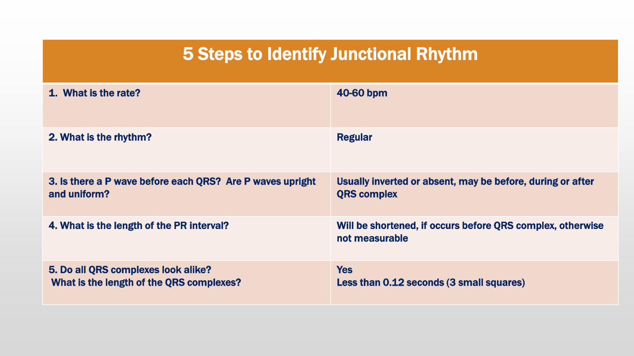
5 Steps to Identify Junctional Rhythm
1. What is the rate? 40-60 bpm
2. What is the rhythm? Regular
3. Is there a P wave before each QRS? Are P waves upright
and uniform?
Usually inverted or absent, may be before, during or after
QRS complex
4. What is the length of the PR interval? Will be shortened, if occurs before QRS complex, otherwise
not measurable
5. Do all QRS complexes look alike?
What is the length of the QRS complexes?
Yes
Less than 0.12 seconds (3 small squares)

Causes and S/S of Junctional Rhythm
Causes
▪ SA node disease
▪ Sick sinus syndrome
▪ Inferior wall MI
▪ Digitalis toxicity
▪ Vagal stimulation
Signs and symptoms
▪ May be asymptomatic
▪ Chest pain
▪ Dyspnea
▪ Hypotension
▪ Altered level of consciousness
▪ Blurred vision

Risk and Medical Tx of Junctional Rhythm
Risk
▪ Reduced cardiac output
Medical Treatment
▪ None if asymptomatic
▪ Treat the underlying cause
▪ Atropine
▪ Temporary or permanent pacemaker

Accelerated Junctional Rhythm
▪ Accelerated junctional rhythms originate in the bundle of His and fire at a rate of 60 - 100bpm
Note: no P waves

5 Steps to Identify Accelerated Junctional Rhythm
1. What is the rate? 60-100 bpm
2. What is the rhythm? Regular
3. Is there a P wave before each QRS? Are P waves upright
and uniform?
Usually inverted or absent, may be before, during or after
QRS complex
4. What is the length of the PR interval? Will be shortened, if occurs before QRS complex, otherwise
not measurable
5. Do all QRS complexes look alike?
What is the length of the QRS complexes?
Yes
Less than 0.12 seconds (3 small squares)

Causes and S/S of Accelerated Junctional Rhythm
Causes
▪ SA node disease
▪ Digitalis toxicity
▪ Hypoxia
▪ Increased vagal tone
▪ Beta blockers and calcium channel blockers
▪ Hypokalemia
▪ Inferior or posterior wall MI
Signs and symptoms
▪ May be asymptomatic
▪ Chest pain
▪ Dyspnea
▪ Hypotension
▪ Altered level of consciousness
▪ Weak peripheral pulses

Risk and Medical Tx of Accelerated Junctional Rhythm
Risk
▪ Reduced cardiac output
Medical Treatment
▪ None if asymptomatic
▪ Treat the underlying cause
▪ Atropine
▪ Temporary or permanent pacemaker

Junctional Tachycardia
♥ Junctional tachycardia is a junctional rhythm with a rate between 101 - 180bpm
Note: inverted P waves

5 Steps to Identify Junctional Tachycardia
1. What is the rate? 101-180 bpm
2. What is the rhythm? Regular
3. Is there a P wave before each QRS? Are P waves upright
and uniform?
Usually inverted or absent, may be before, during or after
QRS complex
4. What is the length of the PR interval? Will be shortened, if occurs before QRS complex, otherwise
not measurable
5. Do all QRS complexes look alike?
What is the length of the QRS complexes?
Yes
Less than 0.12 seconds (3 small squares)
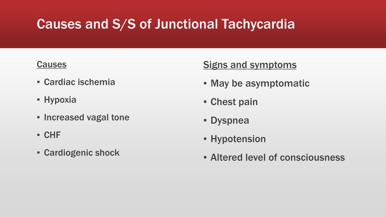
Causes and S/S of Junctional Tachycardia
Causes
▪ Cardiac ischemia
▪ Hypoxia
▪ Increased vagal tone
▪ CHF
▪ Cardiogenic shock
Signs and symptoms
▪ May be asymptomatic
▪ Chest pain
▪ Dyspnea
▪ Hypotension
▪ Altered level of consciousness
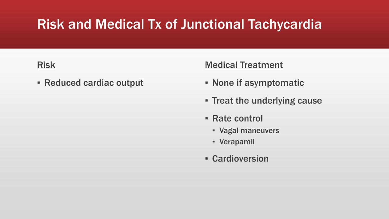
Risk and Medical Tx of Junctional Tachycardia
Risk
▪ Reduced cardiac output
Medical Treatment
▪ None if asymptomatic
▪ Treat the underlying cause
▪ Rate control
▪ Vagal maneuvers
▪ Verapamil
▪ Cardioversion

Premature Junctional Contraction (PJC)
▪ A Premature Junctional Contraction is an early beat that occurs prior to the next sinus beat.
▪ Similar to a PAC EXCEPT P wave is inverted on the PJC!!

5 Steps to Identify Premature Junctional Contraction (PJC)
1. What is the rate? Depends on underlying rhythm
2. What is the rhythm? Irregular noted during the PJC
3. Is there a P wave before each QRS? Are P waves upright
and uniform?
Usually inverted or absent, may be before, during or after
QRS complex
4. What is the length of the PR interval? Will be shortened, if occurs before QRS complex, otherwise
not measurable
5. Do all QRS complexes look alike?
What is the length of the QRS complexes?
Yes
Less than 0.12 seconds (3 small squares)
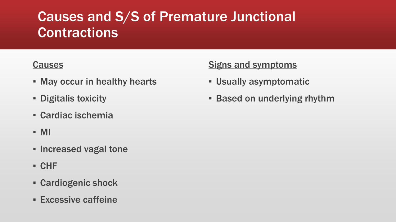
Causes and S/S of Premature Junctional Contractions
Causes
▪ May occur in healthy hearts
▪ Digitalis toxicity
▪ Cardiac ischemia
▪ MI
▪ Increased vagal tone
▪ CHF
▪ Cardiogenic shock
▪ Excessive caffeine
Signs and symptoms
▪ Usually asymptomatic
▪ Based on underlying rhythm

Risk and Medical Tx of Premature Junctional Contraction
Risk
▪ If PJCs occur frequently, junctional tachycardia may result
Medical Treatment
▪ No treatment required unless the PJCs become more frequent
HEART BLOCK RHYTHMS AND NURSING INTERVENTIONS

Objectives
♥ Identify specific cardiac dysrhythmias
♥ Describe appropriate nursing interventions for secific dysrhythmias

The Heart Block Rhythms
♥Heart blocks are arrhythmias caused by an interruption in the conduction of impulses between the atria and the ventricles.
♥The AV block can be total or partial or it may simply delay conduction.

The Heart Block Rhythms
♥First Degree AV Block
♥Second Degree AV Block
♥Type I (Wenckebach)
♥Type II
♥Third Degree AV Block
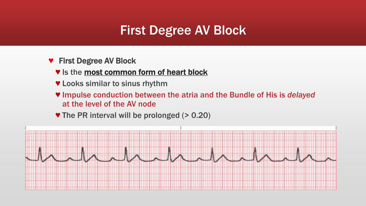
First Degree AV Block
♥ First Degree AV Block
♥ Is the most common form of heart block
♥ Looks similar to sinus rhythm
♥ Impulse conduction between the atria and the Bundle of His is delayed at the level of the AV node
♥ The PR interval will be prolonged (> 0.20)

5 Steps to Identify First Degree AV Block
1. What is the rate? Based on rate of underlying rhythm
2. What is the rhythm? Usually regular
3. Is there a P wave before each QRS? Are P waves upright
and uniform?
Yes
Yes
4. What is the length of the PR interval? >0.20 seconds
5. Do all QRS complexes look alike?
What is the length of the QRS complexes?
Yes
Less than 0.12 seconds (3 small squares)
Causes and S/S of First Degree AV Block
Causes
▪ Can occur in healthy hearts
▪ May be temporary
▪ Myocardial infarction or ischemia
▪ Medications – digitalis, calcium channel blockers, beta blockers
▪ Myocarditis
▪ Degenerative changes in the heart
Signs and Symptoms
▪ Most patients are asymptomatic
▪ Dizziness/syncope
Risk and Medical Tx of First Degree AV Block
Risk
▪ Reduced cardiac output, although this is rare
Treatment
▪ No treatment is usually necessary if the patient is asymptomatic
▪ Treat the underlying cause
▪ Monitor closely to detect progression to a more serious form of block

2nd Degree AV Block Mobitz Type I (Wenckeback)
♥ The delay of the electrical impulse at the AV node produces a progressive increase in the length of the PR interval (> 0.20 seconds)
♥ The PR interval continues to increase in length until the impulse is not conducted or the QRS complex is “dropped”
Dropped QRS! Lengthening PR intervals
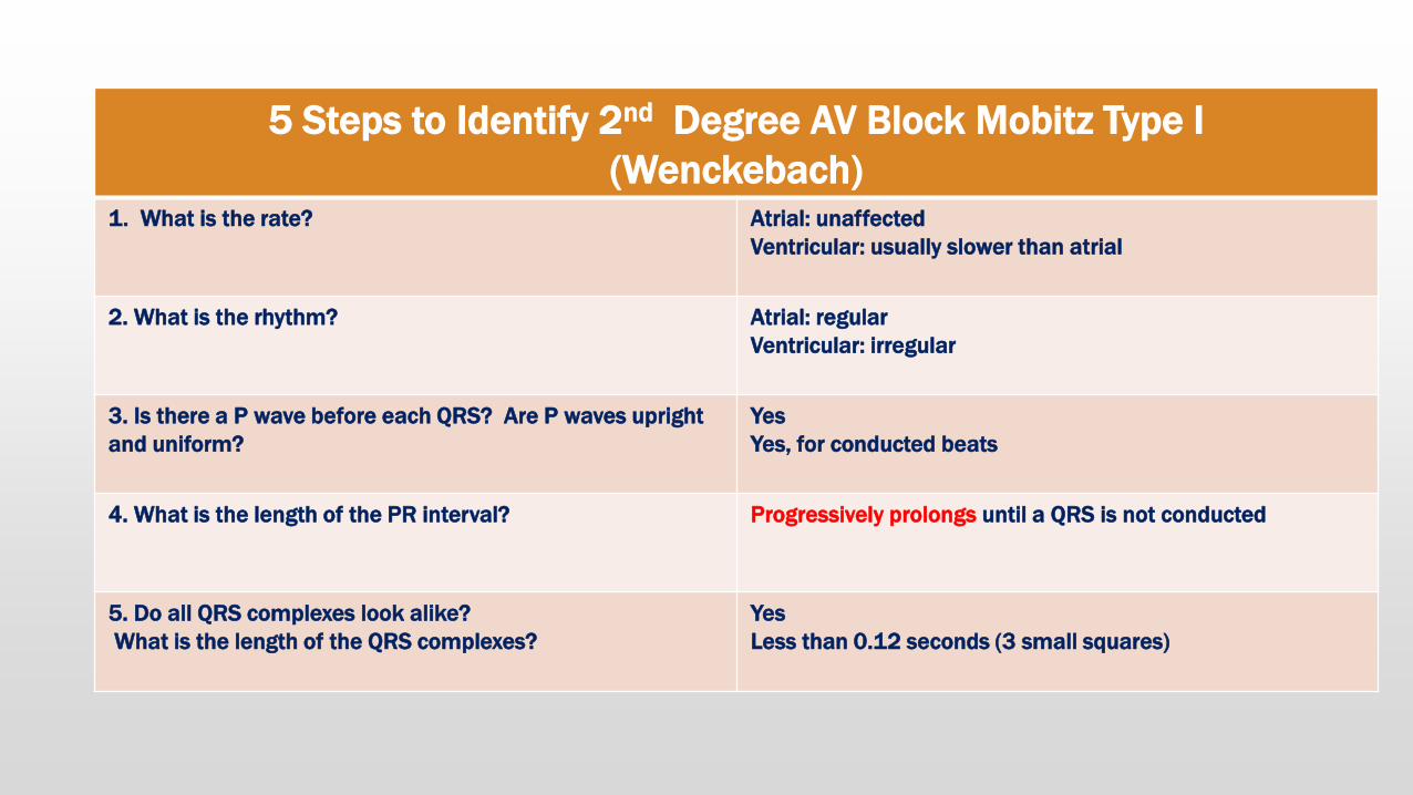
5 Steps to Identify 2nd Degree AV Block Mobitz Type I
(Wenckebach) 1. What is the rate? Atrial: unaffected
Ventricular: usually slower than atrial
2. What is the rhythm? Atrial: regular
Ventricular: irregular
3. Is there a P wave before each QRS? Are P waves upright
and uniform?
Yes
Yes, for conducted beats
4. What is the length of the PR interval? Progressively prolongs until a QRS is not conducted
5. Do all QRS complexes look alike?
What is the length of the QRS complexes?
Yes
Less than 0.12 seconds (3 small squares)
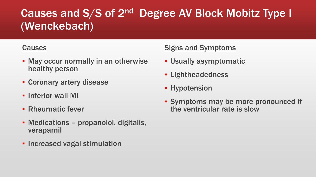
Causes and S/S of 2nd Degree AV Block Mobitz Type I (Wenckebach)
Causes
▪ May occur normally in an otherwise healthy person
▪ Coronary artery disease
▪ Inferior wall MI
▪ Rheumatic fever
▪ Medications – propanolol, digitalis, verapamil
▪ Increased vagal stimulation
Signs and Symptoms
▪ Usually asymptomatic
▪ Lightheadedness
▪ Hypotension
▪ Symptoms may be more pronounced if the ventricular rate is slow
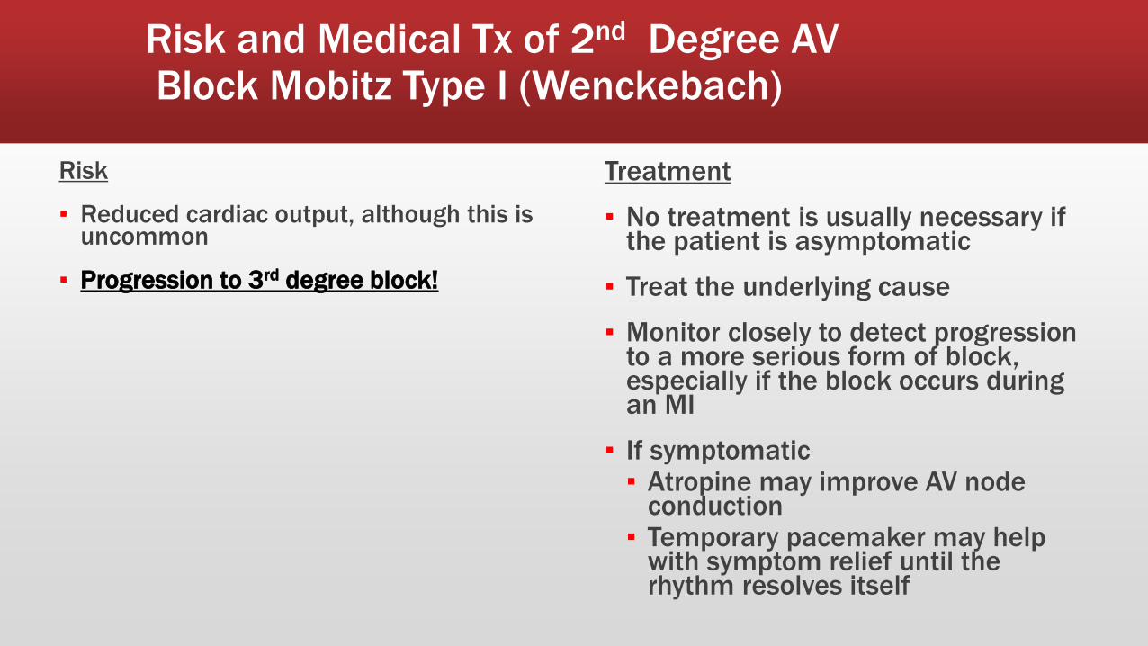
Risk and Medical Tx of 2nd Degree AV Block Mobitz Type I (Wenckebach)
Risk
▪ Reduced cardiac output, although this is uncommon
▪ Progression to 3rd degree block!
Treatment
▪ No treatment is usually necessary if the patient is asymptomatic
▪ Treat the underlying cause
▪ Monitor closely to detect progression to a more serious form of block, especially if the block occurs during an MI
▪ If symptomatic ▪ Atropine may improve AV node
conduction ▪ Temporary pacemaker may help
with symptom relief until the rhythm resolves itself

2nd Degree AV Block Mobitz Type II ♥ Occurs when there is intermittent interruption of conduction
♥ Less common that a 2nd Degree Type 1 block, but more dangerous
♥ PR intervals are regular for conducted beats
♥ The atrial rhythm is regular
♥ The ventricular rhythm is irregular due to dropped or nonconducted (blocked) beats
Will have constant PR intervals with randomly dropped QRS’s
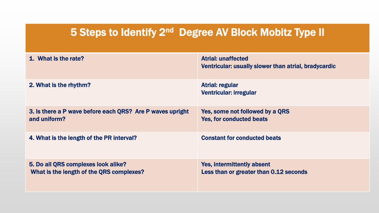
5 Steps to Identify 2nd Degree AV Block Mobitz Type II
1. What is the rate? Atrial: unaffected
Ventricular: usually slower than atrial, bradycardic
2. What is the rhythm? Atrial: regular
Ventricular: irregular
3. Is there a P wave before each QRS? Are P waves upright
and uniform?
Yes, some not followed by a QRS
Yes, for conducted beats
4. What is the length of the PR interval? Constant for conducted beats
5. Do all QRS complexes look alike?
What is the length of the QRS complexes?
Yes, intermittently absent
Less than or greater than 0.12 seconds

Causes and S/S of 2nd Degree AV Block Mobitz Type II
Causes
▪ Anterior wall MI
▪ Degenerative changes in the conduction system
▪ Severe coronary artery disease
Signs and Symptoms
▪ May be asymptomatic as long as cardiac output is maintained
▪ Palpitations
▪ Fatigue
▪ Dyspnea
▪ Chest pain
▪ Lightheadedness
▪ Hypotension

Risk and Medical Tx of 2nd Degree AV Block Mobitz Type II
Risk
▪ Reduced cardiac output
▪ Progression to 3rd degree AV block (complete)
Treatment
▪ May choose to just observe an asymptomatic patient
▪ Bedrest to reduce myocardial oxygen demands
▪ Oxygen therapy
▪ Focus on raising the heart rate to improve cardiac output ▪ Isopreterenol
▪ Permanent pacemaker

3rd Degree AV Block
Complete heart block, AV dissociation
♥ Is the most serious type of heart block
♥ May progress to asystole, because ventricular rate is usually very slow and ineffective
♥ Impulses from the atria are completely blocked at the AV node and can’t be conducted to the ventricles
P waves are usually march consistently through….NO correlation between P’s & QRS’s

5 Steps to Identify 3rd Degree AV Block
1. What is the rate? Atrial: usually 60-100
Ventricular: based on site of pacemaker site
2. What is the rhythm? Regular
3. Is there a P wave before each QRS? Are P waves upright
and uniform?
No relationship to QRS complexes
Yes
4. What is the length of the PR interval? Totally variable, no pattern
5. Do all QRS complexes look alike?
What is the length of the QRS complexes?
Yes
Based on site of pacemaker

Causes and S/S of 3rd Degree AV Block
Causes
▪ Congenital condition
▪ Coronary artery disease
▪ Anterior or inferior wall MI
▪ Degenerative changes in the heart
▪ Digitalis toxicity
▪ Surgical injury
Signs and Symptoms
▪ Severe fatigue
▪ Dyspnea
▪ Chest pain
▪ Lightheadedness
▪ Changes in mental status
▪ Loss of consciousness
▪ Hypotension
▪ Pallor
▪ Diaphoresis
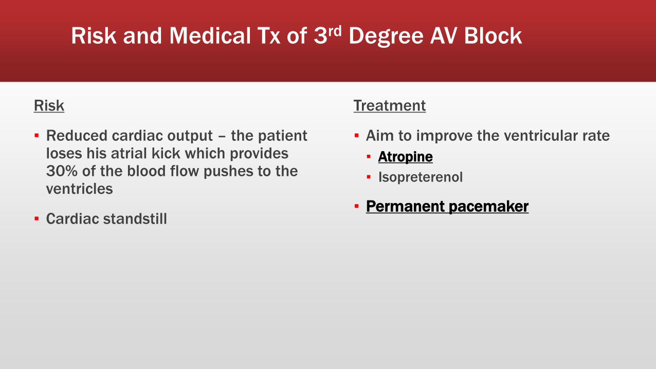
Risk and Medical Tx of 3rd Degree AV Block
Risk
▪ Reduced cardiac output – the patient loses his atrial kick which provides 30% of the blood flow pushes to the ventricles
▪ Cardiac standstill
Treatment
▪ Aim to improve the ventricular rate
▪ Atropine
▪ Isopreterenol
▪ Permanent pacemaker

Click on the link!
▪ Let’s look at what the impulses through the heart look like during a heart block.

Bundle Branch Blocks
♥ The bundle branches split off from the Bundle of His, one branch for each ventricle
♥ Left Bundle Branch
♥ Right Bundle Branch
♥ Bundle Branch blocks
♥ There is an intraventricular conduction delay or block which affects the electrical activity of your heart
♥ These electrical patterns can also point to whether the block is affecting the right or the left bundle branch
Bundle Branch Blocks
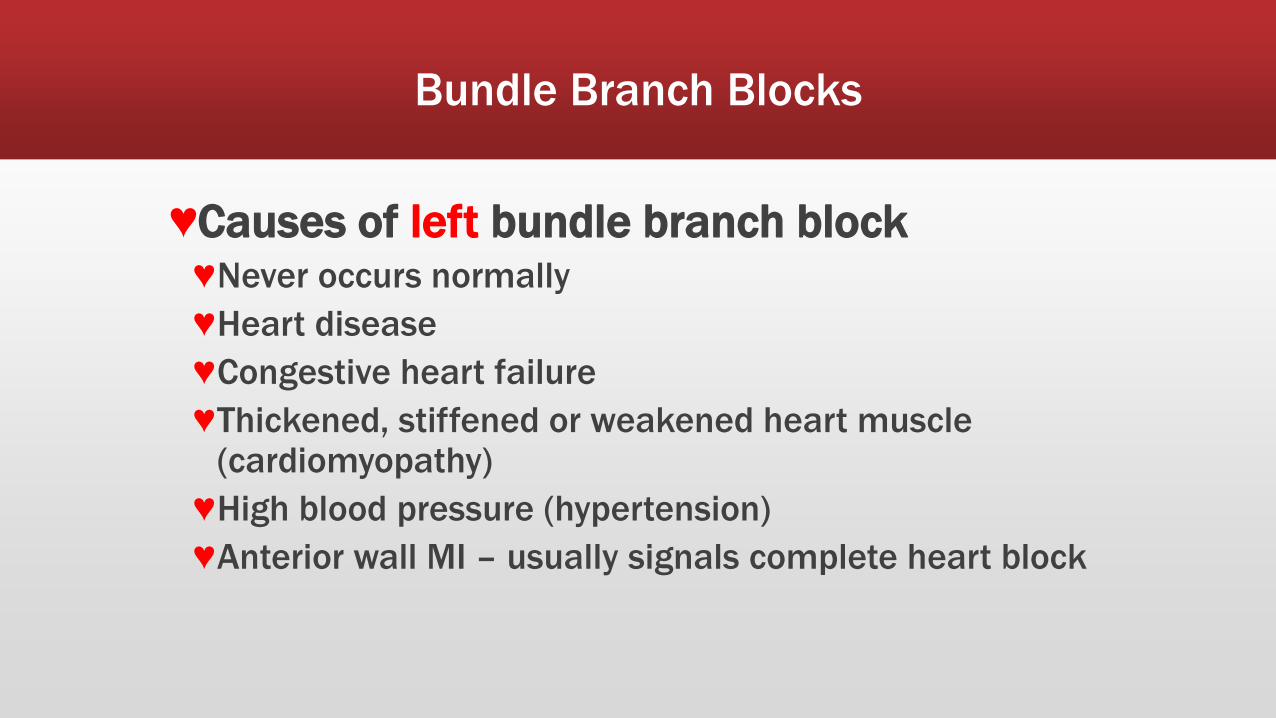
♥Causes of left bundle branch block ♥Never occurs normally
♥Heart disease
♥Congestive heart failure
♥Thickened, stiffened or weakened heart muscle (cardiomyopathy)
♥High blood pressure (hypertension)
♥Anterior wall MI – usually signals complete heart block

Bundle Branch Blocks
♥Criteria for LBBB ♥QRS duration ≥ 0.12 seconds
♥Broad R wave in I and V6
♥Prominent QS wave in V1
♥Absence of q waves (including physiologic q waves) in I and V6

Bundle Branch Blocks
♥ LBBB – notice the broad, notched R wave in V1 and V6 and absent Q wave in lead V6
Notched R wave
Bundle Branch Blocks
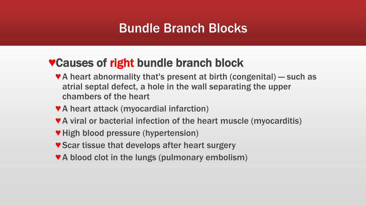
♥Causes of right bundle branch block ♥ A heart abnormality that's present at birth (congenital) — such as
atrial septal defect, a hole in the wall separating the upper chambers of the heart
♥ A heart attack (myocardial infarction)
♥ A viral or bacterial infection of the heart muscle (myocarditis)
♥ High blood pressure (hypertension)
♥ Scar tissue that develops after heart surgery
♥ A blood clot in the lungs (pulmonary embolism)

Bundle Branch Blocks
♥Criteria for RBBB ♥QRS duration ≥ 0.12 seconds
♥rSR’ pattern or notched R wave in V1
♥Wide S wave in I and V6

Bundle Branch Blocks
♥ RBBB – notice the notched R wave of V1 and the broad S wave in I and V6
Notched R wave

Bundle Branch Blocks: Treatment
♥ Treat the underlying heart disease
♥ Reasons for implanting a pacemaker
♥ When disease in both the right and left bundle branches appears after an acute heart attack (complete heart block)
♥ When bundle branch block is associated with syncope (loss of consciousness)
♥ When BBB is associated with heart failure and a reduced left ventricular ejection fraction

TEMPORARY PACEMAKERS

Temporary Pacemakers
▪ Objectives
▪ Explain the situations when temporary pacemakers are indicated.
▪ Describe the principles of pacing.
▪ Illustrate normal and abnormal pacemaker behavior.
▪ Discuss the steps to be taken in troubleshooting a temporary pacemaker.

What is a Pacemaker?
♥ A pacemaker is a small device that's placed in the chest (or on rare occasion, the abdomen) to help control abnormal heart rhythms or arrhythmias.
♥ This device uses electrical pulses to prompt the heart to beat at a normal rate.

Reasons for Pacemaker Insertion
♥ Pacemakers are used to treat arrhythmias.
♥ Arrhythmias are problems with the rate or rhythm of the heartbeat. During an arrhythmia, the heart can beat too fast, too slow, or with an irregular rhythm.
♥ Severe arrhythmias can damage the body's vital organs and may even cause loss of consciousness or death.

Reasons for Pacemaker Insertion
♥During an arrhythmia, the heart may not be able to pump enough blood to the body which may cause symptoms of decreased cardiac output such as fatigue (tiredness), shortness of breath, fainting (syncope), hypotension and decreased LOC.
♥A pacemaker can relieve many arrhythmia symptoms, including fatigue and syncope.
♥A pacemaker also can help a person who has abnormal heart rhythms resume a more active lifestyle.
56

Types of a Pacemakers
♥Pacemakers can be temporary or permanent.
♥Temporary pacemakers ♥Used to treat temporary heartbeat problems, such as a slow heartbeat
that's caused by a heart attack, heart surgery, or an overdose of medicine. ♥Temporary pacemakers are also used during emergencies until a
permanent pacemaker can be implanted or until the temporary condition goes away.
♥If the patient has a temporary pacemaker, they will stay in the hospital as long as the device is in place.
♥Permanent pacemakers are used to control long-term heart rhythm problems.
57

Functions of a Pacemaker
♥ Speeds up a slow heart rate.
♥ Helps control an abnormal rhythm or fast heart rate.
♥ Makes sure the ventricles contract normally if the atria are quivering instead of beating with a normal rhythm (as in atrial fibrillation).
♥ Coordinates the electrical signaling between the upper and lower chambers of the heart.
♥ Coordinates the electrical signaling between the ventricles.
♥ Pacemakers that do this are called cardiac resynchronization therapy (CRT) devices. CRT devices are used to treat heart failure.
♥ Prevents dangerous arrhythmias caused by a disorder called long Q-T syndrome.
58

Who needs a Pacemaker?
♥The most common reasons are bradycardia and heart block.
♥Bradycardia is a slower than normal heartbeat.
♥Those with Heart blocks
♥ Is a problem with the heart's electrical system and occurs when an electrical signal is slowed or disrupted as it moves through the heart.
♥Can happen as a result of aging, damage to the heart from a heart attack, or other conditions that interfere with the heart's electrical activity.
♥Certain nerve and muscle disorders also can cause heart block, including muscular dystrophy.
59

Who needs a Pacemaker?
♥An Aging Heart or Heart Disease
♥Can damage your sinus node's ability to set the correct pace for your heartbeat.
♥Such damage can cause slower than normal heartbeats or long pauses between heartbeats (sinus arrest).
♥The damage also can cause your heart to alternate between slow and fast rhythms (sick sinus syndrome).
60
Who needs a Pacemaker?
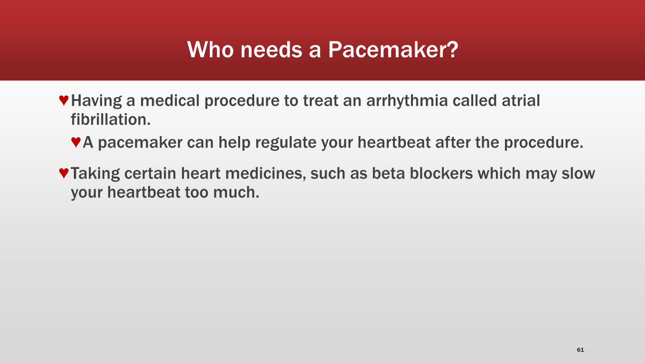
♥Having a medical procedure to treat an arrhythmia called atrial fibrillation.
♥A pacemaker can help regulate your heartbeat after the procedure.
♥Taking certain heart medicines, such as beta blockers which may slow your heartbeat too much.
61

Who needs a Pacemaker?
♥Fainting (syncope) or having other symptoms of a slow heartbeat.
♥For example, this may happen if the main artery in your neck that supplies your brain with blood is sensitive to pressure.
♥Just quickly turning your neck can cause your heart to beat slower than normal. If that happens, not enough blood may flow to your brain, causing you to feel faint or collapse (syncopal episode).
62
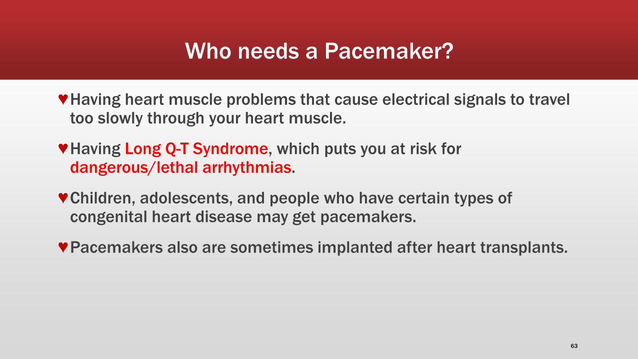
Who needs a Pacemaker?
♥Having heart muscle problems that cause electrical signals to travel too slowly through your heart muscle.
♥Having Long Q-T Syndrome, which puts you at risk for dangerous/lethal arrhythmias.
♥Children, adolescents, and people who have certain types of congenital heart disease may get pacemakers.
♥Pacemakers also are sometimes implanted after heart transplants.
63

How a Pacemaker Functions
♥ A pacemaker system consists of a battery, a computerized generator, and wires with sensors called electrodes on one end.
♥ The battery powers the generator, and both are surrounded by a thin metal box.
♥ The wires connect the generator to the heart.
64

How a Pacemaker Functions
♥A pacemaker monitors and helps control the heartbeat.
♥The electrodes detect the heart's electrical activity and sends data through the wires to the computer in the generator.
♥ If the heart rhythm is abnormal, the computer will direct the generator to send electrical pulses to the heart.
♥The pulses then travel through the wires to reach the heart.
65

How a Pacemaker Functions
♥Newer pacemakers also can monitor your blood temperature, breathing, and other factors and adjust the heart rate to changes in activity.
♥The pacemaker's computer also records the heart's electrical activity and heart rhythm.
♥The physician will use these recordings to adjust the pacemaker so it works better.
♥The physician can program the pacemaker's computer with an external device.
66

How a Pacemaker Functions
♥Pacemakers have one to three wires that are each placed in different chambers of the heart.
♥Single chamber pacemaker
♥The wires in a single-chamber pacemaker usually carry pulses between the right ventricle and the generator.
67


69
Principles of Pacing
▪ Temporary pacing types
▪ Transcutaneous
▪ Emergency use with external pacing/defib unit
▪ Transvenous
▪ Emergency use with external pacemaker
▪ Epicardial
▪ Wires sutured to right atrium & right ventricle
▪ Atrial wires exit on the right of the sternum
▪ Ventricular wires exit on the left of the sternum

70
Principles of Pacing
▪ Wiring systems
▪ Unipolar
▪ One electrode on the heart (-)
▪ Signals return through body fluid and tissue to the pacemaker (+)
▪ Bipolar
▪ Two electrodes on the heart (- & +)
▪ Signals return to the ring electrode (+) above the lead (-) tip

71
Principles of Pacing
▪ Modes of Pacing
▪ Atrial pacing
▪ Intact AV conduction system required
▪ Ventricular pacing
▪ Loss of atrial kick
▪ Discordant ventricular contractions
▪ Sustains cardiac output
▪ Atrial/Ventricular pacing
▪ Natural pacing
▪ Atrial-ventricular synchrony

72
Principles of Pacing
▪ 3-letter NBG Pacemaker Code
▪ First letter: Chamber Paced
▪ V- Ventricle
▪ A- Atrium
▪ D- Dual (A & V)
▪ O- None
73
Principles of Pacing
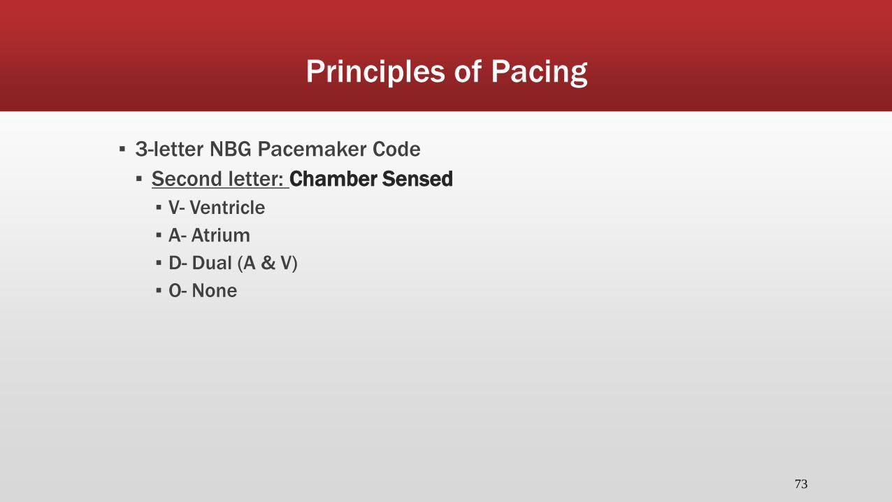
▪ 3-letter NBG Pacemaker Code
▪ Second letter: Chamber Sensed
▪ V- Ventricle
▪ A- Atrium
▪ D- Dual (A & V)
▪ O- None

74
Principles of Pacing
▪ 3-letter NBG Pacemaker Code
▪ Third letter: Sensed Response
▪ T- Triggers Pacing
▪ I- Inhibits Pacing
▪ D- Dual
▪ O- None

75
Principles of Pacing
▪ Commonly used modes:
▪ AAI - atrial demand pacing
▪ VVI - ventricular demand pacing
▪ DDD – atrial/ventricular demand pacing, senses & paces both chambers; trigger or inhibit
▪ AOO - atrial asynchronous pacing

Principles of Pacing
▪ Atrial and ventricular output
▪ Milliamperes (mA)
▪ Typical atrial mA 5
▪ Typical ventricular mA 8-10
▪ AV Interval
▪ Milliseconds (msec)
▪ Time from atrial sense/pace to ventricular pace
▪ Synonymous with “PR” interval
▪ Atrial and ventricular sensitivity
▪ Millivolts (mV)
▪ Typical atrial: 0.4 mV
▪ Typical ventricular: 2.0mV 76

77
Temporary Pacemaker
▪ Medtronic 5388 Dual Chamber (DDD)

78
Pacemaker EKG Strips
▪ Assessing Paced EKG Strips
▪ Identify intrinsic rhythm and clinical condition
▪ Identify pacer spikes
▪ Identify activity following pacer spikes
▪ Failure to capture
▪ Failure to sense
▪ EVERY PACER SPIKE SHOULD HAVE A P-WAVE OR QRS COMPLEX FOLLOWING IT.

79
Normal Pacing
▪ Normal Atrial Pacing
▪ Atrial pacing spikes followed by P waves
Pacing spikes Followed by a P wave

80
Normal Pacing
▪ Normal Ventricular pacing
▪ Ventricular pacing spikes followed by wide, bizarre QRS complexes
▪ QRS will look wide and bizarre b/c impulse is generated in the ventricle…does not follow normal pathways from atria
Pacing spikes Followed by QRS

81
Normal Pacing
▪ Normal A-V Pacing
▪ Atrial & Ventricular pacing spikes followed by atrial & ventricular complexes

82
Normal Pacing
▪ DDD mode of pacing
▪ Ventricle paced at atrial rate

83
Abnormal Pacing
▪ Atrial non-capture
▪ Atrial pacing spikes are not followed by P waves
▪ Impulse is sent from the pacer, but is not captured by the ventricle…therefore no QRS following a pacer spike
Pacer spike No P wave after spike

84
Abnormal Pacing
▪ Ventricular non-capture
▪ Ventricular pacing spikes are not followed by QRS complexes
▪ Impulse is sent from the pacer, but is not captured by the ventricle…therefore no QRS following a pacer spike
Pacing spike No QRS following QRS complex

85
Failure to Capture
▪ Causes
▪ Insufficient energy delivered by pacer
▪ Low pacemaker battery
▪ Dislodged, loose, fibrotic, or fractured electrode
▪ Electrolyte abnormalities
▪ Acidosis
▪ Hypoxemia
▪ Hypokalemia
▪ Danger - poor cardiac output

86
Failure to Capture
▪ Solutions
▪ View rhythm in different leads
▪ Change electrodes
▪ Check connections
▪ Increase pacer output (↑mA)
▪ Change battery, cables, pacer

87
Abnormal Pacing
▪ Atrial undersensing
▪ Atrial pacing spikes occur irregardless of P waves
▪ Pacemaker is not “seeing” intrinsic activity
P wave from pt’s intrinsic Pacer sent impulse anyways
88
Abnormal Pacing
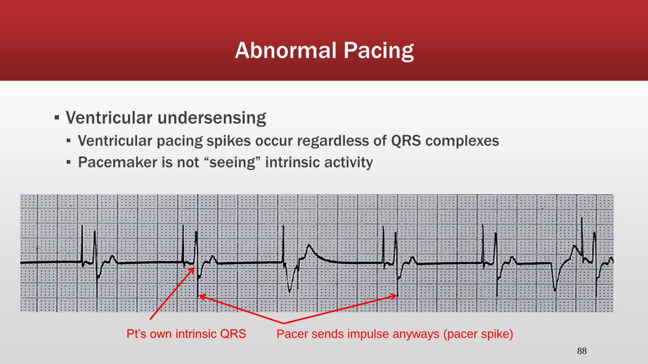
▪ Ventricular undersensing
▪ Ventricular pacing spikes occur regardless of QRS complexes
▪ Pacemaker is not “seeing” intrinsic activity
Pt’s own intrinsic QRS Pacer sends impulse anyways (pacer spike)
89
Failure to Sense
▪ Causes
▪ Pacemaker not sensitive enough to patient’s intrinsic electrical activity (mV)
▪ Insufficient myocardial voltage
▪ Dislodged, loose, fibrotic, or fractured electrode
▪ Electrolyte abnormalities
▪ Low battery
▪ Malfunction of pacemaker or bridging cable
90
Failure to Sense
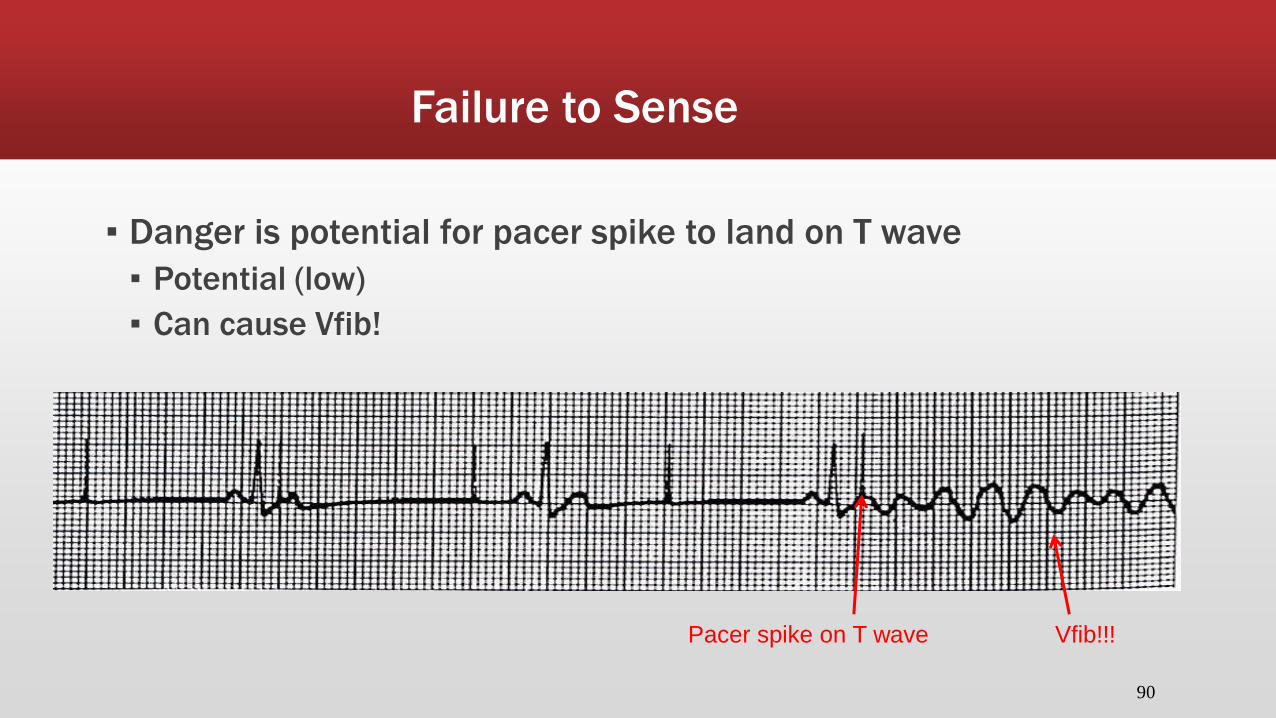
▪ Danger is potential for pacer spike to land on T wave
▪ Potential (low)
▪ Can cause Vfib!
Pacer spike on T wave Vfib!!!
91
Failure to Sense
▪ Solution
▪ View rhythm in different leads
▪ Change electrodes
▪ Check connections
▪ Increase pacemaker’s sensitivity (↓mV)
▪ Change cables, battery, pacemaker
▪ Check electrolytes

Assessing Underlying Rhythm
▪ Carefully assess underlying rhythm
▪ Right way: slowly decrease pacemaker rate until lower than patient’s own intrinsic rhythm
92

93
Assessing Underlying Rhythm
▪ Assessing Underlying Rhythm
▪ Wrong way: pause pacer or unplug cables

94
Practice Strip#1

▪ Mode: AAI
▪ Interpretation: Normal atrial pacing

96
Practice Strip #2

▪ Interpretation: Normal sinus rhythm – no pacing
▪ May possibly have backup demand rate set if HR falls below certain rate

98
Practice Strip #3

▪ Mode: DDD (should be pacing atria and ventricles)
▪ Interpretation: Ventricular failure to sense
▪ Troubleshootin: decrease mV (makes the pacer more sensitive)

100
Practice Strip #4

▪ Mode: VVI
▪ Interpretation: Normal ventricular pacing

102
Practice Strip #5

▪ Mode: DDD
▪ Interpretation: Failure to capture atria & ventricles (the P waves seen
are the pt’s own intrinsic P wave)
▪ Troubleshooting: Increase the atrial and ventricular mA
Atrial pacer spike but no P wave following Ventricular pacer spike, but no QRS following

104
Practice Strip #6

▪ Mode: DDD
▪ Interpretation: Normal atrial & ventricular (A-V) pacing

106
Practice Strip #7

▪ Mode: VVI
▪ Interpretation: Normal ventricular pacing w/ normal atrial sensing (note the p wave present without an atrial pacer spike – pt is generating on atrial impulse)
P waves

108
Practice Strip #8

▪ Mode: DDD
▪ Interpretation: Atrial failure to sense with normal ventricular pacing
▪ Troubleshooting: Decrease mV (making pacer more sensitive)
Pt generated own P wave,
therefore there shouldn’t be a pacer spike This pacer spike ok b/c no prior P wave

4/07 110
Practice Strip #9

▪ Mode: VVI
▪ Interpretation: Ventricular over-sensing (pacemaker failed to send
impulse to generate a ventricular response)
▪ Troubleshooting: Increase mV (making pacer less sensitive)
No pacer spike for ventricles

ASSESSMENT AND TREATMENT OF THE PATIENT WITH CARDIAC EMERGENCIES
Assessment and Treatment of the Patient With Cardiac Emergencies
▪ Objectives
▪ Discuss the indicators used to differentiate chest pain of cardiac origin from noncardiac origin
▪ Describe the pathophysiology of angina pectoris
▪ Describe the assessment and management of the patient with angina pectoris
▪ Describe the pathophysiology of myocardial infarction

Assessment and Treatment of the Patient With Cardiac Emergencies
▪ Objectives (continued)
▪ Discuss the assessment and management of the patient with acute myocardial infarction
▪ Describe the physiology associated with heart failure
▪ Discuss the signs and symptoms of left ventricular failure and those of right ventricular failure
▪ List the interventions prescribed for the patient in congestive heart failure

Assessment and Treatment of the Patient With Cardiac Emergencies
▪ Objectives (continued)
▪ Define and describe the pathophysiology, assessment, and management of cardiac tamponade
▪ Define and describe the pathophysiology, assessment, and management of cardiogenic shock

Chest Pain
▪ Chest pain is the most common presenting symptom of cardiac disease
▪ Patients may express a feeling of “impending doom”
▪ Often patients prefer to believe that they are merely experiencing “indigestion” and symptoms would be gone by morning

Chest Pain
▪ Chest pain of cardiac origin is typically described as “crushing” or “squeezing” in nature
▪ Associated with nausea, vomiting, and diaphoresis
▪ May radiate to other areas (jaw, shoulder, arm, etc.)

Chest Pain

Chest Pain
▪ Neuropathy due to destruction of nerve endings can cause inability to perceive pain due to diseases of the nerves
▪ Diabetics
▪ May present as congestive heart failure as the first symptom of AMI
Non-Cardiac Causes of Chest Pain
▪ Pleurisy
▪ Costrochondritis
▪ Pericarditis
▪ Myocardial contusion
▪ Muscle strain
▪ Trauma
▪ Secondary to trauma
▪ Pneumothorax
▪ Hemopneumothorax
▪ Tension pneumothorax

Angina Pectoris
▪ Pain that results from reduction in blood supply to myocardial tissue
▪ Pain is typically temporary
▪ Commonly caused by atherosclerosis
▪ Often predictably associated with exercise
▪ Pain is felt when the heart requires more oxygen than the narrowed blood vessels can supply

Angina Pectoris
Angina Pectoris
▪ “Stable” or predictable angina
▪ A particular activity may elicit chest pain
▪ Symptoms will usually respond well to appropriate treatment
▪ Rest
▪ Administration of oxygen
▪ “Unstable” angina
▪ Is not elicited by activity
▪ Most often occurs at rest
▪ Indicates a progression of atherosclerotic heart disease, and is also referred to as “preinfarctional” angina

Angina Pectoris
▪ Management
▪ Place patient at rest in calm, quiet area
▪ Provide reassurance
▪ Obtain 12 lead EKG if possible
▪ Administer oxygen
▪ IV line
▪ Administer nitroglycerin

Nitroglycerin
▪ Causes dilation of blood vessels that reduces the workload of the heart
▪ Reduces the need for oxygen because the heart has to pump blood against a lesser pressure
▪ Blood remains in dilated vessels and less is returned to the heart

Acute Myocardial Infarction
▪ Results from a prolonged lack of blood flow to a portion of myocardial tissue and results in a lack of oxygen
▪ Myocardial cellular death will follow
▪ Electrical properties of cardiac muscle altered or lost
▪ Ability of cardiac muscle to function properly is lost

Acute Myocardial Infarction
▪ Most common cause is thrombus formation
▪ Blocks coronary arteries
▪ Arteries narrowed by atherosclerotic disease are one of the conditions that increase likelihood of myocardial infarction (MI)

Acute Myocardial Infarction

Patient Assessment and Management

Patient Assessment and Management
▪ 100 percent oxygen
▪ Establish an IV line
▪ Measure oxygen saturation level
▪ Continuous cardiac monitoring
▪ Pain control and management
▪ Nitroglycerin, Morphine Sulfate, Demerol
▪ Thrombolytic therapy, aspirin

ST Elevation
♥ ST segment elevation MI (STEMI) is caused by:
♥ A complete obstruction of a coronary artery, resulting in damage/necrosis of the full thickness of the heart muscle
♥ Diffuse ST segment elevation may be caused by pericarditis
♥ Non-ST segment elevation MI (NSTEMI) is caused by:
♥ Non-occlusive thrombus
♥ Brief occlusion
♥ Occlusion with adequate collaterals
♥This results in necrosis involving only partial thickness of the heart muscle

ST Elevation
♥ ST elevation due to MI usually demonstrates a regional or territorial pattern
♥ Anatomic region of the heart and the most likely associated coronary artery:
♥ Inferior - Right coronary
artery (RCA)
♥ Anteroseptal - Left anterior
descending (LAD)
♥ Anteroapical - Left anterior
descending (Distal)
♥ Anterolateral - Circumflex
♥ Posterior - Right coronary
artery (RCA)

ST Elevation
♥These regional areas correspond to specific leads on the EKG: ♥ V2 through V5 - Anterior MI (LAD)
♥ V2 and V3 - Septal MI (LAD)
♥ V1, V2, V4, and V5 – Anteroseptal MI (LAD)
♥ I, AVL – Lateral MI (Cx)
♥ V4 through V6 – Anterolateral MI (Cx)
♥ II, III and AVF – Inferior MI (RCA)
♥ V1 and V2 – Posterior MI (RCA)

ST Elevation
♥ Example of ST Elevation

ST Elevation
♥ Example of ST Elevation

ST Elevation
Example of an Inferior MI (Note ST elevation in leads II, III, aVF)
ST Elevation
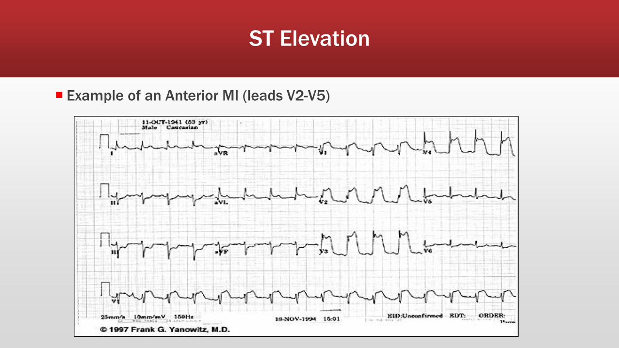
Example of an Anterior MI (leads V2-V5)

Treatment Chest Pain with ST Segment Elevation
♥ Notify MD
♥ Obtain EKG
♥ within 10 minutes of onset of chest pain
♥ preferably before giving NTG
♥ Give Aspirin (81mg x4, chewable)
♥ NTG 0.4mg SL every 5 minutes x3 (hold for SBP <100)
♥ Have patent IV prior to giving
♥ Draw labs
♥ Cardiac enzymes, BMP and CBC

Heart Failure
▪ Is the inability of the myocardium to meet the cardiac output demands of the body caused by:
▪ Coronary disease
▪ Valvular disease
▪ Myocardial injury
Dysrhythmias
Hypertension
Pulmonary emboli
Systemic sepsis
Electrolyte disturbances

Left Ventricular Failure
▪ When a patient’s left ventricle ceases to function in an adequate capacity as to sustain sufficient systemic cardiac output
▪ Stroke volume decreases
▪ Heart rate increases & vasoconstriction occurs to compensate
▪ Increased pressure in left ventricle and left atrium
▪ Blood pushed back into pulmonary system
▪ Can also be pushed back into the right side of the heart if extensive left heart failure
▪ Develop pulmonary edema and hypoxia
▪ Pink, frothy sputum and significant dyspnea

Left Ventricular Failure
▪ Emergency management
▪ Have patient assume position of comfort
▪ 100% high-flow oxygen
▪ Utilize pulse oximetry @ 90% saturation
▪ Monitor LOC for signs of deterioration
▪ Establish IV @ KVO rate
▪ Maintain EKG monitoring
▪ Follow protocol for administration of medications
▪ Morphine
▪ Lasix
▪ Nitroglycerine
Right Heart Failure
▪ When the right ventricle ceases to function properly
▪ Causes increase in pressure in right atrium, forcing blood backward into systemic venous system
▪ Most common cause is left heart failure
▪ Other causes:
▪ Valvular heart disease
▪ COPD or cor pulmonale
▪ Pulmonary embolism
▪ Chronic hypertension


Right Heart Failure
▪ Emergency management
▪ Have patient assume position of comfort
▪ Oxygenation at level to maintain saturation of at least 90%
▪ Establish IV at KVO rate
▪ Maintain EKG monitoring
▪ Consult physician on administration of medications
▪ Observe for signs of developing left heart failure
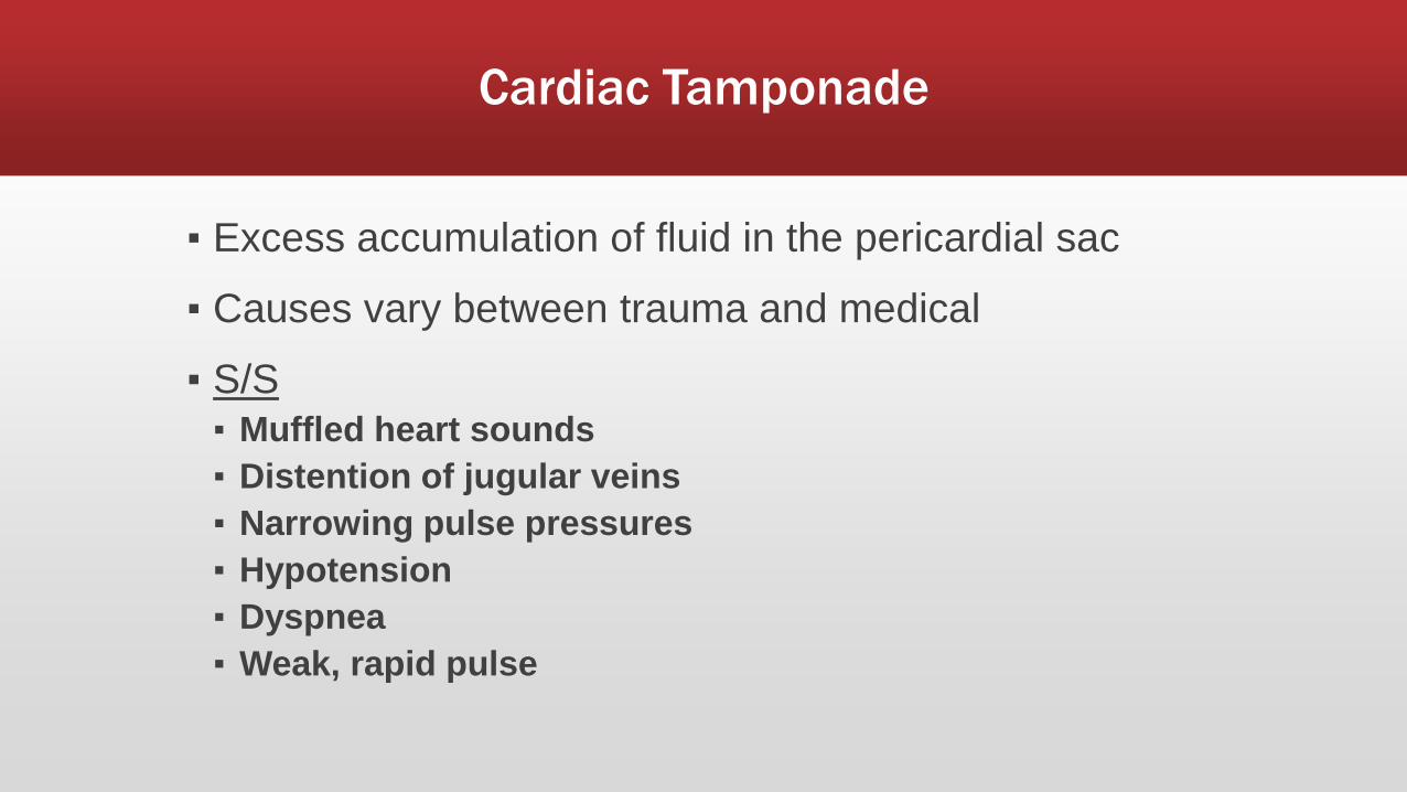
Cardiac Tamponade
▪ Excess accumulation of fluid in the pericardial sac
▪ Causes vary between trauma and medical
▪ S/S
▪ Muffled heart sounds
▪ Distention of jugular veins
▪ Narrowing pulse pressures
▪ Hypotension
▪ Dyspnea
▪ Weak, rapid pulse

Cardiac Tamponade
▪ “Beck’s triad”
▪ Muffled heart sounds
▪ JVD
▪ Narrowed pulse pressure
▪ Pulsus paradoxus
▪ Systolic blood pressure that drops more than 10–15 mmHg during inspiration

Cardiac Tamponade
▪ Emergency management
▪ Ensure and maintain patent airway
▪ Administer 100% high-flow oxygen
▪ Monitor pulse oximetry
▪ Establish and maintain IV support
▪ Administer pharmacological agents as indicated
▪ Pericardiocentesis
▪ Invasive aspiration of fluid from the pericardium with a needle

Cardiogenic Shock
▪ When left ventricular function is so severely compromised that the heart can no longer meet metabolic requirements of the body
▪ Often results from extensive myocardial infarction

Cardiogenic Shock
▪ Most critical form of CHF
▪ Ineffective myocardial contractions result in
▪ Marked decreased stroke volume
▪ Decreased cardiac output
▪ Leading to inadequate tissue perfusion
▪ Profound hypotension
▪ Compensatory tachycardia
▪ Tachypnea, often resulting from pulmonary edema
▪ Cool, clammy skin caused by massive vasoconstriction
▪ Major dysrhythmias
▪ Respiratory difficulty
▪ Peripheral edema
Cardiogenic Shock
▪ Aggressive treatment measures
▪ Airway management with high flow oxygen
▪ Circulatory support, including IV therapy
▪ Patient to assume position of comfort
▪ Cardiac monitoring
▪ Pulse oximetry, maintain O2 saturation @ 90%
▪ Medication therapy
▪ Various vasopressors
▪ Dopamine
▪ Dobutamine
▪ Levophed
▪ Other medications
▪ Morphine Sulfate
▪ Nitroglycerin
▪ Lasix
▪ Digitalis

Time for some practice strips!
Copyright © 2006
by Mosby Inc. All
rights reserved.
Rhythm Identification

Copyright © 2006
by Mosby Inc. All
rights reserved.
Ventricular rate/rhythm 68 bpm/regular
Atrial rate/rhythm 68 bpm/regular
PR interval 0.28 sec
QRS duration 0.06 sec
Identification Sinus rhythm with first-degree AV block, ST-
segment depression

Copyright © 2006
by Mosby Inc. All
rights reserved.
Rhythm Identification
▪ This rhythm strip is from a 52-year-old man with substernal chest pain. He has a history of COPD and mitral valve regurgitation. Blood pressure: 140/78.

Copyright © 2006
by Mosby Inc. All
rights reserved.
Ventricular rate/rhythm 75 bpm/regular
Atrial rate/rhythm 75 bpm/regular
PR interval 0.14 sec
QRS duration 0.06 to 0.08 sec
Identification Sinus rhythm with ST-segment depression

Copyright © 2006
by Mosby Inc. All
rights reserved.
Rhythm Identification
Copyright © 2006
by Mosby Inc. All
rights reserved.
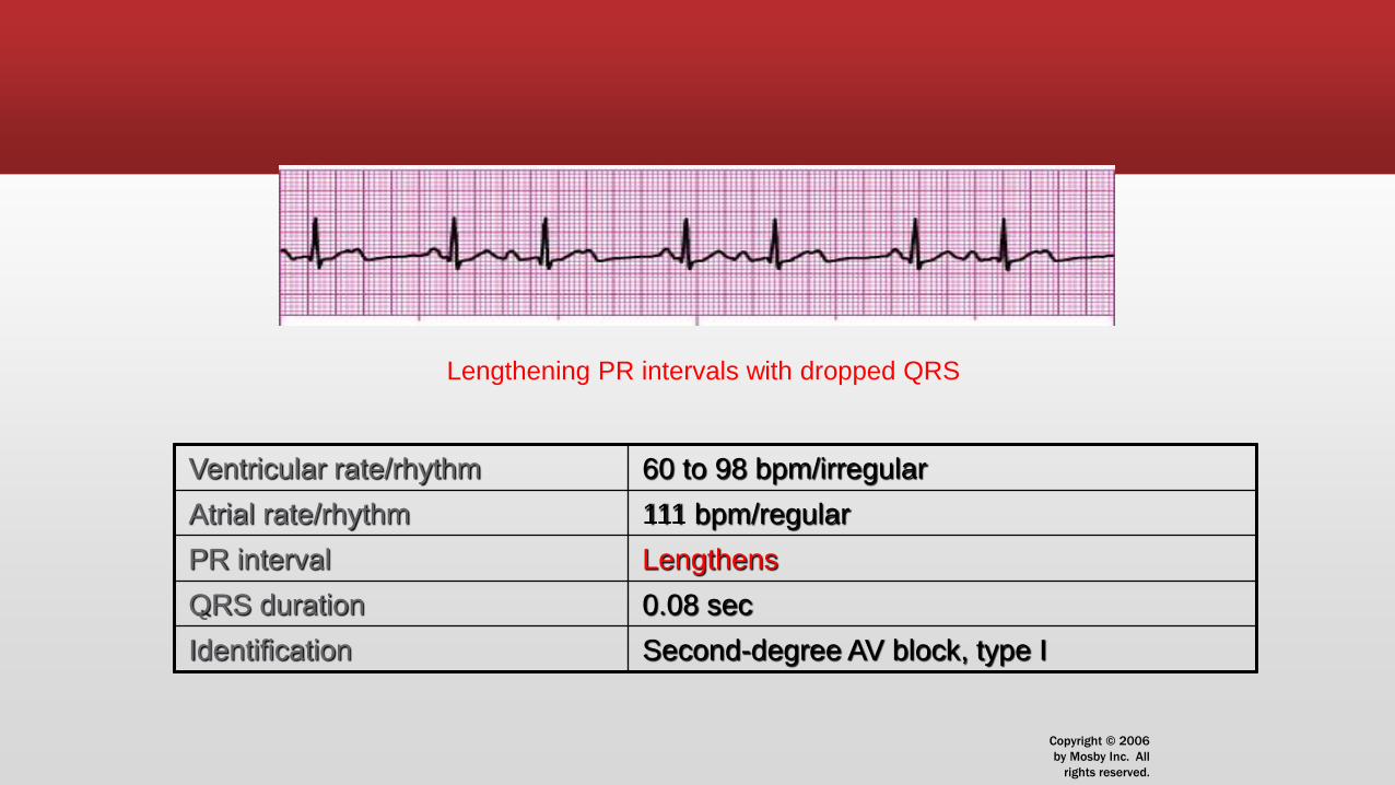
Ventricular rate/rhythm 60 to 98 bpm/irregular
Atrial rate/rhythm 111 bpm/regular
PR interval Lengthens
QRS duration 0.08 sec
Identification Second-degree AV block, type I
Lengthening PR intervals with dropped QRS

Copyright © 2006
by Mosby Inc. All
rights reserved.
Rhythm Identification

Copyright © 2006
by Mosby Inc. All
rights reserved.
Ventricular rate/rhythm 75 bpm/regular
Atrial rate/rhythm None
PR interval None
QRS duration 0.08 sec
Identification Accelerated junctional rhythm

Copyright © 2006
by Mosby Inc. All
rights reserved.
Rhythm Identification
Copyright © 2006
by Mosby Inc. All
rights reserved.
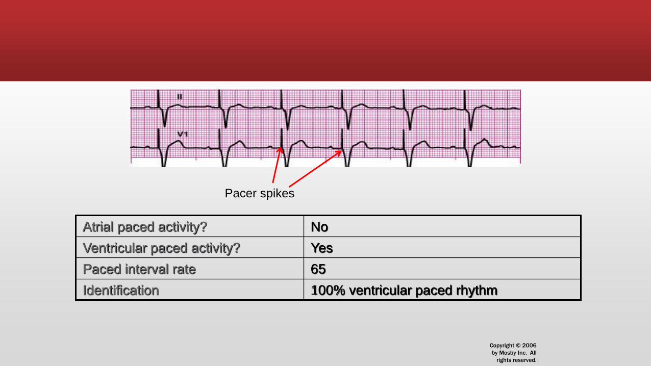
Atrial paced activity? No
Ventricular paced activity? Yes
Paced interval rate 65
Identification 100% ventricular paced rhythm
Pacer spikes

Copyright © 2006
by Mosby Inc. All
rights reserved.
Rhythm Identification

Copyright © 2006
by Mosby Inc. All
rights reserved.
Ventricular rate/rhythm 45 bpm/regular
Atrial rate/rhythm 115 bpm/regular
PR interval No correlation between P & QRS; therefore
cannot be measured!
QRS duration 0.16 sec
Identification Complete (third-degree) AV block

Copyright © 2006
by Mosby Inc. All
rights reserved.
Rhythm Identification
▪ This rhythm strip is from a 66-year-old man complaining of chest pain. Blood pressure: 170/96.

Copyright © 2006
by Mosby Inc. All
rights reserved.
Ventricular rate/rhythm 107 bpm/regular
Atrial rate/rhythm 107 bpm/regular
PR interval 0.24 sec
QRS duration 0.08 sec
Identification Sinus tachycardia with
first-degree AV block

Copyright © 2006
by Mosby Inc. All
rights reserved.
Rhythm Identification
▪ This rhythm strip is from a 76-year-old woman complaining of back pain. Her medical history includes a myocardial infarction 2 years ago.

Copyright © 2006
by Mosby Inc. All
rights reserved.
Atrial paced activity? No
Ventricular paced activity? Yes
Paced interval rate 65
Identification 100% ventricular paced rhythm
Copyright © 2006
by Mosby Inc. All
rights reserved.
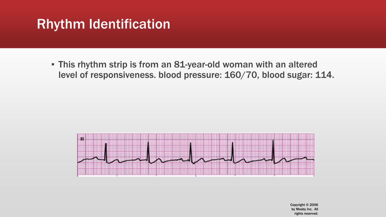
Rhythm Identification
▪ This rhythm strip is from an 81-year-old woman with an altered level of responsiveness. blood pressure: 160/70, blood sugar: 114.

Copyright © 2006
by Mosby Inc. All
rights reserved.
Ventricular rate/rhythm 56 bpm/regular
Atrial rate/rhythm 56 bpm/regular
PR interval 0.24 sec
QRS duration 0.06 sec
Identification Sinus bradycardia with
first-degree AV block

Copyright © 2006
by Mosby Inc. All
rights reserved.
Rhythm Identification

Copyright © 2006
by Mosby Inc. All
rights reserved.
Ventricular rate/rhythm 29 bpm/regular
Atrial rate/rhythm 71 bpm/regular
PR interval Varies
QRS duration 0.16 sec
Identification Complete (third-degree) AV block
with ST-segment elevation

Copyright © 2006
by Mosby Inc. All
rights reserved.
Rhythm Identification

Copyright © 2006
by Mosby Inc. All
rights reserved.
Ventricular rate/rhythm 29 bpm/essentially regular
Atrial rate/rhythm None
PR interval None
QRS duration 0.04 to 0.06 sec
Identification Junctional bradycardia
with ST-segment depression

Copyright © 2006
by Mosby Inc. All
rights reserved.
Rhythm Identification

Copyright © 2006
by Mosby Inc. All
rights reserved.
Ventricular rate/rhythm 44 bpm/regular
Atrial rate/rhythm 44 bpm/regular
PR interval None
QRS duration 0.08 sec
Identification Junctional rhythm; ST-segment elevation
Copyright © 2006
by Mosby Inc. All
rights reserved.
Rhythm Identification

Copyright © 2006
by Mosby Inc. All
rights reserved.
Ventricular rate/rhythm 60 to 98 bpm/irregular
Atrial rate/rhythm 111 bpm/regular
PR interval Lengthens
QRS duration 0.08 sec
Identification Second-degree AV block type I; ST-segment
depression

Copyright © 2006
by Mosby Inc. All
rights reserved.
Rhythm Identification

Copyright © 2006
by Mosby Inc. All
rights reserved.
Atrial pacing? Yes
Ventricular pacing? Yes
Paced interval 71
Identification 100% paced rhythm
– AV pacemaker
