advanced meniscus repair: radial, root, horizontal
TRANSCRIPT

For OSUWMC USE ONLY. To license, please contact the OSU Technology Commercialization Office at https://tco.osu.edu.
ADVANCED MENISCUS REPAIR: RADIAL, ROOT, HORIZONTAL CLEAVAGE TEAR CLINICAL PRACTICE GUIDELINE Disclaimer The following rehabilitation guidelines are specific to patients who have undergone an advanced meniscus repair of a radial, root or horizontal cleavage tear. Please refer to the Ohio States Sports Medicine website for rehabilitation guidelines specific to other procedures and conditions, as appropriate. Progression is criterion-based and dependent on soft tissue healing, patient demographics, and clinical evaluation. The time frames identified for each phase of rehabilitation are approximate times for the average patient and not recommended as guidelines for progression for the individual patient. It is recommended that progression is based upon the achievement of functional criteria demonstrating readiness for progression, noted at the end of each phase. Background Meniscal root/radial tears present in a variety of forms, ranging from partial to complete avulsion. Root and radial tears can have a profound effect on the health of the articular cartilage of the knee with the potential for meniscal extrusion and accelerated arthritic degeneration if left untreated. Horizontal cleavage tears can result in advanced degeneration of the meniscus tissue and underlying cartilage, especially during high-impact activity. The listed clinical recommendations are a result of the complexity of the surgical technique. The rehabilitation recommendations below are based upon the guidance of content experts, surgeons, and evidence-based practice. Progression through each phase, after precautions have been lifted, is based on the patient demonstrating readiness by achieving the listed functional criteria.

For OSUWMC USE ONLY. To license, please contact the OSU Technology Commercialization Office at https://tco.osu.edu.
Summary of Recommendations
Precautions • WBing and bracing recommendations are based on tear morphology and intra-operative findings. Recommendations are below. However, ALWAYS refer to the operative note or contact the surgical team for clarification.
• For root/radial tears – no resisted isotonic hamstring strengthening x8 weeks • Many of these patients will be encouraged to wear a medial unloader brace for the first 12
months post-op
Risk Factors The patient should be monitored for signs and symptoms of DVT (see Red/Yellow Flag section)
Weight Bearing/Bracing
Root Repair: • TROM immobilizer for first 10-14 days post-op • NWBing x 4 weeks, with a goal of crutch discharge by 6 weeks
Radial Repair: • Typically NWBing x 4 weeks, with a goal of crutch discharge by 6 weeks • WBing status and bracing are patient dependent – always refer to the operative note
or contact the surgical team for clarification
Horizontal Cleavage Repair: • No brace • NWBing x 2-4 weeks, crutches should be discharged no later than 6 weeks
Please refer to the “post-op plan” section of the operative note or contact the surgeon for clarification
Range of Motion • Week 0-2: 0-90° • Week 2-4: progress to full PROM • Week 4+: full AROM
Symmetrical knee extension should be achieved by week 4. If not achieved by week 4, contact surgeon
Outcome Tools Collect the LEFS at each visit
You may choose to include IKDC, KOOS, ACL-RSI, Tegner or other questionnaires specific to your patient’s needs
Hamstring Considerations
Root/Radial Repairs: No resisted isotonic hamstring strengthening x8 weeks No hamstring precautions for horizontal cleavage repairs
Functional Testing
• Isometric Testing o Root/Radial Repairs: 4 months o Horizontal Cleavage Repairs: 3 months
• Isokinetic Testing o Root/Radial Repairs: 6 months o Horizontal Cleavage Repairs: 4 months
• Hop testing: should not be performed prior to 6 months post-op and is only appropriate after 80% symmetry is achieved on isokinetic testing
o SL hop for distance o Triple hop o Cross over hop o Timed 6m hop
*Functional strength testing and hop testing should be reserved for patients returning to high level activity*

For OSUWMC USE ONLY. To license, please contact the OSU Technology Commercialization Office at https://tco.osu.edu.
Criteria to Discharge Assistive Device
• ROM: Full active knee extension; no pain on passive overpressure • Strength: Able to perform strong quad isometric with full tetany and superior patellar glide
and able to perform 2x10 SLR without quad lag • Effusion: 1+ or less is preferred (2+ acceptable if all other criteria are met) • Weight Bearing: Demonstrates pain-free ambulation without visible gait deviation
Criteria to D/C NMES
• <20% quad deficit on isometric testing • If Biodex not available:
o 10 SLR without quad lag o Normal gait o 10 heel taps to 60° knee flexion with good quality o 10 rep max on leg press and similar effort bilaterally o Inability to break quad MMT (5/5)
Criteria to Initiate Running and Jumping
• ROM: full, pain-free knee ROM, symmetrical with the uninvolved limb • Strength: Isokinetic testing 80% or greater for hamstring and quad at 60º/sec and 300º/sec • Effusion: 1+ or less • Weight Bearing: normalized gait and jogging mechanics • Neuromuscular Control: Pain-free hopping in place
Criteria to Return to Sports Participation
• ROM: full, pain-free knee ROM, symmetrical with the uninvolved limb • Strength: Isokinetic testing 90% or greater for hamstring and quad at 60º/sec and 300º/sec • Effusion: No reactive effusion ≥ 1+ with sport-specific activity • Weight Bearing: normalized gait and jogging mechanics • Neuromuscular control: appropriate mechanics and force attenuation strategies with high
level agility, plyometrics, and high impact movements • Functional Hop Testing: LSI 90% or greater for all tests • Physician Clearance
Return to Sport Expectation
6-12 months depending on type of repair, patient’s goals and sports-specific demands
RED/YELLOW FLAGS Red Flags Require immediate referral for re-evaluation
• Signs of DVT Refer directly to ED o Localized tenderness along the distribution of deep venous system o Entire LE swelling o Calf swelling >3cm compared to asymptomatic limb o Pitting edema o Collateral superficial veins
• Lack of full knee extension by 4 weeks post-opRefer to surgeon for re-evaluation • Mechanical block or clunkRefer to surgeon for re-evaluation • Reported episodes of instabilityRefer to surgeon for re-evaluation
Yellow Flags Require modifications to plan of care
• Persistent reactive effusion or pain following therapy or ADLs
o Decrease intensity of rehab interventions, continue effusion management, educate patient regarding activity modifications until symptoms resolve

For OSUWMC USE ONLY. To license, please contact the OSU Technology Commercialization Office at https://tco.osu.edu.
Phase I: Protection (Post-Operative—6 weeks)
Goals Protect repair, restore ROM, minimize effusion and pain while adhering to all post-operative precautions
Pain and Effusion
Effusion management strategies: cryotherapy and compression as appropriate
ROM • Week 0-2: 0-90° • Week 2-4: progress to full PROM • Week 4+: full AROM Symmetrical knee extension should be achieved by week 4. If not achieved by week 4, contact
surgeon
Weight Bearing Root Repair: • TROM immobilizer for first 10-14 days post-op • NWBing x 4 weeks, with a goal of crutch discharge by 6 weeks
Radial Repair: • Typically NWBing x 4 weeks, with a goal of crutch discharge by 6 weeks • WBing status and bracing are patient dependent – always refer to the operative note or
contact the surgical team for clarification
Horizontal Cleavage Repair: • No brace • NWBing x 2-4 weeks, crutches should be discharged no later than 6 weeks
Please refer to the “post-op plan” section of the operative note or contact the surgeon for clarification
Suggested Interventions
For root/radial tears – no resisted isotonic hamstring strengthening x8 weeks • Ankle pumps • Quadriceps, hamstring and gluteal isometrics • Diaphragmatic breathing • Effusion management strategies (RICE) • Extension ROM: Seated towel stretch, bag hang (Appendix A) • Flexion ROM: Use PROM/AAROM techniques for first 4 weeks
o Heel slides, wall slides, upright cycling (starting week 2 for ROM only, ½ revolutions full revolutions)
• Strong emphasis on patellar mobilizations – all directions • Prone TKE • SLR-4 way
o Consider progression with progressing to seated, eyes closed, holds, pulses, ABCs • Gait training (as appropriate per WBing status and post-op precautions) • SAQ (unweighted) • LAQ
o Unweighted, through protected range, 90°-45° (weeks 0-4) o Unweighted, through full range (weeks 4-6)
• Clamshell • NMES • Once NWBing precaution is lifted: (patient dependent – refer to op-note or surgical team)
o Standing TKE (perform through phases of gait)

For OSUWMC USE ONLY. To license, please contact the OSU Technology Commercialization Office at https://tco.osu.edu.
o SLS – eyes open eyes closed
NMES Parameters (Appendix B)
• NMES pads are placed on the proximal and distal quadriceps • Patient: Seated with the knee in at least 60º flexion, shank secured with strap and back support
with thigh strap preferred. The ankle pad/belt should be two finger widths superior to the lateral malleoli
• The patient is instructed to relax while the e-stim generates at least 50% of their max volitional quadriceps contraction OR maximal tolerable amperage without knee joint pain
• 20 seconds on/ 50 seconds off x 15 min
Criteria to Discharge Assistive Device
• ROM: Full active knee extension; no pain on passive overpressure • Strength: Able to perform strong quad isometric with full tetany and superior patellar glide and
able to perform 2x10 SLR without quad lag • Effusion: 1+ or less is preferred (2+ acceptable if all other criteria are met) • Weight Bearing: Demonstrates pain-free ambulation without visible gait deviation
Criteria to Progress to Early Loading Phase
• Pain-free knee flexion of >120 degrees • Pain-free and full passive knee extension • Proficient heel-to-toe gait without assistive device • Reduced and well-controlled post-operative pain and edema • Ability to perform a strong isometric quadriceps contraction (full tetany and superior patellar glide)
and SLR without evidence of quad lag • Proficiency with home-exercise program
Phase II: Early Loading (7-9 Weeks) Goals Emphasis is placed on normalizing ROM, improving quadriceps/gluteal/core strength and safe
progression towards functional loading
Pain and Effusion
Cryotherapy/compression as needed for reactive effusion
ROM Full and pain-free ROM Symmetrical knee extension should be achieved by week 4. If not achieved by week 4, contact surgeon
Weight Bearing FWBing with normalized gait pattern
Suggested Interventions
For root/radial tears – no resisted isotonic hamstring strengthening x8 weeks Maintain CKC flexion <70° for all repair types
• Continue Phase I interventions and effusion management strategies • Multi-angle knee isometrics from 90-60 degrees • OKC Knee Extension (90-45° ROM) (week 7+) • Bridges • Side steps/monster walks • Progress SLS (compliant surfaces, dynamic movements) • Add lumbopelvic stability training (TrA progression, prone/side planks, ect) • Partial BW Shuttle Press ( DLSL) • Closed Chain exercises: mini-squats, wall sits, heel raises • Initiate resisted hamstring strengthening at 8+ weeks for root/radial tears
• OKC hamstring strengthening (DL SL) • SB HS curl progression

For OSUWMC USE ONLY. To license, please contact the OSU Technology Commercialization Office at https://tco.osu.edu.
• Continue NMES
Criteria to Progress to Strengthening and Return to Function Phase
• ROM: full and pain-free AROM and normalized patellofemoral mobility • Effusion: <1+ • Strength: Quadriceps set with normal superior patellar translation, SLR x10” without extensor lag • WB: normalized gait, able to tolerate CKC therex program without increased pain or effusion
Phase III: Strengthening/Return to Function (10-15 Weeks) Goals • Progress functional balance/NM control
• Progress LE strengthening • Progress core stability
Pain and Effusion
Monitor reactive effusion as progressive loading is performed
ROM Full ROM with no complaints of pain with end-range overpressure
Weight Bearing FWB with normalized gait pattern
Suggested Interventions
Maintain CKC flexion <70° for all repair types • Continue Phase I/II interventions and effusion management as appropriate • Continue to progress balance and proprioception interventions per patient’s tolerance • BOSU squats (DL SL) • Heel Taps: starting at 2” step and progressing per patient’s tolerance/ability • Step Ups: starting at 4-6” step and progressing per patient’s tolerance/ability • Resisted OKC quadriceps strengthening through full ROM (monitor PF symptoms) • Stationary lunges walking lunges • SL sit to stand, through protected ROM • Core strengthening • Conditioning (permitted at 12+ weeks)
o Elliptical o Treadmill walking o Freestyle swimming (no fins until week 16)
• Continue NMES until 80% symmetry is obtained (see criteria to discharge below)
Strength Testing
• Isometric testing: o Root/Radial Repairs: 16 weeks o Horizontal Cleavage Repairs: 12 weeks
Criteria to Discharge NMES
• <20% quad deficit on isometric testing OR if Biodex machine is not available: • 10 SLR without quad lag • Normal gait • 10 heel taps to 60 deg knee flexion with good quality • 10 rep max on Leg Press and similar effort bilaterally • Inability to break quad MMT
Criteria to Progress to Return to Activity Phase
• ROM: maintain full, pain free AROM • Effusion: <1+ • Strength: 80% LSI (isometric testing or alternative measure – see Appendix D) • Weight Bearing: able to tolerate therapeutic exercise program without increased pain or >1+
effusion • NM control: demonstrates proper lower extremity mechanics with all therapeutic exercises

For OSUWMC USE ONLY. To license, please contact the OSU Technology Commercialization Office at https://tco.osu.edu.
Phase IV: Return to Activity (4 – 6 months) Goals Progress OKC interventions. Patients should continue skilled physical therapy to progress functional
strengthening. Strength testing is performed to determine readiness to initiate Phase V (RTS).
Pain and Effusion
Monitor reactive effusion as progressive loading performed
ROM Full ROM with no complaints of pain with end-range overpressure
Weight Bearing FWB with normalized gait pattern
Strength Testing
• Isometric Testing o Root/Radial Repairs: 4 months o Horizontal Cleavage Repairs: 3 months
• Isokinetic Testing o Root/Radial Repairs: 6 months o Horizontal Cleavage Repairs: 4 months
• Hop testing: should not be performed prior to 6 months post-op and is only appropriate after 80% symmetry is achieved on isokinetic testing
o SL hop for distance o Triple hop o Cross over hop o Timed 6m hop
*Functional strength testing and hop testing should be reserved for patients returning to high level activity*
Suggested Interventions
Maximum CKC flexion: 90° for all repair types Performance of the quadriceps, hamstrings and trunk dynamic stability
• SL squats, SL sit to stand, progress resistance on leg extension/leg curl/leg press machine, RDLs, lunges (multi-direction), crunches, rotational trunk exercises on static and dynamic surfaces, monster walks
• Single-leg squats on BOSU with manual perturbation to trunk or legs, Single-leg BOSU balance with perturbation/dynamic movement, single-leg BOSU Romanian deadlift
Once strength criteria have been met, perform the following progression: • PBW jumping on the shuttle (DL SL) • Full body weight jumping progression • Walk-jog program
Criteria to Initiate Running and Jumping 4+ months for HCT 6+ months for Root/Radial
• ROM: full, pain-free knee ROM, symmetrical with the uninvolved limb • Strength: Isokinetic testing 80% or greater for hamstring and quad at 60º/sec and 300º/sec • Effusion: 1+ or less • Weight Bearing: normalized gait and jogging mechanics • Neuromuscular Control: Pain-free hopping in place
Criteria to Progress to Return to Sport Phase
• Quadriceps and hamstring symmetry of 80% or greater • Ability to tolerate walking distances of 3 miles or greater without reactive pain or effusion • Ability to effectively negotiate uneven ground, including soft sand, without reactive pain or
effusion

For OSUWMC USE ONLY. To license, please contact the OSU Technology Commercialization Office at https://tco.osu.edu.
• Ability to return to pre-operative low-impact recreational activities, including cycling, elliptical and weight training
Phase V: Return to Sport (6 months - RTS) Goals The patient is able to resume all normal functionality and will continue to progress towards return to
sport
Pain and Effusion
Monitor reactive effusion as progressive loading performed
ROM Full ROM with no complaints of pain during end-range overpressure
Weight Bearing FWB with normalized gait pattern
Strength Testing
• Isometric Testing o Root/Radial Repairs: 4 months o Horizontal Cleavage Repairs: 3 months
• Isokinetic Testing o Root/Radial Repairs: 6 months o Horizontal Cleavage Repairs: 4 months
• Hop testing: should not be performed prior to 6 months post-op and is only appropriate after 80% symmetry is achieved on isokinetic testing
o SL hop for distance o Triple hop o Cross over hop o Timed 6m hop
*Functional strength testing and hop testing should be reserved for patients returning to high level activity*
Suggested Interventions
No deep squatting (>90°) for 6+ months Continue progressive strength training per previous phases Agility
• Begin agility exercises between 50-75% effort (utilize visual feedback to improve mechanics as needed)
• Advance plyometrics: Bilateral to single leg, progress by altering surfaces, adding ball toss, 3D rotations, etc.
• Side shuffling, Carioca, Figure 8, Zig-zags, Resisted jogging (Sports Cord) in straight planes, backpedaling
Plyometrics
• Single-leg hop downs from increasing height (up to 12” box), Single-leg hop-holds, Double and single-leg hopping onto unstable surface, Double and single-leg jump turns, Repeated tuck jumps
Sport and position specific training
Criteria for Return to Sport
• ROM: full, pain free knee ROM, symmetrical with the uninvolved limb • Strength: Isokinetic testing 90% or greater for hamstring and quad at 60º/sec and 300º/sec • Effusion: No reactive effusion ≥ 1+ with sport-specific activity • Weight Bearing: normalized gait and jogging mechanics • Neuromuscular Control: appropriate mechanics and force attenuation strategies with high level
agility, plyometrics, and high impact movements • Functional Hop Testing: LSI 90% or greater for all tests • Physician Clearance

For OSUWMC USE ONLY. To license, please contact the OSU Technology Commercialization Office at https://tco.osu.edu.

For OSUWMC USE ONLY. To license, please contact the OSU Technology Commercialization Office at https://tco.osu.edu.
Appendix A: Bag Hang Emphasis on low load, long duration stretching
o Goal: 60 minutes of bag hang time total per day. o Ideally: 4x15 minutes (or greater) per day
Appendix B: NMES Set Up 2 or 4 pad set-up is appropriate
o NMES pads are placed on the proximal and distal quadriceps o Patient: Seated with the knee in at least 60º flexion, shank secured with strap and back support with thigh
strap preferred. The ankle pad/belt should be two finger widths superior to the lateral malleoli o The patient is instructed to relax while the e-stim generates at least 50% of their max volitional contraction
against a fixed resistance OR maximal tolerable amperage without knee joint pain o 10-20 seconds on/ 50 seconds off x 15 min
For OSUWMC USE ONLY. To license, please contact the OSU Technology Commercialization Office at https://tco.osu.edu.
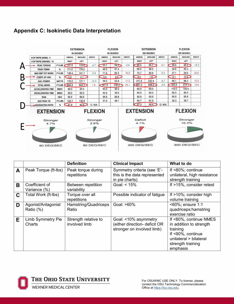
Appendix C: Isokinetic Data Interpretation
Definition Clinical Impact What to do A Peak Torque (ft-lbs) Peak torque during
repetitions Symmetry criteria (see ‘E’- this is the data represented in pie charts)
If <80%; continue unilateral, high resistance strength training
B Coefficient of Variance (%)
Between repetition variability
Goal: < 15% If >15%, consider retest
C Total Work (ft-lbs) Torque over all repetitions
Possible indicator of fatigue If >10%; consider high volume training
D Agonist/Antagonist Ratio (%)
Hamstring/Quadriceps Ratio
Goal: >60% <60%; ensure 1:1 quadriceps:hamstring exercise ratio
E Limb Symmetry Pie Charts
Strength relative to involved limb
Goal: <10% asymmetry (either direction- deficit OR stronger on involved limb)
If <80%, continue NMES in addition to strength training If <90%, continue unilateral > bilateral strength training emphasis
For OSUWMC USE ONLY. To license, please contact the OSU Technology Commercialization Office at https://tco.osu.edu.
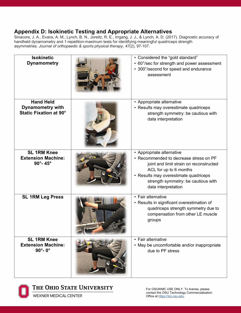
Appendix D: Isokinetic Testing and Appropriate Alternatives Sinacore, J. A., Evans, A. M., Lynch, B. N., Joreitz, R. E., Irrgang, J. J., & Lynch, A. D. (2017). Diagnostic accuracy of handheld dynamometry and 1-repetition-maximum tests for identifying meaningful quadriceps strength asymmetries. Journal of orthopaedic & sports physical therapy, 47(2), 97-107.
Isokinetic Dynamometry
• Considered the “gold standard” • 60°/sec for strength and power assessment • 300°/second for speed and endurance
assessment
Hand Held Dynamometry with
Static Fixation at 90°
• Appropriate alternative • Results may overestimate quadriceps
strength symmetry: be cautious with data interpretation
SL 1RM Knee Extension Machine:
90°- 45°
• Appropriate alternative • Recommended to decrease stress on PF
joint and limit strain on reconstructed ACL for up to 6 months
• Results may overestimate quadriceps strength symmetry: be cautious with data interpretation
SL 1RM Leg Press
• Fair alternative • Results in significant overestimation of
quadriceps strength symmetry due to compensation from other LE muscle groups
SL 1RM Knee Extension Machine:
90°- 0°
• Fair alternative • May be uncomfortable and/or inappropriate
due to PF stress
For OSUWMC USE ONLY. To license, please contact the OSU Technology Commercialization Office at https://tco.osu.edu.
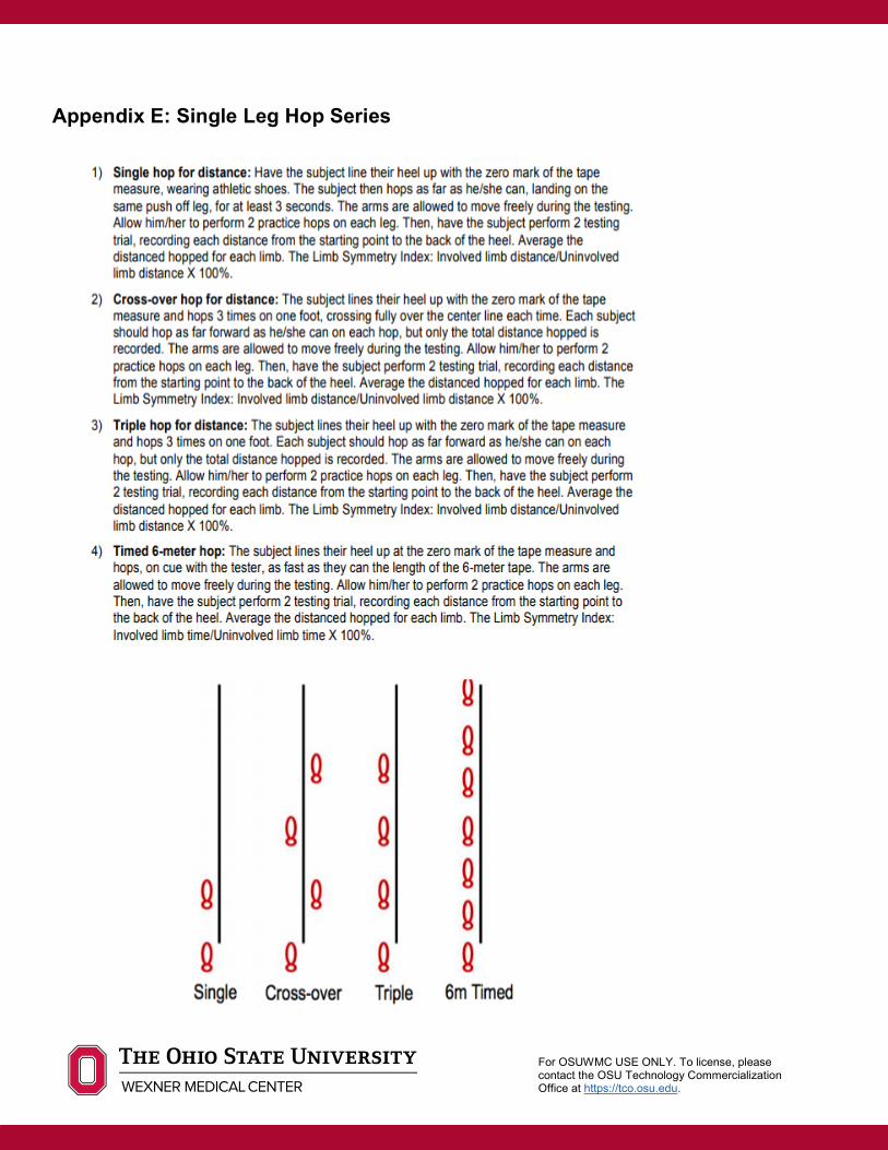
Appendix E: Single Leg Hop Series

For OSUWMC USE ONLY. To license, please contact the OSU Technology Commercialization Office at https://tco.osu.edu.
Author: Caroline Brunst, PT, DPT, SCS, OCS, AT Reviewers: Robert Duerr, MD; Robert Magnussen, MD; David Flanigan, MD; Christopher Kaeding, MD Updated: April 2020 References
1. Arno S, Bell CP, Uquillas C, Borukhov I, Walker PS. “Tibiofemoral Contact Mechanics Following a Horizontal Cleavage Lesion in the Posterior Horn of the Medial Meniscus.” J Orthop Res. 2015; 33(4): 584-590.
2. Becker R, Wirz D, Wolf C, Gopfert B, Nebelung W, Friederich N. “Measurement of meniscofemoral contact pressure after repair of bucket-handle tears with biodegradable implants.” Arch Orthop Trauma Surg. 2005;125:254-260. http://dx.doi. org/10.1007/s00402-004-0739-5.
3. Bhatia S, LaPrade CM, Ellman MB, LaPrade RF. “Meniscal Root Tears: Significance, Diagnosis and Treatment.” Am J Sports Med. 2014; 42(12): 3016-3030.
4. Chung KS, Ha JK, Ra HJ, Kim JG. “A Menta-Analysis of Clinical and Radiographic Outcomes of Posterior Horn Medial Meniscus Root Repairs.” Knee Surg sports Traumatol Arthrosc. 2016; 24: 1455-1468.
5. Frizziero A, Ferrari R, Giannotti E, Ferroni C, Poli P, Masiero S. “The meniscus tear: state of the art of rehabilitation protocols related to surgical procedures.” Muscles, Ligaments and Tendons Journal. 2012;2(4):295-301.
6. Kurzweil PR, Lynch NM, Coleman S, Kearney B. “Repair of Horizontal Meniscus Tears: A Systematic Review.” Arthroscopy. 2014; 30(11): 1513-1519.
7. LaPrade CM, et al. “Meniscal Root Tears: A Classification System Based on Tear Morphology.” Am J Sports Med. 2015; 43(2): 363-369.
8. LaParade CM, et al. “Biomechanical Consequences of a Nonanatomic Posterior Medial Meniscus Root Repair.” Am J Sports Med. 2015; 43(4): 912-920.
9. Lavender CD, Hanzlik SR, Caldwell PE, Pearson SE. “Transosseous Medial Meniscal Root Repair Using a Modified Mason-Allen Suture Configuration.” Arthroscopy Techniques. 2015; 4(6): e781-e784.
10. Mueller BT, Moulton SG, O'Brien L, LaPrade RF. “Rehabilitation Following Meniscal Root Repair: A Clinical Commentary.” J Orthop Sports Phys Ther. 2016; Feb;46(2):104-13.
11. Chimera NJ, Warren M. Use of clinical movement screening tests to predict injury in sport. World Journal of Orthopedics. 2016; 7(4):202-217
12. Myer GD, Schmitt LC, Brent JL, et al. Utilization of Modified NFL Combine Testing to Identify Functional Deficits in Athletes Following ACL Reconstruction. The Journal of orthopedic and sports physical therapy. 2011:41(6):377-387
13. Garrison JC, Shanley E, Thigpen C, et al. The reliability of the Vail Sport Test as a measure of physical performance following anterior cruciate ligament recontruction. Int J Sports Phys Ther. 2012; 7(1):20-30.