ag catalysed cutting of multi-walled carbon nanotubes · f s husairi, s a m zobir, m rusop ......
TRANSCRIPT

This content has been downloaded from IOPscience. Please scroll down to see the full text.
Download details:
IP Address: 134.60.120.187
This content was downloaded on 22/09/2016 at 14:54
Please note that terms and conditions apply.
You may also be interested in:
Engineering the physical parameters for continuous synthesis of fullerene peapods
Neeru Tiwari, Nayancee Pandey, Debmalya Roy et al.
The chemistry and application of carbon nanotubes
Eduard G Rakov
An artificial carbon nano-thorn
T Mitsui, M Nishitani-Gamo, Y F Zhang et al.
Electrical properties of carbon nanotubes synthesis by double furnace thermal-CVD technique at
different temperatures on porous silicon template
F S Husairi, S A M Zobir, M Rusop et al.
The role of defects and doping in 2D graphene sheets and 1D nanoribbons
Humberto Terrones, Ruitao Lv, Mauricio Terrones et al.
Catalyzed oxidation for nanowire growth
Kaiping Tai, Ke Sun, Bo Huang et al.
Ag-catalysed cutting of multi-walled carbon nanotubes
View the table of contents for this issue, or go to the journal homepage for more
2016 Nanotechnology 27 175604
(http://iopscience.iop.org/0957-4484/27/17/175604)
Home Search Collections Journals About Contact us My IOPscience

Ag-catalysed cutting of multi-walled carbonnanotubes
A La Torre1,2, G A Rance1,2, S A Miners1, C Herreros Lucas1, E F Smith1,2,M W Fay2, T Zoberbier3, M C Giménez-López1, U Kaiser3, P D Brown2,4 andA N Khlobystov1,2
1 School of Chemistry, University of Nottingham, University Park, Nottingham, NG7 2RD, UK2Nanoscale and Microscale Research Centre, University of Nottingham, University Park, Nottingham,NG7 2RD, UK3Electron Microscopy Department of Materials Sciences, University of Ulm, D-89081 Ulm, Germany4Division of Materials, Mechanics and Structures, Department of Mechanical, Materials andManufacturing Engineering, Faculty of Engineering, University of Nottingham, University Park,Nottingham, NG7 2RD, UK
E-mail: [email protected] and [email protected]
Received 31 October 2015, revised 18 January 2016Accepted for publication 29 February 2016Published 18 March 2016
AbstractIn this work, the cutting of carbon nanotubes is investigated using silver nanoparticles depositedon arc discharge multi-walled carbon nanotubes. The composite is subsequently heated in air tofabricate shortened multi-walled nanotubes. Complementary transmission electron microscopyand spectroscopy techniques shed light on the cutting mechanism. The nanotube cutting iscatalysed by the fundamental mechanism based on the coordination of the silver atoms to the π-bonds of carbon nanotubes. As a result of the metal coordination, the strength of the carbon–carbon bond is reduced, promoting the oxidation of carbon at lower temperature when heated inair, or lowering the activation energy required for the removal of carbon atoms by electron beamirradiation, assuring in both cases the cutting of the nanotubes.
S Online supplementary data available from stacks.iop.org/NANO/27/175604/mmedia
Keywords: carbon, nanoparticles, silver, electron beam.
(Some figures may appear in colour only in the online journal)
1. Introduction
Shortened multi-walled carbon nanotubes (short-MWNTs)have gained attention in recent years because of their potentialuse as components within nanomechanical devices such asnanobearings [1], nanogears [2], nanoswitches [3], nanorelays[4], gigahertz oscillators [5, 6] and Brownian nanomotors[7, 8]. Short-MWNTs may also find application within elec-tronic nanodevices [9] such as nanotransistors [10], nano-diodes [11] and current modulators [12]. Short-MWNTs maybe less hazardous than long-MWNTs, recognising that thetoxicity of nanotubes increases as a function of increasinglength, heightening the propensity for an immune response[13–15]. Furthermore, the internal channels of MWNTs havebeen found to be suitable for containing chemical reactions[16, 17] and the use of short-MWNTs should assist with the
transport of reactants and products in and out of the nanotubes[18, 19]. However, the lack of suitable methods for the con-trolled growth of short-MWNTs with retained tubularmorphology and graphitic structure has encouraged thedevelopment of a variety of protocols for the cutting ofMWNTs. In this context, chemical oxidation processes in airare generally associated with low yields [13, 14, 20], whilstprocessing in CO2 causes significant structural damage withthe stripping of MWNT outer layers [21]. Mechanical meth-ods, such as ball-milling or diamond abrasion, have beensuccessfully used to cut MWNT, but they create similarstructural damage to the CO2 method, along with substantialamounts of amorphous carbon that are difficult to separatefrom the short-MWNTs [22]. Finally, the combination ofmechanical cutting by ultra-sonication and chemical oxidationproduces a low level of carbon loss and little nanotube
Nanotechnology
Nanotechnology 27 (2016) 175604 (8pp) doi:10.1088/0957-4484/27/17/175604
0957-4484/16/175604+08$33.00 © 2016 IOP Publishing Ltd Printed in the UK1

degradation. However, this method seems to be more timeconsuming and applicable to only single walled carbonnanotubes [23].
It has been shown that alkylthiol-stabilised silver nano-particles (AgNP) can be used to produce short-MWNTs, withmean length being controlled by the number of nanoparticlesdeposited [24]. However, the fundamentals of the chemicalreaction between AgNP and the carbon of the MWNT shells,as precursor to the cutting mechanism, are not understood andrequire detailed investigation. Accordingly, we report here onthe AgNP-catalysed cutting of MWNTs, in air at elevatedtemperature, and in vacuum at room temperature (RT) underthe high-energy electron beam of a transmission electronmicroscope (TEM). Comparison of the oxidative cutting ofMWNTs in air, with time-resolved imaging of the cuttingprocess in situ within the TEM in vacuum, allowed the role ofthe Ag catalyst in the cutting process to be identifiedunambiguously.
2. Experiment
All reagents were purchased from Sigma-Aldrich, UK andused without further purification. Carbon nanotubes werepurchased from MER Corporation. Thermogravimetric ana-lysis (TGA; TA Instruments SDT Q500) was performedunder an air flow of 90 mLmin−1, with heating at a rate of10 °Cmin−1 from RT to 1000 °C. TEM was performed at RTusing a Jeol 2100F TEM (information limit 0.19 nm; accel-erating voltage 100 or 200 kV). AC-HRTEM acquisitionswere carried out on a CS-corrected FEI Titan 80–300 TEM.Gatan Digital Micrograph software was used for the appraisalof carbon nanotube length and Ag nanoparticle diameter.Energy dispersive x-ray (EDX) analysis was performed usingan Oxford Instruments INCA 560 microanalysis system.Samples for TEM/EDX were prepared via the drop-drying ofmethanolic solutions of nanotubes onto Cu grid mounted‘lacey’ carbon films. Complementary x-ray photoelectronspectroscopy (XPS) investigations were performed using aKratos AXIS ULTRA with monochromated Al Kα radiation(10 kV anode potential; 15 mA emission current) in fixedanalyser transmission mode (80 eV pass energy). The spectrawere analysed using Casa XPS software. Raman spectroscopywas conducted using a HoribaJY LabRAM HR spectrometerwith a laser wavelength of 532 nm. Samples were drop-castfrom methanolic solution onto Si(100) wafers and a minimumof three spectra were recorded from different areas of eachsample.
2.1. Synthesis of alkylthiol-stabilised Ag nanoparticles
Dodecanethiol (0.1 ml) was added to a solution of silvernitrate (153 mg) in ethanol (30 ml) and the mixture stirredvigorously for 10 min at RT. A saturated solution of sodiumborohydride in ethanol (60 ml) was added and the resultantmixture stirred vigorously for 2 h at RT. The product wasprecipitated from solution by the further addition of ethanol(350 ml) before storing at −30 °C for 24 h. The precipitate
was collected by filtration using a 0.45 μm pore sized poly-tetrafluoroethylene (PTFE) membrane and washed withethanol (250 ml) and acetone (250 ml), before final dryingunder vacuum to yield a black solid (180 mg AgNP).
2.2. Preparation of shortened, open, arc-discharge (AD)MWNT via oxidative cutting
10 mg of arc-discharge MWNTs (AD-MWNTs; MER Cor-poration; 2–15 nm inner diameter and 2–15 external walls)were heated at reflux point in a solution of 15M nitric acid for5 h. Alkylthiol-stabilised silver nanoparticles were synthe-sised using a modified Brust Schiffrin reduction as describedin our previous reports [24]. The suspension was diluted withwater, and collected by vacuum filtration using a 0.2 μmPTFE membranes. To the resulting black powder was addedto a solution of alkylthiol-stabilised AgNP (10 mg) in cyclo-hexane (10 ml) and then sonicated for 30 min at RT. Theresulting product was collected by filtration using a 0.2 μmpore sized PTFE membrane and washed with cyclohexane(3 ml×50 ml) and acetone (50 ml) before final drying undervacuum to yield a black solid (19 mg). The product was thenplaced in an alumina crucible and heated in air either at550 °C for 14 min or 575 °C for 10 min to yield a black solid(9 mg and 8 mg respectively). This was then added to nitricacid (2M, 18 ml) and the black suspension probe sonicated(power 130W; frequency 20 kHz) for 30 min at RT. Theresulting product was then diluted with deionised water(20 ml), filtered using a 0.45 μm pore sized PTFE membrane,washed with ethanol (50 ml) and acetone (50 ml) and driedunder vacuum to yield a black solid (5 mg of shortened, openAD-MWNTs). This product was then placed in an aluminacrucible and heated in air at 300 °C for 10 min to remove anyorganic residual solvent within the nanotube channels.
3. Results
3.1. AgNP oxidative cutting of AD-MWNTs in air at elevatedtemperature
AD-MWNTs were treated with concentrated nitric acid tofacilitate the formation of defects on the walls of the MWNTs,prior to AgNP deposition. The introduction of defects siteswas confirmed by a ∼50% increase in the ID:IG ratio deter-mined by Raman spectroscopy (figure S1 stacks.iop.org/NANO/27/175604/mmedia). To ensure effective AgNPdecoration and the cutting of long, defective AD-MWNTs(5–15 μm in length, figure S2), a 1:1 AgNP:MWNT massratio was adopted, followed by heat treatment in air either at550 °C for 14 min or 575 °C for 10 min (the high temperatureprocess) to produce shortened AD-MWNTs (mean length450±120 nm and 420±90 nm respectively; figures 1(a)–(c) and S2). TGA investigations demonstrated that the pre-sence of AgNP reduced the nanotube oxidation temperaturefrom 768 °C to 575 °C (figure S3), confirming the catalyticrole of Ag in the cutting process. After oxidative cutting, theAg catalyst was removed effectively by washing with dilute
2
Nanotechnology 27 (2016) 175604 A La Torre et al

HNO3, without the creation of any further defects in theMWNT structure as assessed by Raman spectroscopy. On thebasis of these heat treatment experiments in air, Ag actsclearly as a catalyst in the oxidative cutting of AD-MWNTs.
According to our HRTEM analysis after the cuttingprocess at high temperatures, AgNP residing on the terminusof a cut AD-MWNT indicates the lack of oxygen formation,as the d spacing may be attributable to the 002 plane ofmetallic silver as shown in figure 2.
XPS was used to investigate these AD-MWNT–AgNPcomposites, before and after heating in air, to determineunambiguously the metal oxidation state and appraise thedetailed chemistry of the cutting mechanism, noting that
heating in air under the conditions used would not cause theoxidation of AgNP to form Ag2O, as an intermediate toprovide oxygen for the cutting of MWNTs [25, 26].
Indeed, the XPS survey scan compositional data oftable 1 and detailed Ag 3d5/2 data of figure 3 showed nostrong evidence for the development of Ag2O, with the pre-sence of just ∼2.5 at% of oxygen at the surface of these AD-MWNT–AgNP composite samples being attributable tooxygen-containing functional groups on the acid cleaned AD-MWNTs. Furthermore, no change in the position of the Ag3d5/2 peak at 368.4 eV, before and after heating, wasobserved (figures 3(b) and (c)). These peaks, exhibiting nar-row widths of ∼0.7–0.9 eV, were strongly indicative of the
Figure 1. TEM images illustrating the stages of the AD-MWNT cutting process in air. (a) Long, closed AD-MWNTs (length 5–15 μm) with adistribution of AgNP on the nanotube sidewalls; (b) shortened AD-MWNTs, following thermal treatment at 550 °C for 14 min, with someresidual, coarsened AgNP and extensive side-wall defects (top side) due to the oxidation process; (c) shortened AD-MWNTs (mean length:450±120 nm) following removal of AgNP by washing with dilute nitric acid; accompanied by schematic illustrations of the affordednanostructures. Scale bars are 20 nm.
3
Nanotechnology 27 (2016) 175604 A La Torre et al

metallic state of AgNPs being retained under the heatingconditions used for the cutting of AD-MWNTs in air. Indeed,the absence of Ag2O in these composite samples is consistentalso with the decomposition of Ag2O to Ag metal at 280 °C[25, 26], well below the temperature used for the cutting ofnanotubes in air. Silver oxide might form below 280 °C but itundergoes carbonisation while heated in air. However, underour experimental condition no cutting was observed while thesample was heated at 250 °C in air. Above the temperature of300 °C (low temperature process) it was possible to cutnanotubes even though this process was slower than the hightemperature process (in air either at 550 °C for 14 min or575 °C for 10 min).
3.2. In situ AgNP mediated cutting of AD-MWNTs within a TEMin vacuum at RT
To complement the thermal treatment experiments in air, Ag-catalysed transformations of AD-MWNTs were observeddirectly on the nm-scale in real-time, in vacuum, at RT, underthe imaging electron beam of a TEM. We have carried out thein situ cutting at two different e-beam energies 100 keV and200 keV. According to our time-series imaging, the cutting ofMWNT catalysed by AgNP at 200 keV is two times fasterthan at 100 keV (table 2 and figure 4 below and figure S4).Notably, the presence of AgNP promoted the cutting of theAD-MWNTs at 100 keV, after ∼64 min. Conversely, nosignificant damage was observed to the MWNT underreduced energy conditions of 100 keV, where the nanotubepersists without cutting under the imaging electron beam>240 min. Moreover, additional measurements for Ag-MWNTs at 80 keV showed no cutting of nanotubes under thiscondition. This clearly demonstrated that the nanotube cuttingis driven by the kinetic energy, Ek of fast electrons of thee-beam, as in the case of chemical etching the cross section ofO2 ionisation would increase with the decreasing energy ofthe electron beam [27], which is opposite to what weobserved.
Figure 4 presents a time series of phase contrast TEMimages (100 keV; dose rate 1.64×10⁶ e nm−2 s−1) illustrat-ing the interaction of an AgNP with an AD-MWNT. TheAgNP changes its morphology during the exposure to theelectron beam. The AgNPs loses its capping layer and presentan elongated shape after 5 min. EDX spectra acquired in situin the TEM after 5 min of exposure (figure S5) demonstratedthe loss of sulphur with time, under electron beam conditionsof 100 keV; 1.64×10⁶ e nm−2 s−1 dose rate, consistent withthe removal of the alkylthiol-stabilised capping layers fromthe AgNP surfaces, providing direct contact between Ag andthe C atoms of the MWNTs and mediating the formation ofdefects. It can also be seen the release of small silver particles
Figure 2. Shortened AD-MWNTs following the thermal treatment at 550 °C for 14 min. (a) TEM images showing the nanoparticles residingon a MWNTs terminus. (b) High resolution TEM images (b) showing lattice planes imaged parallel to the edge of a AgNPs correlating with a(002) d-spacing value of 0.220 nm (inset: optical diffractogram). Scale bars are 5 in (a) and 2 nm in (b).
Table 1. XPS surface compositional data (at%±0.1%) for AgNPsand AD-MWNT–AgNP samples before and after heating in air at550 °C. The large C 1s signal in AgNPs, and AD-MWNT–AgNPbefore heating, is due to the organic capping layer surrounding theparticle.
Nanostructure Ag 3d C 1s O 1s S 2p
AgNPs 12.3 82.5 2.5 2.8AD-MWNT–AgNP before heating 6.0 89.5 2.4 2.1AD-MWNT–AgNP after heating 1.6 95.4 2.7 0.3
Table 2. Time required to cut MWNT with and without AgNP underirradiation with 100 keV or 200 keV at 1.64 × 10⁶ e nm−2 S−1
Time taken to cut MWNT/min
Accelerating voltage/keV AgNP–MWNT MWNT
100 64 >240200 31 90
4
Nanotechnology 27 (2016) 175604 A La Torre et al

surrounding the big particle black arrowed in figure 4. TheAgNP decreases its size while the cutting was occurring. Thefracture was created at the proximity of the particle.
The cutting mechanism is also explored using smallAgNPs of 1−2 nm in diameter released by the big particleswhile e-beam irradiation. Figure 5 presents a time series ofphase contrast AC-TEM images (100 keV; dose rate c.a.1.64×106 e nm−2 s−1) illustrating the interaction of smallAgNP with an AD-MWNT. The dynamics of the cutting ofMWNTs in figure 5 and video 1 clearly shows the cuttingprocess catalysed by the silver catalyst. Sidewall defectsemerge at the point of contact of AgNP with MWNTs andpropagate over time leading to pits on the nanotubes. Theexteriors walls of the MWNTs are preferentially damaged inareas in which silver particles are present. Conversely, nodamages are observed on the exterior walls of MWNTs wherethe catalyst is absent.
4. Discussion
4.1. The use of nitric acid
Synthesis by AD methods produces nanotubes that aresuperior structurally to those produced by chemical vapourdeposition (CVD) [28]; this is due to the higher temperaturesassociated with the AD process facilitating structural rear-rangement of carbon atoms in the sidewalls; a process whichdoes not occur readily at the lower temperatures associated
Figure 3. High-resolution XPS data for Ag 3d5/2 corresponding to: (a) synthesised AgNP; (b) AD-MWNT–AgNP before heat treatment; and(c) AD-MWNT–AgNP after heat treatment. (Peaks normalised to background on the lower binding energy side. Spectra were chargecorrected to C 1s at 284.7 eV.) Signal-to-noise in MWNT–AgNP after treatment is lower than the previous two measurements due to theAgNPs becoming encapsulated in nanotubes.
Figure 4. TEM time series showing AgNP mediated cutting of anAD-MWNT (100 keV; dose rate 1.64×10⁶ e nm−2 s−1), withschematic illustrations of the afforded nanostructures inset. Theblack arrows indicate the small AgNPs formed spontaneously whilethe sample was exposed to the e-beam. Scale bars are 10 nm, timeunits are minutes.
5
Nanotechnology 27 (2016) 175604 A La Torre et al

Figure 5. AC-TEM time series showing the interaction of silver particles with an AD-MWNT under low energy electron beam irradiationconditions of 100 keV, for the same dose rate of 1.64×10⁶ e nm−2 s−1. Scale bar is 2 nm.
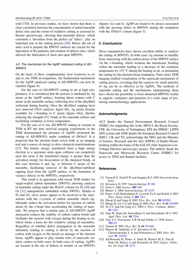
Figure 6. (a) Schematic representation of AgNPs mediating the oxidative cutting of AD-MWNT in air at elevated temperature and by anelectron beam in vacuum at RT. Schematic representations of the synergistic bonding between: (b) the 5s-orbital of an Ag atom and the πbonding orbitals of the carbon–carbon double bond; and (c) the 4d-orbitals of Ag and the π* antibonding orbitals of the carbon–carbondouble bond. The arrows indicate the direction of redistribution of electronic density between metal atom and carbon atom of the nanotube,which acts to weaken the bonding between carbon atoms in the MWNTs, thereby decreasing the activation barrier for oxidative cutting in airat elevated temperature, or cutting under the imaging electron beam in TEM.
6
Nanotechnology 27 (2016) 175604 A La Torre et al
with CVD. In previous studies, we have shown that there isclear correlation between the concentration of initial nanotubedefect sites and the extent of oxidative cutting as assessed byRaman spectroscopy, showing that nanotube defects, whichconstitute a ‘deviation from the perfect sp2 lattice’, play animportant role in the cutting process [29]. Hence, the use ofnitric acid to prepare the MWNT surfaces are crucial for thedeposition of the particles and creation of defects sites, whichallowed the fabrication of short and open MWNTs.
4.2. The mechanism for the AgNP catalysed cutting of AD-MWNTs
On the basis of these complementary heat treatment in airand in situ TEM investigations, the fundamental mechanismfor the AgNP catalysed cutting of AD-MWNTs can now beclarified (figure 6).
For the case of AD-MWNT cutting in air at high tem-peratures, it is considered that the process is mediated by Agatoms at the AgNP surface, forming direct contact with Catoms at the nanotube surface, following loss of the alkylthiolsurfactant during heating. Once the alkylthiol capping layerwas removed (TGA analysis, figure S6), Ag atoms becameavailable for π-bonding to C atoms (figure 6(b)), therebyreducing the strength of C bonds at the nanotube surface andfacilitating oxidation at lower temperature.
For the case of in situ AD-MWNT cutting in vacuum inTEM at RT the time resolved imaging experiments in theTEM demonstrated the presence of AgNPs promoted thecutting of AD-MWNTs under condition of 100 keV. It isproposed that the electron beam serves both as an imagingtool and a source of energy to drive chemical transformations[27]. The kinetic energy transferred from a high energyelectron to a specimen atom upon collision can completelyeject the atom if the transferred energy is greater than theactivation energy for dissociation of the chemical bonds, inthis case between S and Ag, or between C atoms of thenanotube, facilitating removal of the alkylthiol-stabilisedcapping layer from the AgNP surface, or the formation ofvacancy defects in the MWNTs, respectively.
This result is in agreement with recent TEM studies forsingle-walled carbon nanotubes (SWNTs) showing catalysisof nanotube cutting under the 80 keV e-beam for Ni [30] andOs [31] nanoparticles embedded within SWNTs. Similar toNi and Os, silver atoms appear to be involved in the inter-actions with the π-system of carbon nanotube which sig-nificantly reduce the activation barrier for ejection of carbonatoms by the e-beam thus accelerating the cutting of nano-tube. We propose that a similar type of AD-MWNT–AgNPinteraction reduces the stability of carbon–carbon bonds andfacilitates the reaction with oxygen during the heating in air,which forms a basis for the oxidative cutting of nanotubes.Regardless of whether defect propagation in the nanotubeultimately leading to cutting is driven by the reaction ofcarbon with oxygen or the knock-on damage of the electronbeam, AgNPs appear to play similar roles of nanoscale cat-alytic centres in both cases. In both cases of cutting, AgNPsare located at the site of defects or termini of cut MWNTs
(figures 1(c) and 4). AgNP are found to be always associatedwith the growing defect in MWNTs during the irradiationwith the 100 keV e-beam (figure 5).
5. Conclusions
Silver nanoparticles have shown excellent ability to catalysethe cutting of MWNTs. In both cases Ag remains in metallicform, interacting with the carbon atoms of the MWNT surfacevia the π-bonding, which weakens the interatomic bondingwithin the nanotube leading to a decrease of the oxidationtemperature by 155 °C during the heating in air, or promotingthe cutting by the electron beam irradiation. Time series TEMimaging enabled visualisation of the nanoscale mechanism ofcutting process, revealing that the catalysis by small particlesof Ag can be as effective as by AgNPs. The methods ofnanotube cutting and the mechanisms underpinning themhave shown the possibility to form shortened MWNTs usableas support, containers and junctions in a wide range of pro-mising nanotechnology applications.
Acknowledgments
ALT thanks the Natural Environment Research Council(NERC) for supporting this work, MCG-L the Royal Society,CHL the University of Nottingham, EFS the EPRSC: LIPPSXPS system and ANK thanks the European Research Council(ERC). UK and TZ, acknowledge the DFG and the Ministryof Science, Research and the Arts (MWK) of Baden-Wuert-temberg within the frame of the SALVE (Sub Angstrom Low-Voltage Electron microscopy) project. The authors thank theNanoscale and Microscale Research Centre (NMRC) foraccess to TEM and Raman facilities.
References
[1] Tuzun R E, Noid D W and Sumpter B G 1995 Nanotechnology6 64
[2] Srivastava D 1997 Nanotechnology 8 186[3] Forro L 2000 Science 289 560[4] Maslov L 2006 Nanotechnology 17 2475[5] Popov A M, Bichoutskaia E, Lozovik Yu E and Kulish A 2007
Phys. Status Solidi A 204 1911[6] Zheng Q and Jiang Q 2002 Phys. Rev. Lett. 88 045503[7] Zheng Q, Liu J Z and Jiang Q 2002 Phys. Rev. B 65 245409[8] Tu Z C and Ou-Yang Z C 2004 J. Phys.: Condens. Matter
16 1287[9] Saito R, Fujita M, Dresselhaus G and Dresselhaus M S 1992
Appl. Phys. Lett. 60 2204[10] Tans S J, Verscueren A R M and Dekker C 1998 Nature
393 49[11] Tamura R 2001 Phys. Rev. B 64 201404[12] Menon M, Andriotis A N, Srivastava D W,
Chernozatonskii L A and Ponomareva I 2003 Phys. Rev.Lett. 91 145501
[13] Ali-Boucetta H, Nunes A, Sainz R, Herrero M A, Tian B,Prato M, Bianco A and Kostarelos K 2013 Angew. Chem.,Int. Ed. Engl. 52 2274
7
Nanotechnology 27 (2016) 175604 A La Torre et al

[14] Kostarelos K 2008 Nat. Biotechnol. 26 774[15] Muller J, Delos M, Panin N, Rabolli V, Huaux F and Lison D
2009 Toxicol. Sci. 110 442[16] Chen W, Pan X and Bao X 2007 J. Am. Chem. Soc. 129
7421–6[17] Pan X, Fan Z, Chen W, Ding Y and Bao X 2007 Nat. Mater. 6
507–11[18] Wang C F, Guo S J, Pan X L, Chen W and Bao X H 2008
J. Mater. Chem. 18 5782[19] Rao R, Zhang Q, Liu H, Yang H, Ling Q, Yang M,
Zhang A and Chen W 2012 J. Mol. Catal. A 363–364 283[20] Tran M Q, Tridech C, Alfrey A, Bismarck A and Shaffer M S P
2007 Carbon 45 2341[21] Tsang S C, Harris P J F and Green M L H 1993 Nature
362 520[22] Kukovecz A, Kanyo T, Konya Z and Kiricsi I 2005 Carbon
43 994[23] Shuba M V, Paddubskaya A G, Kuzhir P P, Maksimenko S A,
Ksenevich V K, Niaura G, Seliuta D, Kasalynas I andValusis G 2012 Nanotechnology 23 9
[24] La Torre A, Rance G A, El Harfi J, Li J N, Irvine D J,Brown P D and Khlobystov A N 2010 Nanoscale 2 1006
[25] Ferraria A M, Carapeto A P and Botelho do Rego A M 2012Vacuum 86 1988
[26] Perry D L 1995 Handbook of Inorganic Compounds Illustratededn (Boca Raton, FL: CRC Press) p 354
[27] Williams D B and Carter C B 2009 Transmission ElectronMicroscopy: A Textbook for Materials Science 2nd edn(New York: Spinger)
[28] Miners S A, Rance G A, La Torre A, Kenny S M andKhlobystov A N 2014 J. Mater. Chem. C 2 8357
[29] Harris P J F 2009 Carbon Nanotubes Science, Synthesis,Properties and Applications (Cambridge: CambridgeUniversity Press)
[30] Lebedeva I V, Chamberlain T W, Popov A M, Knizhnik A A,Zoberbier T, Biskupek J, Kaiser U and Khlobystov A N2014 Nanoscale 6 14877
[31] Zoberbier T, Chamberlain T W, Biskupek J, Kuganathan N,Eyhusen S, Bichoutskaia E, Kaiser U and Khlobystov A N2012 J. Am. Chem. Soc. 134 3073
8
Nanotechnology 27 (2016) 175604 A La Torre et al