multi-walled carbon nanotubes/manganese dioxide nano
TRANSCRIPT

Multi-walled Carbon Nanotubes/Manganese DioxideNano- �owers-like/Polyaniline NanowiresNanocomposite Modi�ed Electrode: A New Platformfor a Highly Sensitive Electrochemical ImpedanceDNA SensorTuan Chu ( [email protected] )
Hung Yen University of Technology and Education, Khoai Chau DistrictLuyen Thi Tran
Hanoi University of Science and TechnologyHoang Vinh Tran
Hanoi University of Science and TechnologyTrung Tran
Hoa Binh UniversityNghia Trong Nguyen
Hung Yen University of Technology and Education, Khoai Chau DistrictDan Bui
Hung Yen University of Technology and Education, Khoai Chau DistrictPhu Quang Tran
Hung Yen University of Technology and Education, Khoai Chau District
Research Article
Keywords: DNA sensor, nanocomposite, polyaniline nanowires, multi-walled carbon nanotubes,manganese dioxide
Posted Date: March 12th, 2021
DOI: https://doi.org/10.21203/rs.3.rs-280743/v1
License: This work is licensed under a Creative Commons Attribution 4.0 International License. Read Full License

1
Multi-walled Carbon Nanotubes/Manganese Dioxide Nano-
flowers-like/Polyaniline Nanowires Nanocomposite Modified
Electrode: A New Platform for a Highly Sensitive
Electrochemical Impedance DNA Sensor
Luyen Thi Tran 2, Hoang Vinh Tran 2, Trung Tran 3, Nghia Trong Nguyen 1, Dan Van Bui
1, Phu Quang Tran 1, Tuan Van Chu 1, *
1 Hung Yen University of Technology and Education, Khoai Chau District, Hung Yen Province,
Vietnam
2 School of Chemical Engineering (SCE), Hanoi University of Science and Technology, 1st Dai
Co Viet Road, Hai Ba Trung District, Hanoi, Vietnam
3 Hoa Binh University, Nam Tu Liem District, Hanoi, Vietnam
* Corresponding author:
Telephone: 84 221 3713283/Fax: 84 221 3713015
Email address: [email protected] (Tuan Van Chu)

2
Abstract
We describe in this report a development of label-free electrochemical DNA sensor based on a
novel nanostructured electrode of multi-walled carbon nanotubes (MWCNTs)/ nano-flowers-like
manganese dioxide (MnO2)/polyaniline nanowires (PANi NWs) nanocomposite. The
nanocomposite was synthesized in-situ onto an interdigitated platinum microelectrode (Pt) using a
combination of chemical and electrochemical synthesis methods: chemical preparation of
MWCNTs/MnO2 and electropolymerization of PANi NWs. The fabricated MWCNTs/MnO2/PANi
NWs was then used to develop a label-free electrochemical DNA sensor for a specific gene of
Escherichia coli (E.coli) O157:H7 detection. The MWCNTs/MnO2/PANi NWs modified Pt
electrode’s surface can facilitate for probe DNA strands immobilization and, therefore the
electrochemical signal of the DNA sensors has been improved. The electrochemical impedance
spectroscopy (EIS) measurements were conducted to investigate the output signals generated by
the specific binding of probe and target DNA sequences. Obtained results indicated that the
developed electrochemical biosensor can detect the target DNA in the linear range of 5 pM to 500
nM with a low limit of detection (LOD) at 4.42 × 10⁻¹³ M. The research results demonstrated that
the MWCNTs/MnO2/PANi NWs nanocomposite-based electrochemical DNA sensor has a great
potential application to the development of highly sensitive and selective electrochemical DNA
sensors to detect pathogenic agents.
Keywords: DNA sensor, nanocomposite, polyaniline nanowires, multi-walled carbon nanotubes,
manganese dioxide

3
1. Introduction
In recent decades, conducting polymers have been attracted the much attention of scientists
worldwide for biosensing applications thanks to their unique properties [1-4]. Pure conducting
polymers are formed in various structures such as nanowires, nanotubes and nano-thin films to
obtain a large surface area and a high efficiency [5, 6]. The combination of conducting polymers
with dopants can provide additional advantages, such as high conductivity, large surface areas,
environment-friendly features, stability and applicability in biosensors [7-9].
Nanocomposite materials created from conducting polymers such as polyaniline (PANi),
polypyrrole (PPy), poly(3,4-ethylenedioxythiophene) (PEDOT), and inorganic nanomaterials
have excellent characteristics that are not obtained by individual components, including high
conductivity, high stability, and high electroactive surface area, leading to new promising
applications [10-13]. These nanocomposites show advantageous properties of inorganic
nanomaterials distributed in continuous polymer networks [14, 15] and improvement of
specifications of conducting polymers, including changes in electron structure of polymer chains,
enhancement of charge transfer, and changes in conductivity of polymer chains [16, 17]. Actually,
several inorganic nanomaterials have been used to dope into the polymer networks, such as
PANi/Ni [18], PANi/Au [19], PANi/WO3 [20], PANi/Mn2O3 [21], PANi/MnO2 [22] and
PANi/MWCNTs [8] nanocomposites. In general, these nanocomposites can be synthesized by
different techniques including chemical, electrochemical, photochemical and mechano-chemical
methods and these developed materials have been applied for many applications such as energy
storage, electrochemical biosensors, electrochemical sensors, FET-based biosensors, and coating
and metal protecting [1]. To apply of the inorganic nanomaterial/conducting polymer
nanocomposites for development of electrochemical biosensors, these fabricated nanomaterials
have to be coated onto the electrodes using a drop-casting and/or an electrochemical method in-
situ on the working electrode [2, 23]. In which, the electrochemical techniques are often used due

4
to well controlling and high reproducibility. For examples, several PANi nanocomposites-based
sensitive electrochemical DNA sensors have been developed such as PANi-graphene
nanocomposite [24], silver nanoparticles decorated PANi nanowires [2], carbon dot/ZnO
nanorod/PANi composite [25], PANi/gold nanoparticles [26], Sm2O3 NPs-rGO/PANi composite
[27]. These developed PANi-based electrochemical DNA sensors have shown sensitivity at nM to
fM level for DNA detection, however, the fabrication steps are still laborious. Therefore, in this
study, the MWCNTs/MnO2/PANi NWs nanocomposite was synthesized in-situ on Pt
microelectrodes by a novel combined chemical-electrochemical technique, and was used for the
first time to develop a label-free electrochemical DNA sensor for rapid detection of pathogenic
bacteria.
2. Experimentals
2.1. Chemicals and instrumentations
2.1.1. Chemicals
MWCNTs (110–170 nm in diameter, 5–9 μm in length, > 90 wt.%), aniline (C6H5NH2, 99.5 wt.%),
sulfuric acid (H2SO4, 98 wt.%), nitric acid (HNO3, 68 wt.%), phosphate buffer solution (PBS),
potassium permanganate (KMnO4, 99 wt.%), and manganese (II) chloride tetrahydrate
(MnCl2.4H2O, 98 wt.%) were purchased from Sigma Aldrich. The supporting chemicals such as
K2Cr2O7 (99 wt.%) and N2 (99.9 wt.%) were of analytical grade. The DNA probe, the
complementary and non-complementary DNA target sequences are listed in Table 1.
Table 1. The DNA sequences used in this study.
Probe: 5’-AACGCCGATACCATTACTTA-3’
Complementary target: 3’-TTGCGGCTATGGTAATGAAT-5’
Non-complementary target: 5’-AACGCCGATACCATTACTTA-3’

5
2.1.2. Interdigitated Pt electrodes
The interdigitated Pt microelectrodes were fabricated using a standard photolithography technique
with a finger width of 10 μm and a gap size of 20 μm. The fabrication process was conducted by
sputtering 10 nm Ti and 200 nm Pt on a 100 nm thick silicon dioxide (SiO2) layer thermally grown
on top of a silicon wafer. The configuration and fabrication process of these electrodes were
discussed in our previous work [28].
2.1.3. Instrumentations
Scanning electron microscopy (SEM) images and energy dispersive X-ray spectroscopy (EDX)
spectra of the synthesized materials were investigated using a Nova NanoSEM 450 microscope.
The structure of the fabricated samples was examined using Fourier transform infrared
spectroscopy (FT-IR) spectra measured with a Shimadzu IRAffinity-1S FTIR spectrometer.
Electrochemical measurements were performed using the PGSTAT302N AutoLab
electrochemical workstation (Netherlands). A three-electrode configuration consists of the
interdigitated Pt electrode as a working electrode (WE), a Pt plate as a counter electrode (CE), and
a Ag/AgCl electrode (SCE) in 3M KCl solution (Metrohm) as a reference electrode (RE).
2.2. Fabrication of MWCNTs/MnO2/PANi NWs nanocomposite-based Pt microelectrodes
The multi-walled carbon nanotubes were firstly oxidized by a mixture of H2SO4/HNO3 by
following typical protocol: a 21.8 mg of MWCNTs was refluxed with 2 mL of mixture of 1:1 v/v
of 68 wt.% HNO3 and concentrated H2SO4 (98 wt.%) solution for 5 hours at 95 °C. Then the above
mixture was rinsed with distilled water until the pH was neutral, and the residue was dried at 80
oC for 12 hours to obtained the denatured MWCNTs. Secondly, MWCNTs/MnO2 was synthesized
by dispersion of 5.45 mg of the denatured MWCNTs as black powder into 10 mL of distilled water
using the ultrasonic process for 10 minutes. The suspension was then implanted into a water bath
6
at 75 oC under continuous stirring with the fixed rate at 500 rpm using an IKA magnetic stirrer.
After that, a 2 mL of MnCl2 solution (containing 37.2 mg MnCl2.4H2O) was added into a reaction
vessel and the mixture was stirred at 75 oC for 15 minutes. Then, 2 mL of KMnO4 solution
(containing 19.8 mg KMnO4) was dropped slowly into the above mixture at a temperature of 75
°C, under the stirring condition for 2 hours. The MWCNTs/MnO2 nanocomposite was collected
by a centrifugation at 3000 rpm and then it was rinsed many times with distilled water until pH
neutral. The MWCNTs/MnO2 was re-dispersed into 10 mL distilled water to use.
Before fabrication of the nanocomposite onto the surface, Pt microelectrodes were treated in a
solution of saturated K2Cr2O7 in H2SO4 0.5 M and then electro-chemically activated in a solution
of 0.5 M H2SO4 at the voltage range from –1.5 V to 2.2 V and the scanning rate of 50 mV s–1.
Then, 3 μL of the MWCNTs/MnO2 sample was drop-casted onto the surface of the Pt
microelectrode, and it was dried in the air under an infrared lamp. Finally, electrosynthesis of
PANi NWs on the Pt/MWCNTs/MnO2 electrodes was performed. The electrolyte solution
containing 0.05 M aniline and 0.5 M H2SO4 was blown with N2 gas for 15 minutes to purge the
dissolved oxygen. The electropolymerization was conducted at room temperature using the
chronoamperometry (CA) method with an applied voltage of 0.9 V vs. Ag/AgCl RE. After that, the
MWCNTs/MnO2/PANi NWs nanocomposite fabricated on the Pt WE was rinsed with deionized
water, and was dried at room temperature.
2.3. Preparation of MWCNTs/MnO2/PANi NWs-based electrochemical DNA sensors and
DNA hybridization detection
To prepare of electrochemical DNA sensor, a 10 μL of 10 μM DNA probe in PBS solution was
dropped onto the surface of the Pt/MWCNTs/MnO2/PANi NWs electrodes. The probe DNA
immobilization process was kept for 2 hours at room temperature. Then these electrodes were
rinsed with deionized water to remove DNA sequences which did not bind or had weak linkages

7
with the MWCNTs/MnO2/PANi NWs nanocomposite. The DNA sensors were then dried in the air
and were ready for further experiments. To confirm the immobilization of DNA capture probe, the
fluorescent technique was conducted to determine the efficiency of the DNA probe immobilization
on the surface of the Pt/MWCNTs/MnO2/PANi NWs electrode. 10 μL of 10 μM DNA probe with
fluorescent components in PBS solution was dropped onto the surface of the
Pt/MWCNTs/MnO2/PANi NWs electrode, and was annealed for 2 hours at room temperature.
After that, this electrode was rinsed by PBS buffer solution (pH 7.4) and deionized water, and was
dried. Finally, it was observed with a 40x objective lens and a 10x ocular lens, at 343–390 nm
wavelength, in randomly selected locations where DNA probe chains were immobilized.
For DNA target detection, the electrochemical impedance spectroscopy (EIS) spectra in the
electrolyte solution consisting of 0.1 M KCl and 0.1 M PBS buffer (pH 7.4) at the frequency range
from 104 Hz to 0.1 Hz, a DC potential of 230 mV and an AC potential of 5 mV (vs. Ag/AgCl
(SCE) as RE) of the DNA sensors before and after hybridization with the DNA target with different
concentrations (from 5 pM to 500 nM) were recorded. On EIS spectra, the increase of the electron
transfer resistance ΔRct (ΔRct = Rct,i – Rct,0) is used as the DNA hybridization signal, where Rct,0
and Rct,i are the electron transfer resistances in the absence and presence of the DNA target,
respectively.
3. Results and discussion
3.1. Characterization of MWCNTs/MnO2/PANi NWs-modified Pt electrodes
Figure 1 shows the chronoamperometry (CA) results of an unmodified Pt microelectrode (curve
a), a MWCNTs modified Pt microelectrode (Pt/MWCNTs) (curve b) and a MWCNTs/MnO2
modified Pt microelectrode (Pt/MWCNTs/MnO2) (curve c) in 0.5 M H2SO4 solution containing of
0.05 M aniline monomer during electropolymerization. It can be seen that the response currents
were increased vs. electropolymerization time indicating that the PANi was successfully deposited

8
onto the Pt and modified Pt electrodes. Moreover, Fig. 1 also shows the highest response current in
the case of the Pt/MWCNTs/MnO2 electrode and the lowest with the unmodified Pt electrode,
which imply that the MWCNTs and MWCNTs/MnO2 nanomaterials have improved the PANi
growth on the electrodes via enhancing conductivity and electroactive surface [20, 24, 26].
Figure 2 shows the cyclic voltammogram (CV) results (Fig. 2A) and the electrochemical
impedance spectroscopy (EIS) spectra (Fig. 2B) of the Pt microelectrode (curve a), PANi
nanowires grown directly on the unmodified Pt microelectrode (Pt/PANi NWs, curve b), PANi
nanowires grown on the MWCNTs modified Pt microelectrode (Pt/MWCNTs/PANi NWs, curve
c) and PANi nanowires grown on the MWCNTs/MnO2 modified Pt microelectrode
(Pt/MWCNTs/MnO2/PANi NWs, curve d). It can be seen in Fig. 2A that the PANi has improved
the electroactivity of the Pt microelectrode by comparing curve b and curve a. In addition, the
MWCNTs/MnO2/PANi NWs nanocomposite has a stronger improvement in the electrochemical
activity than that of the MWCNTs/PANi NWs or the PANi NWs as comparing curve d with curve
c and with curve b. The EIS spectra in Fig. 2B agree with the CV data in Fig. 2A, i.e., the Rct values
were arranged as following: Rct (Pt/MWCNTs/MnO2/PANi NWs) < Rct (Pt/MWCNTs/PANi NWs)
< Rct (Pt/PANi NWs). These results indicate clearly that the MWCNTs/MnO2/PANi NWs has the
highest electroactive and reduces the electron transfer resistance (Rct), implying that the use of
MWCNTs/MnO2 to modify the Pt electrode has improved the electrical property of PANi NWs
grown onto the electrode surface and has also enhanced the electrochemical active area for DNA
capture probe immobilization, therefore the MWCNTs/MnO2/PANi NWs nanocomposite can
improve the electrochemical signal for electrochemical DNA sensors.
Figure 3 shows the SEM images of MWCNTs (Fig. 3A), PANi NWs (Fig. 3B), MWCNTs/PANi
NWs (Fig. 3C), MWCNTs/MnO2 (Fig. 3D) and MWCNTs/MnO2/PANi NWs (Fig. 3E and Fig.
3F) modified on the surface of the Pt microelectrodes. It can be seen that the MWCNTs are highly

9
uniform with diameters ranging from 100 to 200 nm (Fig. 3A). Fig. 3B shows the PANi NWs with
150–160 nm in diameters which were formed directly on the Pt electrodes by the CA method. The
size of the PANi NWs is uniform, and the nanowires are distributed throughout the surface of the
WE. Moreover, it can be seen in Fig. 3C that, the obtained PANi NWs are relatively uniform and
distributed on the MWCNTs layer. On the other hand, Fig. 3D shows that the MWCNTs are
surrounded by the MnO2 material with the unique flower-like structure. Finally, it can be seen in
Fig. 3E and Fig. 3F, the MWCNTs/MnO2/PANi NWs nanocomposite was successfully
synthesized in-situ on the Pt microelectrodes. The PANi NWs layer which was formed by the
electrochemical method, covers the surface of the MWCNTs/MnO2 film. In the fabrication of the
electrochemical DNA sensors, the DNA probe immobilization on the electrode surface and the
ability of the sensors to detect DNA target depend on compositions and surface structures of the
materials modified the WE. In this study, the PANi NWs contain MWCNTs/MnO2 as an impurity
component. The MWCNTs tubes are surrounded with the flower-like MnO2 blocks, and are
distributed in the PANi nanowires. Polymers are not soluble, however, SO42- ions in H2SO4
solution create bonds with PANi NWs and then increase polarity, so the distribution process of
MWCNTs/MnO2 into PANi nanowires is facilitated. The nanocomposite obtained through
MWCNTs/MnO2 bound to the wall of PANi nanowires, has a soft, porous, regular and specific
structure. This structure has attracted the attention of researchers, and is found highly suitable for
application in the development of DNA sensors.
The EDX spectra in Fig. 4A shows relatively the composition of the elements in samples of PANi
NWs, MWCNTs/PANi NWs, MWCNTs/MnO2/PANi NWs, MWCNTs/MnO2 and MWCNTs on
the Pt microelectrode surfaces (Fig. 4A, from curve a to curve e, respectively). As can be seen in
Fig. 4A, carbon element (C), which is a constituent of MWCNTs and PANi NWs as well, was
observed in all samples at position of 0.27 keV. In Fig. 4A (from curve a to curve c), the nitrogen

10
(N), oxygen (O), and sulfur (S) elements which are constituents of PANi NWs, were observed at
energy levels of 0.39, 0.53 and 2.31 keV, respectively. These results can be explained that H2SO4
was doped into PANi NWs, leading to the appearances of S and O elements in the EDX spectra.
When H2SO4 was doped into PANi NWs, cations which are formed at the imine N atoms play the
role for the electronic conduction of PANi [29, 30]. Thus, in the electrochemical DNA sensor
development, doping PANi with H2SO4 can improve the conductivity of PANi NWs, so the
efficiency of information transmission from DNA hybridization to the transducer will be
enhanced. On the other hand, in Fig. 4A from curve c to curve d, the manganese (Mn) element
which is a constituent of MnO2 was observed at 0.63 keV. These results indicate that the
MWCNTs/MnO2 and MWCNTs/MnO2/PANi NWs nanocomposites were successfully synthesized
on the Pt electrode surface. Figure 4B displays the FT-IR spectra of PANi NWs, MWCNTs/PANi
NWs, MWCNTs/MnO2/PANi NWs, MWCNTs/MnO2, and MWCNTs (from curve a to curve e,
respectively) obtained on the surface of the Pt microelectrodes. It can be seen in Fig. 4B, in the
cases of MWCNTs/MnO2/PANi NWs (curve c), MWCNTs/MnO2 (curve d) and MWCNTs (curve
e), the bands which characterize of the MWCNTs are observed. The band at 1015 cm–1 is assigned
to the C–O stretching mode of the carboxylic acid group formed on the side wall of the MWCNTs
[31]. Thus, the MWCNTs were oxidized. Besides, the band at 1144 cm–1 is attributed to the C–C
stretching mode [32]. On the other hand, for the cases of PANi NWs (curve a), MWCNTs/PANi
NWs (curve b), and MWCNTs/MnO2/PANi NWs (curve c), the bands which characterize the PANi
material appear. The bands attributed to the benzenoid (N–B–N) and quinoid (N=Q=N) ring
stretching modes are observed at 1485 and 1574 cm–1, respectively [33, 34]. Moreover, in the case
of the obtained MWCNTs/MnO2/PANi NWs nanocomposite (Fig. 4B, curve c), the peak intensity
ratio of benzenoid/quinoid increases in comparison with the cases of the PANi NWs and
MWCNTs/PANi NWs materials (Fig. 4B, curve a and curve b, respectively). This result can be
explained that the addition of dopant components (MWCNTs/MnO2) causes the transfer of part of

11
quinoid rings to benzenoid rings, leading to an interesting result that the conductivity of the
obtained nanocomposite will increase. Besides, the bands associated with the C–N stretching and
bending modes are observed at 1300 and 1243 cm–1 [33, 34]. The band observed at 3216 cm–1 is
assigned to the N–H stretching mode [33]. And, the band associated with the S–O vibration mode
is also found at 701 cm–1 [35]. These results indicate that in these samples (Fig. 4B, from curve a
to curve c), the PANi material was formed, and H2SO4 was doped into PANi NWs. So, the above
FT-IR spectra demonstrate that the MWCNTs/MnO2/PANi NWs nanocomposite was successfully
fabricated on the Pt microelectrode. These results are totally suitable with the electrochemical
signals (CA data, CV spectra and EIS results) and EDX results as well. Moreover, in the
synthesized MWCNTs/MnO2/PANi NWs nanocomposite (Fig. 4B, curve c), the MWCNTs
component was oxidized, and the PANi NWs component existed mainly in the emeraldine form
which is the most conductive form of the PANi material.
3.2. Direct immobilization of DNA probe on Pt/MWCNTs/MnO2/PANi NWs electrodes
The electrochemical impedance spectroscopy (EIS) measurements were used to investigate the
efficiency of the DNA probe immobilization on the Pt/MWCNTs/MnO2/PANi NWs electrode due
to the electrical impedance changes of the electrode surface caused by the immobilization process
of DNA capture probe strands. Figure 5A shows the Nyquist plots of the
Pt/MWCNTs/MnO2/PANi NWs electrodes before (curve a) and after (curve b) DNA capture probe
immobilization. In comparison with the case that there is only the MWCNTs/MnO2/PANi NWs
nanocomposite deposited on the working electrode surface (Fig. 5A, curve a), the impedance of
the sensor after DNA probe immobilization increases significantly (Fig. 5A, curve b). It can be
seen in Fig. 5A, the charge transfer resistance (Rct) of the Pt/MWCNTs/MnO2/PANi NWs
electrode before DNA probe immobilization is of 0.26 kΩ, which increases to 34.15 kΩ after DNA
probe immobilization. This result demonstrates that the immobilization of DNA probe strands onto

12
the Pt/MWCNTs/MnO2/PANi NWs electrode surface was conducted effectively. The obtained
MWCNTs/MnO2/PANi NWs structure with soft and porous surface characteristics is favorable for
the immobilization process of DNA probe onto the electrode due to amino groups of PANi NWs
and carboxylic groups of denatured MWCNTs can create bonds with phosphate groups of DNA
strands [36]. To verify the immobilization of DNA capture probe strands onto the
Pt/MWCNTs/MnO2/PANi NWs electrodes, the fluorescent microscopy images have also been
taken. The inserted figures (i) and (ii) in Fig.5A show the surface of the two
Pt/MWCNTs/MnO2/PANi NWs electrodes in absence and presence of DNA probe strands,
respectively. It can be seen in the inserted figure (i), there is only the MWCNTs/MnO2/PANi NWs
nanocomposite which has no light spots. Inversely, in the inserted figure (ii), there are plenty of
light spots as blue colour which are attributed to immobilized DNA strands. This result indicates
that the DNA probe immobilization was performed effectively onto the modified electrode surface.
After many cleaning steps using PBS buffer solution (pH 7.4) and deionized water, DNA probe
sequences maintain bonds with the surface of the MWCNTs/MnO2/PANi NWs nanocomposite. In
comparison with other DNA immobilization methods, in this study, the DNA immobilization
technique which was conducted in-situ on the electrode surface modified by the
MWCNTs/MnO2/PANi NWs nanocomposite is simple, convenient and effective [26, 37]. In
addition, the MWCNTs/MnO2/PANi NWs nanocomposite with high conductivity also plays an
important role in enhancing of signal transmission from DNA hybridization to the transducer, thus,
the sensibility of the electrochemical DNA sensor is expected to be improved significantly.
3.3. Detection of E.coli DNA target using MWCNTs/MnO2/PANi NWs-based electrochemical
DNA sensors
The selectivity of the DNA sensor based on the Pt/MWCNTs/MnO2/PANi NWs electrode has been
evaluated. Fig. 5B shows the Nyquist plots of the DNA sensor before (curve a) and after (curve

13
b) hybridization with a 50 nM of non-complementary DNA target solution. It can be observed that
there was only a slight change in the Rct with an increase about 2.50 kΩ in Rct. This change was
too small when compared with the case of complementary DNA target, the Rct value was 31.00
kΩ at 50 nM complementary DNA target (Fig. 5C, curve f). These data imply the selective
property of this proposed DNA sensor.
The electrochemical impedance spectroscopy (EIS) is an useful technique for DNA sensing due
to the electrical impedance changes of the electrode surface during the hybridization of DNA
capture probe strands vs. complementary DNA target strands. Fig. 5C shows Nyquist impedance
spectra of the Pt/MWCNTs/MnO2/PANi NWs/DNA probe electrodes conducted in the electrolyte
solutions with different concentrations of complementary DNA target (from 5 pM to 500 nM). As
can be observed, specific hybridization between DNA target (in the electrolyte solution) and DNA
probe (on the sensor surface) led to changes of the measured impedance signals. The impedance
values of the DNA sensors in the presence of DNA target (from curve b to curve g) increase
significantly in comparison with the impedance of the DNA sensor in the absence of DNA target
(curve a). These results can be explained that DNA sequences contain negatively charged
phosphate groups while the fabricated MWCNTs/MnO2/PANi NWs nanocomposite is a
conducting material with p-type charge carriers (holes). Thus, with the presence of DNA target in
the electrolyte solution, the DNA hybridization occurring on the sensor surface, leads to reduction
of the density of the charge carriers of the MWCNTs/MnO2/PANi NWs nanocomposite, so, the
conductivity is decreased and the impedance of the sensor is increased. The impedance spectra can
be simulated by equivalent electric circuits. In this study, the EIS results fit well with the Randles
equivalent circuit (Fig. 5D, inserted figure), which includes solution resistance (Rs), charge
transfer resistance (Rct), constant phase element (CPE) and Warburg diffusion coefficient (W). The
Rs value depends on the electrolyte solution. The W value depends on the diffusion process that is
exhibited by a linear region at low frequency range on the EIS spectrum. The presence of the CPE

14
value can be explained by the fact that, an electrochemical system has not an ideal capacitor due
to the roughness of the sensor surface which is related to the porous characteristic of the
MWCNTs/MnO2/PANi NWs structure. Finally, the Rct value that is exhibited by a semicircle
region at high frequency range on the EIS spectrum, characterizes the charge transfer process. This
value depends on changes of the sensor surface where DNA hybridization happens, so it is selected
as the output signal for DNA target detection. The ΔRct values of the DNA sensors corresponding
to different concentrations of complementary DNA target which have been used for the
hybridization, are presented in the calibration curve (Fig. 5D). As it shown in the Fig. 5D, a good
linear relationship between the ΔRct and the logarithm of the DNA target concentration in the range
from 5.0 × 10⁻¹² M to 5.0 × 10⁻⁹ M is obtained. The linear equation is ΔRct (kΩ ) = 6.1259 ×
logCDNA target (nM) + 76.8340 with the correlation coefficient of R2 = 0.9704. The detection limit
(LOD) was estimated of 4.42 × 10⁻¹³ M (at S/N >3) [38].
Table 2. Comparison of the fabricated electrochemical DNA sensor with some ones in the previous
studies.
Surface modification Synthesis method Linear range (M) Detection limit (M) Reference
MWCNTs Chemical method
Drop-casting 1.0 × 10⁻⁹ ̶ 1.0 × 10⁻⁸ 1.0 × 10⁻⁹ [28]
GS*/PANi film/AuNPs Chemical method
Drop-casting 1.25 × 10⁻¹² ̶ 5 × 10‐8 2.5 × 10⁻¹³ [39]
GS*/PANi film/AuNPs Drop-casting
Electrochemical method 1.0 × 10‐11 ̶ 1.0 × 10⁻⁹ 2.11 × 10⁻¹² [37]
PPy–PANi–Au film Combined chemical-
electrochemical method 1.0 × 10⁻¹³ ̶ 1.0 × 10⁻⁶ 1.0 × 10⁻¹³ [15]
MnO2/chitosan film Chemical method
Drop-casting 2.0 × 10⁻¹¹ ̶ 2.0 × 10⁻⁶ 1.0 × 10⁻¹² [40]
MWCNTs/MnO2/PANi NWs
Combined chemical-electrochemical method
5.0 × 10⁻¹² ̶ 5.0 × 10⁻⁹ 4.42 × 10⁻¹³ This work
*GS: Graphene sheets
The electrochemical DNA sensor based on the MWCNTs/MnO2/PANi NWs nanocomposite in
this work can be competitive with some ones in the previous reports in the literature in terms the
15
linear range of detection and LOD as well (Table 2). Moreover, it is noteworthy that the DNA
sensor based on the Pt/MWCNTs/MnO2/PANi NWs microelectrodes required a very small volume
of the DNA sample (only 10 L) for the DNA capture probe immobilization. In addition, the DNA
capture probe immobilization and DNA target hybridization processes can be carried-out at room
temperature with the simple protocols. These properties indicated that the
Pt/MWCNTs/MnO2/PANi NWs microelectrodes can be used as an effective platform for
development of the selective and sensitive DNA sensors for further applications.
4. Conclusions
The MWCNTs/MnO2/PANi NWs nanocomposite was successfully fabricated in-situ onto the Pt
electrodes using a combination of chemical-electrochemical techniques: chemical synthesis of
MWCNTs/MnO2 and electrosynthesis of PANi NWs. The SEM images have demonstrated that the
MWCNTs/MnO2/PANi NWs membrane modified onto Pt microelectrode has an uniform, soft and
porous structure with a large specific surface area. The DNA probe strands can be effectively
immobilized onto Pt/MWCNTs/MnO2/PANi NWs electrodes’ surface which can be directed to
DNA target detection. Under optimal experimental conditions, the established EIS-based DNA
sensor can be used to quantify specific DNA of E.coli O157:H7 based on the linear relationship
between ΔRct and the logarithm of the DNA target concentration in the range from 5.0 pM to 5.0
nM with a detection limit of 0.442 pM. The obtained MWCNTs/MnO2/PANi NWs nanocomposite
can be a potential material for biosensing applications to detect pathogenic agents.
Author contribution statement
Luyen Thi Tran: Investigation.
Hoang Vinh Tran: Writing - review & editing.
Phu Quang Tran: Investigation.
16
Dan Van Bui: Investigation,
Nghia Trong Nguyen: Formal analysis.
Trung Tran: Formal analysis.
Tuan Van Chu: Supervision, Writing - review & editing.
Conflict of interest
There are no conflicts of interest to declare.
Acknowledgements
This research is funded by Vietnam National Foundation for Science and Technology Development
(NAFOSTED) under grant number 103.02-2017.305.
References
[1] P. Singh and S. K. Shukla, Advances in polyaniline-based nanocomposites, Journal of
Materials Science, 55, 1331-1365 (2019).
[2] L. T. Tran, H. V. Tran, H. T. M. Dang, C. D. Huynh, and T. A. Mai, Silver Nanoparticles
Decorated Polyaniline Nanowires-Based Electrochemical DNA Sensor: Two-step
Electrochemical Synthesis, Journal of The Electrochemical Society, 167, Article number
087508 (2020).
[3] E. A. de Oliveira Farias, S. S. Nogueira, A. M. de Oliveira Farias, M. S. de Oliveira, M. de
Fátima Cardoso Soares, H. N. da Cunha, J. R. dos Santos Junior, D. A. da Silva, P. Eaton,
and C. Eiras, A thin PANI and carrageenan ̶ gold nanoparticle film on a flexible gold
electrode as a conductive and low-cost platform for sensing in a physiological
environment, Journal of Materials Science, 52, 13365-13377 (2017).
[4] J. Chen, X. Zheng, Y. Li, H. Zheng, Y. Liu, and S.-i. Suye, A Glucose Biosensor Based on
Direct Electron Transfer of Glucose Oxidase on PEDOT Modified Microelectrode, Journal
of The Electrochemical Society, 167 (6), Article number 067502 (2020).

17
[5] T. X. Chu, V. P. Vu, H. T. Tran, T. L. Tran, Q. T. Tran, and T. L. Manh, Molecularly
Imprinted Polyaniline Nanowire-Based Electrochemical Biosensor for Chloramphenicol
Detection: A Kinetic Study of Aniline Electropolymerization, Journal of The
Electrochemical Society, 167, Article number 027527 (2020).
[6] T. L. Tran, T. X. Chu, D. C. Huynh, D. T. Pham, T. H. T. Luu, and A. T. Mai, Effective
immobilization of DNA for development of polypyrrole nanowires based biosensor,
Applied Surface Science, 314, 260-265 (2014).
[7] C. S. Kushwaha and S. K. Shukla, Non-enzymatic potentiometric malathion sensing over
chitosan-grafted polyaniline hybrid electrode, Journal of Materials Science, 54, 10846-
10855 (2019).
[8] H. T. Hien, H. T. Giang, T. Trung, and C. V. Tuan, Enhancement of biosensing
performance using a polyaniline/multiwalled carbon nanotubes nanocomposite, Journal of
Materials Science, 52, 1694-1703 (2016).
[9] M. J. Dunlop and R. Bissessur, Nanocomposites based on graphene analogous materials
and conducting polymers: a review, Journal of Materials Science, 55, 6721-6753 (2020).
[10] Z. Rahimzadeh, S. M. Naghib, Y. Zare, and K. Y. Rhee, An overview on the synthesis and
recent applications of conducting poly(3,4-ethylenedioxythiophene) (PEDOT) in industry
and biomedicine, Journal of Materials Science, 55, 7575-7611 (2020).
[11] R. Jain, N. Jadon, and A. Pawaiya, Polypyrrole based next generation electrochemical
sensors and biosensors: A review, TrAC Trends in Analytical Chemistry, 97, 363-373
(2017).
[12] B. Hatamluyi, Z. Es'haghi, F. Modarres Zahed, and M. Darroudi, A novel electrochemical
sensor based on GQDs-PANI/ZnO-NCs modified glassy carbon electrode for simultaneous
determination of Irinotecan and 5-Fluorouracil in biological samples, Sensors and
Actuators B: Chemical, 286, 540-549 (2019).
[13] G. Kaladevi, P. Wilson, and K. Pandian, Simultaneous and Selective Electrochemical
Detection of Sulfite and Nitrite in Water Sources Using Homogeneously Dispersed Ag
Nanoparticles over PANI/rGO Nanocomposite, Journal of The Electrochemical Society,
167 (2), Article number 027514 (2020).

18
[14] S. Weng, J. Zhou, and Z. Lin, Preparation of one-dimensional (1D) polyaniline–polypyrrole
coaxial nanofibers and their application in gas sensor, Synthetic Metals, 160, 1136-1142
(2010).
[15] J. Wilson, S. Radhakrishnan, C. Sumathi, and V. Dharuman, Polypyrrole–polyaniline–Au
(PPy–PANi–Au) nano composite films for label-free electrochemical DNA sensing,
Sensors and Actuators B: Chemical, 171-172, 216-222 (2012).
[16] H. T. Hien, H. T. Giang, N. V. Hieu, T. Trung, and C. V. Tuan, Elaboration of Pd-
nanoparticle decorated polyaniline films for room temperature NH3 gas sensors, Sensors
and Actuators B: Chemical, 249, 348-356 (2017).
[17] P. Cavallo, D. F. Acevedo, M. C. Fuertes, G. J. A. A. Soler-Illia, and C. A. Barbero,
Understanding the sensing mechanism of polyaniline resistive sensors. Effect of humidity
on sensing of organic volatiles, Sensors and Actuators B: Chemical, 210, 574-580 (2015).
[18] A. I. Inamdar, H. S. Chavan, H. Kim, and H. Im, Mesoporous Ni-PANI composite
electrode for electrochromic energy storage applications, Solar Energy Materials and
Solar Cells, 201, Article number 110121 (2019).
[19] E. Saeb and K. Asadpour-Zeynali, Facile synthesis of TiO2@PANI@Au nanocomposite
as an electrochemical sensor for determination of hydrazine, Microchemical Journal, 160
(Part A), Article number 105603 (2021).
[20] S. Li, A. Liu, Z. Yang, L. Zhao, J. Wang, F. Liu, R. You, J. He, C. Wang, X. Yan, P. Sun,
X. Liang, G. Lu, Design and preparation of the WO3 hollow spheres@ PANI conducting
films for room temperature flexible NH3 sensing device, Sensors and Actuators B:
Chemical, 289, 252-259 (2019).
[21] A. H. Gemeay, R. G. Elsharkawy, and E. F. Aboelfetoh, Graphene
Oxide/Polyaniline/Manganese Oxide Ternary Nanocomposites, Facile Synthesis,
Characterization, and Application for Indigo Carmine Removal, Journal of Polymers and
the Environment, 26, 655-669 (2017).
[22] I. Izwan Misnon and R. Jose, Charge storage in the PANI–α-MnO2 polymer–
nanocomposite system, Materials Today: Proceedings, In Press (2020).

19
[23] R. Zhang, J. Qian, S. Ye, Y. Zhou, and Z. Zhu, Synthesis and Enhanced Electrochemical
Activity of Ag-Pt Bimetallic Nanoparticles Decorated MWCNTs/PANI Nanocomposites,
Journal of Wuhan University of Technology-Mater. Sci. Ed., 33, 1281-1287 (2018).
[24] Q. Gong, H. Han, H. Yang, M. Zhang, X. Sun, Y. Liang, Z. Liu, W. Zhang, J. Qiao,
Sensitive electrochemical DNA sensor for the detection of HIV based on a
polyaniline/graphene nanocomposite, Journal of Materiomics, 5, 313-319 (2019).
[25] A. Pangajam, K. Theyagarajan, and K. Dinakaran, Highly sensitive electrochemical
detection of E. coli O157:H7 using conductive carbon dot/ZnO nanorod/PANI composite
electrode, Sensing and Bio-Sensing Research, 29, Article number 100317 (2020).
[26] R.-S. Saberi, S. Shahrokhian, and G. Marrazza, Amplified Electrochemical DNA Sensor
Based on Polyaniline Film and Gold Nanoparticles, Electroanalysis, 25, 1373-1380
(2013).
[27] N. Mohammadian and F. Faridbod, ALS genosensing using DNA-hybridization
electrochemical biosensor based on label-free immobilization of ssDNA on Sm2O3 NPs-
rGO/PANI composite, Sensors and Actuators B: Chemical, 275, 432-438 (2018).
[28] N. T. Thuy, P. D. Tam, M. A. Tuan, A. T. Le, L. T. Tam, V. V. Thu, N. V. Hieu, N. D.
Chien, Detection of pathogenic microorganisms using biosensor based on multi-walled
carbon nanotubes dispersed in DNA solution, Current Applied Physics, 12, 1553-1560
(2012).
[29] E. Song and J. W. Choi, Conducting Polyaniline Nanowire and Its Applications in
Chemiresistive Sensing, Nanomaterials (Basel), 3, 498-523 (2013).
[30] S. Wang, S. Lu, X. Li, X. Zhang, S. He, and T. He, Study of H2SO4 concentration on
properties of H2SO4 doped polyaniline counter electrodes for dye-sensitized solar cells,
Journal of Power Sources, 242, 438-446 (2013).
[31] N. H. Metwally, G. R. Saad, and E. A. Abd El-Wahab, Grafting of multiwalled carbon
nanotubes with pyrazole derivatives: characterization, antimicrobial activity and molecular
docking study, International Journal of Nanomedicine, 14, 6645-6659 (2019).
[32] N. S. Alghunaim, Optimization and spectroscopic studies on carbon nanotubes/PVA
nanocomposites, Results in Physics, 6, 456-460 (2016).

20
[33] G.-R. Li, Z.-P. Feng, J.-H. Zhong, Z.-L. Wang, and Y.-X. Tong, Electrochemical Synthesis
of Polyaniline Nanobelts with Predominant Electrochemical Performances,
Macromolecules, 43, 2178-2183 (2010).
[34] M. H. Abdel Rehim, A. M. Youssef, H. Al-Said, G. Turky, and M. Aboaly, Polyaniline
and modified titanate nanowires layer-by-layer plastic electrode for flexible electronic
device applications, RSC Advances, 6, 94556-94563 (2016).
[35] M. Trchová, I. Šeděnková, E. Tobolková, and J. Stejskal, FTIR spectroscopic and
conductivity study of the thermal degradation of polyaniline films, Polymer Degradation
and Stability, 86, 179-185 (2004).
[36] N. Zhu, Z. Chang, P. He, and Y. Fang, Electrochemically fabricated polyaniline nanowire-
modified electrode for voltammetric detection of DNA hybridization, Electrochimica Acta,
51, 3758-3762 (2006).
[37] L. Wang, E. Hua, M. Liang, C. Ma, Z. Liu, S. Sheng, M. Liu, G. Xie, W. Feng, Graphene
sheets, polyaniline and AuNPs based DNA sensor for electrochemical determination of
BCR/ABL fusion gene with functional hairpin probe, Biosensors and Bioelectronics, 51,
201-207 (2014).
[38] H. V. Tran, B. Piro, S. Reisberg, L. D. Tran, H. T. Duc, and M. C. Pham, Label-free and
reagentless electrochemical detection of microRNAs using a conducting polymer
nanostructured by carbon nanotubes: application to prostate cancer biomarker miR-141,
Biosensors and Bioelectronics, 49, 164-169 (2013).
[39] X. Chen, D. Zhou, H. Shen, W. Feng, H. Chen, and G. Xie, A Facile Electrochemical
Biosensor for the Detection of microRNA Based on Graphene Sheets/Polyaniline/AuNPs,
International Journal of Material and Mechanical Engineering, 4, 24-28 (2015).
[40] Z.-M. Liu, Z.-J. Li, G.-L. Shen, and R.-Q. Yu, Label-Free Detection of DNA Hybridization
Based on MnO2 Nanoparticles, Analytical Letters, 42, 3046-3057 (2009).

21
List of Figures
Figure 1. CA results of (a) a bare Pt electrode, (b) a MWCNTs modified Pt electrode
(Pt/MWCNTs) and (c) a MWCNTs/MnO2 modified Pt electrode (Pt/ MWCNTs/MnO2).
Experimental conditions: the CA curves were recorded in solution containing of 0.05 M aniline
monomer and 0.5 M H2SO4 with an applied voltage of 0.9 V vs. SCE at room temperature.

22
Figure 2. (A) CV results and (B) EIS spectra of (a) bare Pt electrode, and modified Pt electrodes
with: (b) MWCNTs, (c) MWCNTs/PANi NWs and (d) MWCNTs/MnO2/PANi NWs films.
Experimental conditions: CV and EIS were measured in K3Fe(CN)6/K4Fe(CN)6 (0.005 M) and 0.1
M KCl solution. The CV was recorded at 25 mV s-1 scan rate. The EIS was measured with
frequency range: 104 Hz to 0.1 Hz, EAC = 5 mV and EDC = 230 mV.

23
Figure 3. SEM images of (A) MWCNTs; (B) PANi NWs; (C) MWCNTs/PANi NWs; (D)
MWCNTs/MnO2; (E, F) MWCNTs/MnO2/PANi NWs on the surface of the Pt electrodes with
different magnifications.

24
Figure 4. (A) EDX spectra and (B) FT-IR spectra of (a) PANi NWs; (b) MWCNTs/PANi NWs;
(c) MWCNTs/MnO2/PANi NWs; (d) MWCNTs/MnO2; and (e) MWCNTs on the surface of the
Pt electrodes.

25
Figure 5. (A) EIS spectra of the Pt/MWCNTs/MnO2/PANi NWs electrode (a) before and (b) after
DNA capture probe immobilization (inserted figure: (a) EIS spectrum of the
Pt/MWCNTs/MnO2/PANi NWs electrode; (i, ii) Fluorescence images of (i)
Pt/MWCNTs/MnO2/PANi NWs; and (ii) Pt/MWCNTs/MnO2/PANi NWs/DNA probe electrode,
respectively); (B) EIS spectra of the DNA sensors (a) before, and (b) after DNA hybridization with
the non-complementary target DNA (50 nM); (C) EIS spectra of the DNA sensors after
hybridization with different concentrations of the complementary target DNA: (a) 0 pM, (b) 5 pM,
(c) 50 pM, (d) 500 pM, (e) 5 nM, (f) 50 nM, and (g) 500 nM; (D) Changes on Rct (Rct) of the
DNA sensors vs. concentrations of DNA target (inset: Randles equivalent circuit which is fitted
with obtained EIS spectra). Experimental conditions: EIS spectra were measured in PBS buffer
(pH 7.4) solution consisted 0.1 M KCl and DNA target sequences. Frequency range: 104 Hz to 0.1
Hz, EAC = 5 mV and EDC = 230 mV.

Figures
Figure 1
CA results of (a) a bare Pt electrode, (b) a MWCNTs modi�ed Pt electrode (Pt/MWCNTs) and (c) aMWCNTs/MnO2 modi�ed Pt electrode (Pt/ MWCNTs/MnO2). Experimental conditions: the CA curveswere recorded in solution containing of 0.05 M aniline monomer and 0.5 M H2SO4 with an appliedvoltage of 0.9 V vs. SCE at room temperature.

Figure 2
(A) CV results and (B) EIS spectra of (a) bare Pt electrode, and modi�ed Pt electrodes with: (b) MWCNTs,(c) MWCNTs/PANi NWs and (d) MWCNTs/MnO2/PANi NWs �lms. Experimental conditions: CV and EISwere measured in K3Fe(CN)6/K4Fe(CN)6 (0.005 M) and 0.1 M KCl solution. The CV was recorded at 25mV s-1 scan rate. The EIS was measured with frequency range: 104 Hz to 0.1 Hz, EAC = 5 mV and EDC =230 mV.

Figure 3
SEM images of (A) MWCNTs; (B) PANi NWs; (C) MWCNTs/PANi NWs; (D) MWCNTs/MnO2; (E, F)MWCNTs/MnO2/PANi NWs on the surface of the Pt electrodes with different magni�cations.
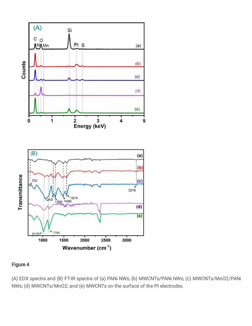
Figure 4
(A) EDX spectra and (B) FT-IR spectra of (a) PANi NWs; (b) MWCNTs/PANi NWs; (c) MWCNTs/MnO2/PANiNWs; (d) MWCNTs/MnO2; and (e) MWCNTs on the surface of the Pt electrodes.

Figure 5
(A) EIS spectra of the Pt/MWCNTs/MnO2/PANi NWs electrode (a) before and (b) after DNA capture probeimmobilization (inserted �gure: (a) EIS spectrum of the Pt/MWCNTs/MnO2/PANi NWs electrode; (i, ii)Fluorescence images of (i) Pt/MWCNTs/MnO2/PANi NWs; and (ii) Pt/MWCNTs/MnO2/PANi NWs/DNAprobe electrode, respectively); (B) EIS spectra of the DNA sensors (a) before, and (b) after DNAhybridization with the non-complementary target DNA (50 nM); (C) EIS spectra of the DNA sensors afterhybridization with different concentrations of the complementary target DNA: (a) 0 pM, (b) 5 pM, (c) 50pM, (d) 500 pM, (e) 5 nM, (f) 50 nM, and (g) 500 nM; (D) Changes on Rct (ΔRct) of the DNA sensors vs.concentrations of DNA target (inset: Randles equivalent circuit which is �tted with obtained EIS spectra).Experimental conditions: EIS spectra were measured in PBS buffer (pH 7.4) solution consisted 0.1 M KCland DNA target sequences. Frequency range: 104 Hz to 0.1 Hz, EAC = 5 mV and EDC = 230 mV.