akt-dependent antiapoptotic action of insulin is sensitive to farnesyltransferase inhibitor
TRANSCRIPT

Articles
Akt-Dependent Antiapoptotic Action of Insulin Is Sensitive to FarnesyltransferaseInhibitor
Doekbae Park,‡ Sanjay K. Pandey, Elena Maksimova, Sutapa Kole, and Michel Bernier*
Diabetes Section, Laboratory of Clinical InVestigation, National Institute on Aging, National Institutes of Health, 5600 NathanShock DriVe, Box 23, Baltimore, Maryland 21224-6825
ReceiVed May 1, 2000; ReVised Manuscript ReceiVed August 7, 2000
ABSTRACT: CHO cells expressing the human insulin receptors (IR) were used to evaluate the effect of thepotent farnesyltransferase inhibitor, manumycin, on insulin antiapoptotic function. Cell treatment withmanumycin blocked insulin’s ability to suppress pro-apoptotic caspase-3 activity which led to time-dependent proteolytic cleavage of two nuclear target proteins. The Raf-1/MEK/ERK cascade and theserine/threonine protein kinase Akt are two survival pathways that may be activated in response to insulin.We tested the hypothesis that inhibition of farnesylated Ras was causally related to manumycin-inducedapoptosis and showed that the response to manumycin was found to be independent of K-Ras functionbecause membrane association and activation of endogenous K-Ras proteins in terms of GTP loading andERK activation were unabated following treatment with manumycin. Moreover, blocking p21Ras/Raf-1/MEK/ERK cascade by the expression of a transdominant inhibitory mSOS1 mutant in CHO-IR cellskept cells sensitive to the antiapoptotic action of insulin. Insulin-dependent activation of Akt was blockedby 4 h treatment with manumycin (P < 0.01), a kinetic too rapid to be explained by Ras inhibition. Thisstudy suggests that the depletion of short-lived farnesylated proteins by manumycin suppresses theantiapoptotic action of insulin at least in part by disrupting Akt activation but not that of the K-Ras/Raf-1/ERK-dependent cascade.
Apoptosis is the most frequent morphological feature ofprogrammed cell death thought to play a pivotal role indiverse physiological and pathological processes. Theseinclude homeostatic maintenance of tissues and organs,autoimmune diabetes and diabetic neuropathy (1, 2). Insulin
exerts protection against apoptotic death mediated by growthfactor withdrawal in a number of cellular models (3-7).Despite recent advance in the identification of gene productsinvolved in the prevention or induction of apoptosis (re-viewed in ref8), the mechanisms by which insulin partici-pates in this process remain largely unknown. Hence,investigating signaling pathways that are involved in insulin-mediated antiapoptotic protection shall play a crucial rolein our understanding of insulin action.
Activation of the insulin receptor (IR) tyrosine kinase isessential for many of the biological actions of insulin. Theliganded receptor initiates intracellular signals by stimulatingtyrosine phosphorylation of endogenous substrates [e.g.,
* To whom correspondence should be addressed. Phone: (410) 558-8199. Fax: (410) 558-8381. E-mail: [email protected].
‡ Present address: Lab. Histology, College of Medicine, ChejuNational University, Cheju, 690-756, Korea.
1 Abbreviations: IR, insulin receptor; ERK, extracellular signal-regulated kinase; CHO, chinese hamster ovary; PI, propidium iodide;FACS, fluorescence-activated cell sorting; PAGE, polyacrylamide gelelectrophoresis; SFM, serum-free medium; RBD, Ras binding domain;FTI, farnesyltransferase inhibitor; PARP, poly(ADP-ribose) polymerase.
© Copyright 2000 by the American Chemical Society Volume 39, Number 41 October 17, 2000
10.1021/bi000995y CCC: $19.00 © 2000 American Chemical SocietyPublished on Web 09/20/2000
insulin receptor substrate (IRS) and Shc proteins]. Tyrosinephosphorylation of IRS-1 allows compartmentalization ofsignaling molecules, including members of the class Iphosphatidylinositol 3-kinase (PI 3-kinase) family (9). Insulininduces also the formation of a Shc-Grb2-SOS ternarycomplex that enables p21Ras activation (10, 11). Expressionof a mutant SOS protein that lacks the guanine nucleotideexchange domain causes a marked inhibition in the formationof GTP-bound p21Ras, thus atttenuating Ras-dependentinsulin signaling pathway (12). p21Ras stimulates down-stream effectors that participate in mitogen-activated protein(MAP) kinase activation, an essential route that conveysmitogenic signals to the nucleus. GTP-bound Ras triggersthe recruitement of Raf-1 to the plasma membrane whereits kinase activity is activated. This results in subsequent Raf-1-mediated phosphorylation and activation of the dualthreonine/tyrosine kinase, MAP kinase kinase (MEK), whichin turn phosphorylates and activates extracellular signal-regulated kinases (ERKs) (13). p21Ras and the 110 kDacatalytic subunit of class I PI 3-kinase are found within aprotein complex in a GTP-dependent manner, indicating thatRas may also contribute to PI 3-kinase activation (14).
Farnesylation is a regulated posttranslational modificationthat allows attachement of a number of proteins, includingp21Ras, to the plasma membrane. By inducing the activityof the enzyme farnesyl protein transferase, insulin increasesthe pool of membrane-associated p21Ras and promotes GTPloading on Ras (15, 16). It has been recently documentedthat manumycin, a selective protein farnesylation inhibitor(FTI), blocks the antiapoptotic protection exerted by insulinin IR-expressing CHO cells maintained in the absence ofgrowth factors (17). However, this study has not addressedthe mechanism by which manumycin induces apoptoticdeath.
A limitation of FTIs as general anti-Ras agents is that Rasisoforms are differentially affected by inhibition of farnes-yltransferase activity. Unlike H-Ras, K-Ras undergoes alter-native lipid modification by geranylgeranyltransferases incells treated with FTI (18, 19), thus allowing this alternativelyprenylated version of K-Ras to remain associated with themembrane fraction and be biologically active (20). BecauseRas isoforms are known to exhibit differential activitiestoward the Raf-1/ERK cascade and the PI 3-kinase/Aktpathway (21), the pro-apoptotic action of manumycin maybe the result of selective inhibition of either one of thesesurvival signaling pathways. Using various experimentalapproaches, we report here that the K-Ras/Raf-1/ERKcascade was not the target of manumycin action, in contrastto the rapid decrease in insulin-stimulated Akt phosphory-lation and activation in untransformed CHO-IR cells. Sinceengagement of H-Ras preferentially activates the PI 3-kinase/Akt pathway (21), we tested also the hypothesis that cellularexpression of a dominant negative H-Ras mutant may mimicmanumycin in its ability to promote apoptosis.
MATERIAL AND METHODS
Materials. Manumycin, FTI-277, porcine insulin, mono-clonal antibodies against pan Ras and K-Ras, the fluorogeniccaspase-3 substrate, Ac-DEVD-AMC, two caspase-3 inhibi-tors, z-DEVD-fmk and DEVD-CHO, and the caspase-3colorimetric substrate, Ac-DEVD-pNA, were obtained from
Calbiochem (La Jolla, CA). Bovine serum albumin (RIAgrade), propidium iodide (PI), and anti-actin antibodies werefrom Sigma Chemical Corp. (St. Louis, MO). Protein G-Plus/protein A-agarose was obtained from Oncogene Science(Manhasset, NY), and horseradish peroxidase-conjugatedanti-phosphotyrosine antibody, polyclonal antibodies againstPARP (A-20) and lamin B, and monoclonal anti-H-Rasantibody were from Santa Cruz Biotechnology (Santa Cruz,CA). Polyclonal antibodies against ERK 1/2, Akt1 (no. 06-558), Akt2 (no. 06-606) and carboxyl-terminal IRS-1, recom-binant histidine-tagged caspase-3, and N17Ras.pUSEamp+vector were from Upstate Biotechnology (Lake Placid, NY).Phospho-specific anti-ERK 1/2 and phospho-Ser473 Aktantibodies were obtained from Promega Corp. (Madison, WI)and New England Biolabs, Inc. (Beverly, MA), respectively.Electrophoresis reagents, such as gels, Tris-glycine SDSrunning buffer, and poly(vinylidene difluoride) (PVDF)membrane were from Novex Corp. (San Diego, CA). Ham’sF-12 medium, D-PBS, and Cell-stripper were from Cellgro(Freiburg, Germany), FBS was from Gemini (Calabasas,CA), and trypsin-EDTA was from NIH (Bethesda, MD).
Cell Culture. Cells used in this study were describedpreviously (12, 17) and include CHO-IR cells and CHO-IRcells expressing a deletion mutant of mSOS1 (CHO-IR/∆SOS). CHO-IR/∆SOS cells were from Dr. M. Sakaue(Kobe University, Kobe, Japan). Cells were grown in Ham’sF-12 medium containing 100 units/mL penicillin, 100µg/mL streptomycin, and 10% fetal bovine serum (FBS), andmaintained in a humidified atmosphere of 5% CO2 in air at37 °C.
Treatments.CHO-IR and CHO-IR/∆SOS cells were grownto confluence in 35-mm tissue culture dishes and thensubjected to growth factor withdrawal in F-12 mediumsupplemented with 0.1% (w/v) bovine serum albumin for 3h followed by the addition of 10µM manumycin or vehicle(dimethyl sulfoxide) for 60 min prior to the addition of 10nM insulin or 10% FBS. Eighteen hours later, floating cellswere collected by centrifugation, harvested, and combinedwith the cells remaining attached to the plate.
Construction of pTracer.N17Ras.GFP. An expression vec-tor that produces both the dominant negative N17 mutant ofhuman H-Ras (N17Ras) and super-green fluorescent protein(GFP) was prepared by inserting full-length N17Ras cDNAfragment into pTracer.GFP plasmid as followed. The N17RascDNA fragment was excised from N17Ras.pUSEamp+vector with KpnI and EcoRV digestion, and the fragment(1.1 kb) was then inserted into pTracer.GFP vector, whichwas linearized withKpnI/EcoRV, by ligation with T4 ligase(Pharmacia Biotech.). Competent DH5R bacterial cells weretransformed with the ligated plasmid, selected with ampicillinand subjected to plasmid preparation. The presence ofN17Ras cDNA was confirmed byKpnI/EcoRV digestion.CHO cells were transiently transfected with pTracer expres-sion vectors containing either GFP cDNA alone or togetherwith N17Ras cDNA. Transfection was done with 1.0µg ofDNA by using the Lipofectamine-plus technique (LifeTechnologies, Gaithersburg, MD) according to the manu-facturer’s protocol. After incubation for 24 h in F-12 medium,the cells were incubated for an additional 18 h in serum-free medium supplemented or not with insulin or 10% FBS.
DNA Laddering. Internucleosomal DNA fragmentationanalysis was performed essentially as described previously
12514 Biochemistry, Vol. 39, No. 41, 2000 Park et al.

(17). Briefly, pooled cellular DNA from adherent anddetached cells was prepared using the Puregene Kit (GentraSystems, Inc., Minneapolis, MN), and the purified DNA wasthen incubated with 20µg/mL RNase A for 1 h at 37°C.Equal amounts of DNA from each sample (0.5µg) were 3′-OH-labeled with 5 units of Klenow fragment of DNApolymerase I (New England Biolabs) and 0.5µCi [R-32P]-dCTP (∼3000 Ci/mmol, Amersham Corp., Arlington Heights,IL) and electrophoresed on 6% (w/v) polyacrylamide gel.DNA was visualized by autoradiography of the dried gelusing Kodak BioMax film and intensifying screens.
Flow Cytometric Analysis.Protocol I. Upon induction ofapoptosis, cytoplasmic phosphotidylserine translocates to theexternal surface of the cell membrane, allowing its in vitrodetection through interaction with annexin V (22). Untreatedcells or cells treated with manumycin, insulin, or FBS wereharvested with Cell-stripper solution and combined with theirmedium to collect any detached cells. The cell suspensionconcentration was adjusted to∼1 × 10⁶ cells/mL withD-PBS (Ca2+/Mg2+ free); aliquots of cell suspension (5×10⁵) were incubated at room temperature with media bindingreagent and Annexin V-FITC as indicated by the manufac-turer (Oncogene Research Products, Cambridge, MA). After15 min in the dark, cells were centrifuged at 1000g for 5min and the cell pellet resuspended in 500µL of ice-coldbinding buffer and 10µL of 30 µg/mL PI supplied by themanufacturer. The samples were immediately analyzed byflow cytometry on a FACScan flow cytometer (BectonDickinson, Cockeysville, MD) equipped with a 15 mWargon-ion laser. Ten thousand events were collected for eachsample. An excitation wavelength of 488 nm was used whilefluorescence emissions of 507 and 580 nm were collectedto detect FITC and PI signals, respectively. The log ofannexin V-FITC fluorescence was displayed on thex-axisand the log of PI fluorescence on they-axis.
Protocol II. Cells were incubated with 60µg/mL PI for20 min and then detached from culture dishes using cell-stripper solution. The cells were collected by low speedcentrifugation, washed twice in D-PBS followed by FACSanalysis where 20 000 events were collected for each sample.An excitation wavelength of 488 nm and fluorescenceemissions of 507 and 580 were used for the detection ofGFP-expressing cells and PI-stained cells, respectively.
Protocol III. Cells were removed from the culture dishusing a 0.05% (w/v) trypsin solution, washed twice in D-PBSand fixed in 70% (v/v) EtOH. The cells were incubated with50 µg/mL PI in D-PBS and 20µg/mL Rnase A for 30 minat room temperature, consistent with the staining techniqueoriginally described by Crissman and Steinkamp (23) toassess apoptosis. An excitation wavelength of 488 nm andfluorescence emission of 580 nm were used. The log of PIfluorescence was converted to linear fluoresence intensity.
Determination of Caspase-3 ActiVity. Cells were lysed inice-cold 0.5 mL of buffer I [50 mM Tris.HCl, pH 7.4, 150mM NaCl, 1% (w/v) NP-40, 0.25% (w/v) sodium deoxy-cholate, and 1 mM EGTA] for 15 min at 4°C. Aftercentrifugation at 10000g for 20 min at 4°C, aliquots ofsupernatant were incubated with 25µM Ac-DEVD-AMCfor 3 h at 37 °C. Experiments were performed with arecombinant hexahistidine-tagged caspase-3 standard (0-2ng/assay) to ensure that under these conditions the cleavageof the fluorogenic substrate was linear with respect to
caspase-3 activity. The generation of fluorescent product wasdetected using excitation and emission wavelengths of 380and 460 nm, respectively. Alternatively, a colorimetric assaywas used where caspase-3 activity of the supernatants fromcells lysed in modified buffer I (where EGTA was replacedwith 1 mM EDTA) was measured by incubating cell lysateswith reaction buffer [100 mM Hepes, pH 7.5, 20% (w/v)glycerol, 5 mM dithiothreitol, and 0.5 mM EDTA] containing100µM of the colorimetric substrate, Ac-DEVD-pNA. Therelease ofp-nitroalinine was detected by monitoring absor-bance at 405 nm for 30 min at 37°C using a microtiter platereader (Bio-Rad, Hercules, CA). In certain experiments, thecell lysate was first incubated with caspase-3 inhibitors (e.g.,z-DEVD-fmk or DEVD-CHO).
Polyacrylamide Gel Electrophoresis and Western BlotAnalysis.Unless otherwise indicated, cells were lysed directlyin Laemmli sample buffer (24) containing 5% (v/v) 2-mer-captoethanol and 1 mM orthovanadate. After heating at 70°C for 10 min, proteins were separated by SDS-PAGE on4-12% polyacrylamide gradient gel along with prestainedprotein markers, and electrotransferred onto PVDF mem-brane. The membrane was incubated with blocking buffer[5% (w/v) nonfat dried milk in Tris-buffered saline (TBS)-0.1% (w/v) Tween-20 (TBS-T)] for 1 h atroom temperatureand then probed with a 1:48000 dilution of HRP-conjugatedphosphotyrosine antibody in blocking buffer to detectchanges in protein tyrosine phosphorylation. After a seriesof washes, positive signals were visualized with the enhancedchemiluminescence (ECL) reagents in combination withHyperfilm-ECL (Amersham). Band intensities were quan-titated by laser densitometry using the ImageQuant software(Molecular Dynamics, Sunnyvale, CA). The membranes werealso reprobed with various primary antibodies (1:1000)followed by the appropriate HRP-conjugated secondaryantibodies (1:3000).
In Vitro Binding of ActiVe Ras to GST-RBD. GST-RBDwas kindly provided by Dr. Johannes L. Bos (UtrechtUniversity, Netherlands) and was used to determine therelative amount of active GTP-bound p21Ras, as previouslydescribed (25). The GST-RBD fusion protein contains theminimal Ras binding domain of Raf-1 (amino acids 51-131). The prokaryotic expression vector (pGEX-2T) contain-ing sequences for a fusion protein of glutathione S-transferase(GST) and RBD was transformed intoE. coli BL21 cells.These transformed cells were grown in LB medium and theinduction of GST-RBD expression was carried out byaddition of 0.2 mM isopropyl-â-thiogalactopropyranoside.After 4 h, cells were pelleted by centrifugation, suspendedin PBS and then disrupted in B-PER reagent (Pierce,Rockford, IL). The cell lysate was centrifuged at 5000g for5 min at 4°C and Triton X-100 was added to the supernatantto reach a final concentration of 1% (w/v). After centrifuga-tion at 12000g for 10 min at 4°C, the supernatant was mixedwith glycerol (10% final, w/v), aliquoted, and stored at-80°C until use. Crude GST-RBD extract was incubated withglutathione-bound agarose 4B beads (Pharmacia, Uppsala,Sweden) for 30 min at 4°C. The beads were recovered bycentrifugation and washed twice with lysis buffer II [25 mMHepes, pH 7.5, 150 mM NaCl, 1% (w/v) NP-40, 0.25% (w/v) sodium deoxycholate, 10% (w/v) glycerol, 25 mM NaF,10 mM MgCl2, 1 mM EDTA, 1 mM orthovanadate, 10µg/mL leupeptin, and 10µg/mL aprotinin].
Manumycin and Insulin Antiapoptotic Function Biochemistry, Vol. 39, No. 41, 200012515

CHO-IR and CHO-IR/∆SOS cells grown either on 35- or60-mm dishes were lysed in 0.5 mL of lysis buffer II onice, centrifuged at 10000g for 20 min, and the clarified lysatesincubated with precoupled GST-RBD beads for 30 min at 4°C. The beads were pelleted by centrifugation and washedthree times in lysis buffer II before solubilization in Laemmlisample buffer. The samples were separated on a 16% SDS-PAGE gel under reducing conditions, and immunoblottedeither with panRas, K-Ras, or H-Ras monoclonal antibodies(each at 2.5µg/mL).
Statistical Analysis. Data are presented as the mean(SEM. Comparison between groups were made by ANOVAcoupled to Fisher’s protected least significant differencespost-hoctest.
RESULTS
CHO-IR cells were pretreated with manumycin, an ana-logue of farnesyl diphosphate, for 1 h followed by a 16-hincubation in the absence or presence of insulin or 10% FBS.The DNA laddering assay was then used to assess thepresence of internucleosomal DNA fragmentation, one ofthe hallmark of apoptotic death (26). Manumycin blockedthe ability of insulin (Figure 1A) and serum (data not shown)at reducing the formation of DNA fragments that weregenerated in response to serum withdrawal (SFM) (Figure1A). To independently verify the pro-apoptotic role ofmanumycin, annexin V binding was then performed on livecells by flow cytometry. As control experiment, we notedthat the proportion of apoptotic cells that were stained with
annexin V-FITC and PI upon serum starvation was decreasedfrom 33.4 ( 3.5 to 7.9( 3.1% and 5.3( 1.6% in thepresence of 10 nM insulin and 10% FBS, respectively (mean( SE, n ) 3) (Figure 1B; upper right quadrant in panelsI-III). Here pretreatment with 10µM manumycin for 1 hrendered cells insensitive to the protection by insulin or FBS(Figure 1B; panel IV).
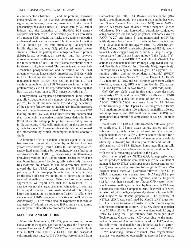
Initial Characterization of the Pro-Apoptotic Action ofManumycin in CHO-IR Cells. The caspase family of cysteineproteases plays a pivotal role in mediating apoptosis throughthe proteolysis of specific targets that include poly(ADP-ribose) polymerase (PARP) and the nuclear lamins (27). Thefunction of caspase-3 (like) activity has been described tobe involved in the execution of apoptosis in a tissue-, celltype-, or death stimulus-specific manner (28). Immunoblotanalysis revealed that the combination of manumycin andinsulin for 4 and 24 h markedly decreased the amount oflamin B and PARP proteins in whole cell lysates withoutaltering the expression or integrity of ERK 1/ERK 2 (Figure2A). CHO-IR cells were treated under various experimentalconditions and activation of caspase-3 was then measuredusing the fluorogenic substrate, Ac-DEVD-AMC. Activecaspase-3 cleaves Ac-DEVD-AMC, thus causing an increasein fluorescence intensity that can be quantitated. When SFM-treated cells were incubated in the presence of 10 nM insulin,the increase in fluorescence intensity was attenuated∼6-fold (Figure 2B). Pretreatment with manumycin blocked thereduction in caspase-3 activity by insulin. Similar results wereobtained when using a colorimetric assay for determining
FIGURE 1: Pro-apoptotic role of manumycin in CHO-IR cells. (A) SFM-treated CHO-IR cells were incubated with 0.1% DMSO (control)or 10µM manumycin for 1 h followed by a 16-h incubation in the absence or presence of 10 nM insulin. Genomic DNA was prepared andanalyzed for internucleosomal DNA fragmentation as described in Materials and Methods. An autoradiogram of a representative DNAfragmentation analysis is shown. Similar results were obtained in at least five independent experiments. (B) Annexin V-FITC staining wasperformed on CHO-IR cells that were incubated for 18 h in SFM (panel I), 10 nM insulin (panel II) or 10% FBS (panel III) as describedin Materials and Methods. Similar experiment were repeated with cells treated with manumycin alone or in combination with insulin orFBS. Panel IV, percent of apoptotic cells that were positively stained for PI and Annexin V-FITC is represented as the mean( SE of threeindependent experiments. (*, **)P < 0.05 and 0.01. (9) Vehicle; (0) manumycin.
12516 Biochemistry, Vol. 39, No. 41, 2000 Park et al.
caspase-3 activity (data not shown). Addition of the cell-permeant z-DEVD-fmk (20µM) (29) markedly inhibitedcaspase-3 activity (Figure 2B), demonstrating the specificnature of this assay.
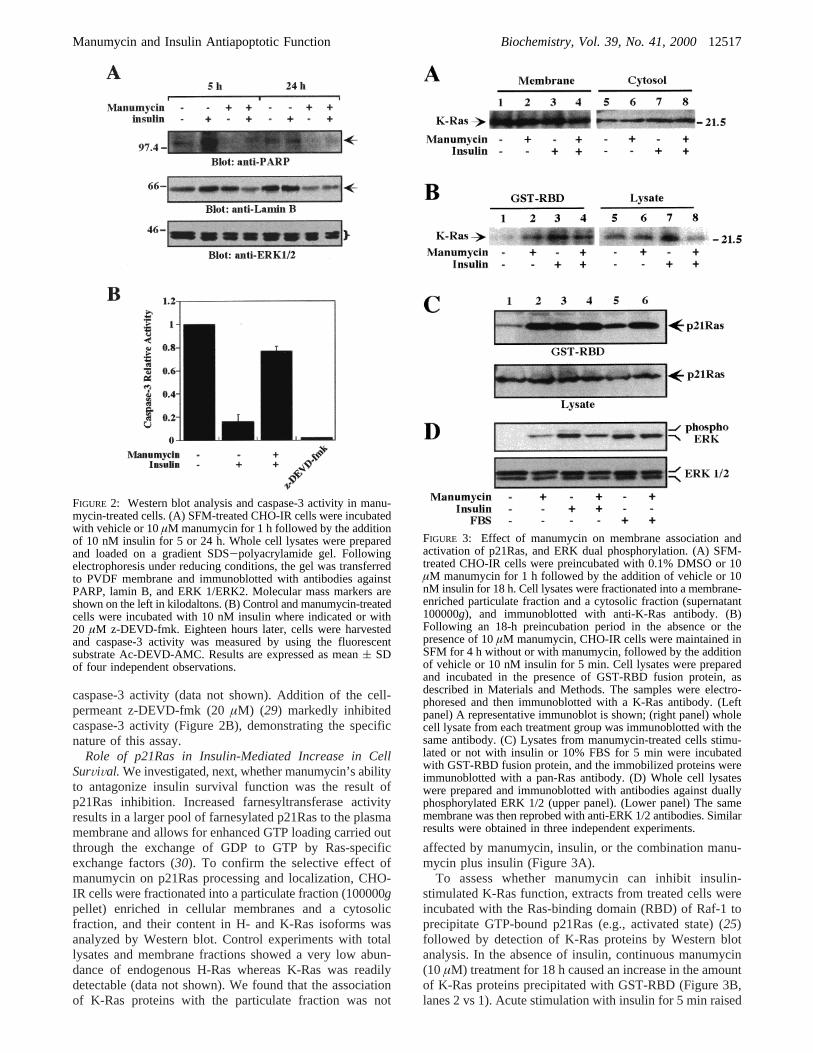
Role of p21Ras in Insulin-Mediated Increase in CellSurViVal. We investigated, next, whether manumycin’s abilityto antagonize insulin survival function was the result ofp21Ras inhibition. Increased farnesyltransferase activityresults in a larger pool of farnesylated p21Ras to the plasmamembrane and allows for enhanced GTP loading carried outthrough the exchange of GDP to GTP by Ras-specificexchange factors (30). To confirm the selective effect ofmanumycin on p21Ras processing and localization, CHO-IR cells were fractionated into a particulate fraction (100000gpellet) enriched in cellular membranes and a cytosolicfraction, and their content in H- and K-Ras isoforms wasanalyzed by Western blot. Control experiments with totallysates and membrane fractions showed a very low abun-dance of endogenous H-Ras whereas K-Ras was readilydetectable (data not shown). We found that the associationof K-Ras proteins with the particulate fraction was not
affected by manumycin, insulin, or the combination manu-mycin plus insulin (Figure 3A).
To assess whether manumycin can inhibit insulin-stimulated K-Ras function, extracts from treated cells wereincubated with the Ras-binding domain (RBD) of Raf-1 toprecipitate GTP-bound p21Ras (e.g., activated state) (25)followed by detection of K-Ras proteins by Western blotanalysis. In the absence of insulin, continuous manumycin(10µM) treatment for 18 h caused an increase in the amountof K-Ras proteins precipitated with GST-RBD (Figure 3B,lanes 2 vs 1). Acute stimulation with insulin for 5 min raised
FIGURE 2: Western blot analysis and caspase-3 activity in manu-mycin-treated cells. (A) SFM-treated CHO-IR cells were incubatedwith vehicle or 10µM manumycin for 1 h followed by the additionof 10 nM insulin for 5 or 24 h. Whole cell lysates were preparedand loaded on a gradient SDS-polyacrylamide gel. Followingelectrophoresis under reducing conditions, the gel was transferredto PVDF membrane and immunoblotted with antibodies againstPARP, lamin B, and ERK 1/ERK2. Molecular mass markers areshown on the left in kilodaltons. (B) Control and manumycin-treatedcells were incubated with 10 nM insulin where indicated or with20 µM z-DEVD-fmk. Eighteen hours later, cells were harvestedand caspase-3 activity was measured by using the fluorescentsubstrate Ac-DEVD-AMC. Results are expressed as mean( SDof four independent observations.
FIGURE 3: Effect of manumycin on membrane association andactivation of p21Ras, and ERK dual phosphorylation. (A) SFM-treated CHO-IR cells were preincubated with 0.1% DMSO or 10µM manumycin for 1 h followed by the addition of vehicle or 10nM insulin for 18 h. Cell lysates were fractionated into a membrane-enriched particulate fraction and a cytosolic fraction (supernatant100000g), and immunoblotted with anti-K-Ras antibody. (B)Following an 18-h preincubation period in the absence or thepresence of 10µM manumycin, CHO-IR cells were maintained inSFM for 4 h without or with manumycin, followed by the additionof vehicle or 10 nM insulin for 5 min. Cell lysates were preparedand incubated in the presence of GST-RBD fusion protein, asdescribed in Materials and Methods. The samples were electro-phoresed and then immunoblotted with a K-Ras antibody. (Leftpanel) A representative immunoblot is shown; (right panel) wholecell lysate from each treatment group was immunoblotted with thesame antibody. (C) Lysates from manumycin-treated cells stimu-lated or not with insulin or 10% FBS for 5 min were incubatedwith GST-RBD fusion protein, and the immobilized proteins wereimmunoblotted with a pan-Ras antibody. (D) Whole cell lysateswere prepared and immunoblotted with antibodies against duallyphosphorylated ERK 1/2 (upper panel). (Lower panel) The samemembrane was then reprobed with anti-ERK 1/2 antibodies. Similarresults were obtained in three independent experiments.
Manumycin and Insulin Antiapoptotic Function Biochemistry, Vol. 39, No. 41, 200012517
the level of active RBD-interacting K-Ras proteins several-fold above basal levels (Figure 3B, lanes 3 vs 1). Thisincrease was only marginally reduced in the presence ofmanumycin. Comparable amount of K-Ras proteins waspresent in total cell lysates (Figure 3B, right panel), indicatingno significant effect of manumycin on Ras expression. In asecond set of experiments, membranes were probed with apanRas antibody that recognizes various Ras isoforms. Asshown in Figure 3C, the ability of insulin or 10% FBS toacutely stimulate Ras interaction with GST-RBD wasmaintained in the presence of manumycin. Recall that K-Rasproteins are far more abundant than H-Ras in CHO-IR cells(see above), suggesting that the signals generated withpanRas antibody may be derived from K-Ras. As shown inFigure 3D (upper panel), manumycin increased basal ERKactivity as determined by the use of phospho-specific ERK1/ERK 2 antibodies. The insulin-mediated activation of ERKwas attenuated somewhat in manumycin-treated cells. Incontrast, manumycin had no apparent effect on ERK activa-tion by 10% FBS. Taken together, these findings provideevidence that induction of apoptosis by manumycin was notthe result of K-Ras inhibition and/or alteration in the Ras/Raf-1/ERK signaling pathway.
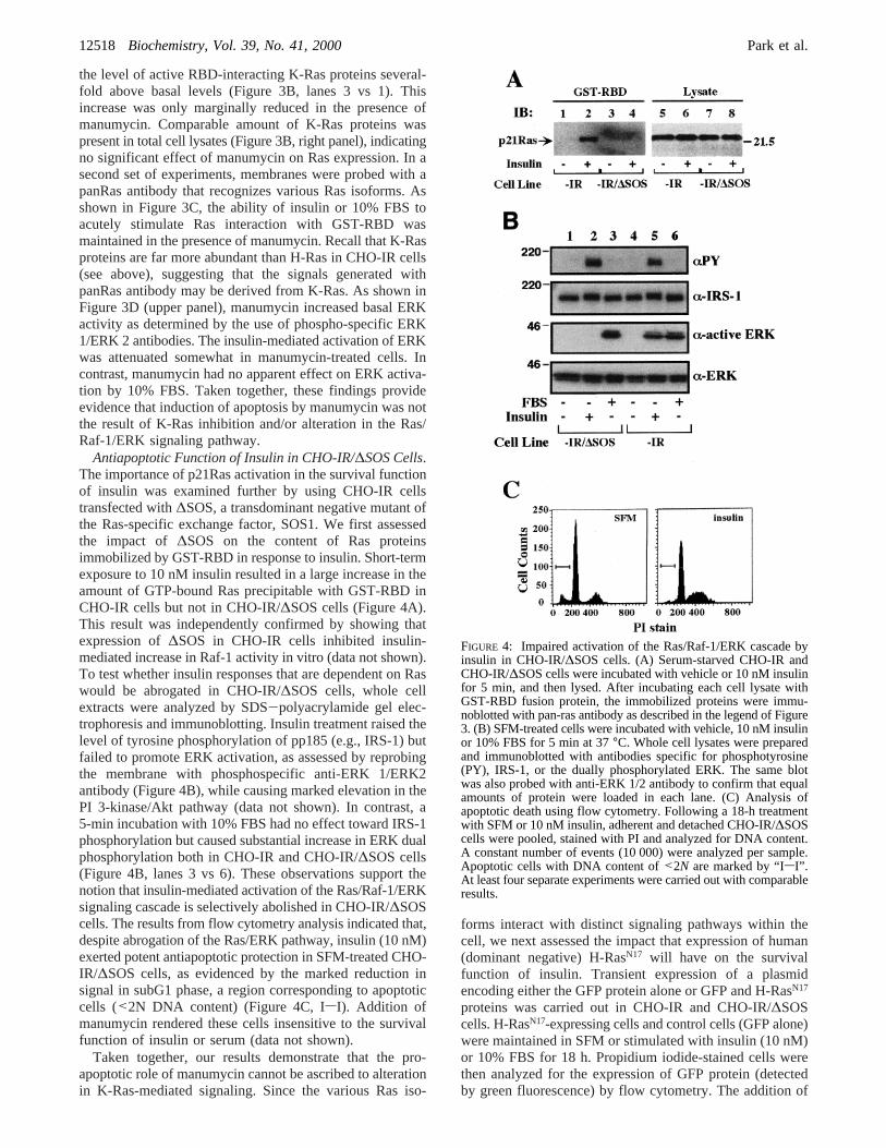
Antiapoptotic Function of Insulin in CHO-IR/∆SOS Cells.The importance of p21Ras activation in the survival functionof insulin was examined further by using CHO-IR cellstransfected with∆SOS, a transdominant negative mutant ofthe Ras-specific exchange factor, SOS1. We first assessedthe impact of ∆SOS on the content of Ras proteinsimmobilized by GST-RBD in response to insulin. Short-termexposure to 10 nM insulin resulted in a large increase in theamount of GTP-bound Ras precipitable with GST-RBD inCHO-IR cells but not in CHO-IR/∆SOS cells (Figure 4A).This result was independently confirmed by showing thatexpression of∆SOS in CHO-IR cells inhibited insulin-mediated increase in Raf-1 activity in vitro (data not shown).To test whether insulin responses that are dependent on Raswould be abrogated in CHO-IR/∆SOS cells, whole cellextracts were analyzed by SDS-polyacrylamide gel elec-trophoresis and immunoblotting. Insulin treatment raised thelevel of tyrosine phosphorylation of pp185 (e.g., IRS-1) butfailed to promote ERK activation, as assessed by reprobingthe membrane with phosphospecific anti-ERK 1/ERK2antibody (Figure 4B), while causing marked elevation in thePI 3-kinase/Akt pathway (data not shown). In contrast, a5-min incubation with 10% FBS had no effect toward IRS-1phosphorylation but caused substantial increase in ERK dualphosphorylation both in CHO-IR and CHO-IR/∆SOS cells(Figure 4B, lanes 3 vs 6). These observations support thenotion that insulin-mediated activation of the Ras/Raf-1/ERKsignaling cascade is selectively abolished in CHO-IR/∆SOScells. The results from flow cytometry analysis indicated that,despite abrogation of the Ras/ERK pathway, insulin (10 nM)exerted potent antiapoptotic protection in SFM-treated CHO-IR/∆SOS cells, as evidenced by the marked reduction insignal in subG1 phase, a region corresponding to apoptoticcells (<2N DNA content) (Figure 4C, IsI). Addition ofmanumycin rendered these cells insensitive to the survivalfunction of insulin or serum (data not shown).
Taken together, our results demonstrate that the pro-apoptotic role of manumycin cannot be ascribed to alterationin K-Ras-mediated signaling. Since the various Ras iso-
forms interact with distinct signaling pathways within thecell, we next assessed the impact that expression of human(dominant negative) H-RasN17 will have on the survivalfunction of insulin. Transient expression of a plasmidencoding either the GFP protein alone or GFP and H-RasN17
proteins was carried out in CHO-IR and CHO-IR/∆SOScells. H-RasN17-expressing cells and control cells (GFP alone)were maintained in SFM or stimulated with insulin (10 nM)or 10% FBS for 18 h. Propidium iodide-stained cells werethen analyzed for the expression of GFP protein (detectedby green fluorescence) by flow cytometry. The addition of
FIGURE 4: Impaired activation of the Ras/Raf-1/ERK cascade byinsulin in CHO-IR/∆SOS cells. (A) Serum-starved CHO-IR andCHO-IR/∆SOS cells were incubated with vehicle or 10 nM insulinfor 5 min, and then lysed. After incubating each cell lysate withGST-RBD fusion protein, the immobilized proteins were immu-noblotted with pan-ras antibody as described in the legend of Figure3. (B) SFM-treated cells were incubated with vehicle, 10 nM insulinor 10% FBS for 5 min at 37°C. Whole cell lysates were preparedand immunoblotted with antibodies specific for phosphotyrosine(PY), IRS-1, or the dually phosphorylated ERK. The same blotwas also probed with anti-ERK 1/2 antibody to confirm that equalamounts of protein were loaded in each lane. (C) Analysis ofapoptotic death using flow cytometry. Following a 18-h treatmentwith SFM or 10 nM insulin, adherent and detached CHO-IR/∆SOScells were pooled, stained with PI and analyzed for DNA content.A constant number of events (10 000) were analyzed per sample.Apoptotic cells with DNA content of<2N are marked by “IsI”.At least four separate experiments were carried out with comparableresults.
12518 Biochemistry, Vol. 39, No. 41, 2000 Park et al.

10% serum reduced SFM-mediated apoptosis in control cellsexpressing GFP alone (Figure 5B vs C, upper right quad-rants). In cells transfected with GFP and H-RasN17 proteins,there was a∼1.6-fold increase in the extent of apoptoticdeath in response to SFM and a complete abolition of cellsurvival by insulin or serum (Figure 5D). The cellularexpression of human H-RasN17 was confirmed by immunoblotanalysis (data not shown).
Manumycin Inhibits Insulin-Induced Akt ActiVation. Theactivation of PKB/Akt by phosphorylation plays a major rolein cell survival by inhibiting apoptosis mediated by a numberof stimuli, including SFM (31). Because of the exquisitesensitivity of H-Ras to farnesylation inhibitors (18, 19) andits ability to potently stimulate the PI 3-kinase/Akt pathway(21), we tested for changes in phospho-Akt levels betweenmanumycin-treated cells and control cells in the absence orpresence of insulin. Serum-starved CHO-IR cells were treatedwith 10 µM manumycin for 4 and 24 h prior to insulinstimulation for 10 min. Aliquots of cell lysate were electro-phoresed and then immunoblotted with an anti-phosphospe-cific Akt antibody that recognize Akt that had been phos-phorylated at Ser473 (Figure 6A). Cell treatment withmanumycin resulted in a significant reduction in insulin-mediated Akt phosphorylation (35( 4 and 66( 10%inhibition after 4 and 24 h treatment, respectively;P < 0.01,n ) 4-5) (Figure 6B, left panel) despite similar levels ofERK dual phosphorylation (Figure 6B, right panel). Thisimplies that activation of H-Ras/PI 3-kinase/Akt pathway
may be impaired in manumycin-treated cells. Similar findingswere obtained with another specific farnesyltransferaseinhibitor, FTI-277. The addition of FTI-277 (1µM) to SFM-treated CHO-IR cells for 4 h led to a 44( 7% inhibition ofinsulin-mediated Akt phosphorylation (n ) 2) withoutaltering ERK dual phosphorylation following treatment withinsulin (data not shown). Immunoprecipitation-based kinaseassay with crosstide as substrate confirmed that insulin-mediated increase in Akt activity was reduced by∼50% after4 h treatment with manumycin (data not shown). Theseresults indicate that a short-lived farnesylated protein(s) maybe involved in the activation of Akt.
DISCUSSION
In this report, it was observed that manumycin influencesapoptosis in a way that sharply differs from that mediatedby growth factor deprivation. Insulin, 10% FBS, or antioxi-dants (our unpublished data) can markedly reduce apoptosismediated by serum starvation in CHO-IR cells. In contrast,cells that were pretreated with manumycin could not berescued from apoptotic death in response to insulin and othersurvival factors. This FTI caused also 3T3-L1 fibroblasts toundergo apoptosis despite cell treatment with 10% serum(our unpublished data). Our results differ from those ofSuzuki et al. (32) who showed that FTIs preferentially induceapoptosis in Ras-transformed normal rat kidney (KNRK)cells but not in untransformed cells and that the presence of10% serum counteracts FTI-induced apoptosis. It is importantto note that the KNRK cells were incubated with 80-100
FIGURE 5: H-Ras-dependent antiapoptotic protection. CHO-IR cellswere transiently transfected with an expression vector encodingeither the GFP protein alone or GFP and RasN17 proteins. After 24h, the cells were serum-starved and then incubated in the absence(SFM) or the presence of insulin or 10% FBS for 18 h. The cellswere subsequently processed for flow cytometry analysis asdescribed in the Experimental Procedures. The quadrant markersfor the bivariate dot plots were set based on the serum-treated GFPcontrol without PI staining (panel A). (B) SFM-treated GFP control;(C) serum-treated GFP control; (D) the data represent the mean(range of two independent experiments, where the relative level ofapoptosis in SFM-treated GFP controls was arbitrarily set at 1.0.(0) CHO-IR cells; (9) CHO-IR/∆SOS cells.
FIGURE 6: Manumycin inhibits insulin-mediated increase in Aktphosphorylation at Ser473. Serum-starved CHO-IR cells wereincubated with vehicle or 10µM manumycin for 4 and 24 hfollowed by the addtion of 10 nM insulin where indicated. Tenminutes later, cells were lysed and the content in phosphorylatedAkt and ERK was analyzed by immunoblotting using phospho-specific antibodies. (A) Blot from a representative experiment isshown. (B) The data represent the mean( SE of four to fiveindependent observations, where the relative level of phospho-473Akt (left panel) and dually phosphorylated ERK 1/2 (right panel)was arbitrarily set at 1.0. (**)P < 0.01 when compared to insulinalone. (9) No insulin; (0) insulin-treated cells.
Manumycin and Insulin Antiapoptotic Function Biochemistry, Vol. 39, No. 41, 200012519

µM FTIs for 36 h prior to analysis (32), conditions that arefar more stringent than the ones used in the work herein.Moreover, the concentrations of manumycin used in ourstudies (5-20 µM) have been reported to selectively inhibitfarnesyltransferase (IC50 ) 5-10 µM) but not geranylgera-nyltransferases (IC50 ) 180 µM) (33). In support of ourearlier observation, we incubated CHO-IR cells with twoother FTIs (FTI-277 andR-hydroxyfarnesylphosphonic acid)at concentrations ranging from 1 to 10µM in the presenceof 10 nM insulin for 16 h and found an induction of apoptosisalthough to a lesser degree than that observed with manu-mycin.2
By competing with normal Ras for binding to specificguanine-nucleotide-exchange factors, the dominant negativemutant of H-Ras (RasN17) prevents the activation of endog-enous Ras and its interaction with downstream targetproteins. We found that transient expression of RasN17 exertedsignificant loss in the ability of insulin and serum to conferantiapoptotic protection, indicating that Ras and/or itsdownstream target proteins may play an important survivalfunction. Although several Ras isoforms are expressedubiquitously and can activate the same effector pathways,quantitative differences exist in their ability to stimulate Raf-1/ERK cascade and the PI 3-kinase/Akt pathway. Therecruitment of Raf-1 to the plasma membrane and itssubsequent activation are influenced to a greater extent byK-Ras proteins, whereas H-Ras is a more potent activatorof PI 3-kinase (21). Furthermore, Ras isoforms vary in theirsensitivity to FTIs as suggested by the observation that K-Rasproteins, but not H-Ras, remain attached to the plasmamembrane following cell treatment with FTI (18, 19). In thepresent study, evidence was provided to suggest that theK-Ras/Raf-1/ERK cascade is not the target of manumycin’spro-apoptotic action in CHO-IR cells. We have shown thatendogenous K-Ras protein remains associated with themembrane fraction and that its activation in term of GST-RBD binding and ERK dual phosphorylation is not blockedby manumycin. Our results demonstrate also that there is anincrease in the level of K-Ras•GTP complex and ERKactivation between control cells and cells treated withmanumycin alone. This implies that blocking protein farne-sylation can relieve a basal inhibitory “tone” on K-Ras-mediated events, presumably at a step uptream of K-Ras.These results indicate that manumycin may target farnesy-lated protein(s) whose function includes the regulation ofIR antiapoptotic pathway. As indicated earlier, it has beenshown that FTIs selectively inhibit H-Ras prenylation therebyblocking H-Ras signaling and transformation (34). It ispossible that manumycin induces apoptosis by targetingH-Ras; however, because of the long half-life of membrane-associated farnesylated Ras (∼24 h, ref 16), farnesylatedprotein(s) other than H-Ras may be responsible for antiapo-ptotic control. Rho B has a short half-life (∼2 h) and is partof the immediate-early inducible response to growth factorsand protein tyrosine kinases (35, 36). The ability of FTIs toinhibit cell growth and Ras-dependent cell transformationis mediated by targeting the farnesylated RhoB protein(36, 37). Thus, investigations of the involvement of RhoBand other small GTPases that are known to be essential inRas transformation (e.g., RhoA, Rac1) (38) are required for
a better understanding of the mechanism of manumycin-induced apoptosis in untransformed CHO-IR cells.
CHO-IR/∆SOS cells were used to further support thenotion that the Ras/Raf-1/MEK/ERK cascade plays nosignificant role in the antiapoptotic function of insulin. Afeature of these cells include the ability of lysophosphatidicacid to potently stimulate ERK activity while being ineffec-tive at reversing SFM-mediated apoptosis (our unpublisheddata). In this cell model, insulin confers antiapoptoticprotection despite its inability to increase Raf-1 activity andERK activation, indicating that stimulation of the Raf-1/MEK/ERK signaling pathway does not participate in thesurvival function of insulin. Other features of CHO-IR/∆SOScells include their sensitivity to manumycin pro-apoptoticaction and their expression of a mutated SOS1 protein thatlacks the guanine nucleotide exchange domain of Ras.Despite being unable to activate p21Ras, SOS1 mutant stillcan bind Grb2 (12) and thus may allow the association ofthe Grb2•∆SOS heteroduplex with other signaling moleculesthat are implicated in control of cell death, which includeCrk (39, 40), and focal adhesion kinase (41-44), a widelyexpressed cytosolic tyrosine kinase. A model has beenrecently proposed whereby dephosphorylation of the focaladhesion kinase precedes caspase-mediated proteolysis offocal adhesion components and cell commitment to theinitiation and execution of apoptosis (42). It would be ofinterest to determine whether a mechanism for manumycin-induced apoptosis involves inhibition of adhesion pathways.
The activation of Akt by phosphorylation plays a majorrole in cell survival by inhibiting apoptosis mediated by anumber of stimuli, including SFM (31). Engagement ofp21Ras by growth factors activates PI 3-kinase, which inturn promotes Akt phosphorylation by PDK1 and PDK2 (45).Akt is maximally activated at the plasma membrane by thephosphorylation of residues Thr308 and Ser473 (45). Ofinterest, growth factor-induced activation of AKT2, amember of the Akt family, can be blocked by cellularexpression of a dominant-negative form of H-Ras (46, 47).Thus, one can speculate that transient expression of H-RasN17
rendered CHO-IR and CHO-IR/∆SOS cells insensitive tothe pro-survival actions of insulin or serum through alterationof ligand-mediated Akt activation. However, the demonstra-tion that manumycin and FTI-277 inhibit insulin-mediatedAkt activation in CHO-IR cells within few hours is indicativeof a kinetic that is too rapid to be explained by inhibition ofRas farnesylation. Because neither farnesylated RhoB nortwo other small GTPases (e.g., Rac1 and RhoA) appear tobe involved in Akt activation (46), our results indicate theneed to identify and further investigate the role of short-lived farnesylated protein(s) that participate in insulin-mediated activation of Akt and its downstream antiapoptoticfunction.
In summary, we have shown that pharmacological inhibi-tion of protein farnesylation with manumycin and other FTIsblocked insulin-mediated Akt phosphorylation and activationwhile maintaining intact the K-Ras/Raf-1/MEK/ERK cas-cade. The inhibition of insulin-stimulated Akt activity wasrapid and preceded suppression of cell survival in untrans-formed CHO-IR cells.2 D. Park and M. Bernier, unpublished observations.
12520 Biochemistry, Vol. 39, No. 41, 2000 Park et al.

ACKNOWLEDGMENT
We thank Francis J. Chrest for FACS analysis, and Drs.Motoyoshi Sakaue and Johannes L. Bos for providingreagents. We also thank Dr. Ronald Wange for his assistancein implementing the Ras binding assay.
REFERENCES
1. Signore, A., Annovazzi, A., Gradini, R., Liddi, R., and Ruberti,G. (1998)Diabetes Metab. ReV. 14, 197-206.
2. Srinivasan, S., Stevens, M. J., Sheng, H., Hall, K. E., andWiley, J. W. (1998)J. Clin. InVest. 102, 1454-1462.
3. Diaz, B., Pimentel, B., De Pablo, F., and De La Rosa, E. J.(1999)Eur. J. Neurosci. 11, 1624-1632.
4. Bertrand, F., Atfi, A., Cadoret, A., L’Allemain, G., Robin,H., Lascols, O., Capeau, J., and Cherqui, G. (1998)J. Biol.Chem. 273, 2931-2938.
5. Yenush, L., Zanella, C., Uchida, T., Bernal, D., and White,M. F. (1998)Mol. Cell. Biol. 18, 6784-6794.
6. Kummer, J. L., Rao, P. K., and Heidenreich, K. A. (1997)J.Biol. Chem. 272, 20490-20494.
7. Rampalli, A. M., and Zelenka, P. S. (1995)Cell Growth Differ.6, 945-953.
8. Jarpe, M. B., Widmann, C., Knall, C., Schlesinger, T. K.,Gibson, S., Yujiri, T., Fanger, G. R., Gelfand, E. W., andJohnson, G. L. (1998)Oncogene 17, 1475-1482.
9. Backer, J. M., Myers, M. G. Jr., Shoelson, S. E., Chin, D. J.,Sun, X. J., Miralpeix, M., Hu, P., Margolis, B., Skolnik, E.Y., and Schlessinger, J., et al. (1992)EMBO J. 11, 3469-3479.
10. Ouwens, D. M., van der Zon, G. C., Pronk, G. J., Bos, J. L.,Moller, W., Cheatham, B., Kahn, C. R., and Maassen, J. A.(1994)J. Biol. Chem. 269, 33116-33122.
11. Sasaoka, T., Draznin, B., Leitner, J. W., Langlois, W. J., andOlefsky, J. M. (1994)J. Biol. Chem. 269, 10734-10738.
12. Sakaue, M., Bowtell, D., and Kasuga, M. (1995)Mol. Cell.Biol. 15, 379-388.
13. Seger, R., and Krebs, E. G. (1995)FASEB J. 9, 726-735.14. Rodriguez-Viciana, P., Warne, P. H., Dhand, R., Vanhaese-
broeck, B., Gout, I., Fry, M. J., Waterfield, M. D., andDownward, J. (1994)Nature 370, 527-532.
15. Goalstone, M. L., and Draznin, B. (1996)J. Biol. Chem. 271,27585-27589.
16. Goalstone, M., Leitner, J. W., and Draznin, B. (1997)Biochem.Biophys. Res. Commun. 239, 42-45.
17. Lee-Kwon, W., Park, D., Baskar, P. V., Kole, S., and Bernier,M. (1998)Biochemistry 37, 15747-15757.
18. Whyte, D. B., Kirschmeier, P., Hockenberry, T. N., Nunez-Oliva, I., James, L., Catino, J. J., Bishop, W. R., and Pai, J.K. (1997)J. Biol. Chem. 272, 14459-14464.
19. Rowell, C. A., Kowalczyk, J. J., Lewis, M. D., and Garcia,A. M. (1997) J. Biol. Chem. 272, 14093-14097.
20. Cox, A. D., and Der, C. J. (1997)Biochim. Biophys. Acta 1333,F51-F71.
21. Yan, J., Roy, S., Apolloni, A., Lane, A., and Hancock, J. F.(1998)J. Biol. Chem. 273, 24052-24056.
22. Boersma, A. W., Nooter, K., Oostrum, R. G., and Stoter, G.(1996)Cytometry 24, 123-130.
23. Crissman, H. A., and Steinkamp, J. A. (1973)J. Cell Biol.59, 766-771.
24. Laemmli, U. K. (1970)Nature 227, 680-685.25. de Rooij, J., and Bos, J. L. (1997)Oncogene 14, 623-625.26. Wyllie, A. H. (1993)Br. J. Cancer. 67, 205-208.27. Stennicke, H. R., and Salvesen, G. S. (1998)Biochim. Biophys.
Acta 1387, 17-31.28. Porter, A. G., and Janicke, R. U. (1999)Cell Death Differ 6,
99-104.29. Pastorino, J. G., Chen, S. T., Tafani, M., Snyder, J. W., and
Farber, J. L. (1998)J. Biol. Chem. 273, 7770-7775.30. Zhang, F. L., and Casey, P. J. (1996)Annu. ReV. Biochem.
65, 241-269.31. Downward, J. (1998)Curr. Opin. Cell Biol. 10, 262-267.32. Suzuki, N., Urano, J., and Tamanoi, F. (1998)Proc. Natl. Acad.
Sci. U.S.A. 95, 15356-15361.33. Hara, M., Akasaka, K., Akinaga, S., Okabe, M., Nakano, H.,
Gomez, R., Wood, D., Uh, M., and Tamanoi, F. (1993)Proc.Natl. Acad. Sci. U.S.A. 90, 2281-2285.
34. Manne, V., Yan, N., Carboni, J. M., Tuomari, A. V., Ricca,C. S., Brown, J. G., Andahazy, M. L., Schmidt, R. J., Patel,D., and Zahler, R., et al. (1995)Oncogene 10, 1763-1779.
35. Jahner, D., and Hunter, T. (1991)Mol. Cell Biol. 11, 3682-3690.
36. Du, W., Lebowitz, P. F., and Prendergast, G. C. (1999)Mol.Cell Biol. 19, 1831-1840.
37. Lebowitz, P. F., Davide, J. P., and Prendergast, G. C. (1995)Mol. Cell Biol. 15, 6613-6622.
38. Khosravi-Far, R., Solski, P. A., Clark, G. J., Kinch, M. S.,and Der, C. J. (1995)Mol. Cell Biol. 15, 6443-6453.
39. Evans, E. K., Lu, W., Strum, S. L., Mayer, B. J., andKornbluth, S. (1997)EMBO J. 16, 230-241.
40. Parrizas, M., Blakesley, V. A., Beitner-Johnson, D., and LeRoith, D. (1997)Biochem. Biophys. Res. Commun. 234, 616-620.
41. Frisch, S. M., Vuori, K., Ruoslahti, E., and Chan-Hui, P. Y.(1996)J. Cell Biol. 134, 793-799.
42. Sonoda, Y., Kasahara, T., Yokota-Aizu, E., Ueno, M., andWatanabe, S. (1997)Biochem. Biophys. Res. Commun. 241,769-774.
43. van de Water, B., Nagelkerke, J. F., and Stevens, J. L. (1999)J. Biol. Chem. 274, 13328-13337.
44. Guan, J. L. (1997)Int. J. Biochem. Cell Biol. 29, 1085-1096.45. Balendran, A., Casamayor, A., Deak, M., Paterson, A.,
Gaffney, P., Currie, R., Downes, C. P., and Alessi, D. R.(1999)Curr. Biol. 9, 393-404.
46. Jiang, K., Coppola, D., Crespo, N. C., Nicosia, S. V., Hamilton,A. D., Sebti, S. M., and Cheng, J. Q. (2000)Mol. Cell Biol.20, 139-148.
47. Liu, A. X., Testa, J. R., Hamilton, T. C., Jove, R., Nicosia, S.V., and Cheng, J. Q. (1998)Cancer Res. 58, 2973-2977.
BI000995Y
Manumycin and Insulin Antiapoptotic Function Biochemistry, Vol. 39, No. 41, 200012521