akt2 sirna-nanoparticulate system as a new tool...
TRANSCRIPT

____ i
UNIVERSIDADE DE LISBOA
FACULDADE DE FARMÁCIA
AKT2 siRNA-NANOPARTICULATE SYSTEM AS A NEW TOOL TO RESTORATION OF E-CADHERIN AND ERADICATE TUMOR METASTIC
PHENOTYPE
DIANA FERNANDES DE SOUSA RAFAEL
Orientadores: Professora Doutora Mafalda Ascenção Marques Videira
Professora Doutora Helena Fialho Florindo
Professor Doutor Simó Schwartz Navarro
Tese especialmente elaborada para a obtenção do grau de Doutor em Farmácia,
especialidade Tecnologia Farmacêutica
Júri
Presidente: Doutora Matilde da Luz dos Santos Duque da Fonseca e Castro, Professora
Catedrática e Directora da Faculdade de Farmácia da Universidade de Lisboa.
Vogais:
- Doutor Luís Fernando Morgado Pereira Almeida, Professor Auxiliar da Faculdade de
Farmácia da Universidade de Coimbra;
- Doutora Fernanda Raquel da Silva Andrade, Research Scientist do Biomedical Research
Networking Center in Bioengineering, Biomaterial and Nanomedicine, Espanha;
- Doutora Luísa Maria Ferreira Romão Loison, Investigadora Principal do Instituto Nacional
de Saúde Dr. Ricardo Jorge;
- Doutor Sérgio Jerónimo Rodrigues Dias, Professor Associado Convidado da Faculdade de
Medicina da Universidade de Lisboa;
- Doutora Maria Beatriz da Silva Lima, Professora Catedrática da Faculdade de Farmácia
da Universidade de Lisboa;
- Doutor Luís Filipe Batista Pleno de Gouveia, Professor Auxiliar da Faculdade de Farmácia
da Universidade de Lisboa;
- Doutora Mafalda de Castro Ascensão Marques Videira, Professora Auxiliar da Faculdade
de Farmácia da Universidade de Lisboa.
2017

AKT2 siRNA-Nanoparticulate System as a New Tool To Restoration of E-Cadherin and Eradicate Tumor Metastic Phenotype
______________________________________________________________________________
_______ ii

_______ iii

AKT2 siRNA-Nanoparticulate System as a New Tool To Restoration of E-Cadherin and Eradicate Tumor Metastic Phenotype
______________________________________________________________________________
_______ iv

Acknowledgements
_______________________________________________________________________________
_______ v
Acknowledgements Starting with the institutional acknowledgments, I would like to thank to Fundação para a
Ciência e a Tecnologia (FCT) for financial support through the grant SFRH/BD/76270/2011
financed by the Programa Operacional Potencial Humano (POPH) do Quadro de
Referência Estratégico Nacional (QREN) Portugal 2007-2013, and by funds from the
Ministério da Ciência, Tecnologia e Ensino Superior (MCTES).
To Instituto de Investigação do Medicamento da Faculdade de Farmácia, Universidade de
Lisboa (iMed.ULisboa) for had receive me as a PhD student.
To Faculdade de Farmácia da Univeridade de Lisboa, all the Professors and non-teaching
staff of this institution that directly or indirectly make part of my life since 2005.
To my supervisor Professor Mafalda Videira, I am grateful for all the opportunities given in
the last 6 years. They allow me to learn a lot, to have remarkable experiences, to travel
around the world, to know incredible people, and to growth as a person.
A deep acknowledgment to Dr. Simó Schwartz Jr. for receive me at CIBBIM-Nanomedicine,
for all the support, for the trust and for the wonderful opportunities. Truly, thank you very
much for all the help.
To the innumerous colleagues and friends that I found during this long way at the different
laboratories where I have been. Thank you for always receive me well and make the work
easier and funniest.
To Petra and Fernanda because without them this thesis would never be possible. You
know how much I will be always grateful for everything.
To my closest friends with whom I shared fantastic moments, support me in the not so good
moments, and advised me when I must needed.
To my family, you are everything I am, my biggest support, my safe harbor and the reason
of being who I am. My beloved parents and grandparents you will be always the most
important thing in my life.

AKT2 siRNA-Nanoparticulate System as a New Tool To Restoration of E-Cadherin and Eradicate Tumor Metastic Phenotype
______________________________________________________________________________
_______ vi

Abstract
_______________________________________________________________________________
_______ vii
Abstract
Cancer has currently a major impact in the worldwide health, with breast and colon
carcinomas presenting the second and third highest incidence rates, respectively. Despite
the important advances in understanding the mechanism of carcinogenesis, disease
progression, and development of new and more effective therapies, advanced cancer is still
generally an incurable disease. The currently used treatments have improved both the
survival and cure rates of patients; however there are some serious drawbacks, mostly
concerning high toxicity and serious side-effects, which may cause even the death of a
small percentage of patients. Furthermore, many patients suffer from tumors that are
resistant to the conventional treatments, thus not taking profit from any therapeutic benefit.
The resistance seen in the aggressive forms of the disease remains to be the biggest
challenge of current treatments. More than half of the treated patients suffer from a disease
relapse, most of them with distant metastases. Cancer maintenance, resistance to therapy,
and metastatic disease seem to be sustained by the presence of cancer stem cells (CSC)
within the tumors. These cells retain the capacity of repopulating the tumor after the
treatment, while being insensitive to conventional anticancer therapies, antimitotic agents
or radiation. It is still not entirely clear if CSC originate from the epithelial stem cells which
are essential to maintain proliferative homeostasis or result by accumulation of mutations
from differentiated cells. However it is known that only a few number of CSC are necessary
and sufficient for tumor regeneration. In this scenario, new cancer therapies under
development should fulfil two conditions: (i) reduce the toxicity displayed in current therapies
and most importantly, (ii) specifically target and eliminate CSC to overcome drug resistance
and tumor recurrence.
Both criteria could be accomplished using innovative targeted nanomedicines for drug
and/or gene delivery, which is widely expected to bring promise and create novel
therapeutics in advanced cancer therapy. Nanoparticles have shown, in both preclinical and
clinical practice, to improve the therapeutic window of the therapeutic agents by specifically
delivering higher concentrations to the tumor lesions, while reducing the systemic toxicity.
This behavior explains the increased interest of research groups and pharmaceutical
companies in nanomedicine-based systems to treat cancer, resulting in an increase in the
approved and clinically tested products in the last decades.
The advances observed in molecular biology and biotechnology have been contributing for
the identification and understanding of the complex biological pathways associated with
cancer development, and consequently for the emergence of gene therapy as a promising
AKT2 siRNA-Nanoparticulate System as a New Tool To Restoration of E-Cadherin and Eradicate Tumor Metastic Phenotype
______________________________________________________________________________
_______ viii
therapeutic alternative in diseases with a strong genetic component. New biomarkers and
therapeutic targets have been identified. AKT2 is one of the three Protein (serine/threonine)
Kinase B (PKB or AKT) isoforms that is amplified in solid tumors. This oncogene is
increased in response to apoptosis and its activation is also associated with
phosphoinositide 3-kinase (PI3K) related effects as well as TWIST-promoted metastatic
process by enhancing cell migration and invasion. Activation of TWIST/AKT2 signaling
pathway is involved in E-cadherin silencing in the population of CSC, conferring them high
tumorigenic potential. Thus, a therapeutic RNAi-mediated strategy using AKT2 as a
downstream target represents an enormous step forward CSC eradication and anti-cancer
therapy.
The effects of AKT2 silencing in different breast and colon cancer cells, expressing and
non-expressing TWIST are discussed in the present work. The obtained results
demonstrated that the AKT2 inhibition is effective in terms of reducing cells migration,
invasion, and transformation via different mechanisms and do not depend on the presence
of TWIST within the tumor. We were able to observe that for cell lines expressing TWIST,
AKT2 acts via Epithelial-Mesenchymal Transition (EMT) reversion, while for cells not
expressing TWIST, AKT2 acts via mTOR pathway promoting the reduction of stemness
markers and alteration in the expression of apoptotic genes such as the Bcl-2 and p53. In
summary, the aim of this work was to silence the AKT2 expression using an small interfering
RNA (siRNA) against the AKT2 (siAKT2) in different cancer cells lines in order to prove that,
not only in bulk cancer cells but also in the sub-population of CSC, the AKT2 knockdown is
able to impair their increased tumorigenic ability, reverting their mesenchymal phenotype,
reducing cell invasion, inhibiting colony formation or inducing apoptosis. Due to the effect
of AKT2 in both bulk cells and CSC, the therapy with siAKT2 could be used as two different
strategies: i) to revert the EMT and maintain the tumors in its primary stage for easier
chirurgical removal, and ii) prevent the tumor recurrence through the inhibition of CSC
tumorigenic and metastatic potential.
Despite the enormous outbreak of gene therapy in the last years, its clinical use is still
limited mainly due to genetic material-associated delivery difficulties, such as fast
degradation, insufficient transfection efficiency or dose limiting vectors toxicity. In order to
overcome these problems, we pursued the development and validation of a nanocarrier
composed by Pluronic® F127 micelles associated with polyethylenimine (PEI)-based
polyplexes to deliver a siAKT2. The proposed system for the delivery of siAKT2 seems to
gather the requirements for an efficient and safe transport of siRNA in terms of their
physicochemical characteristics, internalization capacity, biological efficacy and toxicity
profile. These results make us believe that this new formulation will constitute a
Abstract
_______________________________________________________________________________
_______ ix
technological platform for the development of systems to encapsulate different siRNAs, as
well as other types of genetic material. We are putting efforts in the improvement of these
formulation in order to achieve a potential gene or multifunctional delivery system,
approaching the so increasingly required personalized and combined therapies and
bringing new hope into the field of cancer therapy.
Keywords: Breast and Colon Cancer, AKT2, TWIST, Epithelial-Mesenchymal Transition,
mTOR pathway, Cancer Stem Cells, Gene Delivery, siRNA, Nanoparticles, Polymeric
Micelles.

AKT2 siRNA-Nanoparticulate System as a New Tool To Restoration of E-Cadherin and Eradicate Tumor Metastic Phenotype
______________________________________________________________________________
_______ x

Resumo
_______________________________________________________________________________
_______ xi
Resumo
As doenças oncológicas possuem um enorme impacto na saúde das populações a nível
mundial, apresentando o cancro da mama e o do cólon a segunda e terceira maior taxa de
incidência, respetivamente. Apesar dos importantes avanços observados nos últimos anos
na compreensão dos mecanismos de carcinogénese e progressão da doença, bem como
no desenvolvimento de terapias mais eficazes, o cancro em estadios mais avançados é
ainda geralmente uma doença incurável. Os tratamentos usados atualmente têm vindo a
melhorar as taxas de sobrevivência e de cura dos doentes, contudo existem ainda algumas
dificuldades especialmente relativas à elevada toxicidade e aos efeitos secundários
severos, responsáveis até pela morte de uma pequena percentagem dos doentes.
Adicionalmente, muitos doentes apresentam tumores que são resistentes aos tratamentos
convencionais, não alcançando assim nenhum benefício terapêutico. A resistência à
terapêutica observada nas formas mais agressivas da doença continua a ser o maior
desafio dos tratamentos atuais. Mais de metade dos doentes tratados com terapias
convencionais sofrem de uma recidiva da doença, na sua maioria com metástases
distantes. A manutenção do cancro, a resistência à terapia e o potencial metastático da
doença parece ser sustentado pela presença de células estaminais cancerígenas (CEC)
no microambiente do tumor. Estas células retêm a capacidade de repopular o tumor após
o tratamento e são insensíveis aos tratamentos convencionais com compostos
antimitóticos ou à radiação.
Ainda não está absolutamente esclarecido se as CEC são originadas a partir das células
estaminais epiteliais, essenciais à manutenção da proliferação homeostática, ou se
resultam da acumulação de mutações de células diferenciadas. No entanto, estudos
demonstram que apenas um pequeno número de CEC é suficiente para provocar a
regeneração do tumor. Perante este cenário, as novas terapêuticas para o cancro em
desenvolvimento devem satisfazer duas condições: 1) reduzir a toxicidade causada pelas
terapias atuais e, mais importante ainda, 2) atingir e eliminar especificamente as CEC de
forma a evitar a resistência à terapêutica e a recorrência do tumor. Ambas as condições
podem ser alcançadas usando terapêuticas inovadoras baseadas em nanomedicina
direcionada para a veiculação de fármacos e/ou material genético, nas quais se deposita
uma nova esperança no campo da terapia contra o cancro avançado. As nanopartículas
têm demonstrado, tanto na prática clínica como na pré-clínica, serem capazes de melhorar
a janela terapêutica de um determinado fármaco ao permitir a entrega de maiores

AKT2 siRNA-Nanoparticulate System as a New Tool To Restoration of E-Cadherin and Eradicate Tumor Metastic Phenotype
______________________________________________________________________________
_______ xii
concentrações do mesmo às lesões tumorais, reduzindo ao mesmo tempo a sua toxicidade
sistémica. Este comportamento explica o interesse crescente que se tem verificado por
parte de diversos grupos de investigação e indústrias farmacêuticas na nanomedicina para
o tratamento do cancro. Consequentemente, nos últimos anos, tem-se verificado também
um aumento na aprovação e entrada no mercado, bem como a aplicação clínica de
produtos baseados em nanopartículas.
Os avanços na biologia molecular e biotecnologia têm contribuído para a identificação e
compreensão das complexas vias biológicas associadas ao desenvolvimento do cancro e,
consequentemente têm contribuído para o crescimento da terapia génica como uma
alternativa terapêutica promissora para o tratamento de doenças com um forte componente
genético. Novos biomarcadores e agentes terapêuticos têm sido identificados nos últimos
anos.
O AKT2 é uma das três isoformas da proteína quinase B (PKB ou AKT) que está
amplificada em tumores sólidos. Este oncogene está aumentado em resposta à apoptose
e a sua ativação está também associada com os efeitos da fosfoinositídeo 3-quinase (PI3K)
e com o processo metastático promovido pelo TWIST, nomeadamente o aumento da
migração e invasão celular. A activação da via biológica TWIST/AKT2 está envolvida no
silenciamento da E-caderina na população de CEC, o que lhes confere um elevado
potencial tumorogénico. Desta forma, uma estratégia terapêutica baseada na tecnologia
de RNA de interferência, usando a AKT2 como alvo, surge como uma alternativa
importante na terapia oncológica e, mais importante, na erradicação das CEC. Os efeitos
do silenciamento do AKT2 em diferentes linhas celulares de cancro da mama e do cólon,
com e sem expressão de TWIST, são discutidos no presente trabalho. Os resultados
obtidos demonstram que a inibição do AKT2 é eficaz na redução da migração, invasão e
transformação celular via diferentes mecanismos e que não depende da presença de
TWIST no tumor. Foi possível observar que para as linhas celulares que expressam
TWIST, o AKT2 atua via redução da transição epitelial-mesenquimal (EMT), enquanto que
em células que não expressam TWIST, o AKT2 atua pela via do mTOR promovendo a
redução de marcadores de malignidade e alteração de genes apoptóticos como o Bcl-2 e
o p53. Em resumo, o objetivo deste trabalho consiste no silenciamento da expressão do
AKT2 usando pequenos fragmentos de RNA de interferência (siRNA) contra o AKT2
(siAKT2) com o objetivo de provar que, não apenas em linhas de cancro parentais mas
também nas sub-populações de CEC, o silenciamento do AKT2 é capaz de comprometer
as suas características tumorigénicas através da reversão do seu fenótipo mesenquimal,
redução da invasão celular, inibição da formação de colónias ou indução da apoptose.
Devido ao efeito observado do AKT2 nas células de cancro parentais e também nas CEC,

Resumo
_______________________________________________________________________________
_______ xiii
a terapia com siAKT2 poderia ser usada em duas possíveis estratégias: 1) para reverter o
EMT e manter os tumores no seu estadio inicial, e como tal mais facilmente removíveis
cirurgicamente, e ii) prevenir a recorrência do tumor através da inibição do potencial
tumorigénico e metastático das CEC.
Apesar da enorme eclosão da terapia génica nos últimos anos, o seu uso clínico é ainda
bastante limitado, maioritariamente devido às dificuldades relacionadas com a veiculação
do material genético tais como a rápida degradação dos oligonucleótidos, a insuficiente
eficiência de transfeção ou a toxicidade associada aos vetores usualmente utilizados. Com
o objetivo de ultrapassar esses problemas, este trabalho teve como segundo objetivo o
desenvolvimento e validação de uma formulação para a veiculação de siAKT2, consistindo
em micelas de Pluronic® F127 associadas com poliplexos de polietilenoimina (PEI). O
nanosistema proposto parece preencher os requisitos necessários a um transportador de
siRNA seguro e eficiente em termos das suas características físico-químicas, capacidade
de sofrer internalização pelas células, eficácia biológica, e perfil de toxicidade. Estes
resultados promissores fazem-nos acreditar que esta nova formulação pode servir como
uma plataforma tecnológica ao desenvolvimento de sistemas para encapsular diferentes
siRNAs, bem como outros tipos de material genético. Reunimos esforços no sentido de
melhorar esta formulação com o objetivo de alcançar um sistema de veiculação genético
e/ou multifuncional promissor. Desta forma, será possível alcançar mais facilmente a
desejada terapia combinada e personalizada e com o objetivo final de trazer uma nova
esperança no campo da terapia contra o cancro.
Palavras-chave: Cancro da Mama e Colon, AKT2, TWIST, Transição Epitelial-
Mesenquimal, via do mTOR, Células Estaminais Cancerígenas, Veiculação de Genes,
siRNA, Nanopartículas, Micelas Poliméricas.

AKT2 siRNA-Nanoparticulate System as a New Tool To Restoration of E-Cadherin and Eradicate Tumor Metastic Phenotype
______________________________________________________________________________
_______ xiv
Table of Contents
_______________________________________________________________________________
_______ xv
Table of Contents
Acknowledgements ......................................................................................................... v
Abstract ......................................................................................................................... vii
Resumo ........................................................................................................................... xi
List of Figures .............................................................................................................. xxi
List of Tables .............................................................................................................. xxiv
Abbreviations .............................................................................................................. xxv
Aims and Organization of the Thesis ........................................................................ xxxi
CHAPTER 1 .................................................................................................................... 1
State of Art – Epithelial-Mesenchymal Transition as the Motor for Stemness: AKT2
Emerges as a Potential Therapeutic Target .................................................................. 1
1.1. Cancer Facts ..................................................................................................... 3
1.1.1. Cancer Scenario .......................................................................................... 4
1.1.2. Cancer Treatment ........................................................................................ 5
1.2. The Cancer Stem Cells Theory ......................................................................... 6
1.2.1. Cancer Stem Cell Models ............................................................................. 6
1.2.2. Cancer Stem Cell Properties ........................................................................ 9
1.2.3. Targeting CSC ............................................................................................. 9
1.3. Epithelial-Mesenchymal Transition (EMT) ..................................................... 12
1.3.1. Adhesion-related Proteins: E-cadherin ....................................................... 13
1.3.2. Tumor-associated Proteins in EMT Activation ............................................ 16
1.3.2.1. The influence of tumor microenvironment .................................................. 16
1.3.2.2. Players at the cell membrane level ............................................................. 18
1.3.2.3. Intracellular transcriptional regulation of EMT ............................................. 19
1.4. The Importance of TWIST-based Signaling Pathways in the EMT Program 19

AKT2 siRNA-Nanoparticulate System as a New Tool To Restoration of E-Cadherin and Eradicate Tumor Metastic Phenotype
______________________________________________________________________________
_______ xvi
1.4.1. TWIST Mechanism of Action: AKT2 and PI3K as the Leading Players ....... 21
1.5. RNA Interference Regulation of EMT.............................................................. 24
1.5.1. miRNA and Cancer ..................................................................................... 27
1.5.2. siRNA-mediated Silencing of AKT Isoforms ................................................ 27
1.6. Conclusions ..................................................................................................... 29
1.7. References ....................................................................................................... 30
CHAPTER 2 .................................................................................................................. 41
State of Art – Nanotechnology for Gene Delivery ....................................................... 41
2.1. The Problematic Choice of a Vector for Gene Therapy ................................. 43
2.2. Non-viral Gene Delivery Systems ................................................................... 45
2.2.1. Polymer-based Delivery Systems (Polyplexes) ........................................... 49
2.2.1.1. Cationic polymers ....................................................................................... 49
2.2.1.2. Amphiphilic polymers .................................................................................. 51
2.2.1.3. Characteristics of amphiphilic copolymers and copolymer-based
structures .................................................................................................................. 53
a) Self-assembly ............................................................................................. 53
b) Surface hydrophilicity and functionalization ................................................. 55
c) Stimuli-responsive properties ...................................................................... 55
2.2.1.4. Amphiphilic polymers-based gene delivery systems ................................... 56
2.2.1.5. Pluronic® and its role as biological response modifier ................................. 57
2.2.1.6. Polymer-based non-viral vectors on the way to clinical trials ....................... 58
2.2.2. Lipid-based Delivery Systems (Lipoplexes) ................................................. 60
2.2.2.1. Cationic lipoplexes ...................................................................................... 61
2.2.2.2. Anionic and neutral lipids ............................................................................ 62
2.2.3. Lipid Nanoparticles ..................................................................................... 62
2.2.4. Combination of Polymers and Lipids: does it meet the ideal system? ......... 65
2.3. Conclusions ..................................................................................................... 68
2.4. References ....................................................................................................... 69

Table of Contents
_______________________________________________________________________________
_______ xvii
CHAPTER 3 .................................................................................................................. 89
Formulation Studies and Efficacy Assessment of Different siRNA Delivery Systems
........................................................................................................................................ 89
3.1. Introduction ..................................................................................................... 91
3.2. Materials and Methods .................................................................................... 92
3.2.1. Materials .................................................................................................... 92
3.2.2. Polyplexes Preparation .............................................................................. 93
3.2.3. PEI- and CS-based Micelles Preparation ................................................... 94
3.2.4. Association Efficiency (AE) ........................................................................ 94
3.2.5. Particles Physicochemical Properties Characterization .............................. 95
3.2.6. Serum Stability ........................................................................................... 95
3.2.7. Cell lines Culture Conditions ...................................................................... 95
3.2.8. GFP Reporter Gene Silencing Assay ......................................................... 95
3.2.9. In vitro Cytotoxicity Assay .......................................................................... 96
3.2.10. Conjugation of F127 with 5-DTAF .............................................................. 96
3.2.11. Internalization Assays ................................................................................ 97
3.2.12. Statistical Analysis ..................................................................................... 98
3.3. Results ............................................................................................................. 98
3.3.1. CS-based Polyplexes ................................................................................. 98
3.3.2. Pluronic®-based Micelles Containing CS-polyplexes ................................ 102
3.3.3. PEI-based Systems .................................................................................. 103
3.4. Discussion ..................................................................................................... 107
3.5. Conclusions .................................................................................................. 110
3.6. References ..................................................................................................... 111
CHAPTER 4 ................................................................................................................ 117
AKT2-related Biological Pathway Characterization and Validation in Breast and Colon
Cancer Stem Cells ....................................................................................................... 117
4.1. Introduction ................................................................................................... 119
AKT2 siRNA-Nanoparticulate System as a New Tool To Restoration of E-Cadherin and Eradicate Tumor Metastic Phenotype
______________________________________________________________________________
_______ xviii
4.2. Materials and Methods .................................................................................. 120
4.2.1. Materials ................................................................................................... 120
4.2.2. Cell Lines and Culture Conditions ............................................................. 120
4.2.3. Fluorescence-Activated Cell Sorting (FACS) ............................................ 121
4.2.4. Cell Transfection ....................................................................................... 121
4.2.5. RNA Extraction and Quantitative RT-PCR (qRT-PCR).............................. 122
4.2.6. Protein Extraction and Western Blotting (WB) ........................................... 123
4.2.7. Proliferation Assay .................................................................................... 123
4.2.8. Wound Healing Migration Assay ............................................................... 123
4.2.9. Cell Transformation Assay (Anchorage-independent Growth Assay) ........ 124
4.2.10. Invasion Assay ......................................................................................... 124
4.2.11. Statistical Analysis .................................................................................... 124
4.3. Results............................................................................................................ 125
4.3.1. AKT2 Silencing Impairs Proliferation and Migration of Cancer Cells ......... 125
4.3.2. AKT2 Inhibits Anchorage-independent Growth and Invasion of CSC ........ 125
4.3.3. AKT2 Inhibition is Achieved via Diverse Signaling Pathways .................... 129
4.4. Discussion ..................................................................................................... 134
4.5. Conclusions ................................................................................................... 136
4.6. References ..................................................................................................... 136
CHAPTER 5 ................................................................................................................ 141
Functional Validation of Amphiphilic-based Polymeric Micelles for siRNA Delivery
and Cancer Stem Cells Genes Inhibition ................................................................... 141
5.1. Introduction .................................................................................................... 143
5.2. Material and Methods .................................................................................... 143
5.2.1. Materials ................................................................................................... 143
5.2.2. Polyplexes PEI-siRNA Production............................................................. 144
5.2.3. Production of Micelles ............................................................................... 145
5.2.4. Micelles Physicochemical Characterization ............................................... 145
Table of Contents
_______________________________________________________________________________
_______ xix
5.2.5. Cell Lines and Culture Conditions ............................................................ 145
5.2.6. Cell Transfection with the PM ................................................................... 146
5.2.7. Serum Stability Assay .............................................................................. 146
5.2.8. Assessment of Micelles Toxicity ............................................................... 147
5.2.9. Fluorescence-Activated Cell Sorting (FACS) ............................................ 147
5.2.10. Micelles Internalization ............................................................................. 147
a) Confocal microscopy (qualitative analysis) ............................................... 147
b) Flow cytometry (quantitative analysis) ...................................................... 148
5.2.11. Green Fluorescent Protein Silencing Efficacy ........................................... 148
5.2.12. RNA Extraction and Quantitative RT-PCR (qRT-PCR) ............................. 149
5.2.13. Cell Transformation Assay (Anchorage-independent Growth Assay) ....... 149
5.2.14. Invasion Assay ......................................................................................... 149
5.2.15. In vivo Maximum Tolerated Dose (MTD) Determination ........................... 150
5.2.16. Statistical Analysis ................................................................................... 150
5.3. Results ........................................................................................................... 150
5.3.1. PM are Technologically Favorable for siRNA Delivery ............................. 150
5.3.2. Complete PM Internalization Occurs After 4 hours of Incubation .............. 152
5.3.3. PM do not Present in vitro or in vivo Toxicity ............................................ 155
5.3.4. PM-siGFP Efficiently Silence the GFP Reporter Gene Expression ........... 157
5.3.5. PM-siAKT2 Reduce the Metastatic Potential of CSC ................................ 157
5.4. Discussion ..................................................................................................... 160
5.5. Conclusions .................................................................................................. 162
5.6. References ..................................................................................................... 163
CHAPTER 6 ................................................................................................................ 167
General Conclusions and Future Perspectives......................................................... 167

AKT2 siRNA-Nanoparticulate System as a New Tool To Restoration of E-Cadherin and Eradicate Tumor Metastic Phenotype
______________________________________________________________________________
_______ xx
List of Figures
_______________________________________________________________________________
_______ xxi
List of Figures
CHAPTER 1
Figure 1.1. The complexity of cancer .......................................................................... 3
Figure 1. 2. Cancer statistics ...................................................................................... 4
Figure 1. 3. CSC therapy resistance accordingly the different models ........................ 7
Figure 1. 4. Schematic representation of the EMT process as well as some of the most
representative affected proteins ............................................................................... 12
Figure 1. 5. Schematic representation of E-cadherin ................................................ 14
Figure 1. 6. Schematic representation of the most important biological pathways
involved in cancer and EMT activation ..................................................................... 17
Figure 1. 7. Schematic representation of PI3K/AKT2 mechanism of action .............. 21
Figure 1. 8. RNA interference mechanism ................................................................ 26
CHAPTER 2
Figure 2. 1. In vitro barriers for non-viral vectors based gene delivery ...................... 45
Figure 2. 2. Different types of nanotechnology-based systems for gene delivery ...... 46
Figure 2. 3. Advantages and limitations in nucleic acid nanosystems delivery .......... 47
Figure 2. 4. Schematic representation of the mechanisms of nanoDDS for CSC
targeting. .................................................................................................................. 48
Figure 2. 5. Diagram of an amphiphilic polymers-based micelle ............................... 52
Figure 2. 6. The main features of amphiphilic copolymers and their based
structures ................................................................................................................. 52
Figure 2. 7. Schematic representation of a SNALP and its main components .......... 63
Figure 2. 8. Schematic representation of a type of lipopolyplex and its main
components. ............................................................................................................ 66
CHAPTER 3
Figure 3. 1. General structure of Pluronic® and PEI polymers .................................. 92
AKT2 siRNA-Nanoparticulate System as a New Tool To Restoration of E-Cadherin and Eradicate Tumor Metastic Phenotype
______________________________________________________________________________
_______ xxii
Figure 3. 2. Reaction schematic for the conjugation of F127 with 5-DTAF via
nucleophilic aromatic substitution by an addition-elimination mechanism .................. 97
Figure 3. 3. Polyplexes association efficiency ........................................................... 99
Figure 3. 4. In vitro cytotoxicity of CS-siRNA polyplexes at different N/P ration in MDA-
MB-231 cells ............................................................................................................. 99
Figure 3. 5. Polyplexes CS-siRNA silencing efficacy 72 hours after transfection in RXO-
C colon cancer cells expressing GFP...................................................................... 101
Figure 3. 6. Polyplexes physicochemical characterization using CL213 CS ............ 101
Figure 3. 7. Comparative cytotoxicity and IC50 values of Pluronic® F127 and F108 in
MDA-MB-231 cells .................................................................................................. 103
Figure 3. 8. CS-siRNA-Pluronic® micelles silencing efficacy 72 hours after transfection
with the siGFP and siC in RXO-C colon cancer cells .............................................. 103
Figure 3. 9. PEI-siRNA polyplexes and PEI-siRNA-Pluronic® micelles (obtained by
DM) silencing efficacy in GFP expressing RXO-C cells ........................................... 104
Figure 3. 10. PEI-siRNA-Pluronic® internalization behavior .................................... 105
Figure 3. 11. Physicochemical characterization of PEI-siRNA-Pluronic® micelles .. 106
Figure 3. 12. Serum stability of PEI-siRNA-Pluronic® formulation ........................... 107
Figure 3. 13. Diagram resuming the different developed tested formulations for gene
therapy and the main results. .................................................................................. 108
CHAPTER 4
Figure 4. 1. Effect of AKT2 silencing in breast and colon cancer cell lines .............. 126
Figure 4. 2. siAKT2 transfection reduces cells transformation ability of MDA-MB-231,
HCT8 and MCF7 CSC and non-CSC ...................................................................... 127
Figure 4. 3. Effects of siAKT2 in cells invasive capability of MDA-MB-231, HCT8 and
MCF7 CSC and non-CSC ....................................................................................... 128
Figure 4. 4. Effects of siAKT2 in cells transformation ability and invasive capability of
MDA-MB-468 and SKBR3 ....................................................................................... 129
Figure 4. 5. Cell lines phenotypes based in the EMT/stemness markers ................. 130
Figure 4. 6. Impact of AKT2 silencing in key regulators of EMT reversion and mTOR-
dependent signaling pathways. Expression levels of different genes were quantified by
List of Figures
_______________________________________________________________________________
_______ xxiii
qPCR in different cell lines after the treatment with siAKT2 and siC. Results are
expressed as mean±SD (n≥3). ............................................................................... 131
Figure 4. 7. Effects of AKT2 silencing in different signaling pathways. A) Efects of AKT2
silencing in the mTOR pathway for TWIST+ cells. B) Efffects of AKT2 silencing in the
EMT reversion for TWIST- cells. Results are expressed as mean±SD (n≥3). ......... 132
Figure 4. 8. Summary of AKT2 silencing effects in cancer development in the different
cell lines, accordingly their phenotype and TWIST expression level. ...................... 133
CHAPTER 5
Figure 5. 1. Schematic representation of micelles production ................................. 144
Figure 5. 2. Micelles physicochemical characterization .......................................... 151
Figure 5. 3. FACS quantification of micelles uptake by MDA-MB-231 and MCF7
ALDH1A1 tdTomato+ versus tdTomato- ................................................................. 153
Figure 5. 4. PM internalization visualization throught confocal microscopy ............. 154
Figure 5. 5. In vitro cytotoxicity of PM and its isolated components ........................ 154
Figure 5. 6. Serum stability assay PM-siRNA ......................................................... 155
Figure 5. 7. PM toxicity in vivo ................................................................................ 156
Figure 5. 8. GFP reporter assay for PM-siGFP biological efficacy assessment ...... 158
Figure 5. 9. PM-siAKT2 effects in MDA-MB-231 and MCF7 cells ........................... 159
CHAPTER 6
Figure 6. 1. Schematic representation of an amphiphilic polymer based multifunctional
nanoparticle for gene and drug delivery combination. ............................................. 173

AKT2 siRNA-Nanoparticulate System as a New Tool To Restoration of E-Cadherin and Eradicate Tumor Metastic Phenotype
______________________________________________________________________________
_______ xxiv
List of Tables
CHAPTER 1
Table 1. 1. Drugs in clinical development targeting CSC* ............................................... 11
Table 1. 2. Different types of EMT and their main functions. ........................................... 13
Table 1. 3. Main characteristics and differences between miRNA, siRNA, and shRNA. .. 25
CHAPTER 2
Table 2. 1. Examples of self-assembled particles under clinical trials evaluation. ........... 54
Table 2. 2. Examples of polymeric nanoparticles for gene delivery at different stages of
development. .................................................................................................................. 59
Table 2. 3. Examples of lipidic nanoparticles for gene delivery at different stages of
development. .................................................................................................................. 65
CHAPTER 3
Table 3. 1. Poly(ethylene glycol) and polypropylene oxide units, molecular weight and
critical micelle concentration values of the different Pluronic® used ................................. 92
Table 3. 2. Summary of the different tested conditions regarding the CS-based polyplexes.
..................................................................................................................................... 100
Table 3. 3. Summary of the different tested conditions regarding the branched PEI-based
systems ........................................................................................................................ 104
Table 3. 4. Physicochemical characterization of different Pluronic®-based micelles ...... 102
CHAPTER 4
Table 4. 1. List of the cell lines used in this study and their main phenotype ................. 121
Table 4. 2. List of the primers used in the study and their sequences ........................... 122
Table 4. 3. List of primary antibodies used in the study and their specifications. ........... 123
CHAPTER 5
Table 5. 1. Transfection conditions accordingly the different experiments ..................... 146

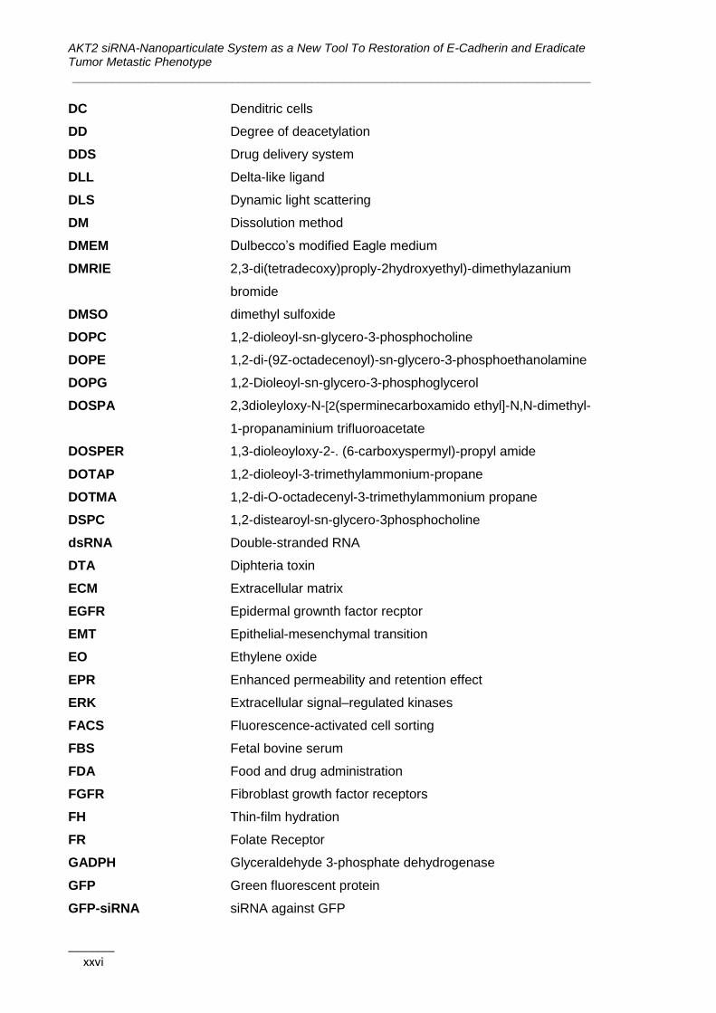
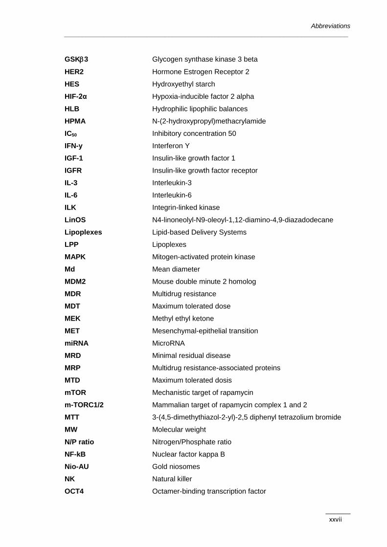
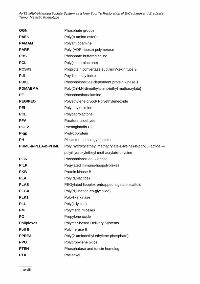
Abbreviations
_______________________________________________________________________________
_______ xxv
Abbreviations
3’UTR Untranslated region
5-DTAF 5-8[4,6-dichlorotriazin-2-yl]amino)fluorescein hydrocloride
ABC ATP-binding cassette
AE Association efficiency
AGO2 Argonaute 2
AKT Protein (serine/threorine) kinase B (PKB)
ALDH1A1 Aldehyde dehydrogenase 1
Alox5 Arachidone 5-lipoxigenase
ATCC American type cell colection
ATP Adenosine triphosphate
BAD Bcl2-associated death promoter
BCA Bicinchoninic acid
Bcl-2 B-cell lymphoma 2
Bcl-xL B-cell lymphoma extra large
BCRP Breast cancer resistance protein
BH3 Bcl-2 Homology 3 domain
bHLH basic helix-loop-helix
BLMH Bleomycin hydrolase
CAMs Cell adhesion molecules
CD Cyclodextrin
CHEMS Cholesteryl hemisuccinate
CMC Critical miccelar concentration
CML Chronic myeloid leukemia
CMT Critical micellar temperature
CRISPR Clustered Regularly Interspaced Short Palindromic Repeats
CryoSEM Cyro-scanning electron microscopy
CS Chitosan
CSC Cancer stem cells
CTAB Cetyltrimethylammonium bromide
CTLA4 CytotoxicT-lymphocyte antigen 4
CXCR4 C-X-C chemokine receptor type 4
DAPI 4’,6-diamidino-2-phenylindole
AKT2 siRNA-Nanoparticulate System as a New Tool To Restoration of E-Cadherin and Eradicate Tumor Metastic Phenotype
______________________________________________________________________________
_______ xxvi
DC Denditric cells
DD Degree of deacetylation
DDS Drug delivery system
DLL Delta-like ligand
DLS Dynamic light scattering
DM Dissolution method
DMEM Dulbecco’s modified Eagle medium
DMRIE 2,3-di(tetradecoxy)proply-2hydroxyethyl)-dimethylazanium
bromide
DMSO dimethyl sulfoxide
DOPC 1,2-dioleoyl-sn-glycero-3-phosphocholine
DOPE 1,2-di-(9Z-octadecenoyl)-sn-glycero-3-phosphoethanolamine
DOPG 1,2-Dioleoyl-sn-glycero-3-phosphoglycerol
DOSPA 2,3dioleyloxy-N-[2(sperminecarboxamido ethyl]-N,N-dimethyl-
1-propanaminium trifluoroacetate
DOSPER 1,3-dioleoyloxy-2-. (6-carboxyspermyl)-propyl amide
DOTAP 1,2-dioleoyl-3-trimethylammonium-propane
DOTMA 1,2-di-O-octadecenyl-3-trimethylammonium propane
DSPC 1,2-distearoyl-sn-glycero-3phosphocholine
dsRNA Double-stranded RNA
DTA Diphteria toxin
ECM Extracellular matrix
EGFR Epidermal grownth factor recptor
EMT Epithelial-mesenchymal transition
EO Ethylene oxide
EPR Enhanced permeability and retention effect
ERK Extracellular signal–regulated kinases
FACS Fluorescence-activated cell sorting
FBS Fetal bovine serum
FDA Food and drug administration
FGFR Fibroblast growth factor receptors
FH Thin-film hydration
FR Folate Receptor
GADPH Glyceraldehyde 3-phosphate dehydrogenase
GFP Green fluorescent protein
GFP-siRNA siRNA against GFP
Abbreviations
_______________________________________________________________________________
_______ xxvii
GSK3 Glycogen synthase kinase 3 beta
HER2 Hormone Estrogen Receptor 2
HES Hydroxyethyl starch
HIF-2α Hypoxia-inducible factor 2 alpha
HLB Hydrophilic lipophilic balances
HPMA N-(2-hydroxypropyl)methacrylamide
IC50 Inhibitory concentration 50
IFN-y Interferon Y
IGF-1 Insulin-like growth factor 1
IGFR Insulin-like growth factor receptor
IL-3 Interleukin-3
IL-6 Interleukin-6
ILK Integrin-linked kinase
LinOS N4-linoneolyl-N9-oleoyl-1,12-diamino-4,9-diazadodecane
Lipoplexes Lipid-based Delivery Systems
LPP Lipoplexes
MAPK Mitogen-activated protein kinase
Md Mean diameter
MDM2 Mouse double minute 2 homolog
MDR Multidrug resistance
MDT Maximum tolerated dose
MEK Methyl ethyl ketone
MET Mesenchymal-epithelial transition
miRNA MicroRNA
MRD Minimal residual disease
MRP Multidrug resistance-associated proteins
MTD Maximum tolerated dosis
mTOR Mechanistic target of rapamycin
m-TORC1/2 Mammalian target of rapamycin complex 1 and 2
MTT 3-(4,5-dimethythiazol-2-yl)-2,5 diphenyl tetrazolium bromide
MW Molecular weight
N/P ratio Nitrogen/Phosphate ratio
NF-kB Nuclear factor kappa B
Nio-AU Gold niosomes
NK Natural killer
OCT4 Octamer-binding transcription factor
AKT2 siRNA-Nanoparticulate System as a New Tool To Restoration of E-Cadherin and Eradicate Tumor Metastic Phenotype
______________________________________________________________________________
_______ xxviii
OGN Phosphate groups
PAEs Poly(b-amino ester)s
PAMAM Polyamidoamine
PARP Poly (ADP-ribose) polymerase
PBS Phosphate buffered saline
PCL Poly(-caprolactone)
PCSK9 Proprotein convertase subtilisin/kexin type 9
Pdi Poydispersity index
PDK1 Phosphoinositide-dependent protein kinase 1
PDMAEMA Poly(2-(N,N-dimethylamino)ethyl methacrylate)
PE Phosphoethanolamine
PEG/PEO Polyethylene glycol/ Polyethyleneoxide
PEI Polyethylenimine
PCL Polycaprolactone
PFA Paraforlmaldehyde
PGE2 Prostaglandin E2
P-gp P-glycoprotein
PH Pleckstrin homology domain
PHML-b-PLLA-b-PHML Poly(hydroxyletheyl methacrylate-L-lysine)-b-poly)L-lactide)—
poly(hydroxyletheyl methacrylate-L-lysine
PI3K Phosphoinositide 3-kinase
PILP Pegylated immuno-lipopolyplexes
PKB Protein kinase B
PLA Poly(d,l-lactide)
PLAS PEGylated lipoplex-entrapped alginate scaffold
PLGA Poly(d,l-lactide-co-glycolide)
PLK1 Polo-like kinase
PLL Poly(L-lysine)
PM Polymeric micelles
PO Propylene oxide
Poliplexes Polymer-based Delivery Systems
Poll II Polymerase II
PPEEA Poly(2-aminoethyl ethylene phosphate)
PPO Polypropylene oxice
PTEN Phosphatase and tensin homolog
PTX Paclitaxel

Abbreviations
_______________________________________________________________________________
_______ xxix
qRT-PCR Quantitative real time polymerase chain reaction
RFP Red Fluorescent Protein
RISC RNA-induced silencing complex
RRM2 Ribonucleoside-diphosphate reductase subunit M2
RTK Receptor tirosine kinase
SCID Severe combined immunodeficiency
SD Mean±standard desviation
SDS-PAGE sodium dodecyl sulfate polyacrylamide gel electrophoresis
Ser Serine
shRNA Short hairpin RNA
SLN Solid lipid nanoparticules
SLUG Zinc finger protein SNAIL2
SNAIL Zinc finger protein SNAIL1
SNALP Stable nucleic acid lipid particles
SPARC Secreted protein acidic and rich in cysteine
STAT3 Signal transducer and actibvator of transcription 3
SV40 Simian vacuolating virus 40
TBE Tris/Borate/EDTA
TEM Transmission electron microscopy
Tf Human transferrin
TfR Transferrin receptors
TGF- Tumor growth factor beta
TGFR Tumor growth factor beta receptor
Thr Threonine
TNF Tumor necrosis factor
TORC2 Target of rapamycin complex 2
TQ Thymoquinone
UV Ultra violet
VEGF Vascular endothelial growth factor
WB Western Blotting
WNT Wingless-related integration site
ZEB Zinc finger E-box-binding homeobox
ZP Zeta potencial

AKT2 siRNA-Nanoparticulate System as a New Tool To Restoration of E-Cadherin and Eradicate Tumor Metastic Phenotype
______________________________________________________________________________
_______ xxx
Aims and Organization of the Thesis
_______________________________________________________________________________
_______ xxxi
Aims and Organization of the Thesis
The high complexity of cancer diseases and their still unknown and incontrollable pattern,
has made prioritary the study and a better understanding of the wide range of biological
pathways involved in their development and progress. Worstly, the heterogeneity of cancer
cell populations within the tumor difficults the complete remission of the disease, being the
cancer recurrence one of the major challenges for the current therapies. The small
subpopulation of CSC within a certain tumor have been reported as the responsible for the
so feared tumor recurrence and resistance to therapy.
This work could be divided in two main research areas and general objectives:
i) Molecular biology of cancer: study and identification of AKT2 as a good target
for cancer therapy;
ii) Pharmaceutical technology and development: design and characterization of a
Nanotechnology-based system for delivery of a siRNA against the AKT2.
Based on the previous, the specific objectives of this work are the follows:
1) Due to its recognized role as a putative oncogene, we aim to understand and
characterize the biological pathway of AKT2 and assess the effects of AKT2
silencing in different breast and colon cancer cell lines in terms of cell migration,
proliferation, and gene expression pattern;
2) Isolate the subpopulations of CSC from breast and colon cancer cells and assess
the effects of AKT2 silencing specifically in this subpolation in terms of cell invasion,
cell anchorage-independent growth, and gene expression pattern;
3) Understand the downstreams effectors of AKT2 for each cell line (TWIST or mTOR);
4) Design a nanoparticle-based system able to complex siRNA and silence the gene
of interest using classic reporter gene assays;
5) Characterize the new nanosystem in terms of their physicochemical features and
stability, in vitro and in vivo toxicity, and cellular internalization;
6) Assess the biological effects of nanoparticles complexing siRNA against the AKT2
in subpopulations of both breast CSC and non-CSC in terms of invasion and cell
anchorage-independent growth abilities.

AKT2 siRNA-Nanoparticulate System as a New Tool To Restoration of E-Cadherin and Eradicate Tumor Metastic Phenotype
______________________________________________________________________________
_______ xxxii
The present thesis is organized in six chapters. In the first two chapters are presented the
main theoretical concepts and the state of art for each of research areas:
Chapter 1 – State of Art – Epithelial-Mesenchymal Transition as the Motor for
Stemness: AKT2 Emerges as a Potential Therapeutic Target
Chapter 2 – State of Art – Nanotechnology for Gene Delivery
After the introductory section is presented the research work developed and the main
results obtained:
Chapter 3 (Formulation Studies and Efficacy Assessment of Different siRNA
Delivery Systems) describes the way paved through the design and development of a
nanoparticulate system based in polymeric micelles (PM) for siRNA delivery until reach the
final selected formulation presented in Chapter 5;
Chapter 4 (AKT2-related Biological Pathway Characterization and Validation in
Breast and Colon Cancer Stem Cells) presents the study and characterization of the
biological pathway and the role of AKT2 in the development and progression of cancer both
in breast and colon CSC and non-CSC;
Chapter 5 (Functional Validation of Amphiphilic-based Polymeric Micelles for siRNA
Delivery and Cancer Stem Cells Genes Inhibition) presents the results obtained with the
chosen PM for the delivery of siAKT2.
In the final chapter (Chapter 6) are presented the Main Conclusions and Future
Perspectives of the work.
Chapter 1 | State of Art – Epithelial-Mesenchymal Transition as the Motor for Stemness: AKT2 Emerges as a Potential Therapeutic Target
_______________________________________________________________________________
_______ 1
CHAPTER 1
State of Art – Epithelial-Mesenchymal Transition as the
Motor for Stemness: AKT2 Emerges as a Potential
Therapeutic Target
The information presented in this chapter was partially published in the following
publications:
1) P Gener, D Rafael, Y Fernández, J Sayos, D Arango, I Abasolo, M Videira, S
Schwartz Jr., Cancer Stem Cells and Personalized Cancer Nanomedicine.
Nanomedicine (Lond), 11(3):307-20, 2016.
2) D Rafael, S Doktorovová, H Florindo, P Gener, I Abasolo, S Schwartz Jr., M Videira,
EMT Blockage Strategies: Targeting Akt Dependent Mechanisms for Breast Cancer
Metastatic Behaviour Modulation, Current Gene Therapy, 15(3) 2015.
AKT2 siRNA-Nanoparticulate System as a New Tool to Restoration of E-Cadherin and Eradicate Tumor Metastic Phenotype ______________________________________________________________________________
_______ 2
Table of contents
1.1. Cancer Facts ......................................................................................................3
1.1.1. Cancer Scenario ...........................................................................................4
1.1.2. Cancer Treatment .........................................................................................5
1.2. The Cancer Stem Cells Theory .........................................................................6
1.2.1. Cancer Stem Cell Models .............................................................................6
1.2.2. Cancer Stem Cell Properties.........................................................................9
1.2.3. Targeting CSC ..............................................................................................9
1.3. Epithelial-Mesenchymal Transition ................................................................ 12
1.3.1. Adhesion-related Proteins: E-cadherin ........................................................ 13
1.3.2. Tumor-associated Proteins in EMT Activation ............................................. 16
1.3.2.1. The influence of tumor microenvironment ................................................... 16
1.3.2.2. Players at the cell membrane level ............................................................. 18
1.3.2.3. Intracellular transcriptional regulation of EMT ............................................. 19
1.4. The Importance of TWIST-based Signaling Pathways in the EMT
Program……… ............................................................................................................... 19
1.4.1. TWIST Mechanism of Action: AKT2 and PI3K as the Leading Players ....... 21
1.5. RNA Interference Regulation of EMT.............................................................. 24
1.5.1. miRNA and Cancer ..................................................................................... 27
1.5.2. siRNA-mediated Silencing of AKT Isoforms ................................................ 27
1.6. Conclusions ..................................................................................................... 29
1.7. References ....................................................................................................... 30

Chapter 1 | State of Art – Epithelial-Mesenchymal Transition as the Motor for Stemness: AKT2 Emerges as a Potential Therapeutic Target
_______________________________________________________________________________
_______ 3
1.1. Cancer Facts
According to World Health Organization (WHO), cancer is a generic term used to describe
a large and heterogeneous group of diseases that can affect any part of the body. A key
feature is the genesis of abnormal cells that rapidly grow beyond their normal boundaries
invading adjacent parts of the body and spreading to other organs (metastasis) (Figure
1.1). Metastization is of major impact in the malignancy of the disease and clinical outcome
(1). Due to its high diversity, complexity, and unpredictable and uncontrollable character,
cancer has become one of the most feared diseases by humans in the last decades. With
the arise of molecular biology and biotechnology, tremendous advances have been
observed in the last decades in the oncology field leading to the identification of biological
pathways involved in cell growth and dissemination, as well as disease players and
therapeutic targets/agents (Figure 1.1).
Figure 1.1. The complexity of cancer. The figure represents different phases of the disease
evolution and the problematic of cancer mutations that increase exponentially along with the tumor
development originating a highly heterogeneous compartment. Circles point out the main cancer
hallmarks as well as the associated most common therapeutic strategies. BH3: Bcl-2 homology 3
domain; CTLA4: cytotoxic T-lymphocyte antigen 4; EGFR: epidermal growth factor receptor; PARP:
poly (ADP-ribose) polymerase; VEGF: Vascular endothelial growth factor.
AKT2 siRNA-Nanoparticulate System as a New Tool to Restoration of E-Cadherin and Eradicate Tumor Metastic Phenotype ______________________________________________________________________________
_______ 4
1.1.1. Cancer Scenario
The progresses referred before enables researchers and pharmaceutical industry with the
information necessary to develop a variety of drugs to treat the different types of cancer,
resulting in an important decrease of the cancer related deaths (superior to 20% since
1990s) and an increase in the patient’s overall survival (2, 3). The incidence and mortality
rates of this disease are superior in developed countries and are expected to increase in
the next years, with an estimated 9 million cancer deaths in 2015 and 11.4 million in 2030
(3). Just in the USA, 1.7 million new cancer cases and 0.6 million cancer deaths are
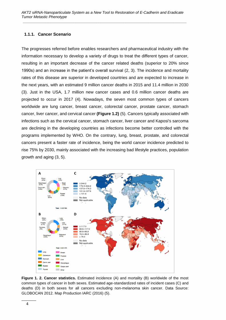
projected to occur in 2017 (4). Nowadays, the seven most common types of cancers
worldwide are lung cancer, breast cancer, colorectal cancer, prostate cancer, stomach
cancer, liver cancer, and cervical cancer (Figure 1.2) (5). Cancers typically associated with
infections such as the cervical cancer, stomach cancer, liver cancer and Kaposi's sarcoma
are declining in the developing countries as infections become better controlled with the
programs implemented by WHO. On the contrary, lung, breast, prostate, and colorectal
cancers present a faster rate of incidence, being the world cancer incidence predicted to
rise 75% by 2030, mainly associated with the increasing bad lifestyle practices, population
growth and aging (3, 5).
Figure 1. 2. Cancer statistics. Estimated incidence (A) and mortality (B) worldwide of the most
common types of cancer in both sexes. Estimated age-standardized rates of incident cases (C) and
deaths (D) in both sexes for all cancers excluding non-melanoma skin cancer. Data Source:
GLOBOCAN 2012. Map Production IARC (2016) (5).
Chapter 1 | State of Art – Epithelial-Mesenchymal Transition as the Motor for Stemness: AKT2 Emerges as a Potential Therapeutic Target
_______________________________________________________________________________
_______ 5
In the last years, due to the growing incidence of the disease, the global market share of
medicines to treat cancer has been growing. In 2014 oncology remained as the largest
therapeutic area worldwide regarding the market share, presenting an increase of 8%
compared to 2013 and counting for more than 10% of the total year sales (6). As a
consequence of the expected growth in the prevalence and incidence of oncological
diseases in the upcoming years, it is also predicted an increase in the prescription of
anticancer drugs, with an escalation of their global market share up to almost 15% in 2020
(6). This disease profile entails high costs to the health care systems related to the patients’
cancer treatment and care. For example, only in USA, cancer care is projected to cost
around 174 billion US dollars in 2020 (7). This scenario boosts researchers, health care
agencies, and pharmaceutical companies to pursuit the urgent need for the development of
new and more cost-effective treatments.
1.1.2. Cancer Treatment
Despite the enormous efforts employed at different fields of science and medicine, the real
cure and eradication of cancer remains a desire far to be achieved and stands as a huge
challenge to researchers around the world.
Major challenges surrounding the administration of current anticancer treatments are their
systemic toxicity, associated with serious side-effects owing this drugs lack of specificity,
and the therapeutic resistance that cancer cells often acquire during treatment. Indeed,
because of the use of chemotherapeutics at an early stage of the disease that renders tumor
resistance, treatment options for late metastatic disease are often reduced (8). The ability
of cancer cells to activate alternative molecular pathways allowing them to escape from the
common drug therapeutic mechanism of action is one of the major mechanisms involved in
the development of drug resistance (9, 10). Further, drug resistance also occurs through
the increased expression of ATP-dependent drug efflux transporters on the surface of
cancer cells (11). This causes a significant decrease of intracellular drug accumulation,
which results into severe limitation of drug’s efficacy (12). Besides, cells with different
phenotypes and proliferative abilities co-exist within a tumor, generating tumor
heterogeneity and contributing to specific selection of resistant clones and disappointing
therapeutic responses, in particular when a single drug treatment is applied.
Special interest has been paid to the use of biopharmaceuticals and gene therapy as
therapeutic strategies to treat cancer owing to its capacity to target the specific antigen or
signaling pathway involved in cancer progression (13). Among the different types of gene
therapy, the silencing of particular genes through the RNA interference (RNAi) therapy has
AKT2 siRNA-Nanoparticulate System as a New Tool to Restoration of E-Cadherin and Eradicate Tumor Metastic Phenotype ______________________________________________________________________________
_______ 6
been intensively investigated as a encouraging strategy in the field of oncology, given the
possibility of targeting oncogenes involved in proliferation, survival, angiogenesis,
metastasis, apoptosis suppression or drug-resistance (14).
Nowadays, more than 800 medicines and vaccines to treat cancer are under development
and clinical assessment (2), with more than 73% of the medicines in the pipeline being
studied based on biomarkers, presenting potential for personalized treatment (15). Some
of the medicines under development are based on biopharmaceuticals, namely monoclonal
antibodies, vaccines, cell or gene therapy. In 2013, the majority of biopharmaceuticals
under development are intended to treat cancer and related diseases (more than 300),
being the monoclonal antibodies the most studied ones with 170 medicines (16). Also,
seventeen products under development/clinical trials were based on gene therapy such as
ALN-VSP from Alnylam Pharmaceuticals (Phase I), B7-2/GM-CSF from NuVax
Therapeutics (Phase I), BC-819 from BioCancell Therapeutics (Phase II), CALAA-01 from
Calando Pharmaceuticals (Phase I), EGEN-001 from EGEN (Phase II) or GliAtak™ from
Advantagene (Phase II) (16).
Unfortunately, even though important clinical breakthroughs in the fight against cancer have
been achieved by using combined protocols such as conventional chemotherapy with
hormonal therapy or therapeutic antibodies, recurrence and metastasis are still observed.
Thus, the development of treatments able to reduce the formation of metastasis and to
avoid or at least reduce the recurrence of the disease is required.
1.2. The Cancer Stem Cells Theory
As referred previously, despite progresses in cancer management, advanced cancers have
still low clinical response rates and high recurrence and mortality rates. Regarding this, a
main issue that should be considered is the fact that cancer cells within a tumor are not
equal in terms of characteristics and tumorigenic potential.
1.2.1. Cancer Stem Cell Models
Before the 1990s, cancer initiation and progression was explained by a clonal cancer model.
It was considered that all tumor cells have similar characteristics and equal tumor formation
capacity and that tumors expansion depended on clonal selection advantages (17-20). The
first evidence regarding the existence of cancer stem cells (CSC) was obtained in acute
myeloid leukemia (21). Subsequently, CSC were identified also in other hematopoietic

Chapter 1 | State of Art – Epithelial-Mesenchymal Transition as the Motor for Stemness: AKT2 Emerges as a Potential Therapeutic Target
_______________________________________________________________________________
_______ 7
cancers and in many solid tumors (breast, brain, colon, prostate, lung, head & neck, among
others) (22-24). It was also shown that the small CSC sub-population has the ability to
generate and maintain the tumor progression (20-22, 25). Based on these observations, a
new hierarchical model describing cancer propagation was postulated (21); CSC can self-
renew their own population and have long-term propagating capacity contrariwise to normal
cells whose division finish by clonal exhaustion (Figure 1.3A) (26, 27).
Figure 1. 3. CSC therapy resistance accordingly the different models. (A) CSC are more
resistant than tumor bulk cells to most conventional therapeutic interventions. Therefore, after
treatment, CSC remain in the tumors or in circulation inducing a rapid recurrence. (B) As for the
hierarchic model, CSC specific therapy results in loss of proliferative capacity and decline of the
malignancy with tumor regression. (C) Considering the novel dynamic model, the non-CSC can
acquire CSC-features through signals from the environment and the stromal cells, thus even with
specific CSC eradication a rapid restoration of CSC features occurs, with a subsequent recurrence
when therapy is discontinued. (D) The best strategy to tumor remission should target not only the
CSC but also the bulk tumor cells as well as the interconversion capacity between them. Stromal
cells represent myofibroblasts, endothelial cells, mesenchymal stem cells, or infiltrating immune
cells. CSC: cancer stem cells.
In summary, CSC have been considered the major critical player in tumor malignancy and
recurrence, thus strong efforts have been recently done to specifically target CSC
subpopulation in order to improve therapy efficacy and avoid recurring tumors. However,

AKT2 siRNA-Nanoparticulate System as a New Tool to Restoration of E-Cadherin and Eradicate Tumor Metastic Phenotype ______________________________________________________________________________
_______ 8
recent data suggest that targeting exclusively the CSC sub-populations may not be
sufficient because acquisition of stemeness phenotype by other cells within tumors seems
to be a bidirectional dynamic process (Figure 1.3B) (26, 27). Because the hierarchical
model of cancer cannot explain the dynamic behavior of CSC, a new interconversion cancer
model has been very recently postulated (Figure 1.3C) (26-28). According to the model,
the amount of CSC within a tumor or cancer cell line seems to be inconstant and finely
tuned, to maintain a specific equilibrium between CSC and non-CSC populations. In one
hand, CSC can differentiate to non-CSC which in turn can de-differentiate and revert into
cells with CSC properties, like resistance and self-renewal capacity with typical stemness
gene expression signature. This cell de-differentiation process was first predicted by Marcov
mathematical model (28) and recently confirmed experimentally in various CSC models (28-
30). In melanoma, a subpopulation of slow-cycling cells that express histone demethylase
JARD1B was identified. As expected from CSC, during propagation of purified JARD1B
positive cells appeared a JARD1B negative, non-CSC population. Conversely, a single
JARD1B negative cell originates a heterogeneous progeny including JARD1B positive cells,
suggesting a bidirectional dynamic of the CSC phenotype (31). Similarly, normal somatic
basal-like mammary epithelial cells are able to spontaneously de-differentiate into stem-like
cells (32). Further, regeneration of stem-like cells is observed as well in vivo after sorting,
suggesting that stable equilibrium of CSC/non-CSC cells within a tumor occurs due to cell-
state interconversion (28). These findings reinforce the hypothesis of the existence of a
controlled balance between CSC and non-CSC populations. It seems that cancer cells
might survive to stress conditions by entering de-differentiation as survival mechanism.
Several external factors and paracrine communications as well as cell-to-cell interactions
are involved in the regulation of this process. As an example, it has been shown that
apoptotic cells excrete prostaglandin E2 (PGE2) which promotes proliferation of
neighbouring CSC after chemotherapy, whereas PGE2-neutralizing antibodies abrogate
CSC repopulation after treatment (33). Similarly, the matrix cellular protein SPARC
(secreted protein acidic and rich in cysteine, also known as osteonectin or BM-40) secreted
by non-CSC is able to modulates the metastatic capacity of CSC in prostate cancer models
(34), and Interleukin-6 (IL-6) secreted by non-CSC is reported to induce formation of breast
CSC in association with octamer-binding transcription factor 4 (OCT4) expression (35).
Furthermore, OCT4 overexpression is induced via IL-6-JAK1-STAT3 signaling pathway in
low attachment conditions as well as in vivo to maintain dynamic equilibrium (36).
Therefore, the dynamic phenotype of CSC represents an important challenge for targeted
cancer therapies, as tumor cell populations are continuously evolving and therapeutic
eradication of existing CSC populations might be followed by their regeneration from non-

Chapter 1 | State of Art – Epithelial-Mesenchymal Transition as the Motor for Stemness: AKT2 Emerges as a Potential Therapeutic Target
_______________________________________________________________________________
_______ 9
CSC. Regarding this, the major goal is to find a way that allows to targeting the CSC but
also stopping the interconversion between CSC and non-CSC (Figure 1.3D).
1.2.2. Cancer Stem Cell Properties
Even though the intratumoral amount of CSC as well as the expression of stemness
markers among this sub-population differs, depending mostly on tumor types, the essential
stemness properties of CSC like self-renewal, tumor initiation capacity and long-term
repopulation potential are common features independently of tumor type. Owing their ability
to survive in non-attachment conditions showing capacity to grow as tridimensional
tumorspheres, CSC have increased capacity to initiate tumor growth in vivo, migrate and
intravasate the blood stream generating distant metastasis at specific sites. CSC are
substantially insensitive to most conventional anticancer therapies, antimitotic agents,
and/or radiation (37). Such aggressive behavior results from a particular CSC phenotype
as well as their characteristic gene expression and epigenetic modifications. In particular,
CSC have a potent machinery of anti-apoptotic proteins and a efficient activation of the DNA
damage sensor and repair machinery for maintenance of genome integrity (9, 37).
Moreover, CSC overexpress a great number of detoxifying enzymes (aldehyde
dehydrogenase 1 (ALDH1A1), bleomycin hydrolase (BLMH)), drug efflux transporters (P-
glycoprotein (P-gp)), breast cancer resistance protein (BCRP), and the multidrug
resistance-associated proteins (MRP) at the cell membrane, which pump-out
chemotherapeutic drugs from the intracellular space (10, 38). In addition, induction of
quiescence in stress conditions and higher stability under hypoxic conditions provide CSC
with further protection against anticancer therapies (10). Accordingly, the percentage of
CSC within a tumor often increases after treatment (33, 37, 39, 40). This often leads to
cancer recurrence and metastatic growth since only few CSC are necessary and sufficient
for tumor regeneration in vivo (41). For all these reasons, if cancer has to be eradicated,
new therapies should specifically target the CSC fraction.
1.2.3. Targeting CSC
In agreement with the interconversion cancer model, traditional cancer treatments appear
to be effective at reducing tumor mass but often fail to produce desired long-term outcomes,
and remission of metastatic disease is common. Moreover, current treatments often render

AKT2 siRNA-Nanoparticulate System as a New Tool to Restoration of E-Cadherin and Eradicate Tumor Metastic Phenotype ______________________________________________________________________________
_______ 10
the tumor resistant at the time of remission, possibly due to their inability to eliminate CSC
(8, 37).
Therapeutic targeting of CSC may have the potential to remove residual disease and
become an important component of any given cancer treatment. Currently, there are
multiple anti-CSC agents in pre-clinical and clinical trials (Table 1.1) (42). The likelihood of
their clinical success will depend on many aspects, including safety, specificity,
pharmacokinetics etc., but also on their ability to effectively target CSC. In fact, most drugs
specifically designed to target CSC are based on the effective inhibition of crucial CSC
signaling pathways as example: i) cell survival and proliferation pathways; including PI3K-
AKT, JAK/STAT, and NF-kB signaling, and ii) signaling linked to the stemness properties of
CSC like self-renewal and pluripotency (Notch pathway, WNT pathway and Hedgehog
signaling). Of note, one of the risks associated to drugs targeting commonly shared cellular
pathways is the difficulty to distinguish normal cells and/or stem cells from CSC. High
throughput screening methodologies are reliable approaches to reveal new therapeutic
targets that effectively eliminate CSC without compromise the normal somatic stem cells.
One of the hopeful targets identified by high throughput screening is Arachidone 5-
lipoxigenase (Alox5). Alox5 was found over-expressed in chronic myeloid leukemia CSC
but not in normal hematopoietic stem cells (43). It was shown that the absence of Alox5 due
to genetic modification or its inhibition with a specific inhibitor, Zileuton, impaired the
induction of leukemia in the BCR-ABL inducible cancer model of chronic myeloid leukemia
(CML) (43). The inhibition of Alox5 leads to an upregulation of the tumor suppressor gene
Msr1 and consequently, to the inhibition of PI3K-AKT and β-catenin pathways (44).
Currently, the use of Zileuton in combination with Imatinib is in phase I clinical trials for first
line therapy in acute myeloid leukemia. Of note, Imatinib alone does not cure CML because
it is not able to kill leukemia stem cells (45) . Another example of a successful high through
screening is the chemical screen method for agents with CSC-specific toxicity. As a result,
salinomycin was identified as a drug showing selective toxicity for CSC (46).
Previous results from preclinical trials in human xenograft mice models and clinical pilot
studies reveal that salinomycin is able to effectively eliminate CSC and to induce partial
clinical regression of heavily pre-treated and therapy-resistant cancers, particularly in
combination with novel tumor-targeted drugs (47). However, a very important drawback for
the potential clinical application of Salinomycin is its marked neural and muscular toxicity
(48). In this context, incorporation of cytotoxic drugs within different particulate nanocarrier
systems, i.e. liposomes and polymeric particles, is a useful approach to modify the
biodistribution of drugs and target tumors. Nanomedicines can in fact address not only the
biodistribution profile of CSC-targeting drugs but also reduce their overall toxicity and

Chapter 1 | State of Art – Epithelial-Mesenchymal Transition as the Motor for Stemness: AKT2 Emerges as a Potential Therapeutic Target
_______________________________________________________________________________
_______ 11
enhance their therapeutic efficacy by promoting higher local drug exposure and improving
their transport properties across biological membranes (49).
Table 1. 1. Drugs under clinical development with therapeutic activity against CSC*
Therapeutic Target
Drug Cancer Type Clinical phase Trial reference
PI3K, mTORC1/2
VS-5584 Solid tumors I NCT01991938
AKT MK2206 Breast cancer II NCT01277757
GSK2141795 Breast cancer II NCT01964924 AZD5363 Breast cancer I/II NCT01625286
STAT3 OPB-31121 Solid tumors I NCT00955812
NF-κB Parthenolid
analog Leukemia II/III ISRTN40571019
Multiple kinases
BBI503 Solid tumors I/II NCT01781455
STAT3, β-catenine,
Nanog
BBI608
Gastric cancer III NCT02178956 Esophageal
cancer III NCT02178956
Colon cancer III NCT01830621
BBI503+ BBI608 Solid tumors I NCT02432326
WNT pathway Vantictumab Solid tumors I NCT0 1345201
Ipafricept Pancreatic
cancer I NCT02050178
Notch 2,3 (receptor)
Tarextumab Pancreatic
cancer II NCT01647828
Lung cancer II NCT01859741
DLL (Notch ligand)
Demcizumab
Ovarian cancer II NCT01952249 Pancreatic
cancer II NCT02289898
Lung cancer II NCT02259582
Hedgehog GDC-449 Solid tumors II NCT00739661
ABC transporters
MS-209 Ovarian cancer II NCT00739661
Tariquidar
Ovarian cancer II NCT00069160 Cervical cancer II NCT00069160
Lung cancer II NCT00069160 Kidney cancer II NCT00069160
Chemokine receptors 1,2
Reparixin Breast cancer II NCT01861054
IL-3 receptor SL-401 Leukemia I/II NCT02268253
Alox5 Zileuton Leukemia I NCT02047149
*Details including dosage of the chemical agents under trial, duration of the trial, inclusion and
exclusion criteria for recruiting patients, contact and locations where the trial is being conducted, and
the current status of the trial can be obtained by searching the ‘Trial reference’ in the US National
Institutes of Health Registry (www.clinicaltrials.gov) and EU Clinical Trials Register
(www.clinicaltrialsregister.eu). ABC: ATP-binding cassette; AKT: protein (serine/threonine) kinase B
(PKB); Alox5: arachidone 5-lipoxigenase; DLL: delta-like ligand; IL-3: interleukin-3; mTORC1/2:
mammalian target of rapamycin complex 1 and 2; NF-κB: nuclear factor kappa B; PI3K:
phosphoinositide 3-kinase; STAT3: signal transducer and activator of transcription 3; WNT: wingless-
related integration site.

AKT2 siRNA-Nanoparticulate System as a New Tool to Restoration of E-Cadherin and Eradicate Tumor Metastic Phenotype ______________________________________________________________________________
_______ 12
Figure 1. 4. Schematic representation of the EMT process as well as some of the most
representative affected proteins. EMT: epithelial-mesenchymal transition.
1.3. Epithelial-Mesenchymal Transition (EMT)
As referred previously, tumor invasion and subsequent metastization are the major causes
of morbidity and mortality in patients with cancer (50). Most of malignant tumors are
carcinomas originated from various epithelial tissues. EMT is the biological process that
triggers the evolution from a well-differentiated adenocarcinoma to an invasive epithelial
carcinoma (Figure 1.4) (51). It is defined as a phenomenon during which cells lose their
characteristic epithelial traits: columnar or polygonal shape, apico-basolateral polarization,
organization in cell layers with strong cell-cell adhesion and limited migratory potential.
During this event, cells acquire mesenchymal features such as, spindle-shaped
morphology, anterior-posterior polarization, focal cell-cell contacts and strong migratory
potential, becoming able to detach from their original site, penetrate the bloodstream, and
subsequently create metastasis at different sites. These morphological and functional
changes, demands several intracellular molecular alterations such as the loss of epithelial
markers including E-cadherin, certain cytokeratins, occludin, and claudin, while
mesenchymal markers, namely N-cadherin, vimentin and fibronectin are upregulated (52,
53).
EMT is an essential process for appropriate embryonic development. In adults, EMT occurs
during wound healing, tissue regeneration, organ fibrosis, and cancer progression (8).
Based on the biological context in which EMT occur, this mechanism can be classified into
three different subtypes presented in Table 1.2 (53). During cancer progression, the genetic
and epigenetic changes that mainly affect oncogenes and tumor suppressor genes of
epithelial carcinoma cells seem to be responsible for the activation of an EMT program (54,
55). Thus, preventing epithelial carcinoma cells to undergo such transition in order to keep
the tumor in its primary stage and, therefore, treatable should be a future goal of therapeutic
intervention.
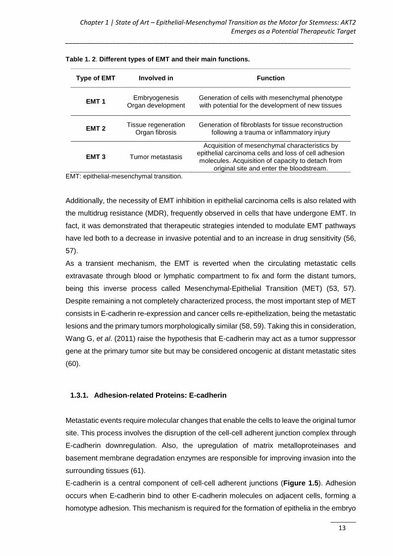
Chapter 1 | State of Art – Epithelial-Mesenchymal Transition as the Motor for Stemness: AKT2 Emerges as a Potential Therapeutic Target
_______________________________________________________________________________
_______ 13
Table 1. 2. Different types of EMT and their main functions.
EMT: epithelial-mesenchymal transition.
Additionally, the necessity of EMT inhibition in epithelial carcinoma cells is also related with
the multidrug resistance (MDR), frequently observed in cells that have undergone EMT. In
fact, it was demonstrated that therapeutic strategies intended to modulate EMT pathways
have led both to a decrease in invasive potential and to an increase in drug sensitivity (56,
57).
As a transient mechanism, the EMT is reverted when the circulating metastatic cells
extravasate through blood or lymphatic compartment to fix and form the distant tumors,
being this inverse process called Mesenchymal-Epithelial Transition (MET) (53, 57).
Despite remaining a not completely characterized process, the most important step of MET
consists in E-cadherin re-expression and cancer cells re-epithelization, being the metastatic
lesions and the primary tumors morphologically similar (58, 59). Taking this in consideration,
Wang G, et al. (2011) raise the hypothesis that E-cadherin may act as a tumor suppressor
gene at the primary tumor site but may be considered oncogenic at distant metastatic sites
(60).
1.3.1. Adhesion-related Proteins: E-cadherin
Metastatic events require molecular changes that enable the cells to leave the original tumor
site. This process involves the disruption of the cell-cell adherent junction complex through
E-cadherin downregulation. Also, the upregulation of matrix metalloproteinases and
basement membrane degradation enzymes are responsible for improving invasion into the
surrounding tissues (61).
E-cadherin is a central component of cell-cell adherent junctions (Figure 1.5). Adhesion
occurs when E-cadherin bind to other E-cadherin molecules on adjacent cells, forming a
homotype adhesion. This mechanism is required for the formation of epithelia in the embryo
Type of EMT Involved in Function
EMT 1 Embryogenesis
Organ development Generation of cells with mesenchymal phenotype with potential for the development of new tissues
EMT 2 Tissue regeneration
Organ fibrosis Generation of fibroblasts for tissue reconstruction
following a trauma or inflammatory injury
EMT 3 Tumor metastasis
Acquisition of mesenchymal characteristics by epithelial carcinoma cells and loss of cell adhesion molecules. Acquisition of capacity to detach from
original site and enter the bloodstream.

AKT2 siRNA-Nanoparticulate System as a New Tool to Restoration of E-Cadherin and Eradicate Tumor Metastic Phenotype ______________________________________________________________________________
_______ 14
and for the maintenance of the epithelial homeostasis in adults. It is a 120 kDa glycoprotein
with a large extracellular domain comprising five cadherin-motif subdomains, a single
transmembrane domain and a short conserved cytoplasmic domain interacting with several
proteins collectively termed catenins. As calcium is essential to mediate adhesive function
of E-cadherin, its extracellular portion contains several calcium-binding sites. For
intracellular adhesion, cytoplasmic portion of E-cadherin bind tightly to β-catenin, a
cytoplasmic protein that is non-covalently linked to α-catenin, which in turn binds to the actin
cytoskeleton, either directly or indirectly via actin-binding proteins as alpha-actinin and
vinculin (62, 63).
Figure 1. 5. Schematic representation of E-cadherin. E-cadherin being part of the cell-cell
adherent junctions, is a crucial player in the cell adhesion and in the maintenance of the epithelial
phenotype.
The maximum homotypic activity of E-cadherin results from it linkage to cytoskeleton.
Therefore, the reduction of E-cadherin expression promotes a sequence of signaling events
and major cytoskeletal reorganization. This reduction of E-cadherin expression in human
tumors may be caused by several mechanisms such as i) proteolytic cleavage of the
extracellular domain; ii) somatic mutations; iii) homozygous chromosomal deletions; iv)
extracellular glycosylation that suppresses hemophilic adhesion; and v) silencing of the
CDH1 promoter by DNA hypermethylation or through transcriptional repression (50, 64, 65).
E-cadherin gene, CDH-1, located on chromosome 16q221, is a member of a large family of
genes coding for calcium-dependent cell adhesion molecules (CAMs). It is a conserved
gene, which, apart from its role in normal cells, can play a major role in malignant cell
transformation and tumor progression (62, 64).
The downregulation of E-cadherin expression, and the consequent dysfunction in cell-cell
adhesion, is a major hallmark of the EMT process and is the main molecular alteration
correlated with the invasive and metastatic process in multiple types of cancer, being then
Chapter 1 | State of Art – Epithelial-Mesenchymal Transition as the Motor for Stemness: AKT2 Emerges as a Potential Therapeutic Target
_______________________________________________________________________________
_______ 15
considered a critical tumor suppressor protein (52, 66). Thus, many studies have focused
on understanding the interaction between loss of E-cadherin expression and the evolution
to an invasive epithelial carcinoma (62, 67).
In fact, many experiments in cancer cell cultures or in vivo tumor models have shown that
the promotion of E-cadherin expression can revert the ability of several types of carcinomas
to invade and metastasize and that, on the contrary, the abrogation of E-cadherin-mediated
cell adhesion in noninvasive tumor cells activates their invasiveness and metastatic powers.
These studies underline the importance of E-cadherin in the inhibition of tumor invasion and
metastasis (58, 68).
E-cadherin loss promotes metastasis not only by enabling cellular disaggregation, but also
by inducing multiple downstream transcriptional pathways and a wide range of functional
changes in human breast epithelial cells. Onder T, et al. (2008) used two different methods
to inhibit E-cadherin function: a) a short hairpin RNA (shRNA)-mediated knockdown of E-
cadherin that lead to a reduction of over 90% in the protein levels, and b) the production of
a truncated form of E-cadherin lacking the protein ectodomain but preserving the
cytoplasmatic tail. They observed that the presence of truncated proteins do not result in
EMT, once the maintenance of the cytoplasmatic tail of E-cadherin avoid the EMT induction.
On the other hand, the significant decrease in E-cadherin expression triggers the EMT
process and the acquisition of metastatic ability (66).
Downregulation of E-cadherin is accompanied by upregulation of N-cadherin and its
placement on cellular membrane. A “cadherin switching” i.e., reduction of E-cadherin
leading to an increased N-cadherin expression (EN-switch), is one of the most important
changes involved in the EMT pathways. Both isoforms are used as determinant marker
proteins to define the epithelial or mesenchymal state of a cell. Additional support for the
relation between tumor malignancy and the ‘‘cadherin switch’’ comes from clinical studies
performed by Gravdal K, et al. (2007) (69). The study concluded that the occurrence of EN-
switch is a more reliable parameter in evaluation of patient prognosis than presence of E-
cadherin or N-cadherin separately. The study stressed out the importance of a dependent
relationship between the patient prognosis and EN-switch. They also conclude that a
‘‘cadherin switch’’ had a prognostic effect on time to both biochemical failure and clinical
recurrence in multivariate survival analyses, stronger than for each protein separately (69).

AKT2 siRNA-Nanoparticulate System as a New Tool to Restoration of E-Cadherin and Eradicate Tumor Metastic Phenotype ______________________________________________________________________________
_______ 16
1.3.2. Tumor-associated Proteins in EMT Activation
1.3.2.1. The influence of tumor microenvironment
The existence of an extracellular matrix in the highly unorganized tumor epithelium brings
together cells and secreted proteins to this environment. The SPARC is one of those
proteins that belong to the matrix cellular family and appears to be closely related with the
E-cadherin expression. It is a nonstructural component of extracellular matrix (ECM) that
regulates interactions with several ECM components, modulating the activity of several
cytokines and growth factors, promoting alterations in cells adhesion and proliferation
capacities (70). Structurally, the SPARC-related proteins subfamily presents three main
components: i) an acidic N-terminal domain with low calcium-binding affinity, ii) a disulfide-
bonded, copper-binding follistatin domain, with homology to the TGF-β inhibitors and iii)
extracellular C-terminal domain with calcium-binding affinity (70). Robert G, et al. (2006)
reported for the first time the interaction between SPARC and E-cadherin, showing that the
overexpression of SPARC in normal human melanocytes give them a fibroblast-like
morphology related with the E-cadherin downregulation via the induction of Snail, an E-
cadherin transcriptional repressor (71). Conversely, SPARC depletion reduced the levels of
Snail promoting the E-cadherin upregulation (71).
The relationship between E-cadherin and SPARC can be understood based on the recently
described mechanism of integrin-linked kinase (ILK) stimulation. Due to their localization,
integrin receptors seem to be the link between SPARC (extracellulary located at points of
integrin-ECM interaction) and ILK (located at the plasma membrane). Subsequently, the
phosphorylation of several effector proteins by the ILK, including AKT and integrins-β1/β3,
modifies cell behavior in terms of adhesion, invasion and metastization ability (72-74).
Nevertheless, SPARC tumorigenicity seems to be dependent on the cell type and the tumor
environment. In fact, its overexpression is associated with metastization and invasiveness
in melanoma, glioma, breast, prostate and colorectal carcinomas, while in the case of
ovarian carcinoma, neuroblastoma and pancreatic adenocarcinoma its expression is
inversely related with the tumor development (70, 71, 75).
Still at the extracellular level, the early observations also suggested that tumor cells bearing
a mesenchymal phenotype massively secrete active TGF-β, which triggers EMT and
enhances angiogenesis in the tumor microenvironment. In early tumor development, TGF-
β has an inhibitory effect on tumor growth, acting as a tumor suppressor. Conversely, at
later stages, it acts as an immunosuppressive cytokine, favoring tumor evasion, by
converting CD4+T cells in immunosuppressive T regulatory cells and inhibiting the effector
T cells functions (76). Tumor cells autocrine TGF-β production aims to circumvent tumor
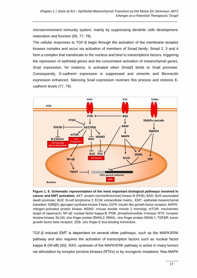
Chapter 1 | State of Art – Epithelial-Mesenchymal Transition as the Motor for Stemness: AKT2 Emerges as a Potential Therapeutic Target
_______________________________________________________________________________
_______ 17
microenvironment immunity system, mainly by suppressing dendritic cells development,
maturation and function (65, 77, 78).
The cellular responses to TGF-β begin through the activation of the membrane receptor
kinases complex and occur via activation of members of Smad family. Smad 2, 3 and 4
form a complex that translocate to the nucleus and bind to transcriptions factors, triggering
the repression of epithelial genes and the concomitant activation of mesenchymal genes.
Snail expression, for instance, is activated when Smad3 binds to Snail promoter.
Consequently, E-cadherin expression is suppressed and vimentin and fibronectin
expression enhanced. Silencing Snail expression reverses this process and restores E-
cadherin levels (77, 79).
Figure 1. 6. Schematic representation of the most important biological pathways involved in
cancer and EMT activation. AKT: protein (serine/threonine) kinase B (PKB); BAD: Bcl2-associated
death promoter; Bcl2: B-cell lymphoma 2; ECM: extracellular matrix; EMT: epithelial-mesenchymal
transition; GSKβ3: glycogen synthase kinase 3 beta; IGFR: insulin-like growth factor receptor; MAPK:
mitogen-activated protein kinase; MDM2: mouse double minute 2 homolog; mTOR: mechanistic
target of rapamycin; NF-κβ: nuclear factor kappa B; PI3K: phosphoinositide 3-kinase; RTK: receptor
tirosine kinase; SLUG: zinc finger protein SNAIL2; SNAIL: zinc finger protein SNAIL1; TGFβR: tumor
growth factor beta receptor; ZEB: zinc finger E-box-binding homeobox.
TGF-β induced EMT is dependent on several other pathways, such as the MAPK/ERK
pathway and also requires the activation of transcription factors such as nuclear factor
kappa B (Nf-κB) (65). RAS, upstream of the MAPK/ERK pathway is active in many tumors
via stimulation by receptor tyrosine kinases (RTKs) or by oncogenic mutations. Ras-MAPK

AKT2 siRNA-Nanoparticulate System as a New Tool to Restoration of E-Cadherin and Eradicate Tumor Metastic Phenotype ______________________________________________________________________________
_______ 18
pathway induces the expression of important EMT-related transcription factors, such as
Snail and Slug. Besides MAPK pathway, Ras also activates other important downstream
pathways such as the Phosphatidyl-inositol-3’-kinase (PI3K) signaling pathway, explained
later on in this chapter (65, 78, 80). The Nf-κB factor has been associated with
tumorigenesis due to its anti-apoptotic effect in several cancer cells. Nf-κB activation,
commonly observed in metastatic breast cancer cells, occurs in response to inflammatory
cytokines and growth factors. It has shown to be a fundamental regulator of EMT and
metastasis in a model of breast cancer progression, once Nf-κB hyperactivation seems to
be related with the increased expression of TWIST in these cells (65, 78, 81).
The referred pathways involved in EMT regulation are summarized and schematized in
Figure 1.6.
1.3.2.2. Players at the cell membrane level
Apart from extracellular signals, the EMT mechanisms also require important players in the
cell membrane. Early observations suggested that the integrin’s family is implicated in cell
adhesion mechanisms. These heterodimeric transmembrane proteins containing one α-
and one β- subunit are involved in the crosstalk between the tumor cell and ECM, with many
implications in crucial cellular processes such as cell proliferation, differentiation, migration,
invasion and apoptosis. During the EMT, changes in the ECM microenvironment or
alterations in intracellular signals that affect the expression of integrin-associated proteins
might cause alterations in integrin activity. Consequently, changes in the overall adhesive
and migrative status of the cells are expected to occur. Studies on integrin-β1 and its
correlation with histological grading of cancer metastasis have insinuated integrin-β1 as a
potential therapeutic target in metastatic cancer (50). However, the study of Parvani JG, et
al. (2013) demonstrated that the specific downregulation of integrin-β1 failed to affect the
growth and dissemination of breast cancer due to the compensatory upregulation of
integrin-β3, which drives EMT through the oncogenic TGF-β signaling pathway. Therefore,
these findings highlight the importance of targeting both β1 and β3 integrins to more
efficiently eradicate breast cancer metastatic disease (82).
The well-known relation between integrins and TGF-β in the stimulation of EMT and
metastasis renders integrin-β1 to be an important player in several key signaling
cytoplasmic transduction pathways (83).
Additionally, several reports linked the integrin-β1 induced signals with the AKT isoforms
activation explained below. As an example, Arboleda MJ, et al. (2003), demonstrated that
AKT2 causes the upregulation of integrin-β1, increasing human breast and ovarian cancer
Chapter 1 | State of Art – Epithelial-Mesenchymal Transition as the Motor for Stemness: AKT2 Emerges as a Potential Therapeutic Target
_______________________________________________________________________________
_______ 19
cells invasion and metastization (84). More recently, Virtakoivu R, et al. (2012)
demonstrated in vitro that in prostate cancer cells, both AKT1 and AKT2 are inhibitors of
integrin-β1. In this model their downregulation promotes the activation of cell surface
integrin-β1, enhancing migration and invasion (85). Taken together, these results indicate
that AKT may have a major role in the regulation of cell motility and invasiveness, being
therefore a possible prognostic factor to identify the patient metastization stage. However,
other EMT players present similar behavior, stressing out that the effect of integrins
activation is both cell-type and differentiation-stage dependent (85-87).
1.3.2.3. Intracellular transcriptional regulation of EMT
E-cadherin downregulation, crucial to the initiation of an EMT process, requires genes
downregulation followed by the activation of mesenchymal oncogenes expression.
Identification of these genes could lead to development of new tumor markers and new
strategies for targeted therapy.
The transcriptional regulation of EMT essentially involves three families of transcription
factors, the Snail, ZEB and bHLH families (50, 58, 65). The zinc-finger transcription factors
Snail, Slug, ZEB1 (δEF1), ZEB2 (SIP1) and E47, a member of the bHLH family, directly
bind to the E-boxes of the E-cadherin promoter, repressing its transcription (88). TWIST-
based signaling pathways also heavily influence loss of E-cadherin expression. However,
as explained below, it is not clear yet how TWIST, another member of the bHLH family,
promotes the E-cadherin downregulation. Nevertheless, the effect of the different
transcription factors in E-cadherin expression highly depends on the histological type of
tumors, not being present or active in all cancer cells (55).
1.4. The Importance of TWIST-based Signaling Pathways in the EMT Program
Due to its recognized importance in EMT activation, a major attention is given to the
transcriptional regulation of E-cadherin by the transcription factor TWIST, and the
intracellular pathway that this putative oncogene regulates (89). TWIST and a wide range
of TWIST-related proteins and genes have been extensively reported as targets for a future
anticancer gene therapy.
TWIST is a key transcription factor that regulates E-cadherin expression, induces EMT and
promotes cell motility. It has a specific role activating the migratory machinery and
facilitating the invasion step of metastasis (50, 56, 58). Yang J, et al. (2004) identified the

AKT2 siRNA-Nanoparticulate System as a New Tool to Restoration of E-Cadherin and Eradicate Tumor Metastic Phenotype ______________________________________________________________________________
_______ 20
transcriptional factor TWIST as being an essential player in metastization (90). In order to
understand how the inhibition of TWIST expression affects the metastatic ability of the
highly metastatic cell lines (4T1 cells), siRNAs targeting TWIST were constructed to further
investigate its effects in lung metastases in vivo. The results of this experiment showed
that the number of lung metastasis was significantly reduced in animals treated with TWIST-
siRNA, comparing with a high metastization level observed in the group transfected with the
siRNA control sequence. Authors also induced TWIST expression in MDCK kidney
epithelial cells and observed morphological and molecular alterations that characterize
EMT. Namely, cells lost their cobblestone appearance and acquired a fibroblastic
morphology whereas at the molecular level, expression of adherent junction proteins such
as E-cadherin, α- β- and γ-catenins was inhibited and mesenchymal markers strongly
increased (90).
Despite the above described TWIST interaction with the E-cadherin promoter remains
unclear. Thiery JP, et al. (2009) and Yang J & Weinberg RA (2008) support that TWIST
repress E-cadherin transcription indirectly (56, 58). Conversely, Vesuna F, et al. (2008)
demonstrated that TWIST binds to the E-cadherin promoter and subsequently
transcriptionally represses E-cadherin expression in breast cancer cells (89).
Importantly, TWIST overexpression is also related with chemotherapy failure due to
acquired drug resistance. Wang X, et al. (2004) described that upregulation of TWIST was
associated with cellular resistance to Taxol® in nasopharyngeal, bladder, ovarian and
prostate cancer cell lines. This suggests that inactivation of TWIST expression in human
cancer cells, could represent an alternative therapeutic strategy to overcome the acquisition
of paclitaxel (PTX) resistance (91). In addition to its interference with microtubules and the
consequent mitotic arrest, the cytotoxic effect of PTX occurs through multiple pathways
related to apoptosis induction. TWIST anti-apoptotic function can occur by i) interfering with
p-53 related pathways; ii) inhibition of p21waf1 via both p53-dependent and independent
pathways and iii) modulating NF-κB pathways (91).
Drug resistance was proved to be related with EMT process and E-cadherin impairment,
since migratory and metastatic potential appeared increased in PTX-resistant cells, which
also showed decreased expression of E-cadherin and increased expression of vimentin,
fibronectin and EMT-regulatory transcription factors, such as Snail and TWIST. In addition,
highly tumorigenic cells able to evade apoptosis through an increase of AKT2 expression
with a consequent TWIST transcriptional activity stimulation have proved to become drug
resistant. Nevertheless, this association between EMT and PTX-resistance should continue
to be studied in order to develop therapeutic alternatives in cancer therapy.

Chapter 1 | State of Art – Epithelial-Mesenchymal Transition as the Motor for Stemness: AKT2 Emerges as a Potential Therapeutic Target
_______________________________________________________________________________
_______ 21
1.4.1. TWIST Mechanism of Action: AKT2 and PI3K as the Leading Players
Specially related to the EMT program is the TWIST capability to target an important group
of proteins, the AKT (92, 93). AKT 1, 2 and 3 isoforms (or PKB α, β and γ) are some of the
most frequently overexpressed kinases in epithelial human cancers, playing a crucial role
on tumor initiation, progression and metastasis formation (94, 95). The three isoforms are
structurally homologous and present identical mechanisms of activation generally triggered
by the stimulation of cell growth factors receptors such as the insulin-like growth factor-I
receptor (IGF-IR) (95-97). PI3-K is one of the signaling proteins that is activated
downstream of IGF-IR and lies upstream of AKT. PI3-K catalyzes the phosphorylation of
membrane inositol lipids (PIs) generating second messengers (3-PIs) that activate the
downstream target proteins by binding to their pleckstrin homology domain (PH) (Figure
1.7).
Figure 1. 7. Schematic representation of PI3K/AKT2 mechanism of action. When activated by
IGF-1, PI3K phosphorylates its substrate, phosphatidylinositol at the 3 position hydroxyl group of the
inositol ring. Phosphorylated PI3 binds to pleckstrin domain of AKT, which can be activated by
phosphorylation of Ser and Thr residues. Once activated, AKT can exert its kinase activity on various
targets in the cell.AKT: protein (serine/threonine) kinase B (PKB); IGF-1: insulin-like growth factor 1;
PDK1: 3-phosphoinositide-dependent protein kinase 1; PH: pleckstrin homology domain; PI3K:
phosphoinositide 3-kinase; PTEN: phosphatase and tensin homolog; Ser: serine; Thr: threonine;
TORC2: target of rapamycin complex 2.
AKT2 siRNA-Nanoparticulate System as a New Tool to Restoration of E-Cadherin and Eradicate Tumor Metastic Phenotype ______________________________________________________________________________
_______ 22
All AKT isoforms, the major downstream effector of PI3-K, present this PH domain
constituted by about 100 amino acids in their N-terminal region. Therefore, due to its PH
domain, AKT is recruited to specific sites on plasma membrane containing increased PIs
and subsequently is activated by phosphorylation. After its activation, AKT is able to
phosphorylate several proteins inside the cell, regulating multiple biological functions
related to cancer progression (95, 96).
The importance of PI3-K/AKT signaling pathway has been recognized as a key player in
processes involved in tumor development as well as chemoresistance. A detailed
knowledge of the structure, regulation and function of PKB/AKT family is nicely reviewed by
Hanada M, et al. (2004) (98). Singularities between AKT isoforms can be found both on
their biological activity and tissue distribution. AKT1 and AKT2 are ubiquitously expressed,
while AKT3 present a more limited distribution, with preferential expression in brain and
testis (99).
Some reports have shown that the overexpression of a specific AKT isoform could be
related with certain type of tumors. For instance AKT1 gene amplification has been
associated with gastric and lung cancers. AKT2 has been presenting a critical role in
invasion and metastization of human breast, pancreas, colon and ovarian cancer cells,
while AKT3 expression has been related with melanoma, brain and steroid hormone-
insensitive carcinomas (84, 100, 101).
The specific role of each AKT isoform in terms of cell motility and invasion is strongly
dependent on the type of cancer, and may present opposite effects.
Even more complex is the relative contributions of the different AKT isoforms in the different
steps of tumor development; however this lack of functional redundancy is still not well
understood (102). As an example of this, Dillon RL, et al. (2010) using EMbB2 and PyVmT
tumor models demonstrated that AKT1 and AKT2 perform opposite roles in mammary
tumorigenesis and metastastization. AKT2 expression did not affect latency of mammary
tumor, but has a crucial role triggering invasion and lung metastization in both models.
Conversely, activated AKT1 promotes mammary tumor development but impairs
metastastization, attenuating cancer cell migration (103). In the case of Linnerth-Petrik, et
al. (2014) the AKT1 ablation significantly delays initiation of lung tumor growth, whereas
AKT2 deficiency dramatically accelerates tumorigenesis in a viral oncogene-induced mouse
model of lung cancer and AKT isoform-specific knockout mice (102). On the other hand
Attoub S, et al. (2015) showed that AKT2 silencing reduced the cellular motility and invasion
in vitro as well reduction of the tumor growth in vivo, showing that AKT2 plays an important
role in lung cancer progression and can be an interesting target for lung cancer therapy
(104).
Chapter 1 | State of Art – Epithelial-Mesenchymal Transition as the Motor for Stemness: AKT2 Emerges as a Potential Therapeutic Target
_______________________________________________________________________________
_______ 23
This non-overlapping and sometimes opposing function of AKT isoforms leads to some
contradictory statements in the literature. Therefore, the consequences of AKT genes
overexpression in epithelial human cancers are critical to determine the way AKT inhibitors
should be used in cancer treatment (105). A state of art of AKT isoforms silencing studies
is present in the next section, aiming to understand their specific role in oncogenesis and
the clinical application potential of AKT silencing based therapies.
Regarding the cancer types we were working with, major attention was given to the AKT2
isoform. AKT2 expression undergoes positive transcriptional regulation by the putative
oncogene TWIST, being a critical player in the TWIST-promoted metastatic process by
enhancing cell migration/invasion. This suggests that TWIST/AKT2 axis regulates the E-
cadherin silencing in tumorigenic cancer cells (84, 92, 97). Cheng G, et al. (2007)
investigated their functional connection in 14 highly invasive breast cancer cells and
concluded that AKT2 is a major downstream target of TWIST playing a crucial role in this
cells mediated drug resistance, migration and invasion ability. Up to now, their studies
demonstrated that i) TWIST binds to a specific E-box and activates the transcriptional
activity of AKT2 promoter gene and positively regulates AKT2 expression; ii) TWIST
knockdown by siRNA significantly decrease AKT2 expression; iii) ectopic expression of
TWIST increase AKT2 at protein and mRNA levels in a dose depend manner; iv) siAKT2
significantly decreased TWIST promoted drug-resistance, invasion and migration; and v)
re-expression of AKT2 rescue the effects of TWIST knockdown. The clinical significance of
these results in vivo was assessed, and it was observed that TWIST-AKT2 co-expression
increased 69% in late stage breast tumors (92, 93). It was also demonstrated that, while
AKT2 knockdown has a strong effect on EMT morphological changes, the knockdown of
TWIST had no significant effect, being the AKT2 the critical player in all the TWIST-
mediated effects.
Moreover, AKT2 oncogene is frequently related with cell survival mechanisms mainly in
response to stress conditions. According to Arboleda MJ, et al. (2003), this protein is crucial
in PI3-K dependent signaling pathways involved in cancer cells motility, invasion and
metastization (84). Regarding cell survival, generally activated AKT2 promote apoptosis
inhibition by different mechanisms such as i) increasing Bcl-2 like anti-apoptotic factors (Bcl-
2 or Bcl-xL); ii) direct phosphorylation of Bad and Bax, leading to the inhibition of both pro-
apoptotic factors with blockage of cytochrome c release from mitochondria; iii)
phosphorylation of NF-κB with consequent transcriptional activation of NF-κB-regulated
anti-apoptotic genes; iv) Inhibition of p53 and p21WAF1/CIP1; v) inactivation of forehead
transcription factors; and vi) promotion of MDM2 nuclear localization and stabilization (92,

AKT2 siRNA-Nanoparticulate System as a New Tool to Restoration of E-Cadherin and Eradicate Tumor Metastic Phenotype ______________________________________________________________________________
_______ 24
96, 106, 107). Thus, phosphorylation of AKT2 may hence prevent programmed cell death
process.
For all these, AKT2 became an interesting target to work with, and appear as a relief in the
control of tumor development and metastization.
The RNAi mediated AKT2 silencing strategy might prompt TWIST downregulation, avoiding
the undesirable TWIST-mediated effects, while restoring E-cadherin expression and
reverting the EMT process. Indeed, silencing of the AKT2 isoform has already shown to
impair cell chemotaxis, crucial during cellular intra- and extravasation, through the blockage
of actin polymerization, impairment of cytoskeletal rearrangement and/or cell adhesion
crucial to mobility of metastatic cells (36, 108). Moreover, AKT2 has been shown to be
involved in the activation of β-catenin and CD44 expression, fostering the preservation of
the CSC subpopulation and mesenchymal traits (92, 109, 110). The involvement of AKT in
maintenance of CSC subpopulation within the tumor by regulating EMT via TWIST
activation is of special interest.
This strategy could have important consequences in the design of new cancer therapeutic
strategies, and special in CSC directed therapeutics.
1.5. RNA Interference Regulation of EMT
Gene therapy is a fascinating strategy that has detained the attention of several researchers
in the last years since it allows the modulation of aberrant cell pathways, and to overcome
the high toxicity and/or low specificity of many chemical drug compounds. Additionally,
many putative protein targets are “non-drugable” due to their inconvenient cellular
localization, binding and function (i.e. transcription factors). Consequently, gene therapy
has been increasingly demanded nowadays. The silencing of particular genes through the
RNAi therapy has been extensively investigated as a promising strategy in the oncological
field, given the possibility of targeting specific oncogenes involved in proliferation, survival,
angiogenesis, metastasis, apoptosis suppression or drug-resistance (111, 112).
RNAi technology that awarded Fire and Craig Mello the Nobel Prize of Physiology and
Medicine in 2006, is a potent technology based on direct homology dependent post-
transcriptional gene silencing. The RNAi-based therapeutics can be classified accordingly
to their mechanism of action. There are mainly three types that include shRNA (short hairpin
RNA), miRNA (micro RNA) and siRNA (small interfering RNA). The differences between
them are summarized in Table 1.3.

Chapter 1 | State of Art – Epithelial-Mesenchymal Transition as the Motor for Stemness: AKT2 Emerges as a Potential Therapeutic Target
_______________________________________________________________________________
_______ 25
Table 1. 3. Main characteristics and differences between miRNA, siRNA, and shRNA.
miRNA siRNA shRNA
Precursor 70nt dsRNA or
shRNA Long dsRNA or
shRNA Created in cell nucleus
Origin/Delivery to the cell
Occurs naturally in plants and animals.
Encoded by their own genes
Via viral and other gene therapy vectors to the
cytoplasm
Via viral and other gene therapy vectors
to the nucleus
Complementary Not exact. A single mRNA may target
hundreds of mRNA
Perfect match (higher than the
shRNA)
Perfect match (lower than the siRNA)
Dose/Administration - High/Local or limited
systemic injection Low/Local and
systemic injection
Function Inhibit translation of
mRNA
Cleave complementary
mRNA
Cleave complementary mRNA
Persistence -
99% degraded after 48 hours. The
silencing is effective up to 5-7 days post-
transfection
Expressed for up to 3 years
Main clinical applications
Expression levels of miRNAs can be
used as diagnostic and biomarker tools.
Therapeutic agents for acute disease conditions where high doses are
tolerable.
Therapeutic agents for chronic, life
threatening diseases where low doses are
desirable.
shRNA are short hairpin expression vectors that after the nuclear entry, transcribe to small
RNA molecules that bind to complementary mRNA and inhibit the expression of
corresponding protein. From the delivery perspective, shRNA delivery is the most
challenging of the three, since it requires entering the nucleus which also makes it less
efficient in slowly dividing and non-dividing cells (113). Since, shRNA is produced inside the
target cell from a DNA construct that has been delivered to the nucleus; the silencing effect
is persistent during a long period of time (up to 3 years). Contrary to shRNA, miRNA and
siRNA are small 21–25 base nucleotides that exert its function directly in the cytoplasm
where they interfere with the target mRNA translation process. miRNA inhibits this
translation process by binding non-specifically to the 3′UTR (untranslated region) of the
mRNA, therefore miRNAs are effective against more than one mRNA with similar
sequences. However as a consequence, miRNA are less specific and more indiscriminate,
usually presenting undesired off-target activity. On the other hand, siRNA binds specifically
to a region of the complementary mRNA knocking down the expression of a certain protein
(114-116).
Despite the therapeutic potential associated to the RNAi technology, this strategy is still
facing the challenge of poor in vivo delivery, uncontrolled off-target effects, and undesired
immune system modulation associated to the use of viral vectors (117, 118). In order to

AKT2 siRNA-Nanoparticulate System as a New Tool to Restoration of E-Cadherin and Eradicate Tumor Metastic Phenotype ______________________________________________________________________________
_______ 26
overcome such problems, gene delivery systems based in nanotechnology have been
developed and proposed, as further discussed in Chapter 2.
Despite the existence of several forms of small RNAs, siRNA and miRNA are the most
commonly used for RNAi therapeutic and diagnosis applications (119, 120). Thus, most
attention will be given to these two strategies. The mechanism of RNAi associated to both
is present in Figure 1.8.
Figure 1. 8. RNA interference mechanism. For siRNA the process begins with the cleavage of a
long double-stranded RNA (dsRNA) by the Dicer enzyme complex into small 21-23 nt double
stranded RNAs. These siRNAs are incorporated into a nuclease-containing multi-protein complex
RNA-induced silencing complex (RISC). RISC is activated when Argonaute 2 (AGO2), the catalytic
component of RISC, cleaves the sense strand of the double strand siRNA. Afterwards, RISC
containing the guide strand binds to the mRNA inducing an endonucleolytic cleavage within the target
site that is also carried out by AGO2. The miRNA mechanism begins with primary miRNA transcripts
(pri-miRNAs) that are transcribed by the RNA polymerase II (Pol II). These pri-miRNAs are then
cleaved by the Drosha enzyme complex into precursor miRNAs (pre-miRNAs) that are then exported
to the cytoplasm by the exportin 5 and incorporated into the Dicer enzyme complex. Dicer will
process the pre-miRNA for loading onto the RISC complex. When the RNA duplex is loaded onto
RISC the sense strand unwinds, leaving a mature miRNA bound to RISC. This miRNA recognizes
target the 3'-UTR sequences in the mRNA and inhibits the mRNA translation.
Chapter 1 | State of Art – Epithelial-Mesenchymal Transition as the Motor for Stemness: AKT2 Emerges as a Potential Therapeutic Target
_______________________________________________________________________________
_______ 27
1.5.1. miRNA and Cancer
The miRNA, such as other conventional cancer genes, appears over- or sub-expressed
accordingly to the types of human and animal tumors. Friedman JM, et al. (2009) reported
that more than half of the total mRNA species encoded in the human genome are controlled
by miRNAs (121). Moreover, usually, one miRNA is able to modulate the expression of a
wide range of targets. As a consequence of this, when miRNA activity is affected, significant
biological consequences are expected. In fact, some human diseases such as cardiac
disorders and cancer had been related with modifications in the expression of a given
miRNA (122, 123). As an example, miRNA families, such as the miR-200 and miR-29,
become a subject for intense scientific investigation regarding their potential as new
therapeutic targets and biomarkers for certain diseases (124, 125).
As recently reported, miR-200 regulates E-cadherin expression through its interaction with
the mRNA of the transcriptional factors ZEB 1 and ZEB 2. It was proved that the inhibition
of miR-200 reduced the E-cadherin expression and increased vimentin, triggering cell EMT
events. Additionally, when miR-200 is overexpressed, E-cadherin is upregulated and
consequently cell motility is reduced. With regards to these findings, was suggested the use
of miR-200 as a biomarker for the E-cadherin positive/vimentin negative cancer cells, and
as a potential regulator of the EMT process (126). More interestingly, miRNA-200 family
was related with the expression of each AKT isoforms. A decrease in the intracellular
concentration of miRNA-200 was observed when the ratio of AKT1 to AKT2 was reduced,
resulting in the EMT triggering and, a stem cell-like phenotype acquisition in breast cancer
cells (124).
Also extensively explored as potential biomarkers or new targets for cancer treatment, due
to their preponderant role in different types of cancer, is the miR-29 family, constituted by
the miR-29a, miR-29b and miR-29c. In fact, the members of this family usually show tumor
suppressing behavior influencing the expression of several transcriptional factors and cell-
matrix adhesion-related proteins such as integrin-β1. Consequently, their downregulation
triggers abnormal migration, invasion and proliferation of cells (125, 127).
1.5.2. siRNA-mediated Silencing of AKT Isoforms
In the last years, several studies have resorted to the RNAi technology to silence the AKT
isoforms in order to understand their specific effects in carcinogenesis. Cheng JQ, et al.
(1992) were the first who identified AKT gene alterations in human epithelial cancers. They
demonstrated that the AKT2 is overexpressed in human ovarian carcinomas (128). Later,

AKT2 siRNA-Nanoparticulate System as a New Tool to Restoration of E-Cadherin and Eradicate Tumor Metastic Phenotype ______________________________________________________________________________
_______ 28
these authors reported the same overexpression in human pancreatic cancer cells. After
PANC1 cells transfection with siAKT2 a marked reduction of their tumorigenecity was
observed, confirming the role of AKT2 overexpression in the malignant phenotype
acquisition (129). The importance of AKT2 as an EMT promoter were also strengthened by
Pu P, et al. (2006) who investigated the specific effect of AKT2 inhibition in a malignant
glioma progression. siAKT2 transfected glioma cells presented both a marked reduction of
cell proliferation and apoptosis induction. The in vivo studies showed a notable increase in
the mean survival rate of the treated animals (130). Irie HY, et al. (2005) also silenced AKT1
and AKT2 isoforms in IGF-IR overexpressing breast epithelial cells and assessed its
individual effects on the displayed phenotypes. The results obtained indicate that due to
AKT1 specific role in inhibiting IGF-I-mediated cell migration and ERK signaling pathway,
its downregulation enhances migration and EMT markers expression. In contrast,
downregulation of AKT2 inhibits migration and reverts the morphological changes induced
by AKT1 downregulation (97).
The study of Virtakoivu R, et al. (2012) about the roles of the different AKT isoforms in
integrin-β1 regulation represents a good example of the cell type dependent AKT activity.
They show that for prostate cancer both AKT1 and AKT2 negatively regulate cell migration
and invasion. Therefore, when AKT1 and AKT2 are silenced, cell surface integrin-β-1 is
activated, favoring tumor development. They also reported that the mechanism by which
each AKT isoform regulates the integrin-β1 activity is different. AKT1 silencing stimulates
integrin-β1 via a feedback downregulation of several receptor tyrosine kinases expression.
On the other hand, the AKT2 silencing induces the overexpression of miRNA-200 family,
which in turn is able to activate integrin-β1 and promote cell migration in the PC3 cells (85).
Besides the well-known importance of AKT in the EMT program, the involvement of AKT
isoforms in the acquired resistance of cancer cells to anticancer treatment have also been
explored. Regarding this, Rajput, S. et al. (2015) defend that the silencing of AKT through
siRNA provides new therapeutic options against the resistance to traditional
chemotherapeutics. In their study they use acidic milieu-sensitive multilamellar gold
niosomes (Nio-Au) to targeted delivery of AKT-siRNA and thymoquinone (TQ) to tamoxifen-
resistant and AKT-overexpressing MCF7 breast cancer cells. With this strategy of co-
delivery of siRNA and a chemical compound, they achieved to effectively knockdown AKT,
thereby sensitizing cells to TQ and induce apoptosis of cancer cells. This result was
confirmed by in vivo experiments (131).
Chapter 1 | State of Art – Epithelial-Mesenchymal Transition as the Motor for Stemness: AKT2 Emerges as a Potential Therapeutic Target
_______________________________________________________________________________
_______ 29
1.6. Conclusions
Despite progress observed in the last decades, an appropriate cure for advanced cancer
was not yet achieved. Conventional treatment resistance, metastatic growth and tumor
recurrence are sustained by the presence of CSC within the tumor. Due to the self-renewal
capacity, CSC represents the essential part of the tumor, while division of any other cell will
finish by clonal exhaustion. The percentage of CSC within a tumor often increases after
chemotherapy or radiation which leads to cancer recurrence and metastatic growth since
only few CSC are necessary and sufficient for tumor regeneration. Therapeutic selective
targeting of CSC (specific pathways and selective CSC drugs/siRNA) may have the
potential to remove residual disease and become an important component of any given
cancer treatment. Currently, there are multiple anti-CSC agents in pre-clinical and clinical
trials. Among the different therapeutic options, RNAi technology emerged as a tool to
silence the expression of specific genes. The use of siRNAs or miRNAs in gene therapy to
reduce the expression of a wide range of oncogenes has been extensively suggested as a
new therapeutic strategy in cancer. Nevertheless, this ambitious goal is conditioned by the
need of reliable systems for an efficient intracellular delivery.
EMT signaling pathways and its upstream and downstream regulators have gained
importance as therapeutic targets for cancer treatment, once they shown to be involved in
cell adhesion and disruption, migration, metastization, angiogenesis, apoptosis, and drug
resistance. Loss of E-cadherin is recognized as a hallmark of EMT since is a decisive signal
for cell invasiveness properties acquisition; however, the network of the involved
mechanisms highlights the importance of other crucial players. The enormous complexity
of EMT regulators lies in the fact that their effects and expression always depend on the
stage of cancer progression and tumor type, sometimes having completely opposite roles
during the EMT and MET. This explains the inconsistencies found in different studies on the
role of a certain gene in tumor progression. In order to be used as therapeutic target, it is of
utmost importance to identify specific EMT and MET regulators for each cell and tumor type
since a generalization that comprises all tumors simply does not exist. Special attention has
been given to the transcriptional regulation of EMT by TWIST and dependent pathways.
Due to its protagonism in malignant progression and in regulating TWIST mediated effects
such as intracellular regulation axis in the decrease of E-cadherin expression, AKT2 was
identified as a promising therapeutic target to revert mesenchymal phenotype in tumorigenic
epithelial carcinoma cells.

AKT2 siRNA-Nanoparticulate System as a New Tool to Restoration of E-Cadherin and Eradicate Tumor Metastic Phenotype ______________________________________________________________________________
_______ 30
1.7. References
1. Organization WH. Cancer http://www.who.int/cancer/en/ [
2. PhRMA. Medicines in development for cancer. www.phrma.org; 2015.
3. American Cancer Society. Cancer facts & figures 2016. www.cancer.org; 2016.
4. Jemal A, Siegel R, Ward E, Murray T, Xu J, Thun MJ. Cancer statistics, 2007. CA:
a cancer journal for clinicians. 2007;57(1):43-66.
5. Organization WH. GLOBOCAN 2012: estimated cancer incidence, mortality, and
prevalence woldwilde in 2012 http://globocan.iarc.fr/ [
6. EvaluatePharma. World Preview 2015, Outlook to 2020. www.evaluatepharma.com;
2015.
7. Mariotto AB, Yabroff KR, Shao Y, Feuer EJ, Brown ML. Projections of the cost of
cancer care in the United States: 2010-2020. J Natl Cancer Inst. 2011;103(2):117-28.
8. Arnedos M, Bihan C, Delaloge S, Andre F. Triple-negative breast cancer: are we
making headway at least? Ther Adv Med Oncol. 2012;4(4):195-210.
9. He YC, Zhou FL, Shen Y, Liao DF, Cao D. Apoptotic death of cancer stem cells for
cancer therapy. Int J Mol Sci. 2014;15(5):8335-51.
10. Vinogradov S, Wei X. Cancer stem cells and drug resistance: the potential of
nanomedicine. Nanomedicine (Lond). 2012;7(4):597-615.
11. Shervington A, Lu C. Expression of multidrug resistance genes in normal and cancer
stem cells. Cancer Invest. 2008;26(5):535-42.
12. Gottesman MM, Fojo T, Bates SE. Multidrug resistance in cancer: role of ATP-
dependent transporters. Nat Rev Cancer. 2002;2(1):48-58.
13. Husain SR, Han J, Au P, Shannon K, Puri RK. Gene therapy for cancer: regulatory
considerations for approval. Cancer Gene Ther. 2015;22(12):554-63.
14. Verma RK, Yu W, Singh SP, Shankar S, Srivastava RK. Anthothecol-encapsulated
PLGA nanoparticles inhibit pancreatic cancer stem cell growth by modulating sonic
hedgehog pathway. Nanomedicine. 2015;11(8):2061-70.
15. Tufts Center for the Study of Drug Development. Personalized Medicine Gains
Traction but Still Faces Multiple Challenges. www.csdd.tufts.edu; 2015.
16. PhRMA. Medicines in development - biologics. www.phrma.org; 2013.
17. Nowell PC. The clonal evolution of tumor cell populations. Science.
1976;194(4260):23-8.
18. Fearon ER, Vogelstein B. A genetic model for colorectal tumorigenesis. Cell.
1990;61(5):759-67.
Chapter 1 | State of Art – Epithelial-Mesenchymal Transition as the Motor for Stemness: AKT2 Emerges as a Potential Therapeutic Target
_______________________________________________________________________________
_______ 31
19. Lengauer C, Kinzler KW, Vogelstein B. Genetic instabilities in human cancers.
Nature. 1998;396(6712):643-9.
20. Wicha MS, Liu S, Dontu G. Cancer stem cells: an old idea--a paradigm shift. Cancer
Res. 2006;66(4):1883-90; discussion 95-6.
21. Bonnet D, Dick JE. Human acute myeloid leukemia is organized as a hierarchy that
originates from a primitive hematopoietic cell. Nat Med. 1997;3(7):730-7.
22. Reya T, Morrison SJ, Clarke MF, Weissman IL. Stem cells, cancer, and cancer stem
cells. Nature. 2001;414(6859):105-11.
23. Darini CY, Pisani DF, Hofman P, Pedeutour F, Sudaka I, Chomienne C, et al. Self-
renewal gene tracking to identify tumour-initiating cells associated with metastatic potential.
Oncogene. 2012;31(19):2438-49.
24. Clevers H. The cancer stem cell: premises, promises and challenges. Nat Med.
2011;17(3):313-9.
25. Shackleton M, Quintana E, Fearon ER, Morrison SJ. Heterogeneity in cancer:
cancer stem cells versus clonal evolution. Cell. 2009;138(5):822-9.
26. Shackleton M. Normal stem cells and cancer stem cells: similar and different.
Seminars in cancer biology. 2010;20(2):85-92.
27. Kreso A, Dick JE. Evolution of the cancer stem cell model. Cell Stem Cell.
2014;14(3):275-91.
28. Gupta PB, Fillmore CM, Jiang G, Shapira SD, Tao K, Kuperwasser C, et al.
Stochastic state transitions give rise to phenotypic equilibrium in populations of cancer cells.
Cell. 2011;146(4):633-44.
29. Lu H, Clauser KR, Tam WL, Fröse J, Ye X, Eaton EN, et al. A breast cancer stem
cell niche supported by juxtacrine signalling from monocytes and macrophages. Nat Cell
Biol. 2014;16(11):1105-17.
30. Sharma RB, Wang Q, Khillan JS. Amplification of tumor inducing putative cancer
stem cells (CSCs) by vitamin A/retinol from mammary tumors. Biochem Biophys Res
Commun. 2013;436(4):625-31.
31. Roesch A, Fukunaga-Kalabis M, Schmidt EC, Zabierowski SE, Brafford PA, Vultur
A, et al. A temporarily distinct subpopulation of slow-cycling melanoma cells is required for
continuous tumor growth. Cell. 2010;141(4):583-94.
32. Chaffer CL, Marjanovic ND, Lee T, Bell G, Kleer CG, Reinhardt F, et al. Poised
chromatin at the ZEB1 promoter enables breast cancer cell plasticity and enhances
tumorigenicity. Cell. 2013;154(1):61-74.

AKT2 siRNA-Nanoparticulate System as a New Tool to Restoration of E-Cadherin and Eradicate Tumor Metastic Phenotype ______________________________________________________________________________
_______ 32
33. Kurtova AV, Xiao J, Mo Q, Pazhanisamy S, Krasnow R, Lerner SP, et al. Blocking
PGE2-induced tumour repopulation abrogates bladder cancer chemoresistance. Nature.
2015;517(7533):209-13.
34. Mateo F, Meca-Cortés O, Celià-Terrassa T, Fernández Y, Abasolo I, Sánchez-Cid
L, et al. SPARC mediates metastatic cooperation between CSC and non-CSC prostate
cancer cell subpopulations. Mol Cancer. 2014;13:237.
35. Iliopoulos D, Hirsch HA, Wang G, Struhl K. Inducible formation of breast cancer stem
cells and their dynamic equilibrium with non-stem cancer cells via IL6 secretion. Proc Natl
Acad Sci U S A. 2011;108(4):1397-402.
36. Kim SY, Kang JW, Song X, Kim BK, Yoo YD, Kwon YT, et al. Role of the IL-6-JAK1-
STAT3-Oct-4 pathway in the conversion of non-stem cancer cells into cancer stem-like
cells. Cell Signal. 2013;25(4):961-9.
37. Dean M, Fojo T, Bates S. Tumour stem cells and drug resistance. Nat Rev Cancer.
2005;5(4):275-84.
38. Emmink BL, Verheem A, Van Houdt WJ, Steller EJ, Govaert KM, Pham TV, et al.
The secretome of colon cancer stem cells contains drug-metabolizing enzymes. J
Proteomics. 2013;91:84-96.
39. Lagadec C, Vlashi E, Della Donna L, Meng Y, Dekmezian C, Kim K, et al. Survival
and self-renewing capacity of breast cancer initiating cells during fractionated radiation
treatment. Breast cancer research : BCR. 2010;12(1):R13.
40. Cho YM, Kim YS, Kang MJ, Farrar WL, Hurt EM. Long-term recovery of irradiated
prostate cancer increases cancer stem cells. The Prostate. 2012;72(16):1746-56.
41. Du L, Wang H, He L, Zhang J, Ni B, Wang X, et al. CD44 is of functional importance
for colorectal cancer stem cells. Clin Cancer Res. 2008;14(21):6751-60.
42. Gener P, Rafael DF, Fernández Y, Ortega JS, Arango D, Abasolo I, et al. Cancer
stem cells and personalized cancer nanomedicine. Nanomedicine (Lond). 2016;11(3):307-
20.
43. Chen Y, Hu Y, Zhang H, Peng C, Li S. Loss of the Alox5 gene impairs leukemia
stem cells and prevents chronic myeloid leukemia. Nat Genet. 2009;41(7):783-92.
44. Chen Y, Sullivan C, Peng C, Shan Y, Hu Y, Li D, et al. A tumor suppressor function
of the Msr1 gene in leukemia stem cells of chronic myeloid leukemia. Blood.
2011;118(2):390-400.
45. Chen Y, Peng C, Sullivan C, Li D, Li S. Novel therapeutic agents against cancer
stem cells of chronic myeloid leukemia. Anticancer Agents Med Chem. 2010;10(2):111-5.
Chapter 1 | State of Art – Epithelial-Mesenchymal Transition as the Motor for Stemness: AKT2 Emerges as a Potential Therapeutic Target
_______________________________________________________________________________
_______ 33
46. Gupta PB, Onder TT, Jiang G, Tao K, Kuperwasser C, Weinberg RA, et al.
Identification of selective inhibitors of cancer stem cells by high-throughput screening. Cell.
2009;138(4):645-59.
47. Naujokat C, Steinhart R. Salinomycin as a drug for targeting human cancer stem
cells. J Biomed Biotechnol. 2012;2012:950658.
48. Boehmerle W, Endres M. Salinomycin induces calpain and cytochrome c-mediated
neuronal cell death. Cell Death Dis. 2011;2:e168.
49. Zhao J, Feng SS. Nanocarriers for delivery of siRNA and co-delivery of siRNA and
other therapeutic agents. Nanomedicine (Lond). 2015;10(14):2199-228.
50. Kang Y, Massague J. Epithelial-mesenchymal transitions: twist in development and
metastasis. Cell. 2004;118(3):277-9.
51. Ozpolat B, Sood AK, Lopez-Berestein G. Nanomedicine based approaches for the
delivery of siRNA in cancer. Journal of internal medicine. 2010;267(1):44-53.
52. Agiostratidou G, Hulit J, Phillips GR, Hazan RB. Differential cadherin expression:
potential markers for epithelial to mesenchymal transformation during tumor progression.
Journal of mammary gland biology and neoplasia. 2007;12(2-3):127-33.
53. Kalluri R. EMT: when epithelial cells decide to become mesenchymal-like cells. The
Journal of clinical investigation. 2009;119(6):1417-9.
54. Peter ME. Let-7 and miR-200 microRNAs: guardians against pluripotency and
cancer progression. Cell Cycle. 2009;8(6):843-52.
55. Ceppi P, Peter ME. MicroRNAs regulate both epithelial-to-mesenchymal transition
and cancer stem cells. Oncogene. 2014;33(3):269-78.
56. Thiery JP, Acloque H, Huang RY, Nieto MA. Epithelial-mesenchymal transitions in
development and disease. Cell. 2009;139(5):871-90.
57. Zeisberg M, Neilson EG. Biomarkers for epithelial-mesenchymal transitions. The
Journal of clinical investigation. 2009;119(6):1429-37.
58. Yang J, Weinberg RA. Epithelial-mesenchymal transition: at the crossroads of
development and tumor metastasis. Developmental cell. 2008;14(6):818-29.
59. Brabletz T. EMT and MET in metastasis: where are the cancer stem cells? Cancer
Cell. 2012;22(6):699-701.
60. Brabletz T, Jung A, Spaderna S, Hlubek F, Kirchner T. Opinion: migrating cancer
stem cells - an integrated concept of malignant tumour progression. Nat Rev Cancer.
2005;5(9):744-9.
61. Maier HJ, Wirth T, Beug H. Epithelial-Mesenchymal Transition in Pancreatic
Carcinoma. Cancers. 2010;2(4):2058-83.

AKT2 siRNA-Nanoparticulate System as a New Tool to Restoration of E-Cadherin and Eradicate Tumor Metastic Phenotype ______________________________________________________________________________
_______ 34
62. Berx G. The E-cadherin/catenin complex: an important gatekeeper in breast cancer
tumorigenesis and malignant progression. 2001.
63. Cowin P, Rowlands TM, Hatsell SJ. Cadherins and catenins in breast cancer.
Current opinion in cell biology. 2005;17(5):499-508.
64. NivesPećina-Slaus. Tumor suppressor gene E-cadherin and its role in normal and
malignant cells. 2003.
65. Xu J, Lamouille S, Derynck R. TGF-beta-induced epithelial to mesenchymal
transition. Cell research. 2009;19(2):156-72.
66. Onder TT, Gupta PB, Mani SA, Yang J, Lander ES, Weinberg RA. Loss of E-
cadherin promotes metastasis via multiple downstream transcriptional pathways. Cancer
research. 2008;68(10):3645-54.
67. Thiery JP. Epithelial-mesenchymal transitions in tumour progression. Nature
reviews Cancer. 2002;2(6):442-54.
68. Wong AS, Gumbiner BM. Adhesion-independent mechanism for suppression of
tumor cell invasion by E-cadherin. The Journal of cell biology. 2003;161(6):1191-203.
69. Gravdal K, Halvorsen OJ, Haukaas SA, Akslen LA. A switch from E-cadherin to N-
cadherin expression indicates epithelial to mesenchymal transition and is of strong and
independent importance for the progress of prostate cancer. Clinical cancer research : an
official journal of the American Association for Cancer Research. 2007;13(23):7003-11.
70. Clark CJ, Sage EH. A prototypic matricellular protein in the tumor microenvironment-
-where there's SPARC, there's fire. J Cell Biochem. 2008;104(3):721-32.
71. Robert G, Gaggioli C, Bailet O, Chavey C, Abbe P, Aberdam E, et al. SPARC
represses E-cadherin and induces mesenchymal transition during melanoma development.
Cancer Res. 2006;66(15):7516-23.
72. Weaver MS, Workman G, Sage EH. The copper binding domain of SPARC mediates
cell survival in vitro via interaction with integrin beta1 and activation of integrin-linked kinase.
J Biol Chem. 2008;283(33):22826-37.
73. Shi Q, Bao S, Song L, Wu Q, Bigner DD, Hjelmeland AB, et al. Targeting SPARC
expression decreases glioma cellular survival and invasion associated with reduced
activities of FAK and ILK kinases. Oncogene. 2007;26(28):4084-94.
74. Barker TH, Baneyx G, Cardó-Vila M, Workman GA, Weaver M, Menon PM, et al.
SPARC regulates extracellular matrix organization through its modulation of integrin-linked
kinase activity. J Biol Chem. 2005;280(43):36483-93.
75. Bradshaw AD, Sage EH. SPARC, a matricellular protein that functions in cellular
differentiation and tissue response to injury. J Clin Invest. 2001;107(9):1049-54.

Chapter 1 | State of Art – Epithelial-Mesenchymal Transition as the Motor for Stemness: AKT2 Emerges as a Potential Therapeutic Target
_______________________________________________________________________________
_______ 35
76. Zou W. Immunosuppressive networks in the tumour environment and their
therapeutic relevance. Nat Rev Cancer. 2005;5(4):263-74.
77. Moustakas A, Souchelnytskyi S, Heldin CH. Smad regulation in TGF-beta signal
transduction. J Cell Sci. 2001;114(Pt 24):4359-69.
78. Katsuno Y, Lamouille S, Derynck R. TGF-β signaling and epithelial-mesenchymal
transition in cancer progression. Curr Opin Oncol. 2013;25(1):76-84.
79. Smad Signal Transduction: Smads in Proliferation, Differentiation and Disease:
Springer; 2006. 473 p.
80. Vincent T, Neve EP, Johnson JR, Kukalev A, Rojo F, Albanell J, et al. A SNAIL1-
SMAD3/4 transcriptional repressor complex promotes TGF-beta mediated epithelial-
mesenchymal transition. Nat Cell Biol. 2009;11(8):943-50.
81. Xue G, Restuccia DF, Lan Q, Hynx D, Dirnhofer S, Hess D, et al. Akt/PKB-mediated
phosphorylation of Twist1 promotes tumor metastasis via mediating cross-talk between
PI3K/Akt and TGF-β signaling axes. Cancer Discov. 2012;2(3):248-59.
82. Parvani JG, Galliher-Beckley AJ, Schiemann BJ, Schiemann WP. Targeted
inactivation of β1 integrin induces β3 integrin switching, which drives breast cancer
metastasis by TGF-β. Mol Biol Cell. 2013;24(21):3449-59.
83. Parsons JT, Horwitz AR, Schwartz MA. Cell adhesion: integrating cytoskeletal
dynamics and cellular tension. Nat Rev Mol Cell Biol. 2010;11(9):633-43.
84. Arboleda MJ. Overexpression of AKT2/Protein Kinase Bb Leads to Up-Regulation
of b1 Integrins, Increased Invasion, and Metastasis of Human Breast and Ovarian Cancer
Cells. 2003.
85. Virtakoivu R, Pellinen T, Rantala JK, Perälä M, Ivaska J. Distinct roles of AKT
isoforms in regulating β1-integrin activity, migration, and invasion in prostate cancer. Mol
Biol Cell. 2012;23(17):3357-69.
86. Régent M, Planus E, Bouin AP, Bouvard D, Brunner M, Faurobert E, et al.
Specificities of β1 integrin signaling in the control of cell adhesion and adhesive strength.
Eur J Cell Biol. 2011;90(2-3):261-9.
87. Vassos N, Rau T, Merkel S, Feiersinger F, Geppert CI, Stürzl M, et al. Prognostic
value of β1 integrin expression in colorectal liver metastases. Int J Clin Exp Pathol.
2013;7(1):288-300.
88. Moreno-Bueno G, Cubillo E, Sarrio D, Peinado H, Rodriguez-Pinilla SM, Villa S, et
al. Genetic profiling of epithelial cells expressing E-cadherin repressors reveals a distinct
role for Snail, Slug, and E47 factors in epithelial-mesenchymal transition. Cancer research.
2006;66(19):9543-56.
AKT2 siRNA-Nanoparticulate System as a New Tool to Restoration of E-Cadherin and Eradicate Tumor Metastic Phenotype ______________________________________________________________________________
_______ 36
89. Vesuna F, van Diest P, Chen JH, Raman V. Twist is a transcriptional repressor of
E-cadherin gene expression in breast cancer. Biochemical and biophysical research
communications. 2008;367(2):235-41.
90. Yang J, Mani SA, Donaher JL, Ramaswamy S, Itzykson RA, Come C, et al. Twist, a
master regulator of morphogenesis, plays an essential role in tumor metastasis. Cell.
2004;117(7):927-39.
91. Wang X, Ling MT, Guan XY, Tsao SW, Cheung HW, Lee DT, et al. Identification of
a novel function of TWIST, a bHLH protein, in the development of acquired taxol resistance
in human cancer cells. Oncogene. 2004;23(2):474-82.
92. Cheng GZ, Chan J, Wang Q, Zhang W, Sun CD, Wang LH. Twist transcriptionally
up-regulates AKT2 in breast cancer cells leading to increased migration, invasion, and
resistance to paclitaxel. Cancer research. 2007;67(5):1979-87.
93. Cheng GZ, Zhang W, Wang LH. Regulation of cancer cell survival, migration, and
invasion by Twist: AKT2 comes to interplay. Cancer research. 2008;68(4):957-60.
94. Stambolic V, Woodgett JR. Functional distinctions of protein kinase B/Akt isoforms
defined by their influence on cell migration. Trends in cell biology. 2006;16(9):461-6.
95. Gonzalez E. The Akt kinases: isoform specificity in metabolism and cancer. 2009.
96. Fresno Vara JA, Casado E, de Castro J, Cejas P, Belda-Iniesta C, Gonzalez-Baron
M. PI3K/Akt signalling pathway and cancer. Cancer treatment reviews. 2004;30(2):193-204.
97. Irie HY, Pearline RV, Grueneberg D, Hsia M, Ravichandran P, Kothari N, et al.
Distinct roles of Akt1 and Akt2 in regulating cell migration and epithelial-mesenchymal
transition. The Journal of cell biology. 2005;171(6):1023-34.
98. Hanada M, Feng J, Hemmings BA. Structure, regulation and function of PKB/AKT—
a major therapeutic target. Biochimica et Biophysica Acta (BBA) - Proteins & Proteomics.
2004;1697(1-2):3-16.
99. Chong ZZ, Li F, Maiese K. Activating Akt and the brain's resources to drive cellular
survival and prevent inflammatory injury. Histol Histopathol. 2005;20(1):299-315.
100. Grille SJ. Invasiveness of Squamous Cell Carcinoma Lines Transition and Promotes
Enhanced Motility and The Protein Kinase Akt Induces Epithelial Mesenchymal. 2003.
101. Tschopp O, Yang ZZ, Brodbeck D, Dummler BA, Hemmings-Mieszczak M,
Watanabe T, et al. Essential role of protein kinase B gamma (PKB gamma/Akt3) in postnatal
brain development but not in glucose homeostasis. Development. 2005;132(13):2943-54.
102. Linnerth-Petrik NM, Santry LA, Petrik JJ, Wootton SK. Opposing Functions of Akt
Isoforms in Lung Tumor Initiation and Progression. PLoS One. 2014;9(4):e94595.
103. Sasaki T, Nakashiro K, Tanaka H, Azuma K, Goda H, Hara S, et al. Knockdown of
Akt isoforms by RNA silencing suppresses the growth of human prostate cancer cells in
Chapter 1 | State of Art – Epithelial-Mesenchymal Transition as the Motor for Stemness: AKT2 Emerges as a Potential Therapeutic Target
_______________________________________________________________________________
_______ 37
vitro and in vivo. Biochemical and biophysical research communications. 2010;399(1):79-
83.
104. Attoub S, Arafat K, Hammadi NK, Mester J, Gaben A-M. Akt2 knock-down reveals
its contribution to human lung cancer cell proliferation, growth, motility, invasion and
endothelial cell tube formation. Scientific Reports, Published online: 3 August 2015; |
doi:101038/srep12759. 2015;5(12759):1-14.
105. Altomare DA, Testa JR. Perturbations of the AKT signaling pathway in human
cancer. Oncogene. 2005;24(50):7455-64.
106. Vestey SB, Sen C, Calder CJ, Perks CM, Pignatelli M, Winters ZE. Activated Akt
expression in breast cancer: correlation with p53, Hdm2 and patient outcome. European
journal of cancer. 2005;41(7):1017-25.
107. Lee CS, Kim YJ, Jang ER, Myung SC, Kim W. Akt inhibitor enhances apoptotic effect
of carboplatin on human epithelial ovarian carcinoma cell lines. European journal of
pharmacology. 2010;632(1-3):7-13.
108. Wang J, Wan W, Sun R, Liu Y, Sun X, Ma D, et al. Reduction of Akt2 expression
inhibits chemotaxis signal transduction in human breast cancer cells. Cell Signal.
2008;20(6):1025-34.
109. Cheng GZ, Zhang WZ, Sun M, Wang Q, Coppola D, Mansour M, et al. Twist is
transcriptionally induced by activation of STAT3 and mediates STAT3 oncogenic function.
The Journal of biological chemistry. 2008;283(21):14665-73.
110. Zhang L, Sun S, Zhou J, Liu J, Lv JH, Yu XQ, et al. Knockdown of Akt1 promotes
Akt2 upregulation and resistance to oxidative-stress-induced apoptosis through control of
multiple signaling pathways. Antioxid Redox Signal. 2011;15(1):1-17.
111. Kanasty R, Dorkin JR, Vegas A, Anderson D. Delivery materials for siRNA
therapeutics. Nature Materials. 2013;12:967-77.
112. Mohr SE, Smith JA, Shamu CE, Neumüller RA, Perrimon N. RNAi screening comes
of age: improved techniques and complementary approaches. Nat Rev Mol Cell Biol.
2014;15(9):591-600.
113. Kapoor M, Burgess DJ, Patil SD. Physicochemical characterization techniques for
lipid based delivery systems for siRNA. Int J Pharm. 2012;427(1):35-57.
114. Bartel DP. MicroRNAs: genomics, biogenesis, mechanism, and function. Cell.
2004;116(2):281-97.
115. Pillai RS, Bhattacharyya SN, Filipowicz W. Repression of protein synthesis by
miRNAs: how many mechanisms? Trends Cell Biol. 2007;17(3):118-26.
AKT2 siRNA-Nanoparticulate System as a New Tool to Restoration of E-Cadherin and Eradicate Tumor Metastic Phenotype ______________________________________________________________________________
_______ 38
116. Alvarez JP, Pekker I, Goldshmidt A, Blum E, Amsellem Z, Eshed Y. Endogenous
and synthetic microRNAs stimulate simultaneous, efficient, and localized regulation of
multiple targets in diverse species. Plant Cell. 2006;18(5):1134-51.
117. Jackson AL, Linsley PS. Recognizing and avoiding siRNA off-target effects for target
identification and therapeutic application. Nat Rev Drug Discov. 2010;9(1):57-67.
118. Whitehead KA, Langer R, Anderson DG. Knocking down barriers: advances in
siRNA delivery. Nat Rev Drug Discov. 2009;8(2):129-38.
119. Singh SK, Hajeri P. siRNAs: their potential as therapeutic agents – Part II. Methods
of delivery. Drug Discovery Today. 2009;14(17-18):859-65.
120. Tokatlian T, Segura T. siRNA applications in nanomedicine. Wiley Interdisciplinary
Reviews: Nanomedicine and Nanobiotechnology. 2010;2(3):305-15.
121. Friedman JM, Jones PA. MicroRNAs: critical mediators of differentiation,
development and disease. Swiss Med Wkly. 2009;139(33-34):466-72.
122. Deng Y, Wang CC, Choy KW, Du Q, Chen J, Wang Q, et al. Therapeutic potentials
of gene silencing by RNA interference: Principles, challenges, and new strategies. Gene.
2014.
123. Carthew RW, Sontheimer EJ. Origins and Mechanisms of miRNAs and siRNAs.
Cell. 2009;136(4):642-55.
124. Iliopoulos D, Polytarchou C, Hatziapostolou M, Kottakis F, Maroulakou IG, Struhl K,
et al. MicroRNAs differentially regulated by Akt isoforms control EMT and stem cell renewal
in cancer cells. Sci Signal. 2009;2(92):ra62.
125. Valastyan S, Weinberg RA. Roles for microRNAs in the regulation of cell adhesion
molecules. J Cell Sci. 2011;124(Pt 7):999-1006.
126. Park SM, Gaur AB, Lengyel E, Peter ME. The miR-200 family determines the
epithelial phenotype of cancer cells by targeting the E-cadherin repressors ZEB1 and ZEB2.
Genes Dev. 2008;22(7):894-907.
127. Schmitt MJ, Margue C, Behrmann I, Kreis S. MiRNA-29: a microRNA family with
tumor-suppressing and immune-modulating properties. Curr Mol Med. 2013;13(4):572-85.
128. Cheng JQ. AKT2, a putative oncogene encoding a member of a subfamily of protein-
serine/threonine kinases, is amplified in human ovarian carcinomas. 1992.
129. CHENG JQ. Amplification ofAKT2 in human pancreatic cancer cells and inhibition
ofAKT2 expression and tumorigenicity by antisense RNA. 1996.
130. Pu P, Kang C, Li J, Jiang H, Cheng J. The effects of antisense AKT2 RNA on the
inhibition of malignant glioma cell growth in vitro and in vivo. Journal of neuro-oncology.
2006;76(1):1-11.

Chapter 1 | State of Art – Epithelial-Mesenchymal Transition as the Motor for Stemness: AKT2 Emerges as a Potential Therapeutic Target
_______________________________________________________________________________
_______ 39
131. Rajput S, Puvvada N, Kumar BN, Sarkar S, Konar S, Bharti R, et al. Overcoming
Akt Induced Therapeutic Resistance in Breast Cancer through siRNA and Thymoquinone
Encapsulated Multilamellar Gold Niosomes. Mol Pharm. 2015;12(12):4214-25.

AKT2 siRNA-Nanoparticulate System as a New Tool to Restoration of E-Cadherin and Eradicate Tumor Metastic Phenotype ______________________________________________________________________________
_______ 40

Chapter 2 | State of Art – Nanotechnology for Gene Delivery _______________________________________________________________________________
_______ 41
CHAPTER 2
State of Art – Nanotechnology for Gene Delivery
The information presented in this chapter was partially published in the following
publications:
1) P Gener, D Rafael, S Schwartz Jr., F Andrade, The Emerging Role of Nanomedicine
in the Advances of Oncological Treatment in Nanoparticles in the Life Sciences and
Biomedicine, Salette Reis and Ana Rute Neves (Eds.), Pan Stanford Publishing Pte
Ltd, 2017, in press.
2) D Rafael, M Videira, M Oliva, D Ferreira, B Sarmento, F Andrade, Amphiphilic
Polymers in Drug Delivery in Encyclopedia of Biomedical Polymers and Polymeric
Biomaterials, Munmaya Mishra (Ed.), CRC Press, 2015, ISBN 9781439898796.
3) D Rafael, F Andrade, A Arranja, AS Luís, M Videira, Lipoplexes and Polyplexes:
Gene Delivery Applications in Encyclopedia of Biomedical Polymers and Polymeric
Biomaterials, Munmaya Mishra (Ed.), CRC Press, 2015, ISBN 9781439898796.
4) M Videira, A Arranja, D Rafael, R Gaspar, Preclinical development of siRNA
therapeutics: towards the match between fundamental science and engineered
systems, Nanomedicine, 10 (4):689–702, 2014.
5) F Andrade, D Rafael, M Videira, D Ferreira, A Sosnik, B Sarmento, Nanotechnology
and pulmonary delivery to overcome resistance in infectious diseases, Advanced
Drug Delivery Reviews, 65 (13–14):1816–1827, 2013.
6) J Viola, D Rafael, E Wagner, R Besch, M Ogris. “Gene Therapy for Advanced
Melanoma: Selective Targeting and Therapeutic Nucleic Acids”, in Journal of Drug
Delivery, Volume 2013 (2013), Article ID 897348.

AKT2 siRNA-Nanoparticulate System as a New Tool to Restoration of E-Cadherin and Eradicate Tumor Metastic Phenotype ______________________________________________________________________________
_______ 42
Table of contents
2.1. The Problematic Choice of a Vector for Gene Therapy ................................. 43
2.2. Non-viral Gene Delivery Systems ................................................................... 45
2.2.1. Polymer-based Delivery Systems (Polyplexes) ........................................... 49
2.2.1.1. Cationic polymers ....................................................................................... 49
2.2.1.2. Amphiphilic polymers .................................................................................. 51
2.2.1.3. Characteristics of amphiphilic copolymers and copolymer-based
structures .................................................................................................................. 53
a) Self-assembly ............................................................................................. 53
b) Surface hydrophilicity and functionalization ................................................. 55
c) Stimuli-responsive properties ...................................................................... 55
2.2.1.4. Amphiphilic polymers-based gene delivery systems ................................... 56
2.2.1.5. Pluronic® and its role as biological response modifier ................................. 57
2.2.1.6. Polymer-based non-viral vectors on the way to clinical trials ....................... 58
2.2.2. Lipid-based Delivery Systems (Lipoplexes) ................................................. 60
2.2.2.1. Cationic lipoplexes ...................................................................................... 61
2.2.2.2. Anionic and neutral lipids ............................................................................ 62
2.2.3. Lipid Nanoparticles ..................................................................................... 62
2.2.4. Combination of Polymers and Lipids: does it meet the ideal system? ......... 65
2.3. Conclusions ..................................................................................................... 68
2.4. References ....................................................................................................... 69
Chapter 2 | State of Art – Nanotechnology for Gene Delivery _______________________________________________________________________________
_______ 43
2.1. The Problematic Choice of a Vector for Gene Therapy
Gene therapy has caught the attention of several researchers as an opportunity to treat
both acquired and innate diseases. Considerable attention was concentrated in biology-
driven therapies since a four-year old girl with severe combined immunodeficiency (SCID)
caused by adenosine deaminase deficiency was treated using a virus based DNA delivery
system in the first gene therapy clinical trial performed in the 1990s (1, 2). Delivery and
subsequent expression of an exogenous oligonucleotide encoding for a missing or defective
gene or to achieve the silence of a particular gene of interest, such as an oncogene are
different approaches that bring new alternatives and effective resolutions for the treatment
of several diseases, including hematological, immunological, ocular, and
neurodegenerative diseases, and especially for the so feared cancer (3).
The use of gene therapy in cancer seems to be the utmost alternative to overcome the well-
known drug-related secondary adverse effects, chemoresistance and problems associated
with the radiotherapy. However, in vivo delivery of OGN has precluding the clinical utilization
of gene-based therapies mainly due to its vulnerability to enzymatic degradation, poor
cellular uptake and rapid renal clearance. Therefore, the great challenge for the successful
clinical applications of gene therapy demands the emergence of new tools to enable the
negatively charged OGN to be effectively delivered into target cells by crossing the
hydrophobic cell membrane and reach the cytoplasm and/or nucleus (4-6). The case of the
patient that developed leukemia after successful treatment of SCID using gene therapy
probably own to an insertional mutagenesis event associated with the retrovirus vector
used, slowed down the euphoria observed with the positive and encouraging results from
the first clinical trials and imposed an reassessment of the strategies used for gene transfer
(7). However, during the last decades, some positive results regarding restoration of vision
in blind patients, eradication of blood cancers or correction of hemoglobinopathies,
especially in pediatric patients, gave hope to the researchers to continuing the development
and clinical evaluation of gene-based therapies (about 2000 clinical studies up to now) (3,
4). Still, until now only Glybera® (alipogene tiparvovec using an adeno-associated virus as
vector) was granted with marketing authorization by European Commission in late 2012 to
treat lipoprotein lipase deficiency (8).
The vectors used for gene delivery can be divided in two main groups: viral and non-viral
vectors. Nowadays, viral vectors are still considered the most efficient, being the most
commonly used for gene transfer in both pre-clinical and clinical research (3). However, the
well-known drawbacks related with viral-based vectors such as their immunogenicity,
mutagenesis, carcinogenesis, limited cargo loading, and time consuming/high cost
procedures, boosted the development of safer vehicles using a wide range of lipids and

AKT2 siRNA-Nanoparticulate System as a New Tool to Restoration of E-Cadherin and Eradicate Tumor Metastic Phenotype ______________________________________________________________________________
_______ 44
polymers (9, 10). This explains the enormous outbreak of the non-viral vectors in the last
years (9, 11-14).
Nevertheless, several limitations were also identified in some of the different non-viral
vectors, such as low specificity, cellular toxicity, low biodegradability, rapid blood-circulation
clearance, or limited transfection efficiencies (5, 15). Additionally, the steps involved in the
gene delivery mediated by these vectors faces biological barriers as described in Figure
2.1. As endocytosis represents the leading pathway for nanoparticles internalization,
endosomal escape is probably one of the most important steps related with the transfection
efficiency. It is well-known that after endocytosis, nanoparticles remain sequestered in
vesicles (endosomes) that release the genetic material upon vector degradation; however,
if naked OGN are not released from the endosome, their degradation will occurs.
Therefore, for effective gene delivery, the vectors must contain compounds presenting
endosomal membrane disruptive properties, which allows the genetic material to be release
into the cytosol, continuing its journey (16-18). The mechanism inherent to endosomal
disruption and the consequent nucleic acids release into the cytoplasm is the so-called
“proton-sponge effect”. This phenomenon is defined as the vectors ability to buffer the
endosomal pH, inducing endosomes osmotic swelling due to the excessive proton and
chloride accumulation leading to a secondary water movement, and consequent disruption
(16, 19).
Regarding siRNA delivery, upon endosomes disruption the siRNA sequences are available
in the cytosol to initiate its therapeutic RNAi mechanism. The same does not apply to DNA,
which still faces additional barriers before being able to exert its effect in the nucleus (Figure
2.1) (17).
The ideal gene delivery system should be efficient, stable, cost effective, and capable to
avoid rapid hepatic or renal clearance. Safety issues such as being biocompatible,
biodegradable and non-immunogenic are also critical. As a pharmaceutical system, their
functionalization and sustained-release profiles are known advantages among these
systems. However, it is always necessary an appropriate balance between protection and
release of the genetic material in order to ensure its biological functionality (5, 20).
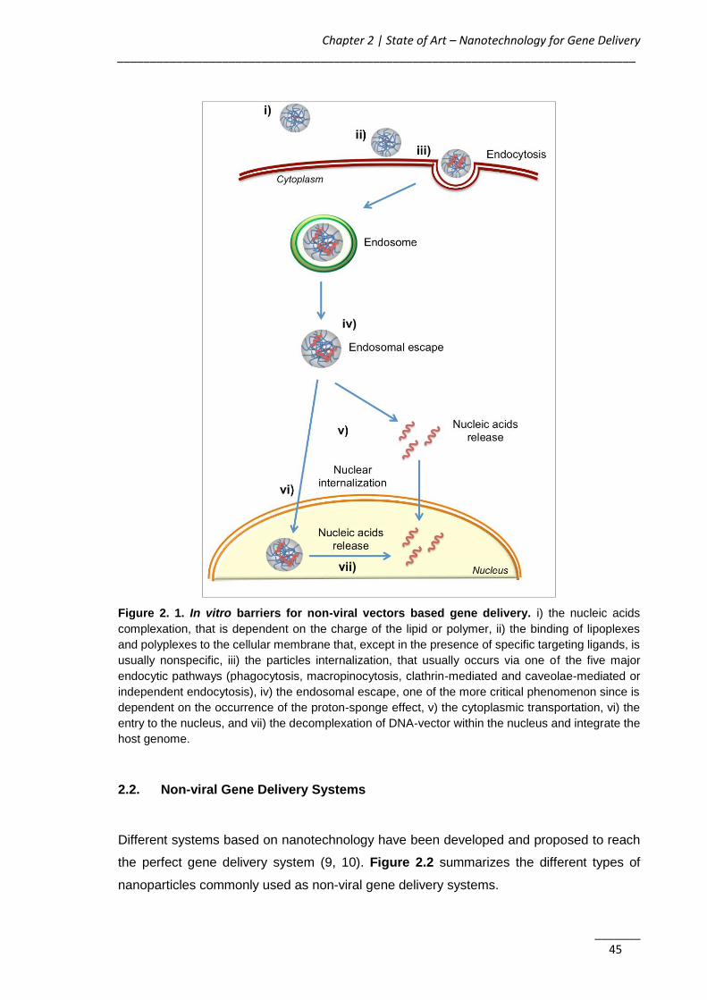
Chapter 2 | State of Art – Nanotechnology for Gene Delivery _______________________________________________________________________________
_______ 45
Figure 2. 1. In vitro barriers for non-viral vectors based gene delivery. i) the nucleic acids
complexation, that is dependent on the charge of the lipid or polymer, ii) the binding of lipoplexes
and polyplexes to the cellular membrane that, except in the presence of specific targeting ligands, is
usually nonspecific, iii) the particles internalization, that usually occurs via one of the five major
endocytic pathways (phagocytosis, macropinocytosis, clathrin-mediated and caveolae-mediated or
independent endocytosis), iv) the endosomal escape, one of the more critical phenomenon since is
dependent on the occurrence of the proton-sponge effect, v) the cytoplasmic transportation, vi) the
entry to the nucleus, and vii) the decomplexation of DNA-vector within the nucleus and integrate the
host genome.
2.2. Non-viral Gene Delivery Systems
Different systems based on nanotechnology have been developed and proposed to reach
the perfect gene delivery system (9, 10). Figure 2.2 summarizes the different types of
nanoparticles commonly used as non-viral gene delivery systems.

AKT2 siRNA-Nanoparticulate System as a New Tool to Restoration of E-Cadherin and Eradicate Tumor Metastic Phenotype ______________________________________________________________________________
_______ 46
Figure 2. 2. Different types of nanotechnology-based systems for gene delivery. The main
polymer and lipid-based systems that can be used for gene delivery are schematized, as well as the
components commonly used in its production. CD: cyclodextrin; DOPE: 1,2-di-(9Z-octadecenoyl)-
sn-glycero-3-phosphoethanolamine; DOSPA: 2,3-dioleyloxy-N-[2(sperminecarboxamido) ethyl]-
N,N-dimethyl-1-propanaminium trifluoroacetate; DOTAP: 1,2-dioleoyl-3-trimethylammonium-
propane; DOTMA: 1,2-di-O-octadecenyl-3-trimethylammonium propane; PAMAM: polyamidoamine;
PCL: poly(ε-caprolactone); PEI: polyethylenimine; PLA: poly(d,l-lactide); PLGA: poly(d,l-lactide-co-
glycolide); PLL: poly(L-lysine); SNALPs: stable nucleic acid lipid particles.
During the design of non-viral vectors, characteristics such as particle size, surface charge
and presence of biological moieties should be finely tuned in order to improve their biological
behavior and activity like blood circulation half-life, transfection activity and intracellular
trafficking (21, 22). For this reason, nanoparticles intended for gene delivery usually
possess in its composition cationic lipids or polymers that interact with the negatively
charged nucleic acids by electrostatic interactions and condense into complexes (lipoplexes
or polyplexes). The resultant polycation-based gene delivery systems net charge dictates
the interaction with serum proteins and cell membrane surface (23-26). Usually, a positive
charge promotes the interaction with negatively charged cell membrane, but also undesired
associations with serum proteins. Therefore, to overcome these safety issues, lipo- and
polyplexes are usually modified or conjugated with another lipids and/or polymers, affecting
the nucleic acids condensation, nanoparticle serum stability and cell surface activity (24).
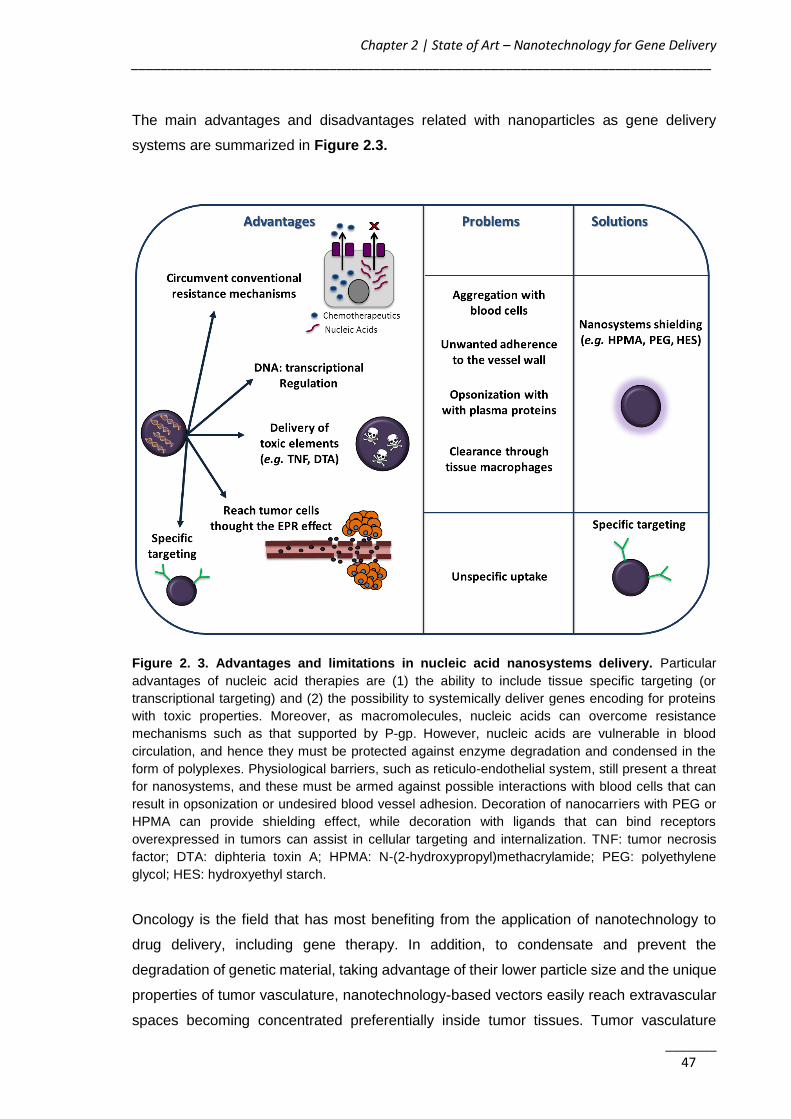
Chapter 2 | State of Art – Nanotechnology for Gene Delivery _______________________________________________________________________________
_______ 47
The main advantages and disadvantages related with nanoparticles as gene delivery
systems are summarized in Figure 2.3.
Figure 2. 3. Advantages and limitations in nucleic acid nanosystems delivery. Particular
advantages of nucleic acid therapies are (1) the ability to include tissue specific targeting (or
transcriptional targeting) and (2) the possibility to systemically deliver genes encoding for proteins
with toxic properties. Moreover, as macromolecules, nucleic acids can overcome resistance
mechanisms such as that supported by P-gp. However, nucleic acids are vulnerable in blood
circulation, and hence they must be protected against enzyme degradation and condensed in the
form of polyplexes. Physiological barriers, such as reticulo-endothelial system, still present a threat
for nanosystems, and these must be armed against possible interactions with blood cells that can
result in opsonization or undesired blood vessel adhesion. Decoration of nanocarriers with PEG or
HPMA can provide shielding effect, while decoration with ligands that can bind receptors
overexpressed in tumors can assist in cellular targeting and internalization. TNF: tumor necrosis
factor; DTA: diphteria toxin A; HPMA: N-(2-hydroxypropyl)methacrylamide; PEG: polyethylene
glycol; HES: hydroxyethyl starch.
Oncology is the field that has most benefiting from the application of nanotechnology to
drug delivery, including gene therapy. In addition, to condensate and prevent the
degradation of genetic material, taking advantage of their lower particle size and the unique
properties of tumor vasculature, nanotechnology-based vectors easily reach extravascular
spaces becoming concentrated preferentially inside tumor tissues. Tumor vasculature
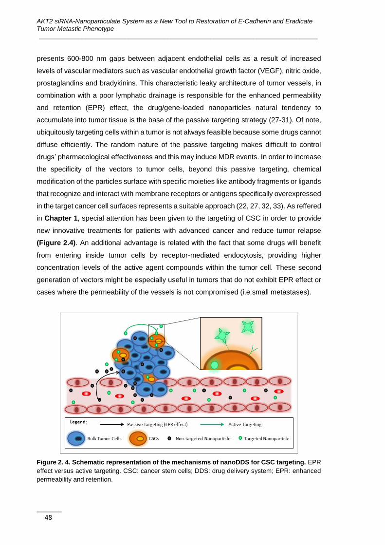
AKT2 siRNA-Nanoparticulate System as a New Tool to Restoration of E-Cadherin and Eradicate Tumor Metastic Phenotype ______________________________________________________________________________
_______ 48
presents 600-800 nm gaps between adjacent endothelial cells as a result of increased
levels of vascular mediators such as vascular endothelial growth factor (VEGF), nitric oxide,
prostaglandins and bradykinins. This characteristic leaky architecture of tumor vessels, in
combination with a poor lymphatic drainage is responsible for the enhanced permeability
and retention (EPR) effect, the drug/gene-loaded nanoparticles natural tendency to
accumulate into tumor tissue is the base of the passive targeting strategy (27-31). Of note,
ubiquitously targeting cells within a tumor is not always feasible because some drugs cannot
diffuse efficiently. The random nature of the passive targeting makes difficult to control
drugs’ pharmacological effectiveness and this may induce MDR events. In order to increase
the specificity of the vectors to tumor cells, beyond this passive targeting, chemical
modification of the particles surface with specific moieties like antibody fragments or ligands
that recognize and interact with membrane receptors or antigens specifically overexpressed
in the target cancer cell surfaces represents a suitable approach (22, 27, 32, 33). As reffered
in Chapter 1, special attention has been given to the targeting of CSC in order to provide
new innovative treatments for patients with advanced cancer and reduce tumor relapse
(Figure 2.4). An additional advantage is related with the fact that some drugs will benefit
from entering inside tumor cells by receptor-mediated endocytosis, providing higher
concentration levels of the active agent compounds within the tumor cell. These second
generation of vectors might be especially useful in tumors that do not exhibit EPR effect or
cases where the permeability of the vessels is not compromised (i.e.small metastases).
Figure 2. 4. Schematic representation of the mechanisms of nanoDDS for CSC targeting. EPR
effect versus active targeting. CSC: cancer stem cells; DDS: drug delivery system; EPR: enhanced
permeability and retention.
Chapter 2 | State of Art – Nanotechnology for Gene Delivery _______________________________________________________________________________
_______ 49
2.2.1. Polymer-based Delivery Systems (Polyplexes)
2.2.1.1. Cationic polymers
Biodegradable polymeric nanoparticles have been highly proposed as gene delivery
systems, not only due to higher complexation capacity, but also to the improved endosomal
escape properties, resulting in enhanced transfection efficiencies (22, 34-38). Due to an
easiest interaction with the negatively charged OGN and cellular membranes, greater
attention is given to the cationic charged polymers (39-41) since its first proposal in 1987
by Wu GY & Wu CH (42).
The most commonly used polycations are synthetic cationic polymers such as chitosan (CS)
and its derivatives, branched or linear PEI and its derivatives, polyamidoamine (PAMAM),
poly(b-amino ester)s (PAEs), poly(2-aminoethyl ethylene phosphate) (PPEEA), and poly(2-
(N,N-dimethylamino)ethyl methacrylate) (PDMAEMA) (15, 43, 44). Cationic peptides and
polypeptides such as the ones based on histidine (45, 46), protamine (47, 48), lysine
(poly(L-lysine) (PLL)) (49, 50) are also been proposed as polymeric non-viral vectors (51-
54). CS is a natural cationic mucopolysaccharide obtained as a raw material from chitin
deacetylation that has been shown to be biocompatible, non-inflammatory, non-toxic and
biodegradable. This polymer unique structural feature is associated to the presence of the
primary amine at the C-2 position of the glucosamine residues that promotes a high positive
charge responsible for the polyelectrolyte complexes formation with negatively charged
nucleotides (55). The properties of CS mainly for siRNA and DNA-mediated gene therapy
depend on the molecular weight (MW) and degree of deacetylation (DD) as it affects the
charge density, solubility, hydrophobicity and its ability to interact electrostatically with
polyanions (56, 57). A high DD (over 80%) has been identified as the most influential factor
in efficient siRNA-mediated knockdown. Regarding the MW, Liu X, et al (2007) reported
that CS molecules 5–10 times the length of the siRNA, could form stable nanoparticles
resulting in high gene silencing efficiency in H1299 human lung carcinoma cells (58). The
molar ratio (N/P ratio) between the positive charges of the polymer (amine groups) and the
negative charges of the OGN (phosphate groups) also influences the polyplexes gene
knockdown efficiency, partially due to the increase in nanoparticle stability specially
observed at high ratio values. For example, polyplexes prepared with a 170 kDa (DD 84%)
CS were only able to promote Green Fluorescent Protein (GFP) knockdown in H1299
human lung carcinoma at higher N/P ratios (50 and 150) (59). Despite its well-established
advantages, CS has a limited buffering capacity when compared with PEI (16, 18).
Therefore numerous studies are centered in CS modifications or conjugation with lipids or
other polymers in order to improve nucleic acid transfection efficiency. Conjugation with PEI
AKT2 siRNA-Nanoparticulate System as a New Tool to Restoration of E-Cadherin and Eradicate Tumor Metastic Phenotype ______________________________________________________________________________
_______ 50
(60, 61), modification with urocanic acid (62), addition of poly(propyl acrylic acid) (63),
introduction of imidazole moieties (64) and CS grafting with stearic acids (65) are few
examples of studies that have been performed aiming to improve the buffering and
transfection properties of CS.
PEI and its derivatives, are a type of high cationic charge density polymers, presenting
branched or linear forms, available in a broad range of MW (34, 66). PEI-based particles
are well-known and well characterized non-viral vectors for efficient delivery of nucleic acids
including DNA and RNA both in vitro and in vivo (22, 35, 67). The high transfection
efficiencies of PEI-based vectors has been mainly related to their substantial buffering
capability at the endosomal pH (from pH 6.0–6.5 in early endosomes to pH 4.5–5.5 in late
endosomes and lysosomes (68)) and ability to promote the endosome osmotic swelling and
rupture, releasing the genetic material into the cytoplasm, thus avoiding its traffic and
degradation in the lysosomal environment (22, 35, 36, 38, 69). Additionally, PEI protonable
amine groups provide a powerful electrostatic capacity, which can promote strong
interactions with the negatively charged nucleic acids molecules, explaining the ability of
this polymer to effectively condense nucleic acids into nanoscale entities, protecting them
from degradation. Moreover, PEI cationic surface charge has also a crucial role in the
binding of polyplexes with cell membrane and consequent internalization (38, 69).
Unfortunately, the formed positively charged polyplexes usually become unstable, showing
a tendency to aggregate throughout the incubation time, depending on the surface charge
and ionic strength of the medium (69, 70). Furthermore, this high cationic charge density is
also responsible for PEI well-known toxicity and side effects such as membrane damage,
interaction with blood cells and activation of the complement system (38, 71).
As with other polymers, key factors such as the linear versus branched structure, the
branching degree and the MW are likely to affect the final PEI-based polyplexes efficiency
and cytotoxicity profile (71-75). In fact, several studies demonstrate that linear PEI presents
lower cytotoxicity compared to the branched ones with similar MW. However, a decreased
condensation capacity of OGN due to lower amount of primary amines and lower
transfection efficiencies are consequently observed (71, 72, 74, 76). Concerning the MW;
high PEI MW, present increased buffering capacity and consequently high transfection
efficiencies due to a higher content of primary, secondary and tertiary amines.
Nevertheless, higher MW values increase cytotoxicity due to polymer precipitation and
further adherence to the outer cell membrane, inducing cell necrosis (19, 71, 72, 77). The
N/P between the nitrogen groups presented in the polymer and the phosphorous atom
content in the nucleic acids, also proved to be a factor affecting significantly the polyplexes’
efficiency. In general, complexes formed at low molar ratio in the range of 2-5 tend to
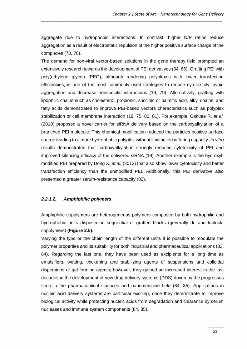
Chapter 2 | State of Art – Nanotechnology for Gene Delivery _______________________________________________________________________________
_______ 51
aggregate due to hydrophobic interactions. In contrast, higher N/P ratios reduce
aggregation as a result of electrostatic repulsion of the higher positive surface charge of the
complexes (70, 78).
The demand for non-viral vector-based solutions in the gene therapy field prompted an
extensively research towards the development of PEI derivatives (34, 66). Grafting PEI with
poly(ethylene glycol) (PEG), although rendering polyplexes with lower transfection
efficiencies, is one of the most commonly used strategies to reduce cytotoxicity, avoid
aggregation and decrease nonspecific interactions (19, 79). Alternatively, grafting with
lipophilic chains such as cholesterol, propionic, succinic or palmitic acid, alkyl chains, and
fatty acids demonstrated to improve PEI-based vectors characteristics such as polyplex
stabilization or cell membrane interaction (19, 75, 80, 81). For example, Oskuee R, et al.
(2010) proposed a novel carrier for siRNA delivery based on the carboxyalkylation of a
branched PEI molecule. This chemical modification reduced the particles positive surface
charge leading to a more hydrophobic polyplex without limiting its buffering capacity. In vitro
results demonstrated that carboxyalkylation strongly reduced cytotoxicity of PEI and
improved silencing efficacy of the delivered siRNA (19). Another example is the hydroxyl-
modified PEI prepared by Dong X, et al. (2013) that also show lower cytotoxicity and better
transfection efficiency than the unmodified PEI. Additionally, this PEI derivative also
presented a greater serum-resistance capacity (82).
2.2.1.2. Amphiphilic polymers
Amphiphilic copolymers are heterogeneous polymers composed by both hydrophilic and
hydrophobic units disposed in sequential or grafted blocks (generally di- and triblock-
copolymers) (Figure 2.5).
Varying the type or the chain length of the different units it is possible to modulate the
polymer properties and its suitability for both industrial and pharmaceutical applications (83,
84). Regarding the last one, they have been used as excipients for a long time as
emulsifiers, wetting, thickening and stabilizing agents of suspensions and colloidal
dispersions or gel forming agents; however, they gained an increased interest in the last
decades in the development of new drug delivery systems (DDS) driven by the progresses
seen in the pharmaceutical sciences and nanomedicine field (84, 85). Applications in
nucleic acid delivery systems are particular exciting, once they demonstrate to improve
biological activity while protecting nucleic acids from degradation and clearance by serum
nucleases and immune system components (84, 85).
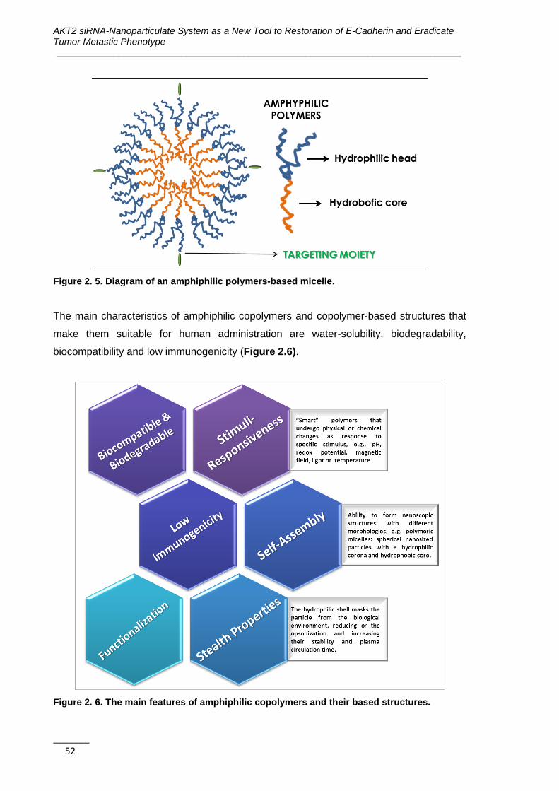
AKT2 siRNA-Nanoparticulate System as a New Tool to Restoration of E-Cadherin and Eradicate Tumor Metastic Phenotype ______________________________________________________________________________
_______ 52
Figure 2. 5. Diagram of an amphiphilic polymers-based micelle.
The main characteristics of amphiphilic copolymers and copolymer-based structures that
make them suitable for human administration are water-solubility, biodegradability,
biocompatibility and low immunogenicity (Figure 2.6).
Figure 2. 6. The main features of amphiphilic copolymers and their based structures.
Chapter 2 | State of Art – Nanotechnology for Gene Delivery _______________________________________________________________________________
_______ 53
Among the different structures available, the most extensively researched for the production
of DDS are composed of PEG as hydrophilic block and a variety of hydrophobic blocks,
namely: (i) polypropylene oxice (PPO); (ii) poly(ester)s like poly(ε-caprolactone) (PCL) or
poly(d,l-lactide-co-glycolide) (PLGA); (iii) poly(amino acid)s such as poly(L-aspartic acid)
and poly(L-glutamic acid); or (iv) lipids like phosphoethanolamine (PE) (86, 87). Particular
interest has been given to PEG-PPO block copolymers (poloxamers and poloxamines) (88,
89).They are commercialy accessible in a wide range of compositions and MW
(Pluronic®/Lutrol®/Kolliphor® and Tetronic®).
2.2.1.3. Characteristics of amphiphilic copolymers and copolymer-based
structures
a) Self-assembly
Amphiphilic polymers are able to the form nanoscopic structures of different morphologies,
e.g. polymersomes, nanocapsules, nanospheres, nanogels or dendrimers, although
polymeric micelles (PM) are the most commonly used and researched (85, 90, 91).
Micellization into spherical nanosized particles with a hydrophilic corona and hydrophobic
core occurs at or above a specific threshold level of polymer concentration (critical micellar
concentration - CMC) and temperature (critical micellar temperature - CMT) (86, 87). In
water this process is driven by an increase in entropy of the solvent molecules in contact to
the hydrophobic units of the polymer and a consequent decrease of free energy (Δ°Gm) as
the hydrophobic components are withdrawn from the aqueous media originating the micelle
core (92). Δ°Gm is given by Equation 2.1 where, R is the gas constant and T is the
temperature of the system.
∆°𝐆𝐦 = 𝐑𝐓 𝐥𝐧 𝐂𝐌𝐂 Equation 2.1
Different parameters such as chemical composition, MW, molecular architecture (linear
versus branched, double bounds, etc.), concentration, temperature, solvent-polymer
interactions, or salt concentration influence the self-assembly of polymers. By increasing
the temperature of the system, the solvency of hydrophilic unit as well as the CMC value
will decrease, promoting the micellization. Similarly, this phenomena is favored when the
attractive hydrophobic interactions increases as a result of a gain in the MW of the
hydrophobic domain (93). For example, PEG-b-PHOHH copolymers with PHOHH
segments of 1500 and 7700 g/mol present a CMC value of 5.50 and 0.93 mg/L, respectively

AKT2 siRNA-Nanoparticulate System as a New Tool to Restoration of E-Cadherin and Eradicate Tumor Metastic Phenotype ______________________________________________________________________________
_______ 54
(94). PM have been developed to modify several major intrinsic characteristics of drugs,
including drug aqueous solubility, release pattern, pharmacokinetics, biodistribution, and in
vivo stability (87). In general, the solubilisation capacity of hydrophobic drugs into micelles
is proportional to the hydrophobicity of the micelle core (95). PM have an higher core
hydrophobicity then surfactant micelles, conferring them higher thermodynamic and
kinetical stability, thus presenting slower and delayed disintegration and drug release in
circulation even upon dilution below the CMC value (84).
Table 2. 1. Examples of self-assembled particles under clinical trials evaluation.
Formulation Copolymer Drug Indication Clinical phase
Reference
SP1049C Pluronic® L61 and
F127 Doxorubicin
Advanced adenocarcinoma of
the esophagus, gastroesophageal
junction and stomach
III (96)
Genexol-PM® PEG-PLA PTX
Breast cancer, non-small cell lung cancer, advanced pancreatic cancer and ovarian
cancer
II/III/IV (97-101)
NK012
PEG-poly(L-glutamic
acid)-SN-38 conjugated
SN-38 Advanced breast
cancer and small cell lung cancer
II (102, 103)
NK105 PEG-poly(L-aspartic acid)
PTX Advanced or recurrent
breast cancer III (104)
NK-911
PEG–poly(aspartic
acid)-doxorubicin conjugated
Doxorubicin Solid tumors I (105)
NC-4016 PEG-DACH-
platin Oxaliplatin
Advanced solid tumors or lymphoma
I (106)
NC-6004 (Nanoplatin®)
PEG-poly(L-glutamic
acid)-cisplatin
conjugated
Cisplatin
Solid tumors, non-small cell lung, biliary and bladder cancer, pancreatic cancer
II/III (107, 108)
Paxceed® PEG-PLA PTX Rheumatoid Arthritis,
Psoriasis II (109, 110)
IT-101
Polymer-cyclodextrin-camptothecin conjugated
Camptothecin Advanced solid tumors, ovarian
cancer I/II (111, 112)
AquADEK®
D-alpha-tocopheryl-
co-PEG 1000 succinate
Vitamins and antioxidants
Multivitamin supplement in Cystic
fibrosis II (113)
DACH-platin: diaminocyclohexane platinum; PEG: polyethylene glycol; PLA: poly(d,l-lactide); PTX: paclitaxel

Chapter 2 | State of Art – Nanotechnology for Gene Delivery _______________________________________________________________________________
_______ 55
Due to their therapeutic efficacy, some PM-based formulations passed from the pre-clinical
assessment to enroll clinical trials (Table 2.1) (114-117). One of them (Genexol-PM®,
Samyang Co.) firstly approved in 2007 in Korea and being evaluated in European Union
and America with the name Cynviloq® for the treatment of breast, non-small cell lung and
ovarian cancers (118-120), while SP1049C (Surpatek Pharma, Inc.) granted orphan drug
designation by FDA for the treatment of gastric cancer (121).
b) Surface hydrophilicity and functionalization
While the hydrophobic core of micelles, in addition to the solubilisation and protection of
hydrophobic drugs, provides appropriate mechanical properties for the desired application;
hydrophilic shell allows the encapsulation of hydrophilic compounds, and masks the particle
from the biological environment, thereby reducing the protein absorption and cellular
adhesion, and enhancing the particle stability (122, 123). Opsonin proteins present in the
blood serum promptly bind to particles, allowing their recognition by macrophages and
consequent elimination of the encapsulated drugs from bloodstream (124). The presence
of a hydrophilic layer on the surface of particles will provide them with stealth properties by
reducing or delaying the opsonization via steric repulsion forces, thus increasing the plasma
circulation time of particles and the half-life of drugs (124, 125). PEG and poloxamers are
commonly used as hydrophilic polymer to covers the surface of many nanoDDS creating
the so called stealth systems (126). Moreover the resultant functionalization potential by
either chemical modification or bioconjugation could be used to bind targeting moieties to
the particles surface allowing a better control of the biodistribution of DDS (127). A variety
of ligands such as peptides, antibodies, folate, transferrin or polysaccharides, can be used,
accordingly to the required application.
c) Stimuli-responsive properties
Among all the characteristics presented by some amphiphilic polymers for the development
of advanced controlled DDS, stimuli-responsiveness appears as one of the most interesting
(128, 129). Stimuli-responsive polymers are a class of “smart” polymers that undergo
chemical or physical alterations as response to a specific stimulus, e.g. temperature, pH,
redox potential, magnetic field or light (129-131). Acrylate/methacrylate derivatives and
monomers of N-alkyl substituted acrylamides are commonly used in the production of pH-
responsive and thermo-responsive copolymers, respectively (129, 132). At acidic pH the
existence of intermolecular polymer complexes lead to the formation of compact gels that

AKT2 siRNA-Nanoparticulate System as a New Tool to Restoration of E-Cadherin and Eradicate Tumor Metastic Phenotype ______________________________________________________________________________
_______ 56
swells and became loose just at basic pH (133, 134). Multi-stimuli-responsive polymers (to
light, temperature, and pH) with possible applications in biomedical field can be also
developed (135).
2.2.1.4. Amphiphilic polymers-based gene delivery systems
Amphiphilic polymers have been proposed as gene carriers in the last years. As an example
Guo S, et al. (2011) engineered ternary complexes by coating the surface of
polycaprolactone-graft-poly(N,N-dimethylaminoethyl methacrylate) (PCL-g- PDMAEMA)
nanoparticles/DNA with polyglutamic acid-graft-poly(ethylene glycol)(PGA-g-mPEG), in
order to decrease the zeta potential of the system without interfere with DNA condensation
ability. In in vitro studies, this non-covalent post-PEGylated ternary complexes presented
high stability, low cytotoxicity, pronunced transfection efficiency, and improved DNA
endosomal escape ability over the binary complexes. Moreover, in vivo results, namely
histochemical analysis of tumor sections showed that the tumor expression of a red
fluorescent protein encoded by the RFP plasmid is higher when the ternary complex was
intravenously administrated (52).
In another study, Liu Y, et al. (2011) constructed a library of mono-methoxyl-poly(ethylene
glycol)-block-poly(ε-caprolactone) (mPEG-PCL)-modified hyperbranched PEI copolymers
(hy-PEI-PCL-mPEG) varying mPEG and PCL segments length, graft density, and hy-PEI
MW in order to explore the influence of polymer compositions on siRNA delivery. Longer
mPEG and PCL segments with higher graft density improve the stability and formation of
polyplexes, reducing their cytotoxicity and zeta-potential. Hy-PEI-PCL-mPEG with very
short PCL segments presented greater transfection efficiencies compared with those
without PCL (hy-PEI25k) (136).
Amino acid-based polymers have also been proposed. In this context, PEO-PLAA-based
structures emerge as an opportunity for the development of more effective therapeutic PM.
The presence of carboxyl or amine groups in the PLAA block is advantageous for chemical
conjugation and electrostatic interactions with drugs and nucleic acids and facilitates the
chemical modification of the micellar core (85).
Poly(d,l-lactide) (PLA) and PLGA have also been formulated into micelles or nanoparticles
and extensively used for sustained nucleic (137). Yang XZ, et al. (2011) produced a
polymeric nanoparticle system for efficient siRNA encapsulation and delivery based on
poly(ethylene glycol)-b-poly(d,l-lactide) (mPEG-PLA) and an amphiphilic cationic lipid
(BHEM-cholesterol) by a non-condensation process and a double-emulsion solvent
evaporation technique. In vitro assays showed that these nanoparticles allow efficient

Chapter 2 | State of Art – Nanotechnology for Gene Delivery _______________________________________________________________________________
_______ 57
siRNA cell internalization with further adequate endosomal escape, which results in the
selective knockdown of the specific gene of interest, in this case, a significant
downregulation of luciferase expression in HepG2-luciferase cells and suppression of polo-
like kinase 1 (Plk1) expression in HepG2 cells. In addition, the nanoparticles encapsulating
siRNA targeting the Plk1 gene showed to induce notable apoptosis in HepG2 and MDA-
MB-435 cancer cell lines. In vivo studies on an orthotopic murine metastatic liver cancer
model (MDA-MB-435 xenographs) further confirm the in vitro data, where luciferase
expression was significantly inhibited and tumor growth suppressed after nanoparticles
systemic administration (137). All these results suggest that mPEG-PLA nanoparticles have
great potential for gene therapy. More recently, Sun J, et al. (2016) tested the amphiphilic
triblock copolymer poly(hydroxyletheyl methacrylate-L-lysine)-b-poly(L-lactide)-b-
poly(hydroxyletheyl methacrylate-L-lysine)s (PHML-b-PLA-b-PHML) to efficiently
condensate a plasmid DNA and protect it from degradation by DNase I, and promoted a
strong transfection efficiency without significant toxicity. The endocytosis of this particles
seems to be through the lipid-raft-mediated endocytosis pathway, similar to that of SV40
virus-based vectors (138).
Murata N, et al. (2008) produced a biodegradable long-term sustained release PLGA
microsphere through the water-in-oil-in-water drying method for encapsulation of anti-VEGF
siRNA in order to suppress angiogenesis and tumor growth in vivo. Arginine or branched
PEI was used as transfection agent/carrier for siRNA delivery. Using mice bearing S-180
tumors they demonstrated that after an intra-tumor injection of these microspheres VEGF
gene expression and consequently the tumor growth were highly suppressed which indicate
that this formulation is an useful approach to cancer treatment (139).
Worth to note is the research of Alshamsan A, et al. (2010), that investigated the
incorporation of siRNA/PEI and siRNA/PEI-stA (the stearic derivative of PEI) complexes
into PLGA nanoparticles for STAT3 knockdown in dendritic cells (DC). The in vivo and in
vitro cytotoxicity that already had been detected for PEI or PEI-StA based polyplexes, was
significantly reduced with this systems. They observed that the successful and specific
STAT3 silencing is associated with the reestablishment of DC maturation and functionality,
therefore this formulation represents an interesting strategy for targeted siRNA delivery to
DCs and a promise for combined cancer immunotherapy (140).
2.2.1.5. Pluronic® and its role as biological response modifier
Pluronic® amphiphilic block copolymers have been shown high stability and the capacity to
improve significantly the transfection efficiency and gene expression via different delivery

AKT2 siRNA-Nanoparticulate System as a New Tool to Restoration of E-Cadherin and Eradicate Tumor Metastic Phenotype ______________________________________________________________________________
_______ 58
routes (24, 89, 141). The mechanism by which poloxamers enhance gene expression is not
clearly understood but seems to be related to its ability to bind to cellular membranes and
further activation of cell signalling pathways, such as the Nf-κB pathway. Nf-κB is a
transcription factor able to bind pDNA in the cytoplasm and transport it to the nucleus using
the nuclear transport machinery. Yang Z, et al. (2008) demonstrate for the first time that
Pluronic® enhances both cellular uptake and nuclear transport of polyplex-delivered pDNA
into NIH 3T3 cells, underling the role of poloxamers as a biological response modifier. On
the other hand, poloxamer did not affect the naked DNA uptake, emphasizing the
importance of delivering pDNA using polyplexes (24).
Another study from Kuo JH, et al. (2003) compared the serum stability of PEI/DNA and
Pluronic-PEI/DNA complexes with different hydrophilic lipophilic balances (HLB) in terms of
DNA delivery capacity to NIH/3T3 cells. The presence of Pluronic® especialy with higher
HLB values, such as F68, conferred higher stability and increased significantly the gene
expression (142). Also, Wang M, et al. (2014) investigated a series of small-size PEI-
conjugated Pluronic® polycarbamates (PCMs) for the ability to modulate the delivery of 2′-
O-methyl phosphorothioate RNA (2′-OMePS) both in vitro and in dystrophic (mdx) mice for
the treatment of muscular dystrophy or other appropriate myodegenerative diseases. They
conclude that the effective PCMs, especially those composed of moderate MW (2k–5kDa)
and intermediate HLB of Pluronic®, enhanced exon-skipping of 2′-OMePS with low toxicity
as compared with Lipofectamine® 2000 in vitro or PEI 25k in vivo (143).
Taking into account the different reports, the use of Pluronic® in the development of non-
viral gene delivery systems seems to be a simple and safer approach that enhances both
the in vitro and in vivo gene expression without inducing significant cytotoxic effects or
inflammatory responses.
2.2.1.6. Polymer-based non-viral vectors on the way to clinical trials
The emerging advances observed in the area of gene delivery using polymeric
nanoparticles technology, including the ones based in amphiphilic polymers, allowed the
opportunity to bring these concepts to the clinical practice. In 2008, CALAA-01 becomes
the first targeted delivery of siRNA to be tested in a phase I clinical trial to treat solid tumors
refractory to standard therapies. CALAA-01 consists in a cyclodextrin-containing polymer
(CDP)-based delivery system loaded with siRNA against ribonucleotide reductase subunit
2 (RRM2) and was design to inhibit tumor growth. The formulation also contains PEG as a
stabilizing agent, and human transferrin (Tf) as a targeting ligand for binding to
overexpressed transferrin receptors (TfR) of cancer cells (144). Besides CALAA-01,
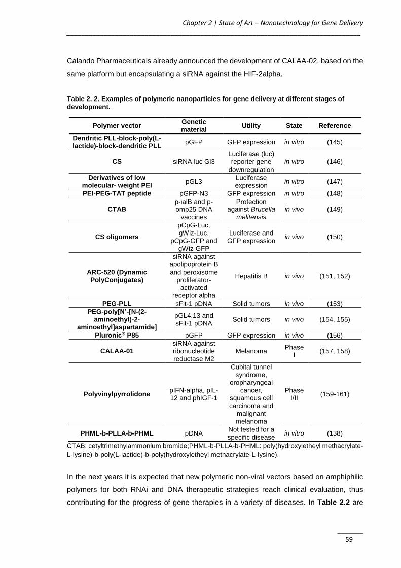
Chapter 2 | State of Art – Nanotechnology for Gene Delivery _______________________________________________________________________________
_______ 59
Calando Pharmaceuticals already announced the development of CALAA-02, based on the
same platform but encapsulating a siRNA against the HIF-2alpha.
Table 2. 2. Examples of polymeric nanoparticles for gene delivery at different stages of development.
Polymer vector Genetic material
Utility State Reference
Dendritic PLL-block-poly(L-lactide)-block-dendritic PLL
pGFP GFP expression in vitro (145)
CS siRNA luc Gl3 Luciferase (luc) reporter gene
downregulation in vitro (146)
Derivatives of low molecular- weight PEI
pGL3 Luciferase expression
in vitro (147)
PEI-PEG-TAT peptide pGFP-N3 GFP expression in vitro (148)
CTAB p-ialB and p-omp25 DNA
vaccines
Protection against Brucella
melitensis in vivo (149)
CS oligomers
pCpG-Luc, gWiz-Luc,
pCpG-GFP and gWiz-GFP
Luciferase and GFP expression
in vivo (150)
ARC-520 (Dynamic PolyConjugates)
siRNA against apolipoprotein B and peroxisome
proliferator-activated
receptor alpha
Hepatitis B in vivo (151, 152)
PEG-PLL sFlt-1 pDNA Solid tumors in vivo (153)
PEG-poly[N’-[N-(2-aminoethyl)-2-
aminoethyl]aspartamide]
pGL4.13 and sFlt-1 pDNA
Solid tumors in vivo (154, 155)
Pluronic® P85 pGFP GFP expression in vivo (156)
CALAA-01 siRNA against ribonucleotide reductase M2
Melanoma Phase
I (157, 158)
Polyvinylpyrrolidone pIFN-alpha, pIL-12 and phIGF-1
Cubital tunnel syndrome,
oropharyngeal cancer,
squamous cell carcinoma and
malignant melanoma
Phase I/II
(159-161)
PHML-b-PLLA-b-PHML pDNA Not tested for a specific disease
in vitro (138)
CTAB: cetyltrimethylammonium bromide;PHML-b-PLLA-b-PHML: poly(hydroxyletheyl methacrylate-
L-lysine)-b-poly(L-lactide)-b-poly(hydroxyletheyl methacrylate-L-lysine).
In the next years it is expected that new polymeric non-viral vectors based on amphiphilic
polymers for both RNAi and DNA therapeutic strategies reach clinical evaluation, thus
contributing for the progress of gene therapies in a variety of diseases. In Table 2.2 are

AKT2 siRNA-Nanoparticulate System as a New Tool to Restoration of E-Cadherin and Eradicate Tumor Metastic Phenotype ______________________________________________________________________________
_______ 60
summarized some of the many works proposed involving polymeric nanoparticles for gene
delivery at different stages of development.
2.2.2. Lipid-based Delivery Systems (Lipoplexes)
Lipid-based non-viral vectors including liposomes, lipidic micelles, solid lipid nanoparticles
(SLN), among others, became under the spotlight of many research teams as a strategy to
overcome positively charge polyplexes toxicity and some possible lack of biocompatibility
and/or biodegradability, while protecting the nucleic acid from nuclease degradation,
allowing systemic delivery, and enhancing cellular uptake (162, 163).
Lipoplexes are usually produced through a simple interaction between nucleic acids and
different lipids through different techniques such as the hydration of a lipid film, the
dehydration-rehydration method, the ethanolic injection, the reverse-phase evaporation or
the detergent dialysis technique (70).
Three types of lipids are usually selected to form the lipoplexes: i) anionic (e.g. cholesteryl
hemisuccinate (CHEMS)), ii) neutral (e.g. 1,2-di-(9Z-octadecenoyl)-sn-glycero-3-
phosphoethanolamine (DOPE) and cholesterol), and iii) cationic (e.g. 1,2-di-O-octadecenyl-
3-trimethylammonium propane (DOTMA) or Lipofectin®, 1,2-dioleoyl-3-trimethylammonium-
propane (DOTAP), 2,3-dioleyloxy-N-[2(sperminecarboxamido) ethyl]-N,N-dimethyl-1-
propanaminium trifluoroacetate (DOSPA) or Lipofectamine®, 1,3-dioleoyloxy-2-(6-
carboxyspermyl)-propyl amide (DOSPER), and Oligofectamine®).
The cationic lipids, due to its high capacity to complex the anionic ONG are commonly
chosen. The final charge of the lipoplex, probably one of the most important factors affecting
lipoplexes stability and transfection efficiency, depends on the ratio between the differently
charged lipid vector and the nucleic acid molecules. Besides its charge and composition,
the size and morphology of the lipoplexes also tremendously affect their transfection
activity. Cationic lipoplexes, for example, does have a vast heterogeneity in terms of shape
and size and different morphologic forms (such as cubic, beads on a string, spaghettis,
meat balls, multilamellar, map-pin, sliding columnar and inverted hexagonal phase
structures) have been reported (164). Regarding lipoplexes size, there is still a large
controversy about which size range promotes better transfection activities. Some authors
such as Esposito C, et al. (165) or Massoti A, et al. (166) advocate that lipoplexes within
200-400 nm or even 1-2 μm are more efficient, arguing that larger particles have a faster
sedimentation process, present a higher contact with cells and form larger intracellular
vesicles, that are more easily disrupted, improving the endosomal escape (165, 166).
However, other authors report that higher transfection efficiencies are obtained using

Chapter 2 | State of Art – Nanotechnology for Gene Delivery _______________________________________________________________________________
_______ 61
particles with a smaller size of around 100 nm or less (167, 168). Although the optimal size
for particles in vitro is still unclear, for the in vivo applications is of general consensus that
small diameter nanoparticles are the more effective ones due to their avoidance from
reticuloendothelial system and improved biodistribution (164).
The lipoplexes internalization by cells depends on the type of lipid used and the type of
genetic material that is delivered, being reported two main mechanisms. The majority of the
cases it is processed by endocytosis and in a low percentage of the cases by direct fusion
with the cellular membrane. As example, Rejman J, et al. (169) demonstrated that
DOTAP/DNA lipoplexes uptake occurs via clathrin-mediated endocytosis, while according
to Wong AW, et al. (170), the uptake of a plasmid DNA associated with DMRIE-C
transfection reagent occurs via the caveolin pathway. For synthetic siRNAs another
mechanism was recently reported by Lu JJ, et al. (171), which showed that besides around
95% of siRNA lipoplexes enter the cells by endocytosis, the functional siRNA delivery
occurs via minor pathways that involve simply the fusion with the plasma membrane. It was
observed that when clathrin, caveolin or macropinocytosis mediated uptake is inhibited, the
knockdown of the protein of interest continues to happen. On the other hand, when the
cholesterol from the plasma membrane is depleted there are a significant reduction of the
knockdown of the protein (171).
2.2.2.1. Cationic lipoplexes
Cationic lipids are generally amphiphilic molecules formed by a positively charged polar
headgroup attached via a linker to a hydrophobic domain, which hydrocarbon chains
lengths vary commonly between C8:0 and C18:1. The characteristics of the headgroup are
responsible for the transfection efficiency of the lipoplexes; therefore several synthetic
cationic lipids with variations in the headgroup composition have been produced.
Headgroups consisting on amines groups with different substitution degrees, guanidinium
or pyridinium groups, amino acids and peptides have been reported (9, 12, 13).
Despite their unquestionable advantages in terms of transfection efficiency, the toxicity of
cationic lipids is a well-known problem that needs to be addressed. Eventhough,
Lipofectamine® 2000 is widely used as transfection reagent in mechanistic studies and gold
standard to assess the efficacy of other systems. Therefore, with the final objective of
reaching an ideal gene delivery system with minimal toxicity and adequate transfection
efficiency, numerous cationic lipids based formulations have been continuously designed
and produced, presenting adequate in vitro and in vivo results and some of them enrolled
already clinical assessment (Table 2.3). These new cationic lipids are obtained through

AKT2 siRNA-Nanoparticulate System as a New Tool to Restoration of E-Cadherin and Eradicate Tumor Metastic Phenotype ______________________________________________________________________________
_______ 62
structural modifications performed in the headgroup, in the linker and/or in the hydrophobic
tail domains that are known to regulate transfection efficiency and cytotoxicity (172).
2.2.2.2. Anionic and neutral lipids
The use of anionic or neutral lipids as gene delivery systems is another strategy that some
authors explore with the objective of override the cationic lipids associated toxicity.
However, due to their negative or neutral charge and the consequent impossibility to
establish electrostatic interactions with negatively charged molecules, the delivery systems
based on anionic or neutral lipids always require the incorporation of a third moiety such as
divalent or monovalent cations (Ca2+, Na+, Mg2+, Mn2+ or Ba2+) to promote the complexation
between the nucleic acids and the lipids (173-175).
Kapoor M, et al. (175) designed a siRNA delivery system using physiologically occurring
anionic lipids (DOPG/DOPE) and calcium ions bridges. The obtained lipoplexes presented
high stability in the presence of serum components, efficient cellular uptake and effective
endosomal release, and a significant silencing capacity comparable with the control cationic
complexes (Lipofectamine® 2000), showing lower toxicity (175).
In another study, Mignet N, et al. (173) proposed an innovative system throughout the
combination of a cationic lipoplex and pegylated anionic liposomes. The ratio between
anionic and cationic lipids was optimized in order to form an anionic pegylated lipoplex
sensitive to pH changes (5.5-6.5). Reversion to a cationic net charge at an adequate pH
value was observed during endosomal acidification which leads to the DNA release and
consequent increase of the gene expression. These result was comparable with the control
CHEMS formulation and more importantly, showed an improved ability to deliver DNA to
tumor tissues (173).
2.2.3. Lipid Nanoparticles
Different types of liposomes (cationic, neutral and anionic) have been developed and
successfully used for nucleic acids administration (57, 176, 177). Among them, Stable
Nucleic Acid Lipid Particles (SNALPs) have been enjoying special interest for the treatment
of several diseases. They consist in a lipid bilayer system composed by a mixture of cationic
and fusogenic lipids shielded with PEG to provide a neutral hydrophilic surface, avoiding
opsonization and rapid systemic clearance in vivo, as well as allowing an efficient particle
targeting (Figure 2.7) (162, 178-182). Neutral lipids such as DOPE, cholesterol or (1,2-
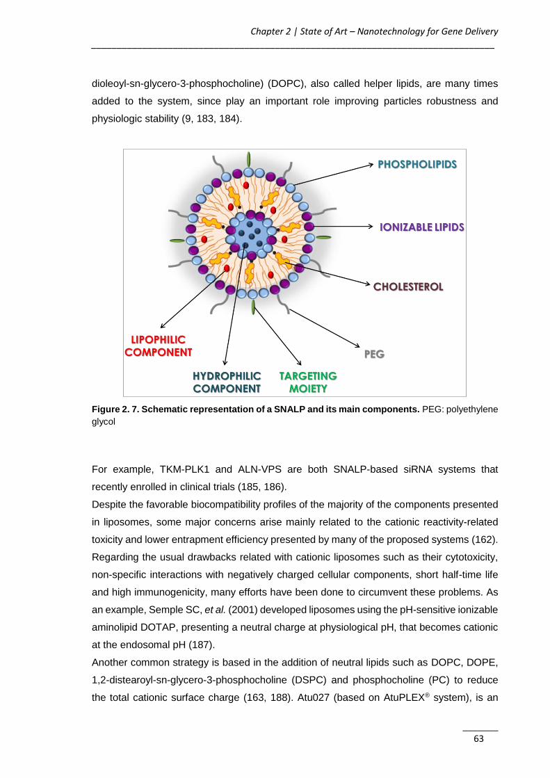
Chapter 2 | State of Art – Nanotechnology for Gene Delivery _______________________________________________________________________________
_______ 63
dioleoyl-sn-glycero-3-phosphocholine) (DOPC), also called helper lipids, are many times
added to the system, since play an important role improving particles robustness and
physiologic stability (9, 183, 184).
Figure 2. 7. Schematic representation of a SNALP and its main components. PEG: polyethylene
glycol
For example, TKM-PLK1 and ALN-VPS are both SNALP-based siRNA systems that
recently enrolled in clinical trials (185, 186).
Despite the favorable biocompatibility profiles of the majority of the components presented
in liposomes, some major concerns arise mainly related to the cationic reactivity-related
toxicity and lower entrapment efficiency presented by many of the proposed systems (162).
Regarding the usual drawbacks related with cationic liposomes such as their cytotoxicity,
non-specific interactions with negatively charged cellular components, short half-time life
and high immunogenicity, many efforts have been done to circumvent these problems. As
an example, Semple SC, et al. (2001) developed liposomes using the pH-sensitive ionizable
aminolipid DOTAP, presenting a neutral charge at physiological pH, that becomes cationic
at the endosomal pH (187).
Another common strategy is based in the addition of neutral lipids such as DOPC, DOPE,
1,2-distearoyl-sn-glycero-3-phosphocholine (DSPC) and phosphocholine (PC) to reduce
the total cationic surface charge (163, 188). Atu027 (based on AtuPLEX® system), is an
AKT2 siRNA-Nanoparticulate System as a New Tool to Restoration of E-Cadherin and Eradicate Tumor Metastic Phenotype ______________________________________________________________________________
_______ 64
example of this approach, a cationic liposomal formulation containing neutral fusogenic
lipids, designed for delivery of siRNA against the protein kinase PKN3. In phase I clinical
trials, the system has proved to be well tolerated in the administered doses, being a hopefull
antiangiogenesis therapeutic strategy for the treatment of advanced solid tumors (189).
Apart from liposomes, SLN have also been proposed as non-viral vectors due to their
proposed advantages such as high stability, protection of the encapsulated agents, high
entrapment ability, small and uniform sizes, easily modulated via the composition, sustained
controlled release properties, biodegradability, ease manufacture and scale up at relatively
low cost, or suitability for sterilization and lyophilization. They showed to be capable of
encapsulating OGN, mainly using plasmids as a model, in the lipid core or on the particle
surface (190).
Bondi L, et al. (2007), proved the suitability of cationic SLN based on acylglicerol lipids, able
to form stable complexes with DNA and to protect DNA against DNase I digestion. The in
vitro studies performed on human liver cancer cells also demonstrated a low degree of
toxicity of both SLN and SLN–DNA complexes (191). With the objective to assess the in
vivo transfection potential of SLN, Rodríguez A, et al. (2010), demonstrated for the first time
the capacity of SLN-DNA vectors to induce the expression of a foreign protein in mice. They
use SLN as a vector to transfect the pCMS-GFP plasmid that encodes the GFP and
observed protein expression in hepatic and splenic tissues (192).
Regarding siRNA delivery, Lobovkina T, et al. (2011), demonstrated that SLN can
incorporate and provide a sustained release of siRNA. SLN were intradermic injected in
mouse footpads and a prolonged siRNA release over a period of 10-13 days was observed,
with retention of the siRNA activity (193). In another study, Yu Y, et al. (2012), proposed a
formulation of cationic SLN for co-delivery of PTX and human myeloid leukemia cell
differentiation protein (MCL1)-specific siRNA (siMCL1). This system significantly reduced
the MCL1 mRNA levels in KB cells, contrary to the group treated with siMCL1 alone.
Additionally, the in vivo results shown significant reduction of the tumor growth in KB cell-
xenografted mice (194).
Some examples of the lipidic non-viral vectors for gene delivery are summarized in Table
2.3.

Chapter 2 | State of Art – Nanotechnology for Gene Delivery _______________________________________________________________________________
_______ 65
Table 2. 3. Examples of lipidic nanoparticles for gene delivery at different stages of development.
DMRIE: 2,3-di(tetradecoxy)propyl-(2-hydroxyethyl)-dimethylazanium bromide; DOPE: 1,2-di-(9Z-
octadecenoyl)-sn-glycero-3-phosphoethanolamine; DOTAP: 1,2-dioleoyl-3-trimethylammonium-
propane; DOTMA: 1,2-di-O-octadecenyl-3-trimethylammonium propane; LinOS: N4-linoleoyl-N9-
oleoyl-1,12-diamino-4,9-diazadodecane; NK: natural killer; PCSK9: Proprotein convertase
subtilisin/kexin type 9; PLAS: PEGylated lipoplex-entrapped alginate scaffold; PLK1: polo-like kinase
1.
2.2.4. Combination of Polymers and Lipids: does it meet the ideal system?
In order to benefit from the advantages of both polymeric and lipidic systems,
Lipopolyplexes (LPP) (Figure 2.8) i.e. ternary complexes composed essentially by cationic
lipids, a cationic polymer/peptide, and nucleic acids, were conceived (210-213). This hybrid
Lipid vector Genetic material Utility State Reference
LinOS/cholesterol GFP siRNA GFP delivery and
silencing in vitro (195)
PEGylated liposomes
Surviving siRNA Tumor reduction in vivo (167)
PEGylated lipossomes
Argonaute2 siRNA
Anti-angiogenesis effect and tumor growth
suppression in vivo (196)
DOTMA CpG DNA
Prevent pulmonary metastasis and
peritoneal dissemination through via NK cell
activation.
in vivo (197)
PLAS Lamin A/C siRNA Sustained delivery of
siRNA to vaginal epithelium.
in vivo (198)
PEI-PEGylated liposomes
AML1/MTG8
Decrease leukemic clonogenicity in CD33-positive acute myeloid
leukemia cells
in vivo (199)
Atu027 (AtuPLEX® system)
Protein kinase N3 siRNA
Advanced solid tumor and pancreatic cancer
Phase I/II
(200, 201)
DMRIE/DOPE pGT-1 gene Cystic fibrosis Phase I (202)
DOTAP/cholesterol Fus 1 gene Non-small-cell lung
cancer Phase
I/II (203, 204)
SNALP (ALN-PCL) PCSK9 Hypercholesterolemia Phase I (186)
SNALP (TKM-PLK1) PLK1
Refractory neuroendocrine tumors,
adrenocortical carcinoma and
metastatic cancers
Phase I/II
(205, 206)
QPI-1002 p53 siRNA Acute kidney injury and prevention of delayed
graft function
Phase II/III
(207, 208)
PF-655 RTP801 siRNA Diabetic macular edema
and wet age-related macular degeneration
Phase II (209)
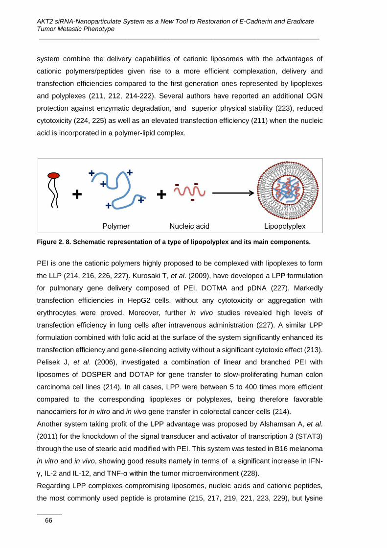
AKT2 siRNA-Nanoparticulate System as a New Tool to Restoration of E-Cadherin and Eradicate Tumor Metastic Phenotype ______________________________________________________________________________
_______ 66
system combine the delivery capabilities of cationic liposomes with the advantages of
cationic polymers/peptides given rise to a more efficient complexation, delivery and
transfection efficiencies compared to the first generation ones represented by lipoplexes
and polyplexes (211, 212, 214-222). Several authors have reported an additional OGN
protection against enzymatic degradation, and superior physical stability (223), reduced
cytotoxicity (224, 225) as well as an elevated transfection efficiency (211) when the nucleic
acid is incorporated in a polymer-lipid complex.
Figure 2. 8. Schematic representation of a type of lipopolyplex and its main components.
PEI is one the cationic polymers highly proposed to be complexed with lipoplexes to form
the LLP (214, 216, 226, 227). Kurosaki T, et al. (2009), have developed a LPP formulation
for pulmonary gene delivery composed of PEI, DOTMA and pDNA (227). Markedly
transfection efficiencies in HepG2 cells, without any cytotoxicity or aggregation with
erythrocytes were proved. Moreover, further in vivo studies revealed high levels of
transfection efficiency in lung cells after intravenous administration (227). A similar LPP
formulation combined with folic acid at the surface of the system significantly enhanced its
transfection efficiency and gene-silencing activity without a significant cytotoxic effect (213).
Pelisek J, et al. (2006), investigated a combination of linear and branched PEI with
liposomes of DOSPER and DOTAP for gene transfer to slow-proliferating human colon
carcinoma cell lines (214). In all cases, LPP were between 5 to 400 times more efficient
compared to the corresponding lipoplexes or polyplexes, being therefore favorable
nanocarriers for in vitro and in vivo gene transfer in colorectal cancer cells (214).
Another system taking profit of the LPP advantage was proposed by Alshamsan A, et al.
(2011) for the knockdown of the signal transducer and activator of transcription 3 (STAT3)
through the use of stearic acid modified with PEI. This system was tested in B16 melanoma
in vitro and in vivo, showing good results namely in terms of a significant increase in IFN-
γ, IL-2 and IL-12, and TNF-α within the tumor microenvironment (228).
Regarding LPP complexes compromising liposomes, nucleic acids and cationic peptides,
the most commonly used peptide is protamine (215, 217, 219, 221, 223, 229), but lysine
Chapter 2 | State of Art – Nanotechnology for Gene Delivery _______________________________________________________________________________
_______ 67
(215, 230-232), arginine and histidine (222, 231) have also been studied. The strong
association between peptides and nucleic acids confers to such LPP particles an enhanced
transfection efficiency and gene silencing. Moreover, this complexation promotes the
capacity to transfect cells in the presence of serum (184, 233). Cationic lipid-protamine-
OGN complexes have presented an increased OGN protection against enzymatic digestion,
a superior physical stability, and higher gene expression in cancer cells after intravenous
administration (219, 223, 229). Li SD & Huang L (219) and Tseng YC, et al. (2009) have
developed a tumor-targeted LPP for siRNA delivery (234). The nucleic acid was condensed
with protamine and the resulting nanoparticles were coated with DOTAP and cholesterol
(219, 234). Further PEGylation of the LPP complex significantly increased the tumor
localization of siRNA by four-fold, while a two- to three-fold increase in the silencing effect
was observed in an in vivo human lung cancer model (219, 220).
Anionic lipids have also been used for the development of LPP systems, comprising pH-
sensitive liposome and protamine, for macrophage gene therapy. In vitro tests revealed a
safety profile and increased transfection efficiency of LPP compared to Lipofectamine®
2000 or protamine/DNA complexes (235).
Another type of LPP nanocarriers is the pegylated immuno-lipopolyplexes (PILP). In these
complexes, the nucleic acids are firstly compacted by a cationic polymer, and complexed
with anionic liposomes to produce the LPP. The surface of LPP is covered with PEG, in
order to promote stabilization in the bloodstream, and the PEG strands are functionalized
with targeting moieties. The system (mAb-PEG2000/DSPE-PEI/DNA) developed by Hu Y,
et al. (2010) demonstrated to be highly effective in protecting DNA from degradation, and
to accumulate at the liver promoting a high efficiency in gene delivery and gene silencing in
cancer cells without significant cytokines production or liver injury (236-238). LPP have also
been described as an efficient system for mRNA (210, 218, 221, 222, 239) and DNA-based
cancer vaccines (217, 218, 240).
Mockey M, et al. (2007) has developed a LPP for the delivery of mRNA encoding a tumor
antigen (MART-1) against the progression of B16F10 melanoma. MART-1 was condensed
in 50 nm particles of PEGylated histidylated PLL and then added to liposomes prepared
with L-histidine-(N,N-di-n-hexadecylamine)ethylamide and cholesterol. Intravenous
administration of this formulation induced the production of IFN-γ and the activation of
cytotoxic T lymphocytes. No effect was observed when the mRNA was complexed with
liposomes or with the cationic polymer alone (222). A similar nanosystem for the vaccination
of B16F10 melanoma was also developed adding mannosylated and histidylated liposomes
to mRNA-PEGylated histidylated PLL polyplexes. Authors were able to enhance the
transfection of tumor antigen mRNA in splenic dendritic cells in vivo inducing therefore an

AKT2 siRNA-Nanoparticulate System as a New Tool to Restoration of E-Cadherin and Eradicate Tumor Metastic Phenotype ______________________________________________________________________________
_______ 68
anticancer immune response (210, 239).
A DNA-based vaccine approach was proposed by Whitmore MM, et al. (241) consisting of
DOTAP:cholesterol cationic liposomes, protamine sulfate, and pDNA with unmethylated
CpG motifs. This complex was capable to stimulate a potent Th-1 cytokine response
producing TNF-α, IL-12, and IFN-γ, and was also able to trigger the antitumor NK activity.
The combination of these two innate immune responses was effective in inhibiting the
growth of established tumors in mice as they provide an acquired tumor-specific cytotoxic
T cell response (241).
LPP stability, efficacy and lack of toxicity justify their fostering arriving to clinical trials. In
fact, EGEN-001 is a LPP formulation that undergoes phase I and II clinical trials, composed
by IL-12 plasmid formulated in PEG-PEI-cholesterol to treat a variety of cancers such as
ovarian, peritoneal, fallopian tube, and colorectal cancer (242-245).
2.3. Conclusions
Despite viral vectors are still the most common systems for gene therapy in clinical trials
and the only ones that already reach the clinical practice, there are no doubts that the
application of non-viral vectors for gene delivery was an amazing and essential discovery
of the last years. Gene therapy mediated by lipoplexes, polyplexes or lipopolyplexes is a
field that is clearly growing both in basic research and pre-clinical stages. It is expected that
very soon new formulations enter in clinical trials while others grant market authorization for
a variety of diseases. Given the chance to promote the silencing of different oncogenes or
correct the expression of other absent or mutant genes, among the different healthcare
areas to which new non-viral vectors based delivery systems are being developed, oncology
is the one where research work is invested and where greater applicability is predictable.
Although lipopolyplexes seems to overcome some of the major drawbacks presented by
the other systems, the assumption that an ideal non-viral system is far from being reached,
lead us to recognize a pressing and urgent need for innovative and improved approaches.
New advances should help to meet quality requirements, which demands a balance
between effectiveness, safety and technological possibilities, aiming market approval.
Attempting this balance with non-viral vectors is a worthy effort to meet critical patients
needs.
Chapter 2 | State of Art – Nanotechnology for Gene Delivery _______________________________________________________________________________
_______ 69
2.4. References
1. Cavazzana-Calvo M, Hacein-Bey S, de Saint Basile G, Gross F, Yvon E, Nusbaum
P, et al. Gene therapy of human severe combined immunodeficiency (SCID)-X1 disease.
Science. 2000;288(5466):669-72.
2. Blaese RM, Culver KW, Miller AD, Carter CS, Fleisher T, Clerici M, et al. T
lymphocyte-directed gene therapy for ADA- SCID: initial trial results after 4 years. Science.
1995;270(5235):475-80.
3. Kumar SR, Markusic DM, Biswas M, High KA, Herzog RW. Clinical development of
gene therapy: results and lessons from recent successes. Mol Ther Methods Clin Dev.
2016;3:16034.
4. Kaufmann KB, Büning H, Galy A, Schambach A, Grez M. Gene therapy on the move.
EMBO Mol Med. 2013;5(11):1642-61.
5. Wang D, Gao G. State-of-the-art human gene therapy: Part I. gene delivery
technologies. Discov Med. 2014;18(97):67-77.
6. Videira M, Arranja A, Rafael D, Gaspar R. Preclinical development of siRNA
therapeutics: towards the match between fundamental science and engineered systems.
Nanomedicine. 2014;10(4):689-702.
7. Hacein-Bey-Abina S, von Kalle C, Schmidt M, Le Deist F, Wulffraat N, McIntyre E,
et al. A serious adverse event after successful gene therapy for X-linked severe combined
immunodeficiency. N Engl J Med. 2003;348(3):255-6.
8. EMA. Glybera. EPAR - Product information. www.ema.europa.eu2012.
9. Wasungu L, Hoekstra D. Cationic lipids, lipoplexes and intracellular delivery of
genes. J Control Release. 2006;116(2):255-64.
10. Eliyahu H, Joseph A, Schillemans JP, Azzam T, Domb AJ, Barenholz Y.
Characterization and in vivo performance of dextran-spermine polyplexes and
DOTAP/cholesterol lipoplexes administered locally and systemically. Biomaterials.
2007;28(14):2339-49.
11. Liu F, Huang L. Development of non-viral vectors for systemic gene delivery. J
Control Release. 2002;78(1-3):259-66.
12. Zhang XX, McIntosh TJ, Grinstaff MW. Functional lipids and lipoplexes for improved
gene delivery. Biochimie. 2012;94(1):42-58.
13. Zhang S, Xu Y, Wang B, Qiao W, Liu D, Li Z. Cationic compounds used in lipoplexes
and polyplexes for gene delivery. J Control Release. 2004;100(2):165-80.
14. Scholz C, Wagner E. Therapeutic plasmid DNA versus siRNA delivery: common and
different tasks for synthetic carriers. J Control Release. 2012;161(2):554-65.

AKT2 siRNA-Nanoparticulate System as a New Tool to Restoration of E-Cadherin and Eradicate Tumor Metastic Phenotype ______________________________________________________________________________
_______ 70
15. Li L, Wei Y, Gong C. Polymeric Nanocarriers for Non-Viral Gene Delivery. J Biomed
Nanotechnol. 2015;11(5):739-70.
16. de Martimprey H, Vauthier C, Malvy C, Couvreur P. Polymer nanocarriers for the
delivery of small fragments of nucleic acids: oligonucleotides and siRNA. Eur J Pharm
Biopharm. 2009;71(3):490-504.
17. Gary DJ, Puri N, Won YY. Polymer-based siRNA delivery: perspectives on the
fundamental and phenomenological distinctions from polymer-based DNA delivery. J
Control Release. 2007;121(1-2):64-73.
18. Mao S, Sun W, Kissel T. Chitosan-based formulations for delivery of DNA and
siRNA. Adv Drug Deliv Rev. 2010;62(1):12-27.
19. Oskuee RK, Philipp A, Dehshahri A, Wagner E, Ramezani M. The impact of
carboxyalkylation of branched polyethylenimine on effectiveness in small interfering RNA
delivery. J Gene Med. 2010;12(9):729-38.
20. Chira S, Jackson CS, Oprea I, Ozturk F, Pepper MS, Diaconu I, et al. Progresses
towards safe and efficient gene therapy vectors. Oncotarget. 2015;6(31):30675-703.
21. Xiong XB, Falamarzian A, Garg SM, Lavasanifar A. Engineering of amphiphilic block
copolymers for polymeric micellar drug and gene delivery. J Control Release.
2011;155(2):248-61.
22. Ozpolat B, Sood AK, Lopez-Berestein G. Nanomedicine based approaches for the
delivery of siRNA in cancer. Journal of internal medicine. 2010;267(1):44-53.
23. Dohmen C, Edinger D, Fröhlich T, Schreiner L, Lächelt U, Troiber C, et al. Nanosized
Multifunctional Polyplexes for Receptor-Mediated SiRNA Delivery. ACS Nano. 2012.
24. Yang Z, Sahay G, Sriadibhatla S, Kabanov AV. Amphiphilic block copolymers
enhance cellular uptake and nuclear entry of polyplex-delivered DNA. Bioconjug Chem.
2008;19(10):1987-94.
25. Dufresne MH, Elsabahy M, Leroux JC. Characterization of polyion complex micelles
designed to address the challenges of oligonucleotide delivery. Pharm Res.
2008;25(9):2083-93.
26. Howard KA. Delivery of RNA interference therapeutics using polycation-based
nanoparticles. Advanced drug delivery reviews. 2009;61(9):710-20.
27. Maeda H. The enhanced permeability and retention (EPR) effect in tumor
vasculature: the key role of tumor-selective macromolecular drug targeting. Adv Enzyme
Regul. 2001;41:189-207.
28. Misra R, Acharya S, Sahoo SK. Cancer nanotechnology: application of
nanotechnology in cancer therapy. Drug Discovery Today. 2010;15(19-20):842-50.

Chapter 2 | State of Art – Nanotechnology for Gene Delivery _______________________________________________________________________________
_______ 71
29. Kim KY. Nanotechnology platforms and physiological challenges for cancer
therapeutics. Nanomedicine : nanotechnology, biology, and medicine. 2007;3(2):103-10.
30. Vasir JK, Labhasetwar V. Biodegradable Nanoparticles for Cytosolic Delivery of
Therapeutics. Advanced Drug Delivery Reviews. 2007;59(8):718-28.
31. Mathur V, Satrawala Y, Rajput MS, Kumar P, Shrivastava P, Vishvkarma A. Solid
lipid nanoparticles in cancer therapy. International Journal of Drug Delivery. 2010;2(3):192-
9.
32. Videira MA, Botelho MF, Santos AC, Gouveia LF, de Lima JJ, Almeida AJ.
Lymphatic uptake of pulmonary delivered radiolabelled solid lipid nanoparticles. Journal of
drug targeting. 2002;10(8):607-13.
33. Videira MA, Gano L, Santos C, Neves M, Almeida AJ. Lymphatic uptake of lipid
nanoparticles following endotracheal administration. Journal of microencapsulation.
2006;23(8):855-62.
34. Pathak A, Patnaik S, Gupta KC. Recent trends in non-viral vector-mediated gene
delivery. Biotechnology Journal. 2009;4(11):1559-72.
35. Zhang S, Zhao B, Jiang H, Wang B, Ma B. Cationic lipids and polymers mediated
vectors for delivery of siRNA. Journal of Controlled Release. 2007;123(1):1-10.
36. Aigner A. Delivery Systems for the Direct Application of siRNAs to Induce RNA
Interference (RNAi) In Vivo. Journal of Biomedicine and Biotechnology. 2006;2006:1-16.
37. Zimmermann TS, Lee ACH, Akinc A, Bramlage B, Bumcrot D, Fedoruk MN, et al.
RNAi-mediated gene silencing in non-human primates. Nature. 2006;441(7089):111-4.
38. Martin I, Dohmen C, Mas-Moruno C, Troiber C, Kos P, Schaffert D, et al. Solid-
phase-assisted synthesis of targeting peptide-PEG-oligo(ethane amino)amides for
receptor-mediated gene delivery. Org Biomol Chem. 2012;10(16):3258-68.
39. Tahara K, Sakai T, Yamamoto H, Takeuchi H, Kawashima Y. Establishing chitosan
coated PLGA nanosphere platform loaded with wide variety of nucleic acid by complexation
with cationic compound for gene delivery. International Journal of Pharmaceutics.
2008;354(1-2):210-6.
40. Cho K, Wang X, Nie S, Chen Z, Shin DM. Therapeutic Nanoparticles for Drug
Delivery in Cancer. Clinical Cancer Research. 2008;14(5):1310-6.
41. Tros de Ilarduya C, Sun Y, Düzgüneş N. Gene delivery by lipoplexes and polyplexes.
European Journal of Pharmaceutical Sciences. 2010;40(3):159-70.
42. Wu GY, Wu CH. Receptor-mediated in vitro gene transformation by a soluble DNA
carrier system. J Biol Chem. 1987;262(10):4429-32.
43. Dizaj SM, Jafari S, Khosroushahi AY. A sight on the current nanoparticle-based
gene delivery vectors. Nanoscale Res Lett. 2014;9(1):252.

AKT2 siRNA-Nanoparticulate System as a New Tool to Restoration of E-Cadherin and Eradicate Tumor Metastic Phenotype ______________________________________________________________________________
_______ 72
44. Nayerossadat N, Maedeh T, Ali PA. Viral and nonviral delivery systems for gene
delivery. Adv Biomed Res. 2012;1:27.
45. Midoux P, Pichon C, Yaouanc JJ, Jaffrès PA. Chemical vectors for gene delivery: a
current review on polymers, peptides and lipids containing histidine or imidazole as nucleic
acids carriers. Br J Pharmacol. 2009;157(2):166-78.
46. Asseline U, Gonçalves C, Pichon C, Midoux P. Improved nuclear delivery of
antisense 2'-Ome RNA by conjugation with the histidine-rich peptide H5WYG. J Gene Med.
2014;16(7-8):157-65.
47. Puras G, Martínez-Navarrete G, Mashal M, Zárate J, Agirre M, Ojeda E, et al.
Protamine/DNA/Niosome Ternary Nonviral Vectors for Gene Delivery to the Retina: The
Role of Protamine. Mol Pharm. 2015;12(10):3658-71.
48. Tao J, Ding WF, Che XH, Chen YC, Chen F, Chen XD, et al. Optimization of a
cationic liposome-based gene delivery system for the application of miR-145 in anticancer
therapeutics. Int J Mol Med. 2016;37(5):1345-54.
49. Johnson RP, Uthaman S, John JV, Lee HR, Park IK, Kim I. Biodegradable
poly(ethylene glycol) methyl ether acrylate-b-poly(l-lysine)-b-poly(l-histidine) triblock
copolypeptides for non-viral gene delivery. J Control Release. 2015;213:e93-4.
50. Feng T, Tian H, Dong X, Lam MH, Liang H, Chen X. pH-sensitive OEI-poly(aspartic
acid-b-lysine) as charge shielding system for gene delivery. J Control Release.
2015;213:e104.
51. Raad M, Teunissen EA, Mastrobattista E. Peptide vectors for gene delivery: from
single peptides to multifunctional peptide nanocarriers. Nanomedicine (Lond).
2014;9(14):2217-32.
52. Guo S, Huang Y, Zhang W, Wang W, Wei T, Lin D, et al. Ternary complexes of
amphiphilic polycaprolactone-graft-poly (N,N-dimethylaminoethyl methacrylate), DNA and
polyglutamic acid-graft-poly(ethylene glycol) for gene delivery. Biomaterials.
2011;32(18):4283-92.
53. Huang Y, Lin D, Jiang Q, Zhang W, Guo S, Xiao P, et al. Binary and ternary
complexes based on polycaprolactone-graft-poly (N, N-dimethylaminoethyl methacrylate)
for targeted siRNA delivery. Biomaterials. 2012;33(18):4653-64.
54. Guo S, Huang Y, Wei T, Zhang W, Wang W, Lin D, et al. Amphiphilic and
biodegradable methoxy polyethylene glycol-block-(polycaprolactone-graft-poly(2-
(dimethylamino)ethyl methacrylate)) as an effective gene carrier. Biomaterials.
2011;32(3):879-89.
55. Katas H, Alpar HO. Development and characterisation of chitosan nanoparticles for
siRNA delivery. Journal of Controlled Release. 2006;115(2):216-25.
Chapter 2 | State of Art – Nanotechnology for Gene Delivery _______________________________________________________________________________
_______ 73
56. Nagpal K, Singh SK, Mishra DN. Chitosan nanoparticles: a promising system in
novel drug delivery. Chem Pharm Bull (Tokyo). 2010;58(11):1423-30.
57. Wang X, Wang Y, Chen ZG, Shin DM. Advances of Cancer Therapy by
Nanotechnology. Cancer Research and Treatment. 2009;41(1):1.
58. Liu X, Howard KA, Dong M, Andersen MO, Rahbek UL, Johnsen MG, et al. The
influence of polymeric properties on chitosan/siRNA nanoparticle formulation and gene
silencing. Biomaterials. 2007;28(6):1280-8.
59. Mao S, Sun W, Kissel T. Chitosan-based formulations for delivery of DNA and
siRNA☆. Advanced Drug Delivery Reviews. 2010;62(1):12-27.
60. Kim TH, Kim SI, Akaike T, Cho CS. Synergistic effect of poly(ethylenimine) on the
transfection efficiency of galactosylated chitosan/DNA complexes. J Control Release.
2005;105(3):354-66.
61. Wong K, Sun G, Zhang X, Dai H, Liu Y, He C, et al. PEI-g-chitosan, a novel gene
delivery system with transfection efficiency comparable to polyethylenimine in vitro and after
liver administration in vivo. Bioconjug Chem. 2006;17(1):152-8.
62. Kim TH, Ihm JE, Choi YJ, Nah JW, Cho CS. Efficient gene delivery by urocanic acid-
modified chitosan. J Control Release. 2003;93(3):389-402.
63. Kiang T, Bright C, Cheung CY, Stayton PS, Hoffman AS, Leong KW. Formulation of
chitosan-DNA nanoparticles with poly(propyl acrylic acid) enhances gene expression. J
Biomater Sci Polym Ed. 2004;15(11):1405-21.
64. Moreira C, Oliveira H, Pires LR, Simões S, Barbosa MA, Pêgo AP. Improving
chitosan-mediated gene transfer by the introduction of intracellular buffering moieties into
the chitosan backbone. Acta Biomater. 2009;5(8):2995-3006.
65. Hu FQ, Zhao MD, Yuan H, You J, Du YZ, Zeng S. A novel chitosan oligosaccharide-
stearic acid micelles for gene delivery: properties and in vitro transfection studies. Int J
Pharm. 2006;315(1-2):158-66.
66. Shim MS, Kwon YJ. Efficient and targeted delivery of siRNA in vivo. FEBS Journal.
2010;277(23):4814-27.
67. Aigner A. Gene silencing through RNA interference (RNAi) in vivo: strategies based
on the direct application of siRNAs. Journal of biotechnology. 2006;124(1):12-25.
68. Sorkin A, Von Zastrow M. Signal transduction and endocytosis: close encounters of
many kinds. Nature reviews Molecular cell biology. 2002;3(8):600-14.
69. Akinc A, Thomas M, Klibanov AM, Langer R. Exploring polyethylenimine-mediated
DNA transfection and the proton sponge hypothesis. J Gene Med. 2005;7(5):657-63.
70. Tros de Ilarduya C, Sun Y, Düzgüneş N. Gene delivery by lipoplexes and polyplexes.
Eur J Pharm Sci. 2010;40(3):159-70.

AKT2 siRNA-Nanoparticulate System as a New Tool to Restoration of E-Cadherin and Eradicate Tumor Metastic Phenotype ______________________________________________________________________________
_______ 74
71. Roesler S, Koch FP, Schmehl T, Weissmann N, Seeger W, Gessler T, et al.
Amphiphilic, low molecular weight poly(ethylene imine) derivatives with enhanced stability
for efficient pulmonary gene delivery. J Gene Med. 2011;13(2):123-33.
72. Godbey WT, Wu KK, Mikos AG. Size matters: molecular weight affects the efficiency
of poly(ethylenimine) as a gene delivery vehicle. J Biomed Mater Res. 1999;45(3):268-75.
73. Thomas M, Klibanov AM. Enhancing polyethylenimine's delivery of plasmid DNA
into mammalian cells. Proc Natl Acad Sci U S A. 2002;99(23):14640-5.
74. Mao S, Neu M, Germershaus O, Merkel O, Sitterberg J, Bakowsky U, et al. Influence
of polyethylene glycol chain length on the physicochemical and biological properties of
poly(ethylene imine)-graft-poly(ethylene glycol) block copolymer/SiRNA polyplexes.
Bioconjug Chem. 2006;17(5):1209-18.
75. Dehshahri A, Oskuee RK, Shier WT, Hatefi A, Ramezani M. Gene transfer efficiency
of high primary amine content, hydrophobic, alkyl-oligoamine derivatives of
polyethylenimine. Biomaterials. 2009;30(25):4187-94.
76. Grayson AC, Doody AM, Putnam D. Biophysical and structural characterization of
polyethylenimine-mediated siRNA delivery in vitro. Pharm Res. 2006;23(8):1868-76.
77. Meiners S, Brinkmann V, Naundorf H, Birchmeier W. Role of morphogenetic factors
in metastasis of mammary carcinoma cells. Oncogene. 1998;16:9-20.
78. Merdan T, Callahan J, Petersen H, Kunath K, Bakowsky U, Kopeckova P, et al.
Pegylated polyethylenimine-Fab' antibody fragment conjugates for targeted gene delivery
to human ovarian carcinoma cells. Bioconjugate chemistry. 2003;14(5):989-96.
79. Malek A, Czubayko F, Aigner A. PEG grafting of polyethylenimine (PEI) exerts
different effects on DNA transfection and siRNA-induced gene targeting efficacy. Journal of
Drug Targeting. 2008;16(2):124-39.
80. Philipp A, Zhao X, Tarcha P, Wagner E, Zintchenko A. Hydrophobically modified
oligoethylenimines as highly efficient transfection agents for siRNA delivery. Bioconjugate
Chemistry. 2009;20(11):2055-61.
81. Guo G, Zhou L, Chen Z, Chi W, Yang X, Wang W, et al. Alkane-modified low-
molecular-weight polyethylenimine with enhanced gene silencing for siRNA delivery. Int J
Pharm. 2013;450(1-2):44-52.
82. Dong X, Lin L, Chen J, Guo Z, Tian H, Li Y, et al. A Serum-Tolerant Hydroxyl-
Modified Polyethylenimine as Versatile Carriers of pDNA/siRNA. Macromol Biosci.
2013;13(4):512-22.
83. Kataoka K, Harada A, Nagasaki Y. Block copolymer micelles for drug delivery:
design, characterization and biological significance. Advanced Drug Delivery Reviews.
2001;47(1):113-31.

Chapter 2 | State of Art – Nanotechnology for Gene Delivery _______________________________________________________________________________
_______ 75
84. Adams ML, Lavasanifar A, Kwon GS. Amphiphilic block copolymers for drug
delivery. J Pharm Sci. 2003;92(7):1343-55.
85. Xiong XB, Binkhathlan Z, Molavi O, Lavasanifar A. Amphiphilic block co-polymers:
Preparation and application in nanodrug and gene delivery. Acta Biomater. 2012;8(6):2017-
33.
86. Andrade F, Videira M, Ferreira D, Sarmento B. Micelle-based systems for pulmonary
drug delivery and targeting. Drug Delivery Letters. 2011;1(2):171-85.
87. Torchilin V. Micellar nanocarriers: pharmaceutical perspectives. Pharm Res.
2007;24(1):1-16.
88. Moghimi SM, Hunter AC. Poloxamers and poloxamines in nanoparticle engineering
and experimental medicine. Trends Biotechnol. 2000;18(10):412-20.
89. Kabanov AV, Batrakova EV, Alakhov VY. Pluronic block copolymers as novel
polymer therapeutics for drug and gene delivery. J Control Release. 2002;82(2-3):189-212.
90. Urbani CN, Bell CA, Lonsdale D, Whittaker MR, Monteiro MJ. Self-Assembly of
amphiphilic polymeric dendrimers synthesized with selective degradable
linkages Macromolecules. 2008;41:76-86.
91. Letchford K, Burt H. A review of the formation and classification of amphiphilic block
copolymer nanoparticulate structures: micelles, nanospheres, nanocapsules and
polymersomes. European Journal of Pharmaceutics and Biopharmaceutics.
2007;65(3):259-69.
92. Myers D. Surfactant science and technology. 3 ed. USA: Wiley-Interscience; 2006.
380 p.
93. Malmsten M. Surfactants and Polymers in Drug Delivery Marcel Dekker, Inc.; 2002.
335 p.
94. Babinot J, Guigner JM, Renard E, Langlois V. A micellization study of medium chain
length poly(3-hydroxyalkanoate)-based amphiphilic diblock copolymers. J Colloid Interface
Sci. 2012;375(1):88-93.
95. Letchford K, Liggins R, Burt H. Solubilization of hydrophobic drugs by methoxy
poly(ethylene glycol)-block-polycaprolactone diblock copolymer micelles: theoretical and
experimental data and correlations. J Pharm Sci. 2008;97(3):1179-90.
96. Sawant R, Jhaveri A. Micellar nanopreparations for medicine. In: Torchilin V, editor.
Handbook of Nanobiomedical Research: Fundamentals, Applications and Recent
Developments. Frontiers in Nanobiomedical Research. 3: World Scientific; 2014.
97. Study of Weekly Genexol®-PM Plus Gemcitabine in Subjects With Recurrent and
Metastatic Adenocarcinoma of the Pancreas 2016. Available from:
https://clinicaltrials.gov/ct2/show/NCT02739633.
AKT2 siRNA-Nanoparticulate System as a New Tool to Restoration of E-Cadherin and Eradicate Tumor Metastic Phenotype ______________________________________________________________________________
_______ 76
98. Evaluate the Efficacy and Safety of Genexol®-PM Compared to Genexol® in
Recurrent or Metastatic Breast Cancer 2016. Available from:
https://clinicaltrials.gov/ct2/show/NCT00876486.
99. A Trial to Evaluate Efficacy and Safety of the Combination Therapy of Genexol®-
PM Plus Carboplatin® Compared to Genexol® Plus Carboplatin® as a Firstline Treatment
in Subjects With Ovarian Cancer 2016. Available from:
https://clinicaltrials.gov/ct2/show/NCT01276548.
100. A Phase II Trial of Genexol-PM and Gemcitabine in Patients With Advanced Non-
small-cell Lung Cancer 2016. Available from:
https://clinicaltrials.gov/ct2/show/NCT01770795.
101. A Clinical Trial of Paclitaxel Loaded Polymeric Micelle in Patients With Taxane-
Pretreated Recurrent Breast Cancer 2016. Available from:
https://clinicaltrials.gov/ct2/show/NCT00912639.
102. A Study of NK012 in Patients With Relapsed Small Cell Lung Cancer 2016. Available
from: https://clinicaltrials.gov/ct2/show/NCT00951613.
103. A Study of NK012 in Patients With Advanced, Metastatic Triple Negative Breast
Cancer 2016. Available from: https://clinicaltrials.gov/ct2/show/NCT00951054.
104. A Phase III Study of NK105 in Patients With Breast Cancer 2016. Available from:
https://clinicaltrials.gov/ct2/show/NCT01644890.
105. Matsumura Y, Hamaguchi T, Ura T, Muro K, Yamada Y, Shimada Y, et al. Phase I
clinical trial and pharmacokinetic evaluation of NK911, a micelle-encapsulated doxorubicin.
Br J Cancer. 2004;91(10):1775-81.
106. A Phase 1 Dose-Escalation and Pharmacokinetic Study of NC-4016 in Patients With
Advanced Solid Tumors or Lymphoma 2016. Available from:
https://clinicaltrials.gov/ct2/show/NCT01999491.
107. Combination Therapy With NC-6004 and Gemcitabine Versus Gemcitabine Alone
in Pancreatic Cancer 2016. Available from:
https://clinicaltrials.gov/ct2/show/NCT02043288.
108. Combination Therapy With NC-6004 and Gemcitabine in Advanced Solid Tumors or
Non-Small Cell Lung, Biliary and Bladder Cancer 2016. Available from:
https://clinicaltrials.gov/ct2/show/NCT02240238.
109. A Study Using Intravenous Paxceedâ„¢ to Treat Patients With Rheumatoid Arthritis
2016. Available from: https://clinicaltrials.gov/ct2/show/NCT00055133.
110. Micellar Paclitaxel to Treat Severe Psoriasis 2016. Available from:
https://clinicaltrials.gov/ct2/show/NCT00006276.
Chapter 2 | State of Art – Nanotechnology for Gene Delivery _______________________________________________________________________________
_______ 77
111. Efficacy Study of Maintenance IT-101 Therapy for Ovarian Cancer Patients 2016.
Available from: https://clinicaltrials.gov/ct2/show/NCT00753740.
112. Study of CRLX101 (Formerly Named IT-101) in the Treatment of Advanced Solid
Tumors 2016. Available from: https://clinicaltrials.gov/ct2/show/NCT00333502.
113. Effects of an Antioxidant-Enriched Multivitamin Supplement on Inflammation and
Oxidative Stress in Cystic Fibrosis 2016. Available from:
https://clinicaltrials.gov/ct2/show/NCT01859390.
114. Matsumura Y. Preclinical and clinical studies of NK012, an SN-38-incorporating
polymeric micelles, which is designed based on EPR effect. Adv Drug Deliv Rev.
2011;63(3):184-92.
115. Kato K, Chin K, Yoshikawa T, Yamaguchi K, Tsuji Y, Esaki T, et al. Phase II study
of NK105, a paclitaxel-incorporating micellar nanoparticle, for previously treated advanced
or recurrent gastric cancer. Invest New Drugs. 2011.
116. Gilani K, Moazeni E, Ramezanli T, Amini M, Fazeli MR, Jamalifar H. Development
of respirable nanomicelle carriers for delivery of amphotericin B by jet nebulization. J Pharm
Sci. 2011;100(1):252-9.
117. Wang CH, Wang WT, Hsiue GH. Development of polyion complex micelles for
encapsulating and delivering amphotericin B. Biomaterials. 2009;30(19):3352-8.
118. Kim DW, Kim SY, Kim HK, Kim SW, Shin SW, Kim JS, et al. Multicenter phase II
trial of Genexol-PM, a novel Cremophor-free, polymeric micelle formulation of paclitaxel,
with cisplatin in patients with advanced non-small-cell lung cancer. Ann Oncol.
2007;18(12):2009-14.
119. Lee KS, Chung HC, Im SA, Park YH, Kim CS, Kim SB, et al. Multicenter phase II
trial of Genexol-PM, a Cremophor-free, polymeric micelle formulation of paclitaxel, in
patients with metastatic breast cancer. Breast Cancer Res Treat. 2008;108(2):241-50.
120. Saif MW, Rubin MS, Figueroa JA, Kerr RO. Multicenter phase II trial of Genexol-PM
(GPM), a novel Cremophor-free, polymeric micelle formulation of paclitaxel in patients with
advanced pancreatic cancer (APC): Final results. . 2008 Gastrointestinal Cancers
Symposium; Orlando2008.
121. Valle JW, Armstrong A, Newman C, Alakhov V, Pietrzynski G, Brewer J, et al. A
phase 2 study of SP1049C, doxorubicin in P-glycoprotein-targeting pluronics, in patients
with advanced adenocarcinoma of the esophagus and gastroesophageal junction. Invest
New Drugs. 2011;29(5):1029-37.
122. Ho K, Li W, Wong C, Li P. Amphiphilic polymeric particles with core-shell
nanostructures: emulsion-based syntheses and potential applications. Colloid and Polymer
Science. 2010;288(16-17):1503-23.
AKT2 siRNA-Nanoparticulate System as a New Tool to Restoration of E-Cadherin and Eradicate Tumor Metastic Phenotype ______________________________________________________________________________
_______ 78
123. Kwon GS. Amphiphilic block copolymer micelles for nanoscale drug delivery. DRUG
DEVELOP RES. 2006;67:15-22.
124. Owens DE, Peppas NA. Opsonization, biodistribution, and pharmacokinetics of
polymeric nanoparticles. Int J Pharm. 2006;307(1):93-102.
125. Moghimi SM, Szebeni J. Stealth liposomes and long circulating nanoparticles:
critical issues in pharmacokinetics, opsonization and protein-binding properties. Prog Lipid
Res. 2003;42(6):463-78.
126. Chaudhari KR, Ukawala M, Manjappa AS, Kumar A, Mundada PK, Mishra AK, et al.
Opsonization, biodistribution, cellular uptake and apoptosis study of PEGylated PBCA
nanoparticle as potential drug delivery carrier. Pharm Res. 2012;29(1):53-68.
127. Rösler A, Vandermeulen GW, Klok HA. Advanced drug delivery devices via self-
assembly of amphiphilic block copolymers. Adv Drug Deliv Rev. 2001;53(1):95-108.
128. Kumar M, Kumar N, Domb A, Arora M. Pharmaceutical polymeric controlled drug
delivery systems. Filled Elastomers Drug Delivery Systems. 2002;160:45-117.
129. Smith A, Xu X, McCormick C. Stimuli-responsive amphiphilic (co)polymers via RAFT
polymerization. Progress in Polymer Science. 2010;35(1-2):45-93.
130. Ward MA, Georgiou TK. Thermoresponsive Polymers for Biomedical Applications.
Polymers. 2011;3:1215-42.
131. Motornov M, Roiter Y, Tokarev I, Minko S. Stimuli-responsive nanoparticles,
nanogels and capsules for integrated multifunctional intelligent systems. Progress in
Polymer Science. 2010;35(1-2):174-211.
132. Hu Y, Litwin T, Nagaraja AR, Kwong B, Katz J, Watson N, et al. Cytosolic delivery
of membrane-impermeable molecules in dendritic cells using pH-responsive core-shell
nanoparticles. Nano Lett. 2007;7(10):3056-64.
133. Morishita M, Goto T, Nakamura K, Lowman AM, Takayama K, Peppas NA. Novel
oral insulin delivery systems based on complexation polymer hydrogels: single and multiple
administration studies in type 1 and 2 diabetic rats. J Control Release. 2006;110(3):587-94.
134. Kamei N, Morishita M, Chiba H, Kavimandan NJ, Peppas NA, Takayama K.
Complexation hydrogels for intestinal delivery of interferon beta and calcitonin. J Control
Release. 2009;134(2):98-102.
135. Guo W, Wang T, Tang X, Zhang Q, Yu F, Pei M. Triple stimuli-responsive
amphiphilic glycopolymer
Journal of Polymer Science Part A: Polymer Chemistry Volume 52, Issue 15. Journal of
Polymer Science Part A: Polymer Chemistry [Internet]. 2014 01; 52(15):[2131-8 pp.].
Available from: http://onlinelibrary.wiley.com/doi/10.1002/pola.27222/abstract.

Chapter 2 | State of Art – Nanotechnology for Gene Delivery _______________________________________________________________________________
_______ 79
136. Liu Y, Samsonova O, Sproat B, Merkel O, Kissel T. Biophysical characterization of
hyper-branched polyethylenimine-graft-polycaprolactone-block-mono-methoxyl-
poly(ethylene glycol) copolymers (hy-PEI-PCL-mPEG) for siRNA delivery. J Control
Release. 2011;153(3):262-8.
137. Yang XZ, Dou S, Sun TM, Mao CQ, Wang HX, Wang J. Systemic delivery of siRNA
with cationic lipid assisted PEG-PLA nanoparticles for cancer therapy. J Control Release.
2011;156(2):203-11.
138. Sun J, Luo T, Sheng R, Li H, Wang Z, Cao A. Intracellular plasmid DNA delivery by
self-assembled nanoparticles of amphiphilic PHML-b-PLLA-b-PHML copolymers and the
endocytosis pathway analysis. J Biomater Appl. 2016.
139. Murata N, Takashima Y, Toyoshima K, Yamamoto M, Okada H. Anti-tumor effects
of anti-VEGF siRNA encapsulated with PLGA microspheres in mice. J Control Release.
2008;126(3):246-54.
140. Alshamsan A, Haddadi A, Hamdy S, Samuel J, El-Kadi AO, Uludag H, et al. STAT3
Silencing in Dendritic Cells by siRNA Polyplexes Encapsulated in PLGA Nanoparticles for
the Modulation of Anticancer Immune Response. Mol Pharm. 2010.
141. Mishra S, Peddada LY, Devore DI, Roth CM. Poly(alkylene oxide) Copolymers for
Nucleic Acid Delivery. Acc Chem Res. 2012.
142. Kuo JH. Effect of Pluronic-block copolymers on the reduction of serum-mediated
inhibition of gene transfer of polyethyleneimine-DNA complexes. Biotechnol Appl Biochem.
2003;37(Pt 3):267-71.
143. Wang M, Wu B, Lu P, Tucker JD, Milazi S, Shah SN, et al. Pluronic-PEI copolymers
enhance exon-skipping of 2'-O-methyl phosphorothioate oligonucleotide in cell culture and
dystrophic mdx mice. Gene Ther. 2014;21(1):52-9.
144. Davis ME. The first targeted delivery of siRNA in humans via a self-assembling,
cyclodextrin polymer-based nanoparticle: from concept to clinic. Mol Pharm. 2009;6(3):659-
68.
145. Zhu Y, Sheng R, Luo T, Li H, Sun W, Li Y, et al. Amphiphilic cationic [dendritic poly(L-
lysine)]-block-poly(L-lactide)-block-[dendritic poly(L-lysine)]s in aqueous solution: self-
aggregation and interaction with DNA as gene delivery carriers. Macromol Biosci.
2011;11(2):174-86.
146. Holzerny P, Ajdini B, Heusermann W, Bruno K, Schuleit M, Meinel L, et al.
Biophysical properties of chitosan/siRNA polyplexes: profiling the polymer/siRNA
interactions and bioactivity. J Control Release. 2012;157(2):297-304.
AKT2 siRNA-Nanoparticulate System as a New Tool to Restoration of E-Cadherin and Eradicate Tumor Metastic Phenotype ______________________________________________________________________________
_______ 80
147. Shum VW, Gabrielson NP, Forrest ML, Pack DW. The effects of PVP(Fe(III))
catalyst on polymer molecular weight and gene delivery via biodegradable cross-linked
polyethylenimine. Pharm Res. 2012;29(2):500-10.
148. Ulasov AV, Khramtsov YV, Trusov GA, Rosenkranz AA, Sverdlov ED, Sobolev AS.
Properties of PEI-based polyplex nanoparticles that correlate with their transfection efficacy.
Mol Ther. 2011;19(1):103-12.
149. Commander NJ, Brewer JM, Wren BW, Spencer SA, Macmillan AP, Stack JA.
Liposomal delivery of p-ialB and p-omp25 DNA vaccines improves immunogenicity but fails
to provide full protection against B. melitensis challenge. Genet Vaccines Ther. 2010;8:5.
150. Klausner EA, Zhang Z, Wong SP, Chapman RL, Volin MV, Harbottle RP. Corneal
gene delivery: chitosan oligomer as a carrier of CpG rich, CpG free or S/MAR plasmid DNA.
J Gene Med. 2012;14(2):100-8.
151. Rozema DB, Lewis DL, Wakefield DH, Wong SC, Klein JJ, Roesch PL, et al.
Dynamic PolyConjugates for targeted in vivo delivery of siRNA to hepatocytes. Proc Natl
Acad Sci U S A. 2007;104(32):12982-7.
152. Mudd SR, Trubetskoy VS, Blokhin AV, Weichert JP, Wolff JA. Hybrid PET/CT for
noninvasive pharmacokinetic evaluation of dynamic PolyConjugates, a synthetic siRNA
delivery system. Bioconjug Chem. 2010;21(7):1183-9.
153. Itaka K, Osada K, Morii K, Kim P, Yun SH, Kataoka K. Polyplex nanomicelle
promotes hydrodynamic gene introduction to skeletal muscle. J Control Release.
2010;143(1):112-9.
154. Uchida S, Itaka K, Chen Q, Osada K, Miyata K, Ishii T, et al. Combination of
chondroitin sulfate and polyplex micelles from Poly(ethylene glycol)-poly{N'-[N-(2-
aminoethyl)-2-aminoethyl]aspartamide} block copolymer for prolonged in vivo gene
transfection with reduced toxicity. J Control Release. 2011;155(2):296-302.
155. Chen Q, Osada K, Ishii T, Oba M, Uchida S, Tockary TA, et al. Homo-catiomer
integration into PEGylated polyplex micelle from block-catiomer for systemic anti-
angiogenic gene therapy for fibrotic pancreatic tumors. Biomaterials. 2012;33(18):4722-30.
156. Chen YC, Jiang LP, Liu NX, Ding L, Liu XL, Wang ZH, et al. Enhanced gene
transduction into skeletal muscle of mice in vivo with pluronic block copolymers and
ultrasound exposure. Cell Biochem Biophys. 2011;60(3):267-73.
157. Davis ME, Zuckerman JE, Choi CH, Seligson D, Tolcher A, Alabi CA, et al. Evidence
of RNAi in humans from systemically administered siRNA via targeted nanoparticles.
Nature. 2010;464(7291):1067-70.
Chapter 2 | State of Art – Nanotechnology for Gene Delivery _______________________________________________________________________________
_______ 81
158. Zuckerman JE, Hsueh T, Koya RC, Davis ME, Ribas A. siRNA knockdown of
ribonucleotide reductase inhibits melanoma cell line proliferation alone or synergistically
with temozolomide. J Invest Dermatol. 2011;131(2):453-60.
159. Phase I Single Dose-Ranging Study Of Formulated hIGF-1 Plasmid In Subjects With
Cubital Tunnel Syndrome. . Available from:
http://www.gemcris.od.nih.gov/Contents/GC_CLIN_TRIAL_RPT_VIEW.asp?WIN_TYPE=
R&CTID=378.
160. Phase I/II Multi-Center, Open-Label, Multiple Administration Trial of the Safety,
Tolerability, and Efficacy of an IFN-alpha/IL-12 Plasmid-Based Therapeutic. . Available
from:
http://www.gemcris.od.nih.gov/Contents/GC_CLIN_TRIAL_RPT_VIEW.asp?WIN_TYPE=
R&CTID=116.
161. A Multicenter, Open-Label, Multiple Administration, Study of the Safety, Tolerability
and Efficacy of IFN α/IL-12 Combination Gene Therapy in Patients with Squamous Cell
Carcinoma of the Head and Neck (SCCHN). . Available from:
http://www.gemcris.od.nih.gov/Contents/GC_CLIN_TRIAL_RPT_VIEW.asp?WIN_TYPE=
R&CTID=184.
162. Oh Y-K, Park TG. siRNA delivery systems for cancer treatment. Advanced Drug
Delivery Reviews. 2009;61(10):850-62.
163. Aliabadi HM, Landry B, Sun C, Tang T, Uludag H. Supramolecular assemblies in
functional siRNA delivery: Where do we stand? Biomaterials. 2012;33(8):2546-69.
164. Ma B, Zhang S, Jiang H, Zhao B, Lv H. Lipoplex morphologies and their influences
on transfection efficiency in gene delivery. J Control Release. 2007;123(3):184-94.
165. Esposito C, Generosi J, Mossa G, Masotti A, Castellano AC. The analysis of serum
effects on structure, size and toxicity of DDAB-DOPE and DC-Chol-DOPE lipoplexes
contributes to explain their different transfection efficiency. Colloids Surf B Biointerfaces.
2006;53(2):187-92.
166. Masotti A, Mossa G, Cametti C, Ortaggi G, Bianco A, Grosso ND, et al. Comparison
of different commercially available cationic liposome-DNA lipoplexes: Parameters
influencing toxicity and transfection efficiency. Colloids Surf B Biointerfaces.
2009;68(2):136-44.
167. Kenny GD, Kamaly N, Kalber TL, Brody LP, Sahuri M, Shamsaei E, et al. Novel
multifunctional nanoparticle mediates siRNA tumour delivery, visualisation and therapeutic
tumour reduction in vivo. J Control Release. 2011;149(2):111-6.
AKT2 siRNA-Nanoparticulate System as a New Tool to Restoration of E-Cadherin and Eradicate Tumor Metastic Phenotype ______________________________________________________________________________
_______ 82
168. Akinc A, Goldberg M, Qin J, Dorkin JR, Gamba-Vitalo C, Maier M, et al.
Development of lipidoid-siRNA formulations for systemic delivery to the liver. Mol Ther.
2009;17(5):872-9.
169. Rejman J, Conese M, Hoekstra D. Gene transfer by means of lipo- and polyplexes:
role of clathrin and caveolae-mediated endocytosis. J Liposome Res. 2006;16(3):237-47.
170. Wong AW, Scales SJ, Reilly DE. DNA internalized via caveolae requires
microtubule-dependent, Rab7-independent transport to the late endocytic pathway for
delivery to the nucleus. J Biol Chem. 2007;282(31):22953-63.
171. Lu JJ, Langer R, Chen J. A novel mechanism is involved in cationic lipid-mediated
functional siRNA delivery. Mol Pharm. 2009;6(3):763-71.
172. Kapoor M, Burgess DJ, Patil SD. Physicochemical characterization techniques for
lipid based delivery systems for siRNA. Int J Pharm. 2012;427(1):35-57.
173. Mignet N, Richard C, Seguin J, Largeau C, Bessodes M, Scherman D. Anionic pH-
sensitive pegylated lipoplexes to deliver DNA to tumors. Int J Pharm. 2008;361(1-2):194-
201.
174. Srinivasan C, Burgess DJ. Optimization and characterization of anionic lipoplexes
for gene delivery. J Control Release. 2009;136(1):62-70.
175. Kapoor M, Burgess DJ. Efficient and safe delivery of siRNA using anionic lipids:
Formulation optimization studies. Int J Pharm. 2012;432(1-2):80-90.
176. Kurreck J. RNA Interference: From Basic Research to Therapeutic Applications.
Angewandte Chemie International Edition. 2009;48(8):1378-98.
177. Petros RA, DeSimone JM. Strategies in the design of nanoparticles for therapeutic
applications. Nat Rev Drug Discov. 2010;9(8):615-27.
178. Rossi JJ. RNAi therapeutics: SNALPing siRNAs in vivo. Gene Therapy.
2005;13(7):583-4.
179. Morrissey DV, Blanchard K, Shaw L, Jensen K, Lockridge JA, Dickinson B, et al.
Activity of stabilized short interfering RNA in a mouse model of hepatitis B virus replication.
Hepatology. 2005;41(6):1349-56.
180. Semple SC, Akinc A, Chen J, Sandhu AP, Mui BL, Cho CK, et al. Rational design of
cationic lipids for siRNA delivery. Nature Biotechnology. 2010;28(2):172-6.
181. Huang L, Liu Y. In Vivo Delivery of RNAi with Lipid-Based Nanoparticles. Annual
Review of Biomedical Engineering. 2011;13(1):507-30.
182. Nguyen T, Menocal EM, Harborth J, Fruehauf JH. RNAi therapeutics: An update on
delivery. Current Opinion in Molecular Therapeutics. 2008;10(2):158-67.

Chapter 2 | State of Art – Nanotechnology for Gene Delivery _______________________________________________________________________________
_______ 83
183. Ciani L, Casini A, Gabbiani C, Ristori S, Messori L, Martini G. DOTAP/DOPE and
DC-Chol/DOPE lipoplexes for gene delivery studied by circular dichroism and other
biophysical techniques. Biophys Chem. 2007;127(3):213-20.
184. Zhang Y, Li H, Sun J, Gao J, Liu W, Li B, et al. DC-Chol/DOPE cationic liposomes:
a comparative study of the influence factors on plasmid pDNA and siRNA gene delivery. Int
J Pharm. 2010;390(2):198-207.
185. Constien Rea, inventorNovel Lipids and compositions for intracellular delivery of
biologically active compounds2012.
186. Simon A. Phase I Safety, Pharmacokinetic and Pharmacodynamic Results for ALN-
PCS - Preliminary Study Results. In: Pharmaceuticals A, editor. 2012.
187. Semple SC, Klimuk SK, Harasym TO, Santos ND, Ansell SM, Wong KF, et al.
Efficient encapsulation of antisense oligonucleotides in lipid vesicles using ionizable
aminolipids: formation of novel small multilamellar vesicle structures. Biochimica et
Biophysica Acta. 2001;1510:152-66.
188. Aliabadi HM, Landry B, Mahdipoor P, Hsu CY, Uludag H. Effective down-regulation
of breast cancer resistance protein (BCRP) by siRNA delivery using lipid-substituted
aliphatic polymers. Eur J Pharm Biopharm. 2012;81(1):33-42.
189. Köhler E, Duberow-Drewe M, Drewe J, Ribes G, Loubatiéres-Mariani M, Mazer N,
et al. Absorption of an aqueous solution of a new synthetic somatostatin analogue
administered to man by gavage. Eur J Clin Pharmacol. 1987;33(2):167-71.
190. Xue HY, Wong HL. Tailoring nanostructured solid-lipid carriers for time-controlled
intracellular siRNA kinetics to sustain RNAi-mediated chemosensitization. Biomaterials.
2011;32(10):2662-72.
191. Bondi ML, Azzolina A, Craparo EF, Lampiasi N, Capuano G, Giammona G, et al.
Novel cationic solid-lipid nanoparticles as non-viral vectors for gene delivery. J Drug Target.
2007;15(4):295-301.
192. del Pozo-Rodríguez A, Delgado D, Solinís MA, Pedraz JL, Echevarría E, Rodríguez
JM, et al. Solid lipid nanoparticles as potential tools for gene therapy: in vivo protein
expression after intravenous administration. Int J Pharm. 2010;385(1-2):157-62.
193. Lobovkina T, Jacobson GB, Gonzalez-Gonzalez E, Hickerson RP, Leake D, Kaspar
RL, et al. In vivo sustained release of siRNA from solid lipid nanoparticles. ACS Nano.
2011;5(12):9977-83.
194. Yu YH, Kim E, Park DE, Shim G, Lee S, Kim YB, et al. Cationic solid lipid
nanoparticles for co-delivery of paclitaxel and siRNA. Eur J Pharm Biopharm.
2012;80(2):268-73.

AKT2 siRNA-Nanoparticulate System as a New Tool to Restoration of E-Cadherin and Eradicate Tumor Metastic Phenotype ______________________________________________________________________________
_______ 84
195. Metwally AA, Blagbrough IS, Mantell JM. Quantitative Silencing of EGFP Reporter
Gene by Self-Assembled siRNA Lipoplexes of LinOS and Cholesterol. Mol Pharm.
2012;9(11):3384-95.
196. Tagami T, Suzuki T, Matsunaga M, Nakamura K, Moriyoshi N, Ishida T, et al. Anti-
angiogenic therapy via cationic liposome-mediated systemic siRNA delivery. Int J Pharm.
2012;422(1-2):280-9.
197. Zhou S, Kawakami S, Higuchi Y, Yamashita F, Hashida M. The involvement of NK
cell activation following intranasal administration of CpG DNA lipoplex in the prevention of
pulmonary metastasis and peritoneal dissemination in mice. Clin Exp Metastasis.
2012;29(1):63-70.
198. Wu SY, Chang HI, Burgess M, McMillan NA. Vaginal delivery of siRNA using a novel
PEGylated lipoplex-entrapped alginate scaffold system. J Control Release.
2011;155(3):418-26.
199. Rothdiener M, Muller D, Castro PG, Scholz A, Schwemmlein M, Fey G, et al.
Targeted delivery of SiRNA to CD33-positive tumor cells with liposomal carrier systems. J
Control Release. 2010;144(2):251-8.
200. Aleku M, Schulz P, Keil O, Santel A, Schaeper U, Dieckhoff B, et al. Atu027, a
liposomal small interfering RNA formulation targeting protein kinase N3, inhibits cancer
progression. Cancer Res. 2008;68(23):9788-98.
201. Atu027 Plus Gemcitabine in Advanced or Metastatic Pancreatic Cancer (Atu027-I-
02) 2016. Available from: https://clinicaltrials.gov/ct2/show/NCT01808638.
202. Phase I Pilot Study of Gene Therapy for Cystic Fibrosis Using Cationic Liposome
Mediated Gene Transfer . Available from:
http://www.clinicaltrials.gov/ct2/show/NCT00004471.
203. Phase I Study of IV DOTAP: Cholesterol-Fus1 in Non-Small-Cell Lung Cancer .
Available from: http://clinicaltrials.gov/show/NCT00059605.
204. FUS1-nanoparticles and Erlotinib in Stage IV Lung Cancer 2016. Available from:
https://clinicaltrials.gov/ct2/show/NCT01455389.
205. A Study to Determine Safety, Pharmacokinetics and Pharmacodynamics of
Intravenous TKM 080301 in Neuroendocrine Tumors (NET) and Adrenocortical Carcinoma
(ACC) Patients 2016. Available from: https://clinicaltrials.gov/ct2/show/NCT01262235.
206. TKM 080301 for Primary or Secondary Liver Cancer 2016. Available from:
https://clinicaltrials.gov/ct2/show/NCT01437007.
207. QPI-1002 for Prevention of Delayed Graft Function in Recipients of an Older Donor
Kidney Transplant (ReGIFT) https://clinicaltrials.gov/ct2/show/NCT02610296 [

Chapter 2 | State of Art – Nanotechnology for Gene Delivery _______________________________________________________________________________
_______ 85
208. A Study to Evaluate Efficacy and Safety of QPI-1002 for Prevention of Acute Kidney
Injury Following Cardiac Surgery https://www.clinicaltrials.gov/ct2/show/NCT02610283 [
209. PF-04523655 Dose Escalation Study, and Evaluation of PF-04523655 With/Without
Ranibizumab in Diabetic Macular Edema (DME) (MATISSE)
https://www.clinicaltrials.gov/ct2/show/NCT01445899 [
210. Perche F, Lambert O, Berchel M, Jaffrès P-A, Pichon C, Midoux P. Gene transfer
by histidylated lipopolyplexes: A dehydration method allowing preservation of their
physicochemical parameters and transfection efficiency. International Journal of
Pharmaceutics. 2012;423:144-50.
211. Whitmore M, Li S, Huang L. LPD lipopolyplex initiates a potent cytokine response
and inhibits tumor growth. Gene Therapy. 1999;6:1867-75.
212. Matsuura M, Yamazaki Y, Sugiyama M, Kondo M, Ori H, Nango M, et al. Polycation
liposome-mediated gene transfer in vivo. Biochimica et Biophysica Acta (BBA) -
Biomembranes. 2003;1612(2):136-43.
213. Urbiola K, Garcia L, Zalba S, Garrido MJ, Tros de Ilarduya C. Efficient serum-
resistant lipopolyplexes targeted to the folate receptor. European journal of pharmaceutics
and biopharmaceutics : official journal of Arbeitsgemeinschaft fur Pharmazeutische
Verfahrenstechnik eV. 2013;83(3):358-63.
214. Pelisek J, Gaedtke L, DeRouchey J, Walker GF, Nikol S, Wagner E. Optimized
lipopolyplex formulations for gene transfer to human colon carcinoma cells under in vitro
conditions. The Journal of Gene Medicine. 2006;8(2):186-97.
215. Gao X, Huang L. Potentiation of cationic liposome-mediated gene delivery by
polycations. Biochemistry. 1996;35(3):1027-36.
216. Garcia L, Bunuales M, Duzgunes N, Ilarduya CTd. Serum-resistant lipopolyplexes
for gene delivery to liver tumour cells. European Journal of Pharmaceutics and
Biopharmaceutics. 2007;67:58-66.
217. Vangasseri DP, Han SJ, Huang L. Lipid-protamine-DNA-mediated antigen delivery.
Curr Drug Deliv. 2005;2(4):401-6.
218. Weide B, Garbe C, Rammensee H-G, Pascolo S. Plasmid DNA- and messenger
RNA-based anti-cancer vaccination. Immunology Letters. 2008;115(1):33-42.
219. Li SD, Huang L. Targeted delivery of antisense oligodeoxynucleotide and small
interference RNA into lung cancer cells. Mol Pharm. 2006a;3(5):579-88.
220. Li SD, Huang L. Surface-modified LPD nanoparticles for tumor targeting. Ann N Y
Acad Sci. 2006b:1-8.
AKT2 siRNA-Nanoparticulate System as a New Tool to Restoration of E-Cadherin and Eradicate Tumor Metastic Phenotype ______________________________________________________________________________
_______ 86
221. Hoerr I, Obst R, Rammensee HG, Jung G. In vivo application of RNA leads to
induction of specific cytotoxic T lymphocytes and antibodies. Eur J Immunol. 2000;30(1):1-
7.
222. Mockey M, Bourseau E, Chandrashekhar V, Chaudhuri A, Lafosse S, Le Cam E, et
al. mRNA-based cancer vaccine: prevention of B16 melanoma progression and metastasis
by systemic injection of MART1 mRNA histidylated lipopolyplexes. Cancer Gene Ther.
2007;14(9):802-14.
223. Birchall JC, Kellaway IW, Gumbleton M. Physical stability and in-vitro gene
expression efficiency of nebulised lipid-peptide-DNA complexes. Int J Pharm. 2000;197(1-
2):221-31.
224. Rehman Zu, Hoekstra D, Zuhorn IS. Kinases in cationic lipid/polymer-mediated gene
delivery. Drug Discovery Today. 2010;15(23–24):1110.
225. Ramezani M, Khoshhamdam M, Dehshahri A, Malaekeh-Nikouei B. The influence
of size, lipid composition and bilayer fluidity of cationic liposomes on the transfection
efficiency of nanolipoplexes. Colloids Surf B Biointerfaces. 2009;72(1):1-5.
226. Garcia L, Urbiola K, Duzgunes N, Tros de Ilarduya C. Lipopolyplexes as
nanomedicines for therapeutic gene delivery. Methods Enzymol. 2012;509:327-38.
227. Kurosaki T, Kishikawa R, Matsumoto M, Kodama Y, Hamamoto T, To H, et al.
Pulmonary gene delivery of hybrid vector, lipopolyplex containing N-lauroylsarcosine, via
the systemic route. J Control Release. 2009;136(3):213-9.
228. Alshamsan A, Hamdy S, Haddadi A, Samuel J, El-Kadi AOS, Uludag H, et al. STAT3
Knockdown in B16 Melanoma by siRNA Lipopolyplexes Induces Bystander Immune
Response In Vitro and In Vivo. Translational Oncology. 2011;4(3):178-88.
229. Li S, Huang L. In vivo gene transfer via intravenous administration of cationic lipid-
protamine-DNA (LPD) complexes. Gene Ther. 1997;4(9):891-900.
230. Kudsiova L, Fridrich B, Ho J, Mustapa MF, Campbell F, Welser K, et al. Lipopolyplex
ternary delivery systems incorporating C14 glycerol-based lipids. Mol Pharm.
2011;8(5):1831-47.
231. Welser K, Campbell F, Kudsiova L, Mohammadi A, Dawson N, Hart SL, et al. Gene
Delivery Using Ternary Lipopolyplexes Incorporating Branched Cationic Peptides: The Role
of Peptide Sequence and Branching. Mol Pharm. 2012;4:4.
232. Yan J, Berezhnoy NV, Korolev N, Su CJ, Nordenskiold L. Structure and internal
organization of overcharged cationic-lipid/peptide/DNA self-assembly complexes. Biochim
Biophys Acta. 2012;6(7):1794-800.
Chapter 2 | State of Art – Nanotechnology for Gene Delivery _______________________________________________________________________________
_______ 87
233. Junghans M, Loitsch SM, Steiniger SC, Kreuter J, Zimmer A. Cationic lipid-
protamine-DNA (LPD) complexes for delivery of antisense c-myc oligonucleotides. Eur J
Pharm Biopharm. 2005;60(2):287-94.
234. Tseng YC, Mozumdar S, Huang L. Lipid-based systemic delivery of siRNA.
Advanced drug delivery reviews. 2009;61(9):721-31.
235. Sun P, Zhong M, Shi X, Li Z. Anionic LPD complexes for gene delivery to
macrophage: preparation, characterization and transfection in vitro. J Drug Target.
2008;16(9):668-78.
236. Hu Y, Li K, Wang L, Yin S, Zhang Z, Zhang Y. Pegylated immuno-lipopolyplexes: A
novel non-viral gene delivery system for liver cancer therapy. Journal of Controlled Release.
2010;144:75-81.
237. Hu Y, Shen Y, Ji B, Wang L, Zhang Z, Zhang Y. Combinational RNAi gene therapy
of hepatocellular carcinoma by targeting human EGFR and TERT. European Journal of
Pharmaceutical Sciences. 2011a;42:387-91.
238. Hu Y, Shen Y, Ji B, Yin S, Ren X, Chen T, et al. Liver-specific gene therapy of
hepatocellular carcinoma by targeting human telomerase reverse transcriptase with
pegylated immuno-lipopolyplexes. European Journal of Pharmaceutics and
Biopharmaceutics. 2011b;78:320-5.
239. Perche F, Benvegnu T, Berchel M, Lebegue L, Pichon C, Jaffrès P-A, et al.
Enhancement of dendritic cells transfection in vivo and of vaccination against B16F10
melanoma with mannosylated histidylated lipopolyplexes loaded with tumor antigen
messenger RNA. Nanomedicine: Nanotechnology, Biology, and Medicine. 2011;7:445-53.
240. Dileo J, Banerjee R, Whitmore M, Nayak JV, Falo LD, Jr., Huang L. Lipid-protamine-
DNA-mediated antigen delivery to antigen-presenting cells results in enhanced anti-tumor
immune responses. Mol Ther. 2003;7(5 Pt 1):640-8.
241. Whitmore MM, Li S, Falo L, Jr., Huang L. Systemic administration of LPD prepared
with CpG oligonucleotides inhibits the growth of established pulmonary metastases by
stimulating innate and acquired antitumor immune responses. Cancer Immunol
Immunother. 2001;50(10):503-14.
242. A Phase I/II Study of Intraperitoneal EGEN-001 . Available from:
http://www.gemcris.od.nih.gov/ .
243. Study of Safety & Biological Activity of IP GEN-1 With Neoadjuvant Chemo in
Ovarian Cancer 2016. Available from: https://clinicaltrials.gov/ct2/show/NCT02480374.
244. EGEN-001 in Treating Patients With Persistent or Recurrent Ovarian Epithelial
Cancer, Fallopian Tube Cancer, or Primary Peritoneal Cancer 2016. Available from:
https://clinicaltrials.gov/ct2/show/NCT01118052.

AKT2 siRNA-Nanoparticulate System as a New Tool to Restoration of E-Cadherin and Eradicate Tumor Metastic Phenotype ______________________________________________________________________________
_______ 88
245. A Study of the Safety and Biological Activity of Intraperitoneal (IP) EGEN-001
Administered Alone and in Combination With Standard Chemotherapy in Colorectal
Peritoneal Carcinomatosis Patients 2016. Available from:
https://clinicaltrials.gov/ct2/show/NCT01300858.

Chapter 3 | Formulation Studies and Efficacy Assessment of Different siRNA Delivery Systems _______________________________________________________________________________
_______ 89
CHAPTER 3
Formulation Studies and Efficacy Assessment of
Different siRNA Delivery Systems
The information presented in this chapter was partially published in the following publication:
1) D Rafael, P Gener, F Andrade, J Seras, S Montero, Y Fernandez, I Abasolo, S
Schwartz Jr., M Videira, Functional Validation of Amphiphilic-Based Polymeric
Micelles for siRNA Delivery and Cancer Stem Cells Genes Inhibition, Article in
preparation.

AKT2 siRNA-Nanoparticulate System as a New Tool to Restoration of E-Cadherin and Eradicate Tumor Metastic Phenotype ______________________________________________________________________________
_______ 90
Table of contents
3.1. Introduction ...................................................................................................... 91
3.2. Materials and Methods .................................................................................... 92
3.2.1. Materials ..................................................................................................... 92
3.2.2. Polyplexes Preparation ............................................................................... 93
3.2.3. PEI- and CS-based Micelles Preparation .................................................... 94
3.2.4. Association Efficiency ................................................................................. 94
3.2.5. Particles Physicochemical Properties Characterization ............................... 95
3.2.6. Serum Stability............................................................................................ 95
3.2.7. Cell lines Culture Conditions ....................................................................... 95
3.2.8. GFP Reporter Gene Silencing Assay .......................................................... 95
3.2.9. In vitro Cytotoxicity Assay ........................................................................... 96
3.2.10. Conjugation of F127 with 5-DTAF ............................................................... 96
3.2.11. Internalization Assays ................................................................................. 97
3.2.12. Statistical Analysis ...................................................................................... 98
3.3. Results.............................................................................................................. 98
3.3.1. CS-based Polyplexes.................................................................................. 98
3.3.2. Pluronic®-based Micelles Containing CS-polyplexes ................................. 102
3.3.3. PEI-based Systems .................................................................................. 103
3.4. Discussion ..................................................................................................... 107
3.5. Conclusions ................................................................................................... 110
3.6. References ..................................................................................................... 111
Chapter 3 | Formulation Studies and Efficacy Assessment of Different siRNA Delivery Systems _______________________________________________________________________________
_______ 91
3.1. Introduction
Different types of non-viral systems for siRNA delivery have been proposed in the last years,
many of them based on cationic polymers or polypeptides such as PEI, CS, PLL, among
others.
As previously referred, CS is a natural cationic polysaccharide composed of glucosamine
and N-acetyl glucosamine, whose ability to interact with the negatively charged OGN is
related, among other factors, with its MW and DD. Usually a higher DD enables a better
interaction with the genetic material and, while higher MW CS promotes a better stability of
the complexes, low MW ones leads to a better intracellular OGN release (1-3). Therefore,
CS MW should be regulated in order to achieve a balance between protection and release
of the OGN. To improve the transfection efficiency of CS-based carriers different chemical
modifications, such as the combination of CS with dextran, N-dodecyl, glycyrrhizin, folic acid
or even with PEI have been investigated (4-10). Additionally, the use of the water-soluble
salt forms of CS (chloride, acetate, aspartate, glutamate or lactate salts) is another possible
strategy to improve the transfection efficiency (1, 2).
Regarding PEI, despite its high transfection efficiencies, by itself cannot be used as a DDS
since presents some drawbacks mainly related with its high cationic charge density (11-13).
The transfection efficiency and cytotoxicity of these polymers are highly dependent on their
linear versus branched structure, the branching degree or their MW (12, 14-17). Usually
PEI branched forms present higher transfection efficiencies than the linear forms.
Furthermore, higher PEI MW are usually associated with higher buffering capacity, higher
transfections efficiencies but also increased cytotoxicity. The adequate N/P ratio should be
optimized for each formulation in order to find an equilibrium of charges to achieve
maximum silencing efficacy with minor toxicity (12, 14, 16, 18). In order to improve PEI
transfection efficiency by reducing cytotoxicity, avoid aggregation and decrease nonspecific
interactions, many different strategies have been explored being PEI grafting with PEG one
of the most common ones (19, 20).
With the objective of develop an innovative and efficient nanosystem for the delivery of
siRNA, different polymeric-based formulations were designed until reach the optimal
formulation proposed in this study, consisting in the combination of PEI and amphiphilic
polymers. Thereby, we were able to reduce the PEI-related toxicity and improve the
biological efficacy of the PEI-siRNA complexes.
The poloxamers (Pluronic®) are polymers consisting of ethylene oxide (EO) and propylene
oxide (PO) chains arranged in a triblock structure (EOa-POb-EOa) (Figure 3.1) (21, 22).
They were chosen to be included in formulation due to their recognized ability to enhance
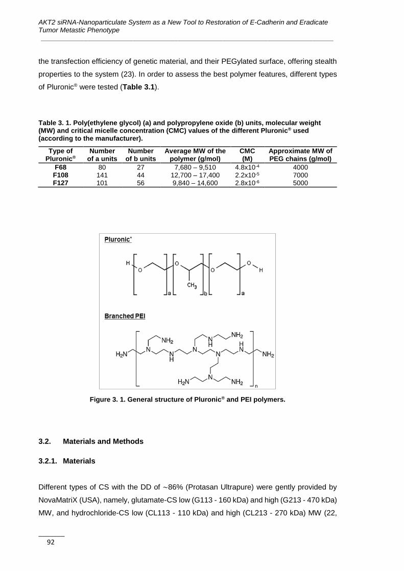
AKT2 siRNA-Nanoparticulate System as a New Tool to Restoration of E-Cadherin and Eradicate Tumor Metastic Phenotype ______________________________________________________________________________
_______ 92
the transfection efficiency of genetic material, and their PEGylated surface, offering stealth
properties to the system (23). In order to assess the best polymer features, different types
of Pluronic® were tested (Table 3.1).
Table 3. 1. Poly(ethylene glycol) (a) and polypropylene oxide (b) units, molecular weight (MW) and critical micelle concentration (CMC) values of the different Pluronic® used (according to the manufacturer).
Type of Pluronic®
Number of a units
Number of b units
Average MW of the polymer (g/mol)
CMC (M)
Approximate MW of PEG chains (g/mol)
F68 80 27 7,680 – 9,510 4.8x10⁻⁴ 4000 F108 141 44 12,700 – 17,400 2.2x10⁻⁵ 7000 F127 101 56 9,840 – 14,600 2.8x10⁻⁶ 5000
Figure 3. 1. General structure of Pluronic® and PEI polymers.
3.2. Materials and Methods
3.2.1. Materials
Different types of CS with the DD of ∼86% (Protasan Ultrapure) were gently provided by
NovaMatriX (USA), namely, glutamate-CS low (G113 - 160 kDa) and high (G213 - 470 kDa)
MW, and hydrochloride-CS low (CL113 - 110 kDa) and high (CL213 - 270 kDa) MW (22,

Chapter 3 | Formulation Studies and Efficacy Assessment of Different siRNA Delivery Systems _______________________________________________________________________________
_______ 93
24-26). Glycol-CS was purchased from Sigma Aldrich (Madrid, Spain). Pluronic® F68, F108
and F127 were kindly provided by BASF (Ludwigshafen, Germany), while 10k branched
and 25k branched PEI were provided by Alfa Aesar (Thermo Fisher GmbH, Karlsruhe,
Germany) and Sigma-Aldrich (Madrid, Spain), respectively.
siRNA against GFP (GFP-siRNA) and the scramble sequence (siC) were provided by two
companies, Shangai Gene Pharma (China) and LifeTechnologies (Spain). siRNA-
AlexaFluor 488 was brought from GE Healthcare Europe GmbH (Barcelona, Spain).
RXO-C colon cancer cells expressing GFP were generously provided by Dr. Diego Arango
from CIBBIM-Nanomedicine. Breast cancer cell lines MDA-MB-231 (ATCC number HTB-
26) and MCF7 (ATCC number HTB-22) were obtained from American Type Culture
Collection (ATCC, LGC Standards, Barcelona, Spain). RPMI medium, phosphate buffered
saline (PBS), and fetal bovine serum (FBS) were purchase from Lonza (Barcelona, Spain).
Penicillin-streptomycin, L-glutamine, non-essential amino acids, sodium pyruvate, 0.25%
Trypsin-EDTA, Lipofectamine® 2000, 4′,6-diamidino-2-phenylindole (DAPI), and
LysoTracker® Red are brought from Life Technologies Ltd. (Madrid, Spain).
Other used reagents are methanol, ethanol, dimethyl sulfoxide (DMSO), 3-(4,5-
dimethythiazol-2-yl)-2,5-diphenyl tetrazolium bromide (MTT), gelatin, paraformaldehyde,
Triton X-100, and 5-([4,6-dichlorotriazin-2-yl]amino)fluorescein hydrochloride (5-DTAF)
from Sigma-Aldrich (Madrid, Spain), and Type 1 ultrapure water (18.2 MΩ.cm at 25 ºC, Milli-
Q®, Billerica, MA, USA).
3.2.2. Polyplexes Preparation
Polymer-siRNA complexes were prepared by simple complexation, adding the polymer
solution dropwise to an equal volume of siRNA solution and quickly vortexed during few
seconds before incubating them at room temperature for 30 minutes.
The CS-siRNA complexes were prepared at different N/P ratios calculated according to
Equation 3.1 and at different conditions (Table 3.2).
𝑵
𝑷𝒓𝒂𝒕𝒊𝒐 =
𝑴𝒂𝒔𝒔 𝒐𝒇 𝑪𝑺
𝑴𝒂𝒔𝒔 𝒑𝒆𝒓 𝒄𝒉𝒂𝒓𝒈𝒆 𝒐𝒇 𝑪𝑺 (=𝟏𝟔𝟎𝒈
𝒎𝒐𝒍)
𝑴𝒂𝒔𝒔 𝒐𝒇 𝒔𝒊𝑹𝑵𝑨
𝑴𝒂𝒔𝒔 𝒑𝒆𝒓 𝒄𝒉𝒂𝒓𝒈𝒆 𝒐𝒇 𝒑𝒉𝒐𝒔𝒑𝒉𝒂𝒕𝒆 (=𝟑𝟑𝟎𝒈
𝒎𝒐𝒍)
Equation 3.1
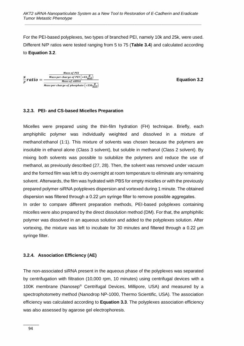
AKT2 siRNA-Nanoparticulate System as a New Tool to Restoration of E-Cadherin and Eradicate Tumor Metastic Phenotype ______________________________________________________________________________
_______ 94
For the PEI-based polyplexes, two types of branched PEI, namely 10k and 25k, were used.
Different N/P ratios were tested ranging from 5 to 75 (Table 3.4) and calculated according
to Equation 3.2.
𝑵
𝑷𝒓𝒂𝒕𝒊𝒐 =
𝑴𝒂𝒔𝒔 𝒐𝒇 𝑷𝑬𝑰
𝑴𝒂𝒔𝒔 𝒑𝒆𝒓 𝒄𝒉𝒂𝒓𝒈𝒆 𝒐𝒇 𝑷𝑬𝑰 (=𝟒𝟑𝒈
𝒎𝒐𝒍)
𝑴𝒂𝒔𝒔 𝒐𝒇 𝒔𝒊𝑹𝑵𝑨
𝑴𝒂𝒔𝒔 𝒑𝒆𝒓 𝒄𝒉𝒂𝒓𝒈𝒆 𝒐𝒇 𝒑𝒉𝒐𝒔𝒑𝒉𝒂𝒕𝒆 (=𝟑𝟑𝟎𝒈
𝒎𝒐𝒍)
Equation 3.2
3.2.3. PEI- and CS-based Micelles Preparation
Micelles were prepared using the thin-film hydration (FH) technique. Briefly, each
amphiphilic polymer was individually weighted and dissolved in a mixture of
methanol:ethanol (1:1). This mixture of solvents was chosen because the polymers are
insoluble in ethanol alone (Class 3 solvent), but soluble in methanol (Class 2 solvent). By
mixing both solvents was possible to solubilize the polymers and reduce the use of
methanol, as previously described (27, 28). Then, the solvent was removed under vacuum
and the formed film was left to dry overnight at room temperature to eliminate any remaining
solvent. Afterwards, the film was hydrated with PBS for empty micelles or with the previously
prepared polymer-siRNA polyplexes dispersion and vortexed during 1 minute. The obtained
dispersion was filtered through a 0.22 μm syringe filter to remove possible aggregates.
In order to compare different preparation methods, PEI-based polyplexes containing
micelles were also prepared by the direct dissolution method (DM). For that, the amphiphilic
polymer was dissolved in an aqueous solution and added to the polyplexes solution. After
vortexing, the mixture was left to incubate for 30 minutes and filtered through a 0.22 μm
syringe filter.
3.2.4. Association Efficiency (AE)
The non-associated siRNA present in the aqueous phase of the polyplexes was separated
by centrifugation with filtration (10,000 rpm, 10 minutes) using centrifugal devices with a
100K membrane (Nanosep® Centrifugal Devices, Millipore, USA) and measured by a
spectrophotometry method (Nanodrop NP-1000, Thermo Scientific, USA). The association
efficiency was calculated according to Equation 3.3. The polyplexes association efficiency
was also assessed by agarose gel electrophoresis.
Chapter 3 | Formulation Studies and Efficacy Assessment of Different siRNA Delivery Systems _______________________________________________________________________________
_______ 95
AE= total amount of siRNA – free siRNA in filtrate
total amount of siRNA × 100 Equation 3.3
3.2.5. Particles Physicochemical Properties Characterization
Particles mean hydrodynamic diameter (md) and polydispersity index (Pdi) were measured
by dynamic light scattering (DLS) and zeta potential was assessed by laser doppler micro-
electrophoresis using a NanoZS (Malvern Instruments, UK). For each formulation, at least
three batches were produced and analyzed.
Particle shape and morphology were observed by transmission electron microscopy (TEM)
and Cryo-scanning electron microscopy (CryoSEM) (external services from IBMC and
CEMUP, University of Porto, Portugal respectively).
3.2.6. Serum Stability
To assess the stability of formulations in the presence of serum the particles were incubated
in a proportion of 1:1 with 50 % FBS and the mean diameter measured by DLS at different
time-points: 0, 6, 12 and 24 hours.
3.2.7. Cell lines Culture Conditions
MDA-MB-231, MCF7 and RXO-C cells were cultured in RPMI medium supplemented with
10% FBS, 1% penicillin-streptomycin, 1% L-glutamine, 1% non-essential amino acids, and
1% of sodium pyruvate and maintained at 37 ⁰ C under a 5% CO2 water saturated
atmosphere. The medium was changed every other day and, upon confluence, cells were
harvested from plates with a 0.25% trypsin-EDTA solution and passed to continue the
expansion.
3.2.8. GFP Reporter Gene Silencing Assay
RXO-C colon cancer cells expressing GFP were used to assess the systems silencing
efficacy. Polyplexes and micelles prepared using a GFP-siRNA and different cationic
AKT2 siRNA-Nanoparticulate System as a New Tool to Restoration of E-Cadherin and Eradicate Tumor Metastic Phenotype ______________________________________________________________________________
_______ 96
polymers were added to cells seeded at a concentration of 5 x 10⁴ cells/ml in 96 well plates.
As positive controls were used complexes formed between Lipofectamine® 2000 and the
GFP-siRNA prepared accordingly to the supplier instructions but at the same concentration
of siRNA used for the tested polyplexes. On the other hand, cells transfected at the same
conditions with polyplexes prepared with a siRNA of a random sequence (siC) were used
as negative control. The expression of GFP in cells after transfection was assessed with a
fluorescence microscope (Olympus, USA). The intensity of cells fluorescence was also
measured using a Fluorescent Microplate reader FLX800 (BioTek, Germany).
3.2.9. In vitro Cytotoxicity Assay
The cytotoxicity of different components in MDA-MB-231 cells was assessed using the MTT
assay. 5000 cells/well/100μl were seeded in 96-well plates and left to attach for 24 hours.
Then, cells were incubated in the presence or absence of increasing concentrations of
polymers/formulations for 24 hours. After this incubation time, the medium was changed
and the cells left for 72 hours. Complete medium was used as negative control and 10%
DMSO as positive control of toxicity. After the 72 hours incubation, 0.5 mg/ml of MTT was
added to each well. The plates were incubated for an additional 4 hours at 37ºC, the medium
discarded, and the formazan crystals produced by mitochondrial succinate dehydrogenase
dissolved with DMSO. The absorbance of each well was read on a microplate reader
(ELx800 absorbance reader, BioTek, Germany) at 590 nm and the cell viability calculated.
The results of cell viability were used for the determination of IC50 index by nonlinear
regression of the concentration-effect curve fit using Prism 6.02 software (GraphPad
Software, Inc., CA, USA).
3.2.10. Conjugation of F127 with 5-DTAF
F127 was fluorescently conjugated with 5-DTAF in an aqueous medium via nucleophilic
aromatic substitution by an addition-elimination pathway as previously described (Figure
3.2) (29).
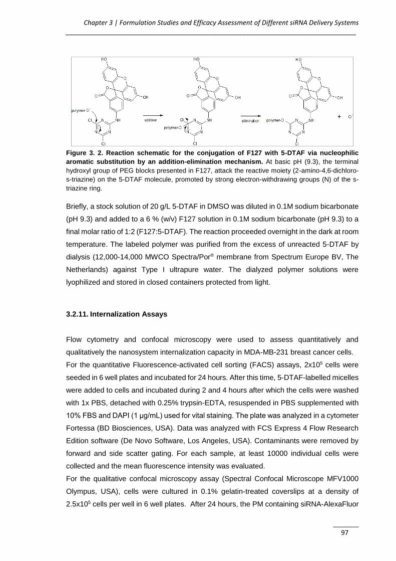
Chapter 3 | Formulation Studies and Efficacy Assessment of Different siRNA Delivery Systems _______________________________________________________________________________
_______ 97
Figure 3. 2. Reaction schematic for the conjugation of F127 with 5-DTAF via nucleophilic
aromatic substitution by an addition-elimination mechanism. At basic pH (9.3), the terminal
hydroxyl group of PEG blocks presented in F127, attack the reactive moiety (2-amino-4,6-dichloro-
s-triazine) on the 5-DTAF molecule, promoted by strong electron-withdrawing groups (N) of the s-
triazine ring.
Briefly, a stock solution of 20 g/L 5-DTAF in DMSO was diluted in 0.1M sodium bicarbonate
(pH 9.3) and added to a 6 % (w/v) F127 solution in 0.1M sodium bicarbonate (pH 9.3) to a
final molar ratio of 1:2 (F127:5-DTAF). The reaction proceeded overnight in the dark at room
temperature. The labeled polymer was purified from the excess of unreacted 5-DTAF by
dialysis (12,000-14,000 MWCO Spectra/Por® membrane from Spectrum Europe BV, The
Netherlands) against Type I ultrapure water. The dialyzed polymer solutions were
lyophilized and stored in closed containers protected from light.
3.2.11. Internalization Assays
Flow cytometry and confocal microscopy were used to assess quantitatively and
qualitatively the nanosystem internalization capacity in MDA-MB-231 breast cancer cells.
For the quantitative Fluorescence-activated cell sorting (FACS) assays, 2x10⁵ cells were
seeded in 6 well plates and incubated for 24 hours. After this time, 5-DTAF-labelled micelles
were added to cells and incubated during 2 and 4 hours after which the cells were washed
with 1x PBS, detached with 0.25% trypsin-EDTA, resuspended in PBS supplemented with
10% FBS and DAPI (1 μg/mL) used for vital staining. The plate was analyzed in a cytometer
Fortessa (BD Biosciences, USA). Data was analyzed with FCS Express 4 Flow Research
Edition software (De Novo Software, Los Angeles, USA). Contaminants were removed by
forward and side scatter gating. For each sample, at least 10000 individual cells were
collected and the mean fluorescence intensity was evaluated.
For the qualitative confocal microscopy assay (Spectral Confocal Microscope MFV1000
Olympus, USA), cells were cultured in 0.1% gelatin-treated coverslips at a density of
2.5x10⁵ cells per well in 6 well plates. After 24 hours, the PM containing siRNA-AlexaFluor
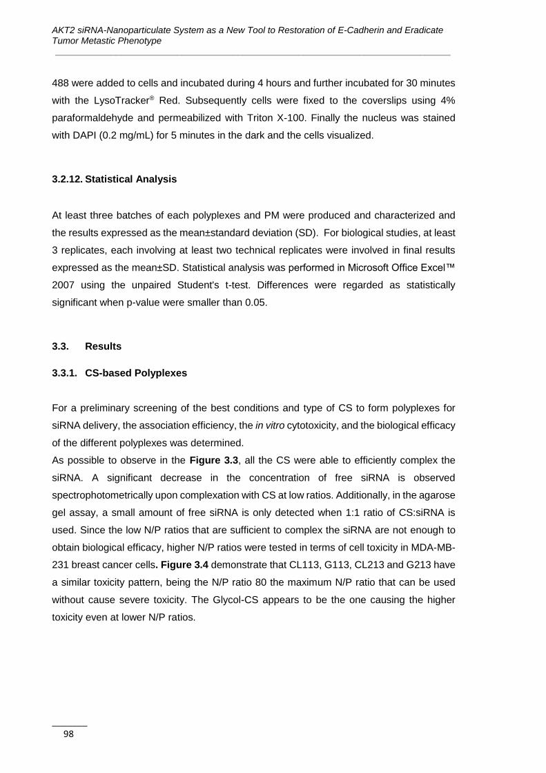
AKT2 siRNA-Nanoparticulate System as a New Tool to Restoration of E-Cadherin and Eradicate Tumor Metastic Phenotype ______________________________________________________________________________
_______ 98
488 were added to cells and incubated during 4 hours and further incubated for 30 minutes
with the LysoTracker® Red. Subsequently cells were fixed to the coverslips using 4%
paraformaldehyde and permeabilized with Triton X-100. Finally the nucleus was stained
with DAPI (0.2 mg/mL) for 5 minutes in the dark and the cells visualized.
3.2.12. Statistical Analysis
At least three batches of each polyplexes and PM were produced and characterized and
the results expressed as the mean±standard deviation (SD). For biological studies, at least
3 replicates, each involving at least two technical replicates were involved in final results
expressed as the mean±SD. Statistical analysis was performed in Microsoft Office Excel™
2007 using the unpaired Student's t-test. Differences were regarded as statistically
significant when p-value were smaller than 0.05.
3.3. Results
3.3.1. CS-based Polyplexes
For a preliminary screening of the best conditions and type of CS to form polyplexes for
siRNA delivery, the association efficiency, the in vitro cytotoxicity, and the biological efficacy
of the different polyplexes was determined.
As possible to observe in the Figure 3.3, all the CS were able to efficiently complex the
siRNA. A significant decrease in the concentration of free siRNA is observed
spectrophotometrically upon complexation with CS at low ratios. Additionally, in the agarose
gel assay, a small amount of free siRNA is only detected when 1:1 ratio of CS:siRNA is
used. Since the low N/P ratios that are sufficient to complex the siRNA are not enough to
obtain biological efficacy, higher N/P ratios were tested in terms of cell toxicity in MDA-MB-
231 breast cancer cells. Figure 3.4 demonstrate that CL113, G113, CL213 and G213 have
a similar toxicity pattern, being the N/P ratio 80 the maximum N/P ratio that can be used
without cause severe toxicity. The Glycol-CS appears to be the one causing the higher
toxicity even at lower N/P ratios.

Chapter 3 | Formulation Studies and Efficacy Assessment of Different siRNA Delivery Systems _______________________________________________________________________________
_______ 99
Figure 3. 3. Polyplexes association efficiency. A) Concentration of free siRNA detected in the
supernatant by spectrophotometry after formulation filtration by centrifugation using different N/P
ratios. Results are expressed as mean±SD, n=3. B) Agarose gel electrophoresis for the CL213-
based polyplex. 1 – free siRNA; 2 – 1:10 ratio; 3 – 1:5 ratio; 4 – 1:2.5 ratio; 5 – 1:1 ratio. In number
5, it is possible to see some free siRNA (marked with the circle).
Figure 3. 4. In vitro cytotoxicity of CS-siRNA polyplexes at different N/P ration in MDA-MB-231
cells. Results are expressed as mean±SD, n=3.
As described in Table 3.2, the most promising polyplexes, i.e. the ones that were able to
cause a visible decrease in the number of cells expressing GFP without promote significant
toxicity, were obtained with the following conditions: GFP-siRNA at a concentration of 200
nM in the well, complexed with CL213 and G213 CS at a N/P ratio of 80 and pH 4.5, and at
a post-transfection incubation time of 72 hours.
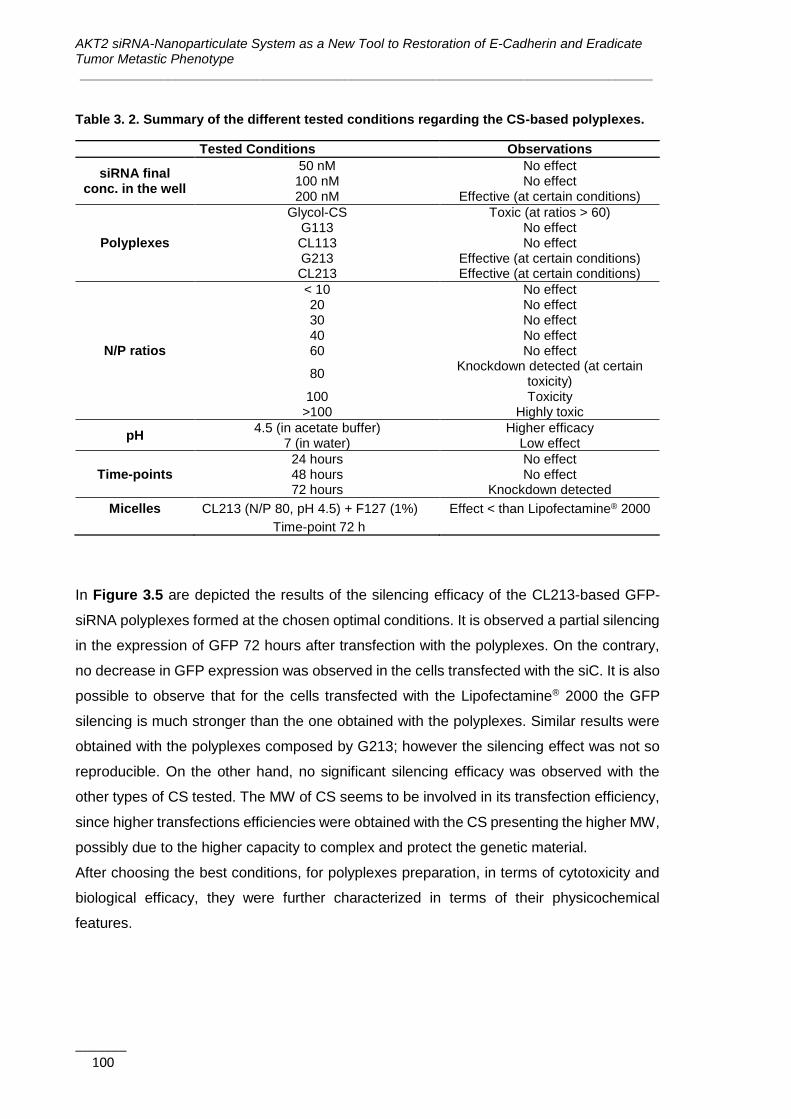
AKT2 siRNA-Nanoparticulate System as a New Tool to Restoration of E-Cadherin and Eradicate Tumor Metastic Phenotype ______________________________________________________________________________
_______ 100
Table 3. 2. Summary of the different tested conditions regarding the CS-based polyplexes.
Tested Conditions Observations
siRNA final conc. in the well
50 nM No effect 100 nM No effect 200 nM Effective (at certain conditions)
Polyplexes
Glycol-CS Toxic (at ratios > 60) G113 No effect CL113 No effect G213 Effective (at certain conditions) CL213 Effective (at certain conditions)
N/P ratios
< 10 No effect 20 No effect 30 No effect 40 No effect 60 No effect
80 Knockdown detected (at certain
toxicity) 100 Toxicity
>100 Highly toxic
pH 4.5 (in acetate buffer) Higher efficacy
7 (in water) Low effect
Time-points 24 hours No effect 48 hours No effect 72 hours Knockdown detected
Micelles CL213 (N/P 80, pH 4.5) + F127 (1%) Effect < than Lipofectamine® 2000
Time-point 72 h
In Figure 3.5 are depicted the results of the silencing efficacy of the CL213-based GFP-
siRNA polyplexes formed at the chosen optimal conditions. It is observed a partial silencing
in the expression of GFP 72 hours after transfection with the polyplexes. On the contrary,
no decrease in GFP expression was observed in the cells transfected with the siC. It is also
possible to observe that for the cells transfected with the Lipofectamine® 2000 the GFP
silencing is much stronger than the one obtained with the polyplexes. Similar results were
obtained with the polyplexes composed by G213; however the silencing effect was not so
reproducible. On the other hand, no significant silencing efficacy was observed with the
other types of CS tested. The MW of CS seems to be involved in its transfection efficiency,
since higher transfections efficiencies were obtained with the CS presenting the higher MW,
possibly due to the higher capacity to complex and protect the genetic material.
After choosing the best conditions, for polyplexes preparation, in terms of cytotoxicity and
biological efficacy, they were further characterized in terms of their physicochemical
features.
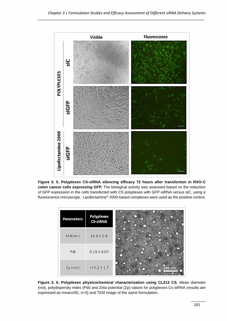
Chapter 3 | Formulation Studies and Efficacy Assessment of Different siRNA Delivery Systems _______________________________________________________________________________
_______ 101
Figure 3. 5. Polyplexes CS-siRNA silencing efficacy 72 hours after transfection in RXO-C
colon cancer cells expressing GFP. The biological activity was assessed based on the reduction
of GFP expression in the cells transfected with CS polyplexes with GFP-siRNA versus siC, using a
fluorescence microscope. Lipofectamine® 2000-based complexes were used as the positive control.
Figure 3. 6. Polyplexes physicochemical characterization using CL213 CS. Mean diameter
(md), polydispersity index (Pdi) and Zeta potential (Zp) values for polyplexes Cs-siRNA (results are
expressed as mean±SD, n=3) and TEM image of the same formulation.
AKT2 siRNA-Nanoparticulate System as a New Tool to Restoration of E-Cadherin and Eradicate Tumor Metastic Phenotype ______________________________________________________________________________
_______ 102
The polyplexes of CS-siRNA produced with the CL213-CS shown to be small (< 17 nm) and
positively charged (+ 15 mV) particles with spherical shape (Figure 3.6). The next step of
the work consisted in the conjugation of this polyplexes with Pluronic® based micelles in
order to improve the siRNA protection and biological activity.
3.3.2. Pluronic®-based Micelles Containing CS-polyplexes
Pluronic®-based empty micelles were prepared using different polymers. The micelles
presented a size lower than 150 nm (inferior to 50 nm for Pluronic® F108 and F127) and
near neutral surface charge (Table 3.3).
Table 3. 3. Physicochemical characterization of different Pluronic®-based micelles. Results
are expressed as mean±SD, n=3.
Due to the high polydispersity of micelles and a low reproducibility obtained with the F68
polymer, this polymer was discarded from further studies.
Concerning the cytotoxicity assessment, both Pluronic® F108 and F127 presented an IC50
superior to 10 mg/ml (Figure 3.7), but since Pluronic® F127 originates the micelles
presenting the lowest values of md and with the less polydispersed population, this polymer
was the chosen one for the production of micelles combined with the CS-based polyplexes.
For further experiments CL213-CS-based GFP-siRNA polyplexes were formed at the
chosen optimal conditions previously referred. These polyplexes were used to hydrate the
Pluronic® F127 film, forming the final micelles-based formulation (CS-siRNA-Pluronic®).
For the biological studies, these micelles were posteriorly incubated with RXO-C colon
cancer cells expressing GFP, being possible to observe that GFP expression was silenced,
conversely with what happens in cells transfected with the siC (Figure 3.8). Despite the
improvement in the silencing efficacy observed in the presence of Pluronic®, the efficacy of
the systems is still lower that the previously obtained using the positive control
(Lipofectamine® 2000).
Polymer Md (nm) Pdi Zp (mV)
F68 128.6±31.2 0.5±0.2 -2.9±1.0
F108 42.6±8.4 0.3±0.2 -2.1±1.2
F127 25.0±3.4 0.1±0.05 -2.7±1.8

Chapter 3 | Formulation Studies and Efficacy Assessment of Different siRNA Delivery Systems _______________________________________________________________________________
_______ 103
Figure 3. 7. Comparative cytotoxicity and IC50 values of Pluronic® F127 and F108 in MDA-MB-
231 cells. Results are expressed as mean±SD, n=3. F127 and F108 IC50 > 10 mg/ml.
Figure 3. 8. CS-siRNA-Pluronic® micelles silencing efficacy 72 hours after transfection with
the siGFP and siC in RXO-C colon cancer cells.
3.3.3. PEI-based Systems
Besides the micelles prepared by the FH technique described in the Chapter 5, it was
decided to produce and characterize micelles prepared also by the direct dissolution
method in order to assess the possibility of producing the formulations without using organic
solvents. For the design of these nanoparticles, the different conditions tested and results
obtained are summarized in Table 3.4.

AKT2 siRNA-Nanoparticulate System as a New Tool to Restoration of E-Cadherin and Eradicate Tumor Metastic Phenotype ______________________________________________________________________________
_______ 104
Table 3. 4. Summary of the different tested conditions regarding the branched PEI-based systems.
Tested Conditions Observations
siRNA final conc. in the well
200 nM Effective (at certain conditions)
Polymers combinations
PEI 10K Low effect PEI 25K Low effect
PEI 10K + F127 (0.5%) Effect < than Lipofectamine® 2000 PEI 25 K + F127 (0.5%) Effect < than Lipofectamine® 2000 PEI 10K + F127 (1%) Effective (at N/P 50) PEI 25 K + F127 (1%) Effective ( at N/P 25 and 50)
N/P ratios PEI:siRNA
5 No effect 25 Effective only for PEI 25K 50 Effective 75 Toxic
In the assessment of the biological efficacy no significant differences were observed
between both types of PEI (Figure 3.9), but since the 10K branched PEI presented lower
toxicity profile (30, 31), it was chosen for further studies.
Figure 3. 9. PEI-siRNA polyplexes and PEI-siRNA-Pluronic® micelles (obtained by DM)
silencing efficacy in GFP expressing RXO-C cells. A) Differences on the intensity of gene
expression upon incubation with different formulations using as reference the Lipofectamine® 2000.
B) Fluorescent microscopy photos confirming the GFP silencing efficacy of the selected formulation.
No gene inhibition was observed in cells transfected with siC. The values of GFP silencing were
normalized to the ones obtained with Lipofectamine® 2000. The red square represents the selected
formulation. Results are expressed as mean±SD (n≥3). * p≤0.05 compared to the polyplexes without
Pluronic® F127.

Chapter 3 | Formulation Studies and Efficacy Assessment of Different siRNA Delivery Systems _______________________________________________________________________________
_______ 105
Formulations composed by PEI:siRNA polyplexes of N/P ratio 50 present higher
transfection efficacy then polyplexes at lower N/P ratios (Figure 3.9). Also, the presence of
1% Pluronic® F127 in the form of micelles (prepared by DM) improved significantly the
transfection efficiency of the polyplexes as observed both quantitatively and qualitatively by
an increase of the gene expression inhibition (Figure 3.9). Thus, Pluronic® 127 micelles
encapsulating PEI:siRNA polyplexes at a N/P ratio 50 were chosen to proceed with further
studies and posteriorly characterized in terms of its internalization profile, md, Pdi, zeta
potential, serum stability, and entrapment efficiency.
Figure 3. 10. PEI-siRNA-Pluronic® internalization behavior. A) Flow cytometry analysis of labelled
particles internalization. B) Confocal Microscopy analysis of particles internalization containing a
fluorescent siRNA (siRNA-AlexaFluor 488). The yellow arrow illustrates the respective intracellular
distribution of nanoparticle and Lysotracker (DND-99, red).
The internalization of the PEI-siRNA-Pluronic® micelles was assessed quantitatively by flow
cytometry (Figure 3.10A) and qualitatively by confocal microscopy (Figure 3.10B). For the
FACS experiments, cells were incubated with 5-DTAF-labeled micelles during 4 hours. For
the qualitative analysis, MDA-MB-231 cells were incubated during 4 hours with micelles
carrying a fluorescent-labeled siRNA (siRNA-AlexaFluor 488) and observed by confocal
microscopy. As expected, in both assays it is possible to observe total micelles
internalization after the incubation time. In the confocal microscopy images it is also
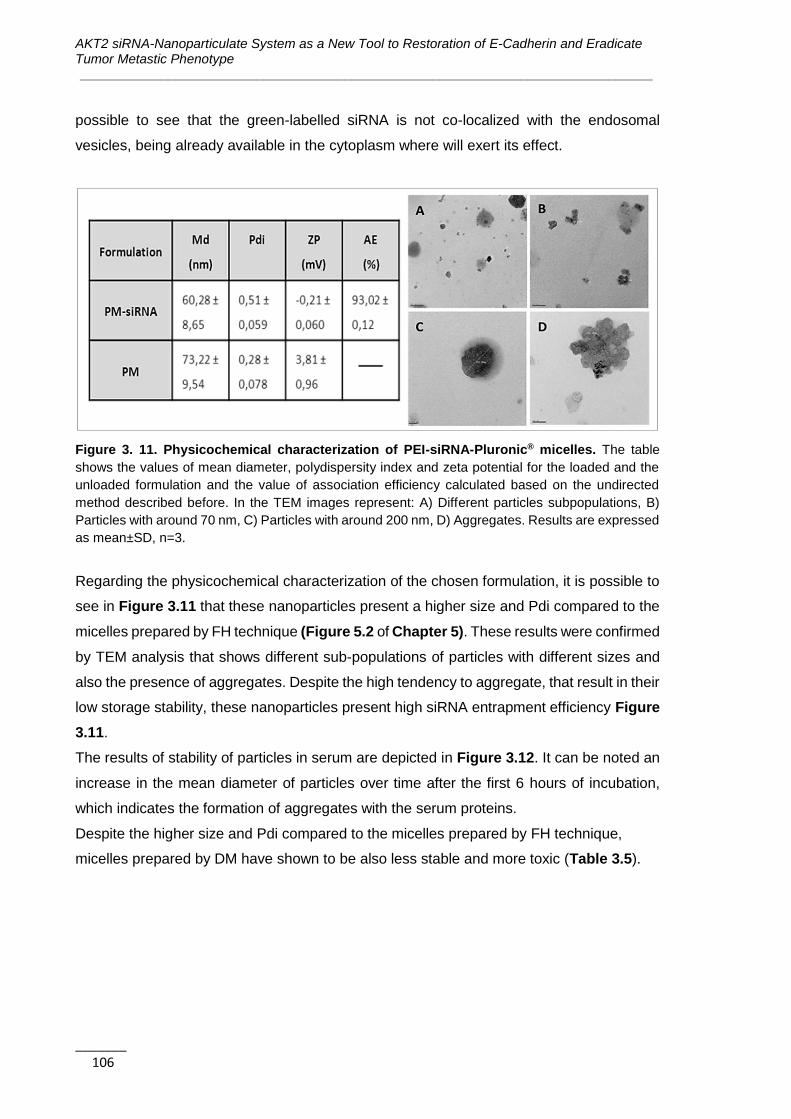
AKT2 siRNA-Nanoparticulate System as a New Tool to Restoration of E-Cadherin and Eradicate Tumor Metastic Phenotype ______________________________________________________________________________
_______ 106
possible to see that the green-labelled siRNA is not co-localized with the endosomal
vesicles, being already available in the cytoplasm where will exert its effect.
Figure 3. 11. Physicochemical characterization of PEI-siRNA-Pluronic® micelles. The table
shows the values of mean diameter, polydispersity index and zeta potential for the loaded and the
unloaded formulation and the value of association efficiency calculated based on the undirected
method described before. In the TEM images represent: A) Different particles subpopulations, B)
Particles with around 70 nm, C) Particles with around 200 nm, D) Aggregates. Results are expressed
as mean±SD, n=3.
Regarding the physicochemical characterization of the chosen formulation, it is possible to
see in Figure 3.11 that these nanoparticles present a higher size and Pdi compared to the
micelles prepared by FH technique (Figure 5.2 of Chapter 5). These results were confirmed
by TEM analysis that shows different sub-populations of particles with different sizes and
also the presence of aggregates. Despite the high tendency to aggregate, that result in their
low storage stability, these nanoparticles present high siRNA entrapment efficiency Figure
3.11.
The results of stability of particles in serum are depicted in Figure 3.12. It can be noted an
increase in the mean diameter of particles over time after the first 6 hours of incubation,
which indicates the formation of aggregates with the serum proteins.
Despite the higher size and Pdi compared to the micelles prepared by FH technique,
micelles prepared by DM have shown to be also less stable and more toxic (Table 3.5).
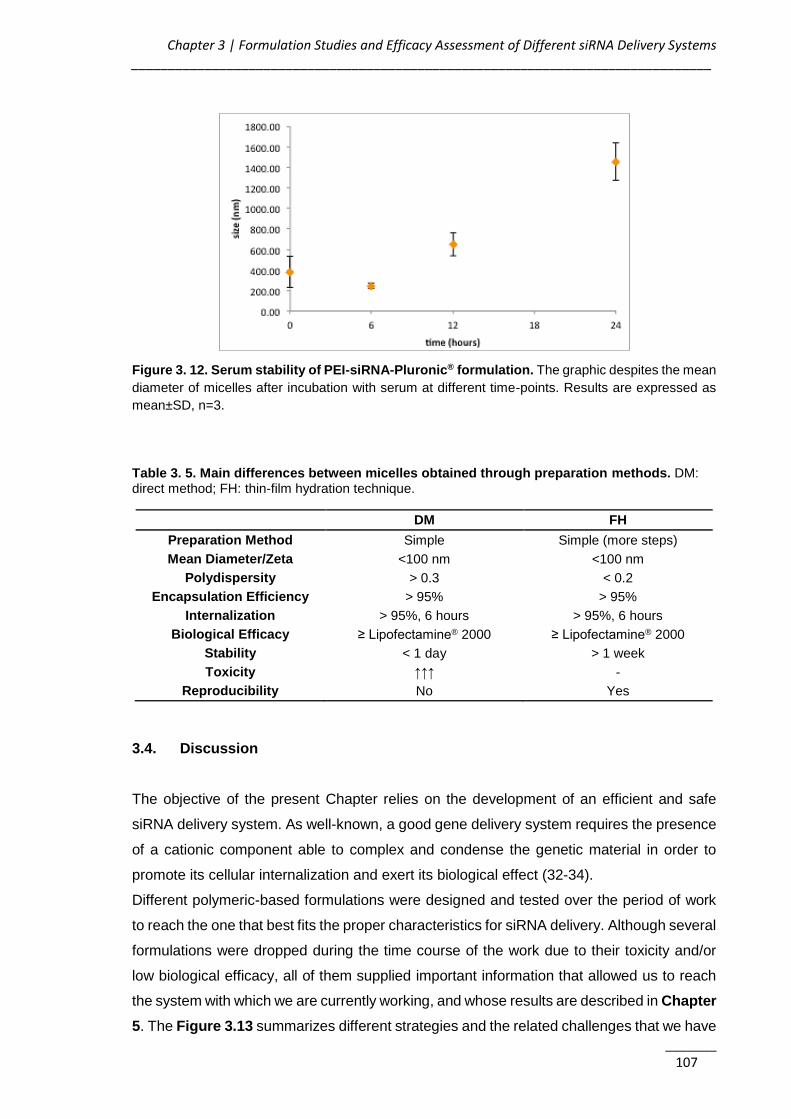
Chapter 3 | Formulation Studies and Efficacy Assessment of Different siRNA Delivery Systems _______________________________________________________________________________
_______ 107
Figure 3. 12. Serum stability of PEI-siRNA-Pluronic® formulation. The graphic despites the mean
diameter of micelles after incubation with serum at different time-points. Results are expressed as
mean±SD, n=3.
Table 3. 5. Main differences between micelles obtained through preparation methods. DM: direct method; FH: thin-film hydration technique.
DM FH
Preparation Method Simple Simple (more steps)
Mean Diameter/Zeta <100 nm <100 nm
Polydispersity > 0.3 < 0.2
Encapsulation Efficiency > 95% > 95%
Internalization > 95%, 6 hours > 95%, 6 hours
Biological Efficacy ≥ Lipofectamine® 2000 ≥ Lipofectamine® 2000
Stability < 1 day > 1 week
Toxicity ↑↑↑ -
Reproducibility No Yes
3.4. Discussion
The objective of the present Chapter relies on the development of an efficient and safe
siRNA delivery system. As well-known, a good gene delivery system requires the presence
of a cationic component able to complex and condense the genetic material in order to
promote its cellular internalization and exert its biological effect (32-34).
Different polymeric-based formulations were designed and tested over the period of work
to reach the one that best fits the proper characteristics for siRNA delivery. Although several
formulations were dropped during the time course of the work due to their toxicity and/or
low biological efficacy, all of them supplied important information that allowed us to reach
the system with which we are currently working, and whose results are described in Chapter
5. The Figure 3.13 summarizes different strategies and the related challenges that we have
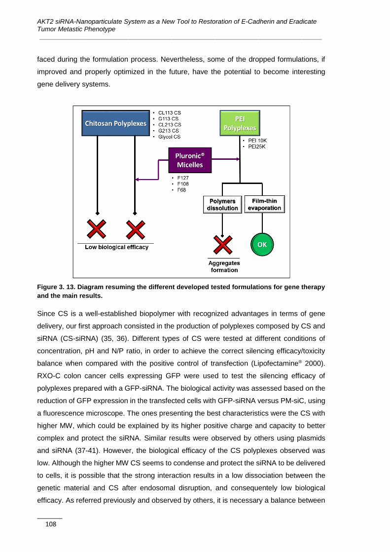
AKT2 siRNA-Nanoparticulate System as a New Tool to Restoration of E-Cadherin and Eradicate Tumor Metastic Phenotype ______________________________________________________________________________
_______ 108
faced during the formulation process. Nevertheless, some of the dropped formulations, if
improved and properly optimized in the future, have the potential to become interesting
gene delivery systems.
Figure 3. 13. Diagram resuming the different developed tested formulations for gene therapy
and the main results.
Since CS is a well-established biopolymer with recognized advantages in terms of gene
delivery, our first approach consisted in the production of polyplexes composed by CS and
siRNA (CS-siRNA) (35, 36). Different types of CS were tested at different conditions of
concentration, pH and N/P ratio, in order to achieve the correct silencing efficacy/toxicity
balance when compared with the positive control of transfection (Lipofectamine® 2000).
RXO-C colon cancer cells expressing GFP were used to test the silencing efficacy of
polyplexes prepared with a GFP-siRNA. The biological activity was assessed based on the
reduction of GFP expression in the transfected cells with GFP-siRNA versus PM-siC, using
a fluorescence microscope. The ones presenting the best characteristics were the CS with
higher MW, which could be explained by its higher positive charge and capacity to better
complex and protect the siRNA. Similar results were observed by others using plasmids
and siRNA (37-41). However, the biological efficacy of the CS polyplexes observed was
low. Although the higher MW CS seems to condense and protect the siRNA to be delivered
to cells, it is possible that the strong interaction results in a low dissociation between the
genetic material and CS after endosomal disruption, and consequentely low biological
efficacy. As referred previously and observed by others, it is necessary a balance between
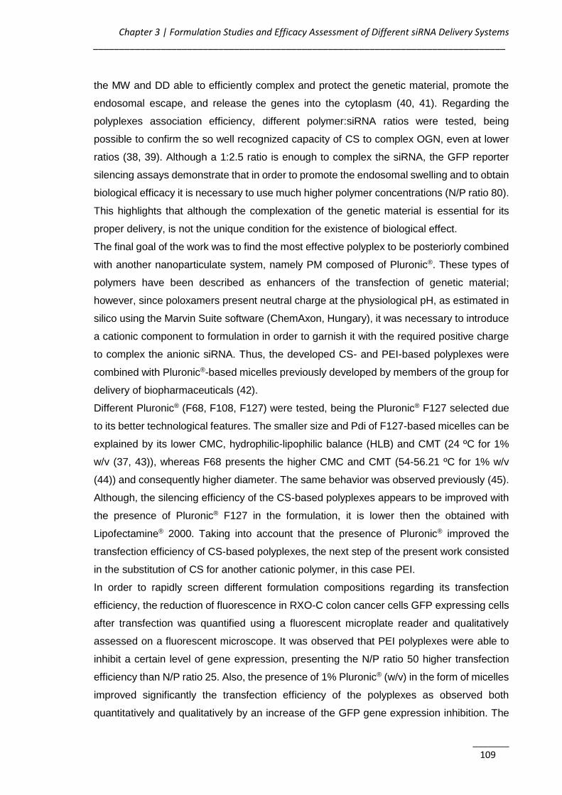
Chapter 3 | Formulation Studies and Efficacy Assessment of Different siRNA Delivery Systems _______________________________________________________________________________
_______ 109
the MW and DD able to efficiently complex and protect the genetic material, promote the
endosomal escape, and release the genes into the cytoplasm (40, 41). Regarding the
polyplexes association efficiency, different polymer:siRNA ratios were tested, being
possible to confirm the so well recognized capacity of CS to complex OGN, even at lower
ratios (38, 39). Although a 1:2.5 ratio is enough to complex the siRNA, the GFP reporter
silencing assays demonstrate that in order to promote the endosomal swelling and to obtain
biological efficacy it is necessary to use much higher polymer concentrations (N/P ratio 80).
This highlights that although the complexation of the genetic material is essential for its
proper delivery, is not the unique condition for the existence of biological effect.
The final goal of the work was to find the most effective polyplex to be posteriorly combined
with another nanoparticulate system, namely PM composed of Pluronic®. These types of
polymers have been described as enhancers of the transfection of genetic material;
however, since poloxamers present neutral charge at the physiological pH, as estimated in
silico using the Marvin Suite software (ChemAxon, Hungary), it was necessary to introduce
a cationic component to formulation in order to garnish it with the required positive charge
to complex the anionic siRNA. Thus, the developed CS- and PEI-based polyplexes were
combined with Pluronic®-based micelles previously developed by members of the group for
delivery of biopharmaceuticals (42).
Different Pluronic® (F68, F108, F127) were tested, being the Pluronic® F127 selected due
to its better technological features. The smaller size and Pdi of F127-based micelles can be
explained by its lower CMC, hydrophilic-lipophilic balance (HLB) and CMT (24 ºC for 1%
w/v (37, 43)), whereas F68 presents the higher CMC and CMT (54-56.21 ºC for 1% w/v
(44)) and consequently higher diameter. The same behavior was observed previously (45).
Although, the silencing efficiency of the CS-based polyplexes appears to be improved with
the presence of Pluronic® F127 in the formulation, it is lower then the obtained with
Lipofectamine® 2000. Taking into account that the presence of Pluronic® improved the
transfection efficiency of CS-based polyplexes, the next step of the present work consisted
in the substitution of CS for another cationic polymer, in this case PEI.
In order to rapidly screen different formulation compositions regarding its transfection
efficiency, the reduction of fluorescence in RXO-C colon cancer cells GFP expressing cells
after transfection was quantified using a fluorescent microplate reader and qualitatively
assessed on a fluorescent microscope. It was observed that PEI polyplexes were able to
inhibit a certain level of gene expression, presenting the N/P ratio 50 higher transfection
efficiency than N/P ratio 25. Also, the presence of 1% Pluronic® (w/v) in the form of micelles
improved significantly the transfection efficiency of the polyplexes as observed both
quantitatively and qualitatively by an increase of the GFP gene expression inhibition. The

AKT2 siRNA-Nanoparticulate System as a New Tool to Restoration of E-Cadherin and Eradicate Tumor Metastic Phenotype ______________________________________________________________________________
_______ 110
formulations composed by PEI:siRNA polyplexes of N/P ratio 50 with F127 1% (w/v) present
similar transfection efficacy and GFP silencing than Lipofectamine® 2000.
Since no significant differences in terms of biological efficacy were observed between both
types of PEI, the 10K branched PEI was chosen due to its recognized lower toxicity profile
(30, 31). Thus, after the first approach based on the assessment of transfection efficiency,
the formulation where PEI 10k was complexed with the siRNA at the N/P ratio of 50 and
further incorporated in Pluronic® F127-based PM was selected to further technological and
biological experiments.
Contrary to the complex chemical modifications proposed by other studies (46, 47), in this
work we propose a simple association of PEI-based polyplexes with Pluronic® F127 based
PM, that significantly increased the polyplexes biological efficacy. This result can be
explained by the reported capacity of poloxamers to enhance themselves the transfection
efficiency of genetic material. Although not completely understood and explained,
poloxamers present the capacity to interfere to some extent to the cell membranes reducing
its structure and microviscosity/fluidity (33, 48), and to improve the escape from the
endosomal compartment via membrane disruption and pore formation (49, 50). Poloxamers
with an intermediate HLB value like F127 (HLB=18-23), have shown higher ability to
interfere with membranes than poloxamers with higher HLB like F68 and F108 (HLB>24)
(51).
In this work, PEI-containing micelles were prepared by two methods in order to compare
their characteristics and to choose the best preparation method: DM (direct dissolution
method described in this chapter) and FH (thin-film hydration technique described in
Chapter 5. Micelles prepared by DM present the advantage of being prepared by a simpler
method avoiding also the use of organic solvents; however, they have shown higher size
and polydispersity, as well as lower stability and higher toxicity profile when compared to
the micelles presented in Chapter 5, which could be related to a less effective coating of
PEI-polyplexes.
3.5. Conclusions
This chapter described the rational design and experimental sequence beyond the
development of a nanocarrier system for siRNA intracellular delivery. Different cationic
polymers for the production of polyplexes and different systems to carry the polyplexes were
tested.
The choice of PM composed by Pluronic® as a possible delivery system is based on their
amphiphilic properties opening the possibility to design a multifunctional-targeted particle
Chapter 3 | Formulation Studies and Efficacy Assessment of Different siRNA Delivery Systems _______________________________________________________________________________
_______ 111
that can be used in the future for multi-therapy, i.e. the blend of gene and chemotherapy in
one single nanoparticle formulation.
It results clear that the presence of Pluronic® in the formulations improved the biological
efficacy of CS-based polyplexes. Nevertheless, even this combination presented limited
transfection efficiency. Thus, the well-establish PEI was used for the production of
polyplexes and for the development of PEI-siRNA-Pluronic® systems.
In the present work was possible to develop a formulation composed by PEI 10k branched-
based polyplexes at an N/P ratio of 50 combined with micelles of Pluronic® F127 to a final
concentration of 1% (w/v) presenting adequate technological properties, biological efficacy
and safety profile. It was also possible to emphasize the importance of the presence of
Pluronic® F127 in the formulation not only to improve the silencing activity but also to reduce
the PEI-associated toxicity.
The direct dissolution method to prepare micelles is easiest and less time consuming, in
addition of avoiding organic solvents; however it presented some disadvantages when
compared with the FH technique to produce the PEI-siRNA-Pluronic® systems. Despite the
observed gene silencing efficacy and safety profile, the higher size and polydispersity of
particles associated to high physical instability observed make us decide to change the
method of micelles production to FH, maintaining the composition of the formulations
(Chapter 5).
3.6. References
1. Mao S, Sun W, Kissel T. Chitosan-based formulations for delivery of DNA and
siRNA. Adv Drug Deliv Rev. 2010;62(1):12-27.
2. Rudzinski WE, Aminabhavi TM. Chitosan as a carrier for targeted delivery of small
interfering RNA. Int J Pharm. 2010;399(1-2):1-11.
3. Holzerny P, Ajdini B, Heusermann W, Bruno K, Schuleit M, Meinel L, et al.
Biophysical properties of chitosan/siRNA polyplexes: profiling the polymer/siRNA
interactions and bioactivity. J Control Release. 2012;157(2):297-304.
4. Park IK, Park YH, Shin BA, Choi ES, Kim YR, Akaike T, et al. Galactosylated
chitosan-graft-dextran as hepatocyte-targeting DNA carrier. J Control Release.
2000;69(1):97-108.
5. Kim TH, Ihm JE, Choi YJ, Nah JW, Cho CS. Efficient gene delivery by urocanic acid-
modified chitosan. J Control Release. 2003;93(3):389-402.

AKT2 siRNA-Nanoparticulate System as a New Tool to Restoration of E-Cadherin and Eradicate Tumor Metastic Phenotype ______________________________________________________________________________
_______ 112
6. Kim TH, Kim SI, Akaike T, Cho CS. Synergistic effect of poly(ethylenimine) on the
transfection efficiency of galactosylated chitosan/DNA complexes. J Control Release.
2005;105(3):354-66.
7. Wong K, Sun G, Zhang X, Dai H, Liu Y, He C, et al. PEI-g-chitosan, a novel gene
delivery system with transfection efficiency comparable to polyethylenimine in vitro and after
liver administration in vivo. Bioconjug Chem. 2006;17(1):152-8.
8. Lin A, Liu Y, Huang Y, Sun J, Wu Z, Zhang X, et al. Glycyrrhizin surface-modified
chitosan nanoparticles for hepatocyte-targeted delivery. Int J Pharm. 2008;359(1-2):247-
53.
9. Gabizon A, Shmeeda H, Horowitz AT, Zalipsky S. Tumor cell targeting of liposome-
entrapped drugs with phospholipid-anchored folic acid-PEG conjugates. Adv Drug Deliv
Rev. 2004;56(8):1177-92.
10. Moreira C, Oliveira H, Pires LR, Simões S, Barbosa MA, Pêgo AP. Improving
chitosan-mediated gene transfer by the introduction of intracellular buffering moieties into
the chitosan backbone. Acta Biomater. 2009;5(8):2995-3006.
11. Akinc A, Thomas M, Klibanov AM, Langer R. Exploring polyethylenimine-mediated
DNA transfection and the proton sponge hypothesis. J Gene Med. 2005;7(5):657-63.
12. Roesler S, Koch FP, Schmehl T, Weissmann N, Seeger W, Gessler T, et al.
Amphiphilic, low molecular weight poly(ethylene imine) derivatives with enhanced stability
for efficient pulmonary gene delivery. J Gene Med. 2011;13(2):123-33.
13. Martin I, Dohmen C, Mas-Moruno C, Troiber C, Kos P, Schaffert D, et al. Solid-
phase-assisted synthesis of targeting peptide-PEG-oligo(ethane amino)amides for
receptor-mediated gene delivery. Org Biomol Chem. 2012;10(16):3258-68.
14. Godbey WT, Wu KK, Mikos AG. Size matters: molecular weight affects the efficiency
of poly(ethylenimine) as a gene delivery vehicle. J Biomed Mater Res. 1999;45(3):268-75.
15. Thomas M, Klibanov AM. Enhancing polyethylenimine's delivery of plasmid DNA
into mammalian cells. Proc Natl Acad Sci U S A. 2002;99(23):14640-5.
16. Mao S, Neu M, Germershaus O, Merkel O, Sitterberg J, Bakowsky U, et al. Influence
of polyethylene glycol chain length on the physicochemical and biological properties of
poly(ethylene imine)-graft-poly(ethylene glycol) block copolymer/SiRNA polyplexes.
Bioconjug Chem. 2006;17(5):1209-18.
17. Dehshahri A, Oskuee RK, Shier WT, Hatefi A, Ramezani M. Gene transfer efficiency
of high primary amine content, hydrophobic, alkyl-oligoamine derivatives of
polyethylenimine. Biomaterials. 2009;30(25):4187-94.
18. Grayson AC, Doody AM, Putnam D. Biophysical and structural characterization of
polyethylenimine-mediated siRNA delivery in vitro. Pharm Res. 2006;23(8):1868-76.
Chapter 3 | Formulation Studies and Efficacy Assessment of Different siRNA Delivery Systems _______________________________________________________________________________
_______ 113
19. Oskuee RK, Philipp A, Dehshahri A, Wagner E, Ramezani M. The impact of
carboxyalkylation of branched polyethylenimine on effectiveness in small interfering RNA
delivery. J Gene Med. 2010;12(9):729-38.
20. Malek A, Czubayko F, Aigner A. PEG grafting of polyethylenimine (PEI) exerts
different effects on DNA transfection and siRNA-induced gene targeting efficacy. Journal of
Drug Targeting. 2008;16(2):124-39.
21. Moghimi SM, Hunter AC. Poloxamers and poloxamines in nanoparticle engineering
and experimental medicine. Trends Biotechnol. 2000;18(10):412-20.
22. Kabanov AV, Batrakova EV, Alakhov VY. Pluronic block copolymers as novel
polymer therapeutics for drug and gene delivery. J Control Release. 2002;82(2-3):189-212.
23. Kabanov A, Zhu J, Alakhov V. Pluronic block copolymers for gene delivery.
Advances in genetics. 2005;53:231-61.
24. Batrakova EV, Kabanov AV. Pluronic block copolymers: evolution of drug delivery
concept from inert nanocarriers to biological response modifiers. J Control Release.
2008;130(2):98-106.
25. Chen YC, Jiang LP, Liu NX, Ding L, Liu XL, Wang ZH, et al. Enhanced gene
transduction into skeletal muscle of mice in vivo with pluronic block copolymers and
ultrasound exposure. Cell Biochem Biophys. 2011;60(3):267-73.
26. Wang F, Li K, Chen Y. Gene transfection mediated by ultrasound and Pluronic P85
in HepG2 cells. J Huazhong Univ Sci Technolog Med Sci. 2007;27(6):700-2.
27. Yamagata M, Kawano T, Shiba K, Mori T, Katayama Y, Niidome T. Structural
advantage of dendritic poly(L-lysine) for gene delivery into cells. Bioorg Med Chem.
2007;15(1):526-32.
28. Inoue Y, Kurihara R, Tsuchida A, Hasegawa M, Nagashima T, Mori T, et al. Efficient
delivery of siRNA using dendritic poly(L-lysine) for loss-of-function analysis. J Control
Release. 2008;126(1):59-66.
29. Andrade F, Neves J, Gener P, Schwartz S, Jr., Ferreira D, Oliva M, et al. Biological
assessment of self-assembled polymeric micelles for pulmonary administration of insulin.
Nanomedicine : nanotechnology, biology, and medicine. 2015;11(7):1621-31.
30. Paul A, Eun C-J, Song JM. Cytotoxicity mechanism of non-viral carriers
polyethylenimine and poly-l-lysine using real time high-content cellular assay. Polymer.
2014;55(20):5178–88.
31. Symonds P, Murray JC, Hunter AC, Debska G, Szewczyk A, Moghimi SM. Low and
high molecular weight poly(L-lysine)s/poly(L-lysine)-DNA complexes initiate mitochondrial-
mediated apoptosis differently. FEBS Lett. 2005;579(27):6191-8.

AKT2 siRNA-Nanoparticulate System as a New Tool to Restoration of E-Cadherin and Eradicate Tumor Metastic Phenotype ______________________________________________________________________________
_______ 114
32. Tros de Ilarduya C, Sun Y, Düzgüneş N. Gene delivery by lipoplexes and polyplexes.
Eur J Pharm Sci. 2010;40(3):159-70.
33. Iyer AK, Duan Z, Amiji MM. Nanodelivery systems for nucleic acid therapeutics in
drug resistant tumors. Molecular pharmaceutics. 2014;11(8):2511-26.
34. Rafael DF, Andrade F, Arranja A, Luís AS, Videira M. Lipoplexes and Polyplexes:
Gene Delivery Applications. . Encyclopedia Biomedical Polymers and Polymeric
Biomaterials, Taylor and Francis Group 2014.
35. Huang M, Fong CW, Khor E, Lim LY. Transfection efficiency of chitosan vectors:
effect of polymer molecular weight and degree of deacetylation. J Control Release.
2005;106(3):391-406.
36. Lavertu M, Méthot S, Tran-Khanh N, Buschmann MD. High efficiency gene transfer
using chitosan/DNA nanoparticles with specific combinations of molecular weight and
degree of deacetylation. Biomaterials. 2006;27(27):4815-24.
37. Andrade F, Fonte P, Oliva M, Videira M, Ferreira D, Sarmento B. Solid state
formulations composed by amphiphilic polymers for delivery of proteins: characterization
and stability. Int J Pharm. 2015;486(1-2):195-206.
38. Mao S, Sun W, Kissel T. Chitosan-based formulations for delivery of DNA and
siRNA. Adv Drug Deliv Rev. 2010;62(1):12-27.
39. Rudzinski WE, Aminabhavi TM. Chitosan as a carrier for targeted delivery of small
interfering RNA. International journal of pharmaceutics. 2010;399(1-2):1-11.
40. Köping-Höggård M, Tubulekas I, Guan H, Edwards K, Nilsson M, Vårum KM, et al.
Chitosan as a nonviral gene delivery system. Structure-property relationships and
characteristics compared with polyethylenimine in vitro and after lung administration in vivo.
Gene Ther. 2001;8(14):1108-21.
41. Köping-Höggård M, Vårum KM, Issa M, Danielsen S, Christensen BE, Stokke BT,
et al. Improved chitosan-mediated gene delivery based on easily dissociated chitosan
polyplexes of highly defined chitosan oligomers. Gene Ther. 2004;11(19):1441-52.
42. Katas H, Alpar HO. Development and characterisation of chitosan nanoparticles for
siRNA delivery. J Control Release. 2006;115(2):216-25.
43. Andrade F, das Neves J, Gener P, Schwartz S, Ferreira D, Oliva M, et al. Biological
assessment of self-assembled polymeric micelles for pulmonary administration of insulin.
Nanomedicine. 2015;11(7):1621-31.
44. Lin Y, Alexandridis P. Temperature-dependent adsorption of Pluronic F127 block
copolymers onto carbon black particles dispersed in aqueous media. J Phys Chem B.
2002;106:10834-44.
Chapter 3 | Formulation Studies and Efficacy Assessment of Different siRNA Delivery Systems _______________________________________________________________________________
_______ 115
45. Tsui H-W, Wang J-H, Hsu Y-H, Chen L-J. Study of heat of micellization and phase
separation for Pluronic aqueous solutions by using a high sensitivity differential scanning
calorimetry. Colloid and Polymer Science. 2010;288:1687-96.
46. Hu Y, Zhou D, Li C, Zhou H, Chen J, Zhang Z, et al. Gene delivery of PEI
incorporating with functional block copolymer via non-covalent assembly strategy. Acta
biomaterialia. 2013;9(2):5003-12.
47. Wen Y, Pan S, Luo X, Zhang W, Shen Y, Feng M. PEG- and PDMAEG-graft-
modified branched PEI as novel gene vector: synthesis, characterization and gene
transfection. Journal of biomaterials science Polymer edition. 2010;21(8-9):1103-26.
48. Williford JM, Wu J, Ren Y, Archang MM, Leong KW, Mao HQ. Recent advances in
nanoparticle-mediated siRNA delivery. Annual review of biomedical engineering.
2014;16:347-70.
49. Demina T, Grozdova I, Krylova O, Zhirnov A, Istratov V, Frey H, et al. Relationship
between the structure of amphiphilic copolymers and their ability to disturb lipid bilayers.
Biochemistry. 2005;44(10):4042-54.
50. Batrakova EV, Li S, Alakhov VY, Miller DW, Kabanov AV. Optimal structure
requirements for pluronic block copolymers in modifying P-glycoprotein drug efflux
transporter activity in bovine brain microvessel endothelial cells. The Journal of
pharmacology and experimental therapeutics. 2003;304(2):845-54.
51. Chen J, Luo J, Zhao Y, Pu L, Lu X, Gao R, et al. Increase in transgene expression
by pluronic L64-mediated endosomal/lysosomal escape through its membrane-disturbing
action. ACS applied materials & interfaces. 2015;7(13):7282-93.

AKT2 siRNA-Nanoparticulate System as a New Tool to Restoration of E-Cadherin and Eradicate Tumor Metastic Phenotype ______________________________________________________________________________
_______ 116
Chapter 4 | AKT2-related Biological Pathway Characterization and Validation in Breast and Colon Cancer Stem Cells
_______________________________________________________________________________
_______ 117
CHAPTER 4
AKT2-related Biological Pathway Characterization and
Validation in Breast and Colon Cancer Stem Cells
The information presented in this chapter was partially published in the following
publications:
1) P Gener, D Rafael, Y Fernández, J Sayos, D Arango, I Abasolo, M Videira, S
Schwartz Jr., Cancer Stem Cells and Personalized Cancer Nanomedicine.
Nanomedicine (Lond), 11(3):307-20, 2016.
2) P Gener, L Gouveia, G Sabat, D Rafael, N Fort, A Arranja, Y Fernández, R Prieto,
J Sayos, D Arango, I Abasolo, M Videira, S Schwartz Jr., Fluorescent CSC models
evidence that targeted nanomedicines improve treatment sensitivity of breast and
colon cancer stem cells. Nanomedicine, 11(8):1883-92, 2015.
3) D Rafael, P Gener, J Seras, A Perez, L Pindado, H Florindo, D Arango, J Sayos, I
Abasolo, Y Fernandez, M Videira, S Schwartz Jr., AKT2 silencing impairs
malignancy of breast and colon Cancer Stem Cells (CSCs) and prevents reversion
from non-CSC to a CSC phenotype, Article in preparation.
AKT2 siRNA-Nanoparticulate System as a New Tool to Restoration of E-Cadherin and Eradicate Tumor Metastic Phenotype ______________________________________________________________________________
_______ 118
Table of contents
4.1. Introduction .................................................................................................... 119
4.2. Materials and Methods .................................................................................. 120
4.2.1. Materials ................................................................................................... 120
4.2.2. Cell Lines and Culture Conditions ............................................................. 120
4.2.3. Fluorescence-Activated Cell Sorting ......................................................... 121
4.2.4. Cell Transfection ....................................................................................... 121
4.2.5. RNA Extraction and Quantitative RT-PCR ................................................ 122
4.2.6. Protein Extraction and Western Blotting .................................................... 123
4.2.7. Proliferation Assay .................................................................................... 123
4.2.8. Wound Healing Migration Assay ............................................................... 123
4.2.9. Cell Transformation Assay (Anchorage-independent Growth Assay) ........ 124
4.2.10. Invasion Assay ......................................................................................... 124
4.2.11. Statistical Analysis .................................................................................... 124
4.3. Results............................................................................................................ 125
4.3.1. AKT2 Silencing Impairs Proliferation and Migration of Bulk Cells .............. 125
4.3.2. AKT2 Inhibits Anchorage Independent Growth and Invasion of CSC ........ 125
4.3.3. AKT2 Inhibition is Achieved Via Diverse Signaling Pathways.................... 129
4.4. Discussion ..................................................................................................... 134
4.5. Conclusions ................................................................................................... 136
4.6. References ..................................................................................................... 136

Chapter 4 | AKT2-related Biological Pathway Characterization and Validation in Breast and Colon Cancer Stem Cells
_______________________________________________________________________________
_______ 119
4.1. Introduction
As mentioned previously, in the last years, accumulating evidences suggest that cancer
dormancy, treatment resistance, and metastatic growth are sustained by the presence of
CSC within the tumor, which provides a reservoir of highly malignant cells that are able to
cause tumor recurrence after the therapeutic regimen (1-4).
Despite the effectiveness of many chemotherapeutic agents and anticancer treatments in
terms of apparent tumor regression, the problematic of drug resistance observed for CSC
and the consequent tumor recurrence arise as an important barrier and a great challenge
to tumors eradication. Thereby, other therapeutic strategies have emerged, in particular the
gene therapy approach and/or its combination with conventional therapies. One of the most
interesting potentialities regarding gene therapy is the RNAi technology (5-7). To be able to
implement effectively this strategy into the clinical practice, it is of utmost importance the
complete understanding of the biological pathways involved in tumor development and
progression in order to select an adequate target gene to be silenced.
AKT2 is one of the three isoforms of the PKB family highly expressed in breast and colon
cancer tumors, which appears generally associated with the acquisition of a malignant
phenotype by cancer cells (8-10). AKT/PKB is a key regulator of various cell processes and
its signaling outcome, similarly to what happens in all cancer-related molecular mechanism,
is highly dependent on cellular background, cell type and phenotype. AKT2 may act via
different signaling mechanisms being TWIST and mTOR the main effectors downstream of
AKT2 (11-14). In the context of CSC, TWIST arose as an interesting cancer target due to
its important and well-known role in tumor invasion, migration, dissemination and drug
resistance (15, 16) Signaling cascades initiated by TWIST-mediated effects regulated by
AKT2 or those involved in EMT through the decrease of E-cadherin expression, bring
reasonability to the expectancy over the selective silencing of this oncogene as an effective
strategy to reduce cell malignancy (12, 14). EMT, usually defined as the process during
which cells loose epithelial traits and acquire a mesenchymal phenotype, is commonly
accepted as the critical step for the acquisition of malignance in epithelial cancer cells and
in some cases as an activator of the CSC phenotype (14, 17-21). At the molecular level,
the EMT is mainly characterized by the downregulation of E-cadherin, an essential cell-cell
adhesion protein whereas mesenchymal markers such as Vimentin and N-cadherin tend to
gradually raise their expression (14, 22). Regarding the importance of EMT in cancer
development and CSC activation, the targeting of EMT related signaling axis like
AKT2/TWIST in order to revert the EMT process and restore the initial epithelial phenotype,
appears to be a promising strategy in cancer gene therapy.
AKT2 siRNA-Nanoparticulate System as a New Tool to Restoration of E-Cadherin and Eradicate Tumor Metastic Phenotype ______________________________________________________________________________
_______ 120
Similarly, PI3K/AKT2/mTOR signaling axis is also involved in acquisition of malignant
phenotype mainly through the over-activation of the mTOR signaling, as a result of
increased activity of PI3K or AKT. mTOR activity was found to be deregulated in many types
of cancer including breast, prostate, lung, melanoma, bladder, brain, and renal carcinomas.
Moreover, recent studies have shown the links between PI3K/AKT2/mTOR signaling
pathway and CSC biology (13, 23-28).
Despite that AKT2 silencing was previously investigated in various tumor types, its precise
function in CSC was not addressed yet. Here, in order to assess if also in the CSC
population, the AKT2 knockdown is able to impair the increased cellular tumorigenic ability,
we silenced the AKT2 expression using a siRNA against AKT2 in different cancer cells lines
with a defined CSC population.
4.2. Materials and Methods
4.2.1. Materials
RPMI medium, PBS, and FBS were purchase from Lonza (Barcelona, Spain). McCoy’s
medium, DMEM F-12 medium, L-glutamine, non-essential amino acids, 10000 U/mL
penicillin and 10000 μg/mL streptomycin, 0.25% Trypsin-EDTA, blasticidin and sodium
pyruvate were purchased from Gibco (Life Technologies Ltd., Madrid, Spain).
Lipofectamine® 2000, trypan blue, DAPI, and SYBR Green were purchased from Molecular
Probes (Life Technologies Ltd., Madrid, Spain). Cell Lytic M reagent and protease inhibitor
cocktail were obtained from Sigma-Aldrich Quimica (Madrid, Spain).
Primary antibodies against TWIST and actin were obtained from Abcam (Cambridge, UK)
while the antibodies against AKT2, mTOR, and E-cadherin purchased from Cell Signaling
Technology (Leiden, The Netherlands) and Invitrogen (Life Technologies Ltd., Madrid,
Spain), respectively. HRP-conjugated secondary antibodies (anti-mouse P0447) and anti-
rabbit (P0217) were purchased from Dako (Barcelona, Spain).
siRNAs were designed by Shanghai Gene Pharma (Shanghai, China). The sense siAKT2
sequence used was 5’-GCUCCUUCAUUGGGUACAATT-3’, while a non-specific sequence
5’-UUCUCCGAACGUGUCACGUTT-3’ was used as control (siC).
4.2.2. Cell Lines and Culture Conditions
Breast cancer cell lines MCF7, MDA-MB-231, MDA-MB-468, and SKBR3; and colon cancer
cell line HCT8 were obtained from American Type Culture Collection (ATCC, LGC

Chapter 4 | AKT2-related Biological Pathway Characterization and Validation in Breast and Colon Cancer Stem Cells
_______________________________________________________________________________
_______ 121
Standards, Barcelona, Spain) and are examples of both mesenchymal and epithelial cell
lines (Table 4.1).
MDA-MB-468, MDA-MB-231 and HCT8 cells were cultured in RPMI medium supplemented
with 10% FBS, 1% penicillin-streptomycin, 1% L-glutamine, 1% non-essential amino acids,
and 1% of sodium pyruvate.
SKBR3 cells were cultured in McCoy’s medium supplemented with 10% FBS and 1%
penicillin-streptomycin, while MCF7 cells were cultured in DMEM F-12 medium
supplemented with 10% FBS and 1% penicillin-streptomycin.
Blasticidin (0.5 mg/ml) was used as a selective antibiotic for ALDH1A1/tdTomato cell lines.
All cell lines were maintained at 37ᵒC under a 5% CO2 water saturated atmosphere. The
medium was changed every other day and, upon confluence, cells were harvested from
plates with a 0.25% trypsin-EDTA solution and further used in in vitro studies or frozen.
Table 4. 1. List of the cell lines used in this study and their main phenotype.
Name ATCC® number Type Main Phenotype
MDA-MB-231 HTB 26™ Breast (Triple Negative) Mesenchymal
MDA-MB-468 HTB-132™ Breast (Triple Negative) Mesenchymal
MCF7 HTB-22™ Breast (ER+) Epithelial
SKBR3 HTB-30™ Breast (HER2+) Mesenchymal
HCT8 CCL-244™ Colon Epithelial
4.2.3. Fluorescence-Activated Cell Sorting (FACS)
FACS was used to sort CSC and non-CSC subpopulations from a heterogeneous
population of MCF7, MDA-MB-231 and HCT8 cells. For the cell sorting, a starting amount
of 5x10⁶ cells were used. Cells were detached, and re-suspended in PBS supplemented
with 10% FBS with DAPI (1 μg/mL) used for vital staining. Cells were sorted according to
tdTomato and DAPI expression in a FACS Aria cell sorter (BD Biosciences, Madrid, Spain)
(29). Sorted cells were collected in complete medium without antibiotic and used for
posterior applications.
4.2.4. Cell Transfection
The siRNAs were transfected into cells using Lipofectamine® 2000 accordingly to the
manufacture instructions in medium without antibiotics. The medium was changed 6 hours
after the transfection and cells were harvested after further 72 hours.

AKT2 siRNA-Nanoparticulate System as a New Tool to Restoration of E-Cadherin and Eradicate Tumor Metastic Phenotype ______________________________________________________________________________
_______ 122
4.2.5. RNA Extraction and Quantitative RT-PCR (qRT-PCR)
Total RNA was extracted from cells using the RNeasy Micro Kit (Qiagen, Madrid, Spain)
and the obtained RNA was reverse transcribed with the High Capacity cDNA Reverse
Transcription Kit (Applied Biosystems, Madrid, Spain) according to the manufacturer
instructions.
Table 4. 2. List of the primers used in the study and their sequences.
Oligo name Oligo Sequence (5’ to 3’)
TWIST F GCAGGGCCGGAGACCTAG
TWIST R TTTTTAGTTATCCAGCTCCAGAGTCTCTA
E-Cadherin F TACGCCTGGGACTCCACCTA
E-Cadherin R CCAGAAACGGAGGCCTGAT
Vimentin F CTCCGGGAGAAATTGCAGG
Vimentin R AGACGTGCCAGAGACGCATT
N-Cadherin F TCAAAGCCTGGAACATATGTGATG
N-Cadherin R GTATACTGTTGCACTTTTTCTCGTACAA
hAKT2F CAAGGATGAAGTCGCTCACACA
hAKT2R GAACGGGTGCCTGGTGTTC
18s F AGT CCC TGC CCT TTG TAC ACA
18s R GAT CCG AGG GCC TCA CTA AAC
Actin 01F CATCCACGAAACTACCTTCAACTCC
Actin 01R GAGCCGCCGATCCACAC
GADPH F ACCCACTCCTCCACCTTTGAC
GADPH R CATACCAGGAAATGAGCTTGACAA
mTOR F CCTTAACGTCATTCGAGTCTGTGA
mTOR R TCACAAAGGACACCAACATTCC
p53 F GAAGAAACCACTGGATGGAGAATATT
p53 R CAGCTCTCGGAACATCTCGAA
Bcl-2 F ACCTGCACACCTGGATCCA
Bcl-2 R AGAGACAGCCAGGAGAAATCAAA
GSK3b F CTGCTTCAACCCCCACAAAT
GSK3b R GATGCAGAAGCAGCATTATTGG
AKT1 F GGCTCCCCTCAACAACTTCTC
AKT1 R ACATGGAAGGTGCGTTCGA
The cDNA reverse transcription product was amplified with specific primers (Table 4.2) by
qPCR using the SYBR Green method. The reaction was performed in triplicate in a 7500
real time PCR system (Applied Biosystems, Madrid, Spain). Actin and S18 were used as

Chapter 4 | AKT2-related Biological Pathway Characterization and Validation in Breast and Colon Cancer Stem Cells
_______________________________________________________________________________
_______ 123
endogenous controls. The relative mRNA levels were calculated using the comparative Ct
method (2e- ΔΔCt).
4.2.6. Protein Extraction and Western Blotting (WB)
Cell pellets were lysed with the Cell Lytic M reagent containing a protease inhibitor cocktail.
Proteins in the crude lysates were quantified using the BCA Protein Assay (Pierce
Biotechnology, Madrid, Spain) and 20 μg of whole-cell lysates were separated by SDS-
PAGE and transferred onto nitrocellulose membranes (Merck Millipore, Madrid, Spain).
Blots were probed using primary antibodies listed in Table 4.3. Proteins were detected
using corresponding HRP-conjugated secondary antibodies (anti-mouse or anti-rabbit) and
incubated as described in Table 4.3.
Table 4. 3. List of primary antibodies used in the study and their specifications. ON – overnight, RT – room-temperature.
Primary antibody
Host Dilution Incubation
AKT2 Rabbit 1:1000 ON, 4°C
TWIST Mouse 1:500 ON, 4°C
mTOR Rabbit 1:1000 ON, 4°C
Actin Rabbit 1:1000 1 hour, RT
4.2.7. Proliferation Assay
Breast cancer cell lines were seeded into six-well plates at a density of 2.5X10⁵ cells per
well and allowed to adhere overnight. The next day cells were transfected as described
previously. For cell counting, cells were trypsinized and stained with trypan blue and viable
cells were counted at times 0, 24, 48 and 72 hours after transfection.
4.2.8. Wound Healing Migration Assay
For these assay, the different cells were seeded into six-well plates and cultured until high
confluence. The monolayer was “wounded” by scraping it with a sterile tip. The wells were
washed twice with medium without serum before being left in complete culture medium.
Immediately after do the wound, the cells were transfected as explained before. The healing

AKT2 siRNA-Nanoparticulate System as a New Tool to Restoration of E-Cadherin and Eradicate Tumor Metastic Phenotype ______________________________________________________________________________
_______ 124
process was examined during the time with an optical microscope and microphotographs
were obtained at 0, 24, 48 and 72 hours. Areas were measured with the Image J software
and compared with the wound area at time 0, in order to evaluate the migration rate of cells.
4.2.9. Cell Transformation Assay (Anchorage-independent Growth Assay)
The anchorage-independent growth of the different breast cancer cell lines were assessed
by the CytoSelect™ Cell Transformation Assay Kit (Cell Biolabs, San Diego, CA, USA). A
semisolid agar media were prepared accordingly the manufacturer instructions being the
complexes Lipofectamine-siAKT2 or Lipofectamine-siC added to each well. After 6-8 days
of incubation the colonies were observed under the microscope and the viable transformed
cells counted using trypan blue.
4.2.10. Invasion Assay
The invasiveness of MDA-MB-468 and SKBR3 cells was assessed using the CytoSelect™
Laminin Cell Invasion Assay Kit (Cell Biolabs, San Diego, CA, USA). The polycarbonate
membrane inserts coated with a uniform layer of Laminin I are used to discriminate invasive
cells from noninvasive cells. The invasive cells are able to degrade the laminin matrix layer
and pass through the pores of the polycarbonate membrane, clinging to the bottom of the
insert membrane, while the noninvasive cells stay in the upper chamber. Briefly, the inserts
were placed in a 24-well plate and a cell suspension containing 1X10⁶ cells/ml, previously
transfected with the siAKT2 and the siC, was added to the insert. After 48 hours incubation,
the invasive cells were dissociated from the membrane, lysed and quantified using the
CyQuant® GR Fluorescent Dye (Cell Biolabs, San Diego, CA, USA).
4.2.11. Statistical Analysis
All the assays were run at least in triplicate, each involving at least two technical replicates.
All results are expressed as the mean±SD. Statistical analysis was performed in Microsoft
Office Excel™ 2007 using the unpaired Student's t-test. Differences were regarded as
statistically significant when p-value were smaller than 0.05.

Chapter 4 | AKT2-related Biological Pathway Characterization and Validation in Breast and Colon Cancer Stem Cells
_______________________________________________________________________________
_______ 125
4.3. Results
4.3.1. AKT2 Silencing Impairs Proliferation and Migration of Cancer Cells
In order to determine the consequence of AKT2 inhibition on cell malignance, we investigate
its effect in various cell lines (MDA-MB-231, HCT8, MCF7, MDA-MB-468, SKBR3). We
confirmed the specificity of the siAKT2 sequence used, since 80% reduction of AKT2
expression was achieved and no reduction of AKT1 isoform was observed (Figure 4.1A).
The downregulation of AKT2 was confirmed also at the intracellular protein level (Figure
4.5D).The effects of AKT2 downregulation in the different cell lines was firstly assessed by
the proliferation and the migration assays. We observed a significant impairment of cell
proliferation in MDA-MB-231, HCT8, MDA-MB-468 and SKBR3 transfected with the siAKT2
in comparison with the cells transfected with the siC or with the untreated cells. On the
contrary, no effect in proliferation was observed after the AKT2 silencing in MCF7 cells
(Figure 4.1B). Similar results were observed for the migration capacity after AKT2 silencing,
particularly in MDA-MB-231, MDA-MB-468 and SKBR3 cells where the migration rates were
significantly impaired. Again, no changes on migration were observed in MCF7 cells (Figure
4.1C). HCT8 showed just minimal migration ability and was thus excluded from the analysis
(data not shown).
4.3.2. AKT2 Inhibits Anchorage-independent Growth and Invasion of CSC
After the functional assessment of the AKT2 silencing effect in parental cells, the behavior
of the CSC fraction isolated from three different cell lines (MDA-MB-231, HCT8, MCF7) was
investigated, after being transfected with siAKT2. CSC were isolated from the parental cells
population by FACS based on the stable expression of ALDH1A1/tdTomato reporter vector.
Only CSC are able to express tdTomato (red fluorescence) since its expression is driven by
CSC specific promoter (ALDH1A1), while the non-CSC are not detected as red fluorescent
cells in the total cell culture mixture (29). The CSC nature of tdTomato+ cells was previously
confirmed by expression of stemness markers, tumorspheres formation and high in vivo
tumorigenic capacity (29).

AKT2 siRNA-Nanoparticulate System as a New Tool to Restoration of E-Cadherin and Eradicate Tumor Metastic Phenotype ______________________________________________________________________________
_______ 126
Fig
ure
4. 1
. Effe
ct o
f AK
T2 s
ilen
cin
g in
bre
ast a
nd
co
lon
can
cer c
ell lin
es. A
: qP
CR
- qu
antific
atio
n o
f AK
T2 a
nd
AK
T1
expre
ssio
n le
vels
in th
e c
ells
tra
nsfe
cte
d w
ith th
e s
iAK
T2
and s
iC; B
: Pro
lifera
tion a
ssay –
Diffe
rences in
ce
ll gro
wth
patte
rn b
etw
een s
iAK
T2 a
nd s
iC tre
ate
d c
ells
; C: M
igra
tion a
ssay –
The im
ages a
nd th
e g
rap
hic
s c
om
pare
the n
um
ber o
f mig
rato
ry c
ells
afte
r treatm
ent w
ith s
iAK
T2 a
nd s
iC. R
esults
are
expre
ssed a
s m
ean±S
D (n
≥3) (* p
<
0.0
5; ** p
<0.0
1, ***p
<0.0
01
).
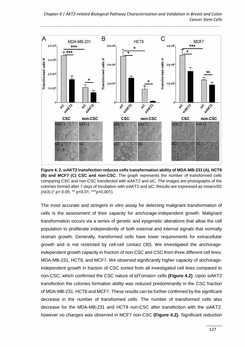
Chapter 4 | AKT2-related Biological Pathway Characterization and Validation in Breast and Colon Cancer Stem Cells
_______________________________________________________________________________
_______ 127
Figure 4. 2. siAKT2 transfection reduces cells transformation ability of MDA-MB-231 (A), HCT8
(B) and MCF7 (C) CSC and non-CSC. The graph represents the number of transformed cells
comparing CSC and non-CSC transfected with siAKT2 and siC. The images are photographs of the
colonies formed after 7 days of incubation with siAKT2 and siC. Results are expressed as mean±SD
(n≥3) (* p< 0.05; ** p<0.01, ***p<0.001).
The most accurate and stringent in vitro assay for detecting malignant transformation of
cells is the assessment of their capacity for anchorage-independent growth. Malignant
transformation occurs via a series of genetic and epigenetic alterations that allow the cell
population to proliferate independently of both external and internal signals that normally
restrain growth. Generally, transformed cells have lower requirements for extracellular
growth and is not restricted by cell-cell contact (30). We investigated the anchorage-
independent growth capacity in fraction of non-CSC and CSC from three different cell lines:
MDA-MB-231, HCT8, and MCF7. We observed significantly higher capacity of anchorage-
independent growth in fraction of CSC sorted from all investigated cell lines compared to
non-CSC, which confirmed the CSC nature of tdTomato+ cells (Figure 4.2). Upon siAKT2
transfection the colonies formation ability was reduced predominantly in the CSC fraction
of MDA-MB-231, HCT8 and MCF7. These results can be further confirmed by the significant
decrease in the number of transformed cells. The number of transformed cells also
decrease for the MDA-MB-231 and HCT8 non-CSC after transfection with the siAKT2;
however no changes was observed in MCF7 non-CSC (Figure 4.2). Significant reduction
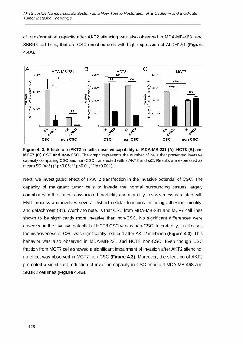
AKT2 siRNA-Nanoparticulate System as a New Tool to Restoration of E-Cadherin and Eradicate Tumor Metastic Phenotype ______________________________________________________________________________
_______ 128
of transformation capacity after AKT2 silencing was also observed in MDA-MB-468 and
SKBR3 cell lines, that are CSC enriched cells with high expression of ALDH1A1 (Figure
4.4A).
Figure 4. 3. Effects of siAKT2 in cells invasive capability of MDA-MB-231 (A), HCT8 (B) and
MCF7 (C) CSC and non-CSC. The graph represents the number of cells that presented invasive
capacity comparing CSC and non-CSC transfected with siAKT2 and siC. Results are expressed as
mean±SD (n≥3) (* p<0.05; ** p<0.01, ***p<0.001).
Next, we investigated effect of siAKT2 transfection in the invasive potential of CSC. The
capacity of malignant tumor cells to invade the normal surrounding tissues largely
contributes to the cancers associated morbidity and mortality. Invasiveness is related with
EMT process and involves several distinct cellular functions including adhesion, motility,
and detachment (31). Worthy to note, is that CSC from MDA-MB-231 and MCF7 cell lines
shown to be significantly more invasive than non-CSC. No significant differences were
observed in the invasive potential of HCT8 CSC versus non-CSC. Importantly, in all cases
the invasiveness of CSC was significantly reduced after AKT2 inhibition (Figure 4.3). This
behavior was also observed in MDA-MB-231 and HCT8 non-CSC. Even though CSC
fraction from MCF7 cells showed a significant impairment of invasion after AKT2 silencing,
no effect was observed in MCF7 non-CSC (Figure 4.3). Moreover, the silencing of AKT2
promoted a significant reduction of invasion capacity in CSC enriched MDA-MB-468 and
SKBR3 cell lines (Figure 4.4B).
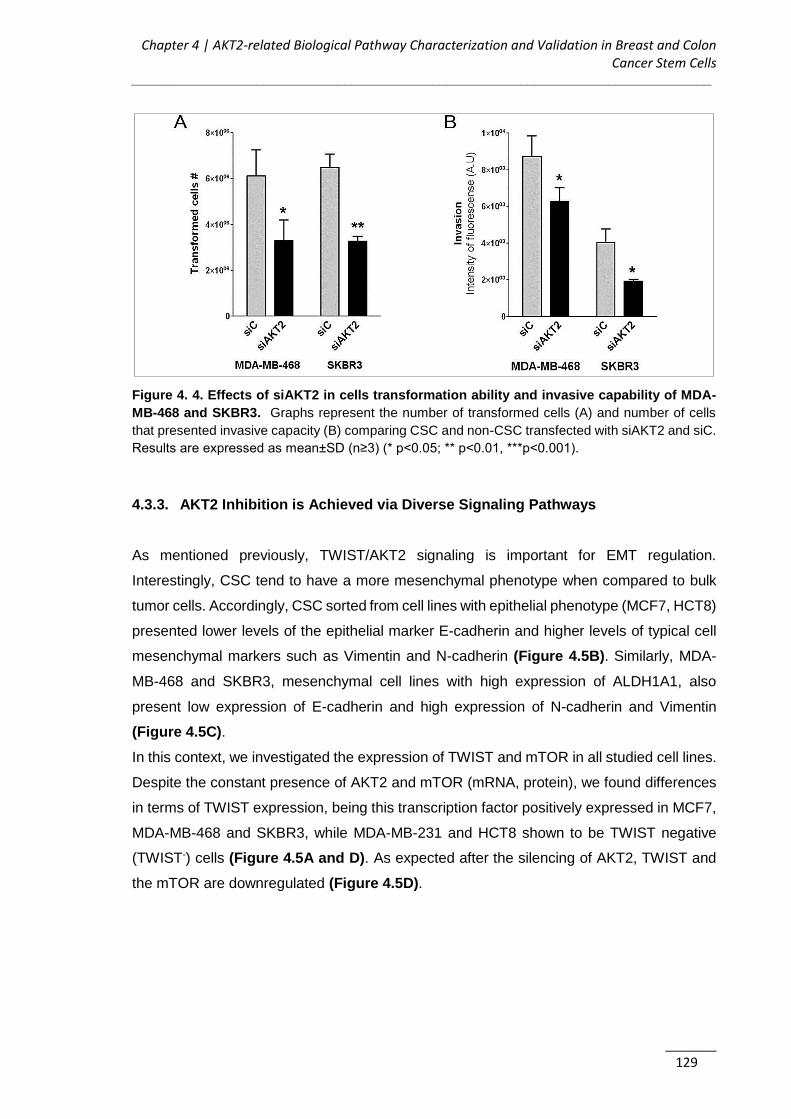
Chapter 4 | AKT2-related Biological Pathway Characterization and Validation in Breast and Colon Cancer Stem Cells
_______________________________________________________________________________
_______ 129
Figure 4. 4. Effects of siAKT2 in cells transformation ability and invasive capability of MDA-
MB-468 and SKBR3. Graphs represent the number of transformed cells (A) and number of cells
that presented invasive capacity (B) comparing CSC and non-CSC transfected with siAKT2 and siC.
Results are expressed as mean±SD (n≥3) (* p<0.05; ** p<0.01, ***p<0.001).
4.3.3. AKT2 Inhibition is Achieved via Diverse Signaling Pathways
As mentioned previously, TWIST/AKT2 signaling is important for EMT regulation.
Interestingly, CSC tend to have a more mesenchymal phenotype when compared to bulk
tumor cells. Accordingly, CSC sorted from cell lines with epithelial phenotype (MCF7, HCT8)
presented lower levels of the epithelial marker E-cadherin and higher levels of typical cell
mesenchymal markers such as Vimentin and N-cadherin (Figure 4.5B). Similarly, MDA-
MB-468 and SKBR3, mesenchymal cell lines with high expression of ALDH1A1, also
present low expression of E-cadherin and high expression of N-cadherin and Vimentin
(Figure 4.5C).
In this context, we investigated the expression of TWIST and mTOR in all studied cell lines.
Despite the constant presence of AKT2 and mTOR (mRNA, protein), we found differences
in terms of TWIST expression, being this transcription factor positively expressed in MCF7,
MDA-MB-468 and SKBR3, while MDA-MB-231 and HCT8 shown to be TWIST negative
(TWIST-) cells (Figure 4.5A and D). As expected after the silencing of AKT2, TWIST and
the mTOR are downregulated (Figure 4.5D).

AKT2 siRNA-Nanoparticulate System as a New Tool to Restoration of E-Cadherin and Eradicate Tumor Metastic Phenotype ______________________________________________________________________________
_______ 130
Figure 4. 5. Cell lines phenotypes based in the EMT/stemness markers. A) Amplification plot for
TWIST, mTOR and AKT2 expression for MDA-MB-231, HCT-8 and MCF7 cells. B) qPCR
quantification of several genes expression levels for MDA-MB-231, HCT-8 and MCF7 CSC. The
presented values are relative to their non-CSC population. C) qPCR quantification of several genes
expression levels for MDA-MB-468 and SKBR3 cells. D) Western Blot for AKT2, TWIST and mTOR
protein expression levels in different cells after the treatment with siAKT2 and siC. β-actin was used
as the housekeeping gene. Results are expressed as mean±SD (n≥3).

Chapter 4 | AKT2-related Biological Pathway Characterization and Validation in Breast and Colon Cancer Stem Cells
_______________________________________________________________________________
_______ 131
Figure 4. 6. Impact of AKT2 silencing in key regulators of EMT reversion and mTOR-dependent
signaling pathways. Expression levels of different genes were quantified by qPCR in different cell
lines after the treatment with siAKT2 and siC. Results are expressed as mean±SD (n≥3).

AKT2 siRNA-Nanoparticulate System as a New Tool to Restoration of E-Cadherin and Eradicate Tumor Metastic Phenotype ______________________________________________________________________________
_______ 132
Accordingly, we observed MET gene expression signature in cell lines expressing TWIST
(MDA-MB-468, SKBR3) after AKT2 silencing like a decrease in the expression of
mesenchymal markers like Vimentin and N-cadherin. Notably, this trend is accompanied by
cell restoration of E-cadherin expression, confirming our expectation that in this conditions
cells will experiencing a switch from a mesenchymal to a more epithelial phenotype (Figure
4.6A and B).
The observation of MET-related events in mesenchymal CSC isolated from epithelial MCF7
cell line upon AKT2 silencing, also related with the restoration of E-cadherin and further
decrease in N-cadherin and Vimentin expression, stresses this players interchangeability
triggered by this knockdown strategy (Figure 4.6C). Even though we observed a
downregulation of TWIST after AKT2 silencing, EMT reversion it was not observed in the
case of non-CSC MCF7 epithelial cell line (Figure 4.6D).
Figure 4. 7. Effects of AKT2 silencing in different signaling pathways. A) Efects of AKT2
silencing in the mTOR pathway for TWIST+ cells. B) Efffects of AKT2 silencing in the EMT reversion
for TWIST- cells. Results are expressed as mean±SD (n≥3).

Chapter 4 | AKT2-related Biological Pathway Characterization and Validation in Breast and Colon Cancer Stem Cells
_______________________________________________________________________________
_______ 133
In accordance with our proposed mechanism, TWIST- cells (MDA-MB-231, HCT8) didn’t
show the typical MET gene signature (Figure 4.7B).
Although AKT2-TWIST cross talk is a key event for malignancy, in non-expressing TWIST
cell lines the observed decrease in CSC malignance upon AKT2 silencing was related
predominantly by the downregulation of mTOR. The essential contribution of mTOR for the
regulation of cell growth arrest and inhibition of apoptosis was sustained by the finding that
in response to the intracellular decrease of AKT2, was detected a constant decrease of
mTOR expression and increase in GSK3b and p53 expression. This was observed in MDA-
MB-231 and HCT8 cells (Figure 4.6F and H). Similar results were observed in the fraction
of sorted MDA-MB-231 CSC and HCT8 CSC (Figure 4.6E and G). As expected, for the
TWIST expressing cells, the downregulation of mTOR after AKT2 silencing didn’t produce
the mTOR-related effects in GSK3b, p53 and Bcl-2 (Figure 4.7A).
The above described findings highlight that AKT2 silencing lead to the decrease of cell
migration, proliferation, invasion and transformation via different signaling axis depending
on the presence or absence of TWIST expression (Figure 4.8). Regardless the signaling
pathway, AKT2 silencing affects CSC malignancy and should be considered as prospective
for successful anti-cancer therapies.
Figure 4. 8. Summary of AKT2 silencing effects in cancer development in the different cell
lines, accordingly their phenotype and TWIST expression level.

AKT2 siRNA-Nanoparticulate System as a New Tool to Restoration of E-Cadherin and Eradicate Tumor Metastic Phenotype ______________________________________________________________________________
_______ 134
4.4. Discussion
Due to their unique phenotype, CSC are resistant to the conventional chemo- and
radiotherapy clinically practiced nowadays (4, 32). In addition, CSC are gaining relevance
as triggering agents of cancer progression and malignancy, showing higher invasive
capability than their non-CSC counterpart. Hereupon, even though several new promising
anti-cancer treatments have been proposed over the past years, just few of them were
extensively studied regarding their therapeutic efficacy also in population of CSC. Moreover,
several reports have shown that if a therapy is not effective against CSC, the reservoir of
resistant CSC will cause recurrence of aggressive tumor and metastatic growth over the
time (33, 34). Study specifically the silencing effects of some genetic players like AKT2 in
the CSC subpopulation is thus essential to develop new therapies that could impact in the
clinical treatment of cancer.
In this study, a CSC model based on the tdTomato vector expression under the control of
CSC specific ALDH1A1 promoter was used in order to study the efficacy of AKT2 silencing
in CSC fraction. This CSC model was previously extensively validated both in vitro and in
vivo (29). Moreover, here we observed higher anchorage-independent growth ability of
tdTomato+ CSC than the tdTomato- non-CSC, which further confirms this population
malignant nature (Figure 4.2). Importantly, after effective AKT2 silencing, the
transformation and invasion capacity in all studied CSC fractions (MDA-MB-231, MCF7,
HCT8) was significantly reduced. These results suggest that AKT2 silencing strategy may
be successful also in fraction of highly aggressive CSC (Figure 4.2 and 4.3).
AKT2 silencing also decreased the proliferation and migration capacity of various breast
and colon cancer cell lines, such as MDA-MB-231, SKBR3, MDA-MB-468 and HCT8, with
the remarkable exception of bulk MCF7 cells. No differences in terms of proliferation,
migration, invasion and anchored independent growth capacity were observed in MCF7
bulk cells (Figure 4.1). Of note, these were the only epithelial cells presenting expression
of TWIST used in this study. Active AKT2/TWIST signaling axis leads to the acquirement of
more mesenchymal phenotype. Contrary, AKT2 silencing confers to the cells more epithelial
phenotype triggering MET (21). Since MCF7 cells regularly shown an epithelial phenotype
with high expression of E-cadherin, the MET triggered via AKT2 silencing had no effect on
bulk MCF7 cell population. On the other hand, CSC subpopulations isolated from MCF7
epithelial cells present a more mesenchymal phenotype than the non-CSC, with a
decreased E-cadherin expression and an increased Vimentin and N-cadherin expression
(Figure 4.5B). In this mesenchymal CSC fraction (1-2%) isolated from epithelial MCF7 cells
we observed a significant effect after AKT2 silencing in terms of reduced transformation
Chapter 4 | AKT2-related Biological Pathway Characterization and Validation in Breast and Colon Cancer Stem Cells
_______________________________________________________________________________
_______ 135
and invasion capacity (Figure 4.2 and 4.3). Owing these cells involvement in tumor growth
and maintenance, its reduction would suppose an important strike for further tumor
progression. We observed similar results in the TWIST+ MDA-MB-468 and SKBR3
mesenchymal cell lines that are also characterized by their high levels of ALDH1A1
expression, conferring them CSC-like properties (35) (Figure 4.4). MET process after AKT2
silencing was confirmed by the genetic expression profile in mesenchymal TWIST+ cell
lines. We demonstrated that the AKT2 silencing is able to restore the levels of E-cadherin
and decrease the levels of TWIST and other EMT markers like N-Cadherin, Vimentin and
Fibronectin (Figure 4.6A-C). In unsorted MCF7 cells, the only gene affected after AKT2
knockdown was TWIST, confirming the direct cross talk between AKT2 and TWIST,
previously reported by Cheng GZ, et al. (2007) (11, 12) (Figure 4.6D). Mani SA, et al.
(2008), previously demonstrated a tight relationship between EMT activation and CSC in
breast cancer. It was shown, that the ectopic expression of several EMT inducers (TWIST,
Snail, TGF-β) triggers the expression of stem cell markers (CD44+ and CD24-) and
enhances tumor development (36). Furthermore, EMT was observed also in vivo in CD8T
immune cells which secrete EMT-inducing factors such as the TGF-β, presenting the
resultant tumors characteristics of breast CSC (CD44+ and CD24-) (37).
In our study, CSC isolated from the TWIST- epithelial cell line, HCT8, shown to be more
mesenchymal than the non-sorted bulk cells, with higher expression of N-cadherin and
Vimentin and lower expression of E-cadherin (Figure 4.5B). Despite the clear effect on the
transformation and invasion capacity after AKT2 silencing in these cells, we did not
observed MET phenotype in HCT8 CSC fraction (Figure 4.7B). However, contrary to MCF7
epithelial cell line, we observed a decrease in the proliferation, invasion and transformation
capacity of HCT8 tumor cells after AKT2 silencing (Figure 4.1, 4.2 and 4.3). Moreover
despite the reduction of the aggressiveness of both sorted CSC and unsorted cells, we have
not observed any effect on EMT genes expression in other mesenchymal cell lines with
TWIST- background (MDA-MB-231) (Figure 4.7B). Taking into account these results, we
conclude that for TWIST- cells, the AKT2 silencing is able to impair invasion and colony
formation via a different pathway. We observed the blockage of the PI3/AKT2/mTOR
signaling and consequent increase of GSK3b and pro-apoptotic genes such as p53 in both
CSC and non-CSC subpopulations of mesenchymal TWIST- cells (Figure 4.6E-H).
The novelty of this work lies in the comparison between CSC and non-CSC regarding the
AKT2 silencing effects and the observation that, specifically for CSC, this inhibition is able
to reverse the EMT phenotype in terms of gene expression, protein expression, migration,
proliferation, anchorage-independent growth and invasion ability, being special interesting
AKT2 siRNA-Nanoparticulate System as a New Tool to Restoration of E-Cadherin and Eradicate Tumor Metastic Phenotype ______________________________________________________________________________
_______ 136
this effect in the CSC population that usually present resistance against the conventional
treatments.
4.5. Conclusions
The biggest challenge in cancer treatment is tumor recurrence and resistance. Both
phenomena are sustained by the presence of CSC within the tumor. In this context, the
potential of new targeted therapies should be investigated especially in fraction of CSC to
provide its real probability to succeed in clinics. Since the RNAi therapy demonstrated
important prospect in targeted cancer treatment, we investigated the efficiency of siAKT2 in
CSC isolated from breast and colon cancer cell lines with different phenotypes. The
obtained results demonstrated that the AKT2 inhibition is effective in terms of reducing cells
migration, invasion, and transformation via different mechanisms and are not dependent on
the presence of TWIST in the tumor. In cell lines expressing TWIST, AKT2 inhibition acts
via MET, while in those cells not expressing TWIST, AKT2 silencing hampers mTOR
pathway and alters the expression of apoptotic genes such as the Bcl-2 and p53.
Regardless the signaling pathway, AKT2 silencing affects CSC malignancy, reverting their
mesenchymal phenotype, reducing cell invasion, inhibiting colony formation or inducing
apoptosis, and should be thus validated in further pre-clinical studies. Due to the beneficial
effect of AKT2 silencing in both bulk cells and CSC, the therapy with AKT2 could be used
i) to revert the tumor to its primary stage for easier chirurgical removal, and/or ii) prevent
the tumor recurrence through the inhibition of CSC tumorigenicity.
4.6. References
1. Charafe-Jauffret E, Ginestier C, Birnbaum D. Breast cancer stem cells: tools and
models to rely on. BMC Cancer. 2009;9:202.
2. Dean M, Fojo T, Bates S. Tumour stem cells and drug resistance. Nat Rev Cancer.
2005;5(4):275-84.
3. Lagadec C, Vlashi E, Della Donna L, Meng Y, Dekmezian C, Kim K, et al. Survival
and self-renewing capacity of breast cancer initiating cells during fractionated radiation
treatment. Breast Cancer Res. 2010;12(1):R13.
4. Yu Y, Ramena G, Elble RC. The role of cancer stem cells in relapse of solid tumors.
Front Biosci (Elite Ed). 2012;4:1528-41.

Chapter 4 | AKT2-related Biological Pathway Characterization and Validation in Breast and Colon Cancer Stem Cells
_______________________________________________________________________________
_______ 137
5. Deng Y, Wang CC, Choy KW, Du Q, Chen J, Wang Q, et al. Therapeutic potentials
of gene silencing by RNA interference: Principles, challenges, and new strategies. Gene.
2014.
6. Videira M, Arranja A, Rafael D, Gaspar R. Preclinical development of siRNA
therapeutics: towards the match between fundamental science and engineered systems.
Nanomedicine. 2014;10(4):689-702.
7. Kurreck J. RNA interference: from basic research to therapeutic applications.
Angewandte Chemie. 2009;48(8):1378-98.
8. Irie HY, Pearline RV, Grueneberg D, Hsia M, Ravichandran P, Kothari N, et al.
Distinct roles of Akt1 and Akt2 in regulating cell migration and epithelial-mesenchymal
transition. The Journal of cell biology. 2005;171(6):1023-34.
9. Arboleda MJ. Overexpression of AKT2/Protein Kinase Bb Leads to Up-Regulation
of b1 Integrins, Increased Invasion, and Metastasis of Human Breast and Ovarian Cancer
Cells. 2003.
10. Dillon RL, Marcotte R, Hennessy BT, Woodgett JR, Mills GB, Muller WJ. Akt1 and
akt2 play distinct roles in the initiation and metastatic phases of mammary tumor
progression. Cancer research. 2009;69(12):5057-64.
11. Cheng GZ, Zhang WZ, Sun M, Wang Q, Coppola D, Mansour M, et al. Twist is
transcriptionally induced by activation of STAT3 and mediates STAT3 oncogenic function.
The Journal of biological chemistry. 2008;283(21):14665-73.
12. Cheng GZ, Chan J, Wang Q, Zhang W, Sun CD, Wang LH. Twist transcriptionally
up-regulates AKT2 in breast cancer cells leading to increased migration, invasion, and
resistance to paclitaxel. Cancer research. 2007;67(5):1979-87.
13. Umemura S, Mimaki S, Makinoshima H, Tada S, Ishii G, Ohmatsu H, et al.
Therapeutic priority of the PI3K/AKT/mTOR pathway in small cell lung cancers as revealed
by a comprehensive genomic analysis. J Thorac Oncol. 2014;9(9):1324-31.
14. Rafael D, Doktorovová S, Florindo HF, Gener P, Abasolo I, Schwartz S, et al. EMT
blockage strategies: Targeting Akt dependent mechanisms for breast cancer metastatic
behaviour modulation. Curr Gene Ther. 2015;15(3):300-12.
15. Beck B, Lapouge G, Rorive S, Drogat B, Desaedelaere K, Delafaille S, et al. Different
levels of Twist1 regulate skin tumor initiation, stemness, and progression. Cell Stem Cell.
2015;16(1):67-79.
16. Zhou M, Hou Y, Yang G, Zhang H, Tu G, Du YE, et al. LncRNA-Hh Strengthen
Cancer Stem Cells Generation in Twist-Positive Breast Cancer via Activation of Hedgehog
Signaling Pathway. Stem Cells. 2015.

AKT2 siRNA-Nanoparticulate System as a New Tool to Restoration of E-Cadherin and Eradicate Tumor Metastic Phenotype ______________________________________________________________________________
_______ 138
17. Onder TT, Gupta PB, Mani SA, Yang J, Lander ES, Weinberg RA. Loss of E-
cadherin promotes metastasis via multiple downstream transcriptional pathways. Cancer
research. 2008;68(10):3645-54.
18. Kalluri R, Weinberg RA. The basics of epithelial-mesenchymal transition. The
Journal of clinical investigation. 2009;119(6):1420-8.
19. Radisky DC, LaBarge MA. Epithelial-mesenchymal transition and the stem cell
phenotype. Cell Stem Cell. 2008;2(6):511-2.
20. Saito T, Mimori K. [Cancer stemness and circulating tumor cells]. Nihon Rinsho.
2015;73(5):806-10.
21. Brabletz T. EMT and MET in metastasis: where are the cancer stem cells? Cancer
Cell. 2012;22(6):699-701.
22. Katsuno Y, Lamouille S, Derynck R. TGF-β signaling and epithelial-mesenchymal
transition in cancer progression. Curr Opin Oncol. 2013;25(1):76-84.
23. Bai H, Li H, Li W, Gui T, Yang J, Cao D, et al. The PI3K/AKT/mTOR pathway is a
potential predictor of distinct invasive and migratory capacities in human ovarian cancer cell
lines. Oncotarget. 2015;6(28):25520-32.
24. Chen J, Shao R, Li F, Monteiro M, Liu JP, Xu ZP, et al. PI3K/Akt/mTOR Pathway
Dual Inhibitor BEZ235 Suppresses the Stemness of Colon Cancer Stem Cells. Clin Exp
Pharmacol Physiol. 2015.
25. Ji Y, Di W, Yang Q, Lu Z, Cai W, Wu J. Inhibition of Autophagy Increases
Proliferation Inhibition and Apoptosis Induced by the PI3K/mTOR Inhibitor NVP-BEZ235 in
Breast Cancer Cells. Clin Lab. 2015;61(8):1043-51.
26. Patruno A, Pesce M, Grilli A, Speranza L, Franceschelli S, De Lutiis MA, et al. mTOR
Activation by PI3K/Akt and ERK Signaling in Short ELF-EMF Exposed Human
Keratinocytes. PLoS One. 2015;10(10):e0139644.
27. Piao Y, Li Y, Xu Q, Liu JW, Xing CZ, Xie XD, et al. Association of MTOR and AKT
Gene Polymorphisms with Susceptibility and Survival of Gastric Cancer. PLoS One.
2015;10(8):e0136447.
28. Xia P, Xu XY. PI3K/Akt/mTOR signaling pathway in cancer stem cells: from basic
research to clinical application. Am J Cancer Res. 2015;5(5):1602-9.
29. Gener P, Gouveia LP, Sabat GR, de Sousa Rafael DF, Fort NB, Arranja A, et al.
Fluorescent CSC models evidence that targeted nanomedicines improve treatment
sensitivity of breast and colon cancer stem cells. Nanomedicine. 2015;11(8):1883-92.
30. Creton S, Aardema MJ, Carmichael PL, Harvey JS, Martin FL, Newbold RF, et al.
Cell transformation assays for prediction of carcinogenic potential: state of the science and
future research needs. Mutagenesis. 2012;27(1):93-101.

Chapter 4 | AKT2-related Biological Pathway Characterization and Validation in Breast and Colon Cancer Stem Cells
_______________________________________________________________________________
_______ 139
31. Shaw LM. Tumor cell invasion assays. Methods Mol Biol. 2005;294:97-105.
32. Dragu DL, Necula LG, Bleotu C, Diaconu CC, Chivu-Economescu M. Therapies
targeting cancer stem cells: Current trends and future challenges. World J Stem Cells.
2015;7(9):1185-201.
33. Nandy SB, Gangwani L, Nahleh Z, Subramani R, Arumugam A, de la Rosa JM, et
al. Recurrence and metastasis of breast cancer is influenced by ovarian hormone's effect
on breast cancer stem cells. Future Oncol. 2015;11(6):983-95.
34. Sehl ME, Shimada M, Landeros A, Lange K, Wicha MS. Modeling of Cancer Stem
Cell State Transitions Predicts Therapeutic Response. PLoS One. 2015;10(9):e0135797.
35. Marcato P, Dean CA, Pan D, Araslanova R, Gillis M, Joshi M, et al. Aldehyde
dehydrogenase activity of breast cancer stem cells is primarily due to isoform ALDH1A3
and its expression is predictive of metastasis. Stem Cells. 2011;29(1):32-45.
36. Mani SA, Guo W, Liao MJ, Eaton EN, Ayyanan A, Zhou AY, et al. The epithelial-
mesenchymal transition generates cells with properties of stem cells. Cell. 2008;133(4):704-
15.
37. Santisteban M, Reiman JM, Asiedu MK, Behrens MD, Nassar A, Kalli KR, et al.
Immune-induced epithelial to mesenchymal transition in vivo generates breast cancer stem
cells. Cancer Res. 2009;69(7):2887-95.

AKT2 siRNA-Nanoparticulate System as a New Tool to Restoration of E-Cadherin and Eradicate Tumor Metastic Phenotype ______________________________________________________________________________
_______ 140
Chapter 5 | Functional Validation of Amphiphilic-based Polymeric Micelles for siRNA Delivery and Cancer Stem Cells Genes Inhibition
_______________________________________________________________________________
_______ 141
CHAPTER 5
Functional Validation of Amphiphilic-based Polymeric
Micelles for siRNA Delivery and Cancer Stem Cells Genes
Inhibition
The information presented in this chapter was partially published in the following publication:
1) D Rafael, P Gener, F Andrade, J Seras, S Montero, Y Fernandez, I Abasolo, S
Schwartz Jr., M Videira, Functional Validation of Amphiphilic-Based Polymeric
Micelles for siRNA Delivery and Cancer Stem Cells Genes Inhibition, Article in
preparation.
AKT2 siRNA-Nanoparticulate System as a New Tool to Restoration of E-Cadherin and Eradicate Tumor Metastic Phenotype ______________________________________________________________________________
_______ 142
Table of contents
5.1. Introduction .................................................................................................... 143
5.2. Material and Methods .................................................................................... 143
5.2.1. Materials ................................................................................................... 143
5.2.2. Polyplexes PEI-siRNA Production............................................................. 144
5.2.3. Production of Micelles ............................................................................... 145
5.2.4. Micelles Physicochemical Characterization ............................................... 145
5.2.5. Cell Lines and Culture Conditions ............................................................. 145
5.2.6. Cell Transfection with the PM ................................................................... 146
5.2.9. Fluorescence-Activated Cell Sorting ......................................................... 147
5.2.10. Micelles Internalization .............................................................................. 147
5.2.11. Green Fluorescent Protein Silencing Efficacy ........................................... 148
5.2.12. RNA Extraction and Quantitative RT-PCR ................................................ 149
5.2.13. Cell Transformation Assay (Anchorage-independent Growth Assay) ........ 149
5.2.14. Invasion Assay ......................................................................................... 149
5.2.15. In vivo Maximum Tolerated Dose Determination ....................................... 150
5.2.16. Statistical Analysis .................................................................................... 150
5.3. Results............................................................................................................ 150
5.3.1. PM are Technologically Favorable for siRNA Delivery .............................. 150
5.3.2. Complete PM Internalization Occurs After 4 hours of Incubation .............. 152
5.3.3. PM do not Present in vitro or in vivo Toxicity ............................................. 155
5.3.4. PM-siGFP Efficiently Silence the GFP Reporter Gene Expression ........... 157
5.3.5. PM-siAKT2 Reduce the Metastatic Potential of CSC ................................ 157
5.4. Discussion ..................................................................................................... 160
5.5. Conclusions ................................................................................................... 162
5.6. References ..................................................................................................... 163
Chapter 5 | Functional Validation of Amphiphilic-based Polymeric Micelles for siRNA Delivery and Cancer Stem Cells Genes Inhibition
_______________________________________________________________________________
_______ 143
5.1. Introduction
The major challenge regarding the RNAi bases therapies is the delivery of OGN to the cells
without suffering premature degradation by serum nucleases. In this context, PM for siRNA
delivery were design based on amphiphilic block-co-polymers previously developed within
the group (1). Pluronic® polymers were chosen due to their recognized ability to enhance
the transfection efficiency of genetic material, and their PEGylated surface, offering stealth
properties to the system (2).
Based on the compounding and formulation development studies presented in Chapter 3,
here is proposed a new system for siRNA delivery that combines i) polyplexes
spontaneously formed by electrostatic interaction between the negatively charged siRNA
molecules and the positively charged chains of 10K branched PEI, with ii) Pluronic® F127-
based PM formed by FH technique.
To access the biological efficacy of these PM we firstly produced the micelles using a siRNA
against GFP in breast cancer cells, which obtained a gene silencing higher than 80%, the
same or even greater than the gold standard vector Lipofectamine® 2000. After we
produced our micelles with the siRNA against AKT2 and tested them in breast CSC isolated
from two different cell lines. With these micelles we were able to observe a reduction of the
CSC invasion and transformation capacities.
5.2. Material and Methods
5.2.1. Materials
Pluronic® F127 was kindly provided by BASF (Ludwigshafen, Germany), while 10k
branched PEI was purchased from Alfa Aesar (Thermo Fisher GmbH, Karlsruhe, Germany).
DMSO, MTT, 5-DTAF were from Sigma-Aldrich (Madrid, Spain), while sodium bicarbonate
and paraformaldehyde (PFA) were obtained from Merck KGaA (Darmstadt, Germany).
RPMI medium, PBS, and FBS were purchase from Lonza (Barcelona, Spain). L-glutamine,
non-essential amino acids, 10000 U/mL penicillin and 10000 μg/mL streptomycin, 0.25%
Trypsin-EDTA, blasticidin and sodium pyruvate were purchased from Gibco (Life
Technologies Ltd., Madrid, Spain). siRNA-GFP positive and negative control,
Lipofectamine® 2000, LysoTracker® Red DND-99, DAPI, SYBR Green, and ProLong® Gold
Antifade Mountant were purchased from Molecular Probes (Life Technologies Ltd., Madrid,
Spain). Angarose and TBE buffer (10X) were obtained Sigma-Aldrich (St. Louis, MO, USA).
siRNAs were designed by Shanghai Gene Pharma (Shanghai, China); sense siAKT2
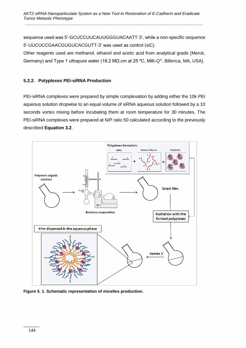
AKT2 siRNA-Nanoparticulate System as a New Tool to Restoration of E-Cadherin and Eradicate Tumor Metastic Phenotype ______________________________________________________________________________
_______ 144
sequence used was 5’-GCUCCUUCAUUGGGUACAATT-3’, while a non-specific sequence
5’-UUCUCCGAACGUGUCACGUTT-3’ was used as control (siC).
Other reagents used are methanol, ethanol and acetic acid from analytical grade (Merck,
Germany) and Type 1 ultrapure water (18.2 MΩ.cm at 25 ºC, Milli-Q®, Billerica, MA, USA).
5.2.2. Polyplexes PEI-siRNA Production
PEI-siRNA complexes were prepared by simple complexation by adding either the 10k PEI
aqueous solution dropwise to an equal volume of siRNA aqueous solution followed by a 10
seconds vortex mixing before incubating them at room temperature for 30 minutes. The
PEI-siRNA complexes were prepared at N/P ratio 50 calculated according to the previously
described Equation 3.2.
Figure 5. 1. Schematic representation of micelles production.
Chapter 5 | Functional Validation of Amphiphilic-based Polymeric Micelles for siRNA Delivery and Cancer Stem Cells Genes Inhibition
_______________________________________________________________________________
_______ 145
5.2.3. Production of Micelles
PM were prepared using the FH technique previously described (1). Briefly, the polymer
was individually weighted and dissolved in a mixture of methanol:ethanol (1:1). Then, the
solvent was removed under vacuum in a rotary evaporator and the formed film was left to
dry overnight at room temperature to eliminate any remaining solvent. The film was then
hydrated with the previously prepared polyplexes and vortexed during 1 minute (Figure
5.1). The obtained dispersion was filtered through a 0.22 μm syringe filter for sterilization
and remove possible aggregates.
5.2.4. Micelles Physicochemical Characterization
PM mean hydrodynamic diameter and polydispersity index were measured by DLS and
zeta potential was assessed by laser doppler micro-electrophoresis using a NanoZS
(Malvern Instruments, UK). Each type of formulation was produced and analyzed in
triplicate.
The morphology of PM was assessed by TEM. For that, samples were placed on a grid,
treated with uranil acetate and then observed in a JEM-1210 Transmission Electron
Microscope (JEOL Ltd., Tokyo, Japan).
The encapsulation efficiency were assessed by quantification of the free siRNA in filtrates
using a UV-Vis spectrophotometers (Nanodrop 2000, Thermo Scientific, USA) after filtration
of the formulations by centrifugation for 10 minutes at 10,000 rpm and 4ºC, using 100k pore
filters (Nanosep® Centrifugal Devices, Pall Corporation, Spain).
Gel electrophoresis was also performed to evaluate siRNA condensation. The PM were
loaded onto 1% agarose gel with 6× loading buffer. The mixture was separated in 0.5×
Tris/Borate/EDTA (TBE) buffer at 100V for 25 minutes. The siRNA bands were visualized
using an ultra violet (UV) imaging system (Uvidoc, UVItec Ltd, Cambridge, UK).
The osmolality of formulations was determined at room temperature using a Micro-
Osmometer M3320 (Advanced Instruments, Inc., MA, USA). Triplicates of each formulation
were analyzed.
5.2.5. Cell Lines and Culture Conditions
MCF7 (ATCC number HTB-22) and MDA-MB-231 (ATCC number HTB-26) breast cancer
cell lines were obtained from American Type Culture Collection (ATTC, LGC Standards,

AKT2 siRNA-Nanoparticulate System as a New Tool to Restoration of E-Cadherin and Eradicate Tumor Metastic Phenotype ______________________________________________________________________________
_______ 146
Barcelona, Spain). The cells were cultured in RPMI medium supplemented with 10% FBS,
1% penicillin-streptomycin, 1% L-glutamine, 1% non-essential amino acids and 1% of
sodium pyruvate. Blasticidin (0.5 mg/ml) was used as a selective antibiotic for
ALDH1A1/tdTomato cell lines. All cell lines were maintained at 37ºC under 5% CO2
saturated atmosphere. The medium was changed every other day and, upon confluence,
cells were harvested from plates with 0.25% trypsin-EDTA to be passed to other plates to
continue expansion, be frozen or used in in vitro studies.
5.2.6. Cell Transfection with the PM
The different PM-siRNA were transfected into cells accordingly Table 5.1. For silencing
efficacy assays the medium was changed after 4 hours of incubation with the PM. For the
toxicity assays the cells were left 24 hours in contact with the PM, while for the
internalization assays the cells were incubated with the PM at several time points (from 3
seconds to 24 hours)
Cells were harvested 72 hours after the transfection. For the internalization experiments the
particles were diluted 1:10
For some experiments, Lipofectamine® 2000 (Life Technologies, Madrid, Spain) were used
as a transfection positive control accordingly the supplier protocol in order to obtain a final
siRNA concentration in the well of 200 nM.
Table 5. 1. Transfection conditions accordingly the different experiments.
Day 0: before transfection Day 1: during transfection time (4 hours)
Plates
Cells
seeded
(cells/well)
Vol. of
medium (µl)
Vol. of
medium
(µl)
Vol. of
formulation
(µl)
Final siRNA
concentration
(nM)
96 well 5.0x10³ 100 50 50 200
24 well 5.0x10⁴ 1000 100 100 200
6 well 2.0x10⁵ 2000 250 250 200
5.2.7. Serum Stability Assay
To study the PM stability in serum, a special medium containing a concentration of FBS 50
% was prepared in order to predict the nanoparticles aggregation pattern in vivo. The PM
were incubated with medium-FBS (50%) and samples were collected at different time-points
(0, 4, 6, 12 and 24 hours) for DLS measurement.
Chapter 5 | Functional Validation of Amphiphilic-based Polymeric Micelles for siRNA Delivery and Cancer Stem Cells Genes Inhibition
_______________________________________________________________________________
_______ 147
5.2.8. Assessment of Micelles Toxicity
In order to assess the effect of formulations on the viability of breast cancer cell lines, 5000
cells/well were seeded in 96-well plates and left to attach for 24 hours. Then, cells were
incubated with increasing concentrations of PM:siC, PEI, F127 and PEI:siC complexes for
24 hours. After this incubation time, the medium was changed and the cells left for 72 hours.
Complete medium was used as negative control and 10% DMSO as positive control of
toxicity. Cell viability was measured 72 hours upon transfection by MTT assay. The
absorbance of each well was read on an absorbance microplate reader ELx800 (BioTek,
Germany), at 590 nm. The results of cell viability were used for the determination of IC50
index by nonlinear regression of the concentration-effect curve fit using Prism 6.02 software
(GraphPad Software, Inc., CA, USA).
5.2.9. Fluorescence-Activated Cell Sorting (FACS)
FACS was used to sort CSC and non-CSC subpopulations from a heterogeneous
population of MCF7 and MDA-MB-231 cells. For the cell sorting, a starting amount of 5X10⁶
cells were used. Cells were detached with 0.25% trypsin-EDTA and resuspended in PBS
supplemented with 10% FBS and DAPI (1 μg/mL) used for vital staining. Cells were sorted
according to tdTomato and DAPI expression in a FACS Aria cell sorter (BD Biosciences,
Madrid, Spain). Sorted cells were collected in complete medium without antibiotic and used
for posterior applications.
5.2.10. Micelles Internalization
To assess the internalization behavior of PM in MCF7 and MDA-MB-231 cells, 5-DTAF-
fluorescently labeled PM were used (conjugation method described in Chapter 3, Figure
3.2).
a) Confocal microscopy (qualitative analysis)
For confocal microscopy analysis, 2.5x10⁵ cells were cultured into glass coverslips coated
with 0.1% gelatin in 6 well plates for 24 hours to allow adhesion. Then, cells were incubated
for 4 hours with the labeled PM and after this time, the lysosomes were stained using the
Lysotracker® Red during 30 minutes at 37 ºC. Subsequently, the cells were fixed in 4% PFA
AKT2 siRNA-Nanoparticulate System as a New Tool to Restoration of E-Cadherin and Eradicate Tumor Metastic Phenotype ______________________________________________________________________________
_______ 148
at 4ºC for 30 minutes followed by nuclei staining with DAPI (0.2 mg/mL) for 5 minutes in the
dark. Cells were viewed under a Spectral Confocal Microscope MFV1000 Olympus
(Olympus, USA). The 561 nm excitation wavelength of the green laser (10mW) was used
for selective detection of the red fluorochrome (Lysotracker® Red). The 488 nm excitation
wavelength of Argon multiline laser (40 mW) was used for selective detection of the green
fluorochrome (5-DTAF). The nuclear staining DAPI was excited at 405 nm with a violet laser
(6 mW). Minimal single optical sections were collected for each fluorochrome sequentially.
The generated images were merged and analyzed with the FV10-ASW 3.1 Viewer software
(Olympus, USA) and exported as jpg image format.
b) Flow cytometry (quantitative analysis)
Flow cytometry analysis was used to verify internalization of nanoparticles in MDA-MB-231
and MCF7 ALDH1A1 tdTomato cells with around 50% of tdtomato+ cells. 2x10⁵ cells were
seeded in complete medium in 96 well plates for 24 hours to allow adhesion. Micelles were
added to cells at different time points: 1, 3, 10, 30 minutes and, 1, 2, 4, 6, 8 and 20 hours.
Then, cells were washed with 1x PBS, detached with 0.25% trypsin-EDTA, and
resuspended in PBS supplemented with 10% FBS and DAPI (1 μg/mL) used for vital
staining. The plate was analyzed in a cytometer Fortessa (BD Biosciences, USA). Data was
analyzed with FCS Express 4 Flow Research Edition software (De Novo Software, Los
Angeles, USA). Contaminants were removed by forward and side scatter gating. For each
sample, at least 10000 individual cells were collected and the mean fluorescence intensity
was evaluated.
5.2.11. Green Fluorescent Protein Silencing Efficacy
MDA-MB-231 cells expressing GFP were used to assess the micelles silencing efficacy.
Micelles prepared using a siRNA against the GFP were added to cells at a concentration of
5x10⁴ cells/ml in 96 well plates. For these test, as a positive control were used cells
transfected with the complexes formed between the Lipofectamine ® 2000 and the GFP
prepared accordingly to the supplier instructions. On the other hand, cells transfected in the
same conditions with PM prepared with a siC were used as negative control. The expression
of GFP in cells after transfection was assessed with a fluorescence microscope (Olympus,
USA). A video of the cells during 72 hours after transfection was recorded using Motorized
Fluorescent Microscope BX61 (Olympus, USA) in order to confirm GFP decreasing over
Chapter 5 | Functional Validation of Amphiphilic-based Polymeric Micelles for siRNA Delivery and Cancer Stem Cells Genes Inhibition
_______________________________________________________________________________
_______ 149
the time. Fluorescence intensity per cell over all the time of the experiment was quantified
using the ImageJ 1.48v software (National Institutes of Health, MD, USA). The intensity of
cells fluorescence was also measured using a Fluorescent Microplate reader FLX800
(BioTek, Germany).
5.2.12. RNA Extraction and Quantitative RT-PCR (qRT-PCR)
Total RNA was extracted from cells using the RNeasy Micro Kit (Qiagen, Madrid, Spain)
and the obtained RNA was reverse transcribed with the High Capacity cDNA Reverse
Transcription Kit (Applied Biosystems, Madrid, Spain) according to the manufacturer
instructions. The cDNA reverse transcription product was amplified with specific primers for
AKT2 (hAKT2 F: 5’ CAA GGA TGA AGT CGC TCA CAC A 3’; hAKT2 R: 5’ GAA CGG GTG
CCT GGT GTT C 3’) GADPH (hGADPH F: 5’ ACC CAC TCC TCC ACC TTT GAC; hGADPH
R: 5’ CAT ACC AGG AAA TGA GCT TGA CAA 3’) and Actin (hActin F: 5’ CAT CCA CGA
AAC TAC CTT CAA CTC C 3’; hActin R: 5’GAG CCG CCG ATC CAC AC 3’) by qPCR using
the SYBR Green method. The reaction was performed in triplicate on a 7500 Real time PCR
system (Applied Biosystems, Madrid, Spain). Actin, GADPH, and S18 were used as
endogenous controls. The relative mRNA levels were calculated using the comparative Ct
method (2e- ΔΔCt).
5.2.13. Cell Transformation Assay (Anchorage-independent Growth Assay)
The anchorage-independent growth of the different breast cancer cell lines were assessed
by the CytoSelect™ Cell Transformation Assay Kit (Cell Biolabs, San Diego, CA, USA). A
semisolid agar media were prepared accordingly the manufacturer instructions prior
addition of PM-siAKT2 or PM-siC to each well. After 6-8 days of incubation the colonies
were observed under an optical microscope and the viable transformed cells counted using
trypan blue.
5.2.14. Invasion Assay
Cells invasiveness was assessed using the CytoSelect™ Laminin Cell Invasion Assay Kit
(Cell Biolabs, San Diego, CA, USA) as previously described in Chapter 4. Briefly, the inserts
were placed in a 24 well plate and 2.5x10⁴ cells previously transfected (24 hours before)
with the PM-siAKT2 and the PM-siC, were added to the upper chamber. After 48 hours

AKT2 siRNA-Nanoparticulate System as a New Tool to Restoration of E-Cadherin and Eradicate Tumor Metastic Phenotype ______________________________________________________________________________
_______ 150
incubation, the invasive cells were dissociated from the membrane, lysed and quantified
using the CyQuant® GR Fluorescent Dye using a fluorescent microplate reader FLX800
(BioTek, Germany).
5.2.15. In vivo Maximum Tolerated Dose (MTD) Determination
Female athymic nude mice (Hsd:Athymic Nude-Foxn1 nu/nu; Harlan Interfauna Iberica,
Barcelona, Spain) were kept in pathogen-free conditions and used at 6 weeks of age.
Animal care was handled in accordance to guidelines for the Care and Use of Laboratory
Animals of the Vall d’Hebron University Hospital based on Federation of Laboratory Animal
Science Associations (FELASA) recommendations and the European Union legislation
(European Parliament and Council Directive 2010/63/EU). The experimental procedures
were approved by the Animal Experimentation Ethical Committee of the institution.
Animals were randomly divided in 4 groups (n=3) and intravenously administered in the tail
vein with PBS 1X (control group) or PM at doses of 20, 40 or 80 mg per kilogram of body
weight (single administration). Body weight changes and side effects were monitored during
12 days post-administration. For side effects evaluation the reduction of voluntary
movement, irregular breathing, motor disturbances, seizures, sudden death, flushed skin,
lacrimation, dehydration and hunched position was monitored.
5.2.16. Statistical Analysis
At least three batches of each PM were produced and characterized and the results
expressed as the mean±SD. For biological studies, at least 3 replicates, each involving at
least two technical replicates were involved in final results expressed as the mean±SD.
Statistical analysis was performed in Microsoft Office Excel™ 2007 using the unpaired
Student's t-test. Differences were regarded as statistically significant when p-value were
smaller than 0.05.
5.3. Results
5.3.1. PM are Technologically Favorable for siRNA Delivery
In order to characterize physicochemically the proposed formulation, several technological
tests were performed. The combination of poloxamer F127 and 10K branched PEI-based
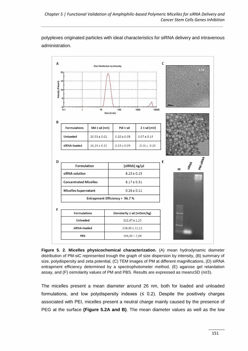
Chapter 5 | Functional Validation of Amphiphilic-based Polymeric Micelles for siRNA Delivery and Cancer Stem Cells Genes Inhibition
_______________________________________________________________________________
_______ 151
polyplexes originated particles with ideal characteristics for siRNA delivery and intravenous
administration.
Figure 5. 2. Micelles physicochemical characterization. (A) mean hydrodynamic diameter
distribution of PM-siC represented trough the graph of size dispersion by intensity, (B) summary of
size, polydispersity and zeta potential, (C) TEM images of PM at different magnifications, (D) siRNA
entrapment efficiency determined by a spectrophotometer method, (E) agarose gel retardation
assay, and (F) osmolarity values of PM and PBS. Results are expressed as mean±SD (n≥3).
The micelles present a mean diameter around 26 nm, both for loaded and unloaded
formulations, and low polydispersity indexes (≤ 0.2). Despite the positively charges
associated with PEI, micelles present a neutral charge mainly caused by the presence of
PEG at the surface (Figure 5.2A and B). The mean diameter values as well as the low

AKT2 siRNA-Nanoparticulate System as a New Tool to Restoration of E-Cadherin and Eradicate Tumor Metastic Phenotype ______________________________________________________________________________
_______ 152
polydispersity were also confirmed by TEM images, where it is possible to observe micelles
of small size and characteristic spherical morphology (Figure 5.2C).
In order to confirm if the siRNA was correctly complexed with PEI and associated with
micelles, the entrapment efficiency was determined by two distinct methods. The indirect
method, that measures the free siRNA concentration at the supernatant after PM
centrifugation with filtration, shows entrapment efficiencies higher than 96% (Figure 5.2D).
Additionally, no free siRNA fraction was detectable by agarose gel retardation assay in the
micelles sample (Figure 5.2E).
Micelles present an osmolarity value slightly superior to the PBS but < 350 mOsm/Kg, which
corroborates their adequate applicability for systemic administration (Figure 5.2F).
5.3.2. Complete PM Internalization Occurs After 4 hours of Incubation
The internalization of the fluorescent-labelled micelles was assessed quantitatively by flow
cytometry (Figure 5.3) and qualitatively by confocal microscopy (Figure 5.4). In order to
study the uptake kinetics the cells were incubated with PM at different time-points. With the
objective to check if there is any difference in the internalization pattern between CSC and
non-CSC cells, MDA-MB-231 and MCF7 ALDH1A1 cells with around 50% of tdTomato+
(CSC) were used (Figure 5.3C and G). It was possible to observe that particles start to be
internalized 10 minutes after incubation, but only after 4 hours an internalization of 90% is
reached (Figure 5.3D and H). Interestingly, although no significant differences were
observed between CSC and non-CSC cells regarding the amount of cells that internalize
micelles (Figure 5.3A and E), is possible to observe superior values of fluorescence
intensity for CSC cells, which possibly represents a higher number of PM inside each cell
(Figure 5.3B and F). Thus, it is possible to predict that tdTomato+ cells internalize
significantly more micelles than the tdTomato- ones. This result needs to be further explored
in the future to clarify the endocytic pathways and understand the possible different behavior
between CSC and non-CSC.
For the qualitative analysis, MDA-MB-231 cells incubated with fluorescent-labeled PM were
observed by confocal microscopy. As expected, it is possible to observe micelles
internalization as well as co-localization with the cell endosomal vesicles (Figure 5.4).
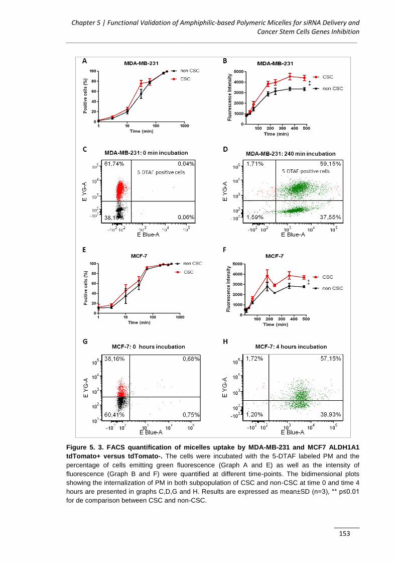
Chapter 5 | Functional Validation of Amphiphilic-based Polymeric Micelles for siRNA Delivery and Cancer Stem Cells Genes Inhibition
_______________________________________________________________________________
_______ 153
Figure 5. 3. FACS quantification of micelles uptake by MDA-MB-231 and MCF7 ALDH1A1
tdTomato+ versus tdTomato-. The cells were incubated with the 5-DTAF labeled PM and the
percentage of cells emitting green fluorescence (Graph A and E) as well as the intensity of
fluorescence (Graph B and F) were quantified at different time-points. The bidimensional plots
showing the internalization of PM in both subpopulation of CSC and non-CSC at time 0 and time 4
hours are presented in graphs C,D,G and H. Results are expressed as mean±SD (n=3), ** p≤0.01
for de comparison between CSC and non-CSC.

AKT2 siRNA-Nanoparticulate System as a New Tool to Restoration of E-Cadherin and Eradicate Tumor Metastic Phenotype ______________________________________________________________________________
_______ 154
Figure 5. 4. PM internalization visualization throught confocal microscopy. The PM are labeled
in green with the 5-DTAF, the endossomal vesicles are labeled in red (Lysotracker® RED and the
cells nuclei labeled in blue (DAPI).
Figure 5. 5. In vitro cytotoxicity of PM and its isolated components. A) Assessment of the effects
of PM and isolated components concentration in the cell viability. To facilitate the comparison
between samples, the concentration presented for the isolated components is not the real one, but
corresponds to the one present in the final PM concentration (for example, in the plot is not being
assessed the toxicity corresponding to 5 mg/ml of PEI, but instead the toxicity of the PEI
concentration present in 5 mg/ml of PM). B) IC50 values for each component and for PM. The values
were obtained by interpolation of y=50 from the dose-effect curves fitting using the real
concentrations used for each sample. Results are expressed as mean±SD (n=3).

Chapter 5 | Functional Validation of Amphiphilic-based Polymeric Micelles for siRNA Delivery and Cancer Stem Cells Genes Inhibition
_______________________________________________________________________________
_______ 155
5.3.3. PM do not Present in vitro or in vivo Toxicity
Effects on cell viability of PM-siRNA as well as its isolated components were assessed in
vitro via MTT assay in MDA-MB-231 and MCF7 (Figure 5.5A) breast cancer cells, with the
additional estimation of their IC50 values (Figure 5.5B). As observed in both dose-effect
graphs and the IC50 values, F127 do not cause significant toxicity to the cells. On the other
side, PEI alone is highly toxic. However, this PEI-related toxicity appears to be reduced
when it is complexed with the negatively charged siRNA and even more reduced in the
presence of the F127 shell. Since the concentrations of PM that have demonstrated to be
efficient in vitro (5 mg/ml) are inferior to their IC50 value, is possible to conclude that PM are
not expected to significantly cause toxicity after administration.
Before to administrate the PM in vivo, a serum stability assay was perform in order to predict
the amount of aggregation between the PM and the serum components in vivo. Figure 5.6
represents the mean size distribution curves by intensity for the PM-siRNA incubated with
medium-FBS (50%) at the different time-points (Figure 5.6A) and for the medium-FBS
(50%) (Figure 5.6B).
Figure 5. 6. Serum stability assay PM-siRNA. The graphs represent the DLS size distributions by
intensity after incubation at different time-points. (A) PMs with siC mean curves at different time-
points (0-24 hours). (B) Medium-serum 50% curve. All measurements were performed in triplicate,
with three technical samples.
A
B

AKT2 siRNA-Nanoparticulate System as a New Tool to Restoration of E-Cadherin and Eradicate Tumor Metastic Phenotype ______________________________________________________________________________
_______ 156
The distribution curves demonstrate some aggregation of the PM with the serum
components over the time since it can be observed a reduction of the medium particles
fraction (20-100nm) with a consequent increase in the percentage of particles with higher
sizes (>100nm). This behavior was especially observed after 12 hours of incubation. Taking
into account that we expect that the PM are eliminated before the 12 hours, the verified
aggregation do not constitute a major problem for the intravenous in vivo administrations of
the formulation.
Figure 5. 7. PM toxicity in vivo. In the graph is presented the body weight variation of animals up
to 12 days post-administration of the samples. The table shows the register of adverse side effects
monitored during the time of the experiment. Results are expressed as mean±SD (n=3).

Chapter 5 | Functional Validation of Amphiphilic-based Polymeric Micelles for siRNA Delivery and Cancer Stem Cells Genes Inhibition
_______________________________________________________________________________
_______ 157
The safety of PM was also assessed in vivo. For this, PM were administered to healthy mice
at increasing concentrations in order to determine the MTD. A maximum administrated dose
of 80 mg of PM per kilogram of body weight was well tolerated, without inducing adverse
side effects up to 12 days post-administration. As observed in Figure 5.7, no significant
changes in weight gain neither weight loss were seen in the animals treated with the PM
when compared to the control group of animals injected with PBS (p≤0.05).
5.3.4. PM-siGFP Efficiently Silence the GFP Reporter Gene Expression
PM entrapping a siRNA against GFP were incubated at different times with GFP expressing
MDA-MB-231 breast cancer cells. As shown in Figure 5.8, a clear and strong qualitative
(Figure 5.8A) and quantitative (Figure 5.8B) reduction of GFP expression were observed
24 hours post-transfection in cells treated with GFP-siRNA when compared with the cells
transfected siC.
5.3.5. PM-siAKT2 Reduce the Metastatic Potential of CSC
Next, we examined whether the PM platform could be used to silence a potential therapeutic
target related with breast CSC. PM were prepared using the siAKT2 and siC and transfected
into MDA-MB-231 and MCF7 breast cancer cells as well as in their CSC fractions isolated
as explained in Chapter 4. During our study we verified the stemness gene expression after
each sorting to guarantee the purity of the samples. The decrease of AKT2 expression in
both cell lines was confirmed by qPCR (Figure 5.9A). Subsequently, in order to determine
the effects related to AKT2 inhibition in the fraction of CSC, cells invasion and anchorage-
independent growth ability were assessed for MDA-MB-231 Td-Tomato+ and MCF7 Td-
Tomato+ cells after treatment with the PM-siAKT2 and PM-siC. Regarding the invasive
potential of CSC, for both cases the invasiveness of CSC was significantly reduced after
PM-siAKT2 transfection (Figure 5.9B). Next, the assessment of the anchorage-
independent growth capacity revealed a reduced number of transformed cells for both MDA-
MB-231 and MCF7 CSC after treatment with the PM-siAKT2 (Figure 5.9C). Accordingly, it
was observed an accentuated impairment of colonies formation after the treatment with PM-
siAKT2 (Figure 5.9D).

AKT2 siRNA-Nanoparticulate System as a New Tool to Restoration of E-Cadherin and Eradicate Tumor Metastic Phenotype ______________________________________________________________________________
_______ 158
Figure 5. 8. GFP reporter assay for PM-siGFP biological efficacy assessment. (A) Fluorescent
images of MDA-MB-231/GFP cells were taken 24, 48 and 72 hours after incubation with PM-siGFP
and PM-siC. (B) Time-lapse experiment in which fluorescent images of cells incubated with siGFP
and siC associated with PM or Lipofectamine® 2000 were captured at each 30 minutes during 72
hours. The values represent the fluorescent intensity detected for each time-point. Results are
expressed as mean±SD (n=3), *** p≤ 0.001 for the comparison between PM-siGFP and PM-siC.

Chapter 5 | Functional Validation of Amphiphilic-based Polymeric Micelles for siRNA Delivery and Cancer Stem Cells Genes Inhibition
_______________________________________________________________________________
_______ 159
Figure 5. 9. PM-siAKT2 effects in MDA-MB-231 and MCF7 cells. (A) The reduction of AKT2 gene
expression was detected in both cell lines through qPCR. The presented values are normalized to
the housekeeping genes (Actin and GADPH). (B) Quantification of the number of invasive cells after
incubation with PM-siAKT2 versus PM-siC. (C) Quantification of the number of transformed cells
after incubation with PM-siAKT2 versus PM-siC. (D) Colonies formation on the soft agar after
incubation of cells with PM-siAKT2 versus PM-siC. Results are expressed as mean±SD (n=3), ***
p≤ 0.001 for the comparison between PM-siC and PM-siAKT2.
AKT2 siRNA-Nanoparticulate System as a New Tool to Restoration of E-Cadherin and Eradicate Tumor Metastic Phenotype ______________________________________________________________________________
_______ 160
5.4. Discussion
In this work PM were chosen as siRNA delivery systems due to their well-known versatility
to produce, among others, systems presenting long circulating profiles in the bloodstream
and able to improve the cellular uptake of compounds, increasing its therapeutic efficacy
(3, 4). Among the amphiphilic polymers used to produce micelles, poloxamers enjoy special
prominence due to their biocompatible and biodegradable nature, well-established status
as drug delivery and biomedical excipients, capacity to improve the transfection of genetic
material, and to support endosomal release of endocytosed nanocarriers (2, 5, 6).
Regarding the choice of the amphiphilic polymer, the Pluronic® F127 presented adequate
physical properties regarding size and Pdi as referred in Chapter 3. Similar
physicochemical results were obtained with PM of poloxamers encapsulating insulin (1).
Since poloxamers present neutral charge at the physiological pH, it was necessary to
introduce a cationic component to formulation in order to obtain required positive charge to
complex the anionic siRNA, so the branched PEI 10K was chosen as the polycation to
produce the polyplexes (as explained in Chapter 3). Like this we can enjoy from the benefits
of both PEI and poloxamers regarding transfection efficiency, and PEGylated surface of
PM.
The morphological analysis has shown micelles with spherical shape and md close to 27
nm (Figure 5.2), corroborating the results of DLS. The lower size of micelles composed by
F127 is explained by its lower critical micelle concentration (CMC) and hydrophilic-lipophilic
balance (HLB) values, resulting in more compact micelles (1).
Due to their small size and the enhanced transfection efficiency capacity of both
components, PM are quickly and efficiently internalized by cells (Figure 5.3) and efficiently
promote the silencing of the gene of interest (Figure 5.8 and 5.9). Moreover, the cytotoxicity
assay reveals that the presence of F127 reduce the PEI-related toxicity, probably due to the
reduction of its surface cationic groups. Therefore, the system do not present significant
toxicity both in vitro (Figure 5.5) as well as in vivo (Figure 5.7), which is related to the
biocompatibility of poloxamers explaining their use on a variety of biomedical applications
including drug delivery (7, 8).
Another advantage about the presence of poloxamers in the formulation was evidenced by
the serum stability assays. The results showed that there is no aggregation with serum
components up to 12 hours of incubation, suggesting that this PM may be adequate for
intravenous administration in vivo (Figure 5.6).
Other research groups developed micellar systems for siRNA delivery proving the utility of
this type of systems. For example, Liu L, et al. (2016) synthesized an amphiphilic and
Chapter 5 | Functional Validation of Amphiphilic-based Polymeric Micelles for siRNA Delivery and Cancer Stem Cells Genes Inhibition
_______________________________________________________________________________
_______ 161
biodegradable ternary copolymer conjugated with folate as ligand, polyethylenimine-graft-
polycaprolactone-block-poly(ethylene glycol)-folate (PEI-PCL-PEG-Fol) for targeted siRNA
delivery via folate-FR recognition (9). They obtained stable micelles with hydrodynamic
diameters about 100 nm and a zeta potential of +8.6 mV. The presence of folate was
responsible for the increased cellular uptake and in vitro gene knockdown observed in a
folate receptor enriched cell line (SKOV-3 cells). Moreover, these PEI-PCL-PEG-Fol/siRNA
micelles exhibited high stability in vivo and a long circulation half-life, which allowed 17% of
the intravenously injected siRNA to reach the tumor mainly due to their effective folate
targeting (10). Furthermore, the study of Salzano G, et al. (2015) brings the interesting
possibility of micelles multifunctionalization (10). In their study they developed self-
assembly PM able to efficiently co-load an anti-survivin siRNA and a chemotherapeutic
agent, such as PTX (survivin siRNA/PTX PM) and evaluated their applicability in vivo in an
animal model of PTX-resistant ovarian cancer. They obtained a significant downregulation
of survivin expression in tumor tissues together with a potent anticancer activity of survivin-
siRNA/PTX PM, while the tumors remained unaffected with the same quantity of free PTX.
These results underline the importance of the combined therapy in the treatment of
chemoresistant tumors (11).
The versatility of the poloxamers used in our system opens the possibility of creating
functionalized systems by conjugation of moieties to the PEG surface, and the co-
encapsulation of polyplexes and anticancer drugs, benefiting from the advantages of the
combined therapy. In fact, poloxamers have shown to reduce the MDR capacity of cancer
cells through a selective energy depletion and inhibition of the drug efflux systems,
improving especially the therapeutic efficacy of the drugs in multidrug resistant tumors (12-
14). Exploring these characteristics of poloxamers, a formulation of PM encapsulating
Doxorubicin (SP1049C) granted orphan designation by FDA to treat advanced carcinoma
of the esophagus and advanced gastric cancers, proving its value in the development of
systems for cancer treatment (15, 16). Additionally, poloxamers present cryoprotective
properties, allowing the lyophilization of the system to obtain a powder with good
redispersion profile, allowing the increase of the product stability and shelf-life, and reducing
the costs associated to liquid formulations (1).
Due to their unique phenotype, CSC are resistant to conventional therapies (17, 18).
Therefore, it is of major importance to find an adequate target against CSC and an effective
delivery system able to reach them, otherwise the reservoir of resistant CSC will cause
recurrence of aggressive tumor and metastatic growth over the time (19, 20). We used a
CSC model based on the tdTomato expression under the control of CSC specific ALDH1A1
promoter, in order to study the efficacy of AKT2 silencing in CSC fraction. This CSC model
AKT2 siRNA-Nanoparticulate System as a New Tool to Restoration of E-Cadherin and Eradicate Tumor Metastic Phenotype ______________________________________________________________________________
_______ 162
was previously extensively validated in vitro and in vivo (21). In Chapter 4, we have
demonstrated that downregulating AKT2 using a siAKT2 have an important role in the EMT
impairment and the consequent reduction metastatization and malignancy of cancer cells
with special importance in CSC. After treatment with the PM-siAKT2, the transformation
capacity of CSC fractions (MDA-MB-231 and MCF7) was significantly abolished (p≤0.001).
Similarly, the invasion capacity of CSC significantly decreased (p≤0.001) after the
transfection with the PM-siAKT2 (Figure 5.9). These results suggest that AKT2 silencing
strategy may be successful as a target for future RNAi-based antitumor therapies
particularly on the challenging fraction of highly aggressive CSC.
The internalization studies demonstrated that micelles are able to be taken up by cells
through endocytosis, being possible to observe their co-localization with the endosomal
vesicles (Figure 5.4). This result is in agreement with the biological efficacy studies
performed, since the internalization by endocytosis of polyplexes/micelles containing the
siRNA is the first step to become available to perform is biological function inside the cell.
It also revealed a significant preferential uptake of PM by CSC (p≤0.01) (Figure 5.3),
resulting in a high biological therapeutic efficacy (Figure 5.9). The mechanism involved in
the enhancement of transfection efficiency by poloxamers is the same underlying its effects
as MDR chemosensitizers. By interfering with the cellular membrane, poloxamers inhibit
the ATPase activity and the efflux function of P-gp, improving the efficacy of anticancer
drugs (14, 22). Moreover, poloxamers interfere with mitochondrial function showing to
enhance proapoptotic signaling and decrease tumorigenicity of cancer cells by altering the
epigenetic regulation of cells mainly in MDR over non-MDR cells (12, 15, 23). Taking into
account that CSC display some of the characteristics of MDR cells, like overexpression of
efflux systems, poloxamers present the capacity to enhance the anticancer activity of drugs
(e.g. Doxorubicin), increase the transfection efficiency of OGN, and to decrease the
tumorigenicity and aggressiveness of tumor cells, especially in CSC sub-population (15).
5.5. Conclusions
In this study, an innovative nanocarrier system composed by Pluronic® F127 micelles
associated with PEI-based polyplexes has been developed with the objective to overcome
the major drawbacks usually associated to the genetic material delivery systems, namely
siRNA degradation, insufficient transfection efficiency or excessive nanoparticles-related
toxicity.
The proposed system seems to gather the requirements for an efficient and safe transport
of siRNA in terms of their physicochemical characteristics, internalization capacity,

Chapter 5 | Functional Validation of Amphiphilic-based Polymeric Micelles for siRNA Delivery and Cancer Stem Cells Genes Inhibition
_______________________________________________________________________________
_______ 163
biological efficacy and toxicity profile. Moreover we also observed a reduction of the
invasion and transformation capacities of breast cancer cells, when transfected with PM
entrapping the polyplexes with the siRNA against the gene of interest, with significant
positive results observed mainly in the so feared CSC population.
These results make us believe that this new formulation will serve as a technological
platform for the development of systems to encapsulate different siRNAs, as well as other
kind of genetic material, and bring new insights into the field of cancer gene therapy.
The next step in the development and optimization of these PM would be the introduction
of specific targeting moieties covalently linked to the polymers chains and assess to which
extent the active targeting could even improve the biological and therapeutic efficacy of the
system. It would be also priority to increase the loading capacity of the micelles to undergo
in vivo assays. First it should be performed a biodistribution study to track how PM behave
once in the blood circulation. Afterwards an in vivo efficacy study in a breast and/or colon
cancer mouse model using the PM carrying the siAKT2 versus siC will be also performed.
Another future objective of this work is the creation of a multifunctional system where can
be combined the delivery of both conventional drugs and genes. The system developed
presents adequate properties to deliver both hydrophobic and hydrophilic substances,
added at different steps of the production method. Therefore, the design of a nanosystem
combining an hydrophobic drug, such as the PTX or zileuton, and the hydrophilic polyplexes
of a certain siRNA, chosen depending on the genetic pattern of the disease, could be a
encouraging strategy in the future of the combined and personalized therapy.
5.6. References
1. Andrade F, Fonte P, Oliva M, Videira M, Ferreira D, Sarmento B. Solid state
formulations composed by amphiphilic polymers for delivery of proteins: characterization
and stability. International journal of pharmaceutics. 2015;486(1-2):195-206.
2. Kabanov A, Zhu J, Alakhov V. Pluronic block copolymers for gene delivery.
Advances in genetics. 2005;53:231-61.
3. Andrade F, Videira M, Ferreira D, Sarmento B. Micelle-based systems for pulmonary
drug delivery and targeting. Drug Delivery Letters. 2011;1(2):171-85.
4. Andrade F, Rafael D, Videira M, Ferreira D, Sosnik A, Sarmento B. Nanotechnology
and pulmonary delivery to overcome resistance in infectious diseases. Adv Drug Deliv Rev.
2013;65(13-14):1816-27.
AKT2 siRNA-Nanoparticulate System as a New Tool to Restoration of E-Cadherin and Eradicate Tumor Metastic Phenotype ______________________________________________________________________________
_______ 164
5. Chen J, Luo J, Zhao Y, Pu L, Lu X, Gao R, et al. Increase in transgene expression
by pluronic L64-mediated endosomal/lysosomal escape through its membrane-disturbing
action. ACS applied materials & interfaces. 2015;7(13):7282-93.
6. Yang Z, Sahay G, Sriadibhatla S, Kabanov AV. Amphiphilic block copolymers
enhance cellular uptake and nuclear entry of polyplex-delivered DNA. Bioconjug Chem.
2008;19(10):1987-94.
7. Jung HH, Park K, Han DK. Preparation of TGF-beta1-conjugated biodegradable
pluronic F127 hydrogel and its application with adipose-derived stem cells. Journal of
controlled release : official journal of the Controlled Release Society. 2010;147(1):84-91.
8. Diniz IM, Chen C, Xu X, Ansari S, Zadeh HH, Marques MM, et al. Pluronic F-127
hydrogel as a promising scaffold for encapsulation of dental-derived mesenchymal stem
cells. Journal of materials science Materials in medicine. 2015;26(3):153.
9. Batrakova EV, Li S, Alakhov VY, Miller DW, Kabanov AV. Optimal structure
requirements for pluronic block copolymers in modifying P-glycoprotein drug efflux
transporter activity in bovine brain microvessel endothelial cells. The Journal of
pharmacology and experimental therapeutics. 2003;304(2):845-54.
10. Liu L, Zheng M, Librizzi D, Renette T, Merkel OM, Kissel T. Efficient and Tumor
Targeted siRNA Delivery by Polyethylenimine-graft-polycaprolactone-block-poly(ethylene
glycol)-folate (PEI-PCL-PEG-Fol). Molecular pharmaceutics. 2016;13(1):134-43.
11. Salzano G, Navarro G, Trivedi MS, De Rosa G, Torchilin VP. Multifunctional
Polymeric Micelles Co-loaded with Anti-Survivin siRNA and Paclitaxel Overcome Drug
Resistance in an Animal Model of Ovarian Cancer. Molecular cancer therapeutics.
2015;14(4):1075-84.
12. Alakhova DY, Rapoport NY, Batrakova EV, Timoshin AA, Li S, Nicholls D, et al.
Differential metabolic responses to pluronic in MDR and non-MDR cells: a novel pathway
for chemosensitization of drug resistant cancers. Journal of controlled release : official
journal of the Controlled Release Society. 2010;142(1):89-100.
13. Batrakova EV, Kelly DL, Li S, Li Y, Yang Z, Xiao L, et al. Alteration of genomic
responses to doxorubicin and prevention of MDR in breast cancer cells by a polymer
excipient: pluronic P85. Mol Pharm. 2006;3(2):113-23.
14. Batrakova EV, Li S, Elmquist WF, Miller DW, Alakhov VY, Kabanov AV. Mechanism
of sensitization of MDR cancer cells by Pluronic block copolymers: Selective energy
depletion. British journal of cancer. 2001;85(12):1987-97.
15. Alakhova DY, Zhao Y, Li S, Kabanov AV. Effect of doxorubicin/pluronic SP1049C
on tumorigenicity, aggressiveness, DNA methylation and stem cell markers in murine
leukemia. PloS one. 2013;8(8):e72238.
Chapter 5 | Functional Validation of Amphiphilic-based Polymeric Micelles for siRNA Delivery and Cancer Stem Cells Genes Inhibition
_______________________________________________________________________________
_______ 165
16. Danson S, Ferry D, Alakhov V, Margison J, Kerr D, Jowle D, et al. Phase I dose
escalation and pharmacokinetic study of pluronic polymer-bound doxorubicin (SP1049C) in
patients with advanced cancer. Br J Cancer. 2004;90(11):2085-91.
17. Yu Y, Ramena G, Elble RC. The role of cancer stem cells in relapse of solid tumors.
Front Biosci (Elite Ed). 2012;4:1528-41.
18. Dragu DL, Necula LG, Bleotu C, Diaconu CC, Chivu-Economescu M. Therapies
targeting cancer stem cells: Current trends and future challenges. World J Stem Cells.
2015;7(9):1185-201.
19. Nandy SB, Gangwani L, Nahleh Z, Subramani R, Arumugam A, de la Rosa JM, et
al. Recurrence and metastasis of breast cancer is influenced by ovarian hormone's effect
on breast cancer stem cells. Future Oncol. 2015;11(6):983-95.
20. Sehl ME, Shimada M, Landeros A, Lange K, Wicha MS. Modeling of Cancer Stem
Cell State Transitions Predicts Therapeutic Response. PLoS One. 2015;10(9):e0135797.
21. Gener P, Gouveia LP, Sabat GR, de Sousa Rafael DF, Fort NB, Arranja A, et al.
Fluorescent CSC models evidence that targeted nanomedicines improve treatment
sensitivity of breast and colon cancer stem cells. Nanomedicine. 2015;11(8):1883-92.
22. Batrakova EV, Li S, Alakhov VY, Elmquist WF, Miller DW, Kabanov AV.
Sensitization of cells overexpressing multidrug-resistant proteins by pluronic P85.
Pharmaceutical research. 2003;20(10):1581-90.
23. Minko T, Batrakova EV, Li S, Li Y, Pakunlu RI, Alakhov VY, et al. Pluronic block
copolymers alter apoptotic signal transduction of doxorubicin in drug-resistant cancer cells.
Journal of controlled release : official journal of the Controlled Release Society.
2005;105(3):269-78.

AKT2 siRNA-Nanoparticulate System as a New Tool to Restoration of E-Cadherin and Eradicate Tumor Metastic Phenotype ______________________________________________________________________________
_______ 166

Chapter 6 | General Conclusions and Future Perspectives _______________________________________________________________________________
_______ 167
CHAPTER 6
General Conclusions and Future Perspectives

AKT2 siRNA-Nanoparticulate System as a New Tool to Restoration of E-Cadherin and Eradicate Tumor Metastic Phenotype ______________________________________________________________________________
_______ 168
Cancer is a higly prevalent disease having a major impact in the population health
worldwide. Even though current treatments have improved the overall survival and life
quality of patients, and despite the new generation of targeted anticancer drugs, cancer
remains difficult to treat and control. Consequentely, the development of therapy-resistant
metastasis that cause cancer relapse is still frequent. Despite the advancements observed
in the fields of molecular biology and biotechnology allowing a better understanding of the
mechanism of carcinogenesis and disease progression, as well as the development of
improved therapies, advanced cancer is generally an incurable disease. As referred, the
conventional treatments have improved the survival and cure rate of patients; however there
are some associated drawbacks, mostly concerning high toxicity and serious side-effects,
which may even cause death. More than half of the treated patients suffer from disease
relapse, including the presence of distant metastasis in other organs. The rapid emergence
of therapy resistance seen in the aggressive, metastatic forms of the disease is, by far, the
biggest obstacle for the success of current cytotoxic and cytostatic therapies. Indeed, many
of the newly developed cancer drugs elicit powerful initial responses, leading to dramatic
effects on tumor reduction and disease control, but still modest results regarding of long-
term overall survival (1-3).
Cancer maintenance, resistance to therapy and metastatic disease seem to be sustained
by the presence of CSC within the tumors. These cells retain the capacity of repopulating
the tumor, while being insensitive to conventional anticancer therapies, antimitotic agents
or radiation. It is still not clear if the CSC are originated from epithelial stem cells (the ones
essential to maintain proliferative homeostasis) or result from accumulation of mutations
from differentiated cells. In this scenario, the new cancer therapies under development
should fulfill two conditions: (i) reduce the toxicity displayed in current therapies and most
importantly, (ii) effectively target and eliminate CSC to overcome drug resistance and tumor
recurrence (4, 5).
One major difference between conventional chemotherapies and targeted therapies with
respect to the development of resistance is that resistance to targeted therapies follows a
more predictable path, being therefore, easier to identify the resistance mechanism and to
choose the more appropriate treatment. However, it remains a major challenge to predict
which patients will develop metastatic disease. As a result, describing specific targets or
signaling pathways is one of the pillars of current cancer research, although most tumors
are still treated with conventional cytotoxic therapies. Thus, the development of more
personalized and targeted anticancer therapies is essential. In that context the importance
of gene therapy gained a new breath in the last decades. This is a result of the important
advances observed in the field of molecular biology and particle engineering technology,
Chapter 6 | General Conclusions and Future Perspectives _______________________________________________________________________________
_______ 169
which allowed, respectively the characterization of new therapeutic targets and the
development of new and improved formulations (4).
Recent advances in genomics make possible now to consider the hypothesis that each
tumor has an independent genetic identity and to enumerate all of the genetic lesions in
specific cancers. These approaches will yield critical information regarding the biological
alterations signature in different tumors and are increasingly being applied prospectively to
patient samples in the clinic. At the same time, complementary approach to decipher the
molecular basis of malignant transformation depends upon the application of genome scale
tools to annotate the function of genes involved in cancer initiation and progression. Over
the past several years, have been developed genome scale RNAi, CRISPR and open
reading frame expression libraries that permit a systematic evaluation of genes involved in
cancer initiation and maintenance. Using these libraries, it was possible to performed
screens in a panel of human cancer cell lines to systematically identify cancer
vulnerabilities. By combining these functional approaches with information derived from
mapping the structural abnormalities present in cancer genomes, have been identified
several new oncogenes that contribute to cancer development. The ability to systematically
manipulate gene expression at genome scale now provides opportunities to investigate the
function of genes involved in cancer initiation and maintenance, which will provide a
framework for therapeutic strategies. For example, assays that apply massive parallel
sequencing approaches to evaluate tumor tissue or blood from clinical samples for evidence
of genomic harbingers of therapy response and resistance are becoming increasingly used.
There are various types of approaches that can be used, including active monitoring of
patients either during or at the end of their therapeutic regimen by comparison to a baseline
sample. The sensitive and quantitative nature of these assays can pinpoint patients with
therapy resistance, by virtue of unaltered prevalence of mutations relative to the baseline,
or also identify patients with an incomplete response such as minimal residual disease
(MRD). Before the treatment, the analysis of the disease genotype could be useful for the
selection of the potential targeted therapy. Therapeutic response monitoring using
biomarkers is also possible during the course of treatment and can indicate those non-
responding patients that might benefit from another therapy. Finally, post-mortem genomic
analyses of therapy resistant disease can be obtained from banked metastatic lesions to
inform about the genomic nature of therapy resistance. These studies can be incredibly
informative towards future monitoring paradigms, therapeutic combinations or new drug
design efforts to fight acquired resistance (6-8). Because of that, worldwide research groups
have been actively focused in the development of molecular therapies that target specific
oncoproteins, with the purpose of developing personalized therapies (e.g. against EGFR,
AKT2 siRNA-Nanoparticulate System as a New Tool to Restoration of E-Cadherin and Eradicate Tumor Metastic Phenotype ______________________________________________________________________________
_______ 170
HER2, FGFR, BRAF, MEK, PI3K, AKT, mTOR or IGF1-R) for patients displaying specific
genetic lesions or pathways deregulations. One of the main objectives nowadays and area
of intensive research is to identify new predictive biomarkers of response to diverse
treatments as well as biomarkers of primary resistance (de novo) and secondary treatment
(9, 10).
Still in the context of advance biomarker and drug target discovery, there are also groups
that are focused on the proteomic analysis of the cancer secretome and in the
characterization of the mechanisms adopted by cancer cells to communicate amongst
themselves as well as with their microenvironment during tumorigenesis. Most secreted
proteins contain a signal peptide that directs their sorting to the extracellular space through
the endoplasmic reticulum–Golgi secretory pathway. One of the most striking observations
when secretome profiles are carefully analyzed is that they contain hundreds of theoretical
intracellular proteins. Recent reports showing intracellular proteins with alternative
extracellular functions, metastasis localizations could be relevant in tumorigenesis and
could lead to novel therapies against cancer. Taking into consideration that metastasis
depends on a gene program expressed by the tumor microenvironment, for example with
the secretion of prosurvival factors by stromal cells during the colonization phase of
metastasis, blockage of this crosstalk between tumor cells and their microenvironment
through the use of chemical inhibitors could be very promising in prevent metastasis
formation. This dependency can also be exploited to improve diagnosis and treatment of
patients with advanced cancer (11, 12).
In this work we focused in the characterization of the pathway related with the AKT2
oncogene, since AKT2 is one of the three AKT isoforms that have shown to be amplified in
solid tumors. This oncogene is increased in response to apoptosis and its activation is also
associated with PI3-K related effects and TWIST-promoted metastatic process by
enhancing cell migration and invasion (13, 14). We have shown that the silencing of AKT2
signaling pathway both in bulk and in sub-populations of CSC is able to block their
tumorigenic potential, and reverting them to a more epithelial and noninvasive phenotype,
as demonstrated by the reduction in cell migration, proliferation, invasion and transformation
capabilities after the treatment with the siAKT2. We also demonstrate that the silencing of
AKT2 can act differently accordingly the cells phenotype. In cells that present TWIST (a
major downstream effector of AKT2) we were able to detect the EMT reversion through the
restoration of E-cadherin expression and the decrease of mesenchymal markers (Vimentin,
N-cadherin and Fibronectin) expression. For cells that do not express TWIST, the silencing
of AKT2 reduced the mTOR and increase the GSKβ expression, contributing for the
reduction of cells tumorigenic potential. Also, in these cell lines the AKT2 silencing promotes

Chapter 6 | General Conclusions and Future Perspectives _______________________________________________________________________________
_______ 171
alteration in apoptosis markers like p53 and Bcl-2. The novelty of this study relies on the
evidence that AKT2 silencing is also effective in the isolated subpopulation of CSC, both in
breast and colon cancer, since we truly believe that the specific targeting of this cell sub-
population will improve the clinical outcomes by reducing the cancer resistance and
recurrence.
In this work we propose the silence of AKT2 as a target using an RNAi approach, with the
final goal of developing a novel gene therapy-based system. However another strategy that
could be worthy to explore in the future is the chemical inhibition of AKT2. CCT128930 is a
novel, selective, and potent AKT2 inhibitor that blocks AKT2 activity exhibiting 28-fold
greater selectivity for AKT2 than for the other isoforms. CCT128930 exhibited marked anti-
tumor responses and inhibited the phosphorylation of a range of downstream AKT2
biomarkers in multiple tumor cell lines both in vitro and in vivo, arising as a new anticancer
drug (15). Moreover, the combination of AKT2 silencing with a traditional chemotherapeutic
agent, such as Doxorubicin or PTX, can represent an important future therapeutic approach
forward the CSC eradication and improved anticancer therapy.
The main goal of the present study is to develop a therapeutic system that reduce the cancer
therapy-related secondary effects, and specifically target the subpopulation of CSC within
the tumors to overcome resistance to therapy and recurrent metastatic and advanced
disease. Both criteria could be accomplished using innovative targeted nanomedicines for
drug and/or gene delivery, which is widely expected to create innovative therapeutics in
cancer therapy thanks to the selective delivery of higher concentrations of encapsulated
drugs, with reduced systemic toxicity, to tumor lesions trough the well-known EPR effect.
Based on these characteristics, the first generation nanomedicines already in clinical
practice or under clinical evaluation have been presenting improved therapeutic efficacy
than the conventional drug formulations, and have changed the scenario of oncological
diseases therapy (16).
Of note, ubiquitously targeting cells within a tumor is not always feasible because some
drugs cannot diffuse efficiently and the random nature of the passive targeting (EPR effect)
makes difficult to control the process. This lack of control may also be responsible for
inducing the problematic MDR. Thus, one strategy, to further enhance targeting of drugs to
cancer cells including CSC, is the conjugation of nanocarriers with molecules that bind to
overexpressed antigens or receptors on the target cells. These second generation vectors
are especially useful in tumors that do not exhibit EPR effect or cases where the
permeability of the vessels is not compromised (i.e. small metastasis). An additional
advantage for using targeting moieties for drug delivery into cancer cells is that drugs will

AKT2 siRNA-Nanoparticulate System as a New Tool to Restoration of E-Cadherin and Eradicate Tumor Metastic Phenotype ______________________________________________________________________________
_______ 172
benefit from entering inside tumor cells by receptor-mediated endocytosis, providing higher
concentration levels of active drug compounds within the tumor cell (17).
The nanodelivery system used in this work consisted in PEI-based polyplexes associated
to PM composed by poloxamers prepared by thin FH technique to deliver siAKT2. PEI was
used due to its well-recognized capacity to complex and transfect genetic material into cells.
PM were chosen mainly due to its easy production, small size, versatility to encapsulate
both hydrophobic and hydrophilic compounds, and the observed capacity of poloxamers to
modify the biological response and decrease the MDR of many cancer cells. Moreover, the
presence of PEG provides the formulation with stealth properties, allows the particles
modification with several targeting ligands, and facilitates the lyophilization of the system.
Regarding the last one, the inherent cryoprotectant properties of PEG reduce aggregation
of particles during lyophilization without the need of additional cryoprotectants, which
simplifies the formulation and could impact in their stability and safety profile. This question
is important in technological terms since a future step of development of the presented
system relies in the production of powders by lyophilization in order to increase its long-
term storage stability (18-20).
After production and physicochemical characterization, biological assessment of
formulations was performed in several cancer cell lines. No significant signs of cytotoxicity
were observed as determined by MTT (in vitro) and MTD (in vivo), thus, no substantial
toxicity is expected at therapeutic doses. The presence of F127 in the formulation not only
serves to increase the particles functionality and their transfection and biological efficiency,
but also proved to significantly reduce the PEI-associated toxicity. Furthermore, PM have
shown to be preferentially taken up by the CSC sub-population, thus suffering a sort of
passive targeting to CSC, which could be predictive indicator of its therapeutic efficacy.
In conclusion, amphiphilic polymers-based formulations combined with PEI presented
appropriate characteristics for delivery of siRNAs and achieve a strong gene silencing,
including in CSC. These formulations serve as a platform for future development of
improved gene delivery systems.
The ultimate goal of this study is to develop a multifunctional targeted nanomedicine against
breast and colorectal CSC by: i) using a specific targeting for CSC surface receptors (i.e.
CD44 for breast and CD133 or CXCR4 for colon) and ii) combine classical chemotherapy
with a specific siRNA against CSC. Thus, the next steps regarding the improvement of this
system will be the addition of a chemotherapeutic agent to the formulation and the
conjugation with targeting moieties (Figure 6.1). By efficiently targeting and killing CSC is
expected to significantly circumvent CSC-related resistance, reduce the number of
metastasis and tumor recurrence, and increase patient overall survival.

Chapter 6 | General Conclusions and Future Perspectives _______________________________________________________________________________
_______ 173
After perform in vitro and in vivo preclinical validation of our nanoconjugate targeting CSC,
the final step would be to initiate scale-up and regulatory studies.
Following the evolution that has been observed in this field during the last decades, is
expected in the near future a further increase in the development of optimized formulations
based on nanoparticles for gene delivery and/or to improve the properties of several
chemotherapeutic drugs. Due to the well-known advantages presented by these systems
would not be surprising to see an exponential increase of marketing authorization and
commercialization of multifunctional nanotechnology products, which we hope will bring a
new breath in the battle against cancer.
Figure 6. 1. Schematic representation of an amphiphilic polymer based multifunctional
nanoparticle for gene and drug delivery combination.
AKT2 siRNA-Nanoparticulate System as a New Tool to Restoration of E-Cadherin and Eradicate Tumor Metastic Phenotype ______________________________________________________________________________
_______ 174
References 1. Miller KD, Siegel RL, Lin CC, Mariotto AB, Kramer JL, Rowland JH, et al. Cancer
treatment and survivorship statistics, 2016. CA: a cancer journal for clinicians.
2016;66(4):271-89.
2. Siegel RL, Miller KD, Jemal A. Cancer Statistics, 2017. CA: a cancer journal for
clinicians. 2017;67(1):7-30.
3. Housman G, Byler S, Heerboth S, Lapinska K, Longacre M, Snyder N, et al. Drug
resistance in cancer: an overview. Cancers. 2014;6(3):1769-92.
4. Gener P, Rafael DF, Fernandez Y, Ortega JS, Arango D, Abasolo I, et al. Cancer
stem cells and personalized cancer nanomedicine. Nanomedicine. 2016;11(3):307-20.
5. Sehl ME, Shimada M, Landeros A, Lange K, Wicha MS. Modeling of Cancer Stem
Cell State Transitions Predicts Therapeutic Response. PLoS One. 2015;10(9):e0135797.
6. Byler S, Goldgar S, Heerboth S, Leary M, Housman G, Moulton K, et al. Genetic
and epigenetic aspects of breast cancer progression and therapy. Anticancer research.
2014;34(3):1071-7.
7. Tabchy A, Eltonsy N, Housman DE, Mills GB. Systematic identification of
combinatorial drivers and targets in cancer cell lines. PloS one. 2013;8(4):e60339.
8. Tachtsidis A, McInnes LM, Jacobsen N, Thompson EW, Saunders CM. Minimal
residual disease in breast cancer: an overview of circulating and disseminated tumour cells.
Clinical & experimental metastasis. 2016;33(6):521-50.
9. Tufts Center for the Study of Drug Development. Personalized Medicine Gains
Traction but Still Faces Multiple Challenges. www.csdd.tufts.edu; 2015.
10. Zeisberg M, Neilson EG. Biomarkers for epithelial-mesenchymal transitions. The
Journal of clinical investigation. 2009;119(6):1429-37.
11. Makridakis M, Vlahou A. Secretome proteomics for discovery of cancer biomarkers.
Journal of proteomics. 2010;73(12):2291-305.
12. Makridakis M, Roubelakis MG, Vlahou A. Stem cells: insights into the secretome.
Biochimica et biophysica acta. 2013;1834(11):2380-4.
13. Rafael D, Doktorovova S, Florindo HF, Gener P, Abasolo I, Schwartz S, Jr., et al.
EMT blockage strategies: Targeting Akt dependent mechanisms for breast cancer
metastatic behaviour modulation. Current gene therapy. 2015;15(3):300-12.
14. Xia P, Xu XY. PI3K/Akt/mTOR signaling pathway in cancer stem cells: from basic
research to clinical application. Am J Cancer Res. 2015;5(5):1602-9.
15. Yap TA, Walton MI, Hunter LJ, Valenti M, de Haven Brandon A, Eve PD, et al.
Preclinical pharmacology, antitumor activity, and development of pharmacodynamic
Chapter 6 | General Conclusions and Future Perspectives _______________________________________________________________________________
_______ 175
markers for the novel, potent AKT inhibitor CCT128930. Molecular cancer therapeutics.
2011;10(2):360-71.
16. Caster JM, Patel AN, Zhang T, Wang A. Investigational nanomedicines in 2016: a
review of nanotherapeutics currently undergoing clinical trials. Wiley interdisciplinary
reviews Nanomedicine and nanobiotechnology. 2017;9(1).
17. Jain V, Jain S, Mahajan SC. Nanomedicines based drug delivery systems for anti-
cancer targeting and treatment. Current drug delivery. 2015;12(2):177-91.
18. Ulasov AV, Khramtsov YV, Trusov GA, Rosenkranz AA, Sverdlov ED, Sobolev AS.
Properties of PEI-based polyplex nanoparticles that correlate with their transfection efficacy.
Mol Ther. 2011;19(1):103-12.
19. Alakhova DY, Rapoport NY, Batrakova EV, Timoshin AA, Li S, Nicholls D, et al.
Differential metabolic responses to pluronic in MDR and non-MDR cells: a novel pathway
for chemosensitization of drug resistant cancers. J Control Release. 2010;142(1):89-100.
20. Biswas S, Kumari P, Lakhani PM, Ghosh B. Recent advances in polymeric micelles
for anti-cancer drug delivery. European journal of pharmaceutical sciences : official journal
of the European Federation for Pharmaceutical Sciences. 2016;83:184-202.