amersham tissue inhibitor of metalloproteinases-1 (timp-1
TRANSCRIPT

GE Healthcare
AmershamTissue Inhibitor of Metalloproteinases-1(TIMP-1), Human, Biotrak, ELISA SystemProduct Booklet
Code: RPN2611 (96 wells)

2
Page finder1. Legal 4
2. Handling 5 2.1. Safety warnings and precautions 5 2.2. Storage 5 2.3. Expiry 5
3. Components 6
4. Other materials required 7
5. Description 8
6. Critical Parameters 10
7. Protocol 11 7.1. Specimen collection and sample preparation 11 7.1.1. Cell culture supernatants 11 7.1.2. Serum 11 7.1.3. Plasma 11 7.1.4. Tissue samples 12 7.2. Assay procedure 12 7.2.1. Reagent preparation 12 7.2.2. Preparation of working standards 13 7.2.3. Assay protocol 14 7.3. Data processing 17 7.3.1. Calculation of results 17 7.3.2. Typical assay data 18
8. Additional information 19 8.1. Specificity 19 8.2. Reproducibility 20 8.3. Sensitivity 22 8.4. Parallelism 23 8.5. Recovery 24

9. Troubleshooting guide 25
10. Background 26
11. References 28
12. Related products 29
3

1. LegalGE and GE monogram are trademarks of General Electric Company.
Amersham and Biotrak is a trademark of GE Healthcare companies.
Tween is a trademark of ICI Americas Inc.
GE Healthcare reserves the right, subject to any regulatory and contractual approval if required, to make changes in specifications and features shown herein, or discontinue the product described at any time without notice or obligation.
Contact your GE Representative for the most current information and a copy of the terms and conditions
© 2006 General Electric Company – All rights reserved.
http://www.gehealthcare.com/lifesciences
GE Healthcare UK Limited. Amersham Place, Little Chalfont, Buckinghamshire, HP7 9NA UK
4

5
2. Handling
2.1. Safety warnings and precautionsWarning: For research use only. Not recommended or intended for diagnosis of disease in humans or animals. Do not use internally or externally in humans or animals.
All chemicals should be considered as potentially hazardous. We therefore recommend that this product is handled only by those persons who have been trained in laboratory techniques and that it is used in accordance with the principles of good laboratory practice. Wear suitable protective clothing such as laboratory overalls, safety glasses and gloves. Care should be taken to avoid contact with skin or eyes. In the case of contact with skin or eyes wash immediately with water. See material safety data sheet(s) and/or safety statement(s) for specific advice.
Note that the assay protocol requires the use of sulfuric acid.
Warning: Sulfuric acid is corrosive. Please follow the manufacturer’s safety data sheet relating to the safe handling and use of this material.
2.2. StorageStore at -15ºC to -30ºC. Once reconstituted components should be stored as 2–8ºC and used within 7 days.
2.3. ExpiryThe expiry date is stated on the package and will be at least 4 weeks from the date of despatch.

3. ComponentsMicroplate: 12 x 8 well strips coated with anti-TIMP-1. Ready for use.
Assay buffer 1: 10 ml of Phosphate Buffer concentrate which when diluted gives a 0.1 M Phosphate Buffer pH 7.5 containing 0.9%(w/v) Sodium Chloride, 0.1%(w/v) Bovine Serum Albumin and 0.1% Tween™ 20.
Standard: 100 ng lyophilized TIMP-1. On reconstitution this gives a concentration of 100 ng/ml TIMP-1 in 0.1 M Phosphate Buffer pH 7.5 containing 0.9%(w/v) Sodium Chloride 0.1%(w/v) Bovine Serum Albumin and 0.1% Tween 20.
Peroxidase conjugate: Lyophilized anti-TIMP-1 Horseradish Peroxidase which on reconstitution gives anti-TIMP-1 Horseradish Peroxidase in 0.1 M Phosphate Buffer pH 7.5 containing 0.9%(w/v) Sodium Chloride, 0.1%(w/v) Bovine Serum Albumin and 0.1% Tween 20.
Wash buffer: 12.5 ml Phosphate Buffer concentrate which on dilution gives a 0.01 M Phosphate Buffer pH 7.5 containing 0.05% Tween 20.
TMB Substrate: 3,3’, 5,5’-Tetramethylbenzidine (TMB)/Hydrogen Peroxide, ready for use.
6

7
4. Other materials requiredThe following materials and equipment are required but not supplied:
• Pipettes or pipetting equipment with disposable polypropylene tips (100 µl, 500 µl, 1 ml and 5 ml)
• Disposable polypropylene test tubes
• Glass measuring cylinders (100 ml and 500 ml)
• Distilled or deionized water
• Spectrophotometric plate reader capable of measuring at 630 or 450 nm
• 1.0 M Sulfuric acid
• Microplate shaker (optional)
• Controlled temperature incubator that can maintain 20–25ºC (optional)
• Automatic plate washer (optional)

8
5. Description• Measures total TIMP-1• Specific for human TIMP-1• Precise and accurate measurement• Non-isotopic protocol• Ready to use substrate• Same day protocol
The TIMP-1, Human Biotrak™ ELISA System from GE Healthcare has been specifically designed for research purposes.
TIMP-1 may be measured in the range 3.13–50 ng/ml.
The assay is based on a two site ELISA ‘sandwich’ format, see figure 1. Standards and samples are incubated in microtitre wells precoated with anti-TIMP-1 antibody. Any TIMP-1 present will be bound to the wells, other components of the sample being removed by washing and aspiration. The TIMP-1 is detected by a Peroxidase labelled antibody to TIMP-1. Any excess is removed by washing and aspiration. The amount of peroxidase bound to each well is determined by the addition of TMB ‘ready to use’ substrate. The reaction is stopped by addition of an acid solution, and the resultant colour read at 450 nm in a microplate spectrophotometer. The concentration of TIMP-1 in a sample is determined by interpolation from a standard curve.
Each pack contains reagent for 96 determinations. This allows the construction of a standard curve plus the measurement of 42 samples in duplicate.

9
solid phase standard or
unknown
conjugate substrate
TMB
HRPAbto
TIMP-1
TIMP-1
Abto
TIMP-1
well +
incubate 2 h RT incubate 2 h RT incubate 30 min
wash wash
stop reactionmeasure OD
Figure 1. TIMP-1 ELISA assay design

6. Critical ParametersThe following points are critical.
• It is important that all the wells are washed thoroughly and uniformly. If using an automatic washer check operation of heads before starting. If washing by hand, use a wash bottle and ensure that all wells are completely filled at each wash.
• Use only coated wells from the same reagent batch for each assay.
• The incubation temperature range is critical (20–25ºC).
• Preparation of working standards and addition of standards to microplate should be performed using polypropylene pipettes.
• Allow samples and all reagents to reach 20–25ºC prior to performing assay.
• Incubation times must be carried out exactly. If more than one plate is being assayed each plate must be timed individually.
• A separate standard curve must be run on each plate.
• Mix samples and all reagents thoroughly before use.
• Avoid excessive foaming of reagents.
• Avoid handling the tops of wells both before and after filling.
• Keep the wells covered with lids except when adding reagents and reading.
• Standards and samples should be assayed in duplicate.
• The total dispensing time for each plate should not exceed 20 minutes.
10

7. Protocol7.1. Specimen collection and sample preparationThe TIMP-1 human, Biotrak ELISA system from GE Healthcare has been tested with several sample types for which representative procedures are described for guidance. It remains the investigators’ responsibility to validate their procedures.
7.1.1. Cell culture supernatants1. If necessary, centrifuge to remove any particulate material and
store at -15ºC to -30ºC.
2. It may be necessary to dilute the samples if levels of TIMP-1 are high. Assay buffer 1 is provided for this.
3. A recommended dilution of 1:10 to 1:160 may be used dependent on the cell line and time of culture. This dilution has been found to be suitable for tendon conditioned media samples for example.
7.1.2. Serum1. Allow samples to coagulate and centrifuge.
2. Remove serum and store at -15ºC to -30ºC.
3. Avoid freeze thaw cycles.
4. It is recommended to dilute serum samples 1:10 to 1:40 or more, depending on the concentration, before assay. Assay buffer 1 is provided for this.
7.1.3. Plasma1. Collect plasma using EDTA, Heparin or Citrate as an anticoagulant.
2. Centrifuge and remove plasma and store at -15ºC to -30ºC.
3. Avoid freeze thaw cycles.
11

4. It is recommended to dilute plasma samples 1:10 to 1:40 or more, depending on the concentration, before assay. Assay buffer 1 is provided for this.
7.1.4. Tissue samplesUsers are advised to carefully validate any tissue extraction procedure employed. The following method is described for the extraction of brain TIMP-1 (18), and is suggested for guidance only as its use has not been validated with this assay or with other tissues.
1. Homogenize in 50 mM Tris-HCl (pH 7.5), 75 mM NaCl, 1 mM Phenylmethyl Sulfonyl Fluoride (PMSF) using 1 ml buffer per 50 mg tissue.
2. Centrifuge at 10 000 xg at 4ºC for 20 min.
3. Assay the supernatant for TIMP-1.
7.2. Assay procedure7.2.1. Reagent preparationNote: All reagents must be allowed to equilibrate to 20–25ºC before preparation. For convenience, the assay buffers may be thawed out overnight at 2–8ºC before use.
Either distilled or deionized water may be used for buffer preparation.
The microplate and enzyme substrate are supplied ready for use when equilibrated to 20–25ºC.
Assay buffer 11. Transfer the contents of the bottle to a 100 ml cylinder by
repeated washing with distilled water.
2. Adjust the final volume to 100 ml with distilled water and mix thoroughly.
12

Note: Solutions may be cloudy on storage. This will disappear on dilution and does not affect the performance of the assay.
Standard1. See standard vial for reconstitution volume.
2. Gently mix until the contents are completely dissolved. Vigorous agitation and foaming should be avoided.
Peroxidase conjugate1. Add 11 ml diluted assay buffer 1 to the bottle and replace the
stopper.
2. Gently mix until the contents are completely dissolved. Vigorous agitation and foaming should be avoided.
Wash buffer1. Transfer the contents of the bottle to a 500 ml cylinder by
repeated washing with distilled water.
2. Adjust the final volume to 500 ml with distilled water and mix thoroughly.
Once reconstituted these components should be stored at 2–8ºC and re-used within seven days.
7.2.2. Preparation of working standardsNote: It is important to use a clean polypropylene pipette tip for each dilution.
Standards should be prepared within 1 hour of being used.
1. Label 5 polypropylene tubes for 3.13, 6.25, 12.5, 25, and 50 ng/ml.
2. Pipette 500 µl of assay buffer 1 into each tube.
3. Pipette 500 µl of the stock standard (100 ng/ml) into the 50 ng tube.
4. Vortex mix.
5. Pipette 500 µl from the 50 ng tube into the 25 ng tube.
13

6. Vortex mix.
7. Repeat this doubling dilution step successively with the remaining tubes.
8. 100 µl aliquots from each serial dilution will give rise to 5 standard levels of TIMP-1 ranging from 3.13 to 50 ng/ml.
Note: Stock solution at 100 ng/ml is not part of the standard curve. This may be stored at 2–8ºC if only running a part plate. Working standards should be freshly prepared before each assay and not reused.
7.2.3. Assay protocolNote: Equilibrate all reagents to 20–25ºC before use. This is particularly important with the enzyme substrate TMB which typically takes four to five hours to reach this temperature from the kit storage temperature of -15ºC to -30ºC. For recommended sample dilutions see page 10.
1. Prepare the reagents as described in ‘Reagent preparation’.
2. Prepare the working standards as described in previous section.
3. Set up the microplate with sufficient wells for running all zero (blanks), standards and samples as required (see figure 2).
4. Pipette 100 µl assay buffer 1 into the zero standard wells.
5. Pipette 100 µl of each standard into the appropriate wells, using a clean polypropylene pipette tip for each standard.
6. Pipette 100 µl of unknown sample into the appropriate wells.
7. Cover the plate with the lid provided and incubate at 20–25ºC for exactly 2 hours.
8. Aspirate and wash all wells 4 times with wash buffer ensuring that the wells are completely filled and emptied at each wash.
9. Blot the plate on tissue paper ensuring any residual volume is removed during the blotting procedure.
14
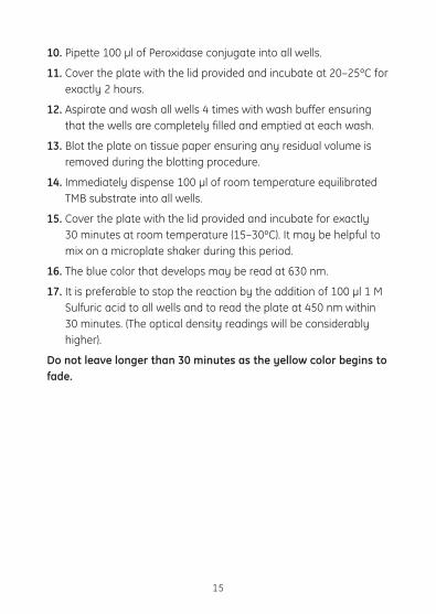
10. Pipette 100 µl of Peroxidase conjugate into all wells.
11. Cover the plate with the lid provided and incubate at 20–25ºC for exactly 2 hours.
12. Aspirate and wash all wells 4 times with wash buffer ensuring that the wells are completely filled and emptied at each wash.
13. Blot the plate on tissue paper ensuring any residual volume is removed during the blotting procedure.
14. Immediately dispense 100 µl of room temperature equilibrated TMB substrate into all wells.
15. Cover the plate with the lid provided and incubate for exactly 30 minutes at room temperature (15–30ºC). It may be helpful to mix on a microplate shaker during this period.
16. The blue color that develops may be read at 630 nm.
17. It is preferable to stop the reaction by the addition of 100 µl 1 M Sulfuric acid to all wells and to read the plate at 450 nm within 30 minutes. (The optical density readings will be considerably higher).
Do not leave longer than 30 minutes as the yellow color begins to fade.
15
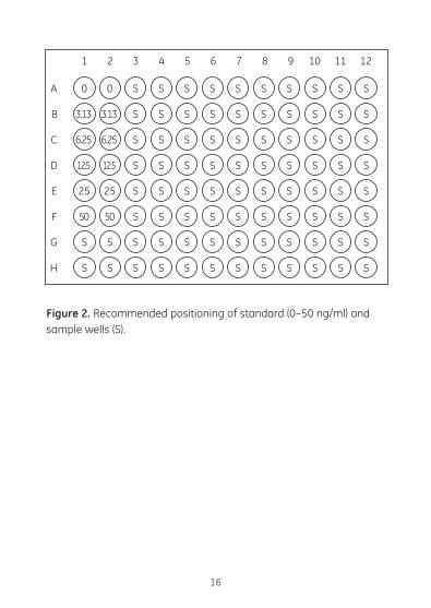
Figure 2. Recommended positioning of standard (0–50 ng/ml) and sample wells (S).
16
0 0 S S S S S S S S S S
3.13 3.13 S S S S S S S S S S
6.25 6.25 S S S S S S S S S S
12.5 12.5 S S S S S S S S S S
25 25 S S S S S S S S S S
50 50 S S S S S S S S S S
S S S S S S S S S S S S
S S S S S S S S S S S S
A
B
C
D
E
F
G
H
1 2 3 4 5 6 7 8 9 10 11 12

7.3. Data processing7.3.1. Calculation of resultsThe calculation is illustrated using representative data:
The assay data should be similar to that shown in table 1.
1. Calculate the average optical density for each set of duplicate wells.
2. A standard curve is generated by plotting the mean optical density (y axis) against ng/ml standard per ml (x axis). The curve shape should be similar to figure 3.
The ng/ml value can be read directly from the graph.
Figure 3. Typical standard curve (mean ± SEM).
17
0 3.13 6.25 12.5 25 500
0.5
1.0
1.5
2.0
TIMP-1 (ng/ml)
OD
at 4
50 n
m
TIMP-1 (ng/ml)
OD
at 4
50 n
m
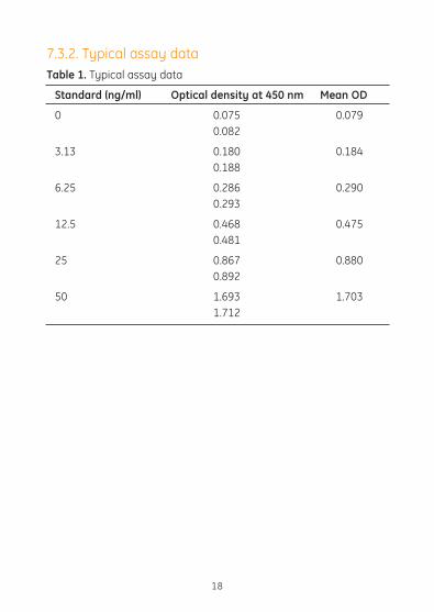
7.3.2. Typical assay dataTable 1. Typical assay data
Standard (ng/ml) Optical density at 450 nm Mean OD
0 0.075 0.079 0.082
3.13 0.180 0.184 0.188
6.25 0.286 0.290 0.293
12.5 0.468 0.475 0.481
25 0.867 0.880 0.892
50 1.693 1.703 1.712
18

8. Additional information
8.1. SpecificityThe assay recognises total human TIMP-1, ie. free TIMP-1 and that complexed with MMPs. The assay will fully cross-react with TIMP-1 in complexes with MMP-1, MMP-3, MMP-2, MMP-9. It does not cross-react with TIMP-2. It does not recognise TIMP-1 from other species as far as GE Healthcare is aware.
The cross-reactivity with MMP-1 and MMP-1/TIMP-1 complex for example was tested (figure 4a). The lack of interference of MMP-1 in the presence of the TIMP-1 standards was also tested and is also shown (figure 4b).
Figure 4a. Cross-reactivity with MMP-1 and MMP-1/TIMP-1 complex
19
0 3.13 6.25 12.5 25 500
0.5
1.0
1.5
2.0
TIMP-1 or MMP-1 (ng/ml)
OD
at 4
50 n
m
TIMP-1 standard curveMMP-1 MMP-1/TIMP-1 complex (concentration of TIMP-1 component)

Figure 4b. Non-interference of MMP-1 in TIMP-1 assay
8.2. ReproducibilityWithin-assay precisionThe within-assay precision for duplicate determinations was calculated by measuring controls in the assay. The results are shown below.
Table 2. (mean values as ng/ml)
Control Mean ± SD % CV n
A 10.3 ± 1.18 11.4 11 B 23.1 ± 2.14 9.3 11 C 39.4 ± 3.50 8.9 11
20
0 3.13 6.25 12.5 25 500
0.5
1.0
1.5
2.0
TIMP-1 (ng/ml)
OD
at 4
50 n
m
TIMP-1 standard curveTIMP-1 standard curve +100 ng/ml MMP-1
OD
at 4
50 n
m
TIMP-1 (ng/ml)
Between-assay precisionThe between-assay precision was assessed by repeated measurement of the same sample in successive assays. The results are shown below.
Table 3. (mean values as ng/ml)
Control Mean ± SD % CV n
A 12.5 ± 1.9 15.2 24 B 24.9 ± 3.1 12.4 24 C 47.3 ± 6.2 13.1 24
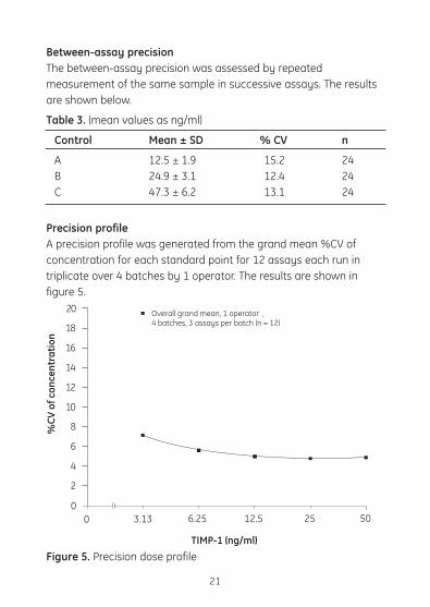
Precision profileA precision profile was generated from the grand mean %CV of concentration for each standard point for 12 assays each run in triplicate over 4 batches by 1 operator. The results are shown in figure 5.
Figure 5. Precision dose profile
21
0 3.13 6.25 12.5 25 500
2
20
TIMP-1 (ng/ml)
%CV
of c
once
ntra
tion
Overall grand mean, 1 operator , 4 batches, 3 assays per batch (n = 12)
4
6
8
10
12
14
16
18

8.3. SensitivityThe sensitivity, defined as two standard deviations above the mean optical density of 10 zero standard replicates was determined for each of 4 batches of reagents. The corresponding concentration was calculated from a standard curve set up in quadruplicate for each batch. The grand mean zero and standard values were then used to calculate the sensitivity. This was determined as 1.25 ng/ml.
22
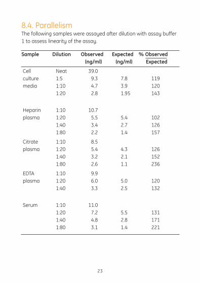
8.4. ParallelismThe following samples were assayed after dilution with assay buffer 1 to assess linearity of the assay.
Sample Dilution Observed Expected % Observed (ng/ml) (ng/ml) Expected
Cell Neat 39.0 culture 1:5 9.3 7.8 119 media 1:10 4.7 3.9 120 1:20 2.8 1.95 143
Heparin 1:10 10.7 plasma 1:20 5.5 5.4 102 1:40 3.4 2.7 126 1:80 2.2 1.4 157
Citrate 1:10 8.5 plasma 1:20 5.4 4.3 126 1:40 3.2 2.1 152 1:80 2.6 1.1 236
EDTA 1:10 9.9 plasma 1:20 6.0 5.0 120 1:40 3.3 2.5 132
Serum 1:10 11.0 1:20 7.2 5.5 131 1:40 4.8 2.8 171 1:80 3.1 1.4 221
23

8.5. RecoveryThe recovery of TIMP-1 standard spiked to levels throughout the range of the assay in various sample types diluted down to 1:40 for serum and plasma and 1:10 for unstimulated tissue culture sample.
Sample Added conc. Expected Measured % Recovery type (ng/ml) (ng/ml) (ng/ml)
Cell 0 - 0.37 - culture 46 46.37 40.78 88 media 22 22.37 18.05 81 11 11.37 8.40 74
Heparin 0 - 4.2 plasma 46 50.0 42.4 85 22 26.2 22.6 86 11 15.2 13.7 90
Citrate 0 - 3.5 plasma 46 49.5 42.3 85 22 25.5 22.6 89 11 14.5 12.8 88
EDTA 0 - 3.6 plasma 46 49.6 42.0 85 22 25.6 22.4 88 11 14.6 13.0 89
Serum 0 - 5.0 46 51.0 41.9 82 22 27.0 22.2 82 11 16.0 13.6 85
24

25
1. Low optical densities
2. High optical densities and/or high zero values
3. Flat curves/poor reproducibility
1. Check reader wavelength.2. Check incubation time and
temperatures.3. Reagents not equilibrated to
RT before use.4. Improper storage of kit
reagents.
1. Ensure that every well is completely filled and emptied at every wash step.
2. Ensure that automatic washers are functioning correctly.
3. Check incubation time and temperatures.
4. Blot plates on tissue paper after washing.
1. Check pipette calibration.2. Check preparation of working
standards.3. Ensure that troughs used
with multichannel pipettes are separate and dedicated to individual components.
4. Insufficient washing procedure.
9. Troubleshooting guideProblems Remedies

10. BackgroundThe family of enzymes known as the matrix metalloproteinases (MMPs) possess the ability to break down all of the components of the extracellular matrix (1). The matrix is maintained by a careful balance between rates of synthesis, degradation and connective tissue cell division. This can be seen in processes such as tissue morphogenesis, wound healing, cell migration and angiogenesis.
As expected for such potent enzymes their activity is tightly controlled. Regulation occurs at both the level of gene expression, with specific stimulation by cytokines for example, and also post-translationally in the extracellular space. Mature active MMPs are then subject to regulation by inhibitors including an MMP-specific family called tissue-inhibitors of metalloproteinases (TIMPs) (2). These complex with the active MMP to inhibit them. So far TIMP-1, 2 and 3 have been identified.
TIMP-1 is a 184 amino acid glycoprotein of 28.5 kDa MW. There is 41% sequence homology with the non-glycosylated 21.5 kDa MW TIMP-2 (3). TIMP-1 inhibits the activity of all the active MMPs and is the more widely distributed TIMP. The inhibition is via reversible non-covalent binding to form a 1:1 complex with a Kd ~10–10M (4). TIMP-1 is not cleaved by this binding and has been recovered with full activity from complexes with MMP-3 (5). The activity of MMPs can also be inhibited by α2-macroglobulin (6). However, it has been shown that no transfer of MMP-1 to α2-macroglobulin occurs if it has already complexed with TIMP-1 (7).
In addition to its inhibitory function TIMP-1 has also been shown to have growth factor activity including erythroid potentiation (8,9). Probably due to the interaction with MMPs, TIMPs inhibit tumour progression. The balance between MMP-1 and its inhibitor TIMP-1 is thought to be important in several disease states such as arthritis, cancer and fibrosis (10-12), TIMP-1 has been shown to stimulate
26

MMP-1 production from fibroblasts (13). Therefore, the ability to accurately measure levels of TIMP-1, MMP-1, and enzyme-inhibitor complexes is crucial to the investigation of their role in these conditions.
Currently MMPs or TIMP-1 are usually assayed by bioassays such as zymography that rely on the enzyme’s biological activity to degrade collagen or TIMP to inhibit this degradation, for example (14). However, these assays have several disadvantages in that firstly they require the MMP to be activated (eg. 4-aminophenyl mercuric acetate (APMA)). Also TIMP-1 is often assayed in a sample where TIMP-1-enzyme complexes are also present, so that the measured TIMP activity will be lower than the true level, as preformed complexes will not be detected. Bioassays for TIMP-1 also lack specificity, as they cannot distinguish between TIMP-1, TIMP-2 or α2-macroglobulin for example. Several immunoassays to TIMP-1 have now been described (15–17).
GE Healthcare has developed ELISA systems for the measurement of TIMP-1, MMP-1 and MMP-1/TIMP-1 complexes to greatly aid the study of this important enzyme. These assays are highly specific and convenient and circumvent the problems described for bioassays, allowing precise and accurate measurements of these components of the TIMP-1 system.
27

11. References1. Docherty, A.J.P. et al., Tibtech 10, 200-207 (1992).
2. Cawston, T.E. et al., Biochem. J. 195, 159-165 (1981).
3. Stetler-Stevenson, W.G., Krutzsch, H.C. and Liotta, L.A., J. Biol. Chem. 264, 17374-17378 (1989).
4. Cawston, T.E. et al., Biochem. J. 211, 313-318 (1983).
5. Murphy, G., Koklitis, P. and Carne, A.F., Biochem. J. 261, 1031-1034 (1989).
6. Barrett, A.J. and Starkey, P.M., Biochem. J. 133, 709-713 (1973).7. Cawston, T.E. and Mercer, E., FEBS Lett. 209, 9-12 (1986).
8. Gasson, J.C. et al., Nature 315, 768-771 (1985).
9. Hayakawa, T. et al., FEBS Lett. 298, 29-32 (1992).
10. Woolley, D.E., Crossley, M.J. and Evanson, J.M., Arth. Rheum. 20, 1231-1239 (1977).
11. Mignatti, P., Robbins, E. and Rifkin, D.B., Cell 47, 487-498 (1986).
12. Montano, M. et al., Chest 96, 1115-1119 (1989).
13. Clark, I.M., Powell, L.K. and Cawston, T.E., Biochem. Biophys. Res. Comm 203, 874-880 (1994).
14. Cawston, T.E. and Barrett, A.J., Anal. Biochem. 99, 340-345 (1979).
15. Clark, I.M. et al., Matrix 11, 76-85 (1991).
16. Cooksley, S. et al., Matrix 10, 285-291 (1990).
17. Kodama, S. et al., Matrix 9, 1-6 (1989).
18. Nakagawa, T. et al., J. Neurosurg. 81, 69-77 (1994).
28

12. Related productsELISA systemsMMP-1, Human RPN2610
MMP-2, Human RPN2617
MMP-3, Human RPN2613
MMP-7, Human RPN2620
MMP-8, Human RPN2619
MMP-9, Human RPN2614
MMP-13, Human RPN2621
TIMP-2, Human RPN2618
Activity assay systemsMMP-1 RPN2629
MMP-2 RPN2631
MMP-9 RPN2634
MMP-8 RPN2635
MMP-14 RPN2637
MMP-3 RPN2639
29

30

31

RPN2611PL Rev D 2006
imagination at work
http://www.gehealthcare.com/lifesciences
GE Healthcare UK LimitedAmersham Place, Little Chalfont, Buckinghamshire, HP7 9NAUK
GE Healthcare regional office contact numbers:
Asia PacificTel: +85 65 62751830
Fax: +85 65 62751829
AustralasiaTel: + 61 2 8820 8299
Fax: +61 2 8820 8200
AustriaTel: 01/57606-1613
Fax: 01/57606-1614
BelgiumTel: 0800 73 890
Fax: 02 416 8206
CanadaTel: 1 800 463 5800
Fax: 1 800 567 1008
Central, East, & South East EuropeTel: +43 1 972 720
Fax: +43 1 972 722 750
DenmarkTel: 45 70 25 24 50
Fax: 45 45 16 2424
EireTel: 1 800 709992
Fax: +44 1494 542010
Finland & BalticsTel: +358 9 512 3940
Fax: +358 9 512 39439
FranceTel: 01 69 35 67 00
Fax: 01 69 41 98 77
GermanyTel: 0800 9080 711
Fax: 0800 9080 712
Greater ChinaTel: +852 2100 6300
Fax: +852 2100 6338
ItalyTel: 02 26001 320
Fax: 02 26001 399
JapanTel: +81 3 5331 9336
Fax: +81 3 5331 9370
KoreaTel: 82 2 6201 3700
Fax: 82 2 6201 3803
Latin AmericaTel: +55 11 3933 7300
Fax: + 55 11 3933 7304
Middle East & AfricaTel: +30 210 96 00 687
Fax: +30 210 96 00 693
NetherlandsTel: 0800-82 82 82 1
Fax: 0800-82 82 82 4
NorwayTel: +47 815 65 777
Fax: +47 815 65 666
GE Healthcare offices:
GE Healthcare Bio-Sciences AB
Björkgatan 30 751 84
Uppsala
Sweden
GE Healthcare Europe GmbH
Munzinger Strasse 5 D-79111
Freiburg
Germany
GE Healthcare UK Limited
Amersham Place
Little Chalfont
Buckinghamshire
HP7 9NA
UK
GE Healthcare Bio-Sciences
Corp
800 Centennial Avenue
P.O. Box 1327
Piscataway
NJ 08855-1327
USA
GE Healthcare Bio-Sciences KK
Sanken Bldg. 3-25-1
Hyakunincho Shinjuku-ku
Tokyo 169-0073
Japan
PortugalTel: 21 417 7035
Fax: 21 417 3184
Russia, C.I.S. & N.I.STel: +7 495 956 5177
Fax: +7 495 956 5176
SpainTel: 902 11 72 65
Fax: 935 94 49 65
SwedenTel: 018 612 1900
Fax: 018 612 1910
SwitzerlandTel: 0848 8028 10
Fax: 0848 8028 11
UKTel: 0800 515 313
Fax: 0800 616 927
USATel: +1 800 526 3593
Fax: +1 877 295 8102