flammation in obesity -...
TRANSCRIPT

Mira Ham,1 Sung Sik Choe,1 Kyung Cheul Shin,1 Goun Choi,1 Ji-Won Kim,2
Jung-Ran Noh,3 Yong-Hoon Kim,3 Je-won Ryu,4 Kun-Ho Yoon,2 Chul-Ho Lee,3
and Jae Bum Kim1
Glucose-6-Phosphate DehydrogenaseDeficiency Improves Insulin ResistanceWith Reduced Adipose TissueInflammation in ObesityDiabetes 2016;65:2624–2638 | DOI: 10.2337/db16-0060
Glucose-6-phosphate dehydrogenase (G6PD), a rate-limiting enzyme of the pentose phosphate pathway, playsimportant roles in redox regulation and de novo lipogen-esis. It was recently demonstrated that aberrant upregu-lation of G6PD in obese adipose tissue mediates insulinresistance as a result of imbalanced energy metabolismand oxidative stress. It remains elusive, however, whetherinhibition of G6PD in vivo may relieve obesity-inducedinsulin resistance. In this study we showed that ahematopoietic G6PD defect alleviates insulin resis-tance in obesity, accompanied by reduced adipose tissueinflammation. Compared with wild-type littermates,G6PD-deficient mutant (G6PDmut) mice were glucosetolerant upon high-fat-diet (HFD) feeding. Intriguingly,the expression of NADPH oxidase genes to produce re-active oxygen species was alleviated, whereas that ofantioxidant genes was enhanced in the adipose tissue ofHFD-fed G6PDmut mice. In diet-induced obesity (DIO),the adipose tissue of G6PDmut mice decreased the ex-pression of inflammatory cytokines, accompanied bydownregulated proinflammatorymacrophages. Accordingly,macrophages from G6PDmut mice greatly suppressedlipopolysaccharide-induced proinflammatory signalingcascades, leading to enhanced insulin sensitivity in ad-ipocytes and hepatocytes. Furthermore, adoptive transferof G6PDmut bone marrow to wild-type mice attenuatedadipose tissue inflammation and improved glucose toler-ance in DIO. Collectively, these data suggest that inhibitionof macrophage G6PD would ameliorate insulin resistance
in obesity through suppression of proinflammatoryresponses.
In obese adipose tissue, chronic low-grade inflammationhas been implicated in metabolic dysregulation and insulinresistance (1,2). Compared with lean subjects, in obesesubjects adipose tissue promotes macrophage recruitmentand cross-talk between adipocytes and macrophages,resulting in adipose tissue inflammation accompanied byhigh levels of inflammatory cytokines such as tumor necrosisfactor-a (TNF-a), interleukin (IL)-1b, IL-6, and MCP-1(1–3). Proinflammatory cytokines secreted by inflamedadipose tissue repress insulin action in metabolic organsby activating stress-induced kinases such as IkB kinase-band Jun N-terminal kinase (JNK), which leads to systemicinsulin resistance (4–6). Moreover, increased oxidativestress in obese adipose tissue contributes to adipose tissueinflammation via activation of mitogen-activated proteinkinases (MAPKs) including JNK and p38 (7,8). In addition,elevated cellular levels of reactive oxygen species (ROS)further stimulate proinflammatory gene expression viathe activation of nuclear factor-kB (NF-kB) pathways inobesity (7–10). It has been suggested that one of themajor sources of cellular ROS in obese adipose tissue ispro-oxidative enzymes, including NADPH oxidase (9–11).Various pro-oxidative enzymes are known to be augmentedin the adipose tissues of obese animals, whereas antioxidative
1Department of Biological Sciences, Institute of Molecular Biology and Genetics,National Creative Research Initiatives Center for Adipose Tissue Remodeling,Seoul National University, Seoul, Korea2Department of Endocrinology and Metabolism, College of Medicine, The CatholicUniversity of Korea, Seoul, Korea3Laboratory Animal Resource Center, Korea Research Institute of Bioscience andBiotechnology, University of Science and Technology, Daejeon, Korea4Department of Radiation Oncology, Asan Medical Center and University of UlsanCollege of Medicine, Seoul, Korea
Corresponding author: Jae Bum Kim, [email protected].
Received 12 January 2016 and accepted 24 May 2016.
This article contains Supplementary Data online at http://diabetes.diabetesjournals.org/lookup/suppl/doi:10.2337/db16-0060/-/DC1.
M.H. and S.S.C. contributed equally to this work.
© 2016 by the American Diabetes Association. Readers may use this article aslong as the work is properly cited, the use is educational and not for profit, andthe work is not altered. More information is available at http://diabetesjournals.org/site/license.
2624 Diabetes Volume 65, September 2016
OBESITY
STUDIES

processes related to ROS scavenging tend to be suppressed(9,10). Accordingly, treatment with NADPH oxidase inhibi-tors as well as antioxidant drugs attenuates ROS productionand adipokine dysregulation, alleviating metabolic disorderssuch as insulin resistance in obesity (9,12). Thus it is likelythat the imbalance between ROS production and scavengingin obese adipose tissue is closely associated with elevatedadipose tissue inflammation and metabolic dysregulation.
Glucose-6-phosphate dehydrogenase (G6PD), a keyenzyme of the pentose phosphate pathway, which shuntsfrom the glycolytic pathway, catalyzes the synthesis of ribosefor nucleic acid production and produces cytosolic NADPH(13). NADPH is one of the crucial cofactors that serve as areducing equivalent in many metabolic pathways such asfatty acid and cholesterol biosynthesis (10). Moreover,G6PD plays a critical role in the regulation of the cellularredox potential by supplying the cofactor NADPH to ROS-producing and scavenging enzymes such as NADPH oxi-dase and glutathione reductase, respectively (10,13). Inpathological conditions such as atherosclerosis, heart fail-ure, and obesity, G6PD promotes cellular ROS productionand proinflammatory signaling through increased avail-ability of NADPH to ROS-producing enzymes (14–19).On the other hand, in various cell types, including eryth-rocytes, cardiomyocytes, endothelial cells, and pancreaticb-cells, G6PD contributes to the clearance of cellular oxi-dative stress by NADPH-dependent ROS-scavenging en-zymes (20–23). We recently demonstrated that G6PD ishighly expressed in the adipose tissues of obese and diabeticsubjects (14,24). The aberrant upregulation of G6PD in ad-ipose tissue leads to the dysregulation of lipid metabolismand adipokines and thus impairs insulin signaling in ad-ipocytes (14,15). Furthermore, overexpression of G6PD inmacrophages promotes oxidative stress and the expression ofproinflammatory cytokines, which induce insulin resistancein adipocytes (24). Similarly, upregulation of G6PD in theliver, heart, and pancreatic b-cells of obese and diabeticanimals also increases oxidative stress, which leads tofunctional defects in the respective tissues (12,25,26).Altogether, these findings suggest that anomalous G6PDupregulation in obese conditions might deteriorate energyhomeostasis and oxidative stress, thereby acceleratingmetabolic complications.
G6PD deficiency is a common enzymopathy, affectingover 400 million people worldwide (13). One of the dis-tinctive characteristics of patients with G6PD deficiency ishemolytic anemia, which is triggered by exposure to oxi-dative stress. As a potential animal model of human G6PDdeficiency, a G6PD-deficient mutant (G6PDmut) mousemodel has been developed, which shows 10–15% of regularG6PD activity because of a reduction in G6PD proteinexpression by a splicing defect (27,28). Since the embryo-protective role of G6PD has been reported (20,29), variouseffects of G6PD deficiency on metabolic processes, immuneresponses, and organ development have been studied inG6PDmut mice (17,18,21,22,30). Furthermore, G6PD defi-ciency affects albuminuria and diet-induced cardiometabolic
aberrations in G6PDmut mice (31,32). However, the ef-fects of a G6PD defect in vivo on chronic inflammationand systemic insulin resistance in diet-induced obesity(DIO) still remain to be elucidated.
In this study we showed that G6PDmut mice exhibitimproved glucose tolerance and reduced systemic insulinresistance in DIO. In the adipose tissue of G6PDmut mice feda high-fat diet (HFD), the expression of NADPH oxidase andinflammatory cytokines was relieved, and the numbers ofcrown-like structures (CLSs) and infiltrated macrophagesalso diminished. Furthermore, proinflammatory responseswere attenuated in the macrophages of G6PDmut mice,thus improving insulin signaling in adipocytes and hepa-tocytes. In line with these results, a transplant of G6PDmut
bone marrow led to protection from diet-induced adiposetissue inflammation in wild-type (WT) recipient mice. Takentogether, these data suggest that macrophage G6PD defi-ciency would mediate to lessen systemic insulin resistancein obesity by relieving proinflammatory signaling upon met-abolic stresses.
RESEARCH DESIGN AND METHODS
Animals and TreatmentG6PDmut mice were provided by R. Matsui. G6PDmut
(C3H) mice were backcrossed with C57BL/6 mice (CentralLaboratory Animal Inc., Seoul, South Korea) more than10 times to change the genetic background (SupplementaryFig. 1). The mice were genotyped by PCR (17), as illustratedin Supplementary Fig. 1. G6PDmut mice and their litter-mates were maintained on a normal chow diet (ND) until8 weeks of age before being switched to a 60% HFD. Forthe oral or intraperitoneal glucose tolerance test, the micewere fasted for 6 h and basal blood samples were collected,then glucose was administered (2 g/kg body weight). Bloodsamples were drawn 15, 30, 60, 90, and 120 min afterglucose administration to measure the glucose concentra-tion. For the insulin tolerance test, the mice were fasted for3 h and then insulin was administered (0.75 unit/kg bodyweight; Lilly, Indianapolis, IN); then blood glucose concen-trations were measured at the indicated time points. Totest in vivo insulin signaling, the mice were euthanized20 min after a PBS or insulin injection (0.75 unit/kg bodyweight), and adipose tissues and livers were excised andquickly frozen for Western blot analysis. For a bone mar-row transplant (BMT), 8-week-old recipient (C57BL/6)mice were lethally irradiated two times (total, 5 Gy) bymeans of a 137Cs source at an interval of 4 h. After 4 h,these mice received a transplant of 5 3 106 bone marrowcells from G6PDmut mice or their WT littermates via tailvein injection, as previously described (33). ELISA kits forTNF-a, MCP-1 (Invitrogen, Grand Island, NY), and adipo-nectin (MIoBS, Japan) were used to measure their con-centrations in protein extract of epididymal adipose tissueand serum. All animal procedures were conducted inaccordance with the research guidelines of the SeoulNational University Institutional Animal Care and UseCommittee.
diabetes.diabetesjournals.org Ham and Associates 2625

Isolation of Peritoneal MacrophagesPeritoneal macrophages were isolated fromWT and G6PDmut
mice, as previously described (24). To detect lipopolysaccha-ride (LPS)–induced inflammatory signaling, peritoneal mac-rophages from WT and G6PDmut mice were treated with LPS(10 ng/mL; Sigma-Aldrich, St. Louis, MO) for 30 min, andthen the cells were harvested for Western blot analyses. Foranalysis of the expression of proinflammatory cytokines, themacrophages were incubated with LPS for 24 h.
Adipose Tissue FractionationAdipose tissue was fractionated as described previously(34). Briefly, epididymal adipose tissues (EATs) of HFD-fedWT and G6PDmut mice were digested with type I collagenasebuffer and filtered through a nylon mesh. After centrifuga-tion, fractions of floating adipocytes and pelleted stromalvascular cells (SVCs) were washed several times with PBSand then used for RNA extraction.
Cell Culture and Conditioned Medium Experiment3T3-L1 preadipocytes were grown in DMEM (HyClone,Logan, UT) supplemented with 10% bovine calf serum(HyClone). 3T3-L1 preadipocytes were differentiated asdescribed previously (35). H4IIE hepatoma cells weregrown in DMEM supplemented with 10% FBS (HyClone).For the conditioned medium (CM) experiment, peritonealmacrophages from WT and G6PDmut mice were incubatedin serum-free DMEM for 24 h, and CM was collected.Fully differentiated 3T3-L1 adipocytes and H4IIE hepa-toma cells were incubated with CM for 24 h. To monitorinsulin signaling, these cells were stimulated with insulin(100 nmol/L) for 30 min. To determine the expressionlevel of gluconeogenic genes, H4IIE cells were stimulatedwith insulin (100 nmol/L) for 8 h. Insulin-stimulatedglucose uptake by 3T3-L1 adipocytes was assessed bymeasuring [14C]-2-deoxy-D-glucose uptake, as describedpreviously (34).
Western Blot AnalysisWestern blot analysis was performed as described pre-viously (34). In brief, EAT, the liver, peritoneal macro-phages, 3T3-L1 adipocytes, and H4IIE cells were lysedwith NETN buffer. Nuclear extracts from macrophageswere isolated as described previously (15). Cytosolic andmembrane fractions from adipose tissues were preparedusing a Proteo Extract Native Membrane Protein Extrac-tion Kit (Merck Millipore, Germany) according to themanufacturer’s protocol. The proteins were separatedby SDS-PAGE and transferred to polyvinylidene fluoridemembranes. The blots were probed with the followingprimary antibodies: anti-adiponectin, anti-G6PD, anti-laminB, anti-pan-cadherin (Abcam, Cambridge, MA); anti-b-actin,anti-GAPDH (Sigma-Aldrich); anti-catalase, anti–superoxidedismutase 2 (Bethyl Laboratories, Montgomery, TX); anti-phospho (p)-AKT (Ser473), anti–fatty acid synthase, anti-p-GSK3b (Ser9), anti-p-JNK, anti-p65, anti-p50 (Cell SignalingTechnology, Danvers, MA); anti-AKT, anti-GSK3b, anti-p-p38,anti-p67phox, anti-SREBP1c (BD Biosciences); anti-NOX2
(Merck Millipore); anti-JNK, anti-p38, anti-p47phox, andanti-SCD (Santa Cruz Biotechnology, Dallas, TX).
Quantitative RT-PCRTotal RNA was isolated from EAT, the liver, peritonealmacrophages, and H4IIE cells, then quantitative real-timeRT-PCR was performed, as described previously (36).The primers were synthesized by Bioneer (Daejeon, SouthKorea), and their sequences are provided in SupplementaryTable 1.
ImmunohistochemistryWhole-mount immunohistochemical analysis of EAT wasperformed as previously described (24). Whole-mountedEATs were incubated with primary antibodies againstG6PD (1:500 dilution; Abcam), perilipin (1:1000; Fitzgerald,Acton, MA), CD11b (1:1000), and CD11c (1:1000; eBioscience,San Jose, CA). After incubation with fluorescently labeledsecondary antibodies (Thermo Fisher Scientific, Waltham,MA) and DAPI (Vector Laboratories, Burlingame, CA) stain-ing, the samples were examined under a Zeiss LSM 510-NLOconfocal microscope. Slices cut from paraffin-embeddedEATs were deparaffinized in xylene and rehydrated in agraded series of ethanol solutions. Endogenous peroxidasewas inactivated using 3% hydrogen peroxide in methanolfor 5 min. After blocking the slices with 10% horse serum,sections were incubated with a primary antibody againstG6PD (1:100). The slices were incubated with horseradishperoxidase–conjugated secondary antibodies (Thermo FisherScientific) and visualized with 3,39-diaminobenzidine (VectorLaboratories). Hematoxylin was used for counterstaining.
Measurement of Cellular ROS LevelsCellular ROS was measured using chloromethyl-29,79-dichlorodihydrofluorescein diacetate (H2DCFDA) (Invi-trogen). Peritoneal macrophages from WT and G6PDmut
mice were incubated with LPS for 30 min and then withDCFDA in the dark for 20 min. After a wash, fluorescentsignals were detected using a confocal microscope.
Statistical AnalysisThe results represent data from multiple independentexperiments. Error bars in the figures denote the SD, andP values were calculated using the Student t test.
RESULTS
G6PDmut Mice Exhibit Lower Levels of Fasting Insulinin HFD-Induced ObesityIt has been demonstrated that upregulated G6PD in obeseadipose tissue contributes to inflammatory signaling viapotentiating oxidative stress (10,15,24). However, it isnot thoroughly understood whether genetic G6PD defectmay alter adipose tissue inflammation and insulin resis-tance in obese animals. To address this, we investigatedvarious aspects of DIO in G6PDmut mice that were createdby N-ethyl-N-nitrosourea (ENU) mutagenesis from C3Hstrain (27). To exclude off-target effects of random mu-tagenesis and the defective Toll-like receptor 4 (TLR4)–mediated inflammatory response in the C3H/HeJ strain
2626 G6PD Deficiency in Obesity Diabetes Volume 65, September 2016

(37–41), the genetic background of G6PDmut mice waschanged to C57BL/6 by backcross breeding more than10 times with WT C57BL/6 mice (Supplementary Fig. 1).Afterward, G6PDmut mice were fed either an ND or HFD for12 weeks and were compared with their WT littermates. Asshown in Fig. 1A–D, HFD increased body weight, adiposetissue weight, and adipocyte size in both WT and G6PDmut
mice. In a C57BL/6 background, the HFD challenge inducedhyperglycemia in both WT and G6PDmut mice (Fig. 1E). Onthe contrary, fasting insulin concentration and HOMA–insulin resistance (HOMA-IR) index, a quantitative anal-ysis to measure insulin resistance, were significantly lowerin HFD-fed G6PDmut mice compared with HFD-fed WTmice(Fig. 1F and G). In addition, HFD-fed G6PDmut mice showeddecreased serum concentrations of free fatty acids that arereadily released in insulin-resistant adipocytes (Fig. 1H).
HFD-Fed G6PDmut Mice Show Ameliorated InsulinResistanceGiven that HFD-fed G6PDmut mice exhibited reduced fast-ing insulin and HOMA-IR index, we hypothesized that theG6PD defect may be involved in the regulation of sys-temic insulin sensitivity in DIO. To test this, oral glucosetolerance and insulin tolerance tests were performed withWT and G6PDmut mice. Under ND feeding, both WT andG6PDmut mice showed similar glucose tolerance. Interestingly,HFD-fed G6PDmut mice were more glucose and insulin
tolerant than HFD-fed WT mice (Fig. 2A and B), whichis consistent with the hypothesis that the reductionof fasting insulin concentration might be a result ofimproved insulin resistance in HFD-fed G6PDmut mice.Next, to explore whether G6PD deficiency in DIO mayinfluence insulin sensitivity in metabolic organs, the in-sulin signaling cascade was examined in the adipose tissueand liver of WT and G6PDmut mice. As shown in Fig. 2C,compared with HFD-fed WT mice, in the adipose tissue ofHFD-fed G6PDmut mice, insulin-stimulated phosphorylationof AKT and GSK3b was significantly enhanced. Similarly,G6PDmut mice also showed enhanced insulin signaling inthe liver (Fig. 2D). Therefore, these observations indicatedthat G6PD deficiency would be associated with improvedinsulin resistance in DIO.
Expression of NADPH Oxidase Is Reduced in theAdipose Tissue of HFD-Fed G6PDmut MiceIt has been reported that G6PD controls the redox balanceby supplying the cofactor NADPH (15,24). NADPH oxidaseis one of the major enzymes in NADPH-dependent ROSproduction, and its mRNA expression is upregulated inresponse to oxidative stress in obese adipose tissue (9). Todetermine whether a G6PD defect may attenuate the in-creased ROS production in DIO, the expression of NADPHoxidase subunits was examined in HFD-fed WT and G6PDmut
mice. As shown in Fig. 3A and B, both mRNA and protein
Figure 1—Fasting insulin and free fatty acids are decreased in HFD-fed G6PDmut mice. G6PDmut mice and their WT littermates (n = 6 pergroup) were fed either an ND or HFD for 12 weeks. A: Body weight was measured during the experimental periods. B and C: The bodyweight gain (B) and weights of various tissues (C) of WT and G6PDmut mice were measured after 12 weeks of ND or HFD feeding. D:Average size of adipocytes was measured in the images of EAT slices. E–H: Fasting serum glucose (E) and insulin (F ), HOMA-IR (G), andserum free fatty acids (FFAs) (H) in WT and G6PDmut mice. The data represent the mean 6 SD. *P < 0.05 and **P < 0.01 vs. WT group,Student t test.
diabetes.diabetesjournals.org Ham and Associates 2627

levels of NADPH oxidase subunits such as NOX2, p22phox,p47phox, and p67phox were diminished in the adipose tissue ofHFD-fed G6PDmut mice compared with that of HFD-fed WTmice. By contrast, mRNA and protein levels of antioxidantgenes including superoxide dismutase 2, catalase, and glu-tathione peroxidase were higher in the adipose tissue ofG6PDmut mice than that of WT mice with HFD feeding
(Fig. 3A and B). It has been demonstrated that membranetranslocation of cytosolic regulatory subunits such as p47phox
and p67phox is required for activation of NADPH oxidase2 (9). Compared with HFD-fed WT mice, the levels ofp47phox and p67phox in the membrane fraction were alsoslightly decreased in HFD-fed G6PDmut mice (Fig. 3C),implying that an active NADPH oxidase complex might
Figure 2—Insulin resistance is improved in HFD-fed G6PDmut mice. A and B: Oral glucose tolerance test (GTT) and area under the curve(AUC) analysis (A) and insulin tolerance test (B) were performed on WT and G6PDmut mice (n = 6 per group) fed an ND or HFD for 12 weeks.The data represent the mean 6 SD. *P < 0.05 vs. WT group at each time point, Student t test. C and D: Insulin signaling in the EAT (C ) andliver (D) of HFD-fed WT and G6PDmut mice injected with saline or insulin (0.75 units/kg body weight) was analyzed by immunoblotting withantibodies against p-AKT, total AKT, p-GSK3b, and total GSK3b. b-Actin was used as the loading control. AU, arbitrary units. Quan-titative data on the ratio of p-AKT and total AKT and the ratio of p-GSK3b and total GSK3b are represented as mean6 SD. *P< 0.05 and**P < 0.01 vs. no insulin control; #P < 0.05 and ##P < 0.01 vs. WT group, Student t test.
2628 G6PD Deficiency in Obesity Diabetes Volume 65, September 2016

be diminished by a G6PD defect in obese adipose tissue.Thus this finding indicated that oxidative stress would bedecreased in the adipose tissue of HFD-fed G6PDmut mice.On the other hand, suppression of NADPH oxidase and
elevation of antioxidant genes by G6PD deficiency werenot observed in the liver of the obese animals (Fig. 3E). Theseresults implied that G6PD defect might selectively reduceoxidative stress in the adipose tissue of obese animals.
Figure 3—HFD-fed G6PDmut mice exhibit downregulated expression of NADPH oxidase in adipose tissue. A and B: In EAT of HFD-fed WTand G6PDmut mice (n = 6 per group), relative mRNA and protein levels of G6PD, NADPH oxidase subunits (NOX2, p22phox, p47phox, andp67phox), and antioxidant genes were analyzed. C: To monitor the translocation of p47phox and p67phox into the membrane, cytosolic andmembrane fractions from adipose tissue of HFD-fed WT and G6PDmut mice were subjected to Western blot analysis. D: Relative mRNAlevels of G6PD, NADPH oxidase subunits, and antioxidant genes in liver. E and F: Relative mRNA of lipogenic genes and oxidation genes(E ) and protein levels of lipogenic genes (F ) in adipose tissue. b-Actin was used as the loading control, and GAPDH and pan-cadherin wereused as a cytosolic marker and a membrane marker, respectively. The data represent the mean 6 SD. *P < 0.05 and ***P < 0.001 vs. WTgroup, Student t test. SOD2, superoxide dismutase 2; GPx, glutathione peroxidase; SREBP1c, sterol regulatory element–binding protein1c; FASN, fatty acid synthase; SCD1, stearoyl-CoA desaturase 1; ACOX, acyl-CoA oxidase; CPT1a, carnitine palmitoyltransferase 1a.
diabetes.diabetesjournals.org Ham and Associates 2629

Adiposity and Fatty Liver Are Not Alleviated in HFD-FedG6PDmut MiceIn addition to redox regulation, G6PD is also involved inlipogenesis (14). Unexpectedly, we did not detect any re-duction in adiposity in G6PDmut mice compared with WTmice upon both ND and HFD feeding (Fig. 1). In addition,mRNA and protein levels of lipogenic genes such as sterolregulatory element-binding protein 1c, fatty acid syn-thase, and stearoyl-CoA desaturase 1, and mRNA levelof lipid oxidation–related genes including acyl-CoA oxi-dase 1 and 2 and carnitine palmitoyltransferase 1a, inthe adipose tissue of HFD-fed G6PDmut mice were not dif-ferent from those of HFD-fed WT mice (Fig. 3E and F).Furthermore, the levels of serum triglycerides and cholesterolwere similar between WT and G6PDmut mice (SupplementaryFig. 2A and B). WT and G6PDmut mice showed a similardegree of nonalcoholic fatty liver upon HFD feeding. Therewas no difference in their morphological features (hematox-ylin and eosin [H&E] staining), lipid contents (oil-red O stain-ing), and tissue triglyceride levels in the liver of WT andG6PDmut mice (Supplementary Fig. 2C and D). In addition,the expression of hepatic lipogenesis and lipid oxidation–related genes was not different between WT and G6PDmut
mice (Supplementary Fig. 2E).
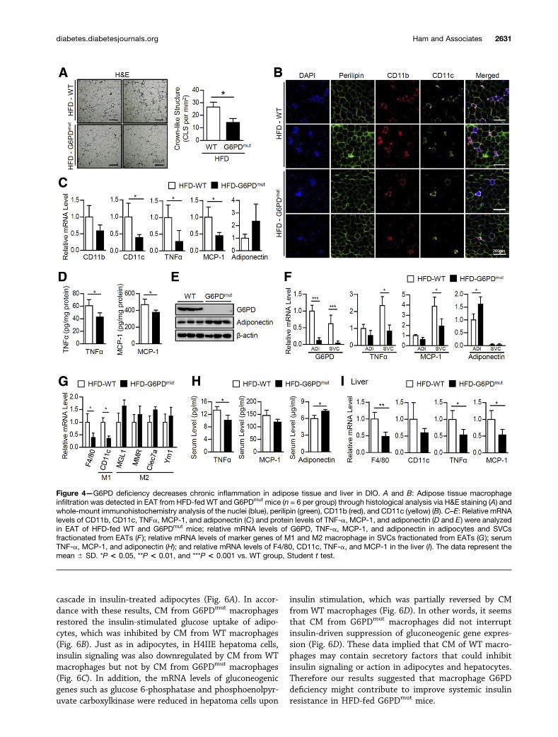
Adipose Tissue Inflammation Is Attenuated in HFD-FedG6PDmut MiceTo test whether the improved insulin resistance of HFD-fed G6PDmut mice might be associated with adipose tissueinflammation, macrophage accumulation was analyzed inthe adipose tissue of WT and G6PDmut mice under HFDfeeding. As shown in Fig. 4A and B, HFD-fed G6PDmut
mice exhibited fewer CLSs and CD11c-positive proinflam-matory macrophages (the classically activated M1 type)than HFD-fed WT mice did. Next, we examined geneexpression profiles of proinflammatory cytokines andmacrophage markers. Although there were no significantdifferences in the expression of proinflammatory genesbetween WT and G6PDmut mice fed an ND (Supplemen-tary Fig. 3), the mRNA level of CD11c was significantlyreduced in the adipose tissue of HFD-fed G6PDmut mice(Fig. 4C). In addition, both mRNA and protein levels ofproinflammatory cytokines such as TNF-a and MCP-1were decreased (Fig. 4C and D). On the contrary, mRNAand protein levels of adiponectin, a well-known anti-inflammatory adipokine, tended to be higher (Fig. 4Cand E). Decreased expression of these inflammatory cyto-kines was more evident in the SVC fraction than in theadipocyte fraction isolated from adipose tissue of G6PDmut
mice (Fig. 4F). Additionally, the expression of M1 markergenes such as CD11c was significantly decreased in SVCs ofG6PDmut mice than those of WT mice fed an HFD (Fig.4G). On the contrary, the expression of M2 marker genessuch asMGL1,MMR, Clec7a, and Ym1, for anti-inflammatorymacrophages (the alternatively activated M2 type) secretinganti-inflammatory cytokines, seemed to be slightly but notsignificantly increased in G6PDmut mice (Fig. 4G). These data
implied that M1 polarization of adipose tissue macro-phages (ATMs) seemed to be partially attenuated inHFD-fed G6PDmut mice. Furthermore, serum TNF-a andMCP-1 were reduced, whereas serum adiponectin was el-evated in HFD-fed G6PDmut mice compared with thatin HFD-fed WT mice (Fig. 4H). Similar to the adiposetissues, mRNA levels of M1 markers (CD11c) and proin-flammatory cytokines (TNF-a and MCP-1) weredownregulated in the livers of HFD-fed G6PDmut mice(Fig. 4I). Taken together, these results suggested that, inDIO, a G6PD defect would be protective against chronicinflammation, which is a key factor in boosting systemicinsulin resistance.
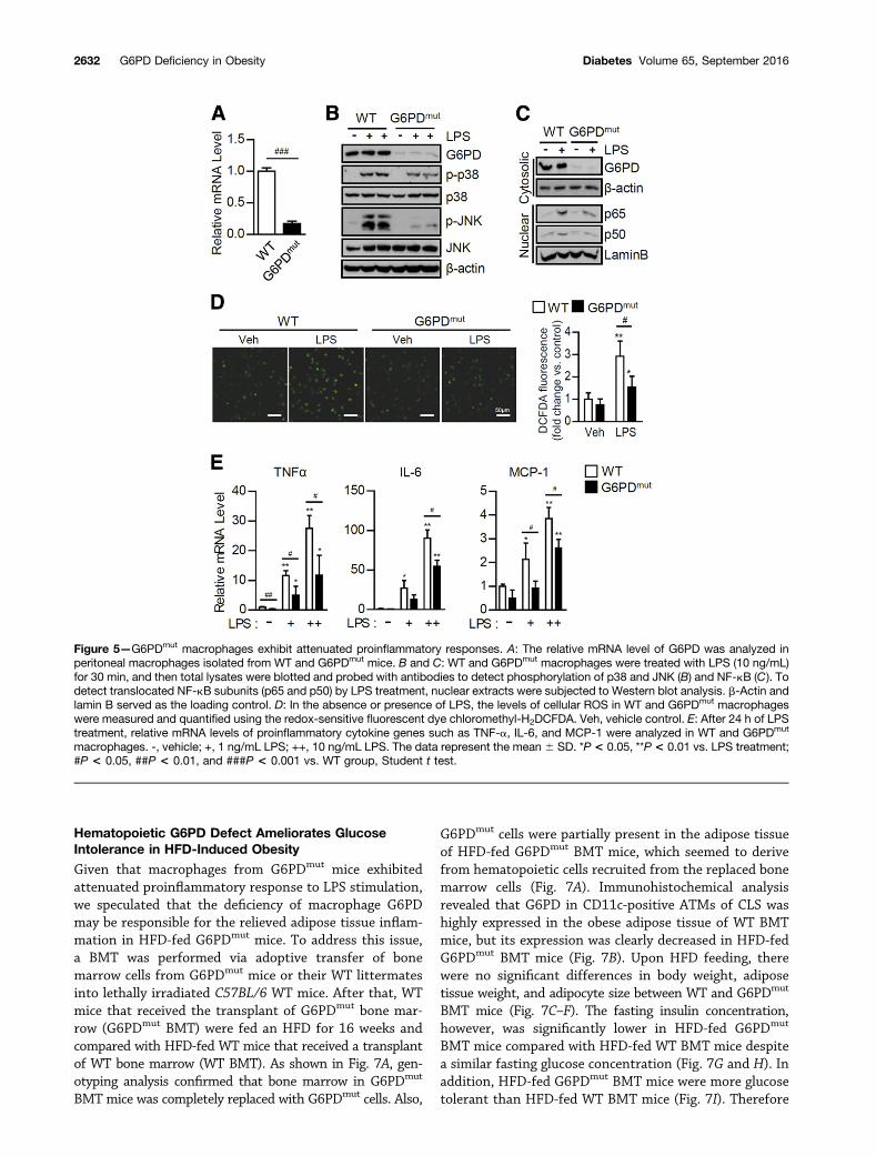
G6PD Defect Suppresses Proinflammatory Responsesin MacrophagesAccumulated ATMs are one of the key hallmarks ofinflammation in obese adipose tissue, eventually leadingto systemic insulin resistance (3). We previously demon-strated that macrophage G6PD modulates proinflammatoryresponses and oxidative stress, which are crucial stimuli forinsulin resistance in adipocytes (24). Thus we decided totest whether a macrophage G6PD defect in G6PDmut micemight indeed alter proinflammatory responses. To addressthis, peritoneal macrophages isolated from WT and G6PDmut
mice (Fig. 5A) were challenged with LPS, which is a potentinitiator to provoke proinflammatory signals through TLR4.As shown in Fig. 5B, macrophages from G6PDmut micegreatly attenuated phosphorylation of MAPKs such as p38and JNK by LPS challenge. In addition, G6PDmut macro-phages exhibited decreased nuclear translocation of NF-kB,p65, and p50 upon LPS stimulation (Fig. 5C). Given thatROS in macrophages is one of the important signalingmolecules participating in proinflammatory responses (7),we were prompted to test the levels of ROS in G6PDmut
macrophages. Compared with WT macrophages, macro-phages from G6PDmut mice showed reduced ROS accumu-lation in the presence of LPS (Fig. 5D). Moreover, themRNA levels of TNF-a, IL-6, and MCP-1 genes were re-duced in LPS-stimulated G6PDmut macrophages (Fig. 5E).These data suggested that a macrophage G6PD defectwould relieve oxidative stress and proinflammatory re-sponses in G6PDmut mice, which may lead to the suppres-sion of adipose tissue inflammation in HFD-fed G6PDmut
mice.
Macrophage G6PD Deficiency Augments InsulinAction in Adipocytes and HepatocytesTo test the idea that the attenuation of proinflammatoryresponses in G6PDmut macrophages might affect insulinsignaling in adipocytes and hepatocytes, 3T3-L1 adipocytesor H4IIE hepatoma cells were incubated with CM fromLPS-stimulated WT or G6PDmut macrophages. As shownin Fig. 6A, CM from WT macrophages decreased the phos-phorylation of AKT and GSK3b in insulin-treated adipo-cytes, in contrast to the fresh medium. Unlike CM fromWT macrophages, CM from G6PDmut macrophages did nothave such inhibitory effects on the insulin signaling
2630 G6PD Deficiency in Obesity Diabetes Volume 65, September 2016
cascade in insulin-treated adipocytes (Fig. 6A). In accor-dance with these results, CM from G6PDmut macrophagesrestored the insulin-stimulated glucose uptake of adipo-cytes, which was inhibited by CM from WT macrophages(Fig. 6B). Just as in adipocytes, in H4IIE hepatoma cells,insulin signaling was also downregulated by CM from WTmacrophages but not by CM from G6PDmut macrophages(Fig. 6C). In addition, the mRNA levels of gluconeogenicgenes such as glucose 6-phosphatase and phosphoenolpyr-uvate carboxylkinase were reduced in hepatoma cells upon
insulin stimulation, which was partially reversed by CMfrom WT macrophages (Fig. 6D). In other words, it seemsthat CM from G6PDmut macrophages did not interruptinsulin-driven suppression of gluconeogenic gene expres-sion (Fig. 6D). These data implied that CM of WT macro-phages may contain secretory factors that could inhibitinsulin signaling or action in adipocytes and hepatocytes.Therefore our results suggested that macrophage G6PDdeficiency might contribute to improve systemic insulinresistance in HFD-fed G6PDmut mice.
Figure 4—G6PD deficiency decreases chronic inflammation in adipose tissue and liver in DIO. A and B: Adipose tissue macrophageinfiltration was detected in EAT from HFD-fed WT and G6PDmut mice (n = 6 per group) through histological analysis via H&E staining (A) andwhole-mount immunohistochemistry analysis of the nuclei (blue), perilipin (green), CD11b (red), and CD11c (yellow) (B). C–E: Relative mRNAlevels of CD11b, CD11c, TNFa, MCP-1, and adiponectin (C) and protein levels of TNF-a, MCP-1, and adiponectin (D and E) were analyzedin EAT of HFD-fed WT and G6PDmut mice; relative mRNA levels of G6PD, TNF-a, MCP-1, and adiponectin in adipocytes and SVCsfractionated from EATs (F ); relative mRNA levels of marker genes of M1 and M2 macrophage in SVCs fractionated from EATs (G ); serumTNF-a, MCP-1, and adiponectin (H); and relative mRNA levels of F4/80, CD11c, TNF-a, and MCP-1 in the liver (I). The data represent themean 6 SD. *P < 0.05, **P < 0.01, and ***P < 0.001 vs. WT group, Student t test.
diabetes.diabetesjournals.org Ham and Associates 2631
Hematopoietic G6PD Defect Ameliorates GlucoseIntolerance in HFD-Induced Obesity
Given that macrophages from G6PDmut mice exhibitedattenuated proinflammatory response to LPS stimulation,we speculated that the deficiency of macrophage G6PDmay be responsible for the relieved adipose tissue inflam-mation in HFD-fed G6PDmut mice. To address this issue,a BMT was performed via adoptive transfer of bonemarrow cells from G6PDmut mice or their WT littermatesinto lethally irradiated C57BL/6 WT mice. After that, WTmice that received the transplant of G6PDmut bone mar-row (G6PDmut BMT) were fed an HFD for 16 weeks andcompared with HFD-fed WT mice that received a transplantof WT bone marrow (WT BMT). As shown in Fig. 7A, gen-otyping analysis confirmed that bone marrow in G6PDmut
BMT mice was completely replaced with G6PDmut cells. Also,
G6PDmut cells were partially present in the adipose tissueof HFD-fed G6PDmut BMT mice, which seemed to derivefrom hematopoietic cells recruited from the replaced bonemarrow cells (Fig. 7A). Immunohistochemical analysisrevealed that G6PD in CD11c-positive ATMs of CLS washighly expressed in the obese adipose tissue of WT BMTmice, but its expression was clearly decreased in HFD-fedG6PDmut BMT mice (Fig. 7B). Upon HFD feeding, therewere no significant differences in body weight, adiposetissue weight, and adipocyte size between WT and G6PDmut
BMT mice (Fig. 7C–F). The fasting insulin concentration,however, was significantly lower in HFD-fed G6PDmut
BMT mice compared with HFD-fed WT BMT mice despitea similar fasting glucose concentration (Fig. 7G and H). Inaddition, HFD-fed G6PDmut BMT mice were more glucosetolerant than HFD-fed WT BMT mice (Fig. 7I). Therefore
Figure 5—G6PDmut macrophages exhibit attenuated proinflammatory responses. A: The relative mRNA level of G6PD was analyzed inperitoneal macrophages isolated from WT and G6PDmut mice. B and C: WT and G6PDmut macrophages were treated with LPS (10 ng/mL)for 30 min, and then total lysates were blotted and probed with antibodies to detect phosphorylation of p38 and JNK (B) and NF-kB (C). Todetect translocated NF-kB subunits (p65 and p50) by LPS treatment, nuclear extracts were subjected to Western blot analysis. b-Actin andlamin B served as the loading control. D: In the absence or presence of LPS, the levels of cellular ROS in WT and G6PDmut macrophageswere measured and quantified using the redox-sensitive fluorescent dye chloromethyl-H2DCFDA. Veh, vehicle control. E: After 24 h of LPStreatment, relative mRNA levels of proinflammatory cytokine genes such as TNF-a, IL-6, and MCP-1 were analyzed in WT and G6PDmut
macrophages. -, vehicle; +, 1 ng/mL LPS; ++, 10 ng/mL LPS. The data represent the mean 6 SD. *P < 0.05, **P < 0.01 vs. LPS treatment;#P < 0.05, ##P < 0.01, and ###P < 0.001 vs. WT group, Student t test.
2632 G6PD Deficiency in Obesity Diabetes Volume 65, September 2016

these data indicated that reconstitution of bone marrowin WT mice with G6PDmut bone marrow could alleviateinsulin resistance in DIO.
Hematopoietic G6PD Defect Protects AgainstDiet-Induced Adipose Tissue InflammationTo explore whether adipose tissue inflammation mightbe associated with improved glucose tolerance in HFD-fed G6PDmut BMT mice, macrophage accumulationwas quantified in the adipose tissue of WT and G6PDmut
BMT mice with HFD. As shown in Fig. 8A, the numbers ofCLSs were significantly decreased in the obese adiposetissue of G6PDmut BMT mice compared with that ofWT BMT mice. Similarly, HFD-fed G6PDmut BMT miceshowed downregulation of CD11c-positive proinflamma-tory macrophages in comparison with HFD-fed WT BMTmice (Fig. 8B). As expected, in WT mice the transplant ofG6PDmut bone marrow reduced the level of G6PD mRNAin adipose tissue in DIO (Fig. 8C). In addition, mRNAlevels of NADPH oxidase subunits such as NOX2 andp22phox were decreased, whereas that of catalase wasincreased in the adipose tissue of HFD-fed G6PDmut BMTmice, implying that the oxidative stress in the adiposetissue might be reduced by a hematopoietic G6PD defect(Fig. 8C). Furthermore, mRNA levels of proinflammatorycytokines such as TNF-a, IL-6, and MCP-1 were de-creased, as was the expression of M1 marker genes suchas CD11c. By contrast, the mRNA level of adiponectintended to be increased (Fig. 8D). Taken together, theseresults suggest that hematopoietic G6PD deficiency wouldprotect against HFD-induced adipose tissue inflammation,
which might contribute to improve insulin resistance inG6PDmut BMT mice.
DISCUSSION
NADPH-producing enzymes such as G6PD, malic enzyme(ME), and isocitrate dehydrogenase (IDH) play key rolesin the regulation of the redox balance and in anabolicprocesses such as de novo lipid synthesis. In metabolictissues such as adipose tissue and liver, dysregulation ofthese NADPH-producing enzymes has been implicated inmetabolic disorders accompanied by chronic low-gradeinflammation in obesity. For instance, it has been demon-strated that G6PD overexpression in adipocytes or macro-phages potentiates inflammatory communications betweenadipocytes and macrophages, leading to insulin resistancein obesity (15,24). In addition, elevated G6PD expression inthe adipose tissue of obese patients shows a positive cor-relation with various indicators of insulin resistance andmacrophage accumulation (24). These findings raise thepossibility that upregulated G6PD might be one of themediators for adipose tissue inflammation in obesity. Totest this hypothesis in an animal model, we investigated aG6PDmut mouse model to decipher the pathophysiologicalroles of G6PD in obesity-induced metabolic complications.
It has been reported that severely G6PD-deficientembryos developed through G6PD-targeted homologousrecombination are lethal because of damage in the centralnervous system and heart on embryonic day 10.5 (29).Meanwhile, G6PDmut mice developed through ENU ran-dom mutagenesis with C3H mice are viable and display no
Figure 6—Macrophage G6PD deficiency improves insulin resistance in adipocytes and hepatocytes. A and B: Insulin-dependent phos-phorylation of AKT (A) and glucose uptake (B) were analyzed in 3T3-L1 adipocytes treated with CM from WT and G6PDmut macrophages.C and D: Insulin-dependent phosphorylation of AKT (C) and suppression of gluconeogenic genes such as glucose 6-phosphatase(G6Pase) and phosphoenolpyruvate carboxylkinase (PEPCK) (D) were analyzed in H4IIE hepatoma cells treated with CM from WT orG6PDmut macrophages. b-Actin was used as the loading control. Quantitative data on the ratio of p-AKT and total AKT are representedas the mean 6 SD. FM, fresh media. *P < 0.05 and **P < 0.01 vs. no treated control; #P < 0.05 vs. WT group, Student t test.
diabetes.diabetesjournals.org Ham and Associates 2633

apparent chronic hemolysis despite low G6PD activity(27). To date, G6PDmut mice have been studied for variouspathological conditions such as sepsis, teratogenesis, cardio-vascular diseases, myocardial dysfunction, and atherosclerosis(17,18,20–22,30–32,42,43). Because of their genetic back-ground, C3H G6PDmut mice have some limitations for inves-tigating obesity-mediated chronic inflammation and metabolicdysregulation. Accumulating evidence has suggested that dif-ferent murine genetic backgrounds could affect diverse meta-bolic phenotypes such as susceptibility to DIO, hyperglycemia,insulin sensitivity, and proliferation and survival of b-cells,which lead to different experimental outcomes upon ex-posure to metabolic stresses (44,45). For example, it hasbeen shown that the C3H strain is less susceptible to
insulin resistance with increased insulin secretion andexhibits improved glucose metabolism (45). In addition,C3H/HeJ mice show defective endotoxin responses be-cause of a mutation in the TLR4 gene, which is a receptoressential for the induction of innate and adaptive immu-nity (37). Given that TLR4 signaling is activated by satu-rated free fatty acids as well as bacteria-derived glycolipidssuch as LPS (46), TLR4 plays key roles in obesity-associateddisorders. Thus functional defects in TLR4 signaling inC3H/HeJ mice reduce adipose tissue inflammation inDIO, which is associated with insulin sensitivity and glucosemetabolism (39–41). Consequently, we had to change thegenetic background of G6PDmut mice to C57BL/6, whichwould be a suitable model to study obesity-related metabolic
Figure 7—HFD-fed G6PDmut BMT mice are glucose tolerant. WT and G6PDmut BMT mice (n = 6 per group) were fed an HFD for 16 weeks. A:Genotyping analysis for WT (lower band) and G6PDmutation (upper band) was performed on genomic DNA from bone marrow (BM), EAT, andliver. B: Expression patterns of G6PD protein in ATMs of WT and G6PDmut BMT mice were detected through immunohistochemical analysis ofthe G6PD (left, 3,39-diaminobenzidine staining), nuclei (blue), G6PD (right, red), CD11c (yellow), and perilipin (green). C: Body weight wasmeasured during the experimental periods. D and E: The body weight gain (D) and weights of various tissues (E) of WT and G6PDmut BMT micewere measured after 16 weeks of HFD feeding. F: The average size of adipocytes wasmeasured from images of EAT slices.G–I: Fasting seruminsulin (G) and glucose (H) and intraperitoneal glucose tolerance test (GTT) and area under the curve (AUC) analysis (I) of HFD-fed WT andG6PDmut BMT mice (n = 6 per group). The data represent the mean 6 SD. *P < 0.05 vs. WT BMT group, Student t test.
2634 G6PD Deficiency in Obesity Diabetes Volume 65, September 2016

disorders. Moreover, multiple rounds of backcrossing ofG6PDmut mice with C57BL/6 mice should help to eliminatepossible residual off-target effects during ENU chemicalmutagenesis. With the C57BL/6 background, G6PDmut
mice grew normally and were indistinguishable from age-matched WT littermates in spite of the reduced G6PD ex-pression. Upon HFD feeding, G6PDmut mice showed a bodyweight gain, hyperglycemia, and hyperinsulinemia in theC57BL/6 background.
Without any metabolic stress such as an HFD, G6PDmut
mice exhibited a similar extent of glucose tolerance comparedwith WT mice. In DIO, however, G6PDmut mice exhibitedimproved glucose tolerance and sensitized insulin signalingin peripheral tissues (Fig. 2). It was recently suggested thatchronic low-grade inflammation in adipose tissue is one ofthe key factors that can exacerbate insulin resistance inobesity (1–3). In this work, several lines of evidence sug-gest that G6PD would exert substantial regulatory actionon chronic inflammation in DIO. First, under HFD feeding,the expression of NADPH oxidase subunits was lower whilethat of antioxidant genes was higher in the adipose tissueof G6PDmut mice compared with WT mice (Fig. 3). Unlikein adipose tissue, this gene expression profile was notaltered in the livers of G6PDmut mice. These results in-dicate that a G6PD defect could relieve oxidative stress,particularly in obese adipose tissue. Second, the popula-tion of CD11c-positive M1 ATMs was greatly reduced inadipose tissues of HFD-fed G6PDmut mice (Fig. 4). In addi-tion, the levels of several proinflammatory cytokines, includ-ing TNF-a and MCP-1, were decreased in adipose tissue and
serum from HFD-fed G6PDmut mice (Fig. 4). By contrast,adiponectin with anti-inflammatory and antidiabeticeffects was elevated in adipose tissue and serum fromHFD-fed G6PDmut mice (Fig. 4). Moreover, G6PDmut mac-rophages were resistant to proinflammatory stimulationby LPS (Fig. 5) and mediated enhanced insulin actionin adipocytes and hepatocytes (Fig. 6), which were mostlikely driven by secretory factors from G6PDmut macro-phages. We recently reported that upregulated G6PDwould induce the expression of proinflammatory cyto-kines by activation of NF-kB in adipose tissue (15,24).In obesity, secreted proinflammatory cytokines could fur-ther aggravate adipose tissue inflammation with promo-tion of M1 ATM recruitment as well as repress insulinaction in adipose tissue by activating stress-activated ki-nases (4–6). Although the role of G6PD during macrophagepolarization remains to be investigated, our recent dataand previous reports have proposed that a G6PD defectin obesity might alleviate the vicious cycles between oxida-tive stress, proinflammatory response, and macrophage re-cruitment in adipose tissue. Therefore it is possible tospeculate that G6PD deficiency in vivo would protect fromsystemic insulin resistance through the attenuation ofchronic inflammation in obesity.
Studies on human G6PD deficiencies have suggestedthat G6PD would contribute to mediating immune cell func-tions (47–49). Severe human G6PD deficiency causes a chronicgranulomatous disease with increased susceptibility to in-fections because of a defect in hydrogen peroxide produc-tion by granulocytes (47,48). Additionally, monocyte-derived
Figure 8—Adipose tissue inflammation is ameliorated in HFD-fed G6PDmut BMT mice. A and B: Macrophage infiltration of adipose tissuewas detected in the EAT of HFD-fed WT and G6PDmut BMT mice (n = 6 per group) by histological analysis via H&E staining (A) and whole-mount immunohistochemistry analysis of the nuclei (blue), perilipin (green), CD11b (red), and CD11c (yellow) (B). C: Relative mRNA levels ofG6PD, NADPH oxidase subunits (NOX2 and p22phox), and catalase by quantitative RT-PCR in total RNA samples from EAT of HFD-fed WTand G6PDmut BMT mice. D: Relative mRNA levels of F4/80, CD11c, TNF-a, IL-6, MCP-1, and adiponectin were analyzed in EAT. The datarepresent the mean 6 SD. *P < 0.05 and **P < 0.01 vs. WT BMT group, Student t test.
diabetes.diabetesjournals.org Ham and Associates 2635

macrophages from subjects with G6PD-deficiency exhibitreduced secretion of inflammatory cytokines such as TNF-a and IL-1b (49). In agreement with these observations,we found that G6PDmut macrophages showed reducedROS production upon LPS stimulation in comparison withWT macrophages; this effect would eventually weakenthe proinflammatory signaling and thereby suppress theinduction of proinflammatory cytokines (Fig. 5). Addi-tionally, in HFD-fed G6PDmut BMT mice, hematopoieticG6PD deficiency attenuated the expression of proinflam-matory cytokines and macrophage accumulation in adi-pose tissue, implying that G6PD in immune cells (thatare recruited into adipose tissue) would be involved ininflammatory responses under metabolic stress (Fig. 8).Of course, it needs to be elucidated whether the reducedadipose tissue inflammation in HFD-fed G6PDmut BMTmice might be mediated by other hematopoietic immunecells such as T lymphocytes, neutrophils, or eosinophils,as well as macrophages with G6PD deficiency. Nonethe-less, our in vitro and in vivo data collectively indicatethat macrophage G6PD deficiency at least would relievechronic inflammation in HFD-fed G6PDmut mice.
Although the transplant of G6PDmut bone marrow intoWT mice diminished the proinflammatory response inobese adipose tissue, we did not observe a marked reduc-tion of proinflammatory cytokines or macrophage markergene expression in the liver of HFD-fed G6PDmut BMTmice (Supplementary Fig. 4). This phenomenon mightbe the result of rare infiltration of bone marrow–derivedcells (with the G6PDmut gene) into the liver of HFD-fedG6PDmut BMT mice. Compared with the adipose tissuesof HFD-fed G6PDmut BMT mice, G6PDmut cells were barelydetectable in the liver according to PCR genotyping (Fig.7A). In line with this finding, the level of G6PD mRNA wasnot reduced in the liver of HFD-fed G6PDmut BMT mice(Supplementary Fig. 4). Although further studies are def-initely needed to investigate the function of G6PD inKupffer cells, our in vitro experiments (Fig. 6) proposethe possibility that Kupffer cells with G6PD defect mayinfluence proinflammatory response in the liver of micewith DIO.
In the process of de novo lipogenesis, NADPH is anessential cofactor that supplies reducing power for conversionof acetyl-CoA into fatty acids. Various NADPH-producingenzymes such as G6PD, ME, and IDH are abundantlyexpressed in adipose tissue (14). Given that NADPH pro-duction by G6PD is positively associated with lipogenicactivity in adipocytes (14), we speculated that G6PD defectmight affect the synthesis and/or accumulation of lipidmetabolites in G6PDmut mice. Unexpectedly, however,when compared with WT littermates, G6PDmut mice didnot show any changes in adiposity, fatty liver, serum tri-glycerides, and cholesterol upon ND or HFD feeding (Fig.1 and Supplementary Fig. 2). The data indicating thatserum free fatty acid levels were decreased in HFD-fedG6PDmut mice (Fig. 1H) are probably the result of theaugmented inhibitory effect of insulin on basal lipolysis
with improved insulin sensitivity. Moreover, the expressionof lipogenic genes was not altered in the adipose tissue andliver of WT and G6PDmut mice (Fig. 3 and SupplementaryFig. 2). Currently, it is unclear why and how G6PD defectfails to influence adiposity or lipid metabolism. One of theputative explanations is a compensatory process driven byother NADPH-producing enzymes such as ME or IDH inG6PDmut mice. It has been reported that NADP+-dependentIDH, one of the IDH isoforms, can also mediate lipidbiosynthesis in adipose tissue and liver via NADPH pro-duction (50). Transgenic mice overexpressing IDH showhyperlipidemia and marked lipid accumulation in adiposetissue and liver (50). On the other hand, we did notobserve any change in the mRNA expression of otherNADPH-producing enzymes such as ME and IDH in theadipose tissue and liver of HFD-fed G6PDmut mice (Sup-plementary Fig. 5). Nevertheless, the possibility that theseenzymes might be relatively activated in G6PDmut micecannot be excluded.
Here we showed that a genetic G6PD defect amelio-rates chronic inflammation and insulin resistance in DIO.It is likely that inhibition of G6PD in obesity reducescellular oxidative stress in adipose tissue, thus attenuat-ing excessive proinflammatory responses and macrophageaccumulation. Moreover, the attenuation of proinflam-matory signaling by inhibiting G6PD may prevent chronicinflammation and systemic insulin resistance in obesity(Supplementary Fig. 6). Collectively, our data suggest thatmanipulation of G6PD activity may be a potential approachfor countering obesity-induced metabolic disorders.
Acknowledgments. The authors thank R. Matsui for providing theG6PDmut mice. The authors also thank Jong In Kim of Seoul National Universityfor critically reading the manuscript.Funding. This work was supported by the National Creative ResearchInitiative Program of the National Research Foundation (NRF) funded by theKorean government (the Ministry of Science, ICT & Future Planning, 2011-0018312). This research was also partly supported by the Bio & Medical Tech-nology Development Program of the National Research Foundation (NRF) fundedby the Ministry of Science, ICT & Future Planning (2012M3A9B6055344). K.C.S.was supported by the BK21 program.Duality of Interest. No conflicts of interest relevant to this article werereported.Author Contributions. M.H. and S.S.C. designed the study, performedexperiments, and wrote the manuscript. K.C.S., G.C., J.-W.K., and J.-w.R.performed experiments and wrote the manuscript. J.-R.N., Y.-H.K., K.-H.Y., andC.-H.L. wrote the manuscript. J.B.K. supervised the whole project, discussed thedata, and edited the final manuscript. J.B.K. is the guarantor of this work and, assuch, had full access to all the data in this study and takes responsibility for theintegrity of the data and the accuracy of the data analysis.
References1. Olefsky JM, Glass CK. Macrophages, inflammation, and insulin resistance.Annu Rev Physiol 2010;72:219–2462. Gregor MF, Hotamisligil GS. Inflammatory mechanisms in obesity. Annu RevImmunol 2011;29:415–4453. Chawla A, Nguyen KD, Goh YP. Macrophage-mediated inflammation inmetabolic disease. Nat Rev Immunol 2011;11:738–749
2636 G6PD Deficiency in Obesity Diabetes Volume 65, September 2016

4. Yuan M, Konstantopoulos N, Lee J, et al. Reversal of obesity- and diet-induced insulin resistance with salicylates or targeted disruption of Ikkbeta.Science 2001;293:1673–16775. Hirosumi J, Tuncman G, Chang L, et al. A central role for JNK in obesity andinsulin resistance. Nature 2002;420:333–3366. Gao Z, Zhang J, Kheterpal I, Kennedy N, Davis RJ, Ye J. Sirtuin 1 (SIRT1)protein degradation in response to persistent c-Jun N-terminal kinase 1 (JNK1)activation contributes to hepatic steatosis in obesity. J Biol Chem 2011;286:22227–222347. Evans JL, Goldfine ID, Maddux BA, Grodsky GM. Oxidative stress andstress-activated signaling pathways: a unifying hypothesis of type 2 diabetes.Endocr Rev 2002;23:599–6228. Yeop Han C, Kargi AY, Omer M, et al. Differential effect of saturated andunsaturated free fatty acids on the generation of monocyte adhesion and che-motactic factors by adipocytes: dissociation of adipocyte hypertrophy from in-flammation. Diabetes 2010;59:386–3969. Furukawa S, Fujita T, Shimabukuro M, et al. Increased oxidative stress inobesity and its impact on metabolic syndrome. J Clin Invest 2004;114:1752–176110. Park J, Chung JJ, Kim JB. New evaluations of redox regulating system inadipose tissue of obesity. Diabetes Res Clin Pract 2007;77(Suppl 1):S11–S1611. Han CY, Umemoto T, Omer M, et al. NADPH oxidase-derived reactive oxygenspecies increases expression of monocyte chemotactic factor genes in culturedadipocytes. J Biol Chem 2012;287:10379–1039312. Gupte RS, Floyd BC, Kozicky M, et al. Synergistic activation of glucose-6-phosphate dehydrogenase and NAD(P)H oxidase by Src kinase elevates superoxidein type 2 diabetic, Zucker fa/fa, rat liver. Free Radic Biol Med 2009;47:219–22813. Cappellini MD, Fiorelli G. Glucose-6-phosphate dehydrogenase deficiency.Lancet 2008;371:64–7414. Park J, Rho HK, Kim KH, Choe SS, Lee YS, Kim JB. Overexpression ofglucose-6-phosphate dehydrogenase is associated with lipid dysregulation andinsulin resistance in obesity. Mol Cell Biol 2005;25:5146–515715. Park J, Choe SS, Choi AH, et al. Increase in glucose-6-phosphate de-hydrogenase in adipocytes stimulates oxidative stress and inflammatory signals.Diabetes 2006;55:2939–294916. Gupte SA, Levine RJ, Gupte RS, et al. Glucose-6-phosphate dehydrogenase-derived NADPH fuels superoxide production in the failing heart. J Mol Cell Cardiol2006;41:340–34917. Matsui R, Xu S, Maitland KA, et al. Glucose-6 phosphate dehydrogenasedeficiency decreases the vascular response to angiotensin II. Circulation 2005;112:257–26318. Matsui R, Xu S, Maitland KA, et al. Glucose-6-phosphate dehydrogenasedeficiency decreases vascular superoxide and atherosclerotic lesions in apoli-poprotein E(-/-) mice. Arterioscler Thromb Vasc Biol 2006;26:910–91619. Hecker PA, Leopold JA, Gupte SA, Recchia FA, Stanley WC. Impact ofglucose-6-phosphate dehydrogenase deficiency on the pathophysiology of car-diovascular disease. Am J Physiol Heart Circ Physiol 2013;304:H491–H50020. Nicol CJ, Zielenski J, Tsui LC, Wells PG. An embryoprotective role forglucose-6-phosphate dehydrogenase in developmental oxidative stress andchemical teratogenesis. FASEB J 2000;14:111–12721. Jain M, Brenner DA, Cui L, et al. Glucose-6-phosphate dehydrogenasemodulates cytosolic redox status and contractile phenotype in adult car-diomyocytes. Circ Res 2003;93:e9–e1622. Zhang Z, Liew CW, Handy DE, et al. High glucose inhibits glucose-6-phosphate dehydrogenase, leading to increased oxidative stress and beta-cellapoptosis. FASEB J 2010;24:1497–150523. Leopold JA, Zhang YY, Scribner AW, Stanton RC, Loscalzo J. Glucose-6-phosphate dehydrogenase overexpression decreases endothelial cell oxidantstress and increases bioavailable nitric oxide. Arterioscler Thromb Vasc Biol2003;23:411–41724. Ham M, Lee JW, Choi AH, et al. Macrophage glucose-6-phosphate de-hydrogenase stimulates proinflammatory responses with oxidative stress. MolCell Biol 2013;33:2425–2435
25. Serpillon S, Floyd BC, Gupte RS, et al. Superoxide production by NAD(P)Hoxidase and mitochondria is increased in genetically obese and hyperglycemic ratheart and aorta before the development of cardiac dysfunction. The role ofglucose-6-phosphate dehydrogenase-derived NADPH. Am J Physiol Heart CircPhysiol 2009;297:H153–H16226. Lee JW, Choi AH, Ham M, et al. G6PD up-regulation promotes pancreaticbeta-cell dysfunction. Endocrinology 2011;152:793–80327. Pretsch W, Charles DJ, Merkle S. X-linked glucose-6-phosphate de-hydrogenase deficiency in Mus musculus. Biochem Genet 1988;26:89–10328. Sanders S, Smith DP, Thomas GA, Williams ED. A glucose-6-phosphatedehydrogenase (G6PD) splice site consensus sequence mutation associated withG6PD enzyme deficiency. Mutat Res 1997;374:79–8729. Longo L, Vanegas OC, Patel M, et al. Maternally transmitted severe glucose6-phosphate dehydrogenase deficiency is an embryonic lethal. EMBO J 2002;21:4229–423930. Wilmanski J, Villanueva E, Deitch EA, Spolarics Z. Glucose-6-phosphatedehydrogenase deficiency and the inflammatory response to endotoxin andpolymicrobial sepsis. Crit Care Med 2007;35:510–51831. Xu Y, Zhang Z, Hu J, et al. Glucose-6-phosphate dehydrogenase-deficientmice have increased renal oxidative stress and increased albuminuria. FASEBJ 2010;24:609–61632. Hecker PA, Mapanga RF, Kimar CP, et al. Effects of glucose-6-phosphatedehydrogenase deficiency on the metabolic and cardiac responses to obesogenicor high-fructose diets. Am J Physiol Endocrinol Metab 2012;303:E959–E97233. Joseph SB, Bradley MN, Castrillo A, et al. LXR-dependent gene expressionis important for macrophage survival and the innate immune response. Cell2004;119:299–30934. Choe SS, Shin KC, Ka S, Lee YK, Chun JS, Kim JB. Macrophage HIF-2aameliorates adipose tissue inflammation and insulin resistance in obesity. Di-abetes 2014;63:3359–337135. Lee YS, Choi JW, Hwang I, et al. Adipocytokine orosomucoid integratesinflammatory and metabolic signals to preserve energy homeostasis by resolvingimmoderate inflammation. J Biol Chem 2010;285:22174–2218536. Lee HW, Suh JH, Kim HN, et al. Berberine promotes osteoblast differenti-ation by Runx2 activation with p38 MAPK. J Bone Miner Res 2008;23:1227–123737. Poltorak A, He X, Smirnova I, et al. Defective LPS signaling in C3H/HeJ andC57BL/10ScCr mice: mutations in Tlr4 gene. Science 1998;282:2085–208838. Ye D, Li FY, Lam KS, et al. Toll-like receptor-4 mediates obesity-inducednon-alcoholic steatohepatitis through activation of X-box binding protein-1 inmice. Gut 2012;61:1058–106739. Tsukumo DM, Carvalho-Filho MA, Carvalheira JB, et al. Loss-of-functionmutation in Toll-like receptor 4 prevents diet-induced obesity and insulin re-sistance. Diabetes 2007;56:1986–199840. Poggi M, Bastelica D, Gual P, et al. C3H/HeJ mice carrying a toll-like receptor4 mutation are protected against the development of insulin resistance in whiteadipose tissue in response to a high-fat diet. Diabetologia 2007;50:1267–127641. Suganami T, Mieda T, Itoh M, Shimoda Y, Kamei Y, Ogawa Y. Attenuation ofobesity-induced adipose tissue inflammation in C3H/HeJ mice carrying a Toll-likereceptor 4 mutation. Biochem Biophys Res Commun 2007;354:45–4942. Leopold JA, Walker J, Scribner AW, et al. Glucose-6-phosphate de-hydrogenase modulates vascular endothelial growth factor-mediated angiogen-esis. J Biol Chem 2003;278:32100–3210643. Spencer NY, Yan Z, Boudreau RL, et al. Control of hepatic nuclear super-oxide production by glucose 6-phosphate dehydrogenase and NADPH oxidase-4.J Biol Chem 2011;286:8977–898744. Montgomery MK, Hallahan NL, Brown SH, et al. Mouse strain-dependentvariation in obesity and glucose homeostasis in response to high-fat feeding.Diabetologia 2013;56:1129–113945. Kaku K, Fiedorek FT Jr, Province M, Permutt MA. Genetic analysis of glu-cose tolerance in inbred mouse strains. Evidence for polygenic control. Diabetes1988;37:707–713
diabetes.diabetesjournals.org Ham and Associates 2637

46. Shi H, Kokoeva MV, Inouye K, Tzameli I, Yin H, Flier JS. TLR4 links innateimmunity and fatty acid-induced insulin resistance. J Clin Invest 2006;116:3015–302547. Cooper MR, DeChatelet LR, McCall CE, LaVia MF, Spurr CL, Baehner RL.Complete deficiency of leukocyte glucose-6-phosphate dehydrogenase withdefective bactericidal activity. J Clin Invest 1972;51:769–77848. Gray GR, Stamatoyannopoulos G, Naiman SC, et al. Neutrophil dysfunction,chronic granulomatous disease, and non-spherocytic haemolytic anaemia caused
by complete deficiency of glucose-6-phosphate dehydrogenase. Lancet 1973;2:530–53449. Sanna F, Bonatesta RR, Frongia B, et al. Production of inflammatory mol-ecules in peripheral blood mononuclear cells from severely glucose-6-phosphatedehydrogenase-deficient subjects. J Vasc Res 2007;44:253–26350. Koh HJ, Lee SM, Son BG, et al. Cytosolic NADP+-dependent isocitratedehydrogenase plays a key role in lipid metabolism. J Biol Chem 2004;279:39968–39974
2638 G6PD Deficiency in Obesity Diabetes Volume 65, September 2016