amplification of single molecule translocation signal using β-strand peptide functionalized...
TRANSCRIPT

LIEBES-PEER ET AL. VOL. 8 ’ NO. 7 ’ 6822–6832 ’ 2014
www.acsnano.org
6822
June 20, 2014
C 2014 American Chemical Society
Amplification of Single MoleculeTranslocation Signal Using β‑StrandPeptide Functionalized NanoporesYael Liebes-Peer,†,‡,§,^ Hanna Rapaport,†,§ and Nurit Ashkenasy‡,§,*
†Department of Biotechnology Engineering, ‡Department of Materials Engineering, and §The Ilze Katz Institute for Nanoscale Technology, Ben-Gurion University ofthe Negev, P.O. Box 653, Beer-Sheva 84105, Israel. ^Present address for Y.L.-P.: School of Chemistry, Tel Aviv University, P.O. Box 39040, 6997801, Israel.
Nanopores have gained a lot of inter-est in recent years due to the abilityto use them for the detection of
biomolecules at the single molecule level,obtained by monitoring changes in ioniccurrent flowing through the nanopore,which are induced by the molecule's trans-location through the nanopore or bindingto the nanopore rims.1�3 Analysis of themagnitude and duration of the current per-turbation event and the frequency of eventsprovides both quantitative and qualitativeanalytical information.4,5 While early studiesutilized biological nanopores,4,6,7 to over-come robustness and size limitations, meth-odologies to prepare single nanopores insolid state membranes have been devel-oped. Additional advantages of these artifi-cial nanopores include the ability to controltheir shape and prospective integration intolab-on-a-chip devices.7�9 Such solid state
nanopores have proven to be useful for thedetection of DNA10,11 and proteins.12�15 Inparticular, large efforts have been devotedto the development of a DNA sequencingmachine based on such chips.11,16 However,thedetection of smallmolecules, which is pos-siblewhen using biological nanopores,17�21
is hampered in solid state nanopores due toincompatibility of the nanopore size and thetemporal resolution of the measurementwith the molecule size and its translocationduration, respectively. This limits the use ofsuch nanopores for the detection of, e.g.,hormones, peptides and small poisonousmolecules, such as organophosphates.4,7
Recent studies have demonstrated that thepresence of small molecules in solid statenanopores can be sensed if these inducesignificant changes to organic layer sensitiz-ing the nanopore. For example, disintegrationof a complex DNA supersandwich structure
* Address correspondence [email protected].
Received for review March 7, 2014and accepted June 20, 2014.
Published online10.1021/nn501331u
ABSTRACT Changes in ionic current flowing through nanopores due to
binding or translocation of single biopolymer molecules enable their
detection and characterization. It is, however, much more challenging to
detect small molecules due to their rapid and small signal signature. Here
we demonstrate the use of de novo designed peptides for functionalization
of nanopores that enable the detection of a small analytes at the single
molecule level. The detection relies on cooperative peptide conformational
change that is induced by the binding of the small molecule to a receptor
domain on the peptide. This change results in alteration of the nanopore
effective diameter and hence induces current perturbation signal. On the basis of this approach, we demonstrate here the detection of diethyl
4-nitrophenyl phosphate (paraoxon), a poisonous organophosphate molecule. Paraoxon binding is induced by the incorporation of the catalytic triad of
acetylcholine esterase in the hydrophilic domain of a short amphiphilic peptide and promotes β-sheet assembly of the peptide both in solution and for
peptide molecules immobilized on solid surfaces. Nanopores coated with this peptide allowed the detection of paraoxon at the single molecule level
revealing two binding arrangements. This unique approach, hence, provides the ability to study interactions of small molecules with the corresponding
engineered receptors at the single molecule level. Furthermore, the suggested versatile platform may be used for the development of highly sensitive small
analytes sensors.
KEYWORDS: solid state nanopore . β-sheet . peptides . single molecule detection . paraoxon . biosensor . de novo designed peptide
ARTIC
LE

LIEBES-PEER ET AL. VOL. 8 ’ NO. 7 ’ 6822–6832 ’ 2014
www.acsnano.org
6823
tethered to a nanopore in the presence of adenosinetriphosphate (ATP) was shown to result in pronouncedincrease in the conductance of the nanopore.22 Asimilar effect was achieved by a substantial conforma-tional effect induced to a Zinc finger peptide mono-layer in the presence of Zinc.23 However, these pro-cesses do not provide single molecule sensitivity,which is presented in biological nanopores utilizinghost guest interactions.24�26
We have postulated that the detection of binding ofsmall molecules at the single molecule level can beachieved if the binding of the molecule would inducepronounced and long-range conformational change ofa receptor layer functionalizing the nanopore, a pro-cess that would invoke a detectable change in thecurrent. As an analyte to test this hypothesis we havechosen to work with organophosphatemolecules. Thisis since organophosphates, which are routinely used aspesticides, are known to be extremely toxic to humansand the ecosystem in general due to their ability tobind irreversibly to the active site of acetylcholinester-ase (AChE, acetylcholine hydrolase, EC 3.1.1.7).27,28 Thetoxicity of organophosphates molecules makes themalso dangerous nerve gases in biochemical warfare.Hence, an ongoing pursuit for improved, rapid, selec-tive and sensitive detection techniques of organopho-sphates is being conducted.29�31 Routine detectiontechniques are based on conventional mass spectros-copy and gas or liquid chromatography methods.32,33
However, these methods suffer from several disadvan-tages, including low detection limit, prolonged assaytime, expensive equipment, and the requirement forhighly trained personnel.34 Enzyme-based biosensorsare becoming highly attractive;35�38 however, thesepresent several limitations such as need to purify awhole enzyme, loss of activity during immobilizationand long distance between the active site and thetransducer site (due to the dimensions of the enzyme)that may reduce assay sensitivity.We show here de novo designed peptides that
allow the detection of diethyl 4-nitrophenyl phosphate(paraoxon), a model organophosphate molecule, atthe single molecule level. The peptide design is basedon the incorporation of the catalytic amino acids triad,His, Glu and Ser, from the active site of AChE into a
β-sheet forming sequence.39 Characterizations ofparaoxon binding to the peptide and the inducedassembly of the peptides to β-sheets in solution andasmonolayers on gold and silicon nitride surfaceswereused to screen for the optimal peptide receptor se-quence. Monitoring the ionic current passing throughpeptide functionalized nanopores allowed detectionof single paraoxon molecule binding and revealedtwo current event signatures, indicating two differentbinding arrangements. These results demonstrate thefeasibility of using nanopore functionalized with spe-cifically designed peptide receptors for the detectionof small molecules at the single molecule level.
RESULTS AND DISCUSSION
Paraoxon Receptor Design. We have postulated thatboth signal and temporal sensitivity limitations for thedetection of small molecules by solid state nanoporescan be overcome by designing a specific receptor layerthat undergoes long-range cooperative change due tomolecule binding. The ongoing need to develop sen-sors for organophosphates made us choose them asthe analyte molecules. AChE active site, to whichorganophosphates bind, consists of a catalytic aminoacids triad, His, Glu and Ser, which are in close proxi-mity and planar arrangement.27,28 In a recent workthese three amino acids were embedded in an amphi-philic β-strand peptide motif, and it was demonstratedthat the peptides have higher tendency to formβ-sheets at the air water interface in the presence ofa model (less toxic) organophosphate, paraoxon.39 Wehave, therefore, hypothesized that paraoxon bindingmay induce long-range folding that may be detectedby changes in the ionic current flowing through nano-pores functionalizedwith amonolayer of such peptides.The peptides consisted of an alternating sequence ofhydrophobic and hydrophilic amino acids that canassemble into β-sheet (Scheme 1),40,41 where thehydrophilic domain (X, Y and Z positions) includedthree combinations of the AChE catalytic triad, His, Gluand Ser,39 in order to select the optimal paraoxonreceptor peptide. We note that the 6.9 Å distances ofX�Y or Y�Z amino acids in the sequence is similar tothe 6.5 Å distance between N and P atoms in paraoxon(Scheme 1), probably giving rise to the observed
Scheme 1. Structure of the β-sheet forming peptides and paraoxon.
ARTIC
LE
LIEBES-PEER ET AL. VOL. 8 ’ NO. 7 ’ 6822–6832 ’ 2014
www.acsnano.org
6824
interactions.39 In order to afford the use of thesesequences to functionalize nanopores, cysteine wasadded at the N-terminus of the peptide to provide afree thiol group, which can be used to bind the peptideto the surface (Scheme 1). The peptides were acety-lated and amidated at the N- and C-termini, respec-tively, in order to prevent electrostatic interactionsbetween peptide molecules. An additional peptide,R-PEP, with the same amino acids but in a scrambledsequence, which is thus not expected to bind paraox-on, nor adopt the β-sheet structure upon assembly,was used as a control.
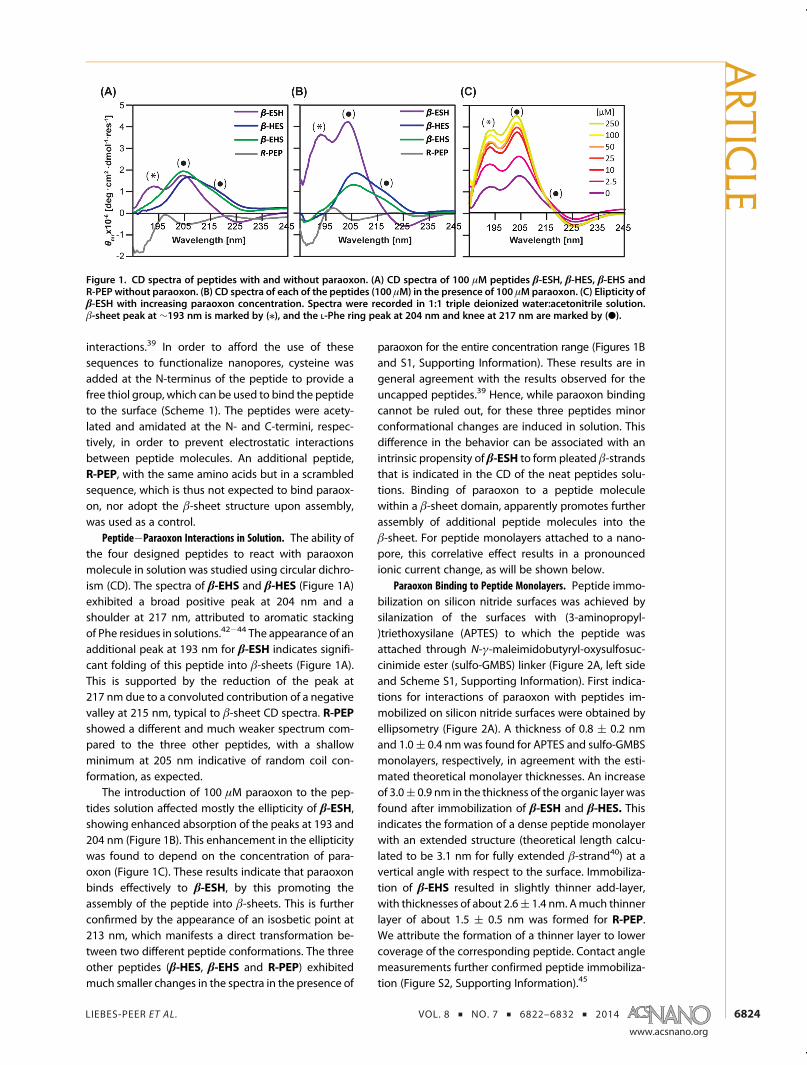
Peptide�Paraoxon Interactions in Solution. The ability ofthe four designed peptides to react with paraoxonmolecule in solution was studied using circular dichro-ism (CD). The spectra of β-EHS and β-HES (Figure 1A)exhibited a broad positive peak at 204 nm and ashoulder at 217 nm, attributed to aromatic stackingof Phe residues in solutions.42�44 The appearance of anadditional peak at 193 nm for β-ESH indicates signifi-cant folding of this peptide into β-sheets (Figure 1A).This is supported by the reduction of the peak at217 nm due to a convoluted contribution of a negativevalley at 215 nm, typical to β-sheet CD spectra. R-PEPshowed a different and much weaker spectrum com-pared to the three other peptides, with a shallowminimum at 205 nm indicative of random coil con-formation, as expected.
The introduction of 100 μM paraoxon to the pep-tides solution affected mostly the ellipticity of β-ESH,showing enhanced absorption of the peaks at 193 and204 nm (Figure 1B). This enhancement in the ellipticitywas found to depend on the concentration of para-oxon (Figure 1C). These results indicate that paraoxonbinds effectively to β-ESH, by this promoting theassembly of the peptide into β-sheets. This is furtherconfirmed by the appearance of an isosbetic point at213 nm, which manifests a direct transformation be-tween two different peptide conformations. The threeother peptides (β-HES, β-EHS and R-PEP) exhibitedmuch smaller changes in the spectra in the presence of
paraoxon for the entire concentration range (Figures 1Band S1, Supporting Information). These results are ingeneral agreement with the results observed for theuncapped peptides.39 Hence, while paraoxon bindingcannot be ruled out, for these three peptides minorconformational changes are induced in solution. Thisdifference in the behavior can be associated with anintrinsic propensity of β-ESH to form pleated β-strandsthat is indicated in the CD of the neat peptides solu-tions. Binding of paraoxon to a peptide moleculewithin a β-sheet domain, apparently promotes furtherassembly of additional peptide molecules into theβ-sheet. For peptide monolayers attached to a nano-pore, this correlative effect results in a pronouncedionic current change, as will be shown below.
Paraoxon Binding to Peptide Monolayers. Peptide immo-bilization on silicon nitride surfaces was achieved bysilanization of the surfaces with (3-aminopropyl-)triethoxysilane (APTES) to which the peptide wasattached through N-γ-maleimidobutyryl-oxysulfosuc-cinimide ester (sulfo-GMBS) linker (Figure 2A, left sideand Scheme S1, Supporting Information). First indica-tions for interactions of paraoxon with peptides im-mobilized on silicon nitride surfaces were obtained byellipsometry (Figure 2A). A thickness of 0.8 ( 0.2 nmand 1.0( 0.4 nmwas found for APTES and sulfo-GMBSmonolayers, respectively, in agreement with the esti-mated theoretical monolayer thicknesses. An increaseof 3.0( 0.9 nm in the thickness of the organic layer wasfound after immobilization of β-ESH and β-HES. Thisindicates the formation of a dense peptide monolayerwith an extended structure (theoretical length calcu-lated to be 3.1 nm for fully extended β-strand40) at avertical angle with respect to the surface. Immobiliza-tion of β-EHS resulted in slightly thinner add-layer,with thicknesses of about 2.6( 1.4 nm. Amuch thinnerlayer of about 1.5 ( 0.5 nm was formed for R-PEP.We attribute the formation of a thinner layer to lowercoverage of the corresponding peptide. Contact anglemeasurements further confirmed peptide immobiliza-tion (Figure S2, Supporting Information).45
Figure 1. CD spectra of peptides with and without paraoxon. (A) CD spectra of 100 μM peptides β-ESH, β-HES, β-EHS andR-PEP without paraoxon. (B) CD spectra of each of the peptides (100 μM) in the presence of 100 μMparaoxon. (C) Elipticity ofβ-ESH with increasing paraoxon concentration. Spectra were recorded in 1:1 triple deionized water:acetonitrile solution.β-sheet peak at ∼193 nm is marked by (/), and the L-Phe ring peak at 204 nm and knee at 217 nm are marked by (b).
ARTIC
LE

LIEBES-PEER ET AL. VOL. 8 ’ NO. 7 ’ 6822–6832 ’ 2014
www.acsnano.org
6825
The thickness of the layers slightly decreased uponimmersing the β-strand peptide modified surfaces inparaoxon solution (Figure 2A), indicating interactionsbetween paraoxon molecules and the functionalizedsurfaces. The reduction in the thickness for monolayerswith extended peptide conformations implies that thetilt angle of the peptides with respect to the surfaceincreases due to paraoxon binding. In contrary, expos-ing surfaces modified with R-PEP to paraoxon solutiondid not result in a change in the thickness of themono-layer, suggesting insignificant binding of paraoxon toR-PEP. This experiment, however, confirms that theintegrity of the organic layer is retained upon exposureto paraoxon; hence, the reduction in the thickness for
the former peptides cannot be attributed to deteriora-tion of the layer.
Further information on paraoxon binding was pro-vided by X-ray photoelectron (XPS) measurements. Forthese measurements, peptides were immobilized ongold surfaces directly through the thiol side chain ofthe Cys residue. Binding was confirmed by ellipsome-try and contact angle measurements, which indicatedsimilar trends to these observed on silicon nitridesurfaces (Figure S3, Supporting Information). The XPSmeasurements indicated carbon/nitrogen atomic ratiovalues that correspond to the peptides' chemical for-mula for all peptides (Table S1, Supporting Information).Paraoxon binding resulted in the appearance of a smallphosphorus binding energy peak (P2p, 138 eV). Roughestimation of the ratio between paraoxon and peptidemolecules was obtained by the atomic ratio betweensulfur and phosphorus, confirming significant binding ofparaoxon to these monolayers (Table S1, SupportingInformation). Only traces of paraoxon were detected forβ-EHS and R-PEP monolayers, indicating less specificinteractions of paraoxonwith these peptidemonolayers.
The evolution of the secondary structure of thepeptides on the surface due to interactions withparaoxon was monitored using polarization modula-tion infrared reflectance absorption spectroscopy(PM-IRRAS). Very small signals were observed for thepeptide monolayers before interactions with paraoxonmolecules (Figure 2B and S4, Supporting Information),indicating that the monolayers are randomly orien-tated on the surface. Remarkable changes were ob-served in the spectra of β-ESH and β-HES monolayersafter paraoxon binding (Figure 2B). Peaks attributed tothe organophosphate group ν(PdO), C;O;P(dO)transitions at 1270 cm�1 and 1050 cm�1, respectively,as well as a shoulder at 1025 cm�1 attributed to theν(P�O�Et) transition, appeared in the spectra.46 Astrong peak at 1120 cm�1, corresponding probablyto both the phosphate group46 and the benzyl ring ofparaoxon, and peaks corresponding to its nitro groupat 1410 cm�1 (ν-C�N) and 1614 cm�1 (ν-NO2),
47�50
were also observed. These peaks, which did not appearforβ-EHSandR-PEP (FigureS4A, Supporting Information),further confirm the binding of paraoxon to thesemonolayers. The appearance of these peaks was ac-companied by the appearance of a pronounced amideI peak at 1660 cm�1, suggesting assembly of thepeptides into ordered and oriented parallel β-sheetstructure.51 These results indicate that paraoxon bind-ing to β-ESH and β-HESmonolayers indeed promotespeptide assembly into β-sheets, similarly to the effectobserved for β-ESH in solution. The self-assembly ofthe β-strands in a parallel β-sheet conformation at theinterface, could be expected, due to the unidirectionalorientation imposed by binding the peptides on thesurface through theN-terminus Cys. The appearance ofsmall Amide II peaks at 1550 cm�1 further confirmed
Figure 2. Structural characterizations of peptide mono-layers assembled on silicon nitride and gold surfaces. (A)Monolayers' thickness, obtained by ellipsometry for pep-tide monolayers before and after exposure to paraoxon.Scheme of peptide arrangement on silicon nitride surface isprovided to the left of the image. Dashed red, yellow andpurple lines indicate theoretically estimated thicknessvalues for APTES, sulfo-GMBS and peptide monolayers,respectively (theoretical length of the peptides was calcu-lated assuming extended β-strand by 6.9 Å � number ofamino acids � 0.540). (B) Polarization modulation infraredreflection adsorption spectroscopy (PM-IRRAS) spectra ofβ-ESH and β-HES peptide monolayers on gold surfacesbefore and after paraoxon binding (spectra are shiftedfrom each other for clarity). Bare gold background wassubtracted from the spectra.
ARTIC
LE

LIEBES-PEER ET AL. VOL. 8 ’ NO. 7 ’ 6822–6832 ’ 2014
www.acsnano.org
6826
paraoxon induced ordering of the peptidemonolayers.The large Amide I/Amide II peaks intensity ratio pointsto a large tilt angle of the peptide backbones in theβ-sheet structure with respect to the surface normal.This apparent peptide tilting is in agreement with thedecrease in peptide thickness, which was indicatedin the ellipsometry measurements on silicon nitride(Figure 2A). Control experiments indicated thatparaoxon does not adsorb directly on the gold surface(Figure S4B, Supporting Information). Additionalexperiments exposing substrates functionalized withL-Cys and heptanethiol further confirmed that paraoxondoes not adsorb nonspecifically to charged or hydro-phobic surfaces, (Figure S4B, Supporting Information).Furthermore, no significant changes in the spectraappeared for β-EHS and R-PEP monolayers upon ex-posure to paraoxon, confirming that the later did notadsorb on surfaces functionalized with these two pep-tides (Figure S4A, Supporting Information).
Detection of Single Paraoxon Molecule Interactions withPeptide Functionalized Nanopores. While enhancement ofβ-sheet formation was observed for both β-ESH andβ-HES monolayers, the relative magnitude of theβ-sheet related peaks in the PM-IRRAS spectra withrespect to these of the paraoxon peaks were found tobe larger for β-ESH monolayers (Figure 2B), indicatinga more significant paraoxon induced β-sheet forma-tion for the later. We have, therefore, decided to checkwhether the interactions of β-ESH with paraoxon canbe detected in the context of nanopore ionic current
measurements. Thepeptidewas attached to the rims ofnanopores prepared in silicon nitride membrane usingthe procedure described above (see also Methodssection). Decrease in ionic current was manifested inthe current�voltage (I�V) curves measured after eachmodification step (Figure S5, Supporting Information),indicating reduction in the nanopore effective diameterwith each step. A general linear Ohmic behavior, espe-cially under low biases, indicates that the tetheredmolecules are deposited homogeneously within thenanopore and that their conformation does not changeby the electric field.52�54
A β-ESH modified nanopore, 11 nm average dia-meter, was used in order to access the ability tomonitorin situ single paraoxon molecule binding to the mono-layer by changes in the ionic current. An average currentof�4.87( 0.02 nA was measured for the peptide func-tionalized nanopore using 500 mM KCl/55 mM TRIS 3HClbuffer (pH=7.4) underbiasvoltageof�190mV (Figure3A,left panel). Upon introduction of paraoxon to the cis-chamber at a final concentration of 250 μM, the base-line current slightly increased to a value of �4.64 (0.01 nA, indicating a decrease of about 5% in the totalcurrent, in similar to the 9% decrease in conductanceobserved in I�V measurements performed after incu-bating β-ESH sensitized nanopore in 250 μMparaoxonbuffer solution (10 mM TRIS 3HCl pH = 7.4) overnight(Figure S5C, Supporting Information). The overall re-duction in the current after the addition of paraoxonto the solution was accompanied by the periodic
Figure 3. Current transients of peptide modified nanopores with and without paraoxon (250 μM) in the cis-chamber. (A)Current transient of a nanopore (average diameter ∼11 nm) modified with β-ESH before (left) and after (right) addition ofparaoxon. Type 1 and type 2 current enhancement events aremarked in red and blue dashed lines, respectively. The baselinecurrent is indicated at the top of the trace (green dashed line). (B) Zoom in on several type 1 current transient events from (A).(C,D) Control experiments showing current transients of pristine nanopore (average diameter ∼9 nm) (C) and nanoporemodifiedwith R-PEP (averagediameter∼16nm) (D) before and after introduction of paraoxon to the cis-chamber. Ion-currenttraceswere recorded at 500mMKCl/55mMTRIS 3HCl buffer solution (pH=7.4) using a voltagebias of�190mV, lowpassfilter1 kHz and data sampling rate of 12.5 kHz.
ARTIC
LE

LIEBES-PEER ET AL. VOL. 8 ’ NO. 7 ’ 6822–6832 ’ 2014
www.acsnano.org
6827
appearance of current enhancement events in thetransient (Figure 3A, right panel). Two types of eventswere observed (Figure 3A,B); frequent short durationevents with larger current enhancement (type 1), andless frequent longer duration events, with smallerenhancement (type 2). Occurrence frequency of bothevents, which will be further discussed below, wasfound to follow Gaussian distribution (Figure 4A,B), asexpected. After thoroughly washing the fluid chamberwith buffer solution, deionized water and ethanol, andrefilling it with a fresh buffer solution, the current levelresumed the original value measured before introduc-tion of paraoxon, and no current enhancement eventswere observed. Furthermore, repeating the experi-ments with uncoated, pristine, nanopores (Figure 3C),and nanopores coatedwith R-PEP (Figure 3D), resultedin smooth current traces without any current enhance-ment or blocking events. Hence, the appearance ofcurrent enhancement events can be correlated withinteractions of paraoxon with the peptide receptorlayer. We note that in similar to the observed forβ-ESH sensitized nanopores, the use of both pristinenanopore and a nanopore functionalize with R-PEPresulted in a decrease of 21 and 7% in the measuredbaseline current level once paraoxon was introducedto the cis chamber, respectively. In addition, a currentdrift observed for the functionalized nanopore, whichwas previously reported by others as well and assignedto heating,12,55 was diminished after paraoxon addi-tion in all cases. This behavior indicates that otherinteractions also occur and influence the current.
The appearance of peaks for the β-ESH sensitizednanopores cannot be explained by rapid translocationof the molecule through the nanopore and/or its loosebinding to the rims since these events are not expectedto induce significant changes in the current. This is
supported by the fact that no peaks were observed forthe pristine nanopores and for a nanopore sensitizedby R-PEP. We, therefore, conclude that these peaksresult from the extended conformational changes in-duced to the β-ESH monolayer by paraoxon bindingthat are observed in the ellipsometry and FTIR studies.These conformational changes result in a local increasein the effective size of the nanopore and thus reducethe resistance of the nanopore, leading to the ob-served current enhancement signal. Hence, the coop-erative effect of peptide β-sheet assembly provides aneffective signal amplification of paraoxon moleculebinding. The appearance of two types of events withdefined sizes indicates that there are two possiblebindingmodes. The fact that type 1 event is bothmorefrequent and is about four times larger in magnitudethen type 2 event rules out the possibility that itdesignate simultaneous binding of several molecules,in which case the most probable event should besmaller in magnitude, and the second probable event,which should have signified the binding of two mol-ecules should be double in size. These observationsstrongly indicate that each of the events is a result oftwo distinct interactions of a single paraoxon with thepeptide monolayer. These two events do not seem todepend on each other and can occur simultaneously,as is observed in themeasurements by the appearanceof type 1 events with and without the background ofthe less frequent type 2 events.
It is important to realize that while the providedsetup allows monitoring the binding of small mol-ecules at the single molecule level, the associationconstant depends not only on the binding affinity ofthe molecule to the receptor peptide, but also on theconformational stability of the monolayer at the vicin-ity of the binding site. The appearance of two types of
Figure 4. Influence of paraoxon concentration on events characteristics. Occurrence histograms as a function of timebetween events (τoff) for type 1 events (A) and type 2 events (B), recorded with different paraoxon concentrations. Intereventtranslocation time was extracted from current time trace. Insets: dependence of event translocation rate on paraoxonconcentration. A linear fit trend-line is plotted to guide the eye (dashed lines). Average values and standard deviations ofpulse height,ΔI, (C) and event duration, τon, (D) for type 1 and type 2 events. Values were calculated from pulse height versusdwell time scatter-plots (Figure S6, Supporting Information). Transients were recorded for β-ESH modified nanopore, usingparaoxon concentration ranging from 31.25 to 250 μM in 500 mM KCl/55 mM TRIS 3HCl buffer (pH = 7.4), using voltage biasof �190 mV.
ARTIC
LE

LIEBES-PEER ET AL. VOL. 8 ’ NO. 7 ’ 6822–6832 ’ 2014
www.acsnano.org
6828
events can, accordingly, be correlated with two typicalorientations of the β-sheet structure (Scheme 2). Thelarger enhancement of type 1 events (1.4% relativeenhancement vs 0.004% for type 2 events) implies thatin this case paraoxon binding induces a much largertilting angle of the β-sheet structure. It is reasonable toassume that such arrangement will bemuch less stabledue to larger spatial strain and smaller in-plane overlapbetween peptide molecules in the β-sheet structure.This is indeed manifested by the much shorter eventduration for these events. Type 2 events, which arecharacterized by a β-sheet conformationwith a smallertilt angle, are indicated to be more stable. Futureinvestigations will address this issue quantitatively.
The current enhancement events offer additionalinformation on paraoxon interactions with the peptidemonolayer. Three parameters were used to analyzeevents characteristics: time between adjacent events,τoff; event duration, τon; and event magnitude, ΔI(these are marked in Figure 3). The effect of paraoxonconcentration was investigated in the range of 31.25�250 μM. For each concentration, a time transient
was recorded and both types of events were detected.For both type of events, the translocation rate, ex-tracted as the inverse of the average value of τoff, wasfound to be linearly dependent on the concentration(Figure 4A,B, Figure S6, Supporting Information), indi-cating that events rate is proportional to the concen-tration of paraoxon molecules in solution at this rangeof concentrations for both type of events. In contraryto the dependence of events rate, the magnitude ofthe events (Figure 4C), or their duration (Figure 4D)were found to be independent of the concentrationof paraoxon. This behavior, which was previouslyobserved for protein-receptor binding within nano-pores,14,54 is reasonable since the concentrationshould not affect the binding kinetics, or the resultinginteractions with the peptide substrate. While the lineardependence of events frequency on the concentrationcould be used to quantify the concentration of para-oxon in solution in sensing applications,1,4 the range ofconcentrations used in this work is far from the lowerlimit of detection (LLD) of 0.4 nM (10�7 g/L) declaredby the European Union (EU) for organophosphates.31
Scheme 2. Schematic representation of interactions between paraoxon and β-ESH sensitized nanopores. In the absence ofparaoxon (OFF state) β-ESH adopts a random coil conformation (A). Upon introduction of paraoxon to the cis-side, binding tothe peptide induces the formation of a β-sheet structure with large (B) or low (C) tilt angle for type 1 and type 2 events,respectively.
Figure 5. Influence of bias on characteristics of type 1 and type 2 current events. (A) Average values and standard deviationsof the pulse height (ΔI). Inset: normalized pulse height (ΔI/I, calculated as pulse height divided by the average baselinecurrent) for each voltage. (B) Average values and standard deviations of events' duration (τon). (C) Average values andstandarddeviationsof event rate (1/τoff). Datawas collected forβ-ESHmodifiednanopore in the presenceof 250μMparaoxonin 500 mM KCl/55 mM TRIS 3HCl buffer (pH = 7.4). Values were calculated from the pulse height versus the dwell time (τon)scatter-plots (Figures S7 and S8, Supporting Information). Empty bar for�100mV in (C) indicates uncertainty in the value dueto the fact that only two events were recorded at this bias.
ARTIC
LE

LIEBES-PEER ET AL. VOL. 8 ’ NO. 7 ’ 6822–6832 ’ 2014
www.acsnano.org
6829
Hence, further studies elucidating the detection limit,sensitivity and specificity of detection are required inorder to assess theutility of thesepeptide functionalizednanopores as organophosphate sensors.
Further insight into event characteristics was ob-tained by monitoring their dependence on the biasapplied between the cis- and trans-chambers (Figure 5and Figures S7 and S8, Supporting Information). Bothtype of event were observed under positive as wellas negative bias polarities. ΔI was found to dependlinearly on the applied bias magnitude for both type ofevents (Figure 5A); however, the relative change of thepulse height (ΔI/I0, where I0 is the baseline current) wasfound to be constant (Figure 5A, inset). This indicatesthat the observed change is due to changes in the rateof ion transport. Furthermore, the duration of theevents was also found to be independent of theapplied bias (Figure 5B). These results indicate thatthe interactions of paraoxon with the peptide mono-layer and the peptide structure are not influenced bythe bias. Finally, events' rate were also found to beindependent of the bias, for both types of events(Figure 5C), indicating the lack of entropic barrier forparaoxon entering the nanopore.11,22,56 This is prob-ably due to the small dimensions of the paraoxoncompared to the nanopore, which makes the entropicbarrier presented for large DNAor polymersmolecules,negligible. Hence, neither the introduction of theparaoxon into the nanopore, nor the interaction ofthe molecule with the receptor within the nanopore,depends on the electric field within the nanopore forboth type of events.
CONCLUSIONS
We have developed a novel methodology for thedetection of small molecules at the single moleculelevel using solid state nanopores functionalized witha monolayer of a specifically designed peptide. Our
method exploits the ability to rationally design peptidereceptors that undergo cooperative self-assembly andfolding as a result of ligand binding. Sensitization ofnanopore rims with a monolayer of such peptidesinduces long duration interactions of the moleculeswith the rims, hence matches the binding time withthe temporal resolution of the measurement. Further-more, it amplifies the signal signature by inducing along-range conformational change in the peptidemonolayer.Specifically, we have presented here the design of a
peptide with a receptor unit for paraoxon, inspired bythe catalytic triad of AChE. The amino acid triad isembedded within the hydrophilic plane of a β-sheetforming peptide. The self-assembly of the peptide intoa β-sheet structure was shown to be promoted byparaoxon binding both in solution and for peptidemonolayers, depending on the order of the aminoacids triad in the sequence. Single molecule interac-tions of paraoxon with a monolayer of the peptideattached to the rims of nanopores could be detected aspeaks in ionic current passing through the nanopore,due to an increase in the effective diameter of thenanopore as a result of the formation of a β-sheetstructure tilted with respect to the nanopore surface.The sensitivity of the method has been demonstratedby the ability to detect two types of interactions,differing in the tilt angle and the binding constant.The ability to detect organophosphate moleculesmakes this method very important for environmentaland warfare applications. Furthermore, this method isvery versatile and can be used for the detection of anymolecule, provided that a proper peptide receptor isdesigned. Hence, the suggested concept can be usedfor the development of highly sensitive small moleculesensors. Moreover, the system can be used to studyreceptor�small molecule interactions with high sensi-tivity at the single molecule level.
METHODS
Peptides and Reagents. Peptides (1201.37 g/mol) with purityabove 95% were purchased from GenScript (Piscataway, NJ).HPLC grade acetonitrile and ethanol were purchased from J.T.Baker (Beit-Dekel, Israel). Sulfo-GMBS was supplied by Pierce(Rockford, IL). For silanization, ethanol was dried over magne-sium sulfate and kept dried using 3 Å molecular sieves untiluse. Paraoxon, dimethyl sulfoxide (DMSO), (3-aminopropyl)-triethoxysilane (APTES) and Tris(2-carboxyethyl)phosphinehydrochloride (TCEP) were purchased from Sigma-Aldrich(Rehovot, Israel) and used as received. Phosphate buffer (PB)(100 mM; 12 g/L NaH2PO4, 14.2 g/L Na2HPO4) and 10 mMTRIS 3HCl buffer (1.2 g/L TRIS 3HCl) were prepared and adjustedto pH 7.4 with NaOH/HCl.
Circular Dichroism (CD). CD measurements were performed ona Jasco J-715 spectropolarimeter (Jasco, Inc., Easton, MD) usingQuartz cuvettes with 1.0 mm path length. Data was collected in180�250 nm wavelength range at 20 �C, with bandwidth of1 nm, in continuous mode with 0.5 nm steps, 4 s response time,and scan speed of 50 nm/min. Spectra are reported as mean
molar ellipticity [θmr, deg 3 cm23dmol�1
3 res�1] versus wave-
length. CD spectra were recorded for peptides concentrationof 0.12 mg/mL (100 μM). Each peptide was first dissolved inacetonitrile and sonicated for 10min, followed by addition of anequal volume of deionized water or the paraoxon solutiondiluted in deionized water. The final concentration of paraoxonwas 2.5�250 μM. Solutions with same concentration of para-oxon were used as references, eliminating possible effect of theparaoxon on the spectrum.
Silicon Nitride Surface Functionalization. Surface functionaliza-tion of silicon nitride samples is shown in Figure 2 andScheme S1 (Supporting Information). Phosphorus dopedn-Type Si wafers ((100), 1�10 Ω 3 cm resistivity) with a top30 nm thick silicon nitride layer prepared by low pressurechemical vapor deposition (LPCVD) were purchased fromVirginia Semiconductor, Inc. (Fredericksburg, VA). Silicon nitridesurfaces were cleaned in piranha solution (7:3 (v/v) concen-trated sulfuric acid:30% hydrogen peroxide) for 30 min at 90 �C,followed by triple washing (10 min each) in deionized water.(Caution! Piranha solution is highly corrosive and very dangerous
ARTIC
LE

LIEBES-PEER ET AL. VOL. 8 ’ NO. 7 ’ 6822–6832 ’ 2014
www.acsnano.org
6830
and should be handled with care.) Samples were dried undernitrogen flow. Immediately after, samples were immersed infreshly prepared solution of 5% (v/v) APTES in dried ethanolunder nitrogen atmosphere for 10 min, followed by triple washin ethanol (10 min each), drying under N2 stream and baking at110 �C for 30 min.45 Samples were immersed in 3 mM sulfo-GMBS/100 mM PB. After 1 h. the solution was removed andsamples were soaked three times for 10 min each in PB anddried under N2 stream.30 The maleimide modified sampleswere immersed in freshly prepared 0.5 mM peptide solution(peptides were dissolved in DMSO to a concentration of 2.5mM,and were further diluted to 0.5 mM solution in 4 mM TCEP/100 mM PB) for 3 h, washed three times in PB and deionizedwater (10 min each) and dried under N2 stream.
Gold Samples Surface Functionalization. Gold samples were pre-pared by sputtering 200 nm thick Gold films on Si (100) wafers(Virginia Semiconductor, Inc., Fredericksburg, VA), using Cr(8 nm) adhesion layer (ODEM sputter system, 4 � 10�9 mbarbase pressure). Prior to peptide assembly samples were cleanedby sonication in ethanol bath for 15 min, hydrogen flameannealed, and were treated by ozone/UV for 20 min at 40 �C(PCD-UVT, Novascan, AMES, IA). Samples were then sonicated inethanol bath for 15min, and dried under N2 flow.
57 Immediatelyafter, samples were immersed in fresh 0.5 mM peptide solutionin 100 mM PB for 3 h. Thereafter, samples were incubated in500 μM paraoxon solution in TRIS 3HCl buffer (pH = 7.4) for 8 h.After each step, excess of material was removed by gentlyshaking for 10 min in PB and deionized water three times,and drying under N2 stream. Layer thickness was calculatedfrom ellipsometry measurements performed using PHE101Ellipsometer (Angstrom Advanced, Inc., Braintree, MA) at awavelength of 632.8 nm and angle of 70�. Refractive index of1.5 was used for all organic substances.
Polarization Modulation Infrared Reflection/Adsorption Spectroscopy(PM-IRRAS). PM-IRRAS characterizations were obtained on pep-tide modified gold samples using Nicolet 6700 spectrophot-ometer equipped with a liquid nitrogen-cooled MCT detector(Thermo, Madison, WI). The detector angle was set to 85� (initialexperiments have shown maximal signal-to-noise ratio at thisangle), the modulation frequency was set at 1600 cm�1 and400 scans at a resolution of 8 cm�1 were collected for eachspectrum. The incident IR beamwas polarized by ZnSe polarizerbetween parallel (p) and perpendicular (s) polarization to theplane of incident. The signal was processed and reported asthe differential reflectivity spectrum ΔR/R = (Rp � Rs)/(Rp þ Rs),where Rp and Rs are the polarized reflectivities for p and spolarizations, respectively. Background subtraction was ob-tained by removing the spectra of a clean gold surface. Spectrawere normalized and smoothed using the Omnic software(Thermo, Madison, WI).
Nanopore Fabrication. Nanopores were fabricated in plasmacleaned (Ar gas) 30 nm thick silicon nitridemembranes (windowsize 50 3 50 μm2) supported by a 300 μm thick Si frame(Protochips, Inc., Raleigh, NC). The nanopores were drilled usinga high intensity focused electron beam based on previouslyreported procedures.58,59 In brief, a single nanopore was drilledin eachmembrane using a converged beamof a high resolutiontransmission electron microscopy (HR-TEM) with an incidentaccelerating voltage of 200 kV from FEG source (JEOL, 2100F).The microscope was operated in converged beam diffractionmode (CBD, 2.4 nm beam diameter) and at 400 Kmagnification.Plan-view TEM imaging was used to monitor the size of thenanopores. An average diameter was extracted from the area ofthe nanopore, which was evaluated by pixel counting usingImageJ software. The drilling process has begun with a nano-pore opening, typically 8 nm in diameter, by a direct illumina-tion of the sample for 1 min. Thereafter, the beam was gentlymoved on the pore�vacuum interface to enlarge the pore up tothe desired size, between 10 to 30 nm diameter in average.
Nanopore Modification. Chips containing the nanopores werecleaned by ozone treatment (PCD-UVT, Novascan, AMES, IA) for1 min from each side and immediately soaked in 1:1 ethanol:triple deionized water solution for a day. This allowed properwetting of the pore region and therefore improved ions andanalyte translocations. Surface modifications were obtained
using the procedure used for silicon nitride surface modifica-tions.54 N2 dried chips were suspended in freshly prepared5% (v/v) APTES/ethanol solution for 10 min under nitrogenatmosphere, followed by washing in ethanol, and drying underN2 stream. Samples where then baked at 110 �C for 30 min andafter cooling to room temperature immersed for 1 h. in 3 mMsulfo-GMBS linker in 100 mM PB. For creating the thioetherbond with the peptide, samples were immersed for 3 h. in0.5 mM β-ESH/4 mM TCEP/100 mM PB containing 20% (v/v)DMSO. Paraoxonwas introduced to the nanopore by incubatingthe chip in solution of 250 μM paraoxon in 10 mM TRIS 3HClbuffer (pH = 7.4) overnight. This pH was used to match thebinding conditions as much as possible to the physiologicalconditions, at which AchE is active. After eachmodification step,the chip was gently agitated five times (10 min each) with thepure solvent that was used during the modification step.Modifications were validated by a decrease in the ionic con-ductance. Prior to electrical characterizations samples werekept for few hours in 1:1 ethanol:triple deionized water solution.
Electrical Measurements. Clean or modified chips weremounted between two Teflon gaskets separating two Teflonreservoirs, filled with KCl solution (250�1000 mM in 10 mMTris 3HCl buffer (pH = 7.4)) and equipped with Ag/AgCl electro-des connected to an Axopatch 200B amplifier that was con-trolled by the pClamp software (Axon Instruments, Sunnyvale,CA). The measurements were recorded using a low pass Besselfilter of 5 kHz and data sampling rate of 12.5 kHz. For I�Vmeasurements the DC current was measured as a functionof the applied voltage, which was stepped between �190and 190 mV with 10 mV intervals every 0.25 s. The conductanceof the nanopore was calculated from the slope of the curve. Forstatistical analysis, different nanopores were measured for eachstep (n = 3�6). For paraoxon translocation experiments, each ofthe reservoirs was filled with 500 mM KCl/55 mM TRIS 3HClbuffer pH = 7.4, and the paraoxon was added to the cis-side ofthe cell to a final concentration of 250 μM. Serial dilutions weredone in the cell to dilute the paraoxon down to 31.25 μM byadding a corresponding volume of 55 mM TRIS 3HCl buffer(pH 7.4). The measurements were recorded using a low passBessel filter (1 kHz) and data sampling rate of 12.5 kHz at anapplied voltage of �190 mV, unless otherwise noted. All datawere analyzed using Clampfit software (Axon Instruments,Sunnyvale, CA). Translocation events were detected by thresh-old search procedure, and only events exceeding three timesthe standard deviation of the noise were accepted. Eachexperiment was repeated successfully at least three times,and representative results are shown.
Conflict of Interest: The authors declare no competingfinancial interest.
Acknowledgment. We thank Prof. A. Kushmaro for provid-ing the paraoxon solutions, and Drs. V. Ezersky, S. Kolusheva,and N. Frumin from the Ilse Katz Institute for Nanoscale Scienceand Technology (IKI, Ben Gurion University) for their assistancein TEM, PM-IRRAS and XPS operation. Y.L.P. is a recipient of aNegev doctoral scholarship and the Shimona Geresh prize. Partof thisworkwasfinancially supportedbyaDIPGrant (AS424/1-1).
Supporting Information Available: Additional CD spectra;characterizations of peptide monolayers on gold and siliconnitride, including contact angle and XPS data, and additionalellipsometry and PM-IRRAS characterizations; nanopores' I�Vcurves and voltage-dependent ion current transient analysishistograms. This material is available free of charge via theInternet at http://pubs.acs.org.
REFERENCES AND NOTES1. Kasianowicz, J. J.; Robertson, J.W. F.; Chan, E. R.; Reiner, J. E.;
Stanford, V. M. Nanoscopic Porous Sensors. Annu. Rev.Anal. Chem. 2008, 1, 737–766.
2. Henriquez, R. R.; Ito, T.; Sun, L.; Crooks, R. M. The Resur-gence of Coulter Counting for Analyzing Nanoscale Ob-jects. Analyst 2004, 129, 478–482.
3. Schmidt, J. Stochastic Sensors. J. Mater. Chem. 2005, 15,831–840.
ARTIC
LE

LIEBES-PEER ET AL. VOL. 8 ’ NO. 7 ’ 6822–6832 ’ 2014
www.acsnano.org
6831
4. Howorka, S.; Siwy, Z. Nanopore Analytics: Sensing of SingleMolecules. Chem. Soc. Rev. 2009, 38, 2360–2384.
5. Martin, C. R.; Siwy, Z. S. Learning Nature's Way: Biosensingwith Synthetic Nanopores. Science 2007, 317, 331–332.
6. Majd, S.; Yusko, E. C.; Billeh, Y. N.; Macrae, M. X.; Yang, J.;Mayer, M. Applications of Biological Pores in Nanomedi-cine, Sensing, and Nanoelectronics. Curr. Opin. Biotechnol.2010, 21, 439–476.
7. Bayley, H.; Cremer, P. S. Stochastic Sensors Inspired byBiology. Nature 2001, 413, 226–230.
8. Rhee, M.; Burns, M. A. Nanopore Sequencing Technology:NanoporePreparations.TrendsBiotechnol.2007,25, 174–181.
9. Dekker, C. Solid-State Nanopores. Nat. Nanotechnol. 2007,2, 209–215.
10. Branton, D.; Deamer, D. W.; Marziali, A.; Bayley, H.; Benner,S. A.; Butler, T.; Di Ventra, M.; Garaj, S.; Hibbs, A.; Huang,X. H.; et al. The Potential and Challenges of NanoporeSequencing. Nat. Biotechnol. 2008, 26, 1146–1153.
11. Wanunu, M. Nanopores: A Journey Towards DNA Sequen-cing. Phys. Life Rev. 2012, 9, 125–158.
12. Han, A.; Creus, M.; Schurmann, G.; Linder, V.; Ward, T. R.; deRooij, N. F.; Staufer, U. Label-Free Detection of SingleProtein Molecules and Protein-Protein Interactions UsingSynthetic Nanopores. Anal. Chem. 2008, 80, 4651–4658.
13. Talaga, D. S.; Li, J. L. Single-Molecule Protein Unfolding inSolid State Nanopores. J. Am. Chem. Soc. 2009, 131, 9287–9297.
14. Sexton, L. T.; Horne, L. P.; Sherrill, S. A.; Bishop, G. W.; Baker,L. A.; Martin, C. R. Resistive-Pulse Studies of Proteins andProtein/Antibody Complexes Using a Conical NanotubeSensor. J. Am. Chem. Soc. 2007, 129, 13144–13152.
15. Wei, R.; Gatterdam, V.; Wieneke, R.; Tampe, R.; Rant, U.Stochastic Sensing of Proteins with Receptor-ModifiedSolid-State Nanopores. Nat. Nanotechnol. 2012, 7, 257–263.
16. Venkatesan, B. M.; Bashir, R. Nanopore Sensors for NucleicAcid Analysis. Nat. Nanotechnol. 2011, 6, 615–624.
17. Boersma, A. J.; Brain, K. L.; Bayley, H. Real-Time StochasticDetection of Multiple Neurotransmitters with a ProteinNanopore. ACS Nano 2012, 6, 5304–5308.
18. Clamer, M.; Höfler, L.; Mikhailova, E.; Viero, G.; Bayley, H.Detection of 30-End Rna Uridylation with a Protein Nano-pore. ACS Nano 2013, 8, 1364–1374.
19. Jayawardhana, D. A.; Crank, J. A.; Zhao, Q.; Armstrong,D. W.; Guan, X. Y. Nanopore Stochastic Detection ofa Liquid Explosive Component and Sensitizers UsingBoromycin and an Ionic Liquid Supporting Electrolyte.Anal. Chem. 2009, 81, 460–464.
20. Wang, H.-Y.; Ying, Y.-L.; Li, Y.; Kraatz, H.-B.; Long, Y.-T.Nanopore Analysis of B-Amyloid Peptide AggregationTransition Induced by Small Molecules. Anal. Chem.2011, 83, 1746–1752.
21. Wang, Y.; Zheng, D.; Tan, Q.; Wang, M. X.; Gu, L. Q.Nanopore-Based Detection of Circulating Micrornas inLung Cancer Patients.Nat. Nanotechnol. 2011, 6, 668–674.
22. Anderson, B. N.; Muthukumar, M.; Meller, A. Ph Tuning ofDNA Translocation Time through Organically Functiona-lized Nanopores. ACS Nano 2013, 7, 1408–14.
23. Tian, Y.; Hou, X.; Wen, L.; Guo,W.; Song, Y.; Sun, H.; Wang, Y.;Jiang, L.; Zhu, D. A Biomimetic Zinc Activated Ion Channel.Chem. Commun. 2010, 46, 1682–1684.
24. Boersma, A. J.; Bayley, H. Continuous Stochastic Detectionof Amino Acid Enantiomers with a Protein Nanopore.Angew. Chem. 2012, 124, 9744–9747.
25. Kawano, R.; Osaki, T.; Sasaki, H.; Takinoue, M.; Yoshizawa, S.;Takeuchi, S. Rapid Detection of a Cocaine-Binding Apta-mer Using Biological Nanopores on a Chip. J. Am. Chem.Soc. 2011, 133, 8474–8477.
26. Ying, Y. L.; Zhang, J.; Meng, F. N.; Cao, C.; Yao, X.; Willner, I.;Tian, H.; Long, Y. T. A Stimuli-Responsive Nanopore Basedon a Photoresponsive Host-Guest System. Sci. Rep. 2013,3, 1662.
27. Millard, C. B.; Kryger, G.; Ordentlich, A.; Greenblatt, H.;Harel, M.; Raves, M. L.; Segall, Y.; Barak, D.; Shafferman, A.;Silman, A.; et al. Crystal Structures of Aged Phosphorylated
Acetylcholinesterase Nerve Agent Reaction Products atAtomic Level. Biochemistry 1999, 38, 7032–7039.
28. Sussman, J. L.; Harel, M.; Silman, I. Three DimensionalStructure of Acetylcholinesterase and of Its Complexeswith Anticholine Drugs. Chem. Biol. Interact. 1993, 87,187–197.
29. Schulze, H.; Vorlová, S.; Villatte, F.; Bachmann, T. T.; Schmid,R. D. Design of Acetylcholinesterases for Biosensor Appli-cations. Biosens. Bioelectron. 2003, 18, 201–209.
30. Singh, A. K.; Flounders, A. W.; Volponi, J. V.; Ashley, C. S.;Wally, K.; Schoeniger, J. S. Development of Sensors forDirect Detection of Organophosphates. Part I: Immobiliza-tion, Characterization and Stabilization of Acetylcholines-terase and Organophosphate Hydrolase on Silica Supports.Biosens. Bioelectron. 1999, 14, 703–13.
31. Van Dyk, J. S.; Pletschke, B. Review on the Use of Enzymesfor the Detection of Organochlorine, Organophosphateand Carbamate Pesticides in the Environment. Chemo-sphere 2011, 62, 291–307.
32. Martinez, R. C.; Gonzalo, E. R.; Moran, M. J. A.; Mendez, J. H.Sensitive Method for the Determination of Organopho-sphorus Pesticides in Fruits and Surface Waters by High-Performance Liquid Chromatography with Ultraviolet De-tection. J. Chromatogr. A 1992, 607, 37–45.
33. Pylypiw, H. M. Rapid Gas Chromatographic Method for theMultiresidue Screening of Fruits and Vegetables for Orga-nochlorine and Organophosphate Pesticides. J. AOAC Int.1993, 76, 1369–1373.
34. Elhanany, E.; Ordentlich, A.; Dgany, O.; Kaplan, D.; Segall, Y.;Barak, R.; Velan, B.; Shafferman, A. Resolving Pathways ofInteraction of Covalent Inhibitors with the Active Siteof Acetylcholinesterases: Maldi-Tof/Ms Analysis of VariousNerve Agent Phosphyl Adducts. Chem. Res. Toxicol. 2001,14, 912–918.
35. Andreescu, D.; Marty, J. L. Twenty Years Research inCholinesterase Biosensors: From Basic Research to Prac-tical Applications. Biomol. Eng. 2006, 23, 1–15.
36. Arduini, F.; Amine, A.; Moscone, D.; Palleschi, G. BiosensorsBased on Cholinesterase Inhibition for Insecticides, NerveAgents and Aflatoxin B1 Detection (Review). Microchim.Acta 2010, 170, 193–214.
37. Liu, S.; Yuan, L.; Yue, X.; Zheng, Z.; Tang, Z. Recent Advancesin Nanosensors for Organophosphate Pesticide Detection.Adv. Powder Technol. 2008, 19, 419–441.
38. Skladal, P. Biosensors Based on Cholinesterase for Detec-tionofPesticides. FoodTechnol. Biotechnol.1996, 34, 43–49.
39. Yaakobi, K.; Liebes-Peer, Y.; Kushmaro, A.; Rapaport, H.Designed Amphiphilic B-Sheet Peptides Monolayers forSensing the Organophosphate Paraoxon. Langmuir 2013,29, 6840–6848.
40. Rapaport, H. Ordered Peptide Assemblies at Interfaces.Supramol. Chem. 2006, 18, 445–454.
41. Rapaport, H.; Kjaer, K.; Jensen, T. R.; Leiserowitz, L.; Tirrell,D. A. Two-Dimensional Order in β-Sheet Peptide Mono-layers. J. Am. Chem. Soc. 2000, 122, 12523–12529.
42. Adler-Abramovich, L.; Reches, M.; Sedman, V. L.; Allen, S.;Tendler, S. J. B.; Gazit, E. Thermal and Chemical Stability ofDiphenylalanine Peptide Nanotubes: Implications for Nano-technological Applications. Langmuir 2006, 22, 1313–1320.
43. Roy, R. S.; Gopi, H. N.; Raghothama, S.; Gilardi, R. D.; Karle,I. L.; Balaram, P. Peptide Hairpins with Strand SegmentsContaining R- and β-Amino Acid Residues: Cross-StrandAromatic Interactions of Facing Phe Residues. Biopolymers2005, 80, 787–99.
44. Castelletto, V.; Hamley, I. W. Self Assembly of a ModelAmphiphilic Phenylalanine Peptide/Polyethylene GlycolBlock Copolymer in Aqueous Solution. Biophys. Chem.2009, 141, 169–74.
45. Aswal, D. K.; Lenfant, S.; Guerin, D.; Yakhmi, J. V.; Vuillaume,D. Self Assembled Monolayers on Silicon for MolecularElectronics. Anal. Chem. Acta 2006, 568, 84–108.
46. Segman-Magidovich, S.; Grisaru, H.; Gitli, T.; Levi-Kalisman,Y.; Rapaport, H. Matrices of Acidic β-Sheet Peptides asTemplates for Calcium Phosphate Mineralization. Adv.Mater. 2008, 20, 2156–2161.
ARTIC
LE

LIEBES-PEER ET AL. VOL. 8 ’ NO. 7 ’ 6822–6832 ’ 2014
www.acsnano.org
6832
47. Wang, C.; Zheng, J.; Oliveira, O. N.; Leblanc, R. M. Nature ofthe Interaction between a Peptidolipid Langmuir Mono-layer and Paraoxon in the Subphase. J. Phys. Chem. C 2007,111, 7826–7833.
48. Zheng, J.; Desbat, B.; Rastogi, V. K.; Shah, S. S.; DeFrank, J. J.;Leblanc, R. M. Organophosphorus Hydrolase at the Air�Water Interface: Secondary Structure and Interaction withParaoxon. Biomacromolecules 2006, 7, 2806–2810.
49. Dziri, L.; Desbat, B.; Leblanc, R. M. Polarization-ModulatedFt-Ir Spectroscopy Studies of Acetylcholinesterase Sec-ondary Structure at the Air�Water Interface. J. Am. Chem.Soc. 1999, 121, 9618–9625.
50. Kim, S. H.; Kim, J. H.; Kang, B.-K. Decomposition Reaction ofOrganophosphorus Nerve Agents on Solid Surfaces withAtmospheric Radio Frequency Plasma Generated GaseousSpecies. Langmuir 2007, 23, 8074–8078.
51. Sneer, R.; Weygand, M. J.; Kjaer, K.; Tirrell, D. A.; Rapaport, H.Parallel B-Sheet Assemblies at Interfaces. ChemPhysChem2004, 5, 747–750.
52. Siwy, Z. S. Ion-Current Rectification in Nanopores andNanotubes with Broken Symmetry. Adv. Funct. Mater.2006, 16, 735–746.
53. Mussi, V.; Fanzio, P.; Repetto, L.; Firpo, G.; Scaruffi, P.;Stigliani, S.; Tonini, G. P.; Valbusa, U. DNA-FunctionalizedSolid State Nanopore for Biosensing. Nanotechnology2010, 21, 145102.
54. Kowalczyk, S. W.; Kapinos, L.; Blosser, T. R.; Magalhaes, T.;van Nies, P.; LimRoderick, Y. H.; Dekker, C. Single-MoleculeTransport across an Individual Biomimetic Nuclear PoreComplex. Nat. Nanotechnol. 2011, 6, 433–438.
55. Ding, S.; Gao, C.; Gu, L. Q. Capturing Single Molecules ofImmunoglobulin and Ricin with an Aptamer-EncodedGlass Nanopore. Anal. Chem. 2009, 81, 6649–6655.
56. Muthukumar, M. Mechanism of DNA Transport throughPores.Annu. Rev. Biophys. Biomol. Struct. 2007, 36, 435–450.
57. Nogues, C.; Wanunu, M. A Rapid Approach to Reproduci-ble, Atomically Flat Gold Films onMica. Surf. Sci. 2004, 573,L383–L389.
58. Kim, M. J.; Wanunu, M.; Bell, D. C.; Meller, A. Rapid Fabrica-tion of Uniformly Sized Nanopores and Nanopore Arraysfor Parallel DNA Analysis. Adv. Mater. 2006, 18, 3149–3153.
59. Ho, C.; Qiao, R.; Heng, J. B.; Chatterjee, A.; Timp, R. J.; Aluru,N. R.; Timp, G. Electrolytic Transport through a SyntheticNanometer-Diameter Pore. Proc. Natl. Acad. Sci. U. S. A.2005, 102, 10445–10450.
ARTIC
LE