an active biopolymer network controlled by molecular motors · an active biopolymer network...
TRANSCRIPT
An active biopolymer network controlledby molecular motorsGijsje H. Koenderinka,b, Zvonimir Dogicc,d, Fumihiko Nakamurae, Poul M. Bendixf, Frederick C. MacKintoshg,John H. Hartwige, Thomas P. Stossele, and David A. Weitza,1
aDepartment of Physics and Harvard School of Engineering and Applied Sciences, Harvard University, Cambridge, MA 02138; bFOM Institute for Atomic andMolecular Physics, Amsterdam 1098 SJ, The Netherlands; cRowland Institute, Harvard University, Cambridge, MA 02142; dPhysics Department, Brandeis University,Waltham, MA 02454; eTranslational Medicine Division, Brigham and Women’s Hospital, Department of Medicine, Harvard Medical School, Boston, MA 02115;fNiels Bohr Institute, 2100 Copenhagen, Denmark; and gDepartment of Physics and Astronomy, Vrije Universiteit, 1081 HV Amsterdam, The Netherlands
Edited by Tom C. Lubensky, University of Pennsylvania, Philadelphia, PA, and approved July 7, 2009 (received for review April 25, 2009)
We describe an active polymer network in which processive molecularmotors control network elasticity. This system consists of actin fila-ments cross-linked by filamin A (FLNa) and contracted by bipolarfilaments of muscle myosin II. The myosin motors stiffen the networkby more than two orders of magnitude by pulling on actin filamentsanchored in the network by FLNa cross-links, thereby generatinginternal stress. The stiffening response closely mimics the effects ofexternal stress applied by mechanical shear. Both internal and exter-nal stresses can drive the network into a highly nonlinear, stiffenedregime. The active stress reaches values that are equivalent to anexternal stress of 14 Pa, consistent with a 1-pN force per myosin head.This active network mimics many mechanical properties of cells andsuggests that adherent cells exert mechanical control by operating ina nonlinear regime where cell stiffness is sensitive to changes inmotor activity. This design principle may be applicable to engineeringnovel biologically inspired, active materials that adjust their ownstiffness by internal catalytic control.
active soft matter � cytoskeleton � filamin A � rheology � myosin II
Reconstituted biopolymer networks are a class of soft materialthat exhibits pronounced solidlike behavior while comprising
only a few percent by volume of protein. They exhibit a richmechanical behavior and pronounced nonlinearity (1). Moreover,they model important features of living cells, because the mechanicsof cells are largely controlled by a network of filamentous proteinsknown as the cytoskeleton. Reconstituted networks comprising oneof the principal components of the cytoskeleton, filamentous actin(F-actin), are interesting models for the rheology of semiflexiblepolymers. Like most filamentous protein networks, they are highlyresponsive to external stress. Cross-linked F-actin networks exhibitstrong nonlinear stiffening with strain (1, 2). This phenomenon isparticularly pronounced for the case of F-actin cross-linked byfilamin A (FLNa), a widely represented protein that is essential forfetal development and cell locomotion (3). FLNa is a large, highlyflexible dimer that promotes orthogonal F-actin cross-linking (4).FLNa–F-actin networks are very soft in their linear regime, withshear moduli as low as 1 Pa, yet they stiffen by up to three ordersof magnitude in response to external shear stress (5–8). In thisrespect, FLNa–F-actin resembles the behavior of living cells, whichalso stiffen if subjected to (tensile) stress (9, 10). Cells, however, arenot passive materials, but rather use molecular motors to convertchemical energy into mechanical work within the cytoskeleton.There is evidence that cells employ internal stress generated bymyosin motors to modulate their mechanics (11–19). This suggestsa strategy of creating a new class of active materials, whose elasticproperties are controlled by enzymatic activity, through addition ofmyosin motors to a passive F-actin network.
In this article, we report that incorporating active and processivemolecular myosin motors in FLNa–F-actin networks markedlyincreases network elasticity. The motors generate internal contrac-tile stress in the network by pulling on F-actin filaments, which arelinked to the network by the FLNa cross-links. This stress drives the
network into a strongly nonlinear, stiffened regime. These resultssuggest that actomyosin contractility allows adherent cells to operate ina nonlinear regime to control their mechanics actively. The experimen-tal system represents a recently discovered class of active soft materi-als—gels whose elasticity can be precisely tuned over three orders ofmagnitude by enzymatic activity. Moreover, unlike passive glassy ma-terials, which are inherently pinned in a nonequilibrium state, thesematerials are driven into nonequilibrium through the internal conver-sion of chemical energy to mechanical energy.
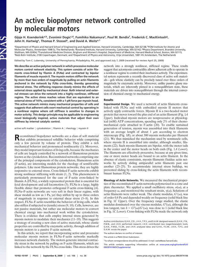
ResultsExperimental Design. We used a network of actin filaments cross-linked with FLNa and with embedded myosin II motors thatactively apply contractile forces. Myosin II is a two-headed motorprotein that moves toward the plus end of an actin filament (Fig. 1AUpper). Individual myosin motors are nonprocessive at physiolog-ical (mM) ATP concentrations, spending only 2% of their chemo-mechanical cycle attached to F-actin (20). To enable sustainedgeneration of tension, myosin was assembled into thick filamentswith an average length of about 1 �m according to electronmicroscopy (Fig. 1B), or about 300 myosin molecules per filament(21). We thus mimicked the mechanism used by nonmuscle cells,where myosin II is assembled into similar, although smaller, fila-ments (22). Such myosin filaments are bipolar, with the motor tailsin the center and the motor heads on both ends (Fig. 1A Lower).The filaments are effectively processive, because at any given time,one or more motor heads are bound to an actin filament. In theabsence of elastic constraints, myosin filaments fluidize actin net-works by actively sliding antiparallel actin filaments past oneanother (23–25). To accommodate internal stress buildup, weprevented sliding by cross-linking the actin filaments with recom-binant human FLNa.
Rheology of Actin Networks. We measured the mechanical proper-ties of the reconstituted F-actin networks polymerized in a cone andplate rheometer. We applied a small oscillatory stress, �(�), at afrequency �, and monitored the resultant strain, �(�). Solutions ofactin filaments were rather weak. The elastic modulus, G�(�), wasof order 0.8 Pa and depended weakly on frequency (solid black linein Fig. 1C Upper). Over the frequency range studied, the elasticmodulus dominated over the viscous modulus, G�(�), although theloss tangent, tan � � G�(�)/G�(�), was close to 1 (solid black linein Fig. 1C Lower). Cross-linking with FLNa made the network only
Author contributions: G.H.K., Z.D., J.H.H., T.P.S., and D.A.W. designed research; G.H.K., F.N.,P.M.B., and J.H.H. performed research; J.H.H. contributed new reagents/analytic tools;G.H.K., P.M.B., F.C.M., and J.H.H. analyzed data; and G.H.K., F.C.M., J.H.H., T.P.S., andD.A.W. wrote the paper.
The authors declare no conflict of interest.
This article is a PNAS Direct Submission.
1To whom correspondence should be addressed. E-mail: [email protected].
This article contains supporting information online at www.pnas.org/cgi/content/full/0903974106/DCSupplemental.
15192–15197 � PNAS � September 8, 2009 � vol. 106 � no. 36 www.pnas.org�cgi�doi�10.1073�pnas.0903974106

slightly stiffer (gray open triangles in Fig. 1C Upper). Based onprevious modeling and experimental results, we interpret thisfinding as resulting from the cross-linking of rodlike F-actin byrelatively flexible cross-links (8, 26). In this situation, the stretchingof the flexible cross-linkers dominates the linear modulus. FLNamolecules have a contour length of 160 nm and persistence lengthof 20 nm, and they are therefore large and floppy compared withF-actin (27). The loss tangent was somewhat enhanced in thepresence of FLNa, reflecting enhanced dissipation (open trianglesin Fig. 1C Lower). However, addition of myosin II thick filamentsto the cross-linked network imparted an enormous increase instiffness, by almost two orders of magnitude (blue solid squares inFig. 1C Upper). At the same time, myosin addition substantiallylowered the loss tangent, indicating a more solidlike response (bluesquares in Fig. 1C Lower).
We observed a marked threshold effect of motor concentrationon network elasticity. The motors had a negligible effect on networkstiffness at low myosin concentration, but they strongly stiffened thenetwork above a molar ratio of [myosin]/[actin] � 0.01 (Fig. 1DLeft). The threshold value was independent of FLNa concentration,at least at [FLNa]/[actin] ratios above 0.0007. We observed a similarthreshold effect of FLNa concentration. Myosin had no appreciableeffect on network stiffness at low FLNa concentrations, but itexerted appreciable stiffening above [FLNa]/[actin] � 0.0007 (Fig.1D Right). This observation held for any myosin concentrationsufficiently high to exert significant stress. FLNa and myosin wererequired together to attain active stiffening, as demonstrated by thelow values of G� in the absence of either component (gray opentriangles in Fig. 1D). These threshold levels are best interpreted interms of actin filaments and myosin thick filaments, instead of actinand myosin monomers. Given an average length for the actinfilaments of 15 �m (or 5,100 monomers), the threshold concentra-tions for stiffening were one myosin filament for every five actinfilaments, and three FLNa cross-linkers for every actin filament(top x axis in Fig. 1D). We found that varying FLNa and myosinconcentrations at fixed actin filament length had the same effect onnetwork stiffness as varying the average actin filament length (withthe capping protein gelsolin) for fixed FLNa and myosin concen-trations (Fig. S1). This confirms that the number of myosin thickfilaments and FLNa cross-linkers per actin filament are usefulcontrol parameters. Remarkably, for a fixed concentration of actin(23.8 �M), we can tune G�(�) over two orders of magnitude, fromonly 0.3 Pa to values as high as 300 Pa. The number of binding siteson actin filaments for myosin and FLNa presumably limits maxi-mum stiffness.
We also expect a strong dependence of the elastic modulus on
actin concentration, because stiffening arises from the simulta-neous interaction of a myosin thick filament with two actin fila-ments, as sketched in Fig. 1A. To test this prediction, we measuredthe variation of G�(�) with actin concentration for networks withfixed molar ratios of [FLNa]/[actin] � 0.01 and [myosin]/[actin] �0.02. We indeed observed a very strong increase of G�(�) withincreasing actin concentration for both long (15 �m) and short (1.5�m) filaments (Fig. S2). The increase occurred at lower actinconcentrations for the longer filaments, because they entangled ata lower concentration.
Effect of Myosin Motor Activity on Network Rheology. The presenceof myosin motors can itself lead to cross-linking (24). However, weused a high ATP concentration, where individual myosin headsremained bound only for about 1 ms (20), which is unlikely to leadto cross-linking. To validate this prediction, we removed all cross-linking proteins by making an F-actin solution in the absence ofFLNa. In this case, we found no effect of myosin on the elasticmodulus of the actin solution (white squares in Fig. 1C). Thus, inthe presence of these high ATP levels, myosin itself does notcrosslink F-actin. Instead, it must be the motor activity of the myosinthat leads to stiffening. As further confirmation, we added bleb-bistatin, a drug that inhibits myosin’s ATPase activity withoutinducing permanent binding (28). We found that 1 mM blebbistatincompletely eliminated stiffening: the elastic modulus (magentasquares in Fig. 1C Upper) was then indistinguishable from that ofpassive actin–FLNa networks in the absence of motors (gray opentriangles in Fig. 1C Upper). Thus, the myosin filaments stiffened theactin networks by an active mechanism, but only in the presence ofFLNa. This is consistent with previous reports that myosin activityleads to stiffening in the presence of cross-links (29) but not in theirabsence (24). The loss tangent of the active actin solution (whitesquares in Fig. 1C Lower) was slightly increased compared with thatof a passive solution (solid black line in Fig. 1C Lower), indicatingthat the motors led to increased viscous dissipation.
Effects of Myosin on Network Architecture. Stiffening likely arisesfrom relative sliding of actin filaments by the myosin filaments.Because the actin filaments are cross-linked into the network, thisinduces a local tension, as sketched in Fig. 2D. However, thisperturbation may also cause some reorganization of the network. Inbundled F-actin networks, myosin filaments have been observed tocause network contraction (30) and pattern formation (31). Toinvestigate possible reorganization of the structure of actin–FLNanetworks by myosin in the presence of mM levels of ATP, welabeled the actin filaments fluorescently by Alexa488-phalloidin
A B
D
C
1 μμ
100
101
102
G' [
Pa]
0.01 0.1 1 10 10010-1
100
tan(
δ )
ω (rad/s)10-3 10-2 10-1
10-1
100
101
102
1030.01 0.1 1myosin filament/F-actin
G' (
Pa)
[myosin]/[actin]
10-4 10-3 10-210-1
100
101
102
1031 10 100
FLNa/F-actin
[FLNa]/[actin]
Fig. 1. Effect of myosin II motors on the linear rheology of F-actinsolutionsandnetworks.(A)MyosinIImotors(blue)aretwo-headed,bind to an actin filament (gray), and move toward its plus end(Upper).Theymultimerizetoformprocessive,bipolarfilamentsthatslide two antiparallel actin filaments toward one another (Lower).(B) Electron micrograph of reconstituted myosin II thick filaments.(C)Frequencydependenceoftheelastic shearmodulus(Upper)andloss tangent (Lower) of F-actin solutions (solid black line, passive;white squares,active), andcross-linkedF-actinnetworks (grayopentriangles, passive; blue solid squares, active; magenta solid squares,myosin activity blocked by blebbistatin). The loss tangent is fairlynoisybecauseit istheratioofG�toG�,whereG� is typicallyfivetimessmaller than G�. Molar ratios are [myosin]/[actin] � 0.02 and [FLNa]/[actin] � 0.005, all at 5 mM ATP. (D) Dependence of stiffness onconcentration of myosin (Left) for [FLNa]/[actin] ratios of 0 (grayopen triangles), 0.005 (blue squares), and 0.010 (solid red triangles);andFLNa(Right)for[myosin]/[actin]ratiosof0(grayopentriangles),0.02 (blue squares), and 0.05 (solid red triangles). The bottom x axisexpressesconcentrations inmolarratiorelativetoactin,andthetopx axis uses numbers of FLNa dimers or myosin filaments per actinfilament. Lines are power law fits with exponents 0.17 and 0.79(Right).
Koenderink et al. PNAS � September 8, 2009 � vol. 106 � no. 36 � 15193
PHYS
ICS
SPEC
IAL
FEA
TURE
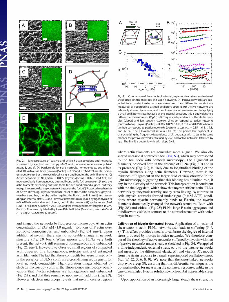
and imaged the networks by fluorescence microscopy. At an actinconcentration of 23.8 �M (1.0 mg/mL), solutions of F actin wereisotropic, homogeneous, and unbundled (Fig. 2A Inset). Uponaddition of myosin, there was no noticeable change in networkstructure (Fig. 2B Inset). When myosin and FLNa were bothpresent, the network still remained homogeneous and unbundled(Fig. 2C Inset). However, we observed small regions of compactedactin dispersed in a homogeneous, isotropic matrix of entangledactin filaments. The fact that these contractile foci were formed onlyin the presence of FLNa confirms a cross-linking requirement forlocal network contractility. High-resolution images obtained byelectron microscopy confirm the fluorescence microscopy obser-vations that F-actin solutions are homogeneous and unbundled(Fig. 2A), and that they remain so upon myosin addition (Fig. 2B).However, electron microscopy reveals that myosin creates regions
where actin filaments are somewhat more aligned. We also ob-served occasional contractile foci (Fig. S3), which may correspondto the foci seen with confocal microscopy. The alignment offilaments, observed both in the absence of FLNa (Fig. 2B) and inits presence (Fig. 2C), is likely due to longitudinal binding of themyosin filaments along actin filaments. However, there is noevidence of alignment in the larger field of view observed in thelight microscopy, suggesting that this binding has a limited role, ifany, in the elasticity of the network. This interpretation is consistentwith the rheology data, which show that myosin stiffens actin–FLNanetworks by enzymatic activity, not by cross-linking. By contrast, inactin–myosin networks formed under rigor (0 mM ATP) condi-tions, where myosin permanently binds to F-actin, the myosinfilaments dramatically changed the network structure. Both with(Fig. 2E) and without (Fig. 2F) FLNa, large F-actin aggregates andbundles were visible, in contrast to the network structure with activemyosin motors.
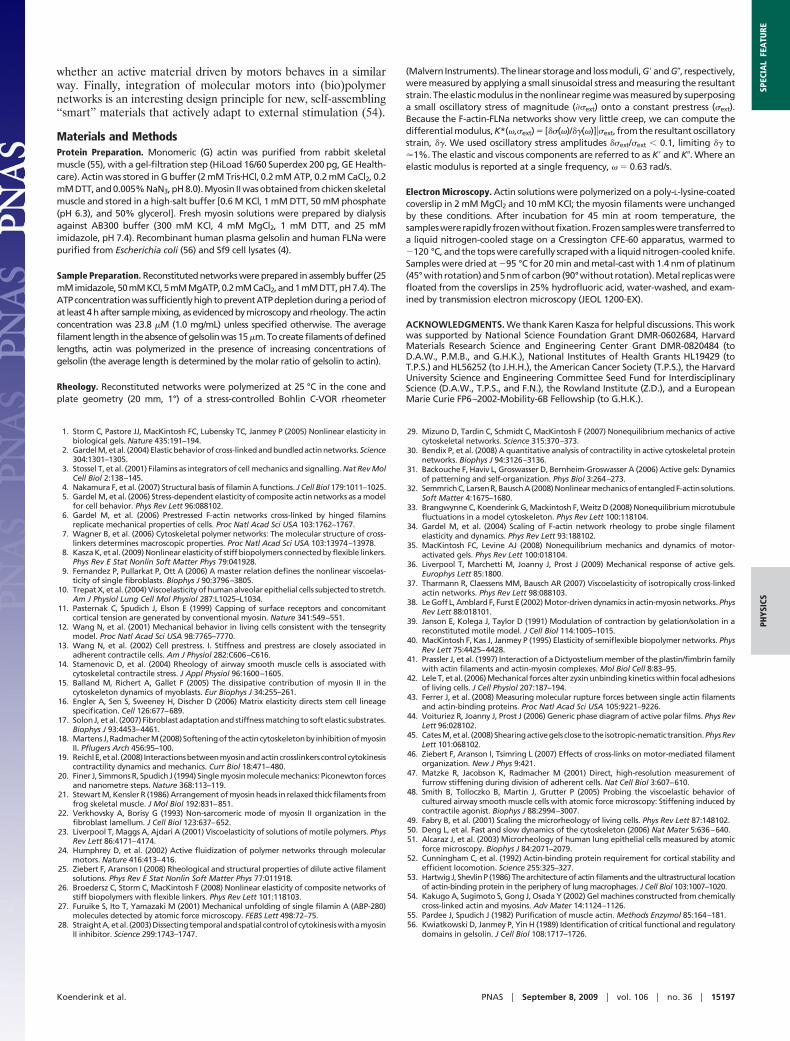
Calibration of Myosin-Generated Stress. Application of an externalshear stress to actin–FLNa networks also leads to stiffening (5, 6,8). This effect provides a means to calibrate the degree of internalstress produced by motors in active networks. We therefore com-pared the rheology of active networks stiffened by myosin with thatof passive networks under shear, as sketched in Fig. 3A. We applieda time-independent, external stress, �ext, to the passive networksand measured the differential elastic, K�, and viscous, K�, modulifrom the strain response to a small, superimposed oscillatory stress,��ext(�) (2, 5, 6, 8, 9). We note that the cross-linked networksdisplay no creep (8), ensuring that this differential technique is theoptimal method for measuring the nonlinear response, unlike in thecase of entangled F-actin solutions, which exhibit appreciable creep(32).
Upon application of an increasingly large, steady shear stress, the
A B
C D
E F
Fig. 2. Microstructure of passive and active F-actin solutions and networksvisualized by electron microscopy (A–C) and fluorescence microscopy (A–CInsets, E, and F). (A) Passive solutions are isotropic, homogeneous, and unbun-dled. (B) Active solutions ([myosin]/[actin] � 0.02 and 5 mM ATP) are still homo-geneous (Inset), but the myosin locally aligns and bundles the actin filaments. (C)Active networks ([FLNa]/[actin] � 0.005, [myosin]/[actin] � 0.02, 5 mM ATP) aremacroscopically homogeneous, but small contractile foci are present (Inset); theactin filaments extending out from these foci are bundled and aligned, but theymerge into a more isotropic network between the foci. (D) Proposed mechanismof active stiffening: myosin filaments (blue) contract actin filaments (gray) to-ward one another, thereby pulling against the FLNa cross-links (red) and gener-ating an internal stress. (E and F) Passive networks cross-linked by rigor myosin (0mM ATP) show bundles and clumps, both in the presence (E) and absence (F) ofFLNa. For all panels, [actin] � 23.8 �M, and the average filament length is 15 �m.F actin is fluorescently labeled by Alexa488-phalloidin. (Scale bars: Insets A–C andF, 10 �m; A–C, 200 nm; E, 20 �m).
σext
A δσext (ω) B
C
σ(ω)
10-1 100 101
10-1
100
tan δ
ω (rad/s)10-2 10-1 100 101 102
10-2
10-1
100
x
σint
or σext
[Pa]
100
101
102
103
K
' (P
a)
Fig. 3. Comparison of the effects of internal, myosin-driven stress and externalshear stress on the rheology of F-actin networks. (A) Passive networks are sub-jected to a constant external shear stress, and their differential moduli aremeasured by superposing a small oscillatory stress (Left). Active networks areinternally stressed by motors, and their linear moduli are measured by applyinga small oscillatory stress; because of the internal prestress, this is equivalent to adifferential measurement (Right). (B) Frequency dependence of the elastic mod-ulus (Upper) and loss tangent (Lower). Lines correspond to active networks(bottom to top: [myosin]/[actin] � 0.005, 0.009, 0.010, 0.039, and 0.050), whereassymbols correspond to passive networks (bottom to top: �ext � 0.35, 1.6, 3.1, 7.8,and 12 Pa). The [FLNa]/[actin] ratio is 0.01. (C) The power law exponent, x,characterizing the frequency dependence of G�, decreases with stress in the samemanner for passive networks (stressed by �ext) and active networks (stressed by�int). The line is a power law fit with slope 0.43.
15194 � www.pnas.org�cgi�doi�10.1073�pnas.0903974106 Koenderink et al.

passively deformed F-actin–FLNa networks increasingly stiffened(symbols in Fig. 3B Upper) and became more solidlike. Thedifferential stiffness became more weakly dependent on frequency,and the loss tangent, tan � � K�/K�, decreased (Fig. 3B Lower).Similarly, when the networks were made active by embeddingmyosin thick filaments, the stiffness increased with increasing motorconcentration (lines in Fig. 3B Upper). Strikingly, for every con-centration of myosin thick filaments, we were able to apply anexternal shear stress that yielded an identical elastic modulus for allfrequencies, as shown by the comparison of the lines (activenetworks) and symbols (passively deformed networks). The viscousresponses reveal subtly different microscopic dynamics. At thehighest values of stress, a noticeable discrepancy appeared atfrequencies below 0.2 rad/s: the active networks exhibited a smalldecrease in the elastic modulus with a concomitant increase of theloss tangent, indicative of increased dissipation (Fig. 3B Lower). Incontrast, the loss tangent for passive networks under high levels ofexternal stress was nearly frequency-independent. This discrepancysuggests that in the active networks, stress relaxation occurs on timescales longer than about 5 s, possibly resulting from myosin motorrelease. This time scale is consistent with the typical release rate ofmyosin filaments observed in microrheology of F-actin–myosinsolutions (33).
An external shear stress is anisotropic, and therefore is borneprimarily by a small number of highly oriented filaments (2, 34). Bycontrast, internal myosin-generated stress is expected to be isotro-pic, and therefore can affect a much larger number of filaments (29,35, 36). Nevertheless, the close correspondence of the elasticity ofactive and passive networks under stress enabled us to calibrate theeffective internal stress, �int, applied by the myosin motors. Themaximum level of internal stress achieved corresponded to anexternally applied shear stress of �ext � 14 Pa (Fig. S4). Weapproximated the maximum force per myosin filament, F, byassuming an isotropic internal stress and by using an average areaper filament, �2 � 0.1 �m2, where � is the network mesh size. Weobtained F � 1 pN, consistent with only a few myosin heads activeat any given time in each myosin filament, each having a stall forceof 4 pN (20). This finding agrees with complementary findings thatwe reported recently on actin–myosin networks (30, 33).
The increased stiffness of the network upon activation of internalstress can also be characterized by the exponent, x, of the power lawdependence of stiffness on frequency, K�(�) � �x. For a purely
elastic solid, x is zero; for a purely viscous liquid, x is one. Theactin–FLNa networks are viscoelastic and have an intermediateexponent of about 0.4 in the unstressed state. Upon application ofexternal shear stress to a passive network, the exponent decreasedto a value close to zero as the level of stress was raised (Fig. 3C, grayopen triangles). Upon incorporation of motors and ATP, theexponent decreased in the same manner as the motor concentrationwas raised (Fig. 3C, solid blue squares). This consistency againemphasizes the close analogy of motor-driven stress to the effect ofan external shear stress.
Nonlinear Response. The effect of the motors is to induce internaltension that brings the network into a nonlinear state. Uponapplication of an additional steady shear stress, the network exhibitsan apparently linear response over an extended range of appliedstress. However, with sufficiently large applied stress, larger than acritical stress (�crit), the network again begins to stiffen. This isillustrated for an active network with [myosin]/[actin] � 0.02, whosestiffness was 100 Pa in the linear regime, nearly two orders ofmagnitude above that of an unstressed network (blue solid squaresin Fig. 4A). Upon lowering the [myosin]/[actin] ratio to 0.005 (greensolid squares), or further to 0.001 (red solid squares), both the initialelastic modulus and �crit decreased (see Fig. 4A). By contrast, thepassive network had a linear modulus of only 1 Pa but started tostiffen at a much smaller �crit (gray open triangles). Strikingly, atlarge shear stress values, the nonlinear response of the activenetworks asymptotically approached the same stiffness as that ofthe passive network. Moreover, passive and active networks allbroke at approximately the same maximum shear stress, �max(indicated by an arrow in Fig. 4A). This rupture stress increasedlinearly with FLNa concentration (Fig. 4B), suggesting that forcedunbinding of FLNa is the dominant failure mode (8, 26). This typeof behavior is typical of weakly binding cross-linkers (37) andcontrasts with strongly cross-linked networks, where the dominantfailure mode is actin filament rupture (2, 34). If we assume that allcross-linkers equally share the load, we estimate a typical failureforce per FLNa cross-link of about 2 pN. The correspondence of thestiffening response of active and passive networks at large shearstresses suggests that the effect of preexisting internal stress gen-erated by motors is equivalent to the effect of an external shearstress. Consistent with this interpretation, the stiffening curves inFig. 4A can be collapsed onto a single master curve by normalizing
DB
CA
10-1 100 101 102
100
101
102
K'/G
'
σext
/σcrit
10-1 100 101
100
101
102
σcrit
K' [
Pa]
σext
[Pa]
σmax
0
10-3 10-210-1
100
101
102
σ ma
x [P
a]
[FLNa]/[actin]
10-1 100 10110-2
100
102
104
σ crit
[Pa]
G' [Pa]
0.1 1 1010-3
10-2
10-1
100
γ crit [-
]
σint
[Pa]
Fig. 4. Nonlinear response of passive and active F-actinnetworks to an external shear stress. (A) Stress-stiffeningcurves for active networks [with myosin to actin ratios of0.02 (blue), 0.005 (green), and 0.001 (red)] and a passivenetwork (gray open triangles) at a fixed [FLNa]/[actin]ratio of 0.005. Arrows indicate critical stress (�crit) andrupture stress (�max) for the passive network. (B) The rup-ture stress increases linearly with FLNa concentration, in-dependently of the myosin content. (C) Normalized stress-stiffening curves collapse onto a single master curve witha power law stiffening regime, K� � G��1 (black line). The[FLNa]/[actin] ratio is 0.005. The [myosin]/[actin] ratios are0 (gray), 0.0005 (magenta), 0.001 (red), 0.0025 (orange),0.005 (olive), 0.008 (green), 0.0133 (navy), 0.0167 (yellow),0.02 (blue), and 0.0286 (turquoise). (Inset) Relation be-tween the scaling factors, according to G� � �crit
1.3 (grayline). (D) The critical strain decreases as the level of motor-driven stress increases, according to an approximatepower law, �crit � � �1 (solid line).
Koenderink et al. PNAS � September 8, 2009 � vol. 106 � no. 36 � 15195
PHYS
ICS
SPEC
IAL
FEA
TURE

K� by its zero-stress value, G�, and �ext by �crit, as shown in Fig. 4C.Stress-stiffening in the nonlinear regime is well approximated by apower law with a slope of 1, shown by the solid black line in Fig. 4C.
Although the critical stress increased upon addition of activemotors, the corresponding critical strain, �crit, decreased, as shownin Fig. 4D. Passive actin–FLNa networks stiffen strongly above �crit� 30%. This value of critical strain reflects the difference incompliance between the FLNa cross-linkers and the actin filaments(8, 26). At small strain, the network compliance is dominated by thesoft FLNa cross-linkers. Upon application of a shear stress, thethermal fluctuations of the cross-linkers are pulled out. Whenthe cross-linkers reach their full extension, the network suddenlybecomes incompliant because the actin filaments have a muchlarger persistence length of 15 �m. The network therefore stiffenswhen the strain exceeds a critical value that is proportional to thecontour length of the cross-linker (26). Upon addition of motors,the critical strain decreases substantially to a final value of only 0.6%at the highest [myosin]/[actin] ratio of 0.02. Data for different FLNaand myosin concentrations may be combined by plotting the criticalstrain as a function of internal tension, which shows that �crit � �int
�1
(line in Fig. 4D). This is a manifestation of the preexisting tensionin the network generated by the motors: the myosin motors activelypull against the FLNa cross-links, thereby pulling out thermalfluctuations of the FLNa cross-linkers and the actin filaments. Avery small, externally applied shear strain is then sufficient to bringthe actively prestressed network out of its linear regime.
DiscussionOur data identify two key design principles of active biopolymernetworks whose mechanical properties are controlled by theaddition of molecular motors. The first principle is that themotors must be sufficiently processive. Nonprocessive myosinsubfragments indeed do not cause active stiffening (38). Thesecond principle is that F-actin cross-linkers must be present toprovide sites for mechanical anchorage, thus accommodatingbuildup of internal tension (30, 31, 39). In the absence ofcross-linkers, myosin filaments weaken F-actin solutions byactive filament sliding (23–25). Moreover, cross-linking serves tomake the actin network sensitive to an applied tension.
In vitro studies have shown that strain-stiffening is a genericfeature of cross-linked F-actin networks. However, the quantitativeform of the nonlinear response depends on the nature of thecross-linking protein. For small, rigid cross-links, strain-stiffeningresults from the nonlinear entropic stretching modulus of the actinfilaments (1, 2, 34, 40). The large, compliant cross-linker FLNasignificantly enhances the strain-stiffening response (5, 6, 8): FLNa–F-actin networks are soft at small strain but stiffen strongly and havea large rupture stress (26). We found that addition of motors to anFLNa cross-linked network directly mimics the effect of an externalshear stress, dramatically stiffening the network. By contrast, wefound that scruin, an incompliant cross-linker, does not promoteactive stiffening of F-actin networks upon addition of myosin (cf.Fig. S5). We ascribe this to the weaker stress-stiffening responseevoked by this cross-linker. Intriguingly, stiffening of actin networkscross-linked by rigid biotin–streptavidin linkages is only reported atATP concentrations in the micromolar range (29). At such lowATP concentrations, myosin is 1,000-fold more processive (20), andthe system is also close to rigor. To resolve the intriguing differencebetween the response of flexibly and rigidly cross-linked networksto motor activity, it will be interesting to systematically change thedegree of motor processivity by adjusting ATP and salt levels.
Our model system provides a platform to analyze the role ofactive motors in the rheology of actin networks in a quantitativemanner. Because the motors pull on actin filaments that arecross-linked by FLNa, there is likely cross-talk between the activityof myosin and FLNa. Myosin-driven tension may, for instance,release FLNa cross-linking. Evidence exists for tension-dependentcross-linker binding kinetics both in vitro (41) and in vivo (19, 42).
Recently, FLNa was shown to unbind at forces in a range of 5–100pN (43). The viscous response of our reconstituted networks showssignatures of FLNa and/or myosin unbinding. It will be interestingto measure on/off rates of both proteins directly (4). Our datasuggest that the mechanical and kinetic properties of the cross-linkers should be included in theoretical models of active polymernetworks (23, 35, 36, 44–46).
Our work also may provide mechanistic insights into cellularmechanics. It supports the observation that myosin-generatedprestress contributes to cell stiffness (9, 12–14, 18, 47) and providesa physical explanation for this phenomenon, which was hithertolacking. The rheology of our reconstituted system exhibits remark-able similarity to the mechanical properties reported for cells. Theactive stiffening effect increases with motor concentration andvanishes when motor activity is inhibited by blebbistatin. Similareffects are observed in adherent cells during interphase (11–18) andmitosis (19, 47). The active stiffening effect also increases when thecross-linker concentration is increased. This is reminiscent of recentobservations in adherent Dictyostelium amoeboid cells, where cross-linkers were shown to enhance cortical tension as well as cellstiffness (19). The elastic modulus increases with frequency, ac-cording to a weak power law with an exponent that decreases whenmotors actively tense the network. This was also observed forsmooth muscle cells (14, 48). Myosin generates the same stiffeningof F-actin–FLNa networks as an external shear stress. This is instriking agreement with experiments on single fibroblasts, whereactive cell contraction and external cell stretching also yieldedidentical stiffening (9). The stress-stiffening curves of our active,reconstituted networks can be rescaled onto a single master curve,when the tangent stiffness is normalized by its zero-force value andthe external stress by the critical stress. This master curve matchesthe master curve that was found for fibroblasts, with a lineardependence of scaled modulus on scaled stress in the nonlinearregime. Both for the cells and for our reconstituted system, themyosin-dependent scaling factors are related by a power law, G� ��crit
y, with exponent y � 1.3 (Fig. 4C Inset), not far from anexponent of 1 expected based on the stress-stiffening exponent. Ourdata suggest that adherent cells inherently operate in a nonlinear,stress-stiffened regime driven by actin–myosin contractility. Theprecise control over network stiffness afforded by variation ofmyosin and FLNa concentrations suggests that cells may spatiallyand temporally control their stiffness at multiple levels.
Despite the qualitative resemblance to the myosin-dependentrheology of cells, the reconstituted networks still have elasticmoduli about an order of magnitude lower than those typicallyreported for adherent cells (0.5–10 kPa; refs. 12, 13, and 49–51).Although we used molar ratios of FLNa and myosin relative to actinthat are similar to those in cells (22, 52, 53), the filament length inmost of our experiments was longer (15 �m compared with �1–2�m in cells; ref. 53) and the absolute concentration of actin lower(1 mg/mL, compared with �10 mg/mL in cells; ref. 53). Indeed, inthe reconstituted system, motor-driven stiffening is strongly depen-dent on filament length and actin concentration. Moreover, in cells,FLNa acts in concert with numerous other cross-linking proteins.An interesting question is whether FLNa has a specific role in themyosin-dependent rheology of cells (52). Another key question is:what is the locus of tension generation in nonmuscle cells? MyosinII is present in the cell cortex but also in stress fibers (22). Our worksuggests that it may not be necessary for actin–myosin to beassembled into highly ordered stress fibers to generate stiffening.
The active protein networks studied here belong to a new classof active soft materials inspired by the cellular cytoskeleton. Suchmaterials evoke fundamental questions: they are driven far out ofequilibrium, so equilibrium concepts, such as causality in oscillatoryshear rheology, may break down. Even concepts that can be appliedto other nonequilibrium systems, such as glasses, may not hold.Even though glasses are out of equilibrium, over certain time scalesthey behave as if they are in quasiequilibrium. It is an open question
15196 � www.pnas.org�cgi�doi�10.1073�pnas.0903974106 Koenderink et al.
whether an active material driven by motors behaves in a similarway. Finally, integration of molecular motors into (bio)polymernetworks is an interesting design principle for new, self-assembling‘‘smart’’ materials that actively adapt to external stimulation (54).
Materials and MethodsProtein Preparation. Monomeric (G) actin was purified from rabbit skeletalmuscle (55), with a gel-filtration step (HiLoad 16/60 Superdex 200 pg, GE Health-care). Actin was stored in G buffer (2 mM Tris�HCl, 0.2 mM ATP, 0.2 mM CaCl2, 0.2mM DTT, and 0.005% NaN3, pH 8.0). Myosin II was obtained from chicken skeletalmuscle and stored in a high-salt buffer [0.6 M KCl, 1 mM DTT, 50 mM phosphate(pH 6.3), and 50% glycerol]. Fresh myosin solutions were prepared by dialysisagainst AB300 buffer (300 mM KCl, 4 mM MgCl2, 1 mM DTT, and 25 mMimidazole, pH 7.4). Recombinant human plasma gelsolin and human FLNa werepurified from Escherichia coli (56) and Sf9 cell lysates (4).
Sample Preparation. Reconstituted networks were prepared in assembly buffer (25mMimidazole,50mMKCl,5mMMgATP,0.2mMCaCl2,and1mMDTT,pH7.4).TheATPconcentrationwas sufficientlyhightopreventATPdepletionduringaperiodofat least 4 h after sample mixing, as evidenced by microscopy and rheology. The actinconcentration was 23.8 �M (1.0 mg/mL) unless specified otherwise. The averagefilament length in theabsenceofgelsolinwas15 �m.Tocreatefilamentsofdefinedlengths, actin was polymerized in the presence of increasing concentrations ofgelsolin (the average length is determined by the molar ratio of gelsolin to actin).
Rheology. Reconstituted networks were polymerized at 25 °C in the cone andplate geometry (20 mm, 1°) of a stress-controlled Bohlin C-VOR rheometer
(Malvern Instruments). The linear storageand lossmoduli,G�andG�, respectively,were measured by applying a small sinusoidal stress and measuring the resultantstrain. The elastic modulus in the nonlinear regime was measured by superposinga small oscillatory stress of magnitude (��ext) onto a constant prestress (�ext).Because the F-actin-FLNa networks show very little creep, we can compute thedifferential modulus, K*(�,�ext) � [��(�)/��(�)]��ext, from the resultant oscillatorystrain, ��. We used oscillatory stress amplitudes ��ext/�ext � 0.1, limiting �� to�1%. The elastic and viscous components are referred to as K� and K�. Where anelastic modulus is reported at a single frequency, � � 0.63 rad/s.
Electron Microscopy. Actin solutions were polymerized on a poly-L-lysine-coatedcoverslip in 2 mM MgCl2 and 10 mM KCl; the myosin filaments were unchangedby these conditions. After incubation for 45 min at room temperature, thesampleswererapidlyfrozenwithoutfixation.Frozensamplesweretransferredtoa liquid nitrogen-cooled stage on a Cressington CFE-60 apparatus, warmed to�120 °C, and the tops were carefully scraped with a liquid nitrogen-cooled knife.Samples were dried at �95 °C for 20 min and metal-cast with 1.4 nm of platinum(45°withrotation)and5nmofcarbon(90°withoutrotation).Metal replicaswerefloated from the coverslips in 25% hydrofluoric acid, water-washed, and exam-ined by transmission electron microscopy (JEOL 1200-EX).
ACKNOWLEDGMENTS. We thank Karen Kasza for helpful discussions. This workwas supported by National Science Foundation Grant DMR-0602684, HarvardMaterials Research Science and Engineering Center Grant DMR-0820484 (toD.A.W., P.M.B., and G.H.K.), National Institutes of Health Grants HL19429 (toT.P.S.) and HL56252 (to J.H.H.), the American Cancer Society (T.P.S.), the HarvardUniversity Science and Engineering Committee Seed Fund for InterdisciplinaryScience (D.A.W., T.P.S., and F.N.), the Rowland Institute (Z.D.), and a EuropeanMarie Curie FP6–2002-Mobility-6B Fellowship (to G.H.K.).
1. Storm C, Pastore JJ, MacKintosh FC, Lubensky TC, Janmey P (2005) Nonlinear elasticity inbiological gels. Nature 435:191–194.
2. Gardel M, et al. (2004) Elastic behavior of cross-linked and bundled actin networks. Science304:1301–1305.
3. Stossel T, et al. (2001) Filamins as integrators of cell mechanics and signalling. Nat Rev MolCell Biol 2:138–145.
4. Nakamura F, et al. (2007) Structural basis of filamin A functions. J Cell Biol 179:1011–1025.5. Gardel M, et al. (2006) Stress-dependent elasticity of composite actin networks as a model
for cell behavior. Phys Rev Lett 96:088102.6. Gardel M, et al. (2006) Prestressed F-actin networks cross-linked by hinged filamins
replicate mechanical properties of cells. Proc Natl Acad Sci USA 103:1762–1767.7. Wagner B, et al. (2006) Cytoskeletal polymer networks: The molecular structure of cross-
linkers determines macroscopic properties. Proc Natl Acad Sci USA 103:13974–13978.8. Kasza K, et al. (2009) Nonlinear elasticity of stiff biopolymers connected by flexible linkers.
Phys Rev E Stat Nonlin Soft Matter Phys 79:041928.9. Fernandez P, Pullarkat P, Ott A (2006) A master relation defines the nonlinear viscoelas-
ticity of single fibroblasts. Biophys J 90:3796–3805.10. Trepat X, et al. (2004) Viscoelasticity of human alveolar epithelial cells subjected to stretch.
Am J Physiol Lung Cell Mol Physiol 287:L1025–L1034.11. Pasternak C, Spudich J, Elson E (1999) Capping of surface receptors and concomitant
cortical tension are generated by conventional myosin. Nature 341:549–551.12. Wang N, et al. (2001) Mechanical behavior in living cells consistent with the tensegrity
model. Proc Natl Acad Sci USA 98:7765–7770.13. Wang N, et al. (2002) Cell prestress. I. Stiffness and prestress are closely associated in
adherent contractile cells. Am J Physiol 282:C606–C616.14. Stamenovic D, et al. (2004) Rheology of airway smooth muscle cells is associated with
cytoskeletal contractile stress. J Appl Physiol 96:1600–1605.15. Balland M, Richert A, Gallet F (2005) The dissipative contribution of myosin II in the
cytoskeleton dynamics of myoblasts. Eur Biophys J 34:255–261.16. Engler A, Sen S, Sweeney H, Discher D (2006) Matrix elasticity directs stem cell lineage
specification. Cell 126:677–689.17. Solon J, et al. (2007) Fibroblast adaptation and stiffness matching to soft elastic substrates.
Biophys J 93:4453–4461.18. MartensJ,RadmacherM(2008)Softeningoftheactincytoskeletonby inhibitionofmyosin
II. Pflugers Arch 456:95–100.19. ReichlE,etal. (2008) Interactionsbetweenmyosinandactincrosslinkerscontrolcytokinesis
contractility dynamics and mechanics. Curr Biol 18:471–480.20. Finer J, Simmons R, Spudich J (1994) Single myosin molecule mechanics: Piconewton forces
and nanometre steps. Nature 368:113–119.21. Stewart M, Kensler R (1986) Arrangement of myosin heads in relaxed thick filaments from
frog skeletal muscle. J Mol Biol 192:831–851.22. Verkhovsky A, Borisy G (1993) Non-sarcomeric mode of myosin II organization in the
fibroblast lamellum. J Cell Biol 123:637–652.23. Liverpool T, Maggs A, Ajdari A (2001) Viscoelasticity of solutions of motile polymers. Phys
Rev Lett 86:4171–4174.24. Humphrey D, et al. (2002) Active fluidization of polymer networks through molecular
motors. Nature 416:413–416.25. Ziebert F, Aranson I (2008) Rheological and structural properties of dilute active filament
solutions. Phys Rev E Stat Nonlin Soft Matter Phys 77:011918.26. Broedersz C, Storm C, MacKintosh F (2008) Nonlinear elasticity of composite networks of
stiff biopolymers with flexible linkers. Phys Rev Lett 101:118103.27. Furuike S, Ito T, Yamazaki M (2001) Mechanical unfolding of single filamin A (ABP-280)
molecules detected by atomic force microscopy. FEBS Lett 498:72–75.28. StraightA,etal. (2003)Dissectingtemporalandspatial controlofcytokinesiswithamyosin
II inhibitor. Science 299:1743–1747.
29. Mizuno D, Tardin C, Schmidt C, MacKintosh F (2007) Nonequilibrium mechanics of activecytoskeletal networks. Science 315:370–373.
30. Bendix P, et al. (2008) A quantitative analysis of contractility in active cytoskeletal proteinnetworks. Biophys J 94:3126–3136.
31. Backouche F, Haviv L, Groswasser D, Bernheim-Groswasser A (2006) Active gels: Dynamicsof patterning and self-organization. Phys Biol 3:264–273.
32. SemmrichC,LarsenR,BauschA(2008)NonlinearmechanicsofentangledF-actin solutions.Soft Matter 4:1675–1680.
33. Brangwynne C, Koenderink G, Mackintosh F, Weitz D (2008) Nonequilibrium microtubulefluctuations in a model cytoskeleton. Phys Rev Lett 100:118104.
34. Gardel M, et al. (2004) Scaling of F-actin network rheology to probe single filamentelasticity and dynamics. Phys Rev Lett 93:188102.
35. MacKintosh FC, Levine AJ (2008) Nonequilibrium mechanics and dynamics of motor-activated gels. Phys Rev Lett 100:018104.
36. Liverpool T, Marchetti M, Joanny J, Prost J (2009) Mechanical response of active gels.Europhys Lett 85:1800.
37. Tharmann R, Claessens MM, Bausch AR (2007) Viscoelasticity of isotropically cross-linkedactin networks. Phys Rev Lett 98:088103.
38. Le Goff L, Amblard F, Furst E (2002) Motor-driven dynamics in actin-myosin networks. PhysRev Lett 88:018101.
39. Janson E, Kolega J, Taylor D (1991) Modulation of contraction by gelation/solation in areconstituted motile model. J Cell Biol 114:1005–1015.
40. MacKintosh F, Kas J, Janmey P (1995) Elasticity of semiflexible biopolymer networks. PhysRev Lett 75:4425–4428.
41. Prassler J, et al. (1997) Interaction of a Dictyostelium member of the plastin/fimbrin familywith actin filaments and actin-myosin complexes. Mol Biol Cell 8:83–95.
42. Lele T, et al. (2006) Mechanical forces alter zyxin unbinding kinetics within focal adhesionsof living cells. J Cell Physiol 207:187–194.
43. Ferrer J, et al. (2008) Measuring molecular rupture forces between single actin filamentsand actin-binding proteins. Proc Natl Acad Sci USA 105:9221–9226.
44. Voituriez R, Joanny J, Prost J (2006) Generic phase diagram of active polar films. Phys RevLett 96:028102.
45. CatesM,etal. (2008)Shearingactivegels closetothe isotropic-nematic transition.PhysRevLett 101:068102.
46. Ziebert F, Aranson I, Tsimring L (2007) Effects of cross-links on motor-mediated filamentorganization. New J Phys 9:421.
47. Matzke R, Jacobson K, Radmacher M (2001) Direct, high-resolution measurement offurrow stiffening during division of adherent cells. Nat Cell Biol 3:607–610.
48. Smith B, Tolloczko B, Martin J, Grutter P (2005) Probing the viscoelastic behavior ofcultured airway smooth muscle cells with atomic force microscopy: Stiffening induced bycontractile agonist. Biophys J 88:2994–3007.
49. Fabry B, et al. (2001) Scaling the microrheology of living cells. Phys Rev Lett 87:148102.50. Deng L, et al. Fast and slow dynamics of the cytoskeleton (2006) Nat Mater 5:636–640.51. Alcaraz J, et al. (2003) Microrheology of human lung epithelial cells measured by atomic
force microscopy. Biophys J 84:2071–2079.52. Cunningham C, et al. (1992) Actin-binding protein requirement for cortical stability and
efficient locomotion. Science 255:325–327.53. Hartwig J, Shevlin P (1986) The architecture of actin filaments and the ultrastructural location
of actin-binding protein in the periphery of lung macrophages. J Cell Biol 103:1007–1020.54. Kakugo A, Sugimoto S, Gong J, Osada Y (2002) Gel machines constructed from chemically
cross-linked actin and myosins. Adv Mater 14:1124–1126.55. Pardee J, Spudich J (1982) Purification of muscle actin. Methods Enzymol 85:164–181.56. Kwiatkowski D, Janmey P, Yin H (1989) Identification of critical functional and regulatory
domains in gelsolin. J Cell Biol 108:1717–1726.
Koenderink et al. PNAS � September 8, 2009 � vol. 106 � no. 36 � 15197
PHYS
ICS
SPEC
IAL
FEA
TURE