analysis of anti-rna polymerase iii antibody-positive
TRANSCRIPT
The Journal of Rheumatology 2020;47:11; doi:10.3899/jrheum.1909451668
The Journal of Rheumatology 2020;47:1668–77doi:10.3899/jrheum.190945First Release September 15 2020
Analysis of Anti-RNA Polymerase III Antibody-positive Systemic Sclerosis and Altered GPATCH2L and CTNND2 Expression in Scleroderma Renal Crisis Edward P. Stern1, Sandra G. Guerra2, Harry Chinque2, Vanessa Acquaah2, David González-Serna3, Markella Ponticos2, Javier Martin3, Voon H. Ong2, Korsa Khan2, Svetlana I. Nihtyanova2, Mark Harber4, Aine Burns4, Maureen D. Mayes5, Shervin Assassi5, Carmen Fonseca2, and Christopher P. Denton2
ABSTRACT. Objective. Scleroderma renal crisis (SRC) is a life-threatening complication of systemic sclerosis (SSc) strongly associated with anti-RNA polymerase III antibody (ARA) autoantibodies. We investigated genetic susceptibility and altered protein expression in renal biopsy specimens in ARA-positive patients with SRC.
Methods. ARA-positive patients (n = 99) with at least 5 years’ follow-up (49% with a history of SRC) were selected from a well characterized SSc cohort (n = 2254). Cases were genotyped using the Illumina Human Omni-express chip. Based on initial regression analysis, 9 single-nucleotide polymorphisms (SNP) were chosen for validation in a separate cohort of 256 ARA-positive patients (40 with SRC). Immunostaining of tissue sections from SRC or control kidney was used to quantify expression of candidate proteins based upon genetic analysis of the discovery cohort.
Results. Analysis of 641,489 SNP suggested association of POU2F1 (rs2093658; P = 1.98 × 10⁻⁵), CTNND2 (rs1859082; P = 5.58 × 10⁻⁵), HECW2 (rs16849716; P = 1.2 × 10⁻⁴), and GPATCH2L (rs935332; P = 4.92 × 10⁻⁵) with SRC. Further, the validation cohort showed an association between rs935332 within the GPATCH2L region, with SRC (P = 0.025). Immunostaining of renal biopsy sections showed increased tubular expression of GPATCH2L (P = 0.026) and glomerular expression of CTNND2 (P = 0.026) in SRC samples (n = 8) compared with normal human kidney controls (n = 8), despite absence of any genetic replication for the associated SNP.
Conclusion. Increased expression of 2 candidate proteins, GPATCH2L and CTNND2, in SRC compared with control kidney suggests a potential role in pathogenesis of SRC. For GPATCH2L, this may reflect genetic susceptibility in ARA-positive patients with SSc based upon 2 independent cohorts.
Key Indexing Terms: autoantibody, catenin, genetics, hypertension, renal, scleroderma
This study was supported in part by Medical Research Council grant MR/K015230/1 with additional support from a research grant from Scleroderma and Raynaud’s UK ( formerly The Scleroderma Society). 1E.P. Stern, MRCP, University College London (UCL) Centre for Rheumatology and Connective Tissue Diseases, and UCL Centre for Nephrology, London; 2S.G. Guerra, PhD, H. Chinque, BSc, V. Acquaah, BSc, M. Ponticos, V.H. Ong, PhD, FRCP, K. Khan, BSc, S.I. Nihtyanova, MD, C. Fonseca, MD, C.P. Denton, PhD, FRCP, Professor of Experimental Rheumatology, UCL Centre for Rheumatology and Connective Tissue Diseases, London, UK; 3D. González-Serna, PhD, J. Martin3, MD, Instituto de Parasitologia y Biomedicina Lopez-Neyra, Consejo Superior de
Investigaciones Científicas (CSIC), Granada, Spain; 4M. Harber, FRCP, A. Burns, MD, UCL Centre for Nephrology, London, UK; 5M.D Mayes, MD, S. Assassi, MD, University of Texas Houston – McGovern Medical School, Houston, Texas, USA.E.P. Stern and S.G. Guerra are joint first authors. Address correspondence to Prof. C.P. Denton, UCL Centre for Rheumatology and Connective Tissue Diseases, UCL Medical School Building, Royal Free Campus, Rowland Hill Street, London NW3 2PF, UK. Email: [email protected]. Accepted for publication February 28, 2020.
Systemic sclerosis (SSc) is a multisystem rheumatic disorder with a high case-specific mortality owing to internal organ complica-tions of the disease. The pattern and frequency of internal organ manifestations reflect the different antinuclear antibody reactiv-ities that are a hallmark of SSc1. These include anticentromere (ACA), antitopoisomerase 1 (ATA), and anti-RNA polymerase III (ARA) antibodies. Frequency of major complications of SSc differs between these clinical and immunological subgroups.
For example, ATA associates with interstitial lung disease2 and ACA with pulmonary arterial hypertension (PAH) in certain subgroups3. A strong association has been observed between the presence of ARA and the occurrence of scleroderma renal crisis (SRC)4,5. SRC is a life-threatening complication of SSc characterized by accelerated-phase hypertension (HTN) and acute kidney injury6. SRC was almost universally fatal until the late 1970s,
Personal non-commercial use only. The Journal of Rheumatology Copyright © 2020. All rights reserved.
www.jrheum.orgDownloaded on February 21, 2022 from
Stern, et al: Scleroderma renal crisis 1669
when its management was revolutionized by the introduction of angiotensin-converting enzyme inhibitors. Nevertheless, in the modern era 40–50% of SRC cases result in early death or renal replacement therapy and the 5-year survival is as low as 50% in some series7,8. The different occurrence of SRC in specific subgroups of SSc, the observation that even within the highest risk groups only a minority of SSc cases develop SRC, and the evidence that it generally occurs only in early-stage disease are all consistent with a genetic predisposition to SRC that may be independent of the inherited risk of SSc itself. Data regarding the genetic contribu-tion to risk of SRC are limited; a single study reports the pres-ence of MHC class I haplotypes, such as HLA-DRB1*04:07 and HLA-DRB1*13:04, as independent risk factors for developing SRC9. Our study has used a novel approach by looking at cases of SRC in the antinuclear antibody (ANA) group at highest risk, comparing cases that develop SRC with those that appear to be protected from development of SRC during long-term follow-up. This strategy provides a platform of clinical and serological homogeneity in which we hypothesize that genetic susceptibility factors may be most relevant.
MATERIALS AND METHODSPatient demographics. For this study patients with SRC (n = 99) were selected from a tertiary UK referral center for SSc. All patients met the 1980 American College of Rheumatology (ACR) or 2013 ACR/European League Against Rheumatism classification criteria for SSc10. To define timing and frequency of SRC in contemporary SSc, we reviewed retrospective clinical and laboratory follow-up data for 2254 patients seen in the center that included 134 SRC episodes (see Results). Working on the assumption that ARA-positive patients who reached 60 months of follow-up without SRC were “SRC-negative,” a group of 99 ARA-positive patients with at least 5 years’ follow-up data was assem-bled. The cohort was roughly evenly split for presence of SRC; 48 were SRC-positive and 51 were SRC-negative (Table 1A). Sex, ethnicity, and other clinical features were typical of the whole cohort of diffuse cutaneous SSc cases. Only European ancestry patients were included for genetic anal-ysis and 4 European ancestry ARA-positive patients with SRC in the overall cohort were excluded. All patients gave prior consent for genetic analysis and histological examination, and the Hampstead Research Ethics Committee approved the study (NRES ref. 6398).Genetic analysis. Genotyping of the Royal Free cohort was performed using the Illumina HumanOmniExpress bead array chip at the UCL genomics center. All data underwent quality control checks for Hardy-Weinberg equi-librium and genotyping rate in PLINK v1.0711. After filtering of single-nucleotide polymorphisms (SNP), a case-con-trol logistic regression was performed in PLINK, comparing patients with and without SRC to determine genetic signature difference between the 2 groups. Further statistical analysis was performed in R v3.4.1 (R Foundation for Statistical Computing)12. There was no imputation of ungenotyped variants. The top 9 autosomal SNP with p < 1.2 × 10⁻⁴ were selected for genetic validation in an independent population. In accord with other recent studies using small samples, the level of significance used to select those SNP for replication in the second cohort was set well below conventional genome-wide association study (GWAS) criteria as it was considered that SNP may
still be relevant in the context of the enriched cohort design of this work and the plan for additional functional validation of potential candidate proteins by immunostaining of SRC biopsy specimens. All subjects were of North European ancestry and were not related. In the validation cohort, genotyping was undertaken using TaqMan SNP genotyping assays13 using primers detailed in Supplementary Table 1 (available with the online version of this article).Histological analysis. Histological validation was performed to investigate candidate protein expression in SRC biopsy specimens. Eight SRC biopsy samples were identified from Royal Free Hospital patients. None were related and they were independent of the samples used for the genetic analysis in this study. Renal biopsies were performed as part of routine clinical practice, and so were undertaken 7 to 14 days after onset of SRC once the blood pressure had stabilized and clotting studies were normal to reduce the risk of biopsy. The biopsies provide information for diagnosis confirmation and risk stratification. These were compared with 8 normal kidney control samples (donated to the UCL Centre for Nephrology by the UK National Health Service Blood and Transplant). Kidney tissues were assessed with polyclonal anti-CTNND2 and anti-GPATCH2L IgG antibodies (Abcam). Distribution of CTNND2 and GPATCH2L staining in glomerular, tubular, interstitial, and vascular compartments was scored blind by 2 of the investigators and scores were aggregated. In brief, there are 2 scores for each compartment — proportion of tissue with positive staining (0–4) and intensity of staining (0–3). These were multiplied together to give a total score (0–12) for each of the tubules, vessels, glomeruli, and interstitium. For localization within the glomerulus with immunofluorescence, nuclei were counterstained with 4’,6-diamid-ino-2-phenylindole (DAPI) and endothelial cells were stained with anti-von Willebrand factor (vWF) antibodies.
RESULTSDefinition of serological risk for SRC. A cohort of 2254 patients with SSc with information on antibody specificity was initially
Table 1A. Clinical and demographic features of the discovery cohort (UK).
Characteristic SRC, n = 48 No SRC During Follow-up, n = 51
Male 8 (17) 8 (16)Age at SSc onset, yrs, mean (range) 50.3 (22–70) 50.1 (20–76)Age at SRC, yrs, mean (range) 50.6 (28–70) European ancestry 48 (100) 48 (100)Diffuse skin involvement 37 (77) 51 (100)PAH 3 (6) 1 (2)ILD 12 (25) 10 (20)
Values are n (%) unless otherwise specified.
Table 1B. Characteristics of the validation cohort (USA).
Characteristic SRC, n = 40 No SRC During Follow-up, n = 216
Male 7 (18) 34 (16)Age at SRC, yrs, mean (range) 57 (44–82) European ancestry 40 (100) 216 (100)Diffuse skin involvement 31 (78) 164 (76)
Values are n (%) unless otherwise specified. SRC: scleroderma renal crisis; SSc: systemic sclerosis; PAH: pulmonary arterial hypertension; ILD: inter-stitial lung disease.
Personal non-commercial use only. The Journal of Rheumatology Copyright © 2020. All rights reserved.
www.jrheum.orgDownloaded on February 21, 2022 from

1670 The Journal of Rheumatology 2020;47:11; doi:10.3899/jrheum.190945
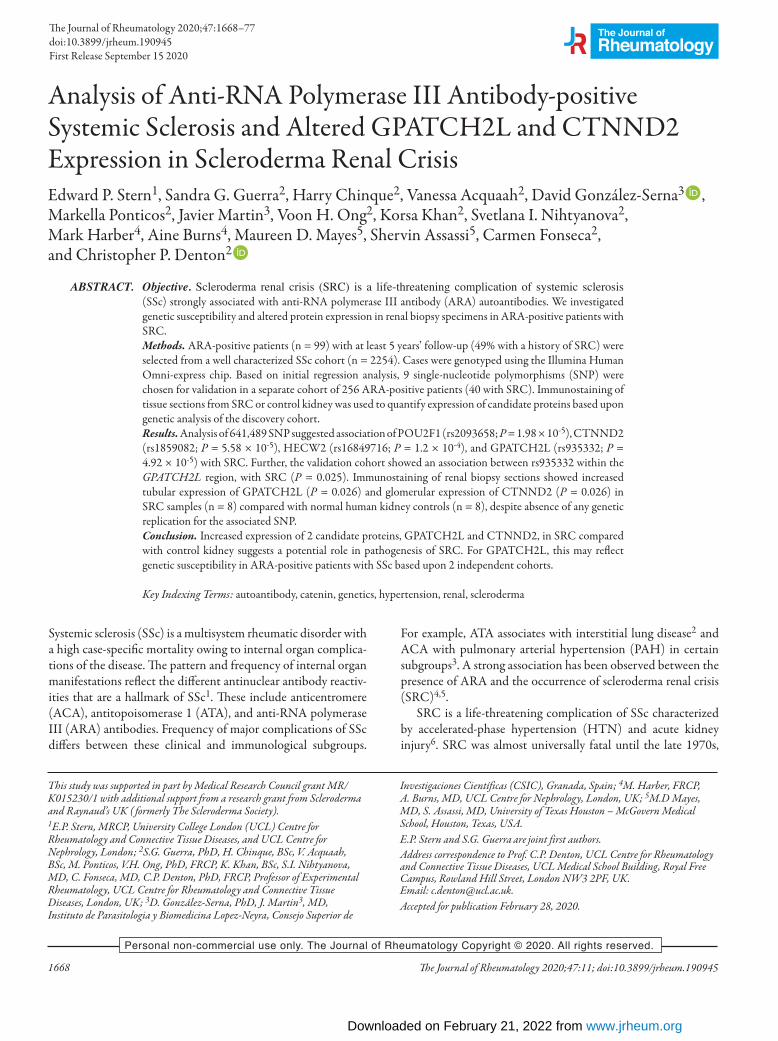
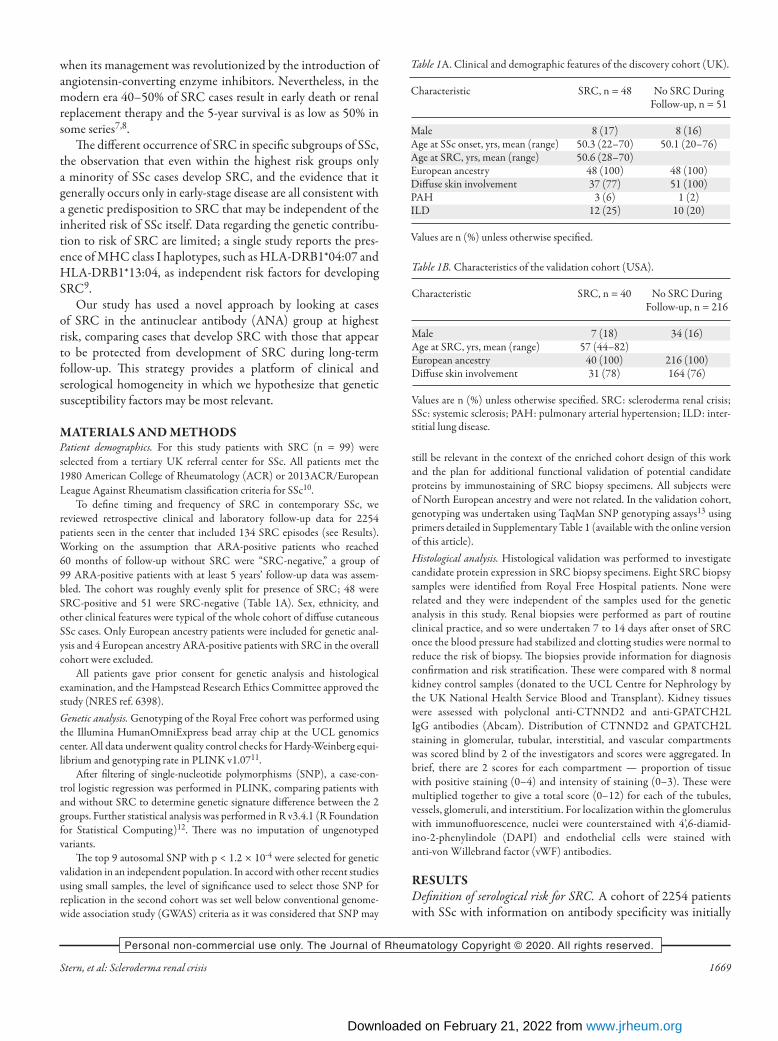
analyzed. Of those, 390 (17.3%) were male and 811 (36%) had the diffuse cutaneous subset of SSc. ARA was positive in 258 (11.5%) patients, ACA in 639 (28.4%), ATA in 508 (22.5%), U3RNP in 91 (4%), and PmScl in 102 (4.5%). Cumulative inci-dence of SRC for the cohort as a whole at 12, 24, 36, and 60 months was 3.3%, 4.3%, 4.8%, and 5.6%, respectively. Of all ARA-positive subjects, 59 (22.9%) had developed SRC. Cumulative incidence of SRC at 12, 24, 36, and 60 months in this subgroup was 12.5%, 18%, 20.4%, and 21.4%. Figure 1 shows cumulative incidence of SRC by antibody subgroup. The high frequency of SRC in ARA-positive cases together with a tendency for earlier occurrence compared with other specifici-ties makes this an especially informative group for exploration of genetic susceptibility to SRC.Genetic analysis. Ninety-nine ARA-positive patients with follow-up of at least 5 years (48 with renal crisis and 51 without) were analyzed. Characteristics are summarized in Table 1, including an indication of the frequency of major organ-based complications and concurrent malignancy in the cohort. In addition to their autoantibody homogeneity, the 2 groups were similar in terms of age, sex, and ethnicity. Limited additional phenotypic data were available for the validation cohort that was selected to be similar in terms of ANA and ethnicity, and with robust categorization by SRC status.GWAS analysis in a UK discovery cohort. Genotype data under-went quality control checks for Hardy-Weinberg equilibrium (HWE) and genotyping rate in PLINK (HWE p < 0.001, geno-typing rate > 90%). There were 2309 SNP removed for miss-ingness and 77,122 failed minor allele frequency (MAF) filters (MAF < 0.01). After quality control, 641,489 SNP were analyzed between the 2 groups (Supplementary Figure 1, available with the online version of this article). Genomic inflation factor λ was 1.10357 and the quantile-quantile plot performed. The presence of relatives and/or duplicates was assessed by computing identi-ty-by-descent estimation using PLINK. Results of this GWAS are illustrated in Figure 2. SNP with a GWAS P value < 3 × 10⁻⁵ are annotated on the figure. In contrast with the majority of previous GWAS analyses in SSc, including the only previously documented association with SRC, there was no marked association with the MHC on chromosome 6.Genetic validation in an independent US cohort. We selected 9 autosomal SNP from the top associations for a further validation analysis in 256 ARA-positive subjects. Characteristics of these individuals are summarized in Table 1B. They were derived from a cohort used and described in other studies of SSc genetics14, and the serological testing is validated wherever possible with central testing using ELISA specific for anti-RNA polymerase III autoantibody15. The top 9 SNP from the Royal Free Hospital cohort and the findings for each in the validation cohort are summarized in Table 2. Of these 9 SNP, only 1 showed replication of significant asso-ciation in this second cohort, for the replicated SNP. This SNP (rs935332) is in the region of GPATCH2L (G patch domain
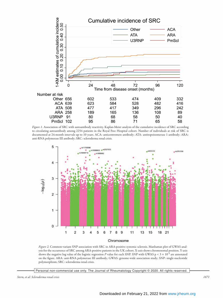
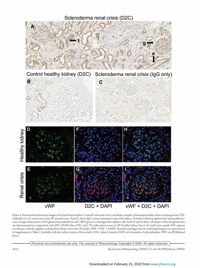
containing 2-like) on chromosome 14. Polymorphisms within this gene region were previously seen to confer significant risk for diastolic HTN in other robust GWAS analyses16,17. While this replication reached nominal significance with a nominal uncorrected P value < 0.05, it should be noted that with 9 SNP tested, an appropriate threshold for replication could be set at 0.05/9, which is 0.0055, a value that no SNP met. For this reason additional histological validation in renal biopsy specimens was sought for GPATCH2L, as outlined below.Altered tissue expression of GPATCH2L and CTNND2 in SRC. To determine whether genes associated with the SNP variants identified in the discovery cohort might show altered protein expression in SRC, we performed immunostaining in renal biopsy specimens from SRC and in healthy renal tissue. First, we stained for GPATCH2L protein to investigate whether there was differential expression in SRC biopsies. There was signifi-cantly increased expression of GPATCH2L protein in SRC samples, localized mainly to the tubular and vascular endothe-lial structures. As described in Materials and Methods, a total biopsy staining score was calculated for control (median 1, range 0–4) and SRC (median 11, range 9–21) samples that confirmed highly increased expression in SRC (P = 0.0009). Using cate-gorical analysis of the tubular staining that was the most striking difference between SRC and control biopsies, there was positive staining (total score > 1) in all 8 SRC biopsies, but in only 3 of 8 controls (P = 0.026; Fisher exact test). Figure 3 shows repre-sentative sections of SRC (Figure 3A–D) together with IgG (Figure 3E) and normal healthy kidney control (Figure 3F), and tabulated quantification for each biopsy specimen is provided in Supplementary Table 2 (available with the online version of this article). Together with the genetic association for ARA-positive SRC in both discovery and validation cohorts in genetic analysis, these results provide strong support for GPATCH2L being rele-vant to susceptibility and potentially implicated in SRC patho-genesis. This is notable given previous association of SNP in this region with diastolic HTN in other studies17. Although the validation genetic analysis achieved only nominal significance for GPATCH2L, among the top associated SNP from the discovery cohort that were tested, we considered that another candidate SNP linked to CTNND2 warranted further evaluation in the renal biopsy samples, based upon mech-anistic plausibility and genetic data from other studies in SSc18. As further evidence of a shared underlying etiopathogenesis, ARA positivity has also been associated with high long-term risk of PAH19. Blinded scoring of CTNND2 expression in SRC biopsy samples versus normal human kidney controls showed distinct glomerular staining, absent in all 8 control samples, in 5 of 8 SRC samples (P = 0.026; Fisher exact test). Again, the total biopsy staining score was significantly increased in SRC (median 12, range 1–17) compared with control biopsies (median 3, range 0–8; P = 0.0135). Consensus scores for individual renal biopsy specimens are summarized in Supplementary Table 2 (available with the online version of this article). Bright-field staining of
Personal non-commercial use only. The Journal of Rheumatology Copyright © 2020. All rights reserved.
www.jrheum.orgDownloaded on February 21, 2022 from
Stern, et al: Scleroderma renal crisis 1671
Figure 1. Association of SRC with autoantibody reactivity. Kaplan-Meier analysis of the cumulative incidence of SRC according to circulating autoantibody among 2254 patients in the Royal Free Hospital cohort. Number of individuals at risk of SRC is documented at 24-month intervals up to 10 years. ACA: anticentromere antibody; ATA: antitopoisomerase 1 antibody; ARA: anti-RNA polymerase III antibody; SRC: scleroderma renal crisis.
Figure 2. Common-variant SNP association with SRC in ARA-positive systemic sclerosis. Manhattan plot of GWAS anal-ysis for the occurrence of SRC among ARA-positive patients in the UK cohort. X-axis shows chromosomal position. Y-axis shows the negative log value of the logistic regression P value for each SNP. SNP with GWAS p < 3 × 10⁻⁵ are annotated on the figure. ARA: anti-RNA polymerase III antibody; GWAS: genome-wide association study; SNP: single-nucleotide polymorphism; SRC: scleroderma renal crisis.
Personal non-commercial use only. The Journal of Rheumatology Copyright © 2020. All rights reserved.
www.jrheum.orgDownloaded on February 21, 2022 from

1672 The Journal of Rheumatology 2020;47:11; doi:10.3899/jrheum.190945
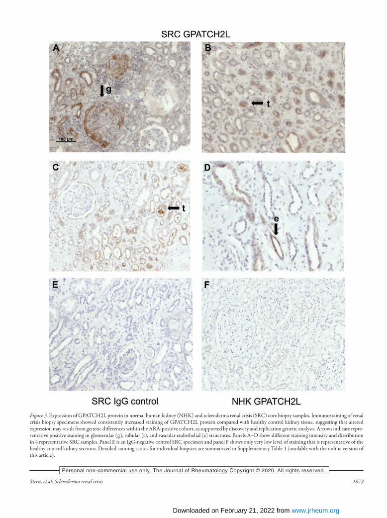
representative sections is shown in Figure 4A with appropriate controls (Figures 4B–C). Immunofluorescence images showed anti-CTNND2 antibody staining in the glomeruli localizing to the capillary loops. Double-staining using the endothelial marker vWF showed collocation with CTNND2, strongly suggesting expression in endothelial cells, which would be consistent with microvascular perturbation in the glomeruli of SRC, associated with altered wingless type (Wnt) signaling pathway activity (Figures 4H–I).
DISCUSSIONThis study used the high risk of SRC in cases of SSc with ARA, together with the fundamental observation that SRC is very rare in established SSc more than 3 years from first non-Ray-naud phenomenon symptom, to powerfully enrich a rare disease population20. Our approach of using enriched cohorts with high susceptibility to a disease complication in GWAS has some simi-larity to a more conventional extreme phenotype design, which has also proven successful in rare diseases where careful pheno-typing can overcome some of the limitations of a necessarily small sample size. Where an extreme phenotype study examines rare causal variants, our focus was on finding susceptibility alleles among functional genetic variants. Previous studies explored the genetic association for ARA and identified MHC alleles associated with this autoantibody21. In addition, cohort studies have investigated associations of the MHC with SRC and identified associations in addition to those associated with ARA4. In our GWAS analysis it is notable that there is no “Manhattan peak” at chromosome 6, representing the loci associated with MHC. MHC associations are an almost-uni-versal finding in GWAS of complex autoimmune disease22. Our results using GWAS analysis for common-variant SNP in a discovery cohort from the UK, and then testing candidate SNP in a second independent validation cohort from the USA, identified only 1 SNP that was associated in both studies. Since GPATCH2L showed nominal replication in the US cohort, we went on to determine protein expression. A possible role in pathogenesis was supported by our results showing increased
expression of the GPATCH2L protein in SRC. This protein is associated with altered RNA processing and could be a marker of cell perturbation that is known to occur in multiple cellular compartments in SSc, and could be relevant to development of hallmark pathologies such as SRC. Overexpression is most clearly observed in renal tubular epithelial structures, which are not generally regarded as a primary site of SRC pathology, but protein expression in the tubular epithelium plays a critical role in control of both intravascular volume and arterial tone, so it is certainly plausible that disruption of transcription in this region could be associated with disorders of arterial blood pressure. While no mechanism for an association between GPATCH2L and essential HTN has been demonstrated, a single molecular basis explaining the link with SRC and essential HTN is feasible and may be elucidated in future work, including investigation of any association with outcomes, such as renal recovery or survival, that may differ for ARA-positive SRC compared with other anti-body subsets23. As well as defining possible genetic susceptibility to SRC, we expected that our enriched cohort design might also suggest possible pathogenetic relevance for genes linked with other SNP associated with SRC risk in our discovery cohort. Based upon other previously published studies, we were especially interested in the SNP rs1859082 (P = 0.000029) on chromosome 5, which lies within the CTNND2 or delta 2 catenin (D2C) gene. It is notable that the same gene CTNND2 was associated with PAH in SSc in a previous genetic study using a combination of direct sequencing and association analysis18. This association is mech-anistically compelling, as CTNND2 is an armadillo-related protein that regulates cell-to-cell adhesion through its interac-tion with the cadherins24,25,26,27. This gene is associated with Wnt regulation and has been associated with altered migration and adhesion in cancer cells28. A recent study demonstrated that hypoxia-inducible factor 1-alpha (HIF1-alpha) regulated CTNND2 expression and that this could be important in modulating Wnt signaling29. Hypoxia and HIF1-α signaling were previously implicated in SSc pathogenesis. The Wnt pathway is increasingly recognized to be associated with fibrosis
Table 2. Candidate single-nucleotide polymorphisms (SNP; n = 9) associated with scleroderma renal crisis (SRC) in the discovery cohort (UK) and their asso-ciation with SRC in the validation cohort (USA).
UK Discovery Cohort USA Validation Cohort Chromosome SNP Gene BP A1 MAF P, Unadjusted GC OR (95% CI) A1 MAF P OR (95% CI)
1 rs2093658 POU2F1 167355192 G 0.490 7.37E-06 1.98E-05 0.24 (0.12–0.45) G 0.359 0.787 1.07 (0.65–1.76)2 rs16849716 HECW2 197195099 G 0.049 0.00012 0.0002512 6.11 (2.21–16.85) G 0.1977 0.962 1.02 (0.56–1.84)3 rs11708596 C3orf20 14689393 A 0.050 2.28E-05 5.53E-05 7.05(2.58–19.29) A 0.137 0.223 0.60 (0.26–1.37)3 rs7643629 IQCJ-SCHIP1 158778092 G 0.324 2.02E-05 4.96E-05 3.52 (1.95–6.35) G 0.471 0.728 0.918(0.568–1.485)3 rs2118096 Near to OTOL1 161525058 C 0.240 5.62E-05 0.0001257 3.455 (1.86–6.38) C 0.368 0.598 0.87 (0.53–1.45)4 rs10008833 Near to EPHA5 66136223 G 0.133 2.05E-05 5.02E-05 4.437 (2.17–9.06) G 0.168 0.078 1.66 (0.94–2.92)5 rs1859082 CTNND2 11372743 G 0.272 5.58E-05 0.000125 3.599 (1.9–6.78) G 0.423 0.9714 0.99 (0.61–1.62)6 rs2327835 LOC101928354 14570894 A 0.080 7.14E-05 0.0001563 4.987 (2.14–11.57) A 0.177 0.258 1.40 (0.78–2.50)14 rs935332 GPATCH2L 76699590 C 0.059 4.92E-05 0.0001114 6.11 (2.38–15.67) C 0.184 0.025 1.86 (1.08–3.20)
A1: minor allele (in controls); BP: base pair position; GC: genomic-control corrected P values; MAF: minor allele frequency in controls; SNP: single-nucleo-tide polymorphism.
Personal non-commercial use only. The Journal of Rheumatology Copyright © 2020. All rights reserved.
www.jrheum.orgDownloaded on February 21, 2022 from
Stern, et al: Scleroderma renal crisis 1673
Figure 3. Expression of GPATCH2L protein in normal human kidney (NHK) and scleroderma renal crisis (SRC) core biopsy samples. Immunostaining of renal crisis biopsy specimens showed consistently increased staining of GPATCH2L protein compared with healthy control kidney tissue, suggesting that altered expression may result from genetic differences within the ARA-positive cohort, as supported by discovery and replication genetic analysis. Arrows indicate repre-sentative positive staining in glomerular (g), tubular (t), and vascular endothelial (e) structures. Panels A–D show different staining intensity and distribution in 4 representative SRC samples. Panel E is an IgG-negative control SRC specimen and panel F shows only very low level of staining that is representative of the healthy control kidney sections. Detailed staining scores for individual biopsies are summarized in Supplementary Table 1 (available with the online version of this article).
Personal non-commercial use only. The Journal of Rheumatology Copyright © 2020. All rights reserved.
www.jrheum.orgDownloaded on February 21, 2022 from
1674 The Journal of Rheumatology 2020;47:11; doi:10.3899/jrheum.190945
Figure 4. Immunohistochemistry images of normal human kidney (control) and renal crisis core biopsy samples. Immunoperoxidase shows staining of anti-D2C antibodies in (A) renal crisis versus (B) normal tissue. Panel C shows IgG control staining of renal crisis kidney. To further delineate glomerular immunofluores-cence, images demonstrate (D,E) glomerular endothelial cells (vWF, green) as a background capillary tuft. Panels F and G show cell nuclei within the glomerulus are demonstrated in conjunction with D2C (DAPI, blue; D2C, red). No collocation is seen in (H) healthy kidney, but in (I) renal crisis sample D2C appears to collocate with the capillary endothelium distinct from the cell nuclei (vWF + D2C + DAPI). Detailed staining scores for individual biopsies are summarized in Supplementary Table 1 (available with the online version of this article). D2C: delta 2 catenin; DAPI: 4’,6-diamidino-2-phenylindole; vWF: von Willebrand factor.
Personal non-commercial use only. The Journal of Rheumatology Copyright © 2020. All rights reserved.
www.jrheum.orgDownloaded on February 21, 2022 from
Stern, et al: Scleroderma renal crisis 1675
and is reported to be altered in SSc, potentially primed by upregulated transforming growth factor β (TGF-β) signaling30. Demonstration of altered delta catenin expression in SRC is notable even if our genetic data do not support a general role in SRC susceptibility across cohorts. There is substantial evidence that delta catenin is not only involved in early development, cell-cell adhesion, and cell motility in neuronal cells, but that it also plays an important role in vascular endothelial cell motility and pathological angiogenesis31 and in SSc fibrosis32. This could be highly relevant to the pathogenesis of SRC. Regulation of Rho GTPases as downstream targets of TGF-β in tissue repair is also supportive of the potential relevance in SSc and preclin-ical models of SSc33. A previous clinical trial of a topical Wnt inhibitor in SSc suggested beneficial effects on tissue remod-eling and adipogenesis34. E-cadherin and Wnt signaling influ-ence the release and differentiation of circulating progenitor cells, including endothelial to mesenchymal transition35,36. It is plausible that variants in the CTNND2 gene have a role in the dysregulation of endothelial progenitor cells that is observed in scleroderma vasculopathy37,38. Both PAH and SRC are prolif-erative vasculopathic complications of SSc, albeit in different vascular beds, and it is notable that the same common variant was associated with SSc in a study that investigated genetic asso-ciation for PAH in SSc18. Our immunofluorescence studies demonstrate CTNND2 in nonnucleated fragments within the glomerular capillary tuft. The typical histopathological finding of SRC is throm-botic microangiopathy (TMA) with obstructive fibrin thrombi within the glomerular capillaries or arterioles, often containing fragmented red blood cells and platelets39,40. A previous in vitro model of TMA demonstrated cell-to-cell adhesion between red blood cells and endothelial cells as opposed to simple mechan-ical sequestration41. CTNND2 would be a potential mediator of such adhesion, and further investigation is justified into the role this protein plays in susceptibility to organ injury in other forms of TMA. A strength of this study is the enriched phenotype method made possible by the very strong risk of SRC conferred by ARA and by the rarity of SRC more than 5 years after onset of SSc. This offers the possibility of identifying genetic risk of a specific organ complication within a rare disease group, and this is the first study of this kind to date, to our knowledge. Using this method, we have provided candidates for further investigation of the genetic risk of SRC and evidence that these candidates may be functionally relevant. However, there are inevitable limitations. First, none of the SNP associations identified in the discovery cohort reached conventional GWAS levels of significance42, although this level may be considered too conservative for a small hypothe-sis-generating study, such as the present analysis, with potential for independent genetic replication and corroborative histo-logical analysis43. This is not surprising given the small sample size we examined, but makes replication approaches particu-larly important for interpretations of our results. It also means
that our findings must be interpreted with caution, and that it is possible that other SNP with associations were not selected for genetic validation. These associations may emerge in future genetic analyses of SRC. Nevertheless, the novel method used to detect difference within a well-phenotyped subgroup may justify investigation of associations seen at a lower P value, as success-fully reported in other recent SSc functional genomic studies where weaker genetic associations have been supported by robust histological and functional assays44. Second, there were differences in allele frequency between the 2 cohorts examined for some SNP that may reflect differences in population genetic structure. Some investigators suggest that population structure analyses of cases and controls with GWAS data do not separate cases and controls, as only a few loci of usually modest effect are different in frequency between them. Apparent differences in population structure may be more likely when testing small numbers of matched cases and controls from a highly enriched disease subset. It is also possible that bias may be introduced from requiring long-term survival of at least 5 years for the non-SRC cohort to be outside the risk period for this complication. However, in other studies we have shown that overall survival in ARA-positive cases is good and that most mortality arises from SRC and associated complications45, so we consider that differences independent of SRC risk are unlikely to confound our results. Another limitation is that our findings may be relevant only to SRC associated with ARA. We previously observed that SRC outcomes may differ according to ANA reactivity, with ARA-positive patients having better overall survival and being more likely to discontinue dial-ysis than non-ARA cases23. Perhaps the most important mecha-nistic limitation is the application of the GWAS approach, using a common-variant analysis platform for a disease area in which rare causal variants might be of prime importance. In future studies, an adaptive approach with extreme phenotyping could identify such variants. At the time of case selection for the study there was no agreed expert consensus definition of SRC. This has now been proposed6, and our definition for this study is largely in accord with the suggested criteria. Future studies should use the new consensus definition to facilitate cross-study comparison. The overall frequency of SRC in the discovery cohort aligns with other published series from a larger series of patients managed in the same center45. We have demonstrated genetic association with SRC in ARA-positive cases of SSc of a common-variant SNP previously associated with essential HTN, and showed significantly altered expression of the GPATCH2L protein in SRC biopsies. Our approach also identified strong overexpression of CTNND2 in SRC glomerular endothelium that is absent from healthy kidney samples, supporting a potential role for altered Wnt signaling in SRC pathogenesis.
ONLINE SUPPLEMENTSupplementary material accompanies the online version of this article.
Personal non-commercial use only. The Journal of Rheumatology Copyright © 2020. All rights reserved.
www.jrheum.orgDownloaded on February 21, 2022 from

1676 The Journal of Rheumatology 2020;47:11; doi:10.3899/jrheum.190945
REFERENCES 1. Denton CP, Khanna D. Systemic sclerosis. Lancet 2017;
390:1685-99. 2. Morelli S, Barbieri C, Sgreccia A, Ferrante L, Pittoni V, Conti F, et
al. Relationship between cutaneous and pulmonary involvement in systemic sclerosis. J Rheumatol 1997;24:8-5.
3. Walker UA, Tyndall A, Czirjak L, Denton C, Farge-Bancel D, Kowal-Bielecka O, et al. Clinical risk assessment of organ manifestations in systemic sclerosis: A report from the EULAR Scleroderma Trials and Research Group database. Ann Rheum Dis 2007;66:754-63.
4. Penn H, Howie AJ, Kingdon EJ, Bunn CC, Stratton RJ, Black CM, et al. Scleroderma renal crisis: Patient characteristics and long-term outcomes. Q J Med 2007;100:485-94.
5. Nguyen B, Assassi S, Arnett FC, Mayes MD. Association of RNA polymerase III antibodies with scleroderma renal crisis. J Rheumatol 2010;37:1068-9.
6. Butler EA, Baron M, Fogo AB, Frech T, Ghossein C, Hachulla E, et al. Generation of a core set of items to develop classification criteria for scleroderma renal crisis using consensus methodology. Arthritis Rheum 2019;71:964-71.
7. Lynch BM, Stern EP, Ong V, Harber M, Burns A, Denton CP. UK Scleroderma Study Group (UKSSG) guidelines on the diagnosis and management of scleroderma renal crisis. Clin Exp Rheumatol 2016;5:106-9.
8. Stern EP, Steen VD, Denton CP. Management of renal involvement in scleroderma. Curr Treat Options Rheumatol 2015;8:1–13.
9. Nguyen B, Mayes MD, Arnett FC, del Junco D, Reveille JD, Gonzalez EB, et al. HLA-DRB1*0407 and *1304 are risk factors for scleroderma renal crisis. Arthritis Rheum 2011;63:530-4.
10. van den Hoogen F, Khanna D, Fransen J, Johnson SR, Baron M, Tyndall A, et al. 2013 classification criteria for systemic sclerosis: An American College of Rheumatology/European League against Rheumatism collaborative initiative. Arthritis Rheum 2013;65:2737-47.
11. Purcell S, Neale B, Todd-Brown K, Thomas L, Ferreira MAR, Bender D, et al. PLINK: A tool set for whole-genome association and population-based linkage analyses. Am J Hum Genet 2007;81:559-75.
12. R Development Core Team. R: A language and environment for statistical computing. Vienna, 2017. Available from: www.r-project.org
13. Shen GQ, Abdullah KG, Wang QK. The TaqMan method for SNP genotyping. Methods Mol Biol 2009;578:293-306.
14. Mayes MD, Bossini-Castillo L, Gorlova O, Martin JE, Zhou X, Chen WV, et al. Immunochip analysis identifies multiple susceptibility loci for systemic sclerosis. Am J Hum Genet 2014;94:47-61.
15. López-Isac E, Acosta-Herrera M, Kerick M, Assassi S, Satpathy AT, Granja J, et al; Australian Scleroderma Interest Group. GWAS for systemic sclerosis identifies multiple risk loci and highlights fibrotic and vasculopathy pathways. Nature Commun 2019;10:4955.
16. Hoffmann TJ, Ehret GB, Nandakumar P, Ranatunga D, Schaefer C, Kwok PY, et al. Genome-wide association analyses using electronic health records identify new loci influencing blood pressure variation. Nature Genet 2017;49:54-64.
17. Warren HR, Evangelou E, Cabrera CP, Gao H, Ren M, Mifsud B, et al. Genome-wide association analysis identifies novel blood pressure loci and offers biological insights into cardiovascular risk. Nature Genet 2017;49:403-15.
18. Gao L, Emond MJ, Louie T, Cheadle C, Berger AE, Rafaels N, et al. Identification of rare variants in ATP8B4 as a risk factor for systemic sclerosis by whole-exome sequencing. Arthritis Rheum 2016; 68:191-200.
19. Nihtyanova SI, Schreiber BE, Ong VH, Rosenberg D, Moinzadeh P, Coghlan JG, et al. Prediction of pulmonary complications and long term survival in systemic sclerosis. Arthritis Rheum 2014; 66:1625-35.
20. Steen VD. Autoantibodies in systemic sclerosis. Semin Arthritis Rheum 2005;35:35-42.
21. Kuwana M, Pandey JP, Silver RM, Kawakami Y, Kaburaki J. HLA class II alleles in systemic sclerosis patients with anti-RNA polymerase I/III antibody: Associations with subunit reactivities. J Rheumatol 2003;30:2392-7.
22. Matzaraki V, Kumar V, Wijmenga C, Zhernakova A. The MHC locus and genetic susceptibility to autoimmune and infectious diseases. Genome Biol 2017;18:76.
23. Lynch B, Penn H, Harvey J, Burns A, Denton CP. The prognosis of scleroderma renal crisis in RNA-polymerase III antibody (ARA) positive compared to ARA negative patients [abstract]. Arthritis Rheum 2013;65 Suppl 10.
24. Carnahan RH, Rokas A, Gaucher EA, Reynolds AB. The molecular evolution of the p120-catenin subfamily and its functional associations. PLoS One 2010;5:e15747.
25. McCrea PD, Gottardi CJ. Beyond β-catenin: Prospects for a larger catenin network in the nucleus. Nat Rev Mol Cell Biol 2016; 17:55-64.
26. Lu Q, Paredes M, Medina M, Zhou J, Cavallo R, Peifer M, et al. Delta-catenin, an adhesive junction-associated protein which promotes cell scattering. J Cell Biol 1999;14:519-32.
27. Lu Q, Aguilar BJ, Li M, Jiang Y, Chen Y-H. Genetic alterations of δ-catenin/NPRAP/Neurojungin (CTNND2): Functional implications in complex human diseases. Hum Genet 2016;135:1107-16.
28. Huang F, Chen J, Wang Z, Lan R, Fu L, Zhang L. δ-Catenin promotes tumorigenesis and metastasis of lung adenocarcinoma. Oncol Rep 2018;39:809-17.
29. Huang F, Chen J, Lan R, Wang Z, Chen R, Lin J, et al. Hypoxia induced δ-catenin to enhance mice hepatocellular carcinoma progression via Wnt signaling. Exp Cell Res 2019;374:94-103.
30. Gillespie J, Ross RL, Corinaldesi C, Esteves F, Derrett-Smith E, McDermott MF, et al. Transforming growth factor β activation primes canonical Wnt signaling through down-regulation of axin-2. Arthritis Rheum 2018;70:932-42.
31. Ghose S, Min Y, Lin PC. δ-Catenin activates Rho GTPase, promotes lymphangiogenesis and growth of tumor metastases. PLoS One 2015;10:e0116338.
32. Bei Y, Hua-Huy T, Nicco C, Duong-Quy S, Le-Dong NN, Tiev KP, et al. RhoA/Rho-kinase activation promotes lung fibrosis in an animal model of systemic sclerosis. Exp Lung Res 2016;42:44-55.
33. Akhmetshina A, Dees C, Pileckyte M, Szucs G, Spriewald BM, Zwerina J, et al. Rho-associated kinases are crucial for myofibroblast differentiation and production of extracellular matrix in scleroderma fibroblasts. Arthritis Rheum 2008;58:2553-64.
34. Lafyatis R, Mantero JC, Gordon J, Kishore N, Carns M, Dittrich H, et al. Inhibition of β-catenin signaling in the skin rescues cutaneous adipogenesis in systemic sclerosis: A randomized, double-blind, placebo-controlled trial of C-82. J Invest Dermatol 2017; 137:2473-83.
35. Liu X, McBride J, Zhou Y, Liu Z, Ma J-X. Regulation of endothelial progenitor cell release by Wnt signaling in bone marrow. Invest Ophthalmol Vis Sci 2013;54:7386-94.
36. Heuberger J, Birchmeier W. Interplay of cadherin-mediated cell adhesion and canonical Wnt signaling. Cold Spring Harb Perspect Biol 2010;2:a002915.
37. Good RB, Gilbane AJ, Trinder SL, Denton CP, Coghlan G,
Personal non-commercial use only. The Journal of Rheumatology Copyright © 2020. All rights reserved.
www.jrheum.orgDownloaded on February 21, 2022 from
Stern, et al: Scleroderma renal crisis 1677
Abraham DJ, et al. Endothelial to mesenchymal transition contributes to endothelial dysfunction in pulmonary arterial hypertension. Am J Pathol 2015;185:1850-8.
38. Good RW, Trinder SL, Denton CP, Abraham DJ, Holmes AM. Dysfunctional endothelial progenitor cells may contribute to vasculopathy in systemic sclerosis. Eur J Vasc Endovasc Surg 2014;47:694.
39. Lusco MA, Fogo AB, Najafian B, Alpers CE. AJKD Atlas of renal pathology: Thrombotic microangiopathy. Am J Kidney Dis 2016;68:e33-e34.
40. Fisher ER, Rodnan GP. Pathologic observations concerning the kidney in progressive systemic sclerosis. AMA Arch Pathol 1958;65:29–39.
41. Smeets MWJ, Bierings R, Meems H, Mul FPJ, Geerts D, Vlaar APJ, et al. Platelet-independent adhesion of calcium-loaded erythrocytes to von Willebrand factor. PLoS One 2017;12:e0173077.
42. Fadista J, Manning AK, Florez JC, Groop L. The (in)famous GWAS P-value threshold revisited and updated for low-frequency variants. Eur J Hum Genet 2016;24:1202-5.
43. Li MX, Yeung JMY, Cherny SS, Shat PC. Evaluating the effective numbers of independent tests and significant p-value thresholds in commercial genotyping arrays and public imputation reference datasets. Hum Genet 2012;131:747-56.
44. Dritsoula A, Papaioannou I, Guerra SG, Fonseca C, Martin J, Herrick AL, et al. Molecular basis for dysregulated activation of NKX2-5 in the vascular remodeling of systemic sclerosis. Arthritis Rheum 2018;70:920-31.
45. Nihtyanova SI, Sari A, Harvey JC, Leslie A, Derrett-Smith EC, Fonseca C, et al. Using autoantibodies and cutaneous subset to develop outcome-based disease classification in systemic sclerosis. Arthritis Rheum 2020;72:465-76.
Personal non-commercial use only. The Journal of Rheumatology Copyright © 2020. All rights reserved.
www.jrheum.orgDownloaded on February 21, 2022 from