analysis of arsenic resistance in the biomining bacterium
TRANSCRIPT

Analysis of Arsenic Resistance in the
Biomining Bacterium, Acidithiobacillus
caldus
Andries Albertus Kotzé
Thesis presented in partial fulfillment of the requirements
for the degree of Master of Sciences at the University of
Stellenbosch
Supervisor: Professor Douglas E. Rawlings
December 2006
i

Declaration I, the undersigned, hereby declare that the work contained in this thesis is my own
original work and has not previously in its entirety or in part been submitted at any
university for a degree.
Signature:_______________________ Date:____________
(Andries A. Kotzé)
ii
Abstract
In this study the chromosomal arsenic resistance (ars) genes shown to be present in all
Acidithiobacillus. caldus isolates were cloned and sequenced from At. caldus #6. Ten
open reading frames (ORFs) were identified on a clone conferring arsenic resistance,
with three homologs to arsenic genes, arsC (arsenate reductase), arsR (regulator) and
arsB (arsenite export). This ars operon is divergent, with the arsRC and arsB genes
transcribed in opposite directions. Analysis of the putative amino acid sequences of these
arsRC and arsB genes revealed that they are the most closely related to the ars genes of
Acidithiobacillus ferrooxidans.
These ars genes were functional when transformed into an Escherichia coli ars deletion
mutant ACSH50Iq, and conferred increased levels of resistance to arsenate and arsenite.
ArsC was required for resistance to arsenate, but not for resistance to arsenite. None of
the other ORFs enhanced arsenic resistance in E. coli. A transposon located arsenic
resistance system (TnAtcArs) has been described for highly arsenic resistant strains of the
moderately thermophilic, sulfur-oxidizing, biomining bacterium At .caldus #6. In the
latter study it was shown that TnAtcArs confers higher levels of resistance to arsenate
and arsenite than the chromosomal operon. TnAtcArs was conjugated into a weakly ars
resistant At. caldus strain (C-SH12) and resulted in greatly increased arsenite resistance.
RT-PCR analysis revealed that arsR and arsC are co-transcribed. Despite ORF1
(cadmium inducible-like protein) and ORF5 (putative integrase for prophage CP-933R)
not being involved in resistance to arsenic, ORF1 was co-transcribed with arsRC and
ORF5 with arsB. Using arsR-lacZ and arsB-lacZ fusions it was shown that the
chromosomal ArsR-like regulator of At. caldus acts as a repressor of the arsR and arsB
promoter expression. Induction of gene expression took place when either arsenate or
arsenite was added. The chromosomal located ArsR was also able to repress TnAtcArs,
but the transposon-located ArsR was unable to regulate the chromosomal system.
iii

Opsomming
In hierdie studie is die chromosomale arseen weerstandbiedendheidsgene (ars gene),
teenwoordig in alle Acidithiobacillus caldus isolate, gekloon en die DNA volgorde
daarvan vanaf At. caldus #6 bepaal. Tien oopleesrame (ORFs) is geïdentifiseer op ‘n
kloon wat arseen weerstandbiedend is, met drie homoloog aan ars gene, nl. arsC
(arsenaat reduktase), arsR (reguleerder) en arsB (membraan-geleë pomp wat arseniet
uitpomp). Die ars operon is gerangskik met die arsRC en arsB gene wat in
teenoorgestelde rigtings getranskribeer word. Analise van die afgeleide
aminosuurvolgorde van dié ars gene het getoon hulle is naverwant aan die ars gene van
Acidithiobacillus ferrooxidans.
Die ars gene was funksioneel na transformasie na ‘n E. coli ars mutant (ACSH50Iq), en
het ‘n hoër vlak van weerstand teen arsenaat en arseniet gebied. ArsC was nodig vir
weerstand teen arsenaat, maar nie vir weerstand teen arseniet nie. Geen van die ander
ORFs het arseen weerstandbiedendheid in E. coli bevorder nie. Voorheen is ‘n ars
operon, geleë op ‘n transposon (TnAtcArs), in ‘n hoogs arseen-weerstandbiedende stam
van die middelmatige termofiliese, swawel-oksiderende, bio-ontgunning (“biomining”)
bakterie Acidithiobacillus caldus #6 beskryf. In laasgenoemde studie is gevind dat
TnAtcArs hoër vlakke van weerstand bied teen arsenaat en arseniet as die chromosomale
operon. TnAtcArs is na ‘n lae arseen-weerstandbiedende At. caldus stam (C-SH12)
gekonjugeer. Die resultaat was ‘n groot verhoging in arseen weerstandbiedendheid. RT-
PCR analise het onthul dat arsR en arsC saam getranskribeer word. Benewens die feit
dat ORF1 (kadmium induseerbare protein) en ORF5 (afgeleide integrase vir profaag CP-
933R) nie betrokke is in weerstand teen arseniet and arsenaat nie, is ORF1 saam met
arsRC getranskribeer en ORF5 saam met arsB. Deur gebruik te maak van die fusie-gene
arsR-lacZ en arsB-lacZ is bewys dat die chromosomale ArsR reguleerder van At. caldus
as ‘n inhibeerder van die arsR en arsB promoter uitdrukking funksioneer. Indusering van
geen uitdrukking vind plaas wanneer arseniet of arsenaat bygevoeg word. Die
chromosomaal-geleë ArsR is ook in staat om TnAtcArs te inhibeer, terwyl die transposon
geleë ArsR nie daartoe in staat is om die chromosomale ars sisteem te reguleer nie.
iv

Acknowledgements
Most importantly, I would like to thank those who have provided me with support in
many ways during this time, including:
My family and most importantly my parents, for giving me the opportunity to attend
Stellenbosch University and for supporting me in all possible ways.
My supervisor, Professor Douglas Rawlings, who always had time in his busy schedule
to give advice and support when problems appeared.
Dr. Marla Tuffin, who always watched over my shoulder as I learned the ropes in the lab.
Thank you for all the friendship, guidance and patience and for always being willing to
lend an ear and share ideas. Thank you also for proof-reading my thesis and correcting
my many mistakes.
The rest of the Biomining Research Group for their friendship, support and for providing
many a fun and amusing moment in the lab.
Finally, our Heavenly Father, for blessing me with the strength and opportunity to
complete this study.
This work was supported by grants from the NRF (National Research Foundation,
Pretoria, South Africa), BHP Billiton Process Research Laboratories (Randburg, South
Africa) and the Biomine Project of the European Framework 6 program.
v

Table of contents Abbreviations vii Chapter one: Literature review 1 Chapter two: Sequence analysis of the Acidithiobacillus 43 caldus chromosomal ars operon
Chapter three: Regulation and cross-regulation by the 71 Acidithiobacillus caldus chromosomal ArsR Chapter four: The Acidithiobacillus caldus transposon ars 90 operon enhances arsenic resistance in strains only harboring a chromosomal copy Chapter five: General Discussion 105
Appendix One: Annotated sequence obtained from 114 pTcC-#4 References 127
vi

Abbreviations aa amino acids ADP adenosine 5’ diphisphate Amp ampicillin At. Acidithiobacillus ATP adenosine 5’ triphosphate AsV arsenate AsIII arsenite bp base pair (s) oC degrees Celsius C-terminal carboxyl-terminus Cys cysteine DNA deoxyribonucleic acid g gram h hour His histidine Inc incompatibility group (s) IPTG isopropyl-β-D-thiogalactopyranoside kb kilobase pair (s) or 1000-bp kDa kilodalton (s) or 1000 daltons Km kanamycin l liter LA Luria agar LB Luria Bertani LMW low molecular weight mg milligram min minute (s) ml milliliter μg microgram μl microliter NCBI National Centre for Biotechnology Information NDB nucleotide binding domain
vii

N-terminal amino-terminus ng nanogram nm nanometer ORF open reading frame p plasmid PCR polymerase chain reaction pH potential of hydrogen PTPase protein-tyrosine-phosphatase RBS ribosome binding site rpm revolutions per minute RNA ribonucleic acid rRNA ribosomal ribonucleic acid Trp tryptophan w/v weight per volume
viii

Chapter One
General Introduction
Contents 1.1. Acidithiobacillus caldus and its role in biomining --------------------------------- 2
1.2. Properties of arsenic ------------------------------------------------------------------- 7
1.3. Bacterial resistance to arsenic -------------------------------------------------------- 9
1.4. Proteins involved in the ars operon
1.4.1. The ArsAB pump ----------------------------------------------------------- 13
1.4.1.1. ArsA (ATPase subunit) ----------------------------------------- 13
1.4.1.2. ArsB (Membrane bound efflux pump) ------------------------ 22
1.4.2. ArsC (Arsenate Reductase) ------------------------------------------------ 25
1.4.3. Regulation of the ars operon
1.4.3.1. ArsR --------------------------------------------------------------- 35
1.4.3.2. ArsD --------------------------------------------------------------- 39
1.5. Aim of thesis ---------------------------------------------------------------------------- 41
1

1.1. Acidithiobacillus caldus and its role in biomining Acidithiobacillus caldus is a moderately thermophilic acidophile with optimum growth at
pH 2.5 and a temperature of 45 oC. It can grow in a temperature range of 32 oC to 52 oC,
and a pH range of 1.0 - 4.6. Cells are short rods, motile, Gram-negative and are capable
of chemolithoautotrophic growth by the oxidation of reduced inorganic sulfur
compounds. At. caldus is incapable of oxidizing ferrous iron or iron sulfides. It obtains
its carbon by the reductive fixation of atmospheric CO2. On solid tetrathionate medium
the colonies are small, circular, convex and with precipitated sulfur in the centre
(Hallberg et al., 1994; Hallberg et al., 1996a and Hallberg et al., 1996b). This bacterium
was previously classified as Thiobacillus caldus, but as a result of 16S rRNA sequence
analysis, it became clear that the genus Thiobacillus included members of the α-, β- and
γ-subclasses of Proteobacteria. A new genus, Acidithiobacillus, was then created to
accommodate these highly acidophilic bacteria. This new genus contains four species of
the γ-subclass of Proteobacteria, namely At. thiooxidans, At. ferrooxidans, At. caldus and
At. albertensis (Kelly and Wood, 2000).
The ability of this organism to oxidize inorganic sulfur makes it very useful in converting
insoluble metal sulfides into soluble metal sulfates which can be leached from their
surroundings. At. caldus plays an important role in a commercial biooxidation process
called biomining. This process uses the oxidizing properties of various acidophilic
bacteria either to convert insoluble metal sulphides to water soluble metal sulphates or as
a pretreatment process to break up the structure of the mineral, thus permitting other
chemicals to penetrate and solubilize the metal. An example of the first process is the
solubilization of copper from minerals such as covellite (CuS) or chalcocite (Cu2S). An
example of the second process is the removal of arsenic, iron and sulfur from gold-
bearing arsenopyrite. The gold that remains in the mineral is then more easily accessible
to cyanide for subsequent extraction (Van Aswegen et al., 1991).
2

There are currently two main types of microbially assisted biomining processes for the
recovery of metals. The first process is an irrigation-type or heap leaching process and
involves the percolation of leaching solutions through crushed ore that have been stacked
in columns, dumps or heaps (Rawlings et al., 1999a). Each dump is irrigated with iron-
and sulfate-rich wastewater from which copper has been removed. Microorganisms
growing on the surface of the mineral in the heap catalyze the chemical reactions that
result in the conversion of insoluble copper sulfides to soluble copper sulfate. The copper
sulfate-containing leach solutions are collected at the bottom of the dump and sent for
metal recovery by a process of solvent extraction and electrowinning. Although heap
reactors are very cost effective, they are very difficult to aerate efficiently and the pH and
nutrient levels are difficult to manage. These dumps are relatively cheap to construct and
mainly consist of waste ore, and therefore it is considered to be a low technology process.
The metal recovered in the largest quantity by this process is copper (Rawlings, 2002,
2005).
The second process is a stirred tank process and employs highly aerated, continuous-flow
bioreactors. The bioreactors are arranged in a series, with finely milled ore or
concentrate being added to the first tank together with inorganic nutrients in the form of
fertilizer-grade (NH4)2SO4 and KH2PO4. The mineral suspension then flows through a
series of highly aerated tanks that are pH and temperature controlled until biooxidation of
the mineral concentrate is complete. Because mineral biooxidation is an exothermic
process, cooling of the bioreactors is important. Large volumes of air are blown through
each reactor, and a large agitator ensures that an even suspension of the solids is carried
over to the next tank. A big advantage of this process is that mineral solubilization in
these tanks takes place in days, while heap reactors can take up to months to solubilize
the minerals. These reactors are extremely expensive to construct and maintain, and
therefore their use is mainly restricted to high-value ores and concentrates. Stirred tank
bioreactors are mostly used as a pretreatment process for the recovery of gold from ores
where the gold is finely divided in a mixture of pyrite or arsenopyrite. These gold
particles cannot easily be accessed by cyanide for subsequent solubilization of the
mineral. Treatment of the ores with this process decomposes the arsenopyrite, which
3

allows the cyanide to come in contact with the gold (Rawlings, 2002, 2005; Rohwerder et
al., 2003).
These biomining organisms have several physiological features in common. They are all
chemolithoautotrophic and use ferrous iron or reduced inorganic sulfur (or both) as an
electron donor. A result of sulfur-oxidation is the production of sulfuric acid, and
therefore these organisms are all acidophilic and have a pH optimum of 1.5 – 2.0. They
can all fix CO2 and generally are resistant to a wide range of metal ions. In these
biomining processes, air provides the carbon source (CO2) and the electron acceptor (O2),
water provides trace elements and is also the medium of growth. The mineral ore supplies
the electron donor in the form of ferrous iron and/or reduced inorganic sulfur (Rawlings,
2002). In general, the same organisms are found in heap-leaching and stirred tank
processes. The proportions of the organisms may vary, however, depending on the
mineral being solubilized and conditions under which the heaps or tanks are operated
(Rawlings, 2005). Unlike other fermentation processes, biomining processes are not
sterile, and no attempt is made to maintain sterility. This is not necessary because these
highly acidophilic organisms create an environment that is only suited for themselves and
not for other organisms. Another reason why this process does not need to be sterile is
because the microorganism that decomposes the mineral the most efficiently will out-
compete those that are less efficient (Rawlings, 2002).
In processes that operate at moderate temperatures in the range of 35oC - 40oC the most
important bacteria are considered to be the iron- and sulfur-oxidizing At. ferrooxidans,
the sulfur-oxidizing At. thiooxidans and At. caldus, and the iron-oxidizing Leptospirillum
ferrooxidans and Leptospirillum ferriphilum (Suzuki, 2001). For many years At.
ferrooxidans was believed to be the most important biomining organism at 40oC, but with
the development of techniques such as PCR amplification of 16S rRNA genes from total
DNA extracted from environmental samples, it became clear that At. ferrooxidans plays a
minor role in continuously operating stirred tank reactors operating under steady-state
conditions. Here a combination of Leptospirillum and At. thiooxidans or At. caldus are
the most important oxidizing bacteria. In a pilot scale, stirred tank operation in which a
4

polymetallic sulfide ore was treated at 45oC, it was found that At. caldus-like, L.
ferriphilum-like and Sulfobacillus-like bacteria was dominant (Okibe et al., 2003). At.
ferrooxidans may be the dominant organism in dump or heap leaching environments
where there is a lower redox potential (Rawlings et al., 1999a; Rawlings, 2002;
Rawlings, 2005).
Because At. caldus and At. thiooxidans are closely related, initial studies indicated that
At. thiooxidans dominated continuous-flow tanks that operated at 40oC. By using
restriction enzyme analysis to identify the bacteria in biooxidation tanks, Rawlings et al.
(1999b) have shown that these tanks used to treat gold-bearing arsenopyrite concentrates
are in fact dominated by At. caldus instead of At. thiooxidans. At. caldus are the most
dominant sulfur-oxidizing bacteria in pilot plants that treat copper concentrates at 40oC
and nickel concentrates at 45-55oC and are considered to be the “weed” of biomining
bacteria under these conditions (de Groot et al., 2003). However, for the biooxidation of
most ores to proceed efficiently, bacteria capable of oxidizing both sulfur and iron need
to be present. At. caldus and At. thiooxidans cannot, like At. ferrooxidans and L.
ferrooxidans, oxidize an ore such as pyrite when growing in pure culture (Okibe and
Johnson, 2004). The reason for this is that the sulfur in the ore is only available to these
bacteria after the iron has been oxidized. Therefore, they need to be grown in a mixed
culture with iron-oxidizing bacteria. Oxidation of an ore by such a consortia of bacteria
generally takes place at a higher rate than in pure culture (Rawlings et al., 1999a).
Several roles of At. caldus in the biomining environment have been suggested. Dopson
and Lindström (1999) showed in laboratory studies that At. caldus is able to enhance the
ability of the iron-oxidizing bacterium Sulfobacillus thermosulfidooxidans to oxidize
arsenopyrite ores at 45 oC. A result of the chemical leaching process is the formation of
sulfur (S0). When sulfur accumulates, it forms a layer on the surface of the mineral. It
might be that the presence of At. caldus increases the leaching rate by removing the
elemental sulfur that builds up on the mineral surface, thereby allowing bacterial and
chemical (Fe3+) access to the mineral (Dopson and Lindström, 1999). Bacteria need to
attach to the mineral to cause leaching (Arredondo et al., 1994). Another possible
5

mechanism by which At. caldus may affect the leaching rate of arsenopyrite is by the
production of organic growth factors. These growth factors may stimulate heterotrophic
and mixotrophic growth of bacteria. It is also possible that At. caldus might provide
organic material in the form of cross-feeding that aids mixotrophic growth. This cross-
feeding could also be in the form of a symbiotic relationship, where the iron oxidizing
bacteria such as Leptospirillum spp. may reduce the concentration of organic chemicals
that inhibits growth by accumulation in the cytoplasm. A last possible mechanism by
which At. caldus might affect the arsenopyrite leaching rate is by production of surface-
active agents to solubilize the sulfur (Dopson and Lindström, 1999).
During this process of mineral solubilization, arsenic compounds are leached from the
arsenopyrite ore. These arsenic compounds are toxic to the bacteria used in this process.
During early stages of development of the biooxidation process, solubilization of the
arsenopyrite ore was inefficient and slow and bacteria required a retention time of over
12 days to oxidize the concentrate efficiently for gold recovery. The reason was that the
bacteria were not tolerant to such high levels of arsenic. Initially the bacteria were
sensitive to less than 1 g arsenic/l. At the Fairview mine in Barberton, South Africa,
soluble AsV concentrations as high as 12 g/l were reported and at the Sao Bento mine in
Brazil AsIII concentrations of 3-6 g/l (Dew et al., 1997). This arsenic inhibition was so
severe that after a period of time the arsenic had to be removed from the tanks before the
process could continue. This removal of the arsenic was time consuming and very
uneconomic. However, in a period of 2 years, through a process of selection using a
bacterial chemostat, the bacterial consortium became sufficiently resistant to these high
levels of arsenic and no further removal of arsenic compounds was needed. The retention
time needed to oxidize the concentrate efficiently was also reduced from 12 days to less
than 7 days (Rawlings and Woods, 1995). After another 3 years of operation at the
Fairview mine, the retention time was reduced to a little over 3 days, and the quantity of
the suspended mineral concentrate had been increased from 10% to 19% w/v.
6

1.2. Properties of arsenic It is believed that Albertus Magnus discovered arsenic around 1250. Arsenic is a semi-
metal or metalloid that exists in the environment in two biologically important oxidation
states, namely AsV (arsenate) and AsIII (arsenite). It is one of the most prevalent toxic
metals in the environment (although in very low concentrations) and is commonly
associated with the ores of metals like gold, copper and lead. Arsenite absorbs less
strongly and to fewer minerals than arsenate, which makes it a more mobile oxyanion
than arsenate. Arsenate is the predominant form of arsenic in aerobic environments,
while arsenite is more predominant in an anoxic environment (Oremland and Stolz, 2003;
Rosen, 2002a).
Arsenite is said to be about 100 times more toxic than arsenate. In a study where the
genotoxic activity of arsenite and arsenate was tested in TK6 human lymphoblastiod cell
lines, arsenite was far more genotoxic than arsenate (Guillamet et al., 2004). The toxicity
of arsenite lies in its ability to bind to the sulfur groups of essential cysteines in proteins.
Because arsenite binds two sulfur groups it can cross link proteins, altering their overall
shape and thereby impeding their function. Because of the similarity in structure and
solubility between arsenate and phosphate, arsenate can act as a phosphate analog and
competes for the formation of ATP (Coddington, 1986). Kenney and Kaplan
strengthened this theory by showing that arsenate substituted phosphate in both the
sodium pump and the anion exchanger of human red blood cells (Kenney and Kaplan,
1988). The transport of arsenate into bacterial cells is via two phosphate transport
membrane systems, the Pit and Pst system, while the Pst system seems to be the
predominant system for arsenate uptake. Arsenite is transported into bacterial cells by
aqua-glycerolporins (glycerol transport proteins) (Sanders et al., 1997) (Figure 1.1). A
mutation in the glycerol facilitator of E. coli (GlpF) converted the E. coli strain to
antimony (SbIII) resistance. Because the chemical properties of arsenite and antimony are
similar, it is very likely that GlpF is also an AsIII transporter (Mukhopadhyay et al., 2002;
Rosen, 2002a; Rosen, 2002b). Recently Meng et al. (2004) confirmed AsIII uptake by
GlpF in E. coli.
7

Figure 1.1: Uptake mechanisms of arsenate and arsenite in E. coli. Arsenate is brought into cells by phosphate transporters, the Pst and the Pit system (top left). Arsenate is then reduced to arsenite by the ArsC (arsenate reductase, middle). Arsenite uptake is facilitated by the aquaglycerolporin GlpF (top right). Arsenite is actively extruded by the ArsAB complex (AsIII/SbIII-translocating ATPase, bottom right). If ArsA is absent, ArsB acts as a secondary arsenite carrier protein and efflux is coupled to membrane potential (bottom left). Adapted from Rensing et al. (1999).
Arsenical compounds have been used for many purposes, mainly in medicine (mostly for
treating protozoan diseases), in agriculture (as herbicides, fungicides, pesticides and
animal feed additives) and as a poison. In 1908 Paul Ehrlich won the Nobel Prize in
medicine for the use of arsenic compounds as chemotherapeutic agents (Mukhopadhyay
et al., 2002). Due to the fact that arsenic is so prevalent in the environment,
microorganisms have developed various resistance mechanisms to counteract its toxicity.
8

1.3. Bacterial resistance to arsenic Due to the abundance of arsenic in the environment, microorganisms come into contact
with this metalloid on a regular basis. Therefore it is not surprising that chromosomal
and plasmid located genes which confer resistance to arsenic have been isolated from
bacteria. Different mechanisms of resistance to arsenic have evolved in microorganisms,
such as the overproduction of intracellular thiols (e.g. the protozoan Leishmania),
phosphate pathways that do not transport arsenate efficiently (e.g. cyanobacteria), the
oxidation of arsenite to the less toxic arsenate (e.g. Alcaligenes faecalis), sequestration in
a vacuole (e.g. fungi) and methylation (Rosen, 1999; Oremland and Stolz, 2003;
Cervantes et al., 1994; Wang et al., 2004). The best characterized and most studied way
of detoxification is a mechanism where intracellular AsV is converted to AsIII and a
specific efflux pump then extrudes AsIII from the cytoplasm (ars operon). This system
has been extensively studied and will be the focus of this study.
The ars operon has been isolated from Gram-positive and Gram-negative bacteria. There
are two common forms of the ars operon. The first and most common form consists of
three genes, arsRBC. This form of the ars operon is located on chromosomes and
plasmids of many organisms, but the best studied is the chromosomal ars system of E.
coli K-12 and the ars systems on plasmids pI258 and pSX267 of Staphylococcus (Diorio
et al., 1995). The second operon is found on plasmids of Gram-negative bacteria (e.g.
plasmid R773 and R46 of E. coli and plasmid pKW301 of Acidiphilium multivorum) and,
more recently, on transposons of At. caldus (AY821803), L. ferriphilum (DQ057986), A.
faecalis (Ay297781) and M. flagellatus (NZ_AADX01000013) (Tuffin et al., 2006). this
operon consists of five genes, arsRDABC (Figure 1.2) (Suzuki et al., 1998). Both these
operons have been organized into a single transcriptional unit. The arsR and arsD genes
encode two different regulatory proteins. ArsR is a trans-acting repressor that senses
intracellular AsIII and controls the basal level of expression of ArsB and ArsC (Yang et
al., 2005). ArsD is an inducer-independent protein and controls the upper level of
expression (Saltikov and Olson, 2002). ArsA is an arsenite stimulated ATPase,
energizing ArsB (a membrane-bound arsenite and antimony efflux pump) by ATP
hydrolysis (Silver, 1996). In the presence of ArsA, arsenite and antimony are exported
9

actively out of the cell by using cellular ATP. If the ArsA is absent, these oxyanions can
still be excreted by ArsB using membrane potential (Cai et al., 1998). ArsC is an
arsenate reductase that converts arsenate to arsenite to get exported by the efflux pump.
Figure 1.2: Homologies among the arsenic resistance determinants of Staphylococcus plasmids pSX267 and pI258, E. coli plasmids R773 and R46, and the chromosomal ars operon of E. coli. The sizes of putative gene products (in amino acids [aa]) are shown above or below the genes (boxes). The numbers between the dashed lines are percentages of similarity among the ArsR, ArsB and ArsC proteins. O/P, operator/promoter. Adapted from Silver and Phung (1996).
With new technology constantly developing and data generated from genome sequencing
projects, it became clear that ars operons may differ from the two common forms
described above. Southern blot hybridization experiments where different arsenic
resistance genes were probed against total DNA isolated from bacteria revealed that these
genes are very widespread among microorganisms (Dopson et al., 2001). PCR
experiments with degenerate primers to these resistance genes also revealed the
prevalence of ars operons. There are however, other forms of arsenic operons that differ
from the arsRBC and arsRDABC conformation. Other genes that have also been reported
10

to be associated with arsenic resistance are arsH and arsM. For example, an operon that
is different in gene order and that contains an extra gene was identified in At.
ferrooxidans (Butcher et al., 2000). This operon consisted of arsCRBH genes with the
arsCR and arsBH genes divergently transcribed. The function of the arsH gene is still
unknown, but it was not required for and did not enhance arsenic resistance in E. coli.
Cyanobacterium Synechocystis sp. strain PCC 6803 has an arsBHC arsenic resistance
operon, where the arsH was also not required for resistance (Lopez-Maury et al., 2003).
Neyt et al. (1997) found an arsHRBC operon on plasmid pYV of Yersinia enterocolitica
with the arsH gene also divergently transcribed. The presence of this gene however,
either in cis or trans was essential for arsenic resistance. This was surprising, because
arsRBC are sufficient to confer resistance in E. coli and staphylococci. The authors
suggested that arsH might have a regulatory function similar to arsD. The same
arsHRBC operon was also found on the IncH12 plasmid R478 (Ryan and Colleran,
2002). PCR amplification using primers to the arsH gene showed that this gene is
present in many other arsenic resistant IncH12 plasmids. Removal of the arsH, like in Y.
enterocolitica, rendered the cell sensitive to arsenic. The legume symbiont
Sinorhizobium meliloti contains a very interesting ars operon. It consists of four genes,
an arsA, followed by an aqpS (aquaglyceroporin) and then arsCH. A deletion of arsH
also rendered the cell sensitive to arsenite and arsenate (Yang et al., 2005).
The archaeon, Halobacterium sp. strain NRC-1 contains a gene encoding a mammalian
arsenite-methyltransferase homolog, named arsM. A deletion of this gene increased
sensitivity to arsenite, indicating a novel mechanism of arsenic resistance, possibly by
methylating intracellular arsenite and thereby creating a concentration gradient to the
outside of the cell. It is also possible that arsenite could be methylated to a volatile
trimethyl-arsine that would leave the cell by diffusion (Wang et al., 2004; Tuffin et al.,
2005).
The ars operon of the acidophilic archaeon Ferroplasma acidarmanus consists of only
two arsenic resistance genes, arsRB (Gihring et al., 2003). An arsC gene was absent in
the genome. Although no arsC gene is present, the organism was still resistant to
11

arsenate. It is suggested that F. acidarmanus employs phosphate specific transporters to
reduce non-specific uptake of arsenate. F. acidarmanus also lacks a complete arsA-
homologous open reading frame (ORF). The ArsB may thus not function as a subunit of
a primary pump, but as a secondary carrier using membrane potential to export arsenite
out of the cell. This may not be the case however, as acidophilic microorganisms
commonly have a reversed membrane potential (Butcher et al., 2000). Membrane
potential may not be a suitable energy source to export arsenite out of the cell. The
mechanism of arsenite efflux by F. acidarmanus is still unknown.
Another variation of the conventional ars operons is in Mycobacterium tuberculosis,
where the arsB and arsC genes are fused into a single gene, encoding a 498-residue
fusion protein (Mukhopadyay et al., 2002). The reason for the fusion of these two genes
is unknown. Another very interesting arsenic operon is that of the legume symbiont
Sinorhizobium meliloti. The operon consists of an arsR gene, followed by an aqpS gene
in place of arsB. A third ORF downstream of aqpS showed homology with an arsC and
the fourth to an arsH gene. AqpS showed homology with the bacterial glycerol
facilitator, GlpF, which facilitates transport of arsenite and antimonite into bacterial cells.
S. meliloti utilizes a unique arsenic detoxification pathway where ArsC converts arsenate
to arsenite, which is then transported down a concentration gradient through the AqpS
channel to the outside of the cell (Yang et al., 2005).
Sato and Kobayashi (1998) found an ars operon on the skin element of Bacillus subtilis
that also differs from the conventional three or five gene operons. This operon contains
the arsRBC genes, but also a fourth gene (ORF2) located between the arsR and arsB
genes. This gene did not show homology to any known ars genes, but did show
homology to an ORF of unknown function situated upstream of the arsRBC operon in
Mycobacterium tuberculosis. These experiments conducted on the ars genes however,
did not define the role of ORF2 in the arsenic resistance mechanism of Bacillus subtilis.
12

1.4. Proteins involved in the ars operon
1.4.1. The ArsAB pump The best studied ArsAB pump is that encoded by the ars operon of E. coli plasmid R773.
ArsA normally is part of an aggregate with the membrane-bound ArsB protein, and
together forms an ArsAB AsIII/SbIII-translocating ATPase, which extrudes AsIII and SbIII
oxyanions out of the cell. The 63 kDa ArsA ATPase is the catalytic component of the
ArsAB pump, with AsIII/SbIII stimulated ATPase activity. It hydrolyses ATP in the
presence of antimonite and arsenite. The 45 kDa ArsB subunit (which forms the
oxyanion-translocating pathway) uses the chemical energy released by ArsA to secrete
arsenite and antimonite out of the cell (Tisa and Rosen, 1990). The arsRDABC operon
confers higher levels of resistance than the arsRBC operon, indicating that the ATP-
driven ArsAB pumps out arsenite more efficiently. This illustrates that ArsB in complex
with ArsA can reduce the intracellular concentration of AsIII/SbIII to a greater extent than
ArsB alone (Rosen, 1999).
1.4.1.1. ArsA (ATPase subunit) When ArsA is expressed in the absence of ArsB, it is found in the cytosol and can be
purified as a soluble protein (Walmsley et al., 1999). The 583-residue ArsA has two
homologous halves, a N-terminal A1 (residues 1-288) and a C-terminal A2 (residues 314-
583), which are connected by a flexible linker of 25 residues (residues 289-313)
(Bhattacharjee et al., 2000) (Figure 1.3). These two homologous halves are most likely
the result of ancestral gene duplication and fusion (Chen et al., 1986). The crystal
structure of the ArsA has been determined (Zhou et al., 2000), and three types of domains
were found. Firstly, there are two nucleotide binding domains (NBDs) for ATP that
contain residues from both A1 and A2. Secondly, there is a metalloid-binding domain
(MBD). This site is allosterically activated and is positioned at the opposite end of the
protein from the NBDs. The third domain is a signal transduction domain (DTAP) that
connects the MBD to the two NBDs (Rosen, 2002a). There is also a linker that connects
the A1 and A2 halves of ArsA.
13

Figure 1.3: Model of the structure of the ArsA ATPase from plasmid R773. The protein consists of two homologous domains, a N-terminal A1 and a C-terminal A2, connected by a flexible linker region. Each domain contains a nucleotide binding domain (NBD), a signal-transduction domain (DTAP) and a metalloid binding domain. The cysteine residues have been found to comprise the metalloid binding domain (MBD). Binding of arsenite to the cysteine residues brings the A1 and A2 domains together, resulting in hydrolysis of ATP. Adapted from Rosen (2002a).
The Nucleotide Binding Domains (Catalytic Site) The ArsA protein has two nucleotide binding domains, NBD1 and NBD2. Both these
domains are composed of residues from the A1 and A2 halves of ArsA (Zhou et al.,
2000). Both NBDs are located at the interface between A1 and A2, in close proximity to
each other (Zhou et al., 2001). Mutations in either A1 or A2 NBD resulted in a sensitive
strain with loss of resistance to arsenite, ATPase activity and transport (Karkaria et al.,
1990; Kaur and Rosen, 1992). This indicates that both the A1 and A2 NBDs are required
for oxyanion-translocating ATPase activity. The sequence for the consensus binding sites
of ATP (P-loop) is G15KGGVGKTSIS25 and G334KGGVGKTTMA344 in the A1 and A2
halves, respectively (Chen et al., 1986). By using site-directed mutagenesis Li et al.
(1996) altered the first glycine residues in A1 and A2 to cysteine residues. Cells
expressing a G15C (A1) mutant and a G15C/G334C double mutant exhibited moderate
sensitivity to arsenite, whereas a G334C mutant in A2 retained arsenite resistance.
Arsenite resistance was restored in the moderate sensitive mutants to the wild type with a
single substitution of A344V. Because A344V is located adjacent to the C-terminal NBD, it
14

suggests a spatial proximity of Gly15 and Ala344 and thus indicates that the A1 and A2
domains do interact to form a catalytic unit. Li and colleagues further studied this
interaction of the A1 and A2 NBDs and found that the bigger the residue substitution at
position 15 in A1, the weaker the resistance to arsenite. The larger the residue at position
344 in A2, the greater the suppression of the G15 mutants (Li et al., 1998). This shows an
inverse relationship between ATPase activity and residue volume at position 15,
indicating that the geometry at the interface between A1 and A2 NBDs imposes steric
constraints on residues allowed in the A1-A2 interface.
By looking at the structure of ArsA in complex with Mg.ADP, it is clear that the A1 NBD
(NBD1) is in a closed conformation and has ADP bound, while the A2 NBD (NBD2) has
an open conformation, and ATP can be exchanged into this site. This indicates that
NBD2 will be much more accessible than the NBD1 (Zhou et al., 2000; Rosen, 2002b).
These findings are consistent with studies done by Kaur (1999) which suggests that A2 is
a low affinity, easily exchangeable site, while A1 is a high affinity, poorly exchangeable
site. Further studies have also shown that the nature of the nucleotide bound at A2 is
reflected in the conformation of the A1 domain (Zhou et al., 2001). In the absence of
antimonite or arsenite, ATP binding and hydrolysis is at a slow basal rate and occurs
firstly in A1, which then results in a change in A2 so that A2 is now more competent to
bind ATP. In the presence of AsIII/SbIII (multisite conditions), ATP binding and
hydrolysis by A2 produces a change in A1, giving it a more open conformation. The
result is the release of tightly bound ADP from A1 and the subsequent binding of another
ATP molecule to A1 (Jia and Kaur, 2003).
In the presence of AsIII/SbIII, Walmsley et al. (2001) found that NBD1 hydrolyzed ATP
250-fold faster than NBD2. Although the two NBDs have overall structural similarities
and similar evolutionary relationships, the two NBDs have intrinsic differences. These
findings gave more weight to the possibility that the two NBDs have separate roles in
catalysis (Zhou et al., 2002). This hypothesis was further strengthened when a thrombin
site was introduced into the linker that connects A1 and A2 to distinguish between events
at NBD1 and NBD2. After labeling and thrombin digestion, A1 and A2 migrated at
15

different mobilities on SDS-PAGE. This showed that the two NBDs have different
properties, and possibly different functions (Jiang et al., 2005). They further showed that
a) both nucleotides are catalytic and hydrolyze ATP in the absence and presence of SbIII,
b) the affinity for ATP is increased in both NBD1 and NBD2 by SbIII, and c) NBD1 has a
higher affinity for ATP than NBD2. These findings raised the question as to the function
of NBD2. One possibility is that it could once have been an important evolutionary
structure, but that it is not involved in ATP transport anymore. This possibility seems
fairly unlikely, however, as Kaur and Rosen (1992) have shown that mutagenesis of
NBD2 resulted in loss of ATPase activity and AsIII resistance. Another possible function
is that it could play a regulatory role. A third possibility is that the two NBDs could play
equivalent roles in an intact ArsAB complex. The noted differences might be the result
of the analysis of ArsA in the absence of ArsB (Jiang et al., 2005).
Previously it was shown that Mg2+ is required for ArsA ATPase activity (Hsu and Rosen,
1989). An increase in intrinsic tryptophan fluorescence occurred only on the addition of
MgATP, which indicated conformational changes at the NBDs. This effect was observed
only in the presence of ATP, suggesting that Mg2+ binds to ArsA as a complex with ATP
(Zhou et al., 1995). From sequence alignment of ArsA homologs with enzymes such as
NifH, RecA and GTP-binding proteins such as Ras p21, it was suggested that Asp45
might be a putative Mg2+ ligand. From the crystal structure of Ras p21 bound to GTP,
the conserved Asp residue formed a portion of the Mg2+ binding site (Pai et al., 1990).
The Mg2+ ion bring together different components of the GTP binding core, resulting in
information flow between domains. To examine the role of Asp45, mutants were
constructed in which Asp45 was changed to Asn, Glu or Ala. Cells expressing these
mutated arsA genes almost completely lost arsenite resistance. These results supported
the role of Asp45 as a Mg2+ ligand (Zhou and Rosen, 1999).
The Metalloid Binding Domain (Allosteric Site) Arsenite and antimonite have been shown to allosterically activate ArsA ATPase activity
(Rosen et al., 1999). AsIII stimulates ATPase activity 3-5 fold, while SbIII as an activator
stimulates ATPase activity 10-20 fold. In the absence of the metalloids, a low level of
16

ATPase activity is observed by ArsA. No other oxyanion tested had an effect on activity
(Hsu and Rosen, 1989). One possibility was that the activation by SbIII or AsIII might be
through the binding as oxyanions to anion binding sites. As metalloids however, they
might react as soft metals through covalent binding with cysteine thiolates. In such a
reaction, where arsenite reacts with thiolates such as dithiothreitol or glutathione, arsenite
acts as a soft metal, forming direct metal-sulfur As-S bonds (Rosen et al., 1999). If the
activation of ArsA does involve direct metal-sulfur bonds, alteration of participating
cysteine residues would greatly affect the activation of ArsA. The thiol-modifying
reagent methyl methanethiosulphonate (MMTS) inhibits ArsA catalysis, suggesting that
cysteine residues are involved in catalysis (Bhattacharjee et al., 1995).
The ArsA of E. coli has four cysteine residues, Cys26, Cys113, Cys172 and Cys422 (Figure
1.4). For AsIII or SbIII to act as an allosteric activator, two or more cysteine residues must
be in close proximity in the folded protein to interact with the metalloid. To investigate
the role of the cysteine residues in the activation of ArsA, each cysteine was altered to a
serine residue by site-directed mutagenesis (Bhattacharjee et al., 1995). The purified
C26S ArsA showed identical properties to those of the wild-type ArsA, with no change in
resistance to AsIII or SbIII. This suggested that Cys26 is not involved in activation of
ArsA. In contrast, cells expressing the other three mutants were sensitive to SbIII and
AsIII. The purified C113S, C172S and C422S enzymes each exhibited a similar affinity for
ATP to the wild-type ArsA, but the concentration of SbIII or AsIII required for activation
of ArsA was substantially increased, most likely reflecting a decrease in affinity for the
metalloid. This resulted in reduced ATPase activity and rendered the mutants sensitive to
AsIII and SbIII.
These three cysteines are distant from each other in the primary structure of ArsA, but as
the protein folds it brings them into close proximity. AsIII or SbIII then interact with these
cysteines to form a metal-thiol cage. To determine the distance between the cysteine
residues, the wild-type ArsA and ArsA with Cys-Ser substitutions were treated with the
homobifunctional crosslinker dibromobimane (bBBr) (Bhattacharjee and Rosen, 1996).
bBBr has two bromomethyl groups that can crosslink a thiol pair located within 3-6 Å of
17

each other to form a fluorescent adduct. An ArsA protein with only one cysteine group
altered by mutagenesis still formed fluorescent adducts. Proteins lacking any two of the
three cysteines at residue 113, 172 or 422 did not form fluorescent adduct. These results
demonstrate that Cys113, Cys172 and Cys422 are in close proximity of each other in the
tertiary structure so that AsIII or SbIII can interact with these three cysteine residues in a
trigonal pyramidal geometry.
Figure 1.4: The primary sequence of ArsA is represented linearly, with the homologs A1 and A2 halves aligned and linked by the flexible linker region. Boxes indicate regions of greatest similarity. The black boxes indicate the location of the P-loop of the NBDs and the signal transduction domains (DTAP). The locations of the four cysteines are indicated. Cys113, Cys172 and Cys422 form the metalloid binding domain (MBD). Adapted from Bhattacharjee et al. (2000)
Based on the structure of the ArsA protein, Zhou et al. (2000) suggested that it may be
possible that a change in the affinity of ArsA for AsIII/SbIII might inject metal ions into
ArsB as a result of conformational changes during ATP hydrolysis. This was contrary to
previous ideas that the ions that activate ArsA are not transported by ArsB. To further
investigate this hypothesis, ArsA was crystallized in complex with ATP, the non-
hydrolyzable ATP analog AMP-PMP and the transition state analog of ATP hydrolysis,
ADP.AIF3 (Zhou et al., 2001). When ArsA crystals were formed or incubated in the
presence of ATP, ATP was found at NBD2, and ADP at NBD1. Surprisingly, when
18

crystals were incubated with AMP-PNP, it was found only at NBD1. The inability to
obtain crystals in the presence of AMP-PNP, suggested that crystals can only be formed
if NBD1 contains ADP. A reason for this might be that conformational changes
associated with ATP binding and hydrolysis at NBD1 is not allowed in the crystal. Using
pre-steady-state kinetics, it has been shown that binding of ATP favors the uptake of
AsIII/SbIII, while the release of ADP from NBD2 is associated with the release of these
ions from the MBD (Walmsley et al., 2001).
Changes in helices of A1 and A2 corresponded with ATP hydrolysis at NBD2. Helices
H9-H10 of A1 and A2 forms the arms of a gate, alternating in the “open” and “closed”
positions at the interface with ArsB (Figure 1.5). When AsIII/SbIII first reacts with ArsA,
the H9-H10 region of A1 provides the ceiling of the cavity where the ion binds. When
hydrolysis occurs at NBD2, the bound ion moves from the cytosolic side into a protected
pocket at the interface with ArsB. Release of ADP from NBD2 triggers the release of the
AsIII/SbIII ion inside this pocket. ATP hydrolysis at NBD1 is then required to bring ArsA
back to the ground state. Based on the above, the catalytic cycle of ArsA functions
similar to that of a reciprocating engine (Zhou et al., 2001). Many elements of this model
remain to be proven.
Signal Transduction Domain (DTAP) The catalytic and allosteric sites of the ArsA ATPase are located distant from each other
in the enzyme, requiring a mechanism that can transmit the information from the MBD
(allosteric site) to the NBD (catalytic site) where ATP hydrolysis occurs. This flow of
information has been shown to be mediated by a 12-residue consensus sequence,
DTAPTGHTIRLL (Bhattacharjee et al., 2000). This sequence is termed DTAP or the
signal transduction domain and is found in each half of ArsA.
19

Figure 1.5: Schematic model of the proposed mechanism of the ArsA ATPase. Helices of the A1 (red) and A2 (green) domains form the arms of a gate that alternates between the open and closed conformations. An AsIII ion is shown as a purple triangle. Adapted from Zhou et al. (2001). See text for details.
ArsA has four tryptophan residues located at positions 159, 253, 522 and 542. The
function of the DTAP domain in ArsA was examined by using the effects of substrates
and effectors on intrinsic tryptophan fluorescence (Zhou et al., 1997). Using site-directed
mutagenesis two single-tryptophan ArsAs were constructed, containing either Trp141 or
Trp159 at the N- and C-terminal side of the A1 DTAP domain respectively. In the Trp141
protein all native tryptophan residues were changed to tyrosine, and Phe141 was changed
to tryptophan. In the Trp159 ArsA the other three tryptophan residues were changed to
tyrosine. These two tryptophan residues served as intrinsic probes of the environment of
the DTAP domain. During ATP hydrolysis this domain undergoes conformational
changes that may be involved in coupling the allosteric and catalytic sites. The
fluorescence spectra indicated that the N-terminal Trp141 is located in a hydrophobic
20

region, while the C-terminal Trp159 is located in a relatively hydrophilic environment.
Binding of Mg.ADP moved Trp141 into a more hydrophilic region, exposing the N-
terminal end of the DTAP domain to a more polar environment. Upon binding of
Mg.ATP, Trp159 moved into a more hydrophobic environment, leaving the C-terminal end
in a less polar environment. The catalytic and allosteric domains are thus connected to
each other by a “flipping” movement in the signal transduction domain during catalysis.
Linker Region The sequence of the E. coli plasmid R773 ArsA indicated that A1 and A2 are connected
by a flexible linker peptide. The function of this linker sequence was examined by
extending it with five glycine residues or by the deletion of 5, 10, 15 or 23 residues (Li
and Rosen, 2000). Cells expressing an arsA gene with the extended linker exhibited
similar resistance and affinity for ATP and SbIII to the wild type. Cells expressing the
arsA genes with deleted linkers had increasing levels of AsIII sensitivity and a decreased
affinity for ATP and SbIII. The authors proposed that the linkers have evolved to the
shortest length that still allow the two halves of the protein to interact and that it was not
the sequence of the linker that was important, but the length of the linker. Jia and Kaur
(2001) however, studied the role of the E. coli R773 linker by creating point mutations
and complementation experiments and found that the nature of the residues in the linker
are important for correct conformation of the NBDs and for catalytic activity. The
increase in sensitivity seen by Li and Rosen may not have been due to the shortening of
the linker, but because of the role the linker plays in the function of ArsA. Jia and Kaur
expressed an N-terminal A1 clone, that lacks the linker sequence, with two different C-
terminal A2 clones, one without the linker and the other one containing the linker
sequence. Because each domain of ArsA was on a separate polypeptide, deletion of the
linker would not inhibit movement of the two domains. Unless the linker is required for
the function of ArsA, the absence of the linker would have little effect on the ArsA
function. The N- and C-terminal halves were able to complement each other only if the
linker was present, indicating that the linker is essential for function. Point mutations of
certain residues in the linker resulted in a loss of AsIII resistance and ATPase activity.
These mutations also affected the conformation of ArsA. The authors proposed that
21

residues in the linker region play an active role in the function of the protein and that
there is an interaction between the linker and the NBDs.
1.4.1.2. ArsB (Membrane bound efflux pump)
The arsB gene of the E. coli R773 ars operon encodes a protein of approximately 45.5
kDa. Hydropathy plots demonstrated the hydrophobic character of the protein. At least
10 regions of 19 or more residues has a high average hydropathy, indicating these regions
may be potential membrane-spanning α-helices. Using an ArsB-β-galactosidase hybrid
protein, β-galactosidase activity of different extracts was measured. Results indicated
that the fusion protein was localized to the inner membrane and it was postulated to be
the anion channel component of the ArsAB pump (Chen et al., 1986; San Francisco et
al., 1989).
The ArsB protein is difficult to study due to the poor expression of the arsB gene. It was
suggested that the level of the arsB expression is controlled at a translational level.
Analysis of the arsB translational initiation region (TIR) revealed a possible hairpin at the
third codon. Another potential mRNA secondary structure was identified immediately
upstream of the ribosome binding site (Dou et al., 1992). These structures may interfere
with the ribosome, causing it to pause at the start of the arsB sequence, thereby reducing
the amounts of ArsB produced. The use of the T7 expression system has allowed San
Francisco and colleagues to visualize the ArsB protein as a [35S] methionine-labeled
membrane protein (San Francisco et al., 1989).
Tisa and Rosen (1990) performed binding studies with purified ArsA protein to
membranes with and without the arsB gene product to determine if expression of the
arsB gene is required to anchor the ArsA protein to the inner membrane. In cells
expressing the arsB gene, the presence of the ArsA protein on the membrane was shown
by immunoblotting of the membrane with antiserum prepared against the ArsA protein.
In cells expressing an ArsA protein with either a truncated or no ArsB protein, ArsA
protein was found only in the cytosol. This indicated that the expression of the arsB gene
is essential to anchor the ArsA protein to the inner membrane. Membranes of cells
22

lacking an arsA gene were able to bind purified ArsA protein when added exogenously,
suggesting that the ArsB protein is inserted into the membrane in a functional form in the
absence of ArsA.
By fusing certain regions of the arsB gene to reporter genes, a more accurate prediction
of the topology of certain regions in the ArsB could be determined compared with the
hydropathy plots reported by Chen et al. (1986). Three types of gene fusions have been
used. Fusions with the phoA gene (which encodes for alkaline phosphatase) shows high
activity if the fusion is made in the coding region for the periplasmic section of the ArsB,
while fusions with the lacZ gene (which encodes for β-galactosidase) shows high activity
if the fusion is made in the coding region for the cytosolic region of the ArsB. Fusions
with the blaM gene (which encodes for β-Lactamase) will provide resistance to ampicillin
if the fusion is made in the coding region for the periplasmic part of the ArsB. Based on
the results of these fusions, a model was proposed where the ArsB protein has 12
membrane-spanning α-helices joined by six periplasmic loops and five cytoplasmic
loops. The N- and C-termini are suggested to be located in the cytosol (Wu et al., 1992).
From studies of the energetics of the ArsAB pump it has been shown that this complex is
an arsenite-translocating ATPase (Dey et al., 1994; Karkaria et al., 1990). Certain
observations suggested that this hypothesis may be too simple. Firstly, a characteristic
motif of secondary carrier proteins is 12 membrane-spanning α-helices. Thus, the ArsB
has a similar structure to secondary carrier proteins. Secondly, staphylococcal ars
operons have no arsA gene, but still confer resistance to arsenite and extrude arsenite in
an energy-dependent manner, suggesting that the ArsB protein can extrude arsenite in the
absence of ArsA (Diorio et al., 1995). Thirdly, when the R773 arsB gene is expressed in
the absence of the arsA gene, it still confers an intermediate level of arsenite resistance in
E. coli. These observations raised the question as to whether energy was supplied by
another ATPase elsewhere in the genome in the absence of arsA, or if another form of
energy drives the pump in the absence of ArsA. Dey and Rosen (1995) investigated the
possibility of the R773 ArsB utilizing two different kinds of energy. To do this they used
an unc strain of E. coli to compare arsenite resistance and in vivo energetics of arsenite
23

transport in a strain expressing both arsA and arsB genes and in a strain only expressing
the arsB gene. The unc strain of E. coli lacks the H+-translocating ATPase and is thus
unable to convert ATP and the electrochemical proton gradient. An arsA deletion mutant
pBC101 (arsRDBC) and a clone that only contains arsA (pArsA) were transformed into
the unc deletion strain of E. coli. ATP levels were controlled by growing the cells in
glucose (generates ATP through substrate-level phosphorylation) or succinate (generates
almost no ATP). Cells expressing both arsA and arsB genes only actively exclude
arsenite in the presence of ATP (when growing in glucose). When fluoride (which
inhibits glycolysis and thus prevents ATP synthesis) was added, the active exclusion of
arsenite was inhibited. Cyanide, which prevents respiration, but does not alter ATP
levels, failed to inhibit exclusion of arsenite (Dey and Rosen, 1995). This result indicated
that the removal of arsenite in cells containing both arsA and arsB genes was ATP
dependent. In cells expressing only the arsB gene, arsenite was excluded in the presence
and absence of ATP. This exclusion was inhibited by the addition of cyanide, showing
that respiration rather than ATP levels sufficiently excludes arsenite. When the
uncoupler CCCP (carbonyl cyanide m-chlorophenylhydrazone) which destroys the pH
and ion gradients was added to cells only expressing arsB, arsenite was immediately
taken up by the cells. This indicated that a proton motive force is necessary for the
transport of arsenite when only the ArsB protein is present (Figure 1.1).
These results indicated that ArsB on its own is significant for arsenite resistance. The
question of why the arsA gene exists was then raised. Dey and Rosen (1995)
hypothesized that the ArsAB complex is a more effective resistance mechanism than
ArsB alone. Under conditions of stress, the ATP levels will drop more slowly than the
membrane potential, making the ArsAB complex a much more effective mechanism in
the environment where conditions can change rapidly. The specificity of this extrusion
system is also of great importance. It only transports arsenite and antimonite. This
specificity may be a mechanism by which the cell circumvents phosphate starvation,
because if the ArsB system were able to transport arsenate, it might also transport the
structurally similar phosphate oxyanion, leaving the cell depleted of phosphate (Messens
et al., 1999).
24

Researchers also wanted to determine the chemical nature of the arsenite that is being
pumped through ArsB. Chen et al. (1996) suggested two possibilities. AsIII or SbIII may
be electrophoretically transported in response to a positive exterior membrane potential.
Another possibility is that AsIII or SbIII binds to the cysteine residue in the ArsB protein
and is then transported as a soft metal. To investigate the role of the only cysteine in
ArsB, Cys369 was changed to serine and alanine codons by site-directed mutagenesis
(Chen et al., 1996). Cells bearing both pBC101 (arsRDBC) and pArsA (arsA) showed
high levels of resistance to arsenite. No change in resistance was detected when pBC101
was replaced with the mutant arsB C369S or arsB C369A. Thus, Cys369 is not required for
arsenite resistance, indicating that the transport of arsenite by ArsB alone does not
involve metal thiol chemistry. The most likely alternative way of transport is suggested
to be electrophoretic anion transport. These weaker interactions assist with easy release
of AsIII or SbIII extracellularly (Rosen, 1999). This was confirmed when the uptake of
labeled arsenite by everted membrane vesicles expressing only arsB was coupled to
membrane potential (Kuroda et al., 1997).
1.4.2. ArsC (Arsenate Reductase) In the primordial anaerobic environment, arsenite would have been the major chemical
species. This led to the evolution of transporters such as ArsB that could extrude arsenite
out of the cell. As the atmosphere became more oxidizing, arsenate would have replaced
arsenite, as arsenate is the thermodynamically favorable form under aerobic conditions.
A mechanism of arsenate resistance is of great importance for survival. Proteins evolved
that were able to reduce arsenate to arsenite, which could then be pumped out of the cell
by existing transport systems, such as ArsB (Rosen, 1999; Jackson and Dugas, 2003).
Chen et al. (1986) identified a 16 kDa protein on plasmid R773 of E. coli that was
essential for resistance to arsenate, but not arsenite. It was first thought that ArsC
functioned as an intracellular substrate-binding protein, making arsenate accessible to the
ArsAB membrane complex, which would then function as an arsenate efflux system. Ji
and Silver (1992) demonstrated that this hypothesis was incorrect. They showed that
25

arsenite was exported from a cell when arsenate was added in the presence of ArsC from
Staphylococcus aureus plasmid pI258. They concluded that the S. aureus ArsC protein
(131 aa) functions as an arsenate reductase, converting intracellular arsenate to arsenite,
which could then be extruded out of the cell by ArsB. They also found arsenate
reductase activity in the cytoplasm. An important question was the mechanism of energy
coupling for the reduction of arsenate to arsenite. Purified ArsC protein coupled in vitro
with thioredoxin and dithiothreitol (a nonbiological thiol compound) reduced arsenate to
arsenite. Reduced glutathione or 2-mercaptoethanol was unable to reduce arsenate to
arsenite. Thioredoxin can regenerate reduced cysteine residues on intracellular enzymes.
The involvement of cysteine residues found in the sequence of ArsC in the reduction of
arsenate to arsenite was suggested and that thioredoxin is necessary to keep the cysteine
residues in a reduced state (Ji and Silver, 1992).
The ArsC protein (141 aa) of E. coli plasmid R773 also functions as an arsenate
reductase, even though it exhibits less that 20% identity to the ArsC of S. aureus plasmid
pI258 (Oden et al., 1994). Mutations in gshA (encodes γ-glutamylcysteine) and gshB
(encodes glutathione synthetase), which form glutathione, showed wild-type levels of
arsenite resistance when the R773 ars operon was present, but reduced levels of arsenate
resistance. Strains with mutations in either trxB (thioredoxin reductase) or trxA
(thioredoxin) had wild type resistance to arsenate and arsenite. These results indicated
that unlike the ArsC of S. aureus that requires thioredoxin for conversion of arsenate to
arsenite, the ArsC of E. coli requires glutathione for the detoxification of arsenate (Oden
et al., 1994).
Sequence homologies and amino acid sequence alignments suggested that arsenate
reducing enzymes arose independently a number of times (Rosen, 1999). Recent X-ray
crystallographic solutions of protein structure and reaction pathways also indicated on an
independent evolvement of different arsenate reductases. Based on the latter findings,
arsenate reductases are divided into three families. The first family includes E. coli and
uses glutathione/glutaredoxin (GSH/Grx) to reduce arsenate. The second family includes
S. aureus and B. subtilis and uses thioredoxin (Trx) to reduce arsenate. The third family
26

is the only eukaryotic family and is called the yeast Arr2p family (Mukhopadhyay et al.,
2002; Mukhopadhyay and Rosen, 2002). This family was not relevant to the work
presented in our study and will therefore not be discussed.
The GSH/Grx clade The main characteristic of this family of arsenate reductases is that arsenate reduction is
dependent on the presence of reduced glutathione and glutaredoxin. Since these reducing
equivalents derive from cysteine thiolates, Liu et al. (1995) investigated the possibility
that cysteinyl residues within the protein are involved in catalysis. The ArsC protein of
plasmid R773 has only two cysteine residues, Cys12 and Cys106. By using site-directed
mutagenesis four mutants in the Cys12 codon were constructed. Cells expressing all the
mutant arsC genes were sensitive to arsenate. This indicated that Cys12 plays an essential
role in catalysis and that it must be located at the active site of ArsC. Tsai et al. (1997)
hypothesized that a single thiol group may be efficient for reductase activity. An enzyme
complex forms by arsenylating Cys12, with the interaction of glutaredoxin with the
complex. The result is the transfer of electrons to reduce arsenate to arsenite.
The product of the grxA gene, glutaredoxin 1 (Grx1), is a glutathione-dependent dithiol
hydrogen donor for enzymes such as arsenate reductases. In the search for alternate
reductants of ribonucleotide reductase, two new glutaredoxins, Grx2 and Grx3 (each of
which has a Cys-Pro-Tyr-Cys dithiol consensus sequence) were identified. By studying
the efficiency of these glutaredoxins to serve as a hydrogen donor, Grx2 was identified as
the predominant glutaredoxin in E. coli cells (Shi et al., 1999). The general function of
Grx is that it can either catalyze intraprotein disulfide bond reduction or catalyze the
reduction of mixed disulfides between a Cys thiol and glutathione (Bushweller et al.,
1992). All three glutaredoxins of E. coli have two cysteine residues in their active site. It
has been shown that the N-terminal cysteine is required for both protein disulfide
reduction and reduction of mixed protein-glutathione disulfides. To determine the
function of the glutaredoxins in arsenate reduction, single cysteine mutants of all three E.
coli glutaredoxins were constructed. Mutants lacking the C-terminal cysteine had no
effect on ArsC activity. When the N-terminal cysteine was substituted with serine,
27

neither Grx1, Grx2 or Grx3 could serve as a hydrogen donor for arsenate reduction. This
was consistent with a reaction cycle where the ArsC forms a mixed disulfide with
glutathione. The role of glutaredoxin is thus to regenerate the reduced arsenate reductase
by reducing the mixed disulfide (Shi et al., 1999).
Martin et al. (2001) reported X-ray crystal structures for three forms of ArsC, without
bound arsenic and crystals complexed with arsenate and arsenite respectively. No
relationship was found between the tertiary structures of E. coli R773 (GSH/Grx clade)
and the structures of S. aureus and B. subtilis (Trx clade). While the Trx clade has a core
of four parallel β-sheet regions, the GSH/Grx clade has one antiparallel β-sheet segment.
The Trx clade ArsC also exhibits a low rate of phosphatase activity, while the ArsC of
the GSH/Grx clade does not exhibit any phosphatase activity (Zegers et al., 2001)
For arsenate reduction to occur in E. coli, ArsC must form an active quaternary complex
with GSH, arsenate and Grx simultaneously (Liu and Rosen, 1997). The catalytic Cys12
in the active site of ArsC is surrounded by a group of five basic residues, His8, Arg16,
Arg60, Arg94 and Arg107. Together these five residues lower the pKa value of Cys12 to 6.4
(Gladysheva et al., 1996; Martin et al., 2001). Three of these basic residues, Arg60, Arg94
and Arg107 interact directly with the arsenate and arsenite intermediates. Arg60 and Arg94
temporarily exchange places in the presence of arsenate to enhance hydrogen-bonding
and stabilization of the intermediate. Arg107 remains stationary and binds the O1 oxygen
attached to arsenic throughout the reduction reaction (Shi et al., 2003). The role of Arg60
in product formation was further evaluated by mutagenesis. Crystal structures of an ArsC
protein with a substitution for Arg60 equilibrated with arsenite revealed that Arg60 plays
an important role in the stability of the bound arsenite product (DeMel et al., 2004).
Based on previous research and crystal structures by Martin et al. (2001) and DeMel et
al. (2004) the following reaction mechanism is proposed (Figure 1.6):
Step1 involves the nucleophilic attack by Cys12 on an arsenate that is noncovalently
bound at the sulfate ion in the active site, followed by the release of OH-. The result is
the formation of a thioarsenate binary adduct of ArsC (Intermediate I). The existence of
28

this adduct was demonstrated by a difference in electron density. The close proximity of
Arg60, Arg94 and Arg107 plays a very important role in the binding of arsenate. Upon
reaction of arsenate with ArsC, Arg60 and Arg94 move to new orientations in the protein
that allow both side chains to bind to the arsenate adduct.
Step 2 involves the nucleophilic attack of glutathione on the arsenate adduct, with the
release of water. The result is the formation of a {ArsC Cys12}S-As-S{glutathione}
tertiary complex (Intermediate II). Glutathione only reacts after arsenate binds to the
active site. The reaction also requires a free thiol on glutathione and ArsC to proceed.
The structure of intermediate II has not yet been obtained.
Step 3 involves the binding of glutaredoxin to intermediate II, with the reduction of
arsenate, producing a dihydroxy arsenite intermediate (Intermediate III). A mixed
disulfide (GrxS-SG) is released that would be recycled by glutathione reductase using a
second equivalent of GSH. This intermediate can only be observed in mutant structures
because it is not stable in the native enzyme.
In step 4 a monohydroxy, positively charged arsenite adduct is formed (Intermediate IV).
This arsenite adduct has an unusual structure because it has only two atoms linked to the
arsenite atom. This thiarsahydroxyl complex is much more unstable that most cysteine-
arsenite complexes. It is suggested that this unstable conformation is necessary to ensure
that arsenite does not function as an inhibitor of ArsC.
In step 5 arsenite is released upon the addition of a free OH-. ArsC now returns to its
original conformation.
29

Figure 1.6: The proposed reaction mechanism of the E. coli R773 ArsC arsenate reductase (taken from DeMel et al., 2004). See text for details.
The ars operon of Synechocystis sp. Strain PCC 6803 contains an ArsC homolog that
exhibits 36 and 45% sequence identity to S. aureus and B. subtilis, respectively. This
ArsC (SynArsC) exhibited weak phosphohydrolase activity, which is indicative of its
descent from the LMW PTPase (low molecular weight protein tyrosine phosphatase)
family of protein phosphatases. Like the ArsC of pI258, SynArsC contains three
essential cysteine residues. All of the above indicate that SynArsC should belong to the
Trx clade of arsenate reductases. SynArsC however, employs glutathione and
glutaredoxin instead of thioredoxin as the source of reducing equivalents, making it part
of the GSH/Grx clade of arsenate reductases. It appears that SynArsC and pI258
represent different branches in the evolution of their shared phosphohydrolytic ancestor
(Li et al., 2003)
30

The catalytic mechanism of SynArsC combines certain features from its S. aureus
homolog with others from the E. coli R773 ArsC. Like R773, SynArsC forms a complex
with glutathione. Both the active site cysteine residue and arsenate were needed for
complex formation. Glutaredoxin also restored SynArsC to its reduced, catalytically
ready state. A major difference between the mechanism of SynArsC and R773 is that the
latter requires only a single active-site cysteine, while SynArsC needed two more
cysteine residues (Cys80 and Cys82) for catalysis. These two cysteines are similarly
positioned to cysteine residues that play a key role in the reaction mechanism of pI258.
Li et al. (2003) proposed a possible reaction scheme for SynArsC:
Step 1-3 is the same as for R773. An arsenoenzyme intermediate is formed that is
attacked by glutathione to form a cysteinyl-arsenoglutathione tertiary complex. This
complex is then broken down to yield arsenite by the binding of glutaredoxin. Step 4-6 is
a modified version of the reaction mechanism of pI258 ArsC.
In step 4 a reaction takes place between the mixed disulfide bond of Cys8 and glutathione
and the internal disulfide between Cys80 and Cys82. The result is a shift of the mixed
disulfide with glutathione to the surface of the enzyme.
In step 5 glutaredoxin reduces the mixed disulfide.
Step 6 involves the restoration of the active site cysteine to its reduced, reactive state.
The original disulfide bond between Cys80 and Cys82 is also restored.
The Trx Clade The most important characteristic of this family of arsenate reductases is that it requires
the coupling to thioredoxin (Trx), thioredoxin reductase (TR) and NADPH to be
enzymatically active (Ji et al., 1994). The ArsC of S. aureus pI258 has a characteristic
PTPase fold. It consists of a four-stranded parallel β-sheet and three α-helices. Unlike
the R773 ArsC, the ArsC of pI258 shows phosphatase activity with the model substrate
p-nitrophenyl phosphate (Zegers et al., 2001). The pI258 ArsC sequence revealed that it
has four cysteine residues, Cys10, Cys15, Cys82 and Cys89. Cys82 and Cys89 are not found
in the R773 ArsC. Messens et al. (1999) found that a substitution for any of Cys10, Cys82
or Cys89 leads to an inactive enzyme. Cys10 is similarly located to Cys12 of E. coli R773
ArsC. Cys10 also functions as the reaction site of the arsenate reductase. By a series of
31

endoproteinase digestions after catalysis and peptide products identified by mass
spectroscopy analysis, a Cys82-Cys89 oxidized cysteine was assigned to be the redox
couple responsible for electron donation to arsenate.
Zegers et al. (2001) determined the crystal structures of the reduced and oxidized forms
of the S. aureus pI258 arsenate reductase. The secondary and tertiary structures showed
great homology to those of LMW-PTPases from mammals, indicating that the pI258
ArsC may have evolved from LMW-PTPases. The homology between pI258 ArsC and
LMW-PTPases is concentrated mainly in the catalytic motif of tyrosine phosphatases (P-
loop), which has an identical CXGNXCR signature. The pI258 ArsC sequence has very
little homology to R773, except for a common Cys-X5-Arg anion binding motif.
Messens et al. (2002) provided further X-ray crystal structures for the pI258 arsenate
reductase, providing evidence that Cys82 nucleophilically attacks Cys10 to form a Cys10-
Cys82 disulfide. This provides electrons to reduce arsenate to arsenite. The overall
structure of the reduced (active) and oxidized (Cys82-Cys89, inactive) forms are basically
the same, except for a major change in conformation, with Cys89 moving towards Cys82
to form a disulfide bond. In addition to the three essential cysteine residues, Arg16 in the
P-loop was also essential for catalysis as a substitution for Arg16 resulted in a loss of
ArsC activity. Mutational studies by Lah et al. (2003) revealed that potassium had a
definite stabilizing effect on pI258 arsenate reductase.
Messens et al. (2004) also studied the mechanism by which thioredoxin regenerates the
pI258 ArsC to its active form. A disulfide bridge between Cys10 and Cys15 in the P-loop
is formed under oxidative stress. This Cys10-Cys15 bridge is buried in the tertiary
structure and is thus not accessible to thioredoxin. It is hypothesized that the protection
of this bond may be a way to protect Cys10 against irreversible oxidation, such as the
oxidation to sulfenic and sulfinic acids. Formation of a disulfide bridge between Cys10
and Cys15 is thus responsible for redox regulation. To reduce this buried Cys10-Cys15
disulfide bridge, thioredoxin first reduces the surface exposed Cys82-Cys89 disulfide
bridge. Released Cys82 then attacks Cys10 in the buried disulfide bridge, forming a Cys10-
32

Cys82 disulfide intermediate. Cys89 then attacks Cys82, forming a Cys82-Cys89 disulfide
bridge, releasing Cys10. A second molecule of thioredoxin reduces the newly formed
Cys82-Cys89 disulfide bridge, leaving a fully reduced ArsC.
A third X-ray crystal structure for an arsenate reductase was reported, this time for the
ArsC in the SKIN element in the chromosome of Bacillus subtilis (Bennet et al., 2001).
The B. subtilis SKIN element ArsC contains 139 aa residues and shares about 65%
identity to the ArsC of S. aureus. It exhibited phosphatase activity with p-nitrophenyl
phosphate. It also uses thioredoxin to convert arsenate to arsenite and it contains the same
essential cysteine residues (Cys10, Cys82 and Cys89) and Arg16 as the S. aureus pI258
ArsC. Arg16 is thought to stabilize the active site and to lower the pKa values of Cys10,
Cys82 and Cys89 in order for the reduction of arsenate to arsenite to occur. By using high
resolution NMR spectroscopy, the solution structures of both the reduced and oxidized
forms of the B. subtilis ArsC has been determined (Guo et al., 2005). Backbone
dynamics demonstrated that in the reduced form the Cys82-Cys89 segment forms a helix
that is not tightly structured and undergoes substantial conformational changes in the
millisecond time scale. This may facilitate the formation of covalent intermediates and
the subsequent reduction of arsenate to arsenite. In the oxidized form Cys82-Cys89 shows
flexibility on a sub-nanosecond time scale, which is favorable for interaction with
thioredoxin, which regenerates the ArsC for the next cycle of arsenate reduction.
Based on work done by Zegers, Messens and Bennet a reaction mechanism for the S.
aureus and B. subtilis ArsC is proposed (Figure 1.7).
Step 1: The reaction starts with ArsC in the reduced state. Arsenate triggers a
nucleophilic attack of the thiol of Cys10. A covalent Cys10-HAsO3- intermediate is
formed with the release of a hydroxyl that is protonated by the acidic Asp105 to form a
water molecule.
Step 2 involves the three essential cysteine residues in a triple redox relay system.
Firstly, the thiol of Cys82 attacks Cys10 to form a Cys10-Cys82 disulfide bond. Cys10
donates its electron pair to the arsenate, reducing it to arsenite, which then dissociates
from the enzyme. Secondly, Cys89 attacks Cys82, forming a Cys82-Cys89 disulfide bond,
33

releasing Cys10. The disulfide bridge formed by Cys82-Cys89 becomes exposed to the
enzyme surface. The positively charged Arg16 plays a very important role in stabilizing
the P-loop and lowering the pKa values of the three cysteines involved to activate them
for the above mentioned reactions.
In Step 3 thioredoxin reduces the exposed Cys82-Cys89 disulfide, regenerating ArsC for
the next cycle of reduction.
Figure 1.7: The proposed reaction mechanism of the B. subtilis ArsC arsenate reductase. The notation H+S- refers to the sulfhydryl group of a cysteine residue that will be protonated when placed in close proximity of a NH2
+ group. The notation E refers to the ArsC enzyme. Adapted from Bennet et al. (2001). See text for details.
34

1.4.3. Regulation of the ars operon
1.4.3.1. ArsR The small 13 kDa ArsR protein from the E. coli plasmid R773 has been intensively
studied. The arsR gene encodes a trans-acting repressor that binds as a dimer to an
operator and represses transcription. Wu and Rosen (1991) showed that the expression of
this ArsR protein was autoregulated and that this expression was repressed in the absence
of inducers (AsIII and SbIII). In the presence of inducers the expression was induced.
Although the ArsR has been shown to be a trans-acting regulator, the arsR sequence
contains no obvious DNA binding motif. Further studies by Wu and Rosen (1993a) led
to the finding of the ArsR protein binding site. Using DNase I footprinting assays they
found that the ArsR repressor binds to the DNA just upstream (from -64 to -40) of the -35
site. The ArsR would thus interfere with transcriptional initiation. The ArsR binding site
contains an imperfectly symmetrical dyad that may be important for repressor recognition
and binding. Hydroxyl radical footprinting revealed two small 4 bp regions of protection.
Ten bp separated these two regions, indicating that the repressor only binds to one side of
the DNA. The two short protected areas also indicated that the active form of ArsR must
be a dimer. Gel retardation assays demonstrated the formation of DNA-ArsR protein
complexes, with the release of the ArsR protein on interaction with AsIII or SbIII (Wu and
Rosen, 1993a).
The ArsR protein belong to a new family of metalloregulatory proteins called the
SmtB/ArsR family. This family functions exclusively as transcriptional repressors and
includes the Synechococcus sp. SmtB repressor (activated by zinc) that represses the
smtA gene (encodes for metallothionein), the E. coli ArsR repressor and the S. aureus
pI258 CadC repressor (activated by cadmium). Cook et al. (1998) determined the crystal
structure of the SmtB metallothionein repressor. It is suggested that the ArsR proteins
will have a similar structure to SmtB. Bairoch (1993) proposed that the E. coli ArsR
contains a DNA-binding helix-turn-helix structure at residues 32-52, containing two
conserved cysteine residues. Sequence alignments of members belonging to the
SmtB/ArsR family of regulators revealed a highly conserved ELCVCDL region (Shi et
al., 1994). However, not all the members of this family contain this consensus sequence,
35

e.g. the ArsR of At. ferrooxidans (Butcher and Rawlings, 2002). To identify the inducer
binding domain in ArsR, three mutants with a noninducible phenotype (C32Y, C32F and
C34Y) were isolated. The mutated ArsR proteins were still able to bind to the ars
promoter, but they had reduced inducer response. This indicated that Cys32 and Cys34
forms part of the metal binding site of ArsR and that the cysteine residues are located
outside of the DNA-binding helix-turn-helix motif. Binding of a metal induces a
conformational change in the helix-turn-helix domain, resulting in the dissociation of the
repressor from the DNA. A His50 mutation (H50Y) located in the helix-turn-helix motif
resulted in constitutive expression, suggesting a decrease in binding of the ArsR protein
to DNA (Shi et al., 1994).
In most molecule thiolates-AsIII complexes, AsIII is three-coordinated, therefore Shi et al.
(1996) proposed that a third arsenic ligand (only Cys32 and Cys34 had been identified)
may be involved in metal binding. In addition to Cys32 and Cys34, the ArsR contains
three more cysteine residues, Cys37, Cys108 and Cys116. Cys108 and Cys116 have been
shown not to be involved in repressor function (Wu and Rosen, 1991). When Cys37 was
altered to alanine, it had no effect on regulation or DNA binding. X-ray absorption
spectroscopy of the AsIII-ArsR complex showed that AsIII is coordinated to three sulfur
atoms. Shi et al. (1996) investigated the possibility that only two coordinates are needed
for metal binding. Phenylarsine oxide (PAO) was used as it can form only two
coordinates, but can still act as a good inducer. Interaction with PAO can be measured
using matrix-bound PAO. ArsR proteins in which only Cy32 or Cys34 were altered to
glycine bound strongly to the PAO resin, but a C32G/C34G double mutant was unable to
bind to POA. If binding requires two groups, an additional ligand must have been
present when only Cys32 or Cys34 was involved. It was suggested that binding requires
two cysteine residues, and that any combination of Cys32, Cys34 and Cys37 will produce a
conformational change that results in the release of the ArsR repressor from the DNA.
Cys37 is thus dispensable for function, but participates in binding when present.
The chromosome of E. coli also contains an arsRBC operon (Carlin et al., 1995). Xu et
al. (1996) further studied the E. coli chromosomal ArsR. The chromosomal and R773
36

ArsRs exhibit 75% sequence similarity and therefore, as expected, most characteristics
were shown to be the same. The chromosomal ArsR was able to regulate expression of
the ars operon, functioned as a homodimer and bound to promoter DNA. Induction also
resulted in the dissociation of the repressor from the DNA. Analysis of the ArsR binding
site revealed that both the chromosomal and R773 ars sequence contained a TCAT and a
TTTG region separated by 7 bp. It was thus possible that the two regulators could bind to
each other’s promoter. It was found that the chromosomal repressor and the repressor
from R773 were interchangeable, as the chromosomally-encoded repressor was able to
regulate expression of the R773 ars operon, and the R773 repressor cross-regulated
expression of the chromosomal ars operon (Xu et al., 1996).
It was proposed that members of the ArsR/SmtB family of repressor proteins have at least
three domains, namely a metal binding domain, a DNA binding domain and a
dimerization domain. While the first two domains have been identified, no evidence
could locate the dimerization domain to a specific region in ArsR. Xu and Rosen (1997)
investigated the dimerization properties of the E. coli chromosomal ArsR by using the
yeast two-hybrid system. Results indicated that residues 1-8 and 90-117 are not required
for the dimerization of ArsR. β-galactosidase assays and gel mobility shift assays
showed that dimers, but not monomeric ArsRs were able to bind to the ars promoter.
This led to the conclusion that a core of approximately 80 residues is sufficient for all the
ArsR regulatory properties: metal binding, DNA binding and dimerization.
An evolutionary and structural study of six members of the SmtB/ArsR family produced
new insights on the evolution of metal resistance in this family (Busenlehner et al.,
2003). The authors found that two distinct metal binding sites exist in this family. The
first is present in the α3 helix [ELCV(C/G)D] and is termed the α3N metal binding site.
The second metal binding site is present in the α5 helix and is called the α5C metal
binding site. The α3N site is represented by S. aureus pI258 CadC, Listeria
monocytogenes CadC and the E. coli ArsR, while the α5C binding site is represented by
Synechococcus SmtB, Synechocystis ZiaR, S. aureus CzrA and Mycobacterium
tuberculosis NmtR (Figure 1.8). The authors suggested that individual members of this
37

family have evolved distinct metal specificity profiles by the alteration of one or both of
the α3N and α5C metal binding sites. The structures of all the metal-inducible operons
contain one or two (in the case of the smt operon) imperfect 12-2-12 inverted repeats
(Busenlehner et al., 2003).
Figure 1.8: Different metal resistance operons regulated by the SmtB/ArsR family of transcriptional regulators. Dark grey boxes indicate repressor genes, while light grey boxes indicate resistance genes. Arrows indicate the positions of the imperfect 12-2-12 inverted repeats. Adapted from Busenlehner et al. (2003).
In an attempt to increase the bioaccumulation of arsenic, the chromosomal ArsR of E.
coli was overexpressed (Kostal et al., 2004). This overexpression resulted in elevated
levels of arsenite in cells. High levels of ArsR could possibly compete with ArsB for
intracellular AsIII, thus rendering the bound AsIII in a nontoxic form by sequestration.
Another possibility is that the overproduction of ArsR may result in repression of the ars
operon, reducing the efflux efficiency of ArsB and increasing the intracellular
sequestration of AsIII. This could be a promising alternative to chemical ligands for the
treatment of contaminated water.
38

1.4.3.2. ArsD A second regulatory protein, the ArsD, has been identified for the plasmid encoded R773
ars operon of E. coli (Wu and Rosen, 1993b). Like the ArsR protein, the ArsD also
encodes a trans-acting regulatory protein. Although both the ArsR and ArsD proteins are
13kDa homodimers, they share no sequence similarity. A frameshift mutation introduced
within the arsD gene resulted in elevated levels of expression of three downstream ars
genes. When the arsD gene was co-expressed in trans the elevated expression of the ars
operon was reduced to wild-type levels. It appeared that the down-regulation of the ars
operon was dependent on the amount of ArsD present. This indicated that the arsD gene
is required for resistance when the arsABC genes are controlled by the ars promoter. The
authors suggested that a regulatory circuit is formed by ArsR and ArsD (Wu and Rosen,
1993b).
Chen and Rosen (1997) revealed that ArsD is a homodimer consisting of two monomeric
129 aa residue monomers. By using DNase I footprinting experiments it was shown that
ArsD binds to the same promoter site occupied by ArsR before induction. The ArsD
however, binds with two orders of magnitude lower affinity than ArsR. Repression of
ArsR was relieved with 10μM arsenite, while ArsD repression could only be relieved
with the addition of 100μM arsenite. This suggested that the ArsR has a higher affinity
for inducer than ArsD.
ArsD has three vicinal cysteine pairs, Cys12-Cys13, Cys112-Cys113 and Cys119-Cys120. Li et
al. (2001) investigated the role of these cysteines in the repression of the ars operon. No
effect on repressor or metalloid responsiveness was observed when the Cys119-Cys120 pair
were mutated or deleted. Mutations in either the Cys12-Cys13 or the Cys112-Cys113 pair
had no effect on repression of the ars operon, but resulted in a loss of inducibility. This
result suggested that both the Cys12-Cys13 and Cys112-Cys113 pair form metalloid-binding
sites. Binding of both of the cysteine pairs to AsIII or SbIII is believed to be required for
ArsD function. The fact that ArsD binds to the promoter as a dimer, suggests that ArsD
appears to have four one-half metalloid-binding sites instead of two (Li et al., 2001).
Based on the above results experiments were designed to test whether these binding sites
39

act in tandem or independently to trigger a conformational change in ArsD in order to
dissociate from the promoter (Li et al., 2002). Results of tryptophan fluorescence
experiments suggested cooperativity between the four binding sites of ArsD. The authors
proposed that metalloid-binding sites are formed between equivalent residues of each
subunit of the dimer. They also propose that the binding sites bind metalloids
sequentially, starting with Cys113 and ending with Cys12. Dissociation will occur when
the concentration of metalloid bound to ArsD is sufficient.
Based on work done by Chen and Rosen (1997) a model for regulation of the ars operon
by ArsR and ArsD was proposed (Figure 1.9): When no environmental arsenite or
antimonite (inducer) is present, ArsR (which has a higher affinity than ArsD for the ars
promoter) would bind to the operator and repress expression of the ars operon. ArsR also
has a higher affinity for inducer than ArsD, and therefore a low level of inducer would
result in the dissociation of the ArsR, resulting in the expression of the ars operon.
Continued expression of the ars operon would result in increasing amounts of ArsD.
When the ArsD concentration reaches a critical level it would bind to the ars operator site
and repress expression. This is important as the overexpression of certain proteins, like
ArsB, is toxic to the cell. Because ArsD has a lower affinity for inducer, a low
concentration of inducer would not prevent the ArsD from binding to the operator site. If
the inducer level reaches a certain concentration, ArsD would dissociate from the
operator and expression would take place. These two regulators thus form a regulatory
circuit with ArsR controlling the basal level of expression, while ArsD controls the upper
level of expression.
40

Figure 1.9: A model for regulation of the E. coli R773 ars operon by the ArsR-ArsD metalloregulatory circuit. Adapted from Chen and Rosen, (1997). See text for details.
1.5. Aim of thesis At. caldus plays a very important role in a commercial biooxidation process called
biomining. During this process various acidophilic bacteria either convert insoluble
metal sulfides to water soluble metal sulphates or they break up the structure of the
mineral, thus permitting other chemicals to penetrate the mineral and solubilize the
desired metal. During this process of mineral solubilization, toxic arsenic compounds are
leached into the environment. To survive in such an arsenic-rich environment, biomining
bacteria evolved arsenic resistance mechanisms. The bacterial consortium used in the
biomining process became more resistant to arsenic over a period of time. Research on
41

the ars systems of At. caldus revealed that At. caldus strain #6 is more resistant to arsenic
than other At. caldus strains (Tuffin et al., 2004).
Two copies of transposon-linked arsenic resistance operons have already been isolated
from At. caldus #6 (Tuffin et al., 2004) and evidence suggests that there may be a third
ars operon. This study is aimed at isolating and characterizing the third ars operon of At
caldus #6. Expression and regulatory studies will be performed on the ars operon and
possible interaction between the three operons analyzed.
42

Chapter Two
Sequence analysis of the Acidithiobacillus caldus chromosomal ars operon
Contents 2.1. Introduction ------------------------------------------------------------------------------- 44
2.2. Methods
2.2.1. Media, bacterial strains and plasmids -------------------------------------- 47
2.2.2. DNA isolation and manipulations ------------------------------------------ 48
2.2.3. Arsenic resistance assays ---------------------------------------------------- 49
2.2.4. Analysis of the arsenate reductase mechanism ---------------------------- 49
2.3. Results
2.3.1. Cloning of construct pTcC-#4 ---------------------------------------------- 49
2.3.2. Sequence analysis of pTcC-#4 ---------------------------------------------- 51
2.3.3. The requirement of glutathione/thioredoxin for arsenate reductase ---- 63
2.3.4. ORF1 and ORF5 are not required for arsenic resistance in E. coli ----- 65
2.4. Discussion ----------------------------------------------------------------------------- 67
43

2.1. Introduction Previously two copies of transposon-linked arsenic resistance operons were described for
At. caldus strain #6 (Figure 2.1 a and b), with operon b a truncated version of operon a.
These two operons were named family A and family B respectively. The ars genes in
clone family A are located within a 12.2 kb transposon. This transposon has Tn21-like
inverted repeats (IR) and contains a resolvase and transposase characteristic of the Tn21-
like family. Located between the transposase and resolvase genes, arsenic resistance
genes with high homology to arsR, arsC, duplicate copies of arsD and arsA, and arsB
were identified. Two more ORFs were identified between the second copy of arsA and
the arsB. ORF7 showed clear homology to the NADH-dependent dehydrogenase of
Thermoanaerobacter tencongensis, while ORF8 showed homology to the cystathione-ß-
synthase (CBS) domain of Desulfitobacterium hafiense. Clone family B contains a series
of genes similar to those found in family A, except that only a single copy of arsD and
arsA is present and no ORF8, arsB and tnpA or second IR have been reported (Tuffin et
al., 2004).
Probing of the DNA of At. caldus strain #6 with the transposon-borne arsB revealed a
strong 15-kb signal, while a faint 2.5-kb signal was detected at low stringency conditions
(Figure 2.2 a; de Groot et al., 2003). The 2.5-kb signal represented a possible
chromosomal arsB gene and the strong 15-kb signal the transposon arsB. Probing with
the chromosomal arsC of At. ferrooxidans yielded a stronger 2.5 kb signal, and a weaker
signal in the 13-15 kb region (Figure 2.2 b). These results suggested that there was a
third arsenic operon in At. caldus. Based on the above evidence, and on the known
arsRBC structure of most chromosomal arsenic operons like that of E. coli, it was thought
that At. caldus might contain a chromosomal arsenic operon.
44

Figure 2.1: Two transposon-linked arsenic resistance operons identified in At. caldus strain #6. IR, inverted repeats; tnpR and tnpA are genes encoding for resolvase and transposase, respectively; R, C, D, A and B are arsenic resistance genes; 7 and 8 are ORF7 and ORF8, respectively; trpL and trbJ are homologs to conjugation genes from the Ti plasmid of A. tumefaciens (taken from Tuffin et al., 2004). A preliminary study involved the screening of an At. caldus genebank for the ability to
confer arsenic resistance to the E. coli arsenic mutant, ACSH50Iq. Colony hybridizations
were performed on clones that were able to confer resistance to arsenic. Plasmids that
did not give a signal when probed with the transposon-borne arsDA, but hybridized with
arsB and arsC of At. ferrooxidans, were believed to contain the At. caldus chromosomal
ars operon. Two plasmids fitted these criteria, pTcC-#4 and pTcC-#6, and were selected
for further studies. Restriction mapping of the inserts of the two plasmids revealed that
pTcC-#4 contained the complete 8.6 kb insert of pTcC-#6, with an additional 1.1 kb on
the one end. Analysis of partial sequences revealed homology to arsR and arsB from
other ars operons (Figure 2.3). The sequences differed from the transposon ars genes that
have already been isolated from At. caldus.
45

Figure 2.2: Southern hybridization of chromosomal DNA from six At. caldus strains, digested with BamHI and probed with (A) the transposon-borne arsB from At. caldus strain #6 (less stringent conditions) and (B) the arsC from At. ferrooxidans (De Groot et al., 2003).
Figure 2.3: Arrangement of the At. caldus #6 chromosomal ars operon (based on preliminary results). Here I report on the sequence analysis of the chromosomal ars operon of the arsenic
tolerant At. caldus strain #6. I further show that this operon is functional in E. coli and
confers arsenite and arsenate resistance in E. coli ACSH50Iq.
46

2.2. Methods
2.2.1. Media, bacterial strains and plasmids
Bacterial strains, plasmids and primers used in this study are listed in Table 2.1. E. coli
strains were grown in Luria-Bertani (LB) broth medium (Sambrook et al., 1989) with
ampicillin (100 μg/ml) or kanamycin (50 μg/ml) added as required. At. caldus strains
were grown at 37oC in tetrathionate medium (3 mM), autoclaved and adjusted to pH 2.5
(Rawlings et al., 1999b)
Table 2.1: Bacterial strains, plasmids and primers used in this study Strain, plasmid or primer
Description Source or reference
Strains
Acidithiobacillus caldus
#6 Fairview mine, Barberton, S. Africa Rawlings et.al. (1999b)
E. coli DH5α Ф80dlacZΔM15, endA1, recA1, gyrA96, thi-1,
hsdR17 (rk-, mk
+), relA1, supE44, deoR,Δ(lacZYA-argF) U169
Promega
XL 1 Blue recA1, endA1, gyrA96, thi, hsdR17 (rk-, mk
+), relA1, supE44, relA1, lac [F’ proAB+ lacIqZΔM15 ::Tn10(tetr)]
Stratagene
ACSH50Iq rpsL, Δ(lac-pro), Δars::cam Butcher & Rawlings (2002)
BH5262 K-12 araD139?, galU, galK, hsdR, rpsL, argH1, trxA7004, ilvC::Tn10, gshA
Lim et al. (1986)
Plasmids pEcoR252 Apr, EcoRI inactivation cloning vector Zabeau &
Stanley (1982) pBluescript SK Apr, LacZ’, ColE replicon, cloning vector Stratagene pGL10 Kmr, RK2/RP4 replicon, cloning vector A. Toukdarian pUCBM21 Apr, LacZ’, ColE1 replicon, cloning vector Boehringer-
Mannheim
47

pTrx6 Apr, contains At. ferrooxidans ATCC 33020 trxA gene cloned in pBluescript
Powles et al. (1995)
pTcC-#4 Apr, 10kb Sau3A fragment of At. caldus #6 cloned into the BglII site of pEcoR252
This study
pTcC-#6 Apr, pTcC-#4 with arsC and ORF1 deleted. This study pTcC-PH29 Apr, A blunted 2.9kb PvuII-HindIII fragment from
pTcC-#4 cloned into a blunted BglII site of pEcoR252
This study
pTcC-PH29GL Kmr, A 2.9kb PvuII-HindIII fragment from pTcC-#4 cloned into a HindIII-SmaI site of pGL10
This study
pEcoBlunt Apr, pEcoR252 blunted at the BglII site and relegated to inactivate the EcoRI endonuclease
Tuffin et al. (2005)
pTcC-N1.3 Apr, A partial 1.3kb NcoI fragment from pTcC-SH3.9 cloned into pUCBM21
This study
pTcC-Bs2.6 Apr, A blunted 2.6kb BstEII fragment from pTcC-#4 cloned into a blunted BglII site of pEcoR252
This study
pTcC-RB+5 Apr, pTcC-#6 cut with BstEII-SphI, blunted and ligated
This study
pTcC-RB-5 Apr, pTcC-#6 cut with PvuII-SphI, blunted and ligated
This study
2.2.2. DNA isolation and manipulations
Small-scale plasmid preparation, restriction endonuclease digestion, ligation, gel
electrophoresis and Southern blot hybridization were performed using standard methods
(Sambrook et al., 1989). Large-scale preparation of plasmid DNA was done with
Nucleobond AX100 (Macherey-Nagel) or by using the alkaline lysis method, followed by
CsCl gradient centrifugation. Sequencing was done by the dideoxy chain termination
method, using an ABI PRISMTM 377 automated DNA sequencer. The sequence was
analyzed using a variety of software programmes, but mainly by DNAMAN (version 4.1;
Lynnon Biosoft). Comparison searches were performed using the gapped-BLAST
program of the National Center for Biotechnology Information (Altschul et al., 1997;
http://www.ncbi.nlm.nih.gov/BLAST). The phylogenetic tree was constructed using the
Multiple Sequence Alignment tool in DNAMAN.
48

49
2.2.3. Arsenic resistance assays
To test for arsenic resistance in E. coli, constructs were transformed into competent E.
coli ACSH50Iq cells. Growth assays to determine resistance to arsenite (AsIII) were
carried out in Luria Bertani (LB) medium. Growth assays to determine the resistance to
arsenate (AsV) were performed in low-phosphate medium (Oden et al., 1994),
supplemented with 2 mM K2HPO4. Overnight cultures were diluted 100-fold into fresh
medium containing the appropriate antibiotics, 0.25 mM sodium arsenite and 0.5 mM
sodium arsenate. Cultures were incubated at 37oC and absorbance reading (600 nm)
determined every hour until stationary phase.
2.2.4. Analysis of the arsenate reductase mechanism
To test for arsenate resistance in the E. coli, pTcC-PH29GL (contains the At. caldus
chromosomal ars genes cloned in pGL10) and pTrx6 (contains the At. ferrooxidans trxA
gene) were transformed in the E. coli BH5262 double mutant (which has mutations in the
thioredoxin gene and the γ-glutamylcysteinyl gene). As controls pGL10 was transformed
into E. coli BH5262 with co-resident plasmids pTrx6 and pKS respectively, and pTcC-
PH29GL in E. coli BH5262 with co-resident pKS (Table 2.3). To determine resistance to
arsenate, cells were plated on Luria agar containing 0, 1 and 2 mM sodium arsenate and
were incubated overnight at 370C.
2.3. Results
2.3.1. Cloning of construct pTcC-#4
The inserts of the two plasmids, pTcC-#4 and pTcC-#6, that were isolated as described in
the introduction, were mapped and revealed that pTcC-#4 contained the complete 8.6 kb
insert of pTcC-#6, with an additional 1.1 kb on one end. The source of the pTcC-#6
insert was confirmed by Southern hybridization. An internal 1.3 kb NcoI fragment
(Figure 2.4) was labeled and used to probe the chromosomal DNA of At. caldus strain #6
and pTcC-#6 plasmid DNA digested with StuI, BamHI, NcoI and SalI. The following
fragments were obtained for both the chromosomal DNA of At. caldus strain #6 and

Figure 2.4: Diagram showing the restriction endonuclease map of pTcC-#4 and clones constructed in this study. Also shown are the
position of the 1.3 kb NcoI probe used to determine the origin of pTcC-#6 and the ten ORFs identified.
50

pTcC-#6: a 0.4 kb and 0.7 kb BamHI fragment, a 4.2 kb and 4.7 kb StuI fragment (the
reason why the 4.2 kb and the 4.7 kb bands are not identical is that the 4.7 kb BamHI
fragment contains part of the vector), a 0.85 kb and 0.4 kb NcoI fragment and a 9.3 kb
SalI fragment (Figure 2.5). This indicated that the insert DNA originated from At. caldus
strain #6 was present in a single copy and that no rearrangements in the region of the
probe and restriction fragments tested had occurred during cloning.
Figure 2.5: Southern hybridization analysis of At. caldus chromosomal DNA and pTcC-#6 plasmid DNA probed with an 1.3 kb internal arsB fragment and digested with StuI, BamHI, NcoI and SalI.
2.3.2. Sequence analysis of pTcC-#4
The entire insert of pTcC-#4 was sequenced in both directions and ten open reading
frames (ORFs) were identified based on sequence similarity with known genes in the
NCBI database. The results and characteristics of the predicted products of the ten ORFs
are shown in Table 2.2 and Figure 2.4. Homologs to the arsB and arsC genes of other
51

bacteria were identified. Between the arsB and arsC genes, a third ORF was identified
that showed homology to many transcriptional regulators of the ArsR family. This ars
operon however, differs from conventional chromosomal arsRBC operons like those of
E. coli and Staphylococcus spp. Unlike other systems where the arsC gene is located
downstream of the arsB gene, the At. caldus chromosomal arsC gene is located upstream
of the arsB gene. The arsC and arsR genes are divergently transcribed to the arsB in a
manner previously reported only for At. ferrooxidans (Butcher and Rawlings, 2000).
Table 2.2: Location and of ORFs identified in pTcC-#4
ORF Position No. of aa
Most related protein and predicted size
%identity/ similarity (part of protein)
Reference NCBI accession number
ORF1 604-131 157 Lactoylglutathione lyase [Thermobifida fusca] 143aa Cadmium inducible protein CadI [Mycobacterium tuberculosis] 152aa
34/52 (63) 24/39 (116)
ZP_00291564 NP_217157
arsC 1106-615 163 arsenate reductase [Acidithiobacillus ferrooxidans] 163aa
78/87 (159) AAF69239
arsR 1477-1112 121 ArsR-like protein [Acidithiobacillus ferrooxidans] 118aa
74/84 (95) AAF69241
arsB 1567-2874 435 arsenical membrane pump [Acidithiobacillus ferrooxidans] 436aa
85/90 (418) AAF69238
ORF5 2911-3303 130 Putative integrase for prophage CP-933R [Shigella flexneri] 201aa
58/70 (96) NP_837464
ORF6 3339-39671 210 Predicted nucleotidyltransferase [Dechloromonas aromatica RCB] 196aa
35/52 (176) ZP_00203765
52

ORF7 4882-3962 306 Permeases of the drug/metabolite transporter (DMT) superfamily [Methylobacillus flagellatus KT] 305aa
36/49 (280) ZP_00172424
ORF8 5975-4962 337 NADH:flavin oxidoreductases [Geobacter metallireducens GS-15] 355aa
63/76 (333) ZP_00298761
ORF9 6763-6035 242 Dienelactone hydrolase [Synechococcus elongatus PCC 7942] 244aa
36/51 (233) ZP_00163798
ORF10 7737-6862 291 Transcriptional regulator [Magnetococcus sp. MC-1] 205aa
37/53 (196) ZP_00290374
Alignments with other ArsB proteins in the NCBI database revealed that the 435 amino
acid At. caldus chromosomal ArsB is most closely related to the ArsB protein of the At.
ferrooxidans ars operon. The predicted amino acid sequence of the At. caldus
chromosomal ArsB exhibits 88% and 67% identity to the ArsB proteins of At.
ferrooxidans and Psychrobacter arcticus 273-4 respectively, while only 60% identical to
the At. caldus transposon ArsB protein. Phylogenetic analysis of ArsB proteins showed
that the At. caldus chromosomal ArsB groups with ArsB proteins of other Gram-negative
bacteria (Figure 2.6). Using the web-based program TMHMM (which predicts trans-
membrane spanning domains; http://www.cbs.dtu.dk/services/TMHMM) the
chromosomal ArsB of At. caldus is predicted to have 11 trans-membrane spanning
domains (Figure 2.7).
53

Figure 2.6: Phylogenetic tree of different ArsB proteins. The chromosomal At. caldus ArsB groups with other ArsB proteins of Gram-negative bacteria. Accession numbers: At. ferrooxidans, AAF69238; P. aeruginosa, NP_250968; P. putida, NP_744082; At. caldus transposon, AAX35679; E. coli R46, NP_511239; K. oxycota pMH12, AAF89641; E. coli R773, ARB1_ECOLI; E. coli chromosome, NP_417959; Y. enterocolitica pYVe227, NP_052439; B. halodurans, NP_243865; B. subtilis ydfA, NP_388415; S. aureus pI258, P30329; S. xylosus pSX267, Q01255; B. subtilis skin element, BAA06969; S. cerevisiae Acr3p, ACR3_YEAST.
54
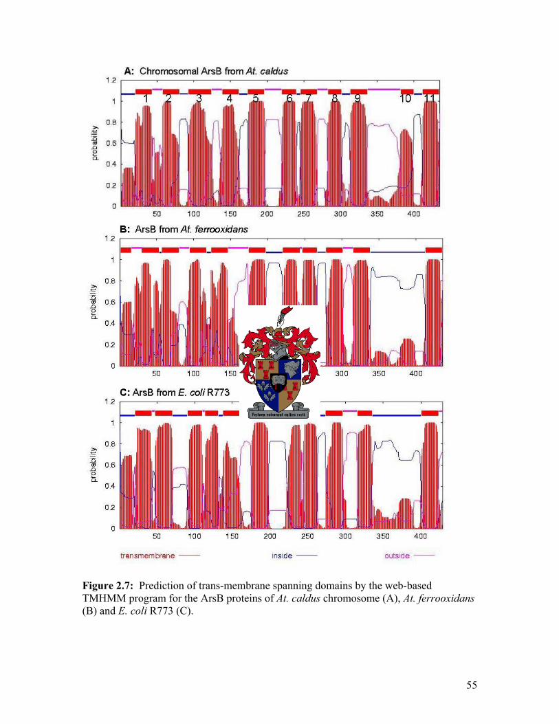
Figure 2.7: Prediction of trans-membrane spanning domains by the web-based TMHMM program for the ArsB proteins of At. caldus chromosome (A), At. ferrooxidans (B) and E. coli R773 (C).
55

As discussed in Chapter 1 (1.4.2) bacterial ArsC proteins can be divided into two
families. The first family (GSH/Grx clade) requires glutathione (GSH) and glutaredoxin
(Grx) to reduce arsenate to arsenite and includes the E. coli R773 ArsC. The second
family of arsenate reductases (Trx clade) includes the S. aureus pI258 and B. subtilis
ArsC proteins. Phylogenetic and alignment analysis showed that the At. caldus
chromosomal ArsC groups with other ArsC proteins of the Trx clade (Figure 2.8 and
2.9). Members of the GSH/Grx clade of ArsC proteins are more closely related to each
other than members of the Trx family with 93.52% sequence identity for the GSH/Grx
family and only 53.3% sequence identity for the Trx family. Alignments with ArsC
proteins in the NCBI database revealed that the At. caldus chromosomal ArsC is most
similar to the ArsC protein of At. ferrooxidans with 78% identity. It is only 72%
identical the At. caldus transposon ArsC protein. The Trx clade ArsC proteins use
thioredoxin as a reducing power and contain four cysteine residues, Cys10, Cys15, Cys82
and Cys89, of which only three are essential (Cys10, Cys82 and Cys89) for the conversion
of arsenate to arsenite. Besides the three cysteine residues, Arg16 and Asp105 are also
essential for catalysis. Sequence analysis of the At. caldus chromosomal ArsC revealed
that it contains four cysteine residues (Cys10, Cys15, Cys82 and Cys90) with spacing similar
to the spacing in the ArsC proteins of the Trx clade (Figure 2.9). The third and fourth
cysteines are separated by seven amino acids instead of six, as is the case with At.
ferrooxidans, P. putida and P. aeruginosa. Arg16 and Asp105, which are essential for
catalysis by the Trx ArsC are also conserved in the At. caldus ArsC.
As discussed in Chapter 1 (1.4.3.1), the ArsR protein of E. coli R773 contains a possible
DNA binding helix-turn-helix domain (Bairoch, 1993) and a highly conserved 30ELCVCDL36 metal binding domain with conserved Cys32, Cys34 and Cys37 residues.
Binding requires only two cysteine residues, and any combination of Cys32, Cys34 and
Cys37 will result in the release of the repressor from the DNA (Shi et al., 1994; Shi et al.,
1996). Busenlehner et al. (2003) reported that two distinct metal binding sites exist in the
SmtB/ArsR family of transcriptional regulators. The first (α3N) metal binding site is
present in the α3 helix and contains the conserved ELCVCDL domain. The second metal
binding site (α5C) is present in the α5 helix (Figure 1.8). The predicted amino acid
56
sequence of the At. caldus chromosomal ArsR was most similar to the ArsR protein of At.
ferrooxidans with 74% similarity, while it was only 45% identical to the transposon-
linked ArsR protein of At. caldus. An alignment of the At. caldus chromosomal ArsR
protein with its closest matches from the NCBI database revealed that none of the ArsR
proteins contained the conserved ELCVCDL domain in the α3 helix region (Figure 2.10),
indicating that the chromosomal ArsR and its closest homologs may use a different metal
binding site than ArsR proteins with the ELCVCDL metal binding domain. The At.
caldus chromosomal ArsR and its eight closest matches contains the conserved GXIA
region (identified by Butcher and Rawlings, 2002) immediately downstream of the region
corresponding to the ELCVCDL binding site. They all also contain another conserved
LVAYLTENCC region near the C-terminal end of the protein. Secondary structure
prediction of the At. caldus chromosomal ArsR (performed by the web-based program
PsiPRED v2.4, http://www.predictprotein.org) revealed that it contains, like other ArsR
proteins, two possible helix-turn-helix motifs (Figure 2.11). The position and spacing of
these helix-turn-helix domains of the At. caldus chromosomal ArsR is very similar to
those of the well studied E. coli R773 ArsR. All proteins that lacked the conserved metal
binding domain have two extra residues before the αR helix (Figure 2.11). The
significance of these extra residues in arsenic resistance has yet to be shown.
Phylogenetic analysis showed that the chromosomal At. caldus ArsR, together with its
closest matches form a subgroup of ArsR regulators (Figure 2.12).
57

Figure 2.8: Phylogenetic tree of different ArsC proteins. The At. caldus chromosomal ArsC is most closely related to the transposon-located ArsC of At. caldus and the ArsC of At. ferrooxidans. Accession numbers: E. coli R773, AAA21096; E. coli chromosome, NP_417960; A. multivorum pKW301, BAA24824; K. oxycota, AF168737_5; E. coli R46, NP_511240; S. marcescens, CAB88404; Y. enterocolitica pYVe227, NP_052438; A. tumefaciens C58, NP_532180; B. subtilis skin element, ARSC_BACSU; S. aureus pI258, ARSC_STAAN; S. xylosus pSX267, ARSC_STAXY, At. ferrooxidans, AAF69239; At. caldus transposon, AAX35680; P. putida, CAC18654; P aeruginosa, NP_250969.
58

GSH/Grx clade 55Ec R773 55Ec chrom 55Ec R46 55K oxytoca 55S marces 55Y ent pYV 55Am pKW301
MSNITIYHNPACGTSRNTLEMIRNSGTEPTIILYLENPPSRDELVKLIADMGISVMSNITIYHNPACGTSRNTLEMIRNSGTEPTIIHYLETPPTRDELVKLIADMGISVMSNITIYHNPHCGTSRNTLEMIRNSGIEPTVILYLETPPSRDELLKLIADMGISVMSNITIYHNPACGTSRNTLEMIRNSGNEPTVIHYLENPPSRDELVKLIADMGISVMSNITIYHNPACGTSRNTLEMIRNSGTEPTVIHYLETPPSRAELVKLIADMGITVMSNITIYHNPTCGTSRNTLEMIRNSGNEPTVIYYLETPPTHDELVKLIADMGITVMSNITIYHNPACGTSRNTLEMIRNSGNEPTVIHYLENPPSRDELVKLIADMGISV
MMMMMMM
SSSSSSS
NNNNNNN
IIIIIII
TTTTTTT
IIIIIII
YYYYYYY
HHHHHHH
NNNNNNN
PPPPPPP
AA
AA
A
CCCCCCC
GGGGGGG
TTTTTTT
SSSSSSS
RRRRRRR
NNNNNNN
TTTTTTT
LLLLLLL
EEEEEEE
MMMMMMM
IIIIIII
RRRRRRR
NNNNNNN
SSSSSSS
GGGGGGG
EEEEEEE
PPPPPPP
TTTTTTT
IIVVVVV
IIIIIII
H
HH
H
YYYYYYY
LLLLLLL
EEEEEEE
TT
TT
PPPPPPP
PPPPPPP
S
SSS
S
RRRRRHR
DDDD
DD
EEEEEEE
LLLLLLL
VVLVVVV
KKKKKKK
LLLLLLL
IIIIIII
AAAAAAA
DDDDDDD
MMMMMMM
GGGGGGG
IIIIIII
SSSS
S
VVVVVVV
110Ec R773 110Ec chrom 110Ec R46 110K oxytoca 110S marces 110Y ent pYV 110Am pKW301
RALLRKNVEPYEQLGLAEDKFTDDQLIDFMLQHPILINRPIVVTPLGTRLCRPSERALLRKNVEPYEELGLAEDKFTDDRLIDFMLQHPILINRPIVVTPLGTRLCRPSERALLRKNVEPYEELGLAEDKFTDDQLIDFMLQHPILINRPIVVTPLGTKLCRPSERALLRKNVEPYEELGLAEDKFTDGELIDFMLQYPILINRPIVVTPLGTRLCRPSERALLRKNVEPFEALGLAEDRFTDEQLIDFMLQHPVLINRPIVVTPLGTRLCRPSERALLRKNVEPYEELGLAEGTFSDEQLIGFMLEHPILINRPIVVTPLGTRLCRPSERALLRKNVEPYEELGLEEDKFTDDQLIDFMLQHPILINRPIVVTPLGTRLCRPSE
RRRRRRR
AAAAAAA
LLLLLLL
LLLLLLL
RRRRRRR
KKKKKKK
NNNNNNN
VVVVVVV
EEEEEEE
PPPPPPP
YYYYFYY
EEEEEEE
QEEE
EE
LLLLLLL
GGGGGGG
LLLLLLL
AAAAAA
EEEEEEE
DDDDDGD
KKKKR
K
FFFFFFF
TTTTT
T
DDDDDDD
DDDGEED
Q
QEQQQ
LLLLLLL
IIIIIII
DDDDDGD
FFFFFFF
MMMMMMM
LLLLLLL
QQQQQEQ
HHH
HHH
PPPPPPP
IIIIVII
LLLLLLL
IIIIIII
NNNNNNN
RRRRRRR
PPPPPPP
IIIIIII
VVVVVVV
VVVVVVV
TTTTTTT
PPPPPPP
LLLLLLL
GGGGGGG
TTTTTTT
RRKRRRR
LLLLLLL
CCCCCCC
RRRRRRR
PPPPPPP
SSSSSSS
EEEEEEE
140Ec R773 140Ec chrom 140Ec R46 137K oxytoca 140S marces 140Y ent pYV 140Am pKW301
VVLDILQDAQKGAFTKEDGEKVVDEAGKRLVVLEILPDAQKGAFSKEDGEKVVDEAGKRLVVLDILPDAQKAAFTKEDGEKVVDDSGKRLVVLDILPDAQKGAFAKEDGEKVVDETR...VVLDILYDAQKSAFTKEDGEKVVDEKGNRLVVLDILPEPQQGAFTKEDGEKITDESGKRLVVLDILPDAQKGAFAKEDGEKVVDEAGKRL
VVVVVVV
VVVVVVV
LLLLLLL
DEDDDDD
IIIIIII
LLLLLLL
PPP
PP
DDDDDED
AAAAAPA
QQQQQQQ
KKKKK
K
GGAGSGG
AAAAAAA
FFFFFFF
T
T
TT
KKKKKKK
EEEEEEE
DDDDDDD
GGGGGGG
EEEEEEE
KKKKKKK
VVVVVIV
VVVVV
V
DDDDDDD
EEDEEEE
GGG
GGG
KKK
KK
RRR
RRR
LLL
LLL
Trx clade
55Bs skin 55At_ferro 55Sx_pSX267 52P_aerug__ 52P_putida 55Sa_pI258 55At_c_Ch
MENKIIYFLCTGNSCRSQMAEGWAKQYLGDEWKVYSAGIEAHGLNPNAVKAMKEVMKTPEILFLCTGNSCRSILAEVTFNALAGPGMHATSAGSHPAGYVHTRSINLLERMDKKTIYFICTGNSCRSQMAEGWGREILGEDWNVYSAGIETHGVNPKAIEAMKEV...MRVLFMCTANSCRSILSEAMFNHLAPPGFEACSAGSQPSGRVHPRSLATLEQ...MKVLFMCTANSCRSILSEAMFNHLAPDGFEAISSGSFPKGLVLPRSLSTLQAMDKKTIYFICTGNSCRSQMAEGWGKEILGEGWNVYSAGIETHGVNPKAIEAMKEVMKTPEILFLCTGNSCRSILAEATLNGLAQGRLHASSAGSHPAGYVHPRSVALLQR
MMM
MM
IIIVVII
L
LL
L
FFFFFFF
CCCCCCC
TTTTTTT
GGGAAGG
NNNNNNN
SSSSSSS
CCCCCCC
RRRRRRR
SSSSSSS
I
II
I
MLMLLML
AAA
AA
EEEEEEE
N
NN
N
LILLIL
A
AA
A
GGG
G
EGDGGG
A
AA
A
SSSSSSS
AAAA
AA
GGGGGGG
S
SS
S
AP
PP
P
GGGGGGG
V
VV
V
R
RR
R
S
SS
S
A
AA
AA
L
LL
L
EEEEQEQ
108Bs skin 109At_ferro 108Sx_pSX267 107P_aerug__ 107P_putida 108Sa_pI258 109At_c_Ch
GIDISNQTSDIIDSDILNNADLVVTLCGDAADK..CPMTPPHVKREHWGFDDPAREGFRTDGLHSKSWEDLKET.PDIVITVCADAAGETCPAYLGPAIRTHWGVEDPAKDIDISNHTSDLIDNHILKQSDLVVTLCSDADDN..CPILPPNVKKEHWGLEDPAGAGIATHGLYSKGSEAFEGAPPDIVITVCDAAAGEACPLYLGAALKAHWGLADPSAAGIRTEGLYSKGNDVFEGSPPDVVITVCDKAAGEACPVYFGPAVKAHWGLEDPSDDIDISNHTSDLIDNDILKQSDLVVTLCSDADNN..CPILPPNVKKEHWGFDDPAGEGFSTEGLASKSWDDLPTT.PDIVITVCASAAGETCPAYLGPALRAHWGVDDPAK
G
GG
G
T
TT
T
G
GG
G
L
LL
L
S
SS
S
K
KK
K
DD
DD
P
PP
P
D
DD
D
VIVIVVI
VVVVVVV
I
II
I
T
TT
T
V
VV
V
C
CC
C
D
DDDD
A
AA
A
AA
AA
A
A
AA
A
G
GG
G
E
EE
E
CCCCCCC
PPPPPPP
Y
YY
Y
G
GG
G
A
AA
A
RRKKKKR
HHHHHHH
WWWWWWW
GGGGGGG
DDDDDDD
PPPPPPP
AAA
AA
139Bs skin 162At_ferro 131Sx_pSX267 156P_aerug__ 156P_putida 131Sa_pI258 162At_c_Ch
AQGTEEEKWAFFQRVRDEIGNRLKEFAETGK......................VTGTEAQIEAAFDTAYHILRHRIEALLQLPVAELLEKDPAKLRQELERIGTLLKEWSE......FQRVRDEIKLAIENFKLR........................LDGDEALRDAAFHATLARIEQRCRAFLGLPFATLDRDQLKR...ELERIGSL.VQGDDARVQAAFDATLKTIATRCRAFFILPFAELSPTELQA...ELARIAEL.KEWSE......FQRVRDEIKLAIEKFKLR........................ATGSEAEIEAAFEKAYRILRHRIEALLQLPVAEWLAEDPSRLRTELERIGHSE
GG
GG
G
EEEEDEE
A
AA
A
AA
AA
A
A
AA
A
FFFFFFF
ILIIIIL
RR
RR
R
LII
II
EE
EE
A
AA
A
FLFFFFL
L
LL
L
P
PP
P
A
AA
A
E
EE
E
L
LL
L
R
RR
R
I
II
I
Figure 2.9: Multiple sequence alignments of different ArsC proteins compared with the At. caldus chromosomal ArsC. The At. caldus chromosomal ArsC protein groups with other ArsC proteins of the Trx clade. See figure 2.5 for accession numbers.
59

Figure 2.10: Multiple sequence alignments of different ArsR proteins (the eight closest matches obtained from a NCBI BLAST search) compared with the chromosomal ArsR of At. caldus. Also included are proteins from the ArsR/SmtB family of transcriptional regulators. Labels and accession numbers are as follows: CadC, S. aureus pI258 CadC (B32561); Ec_chrom, E. coli chromosome (AAS76526); Ec_R773, E. coli R773 (CAA34168); pSX267, S. xylosus pSX267 (AAA27587); SmtB, Synechococcus PCC7942 SmtB, (S31197), ZntR, S. aureus ZntR (AAC32484); Atc_chr, At. caldus chromosome. The nine closest matches are labeled by their accession numbers: AAF69241, At. ferrooxidans; AAX35682, At. caldus transposon; AAS45114, Alcaligenes faecalis; YP_159865, Azoarcus sp. EbN1; AAQ60110, Chromobacterium violaceum; YP_111442, Burkholderia pseudomallei K96243; YP_113846, Methylococcus capsulatus str. Bath; ZP_00627778, Nitrobacter hamburgensis X14. The conserved ELCVCDL metal binding motif found in members of the ArsR/SmtB family of transcriptional regulators has been boxed with solid lines, while conserved regions of an atypical ArsR group identified by Butcher and Rawlings (2002) are boxed in broken lines. The At. caldus chromosomal ArsR groups with the atypical ArsR group and also contains the two conserved domains. Also shown is the At. caldus chromosomal ArsR α3 and α5-helices.
60

Figure 2.11: Secondary structure prediction of the (A) At. caldus chromosomal ArsR, (B) At. ferrooxidans ArsR, (C) At. caldus transposon ArsR and (D) E. coli R773 ArsR by the PsiPRED web-based program. Also included are the α-helices and β-sheets as indicated by Busenlehner et al. (2003). Proposed metal binding domains are located in the α3 and α5-helices, as described in the text.
61

Figure 2.12: Phylogenetic tree of different ArsR proteins. The At. caldus chromosomal ArsR and its eight closest matches cluster as a second subgroup of ArsR regulators. See Figure 2.10 for accession numbers.
62

ORF1 showed sequence identity to a lactoylglutathione lyase (also known as glyoxalase
I) of Thermobifida fusca. Glyoxalase I forms part of the glyoxalase pathway that protects
against cellular damage caused by reactive aldehydes such as methylglyoxal with the help
of glutathione hemithioacetal as substrate (Vickers et al., 2004). Glyoxalase I and
glyoxalase II (hydroxyacylglutathione hydrolase) act in concert to convert the
hemithioacetal adduct formed between glutathione and methylglyoxal into D-lactate and
free glutathione (Thornalley, 1996). Because ORF1 is located adjacent to arsC, the
possibility exists that ORF1 may be involved in the conversion of arsenate to arsenite by
the ArsC (if it uses glutathione as a reducing power). ORF1 also showed sequence
identity to a cadmium inducible protein (CadI) of Mycobacterium tuberculosis. cadI
homologs are often found adjacent to, or within ars operons (Hotter at. al., 2001). CadI
is related to members of the βαβββ metalloenzyme superfamily, which has important
detoxification functions (Bernat et al., 1997). When looking at the sizes of the predicted
lactoylglutathione lyase and CadI proteins, it seems more likely that ORF1 functions as a
CadI protein. Furthermore, ORF1 is located adjacent to the chromosomal ars operon of
At. caldus, as is the case with several CadI proteins of other bacteria (Hotter et al., 2001).
Cadmium induces the ars operon of B. subtilis (Moore et al., 2005). Experiments were
conducted to determine if ORF1 may be involved in the conversion of arsenate to arsenite
(2.3.3) and to determine if the At. caldus chromosomal ars operon can be induced by
cadmium, as is the case of B. subtilis (3.3.1).
2.3.3. The requirement of glutathione/thioredoxin for arsenate reductase
It is well known that ArsC proteins are grouped on their source of reducing power that is
used for the reduction of arsenate to arsenite. The first family (GSH/Grx clade) uses
glutathione and glutaredoxin to reduce arsenate, while the second family (Trx clade) uses
thioredoxin for the reduction of arsenate to arsenite (Mukhopadhyay et al., 2002;
Mukhopadhyay and Rosen, 2002). Based on the observation that the At. caldus
chromosomal ArsC grouped with other ArsC proteins of the Trx clade (Figure 2.8 and
2.9) and that it was 78% identical to the At. ferrooxidans ArsC (which has been shown to
use thioredoxin to reduce arsenate to arsenite, Butcher et al., 2000), tests were conducted
63

to determine whether the At. caldus chromosomal ArsC protein would use thioredoxin
rather than glutathione and glutaredoxin as a reducing power for the reduction of arsenate
to arsenite. To investigate this, an E. coli strain (BH5262) with mutations in the
thioredoxin gene (trxA) and the γ-glutamylcysteinyl synthetase gene (gshA, responsible
for the synthesis of glutathione) was transformed with various plasmids. E. coli BH5262
transformed with the At. caldus chromosomal ars genes (pTcC-PH29GL) and Trx6
(contains the At. ferrooxidans trxA gene) were unable to confer resistance to 1 mM
arsenate. The ars mutant ACSH50Iq transformed with pTcC-PH29GL did confer
resistance to 1 mM arsenate, indicating that the At. caldus chromosomal ars genes were
sufficient for resistance to 1 mM arsenate (Table 2.3). Surprisingly, the At. ferrooxidans
thioredoxin containing plasmid, pTrx6, did not permit the E. coli double mutant BH5262
containing the cloned At. caldus chromosomal ars genes (pTcC-PH29GL) to grow on
arsenate, as reported when the At. ferrooxidans ars genes were tested in a similar
experiment (Butcher and Rawlings, 2000). Because the At. caldus chromosomal ArsC
groups with the ArsC proteins of the Trx clade, it seems very unlikely that it will use
glutathione as a reducing power. It is possible that the At. ferrooxidans trxA gene may
not complement the At. caldus chromosomal ArsC in the reduction of arsenate to
arsenite. This experiment could be repeated using a cloned trxA gene from At. caldus to
confirm if the At. caldus chromosomal ArsC uses thioredoxin in the conversion of
arsenate to arsenite. Despite grouping with other ArsC proteins of the Trx clade (Figure
2.9), it is possible that the At. caldus chromosomal ArsC may use glutathione to reduce
arsenate to arsenite and that ORF1 may play a role in this reduction. To investigate this,
E. coli BH5262 was transformed with pTcC-#4. If ORF1 aids the ArsC in reducing
arsenate to arsenite, arsenate resistance would be acquired. This was not the case
however, as E. coli BH5262 transformed with pTcC-#4 was sensitive to 1mM arsenate
(Table 2.3). This also indicated that none of the other ORFs on pTcC-#4 aid in the
reduction of arsenate to arsenite.
64

Table 2.3: Arsenate resistance in E. coli mutant strains
E. coli strain Transformed with
plasmid
Resistance to 1mM
arsenate a
BH5262
pGL10 + Trx6
–
BH5262
pGL10 + pKS
–
BH5262
pTcC-PH29GL + pKS
–
BH5262
pTcC-PH29GL + Trx6
–
ACSH50Iq
pTcC-PH29GL
+
BH5262
pTcC-#4
–
a – indicates no growth on Luria Agar with 1mM sodium arsenate; + indicates growth on Luria Agar with 1mM sodium arsenate
2.3.4. ORF1 and ORF5 are not required for arsenic resistance in E. coli
Arsenic assays were performed in order to determine (a) the resistance to arsenic that
construct pTcC-#4 isolated from At. caldus confers on the E. coli arsenic mutant
ACSH50Iq, (b) to determine if ORFs 5-10 (Figure 2.4; Table 2.2) play a role in arsenic
resistance and (c) to determine if ORF1 aids the arsC gene product in converting arsenate
to arsenite (see section 2.3.3). Growth assays were performed in 0.25 mM arsenite (AsIII)
and 0.5 mM arsenate (AsV). Plasmid pTcC-#4 conferred greater resistance to the E. coli
ars mutant ACSH50Iq in both arsenate and arsenite in comparison to the negative control
pEcoBlunt (Figure 2.13 a and b). Construct pTcC-#6 conferred lower resistance to
arsenate, which was expected as it does not contain a functional arsC that is required for
the conversion of arsenate to arsenite. Arsenic resistance did not change significantly in
65

cells harboring only arsCRBORF5 (pTcC-PH29), suggesting that ORFs 6-10 were not
required for arsenic resistance in E. coli ACSH50Iq. Since ORFs 6-10 were not needed
for arsenic resistance, we could directly compare pTcC-#4 and pTcC-PH29. No
differences in levels of resistance to arsenate were detectable between pTcC-#4 and
pTcC-PH29. This indicated that ORF1 is not required for the reduction of arsenate to
arsenite in E. coli.
Construct pTcC-Bs2.6 (which contains arsCRB) also showed high levels of resistance to
arsenate and arsenite (Figure 2.13 a and b). As it does not contain ORF5, it was
suggested that ORF5 is not necessary for arsenic resistance in E. coli. To determine the
role ORF5 plays in arsenate resistance, pTcC-RB+5 and pTcC-RB-5 were constructed.
Growth curves done with these constructs showed the same levels of resistance as pTcC-
#6, indicating that ORF5 does not play a role in arsenate resistance in E. coli ACSH50Iq
in the absence of ArsC.
66

Figure 2.13: Growth curves performed with the E. coli ars mutant ACSH50Iq, in the presence of 0.25 mM arsenite (A) and 0.5 mM arsenate (B). Each data point represents the results of three assays of three independent experiments. Error bars indicate standard deviations. See figure 2.4 for location of the constructs. Optical density (OD) readings were recorded at 600 nm. 2.4. Discussion Acidithiobacillus caldus is a member of a consortium of bacteria used in a commercial
biooxidation process called biomining. During this process, toxic arsenic compounds are
released into the environment. Due to the fact that At. caldus is able to live in such a
harsh environment, it was not surprising to find arsenic resistance genes in its genome.
In this study we identified a chromosomal ars operon that conferred resistance to arsenate
and arsenite. It consists of three ars genes, arsCRB, with no arsA or arsD genes present.
This is not surprising, as the conventional chromosomal ars operon of bacteria like E.
coli also contains an arsRBC operon (Diorio et al., 1995). However, the orientation of
the At. caldus chromosomal ars operon was atypical. Where the conventional
chromosomal ars operons are transcribed as a single transcriptional unit, the At. caldus
67

chromosomal ars operon is divergently transcribed, with the arsC and arsR genes
divergently transcribed to the arsB gene, as has also been shown for At. ferrooxidans.
Results of BLAST searches in the NCBI database revealed that all three chromosomal
arsenic resistance genes from At. caldus are most homologous to the arsenic resistance
genes of another biomining bacterium, Acidithiobacillus ferrooxidans. As described
earlier, At. caldus is a moderately thermophilic sulfur oxidizer and At. ferrooxidans a
mesophilic iron- and sulfur oxidizer. The fact that such high homology was found (78%,
74%, and 85% identity for the arsC, arsR and arsB genes, respectively) between the ars
genes of At. caldus and At. ferrooxidans strengthens the hypothesis that they may have a
relatively recent common ancestor. The chromosomal ArsB protein of At. caldus
grouped with ArsB proteins of other Gram-negative bacteria (Figure 2.6). Using a web-
based program that predicts transmembrane spanning domains we predicted that the
chromosomal ArsB has 11 membrane spanning domains. In ars systems where an ArsA
is absent, it is believed that the ArsB cannot function as a primary pump using ATP
hydrolysis as an energy source, but as a secondary carrier using membrane potential to
export arsenite out of the cell. However, acidophilic microorganisms commonly have a
reversed membrane potential, with a positive rather than a negative inside membrane
potential (Martin, 1990). Despite originating from an organism with a positive inside
membrane potential, the chromosomal At. caldus ArsB efflux pump was functional in E.
coli, as resistance to arsenite was observed.
The At. caldus chromosomal ArsC grouped with ArsC proteins of the Trx family (Figure
2.9), suggesting that it will be more likely to use thioredoxin than glutathione and
glutaredoxin as a reducing power for the conversion of arsenate to arsenite. To
investigate this, E. coli BH5262 was transformed with the At. ferrooxidans thioredoxin
gene (trxA) and pTcC-#4 in trans. It was surprising to find that this gene was unable to
confer arsenate resistance to E. coli BH5262. It is possible that the At. ferrooxidans trxA
gene may not complement the At. caldus chromosomal ArsC to reduce arsenate to
arsenite, and therefore results in a arsenate sensitive phenotype. E. coli BH5262
transformed with pTcC-#4 was also sensitive to arsenate, indicating that none of the
68

ORFs on pTcC-#4 were able to function as a reducing power to help ArsC in the
conversion of arsenate to arsenite. Sequence analysis of the At. caldus chromosomal
ArsC revealed that it contains four cysteine residues (Cys10, Cys15, Cys82 and Cys90), and
that it is spaced similar to the spacing in other ArsC proteins of the Trx family. It also
contains Arg16 and Asp105, which have been shown to be essential for catalysis of the Trx
family ArsC. Based on this sequence analysis, we proposed that the At. caldus
chromosomal ArsC will function similar to the B. subtilis ArsC (Figure 1.7).
Sequence alignments of members of the ArsR family of transcriptional regulator proteins
showed that these proteins show low similarity to each other. Alignments of the At.
caldus chromosomal ArsR with eight of its closest matches from the NCBI database
revealed that none of the ArsR proteins contained the conserved ELCVCDL metal-
binding domain (Figure 2.10). Phylogenetic analyses of these ArsR proteins show that
they form a subgroup of ArsR regulators (Figure 2.12). Using a web-based program
(PsiPRED v. 2.4), we predicted that the At. caldus chromosomal ArsR contains, like other
ArsR proteins, two helix-turn-helix domains with 5 α-helices and 2 β-sheets (Figure
2.11). The At. caldus ArsR and its eight closest matches from the NCBI database all
have two extra residues before the first helix of the identified DNA-binding helix-turn-
helix domain.
The SmtB/ArsR family of transcriptional regulators have two metal binding sites, the
α3N in the α3 helix and the α5C metal-binding site in the α5 helix. They have shown that
ArsR proteins contain the α3N metal binding site, with the ELCVCDL domain located in
the α3 helix. The At. caldus chromosomal ArsR and its closest matches all contain a very
conserved LVAYLTENCC domain near the C-terminal end of the protein (Figure 2.10).
This conserved domain is not present in ArsR proteins with the consensus ELCVCDL
domain. Five to seven residues separate another cysteine from the cysteine doublet at the
end of the LVAYLTENCC domain of At. caldus and all of its closest matches. These
three cysteine residues may form the metal-binding domain of these atypical ArsR
proteins. This conserved region identified in the second ArsR group is present in the α5
helix (Figure 2.10). It is thus possible that the ArsR proteins may have two different
69

metal-binding domains, where one is located in the α3 helix and the other in the α5 helix.
Analysis of a possible At. caldus chromosomal ArsR binding site did not reveal the
TTTG and TCAT region identified in both the chromosomal and R773 ars sequences of
E. coli.
Along with the three arsenic resistance genes, seven other ORFs were identified on
pTcC-#4 (Table 2.2). Based on the proximity of ORF7-10 to one another and the
direction of transcription, it is possible that these ORFs form an operon separate form the
ars operon. The function of this possible operon is unknown, but it may play a role in the
transport of certain molecules in and out of the cell (Figure 2.4; Table 2.2).
Both the arsC and arsB genes were expressed and were functional in the E. coli ars
mutant ACSH50Iq. Growth assays in the presence of arsenite and arsenate were
conducted to determine if the other ORFs play a role in arsenic resistance. It was found
that ArsC is needed for resistance to arsenate, but not arsenite, while ArsB was essential
for resistance to arsenite. Comparing arsenic resistance conferred by the At. caldus
chromosomal ars operon to arsenic resistance conferred by the At. caldus transposon ars
operon (Tuffin et al., 2005), it seems that the transposon operon conferred much higher
resistance to E. coli than the chromosomal ars operon. We did expect constructs not
containing ArsC (pTcC-#6, pTcC-RB-5 and pTcC-RB+5) to be more sensitive to
arsenate than was observed (Figure 2.13 a and b). A reason for increased arsenate
resistance by these constructs may be that the ars mutant ACSH50Iq contains an element
on the chromosome that can help with the conversion of arsenate to arsenite, which can
then be transported out of the cell by the At. caldus ArsB.
This studies of the At. caldus chromosomal arsenic resistance system was carried out in
E. coli. It would have been ideal if this studies could be repeated in At. caldus, as this
system may not function the same way in E. coli as in At. caldus. Due to a lack of
mutants, genetic tools for this organism and the difficulty to obtain large quantities of
cells, these studies are limited to E. coli at present.
70

Chapter Three
Regulation and cross-regulation by the Acidithiobacillus caldus chromosomal ArsR
Contents 3.1. Introduction ---------------------------------------------------------------------------- 72
3.2. Methods
3.2.1. Media, bacterial strains and plasmids ----------------------------------- 73
3.2.2. DNA isolation and manipulations --------------------------------------- 75
3.2.3. PCR -------------------------------------------------------------------------- 76
3.2.4. Construction of the promoter-lacZ fusions ----------------------------- 76
3.2.5. β-galactosidase assays ----------------------------------------------------- 76
3.2.6. RNA analysis and RT-PCR ----------------------------------------------- 78
3.3. Results
3.3.1. Reporter-gene studies to investigate regulation of putative
promoter regions ------------------------------------------------------------ 78
3.3.2. ORF1 and ORF5 are co-expressed with arsCRB ----------------------- 82
3.3.3. Cross-regulation by the chromosomal ars system ---------------------- 85
3.4. Discussion ---------------------------------------------------------------------- 87
71

3.1. Introduction Together with the work presented in Chapter 2, it is now clear that At. caldus #6 has three
arsenic resistance operons, with two being located on transposons and a third on the
chromosome. This raises interesting questions about how each of the systems interact to
give arsenic resistance, and how each of these systems are regulated to give optimal
resistance. In Chapter 4 work will be presented on the interaction of the transposon and
chromosomal system to give arsenic resistance, while this chapter will focus on the
regulatory mechanisms of the chromosomal operon, and whether the chromosomal and
transposon resistance determinants can cross-regulate each other.
Sequence analysis (Chapter 2) revealed that the At. caldus chromosomal ars genes are
arranged in the order arsCRB, with the arsC and arsR genes reading divergently to the
arsB gene. This could suggest that arsCR and arsB may be transcribed as two separate
transcriptional units. This is different to the typical arsRBC systems, which are
transcribed as a single transcriptional unit. This divergent arrangement of ars genes has
also been identified in At. ferrooxidans, where the arsCR were transcribed as one unit,
separate to arsBH (Butcher et al., 2000). We have shown in Chapter 2 that ORF1 and
ORF5 were not required for, and did not enhance, arsenic resistance in E. coli. However,
the gene layout (with stop and start codons very close together) could suggest co-
transcription with the ars genes.
In this chapter expression of the At. caldus chromosomal ars system will be analyzed.
Would this system be regulated by ArsR at a transcriptional level, binding to an operator
sequence and thereby blocking transcription? It has been shown that with single unit
operons like the chromosomal ars operon of E. coli K-12, the ArsR binds upstream of the
ars operon (Diorio et al., 1995). However, with the divergent operon, as has been shown
in At. ferrooxidans, the ArsR autoregulates itself as well as arsB expression (Butcher and
Rawlings, 2002). Sequence analysis of the At. caldus chromosomal ArsR revealed that it
does not contain the highly conserved ELCVCDL region that is found in members
belonging to the SmtB/ArsR family of transcriptional regulators (Shi et al., 1994). As
previously described, this domain forms part of metal binding site where arsenite binds in
72

order to induce a conformational change in ArsR, with the dissociation of the repressor
from the DNA. The At. caldus chromosomal ArsR was most similar to the ArsR protein
of At. ferrooxidans with 74% identity. The At. ferrooxidans ArsR and the At. caldus
transposon-located ArsR also lack the conserved metal binding domain, but have been
able to regulate their ars operons respectively (Butcher and Rawlings, 2002; Tuffin et al.,
2005). Based on the above, would ArsR function as a negative transcriptional regulator,
and to what inducers would it respond? Does the At. caldus chromosomal ArsR regulate
both itself and arsB? If it does regulate both, to what extent are they being regulated?
Due to the presence of two different ArsR proteins in one organism, would there be any
cross-regulation? The At. caldus transposon ArsR has been shown to function as a
typical ArsR (Tuffin et al., 2005). Despite the fact that the two ArsRs only share 45%
identity at protein level, there is a possibility of cross-regulation between the two ArsR
proteins.
3.2. Methods
3.2.1. Media, bacterial strains and plasmids
Bacterial strains, plasmids and primers used in this study are shown in Table 3.1. E. coli
strains were grown in Luria-Bertani (LB) broth medium (Sambrook et al., 1989) with
ampicillin (100 μg/ml) or kanamycin (50 μg/ml) added as required. At. caldus strains
were grown at 370C in tetrathionate medium (3 mM), sterilized and adjusted to pH 2.5
(Rawlings et al., 1999b).
Table 3.1: Bacterial strains, plasmids and primers used in this study Strain, plasmid or Primer
Description Source or reference
Strains E. coli DH5α Ф80dlacZΔM15, endA1, recA1, gyrA96, thi-1,
hsdR17 (rk-, mk
+), relA1, supE44, deoR,Δ(lacZYA-argF) U169
Promega
XL 1 Blue recA1, endA1, gyrA96, thi, hsdR17 (rk-, mk
+), relA1, Stratagene
73

supE44, relA1, lac [F’ proAB+ lacIqZΔM15 ::Tn10(tetr)]
ACSH50Iq rpsL, Δ(lac-pro), Δars::cam Butcher & Rawlings (2002)
Plasmids pEcoR252 Apr, EcoRI inactivation cloning vector Zabeau &
Stanley (1982) pBluescript SK Apr, LacZ’, ColE replicon, cloning vector Stratagene pGem-T® Apr, T-tailed PCR product cloning vector Promega pGL10 Kmr, RK2/RP4 replicon, cloning vector A. Toukdarian pMC1403 Apr, promoterless lacZY operon, ColE1 replicon Casadaban et
al. (1983) pKK223-3 Apr, ptac, ColE1 replicon, cloning vector Pharmacia pTcC-#4 Apr, 10kb Sau3A fragment of At. caldus #6 cloned
into the BglII site of pEcoR252 This study
ptacGL Kmr, 1.4 kb SphI-PvuI fragment from pKK223.3 in pGL10
Tuffin et al. (2005)
pGEM-ChArsR
Apr, PCR product of arsR (bp 1049-1544 ) obtained using ChArsRF/ ChArsRR primers, cloned in pGEM-T
This study
pKKChArsR Apr, 460 bp EcoRI-HindIII fragment from pGEM-arsR cloned in pKK223.3
This study
ptacChArsR Kmr, An arsR containing blunted 1.58 kb BamHI-ScaI fragment from pKKArsR cloned into a blunted SmaI site in pGL10
This study
pChArsRLacZ Apr, PCR product of arsR promoter (bp 1455-1955) obtained with ChArsRLacZF/ ChArsRLacZR primers in pMC1403.
This study
pChArsBLacZ Apr, PCR product of arsB promoter (bp 1383-1586) obtained with ChArsBLacZF/ ChArsBLacZR primers in pMC1403.
This study
pTnArs1GL Kmr, The “transposon-like” arsenic operon isolated from At. caldus #6, truncated in the tnpA gene, in the cloning vector pGL10.
Tuffin et al. (2005)
TnArsRLacZ Apr, At. caldus transposon PCR product of arsR promoter in pMC1403 (also named ArsRLacZ , Tuffin et al., 2005)
Tuffin et al. (2005)
ptacTnArsR Kmr, At. caldus transposon arsR in pGL10 (also
named ptacArsR, Tuffin et al., 2005) Tuffin et al. (2005)
Primers* Sequence (5’-3’) ArsR-RT_Fw CGAAGAGCACAGCGATCG This study ArsC-RT_Rev CCAGGATGGAACGACAGG This study
74

ORF1-RT_Rev GATCGCGCAGCCAGAGTT This study ArsC-RT_Fw CGGAAATCGAGGCCGCTT This study ORF5-RT_Rev GTTTGGCAGGGATTGCGG This study ArsB-RT_Fw ACCGTTCTGGCGGCGATT This study ORF6-RT-Rev ATCTTCCTGGGTGCTGCC This study ORF5-RT_Fw CGGGGAAGATCCTAGGTG This study ChArsBLacZF (EcoRI)
CGGAATTCTCGTGCTCCACCAGTAGG This study
ChArsBLacZR (BamHI)
CCGGATCCAAGATGGCCAGGGCAAGC This study
ChArsRLacZF (EcoRI)
CGGAATTCAAGTCGAGCCGCAACAGG This study
ChArsRLacZR (BamHI)
CCGGATCCCGATCGCTGTGCTCTTC This study
ChArsRF (EcoRI)
CGAATTCGCCGGTAGTCTTGACGAG This study
ChArsRR (HindIII)
CCCAAGCTTCCAGGATGGAACGACAGG This study
ArsRLacZR2 (BamHI)
CGCGGATCCAGCGCGTTAGCAATCGC Tuffin et al. (2005)
TnpRLacZR (BamHI)
CGCGGATCCAGGATCGAACGGCGTG Tuffin et al. (2005)
ArsRF2 (EcoRI)
CGAATTCTGATGGTTGTAGTATTATTTAA Tuffin et al. (2005)
ArsRR (HindIII)
CCCAAGCTTCCGGTACAAAGAAACAGG Tuffin et al. (2005)
*Restriction enzyme sites incorporated into primers are indicated in parenthesis and are
shown in bold in the primer sequence.
3.2.2. DNA isolation and manipulations
Small-scale plasmid preparation, restriction endonuclease digestion, ligation and gel
electrophoresis were performed using standard methods described previously (Sambrook
et al., 1989). Large-scale preparation of plasmid DNA was done with the Nucleobond
AX100 (Macherey-Nagel) or by the alkaline lysis method followed by CsCl gradient
centrifugation (Current protocols). Sequencing was done by the dideoxy chain
termination method, using an ABI PRISMTM 377 automated DNA sequencer and the
sequence was analyzed using a variety of software programmes, but mainly by the PC
based DNAMAN (version 4.1) package from Lynnon Biosoft.
75

3.2.3. PCR
The polymerase chain reaction was performed with primers described in Table 3.1, using
50 ng of plasmid DNA in a 50 μl volume containing 2 μM MgCl2, 0.25 μM of each
primer, 200 μM each of dNTP’s and 1 U TaqI polymerase. The reaction was carried out
in a Biometra thermocycler with an initial denaturation temperature of 940C for 60s,
followed by 25 cycles of denaturation (30s at 940C), an annealing step of 30s, and a
variable elongation step at 720C. Annealing temperatures and elongation times were
altered as required.
3.2.4. Construction of the promoter-lacZ fusions
The putative promoter regions for the chromosomal arsR and arsB were amplified by
PCR using the following primer pairs: ChArsBLacZF/ChArsBLacZR (bp 1417-1938) for
arsB and ChArsRLacZF/ChArsRLacZR (bp 1400-1569) for arsR (Figure 3.1). The
cloning of the putative promoter regions for the transposon-located arsR is described by
Tuffin et al. (2005). The PCR products were digested with either BamHI or EcoRI-
BamHI and ligated to the promoterless lacZ reporter gene of pMC1403. Fusions were
confirmed by DNA sequencing.
3.2.5. β-galactosidase assays
Overnight cultures were diluted 1:200 into fresh medium containing the appropriate
antibiotics, 0.4 mM IPTG, sodium arsenate or sodium arsenite (25 μM) when indicated,
and incubated at 300C to an OD600 of 0.5. The β-galactosidase activities were measured
using the method of Miller (1972).
76

arsC start arsR end 1 AGAAAGAGGATCTCGGGGGTTTTCATCGGTGTCACCTCGGTGGGGCGGCGGAAGCGGTGG L F L I E P T K M * R P P A A S A T A 61 CTGCTGCGCTGGATTCGGACTCGTCGGGGATAGTGCAGGATTGGGTGCCGCGACAGCAGT A A S S E S E D P I T C S Q T G R C C N 121 TTTCGGTAAGGTAGGCGACCAGGGAGCGCACCACGGGCATCTGCGCCCGGTAGCGCTGGA E T L Y A V L S R V V P M Q A R Y R Q F 181 AACGTCCCTCGCGGTGGACGATGACGAGATCGGCGTGCTGCAGAACCTTGAGGTGAAAGG R G E R H V I V L D A H Q L V K L H F S 241 ATACGCCGTTGTGCAGTTGCCCGATCCGTTCGGCAATCTGGCCGGAGAAAAGGCCTTCCG V G N H L Q G I R E A I Q G S F L G E P ChArsBLacZF 301 GCTCGTGCTCCACCAGTAGGCGAAAAATGGCGAGCCGCACCGGCGAGGCCAGGGCCTCGA E H E V L L R F I A L R V P S A L A E L ChArsRLacZR arsR start 361 GGCGGGCGACAATGCGATCGCTGTGCTCTTCGGTCATGGGATTCCAATGGTTCAAGTTAG R A V I R D S H E E T M 421 ATTGAATTATTGACAAATGTTTTTGGCTCGTCAAGACTACCGGCAATTTCACCCAAGGAG arsB start ChArsBLacZR 481 TACAGGATGCTTGCCCTGGCCATCTTCGTTGTGACCCTGATCCTCGTCATCTGGCAGCCC M L A L A I F V V T L I L V I W Q P 541 AAGGGACTCCAGATCGGTTGGAGTGCCATGGGCGGTGCGGTCGTCGCCCTGGCCACCGGG K G L Q I G W S A M G G A V V A L A T G 601 GTCATCACCTGGAGCGATATTCCTGTGGTCTGGCATATCGTCTGGGATGCTACCTTTACC V I T W S D I P V V W H I V W D A T F T 661 TTCGTCGCCCTCATCATCATCTCCCTGATTCTGGACGAAGCCGGTTTCTTCCATTGGGCG F V A L I I I S L I L D E A G F F H W A 721 GCTCTGCACGTGGCGCGCTGGGGCGGTGGGCGTGGGCGTCGGCTCTTCCCCCTCATCGTC A L H V A R W G G G R G R R L F P L I V 781 CTGCTGGGGGCGATGATTTCTGCCGTATTCGCCAACGACGGGGCGGCGCTGCTGCTCACC L L G A M I S A V F A N D G A A L L L T ChArsRLacZF 841 CCCATCGTCATGGCCATCCTGTTGCGGCTCGACTTCTCACCCAAGGCCACCTTCGCCTTT P I V M A I L L R L D F S P K A T F A F Figure 3.1: Sequence of the arsR-arsB intergenic region. The primers used to create the arsR-lacZ and arsB-lacZ fusions (underlined), as well as the arsR start and end codons and the arsB start codon (grey boxes) are shown. Arrows indicate the direction of transcription. Putative ribosome binding sites for the arsR and arsB are marked in bold.
77

3.2.6. RNA analysis and RT-PCR
Total RNA was isolated as described previously (Trindade et al., 2003) from 50 ml of
mid-exponential phase cultures of E. coli ACSH50Iq carrying various plasmids, grown in
LB containing 25μM arsenate or μM arsenite or no arsenic, and with antibiotic selection.
RNA was also isolated from At. caldus #6. Cells were first grown in tetrathionate
medium without arsenite, and were then diluted 1:100 into fresh medium containing
25μM arsenite or no arsenite. For RT-PCR, the 1st Strand cDNA synthesis kit (AMV;
Roche) was used for cDNA synthesis and cDNA product detection. The protocol of the
manufacturer was used for the reverse-transcriptase reaction. The PCR was performed as
described above using 1.5μl of the 20μl (total volume) reverse-transcriptase reaction, and
the extension times were altered as required for the different primer pairs. The ORF1-
RT_Rev, ArsC-RT_Rev, ORF5-RT_Rev, and ORF6-RT-Rev primers were used for
cDNA synthesis from ORF1, arsC, ORF5 and ORF6 mRNA respectively (Table 3.1;
Figure 3.5). For the PCR, the following primers were used in combination: ORF1-
RT_Rev/ArsC-RT_Fw; ArsC-RT_Rev/ArsR-RT_Fw; ORF1-RT_Rev/ArsR-RT_Fw;
ORF5-RT_Rev/ArsB-RT_Fw; ORF6-RT-Rev/ORT5-RT_Fw and ORF6-RT-Rev/ArsB-
RT_Fw. To detect DNA contamination in the mRNA extracts, reactions were performed
with each primer pair without any AMV reverse transcriptase.
3.3. Results
3.3.1. Reporter-gene studies to investigate regulation of putative promoter
regions
ArsR and ArsB promoter studies were performed in the E. coli ars mutant ACSH50Iq
using a promoterless lacZ fusion construct (pMC1403) with each of the promoter fusions
(pChArsRLacZ and pChArsBLacZ). Since only the first few amino acids are used when
constructing an arsR-lacZ fusion, a functional ArsR would be absent. Therefore, ArsR
expressed from a tac promoter (ptacChArsR; Table 3.1) was added in trans. β-
78

galactosidase assays were performed with no arsenic added, or 5, 25 or 100 μM arsenate
or arsenite added. It is known that the transport of arsenate into bacterial cells is via
phosphate transport systems (Sanders et al., 1997). To determine if phosphate would
compete with the uptake of arsenate into the cell, β-galactosidase assays with arsenate
were done in LB (Luria Bertani) media and in a low phosphate media.
When transformed into the E. coli ars mutant ACSH50Iq, pChArsRLacZ and
pChArsBLacZ gave β-galactosidase activity of 10-20 and 50-70 Miller units respectively.
This suggested that transcription of the cloned genes from the promoter region was
stronger in the direction of arsB than arsRC. In the presence of ArsR, arsB and arsR
expression was repressed two-fold and twelve-fold respectively. Addition of arsenite and
arsenate lifted the repression, and it was found that 25 μM of arsenite or arsenate is the
best concentration to use for induction, as 5 μM did not fully induce the ars operon and
100 μM affected cell growth. No significant difference in induction between arsenite and
arsenate was observed. Arsenate was able to induce the ars operon more efficiently in
low phosphate media than in LB media (Figure 3.2).
79

Figure 3.2: β-galactosidase activity measured in E. coli ACSH50Iq carrying the arsR (pChArsRLacZ) and arsB (pChArsBLacZ) promoter constructs in the presence of no arsenic and 5, 25 and 100 μM arsenate and arsenite. arsR expressed from a tac promoter (ptacChArsR) and a control plasmid (ptacGL10) were added in trans. Assays in arsenite were done in LB media (A), while assays in arsenate were done in LB media (B) and low phosphate media (C). Units are described as a percentage activity compared to 100 μM AsIII and AsV. All assays were carried out in triplicate and error bars represent standard deviations of two or more experiments.
80

Sequence analysis revealed that ORF1 also showed homology to a cadmium inducible
protein (CadI) of Mycobacterium tuberculosis. CadI proteins have been found in several
different organisms and the genes encoding it are often found between ars genes of
organisms like Mycobacterium bovis and Mycobacterium tuberculosis (Hotter et al.,
2001). Moore et al. (2005) found that 10 μM cadmium (CdII) was able to induce the ars
operon of Bacillus subtilis by almost the same amount as 10 μM arsenate and arsenite.
Based on these findings it was of interest to see if CdII could induce the At. caldus
chromosomal ars operon.
In the presence of ArsR, arsB and arsR expression was 7.3 and 8.7 β-galactosidase units
respectively. When the same constructs were expressed in the presence of CdII, β-
galactosidase units did not increase significantly, indicating that ArsR still repressed arsB
and arsR (Figure 3.3). These results indicated that the At. caldus chromosomal ars
operon is not induced by CdII.
Figure 3.3: β-galactosidase activity measured in E. coli ACSH50Iq carrying the arsR (pChArsRLacZ) and arsB (pChArsBLacZ) promoter constructs in the presence of no cadmium and 25 μM cadmium. pGL10 (ptacGL10) and the arsR expressed from a tac promoter (ptacChArsR) are added in trans.
81

3.3.2. ORF1 and ORF5 are co-expressed with arsCRB
Structural gene arrangement suggests that arsCR may be transcribed as one
transcriptional unit, separate to arsB. Although deletion experiments had indicated that
they were not involved in arsenic resistance, because of their proximity to arsC and arsB,
as well as their direction of transcription (Figure 3.5), we wanted to see if ORF1, ORF5
and ORF6 were transcribed with the ars genes. RT-PCR analysis was performed to
determine if the ars genes were transcribed as a single operon and if ORF1, ORF5 and
ORF6 were co-transcribed with the ars genes. RNA was isolated from E. coli ACSH50Iq
transformed with pTcC-#4 grown in medium in the presence and absence of arsenite and
arsenate. RNA was also extracted from At. caldus strain #6 grown in the presence of
arsenite. The positions of the primers used in this study are shown in Figure 3.5. The
RT-PCR experiment with the RNA extracted from At. caldus #6 was inconclusive, as
some non-specific but no ars specific products could be obtained (data not shown). A
possible reason for the non-specific products may be the degradation of the mRNA as a
result of the slow growth rate of At. caldus. When using RNA samples extracted from E.
coli ACSH50Iq in the presence and absence of arsenite and arsenate with the ORF1-
RT_Rev/ArsC-RT_Fw primer combination, a 522 bp fragment (Figure 3.4 a) was
obtained. This indicated that ORF1 is co-transcribed with arsC. When using the ArsC-
RT_Rev/ArsR-RT_Fw primer combination, a 423 bp fragment was obtained, indicating
that arsC is co-transcribed with arsR (Figure 3.4 a). A 451 bp product was obtained
when primers ArsB-RT_Fw/ORF5-RT_Rev were used, showing that arsB and ORF5 are
co-transcribed (Figure 3.4 b). If ORF5 and ORF6 were co-transcribed, a 473 bp product
would have been obtained using primers ORF5-RT_Fw/ORF6-RT_Rev. No such
product was visible, indicating that co-transcription of ORF5 and ORF6 did not occur
(data not shown). In the absence of AMV no products were obtained, indicating that
there was no DNA in the RNA preparations.
82

83
Figure 3.4: Ethidium bromide-stained agarose gels of RT-PCR amplification products. A: Co-transcription of ORF1-ArsC shown by the presence of a 522 bp fragment in the presence of AMV when using the ORF1-RT_Rev/ArsC-RT_FW primer combination, while co-transcription of ArsC-ArsR is shown by the presence of a 423 bp fragment when using the ArsC-RT_Rev/ArsR-RT_Fw primer combination. B: When using the ArsB-RT_Fw/ORF5-RT_Rev primer combination co-transcription of ArsB-ORF5 was shown by the presence of a 451 bp fragment. pTcC-PH29 plasmid DNA was included as a positive control.

1 3983
ORF 1-6
900bp
ArsR ArsBArsCORF1 ORF5 ORF6
ORF1 RT Rev arsC RT Fw arsC RT Rev arsR RT Fw arsB RT FwORF5 RT Rev
ORF5 RT FwORF6 RT Rev
Sau3
A (0
)
SacI
(195
)
Hin
dIII
(549
)
Nhe
I (67
3)
EcoR
V (8
78)
PstI
(949
)
Sau3
A (1
088)
PstI
(130
2)St
uI (1
373)
Xho
I (14
36)
PvuI
(145
8)N
coI (
1646
)
Kpn
I (21
04)
Bam
HI (
2220
)K
pnI (
2302
)
Nco
I (24
98)
Ksp
I (25
67)
Bam
HI (
2646
)
Nco
I (29
28)
Bam
HI (
3332
)
Nco
I (33
37)
PvuI
I (35
36)
Nco
I (35
98)
Figure 3.5: Diagram showing the restriction endonuclease map of ORF1-ORF6 as well as the directions of the ORFs. Also indicated are the positions of the primers used for the reverse transcriptase PCR experiment.
84

3.3.3. Cross-regulation of the transposon and chromosomal ars systems
To determine whether the chromosomal ArsR can regulate the transposon ars system and
whether the transposon ArsR can regulate the chromosomal ars system, the transposon
ArsR expressed from a tac promoter (ptacTnArsR) was placed in trans with the promoter
constructs pChArsRLacZ and pChArsBLacZ. It was noted that the unregulated
transposon ars operon lacZ fusion was expressed at much higher levels (1250-1450
Miller units) than either the chromosomal arsB (15-24 u) and arsR (40-80 u) fusions.
ptacTnArsR did not affect the chromosomal arsR (Figure 3.6 A) nor the chromosomal
arsB (Figure 3.6 B) expression, as β-galactosidase activities were unchanged whether
ptacTnArsR was present or absent. However, when ptacChArsR and ptacTnArsR were
added to their respective arsR-fusion constructs (pChArsRLacZ and pTnArsRLacZ) in
trans, a two-fold (Figure 3.6 A) and four-fold (Figure 3.6 C) repression was observed,
which was completely alleviated in the presence of arsenate and arsenite. Interestingly,
when ptacChArsR was added to pTnArsRLacZ, a two-fold repression was observed,
which once again was alleviated with the addition of arsenate and arsenite (Figure 3.6 C).
This indicated that the transposon arsR gene product (ptacTnArsR) was unable to
regulate the chromosomal arsenic-resistance operon, but the chromosomal arsR gene
product (ptacChArsR) could regulate the chromosomal and transposon ars systems.
However, the transposon arsR gene product was a much more effective repressor of the
transposon ars system (four-fold repression) than the chromosomal arsR gene product
(only two-fold repression). This indicates that the transposon ArsR is better equipped to
regulate the transposon ars system than the chromosomal ArsR. It was noticeable that
activity was higher when ptacTnArsR was added in trans to pChArsRLacZ (Figure 3.6
A). This higher activity however, was not affected by the addition of arsenate or arsenite,
and is thus not relevant to this regulatory study.
85

Figure 3.6: Expression of β-galactosidase of the following promoter constructs: A: the chromosomal arsR (pChArsRLacZ), B: the chromosomal arsB (pChArsBLacZ) and C: the transposon arsR (pTnArsRLacZ) in the presence and absence of 25 μM arsenite and arsenate. ptacGL (control), ptacChArsR (chromosomal arsR expressed from a tac promoter) and ptacTnArsR (transposon arsR expressed from a tac promoter) were added in trans. All assays were carried out in triplicate and bars represent standard deviations of three experiments.
86

3.4. Discussion The chromosomal arsenic resistance operon of At. caldus contains the same ars genes as
conventional chromosomal ars operons of e.g. E. coli and Staphylococcus, but it has an
atypical arrangement with the arsC and arsR genes divergently transcribed to the arsB
gene (Figure 2.2), although a similar arrangement was also found in At. ferrooxidans. In
such a divergent arrangement, it is expected that the arsRC and arsB genes would be
regulated separately.
RT-PCR analysis carried out with RNA isolated from E. coli ACSH50Iq transformed
with pTcC-#4 revealed that ORF1, arsC and arsR are transcribed as a single
transcriptional unit and that arsB and ORF5 are transcribed as one transcriptional unit.
ORF1 showed homology to a cadmium inducible protein (CadI) of Mycobacterium
tuberculosis. CadI proteins are often found between or adjacent to arsenic resistant
genes, like in Mycobacterium bovis, Bacillus subtilis and Mycobacterium tuberculosis
(Hotter et al., 2001). It was therefore not surprising to find ORF1 transcribed with arsC
and arsR. Moore et al. (2005) found that cadmium was able to induce the ars operon of
B. subtilis. β-galactosidase analysis of ArsR-LacZ promoter fusions showed that
cadmium was unable to induce the At. caldus chromosomal ars operon. This indicated
that the At. caldus chromosomal ArsR is unable to bind cadmium and dissociate from the
promoter region of the DNA like the ArsR of B. subtilis does. ORF5 did not show any
identity to ars genes and therefore it was very surprising that it was transcribed with
arsB, as it shows no connection to arsenic systems of other organisms. We have also
shown in chapter 2 that it was not needed for and did not enhance resistance to arsenate
or arsenite. However, these analysis were conducted in the heterologous host E. coli
ACSH50Iq. No conclusions could be deduced from RT-PCR analysis performed with
RNA isolated from At. caldus #6, possibly due to the inability to isolate mRNA as a
result of the slow growth rate of At. caldus. Different results may be obtained when this
experiment is repeated in At. caldus. As can be seen in Figure 3.4 a and b, ethidium
bromide-stained agarose gels of RT-PCR amplification products showed the same
intensity of the PCR product with RNA isolated from E. coli ACSH50Iq in the absence of
arsenic as with RNA isolated when arsenate or arsenite was present. The RT-PCR
87

experiments however, were not quantitative and therefore do not give an indication of
induction levels. Quantitative RT-PCR experiments as well as mRNA transcriptional
studies using Northern hybridization will have to be performed in order to determine to
what extent arsenate and arsenite induces the At. caldus chromosomal ars operon.
Using promoter fusions to a β-galactosidase reporter gene, we showed that 25 µM is the
optimum concentration of arsenate and arsenite to use for induction of the At. caldus
chromosomal ars operon. Five µM arsenate/arsenite was unable to fully induce the ars
operon and 100 µM had an inhibitory effect on cell growth. Arsenate was more efficient
in inducing the ars operon in low phosphate media than in LB media (Figure 3.2 b and c).
This was expected, as it is well known that arsenate uptake is inhibited in the presence of
phosphate. In the LB medium phosphate would compete with the arsenate for uptake
into the cell. Less arsenate would thus be taken up when cells are grown in LB media
and a true reflection of arsenic uptake and induction would not be given.
β-galactosidase assays also showed that the At. caldus chromosomal ars operon was
regulated by ArsR. The ars operon of E. coli plasmid R773 has been shown to be
induced by arsenite, but not arsenate (Wu and Rosen, 1993). Arsenate is first reduced to
arsenite by the ArsC reductase before induction takes place. We showed that the At.
caldus chromosomal ArsR was induced by arsenate and arsenite in the E. coli ars mutant
ACSH50Iq which contained no ArsC. This suggests that the At. caldus chromosomal
ArsR can either bind arsenate and arsenite, or that another mechanism exists that can
convert arsenate to arsenite, which then binds to the ArsR to induce the ars operon.
Results from Chapter 2 also suggested the possibility that another mechanism may exists
that can convert arsenate to arsenite. Two other ArsR proteins that group with the At.
caldus chromosomal ArsR, the At. caldus transposon ArsR (Tuffin et al., 2005) and the
At. ferrooxidans ArsR (Butcher and Rawlings, 2002) have also been shown to be induced
by arsenate and arsenite. It may be that the metal-binding sites of these atypical ArsRs
(which contains a conserved LVAYLTENCC domain) are able to bind arsenate and
arsenite. The At. caldus chromosomal ArsR was also able to cross-regulate the At. caldus
transposon ars system, but the transposon system was unable to regulate the
chromosomal ars system. This suggested that, in spite of having amino acid sequence
88

identities of 45%, the chromosomal ArsR was able to bind to the DNA of the
chromosomal and transposon ars systems, while the transposon ArsR was only able to
bind to the DNA of the transposon system. It has been shown that the ArsR of E. coli
plasmid R773 was able to regulate the E. coli chromosomal ars system and that the
chromosomal ArsR was able to regulate the ars system of plasmid R773 (Xu et al.,
1996). It was thus not surprising to find that the At. caldus chromosomal ArsR was able
to regulate the two different ars systems. However, it was surprising to find that the
transposon ArsR was unable to regulate the chromosomal ars operon. Due to the fact
that arsR sequences contain no obvious DNA binding motif (Wu and Rosen, 1991), it is
difficult to speculate as to why the chromosomal ArsR can regulate the transposon ars
system, but not visa versa. The chromosomal ArsR however, was not able to regulate the
transposon ars system as well as the transposon ArsR, suggesting that the chromosomal
ArsR is not optimally designed for the regulation of the transposon ars system.
By binding to the promoter sequence, ArsR functions at a transcription level. Regulatory
studies were done by using lacZ fusions, which is both a transcriptional as well as a
translational fusion. It is possible that there may be another level of regulation at the
translational level, which could be masking the full regulatory potential of ArsR. This
could be a reason why the fusion to arsR gave much lower activity than the fusion to
arsB. Another reason for the lower activity of the fusion to arsR may be that the RBS of
the arsR is much weaker than the RBS of the arsB (Figure 3.1). A third possible reason
for the lower arsR expression may be that ArsR must always be made to a certain extent
in order to repress the ars operon in the absence of arsenic. Because arsR and arsC are
transcribed as one transcriptional unit, ArsC will also be made when it is not required.
This low level of expression will ensure that only small amounts of ArsR and ArsC will
be made in the absence of arsenic. To check for regulation on just a transcriptional level,
Northern blots analysis will have to be performed.
89

Chapter Four
The Acidithiobacillus caldus transposon ars operon enhances arsenic resistance in strains only
harboring a chromosomal copy
Contents 4.1. Introduction ------------------------------------------------------------------------------ 91
4.2. Methods
4.2.1. Media, bacterial strains and plasmids -------------------------------------- 93
4.2.2. DNA isolation and manipulations ------------------------------------------ 95
4.2.3. PCR ----------------------------------------------------------------------------- 95
4.2.4. Arsenic resistance assays ---------------------------------------------------- 95
4.2.5. Mating assays ----------------------------------------------------------------- 96
4.3. Results
4.3.1. Difference in effectiveness of the chromosomal and transposon
arsenic operons ---------------------------------------------------------------- 97
4.3.2. Addition of the transposon ars system to strains only carrying
the chromosomal ars genes -------------------------------------------------- 98
4.4. Discussion --------------------------------------------------------------------------------- 103
90

4.1. Introduction As a result of the acidophilic, chemoautolithotrophic mode of life and its inhibition by
organic compounds, At. caldus occupies an environmental niche with relatively few other
bacterial species. In such an isolated niche, the amount of genetic transfer that would
have taken place between At. caldus and other soil microorganisms, is uncertain. It is
more likely that exchange would have occurred between At. caldus and other acidophilic
organisms living in the same environment. However, the finding of a broad-host-range
mobilizable plasmid (pTF-FC2) in At. ferrooxidans (which occupies the same ecological
niche as At. caldus) may indicate that these microorganisms are not as genetically
isolated as was initially thought (Rawlings et al., 1993). Research into the molecular
biology of these biomining bacteria is of great importance in order to optimize the
biomining process. This research however, is inhibited greatly by a lack of an easily
usable genetic system.
Currently two mechanisms exist to transfer genetic material into At. ferrooxidans. The
first mechanism, electroporation, was achieved by Kusano et al. (1992) using natural
plasmids from At. ferrooxidans as the vector and the merA gene (mercury reductase) as a
selectable marker. Researchers were able to reisolate recombinant plasmids, and
restriction enzyme cleavage patterns indicated that no plasmid rearrangements had
occurred. They also showed that an IncQ-type of plasmid replicon was able to reproduce
in At. ferrooxidans. However, this transformation procedure had several difficulties. Of
30 strains electroporated, only one gave transformants, and at a very low frequency.
Selection for mercury resistance was also problematic because At. ferrooxidans cells took
a long time to form colonies and mercury is naturally very volatile.
The second mechanism of gene transfer into At. ferrooxidans concerns the use of
conjugation, using a procedure described by Jin et al. (1992). Matings were carried out
using filters on plates containing an inorganic thiosulphate medium with 0.05% yeast
extract added. Using this method, IncP, IncQ and IncW plasmids could be conjugated
into At. ferrooxidans recipients (Peng et al., 1994). Conjugation frequency was improved
91

by optimizing donor to recipient ratios as well as pH and medium composition (Liu et al.,
1999).
No gene transfer systems for At. caldus have been reported. This limits research into the
molecular biology of At. caldus considerably. Although the external pH environments of
At. caldus and E. coli are very different, the internal pH and cytoplasmic environment of
the two bacteria are similar. There is therefore a good probability that genes from other
bacteria will function in At. caldus. Based on evidence presented by de Groot et al.
(2003) it is known that three At. caldus strains (#6, “f” and MNG) contain ars genes
carried on a transposon as well as a chromosomal copy of ars genes. Three At. caldus
strains (BC13, KU and C-SH12) contain only a chromosomal set of ars genes. It has
been shown that the ars genes located on the transposon confer high levels of resistance
to arsenate and arsenite (Tuffin et al., 2004). It is known that, due to selective pressure,
the biomining bacterial population became more resistant to high levels of arsenic
(Rawlings and Woods, 1995). It is possible that an increase in resistance of At. caldus
was obtained by the acquisition of the transposon systems. Based on the above, it was of
interest to see if the acquisition of a transposon operon by the E. coli ars mutant
ACSH50Iq containing only the chromosomal ars genes would increase arsenic resistance
in E. coli. A gene transfer system for At. caldus will enable the introduction of the
transposon ars operon to At. caldus strains harboring only the chromosomal ars genes.
This will enable the effect the transposon ars system has on ars resistance to be
determined and might shed light as to why certain strains of At. caldus acquired the
transposon system.
Here I report on the levels of arsenic resistance conferred to E. coli by the At. caldus
chromosomal and transposon ars systems individually and in combination. I also report
on the introduction of the At. caldus #6 transposon ars system to a strain of At. caldus
harboring only a chromosomal copy of ars genes by conjugation and on changes in levels
of arsenite resistance after the acquisition of the transposon operon.
92

4.2. Methods
4.2.1. Media, bacterial strains and plasmids
Bacterial strains, plasmids and primers used in this study are shown in Table 4.1. E. coli
strains were grown in Luria-Bertani (LB) broth medium (Sambrook et al., 1989) with
ampicillin (100 μg/ml) or kanamycin (50 μg/ml) added as required. At. caldus strains
were grown at 370C in three different types of media: (a) tetrathionate medium (3 mM),
sterilized and adjusted to pH 2.5 (Rawlings et al., 1999b), (b) sodium thiosulfate medium,
sterilized and adjusted to pH 4.6, or (c) elemental sulfur (S0) medium (Jin et al. 1992),
sterilized and adjusted to pH 4.6. For the sodium thiosulfate medium, the same basal
salts and trace elements as the tetrathionate medium were used, but it contained 50 mM
Na2S2O3 instead of 50 mM K2S4O6. For the S0 medium, elemental sulfur was sterilized
by adding 0.5 g S0 to 5 ml water and heated to 105oC for 1 h on two successive days.
The sterilized sulfur was then added to the basal medium (0.5 g/l).
Table 4.1. Bacterial strains, plasmids and primers used in this study Strain, plasmid or primer
Description Source or reference
Strains At. caldus #6 Fairview mine, Barberton, S. Africa Rawlings
et.al. (1999b) BC13 (ATCC 51756)
Birch coppice, Warwickshire, UK Hallberg & Lindström (1994)
KU (DSM 8584)
Kingsbury coal spoil, UK Hallberg & Lindström (1994)
C-SH12 (DSM 9466)
Continuous bioreaktor, Brisbane, Australia Goebel & Stackebrandt (1994)
E. coli DH5α Ф80dlacZΔM15, endA1, recA1, gyrA96, thi-1,
hsdR17 (rk-, mk
+), relA1, supE44, deoR,Δ(lacZYA-argF) U169
Promega
XL 1 Blue recA1, endA1, gyrA96, thi, hsdR17 (rk-, mk
+), relA1, supE44, relA1, lac [F’ proAB+ lacIqZΔM15
Stratagene
93

::Tn10(tetr)] ACSH50Iq rpsL, Δ(lac-pro), Δars::cam Butcher &
Rawlings (2002)
HB101 F-, Δ(mcrC-mrr), hsdS20, recA13, ara-14, proA2, lacY1 λ-, galK2, rpsL20(Smr), xyl-5, mtl-1, supE44
Boyer and Roulland-Dussoix (1969)
CSH56 F-, ara, Δ(lac pro) supD, nalA, thi Cold Spring Harbor Laboratory, Cold Spring Harbor, N.Y.
Plasmids pEcoR252 Apr, EcoRI inactivation cloning vector Zabeau &
Stanley (1982) pBluescript SK Apr, LacZ’, ColE replicon, cloning vector Stratagene pGL10 Kmr, RK2/RP4 replicon, cloning vector A. Toukdarian pSa Kmr, Cmr, Spr, IncW replicon, mobilizing plasmid Tait et al.
(1982) pTcC-PH29 Apr, A blunted 2.9kb PvuII-HindIII fragment from
pTcC-#4 cloned into a blunted BglII site of pEcoR252 This study
pEcoBlunt Apr, pEcoR252 blunted at the BglII site and relegated to inactivate the EcoRI endonuclease
Tuffin et al. (2005)
pTnArs1GL Kmr, The “transposon-like” arsenic operon isolated from At. caldus #6, truncated in the tnpA gene, in the cloning vector pGL10.
Tuffin et al. (2005)
pTn2 Asr Kmr, transposon transconjugant isolated from a mating experiment performed in E. coli
Tuffin et al. (2005)
Primers* Sequence (5’-3’) RNAarsAF AATACCGCGAACGCATCG Tuffin et al.
(2005) RNAarsAR AGAAGAGGCTGCTCAGC Tuffin et al.
(2005) fDD2 (BamHI)(SalI)
CCGGATCCGTCGACAGAGTTTGATCITGGCTCAG Rawlings et al. (1995)
rPP2 (HindIII)(XbaI)
CCAAGCTTCTAGACGGITACCTTGTTACGACTT Rawlings et al. (1995)
*Restriction enzyme sites incorporated into primers are indicated in parenthesis and are
shown in bold in the primer sequence
94

4.2.2. DNA isolation and manipulations
Small-scale plasmid preparation, restriction endonuclease digestion, ligation, gel
electrophoresis and Southern blot hybridization were performed using standard methods
described previously (Sambrook et al., 1989). Large-scale preparation of plasmid DNA
was done with the Nucleobond AX100 (Macherey-Nagel) or by the alkaline lysis method
followed by CsCl gradient centrifugation (Current protocols). Labeling of probes,
hybridization and detection was performed by using a dioxigenin-dUTP nonradioactive
DNA labeling and detection kit (Roche). Total DNA was extracted from various At.
caldus strains as follows: At. caldus cells were harvested by centrifugation, washed 3
times in acidified water (pH 1.8) and resuspended in TE buffer (pH 7.6). Cell lysis was
done with 10% SDS in the presence of proteinase K (20 mg/ml) and lysozyme (2 mg/ml)
at 370C for 1h and then at 500C for 1h. 7.5 M NH4Ac was added and to the cell lysates
and it was left at room temperature for 45 min. Precipitation of DNA was done with
ethanol, washed twice in 70% ethanol and resuspended in TE buffer (pH 7.6).
4.2.3. PCR
The polymerase chain reaction was performed with primers described in Table 1, using
50 ng of chromosomal DNA in a 50 μl volume containing 2 μM MgCl2, 0.25 μM of each
primer, 200 μM each of dNTP’s and 1 u TaqI polymerase. The reaction was carried out
in a Biometra thermocycler with an initial denaturation temperature of 940C for 60 s,
followed by 25 cycles of denaturation (30s at 940C), an annealing step of 30 s, and a
variable elongation step at 720C. Annealing temperatures and elongation times were
altered as required.
4.2.4. Arsenic resistance assays
To determine the growth of At. caldus in the presence of arsenite, cells were cultured in
tetrathionate medium containing different concentrations of arsenite. Actively growing
cultures were diluted 100-fold into fresh media and incubated for 19 days, and the
absorbance at OD600nm was determined every second day. The growth of At. caldus in the
presence of arsenate was not tested, as the phosphate concentration in the growth medium
would contribute to arsenate resistance (Silver et al. 1981). To test for arsenic resistance
95

in E. coli, constructs were transformed into competent E. coli ACSH50Iq cells. Growth
assays to determine the resistance to arsenate (AsV) were performed in low-phosphate
medium (Oden et al., 1994) supplemented with 2 mM K2HPO4. Overnight cultures were
diluted 100-fold into fresh medium containing the appropriate antibiotics and 1.5 mM
sodium arsenate. The cultures were incubated at 370C and the absorbance at OD600 nm
was determined every hour until stationary phase was reached.
4.2.5. Mating assays
Donor (E. coli HB101 with pSa or pTn2) and recipient (E. coli CSH56 and At. caldus
BC13, KU and C-SH12) strains were cultured separately in sodium thiosulfate medium
(pH 4.6) with appropriate antibiotic selection. For E. coli HB101 and CSH56 the medium
contained 0.05% (m/v) yeast extract. Cells were washed three times in 0.8% (w/v) NaCl
solution and mixed in a donor/recipient ratio of 1:5. The solid medium was prepared in
two parts, A (double-strength basal salts at pH 4.8 [Rawlings et al., 1999b] and agar) and
B (2% [w/v] sodium thiosulfate [Na2S2O3]) which were combined in equal volumes after
autoclaving and cooling to 50oC. The solid medium was supplemented with 0.05% (w/v)
yeast extract when used as a mating medium for E. coli and At. caldus. A 0.22 micron
filter (Osmonics Inc. A02SP02500) was placed on the solid media and 200 μl of the
donor/recipient mixture was spotted on the filter. The plate containing the filter and
mixture was incubated at 37 oC for 11 days. The filter was washed in 5 ml of 0.8% NaCl
solution, and vigorously shaken to dislodge mating cells. Cells were pelleted by a 2-min
spin in a mirocentrifuge and resuspended in 1 ml of 0.8% NaCl solution. For the E.
coli/E. coli matings the solution was plated onto Luria agar (LA) media that selected for
the transconjugant cells, while the E. coli/At. caldus mating solutions were transferred to
the sodium thiosulfate solid and liquid media without yeast extract, with selection for
transconjugant cells.
96
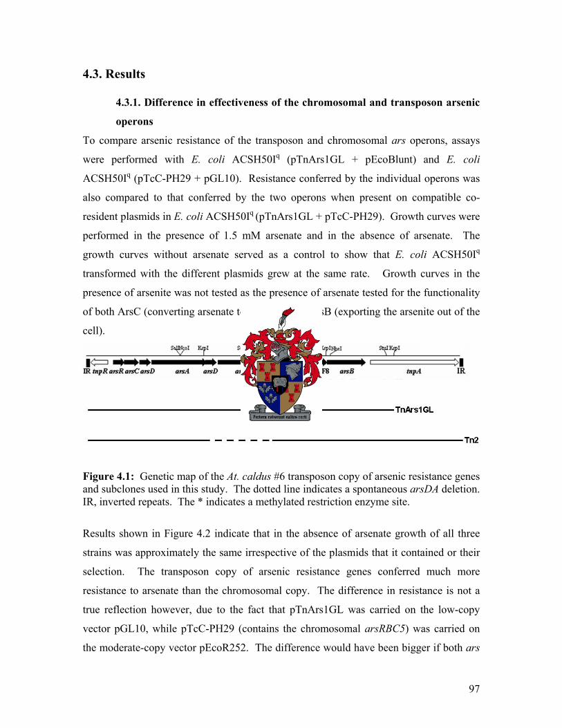
4.3. Results
4.3.1. Difference in effectiveness of the chromosomal and transposon arsenic
operons
To compare arsenic resistance of the transposon and chromosomal ars operons, assays
were performed with E. coli ACSH50Iq (pTnArs1GL + pEcoBlunt) and E. coli
ACSH50Iq (pTcC-PH29 + pGL10). Resistance conferred by the individual operons was
also compared to that conferred by the two operons when present on compatible co-
resident plasmids in E. coli ACSH50Iq (pTnArs1GL + pTcC-PH29). Growth curves were
performed in the presence of 1.5 mM arsenate and in the absence of arsenate. The
growth curves without arsenate served as a control to show that E. coli ACSH50Iq
transformed with the different plasmids grew at the same rate. Growth curves in the
presence of arsenite was not tested as the presence of arsenate tested for the functionality
of both ArsC (converting arsenate to arsenite) and ArsB (exporting the arsenite out of the
cell).
Figure 4.1: Genetic map of the At. caldus #6 transposon copy of arsenic resistance genes and subclones used in this study. The dotted line indicates a spontaneous arsDA deletion. IR, inverted repeats. The * indicates a methylated restriction enzyme site.
Results shown in Figure 4.2 indicate that in the absence of arsenate growth of all three
strains was approximately the same irrespective of the plasmids that it contained or their
selection. The transposon copy of arsenic resistance genes conferred much more
resistance to arsenate than the chromosomal copy. The difference in resistance is not a
true reflection however, due to the fact that pTnArs1GL was carried on the low-copy
vector pGL10, while pTcC-PH29 (contains the chromosomal arsRBC5) was carried on
the moderate-copy vector pEcoR252. The difference would have been bigger if both ars
97
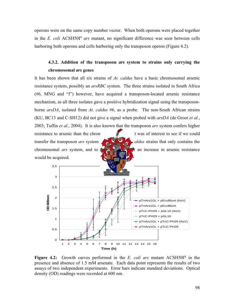
operons were on the same copy number vector. When both operons were placed together
in the E. coli ACSH50Iq ars mutant, no significant difference was seen between cells
harboring both operons and cells harboring only the transposon operon (Figure 4.2).
4.3.2. Addition of the transposon ars system to strains only carrying the
chromosomal ars genes
It has been shown that all six strains of At. caldus have a basic chromosomal arsenic
resistance system, possibly an arsRBC system. The three strains isolated in South Africa
(#6, MNG and “f”) however, have acquired a transposon-located arsenic resistance
mechanism, as all three isolates gave a positive hybridization signal using the transposon-
borne arsDA, isolated from At. caldus #6, as a probe. The non-South African strains
(KU, BC13 and C-SH12) did not give a signal when probed with arsDA (de Groot et al.,
2003; Tuffin et al., 2004). It is also known that the transposon ars system confers higher
resistance to arsenic than the chromosomal system. It was of interest to see if we could
transfer the transposon ars system to one of the At. caldus strains that only contains the
chromosomal ars system, and to determine whether an increase in arsenic resistance
would be acquired.
0
0.5
1
1.5
2
2.5
3
3.5
1 2 3 4 5 6 7 8 9 10 11 12 13 14 15 16
Time (h)
OD
600
nm pTnArs1GL + pEcoBlunt (AsV)
pTnArs1GL + pEcoBlunt
pTcC-PH29 + pGL10 (AsV)
pTcC-PH29 + pGL10
pTnArs1GL + pTcC-PH29 (AsV)
pTnArs1GL + pTcC-PH29
Figure 4.2: Growth curves performed in the E. coli ars mutant ACSH50Iq in the presence and absence of 1.5 mM arsenate. Each data point represents the results of two assays of two independent experiments. Error bars indicate standard deviations. Optical density (OD) readings were recorded at 600 nm.
98

E. coli HB101 containing either the broad host range conjugative plasmid pSa or pTn2
(pSa into which TnAtcArs had been transposed, Tuffin et al., 2005) was mated with At.
caldus strains BC13, KU and C-SH12 for 11 days. At. caldus transconjugants were then
selected in inorganic S2O3 media containing Km at pH 4.6. All of the At. caldus strains
were able to grow on 100 mg/ml kanamycin (which selects for pSa). Difficulty was
experienced in getting At. caldus to form well-isolated colonies on plates. Therefore total
DNA was extracted and a PCR experiment using the transposon arsA primers
(RNAarsAF/RNAarsAR) was conducted (Table 4.1, Figure 4.3). A 600 bp product was
observed when using C-SH12-pTn2 total DNA as template, which corresponds to the size
of the product the ArsA primer pair would form. This indicated that the transposon copy
of ars genes (pTn2) did transfer from HB101 to C-SH12. No products were formed
when using total DNA from BC13-pTn2 and KU-pTn2 as template. It is possible that the
Km resistance observed in At. caldus BC13-pTn2 and KU-pTn2 was due to the
breakdown of Km in the media.
Figure 4.3: An ethidium bromide-stained agarose gel showing PCR amplification products when using an RNAarsAF/RNAarsAR primer combination. A product was only obtained for C-SH12-Tn2 and the positive control pTn2.
99

To confirm that At. caldus was the organism that grew in the S2O3 media and that no
contamination occurred, the chromosomal DNA that was extracted from At. caldus
strains C-SH12, BC13 and KU grown in the presence of 100 mg/ml kanamycin was used
and a 16S rDNA PCR was performed. The PCR product was digested with BamHI and
StuI (Figure 4.4). The fragments obtained indicated that the 16S rDNA was that of At.
caldus and not a possible contaminating organism, or the E. coli HB101 donor strain.
Figure 4.4: An ethidium bromide-stained agarose gel showing the 16S rDNA PCR amplification products when using chromosomal DNA isolated from At. caldus strains BC13, KU and C-SH12, digested with BamHI and StuI.
A Southern blot experiment was carried out where chromosomal DNA from At. caldus C-
SH12 (pSa) and At. caldus C-SH12 (pTn2) transconjugants as well as pSa and pTn2
plasmid DNA were digested with KspI and probed with pSa and the transposon-borne
arsD. When probing with arsD a signal was obtained only for lanes containing At.
caldus C-SH12 (pTn2) and pTn2, indicating that the transposon copy did transfer and that
it is not present in untransformed At. caldus C-SH12 and in At. caldus C-SH12 (pSa)
100

transconjugants (Figure 4.5 a). When probed with total pSa (Figure 4.5 b), several
fragments were obtained for At. caldus C-SH12 (pSa) and At. caldus C-SH12 (pTn2)
transconjugants, as well as for pSa and pTn2 plasmid DNA, and as expected no signal for
untransformed At. caldus C-SH12 DNA. With At. caldus C-SH12 (pTn2) and pTn2
DNA an additional 5 kb KspI signal was obtained that was not obtained with At. caldus
C-SH12 (pSa) and pSa DNA. This 5 kb signal represents a KspI fragment stretching
from a KspI site in TnAtcArs to a KspI site in pSa, and therefore why it was not obtained
in DNA extracts containing pSa only.
Figure 4.5: Southern blot experiment where At. caldus C-SH12 (pTn2) and At. caldus C-SH12 (pSa) transconjugants as well as untransformed At. caldus C-SH12 and pTn2 and pSa DNA were digested with KspI and probed with (A) the transposon arsD and (B) pSa. Next we tested whether At. caldus C-SH12 (pTn2) transconjugants would be more
resistant to arsenic than At. caldus C-SH12 transformed with just pSa and also to show
that the addition of pSa does not have an effect on arsenic resistance. Growth curve
experiments were done where At. caldus C-SH12 (pSa) and At. caldus C-SH12 (pTn2)
transconjugants were grown in the presence of 0, 20, 30, 40 and 50 mM arsenite. After
eight days, At. caldus cells containing the transposon copy of arsenic resistance genes (C-
SH12-pTn2) were able to grow on 30 mM arsenite, while the At. caldus cells harboring
only the chromosomal ars genes (C-SH12-pSa) showed weaker growth, even at 20 mM
101

arsenite. After 13 days, At. caldus C-SH12 (pTn2) was able to grow on 50 mM arsenite,
while At. caldus C-SH12 (pSa) was sensitive to 30 mM arsenite. After 19 days C-SH12-
pSa was still sensitive to 30 mM arsenite (Figure 4.6). These results indicate that the
addition of the transposon ars operon of At. caldus strain #6 to At. caldus C-SH12
increased the resistance to arsenite more than two-fold.
Figure 4.6: Arsenite growth curves performed with At. caldus C-SH12 (pTn2) and At. caldus C-SH12 (pSa) transconjugants in the presence of various concentrations of arsenite. Cell densities were determined (OD 600nm) and a percentage resistance was calculated (growth in the absence of arsenite was equivalent to 100%) after 19 days. Each data point represents duplicate results of at least two experiments. Error bars indicate standard deviations.
102

4.4. Discussion It is known that At. caldus #6 has an ars operon carried on a transposon (de Groot et al.,
2003; Figure 4.1) and in this study we identified the chromosomal ars operon of this
organism. Tuffin et al. (2004) showed that the ars genes on the transposon confer high
levels of resistance to arsenate and arsenite. Growth curve experiments conducted with
the transposon and chromosomal copy of ars genes in E. coli ACSH50Iq revealed that the
transposon system conferred higher levels of resistance to arsenate than the chromosomal
system. To determine arsenate resistance levels when both systems were in the same
organism, growth curves were conducted where the chromosomal and transposon
systems were placed together in trans in E. coli ACSH50Iq. No significant difference was
observed between cells harboring both systems and cells harboring only the transposon
system, indicating that the two operons did not have a synergistic effect on arsenate
resistance and that resistance levels were similar to those of the transposon. However,
this study was done in E. coli, and these results may not be a true reflection on the levels
of resistance in At. caldus.
It is well known that the bacterial biomining population evolved to become more resistant
to high levels of arsenic over a short period of time (Rawlings and Woods, 1995). In
order to demonstrate that the acquisition of the transposon ars system resulted in the
increase of arsenic resistance in At. caldus, a system had to be developed where the
transposon copy of ars genes could be introduced into At. caldus strains only having the
chromosomal ars operon (BC13, KU and C-SH12). Using a conjugational system, the
transposon ars system (carried on the conjugative pSa) was transferred from E. coli
HB101 to At. caldus C-SH12. Transformants were selected with kanamycin. Southern
bolt hybridization (Figure 4.5) and PCR analysis (Figure 4.3) confirmed the transfer of
the transposon ars system from E. coli HB101 to At. caldus C-SH12. The banding
pattern of the Southern blot experiment also indicated that no plasmid rearrangements
occurred during the conjugation process. We were unable to transfer the transposon ars
system to At. caldus BC13 and KU. Although kanamycin resistance was also observed
for At. caldus KU and BC13, it is most likely due to the breakdown of the kanamycin
following extended aeration at low pH. Studies conducted by Hallberg and Lindstrom
103
(1996) showed that antiserum generated against whole cells from At. caldus KU
recognized whole cells of isolates BC13 and KU, but not C-SH12. The authors propose
that this lack of recognition by the antiserum is most likely due to the fact that the
lipopolysaccharide (LPS) of isolate C-SH12 differs from the LPS of BC13 and KU. This
means that At. caldus C-SH12 can be considered to belong to a different serotype from
that of BC13 and KU. A difference in cell structure may thus be the reason why we were
unable to transfer DNA to strain KU and BC13.
Growth curve experiments with At. caldus C-SH12 (pSa) and At. caldus C-SH12 with the
introduced transposon operon (pTn2) were conducted to determine if the acquisition of
the transposon operon could have resulted in the increase in arsenic resistance of certain
At. caldus strains. Introduction of the transposon system to At. caldus C-SH12 resulted in
an increase of arsenite resistance of more than two-fold. In the presence of arsenite the
growth rate of the culture of transconjugants was also much faster than C-SH12 cells with
only the chromosomal ars system. The increased levels of resistance and rate of growth
had a dramatic effect on growth in the presence of arsenite (Figure 4.6). Based on these
results we speculate that At. caldus #6 became more resistant to arsenic most likely by the
acquisition of the transposon ars system.
The development of a conjugation system to transfer genes from E. coli to At. caldus C-
SH12 is a major breakthrough in the study of the molecular biology of At. caldus.
Optimization of this system may be achieved by optimizing donor to recipient ratios, pH
and medium composition. Researchers in our laboratory are currently developing a
mechanism to introduce gene knockouts in At. caldus. This system, together with the
conjugation system, will enable us to study the molecular biology of biomining bacteria
in greater depth than is currently possible. This could enable us to genetically engineer
strains that could grow faster, at lower pH, at higher temperatures, at higher cell densities
and with smaller quantities of added nutrients (Rawlings and Kusano, 1994) in order to
achieve great economical savings in the biomining industry.
104

Chapter 5
General Discussion Arsenic resistance (ars) systems had been identified in many different bacteria. Two
common forms of the ars operon have been identified. The most common form consists
of three genes (arsRBC) and is found on plasmids of S. xylosus (pSX267) and S. aureus
(pI258). The second form consists of five genes (arsRDABC) and was found on plasmids
R773 and R46 of E. coli and more recently on transposons of At. caldus (AY821803), L.
ferriphilum (DQ057986), A. faecalis (Ay297781) and M. flagellatus
(NZ_AADX01000013) (Tuffin et al., 2006). During the sequencing of the E. coli
chromosome, the first chromosomally encoded arsenic resistance genes were identified
(Diorio et al., 1995). Since then, with more chromosomal sequencing projects underway,
arsenic resistance genes have been identified on many more bacterial chromosomes, and
generally take the form arsRBC.
In a previous study two copies of transposon-linked ars operons had been identified in At.
caldus strain #6 (Figure 2.1 a and b; Tuffin et al., 2004). Using Southern blot analysis de
Groot et al. (2003) identified a possible chromosomal ars operon in At. caldus strain #6
that was different to the ars genes in the transposon. The aim of this study was to isolate
and sequence the third ars operon. Once characterized, expression and regulatory studies
were performed and possible interaction between the transposon-located and
chromosomal ars operons was analyzed.
Sequence analysis of the At. caldus #6 chromosomal ars operon revealed an arsCRB
operon. This ars operon however, differs from the conventional chromosomal arsRBC
operons of E. coli and Staphylococcus spp. where the operon is transcribed as one
transcriptional unit (Diorio et al., 1995). The At. caldus chromosomal ars operon is bi-
directional, with the arsC gene located upstream of the arsB gene and the arsC and arsR
genes divergently transcribed to arsB. This divergent arrangement of the ars genes raised
the possibility that transcription in the two directions might be regulated differently.
105
In ars systems where an ArsA is absent (as in the At. caldus chromosomal ars operon), it
is believed that the ArsB uses membrane potential rather that ATP hydrolysis as an
energy source to export arsenite out of the cell. However, with acidophilic
microorganisms like At. caldus this is likely to be different, as they have a reversed
membrane potential, with a positive rather than negative inside membrane potential
(Martin, 1990). Nevertheless, the At. caldus ars genes conferred enhanced resistance to
arsenate and arsenite when cloned in E. coli. The mechanism how arsenite is exported by
ArsB in this acidophilic organism lacking an ArsA is still unknown.
Alignment studies showed that the At. caldus chromosomal ArsC protein grouped with
ArsC proteins of the Trx family (Figure 2.9). This suggested that this ArsC would use
thioredoxin rather than glutathione as a reducing power to convert arsenate to arsenite.
When tested to see if thioredoxin aids the ArsC in converting arsenate to arsenite, it was
surprising to find that the At. ferrooxidans thioredoxin gene (trxA) was unable to help
ArsC convert arsenate to arsenite. Based on the alignment studies and the high identity to
the ArsC of At. ferrooxidans, it is likely that thioredoxin rather than glutathione is used
by the At. caldus chromosomal ArsC as a reducing power. A possible explanation is that
the At. ferrooxidans trxA gene may not complement the At. caldus chromosomal ArsC to
reduce arsenate to arsenite.
Recently the crystal structures of ArsC proteins of the Trx and GSH/Grx clades have
been determined and mechanisms of arsenate reduction predicted (Martin et al., 2001;
Zegers et al., 2001). Sequence analysis of the At. caldus chromosomal ArsC revealed
that it contains, like other ArsC proteins of the Trx clade, four cysteine residues (Cys10,
Cys15, Cys82 and Cys90) with spacing similar to those of other ArsC proteins of the Trx
clade. Based on the above mentioned evidence, we propose that the At. caldus
chromosomal ArsC will function similar to the B. subtilis ArsC (Figure 1.7).
RT-PCR analysis done in E. coli revealed that ORF1, arsC and arsR are transcribed as
one transcriptional unit and that arsB and ORF5 are also transcribed as a single
106

transcriptional unit. Reporter gene studies in the E. coli ars mutant ACSH50Iq indicated
that the level of expression in the direction of arsB was about five-fold higher than in the
direction of arsRC. The arsB expression was also repressed to a greater extent by ArsR
than arsRC expression. A divergent chromosomal ars operon was also identified in At.
ferrooxidans (Butcher et al., 2000). The At. caldus chromosomal arsC, arsR and arsB
genes exhibited 78%, 74% and 85% identity, respectively, to the At. ferrooxidans
chromosomal ars genes. This high similarity between the ars genes of the two organisms
suggests that they may have a common ancestor. Although the genome of At.
ferrooxidans has been sequenced, the ars genes are the first from the chromosome of At.
caldus to be reported. It is therefore too early to know whether the relatedness between
ars genes reflects a general relationship between At. ferrooxidans and At. caldus or
whether it is a feature of ars genes.
Sequence alignments of the At. caldus chromosomal ArsR with its closest matches from
the NCBI database showed that none of these ArsR proteins contain the conserved metal
binding motif (ELCVCDL) known to be involved in the binding of arsenite. Arsenite
binds to the cysteine residues in this domain, resulting in a conformational change to
ArsR that causes the repressor to dissociate from the promoter, which then results in the
expression of the ars genes (Shi et al., 1994). The SmtB/ArsR family of transcriptional
regulators have two metal binding sites, the α3N in the α3 helix and the α5C metal
binding site in the α5 helix (Busenlehner et al., 2003). It is known that the ELCVCDL
motif is located in the α3 helix. The At. caldus chromosomal ArsR and its closest
matches however, contain a conserved LVAYLTENCC domain near the C-terminal end
of the protein (Figure 2.10). The two cysteine residues in this domain, together with
another cysteine five to seven residues towards the C-terminal, may form the metal
binding domain of these atypical ArsR proteins. The fact that this possible metal binding
domain is located in the α5 helix strengthens the possibility that this could be the At.
caldus chromosomal ArsR’s metal binding domain, as Busenlehner et al. (2003) showed
that this family of transcriptional regulators has metal binding domains in the α3 and/or
α5 helices.
107

Despite the lack of the conventional ELCVCDL metal binding motif on the At. caldus
chromosomal ArsR, translational fusions of the predicted promoter regions to a
promoterless reporter gene (lacZ) showed that the expression from the arsB and arsR
promoters were repressed in the presence of an intact arsR gene in trans. This repression
was relieved with the addition of arsenite and arsenate (Figure 3.2). This indicated that
the atypical ArsR does regulate expression from the arsB and arsR promoters. No
detectable difference in the ability of arsenate and arsenite to derepress the transcription
of the ars genes was observed. However, it was very interesting to find that ArsR was
induced by arsenate, as most ArsR regulators respond to arsenite but not arsenate. The
ArsR protein of At. ferrooxidans (with which the At. caldus chromosomal ArsR has 74%
identity) has also been shown to be induced by arsenate (Butcher et al., 2000). The ArsR
protein of At. caldus is only the third example (all from Acidithiobacillus) of an arsenic
regulator that is derepressed by both forms of arsenic. As no ArsC was present when
ArsR was induced by arsenate and arsenite, it suggests that these atypical ArsR proteins
can bind either arsenate or arsenite, or that a mechanism other that ArsC exists that can
convert arsenate to arsenite prior to binding to the ArsR. Metal binding sites for both
arsenate and arsenite might be a common characteristic for this atypical ArsR family of
transcriptional regulators.
In future work it will be important to show that the ArsR binds to the predicted promoter
regions. Shi et al. (1994) showed, by using gel mobility shift assays, that the E. coli
R773 ArsR was able to bind to the predicted promoter region. Gel mobility shift assays
can similarly be used to show that the At. caldus chromosomal ArsR binds to the
promoter sequence in the absence of an inducer. If the ArsR does not bind in the
presence of arsenite and arsenate, it will confirm our results that arsenate and arsenite can
act as inducers of the At. caldus chromosomal ArsR. If mutations of Cys94, Cys95 and
Cys101 by site-directed mutagenesis result in the loss of induction of ArsR by arsenate and
arsenite, it will indicate if the predicted LVAYLTENCC domain is indeed the metal
binding domain and that these three cysteines are involved in arsenic binding.
108

An investigation of how the chromosomal and transposon ars systems were likely to
interact when both were present in the same bacterium was carried out, as TnAtcArs is
also found in At. caldus #6. Although the transposon and chromosomal ars operons are
quite different and although the two ArsR proteins share only 45% sequence identity,
promoter fusion studies in E. coli indicated that the At. caldus chromosomal ArsR was
able to regulate the promoters (both directions) of its own operon as well as cross-
regulate the transposon ars promoter. However, regulation of the transposon promoter by
the chromosomal ArsR was not as effective as regulation by its own ArsR protein,
indicating that the chromosomal ArsR is not optimally designed for the regulation of the
transposon ars operon. Growth curve experiments conducted with the transposon and
chromosomal copies of ars genes revealed that the transposon system conferred much
higher levels of arsenic resistance than the chromosomal system in a heterologous host
(Figure 4.2). The very much higher levels of gene expression from the transposon operon
might be the reason for these higher levels of arsenic resistance conferred by the
transposon operon in E. coli.
However, these experiments were done in a heterologous host, E. coli, and may therefore
not directly reflect what happens in At. caldus. The fact that co-transcription of accessory
genes (ORF1 and ORF5) was observed, but had no effect on arsenic resistance in E. coli
is something of an anomaly. It could be that these ORFs confer some property that was
not examined or that co-transcription in E. coli was artefactual. mRNA studies with
RNA isolated from At. caldus would be able to confirm if the results obtained in the
heterologous host, E. coli, were a true reflection of what happens in At. caldus. If the
accessory genes are co-transcribed with the ars genes in At. caldus, knockout mutants in
At. caldus would then shed more light on the functions of these genes.
It is known that three At. caldus strains (#6, “f” and MNG) that were previously exposed
to arsenic contain two sets of arsenic resistance genes, a set on the chromosome as well
as a set carried on a transposon, while strains BC13, KU and C-SH12 only contain a
chromosomal copy of arsenic resistance genes (de Groot et al., 2003). In this study it
was shown that in E. coli, the transposon copy of ars genes confers much higher levels of
109

arsenic resistance than the chromosomal copy. We wanted to check on a possible
difference in levels of ars resistance if the transposon arsenic system was introduced to a
strain of At. caldus only harboring the chromosomal ars genes. A major problem was
that no easy gene transfer system exists for these acidophilic organisms. No success has
been achieved by chemical transformation and Kusano et al. (1992) were able to
transform DNA to At. ferrooxidans by electroporation to only one strain of 30 strains
electroporated. A more successful conjugational system was used by Jin et al. (1992) and
was further improved by Liu et al. (1999). No DNA transformation systems for At.
caldus had been reported prior to this study.
It is well known that the bacterial population used in the biomining process became
highly resistant to arsenic over a period of three years (Rawlings and Woods, 1995). In
order to demonstrate that the acquisition of the transposon ars system resulted in an
increase in arsenic resistance in At. caldus, a system had to be developed where the
transposon copy of ars genes could be introduced to strains harboring only a
chromosomal copy. Using a conjugative system, the transposon ars system was
introduced to At. caldus C-SH12. This was the first reported study where DNA was
successfully transferred to At. caldus. As expected, the introduction of the transposon ars
operon to At. caldus C-SH12 resulted in an dramatic increase of ars resistance (Figure
4.6). It was not possible to determine by how much arsenic resistance was increased,
because growth of the transformed cultures occurred at concentrations above at which
arsenite precipitated. Based on these results we speculate that At. caldus #6 obtained
higher levels of arsenic resistance most likely by the acquisition of the transposon ars
system from the gene pool.
The development of a genetic transfer system for At. caldus is of great importance in the
future study of the molecular biology of this organism and possibly other acidophilic
organisms. Researchers in our laboratory are currently developing a system to introduce
gene knockouts in At. caldus. These two systems will enable us to study the molecular
biology of these biomining bacteria in greater depth than is currently possible. At. caldus
C-SH12 formed indistinct colonies on solid medium, and it was therefore not possible to
110

calculate the conjugation frequency. A means of obtaining more distinct At. caldus C-
SH12 colonies on solid media has recently been achieved by replacing agarose with
phytogel as a gelling agent. As we are now able to calculate the transformation
frequency, experiments are currently in progress using this plasmid transfer system to
optimize a conjugation system for At. caldus.
As the dominant iron-oxidizing bacterium in arsenopyrite biooxidation tanks, it was
believed that a strain of Leptospirillum ferriphilum (isolated from a commercial
biooxidation tank treating a gold-bearing arsenopyrite concentrate at the Fairview mine in
South Africa) would also possess an effective arsenic resistance mechanism. This was
the case, as Tuffin et al. (2006) showed that L. ferriphilum does, like At. caldus, carry a
set of arsenic resistance genes on a transposon. This was not surprising, as L.
ferriphilum, like At. caldus, was isolated from a very arsenic rich environment. The L.
ferriphilum transposon ars operon had a very similar gene layout as the transposon ars
operon of At. caldus, except for the absence of ORF7 and the arsDA duplication.
Although these two ars operons originated from bacteria from the same environment, the
amino acid sequence of their gene products were not necessarily the most closely related,
with high sequence identity to genes on transposons from A. faecalis and M. flagellatus
as well (Tuffin et al., 2006; Figure 5.1). The differences in DNA sequence between the
transposon ars genes of At. caldus and L. ferriphilum are far to substantial for the
transposons to have passed from At caldus to L. ferriphilum or vice versa in the
biooxidation environment. This suggests that these two types of bacteria have acquired
the ars transposons independently of each other.
111

TnLfArs
tnpR arsR arsC arsD arsA CBS arsB tnpA
TnAtcArs
A. faecalis
tnpR arsR arsC arsD arsA arsD arsA ORF7 CBS arsB tnpA
arsR arsC arsD arsA CBS arsB tnpA
M. flagellatus
tnpA tnpR arsRC arsD arsA CBS arsB
TnLfArs
tnpR arsR arsC arsD arsA CBS arsB tnpA
TnLfArs
tnpR arsR arsC arsD arsA CBS arsB tnpA
TnAtcArs
A. faecalis
tnpR arsR arsC arsD arsA arsD arsA ORF7 CBS arsB tnpA
arsR arsC arsD arsA CBS arsB tnpA
M. flagellatus
tnpA tnpR arsRC arsD arsA CBS arsB
TnAtcArs
A. faecalis
tnpR arsR arsC arsD arsA arsD arsA ORF7 CBS arsB tnpAtnpR arsR arsC arsD arsA arsD arsA ORF7 CBS arsB tnpA
arsR arsC arsD arsA CBS arsB tnpAarsR arsC arsD arsA CBS arsB tnpA
M. flagellatus
tnpA tnpR arsRC arsD arsA CBS arsB
M. flagellatus
tnpA tnpR arsRC arsD arsA CBS arsB
Figure 5.1: Diagrammatic representation of transposon located ars operons isolated from Leptospirillum ferriphilum Fairview (TnLfArs), Acidithiobacillus caldus #6 (TnAtcArs), Methylobacillus flagellatus and Alcaligenes faecalis. Inverted repeats are shown as vertical bars. The white box downstream of M. flagellatus arsB represents a 39 aa TnpA remnant. Taken from Tuffin et al. (2006).
Like in At. caldus, the L. ferriphilum transposon located ars genes conferred higher levels
of arsenic resistance than the chromosomal ars genes in E. coli. The chromosomal ars
genes were also very poorly expressed. Because the overexpression of arsB is lethal to
cells, organisms do not necessarily want high expression levels of ars genes. If they live
in an environment with low levels of arsenic, the low levels of expression from the
chromosomal ars operons will probably be sufficient to confer arsenic resistance. Only
when they are placed under selective pressure would they acquire an additional system
that would confer higher levels of arsenic resistance. Tuffin et al. (2006) also showed
that the L. ferriphilum strain containing the transposon ars operon was more resistant to
arsenic than L. ferriphilum strains harboring only a chromosomal copy of ars genes. It
thus seems that the quickest or easiest way for these bacteria to have survived these
arsenic rich environments, was by the acquisition of another arsenic operon, as the time
to mutate an existing operon (like the chromosomal ars operon) may take thousands of
112

years. The fact that these biooxidation environments are not sterile opens many options
as to where these arsenic transposons originated from. The findings in this study are
consistent with the prediction, reported by Woese (2004), that “cosmopolitan genes”, of
which arsenic resistance genes are a typical example, are more likely to be a
characteristic of a certain environment than of particular lineages. If these “cosmopolitan
genes” are not restricted to certain lineages, they will most likely pass between different
types of bacteria within an environment that selects for them. Kondratyeva et al. (1995)
suggested that both arsenic and zinc resistant strains of At. ferrooxidans have increased
copy numbers of chromosomal genes encoding resistance. In this study we answered the
question of how these biomining organisms became more resistant to arsenic in a relative
short period of time. This increase in resistance was acquired probably not by an increase
in copy number of arsenic resistance genes, but by the acquisition of a highly resistant
arsenic operon carried on a transposon.
113

Appendix One: Annotated sequence obtained from pTcC-#4
Shown below is the double stranded sequence of the At. caldus chromosomal fragment
obtained in pTcC-#4 during the course of this study. Protein translations of the ten open
reading frames encoded on this fragment have been shown below the DNA sequence.
Restriction endonuclease sites are labeled above the DNA sequence. The sequence to
which primers were designed is underlined and labeled in white boxes. The start codons
of open reading frames are labeled in grey boxes and the putative ribosome binding sites
of arsR and arsB are shown in bold. Arrows indicate the direction of transcription.
1 GATCCCGACACCACCGAGATAAGCCCTCACTGCCTACGGCTCGCCGTCCGCAAACCGTTA CTAGGGCTGTGGTGGCTCTATTCGGGAGTGACGGATGCCGAGCGGCAGGCGTTTGGCAAT 61 GGCGCGACACGCTTCCAGGGTGGCTGTGCCTGGTGCTGGGCGAGCGCTTATCCCGAGAAA CCGCGCTGTGCGAAGGTCCCACCGACACGGACCACGACCCGCTCGCGAATAGGGCTCTTT 121 GTCGCACGGTTTACCTCGACTCCGTACCGGGCACCAGAAATACCGGCTCTTGGTCGGCGG CAGCGTGCCAAATGGAGCTGAGGCATGGCCCGTGGTCTTTATGGCCGAGAACCAGCCGCC * R S E T G P V L F V P E Q D A P 181 GCATGGTGGCGAGCTCTTCTTCCGTCATCCGAGCGTAAATTTCGAGGAGATAACCCGTGG CGTACCACCGCTCGAGAAGAAGGCAGTAGGCTCGCATTTAAAGCTCCTCTATTGGGCACC M T A L E E E T M R A Y I E L L Y G T P ORF1-RT_Rev 241 GATCGCGCAGCCAGAGTTCGTGCAGAGGTGTGCCGCGCCAGGTGCTGCGCGGCGGCTTGA CTAGCGCGTCGGTCTCAAGCACGTCTCCACACGGCGCGGTCCACGACGCGCCGCCGAACT D R L W L E H L P T G R W T S R P P K V 301 CCACCTCGGCACCGGCGGCCACGGCGCGATGGTAGGCGGCGATCACCGCTTCCTTGCTCC GGTGGAGCCGTGGCCGCCGGTGCCGCGCTACCATCCGCCGCTAGTGGCGAAGGAACGAGG V E A G A A V A R H Y A A I V A E K S R 361 GCACTCCCAGGCCCACGTGGTAGAGGCTGTAGGTGTCGAGGGGCTCGTCCCGGTCGTTGA CGTGAGGGTCCGGGTGCACCATCTCCGACATCCACAGCTCCCCGAGCAGGGCCAGCAACT V G L G V H Y L S Y T D L P E D R D N V 421 CGAGAATCACCAGATTCAGGCAGAGATCGGGGACGATGAAGGTGCTGAAGCGCGCCGTCT GCTCTTAGTGGTCTAAGTCCGTCTCTAGCCCCTGCTACTTCCACGACTTCGCGCGGCAGA L I V L N L C L D P V I F T S F R A T Q BstEII 481 GTTGTCTGGGCTCAACCCCGAAAAAGTCCCGATAGAAGGCCGTGCTGCGGGCAAGATCGG CAACAGACCCGAGTTGGGGCTTTTTCAGGGCTATCTTCCGGCACGACGCCCGTTCTAGCC Q R P E V G F F D R Y F A T S R A L D T
114
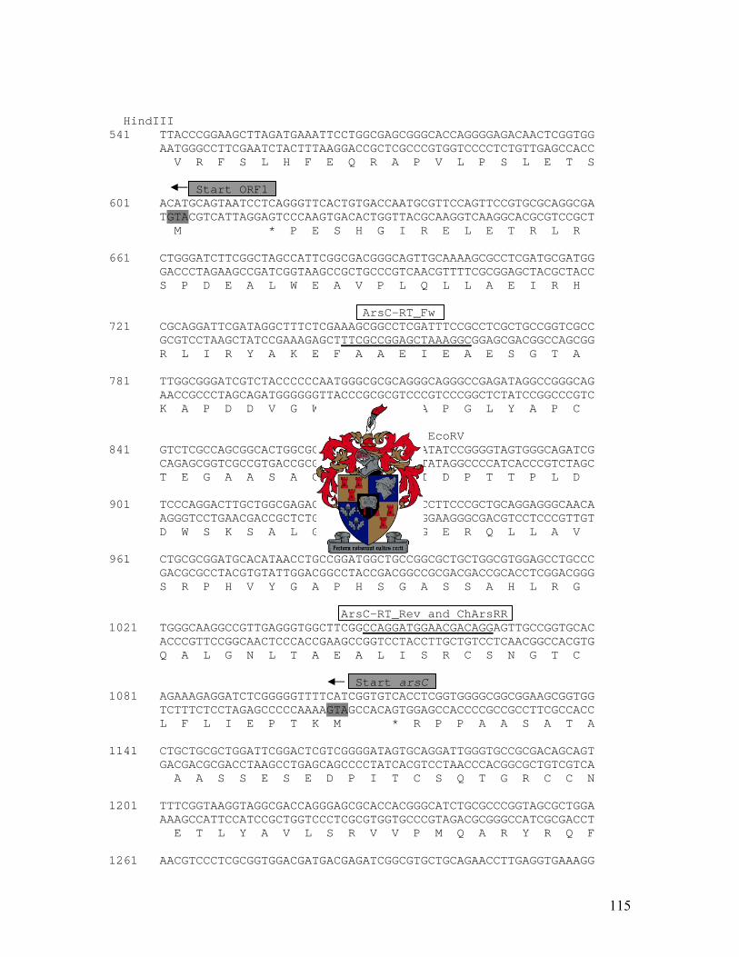
HindIII 541 TTACCCGGAAGCTTAGATGAAATTCCTGGCGAGCGGGCACCAGGGGAGACAACTCGGTGG AATGGGCCTTCGAATCTACTTTAAGGACCGCTCGCCCGTGGTCCCCTCTGTTGAGCCACC V R F S L H F E Q R A P V L P S L E T S Start ORF1 601 ACATGCAGTAATCCTCAGGGTTCACTGTGACCAATGCGTTCCAGTTCCGTGCGCAGGCGA TGTACGTCATTAGGAGTCCCAAGTGACACTGGTTACGCAAGGTCAAGGCACGCGTCCGCT M * P E S H G I R E L E T R L R 661 CTGGGATCTTCGGCTAGCCATTCGGCGACGGGCAGTTGCAAAAGCGCCTCGATGCGATGG GACCCTAGAAGCCGATCGGTAAGCCGCTGCCCGTCAACGTTTTCGCGGAGCTACGCTACC S P D E A L W E A V P L Q L L A E I R H ArsC-RT_Fw 721 CGCAGGATTCGATAGGCTTTCTCGAAAGCGGCCTCGATTTCCGCCTCGCTGCCGGTCGCC GCGTCCTAAGCTATCCGAAAGAGCTTTCGCCGGAGCTAAAGGCGGAGCGACGGCCAGCGG R L I R Y A K E F A A E I E A E S G T A 781 TTGGCGGGATCGTCTACCCCCCAATGGGCGCGCAGGGCAGGGCCGAGATAGGCCGGGCAG AACCGCCCTAGCAGATGGGGGGTTACCCGCGCGTCCCGTCCCGGCTCTATCCGGCCCGTC K A P D D V G W H A R L A P G L Y A P C EcoRV 841 GTCTCGCCAGCGGCACTGGCGCAGACGGTGATGACGATATCCGGGGTAGTGGGCAGATCG CAGAGCGGTCGCCGTGACCGCGTCTGCCACTACTGCTATAGGCCCCATCACCCGTCTAGC T E G A A S A C V T I V I D P T T P L D 901 TCCCAGGACTTGCTGGCGAGACCTTCGGTAGAGAAGCCTTCCCGCTGCAGGAGGGCAACA AGGGTCCTGAACGACCGCTCTGGAAGCCATCTCTTCGGAAGGGCGACGTCCTCCCGTTGT D W S K S A L G E T S F G E R Q L L A V 961 CTGCGCGGATGCACATAACCTGCCGGATGGCTGCCGGCGCTGCTGGCGTGGAGCCTGCCC GACGCGCCTACGTGTATTGGACGGCCTACCGACGGCCGCGACGACCGCACCTCGGACGGG S R P H V Y G A P H S G A S S A H L R G ArsC-RT_Rev and ChArsRR 1021 TGGGCAAGGCCGTTGAGGGTGGCTTCGGCCAGGATGGAACGACAGGAGTTGCCGGTGCAC ACCCGTTCCGGCAACTCCCACCGAAGCCGGTCCTACCTTGCTGTCCTCAACGGCCACGTG Q A L G N L T A E A L I S R C S N G T C Start arsC 1081 AGAAAGAGGATCTCGGGGGTTTTCATCGGTGTCACCTCGGTGGGGCGGCGGAAGCGGTGG TCTTTCTCCTAGAGCCCCCAAAAGTAGCCACAGTGGAGCCACCCCGCCGCCTTCGCCACC L F L I E P T K M * R P P A A S A T A 1141 CTGCTGCGCTGGATTCGGACTCGTCGGGGATAGTGCAGGATTGGGTGCCGCGACAGCAGT GACGACGCGACCTAAGCCTGAGCAGCCCCTATCACGTCCTAACCCACGGCGCTGTCGTCA A A S S E S E D P I T C S Q T G R C C N 1201 TTTCGGTAAGGTAGGCGACCAGGGAGCGCACCACGGGCATCTGCGCCCGGTAGCGCTGGA AAAGCCATTCCATCCGCTGGTCCCTCGCGTGGTGCCCGTAGACGCGGGCCATCGCGACCT E T L Y A V L S R V V P M Q A R Y R Q F 1261 AACGTCCCTCGCGGTGGACGATGACGAGATCGGCGTGCTGCAGAACCTTGAGGTGAAAGG
115

TTGCAGGGAGCGCCACCTGCTACTGCTCTAGCCGCACGACGTCTTGGAACTCCACTTTCC R G E R H V I V L D A H Q L V K L H F S StuI 1321 ATACGCCGTTGTGCAGTTGCCCGATCCGTTCGGCAATCTGGCCGGAGAAAAGGCCTTCCG TATGCGGCAACACGTCAACGGGCTAGGCAAGCCGTTAGACCGGCCTCTTTTCCGGAAGGC V G N H L Q G I R E A I Q G S F L G E P ChArsBLacZF 1381 GCTCGTGCTCCACCAGTAGGCGAAAAATGGCGAGCCGCACCGGCGAGGCCAGGGCCTCGA CGAGCACGAGGTGGTCATCCGCTTTTTACCGCTCGGCGTGGCCGCTCCGGTCCCGGAGCT E H E V L L R F I A L R V P S A L A E L PvuI ChArsRLacZR Start arsR 1441 GGCGGGCGACAATGCGATCGCTGTGCTCTTCGGTCATGGGATTCCAATGGTTCAAGTTAG CCGCCCGCTGTTACGCTAGCGACACGAGAAGCCAGTACCCTAAGGTTACCAAGTTCAATC R A V I R D S H E E T M ArsR-RT Fw ChArsRF 1501 ATTGAATTATTGACAAATGTTTTTGGCTCGTCAAGACTACCGGCAATTTCACCCAAGGAG TAACTTAATAACTGTTTACAAAAACCGAGCAGTTCTGATGGCCGTTAAAGTGGGTTCCTC Start arsB ChArsBLacZR 1561 TACAGGATGCTTGCCCTGGCCATCTTCGTTGTGACCCTGATCCTCGTCATCTGGCAGCCC ATGTCCTACGAACGGGACCGGTAGAAGCAACACTGGGACTAGGAGCAGTAGACCGTCGGG M L A L A I F V V T L I L V I W Q P 1621 AAGGGACTCCAGATCGGTTGGAGTGCCATGGGCGGTGCGGTCGTCGCCCTGGCCACCGGG TTCCCTGAGGTCTAGCCAACCTCACGGTACCCGCCACGCCAGCAGCGGGACCGGTGGCCC K G L Q I G W S A M G G A V V A L A T G 1681 GTCATCACCTGGAGCGATATTCCTGTGGTCTGGCATATCGTCTGGGATGCTACCTTTACC CAGTAGTGGACCTCGCTATAAGGACACCAGACCGTATAGCAGACCCTACGATGGAAATGG V I T W S D I P V V W H I V W D A T F T 1741 TTCGTCGCCCTCATCATCATCTCCCTGATTCTGGACGAAGCCGGTTTCTTCCATTGGGCG AAGCAGCGGGAGTAGTAGTAGAGGGACTAAGACCTGCTTCGGCCAAAGAAGGTAACCCGC F V A L I I I S L I L D E A G F F H W A 1801 GCTCTGCACGTGGCGCGCTGGGGCGGTGGGCGTGGGCGTCGGCTCTTCCCCCTCATCGTC CGAGACGTGCACCGCGCGACCCCGCCACCCGCACCCGCAGCCGAGAAGGGGGAGTAGCAG A L H V A R W G G G R G R R L F P L I V 1861 CTGCTGGGGGCGATGATTTCTGCCGTATTCGCCAACGACGGGGCGGCGCTGCTGCTCACC GACGACCCCCGCTACTAAAGACGGCATAAGCGGTTGCTGCCCCGCCGCGACGACGAGTGG L L G A M I S A V F A N D G A A L L L T ChArsRLacZF 1921 CCCATCGTCATGGCCATCCTGTTGCGGCTCGACTTCTCACCCAAGGCCACCTTCGCCTTT GGGTAGCAGTACCGGTAGGACAACGCCGAGCTGAAGAGTGGGTTCCGGTGGAAGCGGAAA P I V M A I L L R L D F S P K A T F A F 1981 GTCATCGCCACCGGTTTTGTGGCCGATACCACCAGTCTGCCGCTGATGATCTCTAACCTC CAGTAGCGGTGGCCAAAACACCGGCTATGGTGGTCAGACGGCGACTACTAGAGATTGGAG V I A T G F V A D T T S L P L M I S N L
116

2041 GTCAACATCGTCAGCGCCAATTATTTCAATATCACGTTTGATCGCTATGCCCTGGTGATG CAGTTGTAGCAGTCGCGGTTAATAAAGTTATAGTGCAAACTAGCGATACGGGACCACTAC V N I V S A N Y F N I T F D R Y A L V M 2101 GTACCGGTGGATCTGGTGGCACTGGCGGCCACCCTGGGGGTGCTCTGGCTTTACTTTCGT CATGGCCACCTAGACCACCGTGACCGCCGGTGGGACCCCCACGAGACCGAAATGAAAGCA V P V D L V A L A A T L G V L W L Y F R BamHI 2161 CGCTCCGTGCCCGCGTCCTATCCTACCGCCCACCTCGACGCTCCGCGCACGGCCATCCAG GCGAGGCACGGGCGCAGGATAGGATGGCGGGTGGAGCTGCGAGGCGCGTGCCGGTAGGTC R S V P A S Y P T A H L D A P R T A I Q 2221 GATCCTGTGGTCTTCCGTGCCGCCTTTCCGGTGCTGGTTTTGCTACTAATCGCCTACTTC CTAGGACACCAGAAGGCACGGCGGAAAGGCCACGACCAAAACGATGATTAGCGGATGAAG D P V V F R A A F P V L V L L L I A Y F 2281 GTTACCGCGCAGTGGCAGGTACCGGTATCGGTGGTGACGGGGGCTGGGGCGCTGATCCTC CAATGGCGCGTCACCGTCCATGGCCATAGCCACCACTGCCCCCGACCCCGCGACTAGGAG V T A Q W Q V P V S V V T G A G A L I L 2341 CTCGCTTTGGCCGGACGTTGGTTGCAGGGCGGGCGCGGCGCCCGCATTCCCGTACGCAAG GAGCGAAACCGGCCTGCAACCAACGTCCCGCCCGCGCCGCGGGCGTAAGGGCATGCGTTC L A L A G R W L Q G G R G A R I P V R K 2401 GTCTTGCACGAGGCCCCCTGGCAGATCGTGATTTTCAGCCTCGGGATGTATCTGGTGGTC CAGAACGTGCTCCGGGGGACCGTCTAGCACTAAAAGTCGGAGCCCTACATAGACCACCAG V L H E A P W Q I V I F S L G M Y L V V 2461 TATGGGCTGCGCAATGCCGGTTTGACCAACTATGTGGCCATGGCCCTGGCGTATCTGGCG ATACCCGACGCGTTACGGCCAAACTGGTTGATACACCGGTACCGGGACCGCATAGACCGC Y G L R N A G L T N Y V A M A L A Y L A ArsB-RT Fw 2521 GCACAAGGTACCGTTCTGGCGGCGATTGGGTCAGGCTTCCTGGCCGCGGCGCTGTCCTCG CGTGTTCCATGGCAAGACCGCCGCTAACCCAGTCCGAAGGACCGGCGCCGCGACAGGAGC A Q G T V L A A I G S G F L A A A L S S 2581 GTGATGAACAACATGCCGGCGGTTTTGGTGGGCGCCTTGGCCATCCACCAACTGCCGGTG CACTACTTGTTGTACGGCCGCCAAAACCACCCGCGGAACCGGTAGGTGGTTGACGGCCAC V M N N M P A V L V G A L A I H Q L P V BamHI 2641 GCCACGGATCCGTTGATACGGGAAATCTTCGTCTACGCCAACATCATCGGTTGCGATCTT CGGTGCCTAGGCAACTATGCCCTTTAGAAGCAGATGCGGTTGTAGTAGCCAACGCTAGAA A T D P L I R E I F V Y A N I I G C D L 2701 GGCCCCAAGTTCACGCCCATCGGCAGTCTGGCCACCCTGCTCTGGCTGCATGTGCTGGGC CCGGGGTTCAAGTGCGGGTAGCCGTCAGACCGGTGGGACGAGACCGACGTACACGACCCG G P K F T P I G S L A T L L W L H V L G 2761 CGCAAAGGCATGACCGTGACCTGGGGCCAATACATGCGCACCGGACTCCTCATCACCCCG GCGTTTCCGTACTGGCACTGGACCCCGGTTATGTACGCGTGGCCTGAGGAGTAGTGGGGC R K G M T V T W G Q Y M R T G L L I T P
117

2821 CCGGTACTGTTCCTGACGCTGCTGGCCTTGGCGGCTTGGTTGCCACTACTCTGAAAAAGG GGCCATGACAAGGACTGCGACGACCGGAACCGCCGAACCAACGGTGATGAGACTTTTTCC P V L F L T L L A L A A W L P L L * Start ORF5 2881 CGTTTGAGAAATTTTGTCTTCCCTCGGGTCATGCGCGAACGGTTCGGCCATGGCCGGATG GCAAACTCTTTAAAACAGAAGGGAGCCCAGTACGCGCTTGCCAAGCCGGTACCGGCCTAC M R E R F G H G R M ORF5-RT Rev 2941 TTGCGACCCGGTGAGGTCCGCCACCGCAATCCCTGCCAAACGCGGCACACCTGCGCTTCC AACGCTGGGCCACTCCAGGCGGTGGCGTTAGGGACGGTTTGCGCCGTGTGGACGCGAAGG L R P G E V R H R N P C Q T R H T C A S ORF5-RT Fw 3001 ATGTTGCGGACTGCCGGGGAAGATCCTAGGTGGATTGCCGGCCAGATGGGGTATGAAGAC TACAACGCCTGACGGCCCCTTCTAGGATCCACCTAACGGCCGGTCTACCCCATACTTCTG M L R T A G E D P R W I A G Q M G Y E D 3061 GGGGTACGACTCGACTGGGGTAGGCTCATCAGGTGCCCCATGTCAACCCATCTGCCTAGA CCCCATGCTGAGCTGACCCCATCCGAGTAGTCCACGGGGTACAGTTGGGTAGACGGATCT G V R L D W G R L I R C P M S T H L P R BstEII 3121 GCAGGACTCTCGCCCTTTGGTCACCATTGGATCAAAAGAACGGGGCGGCGAAGCATAGGG CGTCCTGAGAGCGGGAAACCAGTGGTAACCTAGTTTTCTTGCCCCGCCGCTTCGTATCCC A G L S P F G H H W I K R T G R R S I G 3181 CTGGAAATGGCAATCTCATGGATGGTGCGAAAGGGGAGACTCGAACTCCCACGCCTTGCG GACCTTTACCGTTAGAGTACCTACCACGCTTTCCCCTCTGAGCTTGAGGGTGCGGAACGC L E M A I S W M V R K G R L E L P R L A 3241 GCGCTGGAACCTAAATCCAGTGCGTCTACCAATTCCGCCACTCTCGCCTTTACGCTATCT CGCGACCTTGGATTTAGGTCACGCAGATGGTTAAGGCGGTGAGAGCGGAAATGCGATAGA A L E P K S S A S T N S A T L A F T L S BamHI Start ORF6 3301 TAAGCACTAACCGCTCTTTTCACAACGACGAGGATCCCATGGCCAAGTCATCATCCCAAC ATTCGTGATTGGCGAGAAAAGTGTTGCTGCTCCTAGGGTACCGGTTCAGTAGTAGGGTTG * M A K S S S Q 3361 CCTCTCGGTTGCGTCGCCTTCTGGCTGTCGAGTCGGCGCGGATCATGGCGGAGGAGGGCA GGAGAGCCAACGCAGCGGAAGACCGACAGCTCAGCCGCGCCTAGTACCGCCTCCTCCCGT P S R L R R L L A V E S A R I M A E E G ORF6-RT Rev 3421 TTGCGGACTACCGCTTTGCCAAGGAGAAGGCGGCCCGCCGCCTGGGTAGCGGCAGCACCC AACGCCTGATGGCGAAACGGTTCCTCTTCCGCCGGGCGGCGGACCCATCGCCGTCGTGGG I A D Y R F A K E K A A R R L G S G S T PvuII 3481 AGCAAGATTGGCCCAGCAACTCGGAGATCCAGGCCGAACTCAAGGCGCGGTTGCAGCTGT TCGTTCTAACCGGGTCGTTGAGCCTCTAGGTCCGGCTTGAGTTCCGCGCCAACGTCGACA Q Q D W P S N S E I Q A E L K A R L Q L
118

3541 TTCACGGCGAATCCCAACCCCTCGAACTGCGCCGCCTTCGGGAGGTTGCGCTGGAGGCCA AAGTGCCGCTTAGGGTTGGGGAGCTTGACGCGGCGGAAGCCCTCCAACGCGACCTCCGGT F H G E S Q P L E L R R L R E V A L E A 3601 TGGGCTGGCTCAAGGATTTTCGTCCCTTGCTCGCAGGTGCGGTACTCAACGGCACGGCCA ACCCGACCGAGTTCCTAAAAGCAGGGAACGAGCGTCCACGCCATGAGTTGCCGTGCCGGT M G W L K D F R P L L A G A V L N G T A 3661 CGCGCCATAGCGCCATCGTTCTGCATCTCTTTGCCGATACCCCGGAGAGCGTCATCTTTT GCGCGGTATCGCGGTAGCAAGACGTAGAGAAACGGCTATGGGGCCTCTCGCAGTAGAAAA T R H S A I V L H L F A D T P E S V I F 3721 TTCTCATGGATCAGCAGGTGGCCTATGAAGAGGGCTGGCAGCGGCTGCATTTCGGCGACG AAGAGTACCTAGTCGTCCACCGGATACTTCTCCCGACCGTCGCCGACGTAAAGCCGCTGC F L M D Q Q V A Y E E G W Q R L H F G D 3781 AAGCGCCCCAGGAGTATCCCCAGATCCGCATGACCCGTGGGGGGGCAGAATTGCGTCTGG TTCGCGGGGTCCTCATAGGGGTCTAGGCGTACTGGGCACCCCCCCGTCTTAACGCAGACC E A P Q E Y P Q I R M T R G G A E L R L 3841 TCATCTTTGCTCCCGACGAGGATCGGCGCCGTCCGGCATCGGCGGTGGACGGTAAGCCTC AGTAGAAACGAGGGCTGCTCCTAGCCGCGGCAGGCCGTAGCCGCCACCTGCCATTCGGAG V I F A P D E D R R R P A S A V D G K P 3901 TACAGCGGGTCAACGCCCAGCAGTTGCAACGTTTGCTGGATTCCGAGGCATCACTGCCCG ATGTCGCCCAGTTGCGGGTCGTCAACGTTGCAAACGACCTAAGGCTCCGTAGTGACGGGC L Q R V N A Q Q L Q R L L D S E A S L P 3961 CTTATTCCTGAGGTGTCGGTAAAACCTCTGGACTGGGCGTTGGACGAAGGTGCTGCCAAC GAATAAGGACTCCACAGCCATTTTGGAGACCTGACCCGCAACCTGCTTCCACGACGGTTG A Y S * * E Q P T P L V E P S P T P R L H Q W S 4021 TTACCAGACTGAGGGCGATGAAGATGGCGATCATGCCCAGGCTTTCCCAGCCGCCCGGTA AATGGTCTGACTCCCGCTACTTCTACCGCTAGTACGGGTCCGAAAGGGTCGGCGGGCCAT V L S L A I F I A I M G L S E W G G P V PvuII 4081 CCTCGTCCAGCTGCCACCAGGCCGCGAGGACGCCGATGAGCGGCGCCAGCAGCATGGCCA GGAGCAGGTCGACGGTGGTCCGGCGCTCCTGCGGCTACTCGCCGCGGTCGTCGTACCGGT E D L Q W W A A L V G I L P A L L M A M 4141 TGCCGGCTACCCCCGACGGTAGATGCTTGAGGGCATAGGCCCAAAGCAGGTAGGCAATGG ACGGCCGATGGGGGCTGCCATCTACGAACTCCCGTATCCGGGTTTCGTCCATCCGTTACC G A V G S P L H K L A Y A W L L Y A I A 4201 CCGTACCCGGTATGACATTGTAGACTAGGGCGGCGGTGAAGAGCGGGCTCCACTGGATAC GGCATGGGCCATACTGTAACATCTGATCCCGCCGCCACTTCTCGCCCGAGGTGACCTATG T G P I V N Y V L A A T F L P S W Q I R 4261 GCAGGGGGTCTATCCAAAGGCTTACGACAATGAGACCGACGGCGCCCAAGGCCGTCTGCC CGTCCCCCAGATAGGTTTCCGAATGCTGTTACTCTGGCTGCCGCGGGTTCCGGCAGACGG L P D I W L S V V I L G V A G L A T Q W 4321 AGAAAGTCGCGTTGAGGAGATCCTGGCCCGGTGGCATGTGGCGTTTTTGCCAGATGGCGG
119

TCTTTCAGCGCAACTCCTCTAGGACCGGGCCACCGTACACCGCAAAAACGGTCTACCGCC F T A N L L D Q G P P M H R K Q W I A A 4381 CCCCCGCCCACAGTAGGCCCGATAACAGCGCAAAGGCCGGTCCTTGCCAGGCGCCATGAA GGGGGCGGGTGTCATCCGGGCTATTGTCGCGTTTCCGGCCAGGAACGGTCCGCGGTACTT G A W L L G S L L A F A P G Q W A G H F 4441 AATCCCACGGGCTCAATATGCACAAGAGCCCCAGTAGCGCCAGAAGGAGCGCTGGCCAAT TTAGGGTGCCCGAGTTATACGTGTTCTCGGGGTCATCGCGGTCTTCCTCGCGACCGGTTA D W P S L I C L L G L L A L L L A P W H 4501 GCAGTCCGTGCAGACGCTCCCCCAAAAGTGGCCAAGCCAGCAGCATGAGCCAGATGGGCA CGTCAGGCACGTCTGCGAGGGGGTTTTCACCGGTTCGGTCGTCGTACTCGGTCTACCCGT L G H L R E G L L P W A L L M L W I P M 4561 TCATGTACACGAGGATGGCCGTCTTTCCCGCGCCGCCATATTCCAGGGCCCAGAGGGTGA AGTACATGTGCTCCTACCGGCAGAAAGGGCGCGGCGGTATAAGGTCCCGGGTCTCCCACT M Y V L I A T K G A G G Y E L A W L T F 4621 AACCCACAAAGCCGGTGCTCTGCAGCAGGCCAAAGGGCAGGACGTAGCGTAGGGGAGGTC TTGGGTGTTTCGGCCACGAGACGTCGTCCGGTTTCCCGTCCTGCATCGCATCCCCTCCAG G V F G T S Q L L G F P L V Y R L P P R 4681 GCCGCAGGGGACGTTTCAGGAGCAGTAGCAAGGGGAGGAGCACGACCGCCCCCAGGGCCA CGGCGTCCCCTGCAAAGTCCTCGTCATCGTTCCCCTCCTCGTGCTGGCGGGGGTCCCGGT R L P R K L L L L L P L L V V A G L A V 4741 CGCGTAGTGCCGCAAAGATCAGGGGTGGGCAGTCCTCTATGGCTACCTTCATGACCACCC GCGCATCACGGCGTTTCTAGTCCCCACCCGTCAGGAGATACCGATGGAAGTACTGGTGGG R L A A F I L P P C D E I A V K M V V W 4801 AGTTGTAGCCCCAGATCAGGGCAATGGTGACGATGGCGGCAAGGGGAAGCCAGGGAGCAG TCAACATCGGGGTCTAGTCCCGTTACCACTGCTACCGCCGTTCCCCTTCGGTCCCTCGTC N Y G W I L A I T V I A A L P L W P A P Start ORF7 4861 GGGTCGGGGAGTGAGTGCGCATGAAAGTCCGCGGGCGGCCAAAAAAGATCGGGAAAGACC CCCAGCCCCTCACTCACGCGTACTTTCAGGCGCCCGCCGGTTTTTTCTAGCCCTTTCTGG T P S H T R M 4921 ACCGCGCCGCTACGGTGGCCTCTCCCGTATCTTAGAATAGCCTAATGATGCACTTTGGCG TGGCGCGGCGATGCCACCGGAGAGGGCATAGAATCTTATCGGATTACTACGTGAAACCGC * H H V K A PvuI SalI 4981 CGATCGTAGGCAGCCGGCCACGGCCAGTCGACGCCCAACCGTGCGGCCGCCTGCAAGGGC GCTAGCATCCGTCGGCCGGTGCCGGTCAGCTGCGGGTTGGCACGCCGGCGGACGTTCCCG R D Y A A P W P W D V G L R A A A Q L P 5041 CAGTATGGATTACGCAGCAGTTCCCGCGCCAGAGCCACGGCGTCCGCCTGTTCCGTGACG GTCATACCTAATGCGTCGTCAAGGGCGCGGTCTCGGTGCCGCAGGCGGACAAGGCACTGC W Y P N R L L E R A L A V A D A Q E T V 5101 AGAATGTGCTCTGCCTGCATGGCCTCAGTGATGAGGCCGACGGCGACGGTAGGAATGCCG TCTTACACGAGACGGACGTACCGGAGTCACTACTCCGGCTGCCGCTGCCATCCTTACGGC L I H E A Q M A E T I L G V A V T P I G
120

5161 ACGGCCCGACGGATGGCTGCGGCGAAGGGCGTCTGGAAGCCGGGGCCAGCGGGGATTTTC TGCCGGGCTGCCTACCGACGCCGCTTCCCGCAGACCTTCGGCCCCGGTCGCCCCTAAAAG V A R R I A A A F P T Q F G P G A P I K StuI 5221 GCGTCTGGAGTCAGGCCGCCGCTACTGCAGTGTACGACATCCACGCCGCAGGCCTGGAGT CGCAGACCTCAGTCCGGCGGCGATGACGTCACATGCTGTAGGTGCGGCGTCCGGACCTCA A D P T L G G S S C H V V D V G C A Q L 5281 TCCCGACTGAAGGCGATGGACTGCTCCAGATCCCAGCCTCCCTCCACCCAATCCGTGGCC AGGGCTGACTTCCGCTACCTGACGAGGTCTAGGGTCGGAGGGAGGTGGGTTAGGCACCGG E R S F A I S Q E L D W D D E V W D T A 5341 GATATGCGCACCCAGAGGGGGAGCTCCTCGGGCCAGACGGCGCGTACCGCGGTCGCCACC CTATACGCGTGGGTCTCCCCCTCGAGGAGCCCGGTCTGCCGCGCATGGCGCCAGCGGTGG S I R V W L P L E E P W V A R V A T A V 5401 TCCAGCGGAAAGCGCACACGTCCGGAAAAATCGCCGCCGTAAGCGTCGTCCCGTCGGTTG AGGTCGCCTTTCGCGTGTGCAGGCCTTTTTAGCGGCGGCATTCGCAGCAGGGCAGCCAAC E L P F R V R G S F D G G Y A D D R R N 5461 CTGATGGGGGAGAGGAAACTGTGCAGGAGATAGCCATGGGCCATGTGCAGTTCCAGCACC GACTACCCCCTCTCCTTTGACACGTCCTCTATCGGTACCCGGTACACGTCAAGGTCGTGG S I P S L F S H L L Y G H A M H L E L V 5521 TGAAAGCCGGCCCGGTGCGCACGCCGGGCGGCCGCCACGAAGTCCGCCTTCACCCGCTCC ACTTTCGGCCGGGCCACGCGTGCGGCCCGCCGGCGGTGCTTCAGGCGGAAGTGGGCGAGG Q F G A R H A R R A A A V F D A K V R E 5581 ATATCCTCTGTGCTGAGGGGACGGGGGATGGTGTGGGAAGGGCTGAAGGCCAATGCACTG TATAGGAGACACGACTCCCCTGCCCCCTACCACACCCTTCCCGACTTCCGGTTACGTGAC M D E T S L P R P I T H S P S F A L A S 5641 GGGGCTACCGGCTGCCAGCCCAGTTCCGCCTCGGGTGGGATGGGTTGACCTCCATGGAAG CCCCGATGGCCGACGGTCGGGTCAAGGCGGAGCCCACCCTACCCAACTGGAGGTACCTTC P A V P Q W G L E A E P P I P Q G G H F StuI 5701 GGGGCTGCCGTCGAGGCCTTGCGGCCCGCATGGGCGATCTGTATGCCGGCAACGGCACCT CCCCGACGGCAGCTCCGGAACGCCGGGCGTACCCGCTAGACATACGGCCGTTGCCGTGGA P A A T S A K R G A H A I Q I G A V A G 5761 TGCTCCTGCAAAAAGCGAACGATGGGCGCCAGGGCGTCGGCCTGGACATCGTTCCAGATG ACGAGGACGTTTTTCGCTTGCTACCCGCGGTCCCGCAGCCGGACCTGTAGCAAGGTCTAC Q E Q L F R V I P A L A D A Q V D N W I 5821 CCCAGATCTTGTGGCGATATGCGACCTTCCGGACTTACCGCCGATGCCTCCACCAAAATG GGGTCTAGAACACCGCTATACGCTGGAAGGCCTGAATGGCGGCTACGGAGGTGGTTTTAC G L D Q P S I R G E P S V A S A E V L I StuI 5881 AGGCCTGCTCCGCCCACGGCACGACTGCCCAGGTGCACCCGGTGCCAGTCTCCGGCGACG TCCGGACGAGGCGGGTGCCGTGCTGACGGGTCCACGTGGGCCACGGTCAGAGGCCGCTGC L G A G G V A R S G L H V R H W D G A V
121

Start ORF8 5941 CCATCGGGAGTCGAATACTGGCACATGGGGGCCATTACGATCCGGTTGCGTAAGGTGACG GGTAGCCCTCAGCTTATGACCGTGTACCCCCGGTAATGCTAGGCCAACGCATTCCACTGC G D P T S Y Q C M P A M 6001 GAACGCAGTGTCCAGGGGGTGAACAGACGCTGGGTCACGCCTGCACTCCCAAGCCGCGCC CTTGCGTCACAGGTCCCCCACTTGTCTGCGACCCAGTGCGGACGTGAGGGTTCGGCGCGG * A Q V G L G R G 6061 CGAGATGCGCCAGGGTCATTTCCCAGCCCTCCTGCGCCGGTCCGGCGCGGTAGCTGTCGC GCTCTACGCGGTCCCAGTAAAGGGTCGGGAGGACGCGGCCAGGCCGCGCCATCGACAGCG L H A L T M E W G E Q A P G A R Y S D R 6121 GGCGGTCACTGAAGAAGCCGTGCGGGGCCTCCGGAAAGACCGTGAGCGTATAACGCTTCT CCGCCAGTGACTTCTTCGGCACGCCCCGGAGGCCTTTCTGGCACTCGCATATTGCGAAGA R D S F F G H P A E P F V T L T Y R K K 6181 TGGCGGTGCTCAGGGCCGCAGCGATGCGGCCATGCTCTTCCGGGGTAATGGAGCTGTCCT ACCGCCACGAGTCCCGGCGTCGCTACGCCGGTACGAGAAGGCCCCATTACCTCGACAGGA A T S L A A A I R G H E E P T I S S D E 6241 CGGCGCCGTAGAGCAGGAGCAGGGGAGCCTGCATCTGGTTCACGTGACCCAGGAGCGGCG GCCGCGGCATCTCGTCCTCGTCCCCTCGGACGTAGACCAAGTGCACTGGGTCCTCGCCGC A G Y L L L L P A Q M Q N V H G L L P P 6301 GACGTCCGGCAGGGTCTTTATCCGGGGCGATACCGCCGCCGTAAAAAGCTACGGTCGCCG CTGCAGGCCGTCCCAGAAATAGGCCCCGCTATGGCGGCGGCATTTTTCGATGCCAGCGGC R G A P D K D P A I G G G Y F A V T A A 6361 CCACGCGCTCAGCGAGCTGGGCATTGGCGAGGAAGGCATAGCGCCCACCCATACAAAAGC GGTGCGCGAGTCGCTCGACCCGTAACCGCTCCTTCCGTATCGCGGGTGGGTATGTTTTCG V R E A L Q A N A L F A Y R G G M C F G 6421 CCAGCACACCGACCTTTGGACCACGTACCTCAGGACGCTGGAGTACACTCGCCACGGTGG GGTCGTGTGGCTGGAAACCTGGTGCATGGAGTCCTGCGACCTCATGTGAGCGGTGCCACC L V G V K P G R V E P R Q L V S A V T T 6481 TTTGGGCTTCCGCCATGACCTGGTCGTCCTTGAGCGTGCGCAGGTGCCCGATGGCGCCGT AAACCCGAAGGCGGTACTGGACCAGCAGGAACTCGCACGCGTCCACGGGCTACCGCGGCA Q A E A M V Q D D K L T R L H G I A G D 6541 CGAAGTCCGTGTATTCATAGACCTTGCCGTGATAGATGTCGGGCACGATGGCGCAGTAAC GCTTCAGGCACATAAGTATCTGGAACGGCACTATCTACAGCCCGTGCTACCGCGTCATTG F D T Y E Y V K G H Y I D P V I A C Y G StuI 6601 CGGCCTTGGCGAGGCGCTCTGCCAGCTCTCGAAAATGCTCGTTCACGCCAAAGGCCTCGA GCCGGAACCGCTCCGCGAGACGGTCGAGAGCTTTTACGAGCAAGTGCGGTTTCCGGAGCT A K A L R E A L E R F H E N V G F A E I 6661 TGAATACGAGTACCGCGGGGTGCGGTCCAGGATTCGTGGGGCGCGCGTACCAGGCGCGAA ACTTATGCTCATGGCGCCCCACGCCAGGTCCTAAGCACCCCGCGCGCATGGTCCGCGCTT F V L V A P H P G P N T P R A Y W A R L
122

Start ORF9 6721 GCGAAGGGTTCAGTTCCAGCCACTCGGATTGTATTGCAGACATGGAATCTCCTGTTGCTA CGCTTCCCAAGTCAAGGTCGGTGAGCCTAACATAACGTCTGTACCTTAGAGGACAACGAT S P N L E L W E S Q I A S M PvuI 6781 TTCACGGGGTGGGCGACACCATTATCCTACCGACCGATCGGTACTTTCGTAAAGTCTCTG AAGTGCCCCACCCGCTGTGGTAATAGGATGGCTGGCTAGCCATGAAAGCATTTCAGAGAC 6841 GAGAGGGCTCGAATCCAGGACTCAGGGACGCAAGGAGCCCAGGTACAGGCGGAGCTGCCC CTCTCCCGAGCTTAGGTCCTGAGTCCCTGCGTTCCTCGGGTCCATGTCCGCCTCGACGGG * P R L S G L Y L R L Q G 6901 AAGGCAGCGCCGGTACGCATCCACCGACTGGGCATTTTTGCCAAGACCGACGCAACCCTC TTCCGTCGCGGCCATGCGTAGGTGGCTGACCCGTAAAAACGGTTCTGGCTGCGTTGGGAG L C R R Y A D V S Q A N K G L G V C G E 6961 GACTCCGGCAACGATGAACAGGGCCGCCTCAGCGGCGACGACATCGCCACGCACGTGACC CTGAGGCCGTTGCTACTTGTCCCGGCGGAGTCGCCGCTGCTGTAGCGGTGCGTGCACTGG V G A V I F L A A E A A V V D G R V H G 7021 CGCCTCCTGACCGCGTGCCAGGGCTTGGGCAAGCACCTCCGTCCAGCGCTCGAGGATCAC GCGGAGGACTGGCGCACGGTCCCGAACCCGTTCGTGGAGGCAGGTCGCGAGCTCCTAGTG A E Q G R A L A Q A L V E T W R E L I V 7081 CTGCAGGGCTTGGCGAAATTGCTCGTCCAGCGGGCTCATTTCTTGAATCAGGTTGTTGAG GACGTCCCGAACCGCTTTAACGAGCAGGTCGCCCGAGTAAAGAACTTAGTCCAACAACTC Q L A Q R F Q E D L P S M E Q I L N N L 7141 GGGGCAGCCGTAGCGCAAAACCCGTTCGTCCGCGCTGGCGATCTTGTCGCAGAGGATTTT CCCCGTCGGCATCGCGTTTTGGGCAAGCAGGCGCGACCGCTAGAACAGCGTCTCCTAAAA P C G Y R L V R E D A S A I K D C L I K 7201 GTCCAGTTGCTCCAGAGGGTCATCGCCCTCGCTGAGGGGCGCAAACCAGCGTCTTGCCAG CAGGTCAACGAGGTCTCCCAGTAGCGGGAGCGACTCCCCGCGTTTGGTCGCAGAACGGTC D L Q E L P D D G E S L P A F W R R A L 7261 ATCGGGAGCGATTACGTCGTGCACCACCGCAAGGCCAAGTTCCTGCTTGGTGCCGAAGTG TAGCCCTCGCTAATGCAGCACGTGGTGGCGTTCCGGTTCAAGGACGAACCACGGCTTCAC D P A I V D H V V A L G L E Q K T G F H 7321 GTGGTACAGCGCGCCCTTGCTCACAGAGGTCTTGGCCAGGATGCGCTCCAGACTTGCCGC CACCATGTCGCGCGGGAACGAGTGTCTCCAGAACCGGTCCTACGCGAGGTCTGAACGGCG H Y L A G K S V S T K A L I R E L S A A 7381 CTGAAAGCCGCATTCCAGTATCTCAGCGTAGGCTGCTGCGAGCAGGTCGTTGCGCGTCTG GACTTTCGGCGTAAGGTCATAGAGTCGCATCCGACGACGCTCGTCCAGCAACGCGCAGAC Q F G C E L I E A Y A A A L L D N R T Q 7441 CTCCGGATTACGAGATTTTCTCAGTGTGTCCACCATGGTCTGATCAAAATACCGACCAAT GAGGCCTAATGCTCTAAAAGAGTCACACAGGTGGTACCAGACTAGTTTTATGGCTGGTTA E P N R S K R L T D V M T Q D F Y R G I 7501 CGGTATTTCGTCAAGACCTTGTATGCGTCCACCACCTTTGGCACTGGGGCTGATGTTGCA GCCATAAAGCAGTTCTGGAACATACGCAGGTGGTGGAAACCGTGACCCCGACTACAACGT P I E D L G Q I R G G G K A S P S I N C
123

7561 GGAGGAAGGATAGCGGTGGTTGTTGGCCGAGTCGATGGTGTGGTCGTTCGGCGTTGTAGA CCTCCTTCCTATCGCCACCAACAACCGGCTCAGCTACCACACCAGCAAGCCGCAACATCT S S P Y R H N N A S D I T H D N P T T S 7621 AGACGAGCCACTCGGCGAGTTTTTGGTTGAAGCGGGCGAGGTCGGTGAAGAGCAGGTCTT TCTGCTCGGTGAGCCGCTCAAAAACCAACTTCGCCCGCTCCAGCCACTTCTCGTCCAGAA S S G S P S N K T S A P S T P S S C T K SalI Start ORF10 7681 CGTGGTAGTCGACGAAGGATTCCTGGATGGTGCGATTGAAGCGTTCGGCATGGGCATTCA GCACCATCAGCTGCTTCCTAAGGACCTACCACGCTAACTTCGCAAGCCGTACCCGTAAGT T T T S S P N R S P A I S A N P M P M 7741 TTTTTGGGGTCTTGGGGTAGGTATACCAACGGGCGATCTGCTGTTCCGTGAGGGTTTGGG AAAAACCCCAGAACCCCATCCATATGGTTGCCCGCTAGACGACAAGGCACTCCCAAACCC 7801 ACGGCGGCGAGGGCCGCCTGGGTGTGGCGTGTGGCTTTGCTGGGTAAAGCGACGGCCAAG TGCCGCCGCTCCCGGCGGACCCACACCGCACACCGAAACGACCCATTTCGCTGCCGGTTC 7861 GCAAAGGCGCTGGCTGGATCGATGAAGGTGACGAGGTAGCGTTTCAGCCCGTCGCGAACC CGTTTCCGCGACCGACCTAGCTACTTCCACTGCTCCATCGCAAAGTCGGGCAGCGCTTGG 7921 TGCTCGATAGTGTCCACCGCCAGGCACTGCAAGGCAGTGGTGGCCACCCCCTTGGGCTTG ACGAGCTATCACAGGTGGCGGTCCGTGACGTTCCGTCACCACCGGTGGGGGAACCCGAAC 7981 CGCGGCTTGCGAGAACGCCGCACAGGCTTGGCGCGACCACGGGCATCGAGGCGCGCGGGG GCGCCGAACGCTCTTGCGGCGTGTCCGAACCGCGCTGGTGCCCGTAGCTCCGCGCGCCCC 8041 CTGTGGCGCATTTTATCCGGTGCACGGGCGATGATTCTGCCGAGGGTGGACACCGATGGC GACACCGCGTAAAATAGGCCACGTGCCCGCTACTAAGACGGCTCCCACCTGTGGCTACCG 8101 AGCGCGATACCCTTCTCCGCACACCAGGGGCGCAGCAGGACGTGCAGCTTGTCCTTGCCG TCGCGCTATGGGAAGAGGCGTGTGGTCCCCGCGTCGTCCTGCACGTCGAACAGGAACGGC 8161 AGATTGGGGTGAAGGGTACGTAGCCGCCGAATTTCCGCTACCAGCCTGGGGTCGGTCTTG TCTAACCCCACTTCCCATGCATCGGCGGCTTAAAGGCGATGGTCGGACCCCAGCCAGAAC 8221 GGGCTGCGTCGGCGTTTGGGCGCGCTGGACTGGCTGGCCAGGGCGGCGGGATTGCCGCCT CCCGACGCAGCCGCAAACCCGCGCGACCTGACCGACCGGTCCCGCCGCCCTAACGGCGGA 8281 TCGGCCTTCAAGGCCGCTCGCCACCGATAGAGCGTCCTTCGGGACACCCCAAAGGCATCG AGCCGGAAGTTCCGGCGAGCGGTGGCTATCTCGCAGGAAGCCCTGTGGGGTTTCCGTAGC 8341 CAGGCGGCCACCAAACCGTGTTTGTCCCAGAACTGTACAATTCTGAGTCGCTCTTGCGCA GTCCGCCGGTGGTTTGGCACAAACAGGGTCTTGACATGTTAAGACTCAGCGAGAACGCGT BamHI 8401 TCTTTCGGAGTCATCTCCCAACGATGCCCCAAGCCCGCTACCCGGTAAAACCCTCGGATC AGAAAGCCTCAGTAGAGGGTTGCTACGGGGTTCGGGCGATGGGCCATTTTGGGAGCCTAG 8461 CAGTTCTTAATAAGAGGTCATTGTTAATACGCAAAAAACGGGGGTTGGGATGCGTTGAAT GTCAAGAATTATTCTCCAGTAACAATTATGCGTTTTTTGCCCCCAACCCTACGCAACTTA 8521 AGGGTAGCCTTGGCGACGATCAGCGCAGCCACCACAGCAATAACAGCAATAGCGCGCTGC TCCCATCGGAACCGCTGCTAGTCGCGTCGGTGGTGTCGTTATTGTCGTTATCGCGCGACG
124

EcoRI 8581 CGATGGTGACGGCACCGCTGATCAGGGCGACGCCGAGAATTCTGCGTGCCTGCACGTACT GCTACCACTGCCGTGGCGACTAGTCCCGCTGCGGCTCTTAAGACGCACGGACGTGCATGA 8641 CGCTCTGGGGTGATTGCTCTTTATCGCTCACGGTGTCGGCTCCCGCGCCTCTTCAAAGGC GCGAGACCCCACTAACGAGAAATAGCGAGTGCCACAGCCGAGGGCGCGGAGAAGTTTCCG EcoRV 8701 ACAGACCCAAATGTCACGCCCGAGTTCCCGCGTCGGTAGCCGATATCCCTCCTCCCGGTC TGTCTGGGTTTACAGTGCGGGCTCAAGGGCGCAGCCATCGGCTATAGGGAGGAGGGCCAG 8761 CAGACTGTTGGGAGAACGAAGGCTCAGGGCGTAAGCACCACTCCGACGGAAATCGTCCAC GTCTGACAACCCTCTTGCTTCCGAGTCCCGCATTCGTGGTGAGGCTGCCTTTAGCAGGTG 8821 GTATCCGGATGCTCGCCTCTGCGAACAGCGGAGCGGTAGGAACAGATCTCCGCGCAGGCG CATAGGCCTACGAGCGGAGACGCTTGTCGCCTCGCCATCCTTGTCTAGAGGCGCGTCCGC 8881 TGCATAAGTTCACAAACCTTTGGCACTTCTTTGGCGAGGGAGGAGTGCCATGCAGGTTCA ACGTATTCAAGTGTTTGGAAACCGTGAAGAAACCGCTCCCTCCTCACGGTACGTCCAAGT BamHI 8941 ACATGTCGGGTACCGGATCCAGGCGTCTTCCAACGCTTGCAAAAAATGCGTGTGGCCACC TGTACAGCCCATGGCCTAGGTCCGCAGAAGGTTGCGAACGTTTTTTACGCACACCGGTGG 9001 GTCCATAGTCGCTGGAGTGTGTTCAGGAGGTGAGCTGTGGCTGCGGAACGCGGTGGCGGT CAGGTATCAGCGACCTCACACAAGTCCTCCACTCGACACCGACGCCTTGCGCCACCGCCA 9061 GTGCGTTTGCTGCGATACAGGGCTAGCGTGTTGTGGTTGGCGCTGCTCTTGTTGGCCTTT CACGCAAACGACGCTATGTCCCGATCGCACAACACCAACCGCGACGAGAACAACCGGAAA 9121 TCGGCGCAGGCTCTGGGGGTAGACGAGGGGAATCTGCAGGTTGAGGTACAGCGGGTTGCC AGCCGCGTCCGAGACCCCCATCTGCTCCCCTTAGACGTCCAACTCCATGTCGCCCAACGG 9181 GATGGCTTCGCGGTATATGCGGTATTCCCGGTAGCGGTGACGCCAGCTCGAGCCTTTGCG CTACCGAAGCGCCATATACGCCATAAGGGCCATCGCCACTGCGGTCGAGCTCGGAAACGC 9241 GTGATGACGGATTACGGGCATATGGCGCAGTTCATCCCGCAGATGCACAAAAGCCAGGTG CACTACTGCCTAATGCCCGTATACCGCGTCAAGTAGGGCGTCTACGTGTTTTCGGTCCAC 9301 CTGTGGCGTGACGGAGCGCACGAGTCGGTACTCCAGGAAGGGTCCATCAAACTCCTGTGG GACACCGCACTGCCTCGCGTGCTCAGCCATGAGGTCCTTCCCAGGTAGTTTGAGGACACC 9361 TTACGTATCCCCACCTTTGTGGTGATGTCCGTGCATCGGCTGTCGGATCGAGCGGTGCGC AATGCATAGGGGTGGAAACACCACTACAGGCACGTAGCCGACAGCCTAGCTCGCCACGCG 9421 TTTCACAGTACCGGAGGGAGCATGGCGATCCGCGGGCAGGCCGAGGTCCGCGCGTCGGCC AAAGTGTCATGGCCTCCCTCGTACCGCTAGGCGCCCGTCCGGCTCCAGGCGCGCAGCCGG 9481 GACGGCAGCGTGGTGGAGTATCGCGCCACACTGGAACCCCAGACCCTGGTGCCCATGGGT CTGCCGTCGCACCACCTCATAGCGCGGTGTGACCTTGGGGTCTGGGACCACGGGTACCCA StuI 9541 CTTGCCCAGAATCTCGTGGCTCGCTACATCCGCGGGCAGATGGAGGCCTTACGCGCGGAA GAACGGGTCTTAGAGCACCGAGCGATGTAGGCGCCCGTCTACCTCCGGAATGCGCGCCTT
125

9601 ATGCTGCGGTTTGGGTGACGGGCGCCTACATCCAAAAGTCTGCCGCTGTCGTAGTGTGGG TACGACGCCAAACCCACTGCCCGCGGATGTAGGTTTTCAGACGGCGACAGCATCACACCC 9661 TGGGCAGCATGCGATTGCCCACGTTTCCTTCCTTCCTGATC ACCCGTCGTACGCTAACGGGTGCAAAGGAAGGAAGGACTAG
126

Reference List 1. Altshul S.F., Madden T.L., Schaffer A.A., Zhang Z., Miller W., and Lipman
D.J. (1997). Gapped BLAST abd PSI-BLAST: A new generation of protein
database search programs. Nucl. Acids Res. 25:3308-3402.
2. Arredondo R., Garcia A., and Jerez C.A. (1994). Partial removal of
lipopolysaccharide from Thiobacillus ferrooxidans affects its adhesion to solids.
Appl. Envir. Microbiol. 60:2846-2851.
3. Bairoch A. (1993). A possible mechanism for metal-ion induced DNA-protein
dissociation in a family of prokaryotic transcriptional regulators. Nucl. Acids Res.
21:2515.
4. Bennett M.S., Guan Z., Laurberg M., and Su X.D. (2001). Bacillus subtilis
arsenate reductase is structurally and functionally similar to low molecular weight
protein tyrosine phosphatases. PNAS 98:13577-13582.
5. Bernat B.A., Laughlin L.T., and Armstrong R.N. (1997). Fosfomycin
resistance protein (FosA) is a manganese metalloglutathione transferase related to
glyoxalase I and the extradiol dioxygenases. Biochemistry 36:3050-3055.
6. Bhattacharjee H., Li J., Ksenzenko M., and Rosen B. (1995). Role of
cysteinyl residues in metalloactivation of the oxyanion- translocating ArsA
ATPase. J. Biol. Chem. 270:11245-11250.
7. Bhattacharjee H., Zhou T., Li J., Gatti D.L., Walmsley A.R., and Rosen B.P.
(2000). Structure-function relationships in an anion-translocating ATPase.
Biochem. Soc. Trans. 28:520-523.
8. Bhattacharjee H. and Rosen B.P. (1996). Spatial Proximity of Cys113, Cys172,
and Cys422 in the Metalloactivation Domain of the ArsA ATPase. J. Biol. Chem.
271:24465-24470.
127

9. Boyer H.W. and Roulland-Dussoix D. (1969). A complementation analysis of
the restriction and modification DNA in Escherichia coli. J. Mol. Biol. 41:459-
472.
10. Busenlehner L.S., Pennella M.A., and Giedroc D.P. (2003). The SmtB/ArsR
family of metalloregulatory transcriptional repressors: structural insights into
prokaryotic metal resistance. FEMS Microbiology Rev. 27:131-143.
11. Bushweller J.H., Aslund F., Wuthruch K., and Holmgren A. (1992).
Structural and functional characterization of the mutant Escherichia coli
glutaredoxin (C14 - S) and its mixed disulfide with glutathione. Biochemistry
31:9288-9293.
12. Butcher B.G., Deane S.M., and Rawlings D.E. (2000). The chromosomal
arsenic resistance genes of Thiobacillus ferrooxidans have an unusual
arrangement and confer increased arsenic and antimony resistance to Escherichia
coli. Appl. Environ. Microbiol. 66:1826-1833.
13. Butcher B.G. and Rawlings D.E. (2002). The divergent chromosomal ars
operon of Acidithiobacillus ferrooxidans is regulated by an atypical ArsR protein.
Microbiology 148:3983-3992.
14. Cai J., Salmon K., and DuBow M.S. (1998). A chromosomal ars operon
homolog of Pseudomonas aeruginosa confers increased resistance to arsenic and
antimony in Escherichia coli. Microbiology 144:2705-2713.
15. Carlin A., Shi W., Dey S., and Rosen B.P. (1995). The ars operon of
Escherichia coli confers arsenical and antimonial resistance. J. Bacteriol.
177:981-986.
16. Casadaban M.J., Martinez-Arias A., Shapira S.K., and Chou J. (1983). β-
Galactosidase gene fusions for analysing gene expression in Escherichia coli and
yeast. Methods Enzymol. 100:293-308.
128

17. Cervantes C., ji G., Ramirez J., and Silver S. (1994). Resistance to arsenic
compounds in microorganisms. FEMS Microbiology Reviews 15:355-367.
18. Chen C.M., Misra T.K., Silver S., and Rosen B.P. (1986). Nucleotide sequence
of the structural genes for an anion pump. The plasmid-encoded arsenical
resistance operon. J. Biol. Chem.261:15030-15038.
19. Chen Y., Dey S., and Rosen B.P. (1996). Soft metal thiol chemistry is not
involved in the transport of arsenite by the Ars pump. J. Bacteriol. 178:911-913.
20. Chen Y. and Rosen B.P. (1997). Metalloregulatory Properties of the ArsD
Repressor. J. Biol. Chem.272:14257-14262.
21. Ching M., Kaur P., Karkaria C., Steiner R., and Rosen B. (1991). Substrate-
induced dimerization of the ArsA protein, the catalytic component of an anion-
translocating ATPase. J. Biol. Chem. 266:2327-2332.
22. Coddington K. (1986). A review of Arsenicals in Biology. Toxicol. Environ.
Chem. 11:281-290.
23. Cook W.J., Kar S.R., Taylor K.B., and Hall L.M. (1998). Crystal structure of
the cyanobacterial metallothionein repressor SmtB: a model for metalloregulatory
proteins. J. Mol. Biol. 275:337-346.
24. De Groot P., Deane S.M., and Rawlings D.E. (2003). A transposon-located
arsenic resistance mechanism from a strain of Acidithiobacillus caldus isolated
from commercial, arsenopyrite biooxidation tanks. Hydrometallurgy 71:115-123.
25. DeMel S., Shi J., Martin P., Rosen B.P., and Edwards B.F.P. (2004). Arginine
60 in the ArsC arsenate reductase of E. coli plasmid R773 determines the
chemical nature of the bound As(III) product. Protein Sci. 13:2330-2340.
26. Dew D.W., Lawson E.N., and Broadhurst J.L. (1997). The BIOX® process for
bioxidation of gold-bearing ores or concentrates. In Biomining: Theory, Microbes
and Industrial Processes. p45-80. Edited by D.E. Rawlings. Berlin:Springer.
129

27. Dey S., Dou D., and Rosen B.P. (1994). ATP-dependent arsenite transport in
everted membrane vesicles of Escherichia coli. J. Biol. Chem. 269:25442-25446.
28. Dey S. and Rosen B.P. (1995). Dual mode of energy coupling by the oxyanion-t
ranslocating ArsB protein. J. Bacteriol.177:385-389.
29. Diorio C., Cai J., Marmor J., Shinder R., and DuBow M.S. (1995). An
Escherichia coli chromosomal ars operon homolog is functional in arsenic
detoxification and is conserved in gram-negative bacteria. J. Bacteriol. 177:2050-
2056.
30. Dopson M., Lindstrom E.B., and Hallberg K.B. (2001). Chromosomally
encoded arsenical resistance of the moderately thermophilic acidophile
Acidithiobacillus caldus. Extremophiles 5:247-255.
31. Dopson M. and Lindstrom E.B. (1999). Potential Role of Thiobacillus caldus
in Arsenopyrite Bioleaching. Appl. Environ. Microbiol. 65:36-40.
32. Dou D., Owolabi J.B., Dey S., and Rosen B.P. (1992). Construction of a
chimeric ArsA-ArsB protein for overexpression of the oxyanion-translocating
ATPase. J. Biol. Chem. 267:25768-25775.
33. Gihring T.M., Bond P.L., Peters S.C., and Banfield J.F. (2003). Arsenic
resistance in the archaeon "Ferroplasma acidarmanus": new insights into the
structure and evolution of the ars genes. Extremophiles 7:123-130.
34. Gladysheva T., Liu J., and Rosen B.P. (1996). His-8 lowers the pKa of the
essential Cys-12 residue of the ArsC arsenate reductase of plasmid R773. J. Biol.
Chem. 271:33256-33260.
35. Goebel B.M. and Stackebrandt E. (1994). Cultural and phylogenetic analysis
of mixed microbial populations found in natural and commercial bioleaching
environments. Appl. Environ. Microbiol. 60:1614-1621.
130

36. Guillamet E., Creus A., Ponti J., Sabbioni E., Fortaner S., and Marcos R.
(2004). In vitro DNA damage by arsenic compounds in a human lymphoblastoid
cell line (TK6) assessed by the alkaline Comet assay. Mutagenesis 19:129-135.
37. Guo X., Li Y., Peng K., Hu Y., Li C., Xia B., and Jin C. (2005). Solution
Structures and Backbone Dynamics of Arsenate Reductase from Bacillus subtilis:
Reversible conformational switch associated with arsenate reduction. J. Biol.
Chem. 280:39601-39608.
38. Hallberg K.B. and Lindstrom E.B. (1994). Characterization of Thiobacillus
caldus sp. nov., a moderately thermophilic acidophile. Microbiology 140:3451-
3456.
39. Hallberg K.B., Dopson M., and Lindstrom E.B. (1996a). Reduced sulfur
compound oxidation by Thiobacillus caldus. J. Bacteriol. 178:6-11.
40. Hallberg K.B. and Lindstrom E.B. (1996). Multiple serotypes of the moderate
thermophile Thiobacillus caldus, a limitation of immunological assays for
biomining microorganisms. Appl. Environ. Microbiol. 62:4243-4246.
41. Hallberg K.B., Dopson M., and Lindstrom E.B. (1996b). Arsenic toxicity is
not due to a direct effect on the oxidation of reduced inorganic sulfur compounds
by Thiobacillus caldus. FEMS Microbiol. Lett. 145:409-414.
42. Hotter G.S., Wilson T., and Collins D.M. (2001). Identification of a cadmium-
induced gene in Mycobacterium bovis and Mycobacterium tuberculosis. FEMS
Microbiol. Lett. 200:151-155.
43. Hsu C.M. and Rosen B.P. (1989). Characterization of the catalytic subunit of an
anion pump. J. Biol. Chem. 264:17349-17354.
44. Jackson C.R. and Dugas S.L. (2003). Phylogenetic analysis of bacterial and
archaeal arsC gene sequences suggests an ancient, common origin for arsenate
reductase. BMC Evol. Biol. 3:18.
131

45. Ji G., Garber E., Armes L., Chen C., Fuchs J., and Silver S. (1994). Arsenate
reductase of Staphylococcus aureus plasmid pI258. Biochemistry 33:7294-7299.
46. Ji G. and Silver S. (1992). Reduction of arsenate to arsenite by the ArsC protein
of the arsenic resistance operon of Staphylococcus aureus plasmid pI258. PNAS
89:9474-9478.
47. Jia H. and Kaur P. (2001). Role of the Linker Region of the Anion-stimulated
ATPase ArsA. Effect of deletion and point mutations in the linker region J. Biol.
Chem. 276:29582-29587.
48. Jia H. and Kaur P. (2003). Biochemical evidence for interaction between the
two nucleotide binding domains of ArsA. Insights from mutants and ATP
analogs. J. Biol. Chem. 278:6603-6609.
49. Jiang Y., Bhattacharjee H., Zhou T., Rosen B.P., Ambudkar S.V., and Sauna
Z.E. (2005). Nonequivalence of the nucleotide binding domains of the ArsA
ATPase. J. Biol. Chem. 280:9921-9926.
50. Jin S.M., Yan W.M., and Wang Z.N. (1992). Transfer of IncP plasmids to
extremely acidophilic Thiobacillus thiooxidans. Appl. Environ. Microbiol.
58:429-430.
51. Karkaria C.E., Chen C.M., and Rosen B.P. (1990). Mutagenesis of a
nucleotide-binding site of an anion-translocating ATPase. J. Biol. Chem.
265:7832-7836.
52. Kaur P. and Rosen B.P. (1992). Mutagenesis of the C-terminal nucleotide-
binding site of an anion- translocating ATPase. J. Biol. Chem. 267:19272-19277.
53. Kaur P. (1999). The anion-stimulated ATPase ArsA shows unisite and multisite
catalytic activity. J. Biol. Chem. 274:25849-25854.
54. Kelly D.P. and Wood A.P. (2000). Reclassification of some species of
Thiobacillus to the newly designated genera Acidithiobacillus gen. nov.,
132

Halothiobacillus gen. nov. and Thermithiobacillus gen. nov. Int. J. Syst. Evol.
Microbiol. 50:511-516.
55. Kenney L.J. and Kaplan J.H. (1988). Arsenate substitutes for phosphate in the
human red cell sodium pump and anion exchanger. J. Biol. Chem. 263:7954-7960.
56. Kondratyeva T.F., Muntyan L.N. and Karavaiko G.I. (1995). Zinc- and
arsenic-resistant strains of Thiobacillus ferrooxidans have increased copy
numbers of chromosomal resistence genes. Microbiol. 141:1157-1162.
57. Kostal J., Yang R., Wu C.H., Mulchandani A., and Chen W. (2004).
Enhanced arsenic accumulation in engineered bacterial cells expressing ArsR.
Appl. Environ. Microbiol. 70:4582-4587.
58. Kuroda M., Dey S., Sanders O.I., and Rosen B.P. (1997). Alternate energy
coupling of ArsB, the membrane subunit of the ars anion-translocating ATPase. J.
Biol. Chem. 272:326-331.
59. Kusano T., Sugawara K., Inoue C., Takeshima T., Numata M., and Shiratori
T. (1992). Electrotransformation of Thiobacillus ferrooxidans with plasmids
containing a mer determinant. J. Bacteriol. 174:6617-6623.
60. Lah N., Lah J., Zegers I., Wyns L., and Messens J. (2003). Specific potassium
binding stabilizes pI258 arsenate reductase from Staphylococcus aureus. J. Biol.
Chem. 278:24673-24679.
61. Li J. and Rosen B.P. (2000). The linker peptide of the ArsA ATPase. Mol.
Microbiol. 35:361-367.
62. Li J., Liu S., and Rosen B.P. (1996). Interaction of ATP binding sites in the
ArsA ATPase, the catalytic subunit of the ars pump. J. Biol. Chem. 271:25247-
25252.
63. Li J. and Rosen B.P. (1998). Steric limitations in the interaction of the ATP
binding domains of the ArsA ATPase. J. Biol. Chem. 273:6796-6800.
133

64. Li R., Haile J.D., and Kennelly P.J. (2003). An arsenate reductase from
Synechocystis sp. strain PCC 6803 exhibits a novel combination of catalytic
characteristics. J. Bacteriol. 185:6780-6789.
65. Li S., Chen Y., and Rosen B.P. (2001). Role of vicinal cysteine pairs in
metalloid sensing by the ArsD As(III)-responsive repressor. Mol. Microbiol.
41:687-696.
66. Li S., Rosen B.P., Borges-Walmsley M.I., and Walmsley A.R. (2002).
Evidence for cooperativity between the four binding sites of dimeric ArsD, an
As(III)-responsive transcriptional regulator. J. Biol. Chem. 277:25992-26002.
67. Lim C.J., Gleason F.K., and Fuchs J.A. (1986). Cloning, expression, and
characterization of the Anabaena thioredoxin gene in Escherichia coli. J.
Bacteriol. 168:1258-1264.
68. Liu J., Gladysheva T.B., Lee L., and Rosen B.P. (1995). Identification of an
essential cysteinyl residue in the ArsC arsenate reductase of plasmid R773.
Biochemistry 34:13472-13476.
69. Liu J. and Rosen B.P. (1997). Ligand interactions of the ArsC arsenate
reductase. J. Biol. Chem. 272:21084-21089.
70. Liu Z., Borne F., Ratouchniak J., and Bonnefoy V. (1999). Genetic tranfer of
IncP, IncQ, IncW plasmids to four Thiobacillus ferrooxidans strains by
conjugation. In: Biohydrometallurgy and the environment toward the 21st
century, PartB, Elsevier, Amsterdam. p39-50. Edited by R. Amils and A.
Ballester.
71. Lopez-Maury L., Florencio F.J., and Reyes J.C. (2003). Arsenic sensing and
resistance system in the Cyanobacterium synechocystis sp. strain PCC 6803. J.
Bacteriol. 185:5363-5371.
72. Martin A. (1990). Keeping a neutral cytoplasm: the bioenergetics of obligate
acidophiles. FEMS Microbiol. Rev. 75:307-318.
134

73. Martin P., DeMel S., Shi J., Gladysheva T., Gatti D.L., Rosen B.P., and
Edwards B.F.P. (2001). Insights into the structure, solvation, and mechanism of
ArsC arsenate reductase, a novel arsenic detoxification enzyme. Structure 9:1071-
1081.
74. Meng Y.L., Liu Z., and Rosen B.P. (2004). As(III) and Sb(III) uptake by GlpF
and efflux by ArsB in Escherichia coli. J. Biol. Chem. 279:18334-18341.
75. Messens J., Hayburn G., Desmyter A., Laus G., and Wyns L. (1999). The
essential catalytic redox couple in arsenate reductase from Staphylococcus aureus.
Biochemistry 38:16857-16865.
76. Messens J., Martins J.C., Van Belle K., Brosens E., Desmyter A., De Gieter
M., Wieruszeski J.M., Willem R., Wyns L., and Zegers I. (2002). All
intermediates of the arsenate reductase mechanism, including an intramolecular
dynamic disulfide cascade. PNAS 99:8506-8511.
77. Messens J., Molle I.V., Vanhaesebrouck P., Limbourg M., Belle K.V., Wahni
K., Martins J.C., Loris R., and Wyns L. (2004). How thioredoxin can reduce a
buried disulphide bond. J. Mol. Biol. 339:527-537.
78. Miller J.H. (1972). Experiments in molecular genetics. Cold Spring Harbor, NY:
Cold Spring Harbor Laboratory.
79. Moore C.M., Gaballa A., Hui M., Ye R.W., and Helmann J.D. (2005).
Genetic and physiological responses of Bacillus subtilis to metal ion stress. Mol.
Microbiol. 57:27-40.
80. Mukhopadhyay R., Rosen B.P., Phung L.T., and Silver S. (2002). Microbial
arsenic: from geocycles to genes and enzymes. FEMS Microbiol. Rev. 26:311-
325.
81. Mukhopadhyay R. and Rosen B.P. (2002). Arsenate reductases in prokaryotes
and eukaryotes. Environ. Health Perspec. 110:745-748.
135

82. Neyt C., Iriarte M., Thi V.H., and Cornelis G.R. (1997). Virulence and arsenic
resistance in Yersiniae. J. Bacteriol. 179:612-619.
83. Oden K.L., Gladysheva T.B., and Rosen B.P. (1994). Arsenate reduction
mediated by the plasmid-encoded ArsC protein is coupled to glutathione. Mol.
Microbiol. 12:301-306.
84. Okibe N., Gericke M., Hallberg K.B., and Johnson D.B. (2003). Enumeration
and characterization of acidophilic microorganisms isolated from a pilot plant
stirred-tank bioleaching operation. Appl. Environ. Microbiol. 69:1936-1943.
85. Okibe N. and Johnson B. (2004). Biooxidation of pyrite by defined mixed
cultures of moderately thermophilic acidophiles in pH-controlled bioreactors:
Significance of microbial interactions. Biotechnol. Bioeng. 87:574-583.
86. Oremland R.S. and Stolz J.F. (2003). The ecology of arsenic. Science 300:939-
944.
87. Pai E.F., Krengel U., Petsko G.A., Goody R.S., Kabsch W., and Wittinghofer
A. (1990). Refined crystal structure of the triphosphate conformation of H-ras
p21 at 1.35 A resolution: implications for the mechanism of GTP hydrolysis.
EMBO J. 9:2351-2359.
88. Peng J.B., Yan W.M., and Bao X.Z. (1994). Expression of heterogenous
arsenic resistance genes in the obligately autotrophic biomining bacterium
Thiobacillus ferrooxidans. Appl. Environ. Microbiol. 60:2653-2656.
89. Powles R.E., Deane S.M., and Rawlings D.E. (1995). Molecular genetic
analysis of a thioredoxin gene from Thiobacillus ferrooxidans. Microbiology
141:2175-2181.
90. Rawlings D.E. and Woods D.R. (1995). Development of improved biomining
bacteria. In Bioextraction and Biodeterioration of Metals. p64-84. Edited by
Gaylarde, C and Videla, H. Cambridge University Press, UK.
136

91. Rawlings D.E. and Kusano T. (1994). Molecular genetics of Thiobacillus
ferrooxidans. Microbiol. Rev. 58:39-55.
92. Rawlings D.E., Tributsch H., and Hansford G.S. (1999a). Reasons why
'Leptospirillum'-like species rather than Thiobacillus ferrooxidans are the
dominant iron-oxidizing bacteria in many commercial processes for the
biooxidation of pyrite and related ores. Microbiology 145:5-13.
93. Rawlings D.E. (1995). Restriction enzyme analysis of 16S rRNA genes for the
rapid identification of Thiobacillus ferrooxidans, Thiobacillus thiooxidans and
Leptospirillum ferrooxidans strains in leaching environments. In:
Biohydrometallurgical Processing. p9-17. Edited by Jerez, C.A., Vargas, T.,
Toledo, H and Wiertz, J.V. University of Chile.
94. Rawlings D.E. (2002). Heavy metal mining using microbes. Annu. Rev.
Microbiol. 56:65-91.
95. Rawlings D.E. (2005). Characteristics and adaptability of iron- and sulfur-
oxidizing microorganisms used for the recovery of metals from minerals and their
concentrates. Microb. Cell Fact. 4:13.
96. Rawlings D.E., Coram N.J., Gardner M.N. and Deane S.M. (1999b).
Thiobacillus caldus and Leptospirillum ferrooxidans are widely distributed in
continuous flow biooxidation tanks used to treat a variety of metal containing ores
and concentrates. In: Biohydrometallurgy and the environment: toward the mining
of the 21st century. Part A. Elsevier, Amsterdam. p777-786. Edited by R. Amils
and A. Ballester. .
97. Rawlings D.E., Dorrington R.A., Rohrer J., and Clennel A.M. (1993). A
molecular analysis of a broad-host-range plasmid isolated from Thiobacillus
ferrooxidans. FEMS Microbiol. Rev. 11:3-7.
98. Rensing C., Ghosh M., and Rosen B.P. (1999). Families of soft-metal-ion-
transporting ATPases. J. Bacteriol. 181:5891-5897.
137

99. Rohwerder T., Gehrke T., Kinzler K., and Sand W. (2003). Bioleaching
review part A. Appl. Microbiol. and Biotech. 63:239-248.
100. Rosen B.P. (2002a). Biochemistry of arsenic detoxification. FEBS Lett. 529:86-
92.
101. Rosen B.P., Bhattacharjee H., Zhou T., and Walmsley A.R. (1999).
Mechanism of the ArsA ATPase. Biochimica et Biophysica Acta (BBA) -
Biomembranes 1461:207-215.
102. Rosen B.P. (1999). Families of arsenic transporters. Trends Microbiol. 7:207-
212.
103. Rosen B.P. (2002b). Transport and detoxification systems for transition metals,
heavy metals and metalloids in eukaryotic and prokaryotic microbes.
Comparative Biochemistry and Physiology - Part A: Molecular & Integrative
Physiology 133:689-693.
104. Ryan D. and Colleran E. (2002). Arsenical resistance in the IncHI2 plasmids.
Plasmid 47:234-240.
105. Saltikov C.W. and Olson B.H. (2002). Homology of Escherichia coli R773
arsA, arsB, and arsC genes in arsenic-resistant bacteria isolated from raw sewage
and arsenic-enriched creek waters. Appl. Environ. Microbiol. 68:280-288.
106. Sambrook J., Fritsch E.F., and Maniatis T. (1989). Molecular cloning: a
Laboratory Manual, 2nd edn. Cold Spring Harbor, NY: Cold Spring Harbor
Laboratory.
107. San Francisco M.J.D., Tisa L.S., and Rosen B.P. (1989). Identification of the
membrane component of the anion pump encoded by the arsenical resistance
operon of R-factor R773. Mol. Microbiol. 3:15-21.
138

108. Sanders O.I., Rensing C., Kuroda M., Mitra B., and Rosen B.P. (1997).
Antimonite is accumulated by the glycerol facilitator GlpF in Escherichia coli. J.
Bacteriol. 179:3365-3367.
109. Sato T. and Kobayashi Y. (1998). The ars operon in the skin element of
Bacillus subtilis confers resistance to arsenate and arsenite. J. Bacteriol.
180:1655-1661.
110. Shi J., Vlamis-Gardikas A., Aslund F., Holmgren A., and Rosen B.P. (1999).
Reactivity of glutaredoxins 1, 2, and 3 from Escherichia coli shows that
glutaredoxin 2 is the primary hydrogen donor to ArsC-catalyzed arsenate
reduction. J. Biol. Chem. 274:36039-36042.
111. Shi J., Mukhopadhyay R., and Rosen B.P. (2003). Identification of a triad of
arginine residues in the active site of the ArsC arsenate reductase of plasmid
R773. FEMS Microbiol. Lett. 227:295-301.
112. Shi W., Wu J., and Rosen B.P. (1994). Identification of a putative metal
binding site in a new family of metalloregulatory proteins. J. Biol. Chem.
269:19826-19829.
113. Shi W., Dong J., Scott R.A., Ksenzenko M.Y., and Rosen B.P. (1996). The
role of arsenic-thiol interactions in metalloregulation of the ars operon. J. Biol.
Chem. 271:9291-9297.
114. Silver S., Budd K., Leahy K.M., Shaw W.V., Hammond D., Novick R.P.,
Willsky G.R., Malamy M.H., and Rosenberg H. (1981). Inducible plasmid-
determined resistance to arsenate, arsenite, and antimony (III) in Escherichia coli
and Staphylococcus aureus. J. Bacteriol. 146:983-996.
115. Silver S. (1996). Bacterial resistances to toxic metal ions - a review. Gene 179:9-
19.
116. Silver S. and Phung L.T. (1996). Bacterial heavy metal resistance: New
Surprises. Annu. Rev. Microbiol. 50:753-789.
139

117. Suzuki I. (2001). Microbial leaching of metals from sulfide minerals. Biotechnol.
Adv. 19 :119-132.
118. Suzuki K., Wakao N., Kimura T., Sakka K., and Ohmiya K. (1998).
Expression and regulation of the arsenic resistance operon of Acidiphilium
multivorum AIU 301 plasmid pKW301 in Escherichia coli. Appl. Environ.
Microbiol. 64:411-418.
119. Tait R.C., Lundquist R.C., and Kado C.I. (1982). Genetic map of the crown
gall supressive IncW plasmid pSa. Mol. Gen. Genet. 186:10-15.
120. Thornalley P.J. (1996). Pharmacology of methylglyoxal: formation,
modification of proteins and nucleic acids, and enzymatic detoxification. A role in
pathogenesis and antiproliferative chemotherapy. Gen. Pharmacol. 27:565-573.
121. Tisa L.S. and Rosen B.P. (1990). Molecular characterization of an anion pump.
The ArsB protein is the membrane anchor for the ArsA protein. J. Biol. Chem.
265 :190-194.
122. Trinidade M., Abratt V.R., and Reid S.J. (2003). Induction of sucrose
utilisation genes from Bifidobacterium lactis by sucrose and raffinose. Appl.
Environ. Microbiol. 69:24-32.
123. Tsai K.-J., Hsu C.-M., and Rosen B.P. (1997). Efflux mechanisms of resistance
to cadmium, arsenic and antimony in prokaryotes and eukaryotes. Zoological
Studies 31:1-16.
124. Tuffin I.M., De Groot P., Deane S.M., and Rawlings D.E. (2005). An unusual
Tn21-like transposon containing an ars operon is present in highly arsenic-
resistant strains of the biomining bacterium Acidithiobacillus caldus.
Microbiology 151:3027-3039.
125. Tuffin I.M., Hector S.B., Deane S.M., and Rawlings D.E. (2006). Resistance
determinants of a highly arsenic-resistant strain of Leptospirillum ferriphilum
140

isolated from a commercial biooxidation tank. Appl. Environ. Microbiol.
72:2247-2253
126. Tuffin M., De Groot P., Deane S.M., and Rawlings D.E. (2004). Multiple sets
of arsenic resistance genes are present within highly arsenic-resistant industrial
strains of the biomining bacterium, Acidithiobacillus caldus. International
Congress Series 1275 :165-172.
127. Van Aswegen P.C., Godfrey M.W., Miller D.M., and Haines A.K. (1991).
Developments and innovations in bacterial oxidation of refractory ores. Miner
Metallurg Processing 8:188-192.
128. Vickers T.J., Greig N., and Fairlamb A.H. (2004). A trypanothione-dependent
glyoxalase I with a prokaryotic ancestry in Leishmania major. PNAS 101:13186-
13191.
129. Walmsley A.R., Zhou T., Borges-Walmsley M.I., and Rosen B.P. (1999). The
ATPase mechanism of ArsA, the catalytic subunit of the arsenite pump. J. Biol.
Chem. 274:16153-16161.
130. Walmsley A.R., Zhou T., Borges-Walmsley M.I., and Rosen B.P. (2001). A
kinetic model for the action of a resistance efflux pump. J. Biol. Chem. 276:6378-
6391.
131. Wang G., Kennedy S.P., Fasiludeen S., Rensing C., and DasSarma S. (2004).
Arsenic resistance in Halobacterium sp. Strain NRC-1 examined by using an
improved gene knockout system. J. Bacteriol. 186:3187-3194.
132. Woese C.R. (2004). A new biology for a new century. Microbiol. Mol. Biol.
Rev. 68:173-186.
133. Wu J. and Rosen B.P. (1991). The ArsR protein is a trans-acting regulatory
protein. Mol. Microbiol. 5:1331-1336.
141

134. Wu J. and Rosen B.P. (1993b). The arsD gene encodes a second trans-acting
regulatory protein of the plasmid-encoded arsenical resistance operon. Mol.
Microbiol. 8:615-623.
135. Wu J., Tisa L.S., and Rosen B.P. (1992). Membrane topology of the ArsB
protein, the membrane subunit of an anion- translocating ATPase. J. Biol. Chem.
267:12570-12576.
136. Wu J. and Rosen B.P. (1993a). Metalloregulated expression of the ars operon.
J. Biol. Chem. 268:52-58.
137. Xu C., Shi W., and Rosen B.P. (1996). The chromosomal arsR gene of
Escherichia coli encodes a trans-acting metalloregulatory protein. J. Biol. Chem.
271:2427-2432.
138. Xu C. and Rosen B.P. (1997). Dimerization is essential for DNA binding and
repression by the ArsR metalloregulatory protein of Escherichia coli. J. Biol.
Chem. 272:15734-15738.
139. Yang H.C., Cheng J., Finan T.M., Rosen B.P., and Bhattacharjee H. (2005).
Novel pathway for arsenic detoxification in the legume symbiont Sinorhizobium
meliloti. J. Bacteriol. 187:6991-6997.
140. Zabeau M. and Stanley K.K. (1982). Enhanced expression of the cro-β-
galactosidase fusion proteins under the control of the PR promoter of the
bacteriophage lambda. EMBO J. 1:1217-1224.
141. Zegers I., Martins J.C., Willem R., Wyns L., and Messens J. (2001). Arsenate
reductase from S. aureus plasmid pI258 is a phosphatase drafted for redox duty.
Nat. Struct. Biol. 8:843-847.
142. Zhou T., Liu S., and Rosen B.P. (1995). Interaction of substrate and effector
binding sites in the ArsA ATPase. Biochemistry 34:13622-13626.
142

143. Zhou T. and Rosen B.P. (1997). Tryptophan fluorescence reports nucleotide-
induced conformational changes in a domain of the ArsA ATPase. J. Biol. Chem.
272:19731-19737.
144. Zhou T. and Rosen B.P. (1999). Asp45 is a Mg2+ ligand in the ArsA ATPase.
J. Biol. Chem. 274:13854-13858.
145. Zhou T., Radaev S., Rosen B.P., and Gatti D.L. (2000). Structure of the ArsA
ATPase: the catalytic subunit of a heavy metal resistance pump. EMBO J.
19:4838-4845.
146. Zhou T., Radaev S., Rosen B.P., and Gatti D.L. (2001). Conformational
changes in four regions of the Escherichia coli ArsA ATPase link ATP hydrolysis
to ion translocation. J. Biol. Chem. 276:30414-30422.
147. Zhou T., Shen J., Liu Y., and Rosen B.P. (2002). Unisite and multisite catalysis
in the ArsA ATPase. J. Biol. Chem. 277:23815-23820.
143