analysis of technological and probiotic properties of ... .pdf · transient colonization by...
TRANSCRIPT

1
Analysis of technological and probiotic properties of Algerian L. mesenteroides strains
isolated from dairy and non-dairy products
Kenza Zaroura,b
, Alicia Prietoa, Adrián Pérez- Ramos
a, Mebrouk Kihal
b and Paloma
Lópeza,*
aDepartment of Microbial and Plant Biotechnology. Biological Research Center, CSIC. Ramiro
de Maeztu 9, 28040 Madrid, Spain
bLaboratoire de Microbiologie Appliquée (LMA). Faculté des Sciences de la Nature et de la Vie,
Université d’Oran 1 Ahmed Ben Bella, Es Senia, 31100 Oran, Algeria.
*Corresponding author. Tel.: 0034 918373112 Ext. 4202; Fax: 0034 915360432. E-mail address:
[email protected] (P. López).

2
Highlights
Six leuconostoc strains have proteolytic, lipolytic and acidifying activities.
Five of these strains produce fructose, glucose, lactic acid, dextran and mannitol.
The CM9 strain has high resistance to acid and stomach-duodenum passage stresses.
The CM9 strain colonizes the intestinal tract of gnotobiotic zebrafish larvae.

3
Abstract
Six Leuconostoc mesenteroides strains isolated from camel (3) and sheep (1) milk, silage (1)
and honey (1) have been investigated. All showed probiotic properties due to their resistance
to gastrointestinal tract stresses, antibiogram profiles, hydrophobicity levels and antimicrobial
activities against Staphylococcus aureus and Escherichia coli. Six had proteolytic, lipolytic,
acidifying, and coagulation activities and five produced dextran. Metabolic flux analysis
during growth in a sucrose-containing medium of two representative dextran-producing
strains, and a non-producing strain revealed differences in the levels of the sucrose
metabolites: fructose, glucose, lactic acid and mannitol, and a correlation between sucrose
consumption and dextran synthesis for the producing strains. CM9 from camel milk showed
the highest dextran production and the best pattern of intestinal tract colonization of
gnotobiotic zebrafish embryos. These facts together with CM9’s technological and probiotic
properties indicate that this strain may be useful for the production of functional dairy food.
Keywords
Leuconostoc mesenteroides, technological properties, dextran, metabolic study, probiotic
properties, zebrafish.
Abbreviations
CDMS, CDM define medium lacking glucose and supplemented with sucrose; cfu, colony
forming unit; CO2, carbon dioxide; EDTA, ethylenediaminetetraacetic acid; E. coli,
Escherichia coli; EPS, exopolysaccharide; GRAS, Generally Recognized As Safe; KMK,
Kempler MacKay agar medium; LAB, lactic acid bacteria; MRSG, Man Rogosa Sharpe broth
containing 2% glucose; MRSS, MRS medium containing 2% sucrose instead of glucose;
MSE, Mayeux Sandine Elliker agar medium; PBS, phosphate-buffered saline; S. aureus,
Staphylococcus aureus; PCA, plate count agar medium; TAE, tris acetate buffer; TEM,
transmission electron microscopy.

4
Introduction 1.
Lactic acid bacteria (LAB) are a heterogeneous group of strains of different genera including
Leuconsotoc and belonging to the Lactobacillales order, which synthesize lactic acid as the
major product of sugar fermentations (Arena, Capozzi, Spano, & Fiocco, 2017). LAB
colonize many food matrices such as milk, meat, vegetables and cereals. Moreover, they are
members of the digestive tract and vaginal microbiota. This adaptation to various
environments is in part due to their metabolism, which results in the production of metabolites
which can be exploited in the food and pharmaceutical industries (Mazzoli, Bosco, Mizrahi,
Bayer, & Pessione, 2014). Among the LAB, Leuconostoc species can produce valuable
compounds such as ethanol, antimicrobial agents (Tropcheva, Nikolova, Evstatieva, &
Danova, 2014), vitamins (Nuraida, 2015) and mannitol, a polyol which is used in the
pharmaceutical industry (Bhatt, Mohan, & Srivastava, 2013) and also included as a low-
calorie ingredient in food products (Ghoreishi & Shahrestani, 2009). Leuconostoc strains can
also produce exopolysaccharides, such as dextran, a homopolysaccharide which has various
uses in the food, pharmaceutical, medical or oil drilling industries (Aman, Siddiqui, & Qader,
2012). In the food industry, it is added to bakery products and confectionery to improve
softness or moisture retention and to increase viscosity, rheology and texture (Pérez-Ramos et
al., 2015). The increased viscosity of EPS-containing foods may increase the time that the
ingested fermented milks remain in the gastrointestinal tract and therefore be beneficial for a
transient colonization by probiotic bacteria (Hemme, 2012). In addition, we have
demonstrated that dextrans synthesized by LAB have beneficial health properties as antivirals
and immunomodulators (Nacher-Vazquez et al., 2015; Zarour et al., 2017). Also, Leuconostoc
produces acids, which preserve several foods by inhibiting the proliferation of undesirable
microorganisms (Papagianni, 2012). This acidity also contributes to flavour formation and the
coagulation of caseins (the main milk proteins), which modifies the texture of various
fermented dairy products (Smid & Kleerebezem, 2014). During the fermentation process,
Leuconostoc strains contribute in making the final product more edible, safe, and healthy as
well as having pleasant texture and aroma. Among the various Leuconostoc species, the
subspecies L. mesenteroides is probably the most used in the dairy field. This is due to its
broad utilization as an aroma producer in the dairy industry for different fermented milk. For
a long time, L. mesenteroides subsp. cremoris strains were the sole starters available on the
starter market (Hemme, 2012). Leuconostoc species can (i) hydrolyze α-galactosides (Park et
al., 2007) and (ii) produce prebiotics oligosaccharides with bifidogenic effects, such as the α-

5
gluco-oligosaccharides produced by L. mesenteroides NRRL-B-18242 (Chung & Day, 2002).
In addition, for organoleptic, rheological and functional properties, L. mesenteroides specie
are involved in various commercial dairy products such as the blue cheeses as well as the
fermented milks Dahi, Ymer and Viili, (Yerlikaya, 2014) and non-dairy products like Pulque,
a Mexican fermented beverage (Adelfo et al., 2004; Escalante et al., 2016).
This state of the art reveal that new L. mesenteroides strains having the aforemention
biological properties and able to synthesize products such as dextran able to contribute to their
probiotic and technological profile, complemented with tolerance and adaptation to the
digestive tract could be good candidates for the development of functional dairy products.
Therefore, in this work we report on six L. mesenteroides strains isolated from Algerian milk,
silage or honey, which have been evaluated in vitro for their probiotic potential and
technological properties with a future aim of using them as bioactive starters for the
development of Algerian fermented functional dairy products.
Materials and methods 2.
2.1. Bacteria and growth conditions. Samples of the following Algerian non-dairy matrices
were used for isolation of Leuconostoc strains: (i) 6 months fermented silage composed of
Avena sativa (oat) and Hordeum vulgare (barley) and (ii) natural honey collected directly
from Apis mellifera beehives. Samples were collected from western Algerian (Mascara
region) in June 2011 and March 2012, respectively. Leuconostoc strains were isolated using
agar plates containing the MSE selective medium (Mayeux, Sandine, & Elliker, 1962),
supplemented with 10% sucrose and vancomycin (30 µg mL-1
) (Mathot, Kihal, Prevost, &
Divies, 1994) at 30 ◦C for 72 h. The cultures were then grown on MRS medium (de Man,
Rogosa, & Sharpe, 1960) supplemented with 2% of glucose (MRSG). Three LAB strains
were selected on the basis of Gram-staining and lack of catalase activity (Fortin, Messier,
Paré, & Higgins, 2003). In addition, the dextran-producing Algerian isolates L. mesenteroides
CM9 and CM30 from camel milk collected from south Algerian Sahara (Bechar region) in
January 2011 and SM34 from sheep milk of Mascara city in April 2011 (Zarour et al., 2017)
were also studied in this work. MRSG was used for routine growth of the LAB, and MRS
supplemented with 2.0% sucrose instead of glucose (MRSS) and defined CDM medium
(Sánchez et al., 2008) supplemented with 0.8% sucrose (CDMS) were used for analysis of the
bacterial metabolic fluxes and to quantify the EPS production. The pathogens used in this
study were Staphylococcus aureus ATCC 25923 and Escherichia coli ATCC 25922. For the

6
assays, these bacteria were grown in nutrient broth (BD Difco™, USA) at 37 ºC until the
phase, or for the times, indicated below in sections 2.4-2.13. Müller-Hinton-agar medium
(BD Difco™, USA) was used for the antagonist tests. For long-term bacterial storage at -80
ºC, the adequate medium was supplemented with 20% (v/v) glycerol. Phenotypic characterization. The cell morphology of all isolates was characterized 2.2.
by optical and transmission electron microscopies. For the latter, colonies from MRSG-agar
plates were treated as previously described (Nácher-Vázquez et al., 2017) and examined using
a JEOL 1230 microscope operated at 100 kV at the Electron Microscopy service of the Centro
de Investigaciones Biológicas (CIB, Madrid, Spain). Gram-positive, catalase-negative isolates
were tested for: (i) the sugar fermentation patterns, using the API 50 CHL system
(bioMérieux, France) and subsequent strain identification with the apiweb™ software
(BioMérieux, France); (ii) gas production from glucose in Durham tubes containing MRSG
(Ortakci, Broadbent, Oberg, & McMahon, 2015); (iii) hydrolysis of arginine on M16BCP
medium (Thomas, 1973); (iv) capability to metabolise citrate by growth on KMK-agar
medium plates (Kempler & McKay, 1980) and (v) growth in MRSG: (a) at different
temperatures (4 °C, 15 °C, 37 °C or 45 ºC); (b) in the presence of NaCl (either at 3% or 6.5%)
and (c) at pH values of 4.0 or 6.5.
Genotypic characterization. Genomic DNA preparations were obtained from LAB 2.3.
cultures as previously described (Nacher-Vazquez et al., 2017b) and the identification of
species was achieved by sequencing their 16S rRNA coding genes at Secugen (Madrid,
Spain). The PCR reaction specific for detection of L. mesenteroides subsp. mesenteroides
strains was performed using the primers LmmF (5’-CCGTTACCCCTAAATT-3’) and LmmR
(5’-GACCAAATACAATAGGTTGCG-3’) (Sigma-Aldrich) as previously described
(Moschetti, Blaiotta, Villani, & Coppola, 2000). The PCR products were analysed in 0.8%
agarose gel using Smart Ladder size marker MW-1700-10 (Eurogentec, Osaka, Japón). The
amplicons were visualised by staining with GelRed 1X (Biotium Inc., Hayward, CA, USA)
and images were captured, digitized and analysed using the Gel Doc 2000 Bio-Rad gel
documentation system (West Berkeley, California, USA) and the Quantity One 4.5.2 Bio-Rad
software.
2.4. Resistance to gastro-intestinal conditions. Exponential LAB cultures grown in MRSG
to an absorbance at 600 nm (A600nm) of 1.0 were sedimented by centrifugation at 12,000 × g
for 5 min at 5 ºC, washed, resuspended in fresh medium and used as inocula to test at 30 °C,
in MRSG medium, survival under gastro-intestinal stresses as described below.

7
The ability of L. mesenteroides strains to resist gastric acidity was determined according to
the technique described by Pieniz, Andreazza, Anghinoni, Camargo, & Brandelli (2014) upon
growth in medium adjusted to pH 7.0, 3.0 or 2.0. The ability of the strains to resist bile was
determined by the method described by Hyronimus et al. (2000) upon growth in medium
lacking (control) or supplemented with bile salts at 0.3%, 1% or 2%. Then, the colony
forming units (cfu) were determined at the time of inoculation (t0h) and after 3 h incubation
(t3h). The results were expressed as percentages of the initial cfu, according to the following
Eq. (1). Experiments were carried out in triplicate.
Survival rate (%) = (log cfu (t3h) / log cfu (t0h)) x 100 (1)
2.5. Response to simulated stomach duodenum-passage (SSDP). To test the LAB
resistance to the stomach-duodenal stimulus containing several barriers, the technique
described by Mathara et al. (2008) was used. Briefly, bacterial pre-cultures grown to
A600nm=1.0 were diluted ten-fold in Ringer solution (NaCl 9 g, KCl 0.42 g, CaCl2 0.48 g and
NaHCO3 0.2 g per L) and used to inoculate MRSG medium at pH 3.0 by dilution (1:3).
Bacterial cfu were determined at t0h and after 1 h of incubation at 37 °C. Then, 4 mL of
reconstituted bile salts in a proportion of 10% followed by 17 mL of a synthetic duodenal
secretion (NaHCO3 6.4 g, KCl 0.239 g, NaCl 1.28 g per L) were added to the cultures.
Afterwards, samples were withdrawn after 1 h (t1h) and 3 h (t3h) at 37 ºC and the number of
cfu determined as described above. Experiments were carried out in triplicate.
2.6. Bacterial hydrophobicity. This property was analysed according to the method
described by Iyer et al. (2010). Briefly, exponential LAB cultures were sedimented as above
and, after washing, resuspended in phosphate urea magnesium sulphate buffer (pH 6.5). The
initial absorbance at 600 nm (Ainitial) of the cell suspension was adjusted to 1.0. Then, 0.6 mL
of xylene was added to 3 mL of the bacterial suspension and the hydrocarbon layers were
mixed by incubation at 37 ºC for 10 min and vortex for 2 min. After further incubation for 15
min, the aqueous phase was carefully removed with a Pasteur pipette and the final absorbance
(Afinal) measured. The decrease in the absorbance was taken as a measure of the cell surface
hydrophobicity calculated with the Eq. (2).
Hydrophobicity (%) = (Ainitial - Afinal / Ainitial) x 100 (2)
2.7. Bacterial sensitivity to antibiotics. This was determined by the disc diffusion method on
MRSG-agar plates as recommended in the Performance Standards for Antimicrobial Disk
Susceptibility tests (2007). Various antibiotics (Bio-rad, Marnes-la-Coquette, France) were

8
used (see details in Table 2). Briefly, freshly prepared LAB cultures (A600nm=1.0) were seeded
on Müller-Hinton agar plates. Then, discs containing each antimicrobial agent were placed on
the surface of the plates. Inhibition zone was assessed after 24 h of incubation at 30 ºC, by
measuring the diameter around the discs (mm) (Liasi et al., 2009).
2.8. LAB antagonist activities. The antimicrobial activity of the L. mesenteroides strains was
evaluated on a solid medium according to the diffusion method (Barefoot & Klaenhammer,
1983). The MRSG pH 6.2 and MRSG pH 7.0 media were consecutively used to discard the
acidity effect. Bacterial cultures (A600nm=1.0) were spotted on both types of MRSG-agar
media plates. After 24 h of incubation at 30 ºC, the soft Müller-Hinton-agar medium was
inoculated with either S. aureus ATCC 25923 or E. coli ATCC 25922, and poured
independently as an overlay on the plates containing the MRSG medium. The plates were
then incubated for 24 h to 48 h at 37 ºC. LAB strains with translucent halos around the spots
were classified as antimicrobial producing bacteria.
2.9. Acidification activity and coagulation ability. The acidification activity was tested
according to Kihal, Prevost, Henni, Benmechernene, & Diviès (2009), in 10% reconstituted
skim milk and supplemented with 0.3% of yeast extract. The ability of strains to coagulate
milk was revealed by the appearance of a coagulum with the presence of cracks or voids.
2.10. Proteolytic and lipolytic activities. LAB strains were grown in MRSG to A600nm=0.6,
prior to testing in solid medium by spotting 10 µL of each bacterial culture. To determine the
proteolytic activity, PCA medium (Massa, Caruso, Trovatelli, & Tosques, 1998) agar plates
supplemented with 1%, 3% or 5% (v/v) of 10% skim milk were used as previously described
(Moslehishad, Mirdamadi, Ehsani, Ezzatpanah, & Moosavi‐Movahedi, 2013). After
incubation of the inoculated plates at 30 ºC for 48 h, the activities were estimated by
measurement of the diameter of the clear zone surrounding the inoculated spots (mm).
Lipolytic activity was assessed on MRSG-agar plates buffered to pH 7.0 and supplemented
with 1%, 3% or 5% of tween 20 (artificial lipid source) (Samad et al., 1989) or olive oil
(natural lipid source). The medium was opacified by addition of 0.5% calcium carbonate.
After incubation of the inoculated plates at 30 °C for 48 h, the activity was estimated by
measurement of the diameter of the clear zone surrounding the inoculated spots (mm) as
previously described (Kalbaza et al., 2018).
2.11. EPS production and quantification. The production of EPS from sucrose by LAB was
detected by streaking on MSE-agar medium plates and incubation for 24 h at 30 ºC.

9
Producing strains were identified by the formation of large, viscous and sticky colonies. The
quantification of EPS was carried out in triplicate, from the supernatants of the bacterial
cultures grown to A600nm=1.5, on CDM medium supplemented with 2% of sucrose, as sole
carbon source, by the phenol-sulfuric method as previously described (Nacher-Vazquez et al.,
2015).
2.12. Analysis of metabolic fluxes and dextran synthesis. To determine the metabolic
behaviour of CM30, CM9 and CM70 strains in the presence of sucrose as the only carbon
source, the bacteria were grown in MRSS, to A600nm of 2. Cells were sedimented by
centrifugation (9300 × g, 10 min, 4 ºC), resuspended in the same volume of fresh MRSS,
diluted 1:100 in CDMS and grown until the beginning of the stationary phase. Batch
fermentations of each strain without pH control were performed at 30 ºC in triplicate. The
cultures were sampled every hour to monitor growth, by determining A600nm and acidification
of the media, by measuring pH. The cultures were finally centrifuged as above, and the
supernatants used to assess the amount of EPS by the phenol-sulphuric acid method and the
concentration of sucrose, glucose, fructose, mannitol and lactic acid by gas chromatography–
mass spectrometry (GC-MS) using myo-inositol as internal standard, as previously described
(Nácher-Vázquez et al., 2017).
2.13. Colonisation of zebrafish gut by Leuconostoc strains. CM30, CM9 and CM70 strains
were grown at 30 ºC in MRSG until A600nm=1.0 (5 x 10⁸ cfu mL
-1). Then, cells were
sedimented as above, washed three times with PBS buffer pH 7.0 and resuspended in the
sterilized embryo water solution (EW) (CaCl2.2H2O 7.35 mg, MgCl2.7H2O 3.08 mg, NaHCO3
1.58 mg and KCl 0.14 mg L-1
) to reach 5 x 10⁷ cfu mL
-1 prior to addition to the zebrafish
embryos. The zebrafish embryos used in the experiment were provided by the company ZF
Biolabs (Madrid, Spain). The embryos obtained by in vitro fecundation were washed and
treated to produce gnotobiotic zebrafish embryos, modifying the protocol previously
described by Oyarbide, Iturria, Rainieri, & Pardo (2015). Embryos were washed five times in
EWB (EW solution plus 0.01% (w/v) of methylene blue). Then, embryos were washed ten
times in AB solution (EWB containing three antibiotics: kanamycin at 15 μg mL−1
, ampicillin
at 300 μg mL−1
and amphotericin B at 1.25 μg mL−1
). After that, the AB solution was
removed, and embryos were gently immersed in 0.01% (w/v) polyvinylpyrrolidone (PVP) for
exactly 1 min. The PVP was then removed by washing the embryos ten times in EWB
followed by ten times in AB solution. Finally, embryos were incubated overnight in AB
solution. The following day, the AB solution was removed by washing the embryos ten times

10
in sterile EWB solution and keeping them in Petri dishes with EW until the experiments were
performed. Forty-five gnotobiotic zebrafish embryos of 4 days post-fecundation (dpf) were
placed into each Petri dish and 30 mL of the dilution containing the bacterial culture were
added. Embryos were incubated at 28 ºC with agitation (25 rpm) . Then, the solution
containing the bacteria was removed and the embryos were washed five times with sterile EW
and transferred to new sterile dishes. After 6 and 24 h post-infection (hpi), embryos were
euthanized with tricaine at 300 mg mL-1
, and groups of 5 embryos were placed in 1.5 mL
tubes and were washed twice with sterile EW supplemented with 0.1% (v/v) of Tween 20 and
washed once with only EW, to remove bacteria loosely attached to the skin (Rendueles et al.,
2012). Then, 400 µL of sterile EW were added to embryos for manual homogenisation with a
Pellet Pestle. Finally, 200 µL of the homogenate were spotted on each MRSG plate. The
initial and final numbers of cfu mL-1
were counted after incubation for 24 h at 30 ºC. The
colonization rate was determined by expressing the cfu per larva. Two independent
experiments were performed and in each one the assays were performed in triplicate.
2.14. Statistical analysis. In the EPS quantification assays as well as probiotic proprieties
analyses, the data are expressed as the mean of three independent experiments and the
corresponding standard deviation. For the in vivo test of LAB in zebrafish embryos two
experiments were performed. All the data were subjected to one-way analysis of variance
(ANOVA) by using the statistical SAS software. The Tukey’s test was employed to determine
the significant differences between the variables at p ≤ 0.05.
Results 3.
3.1. Identification of the isolates. Ten Gram positive and catalase negative LAB strains were
isolated from four dairy and non-dairy Algerian products (Zarour et al., 2017 and this work).
Results of physiological and biochemical identification showed that only three strains from
camel milk (CM9, CM30, and CM70), one from sheep milk (SM34), one from silage (E14)
and one from honey (M67) were cocci with ovoid shape (CM30 strain in Fig. 1A and 1B)
associated in pairs and short chains (Fig. 1A), when observed by optical (Fig. 1A) and
electronic (Fig. 1B) microscopies. These six strains were able to produce CO2 from glucose
and unable to hydrolyse arginine, therefore these bacteria should belong to the genus
Leuconostoc. They were also able to grow at 15 °C and 37 °C but not at 4 °C and 45 °C,
consequently they are mesophilic bacteria. All strains were resistant to a concentration of
6.5% of NaCl, and to pH 4.0. The formation of blue colonies on KMK-agar medium revealed

11
that all strains were able to metabolise citrate as a precursor of the aromatic compounds
involved in the organoleptic properties of butter and cheese, which is a technological
character for the selection of Leuconostoc strains (Figure 1S).
Sequencing of 16S rRNA coding genes confirmed the six isolates as L. mesenteroides (since
the genes showed 99% homology with the L. mesenteroides sequences deposited in the
GeneBank). The determined sequences have been deposited in the GeneBank with the
following accession numbers: KY083048 (CM9), KY082929 (CM30), KY780576 (CM70),
KY083047 (SM34), MF977748 (E14) and MF977749 (M67). The genotypic characterization
was complemented with a subspecies-specific PCR amplification analysis, and only the
genomic DNA of the strains CM30 and E14 served as substrate to generate the expected 0.9-
kbp amplicon specific for L. mesenteroides subsp. mesenteroides strains (results not shown).
In addition, analysis of the sugar fermentation profile of LAB strains with the API 50 CHL
biochemical galleries supported that all belonged to the L. mesenteroides species. Moreover,
since only the CM30 and E14 strains fermented arabinose, this analysis also supported that
these bacteria belonged to the subsp. mesenteroides.
Technological properties of the L. mesenteroides strains. The acidification activity 3.2.
of the six strains in milk was investigated. The decrease in pH and the evolution of the
titratable acidity of the analysed LAB started without a latent phase with two different
profiles. Thus, the strains were classified in two groups, the first (GI) included the CM9,
CM30, CM70 and SM34 strains isolated from mammalian milk and characterized by rapid
and strong acidification, while the second group (GII) included E14 and M67 strains isolated
from non-dairy origin, silage and honey, respectively, having a low and slow acidification
activity compared to the first group. As an example, the results obtained for one
representative of each group are depicted in Figure 2. CM9 strain provoked a rate of pH
decrease of -0.12 ± 0.05 pH units h-1
(Fig. 2A), whereas M67 cultures displayed a slower rate
of -0.07 ± 0.03 pH units h-1
(Fig. 2B). Also, the evolution of dornic acidity of these two
strains (Figs. 2A and 2B) was inversely proportional to the decrease in pH.
During an 8 h incubation period, the CM9 cultures increased the dornic acidity from 34 ± 1.00
D to 55.33 ± 1.53 D, whereas M67 cultures augmented the acidity from 28 ± 2.00 D to 39.33
± 0.58 D. Therefore, a difference between the two strains of 31% in total dornic acidity
activity was detected. Coagulum formation from milk proteins, after 24 h of incubation, was
observed only in GI and not in GII (results not shown).

12
All strains expressed proteolytic activity, manifested by the appearance of a clear halo around
spots (between 12 mm and 21 mm). Only the M67 strain, isolated from honey, exhibited a
low activity and generated a halo whose diameter varied between 9 mm and 10 mm. All
strains, except CM9, were able to degrade the two lipid sources (natural and artificial) tested
(results not shown).
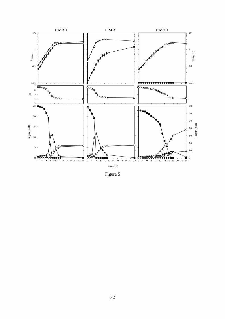
3.3. EPS production and central metabolism. Detection of EPS production by the six
strains on MSE-agar medium, showed that only CM9, CM30 and SM34 were able to form
viscous and mucous colonies (Fig. 3A). CM70, M67 and E14 were devoid of this phenotype,
showing small round and lenticular colonies (Fig. 3B).
The quantification of EPS production by the strains grown on CDMS showed the existence of
three categories of producers: (i) high (CM30 and SM34), medium (CM9) and low (E14 and
M67) (Fig. 4A). The results revealed EPS production levels of 0.55 ± 0.17 g L-1
, 0.63 ± 0.15 g
L-1
, 2.4 ± 0.13 g L-1
, 3.25 ± 0.16 g L-1
, 3.52 ± 0.18 g L-1
for E14, M67, CM9, SM34 and
CM30 strains, respectively (Fig. 4A). The results confirmed the preliminarily classification of
CM70 as a non-EPS producer. The culture on CDMS of CM30, one of the high EPS-
producers, has the appearance of a dense gel, a property defined as “ropiness” (Fig. 4B).
In a previous work, we identified the EPS produced by CM9 and CM30 strains as α-(1,6)
glucans with approximately 9% substitution, at positions O-3 (Zarour et al., 2017). These
dextrans are synthesised by dextransucrases extracellularly, using sucrose as substrate with a
concomitant release of fructose (Leemhuis et al., 2013). Thus, in this work, the EPS-
producing CM9 and CM30 strains as well as the non-EPS producer CM70 were subjected to a
metabolic study during their planktonic growth in CDMS, containing sucrose as carbon
source, determining the concentration of sucrose and its metabolites in the culture
supernatants (Fig. 5).
In this medium, the CM9 strain showed the best pattern of growth with a generation time of
91 min and a growth rate of 0.66 ± 0.01 h-1
. The CM30 strain had a growth rate of 0.43 ± 0.02
h-1
and a generation time of 140 min. In addition, the CM70 strain presented the longest
generation time (205 min). Moreover, the two dextran-producing strains synthesized the
polymer, in the exponential phase of growth, in parallel with an increase in A600nm and a
decrease of the extracellular pH. The dextran levels in the extracellular medium increased
with time and after 24 h of growth reached levels of 2.15 ± 0.15 g L-1
and 1.47 ± 0.08 g L-1
for CM30 and CM9, respectively.

13
In addition, after 6 h of incubation, the CM9 strain consumed the sucrose added to the
medium (≈ 25 mM), whereas the non-EPS producer CM70 required 17 h before the sucrose
was not detected in the supernatant (Fig. 5). Moreover, the three strains accumulated fructose
in the culture medium during their consumption of sucrose, and when the extracellular
disaccharide reached undetectable levels, the strains began to incorporate the accumulated
fructose. Glucose, the primary product of sucrose metabolism, was efficiently metabolized by
the three strains without any transient accumulation. At the end of the incubation period, the
accumulation of lactic acid in the medium of the EPS-non-producing CM70 cultures (30.53 ±
0.56 mM) was higher than those produced by the two EPS-producing strains (around 19 mM).
Finally, mannitol production seemed to be proportional to the consumption of the
accumulated fructose. The production rates of CM30, CM9 and CM70 strains at the
beginning of the stationary phase were 5.42 ± 0.32 mM, 5.63 ± 21 mM and 2.66 ± 0.17 mM,
respectively. Mannitol production was almost stable up to 24 h.
Probiotic profile of the L. mesenteroides strains. The antibiotics recommended by 3.3.
the European Food Safety Authority (EFSA, 2012) were tested to identify bacterial strains
with potential acquired resistance to: inhibitors of cell wall synthesis (amoxycillin, clavulanic
acid, ampicillin, cefazolin, cefotaxime, cefoxitin, oxacillin, penicillin and vancomycin),
inhibitors of nucleic acid synthesis (ofloxacin and pefloxacin) and lately inhibitors of
ribosome function (tetracycline) was tested (Table 1). The strains were classified as sensitive
(S), intermediate (I) and resistant (R) according to the cut-off values of each antibiotic
proposed by the Antibiogram Committee of the French Microbiology Society (Soussy et al.,
2000). All strains showed resistance to cefazolin, ofloxacin, oxacillin, pefloxacin and
vancomycin as well as sensitivity to cefotaxime, cefoxitin, penicillin and tetracycline.
Moreover, only CM70 was resistant to amoxicillin plus clavulanic acid.

14
Table 1. Antibiotic susceptibility profile of L. mesenteroides strains
Diameter area (mm)
Antibiotics Disc load (µg) CM9 CM30 CM70 SM34 E14 M67
Amoxicillin ± Clavulanic
acid
20 µg ±
10 µg
30 S 28 S 18 R 25 S 28 S 31 S
Ampicillin 10 µg 0 R 32 S 27 S 35 S 25 S 22 S
Cefazoline 30 µg 15 R 15 R 13 R 12 R 10 R 14 R
Cefotaxime 30 µg 19 I 23 S 25 S 24 S 27 S 24 S
Cefoxitin 30 µg 23 S 25 S 17 I 22 S 23 S 23 S
Ofloxacin 5 µg 13 R 15 R 14 R 10 R 13 R 09 R
Oxacillin 1 µg 0 R 0 R 0 R 0 R 0 R 0 R
Pefloxacin 5 µg 0 R 0 R 0 R 0 R 0 R 0 R
Penicillin 6 µg/10 IU 31 S 28 S 35 S 32 S 35 S 30 S
Tetracycline 30 µg 29 S 31 S 40 S 25 S 28 S 35 S
Vancomycin 30 µg 0 R 0 R 0 R 0 R 0 R 0 R
(R) resistant, (I) intermediate and (S) sensitive.
In addition, the six strains were able to diminish the growth of S. aureus ATCC 25923 and E.
coli ATCC 25922 indicator strains, having the inhibition halos diameters varying from 17 mm
to 22 mm and from 14 mm to 17 mm, respectively. However, the low extracellular pH,
produced by acidic metabolites (such as lactate) secreted by LAB, could be responsible for
the observed inhibitory effect. To test this hypothesis, the inhibitory activity of LAB against
pathogens was evaluated using MRSG-agar buffered at pH 7.0 instead of 6.2, to eliminate the
acids effect. The results did not reveal any antagonist activity of the LAB, except for the
CM70 strain, which generated halos with smaller diameter, 15 mm and 14 mm against S.
aureus and E. coli, respectively (Table 2S).
The six Leuconostoc strains were exposed to conditions resembling the human gastro-
intestinal tract. First, the LAB were subjected to the stomach stresses (Fig. 6). Different
resistance levels to acidity and bile salts were observed among the various strains. After 3 h
exposure to pH 3.0 and 2.0 the survival ranged from 27% to 8% and from 19% to 6% of the
initial population. Also, there was observed a resistance to 0.3%, 1.0% or 2.0% bile salts
exposure ranging from 29% to 8%, 22% to 5% or 17% to 2%, respectively. In addition, the
CM9 strain showed the highest tolerance to acidic and bile salts stresses. As an additional test,
the bacterial response to the stomach duodenum-passage was assessed by exposing the
bacteria to synthetic duodenal secretions at pH 3.0 for 1 h. Analysis of the bacterial viability
after duodenal treatment revealed slight decrease of viability during the incubation time

15
(Table 1). Moreover, the CM9 strain, as for the other stress treatments, had the highest rate of
survival, 33% and 24% after 1 h and 3 h of exposure, respectively.
The hydrophobicity of the cell surface of the bacteria, which is an indicator of their adhesion
capability, was tested using xylene. The results presented in Figure 6 showed different levels
of hydrophobicity among the six strains, which indicates a poor selectivity of the membrane
surfaces for some of them. The highest value (46%) was detected for CM9 and the lowest
(13%) for E14.
Moreover, based on their probiotic properties, CM9, CM30 and CM70 were chosen to analyse
their ability to colonize the intestine of the gnotobiotic embryos of zebrafish (Fig. 7). The
CM9 strain exhibited an average colonization capacity with a value of 16 cfu per larva after 6
h of exposure, compared to the two other strains that showed almost no colonization, with an
average of 3 cfu per larva for CM30 and 1 cfu per larva for CM70. After 24 hours, the
number of adherent bacteria was reduced to an average value of 2.12 cfu per embryos and
0.33 cfu per larva for CM9 and CM70 strains, respectively, and was undetectable for CM30.
Therefore, in vivo, the strain CM9 of L. mesenteroides showed the best colonization profile of
the gut of zebrafish gnotobiotic embryos.
Discussion 4.
The form, mode of association, inability to hydrolyse arginine, and heterofermentative
metabolism are morphological and biochemical characteristics that distinguish the strains
belonging to Leuconostoc from other Gram-positive and catalase-negative bacteria (Mozzi,
Raya, & Vignolo, 2015), and the six strains studied in this work conform with the definition
of this genus. Here, the phenotypic identification of the isolates was conducted according to
the results of physiological and biochemical analyses, such as sugar fermentation profiles.
The results for the six strains were consistent with the data and identification schemes
proposed in several works for L. mesenteroides (Björkroth & Holzapfel, 2006; Li et al., 2015 ;
Hui & Evranuz, 2016) and sequencing of their 16S rRNA coding gene confirmed this identity.
In addition, the use of specific primers for PCR amplification revealed that two out of the six
strains were L. mesenteroides subsp. mesenteroides. These results agree with those of
Moschetti et al. (2000) obtained for Leuconostoc strains isolated from pizza, field grass,
natural whey, mozzarella cheese and sausages.
The most important characteristic for potential starter-fermentation strains is their ability to
acidify the fermentation matrix, since the acid production and the accompanying pH decrease

16
give a specific aroma and extend the lag phase of sensitive organisms including foodborne
pathogens (Kostinek et al., 2007). All the strains studied in the current work had a progressive
production of lactic acid during a 24 h incubation period, as already reported by
Benmechernene et al. (2013) for L. mesenteroides species isolated from camel milk. The
different acidity profile and coagulation effect observed for the isolates from milks, honey and
silage could be due to several factors such as the original habitat, the production and
fermentation process and the enzymes involved. It is strongly suggested that dairy
leuconostoc is already adapted to this acidic environment, unlike non-dairy leuconostoc. As
far as we know, this is the first analysis of acidity carried out with Leuconostoc strains
isolated from honey. However, several studies report the use of silage as a raw material (Ni,
Wang, Cai, & Pang, 2015 ; Wang, Yuan, Dong, Li, & Shao, 2017), which clearly shows the
importance of acid production on forage quality.
The proteolytic activity of LAB plays a major role in the growth in milk and contributes
significantly to the development of the organoleptic properties of various fermented dairy
products (Smid & Kleerebezem, 2014). Our results are consistent with previous works (Idoui
and Karam, 2008; Tulini, Hymery, Haertlé, Le Blay, & De Martinis, 2015) indicating that
Leuconostoc species have low proteolytic activity, which results in limited growth in milk
(Alegría, Delgado, Flórez, & Mayo, 2013).
The flavour characteristic of food products may also be due to lipolysis, the incorporation of
lipolytic and/or proteolytic enzymes accelerating the formation of aroma (Holland et al.,
2005). From the results obtained, it appears that most of the strains have lipolytic activity over
lipids from natural and artificial sources. These results are in agreement with those obtained
by other authors (Katz, Medina, Gonzalez, & Oliver, 2002), which showed that L.
mesenteroides species isolated from sheep milk and cheeses could hydrolyse tributyrin.
In order to avoid interference from certain components of complex media, like MRS, in this
work, CDM defined medium was used for bacterial growth. We had previously demonstrated
that L. mesenteroides strains isolated from meat and milk do not produce dextran in the
presence of glucose, as the only carbon source (Nácher-Vázquez et al., 2017; Zarour et al.,
2017). The production levels obtained under discontinuous growth conditions are consistent
with those detected by Notararigo et al., (2013). In addition, this high production of dextran
by L. mesenteroides strains could be improved by optimizing the culture conditions and it
should be noted that, in general, very high values are characteristic of α-glucans.
The analysis of metabolic fluxes performed showed that the CM9 and CM30 strains
consumed sucrose and produced dextran with transient accumulation of fructose in the culture

17
medium as has been described for other Leuconostoc and other bacteria producing dextran
(Dols, Chraibi, Remaud-Simeon, Lindley, & Monsan, 1997; Santos, Teixeira, & Rodrigues,
2000; Nácher-Vázquez et al., 2017a). However, this is not a general feature for L.
mesenteroides, since we have not observed this pattern in the RTF10 strain isolated from meat
products (Nacher-Vázquez et al., 2017a). The difference between EPS production models is
influenced by various factors, including the acidification capacity of the culture medium due
to the production of lactic acid by LAB and the optimum pH for the enzymatic activity
responsible for the synthesis of the EPS (Rühmkorf et al., 2013). Thus, some strains of
Leuconostoc (Dols et al., 1997) and Lactobacillus (Rühmkorf et al., 2013) have maximum
efficacy during the exponential phase of growth. In the case of the CM9 and CM30 strains,
the production of EPS seems to be different, as revealed by the calculation of the relative
dextran production (the ratio of concentration per biomass estimated from the absorbance
values of the culture). CM9 showed a ratio of 0.15 (11 h) and 0.45 (24 h) during the
exponential and the stationary phase revealing that the EPS is produced during both phases,
whereas the CM30 strain displayed ratios of 0.78 (11 h), 1.01 (13 h) and 1.35 (24 h)
correlating with production only during the exponential phases of growth.
CM9, CM30 and the non-EPS-producing CM70 strain were able to consume fructose and
produce mannitol, and the beginning of the fructose consumption correlated with the
beginning of the production of this polyol, whose levels are low and vary among the three
strains. These results are consistent with those detected by several researchers (Patra, Tomar,
Rajput, & Singh, 2011; Zahid & Deppenmeier, 2016). Analysis of the pH of the extracellular
medium and the production of lactic acid revealed that CM70 had the highest acidifying
effect, presumably due to its inability to synthesize dextran, which means that the sucrose
would be phosphorylated by the action of a sucrose phosphorylase which would generate
glucose-1-phosphate and glucose-6-phosphate, consecutively (Dols, Chraibi, Remaud-
Simeon, Lindley, & Monsan, 1997) that would then be converted through glycolysis and by
the action of the lactate dehydrogenase into lactic acid. Finally, the efficacy of glucansucrase
was estimated for the in vivo synthesis of EPS, expressing its concentration in glucose
molecules. The results indicated that apparently 50% and 34% of the glucose released by
hydrolysis of sucrose had been used for the synthesis of EPS by the CM30 and CM9 strains,
respectively.
Being destined for human consumption, the safety of the probiotics is important because their
resistance to antibiotics may represent a possible threat. For this reason, in this study,

18
antibiotics from several groups, with effect on different targets, were tested. Antibiotic
resistance in probiotics is not usually a safety issue when mutations or intrinsic mechanisms
are responsible for the phenotype. However, the resistance to vancomycin seems to be a
common constitutive chromosomal encoded characteristic of Leuconostoc strains (Hemme &
Foucaud-Scheunemann, 2004), and in fact it was used as a selective agent, for the isolation of
the LAB studies in this work. On the contrary, the transmissible resistance to antibiotics, such
as the resistance to tetracycline encoded by host range mobilizable plasmids (Teuber, Meile,
& Schwarz, 1999) would be a threat, but the studied strains did not bear this resistance.
Moreover, some probiotic strains with intrinsic resistance to antibiotics may be useful for
restoring intestinal microbiota after treatment with antibiotics. Among the studied strains,
some were resistant to certain antibiotics, and CM70 was the only one resistant to amoxicillin
plus clavulanic acid, a combination of a -lactamase with a -lactamase inhibitor currently
used to combat nosocomial infections.
We also tested the ability of the Leuconostoc strains to produce antimicrobial substances
evaluating their inhibitory effect against the pathogenic strains S. aureus and E. coli. As
previously commented, this activity is generally due to the acidic environment, competition
for substrates or the production of bacteriocidal or bacteriostatic substances (Parente &
Ricciardi, 1999). Our results revealed that, apart from CM70, the antagonist action of the
studied strains against bacterial pathogens detected in MRS pH 6.2 was abolished by usage of
MRS pH 7.0 indicating that the activity was due to the acidic compounds produced.
Characterization of a putative production of a bacteriocin by CM70 would require further
studies.
It is important to note that the human response to probiotics is difficult to predict only from
these tests. Many other factors affect the survival of microorganisms in the upper
gastrointestinal tract, such as presence of different enzymes, peristaltic movements, digestion,
transit time (from 1 h to 4 h depending on the individual), the diet and other conditions, which
may influence the viability of probiotic bacteria (Vizoso Pinto, Franz, Schillinger, &
Holzapfel, 2006). However, the in vitro and in vivo assays performed here provide useful
information about the probiotic potential of the studied strains.
The in vitro probiotic study showed that strains of L. mesenteroides can partially counteract
(less than 50%) the stress conditions found in the gastrointestinal tract (acidity, bile stress and
stomach-duodenal passage). The decrease in survival rate was presumably due to acid

19
regulation mechanisms that failed to maintain intracellular pH. This would reduce enzyme
activity by damaging certain proteins and DNA (van de Guchte et al., 2002). The number of
viable cells fluctuated, but it tended to decrease after 3 h of incubation. Recent studies
suggested that some Leuconostoc strains are able to grow and survive at these barrier levels
(de Paula et al., 2015; Diana, Humberto, & Jorge, 2015; Giles-Gómez et al., 2016).
In addition, the test to evaluate the surface hydrophobicity of bacterial cells with xylene
indicated that the CM9 strain is the most adherent (45.6%). Moreover, these strains have a
significantly higher adhesion capacity than the probiotic strain Lb. acidophilus LA-5, used as
a reference in the work of Ng, Koon, Padam, & Chye (2015). According to Giaouris, Chapot-
Chartier, & Briandet (2009), L. lactis strains with more than 40% affinity for polar solvents
are particularly hydrophobic, thus, only the CM9 strain had a hydrophobic surface (Habimana
et al., 2007). Recently, we have analysed the ability of the CM30, SM34 and CM9 strains to
bind to Caco-2 cells, detecting that, in the presence of glucose, they are able to bind to the
enterocytes at similar levels (4.5%-3%) (Zarour et al., 2017). Here, we have used zebrafish
embryos to study the capacity of L. mesenteroides strains for colonization in vivo, since
various works support the utility of this model for evaluation of probiotic bacteria, in
particular LAB (Nácher-Vázquez et al. 2017a; Wang et al., 2016; Russo et al., 2015). The
results revealed that CM9 was the most adhesive among the three strains, as it was detected in
a statistical significant higher number in the digestive tract of the embryos 6 h after
administration and still detectable together with the other strains at a very low level after 24 h.
The lack of permanence of the LAB in the digestive tract is the expected behaviour for
probiotic bacteria, which require a daily administration. Since, in addition to these
characteristics, L. mesenteroides CM9 produces an immunomodulatory dextran with potential
anti-inflammatory properties and interesting pseudoplastic rheological properties (Zarour et
al., 2017), this strain seems to be a good candidate for its use as a component of functional
fermented dairy products.
Conclusions 5.
The results of isolation, identification and characterization of strains isolated from dairy and
non-dairy habitats permitted the evaluation of dairy Leuconostoc for their ability to: (i)
produce CO2, dextran, with potential antiviral and immunomodulatory activity (Nacher-
Vazquez et al., 2015; Zarour et al., 2017), the low-calorie compound mannitol used in
products for diabetics, as well as lactic acid, which is used mainly for food preservation; (ii)

20
use citrate, (iii) degrade milk protein and lipid sources and (iv) to resist gastrointestinal
stresses. In addition, the probiotic properties of dairy Leuconostoc, particularly CM9 isolated
from camel milk, suggested that it could be a promising candidate for production of probiotic
functional and safe dairy products.

21
6. Acknowledgements
This work was supported by the Spanish Ministry of Economy and Competitiveness (grant
AGL2015-65010-C3-1-R). We thank Dr. Stephen W. Elson for critical reading of the
manuscript. We thank Dr. Guillermo Padilla Alonso for his valuable assistance in the bio-
statistical analysis.
7. References
Alegría, A., Delgado, S., Flórez, A. B., & Mayo, B. (2013). Identification, typing, and
functional characterization of Leuconostoc spp. strains from traditional, starter-free
cheeses. Dairy Science & Technology, 93(6), 657-673. doi: 10.1007/s13594-013-
0128-3
Aman, A., Siddiqui, N. N., & Qader, S. A. U. (2012). Characterization and potential
applications of high molecular weight dextran produced by Leuconostoc
mesenteroides AA1. Carbohydrate Polymers, 87(1), 910-915. doi:
http://dx.doi.org/10.1016/j.carbpol.2011.08.094.
Adelfo, E., María, E. R., Alfredo, M., Agustín, L.-M., Francisco, B., & Guillermo, G. (2004).
Characterization of bacterial diversity in Pulque, a traditional Mexican alcoholic
fermented beverage, as determined by 16S rDNA analysis. FEMS Microbiology
Letters, 235(2), 273-279. doi: doi:10.1111/j.1574-6968.2004.tb09599.x
Arena, M. P., Capozzi, V., Spano, G., & Fiocco, D. (2017). The potential of lactic acid
bacteria to colonize biotic and abiotic surfaces and the investigation of their
interactions and mechanisms. Applied Microbiology and Biotechnology, 101(7), 2641-
2657. doi: 10.1007/s00253-017-8182-z
Barefoot, S. F., & Klaenhammer, T. R. (1983). Detection and activity of lactacin B, a
bacteriocin produced by Lactobacillus acidophilus. Applied and Environmental
Microbiology, 45(6), 1808-1815.
Benmechernene, Z., Chentouf, H. F., Yahia, B., Fatima, G., Quintela-Baluja, M., Calo-Mata,
P., & Barros-Velázquez, J. (2013). Technological aptitude and applications of
Leuconostoc mesenteroides bioactive strains isolated from algerian raw camel milk.
BioMed Research International, 2013, 418132. doi: 10.1155/2013/418132
Bhatt, S. M., Mohan, A., & Srivastava, S. K. (2013). Challenges in enzymatic route of
mannitol production. International Scholarly Research Notices: Biotechnology,
914187. doi: 10.5402/2013/914187
Björkroth, J., & Holzapfel, W. (2006). Genera Leuconostoc, Oenococcus and Weissella. In M.
Dworkin, S. Falkow, E. Rosenberg, K.-H. Schleifer & E. Stackebrandt (Eds.), The
Prokaryotes: Volume 4: Bacteria: Firmicutes, Cyanobacteria (pp. 267-319). New
York, NY: Springer US.
Chung, C.-H., & Day, D. F. (2002). Glucooligosaccharides from Leuconostoc mesenteroides
B-742 (ATCC 13146): A potential prebiotic. Journal of Industrial Microbiology and
Biotechnology, 29(4), 196-199. doi: 10.1038/sj.jim.7000269
de Man, J. C., Rogosa, M., & Sharpe, M. E. (1960). A medium for the cultivation of
Lactobacilli. Journal of Applied Microbiology, 23(1), 130-135. doi: 10.1111/j.1365-
2672.1960.tb00188.x
de Paula, A. T., Jeronymo-Ceneviva, A. B., Silva, L. F., Todorov, S. D., Franco, B. D. G. M.,
& Penna, A. L. B. (2015). Leuconostoc mesenteroides SJRP55: a potential probiotic

22
strain isolated from Brazilian water buffalo mozzarella cheese. Annals of
Microbiology, 65(2), 899-910. doi: 10.1007/s13213-014-0933-9
Diana, C.-R., Humberto, H.-S., & Jorge, Y. F. (2015). Probiotic properties of Leuconostoc
mesenteroides isolated from aguamiel of agave salmiana. Probiotics and
Antimicrobial Proteins, 7(2), 107-117. doi: 10.1007/s12602-015-9187-5
Dols, M., Chraibi, W., Remaud-Simeon, M., Lindley, N. D., & Monsan, P. F. (1997). Growth
and energetics of Leuconostoc mesenteroides NRRL B-1299 during metabolism of
various sugars and their consequences for dextransucrase production. Applied and
Environmental Microbiology, 63(6), 2159-2165.
Escalante, A., López Soto, D. R., Velázquez Gutiérrez, J. E., Giles-Gómez, M., Bolívar, F., &
López-Munguía, A. (2016). Pulque, a Traditional Mexican Alcoholic Fermented
Beverage: Historical, Microbiological, and Technical Aspects. Frontiers in
Microbiology, 7(1026). doi: 10.3389/fmicb.2016.01026
European Food Safety Authority (EFSA). (2012). Panel on Additives and Products or
Substances used in Animal Feed (FEEDAP); Guidance on the assessment of bacterial
susceptibility to antimicrobials of human and veterinary importance, EFSA Journal,
10, 2740.
Fortin, M., Messier, S., Paré, J., & Higgins, R. (2003). Identification of catalase-negative,
non-beta-hemolytic, gram-positive cocci isolated from milk samples. Journal of
Clinical Microbiology, 41(1), 106-109. doi: 10.1128/JCM.41.1.106-109.2003
Ghoreishi, S. M., & Shahrestani, R. G. (2009). Innovative strategies for engineering mannitol
production. Trends in Food Science & Technology, 20(6), 263-270. doi:
https://doi.org/10.1016/j.tifs.2009.03.006
Giaouris, E., Chapot-Chartier, M.-P., & Briandet, R. (2009). Surface physicochemical
analysis of natural Lactococcus lactis strains reveals the existence of hydrophobic and
low charged strains with altered adhesive properties. International Journal of Food
Microbiology, 131(1), 2-9.
Giles-Gómez, M., Sandoval García, J. G., Matus, V., Campos Quintana, I., Bolívar, F., &
Escalante, A. (2016). In vitro and in vivo probiotic assessment of Leuconostoc
mesenteroides P45 isolated from pulque, a Mexican traditional alcoholic beverage.
SpringerPlus, 5(1), 708. doi: 10.1186/s40064-016-2370-7
Habimana, O., Le Goff, C., Juillard, V., Bellon-Fontaine, M.-N., Buist, G., Kulakauskas, S.,
& Briandet, R. (2007). Positive role of cell wall anchored proteinase PrtP in adhesion
of Lactococci. BMC Microbiology, 7, 36-36. doi: 10.1186/1471-2180-7-36
Hemme, D., & Foucaud-Scheunemann, C. (2004). Leuconostoc, characteristics, use in dairy
technology and prospects in functional foods. International Dairy Journal, 14(6), 467-
494. doi: http://dx.doi.org/10.1016/j.idairyj.2003.10.005
Hemme, D. (2012). Leuconostoc and its use in dairy technology. In Y. H. Hui & E. Ö.
Evranuz (Eds.), Handbook of Animal-Based Fermented Food and Beverage
Technology, Second Edition (pp. 73-108): CRC Press.
Holland, R., Liu, S. Q., Crow, V. L., Delabre, M. L., Lubbers, M., Bennett, M., & Norris, G.
(2005). Esterases of lactic acid bacteria and cheese flavour: Milk fat hydrolysis,
alcoholysis and esterification. International Dairy Journal, 15(6), 711-718. doi:
http://dx.doi.org/10.1016/j.idairyj.2004.09.012
Hui, Y. H., & Evranuz, E. Ö. (2016). Handbook of animal-based fermented food and
beverage technology, Second Edition: CRC Press.
Hyronimus , B., Le Marrec, C., Hadj Sassi, A., & Deschamps, A. (2000). Acid and bile
tolerance of spore-forming lactic acid bacteria. International Journal of Food
Microbiology, 61 193-197.

23
Idoui, T., & Karam, N.-E. (2008). Lactic acid bacteria from Jijel’s traditional butter: Isolation,
identification and major technological traits. 59(4), 7. doi:
10.3989/gya.2008.v59.i4.530
Iyer, R., Tomar, S. K., Kapila, S., Mani, J., & Singh, R. (2010). Probiotic properties of folate
producing Streptococcus thermophilus strains. Food Research International, 43(1),
103-110. doi: http://dx.doi.org/10.1016/j.foodres.2009.09.011
Jung, S. H., Park, J. W., Cho, I. J., Lee, N. K., Yeo, I.-C., Kim, B. Y., Kim, H. K., & Hahm,
Y. T. (2012). Characterization of lactic acid bacteria isolated from sauce-type kimchi.
Preventive Nutrition and Food Science, 17(3), 217-222. doi:
10.3746/pnf.2012.17.3.217
Kalbaza, K., Zadi-Karam, H., & Karam, N. (2018). Identification and major technological
characteristics of Lactococcus and Lactobacillus strains isolated from "hamoum", an
Algerian fermented wheat. African Journal of Biotechnology , 17(5), 108-117.
Katz, M., Medina, R., Gonzalez, S., & Oliver, G. (2002). Esterolytic and lipolytic activities of
lactic acid bacteria isolated from ewe's milk and cheese. Journal of Food Protection,
65(12), 1997-2001.
Kempler, G. M., & McKay, L. L. (1980). Improved medium for detection of citrate-
fermenting Streptococcus lactis subsp. diacetylactis. Applied and Environmental
Microbiology, 39(4), 926-927.
Kihal, M., Prevost, H., Henni, D. E., Benmechernene, Z., & Diviès, C. (2009). Carbon
dioxide production by Leuconostoc mesenteroîdes grown in single and mixed culture
with Lactococcus lactis in skim milk. Scientific Research and Essay, 4(11), 1348-
1353.
Kostinek, M., Specht, I., Edward, V. A., Pinto, C., Egounlety, M., Sossa, C., Mbugua, S.,
Dortu, C., Thonart, P., Taljaard, L., Mengu, M., Franz, C. M. A. P., & Holzapfel, W.
H. (2007). Characterisation and biochemical properties of predominant lactic acid
bacteria from fermenting cassava for selection as starter cultures. International
Journal of Food Microbiology, 114(3), 342-351. doi:
https://doi.org/10.1016/j.ijfoodmicro.2006.09.029
Leemhuis, H., Pijning, T., Dobruchowska, J. M., van Leeuwen, S. S., Kralj, S., Dijkstra, B.
W., & Dijkhuizen, L. (2013). Glucansucrases: Three-dimensional structures, reactions,
mechanism, α-glucan analysis and their implications in biotechnology and food
applications. Journal of Biotechnology, 163(2), 250-272. doi:
http://dx.doi.org/10.1016/j.jbiotec.2012.06.037
Li, D., Ni, K., Pang, H., Wang, Y., Cai, Y., & Jin, Q. (2015). Identification and antimicrobial
activity detection of lactic acid bacteria isolated from corn stover silage. Asian-
Australasian Journal of Animal Sciences, 28(5), 620-631. doi: 10.5713/ajas.14.0439
Liasi, S. A., Azmi, T. I., Hassan, M. D., Shuhaimi, M., Rosfarizan, M., & Ariff, A. B. (2009).
Antimicrobial activity and antibiotic sensitivity of three isolates of lactic acid bacteria
from fermented fish product, Budu Malaysian Journal of Microbiology, 5(1), 33-37
Massa, S., Caruso, M., Trovatelli, F., & Tosques, M. (1998). Comparison of plate count agar
and R2A medium for enumeration of heterotrophic bacteria in natural mineral water.
World Journal of Microbiology and Biotechnology, 14(5), 727-730. doi:
10.1023/a:1008893627877
Mathara, J. M., Schillinger, U., Guigas, C., Franz, C., Kutima, P. M., Mbugua, S. K., Shin, H.
K., & Holzapfel, W. H. (2008). Functional characteristics of Lactobacillus spp. from
traditional maasai fermented milk products in Kenya. International Journal of Food
Microbiology, 126(1), 57-64. doi: http://dx.doi.org/10.1016/j.ijfoodmicro.2008.04.027

24
Mathot, A. G., Kihal, M., Prevost, H., & Divies, C. (1994). Selective enumeration of
Leuconostoc on vancomycin agar media. International Dairy Journal, 4(5), 459-469.
doi: http://dx.doi.org/10.1016/0958-6946(94)90059-0
Mayeux, J. V., Sandine, W. E., & Elliker, P. R. (1962). A selective medium for detecting
Leuconostoc in mixed-strain starter cultures. Journal of Dairy Science, 45, 655.
Mazzoli, R., Bosco, F., Mizrahi, I., Bayer, E. A., & Pessione, E. (2014). Towards lactic acid
bacteria-based biorefineries. Biotechnological Advances, 32(7), 1216-1236.
Moschetti, G., Blaiotta, G., Villani, F., & Coppola, S. (2000). Specific detection of
Leuconostoc mesenteroides subsp. mesenteroides with dna primers identified by
randomly amplified polymorphic DNA analysis. Applied and Environmental
Microbiology, 66(1), 422-424.
Moslehishad, M., Mirdamadi, S., Ehsani, M. R., Ezzatpanah, H., & Moosavi‐Movahedi, A. A.
(2013). The proteolytic activity of selected lactic acid bacteria in fermenting cow's and
camel's milk and the resultant sensory characteristics of the products. International
Journal of Dairy Technology, 66(2), 279-285. doi: 10.1111/1471-0307.12017
Mozzi, F., Raya, R. R., & Vignolo, G. M. (2015). Biotechnology of lactic acid bacteria:
Novel Applications: Wiley.
Nácher-Vázquez, M., Ballesteros, N., Canales, A., Rodriguez Saint-Jean, S., Perez-Prieto, S.
I., Prieto, A., Aznar, R., & López, P. (2015). Dextrans produced by lactic acid bacteria
exhibit antiviral and immunomodulatory activity against salmonid viruses.
Carbohydrate Polymers, 124, 292-301. doi: 10.1016/j.carbpol.2015.02.020
Nácher-Vázquez, M., Iturria, I., Zarour, K., Mohedano, M. L., Aznar, R., Pardo, M. Á., &
López, P. (2017a). Dextran production by Lactobacillus sakei MN1 coincides with
reduced autoagglutination, biofilm formation and epithelial cell adhesion.
Carbohydrate Polymers, 168, 22-31. doi: http://doi.org/10.1016/j.carbpol.2017.03.024
Nácher-Vázquez, M., Ruiz-Masó, J. A., Mohedano, M. L., del Solar, G., Aznar, R., & López,
P. (2017b). Dextransucrase expression is concomitant with that of replication and
maintenance functions of the pMN1 plasmid in Lactobacillus sakei MN1. Frontiers in
Microbiology, 8, 2281. doi: 10.3389/fmicb.2017.02281
Neu, H. C., & Gootz, T. D. (1996). Antimicrobial Chemotherapy. In U. o. T. M. B. a.
Galveston (Ed.), Medical Microbiology. (4th ed.). Galveston Baron, S.
Ng, S. Y., Koon, S. S., Padam, B. S., & Chye, F. Y. (2015). Evaluation of probiotic potential
of lactic acid bacteria isolated from traditional Malaysian fermented Bambangan
(Mangifera pajang). CyTA - Journal of Food, 13(4), 563-572. doi:
10.1080/19476337.2015.1020342
Ni, K., Wang, Y., Cai, Y., & Pang, H. (2015). Natural lactic acid bacteria population and
silage fermentation of whole-crop wheat. Asian-Australasian Journal of Animal
Sciences, 28(8), 1123-1132. doi: 10.5713/ajas.14.0955
Notararigo, S., Nácher-Vázquez, M., Ibarburu, I., Werning, M. L., Fernández de Palencia, P.,
Dueñas, M. T., Aznar, R., López, P., & Prieto, A. (2013). Comparative analysis of
production and purification of homo- and hetero-polysaccharides produced by lactic
acid bacteria. Carbohydrate Polymers, 93(1), 57-64. doi:
10.1016/j.carbpol.2012.05.016
Nuraida, L. (2015). A review: Health promoting lactic acid bacteria in traditional Indonesian
fermented foods. Food Science and Human Wellness, 4(2), 47-55. doi:
http://dx.doi.org/10.1016/j.fshw.2015.06.001
Ortakci, F., Broadbent, J. R., Oberg, C. J., & McMahon, D. J. (2015). Growth and gas
production of a novel obligatory heterofermentative Cheddar cheese nonstarter
Lactobacilli species on ribose and galactose. Journal of Dairy Science, 98(6), 3645-
3654. doi: https://doi.org/10.3168/jds.2014-9293

25
Oyarbide, U., Iturria, I., Rainieri, S., & Pardo, M. A. (2015). Use of gnotobiotic zebrafish to
study vibrio anguillarum pathogenicity. Zebrafish, 12(1), 71-80. doi:
10.1089/zeb.2014.0972
Papagianni, M. (2012). Food fermentation and production of biopreservatives handbook of
animal-based fermented food and beverage technology, Second Edition (pp. 109-124):
CRC Press.
Parente, E., & Ricciardi, A. (1999). Production, recovery and purification of bacteriocins
from lactic acid bacteria. Applied Microbiology and Biotechnology, 52(5), 628-638.
Park, J.-Y., Jeong, S.-J., Lee, A. R., Park, J.-Y., Jeong, W. J., & Kim, J. H. (2007). Expression
of alpha-galactosidase gene from Leuconostoc mesenteroides SY1 in Leuconostoc
citreum. Journal of Microbioogy and. Biotechnology, 17(12), 2081-2084.
Park, J.-M., Shin, J.-H., Lee, D.-W., Song, J.-C., Suh, H.-J., Chang, U.-J., & Kim, J.-M.
(2010). Identification of the lactic acid bacteria in Kimchi according to initial and
over-ripened fermentation using PCR and 16S rRNA gene sequence analysis. Food
Science and Biotechnology, 19(2), 541-546. doi: 10.1007/s10068-010-0075-1
Patra, F., Tomar, S. K., Rajput, Y. S., & Singh, R. (2011). Characterization of mannitol
producing strains of Leuconostoc species. World Journal of Microbiology and
Biotechnology, 27(4), 933-939. doi: 10.1007/s11274-010-0536-y
Pérez-Ramos, A., Nácher-Vázquez, M., Notararigo, S., López, P. and Mohedano, M. L.
(2015). Current and future applications of bacterial extracellular polysaccharides In V.
R. P. a. R. R. Watson (Ed.), Probiotics, Prebiotics, and Synbiotics (Elsevier Oxford
ed.). UK.
Performance Standards for Antimicrobial Disk Susceptibility tests. (2007). Clinical and
Laboratory Standards Institute, 27(1).
Pieniz, S., Andreazza, R., Anghinoni, T., Camargo, F., & Brandelli, A. (2014). Probiotic
potential, antimicrobial and antioxidant activities of Enterococcus durans strain
LAB18s. Food Control, 37(Supplement C), 251-256. doi:
https://doi.org/10.1016/j.foodcont.2013.09.055
Rendueles, O., Ferrières, L., Frétaud, M., Bégaud, E., Herbomel, P., & Levraud, J. P. (2012).
A new Zebrafish model of oro-Intestinal pathogen colonization reveals a key role for
adhesion in protection by probiotic bacteria. PLoS Pathogens, 8, e1002815.
Rühmkorf, C., Bork, C., Mischnick, P., Rübsam, H., Becker, T., & Vogel, R. F. (2013).
Identification of Lactobacillus curvatus TMW 1.624 dextransucrase and comparative
characterization with Lactobacillus reuteri TMW 1.106 and Lactobacillus animalis
TMW 1.971 dextransucrases. Food Microbiology, 34, 52-61.
Russo, P., Iturria, I., Mohedano, M. L., Caggianiello, G., Rainieri, S., Fiocco, D., Angel
Pardo, M., López, P., & Spano, G. (2015). Zebrafish gut colonization by mCherry-
labelled lactic acid bacteria. Appl Microbiol Biotechnol. doi: 10.1007/s00253-014-
6351-x
Samad, M. Y. A., Razak, C. N. A., Salleh, A. B., Zin Wan Yunus, W. M., Ampon, K., &
Basri, M. (1989). A plate assay for primary screening of lipase activity. Journal of
Microbiological Methods, 9(1), 51-56. doi: https://doi.org/10.1016/0167-
7012(89)90030-4
Sánchez, C., Neves, A. R., Cavalheiro, J., dos Santos, M. M., García-Quintáns, N.,López, P.,
et al. (2008). Contribution of citrate metabolism to the growth ofLactococcus lactis
CRL264 at low pH. Applied and Environmental Microbiology,74(4), 1136–1144.
http://dx.doi.org/10.1128/AEM. 01061-07,
Santos, M., Teixeira, J., & Rodrigues, A. r. (2000). Production of dextransucrase, dextran and
fructose from sucrose using Leuconostoc mesenteroides NRRL B512(f). Biochemical

26
Engineering Journal, 4(3), 177-188. doi: http://dx.doi.org/10.1016/S1369-
703X(99)00047-9
Smid, E. J., & Kleerebezem, M. (2014). Production of aroma compounds in lactic
fermentations. Annuel Review of Food Science Technology, 5, 313-326. doi:
10.1146/annurev-food-030713-092339
Soussy, C. J., Carret, G., Cavallo, J. D., Chardon, H., Chidiac, C., Choutet, P., Courvalin, P.,
Dabernat, H., Drugeon, H., Dubreuil, L., Goldstein, F., Jarlier, V., Leclercq, R.,
Nicolas-Chanoine, M. H., Philippon, A., Quentin, C., Rouveix, B. & Sirot, J. (2000).
Antibiogram Committee of the French Microbiology Society. Report 2000-2001.
Pathol Biol (Paris), 48(9), 832-871.
Teuber, M., Meile, L., & Schwarz, F. (1999). Acquired antibiotic resistance in lactic acid
bacteria from food. Antonie Van Leeuwenhoek, 76(1-4), 115-137.
Thomas, T. D. (1973). Agar medium for differentiation of Streptococcus cremoris from the
other bacteria. Journal of Dairy Sciences and Technology, 8, 70-71.
Tropcheva, R., Nikolova, D., Evstatieva, Y., & Danova, S. (2014). Antifungal activity and
identification of Lactobacilli, isolated from traditional dairy product “katak”.
Anaerobe, 28, 78-84. doi: http://dx.doi.org/10.1016/j.anaerobe.2014.05.010
Tulini, F. L., Hymery, N., Haertlé, T., Le Blay, G., & De Martinis, E. C. P. (2015). Screening
for antimicrobial and proteolytic activities of lactic acid bacteria isolated from cow,
buffalo and goat milk and cheeses marketed in the southeast region of Brazil. Journal
of Dairy Research, 83(1), 115-124. doi: 10.1017/S0022029915000606.
van de Guchte, M., Serror, P., Chervaux, C., Smokvina, T., Ehrlich, S. D., & Maguin, E.
(2002). Stress responses in lactic acid bacteria. Antonie Van Leeuwenhoek, 82(1-4),
187-216.
Vizoso Pinto, M. G., Franz, C. M. A. P., Schillinger, U., & Holzapfel, W. H. (2006).
Lactobacillus spp. with in vitro probiotic properties from human faeces and traditional
fermented products. International Journal of Food Microbiology, 109(3), 205-214.
doi: http://dx.doi.org/10.1016/j.ijfoodmicro.2006.01.029.
Wang, Y., Ren, Z., Fu, L., & Su, X. (2016). Two highly adhesive lactic acid bacteria strains
are protective in zebrafish infected with Aeromonas hydrophila by evocation of gut
mucosal immunity. Journal of Applied Microbiology, 120(2), 441-451. doi:
10.1111/jam.13002.
Wang, S., Yuan, X., Dong, Z., Li, J., & Shao, T. (2017). Isolating and evaluating lactic acid
bacteria strains for effectiveness on silage quality at low temperatures on the Tibetan
Plateau. Animal Science Journal, 88(11). 1722-1729. doi: 10.1111/asj.12852.
Yerlikaya, O. (2014). Starter cultures used in probiotic dairy product preparation and popular
probiotic dairy drinks. Food Science and Technology, 34, 221-229.
Zahid, N., & Deppenmeier, U. (2016). Role of mannitol dehydrogenases in osmoprotection of
Gluconobacter oxydans. Applied Microbiology and Biotechnology, 100(23), 9967-
9978. doi: 10.1007/s00253-016-7680-8
Zarour, K., Llamas, M. G., Prieto, A., Ruas-Madiedo, P., Dueñas, M. T., Fernández de
Palencia, P., Aznar, R., Kihal, M., & López, P. (2017). Rheology and bioactivity of
high molecular weight dextrans synt hesised by lactic acid bacteria.
27
Legends of figures
Fig. 1. Analysis of bacterial colonies of L. mesenteroides CM30 strain by phase contrast
optical (A) and transmission electron (B) microscopies
Fig. 2. Acidification kinetics of L. mesenteroides CM9 (A) and M67 (B) in skim milk
medium supplemented with 0.3% yeast extract. Symbols: ▲, pH and ■, dornic acidity.
Fig. 3. Detection of EPS production by L. mesenteroides strains on MSE solid medium. CM9
(A) and CM70 (B) strains.
Fig. 4. Quantification of EPS production by L. mesenteroides strains on CDMS liquid
medium (A) and ropy appearance of CM30 culture (B). Statistical significances are
represented by different letters that mean a P≤0.05.
Fig. 5. Analysis of central metabolism and EPS production of L. mesenteroides strains during
planktonic growth. Bacteria were grown in CDMS at 30 ºC. Symbols: , A600nm; ●, Dextran;
□, pH; ■, sucrose; ▲, fructose; ♦, glucose; ◊, mannitol and , lactic acid.
Fig. 6. Survival of L. mesenteroides strains to acidic, biliary salts and gastro-intestinal
stresses. Statistical significances are represented by different letters that mean a p ≤0.05.
Fig. 7. Colonization of zebrafish embryos by strains of L. mesenteroides in the
gastrointestinal tract. (A) Protocol strains prevalence in zebrafish embryos digestive tract. (B)
The number of cfu per larva was determined after 6 h (black bars) and 24 h (white bars) post
infection. Statistical significances are represented by different letters that mean a P≤0.05.

28
Figure 1

29
Figure 2

30
Figure 3

31
Figure 4
32
Figure 5

33
Figure 6

34
Figure 7