anatomical position & directional terms sports medicine 1
TRANSCRIPT
ANATOMICAL POSITION & DIRECTIONAL TERMS
Sports Medicine 1
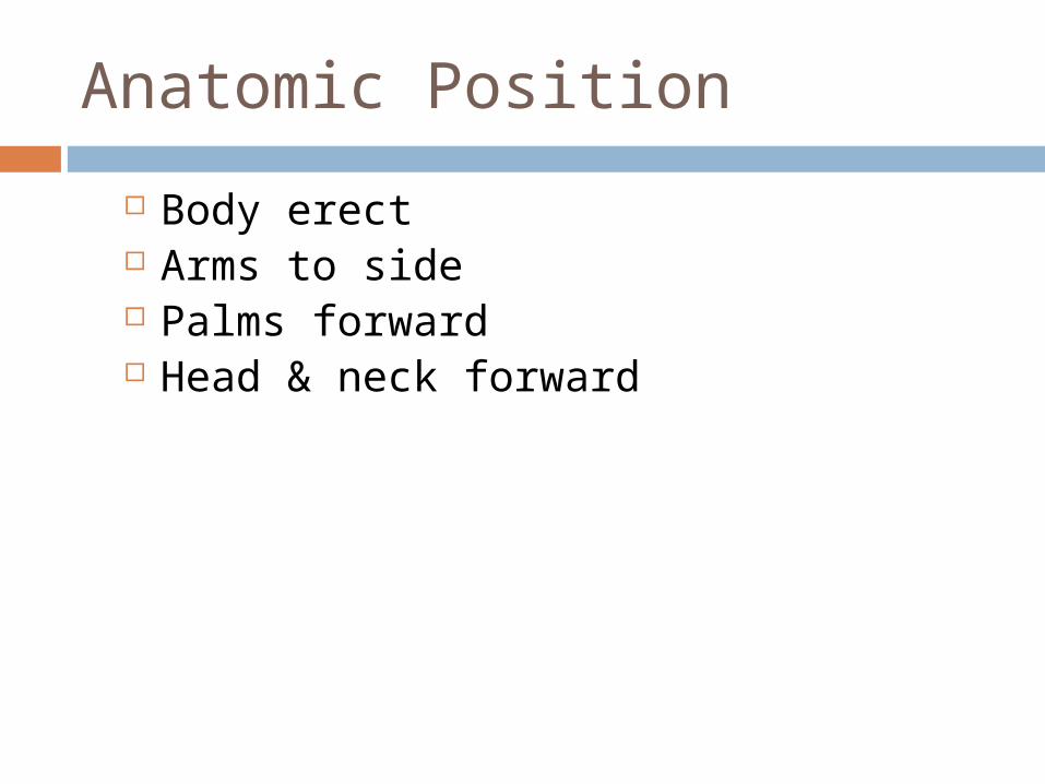
Anatomic Position
Body erect Arms to side Palms forward Head & neck forward

Body Planes
Sagittal
Medial/Mid-sagittal
Coronal/Frontal
Transverse

transversetransverse
midsaggitalmidsaggitalcoronalcoronal
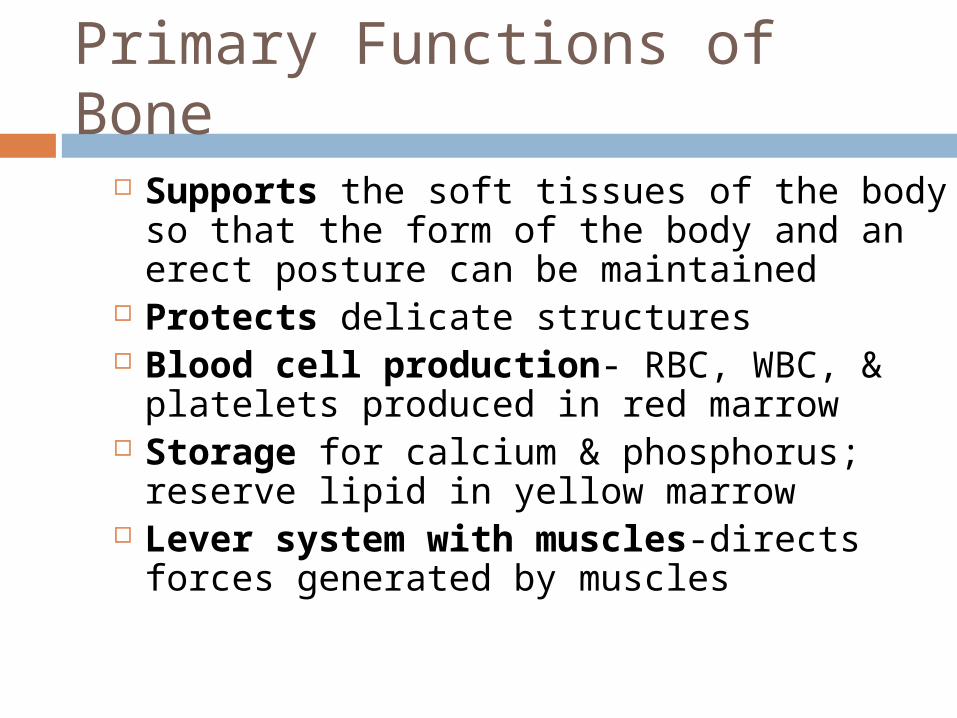
Primary Functions of Bone
Supports the soft tissues of the body so that the form of the body and an erect posture can be maintained
Protects delicate structures Blood cell production- RBC, WBC, &
platelets produced in red marrow Storage for calcium & phosphorus;
reserve lipid in yellow marrow Lever system with muscles-directs
forces generated by muscles
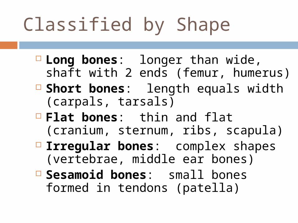
Classified by Shape
Long bones: longer than wide, shaft with 2 ends (femur, humerus)
Short bones: length equals width (carpals, tarsals)
Flat bones: thin and flat (cranium, sternum, ribs, scapula)
Irregular bones: complex shapes (vertebrae, middle ear bones)
Sesamoid bones: small bones formed in tendons (patella)

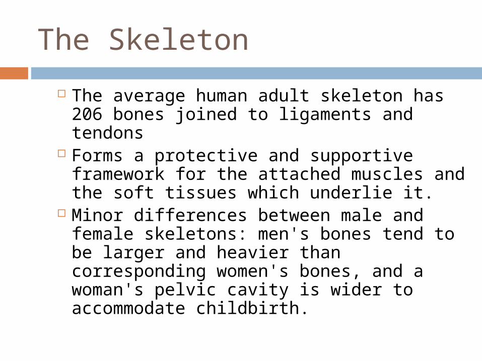
The Skeleton
The average human adult skeleton has 206 bones joined to ligaments and tendons
Forms a protective and supportive framework for the attached muscles and the soft tissues which underlie it.
Minor differences between male and female skeletons: men's bones tend to be larger and heavier than corresponding women's bones, and a woman's pelvic cavity is wider to accommodate childbirth.

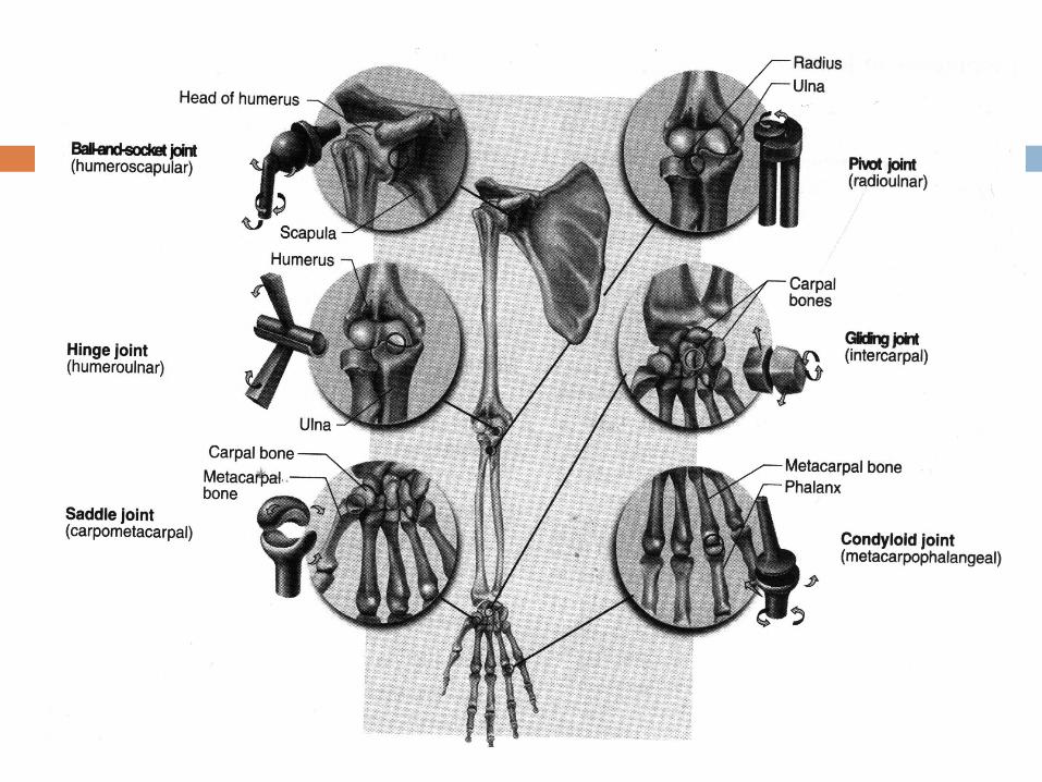
Joint Types Ball and Socket
The greatest range of joint movement is provided by a "ball-and- socket" joint, in which the spherical head of one bone lodges in the spherical cavity of another

Joint Types: Hinge
The simplest type of joint is the "hinge," as found in the elbows and the joints of the fingers and toes.
Hinge joints allow movement in only one direction.
Elbow
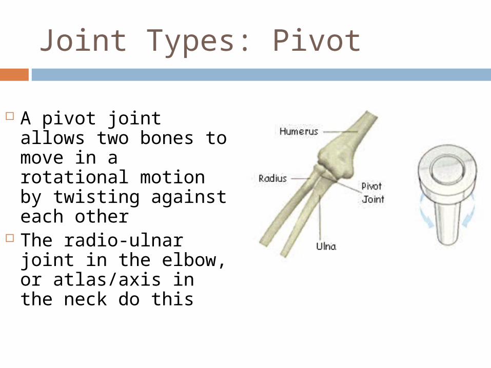
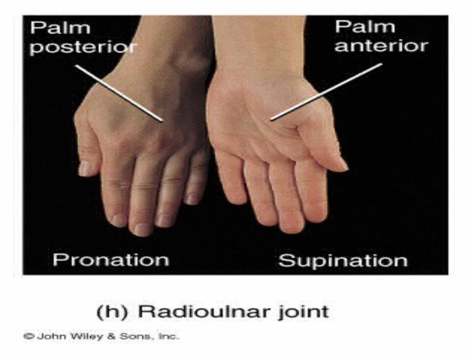
Joint Types: Pivot
A pivot joint allows two bones to move in a rotational motion by twisting against each other
The radio-ulnar joint in the elbow, or atlas/axis in the neck do this
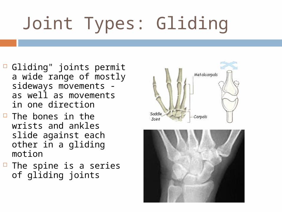
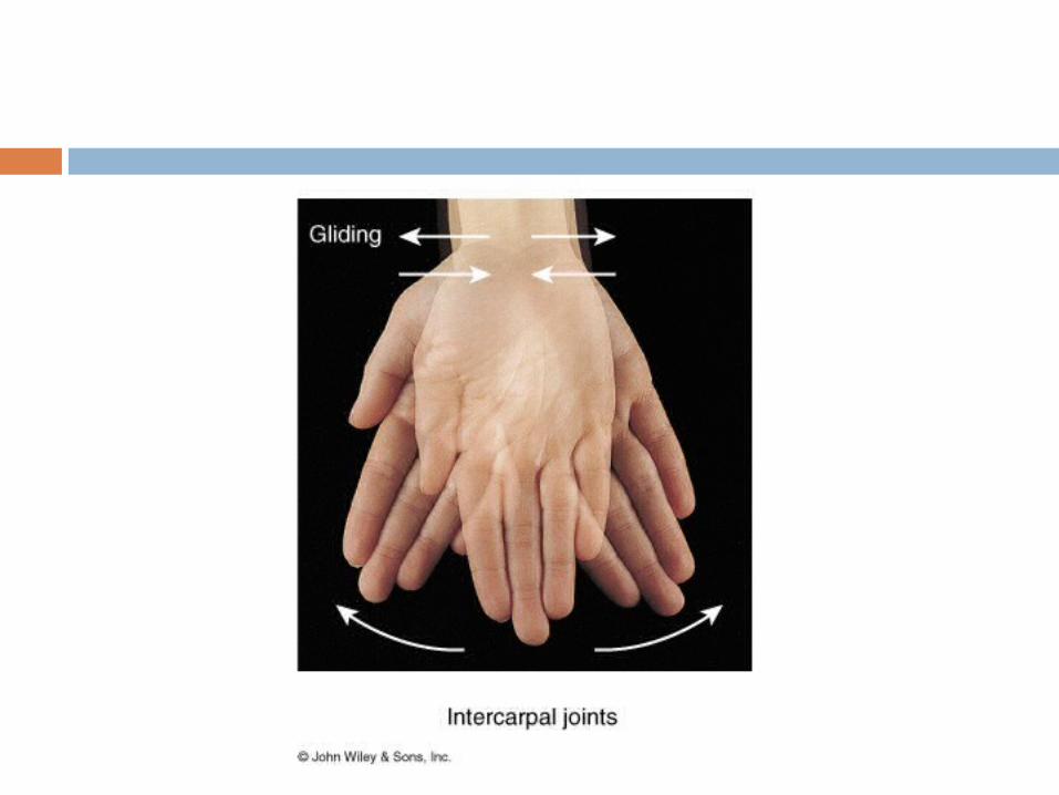
Joint Types: Gliding
Gliding" joints permit a wide range of mostly sideways movements - as well as movements in one direction
The bones in the wrists and ankles slide against each other in a gliding motion
The spine is a series of gliding joints
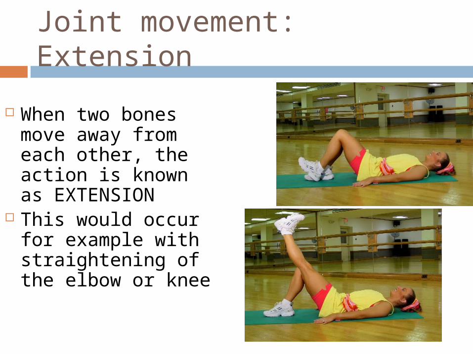
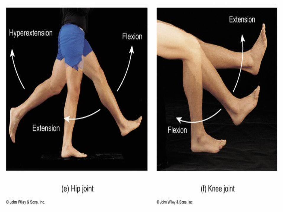
Joint movement: Extension
When two bones move away from each other, the action is known as EXTENSION
This would occur for example with straightening of the elbow or knee
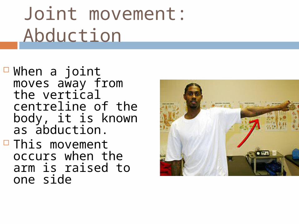
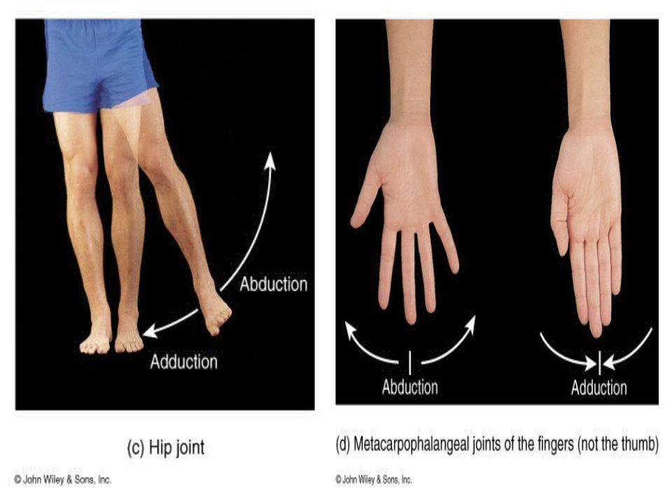
Joint movement: Abduction
When a joint moves away from the vertical centreline of the body, it is known as abduction.
This movement occurs when the arm is raised to one side
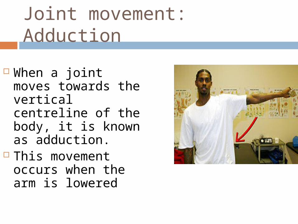
Joint movement: Adduction
When a joint moves towards the vertical centreline of the body, it is known as adduction.
This movement occurs when the arm is lowered

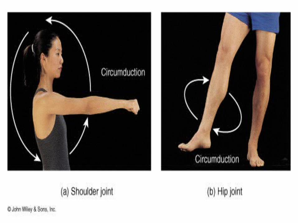
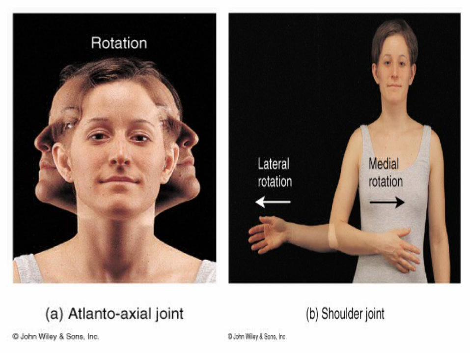
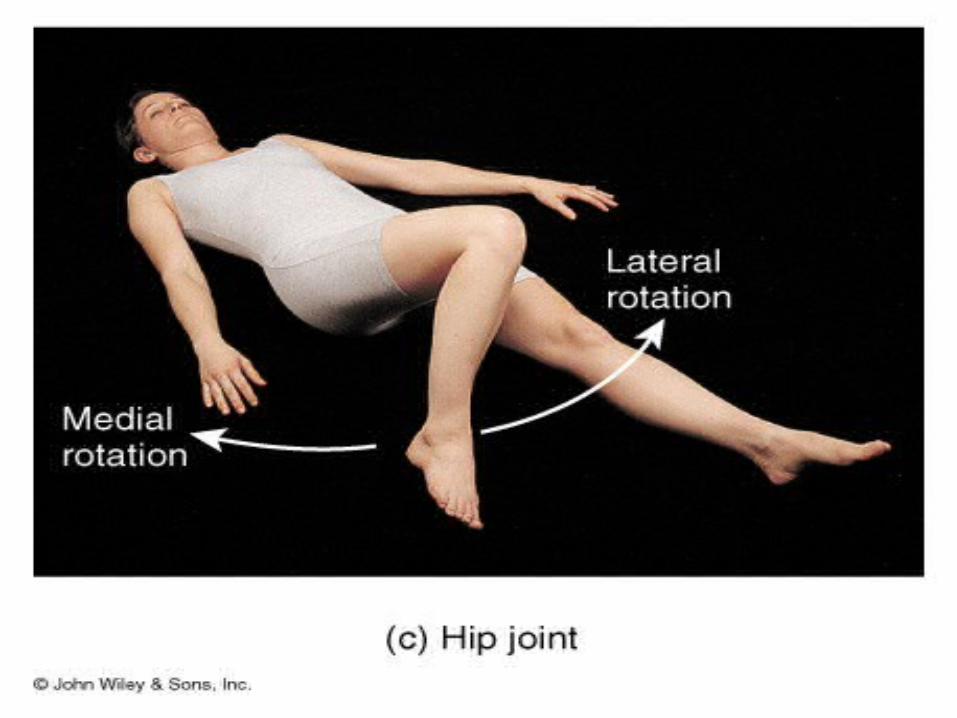
Joint movement: Rotation
This occurs when a bone rotates, either in a socket or relative to another bone.
It can occur at ball & socket or gliding type joints
Lowering (blue arrow) is internal rotation. Raising is external

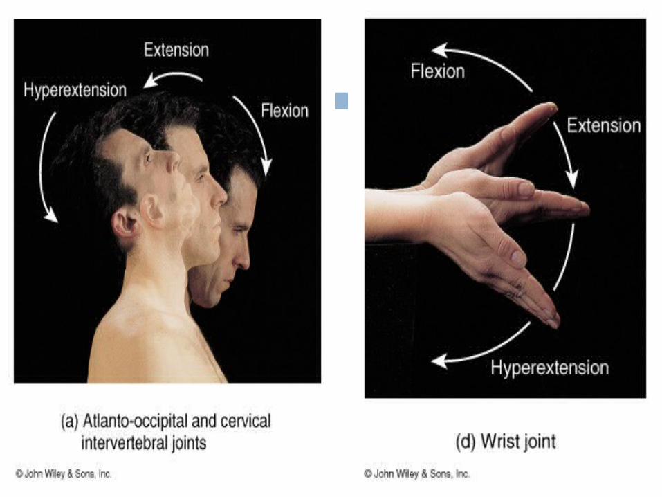
Joint movement:
Forward movement is flexion, (except the knee and elbow) Rearward
movement is extension


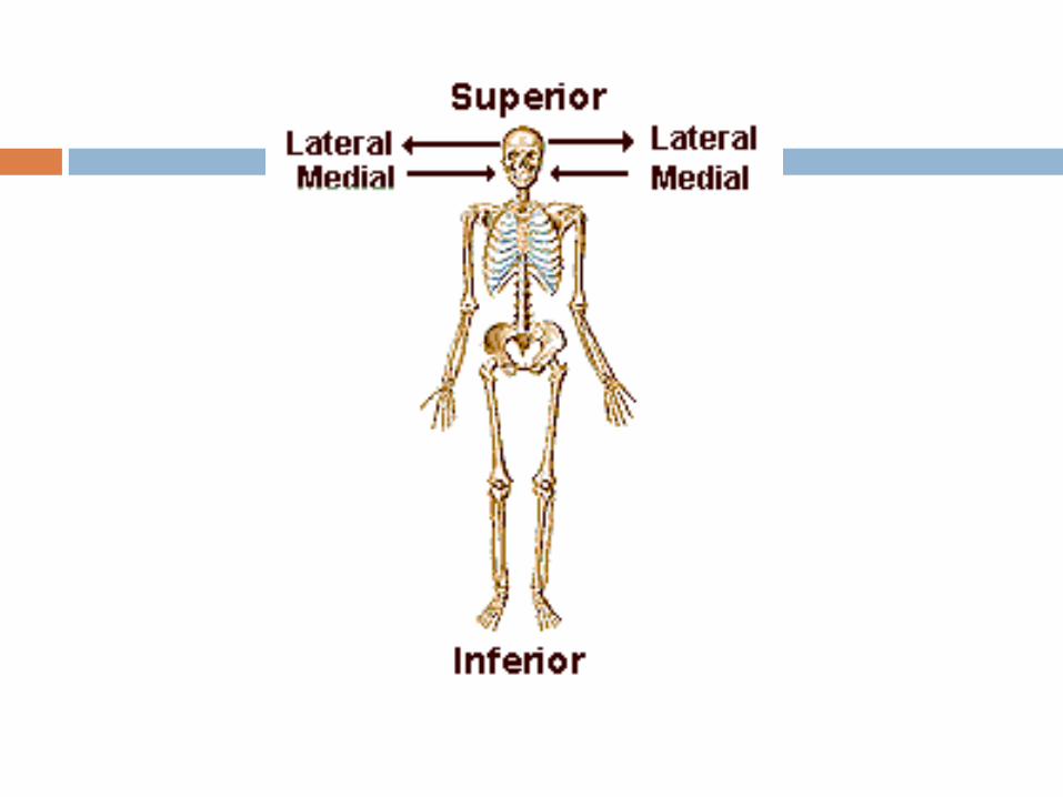
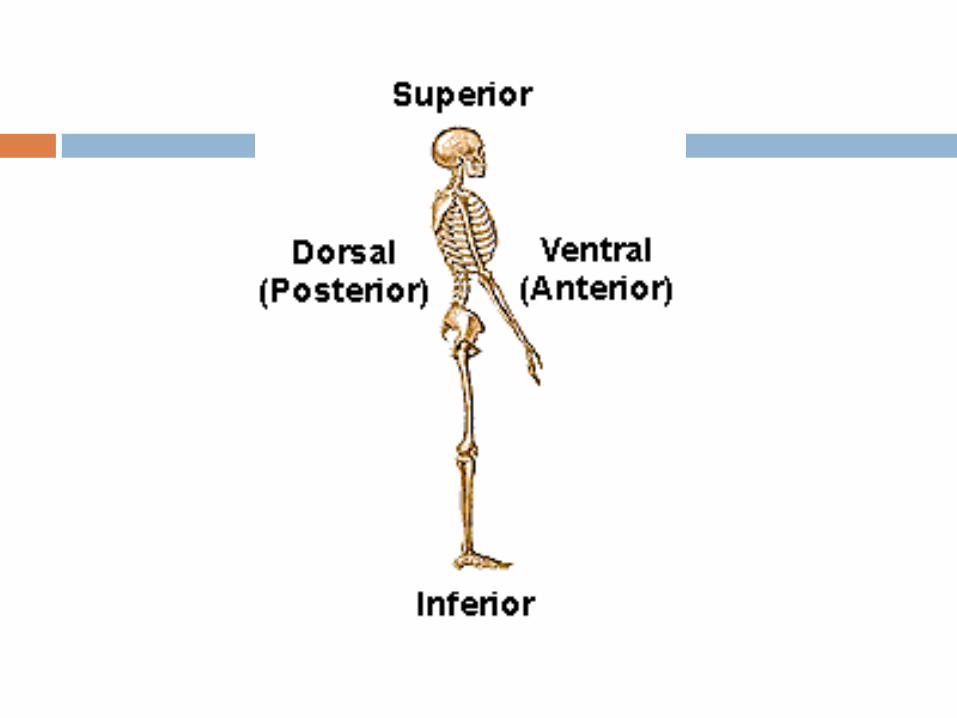
Anatomical directions
Superior: nearer the head Inferior: nearer the feet Lateral: away from the midline Medial: towards the midline
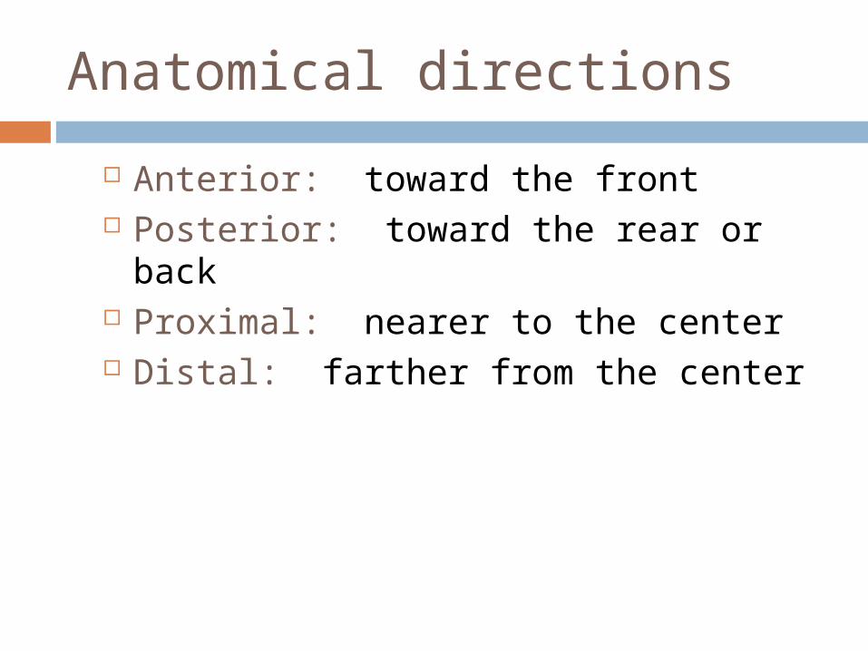
Anatomical directions
Anterior: toward the front Posterior: toward the rear or back Proximal: nearer to the center Distal: farther from the center
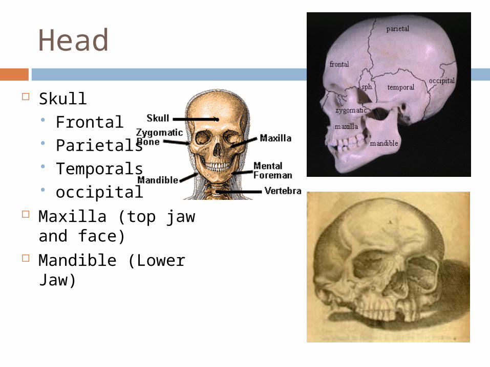
Head
Skull Frontal Parietals Temporals occipital
Maxilla (top jaw and face)
Mandible (Lower Jaw)
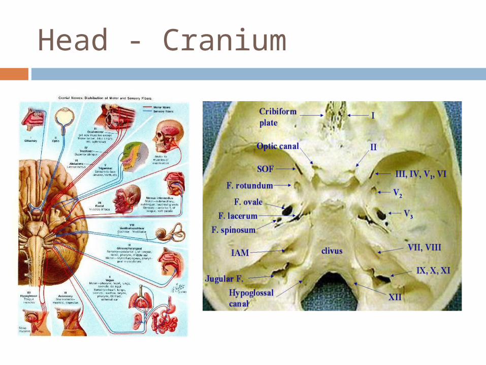
Head - Cranium
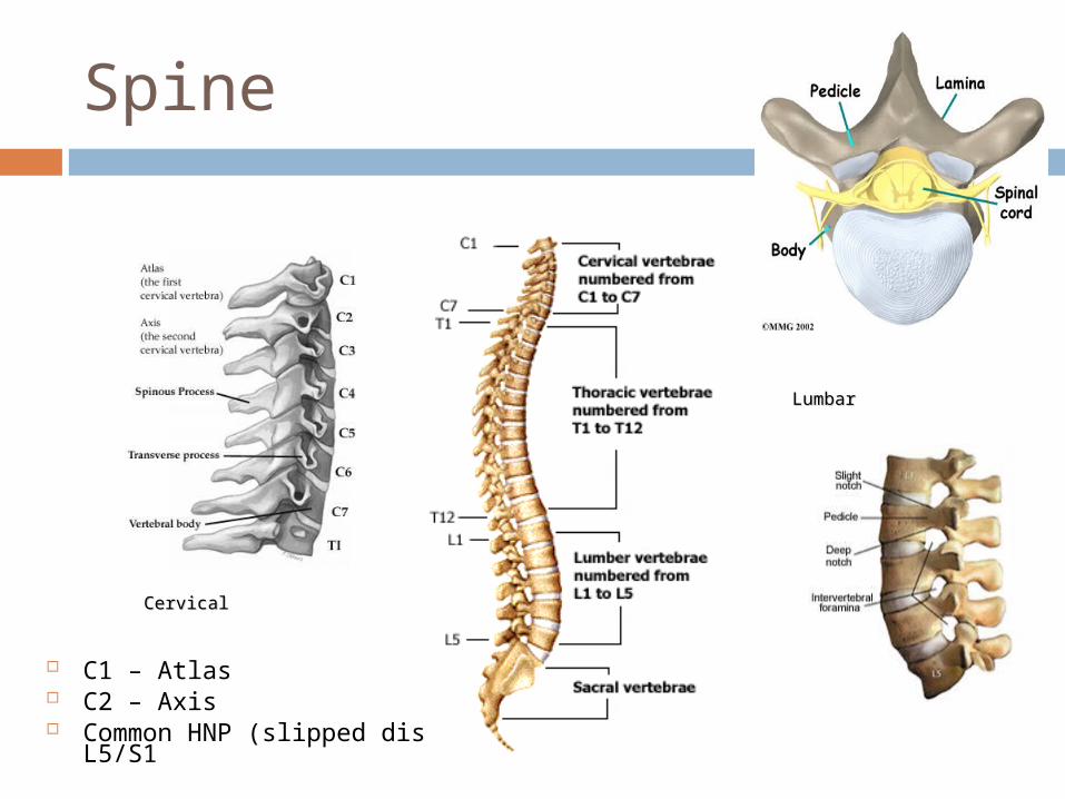
Spine
C1 – Atlas C2 – Axis Common HNP (slipped disc): L4/L5,
L5/S1
LumbarLumbar
CervicalCervical
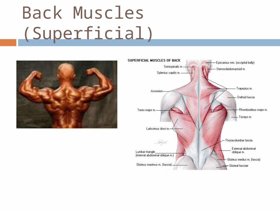
Back Muscles (Superficial)
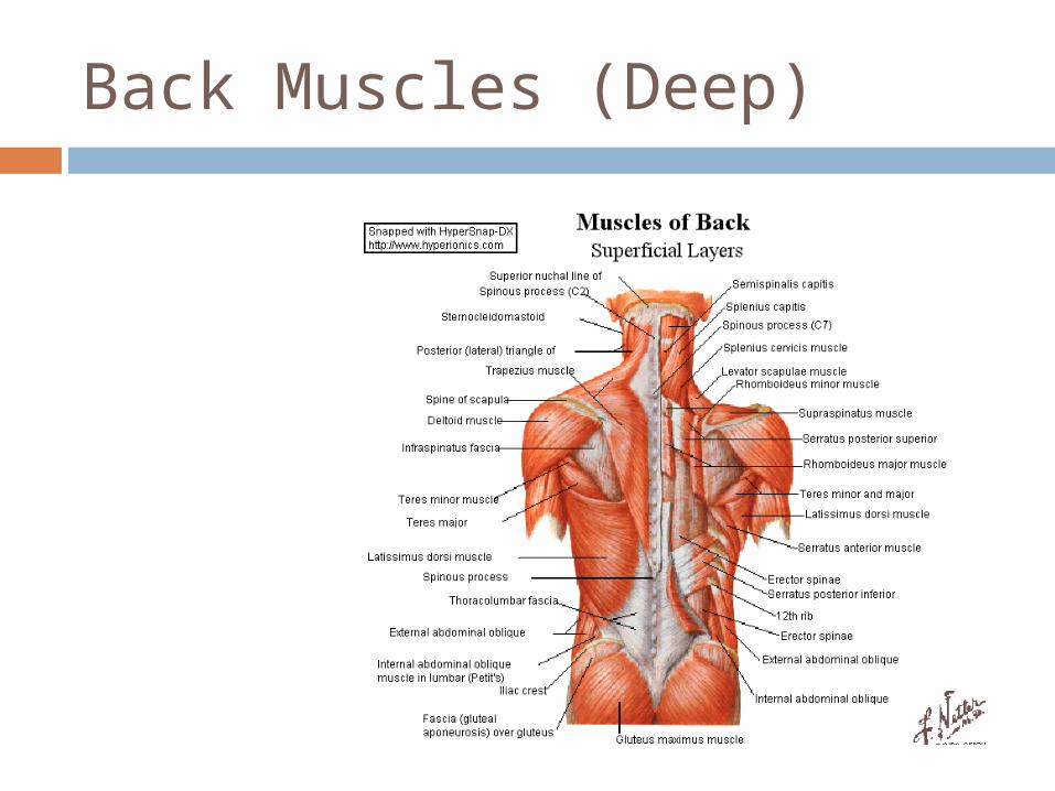
Back Muscles (Deep)
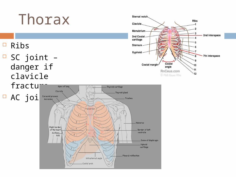
Thorax
Ribs SC joint –
danger if clavicle fracture
AC joint
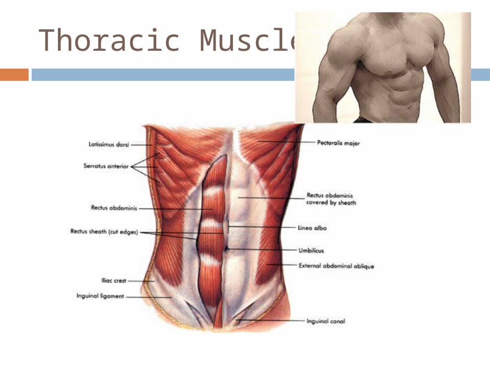
Thoracic Muscles

Shoulder – Bony Anatomy
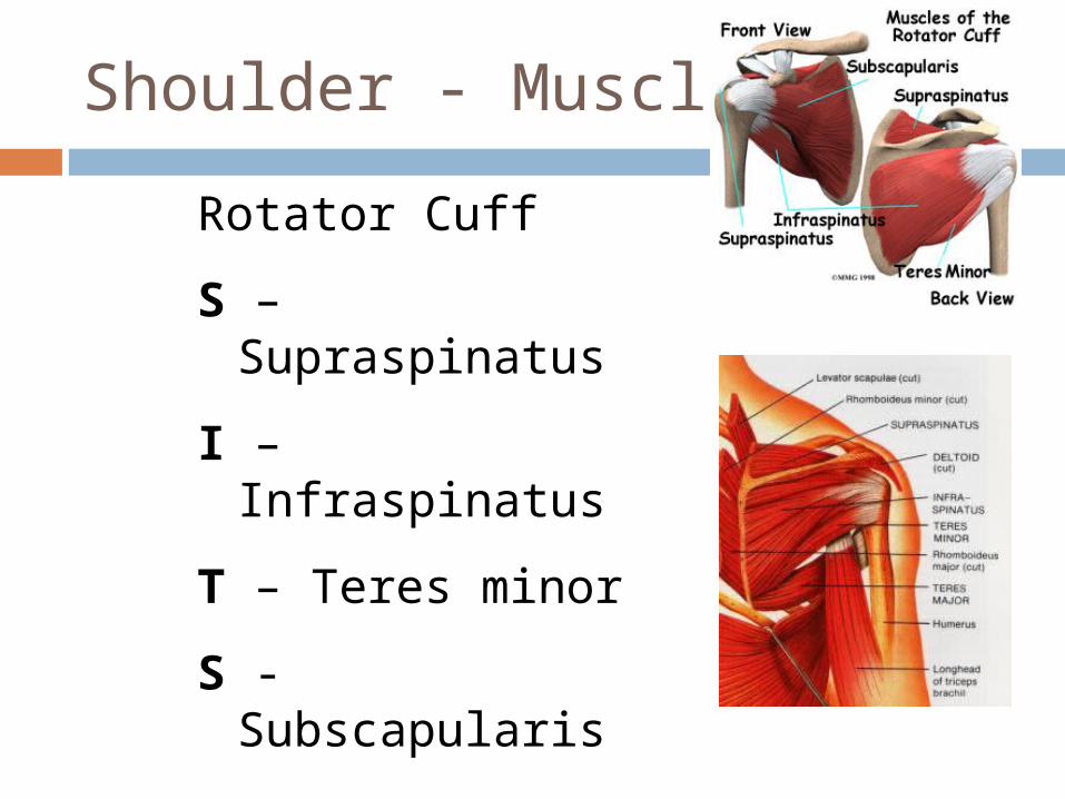
Shoulder - Muscles
Rotator Cuff
S – Supraspinatus
I – Infraspinatus
T – Teres minor
S - Subscapularis
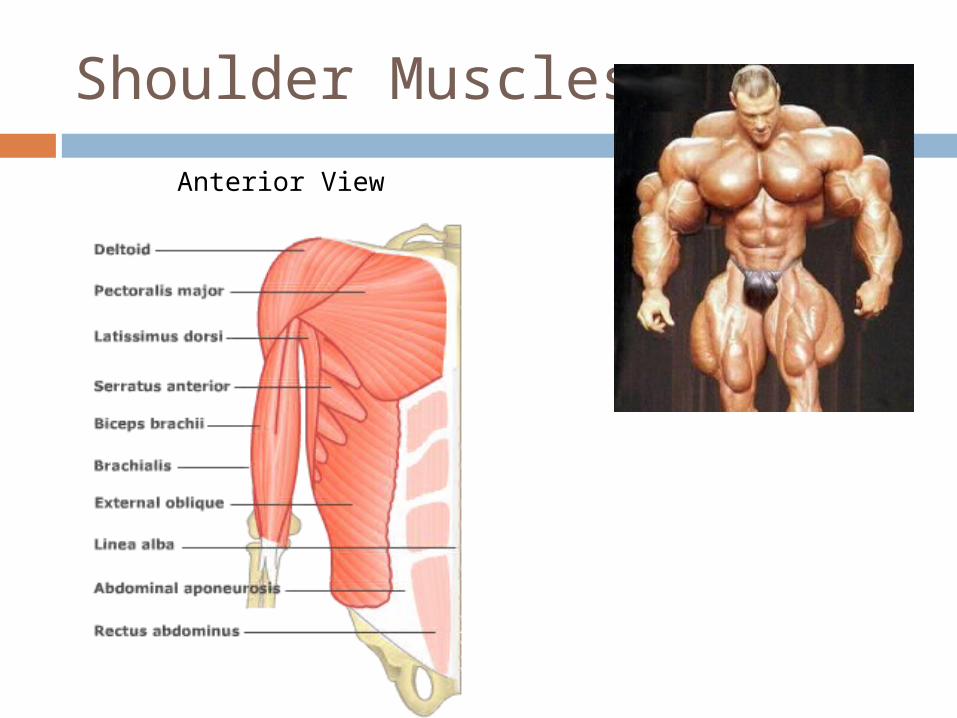
Shoulder Muscles
Anterior View
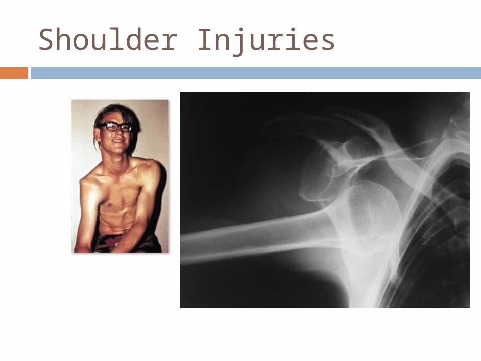
Shoulder Injuries
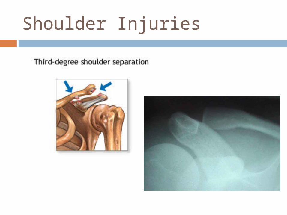
Shoulder Injuries
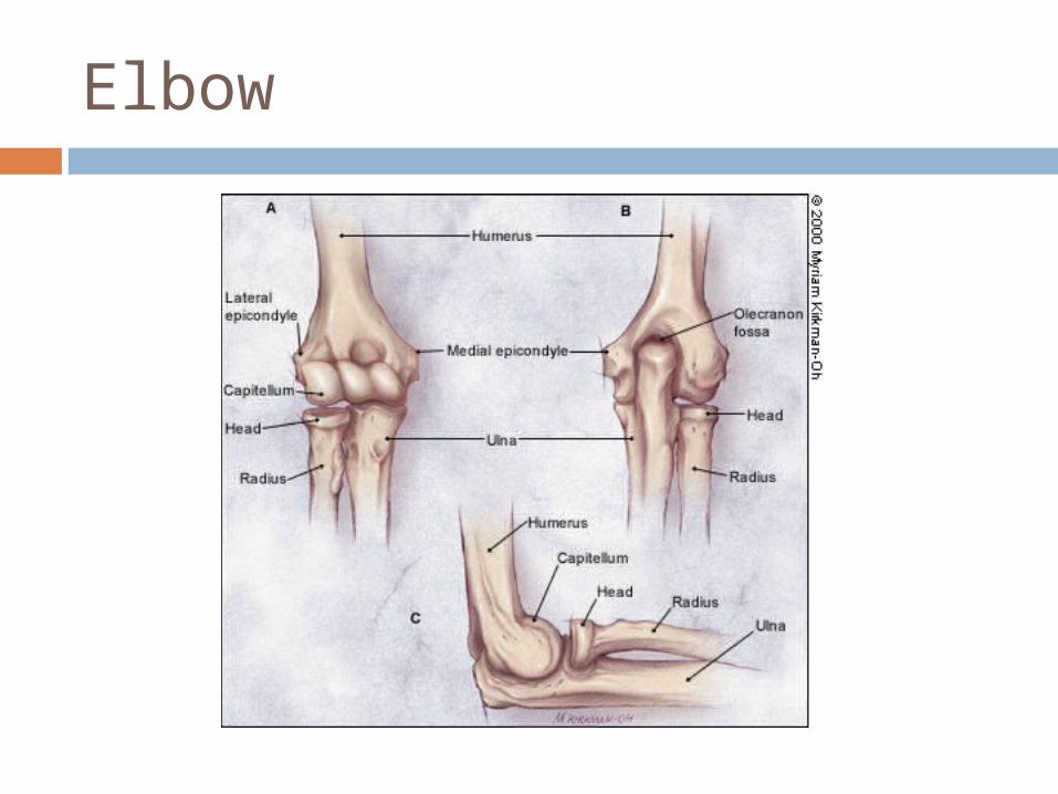
Elbow
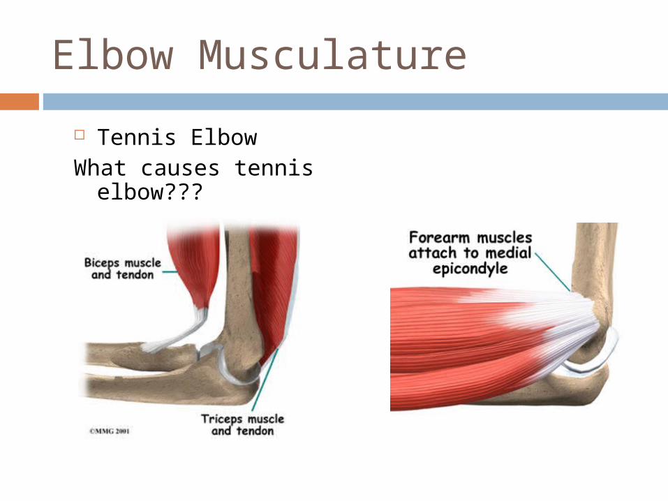
Elbow Musculature
Tennis ElbowWhat causes tennis
elbow???
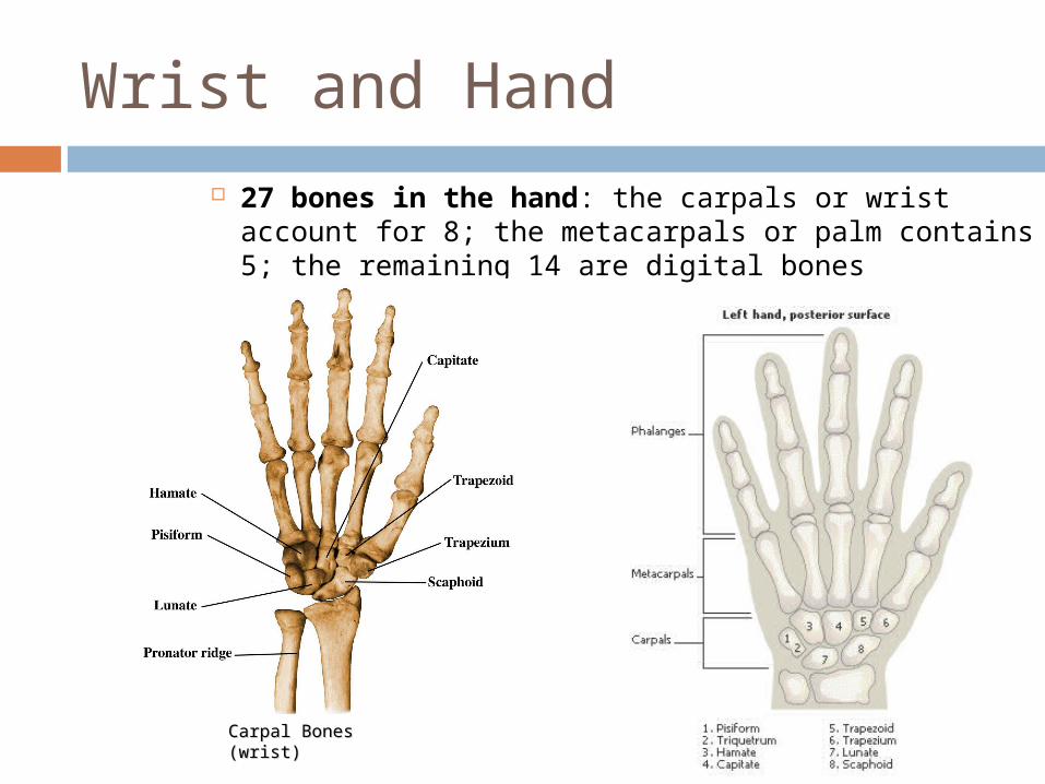
Wrist and Hand
27 bones in the hand: the carpals or wrist account for 8; the metacarpals or palm contains 5; the remaining 14 are digital bones
Carpal Bones Carpal Bones (wrist)(wrist)
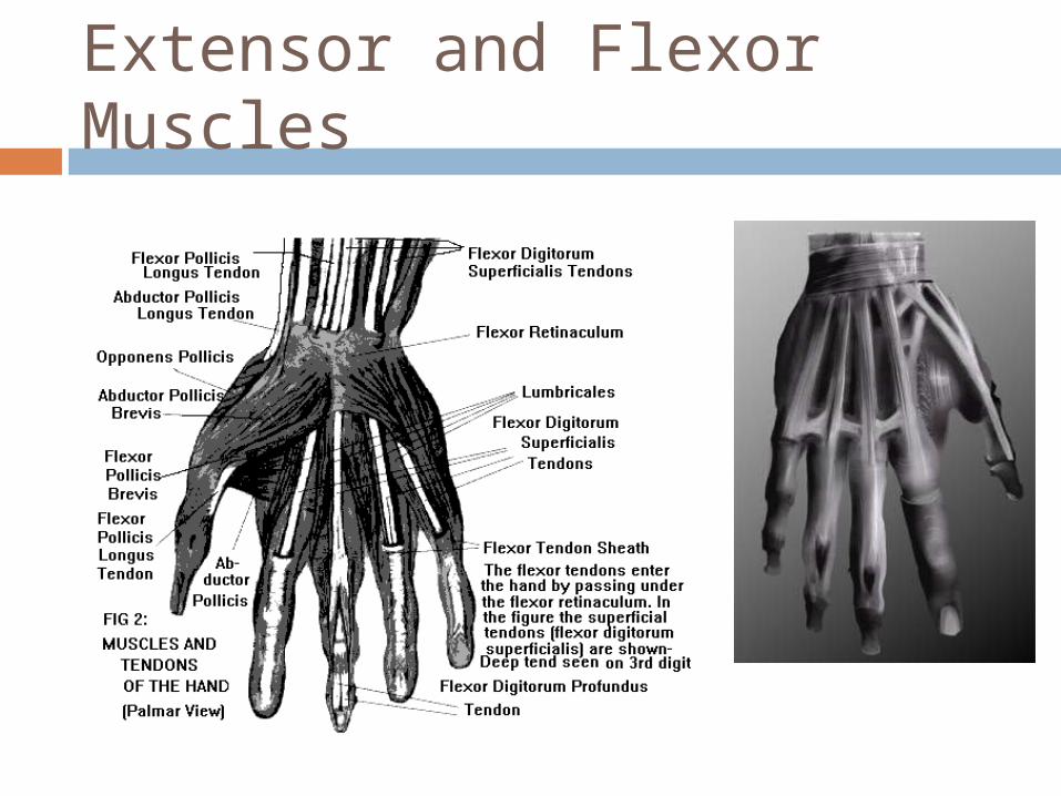
Extensor and Flexor Muscles
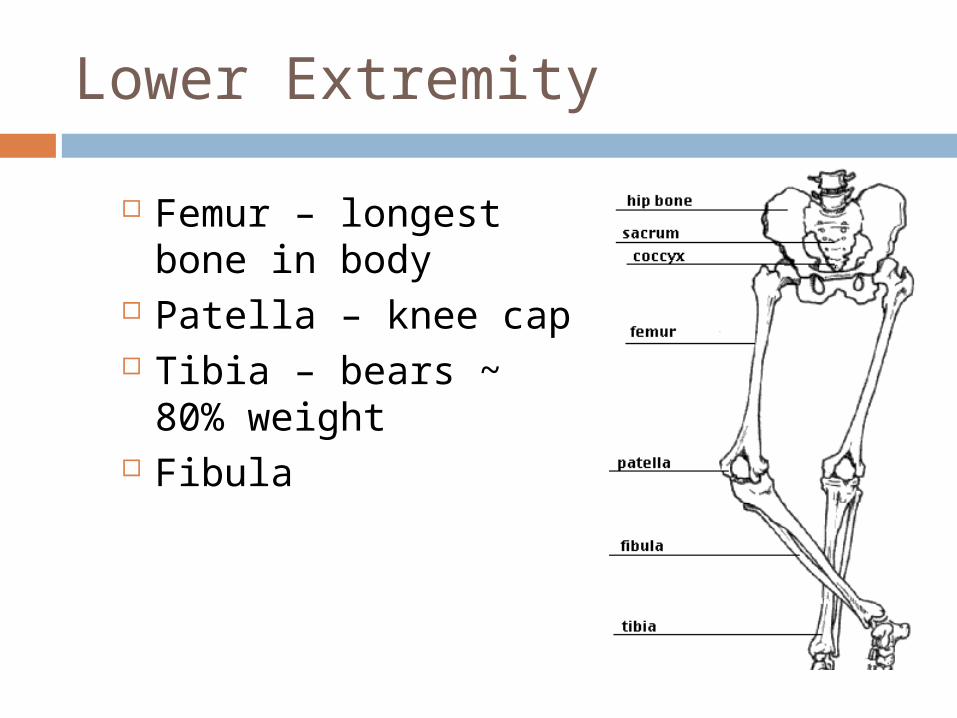
Lower Extremity
Femur – longest bone in body
Patella – knee cap Tibia – bears ~ 80%
weight Fibula
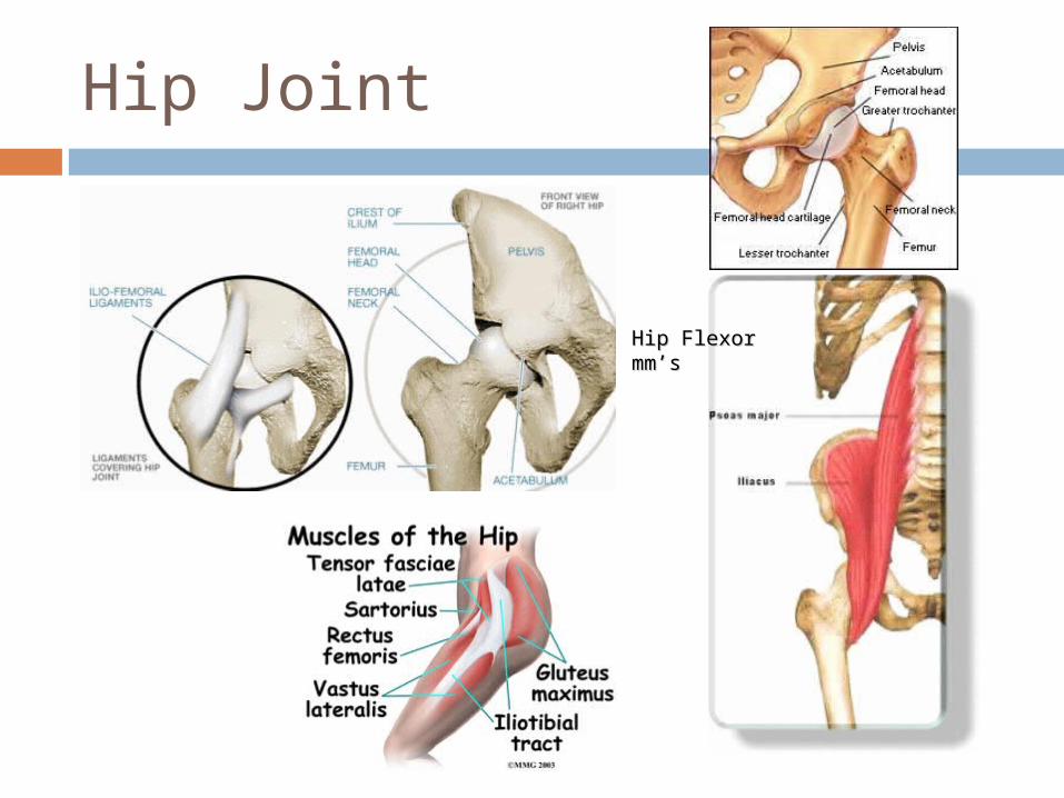
Hip Joint
Hip Flexor mm’sHip Flexor mm’s

Quadriceps
• Rectus Femoris
• Vastus Intermedialis
• Vastus Lateralis
• Vastus Medialis
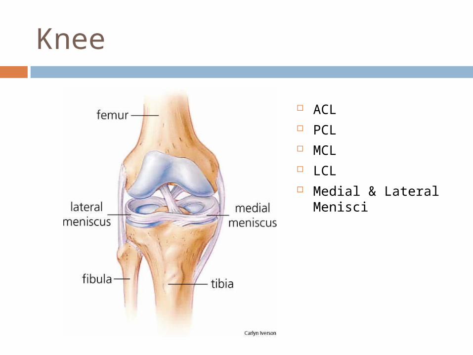
Knee
ACL PCL MCL LCL Medial & Lateral
Menisci
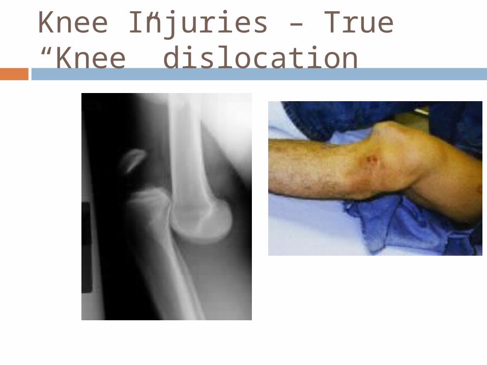
Knee Injuries – True “Knee” dislocation
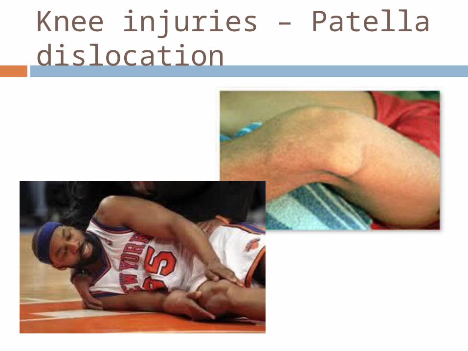
Knee injuries – Patella dislocation
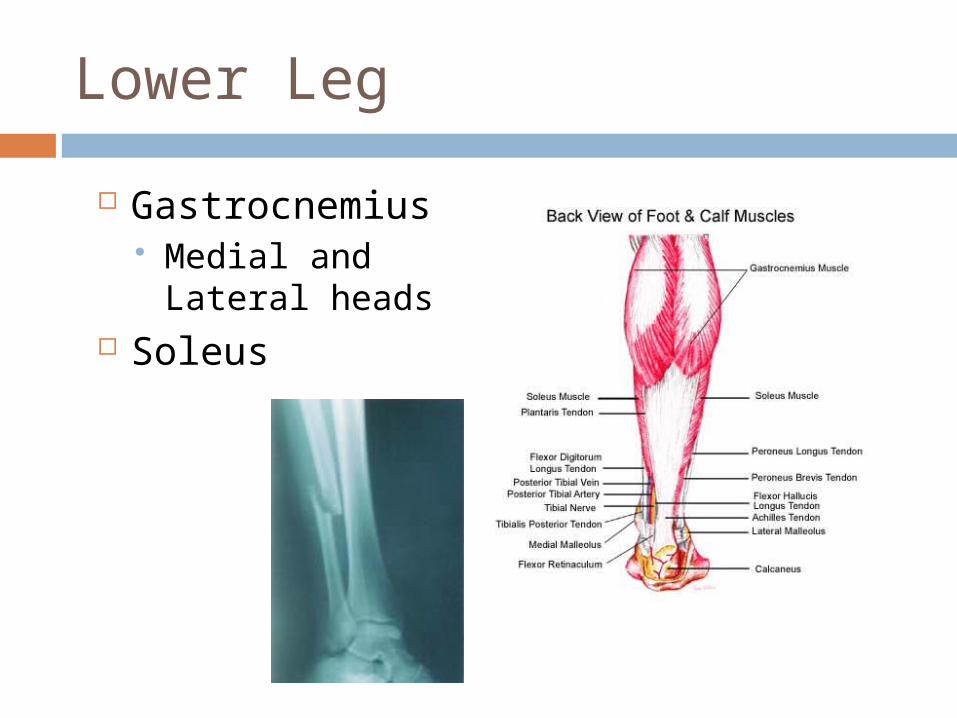
Lower Leg
Gastrocnemius Medial and
Lateral heads Soleus
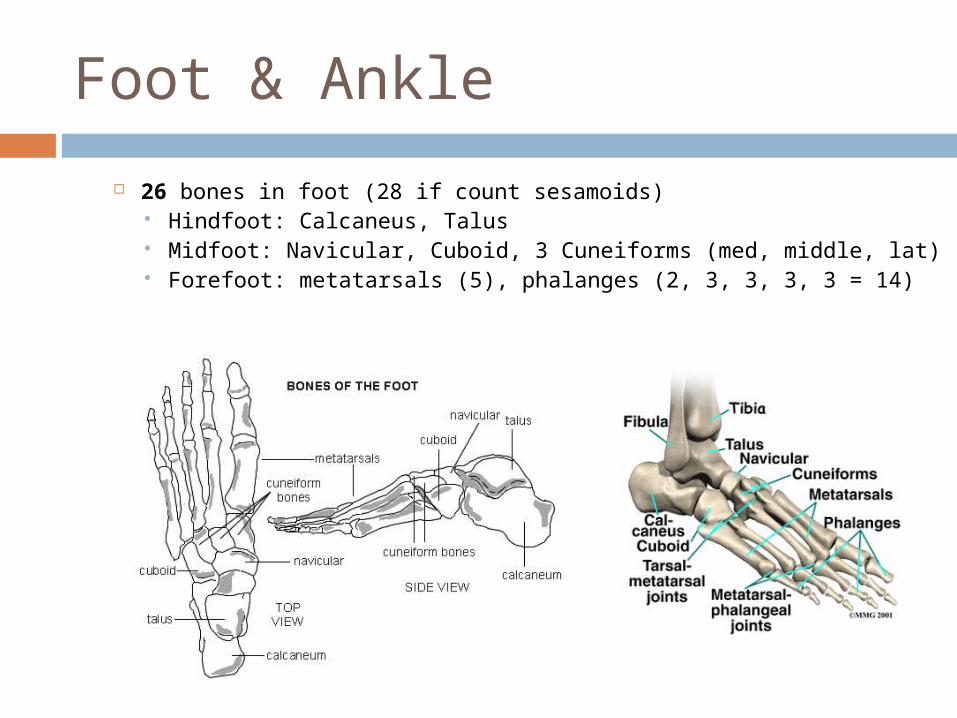
Foot & Ankle
26 bones in foot (28 if count sesamoids) Hindfoot: Calcaneus, Talus Midfoot: Navicular, Cuboid, 3 Cuneiforms (med, middle, lat) Forefoot: metatarsals (5), phalanges (2, 3, 3, 3, 3 = 14)
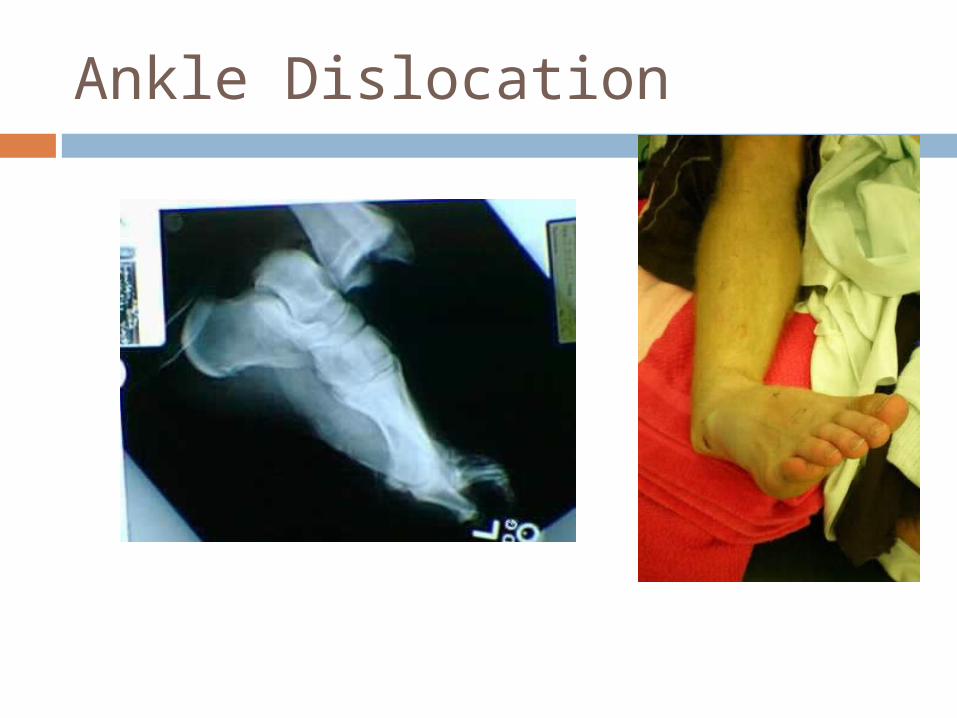
Ankle Dislocation