anatomy heart & circulation
DESCRIPTION
anatomyTRANSCRIPT

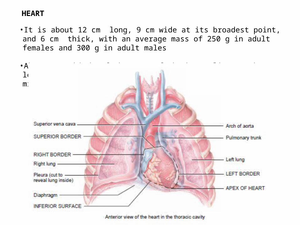
•It is about 12 cm long, 9 cm wide at its broadest point, and 6 cm thick, with an average mass of 250 g in adult females and 300 g in adult males
•About two-thirds of the mass of the heart lies to the left of the body’s midline, on mediastinum inferior middle
HEART

Mediastinum occupied by the mass of tissue between the two pulmonary cavities, is the central compartment of the thoracic cavity
Contains all thoracic viscera except the lungs


The pericardium is a fibroserous membrane that covers the heart and the beginning of its great vessels
The pericardium is a closed sac composed of two layers :1.Fibrous pericardium2.Serous pericardium• Parietal layer• Visceral layer


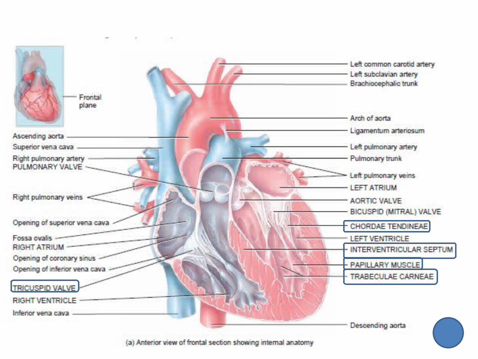
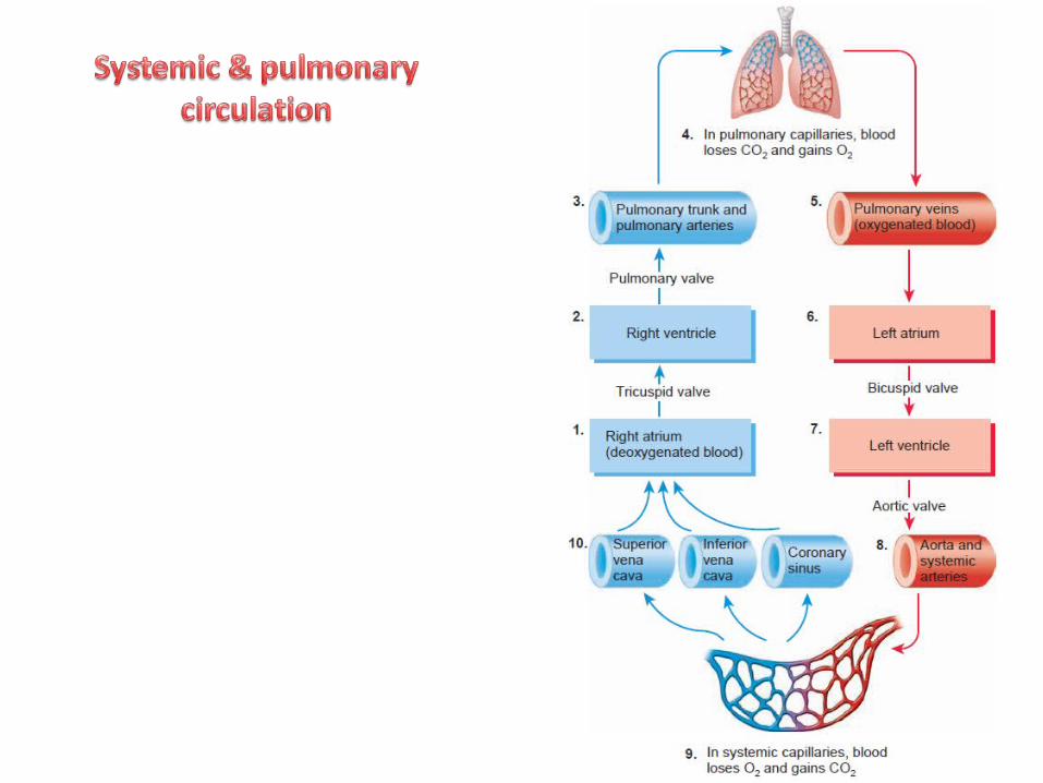
The heart has four chambers: right and left atria and right and left ventricles
Valves of the heart :A.Atrioventricular valves - Tricuspid valve - Bicuspid valveB.Semilunar valves - Aortic valve - Pulmonary valve
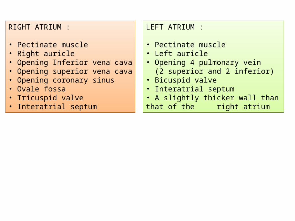
RIGHT ATRIUM :
• Pectinate muscle• Right auricle• Opening Inferior vena cava• Opening superior vena cava• Opening coronary sinus• Ovale fossa• Tricuspid valve• Interatrial septum
RIGHT ATRIUM :
• Pectinate muscle• Right auricle• Opening Inferior vena cava• Opening superior vena cava• Opening coronary sinus• Ovale fossa• Tricuspid valve• Interatrial septum
LEFT ATRIUM :
• Pectinate muscle• Left auricle• Opening 4 pulmonary vein (2 superior and 2 inferior)• Bicuspid valve • Interatrial septum• A slightly thicker wall than that of the right atrium
LEFT ATRIUM :
• Pectinate muscle• Left auricle• Opening 4 pulmonary vein (2 superior and 2 inferior)• Bicuspid valve • Interatrial septum• A slightly thicker wall than that of the right atrium


RIGHT VENTRICLE :
• Trabecula carnae• AV Tricuspid valve• Chorda tendinae• Papillary muscle• Semilunar pulmonary valve• Interventricular septum
RIGHT VENTRICLE :
• Trabecula carnae• AV Tricuspid valve• Chorda tendinae• Papillary muscle• Semilunar pulmonary valve• Interventricular septum
LEFT VENTRICLE :
• Trabecula carnae• AV Bicuspid valve• Chorda tendinae• Papillary muscle• Semilunar aortic valve• Interventricular septum Walls that are two to three times as thick as those of the right ventricle
LEFT VENTRICLE :
• Trabecula carnae• AV Bicuspid valve• Chorda tendinae• Papillary muscle• Semilunar aortic valve• Interventricular septum Walls that are two to three times as thick as those of the right ventricle

a
Atrium kanan ?? Atrium kiri ??
Ventrikel kanan ?? Ventrikel kiri ??


The pointed apex is formed by the tip ofthe left ventricle and rests on thediaphragm.
It is directed anteriorly, inferiorly, and to the left
Lies posterior to the left 5th intercostal space in adults

Is the heart's posterior aspect
Is formed mainly by the left atrium, with a lesser contribution by the right atrium.
Extends superiorly to the bifurcation of the pulmonary trunk and inferiorly to the coronary sulcus

1. Diaphragmatic (inferior) surface, formed mainly by the left ventricle and partly by the right ventricle; it is related mainly to the central tendon of the diaphragm.
2. Left pulmonary surface, formed mainly by the left ventricle;
it forms the cardiac impression in the left lung.
Atrium kanan
Atrium kiri
Ventrikelkanan
Ventrikelkiri

3. Anterior (sternocostal) surface, formed mainly by the right ventricle.
4. .Right pulmonary surface,
formed mainly by the right atrium.
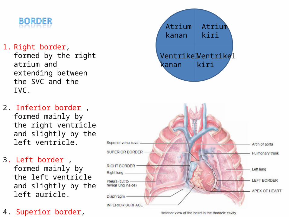
1. Right border, formed by the right atrium and extending between the SVC and the IVC.
2. Inferior border , formed mainly by the right ventricle and slightly by the left ventricle.
3. Left border , formed mainly by the left ventricle and slightly by the left auricle.
4. Superior border, formed by the right and left atria and auricles in an anterior view
Atrium kanan
Atrium kiri
Ventrikelkanan
Ventrikelkiri

Endocardium, a thin internal layer (endothelium and subendothelial connective tissue) or lining membrane of the heart that also covers its valves.
Myocardium, a thick, helical middle layer composed of cardiac muscle.
Epicardium, a thin external layer (mesothelium) formed by the visceral layer of serous pericardium

The isolated fibrous skeleton is composed of :
four fibrous rings (or two rings and two “coronets”)
two trigone : 1. Left fibrous trigone 2. Right fibrous trigone
Membranous portions of the interatrial, interventricular, and atrioventricular septa.


Nutrients are not able to diffuse quickly enough from blood in the chambers of the heart to supply all the layers of cells that make up the heart wall.
For this reason, the myocardium has its own network of blood vessels, the coronary or cardiac circulation

Right coronary arteryRight coronary arteryLeft coronary areryLeft coronary arery
anterior interventricularanterior interventricular
circumflex branchescircumflex branches
supplies oxygenatedblood to the walls of both ventricles
supplies oxygenatedblood to the walls of both ventricles To the walls of the
left ventricle and left atrium.
To the walls of the left ventricle and left atrium.
supplies small branches (atrialbranches) to the right atriumsupplies small branches (atrialbranches) to the right atrium
posterior interventricularposterior interventricular
marginal branchesmarginal branches
To walls of the two ventricles
To walls of the two ventricles
to the myocardium of the right ventricle
to the myocardium of the right ventricle


blood passes through the arteries of the coronary circulation,
flows into capillaries
delivers oxygen and nutrients to the heart muscle and collects carbon dioxide and waste
moves into coronary veins
coronary sinus
Great cardiac vein
Middle cardiac vein
Small cardiac vein
Anterior cardiac veins


Atrium kanan menerima darah dari vena cava superior, vena cava inferior dan coronary sinusAtrium kanan menerima darah dari vena cava superior, vena cava inferior dan coronary sinus
Tekanan dalam atrium kanan meningkat menyebabkan atrium kanan sistole/kontraksi
Katup AV tricuspid terbuka
Darah masuk keventrikel kanan sebanyak 25 ml, sebelumnya di ventrikel ada 105 ml, jadi sekarang jumlah darah di ventrikel = 130 ml disebut EDV(END DIASTOLIC VOLUME)
Tekanan dalam ventrikel kanan meningkat ventrikel kanan sistole/kontraksi
Katup AV tricuspid terttutup. KARENA katup AV tricuspid dan katup semilunar pulmonary juga tertutup, maka kondisi ini disebut ISOVOLUMETRIC CONTRACTION
Karena ventrikel kanan terus menerus kontraksi, mengakibatkan katup semilunar pulmonary terbuka
Darah mengalir ke pulmonary trunk sebanyak 70 ml, volume darah sisa di ventrikel kanan 60 ml, disebut ESV(END SYSTOLIC VOLUME)
Tekanan dalam ventrikel kanan berkurang, ventrikel diastole/relaksasi
Darah di pulmonary trunk turun kembali menuju ke arah ventrikel kanan yg tekanannya mulai berkurang katup semilunar pulmonary tertutup karena katup AV tricuspid dan semiluinar pulm,onary tertutup maka kondisi ini disebut ISOVOLUMETRIC RELAXATION
Darah menuju ke artery pulmonaryparu-paruvena pulmonary atrium kiri
Atrium kiri menerima darah dari vena pulmonaryAtrium kiri menerima darah dari vena pulmonary
Katup AV bicuspid terbukaKatup AV bicuspid terbuka
Tekanan dalam atrium kiri meningkat menyebabkan atrium kiri sistole/kontraksiTekanan dalam atrium kiri meningkat menyebabkan atrium kiri sistole/kontraksi
Darah masuk keventrikel kiri sebanyak 25 ml, sebelumnya di ventrikel ada 105 ml, jadi sekarang jumlah darah di ventrikel = 130 ml disebut EDV(END DIASTOLIC VOLUME)
Darah masuk keventrikel kiri sebanyak 25 ml, sebelumnya di ventrikel ada 105 ml, jadi sekarang jumlah darah di ventrikel = 130 ml disebut EDV(END DIASTOLIC VOLUME)
Tekanan dalam ventrikel kiri meningkat ventrikel kiri sistole/kontraksiTekanan dalam ventrikel kiri meningkat ventrikel kiri sistole/kontraksi
Katup AV bicuspid terttutup. KARENA katup AV bicuspid dan katup semilunar aortic juga tertutup, maka kondisi ini disebut ISOVOLUMETRIC CONTRACTION
Katup AV bicuspid terttutup. KARENA katup AV bicuspid dan katup semilunar aortic juga tertutup, maka kondisi ini disebut ISOVOLUMETRIC CONTRACTION
Karena ventrikel kiri terus menerus kontraksi, mengakibatkan katup semilunar aortic terbukaKarena ventrikel kiri terus menerus kontraksi, mengakibatkan katup semilunar aortic terbuka
Darah mengalir ke aorta sebanyak 70 ml, volume darah sisa di ventrikel kanan 60 ml, disebut ESV(END SYSTOLIC VOLUME)Darah mengalir ke aorta sebanyak 70 ml, volume darah sisa di ventrikel kanan 60 ml, disebut ESV(END SYSTOLIC VOLUME)
Tekanan dalam ventrikel kiri berkurang, ventrikel diastole/relaksasiTekanan dalam ventrikel kiri berkurang, ventrikel diastole/relaksasi
Darah di aorta turun kembali menuju ke arah ventrikel kiri yg tekanannya mulai berkurang katup semilunar aortic tertutup karena katup AV bicuspid dan semiluinar aortic tertutup maka kondisi ini disebut ISOVOLUMETRIC RELAXATION
Darah di aorta turun kembali menuju ke arah ventrikel kiri yg tekanannya mulai berkurang katup semilunar aortic tertutup karena katup AV bicuspid dan semiluinar aortic tertutup maka kondisi ini disebut ISOVOLUMETRIC RELAXATION
Darah menuju ke seluruh tubuh vena cava superior,vena cava inferior dan coronary sinus atrium kananDarah menuju ke seluruh tubuh vena cava superior,vena cava inferior dan coronary sinus atrium kanan