androgen receptor signaling pathway in prostate cancer - tampub
TRANSCRIPT

KATI WALTERING
Androgen Receptor Signaling Pathway in Prostate Cancer
ACADEMIC DISSERTATIONTo be presented, with the permission of
the Faculty of Medicine of the University of Tampere,for public discussion in the Auditorium of Finn-Medi 1,
Biokatu 6, Tampere, on September 10th, 2010, at 12 o’clock.
UNIVERSITY OF TAMPERE

Reviewed byDocent Pia AarnisaloUniversity of HelsinkiFinlandDocent Kaisa LehtiUniversity of Helsinki Finland
DistributionBookshop TAJUP.O. Box 61733014 University of TampereFinland
Tel. +358 40 190 9800Fax +358 3 3551 7685 [email protected]/tajuhttp://granum.uta.fi
Cover design byJuha Siro
Acta Universitatis Tamperensis 1541ISBN 978-951-44-8179-6 (print)ISSN-L 1455-1616ISSN 1455-1616
Acta Electronica Universitatis Tamperensis 985ISBN 978-951-44-8180-2 (pdf )ISSN 1456-954Xhttp://acta.uta.fi
Tampereen Yliopistopaino Oy – Juvenes PrintTampere 2010
ACADEMIC DISSERTATIONUniversity of Tampere, Institute of Medical TechnologyTampere University Hospital, Laboratory CentreTampere Graduate School in Biomedicine and Biotechnology (TGSBB)Finland
Supervised byProfessor Tapio VisakorpiUniversity of TampereFinland

3
CONTENTS
Contents 3List of original publications 5Abbreviations 6Abstract 8Yhteenveto 9
1 INTRODUCTION 10
2 REVIEW OF THE LITERATURE 11
2.1 Normal anatomy and function of the prostate gland 11
2.2 Androgen signaling in normal prostate 112.2.1 Genetic structure of AR 122.2.2 Protein structure and function of AR 142.2.3 AR coregulators 16
2.3 Cancer of the prostate gland 172.3.1 Genetic predisposition 182.3.2 Somatic genetic changes in prostate cancer 19
2.3.2.1 Loss of function alterations in prostate cancer 192.3.2.2 Gain of function alterations in prostate cancer 20
2.4 Androgen receptor (AR) signaling pathway in PC 212.4.1 Changes in androgen metabolism 212.4.2 Somatic mutations of AR 222.4.3 Amplification and overexpression of AR 242.4.4 AR splice variants 252.4.5 Changes in AR cofactors 262.4.6 Wellknown AR target genes 26
2.4.6.1 ACPP (PAP) 272.4.6.2 KLK3 (PSA) 272.4.6.3 TMPRSS2:ERG and other fusion ETS 272.4.6.4 NKX31 28
2.5 Endocrine treatments of prostate cancer 292.5.1 Targeting androgen synthesis 292.5.2 AR targeting inhibitors 29
3. AIMS OF THE STUDY 31
4. MATERIALS AND METHODS 32
4.1 Cell lines and xenografts 324.2 Clinical samples 324.3 DNA and RNA extractions, DNA amplification and PCR 32

4
4.4 DHPLC and sequencing 334.5 mRNA and miRNA qRTPCR reactions 334.6 Western blot 334.7 Transfection methods 334.8 DHT and roscovitine treatments and cell proliferation assays 344.9 Microarray hybridizations 344.10 Data analysis 344.11 ChIPonchip assays 354.12 Statistical methods 35
5. RESULTS AND DISCUSSION 36
5.1 Mutations in the regulatory regions of the AR gene are rare in PC 36
5.2 Increased AR levels sensitize PC cells to low androgen levels 385.2.1 The growth of the ARoverexpressing LNCaP cells 385.2.2 The effect of increased AR levels on the transcription
of target genes 385.2.3 Gene ontology classes associated with androgen
and AR levels 405.2.4 Direct AR target genes involved in growth during
PC progression 40
5.3 Androgen regulation of microRNAs in prostate cancer 435.3.1 Androgen regulated miRNAs 435.3.2 Differentially expressed miRNAs in clinical samples 445.3.3 Effect of miR141 on the growth of PC cells 44
6. CONCLUSIONS 47
Acknowledgements 48References 49Original communications 71

5
LIST OF ORIGINAL COMMUNICATIONS
This thesis is based on the following original publications referred to in the text byRoman numbers I-III:
I Waltering KK, Wallén MJ, Tammela TL, Vessella RL, Visakorpi T (2006):Mutation screening of the androgen receptor promoter and untranslated regionsin prostate cancer. Prostate 66;15:1585-91.
II Waltering KK1, Helenius MA1, Sahu B, Manni V, Linja MJ, Jänne OA,Visakorpi T (2009): Increased expression of androgen receptor sensitizesprostate cancer cells to low levels of androgens. Cancer Res 69;20:8141-9.
III Waltering KK, Porkka KP, Jalava SE, Urbanucci A, Kohonen P, Latonen L,Kallioniemi O, Jenster G and Visakorpi T (2010): Androgen regulation ofmicroRNAs in prostate cancer. Accepted to be published in Prostate.
1Authors contributed egually to this work
6
ABBREVIATIONS
ACPP acid phosphatase, prostate; PAPAF activation functionAKT vakt murine thymoma viral oncogene homolog 1APC adenomatous polyposis coliAR androgen receptorARA androgen receptor coactivatorARE AR responsive elementARTIS AR transcription initiation siteATAD2 ATPase family, AAA domain containing 2, ANCCACAB complete androgen blockadecAMP 3'5'cyclic adenosine monophosphateChIP chromatin immunoprecipitationCDC20 cell division cycle 20 homologCDK1 cyclindependent kinase 1CDK2 cyclindependent kinase 2cDNA complementary DNACP 3’CCCUCCC poly(C)binding proteinCRPC castration resistant prostate cancerDBD DNAbinding domainDHEA dehydroepiandrosteroneDHPLC denaturing highperformance liquid chromatographyDHT dihydrotestosteroneDNA deoxyribonucleic acidEIF3S3 a subunit of translation factor eIF3EP300 E1A binding protein p300, acetyltransferase p300ERG vets erythroblastosis virus E26 oncogene homolog (avian)ETS avian erythroblastosis virus E26 homologETV1 ets variant 1EZH2 enhancer of zeste homolog 2FOS FBJ murine osteosarcoma viral oncogene homologFOXA1 forkhead box A1GTF2F general transcription factor IIF, TFIIFGTF2H3 general transcription factor IIH, TFIIHGSTP1 glutathione StransferaseHAT histone acetylaseHDAC histone deacetylaseITGA6 integrin alpha 6HSPA4 heat shock 70kDa protein 4JUN jun oncogeneKLK3 kallikreinrelated peptidase 3KXKK LysxLysLys motifLBD ligandbinding domainLOH loss of heterozygosityLH luteinizing hormoneLHRH luteinizing hormone releasing hormone

7
MAK male germ cellassociated kinaseMCM minichromosome maintenance proteinmiR141 microRNA 141miRNA microRNAMYC vmyc myelocytomatosis viral oncogene homologNCOA1 nuclear receptor coactivator 1, SRC1NCOR1 nuclear receptor corepressor 1NKX31 NK3 homeobox 1NLS nuclear localization signalPAP prostatic acid phosphatasePC prostate cancerPCR polymerase chain reactionPIAS protein inhibitor of activated STATPIN prostatic intraepithelial neoplasiaPOU2F1 POU class 2 homeobox 1, octamerbinding transcription factor 1, OCT1PSA prostatespecific antigenPTEN phosphatase and tensin homologRAS vHaras Harvey rat sarcoma viral oncogene homolog, HRASRNA ribonucleic acidqRTPCR quantitative realtime PCRsiRNA small interfering RNASNP single nucleotide polymorphismSP1 Sp1 transcription factorSRD5A1 steroid5alphareductase, alpha polypeptide 1SIRT1 sirtuin (silent mating type information regulation 2 homolog) 1STAT signal transducer and activator of transcriptionSUMO small ubiquitinrelated modifierTAU transcription activation unitTGFB1I1 transforming growth factor beta 1 induced transcript 1, androgen
receptor coactivator ARA55TMPRSS2 transmembrane protease, serine 2TP53 tumor protein 53, p53TSS transcription start siteTURP transurethral resection of prostateUBE2I ubiquitinconjugating enzyme E2IUTR untranslated region
8
ABSTRACT
The progression and growth of prostate cancer (PC) has been shown to be dependenton androgens. The standard treatment of advanced PC is androgen deprivation, whichreduces the levels of testosterone in the body. Initially, the treatment inhibits tumorgrowth effectively, but it ultimately fails and leads to the emergence of castrationresistant PC (CRPC). Presently, no truly effective treatment for CRPC has beendiscovered. The androgen receptor gene (AR) is known to be altered in several waysduring PC progression. Thus, AR is believed to be the one of the major contributors tothe emergence of CRPC.
The objective of this thesis was to identify genetic alterations, other than geneamplification, which result in the overexpression of AR during the progression of PC.Furthermore, we investigated the effects of AR overexpression on the growth of PCcells and on the transcription of proteincoding and microRNA (miRNA) genes usingcell line and xenograft models as well as clinical patient samples.
No novel genetic alterations were identified that could explain AR overexpression.Overexpression of AR was found to enhance the growth of PC cells and the expressionof AR target genes under low androgen conditions. Overexpression of AR increasedsignificantly the number of upregulated genes. Additionally, several novel AR targetgenes associated with regulation of the cell cycle and mitosis were identified. Thus,one effect of the overexpression of AR seems to be the enhancement of the cell cycleunder low androgen conditions. Inhibition of these target genes significantly decreasedthe growth of AR overexpressing cells. Novel androgenregulated and differentiallyexpressed miRNAs, such as miR18a, miR141, miR375 and miR221, were alsoidentified in the study. The exogenous overexpression of miR141 was found toenhance the androgendependent growth of PC cells.
At present, androgen deprivation is the standard treatment for advanced PC, and it isknown that the overexpression of AR is a common event in CRPC. Thus, this thesisprovides important information, especially regarding AR target genes in PC cellsexpressing high levels of AR.

9
YHTEENVETO
Eturauhassyövän etenemisen ja kasvun on todettu olevan riippuvainen androgeenien elimiessukupuolihormonien toiminnasta. Edenneen eturauhasen syövän standardihoito,kastraatio, vähentää elimistön vapaan testosteronin määrää ja estääkin aluksitehokkaasti syövän etenemisen. Hoitoa jatkettaessa vasteen tiedetään kuitenkinhäviävän, ja eturauhassyöpä muuttuu kastraatioresistentiksi. Uusiutuneeseenkastraatioresistentiin eturauhassyöpään ei ole löydetty hyvää hoitomuotoa.Androgeenireseptorigeenin (AR) tiedetään muuttuvan eri tavoin eturauhassyövänedetessä ja AR:n oletetaankin olevan yksi tärkeimmistä tekijöistä kastraatioresistentinsyövän kehittymisessä.
Tämän väitöskirjatutkimuksen tavoitteena oli määrittää muita kuingeenimonistumisesta aiheutuvia geneettisiä AR:n yliilmentymisen selittäviämuutoksia. Lisäksi tutkittiin eturauhassyövän etenemisen aikana yleisesti todettavanAR:n yliilmentymisen vaikutusta solukasvuun ja proteiineja koodaavien, sekämicroRNA (miRNA) geenien ilmentymiseen hyödyntäen solulinja jakudossiirremalleja, sekä kliinisiä syöpänäytteitä.
Tutkimuksessa ei löydetty uusia AR:n yliilmentymistä selittäviä yleisiävaikutusmekanismeja. Yliilmentyneen AR:n todettiin herkistävän eturauhassyöpäsolutmatalille androgeenipitoisuuksille lisäten syöpäsolujen kasvua sekä kohdegeenienilmentymistä. Yliilmentyneen AR:n todettiin lisäävän merkittävästi ylössäädeltyjengeenien lukumäärää. Tutkimuksessa tunnistettiin useita aikaisemmin julkaisemattomiasuoria AR:n kohdegeenejä joiden tiedetään toimivan solusykliä ja mitoosia edistävinätekijöinä. Kohonneen AR:n ilmentymisen yksi vaikutusmekanismi näyttääkin liittyvänsolusyklin lisääntymiseen matalissa androgeenipitoisuuksissa. Näiden kohdegeenientoiminnan estäminen vaikutti lisäksi erityisesti AR:ia yliilmentävien solujen kasvunhidastumiseen. Työssä löydettiin uusia androgeenisäädeltyjä ja eturauhassyövässäilmenemiseltään muuttuneita miRNA:ta, kuten miR18a, miR141, miR375 ja miR221. Keinotekoisesti yliilmennetyn miR141:n todettiin lisäävän eturauhassyöpäsolujen androgeeniriippuvaista kasvua.
Koska androgeenien vaikutuksen estäminen on edenneen eturauhassyövän vallitsevahoitomuoto, jonka aikana tiedetään AR:n yliilmentyvän, antoi tämä väitöskirjamerkittävää tutkimustietoa erityisesti AR:n kohdegeeneistä korkeasti AR:iailmentävissä eturauhassyöpäsoluissa.

10
1 INTRODUCTION
Prostate cancer (PC) is the most common malignancy in males and is the secondhighest cause of cancerrelated mortality in developed countries (Curado et al. 2007,Coleman et al. 2008). The mean age at diagnosis is approximately 71 years. The ratesof PC incidence have steadily increased in many developed countries over the last fewdecades. The ageadjusted incidence was 103.9 per 100,000 males in Finland duringthe period of 20022006. However, this trend seems to be reversed itself; in 2007 and2008, the ageadjusted incidence was 85.6 and 83.1, respectively. In 2008, over 4200new PC diagnoses were made in Finland, accounting for just over 30% of all new malecancers, with more than 800 men dying from the disease that year. In 2009, theprevalence of prostate cancer rose to over 35,000 in Finland. (Finnish Cancer Registry2010, www.syoparekisteri.fi).
PC is a complex, multifactorial disease. Despite its high prevalence, the molecularmechanisms that induce PC progression are poorly understood. Tumorigenesis isgenerally shown to be driven by stepwise processes that involve the genetic alterationof critical genes, resulting in altered expression and function (Vogelstein and Kinzler1993, Hanahan and Weinberg 2000). As the majority of prostate cancers arise fromandrogendependent secretory epithelial cells, androgen receptor (AR) signaling is onecommon element that affects both the development and progression of PC. Thestandard treatment for advanced PC is androgen deprivation, which has been used forover half a century (Huggins and Hodges 1941). Under normal conditions, localandrogen metabolism maintains a balance between the proliferation and apoptotic celldeath of prostatic epithelial cells. In PC, this balance is disturbed to drive proliferationand survival of the cancerous cells (Isaacs et al. 1994). The AR has also been shown tobe altered in several ways during the progression of hormoneindependent, castrationresistant PC (CRPC) (Visakorpi et al. 1995, Taplin et al. 1995, Dehm et al. 2008).
The first aim of this thesis was to identify novel genetic alterations that induce theincreased expression of AR using PC cell lines, xenograft models and clinical PCsamples. The other aim of this thesis was to investigate AR overexpression underconditions of varying androgen levels and the effect on the growth of PC cells using anin vitro AR overexpression model. An additional aim was to identify novel downstreamcandidate protein coding genes and microRNA genes that are involved in theemergence of CRPC.

11
2 REVIEW OF THE LITERATURE
2.1 Normal anatomy and function of the prostate gland
The prostate gland is a walnutsized exocrine gland that belongs to the malereproductive system. It is located just below the bladder and surrounds the urethra. Thefunction of the prostate is to store and secrete a slightly alkaline seminal fluid thatusually constitutes 2530% of the total volume of semen along with spermatozoa.Seminal fluid is generally composed of simple sugars, zinc and the proteolyticenzymes, prostatic acid phosphatase (PAP) and prostatespecific antigen (PSA). Thefunction of the seminal fluid is to protect the genetic material (DNA) of thespermatozoa by aiding in sperm motility and promoting survival within the acidicvaginal tract. Anatomically, the prostate can be divided into three different zones: theperipheral zone, the central zone and the transition zone. Each glandular zone has aspecific architecture with varying composition of stromal and epithelial (both basal anddifferentiated secretory luminal epithelial) cells (reviewed by Cunha et al. 1987, Taplinand Ho 2001).
2.2 Androgen signaling in normal prostate
Androgens belong to the group of male steroid hormones that are produced by Leydigcells in the testicles. The hypothalamus initially regulates androgen production. Itreleases luteinizing hormone releasing hormone (LHRH) in short pulses when levels ofblood testosterone are decreased. The activation of the LHRH receptors of the anteriorpituitary gland leads to increased synthesis and release of luteinizing hormone (LH)into the circulation, which induces steroidogenesis in Leydig cells. Some androgens,like dehydroepiandrosterone (DHEA), are also produced in small amounts by theadrenal cortex. In the prostate, testosterone is transformed into a more active form,dihydrotestosterone (DHT), by 5alphareductase enzymes. Testosterone (T) can befurther metabolized into several different conjugates, such as androstenediol, whichstimulates the hypothalamus, or androsterone, which is secreted into the urine(reviewed by Cunha et al. 1987, Taplin and Ho 2001).
To function correctly, the prostate gland requires androgens, especially testosteroneand DHT. Prostate epithelial cells are androgendependent. The normal differentiationof prostatic basal epithelial cells into secretory luminal epithelial cells is androgenregulated. Differentiation is the direct effect of androgens on prostatic epithelial cells.However, androgens also stimulate the proliferation of the epithelial cells via theparacrinal support of the stromal cells. These cells secrete andromedins, which arecrucial for the survival of the epithelial cells. This survival support is mediated byandrogens and AR signaling. Without androgens, e.g., after castration, the stromalsupport of the epithelia is blocked, causing the rapid apoptosis of prostatic epithelialcells. The function of androgens is mediated by the androgen receptor (AR), which is aligandinducible transcription factor (reviewed by Leenders and Schalken 2003, Isaacsand Isaacs 2004, Vander Griend et al. 2010).

12
The androgen receptor (AR) modulates the expression of genes involved inproliferation and differentiation. The AR belongs to the steroid receptor family of thenuclear receptor superfamily. This family consists of the glucocorticoid, estrogene,progesterone and mineralocorticoid receptors. Androgens and AR are important notonly in the prostate but also for the development and maintenance of the male sexualphenotype during embryogenesis and for male sexual maturation at puberty. Inadulthood, androgens remain essential for the maintenance of reproductive function(prostate gland) and sexual drive. They are also important in a wide variety of nonreproductive tissues, including the skin, bone, muscle, and adipose tissues (Lubahn etal. 1988, Jenster et al. 1991, reviewed by Gelmann 2002, Heinlein and Chang 2002,Lee and Chang 2003).
2.2.1 Genetic structure of AR
Genes are composed of DNA located in specific and highly regulated regions. Regionsthat encode proteins are called exons, and the regions between exons are called introns.Every gene also contains untranslated expression regulation sites at the 5’ and 3’ endsof the gene called the 5’ and 3’ untranslated regions (UTRs), respectively. Thetranscription of the gene starts when certain transcription factors (TFs) bind to the openregion of the promoter sites at the 5’ end of a gene. Gene promotion sites are oftenhighly conserved between species and consist of DNA sequences such as the TATAbox (5'TATAAA3' sequence) and are often guanine and cytosine (GC) rich.
The human AR gene, located in the chromosome Xq11–12 region, is over 90 kb longand contains eight exons (Chang et al. 1988, Lubahn et al. 1988 and Trapman et al.1988). The genetic structure of AR is illustrated in Figure 1, which has been adaptedfrom Gelmann (2002). The first exon is approximately 1580 bp long and encodes themain portion of the activation function1 (AF1) domain (AR protein function will bediscussed in the next paragraph). Exon 1 contains two highly polymorphic repeatregions (CAG and GGN) (Chamberlain et al. 1994, Choong et al. 1998). The length ofthe CAG (glutamine triplet) varies from 14 to 35 repeats, with an average of 21 ± 2repeats (Irvine et al. 1995). The Cterminal polyglycine (GGN) repeat has an averageof 16 repeats and shows a lesser degree of polymorphism than the CAG repeat (Mackeet al. 1993, Irvine et al. 1995). Two transcription activation units (TAUs) have beenidentified in the Nterminal domain. The first (TAU1) is responsible for ARtransactivation capability (Jenster et al. 1995, Callewaert et al. 2006).
The second domain, which is a DNAbinding domain (DBD), is encoded by exon 2and partially by exon 3. This domain contains a DNAbinding structure formed by twozinc fingers. A hinge region is located at the end of exon 3 and the beginning of exon4, which contains the major nuclear localization signal (NLS). The hinge region isneeded for intraprotein interaction between AF1 and AF2 domains (AR proteinfunction is discussed in the next paragraph). The Cterminal domain is encoded byexons 48 and forms a ligandbinding domain (LBD), which includes the transcriptionactivation function domains (AF2) (Simental et al. 1991, Gelmann 2002).

13
Figure 1. Genomic organization of the AR gene. The genome spans more than 90 kb,which includes the exon organization shown in the second panel. The location ofrepeat regions in the first exon, which codes for the Nterminal domain, is shown in thethird panel. The diagram of the protein structure demonstrates how the exonorganization translates into discrete functional regions of the receptor. (Gelmann EP:J Clin Oncol. Molecular Biology of Androgen Receptor. 20. (13), 2002: 300115.Reprinted with permission from © 2002 American Society of Clinical Oncology. Allrights reserved.)
The AR gene has two transcription initiation sites (ARTIS I and ARTIS II) in a 13basepair region (Faber et al 1991 and 1993). The core promoter (74 to + 87) of AR lacksboth a TATA and CAAT box but has an SP1binding site (5257) and a palindromichomopurine (12970) repeat. ARTIS I and II have been demonstrated to function asindependent overlapping pathways, in which SP1 binding induces the transcription ofAR through ARTIS II but has no influence on ARTIS I (Faber et al. 1991 and 1993).However, the regulation and the roles of these two overlapping pathways are unclear.Several putative positively regulating cisacting elements can be found upstream of AR(Mizokami et al. 1994a, Takane et al. 1996). Functional studies of the promoter haveshown that the palindromic homopurine repeat is important for AR transcription andmay facilitate transcription initiation from the GCrich region (Chen et al. 1997,Takane et al. 1996). Mizokami et al. (1994) identified a cAMPresponsive element 518bp upstream of the core promoter. They also found a putative suppression region from540 to 150 bp from the core promoter and another cisacting region(s) at 1390 to 940 bp. Other functional regulatory elements that may alter AR transcription have beenfound in helixloophelixlike motifs 1 and 2, 179 and 37 bp upstream from the corepromoter (Takane et al 1996), and nuclear factor I/C (CCAATbinding transcriptionfactor, NFIC) in the distal part of the promoter (Song et al. 1999). AR also regulatesitself via exonic androgen responsive elements (AREs) (Grad et al. 1999).
The untranslated regions (UTRs) of AR are very long. The 5’UTR is approximately 1.1kb whilst this region usually spans a few hundred base pairs in most genes. The 5’UTRcontains SP1 sites, which are binding sites that are essential for AR translation(Mizokami et al. 1994b). The 3’UTR is even longer at approximately 7 kb according tonorthern blot analysis (Lubahn et al. 1988, Trapman et al. 1988). AR has twodifferently spliced mRNAs in its 3’UTR region (the major forms are 10.6 kb and 7 kb)

14
(Faber et al. 1993 and 1991). The 3’UTR also contains highly conserved UCrichmotifs and 3’CCCUCCC poly(C)binding protein (CP) motifs that are 4036 and 4071bp downstream of the ARTIS. The UCrich region is a target of the Elav/Hu family ofRNA binding proteins such as HuR, which is involved in the stabilization of severalmRNAs containing AUrich elements. The UCrich region also simultaneously bindsCP1 and 2, which both have a role in the control of mRNA turnover and the rate oftranslation. Thus, these proteins are suggested to have a cooperative role in controllingAR expression in prostate cancer (Wang et al. 2004). Many growthrelated mRNAs areknown to have atypical 5’UTRs, which are often long and GCrich (Pickering et al.2005). Interestingly, relatively recent findings have shown that short noncodingmicroRNAs (miRNAs) are known to downregulate growth related genes, in particular,by binding in a sequencespecific manner in their 3’UTR. miRNA controlled genes arealso known to contain often AUrich elements in their UTRs (Vasudevan et al. 2008).
2.2.2 Protein structure and function of AR
The most important domains of AR include the aminoterminal activation function1(AF1) domain, the DNAbinding domain (in the middle) and the carboxyterminalligandbinding (LBD) activation function2 (AF2) domain. The LBD folds into 12helices, which form a ligandbinding pocket also known to exist in other members ofthe steroid receptor family. Ligand (DHT or T) binding to AR induces the folding ofhelix 12 over the ligand pocket, enabling the interaction of AF1 and 2 and thedimerization and activation of the protein (Matias et al. 2000, Gelmann 2002). Withoutits ligand, AR is located in the cytoplasm where it is bound with high affinity to acomplex of chaperone proteins, which belong to the heat shock protein family. In thepresence of ligand, the composition and conformation of the ARchaperone complex ischanged causing the release of AR. This release allows intramolecular interactions,activation and translocation of AR to the nucleus (Matias et al. 2000, Gelmann 2002,McEwan 2004). In the nucleus, the dimerized receptor complex binds to a palindromicAR response element (ARE) in the target genes, thus influencing their expression.Androgens are capable of regulating the expression of hundreds of target genes in theprostate gland including prostatespecific antigen (PSA) (Young et al. 1992), prostateacid phosphatase (PAP), many growth factors, and genes involved in cell cycle controland apoptosis (Perry et al. 1996, Fasciana et al. 1996).
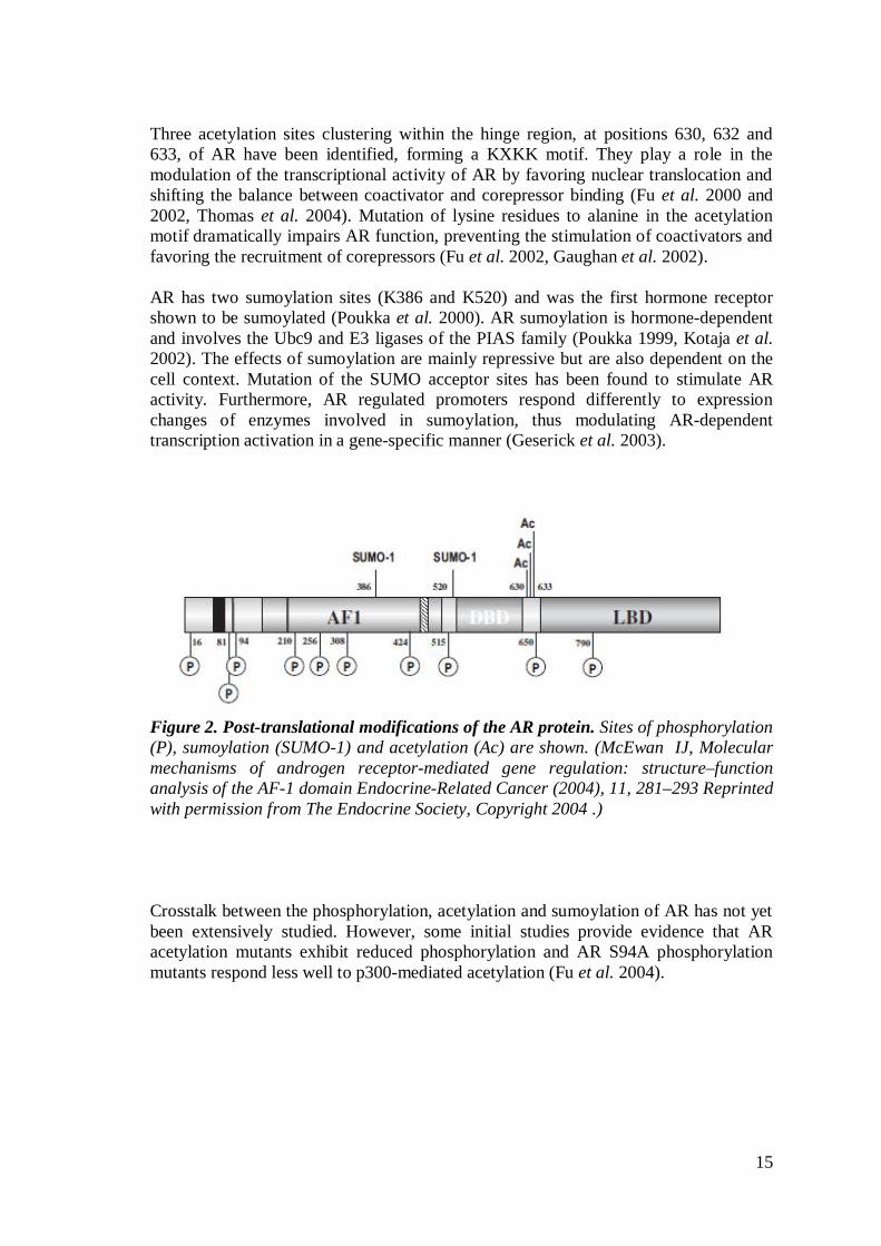
The interaction between the LBD and the Nterminal transactivation domain is neededfor the full liganddependent transactivation and stabilization of AR (Ikonen et al.1997, Schaufele et al. 2005). In total, ten phosphorylation, three acetylation, and twosumoylation sites of AR have been documented to influence and regulate thetranscriptional activity, localization and stability of AR (Fig. 2, reviewed by McEwan2004, Faus and Haendler 2006, AR coregulators to be discussed later). One site, Ser94,is constitutively phosphorylated, whereas S16, S81, S256, S308, S424 and S650exhibit elevated phosphorylation following ligand binding in response to androgen(Zhou et al. 1995, Gioeli et al. 2002 and 2006). Phosphorylation of S650 by MAPKkinases in the hinge region is shown to increase the nuclear localization of AR (Gioeliet al. 2006). However, no single phosphorylation site seems to have a major impact onAR activity since no mutated phosphorylation site alone has been shown todramatically affect the transcriptional activity of AR (Gioeli et al. 2006, Faus andHaendler 2006)
15
Three acetylation sites clustering within the hinge region, at positions 630, 632 and633, of AR have been identified, forming a KXKK motif. They play a role in themodulation of the transcriptional activity of AR by favoring nuclear translocation andshifting the balance between coactivator and corepressor binding (Fu et al. 2000 and2002, Thomas et al. 2004). Mutation of lysine residues to alanine in the acetylationmotif dramatically impairs AR function, preventing the stimulation of coactivators andfavoring the recruitment of corepressors (Fu et al. 2002, Gaughan et al. 2002).
AR has two sumoylation sites (K386 and K520) and was the first hormone receptorshown to be sumoylated (Poukka et al. 2000). AR sumoylation is hormonedependentand involves the Ubc9 and E3 ligases of the PIAS family (Poukka 1999, Kotaja et al.2002). The effects of sumoylation are mainly repressive but are also dependent on thecell context. Mutation of the SUMO acceptor sites has been found to stimulate ARactivity. Furthermore, AR regulated promoters respond differently to expressionchanges of enzymes involved in sumoylation, thus modulating ARdependenttranscription activation in a genespecific manner (Geserick et al. 2003).
Figure 2. Posttranslational modifications of the AR protein. Sites of phosphorylation(P), sumoylation (SUMO1) and acetylation (Ac) are shown. (McEwan IJ, Molecularmechanisms of androgen receptormediated gene regulation: structure–functionanalysis of the AF1 domain EndocrineRelated Cancer (2004), 11, 281–293 Reprintedwith permission from The Endocrine Society, Copyright 2004 .)
Crosstalk between the phosphorylation, acetylation and sumoylation of AR has not yetbeen extensively studied. However, some initial studies provide evidence that ARacetylation mutants exhibit reduced phosphorylation and AR S94A phosphorylationmutants respond less well to p300mediated acetylation (Fu et al. 2004).

16
2.2.3 AR coregulators
ARmediated transactivation requires several auxiliary protein complexes. Thetranscriptional activity of AR is modulated by the interaction of AR with hundreds ofcoregulators and by posttranslational modifications of both AR and its coregulators(reviewed by Heemers and Tindall 2007). According to Heemers and Tindall (2007),AR coregulators may be divided into three general classes: 1) general transcriptionfactors, 2) AR coregulators with diverse properties, and 3) specific transcriptionfactors. The most commonly known and extensively studied coregulator is NCOA1(SRC1), which was the first isolated nuclear receptor coactivator (Onate et al. 1995).Group 1 includes coregulators of the direct interaction of AR with the generaltranscription units GTF2F2 (TFIIF) and GTF2H3 (TFIIH), which facilitates ARtranscription activity in a direct or indirect manner (McEwan et al. 1997, Lee et al.2000). Group 2 can be further divided into several subclasses according to their mainfunction in the nucleus. Especially interesting subclasses are those that include ARmodifying properties such as phosphorylation, acetylation, sumoylation/ubiquitination,mentioned above, as well as those that interact directly with the chromatin. Severalhistone acetylases (HATs) have been shown to interact with the AR and modulate itstransactivating properties, such as the coactivators NCOA1 (SRC1, AIB1), NCOA2(TIF2, SRC2), NCOA3 (SRC3), EP300 (p300), KAT2B (P/CAF), KAT5 (Tip60) andthe corepressors SIRT1, NCOR1 and the HDACs (Heemers and Tindall 2007).Interestingly, Tip60, p300 and P/CAF have also been shown to directly acetylate ARitself whilst AR activity is inhibited by the histone deacetylase activity of HDAC1 (Fuet al. 2000, Gaughan et al. 2002). Group 3 consists of multiple specific transcriptionfactors including, e.g., Foxa1, Oct1, ETS1, AP1, and EGR (Heemers and Tindall2007). Overall, several dynamic changes in covalent histone modification status havebeen associated with androgen/ARstimulated transcription (Kang et al. 2004).
Coregulators that modify sumoylation and ubiquitination of AR are, e.g., SUMO3,UBE2I and PIAS proteins (Zheng et al. 2006, Poukka et al. 1999, Kotaja et al. 2002).Several kinases, cell cycle regulators, chaperones and cytoskeletal proteins, as well assignal integrators and transducers such as MAK, CDK6, HSPA4 (Hsp70), TGFB1I1(ARA55), ATAD2 (ANCCA) and STAT3, have been shown to directly interact withAR (Yeah et al. 1996, Zou et al. 2009). Classical transcription factors such as JUN,FOS, FOXA1 and POU2F1 (OCT1) have also been suggested to interact with ARfunctioning as coactivators or repressors (Sato et al. 1997, Yu et al. 2005, Wang et al.2009b, reviewed by Heemers and Tindall 2007). The modulation and recruitment ofAR and its coregulators in the transcriptome is a slow and very complex mechanism, asrecently demonstrated by Wang et al. (2005 and 2009b).

17
2.3 Cancer of the prostate gland
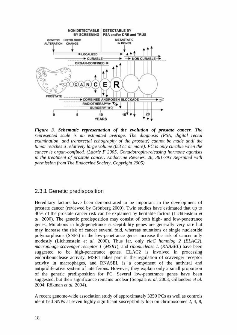
Prostate cancer (PC) originates from glandular epithelial cells. Histological changesresembling in situ cancer are called prostatic intraepithelial neoplasias (PIN). Thetumor normally grows very slowly, remaining confined to the organ and leaving thepatient asymptomatic for decades. As the cancer advances, it first invades through thecapsule and spreads locally to the surrounding tissues. It finally metastasizes further tolymph nodes and bones and to other organs such as the lungs and liver. Localizedintracapsular prostate cancer can be cured by radical prostatectomy. However, 2040%of cancers relapse (Van Poppel et al. 2009). Once the tumor has invaded the capsule,the rate of relapse increases significantly (Carver et al. 2006, BillAxelson et al. 2008).Locally advanced and metastasized PC is treated by androgen deprivation. Eventually,an androgen independent cell population arises during hormonal treatment andcastrationresistant PC (CRPC) develops with an average expected survival period of17 months (Fig. 3, Labrie et al. 2005, Isaacs and Isaacs 2004, Petrylak et al. 2004).
The most widespread method for PC screening is via the serum measurement ofprostatespecific antigen (PSA). Increased levels of PSA may suggest the presence ofPC, but PSA levels can be also increased by, e.g., infection or benign prostatichyperplasia (BPH). Thus, PSA is not a PCspecific marker (Gleason 1966, Papsidero etal. 1985, Stamey et al. 1987). TNM classification and Gleason scoring are morecommonly used as prognostic tools for diagnosed cancer (Kattan et al. 1998, Epstein etal. 2006). TNM classification evaluates the size of the tumor (T), lymph nodemetastasis (N), and distal metastasis (M) (Chisholm et al. 1992). The Gleason score isthe sum of the primary and secondary grades of the glandular differentiation. Gleasongrades range from 1 (mild structural changes) to 5 (full disappearance of glandularstructure). Thus, the sum of scores ranges from 2 to 10. A high Gleason score predictsa poorer prognosis for the patient (Epstein et al. 2006)
18
Figure 3. Schematic representation of the evolution of prostate cancer. Therepresented scale is an estimated average. The diagnosis (PSA, digital rectalexamination, and transrectal echography of the prostate) cannot be made until thetumor reaches a relatively large volume (0.3 cc or more). PC is only curable when thecancer is organconfined. (Labrie F 2005, Gonadotropinreleasing hormone agonistsin the treatment of prostate cancer. Endocrine Reviews. 26, 361793 Reprinted withpermission from The Endocrine Society, Copyright 2005)
2.3.1 Genetic predisposition
Hereditary factors have been demonstrated to be important in the development ofprostate cancer (reviewed by Grönberg 2000). Twin studies have estimated that up to40% of the prostate cancer risk can be explained by heritable factors (Lichtenstein etal. 2000). The genetic predisposition may consist of both high and lowpenetrancegenes. Mutations in highpenetrance susceptibility genes are generally very rare butmay increase the risk of cancer several fold, whereas mutations or single nucleotidepolymorphisms (SNPs) in the lowpenetrance genes increase the risk of cancer onlymodestly (Lichtenstein et al. 2000). Thus far, only elaC homolog 2 (ELAC2),macrophage scavenger receptor 1 (MSR1), and ribonuclease L (RNASEL) have beensuggested to be highpenetrance genes. ELAC2 is involved in processingendoribonuclease activity. MSR1 takes part in the regulation of scavenger receptoractivity in macrophages, and RNASEL is a component of the antiviral andantiproliferative system of interferons. However, they explain only a small proportionof the genetic predisposition for PC. Several lowpenetrance genes have beensuggested, but their significance remains unclear (Seppälä et al. 2003, Gillanders et al.2004, Rökman et al. 2004).
A recent genomewide association study of approximately 3350 PCs as well as controlsidentified SNPs at seven highly significant susceptibility loci on chromosomes 2, 4, 8,
19
11 and 22 (Eeles et al. 2009). One SNP was located in 2q31 (in intron 1 of ITGA6, thegene encoding integrin alpha 6), and one was located in 8p21 (10 kb downstream ofNKX31, which codes for an androgenregulated homeobox protein, NKX31). Theremaining SNPs were located at 2p21 (THADA), 4q22 (PDLIM5), 4q24 (TET2), 11p15(IGF2) and 22q13 (TTLL1). Another recent genomewide association study found fourvariants associated with the susceptibility to PC (Gudmundsson et al. 2009). Thevariants were one in 3q21.3, two in 8q24.21 and one in 11q13. Interestingly, humanprostatic acid phosphatase (ACPP), which has been used as a diagnostic marker forPC, is located at one of the locations identified in this study (Li and Sharief 1993,Sotelo et al. 2010). Another interesting finding was that two SNPs were located withinthe 8q24 region, a region containing enhancer elements that have been suggested toregulate the transcription of the MYC oncogene (Sotelo et al. 2010). The fourth SNPwas located near the CCND1 (cyclin D1) gene in the 11q13 region. This gene is alsoknown to be amplified and functions as an oncogene in many cancers (Fu et al. 2004).
2.3.2 Somatic genetic changes in prostate cancer
Somatic genetic changes in PC can include mutations, copy number alterations,translocations or epigenetic changes. Aberrations may include gain or lossoffunctionchanges depending on the change and the target gene (Vogelstein and Kinzler 1993,Hanahan and Weinberg 2000). In the next chapters, the genes that are commonlyknown to carry somatic alterations in prostate cancer are introduced, and they will befurther subdivided into both gain and lossoffunction categories. Some of thesegenes are known to participate in AR signaling; however, gainoffunction alterationswithin the AR gene itself will be discussed later in their own individual chapters.
2.3.2.1 Lossoffunction alterations in prostate cancerThe most common regions for lossoffunction changes (and the putative target genestherein) are chromosome 5q (APC), 6q (not known), 8p (NKX31), 10q (PTEN), 13q(RBI), 16q, 17p, and 18q (Saramäki and Visakorpi 2007). Phosphatase and tensinhomolog (PTEN), located at 10q23, negatively regulates intracellular levels ofdephosphorylated phosphoinositide substrates and functions as a tumor suppressor bynegatively regulating the AKT/PKB signaling pathway, which promotes cell survivaland inhibits apoptosis. The PTEN locus has been shown to be deleted and/or mutatedin roughly 40% of late stage prostate cancer cases (Li et al. 1997, Dong et al. 2006).APC acts as an antagonist of the Wnt signaling pathway. Mutations of APC are knownto cause familial adenomatous polyposis (FAP) (Phelps et al. 2009). NKX31 is a wellknown AR target gene and will be discussed later.
Tumor protein p53 (TP53, 17p13.1) is a transcription factor that regulates cell cyclearrest, apoptosis and DNA repair. It is commonly known as “the guardian of thegenome”, and the protein functions as a tumor suppressor. TP53 is commonly deletedduring the later stages of prostate cancer. Mutated TP53 protein has a prolonged halflife, leading to the nuclear accumulation of the abnormal protein, which fails to bindthe consensus DNA binding site. Mutations of TP53, PTEN and RB1 are rare in PC.(Visakorpi 1992, Isaacs WB 1995, Hollstein and Hainaut 2010, Taylor et al. 2010).Hypermethylation of the glutathione Stransferase gene (GSTP1) is the most

20
commonly reported epigenetic alteration in PC (reviewed by Meiers et al. 2007). Inaddition, hypermethylation of the wellknown tumor suppressor, APC, has beenreported to occur frequently in the early stages of PC, suggesting its potential use as abiomarker (Yegnasubramanian et al. 2004).
2.3.2.2 Gainoffunction alterations in prostate cancer
Somatic gainoffunction alterations, excluding mutations of the AR gene and ETSfusion genes, are largely unknown. The AR gene and ETS fusion genes will bediscussed separately. Gainoffunction mutations in signature oncogenes, such as RASand EGFR, have been found in several other cancers (reviewed by Dong 2005).However, only few have been identified in prostate cancer (Taylor et al. 2010). Themost common chromosomal gains in PC are located in the 7p/q, 8q, 9p and Xq regionsof the genome. In addition, a chromosomal rearrangement in 21q has been observed inover 50% of prostate cancers. The putative target genes include, 7q: MCM7 and EZH2,8q: TCEB1, MYC, and EIF3S3, Xq: AR (discussed later) and 21q: TMPRSS2:ERGfusion (discussed later) (Nupponen et al. 2000, Saramäki et al. 2001, Savinainen et al.2004, Saramäki and Visakorpi 2007, Taylor et al. 2010).
MCM7 and EZH2, located in 7q, have both been suggested as potential prognosticmarkers in prostate cancer (Laitinen et al. 2008, Ren et al. 2006, Saramäki et al. 2006,Varambally et al. 2002). EZH2 functions in a multiprotein complex called polycombrepressive complex 2 (PRC2). The primary activity of the EZH2 protein complex is totrimethylate histone H3 lysine 27 (H3K27) at target gene promoters, leading toepigenetic silencing. Overexpression of EZH2 promotes cell proliferation, colonyformation and increased invasion of benign cells both in vitro and in vivo (Saramäki etal. 2006, reviewed by Simon and Lange 2008).
The most frequent highlevel amplification in late stage prostate cancer is found at the8q region. This region harbors MYC, a known oncogene; however, this region alsocontains TCEB1 and EIF3H, which have been suggested to function as oncogenes(Savinainen et al. 2006, Jalava et al. 2009). MYC (8q24.21) encodes the vmycmyelocytomatosis viral oncogene homolog, which is a transcription factor involved incell cycle progression, apoptosis and cellular transformation. Amplification of MYChas been shown to be a common event in late state PC. However, gene amplification isnot necessarily correlated with overexpression at the protein level (Edwards et al.2003, Savinainen et al. 2004, Li et al. 2008). Still, overexpression of MYC intransgenic mouse models results in prostatic intraepithelial neoplasia (PIN), andtogether with loss of NKX31 it is associated in carcinogenesis (Zhang et al. 2000,EllwoodYen et al. 2003, Williams et al. 2005).

21
2.4 Androgen receptor (AR) signaling pathway in PC
The AR signaling pathway is dependent on androgen metabolism, ligand specificity,the expression level of AR, ligandindependent activation and cofactor interactions.Several alterations take place in the AR signaling pathway during the development andprogression of PC. These changes include somatic mutations of AR that allow theusage of a wider spectrum of ligands, the amplification of AR, leading to higherexpression levels and a shift from paracrine stromal growth support to an autocrinemode. In addition, changes in the balance of AR coregulators and AR splice variantsallowing ligandindependent AR action have been suggested. It has also been shownthat many of the androgenregulated genes become upregulated during the progressionof the disease to CRPC (Holzbeierlein et al. 2004). Only a very few PCs and CRPCsare considered to have an inactive AR signaling pathway. In the following chapters, themost frequently studied alterations of AR and AR signaling in PC are introduced(reviewed by Feldman and Feldman 2001, Isaacs and Isaacs 2004). Changes indifferent parts of AR pathway signaling during PC progression are discussedindividually.
2.4.1 Changes in androgen metabolism
There is abundant evidence that androgens influence the development of PC. In a largerandomized PC prevention trial, over 18,800 men aged 55 years or older were treatedwith finasteride, an inhibitor of steroid 5 reductase that converts testosterone to DHT.Finasteride treatment was found to reduce the risk of developing prostate cancer by20% (Thompson et al. 2003). Similar results were shown in a prevention trial in which6729 men aged 50 to 75 were treated with another 5 reductase inhibitor, dutasteride.The relative risk reduction with dutasteride was 22.8% (Andriole et al. 2010). Inadvanced prostate cancer, even when androgen deprivation therapy is used, theintraprostatic DHT levels remain relatively high. Generally only a 50% reduction inDHT levels is observed after androgen depletion (reviewed by Labrie et al. 2005).Intracrine activity of the PC cells themselves has been suggested to be involved inincreasing DHT levels (Gao et al. 2001, Vander Griend et al. 2010). It has also beenreported that the expression of many enzymes involved in steroidogenesis areupregulated during CRPC progression (Holzbeierlein et al. 2004, Montgomery et al.2008, Locke et al. 2010, Leon et al. 2010). However, in the recent study, Hofland et al.(2010) could only detect a low level of simultaneous expression of the enzymesCYP17A1 and HSD3B1, which are essential for de novo synthesis of androgens, in 5 of88 patients. SRD5A1 and AKR1C3 expression were shown to be increased duringandrogen deprivation, suggesting the importance of DHT synthesis of an adrenal originandrogens instead of from cholesterol (Fig. 4, adapted from Hofland et al. 2010). Theimportance of androgen metabolism was confirmed by recent clinical trials withabiraterone, a CYP17A1 inhibitor, which directly indicated that CRPC is stillandrogendependent (Attard et al. 2008, 2009a and b, Ryan et al. 2010).

22
Figure 4. Scheme of the classic steroid biosynthetic pathway. CYP, cytochromeP450; HSD, hydroxysteroid dehydrogenase; CYB5, cytochrome b5; AKR, aldoketoreductase; SRD, 5alpha reductase. (Hofland J et al. Evidence of limited contributionsfor intratumoral steroidogenesis in prostate cancer. Cancer Research. 70(3):125664Apated by permission from American Association for Cancer Research, Copyright2010)
2.4.2 Somatic mutations of AR
AR mutations seem to be rare in early stage, untreated PC (Newmarck et al. 1992,Culig et al. 1993, Taplin et al. 1995 & 2003, Wallen et al. 1999). However, the numberof AR mutations increases with progression of the disease and after hormonaltreatments. AR mutations can be identified in approximately 2030% of late stageCRPC tumors. Collectively, only two reports identify point mutations within AR in asignificant number of untreated tumor samples. Gaddipati and coauthors (1994) foundthe LNCaP mutation (T877A) in 25% of the transurethral resections of prostate(TURP) specimens from patients with untreated metastatic PC, whereas Tilley et al.(1996) reported that roughly 50% of hormone naïve PCs have a mutated AR. However,the tumors used in these studies were late stage and rare forms of PC. Generally, thehighest frequency of mutations seems to be in PCs treated with antiandrogens(especially in patients treated with flutamide). Mutation frequencies of 1030% havebeen reported in such cases (Suzuki et al. 1996, Taplin et al. 1999 and 2003, Buchananet al. 2001).
The most significant known AR mutations are known to affect the ligand specificity ofAR. The most frequently found point mutation of AR is the T877A mutation (threonineat position 877 is substituted to alanine). This mutation was the first AR mutationidentified in PC and was originally characterized in the LNCaP cell line (Veldscholteet al. 1990). This amino acid is located on helix 11 at the ligandbinding pocket, whichinteracts directly with the ligand. It alters the stereochemistry of the binding pocket andbroadens the ligand binding of AR (Sack et al. 2001). It allows other nuclear hormones(estrogen and progestin), corticosteroids (cortisol and cortisone) and antiandrogens(cyproterone and hydroxyflutamide) to activate AR (Culig et al. 1993 and 1999, Changet al. 2001, Steketee et al. 2002).

23
In addition to the T877A, several other mutations at the ARLBD, e.g., L701H,V715M, V730M and H874Y, which enhance the transcriptional sensitivity of AR toother steroids including adrenal androgens and/or antiandrogens, have been identified(Suzuki et al. 1993, Culig et al. 1993, Newmark et al. 1992 and Taplin et al. 1995).L701H was originally found in CRPC (Suzuki et al. 1993, Watanabe et al. 1997) andin the MDA PCa 2a cell line, which also harbors the T877A mutation (Zhao et al.1999). L701H mutated cells are highly responsive to glucocorticoids (cortisol andcortisone) at the concentrations found in humans (Zhao et al. 1999, van de Wijngaart etal. 2010).
H874Y was originally identified in CRPC patients treated with flutamide (Taplin et al.1995). This mutation was also identified in the xenograft CWR22, which was derivedfrom a patient suffering from primary PC who also had symptoms of bone metastasis.The original patient tumor was graded with a Gleason score 9 (Weinstain et al. 1994,Tan et al. 1997). DHEA, estradiol, progesterone, and hydroxyflutamide induced agreater transcriptional response from the H874Y mutant than the wildtype AR (Taplinet al. 1995, Tan et al. 1997, Steketee et al. 2002). This site is located distant from theligandbinding pocket and affects the binding of coregulator proteins (enhancing e.g.p160 mediated AR transactivation). Thus, H874Y indirectly affects ligand specificityby causing a conformational change in the AR protein (Steketee et al. 2002, Duff et al.2005). The 22Rv1 cell line, which is derived from the castrationresistant form of PCxenograft CWR22 (CWR22R), also carries an LBD deletion and a duplication of theDBD domain (exon 3). These mutations are not present in the androgensensitiveCWR22Pc cell line (Dagvadorj et al. 2008, Dehm et al. 2008).
V715M and W741C are less frequently studied, as these mutations are rare within PC.However, they do result in functional changes in AR. V715M was originally found inCRPC patients and is reported to be activated by adrenal androgens and progesteroneand is sensitive to low androgen concentrations (Culig et al. 1993, Thompson et al.2001). The W741C mutation was found in bicalutamide treated patients (Haapala et al.2001, Taplin et al. 2003). The growth of KUCaP xenografts carrying the W741Cmutation is accelerated by treatment with bicalutamide and flutamide (Yoshida et al.2005, Terada et al. 2010). Additionally, the LNCaP cell line has been shown to acquirethe ability for bicalutamideresistant growth via the W741C mutation when exposed tolongterm treatment with bicalutamid (Hara et al. 2003).
A smaller number of missense mutations have been detected in other domains of AR.Missense mutations (K179R and C619Y) which affect the Nterminal and DBDregions of the AR protein have been identified in two patients with untreated primaryprostate cancer (Tilley et al. 1996, Marcelli et al. 2000). K179R has been suggested toplay a more potent role in AR deregulation (Callewaert et al 2006), whereas C619Yhas been found to cause inactivation and mislocation of the receptor (Nazareth et al.1999).
Buchanan et al. (2001b) found F671I at the boundary of the hinge and LBD regions inthe TRAMP mouse model. This mutation broadens the range of AR ligand specificityand increases the transactivation capacity by 2 to 4fold. The AR/E231G transgenicmouse model provided evidence that mutations within the Nterminal region of the ARprotein may have an oncogenic effect (Han et al. 2005). Such mutations led to thedevelopment of PIN, which progressed further to the invasive, metastatic disease in

24
100% of the mouse models studied. Neither the F671I nor the E231G mutation havebeen found to occur in human PC. The locations of all AR mutations found in PC andhave been shown to have a functional effect on AR action are shown in Figure 5.
Figure 5. The genetic alterations of AR in PC. Functionally active regions areillustrated under the structure and functionally active genetic changes involved in PCare noted above the structure. *) Germline mutation, phosphorylation sites (8) markedwith pinheads. NTD = Nterminal Domain, DBD = DNAbinding Domain, NLS =Nuclear Localization Signal, LBD = Ligandbinding Domain, AF1 & 2 = ActivationFunction 1 & 2, TAU1 & 5 = Transactivation Units 1 and 5, ARE1 & 2 = AndrogenResponsive Elements 1 and 2, SUMO1 & 2 = Sumoylation sites 1 and 2, Ac =Acetylation sites. (Current Clinical Oncology:Prostate Cancer: Signaling Networks,Genetics and New Treatment Strategies by R.G. Pestell and M.T. Nevalainen. 2008.Somatic Genetic Changes in Prostate Cancer: Androgen Receptor Alterations. pp 99128. Reprinted by permission from ©2008 Humana Press).
2.4.3 Amplification and overexpression of AR
Almost all PCs, except rare small cell carcinomas of the prostate, express AR at boththe mRNA and protein level. Expression of AR is maintained and often elevated duringprostate carcinogenesis from androgendependent PC to hormonerefractory CRPC,especially during longterm androgen ablation (Ruizeveld de Winter et al. 1994,Kokontis et al. 1994, Visakorpi et al. 1995, Hobisch et al. 1995, Culig et al. 1999, Latilet al. 2001, Linja et al. 2001, Edwards et al. 2003, Hofland et al. 2010). High ARexpression has been suggested to be associated with short recurrentfree survival(Henshall et al. 2001, Lee et al. 2003, Donavan et al. 2009). Interestingly, AR alwayslocalizes to the nucleus in clinical tumors, irrespective of circulating androgen levelsand androgendependence status, i.e., hormonenaïve, recently castrated or castrationresistant. This finding indicates that AR is continuously activated during PCprogression (Laitinen et al. 2007). The expression of AR is abolished in only a veryrare fraction of CRPCs, possibly through hypermethylation of the AR promoter(Kinoshita et al. 2000).
SUMO1
M715V
L701H
ATTAA… CATAAA àPolyA 10409… 10636
5’UTR exon 1 2 3 4 5 6 7 8
UC motif4036… 4071
3’UTR
TATAlessPromoter
NTD DBD LBD
Hinge, NLSAF1 AF2
919: Stop1: Met
T877A; LNCaPmutationGACrepeat GCCrepeat
M749I
W741C/L; Bicalutamide resistance
A726L*)K179R
H874Y; CWR22xenograft
E231G
668QPIF671; TRAMPmodel
A748T
C619Y
TAU1 TAU5
AcSUMO2
ARE1 ARE2

25
Gain and amplification of the AR gene is one of the most frequent chromosomal gainsin CRPC (reviewed by Nupponen & Visakorpi 1999). Nearly 80% of CRPCs havebeen reported to carry an elevated AR gene copy number (Edwards et al. 2003), with2030% showing a high level of AR gene amplification. In contrast, untreated primaryPCs very rarely contain an AR gene amplification (Visakorpi et al. 1995, Koivisto et al.1997, Bubendorf et al. 1999, KaltzWittmer 2000, Edwards et al. 2003). Amplificationwas detected in the untreated samples of only 2 cases out of 205 primary PCs,indicating that the amplification is selected during the emergence of CRPC (Bubendorfet al. 1999). Recently, two studies reported that AR gene amplification was found in>50% of circulating tumor cells (CTC) from metastasized CRPC cases (Leversha et al.2009, Attard et al. 2009b). However, AR gene amplification only partially explains theoverexpression of AR. The other still unknown mechanisms leading to ARoverexpression could include, e.g., genetic aberrations in the regulatory regions of ARand miRNA deregulation.
The significance of the overexpression of AR in CRPC was first demonstrated by Chenand coauthors (2004), who showed that the common nominator in gene expressionprofiles of CRPC xenograft models as compared to androgendependent counterpartswas the increased AR level. They also showed that overexpression of AR alone wasnecessary and sufficient to cause androgensensitive xenografts to become castrationand bicalutamideresistant.
2.4.4 AR splice variants
Three novel AR isoforms lacking the ligandbinding domain (designated as AR3, AR4,and AR5, according to Guo et al. 2009, Fig. 6) have been reported in CRPC. AR3, oneof the major splice variants expressed in human prostate tissues, has been suggested tobe constitutively active (Guo et al. 2009, Hu et al. 2009). The molecular mechanismsthat lead to differential AR splicing are not known. Immunohistochemical analysis of429 PC tissues showed that AR3 is significantly upregulated during CRPCprogression and AR3 expression levels are correlated with the risk of tumor recurrenceafter radical prostatectomy. Unlike wildtype AR, AR3 was shown to directly increaseAKT1 expression (Dehm et al. 2008, Guo et al. 2009, Hu et al. 2009).
Figure 6. Schematic structure of the human AR splice variants. (Guo Z et al. 2009. Anovel androgen receptor splice variant is upregulated during prostate cancerprogression and promotes androgen depletionresistant growth. Cancer Research.69(6):230513 Reprinted by permission from American Association for CancerResearch, Copyright 2009)
ARAR3AR4
AR5
NTD DBD Hinge LBD

26
2.4.5 Changes in AR cofactors
AR cofactor imbalances have been studied in PC progression. Thus far, no AR cofactorhas been shown to influence a great number of cancers. However, some nonrecurrentcofactor changes have been reported in a small number of PC cases. More studies arerequired to clearly define the changes of all the cofactors implicated in PC (reviewedby Xu et al. 2009a, Fujimoto et al. 2001, Linja et al. 2004, Mäki et al. 2006 and 2007,Zou et al. 2009).
High expression levels of some AR cofactors have been reported in PC. The increasedexpression of NCOA1 (SRC1), NCOA2 (TIF2, SRC2), KDM1A (LSD1), KDM4C(JMJD2C), RNF6, TRIM68 and TGFB1I1 (ARA55) has been linked with increasedactivation of AR in PC (Fujimoto et al. 2001, Agoulnik et al. 2006, Mäki et al. 2006and 2007, Miyajima et al. 2008, Xu et al. 2009b, Taylor et al. 2010). Increased levelsof NCOA2, BAG1 and ATAD2 (ANCCA) protein have also been associated with theprogression of CRPC or higher grade PCs (Fujimoto et al. 2001, Agoulnik et al. 2006,Mäki et al. 2007, Zou et al. 2009). Interestingly, ATAD2 was also shown to beupregulated by androgens (Zou et al. 2009).
Genetic alterations of AR coregulators have not been intensively studied. Only a fewstudies have reported somatic DNA copy number alterations or nonsense mutations ofknown AR coregulators. Two cases of NCOA1 gene amplification and one case of aNCOA1 missense mutation in PC have been reported thus far (Linja et al. 2004, Mäkiet al. 2006). The low frequency of such aberrations suggests that genetic alterations inNCOA1 are not commonly involved in the progression of PC (Linja et al. 2001, Mäkiet al. 2006, Xu et al. 2009). Another AR coregulator reported to be amplified in PC isBAG1, which has been shown to activate AR by interacting with the Nterminal regionof the receptor. BAG1 was found to be amplified in 7% of CRPC samples, withsignificantly higher protein expression compared to primary PC samples (Shatkina etal. 2003, Mäki et al. 2007). Recently NCOA2 was also shown to be amplified andoverexpressed in PC (Taylor et al. 2010).
2.4.6 Well known AR target genes
Microarray studies of androgenregulated genes suggest that approximately 24% of alltranscripts could be directly or indirectly regulated by androgens (Amler et al. 2000,Nelson et al. 2002). AR regulates the expression of androgenresponsive genes bybinding to the androgen response elements (AREs) of target genes. Sequence analysisand new chromatin immunoprecipitation (ChIP) technologies have identified, inaddition to classical AREs, several new noncanonical AR binding sites, which havebeen shown to function even up to 300 kb up or downstream from the target gene(Wang et al. 2007a). The most commonly known and studied AR target genes includeKLK2, KLK3(PSA) and TMPRSS2, which produce prostate gland enzymes includingphosphatases and several serine proteases secreted by the epithelial cells into seminalplasma (Young et al. 1992, Perry et al. 1996). The following paragraphs introduce themost well known AR target genes.
27
2.4.6.1 ACPP (PAP)
The acid phosphatase prostate (ACPP) gene, also known as prostate acidicphosphatase (PAP), is located in chromosome 3q21q23. ACPP encodes a 100 kDatyrosine and lipid phosphatase which is synthesized in the prostatic epithelial cells andsecreted into prostatic fluid. There are two forms of PAP, a cellular and secreted form,which have different biochemical properties. PAP has been shown to be directlyregulated by androgens in a biphasic manner and is highly expressed in both thenormal prostate and in PC (Gutman and Gutman 1938, Vihko 1979, Li and Sharief1993, Henttu et al. 1992, Lin et al. 1993, Ulrix et al. 1998). PAP was used as early asthe 1930s as a biomarker for PC (Gutman and Gutman 1938, Huggins and Hodges1941). Cellular PAP levels are decreased in advanced PC. PAP expression correlatesnegatively with cell growth and cancer progression. It has been suggested that PAPdephosphorylates HER2, which in turn activates ERK/MAPK signaling (Sharma et al.2005). Decreased PAP levels and increased tyrosine phosphorylation of HER2correlate with Gleason score and PC progression. The molecular mechanisms thatcause decreased PAP levels in PC are not known (Veeramani et al. 2005).
2.4.6.2 KLK3 (PSA)
Kallikreinrelated peptidase 3 (KLK3), better known as prostatespecific antigen(PSA), is located in chromosome 19q13.41. KLK3 encodes a single chain glycoproteinwith a molecular mass of 33 kDa and functions as a serine protease. It belongs to thefamily of the fifteen kallikrein members located in a cluster in the same chromosomalregion. All kallikrein genes encode five exons of similar size and have high sequencehomology with other family members. Many of these peptidases also have severalalternative splice variants and are known to be regulated by androgens (reviewed byLawrence et al. 2010). KLK3 was cloned in 1987 (Lundwall and Linja). KLK3expression has been shown to be elevated in BPH and in highly differentiated PCs, butit is decreased during PC progression (Abrahamsson et al. 1988, Hakalahti et al. 1993).The use of KLK3 as a PC biomarker (the socalled PSA test) began in the mid 1980s(Stamey et al. 1987). In a recent European study, which included more than 160,000men aged 55 to 69, it was found that PSAbased screening reduced PC mortality by20%. However, there was a high risk of overdiagnosis (Schröder et al. 2009).Androgen regulation of KLK3 includes both the proximal promoter and the enhancerARE located 4 kb upstream from the TSS. Recruitment of AR and its coregulatorscreate a chromosomal loop from the enhancer to the core promoter (Young et al. 1992,Riegman et al. 1991, Wang et al. 2005). Kallikrein family members have also beensuggested to play a putative role in PC progression. For example, KLK3 has beensuggested to directly degrade extracellular matrix glycoproteins and facilitate cellmigration (reviewed by Lilja 2003, Hollenberg et al. 2008).
2.4.6.3 TMPRSS2:ERG and other fusion ETS
The fusion of Etwentysix family (ETS) genes with a hormonedependent promoterregion occurs in 3070% of therapynaîve prostate cancers (Tomlins et al. 2005 and2007, Saramäki et al. 2008, reviewed by KumarSinha et al. 2008, Tomlins et al.2009). Thus far, rearrangements of the ERG, ETV1, ETV4 and ETV5 gene loci have

28
been reported (Tomlins et al. 2005, 2006 and 2007, Helgeson et al. 2008). The mostcommon variants involve transmembrane protease, serine 2 (TMPRSS2) exon 1 or 2fused to vets erythroblastosis virus E26 oncogene homolog (ERG) exon 2, 3, 4 or 5(Tomlins et al. 2005 and 2007). These two genes are located at the same chromosomalregion, 21q22.3. TMPRSS2 is an androgenregulated gene which is highly expressed inthe normal prostate and in PC (Lin et al. 1999, Vaarala et al. 2001). The gene encodesa serine protease that contains a predicted protein of 492 amino acids in length.Relatively recently, androgen regulation by cisregulation of noncanonical AREs inTMPRSS2 gene was discovered (Wang et al. 2007a).
Additional 5’ partners for ETV1, ETV4, ETV5 and ELK4 have also been identified.These 5’ partners include SLC45A3, HERVK_22q11.23, CANT1 and KLK2, which areprostatespecific and androgeninducible (reviewed by KumarSinha et al. 2008,Tomlins et al. 2009). It is not clear how these gene fusions participate incarcinogenesis in the prostate. However, in vitro studies of overexpressed ETV1 orERG have suggested their role in invasion via the urokinase plasminogen activator(UPA, or PLAU) pathway. It has also been shown that transgenic mice thatoverexpress ETV1 or ERG develop mouse prostatic intraepithelial neoplasia (mPIN),but not tumors. (Tomlins et al. 2007, Cai et al. 2007, Klezovitch et al. 2008). Thesefusion genes are early events that are most commonly associated with localized PC.They are found in similar frequency in CRPC, suggesting their role in driving thetransformation but probably having less significance in the CRPC progression (Perneret al. 2007, Saramäki et al. 2008, reviewed by Tomlins et al. 2009). Recently, twostudies with transgenic ERG overexpression in Pten heterozygous background miceshowed significant progression of highgrade PIN and PC by inducing downstreamcheckpoint genes that would usually be blocked by AKT (Carver et al. 2009, King etal. 2009, Squire 2009).
2.4.6.4 NKX31
The NK3 homeobox 1 (NKX31) gene encodes a transcription factor with tumorsuppressor functions. This gene is localized in the chromosomal region 8p21, which iscommonly deleted in PC. The expression of NKX31 is androgenregulated andstimulates the differentiation of prostatic epithelial cells. NKX31 expression is oftenlost during the progression of PC (He et al. 1997, Asatiani et al. 2005, Bethel et al.2007). However, some examples of NKX31 overexpression have been reported (Xu etal. 2000). NKX31 has also been shown to regulate AR expression in LNCaP cells(Possner et al. 2008). Interestingly, Wang et al. (2009a) recently suggested that inmouse, in rare castration resistant Ar and Nkx31 positive luminal epithelial PC stemcells, Nkx31 is required for stem cell maintenance in vivo during hormonal treatment.This finding may indicate that NKX31 is not a classical tumor suppressor gene butcould have a role in PC stem cell survival and differentiation in androgendeprivedconditions.

29
2.5 Endocrine treatments of prostate cancer
PC treatments that target AR signaling have been investigated heavily for over half acentury. The revolutionary finding was made in 1941 by Huggins and Hodges, whoshowed that castration or estrogen treatment inhibits the growth of PC and that thegrowth of PC was activated by androgen injections (Huggins and Hodges 1941). Theyalso noticed the association between hormonal manipulations and the serum level ofPAP. Currently, a wide spectrum of ARtargeted therapies is available. These therapiesfocus mainly on either preventing androgen production from the testes and/or blockingthe function of AR with antiandrogens.
2.5.1 Targeting androgen synthesis
Androgen synthesis can be prevented by classical orchiectomy or by continuousstimulation of the pituitary with high concentrations of luteinizing hormone releasinghormone (LHRH) agonist or antagonist. Continuous agonist stimulation results inreceptor desensitization and inhibition of LH release, which further inhibits theproduction of testosterone by the testes. Several agonists, e.g., goserelin andleupropelin, are used in the clinic with wellproven equivalence to orchiectomy. LHRHantagonists directly affect the LHRH receptor regulatory system, causing a rapiddecrease in serum androgen levels (reviewed by Tammela 2004, Weckermann andHarzmann 2004, Labrié et al. 2005). Since residual serum androgens, as well asupregulated intracrine androgen synthesis, may be sufficient to promote CRPC growthin patients receiving androgendeprivation therapy, strategies to further lower androgenlevels have been suggested. One of the promising new drugs that targets both adrenaland tumor intracrine androgen synthesis is abiraterone. It is a selective irreversibleinhibitor of the p450 enzyme, 17 hydroxylase/C17,20lyase (CYP17), whichcatalyzes the biosynthesis of androgens from pregnane precursors. Recent phase I/IIclinical trials of patients with CRPC have shown significant antitumor activity and upto 70% of PSA response (Attard et al. 2008 and 2009a, Ryan et al. 2010).
2.5.2 AR targeting inhibitors
Direct AR inhibitors can be divided into steroidal or nonsteroidal antiandrogens. Theyspecifically block T and DHT from binding the ligandbiding pocket of AR.Nonsteroidal antiandrogens such as bicalutamide, flutamide and nilutamide inhibit theactivity of androgens by their competitive interaction with AR. Steroidal antiandrogens(cyproterone acetate) also function in the pituitary axis and inhibit LH release.However, with the use of steroidal antiandrogens, more complications have beenreported compared to LHRH analogs and nonsteroidal antiandrogens (reviewed byTammela 2004, Loblaw et al. 2007). The binding of flutamide or bicalutamide to ARprevents androgen binding and modulates ARprotein structure, allowing the bindingof AR corepressors (Yoon & Wong 2006). However, somatic AR mutations(introduced above) that occur after the use of antiandrogens have been reported torelease the transcriptional inhibition (Chen et al. 2004, Steketee et al. 2002, Culig et al.1999). To improve the inhibition of the androgen signaling axis, complete androgen

30
blockade (CAB) therapies have been investigated. In CAB, castration is combined withantiandrogen therapy. CAB has been shown to prolong life in CRPC patients, but thesignificance is only marginal (Palmberg et al. 2000, Labrie et al. 2002 and Scher et al.2004). Recently, new antiandrogens for CRPC have been developed. For example,MDV3100 binds to AR with 10fold higher affinity than bicalutamide and inhibitsPSA secretion at 10fold lower concentrations. Unlike bicalutamide, MDV3100impairs AR nuclear translocation and blocks DNA binding (Tran et al. 2009, Jung etal. 2010). MDV3100 is currently in a phase III clinical trial for use in CRPC(reviewed by Chen et al. 2008, Tran et al. 2009, Jung et al. 2010).

31
3. AIMS OF THE STUDY
The purpose of the study was to investigate the molecular mechanisms leading to ARoverexpression and to establish an AR overexpression model that can be used toidentify and investigate the downstream AR target genes involved in PC progression.
The specific aims were the following:
1) To investigate whether mutations in the AR gene regulatory regions couldunderlie its overexpression;
2) To establish an in vitro model of AR overexpression;3) To identify AR downstream proteincoding genes involved in PC progression;4) To identify AR downstream miRNAs involved in PC progression.
32
4. MATERIALS AND METHODS
4.1 Cell lines and xenografts
DU145, LNCaP, PC3 and 22Rv1 PC cell lines were obtained from the American TypeCulture Collection (ATCC, Manassas, VA, USA). LAPC4 cells were kindly given byProf. C. Sawyers (University of California at Los Angeles, Los Angeles, CA, USA).VCaP and DuCaP were provided by Prof. J. Schalken (Radboud University NijmegenMedical Center, Nijmegen, The Netherlands). Cells were cultured under therecommended conditions (original communications IIII).
Nineteen LuCaP series xenografts (LuCap 23.1, 23.8, 23.12, 35, 35V, 49, 58, 69, 70,73, 77, 78, 81, 86.2, 92.1, 93, 96, 105, 115) were obtained from Prof. R. Vessella(University of Washington, Seattle, WA, USA), and 13 PC series xenografts (PC82,133, 135, 295, 310, 324, 339, 346, 346B, 346I, 346BI, 374 and 374F) were obtainedfrom Prof. G. Jenster (University of Rotterdam, The Netherlands). Tumor sampleswere collected either from intact male (LuCaP series) mice or both intact mice and 7 to14 days after castration (PC xenografts) and were freshly frozen at 80°C (originalcommunications I and III).
4.2 Clinical samples
All together, 52 freshly frozen clinical samples (6 BPH, 30 primary prostatectomy PCand 14 CRPC samples described in original communication I, and 7 BPH and 15CRPC samples described in original communication III) were used in the study. Ofthose, the BPH and CRPC samples were transurethal resections (TURP). Theendocrine therapy included either orchiectomy, LHRH analog, estrogens, orchiectomyand estrogen, bicalutamide or unspecified hormone therapy. Samples were snap frozenin liquid nitrogen. Tumor samples contained at least 60% cancer cells. Thirty normalsamples for general SNP analysis (60 chromosome X) were obtained from healthyFinnish female blood donors. The use of the clinical material was approved by theethical committee of the Tampere University Hospital (original communications I andIII).
4.3 DNA and RNA extractions, DNA amplification and PCR
DNA and RNA were purified using routine phenolchloroform and Trizol™techniques. For mutation analysis, DNA samples were first amplified with a GenomePhi™ genome amplification kit (Invitrogen) according to the manufacturer’sinstructions. For PCR reactions, Accutype (Stratagene) or Platinum Taq (Invitrogen)added with 1:4 ratio Pfu (Fermentas) polymerases. The enzymes were used indetergentfree buffer according to the manufacturer’s instructions. Primers andannealing temperatures are listed in the original publications. All primers weredesigned with the publicly available Primer3 program (http://frodo.wi.mit.edu/primer3/) (original communications IIII).
33
4.4 DHPLC and sequencing
For promoter and 5’UTR mutation analyses, sample PCR fragments were each mixedat a 1:1 ratio with the corresponding normal PCR fragment, denatured (95°C for 3 min)and renatured (65°C for 30 min). DHPLC (denaturing highperformance liquidchromatography) was performed with an Agilent 1100 LC machine and a VarianCP28353 Helix DNA column (50 x 3.0 mm) at a 12% diluent gradient over 8 min atfragmentspecific temperatures. Primers and DHPLC driving conditions were designedwith the freely available DHPLC Melt program (http://insertion.stanford.edu/melt.html). Sequencing was performed with a BigDye® Terminator v3.1 Cycle SequencingKit (Applied Biosystems) and run in the ABI3100 Genetic Analyzer according to themanufacturer’s instructions (original communication I).
4.5 mRNA and miRNA qRTPCR reactions
First strand cDNA synthesis was carried out from total RNA using either AMV reversetranscriptase (Finnzymes) or SuperScriptIII (Invitrogen) for miRNAspecific targetsaccording to the manufacturer’s instructions. To measure mRNA expression, a SYBRGreenII Fast Start kit (Roche Diagnostics) was used according to the manufacturer’sinstructions using TBP (TATAbox binding protein) mRNA as a reference gene. Formeasuring mature miRNA expression, TaqMan® MicroRNA Assays (AppliedBiosystems) and a Probe Fast Start kit (Roche Diagnostics) were used. A Light Cyclerapparatus (Roche Diagnostics) was used. The RT reactions and the qPCR reactionswere performed separately for each miRNA using RNU6b as a small reference RNA(original communications II and III).
4.6 Western blot
Protein extracts (12.5 µg) were separated by 12% SDSPAGE and transferred to PVDFmembranes (ImmobilonP, Millipore Corp.) using the standard semidry transfertechnique (BIORAD Transblot, BioRad Lab.). Mouse antiAR 441 was used as theprimary antibody (NeoMarkers), and antimouse IgGHRP conjugate (DAKO A/S)was used as the secondary antibody (original communications II and III).
4.7 Transfection methods
The parental LNCaP cells were stably transfected either with the pcDNA3.1(+) emptyexpression vector (Invitrogen Inc., Carlsbad, CA, USA) or with the pcDNA3.1AR(Acc#_M23263) into the NotI/BamHI site in the pcDNA3.1 vector) withLipofectamine Plus transfection reagent (Invitrogen, Inc.), according to themanufacturer’s instructions. Transfected clones were selected under 400 µg/mlgeneticin (G418, Invitrogen, Inc.). Two clones expressing moderate and high levels ofAR protein (LNCaPARmo and LNCaPARhi) were selected for further studies andmaintained in 200 µg/ml geneticincontaining medium (original communication II).
The LNCaPpcDNA3.1 cells were transiently transfected with 20 nM PremiR™ miR141 precursor (#PM10860, Ambion, Inc., Austin, TX, USA), and the LNCaPARhicells with 100 nM AntimiR™ miR141 inhibitor (#AM10860, Ambion, Inc.) usingINTERFERINin™ siRNA transfection reagent (POLYPLUSTRANSFECTION Inc.,

34
NY, USA) under various DHT concentrations, according to manufacturer’s protocol.The scrambled Anti or PremiR™ negative controls (#AM17010, #AM17110,Ambion, Inc.) were used as corresponding reference treatments (originalcommunication III).
4.8 DHT and roscovitine treatments and cell proliferation assays
Before each hormone exposure experiment, cells were grown in 5% charcoal/dextrantreated serum (CSS, HyClone, Inc.) in medium without phenol red for three or fourdays. The medium was subsequently replaced with various concentrations of DHT(Steraloids, Inc., USA). Roscovitine (Calbiochem®, EMD Chemicals Inc., Germany)was used for CDK1/2 growth inhibition treatments. The relative number of cells ateach time point was analyzed either by using the Beckman Coulter® Z2series particlecounter (Beckman Coulter, Inc., USA) or AlamarBlue reagent (AbD Serotec, UK) andluminometric detection using a fluorometer (Wallac 1420 Victor, PerkinElmer, USA)(original communications II and III).
4.9 Microarray hybridizations
AR and androgen regulation of different protein coding genes were studied with anIllumina Microarray platform on a HumanRef8 v2 chip and performed in the FinnishDNA Microarray Center at the Turku Centre for Biotechnology. Total RNA (300 ng)from each sample was transcribed in vitro, biotinylated and amplified with an IlluminaRNA TotalPrep Amplification kit (Ambion, #IL1791, USA), and hybridized to anIllumina Sentrix HumanRef8_V2 Expression Bead Chip (cat. no. BD25213). Theprobes (>22,000) of the Illumina HumanRef8 v2 chip are based on the well annotatedprotein coding genes of the Reference Sequence (RefSeq) database 1, release 17(original communication II).
miRNA expression was analyzed with Agilent human miRNA microarray v2 chips(Agilent Technologies, Santa Clara, CA, USA) containing 723 human and 76 humanviral miRNAs (Sanger Cambridge, Database v10.1, http://microrna.sanger.ac.uk)according to the manufacturer’s instructions. Total RNA (100 ng) was labeled withpCpCy3 and hybridized according to the manufacturer’s instructions (AgilentTechnologies, Santa Clara, CA, USA). miR141 target gene expression was studiedwith Whole Genome Human 4x44K microarray chips (Agilent Technologies)according to the manufacturer’s instructions (original communication III).
4.10 Data analysis
The data analysis was performed with the GeneSpring Analysis Platform, version GX7.3.1 (Agilent Technologies, CA, USA) using standard normalization methods such asmedian normalization and Lowess smoothing. The average linkage method was usedfor unsupervised hierarchical clustering, and the similarities were estimated withPearson’s correlation. For ontology classifications, all gene ontology (GO) listscontaining at least ten genes with pvalues <0.001 (hypergeometric pvalue withoutmultiple testing correction) were filtered and organized with the GeneSpring Ontologybrowser (original communications II and III).

35
4.11 ChIPonchip assays
ChIPonchip assays were performed with custom antiAR antibody (BJ14AR3) usingLNCaP cells that were treated with 100 nM DHT or vehicle for 2 h. Three microgramsof the immunoprecipitated DNA samples were hybridized to Affymetrix wholegenome tiling arrays (GeneChip Human Tiling 2.OR Array Set, Affymetrix Inc.,USA). The regions enriched for ARbinding sites (ARBs) were identified using theMAT algorithm, scored at a pvalue of 104 and mapped to the differentially expressedgenes in the AR overexpression model (original communication II).
4.12 Statistical methods
The unpaired, twotailed Student’s ttest was used to examine the statisticalsignificance of the differences in growth and gene expression between the DHTtreatedand vehicletreated cells (original communication II and III). The significance of thenumber of genes in different gene ontology classes between DHTtreated and untreatedcells, as well as between different cell lines, was calculated by using the chisquare test(original communication II). To evaluate the significance of the gene expression levelsbetween the two cancer groups (PC and CRPC) and the noncancer group (BPH), thetwotailed MannWhitney Utest was used (original communication III).
36
5. RESULTS AND DISCUSSION
5.1 Mutations in the regulatory regions of the AR gene arerare in PC
To study the possible mutations that may cause AR overexpression, the wide promoterregion, the entire 5’UTR and a 4.6 kb region of the putative 3’UTR were screened forunknown mutations in PC cell lines, PC xenografts and in clinical samples. In thepromoter and the >2 kb upstream regulatory region, four single base changes werefound in the cell lines and xenografts. Single substitutions C1863T, G1474A, T640Gand G572T were found in LuCaP35, LuCaP69, LAPC4 and DU145, respectively. Thealteration found in LuCaP35 was also present in the CRPC subline of the xenograft(LuCaP35V). In the 5’UTR, only C+456G, was found in the LAPC4 cell line. In the4.6 kb region of the putative 3’UTR, three substitutions were found: G4550C, T5729Aand C7696A were found in DU145, LAPC4 and LuCaP 73, respectively. In addition, asingle base deletion (4037delT) was found in DU145. None of these alterations wererecurrent nor were they present in the 30 PC, 6 BPH, 14 CRPC specimens or in the 60normal female germ line DNA samples used in the study, indicating that the foundvariants are rare mutations (original communication I).
Of the mutations found, only the 4037delT mutation in DU145 is located in a knownfunctionally active motif (Faber et al. 1993, Mizokami et al. 1994a, Takane et al. 1996,Chen et al. 1997, Yeap et al. 2002, Wang et al. 2004). However, the functionalanalysis of the C(U)9C motif has shown that a single base substitution has no effect onthe stability of the AR mRNA (Yeap et al. 2002). On the other hand, DU145 cells arecompletely AR negative, indicating that the mutation cannot be involved in ARoverexpression.
Single substitutions C1863T and G1474A were found in LuCaP35 and LuCaP69,which both contain an amplification of the AR gene and express high levels of AR(Linja et al. 2001). The amplification of the AR by itself is already a mechanism forhigh AR levels. Both DU145 and LAPC4 had three single base mutations, indicatingthat these cell lines are genetically unstable. DU145 has already been shown to lackfunctional MLH1, leading to a loss of mismatch repair (Chen et al. 2001, Martin et al.2009). These mutations were found only in the most genetically unstable cancersamples with no recurrence; none were found in the clinical samples. Therefore, it isunlikely that they contribute to the overexpression of AR in CRPC.
It is, however, possible that mutations existing outside the studied regions affect theexpression of AR. An interesting and still poorly characterized example of thispossibility is the full length of the 3’UTR. The databases, such as GenBank, recognizeonly a 4313 bp mRNA, whereas the size of the 3’UTR is 7 kb long according tonorthern analysis (Lubahn et al. 1988, Trapman et al. 1988). Due to the heterogenicdistribution of ATrepetitive sequences in the 3’UTR, only 4.6 kb of the 3’UTR regioncould be sequenced. The function of the ATrepetitive sequence in the putative 3’UTRis unknown. This type of repetition belongs to a nonrandom pattern of repeatedelements that are typically 6080 nucleotides long. These repeats are also known as

37
pyknons and are found more frequently in the 3’UTR regions of the human genome(Rigoutsos et al. 2006). The pyknons include approximately 40% of the knownmiRNA sequences, suggesting a novel putative regulatory mechanism possibly linkedwith posttranscriptional regulation and RNA interference (Meynert & Birney 2006,Glinsky 2009).
Other mechanisms of protein overexpression might include several posttranslationalmodification of the AR. Increased stability and the nuclear localization of AR in CRPCcells has been associated with an increased sensitivity to the growthpromoting effectsof dihydrotestosterone (Gregory et al. 2001, Chen et al. 2004). Akt has been shown toboth stimulate and inhibit AR activity in LNCaP cells at high and low passagenumbers, respectively (Lin et al. 2003, Ghosh et al. 2005). Akt has been shown todirectly phosphorylate AR at Ser210 and Ser790. However, this phosphorylationleads to the inhibition of ARregulated p21mediated apoptosis and has not been shownto increase or stabilize AR levels (Lu et al. 1999, Lin et al. 2001). Cyclin dependentkinase 1 (CDK1) has been shown to both phosphorylate and stabilize AR (Chen et al.2006). CDK1 protein levels also correlate with a high Gleason grade (Kallakury et al.1997). However, these mechanisms could only explain the observed overexpression ofthe protein product, but not the changes in mRNA level.
miRNAs are a relatively recently discovered group of small noncoding RNAsinvolved in the posttranscrptional regulation of mRNA levels. miRNAs most oftentarget AUrich 3'UTRs (Jing et al. 2005). Since the AR has a relatively AUrich and avery long 3’UTR, several miRNAs might regulate AR mRNA. Deletion orhypermethylation of ARtargeting miRNAs may increase the expression of AR mRNA.On the other hand, a shortened AR 3’UTR might induce higher AR expression even inthe presence ARtargeting miRNAs. However, no AR targeting miRNAs have yet beenreported. One challenge in this field of research is the complexity of the putative3’UTR of AR.

38
5.2 Increased AR levels sensitize PC cells to low androgenlevels
5.2.1 The growth of the ARoverexpressing LNCaP cells
To study the consequences of overexpressed AR, three different levels of ARexpressing LNCaP clones were constructed and then selected for detailed studies andfurther experiments. These constructs were the empty vector controlcarrying LNCaPpcDNA3.1, the LNCaPARmo and the LNCaPARhi cells. LNCaPARmo cellsexpressed approximately four times more AR mRNA and two to four times more ARprotein; whereas, LNCaPARhi cells expressed 13 times more mRNA and 56 timesmore protein than the LNCaPpcDNA3.1 cells. The most intense AR nuclear stainingwas seen in the LNCaPARhi cells. The proliferation rates of all three cell lines werehighest under 10 nM DHT conditions, with no differences between the cell lines.However, in the presence of 1 nM DHT or lower, LNCaPARhi cells grewsignificantly faster than the LNCaPARmo or LNCaPpcDNA3.1 cells. The cellsoverexpressing AR were also capable of growing longer in charcoalstripped fetalbovine serum (CSS) medium without any DHT supply (original communication II).
The results coincide with earlier studies of Kokontis et al. (1994 and 1998), Gregory etal. (2001) and Chen et al. (2004), indicating that high levels of AR sensitizes thegrowth of the cells under low androgen levels. In low androgen levels, the growth isenhanced by higher AR levels, and in higher DHT concentrations, the proliferationeffect is decreased in high ARexpressing cells, whereas the growth of low ARexpressing cells is still enhanced. In the original studies of Kokonties et al. (1994),LNCaP cells adapted to the low androgen levels. Chen et al. (2004) showed that thecommon denominator for the PC cells adapted to growth in castrated mice was theincreased AR level. We showed that exogenously enhanced AR directly increases theproliferation of PC cells in low androgen medium. The adaptation of the PC cells tolow androgen levels is also supported in CRPC cells in vivo. It has been shown thatintraprostatic DHT levels remain relatively high and, in general, only 50% reductionshave been observed, despite androgen deprivation therapy (Labrie et al. 2005).
5.2.2 The effect of increased AR levels on the transcription of targetgenes
To study the effect of overexpressed AR on transcription, microarrays were performedat different time points and DHT concentration for all of the different ARoverexpressing cells. The number of androgenresponsive genes was clearly associatedwith the AR expression level. The number of androgenregulated genes was highest inthe LNCaPARhi and VCaP cells expressing the highest amount of AR. However, thenumber of differentially expressed genes was not significantly higher in the VCaP cellscompared to the LNCaPARhi cells. LNCaPARmo cells had concordantly moreandrogenregulated genes than the LNCaPpcDNA3.1 cells, but less than the LNCaPARhi or VCaP cells (original publication II). Interestingly, in unsupervised hierarchicalclustering which was based on differentially expressed genes, the VCaP and the

39
LNCaPARhi cells clustered together despite their different genetic backgrounds. Thisresult indicates a very strong influence of AR on the genomewide expression of PCcells. The wellknown androgenregulated genes, such as PSA, ACPP, TMPRSS2 andNKX31, were found to be sensitized up to 10fold in the LNCaPARhi cells comparedto the LNCaPpcDNA3.1 cells. Approximately, a 10fold higher DHT concentrationwas required for the LNCaPpcDNA3.1 cells than for the LNCaPARhi or LNCaPARmo cells for an equivalent level of transcription. Thus, the higher AR levelsensitizes both the number of androgenregulated genes as well as the level oftranscription of the target genes (original publication II).
Several microarray studies have addressed androgen regulation of gene expression(Amler et al. 2000, Nelson et al. 2002, Velasco et al. 2004, York et al. 2005,Hendriksen et al. 2006, Steele et al. 2006), the effects of castration on gene expression(Holzbeierlein et al. 2004, Hendriksen et al. 2006, Wang et al 2007b, Mostaghel et al.2007, Ma et al. 2009) and expression profiles comparing different stages of PC(Sirotnak et al. 2004, Chen et al. 2006, Murillo et al. 2006, Morgenbesser et al. 2007,Kawada et al. 2007, Tamura et al. 2007). However, the data from these studies are onlypartially concordant. Reasons for the discrepant findings could be the use of differentligands in the different studies (T, DHT or synthetic androgen R1881), differentconcentrations, different time points and heterogeneity of the PC cells in differentlaboratories. Importantly, in all other studies, only one androgen concentration wasused. The expression profiles are also timedependent. In our data, e.g., several genesincluding tumor necrosis factor receptor superfamily member 10b (TNFRSF10B),forkhead box O1 (FOXO1), tumor necrosis factor alphainduced protein3 (TNFAIP3),and serum/glucocorticoid regulated kinase (SGK) were upregulated strongly at the 4 htime point, but showed less or very little upregulation at the 24 h time point (originalcommunication II).
LNCaP and VCaP cells contain the known genetic rearrangements of TMPRSS2:ETV1and TMPRSS2:ERG, respectively (Tomlins et al. 2005, Saramäki et al. 2008, KumarSinha et al. 2008). Over twofold upregulation of ERG and ETV1 expression wasdetected in the VCaP cells in 1 nM DHT and in LNCaPpcDNA3.1 in 10 nM DHTcompared to untreated cells. In LNCaPARhi cells, ETV1 expression was higher underlower androgen level conditions compared to LNCaPpcDNA3.1 cells (originalcommunication II). On the other hand, the ETV1 promoter has also been shown to bedirectly ARregulated (Cai et al. 2007). Of the other Ets family members, ELK4 andEHF mRNA levels were also increased with androgens in LNCaPARhi cells (originalcommunication II). The ELK4 gene is known to be an AR target in human prostatecancer cells with an association with cell growth in vitro (Makkonen et al. 2007).ELK4 has also been reported to be fused with the SLC45A3 gene, which is stronglyregulated by androgens. This fusion is possibly due to an over transsplicingmechanism (Rickman et al. 2009). EHF is methylated and has been suggested tofunction as a candidate tumor suppressor in ARnegative prostate cancer cells(Cangemi et al. 2008).

40
5.2.3 Gene ontology classes associated with androgen and ARlevels
To identify the pathways that are differentially regulated, gene ontology analyses wereperformed for both up and downregulated genes. LNCaPARhi cells had moreupregulated genes related to the mitotic cell cycle, regulation of cell cycle, organelleorganization and biogenesis, cellular protein metabolism and DNA metabolism whencompared to both LNCaPpcDNA3.1 and LNCaPARmo cells at 24 h. At the 4 h timepoint, intracellular signaling cascades, including phosphoinositidemediated signalingand the androgen receptor signaling pathway, cell cycle, and lipid metabolism(including cholesterol biosynthesis) were also upregulated significantly. Onlyapoptosis was significantly upregulated at the 4 h time point, but was no longerupregulated at the 24 h time point (original communication II).
DNA metabolism (DNA replication), cell cycle, cell organization and biogenesis(spindle organization and biogenesis), cell division and intracellular signaling(phosphoinositidemediated signaling) were found to be significantly upregulated inLNCaPARhi cells treated with 1 nM DHT, but not in LNCaPpcDNA3.1 or LNCaPARmo cells. At higher DHT concentrations, the same ontology classes wereupregulated in all cells. The number of upregulated genes was, however, alwayshighest in LNCaPARhi cells. The most significantly upregulated gene classes inLNCaPARmo cells were lipid metabolism (cholesterol biosynthesis) and secretorypathway genes (ERtoGolgi transport). No significantly downregulated ontologyclasses were found (original communication II).
The gene expression profiles and ontology analysis are well in concordance with thegrowth analysis data. DHT treatments showed the most significant increased growth in1 nM DHT and the most significant upregulation of genes associated withproliferation and mitotic spindleassociated genes in LNCaPARhi cells at the sameconcentration (original communication II). The upregulation of cell cycle genes andenhanced steroidogenesis, DNA, RNA and protein metabolism have also been reportedby others (Gregory et al. 1998 and 2001, Sirotnak et al. 2004, Zimmerman et al. 2004,Swinnen et al. 2004, Ma et al. 2009, Mostaghel et al. 2009, Leon et al. 2010, Locke etal. 2010, Vander Griend et al. 2010).
5.2.4 Direct AR target genes involved in growth during PCprogression
To see what genes are concordantly upregulated by androgens and AR, and are specificfor AR overexpression as well as PC progression, Venn diagrams were constructedfrom the up and downregulated genes and were studied in the clinical PC datasets. Alltogether, 55 genes showed a greater than twofold higher expression in LNCaPARhicells compared to LNCaPpcDNA3.1 or LNCaPARmo cells in 1 nM DHT, wereupregulated at least >1.5 fold and >2 fold by 1 nM DHT stimulation after 4 h and 24 h,respectively, and were expressed at similar or higher levels in the VCaP as the LNCaPARhi cells at any DHT concentration (original communication II). Of those genes, 27were expressed significantly higher in CRPC or metastatic PC and were found to havean AR binding site (ARB) <200 kb from the transcription start site (TSS), indicatingdirect AR regulation. The list of genes consists of cell cycle genes, e.g., CDK1, CDK2,

41
cyclin A2, cyclin B2, and CDC20. All of them are involved in cell cycle, cell cyclecontrol, the mitotic spindle or DNA replication (original communication II, Fig. 7).Since several cell cycle genes were upregulated, we tested the effect of a smallmolecule CDK1/2 and cyclin inhibitor, roscovitine, on the growth of the LNCaPARcells. The growth of the LNCaPARhi cells was significantly more sensitive to theroscovitine in the presence of androgens than the growth of the LNCaPpcDNA3.1 andLNCaPARmo cells (original communication II).
Figure 7. Direct AR target genes upregulated in CRPC or metastatic PC. Direct ARtarget genes overexpressed in CRPC. ARB (AR binding sites) are indicated as thicklines with the number of observed bindings varying from 1 to 5. Simplified figure fromoriginal communication II.
Cyclin A1, B1 and CDK1, CDK2, CDK4 have previously been reported to be increasedby androgens in PC cells and decreased after castration in CWR22 xenografts (Lu et al.1997, Gregory et al. 1998 and 2001, Wang et al. 2009b). Wang et al. (2009b) alsoshowed that greater AR binding to the enhancer elements of the Mphase genes CDK1,cyclin A2, CDC20, UBE2C, NUSAP1 and PTTG1 correlated with increased expressionin LNCaPabl cells. On the contrary, the rest of the 27 mitotic phaseassociated genessuch as cyclin B2, CDCA7, FANCI, KIF20A, MCM4, MELK and NCAPG have notpreviously been reported to be directly androgenregulated (Fig. 7). According to theresults of Wang et al. (2009b), AR selectively upregulates Mphase cell cycle genes inCRPC cells by histone H3K4 methylation and by binding the FoxA1 transcriptionfactor. In a study of 140 radical prostatectomy specimens, CDK1 protein expressionwas correlated with a high Gleason score, advanced pathologic stage, nondiploid DNAcontent, and metastases. On multivariate analysis, a high Gleason grade and CDK1immunoreactivity predicted disease recurrence independently of the pathologic stage
ARB:
ATAD2 ATPase family, AAA domain
MELK maternal embryonic leuzipper kinaseNCAPG nonSMC condensin I complex
CCNA2 cyclin A2CCNB2 cyclin B2
NUSAP1 nucleolar and spindle associated prot.
CDK1 CDK1, cell division cycle 2CDC20 cell division cycle 20 homolog
PRC1 protein regulator of cytokinesis 1
CDK2 cyclindependent kinase 2
PTTG1 pituitary tumortransforming 1
CDKN3 CDK inhibitor 3
RAD54L RAD54like (S. cerevisiae)
HMGB2 highmobility group box 2
SPAG5 sperm associated antigen 5
HMMR hyaluronanmediated motility receptor
TK1 thymidine kinase 1, soluble
KIF20A kinesin family member 20A
UBE2C ubiquitinconjugating enzyme E2C
MAD2L1 MAD2 mitotic arrest deficientlike
ZWINT ZW10 interactor
MCM4 minichromosome maintenance
pvalue <0.005pvalue <0.05pvalue >0.05data not available
1. 25 metastatic PC vs. 64 primary PC (Yu et al. J Clin Oncol 2004)2. 6 hormone refractory PC vs. 7 primary PC (Varambally at al. Cancer Cell 2005)
Gene: 1. 2.
ARcofactor
Cell cycleChr. condensation
Cell cycleCell cycle
mitotic spindle
Cell cycleCell cycle
cytokinesis
Cell cycle
Cell cycle regulation
DNA pairing/repair
transcription
mitotic spindle
centrosome
cell cycle
microtubule
cell cycle
mitotic spindle
mitotic spindle
DNA replication
function
CDCA7 Cell division cycle assiated 7 Cell cycle
FANCI Fanconianemia, complementation group I Cell cycle regulation
PRIM primase, polypeptide 1, 49kDa DNA replicationCell cycle regulation
RNASEH2A ribonucleaseH2, subunit A DNA replication
TYMS thymidylatesynthetase DNA replication

42
(Kallakury et al. 1997, 1999). Interestingly, CDK1 has previously been shown tophosphorylate and stabilize AR (Chen et al. 2006). Thus, there may also be a positivefeedback mechanism between AR and CDK1. These results, together with the highersensitivity to roscovitine inhibition in LNCaPARhi cells, indicate the highsignificance of Mphase genes for causing a growth advantage in the CRPC cells.
Approximately 40% of the 55 upregulated genes had no ARB within 200 kb of theTSS. These genes also included several Mphaseassociated genes known to beupregulated in CRPC, such as aurora kinase A and B, topoisomerase (DNA) II alpha,cell division cycle 25 homolog A and cell division cycle associated 3,4 and 5. It hasbeen demonstrated that the chromosomal binding sites of steroid hormone receptors areoften distal and can even be located as far as 1 Mb away from the TSS (Fullwood et al.2009, Paakinaho et al. 2010). Thus, these genes might still be directly regulated by AR.
Biphasic androgen regulation of the MYC oncogene was detected in the LNCaPARmodel (original communication II), which is concordant with earlier studies (Katz et al.1989, Wolf et al. 1992, Kokontis et al. 1994, Bièche et al. 2001). Similar to ourstudies, the AR overexpressing cell line showed higher expression of MYC in lowlevels of androgens than the parental cell line. In addition, the expression wasdecreased under higher concentrations of DHT (Kokontis et al. 1994, originalcommunication II). MYC regulates the cell cycle and apoptosis in a complex manner(reviewed by Herold et al. 2009). MYC has been suggested, for example, to inhibitcyclindependent kinase inhibitor 1A (CDKN1A, p21) and enhance the transcription ofE2F transcription factors (E2F1 and E2F2) in cancer cells via a mechanism that alsoincludes miRNAs (Wang et al. 2008, Leone et al. 2001, O'Donnell et al. 2005). On theother hand, putative MYC regulation of AR expression has also been suggested (Gradet al. 1999). Interestingly, ATAD2 has been suggested to act as coactivator of bothMYC and AR, and according to our results, it is androgenregulated (Ciró et al. 2009,Zou et al. 2009, original communication II).
MYC and PI3K (PTENAkt pathway)overexpressing, immortalized normal humanprostate epithelial cells (PrECs) have been shown to be very aggressive, closelyresembling human PC, when also transduced with AR and injected into the mouseprostate. Tumor size correlated directly with blood androgen levels (Berger et al.2004). A simplified schematic presentation of ARinduced gene expression in PC withpossible feedback interactions is illustrated in Figure 8.

43
5.3 Androgen regulation of microRNAs in prostate cancer
5.3.1 Androgenregulated miRNAs
To study putative androgen and ARregulated miRNAs, ARoverexpressing cell linesand castrated xenograft sample pairs were used for miRNA microarray studies.Seventeen miRNAs were found to be >1.5fold up or downregulated in LNCaP orVCaP cell lines 24 h after DHT treatment. Only two miRNAs, miR29a and miR29b,were >2fold upregulated by 100 nM DHT. No miRNA was up or down regulated>1.5fold after 4 h of DHT stimulation. A total of 103 miRNAs were >1.5fold up ordownregulated after castration in all of the xenografts. A total of 42 miRNAs were upor downregulated in ARpositive xenografts (n=9), whereas 49 were found in both ARpositive and ARnegative xenografts; 12 miRNAs were only affected in the ARnegative xenografts. Altogether, 34 miRNAs showed similar DHT regulationaccording to standardized RANKbased analysis of the combined data for both the celllines and the xenografts (original communication III).
Three studies were previously published using R1881stimulated LNCaP and/orLAPC4 cells (Shi et al. 2007, Ambs et al. 2008 and Ribas et al 2009). Compared tothose studies, similar androgen regulation was seen our study for miR21, 29a, 29b and221 in cell lines and for miR17, 18b, 19b, 20a, 20b, 93 and 148a in xenografts. Asseen in mRNA studies, differences may result from variations in the ligands used, thetime points assessed and the cell lines used in the different studies. It is noteworthy thatin the Ribas et al. (2009) study, a 72 h time point was used, which means that the cellswere exposed to the synthetic androgen for three times longer than in our study.Similar to our study, Ambs et al. (2008) used a 24 h time point and detected only oneupregulated miRNA in the parental LNCaP cells. The need for longer time points inorder to detect miRNA regulation may indicate the slow progression of miRNAmaturation via the drosha and dicer enzymes (reviewed by Bartel 2009), or that theregulation involves a very complex system that requires the assembly of several cisacting coregulators. It may also indicate that androgen regulation of these miRNAs isnot directly AR induced.
While 1.6% (349/22,177) of proteincoding genes showed >2fold upregulation inLNCaPARhi cells after 24 h at 100 nM DHT, only 2 of the 723 (0.3%) miRNAsshowed >2fold upregulation in the same cells at the same time point and DHTconcentration (original communication III). This finding indicates that there areprobably less directly androgenregulated miRNAs than mRNAs. In light of the recentdocumentation which shows that many miRNAs specifically downregulate cell cycleand proliferation genes (reviewed by Bueno et al. 2008), the reduction of androgenregulated miRNA expression during CRPC progression seems reasonable. In fact, inthe PC xenograft panel, the number of differentially expressed miRNAs after castrationwas highest in androgendependent compared to androgenindependent xenografts andwas lowest in the totally ARnegative and androgenindependent xenografts (originalcommunication III).
DHTregulated miRNAs in the PC cell lines were also very different from the miRNAsinduced or suppressed by castration in the xenografts. The expression level changes
44
were also more moderate in the PC cell lines. One reason for this finding could be, asdiscussed above, that DHT stimulation time was 24 h, whereas the time of castrationwas 714 days before sample collection. Only four miRNAs were regulated withsimilar intensity (>1.5 fold) in cell lines upon DHT stimulation and in xenografts aftercastration. Strikingly, some of the miRNAs, which were upregulated in LNCaP andVCaP cells, were also upregulated after castration (e.g., miR29a and miR29b) in bothARpositive and negative xenografts (original communication III). Differentialregulation may also reflect the ongoing apoptosis after castration. In particular, themiRNAs altered in ARnegative xenografts cannot be directly regulated by theclassical ARdependent pathway. The differential expression could be due to the nonARmediated action of androgens (Heinlein and Chang 2002).
5.3.2 Differentially expressed miRNAs in clinical samples
To study the behavior of androgenregulated miRNAs in clinical PC samples, bothuntreated and CRPC samples were assessed for miRNA expression. Of the androgenregulated miRNAs, the expression levels of miR18a, 18b, 19a, 20b, 21, 32, 126, 141,148a, 203 and 375 were significantly upregulated in CRPC, and miR100, 125b, 199a5p, 214 and 221 were consistently downregulated in CRPC compared to BPH. miR18a, 141, and 375 were also significantly upregulated and miR221 wasdownregulated in untreated PC samples (original communication III).
Consistent with our expression profiles of androgenregulated miRNAs in clinicalsamples, miR21, miR100, miR125b, miR141 and miR221 were differentiallyexpressed in other studies as well. miR21, miR100, miR141, miR375 and miR221show concordant results with other studies both in terms of androgenregulation and inthe expression profiles of clinical PC samples (Leite et al. 2009, Schaefer et al. 2009,Ribas et al. 2009 and 2010, Spahn et al. 2010, Szczyrba et al. 2010). Oncogenic miR21 has been shown to be androgenupregulated. It has also been shown to targetprogrammed cell death 4 (PDGF4) and is upregulated in PC (Ribas et al. 2009 and2010, Lu et al. 2008). miR125b, the first reported androgenregulated miRNA, hasbeen shown to be both up and downregulated in clinical samples and dysregulated byandrogens (Shi et al. 2007, Schaefer et al. 2010).
5.3.3 Effect of miR141 on the growth of PC cells
Since miR141 was concordantly androgenupregulated and overexpressed during PCprogression, the biological effect of overexpressed miR141 was studied in vitro. Theforced overexpression of miR141 by transient transfection enhanced the growth ofLNCaPpcDNA3.1 cells in the presence of low levels of DHT. Similarly, inhibition ofmiR141 by transient transfection (antimiR141) reduced the growth of LNCaPARhicells in low or depleted androgen medium. Ten putative miR141 mRNA targets wereaffected by transfections with miR141 and antimiR141 over 1.5 fold. Of those,diaphanous homolog 3 (Drosophila) (DIAPH3), clock homolog (mouse) (CLOCK),zinc finger protein 800 (ZNF800) and OTU domain containing 4 (OTUD4) containedpredicted miR141 binding sites (original communication III).

45
miR141 is an epithelialspecific microRNA belonging to an evolutionarily conservedfamily of miRNAs including miR200a, miR200b, miR200c, and miR429. miR141has been shown to inhibit the differentiation of preosteoblasts to mature osteoblasts bytargeting the bonegenerating transcription factor Dlx5 and can also induce embryonicstem cell differentiation in mice, which is regulated by Myc (Itoh et al. 2009, Lin et al.2009). High expression of miR141 has been reported in a wide range of commonepithelial cancers including breast, lung, nasopharyngeal and ovarian cancer (Iorio etal. 2007, Nam et al. 2008, Zhang et al. 2010, Szczyrba et al. 2010). Higher serumlevels of miR141 have also been suggested as a novel biomarker for PC with highsensitivity (Mitchell et al. 2008). The downregulation of miR141 has been reported inrenal cancers and pairmatched gastric, colon, lung and breast cancers (Nakada et al.2008, Du et al. 2009, Baffa et al. 2009). Interestingly, the putative miR141 target genediaphanous homolog 3 (DIAPH3) has been reported to be deleted, controlling anoncosomic secretion formation in PC cells (Di Vizio et al. 2009). The SNPs in theother putative miR141 target gene, clock homolog (CLOCK), have also been linked toPC (Zhu et al. 2009). CLOCK genes are responsible for the circadian rhythmsimportant for hormone balance and have been suggested as possible tumor suppressorgenes (Fu et al. 2003). However, we were not able to confirm the reduction of theputative target genes at the protein level. A summary and simplified schematicpresentation of ARinduced miRNAs and the protein coding genes putatively involvedin the emergence of PC and CRPC, according to our results and the literature, with apossible feedback interaction is illustrated in Figure 8.

46
Figure 8. Simplified schematic presentation of the AR signaling pathway in prostatecancer. This figure shows the main activities associated with AR which promote PCprogression, CRPC, cell growth and invasion. Solid lines indicate transcriptionalactivation (arrowhead) or inhibition (blunt end), according to our stated results andthe previous literature. A dashed line indicates posttranslational regulation accordingto the literature referred to in the text. Bold lines show the downstream biologicalevents.

47
6. CONCLUSIONS
The main findings and conclusions of this thesis were:
Altogether, nine novel mutations were found in the promoter and UTR regions ingenetically unstable cell lines and xenografts. None of these alterations were recurrent,nor were they present in the clinical prostate cancer specimens or normal controls. Sixsingle base changes were found in two geneticly unstable cell lines. Results indicatethat the found variants are rare mutations which are unlikely to be found in themajority of PC or CRPC cases.
In the presence of 1 nM DHT or lower, high ARexpressing cells grew significantlyfaster than lower level ARexpressing LNCaP cells. The cells overexpressing AR werealso capable of growing longer in CSS medium without any DHT supplementation.The results indicate that AR overexpression increases the proliferation of PC cells inlow androgen medium and the growth is balanced by the level of both the ligand andthe receptor.
The number of androgenresponsive genes was clearly associated with AR expressionlevel. The number of androgenregulated genes was highest in LNCaPARhi and VCaPcells expressing the highest amount of AR. VCaP and the LNCaPARhi cells clusteringtogether in unsupervised hierarchical clustering indicates a very strong influence of ARon the genomewide gene expression in PC cells. Wellknown androgenregulatedgenes, such as PSA, ACPP, TMPRSS2 and NKX31, were found to be sensitized up to10fold in the LNCaPARhi cells compared to the LNCaPpcDNA3.1 cells.
Altogether, 55 genes were >2fold induced in low androgen levels and were morehighly expressed in high ARcontaining cells. Of those, 27 had an AR binding site<200 kb from the TSS and were expressed at significantly higher levels in CRPC. Allof these genes were mitosis or cell cycleassociated genes. The growth of the LNCaPARhi cells was consistently more sensitive to the inhibition of CDK1 and CDK2 by thesmall molecule inhibitor, roscovitine, than the growth of LNCaPpcDNA3.1 andLNCaPARmo cells. Results indicate that at least one of the main mechanisms ofCRPC growth is the direct ability of AR to enhance the transcription of cell cycle andmitosisassociated genes.
Only two miRNAs were >2fold upregulated by 100 nM DHT and no miRNAs were>1.5fold up or downregulated at lower, physiological DHT concentrations. One totwo weeks after castration, more androgenregulated miRNAs were found. The highestnumber of castrationaffected miRNAs was found in androgendependent xenograftsand the lowest was found in androgenindependent and ARnegative xenografts. Of theandrogen or castrationregulated miRNAs, the expression levels of miR18a, 141,and 375 were significantly upregulated and miR221 was downregulated in untreatedPC and CRPC. The transient transfection studies with miR141 suggest that miR141might have a role in supporting androgendependent and independentgrowth, butoverexpression of miR141 alone was not sufficient to induce androgenindependentgrowth in PC cells.

48
ACKNOWLEDGEMENTS
This study was carried out in the Molecular Biology of Prostate Cancer – Group,Institute of Medical Technology, the University of Tampere and Tampere UniversityHospital, during years 20032010. Professor Olli Silvennoinen, M.D., Ph.D., thedirector of the Institute of Medical Technology, and Docent Erkki Seppälä, M.D.,Ph.D., the Head of the Department of Clinical Chemistry, are acknowledged forproviding the research facilities for the study. Tampere Graduate School inBiomedicine and Biotechnology is also acknowledged for providing the possibilitiesand support for effective learning, both local and abroad.
I express my most sincere thanks to my supervisor, Professor Tapio Visakorpi, M.D.,Ph.D. who introduced me into the scientific work. His personal guidance, alwaysoptimistic and humoristic attitude have encouraged and supported me greatly duringthese years. Docent Minna Tanner, M.D., Ph.D. and Tarja Ikonen Ph.D. areacknowledged for their support as thesis committee members, and Docent PiiaAarnisalo, M.D., Ph.D. and Docent Kaisa Lehti, Ph.D. for their invaluable advice,comments and criticism on this thesis.
I thank all of my coauthors, Professor Teuvo Tammela, M.D., Ph.D, Professor OlliJänne, M.D., Ph.D, Professor Olli Kallioniemi, M.D., Ph.D, Robert Vessella, M.D.,Ph.D., Guido Jenster Ph.D., Merja Helenius, Ph.D., Mika Wallén, Ph.D., Marika Linja,Ph.D., Kati Porkka, Ph.D., Leena Latonen, Ph.D, Biswajyoti Sahu, M.Sc., AlfonsoUrbanucci, M.Sc., Sanni Jalava, M.Sc., Pekka Kohonen, M.Sc. and Visa Manni fortheir invaluable contribution to this work. My highly sincere thanks go to Mrs. MariittaVakkuri, Ms. Maarit Ohranen and Ms. Päivi Martikainen for their skillful assistance inthe lab, to Ms. ErinnLee Ogg for her invaluable help in English, and to all mycolleagues and dearest friends in Tapio’s Lab both for their work and for fruitfulfriendship. Especially thanks for Outi Saramäki, Ph.D., for endless discussions aboutscience, life and kids, and for Hanna Rauhala, Ph.D., Merja Helenius, Ph.D. SaaraLehmusvaara, M.Sc., and Alfonso Urbanucci, M.Sc for sharing the office, lab andshoulders in good and let’s just say in not so good days.
Finally, my warmest thanks go to my dear parents Eija and Tapani Kivinummi and tomy dear sister Helena Hätönen and brother Ilkka Kivinummi and to their families. Myprivileged, deepest thanks are devoted to my supporting and loving husband, Olaf andto my sweet sons Joosua and Vilfred. You are the greatest joy of my life.
This study was financially supported by the Finnish Cultural Foundation, thePirkanmaa Cultural Foundation, the University of Tampere, the Medical ResearchFund of Tampere University Hospital, the Sigrid Juselius Foundation, the ReinoLahtikari Foundation, the Tampere University Foundation, and the ScientificFoundation of the City of Tampere.
Tampere, August 2010
Kati Waltering

49
REFERENCESAbrahamsson PA, Lilja H, Falkmer S, Wadström LB (1988). Immunohistochemical
distribution of the three predominant secretory proteins in the parenchyma ofhyperplastic and neoplastic prostate glands. Prostate.12(1):3946.
Agoulnik IU, Vaid A, Nakka M, Alvarado M, Bingman WE 3rd, Erdem H, Frolov A,Smith CL, Ayala GE, Ittmann MM, Weigel NL. (2006) Androgens modulateexpression of transcription intermediary factor 2, an androgen receptorcoactivator whose expression level correlates with early biochemical recurrencein prostate cancer. Cancer Res. 66(21):10594602.
Andriole GL, Bostwick DG, Brawley OW, Gomella LG, Marberger M, Montorsi F,Pettaway CA, Tammela TL, Teloken C, Tindall DJ, Somerville MC, Wilson TH,Fowler IL, Rittmaster RS; REDUCE Study Group. (2010) Effect of dutasterideon the risk of prostate cancer. N Engl J Med. 362(13):1192202.
Ambs S, Prueitt RL, Yi M, Hudson RS, Howe TM, Petrocca F, Wallace TA, Liu CG,Volinia S, Calin GA, Yfantis HG, Stephens RM, Croce CM. (2008) Genomicprofiling of microRNA and messenger RNA reveals deregulated microRNAexpression in prostate cancer. Cancer Res. 68(15):616270.
Amler LC, Agus DB, LeDuc C, Sapinoso ML, Fox WD, Kern S, Lee D, Wang V,Leysens M, Higgins B, Martin J, Gerald W, Dracopoli N, CordonCardo C,Scher HI, Hampton GM. (2000) Dysregulated expression of androgenresponsive and nonresponsive genes in the androgenindependent prostate cancerxenograft model CWR22R1. Cancer Res. 60(21):613441.
Asatiani E, Huang WX, Wang A, Rodriguez Ortner E, Cavalli LR, Haddad BR,Gelmann EP.( 2005) Deletion, methylation, and expression of the NKX3.1suppressor gene in primary human prostate cancer. Cancer Res. 65(4):116473.
Attard G, Reid AH, Yap TA, Raynaud F, Dowsett M, Settatree S, Barrett M, Parker C,Martins V, Folkerd E, Clark J, Cooper CS, Kaye SB, Dearnaley D, Lee G, deBono JS. (2008) Phase I clinical trial of a selective inhibitor of CYP17,abiraterone acetate, confirms that castrationresistant prostate cancer commonlyremains hormone driven. J Clin Oncol. 26(28):456371.
Attard G, Reid AH, A'Hern R, Parker C, Oommen NB, Folkerd E, Messiou C, MolifeLR, Maier G, Thompson E, Olmos D, Sinha R, Lee G, Dowsett M, Kaye SB,Dearnaley D, Kheoh T, Molina A, de Bono JS. (2009a) Selective inhibition ofCYP17 with abiraterone acetate is highly active in the treatment of castrationresistant prostate cancer. J Clin Oncol. 27(23):37428.
Attard G, Swennenhuis JF, Olmos D, Reid AH, Vickers E, A'Hern R, Levink R,Coumans F, Moreira J, Riisnaes R, Oommen NB, Hawche G, Jameson C,Thompson E, Sipkema R, Carden CP, Parker C, Dearnaley D, Kaye SB, CooperCS, Molina A, Cox ME, Terstappen LW, de Bono JS. (2009b) Characterizationof ERG, AR and PTEN gene status in circulating tumor cells from patients withcastrationresistant prostate cancer. Cancer Res. 69(7):29128.
Baffa R, Fassan M, Volinia S, O'Hara B, Liu CG, Palazzo JP, Gardiman M, Rugge M,Gomella LG, Croce CM, Rosenberg A. (2009) MicroRNA expression profilingof human metastatic cancers identifies cancer gene targets. J Pathol.219(2):21421.
Bartel DP. (2009) MicroRNAs: target recognition and regulatory functions.Cell.136:21533.
Berger R, Febbo PG, Majumder PK, Zhao JJ, Mukherjee S, Signoretti S, Campbell KT,Sellers WR, Roberts TM, Loda M, Golub TR, Hahn WC. (2004) Androgen

50
induced differentiation and tumorigenicity of human prostate epithelial cells.Cancer Res. 64(24):886775.
Bethel CR, Faith D, Li X, Guan B, Hicks JL, Lan F, Jenkins RB, Bieberich CJ, DeMarzo AM. (2006) Decreased NKX3.1 protein expression in focal prostaticatrophy, prostatic intraepithelial neoplasia, and adenocarcinoma: associationwith gleason score and chromosome 8p deletion. Cancer Res. 66(22):1068390.
Bièche I, Parfait B, Tozlu S, Lidereau R, Vidaud M. (2001) Quantitation of androgenreceptor gene expression in sporadic breast tumors by realtime RTPCR:evidence that MYC is an ARregulated gene. Carcinogenesis. 22(9):15216.
BillAxelson A, Holmberg L, Filén F, Ruutu M, Garmo H, Busch C, Nordling S,Häggman M, Andersson SO, Bratell S, Spångberg A, Palmgren J, Adami HO,Johansson JE; Scandinavian Prostate Cancer Group Study Number 4. (2008)Radical prostatectomy versus watchful waiting in localized prostate cancer: theScandinavian prostate cancer group4 randomized trial. J Natl Cancer Inst.100(16):114454.
Bubendorf L, Kononen J, Koivisto P, Schraml P, Moch H, Gasser TC, Willi N,Mihatsch MJ, Sauter G, Kallioniemi OP. (1999) Survey of gene amplificationsduring prostate cancer progression by highthroughout fluorescence in situhybridization on tissue microarrays. Cancer Res. 59(4):8036.
Buchanan G, Greenberg NM, Scher HI, Harris JM, Marshall VR, Tilley WD. (2001a)Collocation of androgen receptor gene mutations in prostate cancer. Clin CancerRes. 7(5):127381.
Buchanan G, Yang M, Harris JM, Nahm HS, Han G, Moore N, Bentel JM, Matusik RJ,Horsfall DJ, Marshall VR, Greenberg NM, Tilley WD. (2001b) Mutations at theboundary of the hinge and ligand binding domain of the androgen receptorconfer increased transactivation function. Mol Endocrinol. 15(1), 4656.
Bueno MJ, de Castro IP, Malumbres M. (2008) Control of cell proliferation pathwaysby microRNAs. Cell Cycle. 7(20):31438.
Cai C, Hsieh CL, Omwancha J, Zheng Z, Chen SY, Baert JL, Shemshedini L. ETV1 isa novel androgen receptorregulated gene that mediates prostate cancer cellinvasion. Mol Endocrinol. 2007;21(8):183546.
Callewaert L, Van Tilborgh N, Claessens F. (2006) Interplay between two hormoneindependent activation domains in the androgen receptor. Cancer Res. 66(1),54353.
Cangemi R, Mensah A, Albertini V, Jain A, MelloGrand M, Chiorino G, CatapanoCV, Carbone GM.(2008) Reduced expression and tumor suppressor function ofthe ETS transcription factor ESE3 in prostate cancer. Oncogene. 27(20):287785.
Carver BS, Bianco FJ Jr, Scardino PT, Eastham JA. (2006) Longterm outcomefollowing radical prostatectomy in men with clinical stage T3 prostate cancer. JUrol. 176(2):5648.
Carver BS, Tran J, Gopalan A, Chen Z, Shaikh S, Carracedo A, Alimonti A, NardellaC, Varmeh S, Scardino PT, CordonCardo C, Gerald W, Pandolfi PP. (2009)Aberrant ERG expression cooperates with loss of PTEN to promote cancerprogression in the prostate. Nat Genet. 41(5):61924.
Chamberlain NL, Driver ED, Miesfeld RL. (1994) The length and location of CAGtrinucleotide repeats in the androgen receptor Nterminal domain affecttransactivation function. Nucleic Acids Res. 22: 3181–6.

51
Chang C, Kokontis J, Liao S. (1988) Molecular cloning of human and ratcomplementary DNA encoding androgen receptors. Science. 240: 324326.
Chang CY, Walther PJ, McDonnell DP. (2001) Glucocorticoids manifest androgenicactivity in a cell line derived from a metastatic prostate cancer.Cancer Res.61(24):87127.
Chen S, Supakar PC, Vellanoweth RL, Song CS, Chatterjee B and Roy AK. (1997):Functional Role of a Conformationally Flexible Homopurine/ HomopyrimidineDomain of the Androgen Receptor Gene Promoter Interacting with SP1 and aPyrimidine Single Strand DNABinding Protein.Mol Endocrinol. 11:315.
Chen Y, Wang J, Fraig MM, Metcalf J, Turner WR, Bissada NK, Watson DK,Schweinfest CW. (2001) Defects of DNA mismatch repair in human prostatecancer. Cancer Res. 61:41124121.
Chen CD, Welsbie DS, Tran C, Baek SH, Chen R, Vessella R, Rosenfeld MG,Sawyers CL. (2004) Molecular determinants of resistance to antiandrogentherapy. Nat Med. 10(1):339.
Chen S, Xu Y, Yuan X, Bubley GJ, Balk SP. (2006a) Androgen receptorphosphorylation and stabilization in prostate cancer by cyclindependent kinase1. Proc Natl Acad Sci U S A. 103(43):1596974.
Chen Q, Watson JT, Marengo SR, Decker KS, Coleman I, Nelson PS, Sikes RA.(2006b)Gene expression in the LNCaP human prostate cancer progressionmodel: progression associated expression in vitro corresponds to expressionchanges associated with prostate cancer progression in vivo. Cancer Lett.244(2):27488.
Chen Y, Sawyers CL, Scher HI. (2008) Targeting the androgen receptor pathway inprostate cancer. Curr Opin Pharmacol. 8(4):4408.
Chisholm GD, Hedlund PO, Adolfsson J, Denis LJ, Friberg S, Johansson JE, PeelingB. The TNM system of 1992. Comments from the TNM working group.Consensus Conference on Diagnosis and Prognostic Parameters in LocalizedProstate Cancer. Stockholm, Sweden (1994). Scand J Urol Nephrol Suppl.162:10714;
Choong CS, Wilson EM. (1998): Trinucleotide repeats in the human androgenreceptor: a molecular basis for disease. J Mol Endocrinol. 21: 235257.
Ciró M, Prosperini E, Quarto M, Grazini U, Walfridsson J, McBlane F, Nucifero P,Pacchiana G, Capra M, Christensen J, Helin K. (2009) ATAD2 is a novelcofactor for MYC, overexpressed and amplified in aggressive tumors. CancerRes. 69(21):84918.
Coleman MP, Quaresma M, Berrino F, Lutz JM, De Angelis R, Capocaccia R, Baili P,Rachet B, Gatta G, Hakulinen T, Micheli A, Sant M, Weir HK, Elwood JM,Tsukuma H, Koifman S, E Silva GA, Francisci S, Santaquilani M, Verdecchia A,Storm HH, Young JL; CONCORD Working Group. (2008) Cancer survival infive continents: a worldwide populationbased study (CONCORD). LancetOncol 9:73056.
Culig Z, Hobisch A, Cronauer MV, Cato AC, Hittmair A, Radmayr C, Eberle J,Bartsch G, Klocker H. (1993) Mutant androgen receptor detected in anadvancedstage prostatic carcinoma is activated by adrenal androgens andprogesterone. Mol Endocrinol. 7(12):154150
Culig Z, Hoffmann J, Erdel M, Eder IE, Hobisch A, Hittmair A, Bartsch G, UtermannG, Schneider MR, Parczyk K, Klocker H. (1999) Switch from antagonist toagonist of the androgen receptor bicalutamide is associated with prostate tumourprogression in a new model system. Br J Cancer. 81(2), 24251

52
Cunha GR, Donjacour AA, Cooke PS, Mee S, Bigsby RM, Higgins SJ, Sugimura Y.(1987) The endocrinology and developmental biology of the prostate. EndocrRev. 8(3):33862.
Curado. MP, Edwards B, Shin HR, Storm H, Ferlay J, Heanue M and Boyle P, edsCancer Incidence in Five Continents (2007) Vol. IX IARC ScientificPublications No. 160, Lyon, IARC.
Dagvadorj A, Tan SH, Liao Z, Cavalli LR, Haddad BR, Nevalainen MT. (2008)Androgenregulated and highly tumorigenic human prostate cancer cell lineestablished from a transplantable primary CWR22 tumor. Clin Cancer Res.14(19):606272.
Dehm SM, Schmidt LJ, Heemers HV, Vessella RL, Tindall DJ. (2008) Splicing of anovel androgen receptor exon generates a constitutively active androgen receptorthat mediates prostate cancer therapy resistance. Cancer Res. 68(13):546977.
Dong JT. (2006) Prevalent mutations in prostate cancer. J Cell Biochem. 97(3):43347.Di Vizio D, Kim J, Hager MH, Morello M, Yang W, Lafargue CJ, True LD, Rubin
MA, Adam RM, Beroukhim R, Demichelis F, Freeman MR. (2009) Oncosomeformation in prostate cancer: association with a region of frequent chromosomaldeletion in metastatic disease. Cancer Res. 69(13):56019.
Donovan MJ, Osman I, Khan FM, Vengrenyuk Y, Capodieci P, Koscuiszka M, AnandA, CordonCardo C, Costa J, Scher HI. (2009) Androgen receptor expression isassociated with prostate cancerspecific survival in castrate patients withmetastatic disease. BJU Int. Jul 9. [Epub ahead of print]
Du Y, Xu Y, Ding L, Yao H, Yu H, Zhou T, Si J. (2009) Downregulation of miR141in gastric cancer and its involvement in cell growth. J Gastroenterol. 44(6):55661.
Duff J, McEwan IJ. (2005) Mutation of histidine 874 in the androgen receptor ligandbinding domain leads to promiscuous ligand activation and altered p160coactivator interactions.Mol Endocrinol. 19(12):294354.
Edwards J, Krishna NS, Witton CJ, Bartlett JM. (2003a) Gene amplificationsassociated with the development of hormoneresistant prostate cancer. ClinCancer Res. 9(14):527181.
Edwards J, Krishna NS, Grigor KM, Bartlett JM. (2003) Androgen receptor geneamplification and protein expression in hormone refractory prostate cancer. Br JCancer. 89(3), 5526.
EllwoodYen K, Graeber TG, Wongvipat J, IruelaArispe ML, Zhang J, Matusik R,Thomas GV, Sawyers CL. (2003) Mycdriven murine prostate cancer sharesmolecular features with human prostate tumors. Cancer Cell. 4:223–38
Eeles RA, KoteJarai Z, Al Olama AA, Giles GG, Guy M, Severi G, Muir K, HopperJL, Henderson BE, Haiman CA, Schleutker J, Hamdy FC, Neal DE, DonovanJL, Stanford JL, Ostrander EA, Ingles SA, John EM, Thibodeau SN, Schaid D,Park JY, Spurdle A, Clements J, Dickinson JL, Maier C, Vogel W, Dörk T,Rebbeck TR, Cooney KA, CannonAlbright L, Chappuis PO, Hutter P, ZeegersM, Kaneva R, Zhang HW, Lu YJ, Foulkes WD, English DR, LeongamornlertDA, Tymrakiewicz M, Morrison J, ArdernJones AT, Hall AL, O'Brien LT,Wilkinson RA, Saunders EJ, Page EC, Sawyer EJ, Edwards SM, Dearnaley DP,Horwich A, Huddart RA, Khoo VS, Parker CC, Van As N, Woodhouse CJ,Thompson A, Christmas T, Ogden C, Cooper CS, Southey MC, LophatananonA, Liu JF, Kolonel LN, Le Marchand L, Wahlfors T, Tammela TL, Auvinen A,Lewis SJ, Cox A, FitzGerald LM, Koopmeiners JS, Karyadi DM, Kwon EM,Stern MC, Corral R, Joshi AD, Shahabi A, McDonnell SK, Sellers TA, Pow

53
Sang J, Chambers S, Aitken J, Gardiner RA, Batra J, Kedda MA, Lose F,Polanowski A, Patterson B, Serth J, Meyer A, Luedeke M, Stefflova K, Ray AM,Lange EM, Farnham J, Khan H, Slavov C, Mitkova A, Cao G; UK GeneticProstate Cancer Study Collaborators/British Association of Urological Surgeons'Section of Oncology; UK ProtecT Study Collaborators; PRACTICALConsortium, Easton DF. (2009) Identification of seven new prostate cancersusceptibility loci through a genomewide association study. Nat Genet.41(10):111621.
Epstein JI, Allsbrook WC Jr, Amin MB, Egevad LL. (2006) Update on the Gleasongrading system for prostate cancer: results of an international consensusconference of urologic pathologists.Adv Anat Pathol. 13(1):579.
Faber PW, van Rooij HC, van der Korput HA, Baarends WM, Brinkmann AO,Grootegoed JA, Trapman J. (1991): Characterization of the human androgenreceptor transcription unit. J Biol Chem 266: 1074310749.
Faber PW, van Rooij HC, Schipper HJ, Brinkmann AO, Trapman J. (1993): Twodifferent, overlapping pathways of transcription initiation are active on theTATAless human androgen receptor promoter. The role of Sp1. J Biol Chem268: 92969301.
Fasciana C, van der Made AC, Faber PW, Trapman J. (1996) Androgen regulation ofthe rat keratinocyte growth factor (KGF/FGF7) promoter. Biochem Biophys ResCommun. 220(3):85863.
Faus H, Haendler B. (2006) Posttranslational modifications of steroid receptors.Biomed Pharmacother. 60(9):5208.
Feldman BJ, Feldman D. (2001) The development of androgenindependent prostatecancer. Nat Rev Cancer. 1(1):3445.
Fu M, Wang C, Reutens AT, Wang J, Angeletti RH, SiconolfiBaez L, Ogryzko V,Avantaggiati ML, Pestell RG. (2000) p300 and p300/cAMPresponse elementbinding proteinassociated factor acetylate the androgen receptor at sitesgoverning hormonedependent transactivation. J Biol Chem 275(27):20853–60.
Fu M, Wang C, Wang J, Zhang X, Sakamaki T, Yeung YG, Chang C, Hopp T, FuquaSA, Jaffray E, Hay RT, Palvimo JJ, Jänne OA, Pestell RG. (2002) Androgenreceptor acetylation governs trans activation and MEKK1induced apoptosiswithout affecting in vitro sumoylation and transrepression function. Mol CellBiol. 22(10):337388.
Fu L, Lee CC. (2003) The circadian clock: pacemaker and tumour suppressor. Nat RevCancer. 3(5):35061.
Fu M, Rao M, Wu K, Wang C, Zhang X, Hessien M, Yeung YG, Gioeli D, Weber MJ,Pestell RG. (2004a) The androgen receptor acetylation site regulates cAMP andAKT but not ERKinduced activity. J Biol Chem. 279(28):2943649.
Fu M, Wang C, Li Z, Sakamaki T, Pestell RG. (2004b) Minireview: Cyclin D1: normaland abnormal functions. Endocrinology. 145(12):543947.
Fu YF, Liu J, Xu H, Mohamed YB, Orlov YL, Velkov S, Ho A, Mei PH, Chew EG,Huang PY, Welboren WJ, Han Y, Ooi HS, Ariyaratne PN, Vega VB, Luo Y,Tan PY, Choy PY, Wansa KD, Zhao B, Lim KS, Leow SC, Yow JS, Joseph R,Li H, Desai KV, Thomsen JS, Lee YK, Karuturi RK, Herve T, Bourque G,Stunnenberg HG, Ruan X, CacheuxRataboul V, Sung WK, Liu ET, Wei CL,Cheung E, Ruan Y. (2009) An oestrogenreceptoralphabound human chromatininteractome. Nature. 462(7269):5864.
Fujimoto N, Mizokami A, Harada S, Matsumoto T. (2001) Different expression ofandrogen receptor coactivators in human prostate. Urology. 58(2):28994.
54
Fullwood MJ, Liu MH, Pan YF, Liu J, Xu H, Mohamed YB, Orlov YL, Velkov S, HoA, Mei PH, Chew EG, Huang PY, Welboren WJ, Han Y, Ooi HS, AriyaratnePN, Vega VB, Luo Y, Tan PY, Choy PY, Wansa KD, Zhao B, Lim KS, LeowSC, Yow JS, Joseph R, Li H, Desai KV, Thomsen JS, Lee YK, Karuturi RK,Herve T, Bourque G, Stunnenberg HG, Ruan X, CacheuxRataboul V, SungWK, Liu ET, Wei CL, Cheung E, Ruan Y. (2009) An oestrogenreceptoralphabound human chromatin interactome. Nature. 462(7269):5864.
Gaddipati JP, McLeod DG, Heidenberg HB, Sesterhenn IA, Finger MJ, Moul JW,Srivastava S. (1994) Frequent detection of codon 877 mutation in the androgenreceptor gene in advanced prostate cancers. Cancer Res. 54(11):28614.
Gao J, Arnold JT, Isaacs JT. (2001) Conversion from a paracrine to an autocrinemechanism of androgenstimulated growth during malignant transformation ofprostatic epithelial cells. Cancer Res. 61(13), 503844.
Gaughan L, Logan IR, Cook S, Neal DE, Robson CN. (2002) Tip60 and histonedeacetylase 1 regulate androgen receptor activity through changes to theacetylation status of the receptor. J Biol Chem. 277(29):2590413
Gelmann EP. (2002): Molecular biology of the androgen receptor. J Clin Oncol 20:30013015.
Geserick C, Meyer HA, Barbulescu K, Haendler B. (2003) Differential modulation ofandrogen receptor action by deoxyribonucleic acid response elements. MolEndocrinol. 17(9):1738–50.
Ghosh PM, Malik SN, Bedolla RG, Wang Y, Mikhailova M, Prihoda TJ, Troyer DA,Kreisberg JI. (2005) Signal transduction pathways in androgendependent and independent prostate cancer cell proliferation. Endocr Relat Cancer. 12(1):11934.
Gillanders EM, Xu J, Chang BL, Lange EM, Wiklund F, BaileyWilson JE, BaffoeBonnie A, Jones M, Gildea D, Riedesel E, Albertus J, Isaacs SD, Wiley KE,Mohai CE, Matikainen MP, Tammela TL, Zheng SL, Brown WM, Rökman A,Carpten JD, Meyers DA, Walsh PC, Schleutker J, Gronberg H, Cooney KA,Isaacs WB, Trent JM. (2004) Combined genomewide scan for prostate cancersusceptibility genes. J Natl Cancer Inst. 96(16):12407.
Gioeli D, Ficarro SB, Kwiek JJ, Aaronson D, Hancock M, Catling AD, White FM,Christian RE, Settlage RE, Shabanowitz J, Hunt DF, Weber MJ. (2002)Androgen receptor phosphorylation. Regulation and identification of thephosphorylation sites. J Biol Chem. 277(32):2930414.
Gioeli D, Black BE, Gordon V, Spencer A, Kesler CT, Eblen ST, Paschal BM, WeberMJ. (2006) Stress kinase signaling regulates androgen receptor phosphorylation,transcription, and localization. Mol Endocrinol. 20(3):50315.
Gleason D. Classification of prostate carcinomas. Cancer Chemother Rep. (1966)50:125128.
Glinsky GV. Human genome connectivity code links diseaseassociated SNPs,microRNAs and pyknons. Cell Cycle. 2009;8(6):92530.
Grad JM, Dai JL, Wu S, Burnstein KL. (1999) Multiple androgen response elementsand a Myc consensus site in the androgen receptor (AR) coding region areinvolved in androgenmediated upregulation of AR messenger RNA. MolEndocrinol. 13(11):1896911.
Gregory CW, Hamil KG, Kim D, Hall SH, Pretlow TG, Mohler JL, French FS.(1998)Androgen receptor expression in androgenindependent prostate cancer isassociated with increased expression of androgenregulated genes. Cancer Res.58(24):571824.

55
Gregory CW, Johnson RT Jr, Mohler JL, French FS, Wilson EM. (2001) Androgenreceptor stabilization in recurrent prostate cancer is associated withhypersensitivity to low androgen. Cancer Res. 61(7):28928.
Grönberg H. Prostate cancer epidemiology. Lancet 361 (2003), 859–864.Gutman AB, Gutman EB. (1938) An "acid " phosphatase occurring in the serum of
patients with metastasizing carcinoma of the prostate gland. J Clin Invest.17(4):4738.
Gudmundsson J, Sulem P, Gudbjartsson DF, Blondal T, Gylfason A, Agnarsson BA,Benediktsdottir KR, Magnusdottir DN, Orlygsdottir G, Jakobsdottir M, StaceySN, Sigurdsson A, Wahlfors T, Tammela T, Breyer JP, McReynolds KM,Bradley KM, Saez B, Godino J, Navarrete S, Fuertes F, Murillo L, Polo E, AbenKK, van Oort IM, Suarez BK, Helfand BT, Kan D, Zanon C, Frigge ML,Kristjansson K, Gulcher JR, Einarsson GV, Jonsson E, Catalona WJ,Mayordomo JI, Kiemeney LA, Smith JR, Schleutker J, Barkardottir RB, KongA, Thorsteinsdottir U, Rafnar T, Stefansson K. (2009) Genomewide associationand replication studies identify four variants associated with prostate cancersusceptibility. Nat Genet. 41(10):11226.
Guo Z, Yang X, Sun F, Jiang R, Linn DE, Chen H, Chen H, Kong X, Melamed J,Tepper CG, Kung HJ, Brodie AM, Edwards J, Qiu Y. (2009) A novel androgenreceptor splice variant is upregulated during prostate cancer progression andpromotes androgen depletionresistant growth. Cancer Res. 69(6):230513.
Haapala K, Hyytinen ER, Roiha M, Laurila M, Rantala I, Helin HJ, Koivisto PA.(2001) Androgen receptor alterations in prostate cancer relapsed during acombined androgen blockade by orchiectomy and bicalutamide. Lab Invest.81(12):164751.
Hakalahti L, Vihko P, Henttu P, AutioHarmainen H, Soini Y, Vihko R. (1993)Evaluation of PAP and PSA gene expression in prostatic hyperplasia andprostatic carcinoma using northernblot analyses, in situ hybridization andimmunohistochemical stainings with monoclonal and bispecific antibodies. Int JCancer. 55(4):5907.
Han G, Buchanan G, Ittmann M, Harris JM, Yu X, Demayo FJ, Tilley W, GreenbergNM. (2005) Mutation of the androgen receptor causes oncogenic transformationof the prostate. Proc Natl Acad Sci U S A. 102(4), 11516.
Hanahan D, Weinberg RA. (2000) The hallmarks of cancer. Cell. 100(1):5770.Hara T, Miyazaki J, Araki H, Yamaoka M, Kanzaki N, Kusaka M, Miyamoto M.
(2003) Novel mutations of androgen receptor: a possible mechanism ofbicalutamide withdrawal syndrome. Cancer Res. 63(1):14953.
He WW, Sciavolino PJ, Wing J, Augustus M, Hudson P, Meissner PS, Curtis RT, ShellBK, Bostwick DG, Tindall DJ, Gelmann EP, AbateShen C, Carter KC. (1997)A novel human prostatespecific, androgenregulated homeobox gene (NKX3.1)that maps to 8p21, a region frequently deleted in prostate cancer. Genomics.43(1):6977.
Heemers HV, Tindall DJ. (2007) Androgen receptor (AR) coregulators: a diversity offunctions converging on and regulating the AR transcriptional complex. EndocrRev. 28(7):778808.
Heinlein CA, Chang C. (2002) The roles of androgen receptors and androgenbindingproteins in nongenomic androgen actions. Mol Endocrinol. 16:21817.
Helgeson BE, Tomlins SA, Shah N, Laxman B, Cao Q, Prensner JR, Cao X, Singla N,Montie JE, Varambally S, Mehra R, Chinnaiyan AM. (2008) Characterization of

56
TMPRSS2:ETV5 and SLC45A3:ETV5 gene fusions in prostate cancer. CancerRes. 68(1):7380.
Helenius MA, Waltering KK and Visakorpi T. (2008) Somatic Genetic Changes inProstate Cancer: Current Clinical Oncology Ser. Prostate Cancer: SignalingNetworks, Genetics, and New Treatment. Richard G. Pestell, Marja T.Nevalainen. SpringerVerlag New York, LLC
Hendriksen PJ, Dits NF, Kokame K, Veldhoven A, van Weerden WM, Bangma CH,Trapman J, Jenster G. (2006) Evolution of the androgen receptor pathway duringprogression of prostate cancer. Cancer Res 66:501220.
Henshall SM, Quinn DI, Lee CS, Head DR, Golovsky D, Brenner PC, Delprado W,Stricker PD, Grygiel JJ & Sutherland RL (2001) Altered expression of androgenreceptor in the malignant epithelium and adjacent stroma is associated with earlyrelapse in prostate cancer. Cancer Res. 61;423–7.
Henttu P, Liao SS, Vihko P. (1992) Androgens upregulate the human prostatespecificantigen messenger ribonucleic acid (mRNA), but downregulate the prostaticacid phosphatase mRNA in the LNCaP cell line. Endocrinology. 130(2):76672.
Herold S, Herkert B, Eilers M. (2009) Facilitating replication under stress: anoncogenic function of MYC? Nat Rev Cancer. 9(6):4414.
Hobisch A, Culig Z, Radmayr C, Bartsch G, Klocker H, Hittmair A. Distant metastasesfrom prostatic carcinoma express androgen receptor protein, Cancer Res. 55(1995) 3068–3072.
Hofland J, van Weerden WM, Dits NF, Steenbergen J, van Leenders GJ, Jenster G,Schröder FH, de Jong FH. (2010) Evidence of limited contributions forintratumoral steroidogenesis in prostate cancer. Cancer Res. 70(3):125664.
Hollenberg MD, Oikonomopoulou K, Hansen KK, Saifeddine M, Ramachandran R,Diamandis EP. (2008) Kallikreins and proteinasemediated signaling: proteinaseactivated receptors (PARs) and the pathophysiology of inflammatory diseasesand cancer. Biol Chem. 389(6):64351.
Holzbeierlein J, Lal P, LaTulippe E, Smith A, Satagopan J, Zhang L, Ryan C, Smith S,Scher H, Scardino P, Reuter V, Gerald WL. (2004) Gene expression analysis ofhuman prostate carcinoma during hormonal therapy identifies androgenresponsive genes and mechanisms of therapy resistance. Am J Pathol.164(1):21727.
Hollstein M, Hainaut P. (2010) Massively regulated genes: the example of TP53. JPathol. 220(2):16473.
Hu R, Dunn TA, Wei S, Isharwal S, Veltri RW, Humphreys E, Han M, Partin AW,Vessella RL, Isaacs WB, Bova GS, Luo J. (2009) Ligandindependent androgenreceptor variants derived from splicing of cryptic exons signify hormonerefractory prostate cancer. Cancer Res. 69(1):1622.
Huggins CB, Hodges CV. (1941) Studies on prostate cancer: 1. The effects ofcastration, of estrogen and androgen injection on serum phosphatases inmetastatic carcinoma of the prostate. Cancer Res. 1:2937.
Ikonen T, Palvimo JJ, Janne OA. (1997): Interaction between the amino andcarboxylterminal regions of the rat androgen receptor modulates transcriptionalactivity and is influenced by nuclear receptor coactivators. J Biol Chem 272:2982129828.
Iorio MV, Visone R, Di Leva G, Donati V, Petrocca F, Casalini P, Taccioli C, VoliniaS, Liu CG, Alder H, Calin GA, Ménard S, Croce CM. (2007) MicroRNAsignatures in human ovarian cancer.Cancer Res. 67:8699707.

57
Irvine RA, Yu MC, Ross RK, Coetzee GA. (1995): The CAG and GGC microsatellitesof the androgen receptor gene are in linkage disequilibrium in men with prostatecancer.Cancer Res 55: 1937–40.
Isaacs WB, Bova GS, Morton RA, Bussemakers MJ, Brooks JD, Ewing CM.Molecular biology of prostate cancer. (1994) Semin Oncol. 21(5):51421.
Isaacs JT, Isaacs WB. (2004) Androgen receptor outwits prostate cancer drugs. NatMed. 10(1):267.
Isaacs JT, Furuya Y, Berges R. (1994) The role of androgen in the regulation ofprogrammed cell death/apoptosis in normal and malignant prostatic tissue.Semin Cancer Biol. 5(5):391400.
Itoh T, Nozawa Y, Akao Y. (2009) MicroRNA141 and 200a are involved in bonemorphogenetic protein2induced mouse preosteoblast differentiation bytargeting distalless homeobox 5. J Biol Chem. 284:192729.
Jalava SE, Porkka KP, Rauhala HE, Isotalo J, Tammela TL, Visakorpi T. (2009)TCEB1 promotes invasion of prostate cancer cells. Int J Cancer. 124(1):95102.
Jenster G, van der Korput HA, van Vroonhoven C, van der Kwast TH, Trapman J,Brinkmann AO. (1991): Domains of the human androgen receptor involved insteroid binding, transcriptional activation, and subcellular localization. MolEndocrinol 5: 1396–1404.
Jenster G, van der Korput HA, Trapman J, Brinkmann AO. (1995) Identification oftwo transcription activation units in the Nterminal domain of the humanandrogen receptor. J Biol Chem 270:7341–6.
Jing Q, Huang S, Guth S, Zarubin T, Motoyama A, Chen J, Di Padova F, Lin SC,Gram H, Han J. (2005) Involvement of microRNA in AUrich elementmediatedmRNA instability. Cell. 120(5):62334.
Jung ME, Ouk S, Yoo D, Sawyers CL, Chen C, Tran C, Wongvipat J. (2010)Structureactivity relationship for thiohydantoin androgen receptor antagonistsfor castrationresistant prostate cancer (CRPC). J Med Chem. 53(7):277996.
Kallakury BV, Sheehan CE, Ambros RA, Fisher HA, Kaufman RP Jr, Ross JS. (1997)The prognostic significance of p34cdc2 and cyclin D1 protein expression inprostate adenocarcinoma. Cancer. 80(4):75363.
Kallakury BV, Sheehan CE, Rhee SJ, Fisher HA, Kaufman RP Jr, Rifkin MD, Ross JS.(1999) The prognostic significance of proliferationassociated nucleolar proteinp120 expression in prostate adenocarcinoma: a comparison with cyclins A andB1, Ki67, proliferating cell nuclear antigen, and p34cdc2. Cancer. 85(7):156976.
KaltzWittmer C, Klenk U, Glaessgen A, Aust DE, Diebold J, Löhrs U, Baretton GB.(2000) FISH analysis of gene aberrations (MYC, CCND1, ERBB2, RB, and AR)in advanced prostatic carcinomas before and after androgen deprivation therapy.Lab Invest. 80(9):145564.
Kang Z, Jänne OA, Palvimo JJ (2004) Coregulator recruitment and histonemodifications in transcriptional regulation by the androgen receptor. MolEndocrinol. 18:263348
Kattan MW, Eastham JA, Stapleton AM, Wheeler TM, Scardino PT. (1998) Apreoperative nomogram for disease recurrence following radical prostatectomyfor prostate cancer. J Natl Cancer Inst. 90(10):76671.
Katz AE, Benson MC, Wise GJ, Olsson CA, Bandyk MG, Sawczuk IS, TomashefskyP, Buttyan R. (1989) Gene activity during the early phase of androgenstimulated rat prostate regrowth. Cancer Res. 49(21):588994.

58
Kawada M, Inoue H, Arakawa M, Takamoto K, Masuda T, Ikeda D. (2007) Highlytumorigenic human androgen receptorpositive prostate cancer cells overexpressangiogenin. Cancer Sci. 98(3):3506.
King JC, Xu J, Wongvipat J, Hieronymus H, Carver BS, Leung DH, Taylor BS, SanderC, Cardiff RD, Couto SS, Gerald WL, Sawyers CL. (2009) Cooperativity ofTMPRSS2ERG with PI3kinase pathway activation in prostate oncogenesis.Nat Genet. 41(5):5246.
Kinoshita H, Shi Y, Sandefur C, Meisner LF, Chang C, Choon A, Reznikoff KA, BovaGS, Friedl A, Jarrard DF. (2000) Methylation of the androgen receptor minimalpromoter silences transcription in human prostate cancer, Cancer Res. 60: 362330.
Klezovitch O, Risk M, Coleman I, Lucas JM, Null M, True LD, Nelson PS,Vasioukhin V. (2008) A causal role for ERG in neoplastic transformation ofprostate epithelium. Proc Natl Acad Sci U S A. 105(6):210510.
Koivisto P, Kononen J, Palmberg C, Tammela T, Hyytinen E, Isola J, Trapman J,Cleutjens K, Noordzij A, Visakorpi T, Kallioniemi OP. (1997) Androgenreceptor gene amplification: a possible molecular mechanism for androgendeprivation therapy failure in prostate cancer. Cancer Res. 57(2), 3149.
Kokontis J, Takakura K, Hay N, Liao S. (1994) Increased androgen receptor activityand altered cmyc expression in prostate cancer cells after longterm androgendeprivation. Cancer Res. 54(6), 156673.
Kokontis JM, Hay N, Liao S. (1998) Progression of LNCaP prostate tumor cells duringandrogen deprivation: hormoneindependent growth, repression of proliferationby androgen, and role for p27Kip1 in androgeninduced cell cycle arrest. MolEndocrinol. 12(7):94153.
Kotaja N, Karvonen U, Jänne OA, Palvimo JJ. (2002) PIAS proteins modulatetranscription factors by functioning as SUMO1 ligases. Mol Cell Biol.22(14):5222–34.
KumarSinha C, Tomlins SA, Chinnaiyan AM. (2008) Recurrent gene fusions inprostate cancer. Nat Rev Cancer. 8(7):497511.
Labrie F, Candas B, Gomez JL, Cusan L. (2002) Can combined androgen blockadeprovide longterm control or possible cure of localized prostate cancer? Urology.60(1):1159.
Labrie F, Bélanger A, LuuThe V, Labrie C, Simard J, Cusan L, Gomez J, Candas B.(2005) Gonadotropinreleasing hormone agonists in the treatment of prostatecancer. Endocr Rev. 26(3):36179.
Laitinen S, Martikainen PM, Tammela TL, Visakorpi T. (2007) Cellular changes inprostate cancer cells induced by intermittent androgen suppression. Eur Urol.52(3):72532.
Laitinen S, Martikainen PM, Tolonen T, Isola J, Tammela TL, Visakorpi T. (2008)EZH2, Ki67 and MCM7 are prognostic markers in prostatectomy treatedpatients. Int J Cancer. 122(3):595602.
Latil A, Bieche I, Vidaud D, Lidereau R, Berthon P, Cussenot O, Vidaud M. (2001)Evaluation of androgen, estrogen (ER alpha and ER beta), and progesteronereceptor expression in human prostate cancer by realtime quantitative reversetranscriptionpolymerase chain reaction assays. Cancer Res. 61(5), 191926.
Lawrence MG, Lai J, Clements JA. (2010) Kallikreins on Steroids: Structure, Function,and Hormonal Regulation of ProstateSpecific Antigen and the ExtendedKallikrein Locus. Endocr Rev. Jan 26 [Epub ahead of print].

59
Lee DK, Duan HO, Chang C (2000) From androgen receptor to the generaltranscription factor TFIIH. Identification of cdk activating kinase (CAK) as anandrogen receptor NH(2)terminal associated coactivator. J Biol Chem275:9308–9313
Lee DK, Chang C. (2003) Endocrine mechanisms of disease: Expression anddegradation of androgen receptor: mechanism and clinical implication. J ClinEndocrinol Metab. 88(9), 404354.
Leite KR, SousaCanavez JM, Reis ST, Tomiyama AH, CamaraLopes LH, Sañudo A,Antunes AA, Srougi M. (2009) Change in expression of miRlet7c, miR100,and miR218 from high grade localized prostate cancer to metastasis. UrolOncol. Apr 15. [Epub ahead of print]
Leon CG, Locke JA, Adomat HH, Etinger SL, Twiddy AL, Neumann RD, Nelson CC,Guns ES, Wasan KM. (2010) Alterations in cholesterol regulation contribute tothe production of intratumoral androgens during progression to castrationresistant prostate cancer in a mouse xenograft model. Prostate. 70(4):390400.
Leone G, Sears R, Huang E, Rempel R, Nuckolls F, Park CH, Giangrande P, Wu L,Saavedra HI, Field SJ, Thompson MA, Yang H, Fujiwara Y, Greenberg ME,Orkin S, Smith C, Nevins JR. (2001) Myc requires distinct E2F activities toinduce S phase and apoptosis. Mol Cell. 8(1):10513.
Leversha MA, Han J, Asgari Z, Danila DC, Lin O, GonzalezEspinoza R, Anand A,Lilja H, Heller G, Fleisher M, Scher HI. (2009) Fluorescence in situhybridization analysis of circulating tumor cells in metastatic prostate cancer.Clin Cancer Res. 15(6):20917.
Li SS, Sharief FS. (1993) The prostatic acid phosphatase (ACPP) gene is localized tohuman chromosome 3q21q23. Genomics. 17(3):7656.
Li J, Yen C, Liaw D, Podsypanina K, Bose S, Wang SI, Puc J, Miliaresis C, Rodgers L,McCombie R, Bigner SH, Giovanella BC, Ittmann M, Tycko B, Hibshoosh H,Wigler MH, Parsons R. PTEN, a putative protein tyrosine phosphatase genemutated in human brain, breast, and prostate cancer. Science. 1997 275: 19431947.
Lichtenstein P, Holm NV, Verkasalo PK, Iliadou A, Kaprio J, Koskenvuo M, PukkalaE, Skytthe A, Hemminki K. (2000) Environmental and heritable factors in thecausation of canceranalyses of cohorts of twins from Sweden, Denmark, andFinland. N Engl J Med. 343(2):7885.
Lilja H. (2003) Biology of prostatespecific antigen. Urology. 62(5 Suppl 1):2733.Lin B, Ferguson C, White JT, Wang S, Vessella R, True LD, Hood L, Nelson PS.
(1999) Prostatelocalized and androgenregulated expression of the membranebound serine protease TMPRSS2. Cancer Res. 59(17):41804.
Lin HK, Hu YC, Yang L, Altuwaijri S, Chen YT, Kang HY, Chang C. (2003)Suppression versus induction of androgen receptor functions by thephosphatidylinositol 3kinase/Akt pathway in prostate cancer LNCaP cells withdifferent passage numbers. J Biol Chem. 278:50902–7.
Lin HK, Yeh S, Kang HY, Chang C. (2001) Akt suppresses androgeninducedapoptosis by phosphorylating and inhibiting androgen receptor. Proc Natl AcadSci U S A. 98(13):72005.
Lin CH, Jackson AL, Guo J, Linsley PS, Eisenman RN. (2009) MycregulatedmicroRNAs attenuate embryonic stem cell differentiation. EMBO J. 28:315770.
Linja M, Savinainen K, Saramäki O, Tammela T, Vessella R, Visakorpi T. (2001)Amplification and overexpression of androgen receptor gene in hormonerefractory prostate cancer. Cancer Res. 61:35503555.

60
Linja MJ, Porkka KP, Kang Z, Savinainen KJ, Jänne OA, Tammela TL, Vessella RL,Palvimo JJ, Visakorpi T. (2004) Expression of androgen receptor coregulators inprostate cancer. Clin Cancer Res. 10(3):103240.
Liu W, Xie CC, Zhu Y, Li T, Sun J, Cheng Y, Ewing CM, Dalrymple S, Turner AR,Sun J, Isaacs JT, Chang BL, Zheng SL, Isaacs WB, Xu J. (2008) Homozygousdeletions and recurrent amplifications implicate new genes involved in prostatecancer. Neoplasia. 10(8):897907.
Loblaw DA, Virgo KS, Nam R, Somerfield MR, BenJosef E, Mendelson DS,Middleton R, Sharp SA, Smith TJ, Talcott J, Taplin M, Vogelzang NJ, Wade JL3rd, Bennett CL, Scher HI; American Society of Clinical Oncology. (2007)Initial hormonal management of androgensensitive metastatic, recurrent, orprogressive prostate cancer: 2006 update of an American Society of ClinicalOncology practice guideline. J Clin Oncol. 25(12):1596605.
Locke JA, Guns ES, Lehman ML, Ettinger S, Zoubeidi A, Lubik A, Margiotti K, FazliL, Adomat H, Wasan KM, Gleave ME, Nelson CC. (2010) Arachidonic acidactivation of intratumoral steroid synthesis during prostate cancer progression tocastration resistance. Prostate. 70(3):23951.
Lu S, Tsai SY, Tsai MJ. (1997) Regulation of androgendependent prostatic cancer cellgrowth: androgen regulation of CDK2, CDK4, and CKI p16 genes. Cancer Res.57(20):45116.
Lu S, Liu M, Epner DE, Tsai SY, Tsai MJ. (1999) Androgen regulation of the cyclindependent kinase inhibitor p21 gene through an androgen response element inthe proximal promoter. Mol Endocrinol. 13(3):37684.
Lu Z, Liu M, Stribinskis V, Klinge CM, Ramos KS, Colburn NH, Li Y. (2008)MicroRNA21 promotes cell transformation by targeting the programmed celldeath 4 gene. Oncogene. 27(31):43739.
Lubahn DB, Joseph DR, Sullivan PM, Willard HF, French FS, Wilson EM. (1988):Cloning of human androgen receptor complementary DNA and localization tothe X chromosome. Science 240: 327330.
Lundwall A, Lilja H. (1987) Molecular cloning of human prostate specific antigencDNA. FEBS Lett. 214(2):31722.
Lundwall A, Brattsand M. (2008) Kallikreinrelated peptidases. Cell Mol Life Sci.65(13):201938.
Ma C, Yoshioka M, Boivin A, Gan L, Takase Y, Labrie F, StAmand J. (2009) Atlas ofdihydrotestosterone actions on the transcriptome of prostate in vivo. Prostate.69(3):293316.
Macke JP, Hu N, Hu S, Bailey M, King VL, Brown T, Hamer D, Nathans J. (1993)Sequence variation in the androgen receptor gene is not a common determinantof male sexual orientation. Am J Hum Genet. 53(4):84452.
Makkonen H, Jääskeläinen T, PitkänenArsiola T, Rytinki M, Waltering KK, Mättö M,Visakorpi T, Palvimo JJ. (2008) Identification of ETSlike transcription factor 4as a novel androgen receptor target in prostate cancer cells. Oncogene.27(36):486576.
Marcelli M, Ittmann M, Mariani S, Sutherland R, Nigam R, Murthy L, Zhao Y,DiConcini D, Puxeddu E, Esen A, Eastham J, Weigel NL, Lamb DJ. (2000)Androgen receptor mutations in prostate cancer. Cancer Res. 60(4):9449.
Martin L, Coffey M, Lawler M, Hollywood D, Marignol L. (2009) DNA mismatchrepair and the transition to hormone independence in breast and prostate cancer.Cancer Lett. [Epub ahead of print]

61
Matias PM, Donner P, Coelho R, Thomaz M, Peixoto C, Macedo S, Otto N, Joschko S,Scholz P, Wegg A, Bäsler S, Schäfer M, Egner U, Carrondo MA. (2000)Structural evidence for ligand specificity in the binding domain of the humanandrogen receptor. Implications for pathogenic gene mutations. J Biol Chem.275(34):2616471.
McEwan IJ, Gustafsson J. (1997) Interaction of the human androgen receptortransactivation function with the general transcription factor TFIIF. Proc NatlAcad Sci USA. 94:8485–8490
McEwan IJ. (2004) Molecular mechanisms of androgen receptormediated generegulation: structurefunction analysis of the AF1 domain. Endocr Relat Cancer.11: 28193.
Meiers I, Shanks JH, Bostwick DG. (2007) Glutathione Stransferase pi (GSTP1)hypermethylation in prostate cancer: review 2007. Pathology. 39(3):299304.
Meynert A, Birney E. (2006) Picking pyknons out of the human genome. Cell.125(5):8368.
Mitchell PS, Parkin RK, Kroh EM, Fritz BR, Wyman SK, PogosovaAgadjanyan EL,Peterson A, Noteboom J, O'Briant KC, Allen A, Lin DW, Urban N, DrescherCW, Knudsen BS, Stirewalt DL, Gentleman R, Vessella RL, Nelson PS, MartinDB, Tewari M. (2008) Circulating microRNAs as stable bloodbased markers forcancer detection. Proc Natl Acad Sci U S A. 105(30):105138.
Miyajima N, Maruyama S, Bohgaki M, Kano S, Shigemura M, Shinohara N,Nonomura K, Hatakeyama S. (2008) TRIM68 regulates liganddependenttranscription of androgen receptor in prostate cancer cells. Cancer Res.68(9):348694.
Mizokami A, Yeh SY and Chang C. (1994a) Identification of 3',5'cyclic adenosinemonophosphate response element and other cisacting elements in the humanandrogen receptor gene promoter. Mol Endocrinol. 8:7788.
Mizokami A and Chang C. (1994b) Induction of Translation by the 5’UntranslatedRegion of Human Androgen Receptor mRNA. J Biol Chem.( 269:2565525659.
Montgomery RB, Mostaghel EA, Vessella R, Hess DL, Kalhorn TF, Higano CS, TrueLD, Nelson PS. (2008) Maintenance of intratumoral androgens in metastaticprostate cancer: a mechanism for castrationresistant tumor growth. Cancer Res.68:444754.
Morgenbesser SD, McLaren RP, Richards B, Zhang M, Akmaev VR, Winter SF,Mineva ND, KaplanLefko PJ, Foster BA, Cook BP, Dufault MR, Cao X, WangCJ, Teicher BA, Klinger KW, Greenberg NM, Madden SL. (2007) Identificationof genes potentially involved in the acquisition of androgenindependent andmetastatic tumor growth in an autochthonous genetically engineered mouseprostate cancer model. Prostate. 67(1):83106.
Mostaghel EA, Page ST, Lin DW, Fazli L, Coleman IM, True LD, Knudsen B, HessDL, Nelson CC, Matsumoto AM, Bremner WJ, Gleave ME, Nelson PS. (2007)Intraprostatic androgens and androgenregulated gene expression persist aftertestosterone suppression: therapeutic implications for castrationresistant prostatecancer. Cancer Res. 67(10):503341.
Mostaghel EA, Montgomery B, Nelson PS. (2009) Castrationresistant prostate cancer:targeting androgen metabolic pathways in recurrent disease. Urol Oncol.27(3):2517.
Murillo H, Schmidt LJ, Karter M, Hafner KA, Kondo Y, Ballman KV, Vasmatzis G,Jenkins RB, Tindall DJ. (2006) Prostate cancer cells use genetic and epigenetic

62
mechanisms for progression to androgen independence. Genes ChromosomesCancer. 45(7):70216.
Mäki HE, Waltering KK, Wallén MJ, Martikainen PM, Tammela TL, van WeerdenWM, Vessella RL, Visakorpi T. (2006) Screening of genetic and expressionalterations of SRC1 gene in prostate cancer. Prostate. 66(13):13918.
Mäki HE, Saramäki OR, Shatkina L, Martikainen PM, Tammela TL, van WeerdenWM, Vessella RL, Cato AC, Visakorpi T. (2007) Overexpression and geneamplification of BAG1L in hormonerefractory prostate cancer. J Pathol.212(4):395401.
Nakada C, Matsuura K, Tsukamoto Y, Tanigawa M, Yoshimoto T, Narimatsu T,Nguyen LT, Hijiya N, Uchida T, Sato F, Mimata H, Seto M, Moriyama M.(2008) Genomewide microRNA expression profiling in renal cell carcinoma:significant downregulation of miR141 and miR200c. J Pathol. 216(4):41827.
Nam EJ, Yoon H, Kim SW, Kim H, Kim YT, Kim JH, Kim JW, Kim S. (2008)MicroRNA expression profiles in serous ovarian carcinoma. Clin Cancer Res.14(9):26905.
Nazareth LV, Stenoien DL, Bingman WE 3rd, James AJ, Wu C, Zhang Y, EdwardsDP, Mancini M, Marcelli M, Lamb DJ, Weigel NL. (1999) A C619Y mutation inthe human androgen receptor causes inactivation and mislocalization of thereceptor with concomitant sequestration of SRC1 (steroid receptor coactivator1) Mol Endocrinol. 13(12):206575.
Nelson PS, Clegg N, Arnold H, Ferguson C, Bonham M, White J, Hood L, Lin B.(2002) The program of androgenresponsive genes in neoplastic prostateepithelium. Proc Natl Acad Sci U S A. 99(18):118905.
Newmark JR, Hardy DO, Tonb DC, Carter BS, Epstein JI, Isaacs WB, Brown TR,Barrack ER. (1992) Androgen receptor gene mutations in human prostate cancer.Proc Natl Acad Sci U S A. 89(14):631923.
Nupponen N, Visakorpi T. (1999) Molecular biology of progression of prostate cancer.Eur Urol. 35(56):3514.
Nupponen NN, Isola J, Visakorpi T. (2000) Mapping the amplification of EIF3S3 inbreast and prostate cancer. Genes Chromosomes Cancer. 28(2):20310.
O'Donnell KA, Wentzel EA, Zeller KI, Dang CV, Mendell JT. (2005) cMycregulatedmicroRNAs modulate E2F1 expression. Nature. 435(7043):83943.
Onate SA, Tsai SY, Tsai MJ, O’Malley BW (1995) Sequence and characterization of acoactivator for the steroid hormone receptor superfamily. Science. 270:1354–57
Papsidero LD, Croghan GA, Asirwatham J, Gaeta J, Abenoza P, Englander L,Valenzuela L.(1985) Immunohistochemical demonstration of prostatespecificantigen in metastases with the use of monoclonal antibody F5. Am J Pathol.121(3):4514.
Perry JE, Grossmann ME, Tindall DJ. (1996) Androgen regulation of gene expression.Prostate Suppl. 6, 7981.
Paakinaho V, Makkonen H, Jääskeläinen T, Palvimo JJ. (2010) Glucocorticoidreceptor activates poised FKBP51 locus through longdistance interactions. MolEndocrinol.;24(3):51125.
Palmberg C, Koivisto P, Kakkola L, Tammela TL, Kallioniemi OP, Visakorpi T.(2000) Androgen receptor gene amplification at primary progression predictsresponse to combined androgen blockade as second line therapy for advancedprostate cancer. J Urol. 164(6):19925.
Perner S, Mosquera JM, Demichelis F, Hofer MD, Paris PL, Simko J, Collins C,Bismar TA, Chinnaiyan AM, De Marzo AM, Rubin MA. (2007) TMPRSS2

63
ERG fusion prostate cancer: an early molecular event associated with invasion.Am J Surg Pathol. 31(6):8828.
Petrylak DP, Tangen CM, Hussain MH, Lara PN Jr, Jones JA, Taplin ME, Burch PA,Berry D, Moinpour C, Kohli M, Benson MC, Small EJ, Raghavan D, CrawfordED. (2004) Docetaxel and estramustine compared with mitoxantrone andprednisone for advanced refractory prostate cancer. N Engl J Med.351(15):151320.
Phelps RA, Broadbent TJ, Stafforini DM, Jones DA. (2009) New perspectives on APCcontrol of cell fate and proliferation in colorectal cancer. Cell Cycle. 8(16):254956.
Pickering BM, Willis AE. (2005) The implications of structured 5' untranslated regionson translation and disease. Semin Cell Dev Biol. 16(1):3947.
Possner M, Heuser M, Kaulfuss S, Scharf JG, Schulz W, HermannRingert R, ThelenP. (2008) Functional analysis of NKX3.1 in LNCaP prostate cancer cells byRNA interference. Int J Oncol. 32(4):87784.
Poukka H, Aarnisalo P, Karvonen U, Palvimo JJ, Jänne OA. (1999) Ubc9 interactswith the androgen receptor and activates receptordependent transcription. J BiolChem.274(27):19441–6.
Poukka H, Karvonen U, Jänne OA, Palvimo JJ. (2000) Covalent modification of theandrogen receptor by small ubiquitinlike modifier 1 (SUMO1). Proc Natl AcadSci USA 97(26):1414550.
Ryan CJ, Smith MR, Fong L, Rosenberg JE, Kantoff P, Raynaud F, Martins V, Lee G,Kheoh T, Kim J, Molina A, Small EJ. (2010) Phase I clinical trial of the CYP17inhibitor abiraterone acetate demonstrating clinical activity in patients withcastrationresistant prostate cancer who received prior ketoconazole therapy. JClin Oncol. 28(9):14818.
Ren B, Yu G, Tseng GC, Cieply K, Gavel T, Nelson J, Michalopoulos G, Yu YP, LuoJH. (2006) MCM7 amplification and overexpression are associated with prostatecancer progression. Oncogene. 25(7):10908.
Ribas J, Ni X, Haffner M, Wentzel EA, Salmasi AH, Chowdhury WH, Kudrolli TA,Yegnasubramanian S, Luo J, Rodriguez R, Mendell JT, Lupold SE. (2009) miR21: an androgen receptorregulated microRNA that promotes hormonedependent and hormoneindependent prostate cancer growth. Cancer Res.69(18):71659.
Ribas J, Lupold SE. (2010) The transcriptional regulation of miR21, its multipletranscripts, and their implication in prostate cancer. Cell Cycle. 9(5):9239.
Rickman DS, Pflueger D, Moss B, VanDoren VE, Chen CX, de la Taille A, Kuefer R,Tewari AK, Setlur SR, Demichelis F, Rubin MA. (2009) SLC45A3ELK4 is anovel and frequent erythroblast transformationspecific fusion transcript inprostate cancer. Cancer Res. 69(7):27348.
Rigoutsos I, Huynh T, Miranda K, Tsirigos A, McHardy A, Platt D. (2006) Shortblocks from the noncoding parts of the human genome have instances withinnearly all known genes and relate to biological processes. Proc Natl Acad Sci US A. 103(17):660510.
Ruizeveld de Winter JA, Janssen PJ, Sleddens HM, VerleunMooijman MC, TrapmanJ, Brinkmann AO, Santerse AB, Schroder FH, van der Kwast TH. (1994)Androgen receptor status in localized and locally progressive hormone refractoryhuman prostate cancer. Am J Pathol. 144(4), 73546.
Russo A, Bazan V, Iacopetta B, Kerr D, Soussi T, Gebbia N; TP53CRC CollaborativeStudy Group. (2005) The TP53 colorectal cancer international collaborative

64
study on the prognostic and predictive significance of p53 mutation: influence oftumor site, type of mutation, and adjuvant treatment. J Clin Oncol. 23(30):751828.
Rökman A, Ikonen T, Seppälä EH, Nupponen N, Autio V, Mononen N, BaileyWilsonJ, Trent J, Carpten J, Matikainen MP, Koivisto PA, Tammela TL, KallioniemiOP, Schleutker J. (2002) Germline alterations of the RNASEL gene, a candidateHPC1 gene at 1q25, in patients and families with prostate cancer. Am J HumGenet. 70(5):1299304.
Sack JS, Kish KF, Wang C, Attar RM, Kiefer SE, An Y, Wu GY, Scheffler JE, SalvatiME, Krystek SR Jr, Weinmann R, Einspahr HM. (2001) Crystallographicstructures of the ligandbinding domains of the androgen receptor and its T877Amutant complexed with the natural agonist dihydrotestosterone. Proc Natl AcadSci U S A. 98(9):49049.
Saramäki O, Willi N, Bratt O, Gasser TC, Koivisto P, Nupponen NN, Bubendorf L,Visakorpi T. (2001a) Amplification of EIF3S3 gene is associated with advancedstage in prostate cancer. Am J Pathol. 159(6):208994.
Saramäki OR, Tammela TL, Martikainen PM, Vessella RL, Visakorpi T. (2006) Thegene for polycomb group protein enhancer of zeste homolog 2 (EZH2) isamplified in latestage prostate cancer. Genes Chromosomes Cancer. 45(7):63945.
Saramaki O, Visakorpi T. (2007) Chromosomal aberrations in prostate cancer. FrontBiosci. 12:3287301.
Saramäki OR, Harjula AE, Martikainen PM, Vessella RL, Tammela TL, Visakorpi T.(2008) TMPRSS2:ERG fusion identifies a subgroup of prostate cancers with afavorable prognosis. Clin Cancer Res. 14:3395400.
Sato N, Sadar MD, Bruchovsky N, Saatcioglu F, Rennie PS, Sato S, Lange PH, GleaveME. (1997) Androgenic induction of prostatespecific antigen gene is repressedby proteinprotein interaction between the androgen receptor and AP1/cJun inthe human prostate cancer cell line LNCaP. J Biol Chem. 272(28):1748594.
Savinainen KJ, Linja MJ, Saramäki OR, Tammela TL, Chang GT, Brinkmann AO,Visakorpi T. (2004) Expression and copy number analysis of TRPS1, EIF3S3and MYC genes in breast and prostate cancer. Br J Cancer. 90(5):10416.
Savinainen KJ, Helenius MA, Lehtonen HJ, Visakorpi T. (2006) Overexpression ofEIF3S3 promotes cancer cell growth. Prostate. 66(11):114450.
Schaufele F, Carbonell X, Guerbadot M, Borngraeber S, Chapman MS, Ma AA, MinerJN, Diamond MI. (2005): The structural basis of androgen receptor activation:intramolecular and intermolecular aminocarboxy interactions. Proc Natl AcadSci USA 102: 98029807.
Scher HI, Buchanan G, Gerald W, Butler LM, Tilley WD. (2004) Targeting theandrogen receptor: improving outcomes for castrationresistant prostate cancer.Endocr Relat Cancer. 11(3):45976.
Schröder FH, Hugosson J, Roobol MJ, Tammela TL, Ciatto S, Nelen V, KwiatkowskiM, Lujan M, Lilja H, Zappa M, Denis LJ, Recker F, Berenguer A, Määttänen L,Bangma CH, Aus G, Villers A, Rebillard X, van der Kwast T, Blijenberg BG,Moss SM, de Koning HJ, Auvinen A; ERSPC Investigators. (2009) Screeningand prostatecancer mortality in a randomized European study. N Engl J Med.360(13):13208.
Seppälä EH, Ikonen T, Autio V, Rökman A, Mononen N, Matikainen MP, TammelaTL, Schleutker J. (2003) Germline alterations in MSR1 gene and prostatecancer risk. Clin Cancer Res. 9(14):52526.

65
Schaefer A, Jung M, Mollenkopf HJ, Wagner I, Stephan C, Jentzmik F, Miller K, LeinM, Kristiansen G, Jung K. (2010) Diagnostic and prognostic implications ofmicroRNA profiling in prostate carcinoma. Int J Cancer. 126(5):116676.
Sharma S, Pirilä P, Kaija H, Porvari K, Vihko P, Juffer AH. (2005) Theoreticalinvestigations of prostatic acid phosphatase. Proteins. 58(2):295308.
Shatkina L, Mink S, Rogatsch H, Klocker H, Langer G, Nestl A, Cato AC. (2003) Thecochaperone Bag1L enhances androgen receptor action via interaction with theNH2terminal region of the receptor. Mol Cell Biol. 23(20):718997.
Shi XB, Xue L, Yang J, Ma AH, Zhao J, Xu M, Tepper CG, Evans CP, Kung HJ,deVere White RW. (2007) An androgenregulated miRNA suppresses Bak1expression and induces androgenindependent growth of prostate cancer cells.Proc Natl Acad Sci U S A. 104(50):199838.
Simental JA, Sar M, Lane MV, French FS, Wilson EM. (1991): Transcriptionalactivation and nuclear targeting signals of the human androgen receptor. J BiolChem 266: 510518.
Simon JA, Lange CA. (2008) Roles of the EZH2 histone methyltransferase in cancerepigenetics. Mutat Res. 647(12):219.
Sirotnak FM, She Y, Khokhar NZ, Hayes P, Gerald W, Scher HI. (2004) Microarrayanalysis of prostate cancer progression to reduced androgen dependence: studiesin unique models contrasts early and late molecular events. Mol Carcinog.41(3):15063.
Song CS, Jung MH, Supakar PC, Chatterjee B, Roy AK. (1999) Negative regulation ofthe androgen receptor gene promoter by NFI and an adjacently locatedmultiproteinbinding site. Mol Endocrinol. 13(9):148796.
Sotelo J, Esposito D, Duhagon MA, Banfield K, Mehalko J, Liao H, Stephens RM,Harris TJ, Munroe DJ, Wu X. (2010) Longrange enhancers on 8q24 regulate cMyc. Proc Natl Acad Sci U S A. [Epub ahead of print]
Squire JA. (2009) TMPRSS2ERG and PTEN loss in prostate cancer. Nat Genet.41(5):50910.
Spahn M, Kneitz S, Scholz CJ, Nico S, Rüdiger T, Ströbel P, Riedmiller H, Kneitz B.(2009) Expression of microRNA221 is progressively reduced in aggressiveprostate cancer and metastasis and predicts clinical recurrence. Int J Cancer. Jul7. [Epub ahead of print]
Stamey TA, Yang N, Hay AR, McNeal JE, Freiha FS, Redwine E. (1987) Prostatespecific antigen as a serum marker for adenocarcinoma of the prostate. N Engl JMed. 317(15):90916.
Steele VE, Arnold JT, Lei H, Izmirlian G, Blackman MR. (2006) Comparative effectsof DHEA and DHT on gene expression in human LNCaP prostate cancer cells.Anticancer Res. 26(5A):320515.
Steketee K, Timmerman L, Zielvan der Made AC, Doesburg P, Brinkmann AO,Trapman J. (2002) Broadened ligand responsiveness of androgen receptormutants obtained by random amino acid substitution of H874 and mutation hotspot T877 in prostate cancer. Int J Cancer. 100(3):30917.
Suzuki H, Sato N, Watabe Y, Masai M, Seino S, Shimazaki J. (1993) Androgenreceptor gene mutations in human prostate cancer. J Steroid Biochem Mol Biol.46(6):75965.
Suzuki H, Akakura K, Komiya A, Aida S, Akimoto S, Shimazaki J. (1996) Codon 877mutation in the androgen receptor gene in advanced prostate cancer: relation toantiandrogen withdrawal syndrome. Prostate. 29(3):1538.
66
Swinnen JV, Heemers H, van de Sande T, de Schrijver E, Brusselmans K, Heyns W,Verhoeven G. (2004) Androgens, lipogenesis and prostate cancer. J SteroidBiochem Mol Biol. 92:2739.
Szczyrba J, Löprich E, Wach S, Jung V, Unteregger G, Barth S, Grobholz R, WielandW, Stöhr R, Hartmann A, Wullich B, Grässer F. (2010) The microRNA profileof prostate carcinoma obtained by deep sequencing. Mol Cancer Res. 8(4):52938.
Takane KK, McPhaul MJ. (1996) Functional analysis of the human androgen receptorpromoter. Mol Cell Endocrinol. 119:8393.
Tammela T. (2004) Endocrine treatment of prostate cancer. J Steroid Biochem MolBiol. 92(4):28795.
Tamura K, Furihata M, Tsunoda T, Ashida S, Takata R, Obara W, Yoshioka H, DaigoY, Nasu Y, Kumon H, Konaka H, Namiki M, Tozawa K, Kohri K, Tanji N,Yokoyama M, Shimazui T, Akaza H, Mizutani Y, Miki T, Fujioka T, Shuin T,Nakamura Y, Nakagawa H. (2007) Molecular features of hormonerefractoryprostate cancer cells by genomewide gene expression profiles. Cancer Res.67(11):511725.
Tan J, Sharief Y, Hamil KG, Gregory CW, Zang DY, Sar M, Gumerlock PH, deVereWhite RW, Pretlow TG, Harris SE, Wilson EM, Mohler JL, French FS. (1997)Dehydroepiandrosterone activates mutant androgen receptors expressed in theandrogendependent human prostate cancer xenograft CWR22 and LNCaP cells.Mol Endocrinol. 11(4):4509
Taplin ME, Bubley GJ, Shuster TD, Frantz ME, Spooner AE, Ogata GK, Keer HN,Balk SP. (1995) Mutation of the androgenreceptor gene in metastatic androgenindependent prostate cancer. N Engl J Med. 332(21):13938.
Taplin ME, Ho SM. (2001) Clinical review 134: The endocrinology of prostate cancer.J Clin Endocrinol Metab. 86(8):346777.
Taplin ME, Rajeshkumar B, Halabi S, Werner CP, Woda BA, Picus J, Stadler W,Hayes DF, Kantoff PW, Vogelzang NJ, Small EJ; Cancer and Leukemia GroupB Study 9663. (2003) Androgen receptor mutations in androgenindependentprostate cancer: Cancer and Leukemia Group B Study 9663. J Clin Oncol.21(14):26738.
Taylor BS, Schultz N, Hieronymus H, Gopalan A, Xiao Y, Carver BS, Arora VK,Kaushik P, Cerami E, Reva B, Antipin Y, Mitsiades N, Landers T, Dolgalev I,Major JE, Wilson M, Socci ND, Lash AE, Heguy A, Eastham JA, Scher HI,Reuter VE, Scardino PT, Sander C, Sawyers CL, Gerald WL. (2010) Integrativegenomic profiling of human prostate cancer. Cancer Cell. 18(1):1122.
Terada N, Shimizu Y, Yoshida T, Maeno A, Kamba T, Inoue T, Nakamura E, KamotoT, Ogawa O. (2010) Antiandrogen withdrawal syndrome and alternativeantiandrogen therapy associated with the W741C mutant androgen receptor in anovel prostate cancer xenograft. Prostate. 70(3):25261.
Thomas M, Dadgar N, Aphale A, Harrell JM, Kunkel R, Pratt WB, Lieberman AP.(2004) Androgen receptor acetylation site mutations cause trafficking defects,misfolding, and aggregation similar to expanded glutamine tracts. J Biol Chem.279(9):838995.
Thompson IM, Goodman PJ, Tangen CM, Lucia MS, Miller GJ, Ford LG, LieberMM, Cespedes RD, Atkins JN, Lippman SM, Carlin SM, Ryan A, SzczepanekCM, Crowley JJ, Coltman CA Jr. (2003) The influence of finasteride on thedevelopment of prostate cancer. N Engl J Med. 349(3):21524.

67
Thompson J, Saatcioglu F, Jänne OA, Palvimo JJ. (2001) Disrupted amino andcarboxylterminal interactions of the androgen receptor are linked to androgeninsensitivity. Mol Endocrinol. 15(6):92335.
Tilley WD, Buchanan G, Hickey TE, Bentel JM. (1996) Mutations in the androgenreceptor gene are associated with progression of human prostate cancer toandrogen independence. Clin Cancer Res. 2(2):27785.
Tomlins SA, Rhodes DR, Perner S, Dhanasekaran SM, Mehra R, Sun XW, VaramballyS, Cao X, Tchinda J, Kuefer R, Lee C, Montie JE, Shah RB, Pienta KJ, RubinMA, Chinnaiyan AM. (2005) Recurrent fusion of TMPRSS2 and ETStranscription factor genes in prostate cancer. Science. 310(5748):6448.
Tomlins SA, Mehra R, Rhodes DR, Smith LR, Roulston D, Helgeson BE, Cao X, WeiJT, Rubin MA, Shah RB, Chinnaiyan AM. (2006) TMPRSS2:ETV4 genefusions define a third molecular subtype of prostate cancer. Cancer Res.66(7):3396400.
Tomlins SA, Laxman B, Dhanasekaran SM, Helgeson BE, Cao X, Morris DS, MenonA, Jing X, Cao Q, Han B, Yu J, Wang L, Montie JE, Rubin MA, Pienta KJ,Roulston D, Shah RB, Varambally S, Mehra R, Chinnaiyan AM. (2007) Distinctclasses of chromosomal rearrangements create oncogenic ETS gene fusions inprostate cancer. Nature. 448(7153):5959.
Tomlins SA, Bjartell A, Chinnaiyan AM, Jenster G, Nam RK, Rubin MA, SchalkenJA. (2009) ETS gene fusions in prostate cancer: from discovery to daily clinicalpractice. Eur Urol. 56(2):27586.
Tran C, Ouk S, Clegg NJ, Chen Y, Watson PA, Arora V, Wongvipat J, SmithJonesPM, Yoo D, Kwon A, Wasielewska T, Welsbie D, Chen CD, Higano CS, BeerTM, Hung DT, Scher HI, Jung ME, Sawyers CL. (2009) Development of asecondgeneration antiandrogen for treatment of advanced prostate cancer.Science. 324(5928):78790.
Trapman J, Klaassen P, Kuiper GG, van der Korput JA, Faber PW, van Rooij HC,Geurts van Kessel A, Voorhorst MM, Mulder E, Brinkmann AO. (1988):Cloning, structure and expression of a cDNA encoding the human androgenreceptor. Biochem Biophys Res Commun 153: 241248.
Ulrix W, Swinnen J, Heyns W and Verhoeven G. (1998) J. Biol. Chem. 273(8):46605Vaarala MH, Porvari K, Kyllönen A, Lukkarinen O, Vihko P. (2001) The TMPRSS2
gene encoding transmembrane serine protease is overexpressed in a majority ofprostate cancer patients: detection of mutated TMPRSS2 form in a case ofaggressive disease. Int J Cancer. 94(5):70510.
Wainstein MA, He F, Robinson D, Kung HJ, Schwartz S, Giaconia JM, EdgehouseNL, Pretlow TP, Bodner DR, Kursh ED (1994) CWR22: Androgendependentxenograft model derived from a primary human prostatic carcinoma. Cancer Res.54:6049–6052
Wallén MJ, Linja M, Kaartinen K, Schleutker J, Visakorpi T. (1999) Androgenreceptor gene mutations in hormonerefractory prostate cancer. J Pathol.189(4):55963.
Vander Griend DJ, D'Antonio J, Gurel B, Antony L, Demarzo AM, Isaacs JT.(2010)Cellautonomous intracellular androgen receptor signaling drives the growth ofhuman prostate cancer initiating cells. Prostate. 70(1):909.
van de Wijngaart DJ, Molier M, Lusher SJ, Hersmus R, Jenster G, Trapman J, DubbinkHJ. (2010) Systematic StructureFunction Analysis of Androgen ReceptorLeu701 Mutants Explains the Properties of the Prostate Cancer Mutant L701H. JBiol Chem. 285(7):5097105.

68
van Leenders GJ, Schalken JA. (2003) Epithelial cell differentiation in the humanprostate epithelium: implications for the pathogenesis and therapy of prostatecancer. Crit Rev Oncol Hematol. 46 Suppl:S310.
Van Poppel H, Joniau S, Van Cleynenbreugel B, Mottaghy FM, Oyen R. (2009).Diagnostic evaluation of PSA recurrence and review of hormonal managementafter radical prostatectomy. Prostate Cancer Prostatic Dis. 12(2):11623.
Wang LG, Ossowski L, Ferrari AC. (2004) Androgen receptor level controlled by asuppressor complex lost in an androgenindependent prostate cancer cell line.Oncogene. 23:51755184.
Wang Q, Carroll JS, Brown M. (2005) Spatial and temporal recruitment of androgenreceptor and its coactivators involves chromosomal looping and polymerasetracking. Mol Cell. 19(5):63142.
Wang Q, Li W, Liu XS, Carroll JS, Jänne OA, Keeton EK, Chinnaiyan AM, Pienta KJ,Brown M. (2007a) A hierarchical network of transcription factors governsandrogen receptordependent prostate cancer growth. Mol Cell. 27(3):38092.
Wang XD, Wang BE, Soriano R, Zha J, Zhang Z, Modrusan Z, Cunha GR, Gao WQ.(2007b) Expression profiling of the mouse prostate after castration and hormonereplacement: implication of Hcadherin in prostate tumorigenesis.Differentiation. 75(3):21934.
Wang H, Mannava S, Grachtchouk V, Zhuang D, Soengas MS, Gudkov AV,Prochownik EV, Nikiforov MA. (2008) cMyc depletion inhibits proliferation ofhuman tumor cells at various stages of the cell cycle. Oncogene. 27(13):190515.
Wang X, Kruithofde Julio M, Economides KD, Walker D, Yu H, Halili MV, Hu YP,Price SM, AbateShen C, Shen MM. (2009a) A luminal epithelial stem cell thatis a cell of origin for prostate cancer. Nature. 461(7263):495500.
Wang Q, Li W, Zhang Y, Yuan X, Xu K, Yu J, Chen Z, Beroukhim R, Wang H,Lupien M, Wu T, Regan MM, Meyer CA, Carroll JS, Manrai AK, Jänne OA,Balk SP, Mehra R, Han B, Chinnaiyan AM, Rubin MA, True L, Fiorentino M,Fiore C, Loda M, Kantoff PW, Liu XS, Brown M. (2009b) Androgen receptorregulates a distinct transcription program in androgenindependent prostatecancer. Cell. 138(2):24556.
Varambally S, Dhanasekaran SM, Zhou M, Barrette TR, KumarSinha C, Sanda MG,Ghosh D, Pienta KJ, Sewalt RG, Otte AP, Rubin MA, Chinnaiyan AM. (2002)The polycomb group protein EZH2 is involved in progression of prostatecancer.Nature. 419(6907):6249.
Vasudevan S, Tong Y, Steitz JA. (2008) Cellcycle control of microRNAmediatedtranslation regulation. Cell Cycle. 7(11):15459.
Watanabe M, Ushijima T, Shiraishi T, Yatani R, Shimazaki J, Kotake T, Sugimura T,Nagao M. (1997) Genetic alterations of androgen receptor gene in Japanesehuman prostate cancer. Jpn J Clin Oncol. 27(6):38993.
Weckermann D, Harzmann R. Hormone therapy in prostate cancer: LHRH antagonistsversus LHRH analogues. Eur Urol. 2004;46(3):27983.
Veeramani S, Yuan TC, Chen SJ, Lin FF, Petersen JE, Shaheduzzaman S, SrivastavaS, MacDonald RG, Lin MF. (2005) Cellular prostatic acid phosphatase: a proteintyrosine phosphatase involved in androgenindependent proliferation of prostatecancer. Endocr Relat Cancer. 12(4):80522.
Velasco AM, Gillis KA, Li Y, Brown EL, Sadler TM, Achilleos M, Greenberger LM,Frost P, Bai W, Zhang Y. (2004) Identification and validation of novelandrogenregulated genes in prostate cancer. Endocrinology. 145(8):391324.

69
Veldscholte J, RisStalpers C, Kuiper GG, Jenster G, Berrevoets C, Claassen E, vanRooij HC, Trapman J, Brinkmann AO, Mulder E. (1990) A mutation in theligand binding domain of the androgen receptor of human LNCaP cells affectssteroid binding characteristics and response to antiandrogens. Biochem BiophysRes Commun. 173(2):53440.
Vihko P. (1979) Human prostatic acid phosphatases: purification of a minor enzymeand comparisons of the enzymes. Invest Urol. 16(5):34952.
Williams K, Fernandez S, Stien X, Ishii K, Love HD, Lau YF, Roberts RL, HaywardSW. (2005) Unopposed cMYC expression in benign prostatic epithelium causesa cancer phenotype. Prostate. 63(4):36984.
Visakorpi T, O.P Kallioniemi, A Heikkinen, T Koivula, J Isola. (1992) Small subgroupof aggressive, highly proliferative prostatic carcinomas defined by p53accumulation. J. Natl. Cancer Inst. 84: 8837
Visakorpi T, Hyytinen E, Koivisto P, Tanner M, Keinanen R, Palmberg C, Palotie A,Tammela T, Isola J, Kallioniemi OP. (1995) In vivo amplification of theandrogen receptor gene and progression of human prostate cancer. Nat Genet.9(4), 4016.
Vogelstein B, Kinzler KW. (1993) The multistep nature of cancer. Trends Genet.9(4):13841.
Wolf DA, Schulz P, Fittler F. (1992) Transcriptional regulation of prostate kallikreinlike genes by androgen. Mol Endocrinol. 6(5):75362.
Xu LL, Srikantan V, Sesterhenn IA, Augustus M, Dean R, Moul JW, Carter KC,Srivastava S. (2000) Expression profile of an androgen regulated prostatespecific homeobox gene NKX3.1 in primary prostate cancer. J Urol. 163(3):9729.
Xu J, Wu RC, O'Malley BW. (2009a) Normal and cancerrelated functions of the p160steroid receptor coactivator (SRC) family. Nat Rev Cancer. 9(9):61530.
Xu K, Shimelis H, Linn DE, Jiang R, Yang X, Sun F, Guo Z, Chen H, Li W, Chen H,Kong X, Melamed J, Fang S, Xiao Z, Veenstra TD, Qiu Y. (2009b) Regulationof androgen receptor transcriptional activity and specificity by RNF6inducedubiquitination. Cancer Cell. 15(4):27082.
Yeap BB, Voon DC, Vivian JP, McCulloch RK, Thomson AM, Giles KM, CzyzykKrzeska MF, Furneaux H, Wilce MC, Wilce JA, Leedman PJ. (2002) Novelbinding of HuR and poly(C)binding protein to a conserved UCrich motifwithin the 3'untranslated region of the androgen receptor messenger RNA. JBiol Chem. 277:2718327192.
York TP, Plymate SR, Nelson PS, Eaves LJ, Webb HD, Ware JL.(2005) cDNAmicroarray analysis identifies genes induced in common by peptide growthfactors and androgen in human prostate epithelial cells. Mol Carcinog.44(4):24251.
Yoshida T, Kinoshita H, Segawa T, Nakamura E, Inoue T, Shimizu Y, Kamoto T,Ogawa O. (2005) Antiandrogen bicalutamide promotes tumor growth in a novelandrogendependent prostate cancer xenograft model derived from abicalutamidetreated patient. Cancer Res. 65(21):96116.
Yegnasubramanian S, Kowalski J, Gonzalgo ML, Zahurak M, Piantadosi S, Walsh PC,Bova GS, De Marzo AM, Isaacs WB, Nelson WG. (2004) Hypermethylation ofCpG islands in primary and metastatic human prostate cancer. Cancer Res.64(6):197586.
Yoon HG, Wong J. (2006) The corepressors silencing mediator of retinoid and thyroidhormone receptor and nuclear receptor corepressor are involved in agonist and

70
antagonistregulated transcription by androgen receptor. Mol Endocrinol.20(5):104860.
Young CY, Andrews PE, Montgomery BT, Tindall DJ. (1992) Tissuespecificandhormonal regulation of human prostatespecific glandular kallikrein.Biochemistry. 31(3), 81824.
Yu X, Gupta A, Wang Y, Suzuki K, Mirosevich J, OrgebinCrist MC, Matusik RJ.(2005) Foxa1 and Foxa2 interact with the androgen receptor to regulate prostateand epididymal genes differentially. Ann N Y Acad Sci. 1061:7793.
Zhang X, Lee C, Ng PY, Rubin M, Shabsigh A, Buttyan R. (2000) Prostatic neoplasiain transgenic mice with prostatedirected overexpression of the cmyconcoprotein. Prostate 43(4): 278–285.
Zhang L, Deng T, Li X, Liu H, Zhou H, Ma J, Wu M, Zhou M, Shen S, Li X, Niu Z,Zhang W, Shi L, Xiang B, Lu J, Wang L, Li D, Tang H, Li G. (2010)microRNA141 is involved in a nasopharyngeal carcinomarelated genesnetwork. Carcinogenesis. 31(4):55966.
Zheng Z, Cai C, Omwancha J, Chen SY, Baslan T, Shemshedini L. (2006) SUMO3enhances androgen receptor transcriptional activity through a sumoylationindependent mechanism in prostate cancer cells. J Biol Chem. 281(7):400212.
Zhao XY, Boyle B, Krishnan AV, Navone NM, Peehl DM, Feldman D. (1999) Twomutations identified in the androgen receptor of the new human prostate cancercell line MDA PCa 2a. J Urol. 162(6):21929.
Zhou ZX, Kemppainen JA, Wilson EM. (1995) Identification of three prolinedirectedphosphorylation sites in the human androgen receptor. Mol Endocrinol. 9(5),60515.
Zhu Y, Stevens RG, Hoffman AE, Fitzgerald LM, Kwon EM, Ostrander EA, Davis S,Zheng T, Stanford JL.(2009) Testing the circadian gene hypothesis in prostatecancer: a populationbased casecontrol study. Cancer Res. 69(24):931522.
Zimmerman RA, Dozmorov I, Nunlist EH, Tang Y, Li X, Cowan R, Centola M, FrankMB, Culkin DJ, Lin HK. (2004) 5alphaAndrostane3alpha,17betadiol activatespathway that resembles the epidermal growth factor responsive pathways instimulating human prostate cancer LNCaP cell proliferation. Prostate CancerProstatic Dis. 7(4):36474.
Zou JX, Zhong Z, Shi XB, Tepper CG, deVere White RW, Kung HJ, Chen H. (2006)ACTR/AIB1/SRC3 and androgen receptor control prostate cancer cellproliferation and tumor growth through direct control of cell cycle genes.Prostate. 66(14):147486.
Zou JX, Guo L, Revenko AS, Tepper CG, Gemo AT, Kung HJ, Chen HW. (2009)Androgeninduced coactivator ANCCA mediates specific androgen receptorsignaling in prostate cancer. Cancer Res. 69(8):333946.

71
ORIGINAL COMMUNICATIONS IIII

The Prostate 66:1585 ^1591 (2006)
Mutation Screeningof theAndrogenReceptorPromoter andUntranslatedRegions in
ProstateCancer
Kati K. Waltering,1 Mika J. Wallen,1 Teuvo L.J. Tammela,2 Robert L. Vessella,3,4
and Tapio Visakorpi1*1CancerGenetics, Institute ofMedical Technology,Universityof Tampere
andTampereUniversityHospital,Tampere, Finland2DepartmentofUrology,Universityof TampereandTampereUniversityHospital,Tampere, Finland
3DepartmentofUrology,UniversityofWashington,Washington,DC4Puget SoundVAMedical System, Seattle,Washington
BACKGROUND. Mechanisms, other than gene amplification, leading to overexpression ofAR in androgen ablation-resistant prostate cancer remain unknown and could include geneticalterations in the promoter or untranslated regions (UTR) of the AR gene.MATERIALS AND METHODS. DNAs from five prostate cancer cell lines, 19 LuCaP xeno-grafts, 44 clinical tumors, and 36 non-malignant controls were used for screening mutations inthe upstream regulatory region, promoter and the 50- and 30-UTRs of the AR gene with denatu-rating high performance liquid chromatography (DHPLC) and sequencing.RESULTS. Ten different sequence variations were found in prostate cancer cell lines andxenografts. However, none of them were recurrent or were found in clinical prostate cancerspecimens or in normal controls.CONCLUSIONS. Recurrent mutations in the promoter or UTRs of AR seem to be rare, andthus not likely mechanisms for the increased expression of the gene in the androgen ablation-resistant prostate cancer. Prostate 66: 1585–1591, 2006. # 2006 Wiley-Liss, Inc.
KEY WORDS: genetic alterations; neoplasia; prostatic; carcinoma
INTRODUCTION
The growth of prostate cancer is dependent onandrogens. Therefore, endocrine therapy has been astandard treatment in advanced prostate cancer formore than a half century [1]. During the hormonaltherapy, an ablation-resistant or androgen-indepen-dent tumor clone eventually emerges leading to theclinical progression of the disease.
Recent findings suggest that AR plays a major role inthe emergence of the hormone-refractory prostatecarcinoma. We have shown that about 30% of abla-tion-resistant prostate cancers contain AR gene ampli-fication leading to overexpression of the gene [2,3].Mutations in the coding region of the AR have beenfound in 10–30% of the ablation-resistant prostatecancer treated with antiandrogens, such as flutamide orbicalutamide [4–6]. In addition, Chen and co-workershave shown that overexpression of AR is required and
sufficient to transform androgen-dependent prostatecancer xenografts to ablation-resistant ones [7]. Thestudy indicated also that the only common nominatorin the expression profiles of androgen-dependent and-independent xenografts was the increased expressionof AR in the independent counter parts. The finding is
Grant sponsor: Academy of Finland; Grant sponsor: Cancer Societyof Finland; Grant sponsor: Reino Lahtikari Foundation; Grantsponsor: the Medical Research Fund of Tampere UniversityHospital; Grant sponsor: Sigrid Juselius Foundation; Grant sponsor:The Richard M. Lucas Foundation.
*Correspondence to: Dr. Tapio Visakorpi, Institute of MedicalTechnology, FIN-33014 University of Tampere, Tampere, Finland.E-mail: [email protected] 5 October 2005; Accepted 24 October 2005DOI 10.1002/pros.20387Published online 22 August 2006 in Wiley InterScience(www.interscience.wiley.com).
� 2006 Wiley-Liss, Inc.

in a good agreement with studies showing an overallincreased expression of AR in a majority of ablation-resistant clinical prostate cancer specimens [2,3,8–11].Although, when a large series of bone metastases frompatients who had expired from advanced androgen-independent disease were examined by immunohis-tochemistry there was extreme variability in expressionlevels of the androgen receptor among the lesions inindividual patients [12].
The regulatory regions of the AR gene are incom-pletely known (Fig. 1). The core promoter (�74 to þ87)ofARhas no TATA or CAAT box, but does have an SP1-binding site (�52–57) and a palindromic homopurine(�129–70) repeat. TwoAR transcription initiation sites,ARTIS I (�12/�11/�10) and ARTIS II (�1/þ1), havebeen demonstrated to function as independent over-lapping pathways, where SP1-binding is inducingthe transcription through ARTIS II, but has noinfluences on ARTIS I [13,14]. Regulation and the roleof these two overlapping pathways are still unclear.Upstream several putative positively regulatingcis-acting elements can be found [15,16]. Functionalstudies of the promoter have shown that the palidromichomopurine repeat is important for AR transcriptionand may facilitate transcription initiation from the GC-box [15,17]. Mizokami et al. [15] identified a cAMPresponsive element �518 bp upstream of the corepromoter. They also found a putative suppressionregion from �540 to �150 bp from core promoter andanother cis-acting region(s) in �1,390 to �940 bp. Otherfunctional regulatory elements, which may alter theARtranscription, have been found for HL (helix-loop-helix-like) motifs 1 and 2, �179 and �37 bp upstreamfrom the core promoter [16], and for NF-kB and TNFain distal part of rat AR promoter [18].
AR is known to have a long (1.1 kb) 50-UTR and avery long (�7 kb) 30-UTR [19–21]. The 50-UTR contains
a stem-loop structure and is essential forAR translation[21]. Recently the androgen-independent LNCaP-AIcell line, which overexpresses AR, was found to have aloss of an unidentified suppressor complex that bindsto a suppressor element 313 bp downstream fromARTIS II [22]. 30-UTR contains highly conserved UC-rich motifs and 30-CCCUCCC poly(C)-binding protein(CP) motifs 4,036 and 4,071 bp downstream of theARTIS. The UC-rich region is a target for Elav/Hufamily of RNA binding proteins, such as HuR, that areinvolved in stabilation of several AU-rich elementscontaining mRNAs. UC-rich region binds also simul-taneously CP1 and 2, which have a role in control ofmRNA turnover and translation rate, and thus theseproteins are suggested to have a co-operative role incontrolling AR expression in prostate cancer [23].
Two single cases of germ-line alterations have beenidentified in the 50-UTR and one in the promoter region,two of these in prostate cancer patients [24], and one in ahealthy male blood donor [25]. The alterations thatwere found in one prostate cancer patient were G-10Twithin the ARTIS I and C203A within a GC rich regionof the 50-UTR [24]. The third alteration ,which wasfound in the one healthy man out of 100 blood donors,was 25delT in a conserved region of 50-UTR. In the samestudy no germ-line alterations were found among 92prostate cancer patients [25]. In addition, one somaticchange in 30-UTR of a prostate cancer specimen hasbeen reported [26].
Since the amplification of AR gene can explain onlypartly the overexpression of the gene in the ablation-resistant prostate cancer, we decided to screen muta-tions in prostate cancer cell lines, xenografts, andclinical samples, the promoter and the 50-UTR regions,which could alter the transcriptional rate of AR gene,and the 30-UTR, which could affect the stability of theandrogen receptor mRNA.
The Prostate DOI 10.1002/pros
Fig. 1. Schematic structure of the AR promoter (2.3 kb), 50 - and 30 -UTR regions (1.1and 6.8 kb, respectively) and coding regions (exons1^8,2.7kb).Thecorepromoterregion(�74toþ87)thatincludesSp1andHL2bindingsites,andother functionallyknownmotifsaremarkedabove,andthealterationsfoundinthepresentstudyaremarkedwitharrowsbelowtheschematicstructureof thegene.Thecoordinatesarecalculatedfrom transcription initiation site (ARTIS II) usingGenBank sequences (accessionnumbersNM_000044 andAL049564 for upstreamregulatoryregionandAL356358 for the30UTR).
1586 Waltering et al.

MATERIALSANDMETHODS
Materials
DU145, LNCaP, PC-3, and 22Rv1 prostate cancercell lines were obtained from the American TypeCulture Collection (ATCC, Manassas, VA), and LAPC4was kindly provided by Dr. Charles Sawyers (UCLA,Los Angeles, CA) and were cultured under therecommended conditions. Freshly frozen samples of19 xenografts (LuCaP 23.1, 23.8, 23.12, 35, 35 V, 49, 58,69, 70, 73, 77, 78, 81, 86.2, 92.1, 93, 96, 105, 115) were
made available for the analyses by one of the co-authors(R.L.V.). Freshly frozen clinical samples (6 benignprostate hyperplasia, 30 untreated and 14 ablation-resistant prostate cancers) were obtained form theTampere University Hospital (Tampere, Finland). Thespecimens were histologically examined for the pre-sence of tumor tissue (>50% of cells) using hematoxylinand eosin-stained slides. Among the untreated carci-nomas, the distribution of WHO-grade among was: 9grade I, 14 grade II, and 7 grade III. The TNM-stagedistribution was: 14 T2N0M0, 1 T2N1M0, 10 T3N0M0,
The Prostate DOI 10.1002/pros
TABLE I. Primers forDHPLCand Sequencing
PrimersaBinding regionfrom ARTIS IIb
Fragmentsize (bp)
Annealingtemperature (8C) Sequence in 50 ! 30 orientation
PRO6for �2,264–2,284 400 58/55 CTGCAAAGAACAGGAGGAGAAPRO6rev �1,885–1,904 58/55 ATGTTGCTTCCACATCACCAPRO5for �1,923–1,943 436 58/55 GGCAACAGTTTTCAGATGTGGPRO5rev �1,508–1,572 58/55 ATGGCTACAGCCTTCCTTGAPRO4for �1,589–1,609 531 58/55 TGTGGTGGGATTAAATGTTGCPRO4rev �1,079–1,098 58/55 ATTCTGGGGAGGCTCTCTGTPRO3for �1,119–1,138 572 58/55 CAAAAGAGACCCAGGCAAAAPRO3rev �567–586 58/55 CTCAGCCCCAAGAATCAGAGPRO2for �659–678 525 58/55 GGGTGATTTTGCCTTTGAGAPRO2rev �154–173 58/55 GGCTTTGGAGAAACAAGTGCPRO1for �228–247 527 60/58 GCAGGAGCTATTCAGGAAGCPRO1rev þ261–280 60/58 CTACCAGGCACTTTCCTTGC50UTR-1for þ240–259 400 60/58 GCTGCTAAAGACTCGGAGGA50UTR-1rev þ620–639 60/58 CAGGAGGAGGTGGAGAGAGA50UTR2 for þ507–526 192 60/58 CACATTGCAAAGAAGGCTCT50UTR2 rev þ678–698 60/58 GAAGACCTGACTGCCTTTTCA50UTR-3 for þ658–677 272 58/55 CGGAGCCAGAGATCAAAAGA50UTR-3 rev þ910–929 58/55 CGTGCAGAAGAAGACACACG50UTR-4 for þ886–905 388 58/55 CCTAGCAGGGCAGATCTTGT50UTR-4 rev þ1,170–1,189 58/55 TTCTGGAAAGCTCCTCGGTA30UTR-1for 3,822–3,841 828 58 CAAGTGCCCAAGATCCTTTC30UTR-1rev 4,631–4,650 58 GGCAGGTACTGATGCTCCAT30UTR-2afor 4,445–4,464 623 58 CCCAAAGAGGCCAATAGTGA30UTR-2arev 5,049–5,068 58 AGGTGGGGAAAAGGTAGTGG30UTR-2bfor 4,971–4,990 691 58 TGGAGCCAGAGGAGAAGAAA30UTR-2brev 5,643–5,662 58 CATCTGGCTTTAGGCTTTGC30UTR-3for 5,455–5,474 750 58 AGCTAAAGGGGCTACCCAGA30UTR-3rev 6,186–6,205 58 TAGGTTCCCCTCTCCCTTGT30UTR-4for 6,112–6,131 875 58 ATCCACAAGGGTTTCCTTCC30UTR-4rev 6,968–6,987 58 TGCCAACTTGTTTGGAGATG30UTR-5for 6,901–6,920 758 58 GCCACTCAGACCCACTTAGC30UTR-5rev 7,640–7,659 58 CCTTTATGCCCTGCCAGATA30UTR-6afor 7,534–7,553 592 58 TCCACATGATGCACAAATGA30UTR-6arev 8,107–8,126 58 CCCCTGCCCTTATGAATTTT30UTR-6bfor 7,990–8,009 539 58 AGGCAGATCTGTTCTCACCA30UTR-6brev 8,510–8,529 58 CATCCAAAGTGGGCAGAAAT
aPrimers PRO1 to PRO6 and 50UTR1 to 50UTR4 were used for DHPLC analysis of promoter and 50-UTR regions, respectively. Primers30UTR1 to 30UTR6b were used to sequencing analysis of 30UTR sequence.bAccording to NM_000044 and deduced from AL049564 for promoter and AL356358 for 30-UTR.
Androgen ReceptorGeneMutations 1587

1 T3NXM0, 1 T3NXM1, 1 T4NXM0, 1 T4NXM1, and 1TXN0M0. The ablation-resistant samples were frompatients who had experienced a local progression of thedisease during hormonal therapy. The therapy mod-alities were: four orchiectomy, three LHRH (luteinizinghormone-releasing hormone) analog, two estrogen,two orchiectomy and estrogen, one orchiectomy andbicalutamide, one LHRH analog and bicalutamide,and one unknown. The median time from the onsetof the androgen ablation to the progression was38 months (range: 15–68).
Thirty normal samples were obtained from healthyFinnish female blood donors. DNA samples, extractedwith routine techniques, were first amplified withGenomiPhiTM - DNA amplification kit (Amersham, GE
Healthcare, UK) according to the manufacturer’s instruc-tions and diluted 1:5 for the subsequent PCR reactions.
PCR
The primers and annealing temperatures usedare listed in Table I. All primers were designed withPrimer3-program (http://frodo.wi.mit.edu/cgi-bin/primer3/primer3_ www.cgi). Amplification of thepromoter and the 50-UTR fragments was done withAccutype polymerase (Stratagene, La Jolla, CA) indetergent-free buffer according to the manufacturer’sinstructions. After 5 min denaturation at 988C, the10 cycles consisted of denaturation at 988C for 50 sec,annealing at 608C or 588C for 40 sec, and elongation at728C for 2 min, followed by additional 25 cyclesconsisting of denaturation at 988C for 50 sec, annealingat 57 or 558C for 40 sec, and elongation at 728C initiallyfor 2 min, and subsequently additional 10 sec in eachcycle followed by final elongation at 728C for 10 min.Amplification of the 30-UTR fragments was donewith Platinum Taq (InvitrogenTM, Carlsbad, CA)added with 1:4 ratio of Pfu polymerases (Fermentas,Inc. Hanover, MD) in 1.0 or 1.5 mM MgCl2 supple-mented Platinum-buffer according to the manufac-turer’s instructions. After 3 min denaturation at 968C,the 35 cycles consisted of denaturation at 968C for 40 sec,annealing at 588C for 30 sec, and elongation at 728C for1 min followed by final elongation at 728C for 2 min.
DenaturatingHigh Performance LiquidChromatograph (DHPLC)
For heteroduplex analysis, each fragment was mixedin a 1:1 ratio with a normal correspondingly amplified
The Prostate DOI 10.1002/pros
TABLE II. DHPLCRunningConditions
FragmentSize(bp)
Temperatures(8C)
Gradient(% of Buffer Ba in 8 min)
PRO6 400 53, 55, 57 55–67PRO5 436 56, 58, 59 59–71PRO4 531 56, 58, 59 56–68PRO3 572 57, 59, 61 57–67PRO2 525 59, 61, 63 55–67PRO1 527 64, 65, 66 55–67UTR1 400 61, 64, 65 58–70UTR2 192 61, 63, 65 49–61UTR3 272 57, 59, 60 55–67UTR4 388 62, 63, 65 57–69
aHelixTM buffer B, 25% acetonitrile, 1% triethylamine, 0.6% aceticacid, 0.01% ethylenediaminetetraacetic acid disodium salt(Varian, Inc., Palo Alto, CA).
TABLE III. SequenceAlterations in ARPromoter andUTR-regions
Sample RegionBase pairs
from ARTIS IIa Change Sequence variations (changed base bolded)
DU145 Promoter �572 G!T GATTCTTGGGTCTGAGGGTTDU145 30UTR 4,037 Del T CTATTTGCTGGGC-TTTTTTTTCTCTDU145 30UTR 4,547 Ins A TTCTGCCAAATGCCTATTGCDU145 30UTR 4,550 G!C TTCTGCCAAATGCCTATTGCLAPC4 Promoter �640 T!G GAGAAATGCAGGGTTAAAGGLAPC4 50UTR 456 C!G GCTGCCAGCCGGAGTTTGCALAPC4 30UTR 5,729 T!A ACATTGCCCAAACTCACTCALuCaP 35 Promoter �1,863 C!T TCTTTCAGACTCAGGTTTGALuCaP 35V Promoter �1,863 C!T TCTTTCAGACTCAGGTTTGALuCaP 69 Promoter �1,474 G!A CACCTCCTCAAGTGAAAGGGLuCaP 73 30UTR 7,696 C!A CAGCCCTGCAACAAAGCTGC
aAR transcription initiation site. Base pair position according to according to NM_000044 anddeduced from AL049564 for promoter and upstream regulator regions and for AL356358 for 30-UTR.
1588 Waltering et al.

DNA fragment and denatured at 958C for 5 min andthen reannealed over 30 min using a temperaturegradient from 95 to 658C. DHPLC analysis wasperformed using Agilent 1100 LC HPLC instrumenta-tion (Agilent Technologies, Palo Alto, CA) equippedwith Varian CP28353 Helix DNA Column (50 mm�3.0 mm) (Varian, Inc., Palo Alto, CA) in a 12% gradientat suitable temperatures for 8 min. The optimal meltingtemperatures and gradients (Table II) for each PCRamplicon were obtained by analysis of the wildtype sequence, using the DHPLC Melt Programat the Stanford Genome Technology Center website (http://insertion.stanford.edu/), and by empiricaltesting.
Sequencing
For sequencing analysis, the PCR reactions werepurified using either QIAquick PCR purificationcolumns (Qiagen, Inc. Valencia, CA) or MultiScreenTM
Filtration System (Millipore, Billerica, MA). Sequen-cing was performed using the BigDye1 Terminatorv3.1 Cycle Sequencing Kit (Applied Biosystems, FosterCity, CA) and the ABI PRISM1 3100 sequencer(Applied Biosystems) according to the manufacturer’sinstructions. All found alterations were confirmed byan independent amplification of the PCR fragment and
subsequent re-sequencing from the original non-wholegenome amplified samples.
Controls
For DHPLC analysis, commercially availableDYS271-control (Varian, Inc.) and one base substitutionprimers for each fragment were used to test thesuitability of the column and assay conditions.
RESULTSANDDISCUSSION
Although mutations in the coding region of AR havethoroughly been studied in prostate cancer [4–6,27,28],alterations in the regulator regions of the gene have notbeen systematically analyzed. However, the commonoverexpression of AR in ablation-resistant prostatecancer suggests that such alterations could be common.In order to screen for mutations in the regulatoryregions of AR, we first utilized prostate cancer cell linesand xenografts (n¼ 24), since almost all of them derivefrom ablation-resistant prostate cancers. Also, thesemodels contain high frequency of genetic alterationssuggesting that common mutations, if present inablation-resistant prostate cancer, should be found inthe cell lines and xenografts [29,30].
In the promoter and upstream regulatory region,four single base changes were found in the cell lines
The Prostate DOI 10.1002/pros
Fig. 2. Analyses of heteroduplexeswithDHPLC.Above is shown a normalhomoduplex sample (onepeakcurve) andbelow twomutationsamples (threepeakcurves).Bothchanges are singlebasesubstitutions.Ontheright sideare showntheexact sequencesofeachfragment.
AndrogenReceptorGeneMutations 1589

and xenografts (Table III). One (G-572T) was found inAR negative cell line DU145 and another (T-640G)in AR positive LAPC4 cell line. Two other alterations,C-1863T and G-1474A, were found in xenografts LuCaP35 and 69, respectively, which both express high-levelsofAR, and also contain amplification of theAR gene [3].The alteration found in the LuCaP 35 was also presentin the subline of the xenograft (LuCaP 35 V). In the50-UTR, only one change, C456G, was found in LAPC4cell line. None of the alterations are located in theknown functional regions of the promoter or the50-UTR.
AR contains a very long but poorly characterized 30-UTR. The databases, such as GenBank, recognize only a4313 bp long mRNA, whereas according to Northernanalysis, the size of the major form of the mRNA isabout 11 kb, indicating about 6.8 kb long 30-UTR [18].However, only a small part of the 30-UTR has beensequenced from cDNA, and thus the sequence has to beinferred from the genomic sequence. Here, we ana-lyzed 4.6 kb of the putative 30-UTR. Three substitutions,G4550C in DU145, T5729A in LAPC4, and C7696A inLuCaP 73 were found. In addition, there was one singlebase deletion (4037delT) and single base insertion(4547insA) in DU145. Except the 4037delT, the muta-tions are not located in the known functionally activeelements or motifs [14–17,21–23] (Fig. 1). The func-tional analyses by Yeap et al. [23] showed that one basesubstitution in the C(U)9C–motif, the region wheredelT was found in DU145, has no effect on the stabilityof the mRNA-protein interaction. Thus, the functionalsignificance of these rare alterations, if any, remainsunpredictable.
To find out the frequency of each alteration in clinicalprostate cancer as well as to deduce whether theyare polymorphisms, 44 prostate cancer specimens,six BPH specimens, and 30 (60 chromosomes)normal female germ-line DNAs were analyzed forthese variants. None of these alterations were in thesesamples indicating that the variants are rare mutations.Of the 10 mutations, 4 were found in cell line DU145and 3 in LAPC4 suggesting that these cell lines arepossible genetically instable, and thus, contain highfrequency of sequence alterations. Indeed, DU145 hasbeen shown to lack functional MLH1 leading to loss ofmismatch repair [31].
To screen mutations in the promoter and 50-UTR ofthe AR, we utilized DHPLC. It is a very specific andsensitive HPLC-based heteroduplex-analysis at partlydenaturing temperatures in a gradient of organicdiluent [32]. The sensitivity and specificity of DHPLChave been reported to be from 96% up to as high as100% in blind analysis [32]. We analyzed all samples ina gradient of 1.5% per minute of HelixTM Buffer B for8 min in three different temperatures suggested by the
DHPLC-melt program and empirical testing (Table II).All samples showing additional peaks in DHPLC(Fig. 2) were sequenced from non-GenomiPhiTM
amplified template. Also 80 other PCR fragments werescreened by both DHPLC and sequencing with fullyconcordant results. In addition, synthetic mutationcontrols were constructed for all fragments and theywere all detected by DHPLC. Due to the heterogenicdistribution of AT-repetitive sequences in 30-UTR,which would have make the DHPLC analysis practi-cally impossible, we used direct sequencing for the30-UTR mutation screening. Altogether, we believe ouranalyses should have been sensitive enough to detectmost, if not all, sequence variations present in thesamples. However, it is naturally possible that muta-tions, affecting the expression of AR, exist outside theregions that were analyzed here.
CONCLUSIONS
Altogether, only five cancer samples showed non-recurrent sequence alterations in the promoter as well as50- and 30-UTR of AR. The data indicate that recurrentmutations in these regions are rare in prostate cancer andthus not likely to contribute to the common over-expression of AR in ablation-resistant prostate cancers.
ACKNOWLEDGMENTS
The authors thank Ms. Mariitta Vakkuri and Ms.Maarit Ohranen for technical assistance.
REFERENCES
1. Huggins C, Hodge CV. The effect of castration, of estrogen and ofandrogen injection on serum phosphatases in metastaticcarcinoma of the prostate. Cancer Res 1941;1:293–297.
2. Visakorpi T, Hyytiainen E, Koivisto P, Tanner M, Keinanen R,Palmberg C, Palotie A, Tammela T, Isola J, Kallioniemi OP. Invivo amplification of the androgen receptor gene and progres-sion of human prostate cancer. Nat Genet 1995;9:401–406.
3. Linja M, Savinainen K, Saramaki O, Tammela T, Vessella R,Visakorpi T. Amplification and overexpression of androgenreceptor gene in hormone-refractory prostate cancer. Cancer Res2001;61:3550–3555.
4. Taplin ME, Rajeshkumar B, Halabi S, Werner CP, Woda BA,Picus J, Stadler W, Hayes DF, Kantoff PW, Vogelzang NJ, SmallEJ. Cancer and Leukemia Group B Study 9663. Androgenreceptor mutations in androgen-independent prostate cancer:Cancer and Leukemia Group B Study 9663. J Clin Oncol2003;21:2673–2678.
5. Marcelli M, Ittmann M, Mariani S, Sutherland R, Nigam R,Murthy L, Zhao Y, DiConcini D, Puxeddu E, Esen A, Eastham J,Weigel NL, Lamb DJ. Androgen receptor mutations in prostatecancer. Cancer Res 2000;60:944–949.
6. Haapala K, Hyytinen ER, Roiha M, Laurila M, Rantala I, HelinHJ, Koivisto PA. Androgen receptor alterations in prostatecancer relapsed during a combined androgen blockade byorchiectomy and bicalutamide. Lab Invest 2001;81:1647–1651.
The Prostate DOI 10.1002/pros
1590 Waltering et al.
7. Chen CD, Welsbie DS, Tran C, Baek SH, Chen R, Vessella R,Rosenfeld MG, Sawyers CL. Molecular determinants of resis-tance to antiandrogen therapy. Nat Med 2004;10(1):33–39.
8. Henshall SM, Quinn DI, Lee CS, Head DR, Golovsky D, BrennerPC, Delprado W, Stricker PD, Grygiel JJ, Sutherland RL. Alteredexpression of androgen receptor in the malignant epitheliumand adjacent stroma is associated with early relapse in prostatecancer. Cancer Res 2001;61:423–427.
9. Edwards J, Krishna NS, Grigor KM, Bartlett JM. Androgenreceptor gene amplification and protein expression in hormonerefractory prostate cancer. Br J Cancer 2003;89:552–556.
10. Koivisto P, Kononen J, Palmberg C, Tammela T, Hyytinen E,Isola J, Trapman J, Cleutjens K, Noordzij A, Visakorpi T,Kallioniemi OP. Androgen receptor gene amplification: Apossible molecular mechanism for androgen deprivation ther-apy failure in prostate cancer. Cancer Res 1997;57:314–319.
11. Latil A, Bieche I, Vidaud D, Lidereau R, Berthon P, Cussenot O,Vidaud M. Evaluation of androgen, estrogen (ER alpha and ERbeta), and progesterone receptor expression in human prostatecancer by real-time quantitative reverse transcription-polymer-ase chain reaction assays. Cancer Res 2001;61:1919–1926.
12. Roudier MP, True LD, Higano CS, Vesselle H, Ellis W, Lange P,Vessella RL. Phenotypic heterogeneity of end-stage prostatecarcinoma metastatic to bone. Hum Pathol. 2003;34:646–653.
13. Faber PW, van Rooij HC, van der Korput HA, Baarends WM,Brinkmann AO, Grootegoed JA, Trapman J. Characterization ofthe human androgen receptor transcription unit. J Biol Chem1991;266:10743–10749.
14. Faber PW, van Rooij HC, Shipper HJ, Brinkmann AO,Grootegoed JA, Trapman J. Two different, overlappingpathways of transcription initiation are active on the TATA-lesshuman androgen promoter. J Biol Chem. 1993;268:9296–9301.
15. Mizokami A, Yeh SY, Chang C. Identification of 30,50-cyclicadenosine monophosphate response element and other cis-acting elements in the human androgen receptor gene promoter.Mol Endocrinol 1994;8:77–88.
16. Takane KK, McPhaul MJ. Functional analysis of the humanandrogen receptor promoter. Mol Cell Endocrinol 1996;119:83–93.
17. Chen S, Supakar PC, Vellanoweth RL, Song CS, Chatterjee B, RoyAK. Functional role of a conformationally flexible homopurine/homopyrimidine domain of the androgen receptor genepromoter interacting with SP1 and a pyrimidine single strandDNA-binding protein. Mol Endocrinol 1997;11:3–15.
18. Delfino FJ, Boustead JN, Fix C, Walker WH. NF-kappaB andTNF-alpha stimulate androgen receptor expression in Sertolicells. Mol Cell Endocrinol 2003;201(1-2):1–12.
19. Lubahn DB, Joseph DR, Sullivan PM, Willard HF, French FS,Wilson EM. Cloning of human androgen receptor complemen-
tary DNA and localization to the X chromosome. Science 1988;240:327–330.
20. Trapman J, Klaassen P, Kuiper GG, van der Korput JA, FaberPW, van Rooij HC, Geurts van Kessel A, Voorhorst MM, MulderE, Brinkmann AO. Cloning, structure and expression of a cDNAencoding the human androgen receptor. Biochem Biophys ResCommun 1988;153:241–248.
21. Mizokami A, Chang C. Induction of Translation by the50untranslated region of human androgen receptor mRNA.J Biol Chem 1994;269:25655–25659.
22. Wang LG, Ossowski L, Ferrari AC. Androgen receptor levelcontrolled by a suppressor complex lost in an androgen-independent prostate cancer cell line. Oncogene 2004;23:5175–5184.
23. Yeap BB, Voon DC, Vivian JP, McCulloch RK, Thomson AM,Giles KM, Czyzyk-Krzeska MF, Furneaux H, Wilce MC, WilceJA, Leedman PJ. Novel binding of HuR and poly(C)-bindingprotein to a conserved UC-rich motif within the 30-untranslatedregion of the androgen receptor messenger RNA. J Biol Chem2002;277:27183–27192.
24. Crocitto LE, Henderson BE, Coetzee GA. Identification oftwo germline point mutations in the 50UTR of the androgenreceptor gene in men with prostate cancer. J Urol 1997;158:1599–1601.
25. Cabral DF, Santos A, Ribeiro ML, Mesquita JC, Carvalho-SallesAB, Hackel C. Rarity of DNA sequence alterations in thepromoter region of the human androgen receptor gene. Braz JMed Biol Res 2004;37:1789–1794.
26. Paz A, Lindner A, Zisman A, Siegel Y. A genetic sequence changein 30-noncoding region of the androgen receptor gene in prostatecarcinoma. Eur Urol 1997;31:209–215.
27. Wallen MJ, Linja M, Kaartinen K, Schleutker J, Visakorpi T.Androgen receptor gene mutations in hormone-refractoryprostate cancer. J Pathol 1999;189:559–563.
28. Gottlieb B, Beitel LK, Wu JH, Trifiro M. The androgen receptorgene mutations database (ARDB): 2004 update. Hum Mutat2004;23:527–533.
29. Laitinen S, Karhu R, Sawyers CL, Vessella RL, Visakorpi T.Chromosomal aberrations in prostate cancer xenografts detectedby comparative genomic hybridization. Genes ChromosomeCancer 2002;35:66–73.
30. Nupponen NN, Hyytinen ER, Kallioniemi AH, Visakorpi T.Genetic alterations in prostate cancer cell lines detected bycomparative genomic hybridization. Cancer Genet Cytogenet1998;101:53–57.
31. Chen Y, Wang J, Fraig MM, Metcalf J, Turner WR, Bissada NK,Watson DK, Schweinfest CW. Defects of DNA mismatch repairin human prostate cancer. Cancer Res 2001;61:4112–4121.
32. Xiao W, Oefner PJ. Denaturating high-performance liquidchromagraphy: a review. Hum Mutat 2001;17:439–474.
The Prostate DOI 10.1002/pros
Androgen ReceptorGeneMutations 1591

Increased Expression of Androgen Receptor Sensitizes
Prostate Cancer Cells to Low Levels of Androgens
Kati K. Waltering,1Merja A. Helenius,
1Biswajyoti Sahu,
2Visa Manni,
1
Marika J. Linja,1Olli A. Janne,
2and Tapio Visakorpi
1
1Laboratory of Cancer Genetics, Institute of Medical Technology, University of Tampere and Tampere University Hospital, Tampere,Finland and 2Institute of Biomedicine (Physiology), Biomedicum Helsinki, University of Helsinki, Helsinki, Finland
Abstract
Androgen receptor (AR) is known to be overexpressed incastration-resistant prostate cancer. To interrogate thefunctional significance of the AR level, we established twoLNCaP cell sublines expressing in a stable fashion two to fourtimes (LNCaP-ARmo) and four to six times (LNCaP-ARhi)higher level of AR than the parental cell line expressing theempty vector (LNCaP-pcDNA3.1). LNCaP-ARhi cell line grewfaster than the control line in low concentrations, especially in1 nmol/L 5A-dihydrotestosterone (DHT). Microarray-basedtranscript profiling and subsequent unsupervised hierarchicalclustering showed that LNCaP-ARhi cells clustered togetherwith VCaP cells, containing endogenous AR gene amplificationand overexpression, indicating the central role of AR in theoverall regulation of gene expression in prostate cancer cells.Two hundred forty genes showed >2-fold changes on DHTtreatment in LNCaP-ARhi at 4 h time point, whereas only 164and 52 showed changes in LNCaP-ARmo and LNCaP-pcDNA3.1, respectively. Many androgen-regulated genes wereupregulated in LNCaP-ARhi at 10-fold lower concentration ofDHT than in control cells. DHT (1 nmol/L) increasedexpression of several cell cycle–associated genes in LNCaP-ARhi cells. ChIP-on-chip assay revealed the presence ofchromatin binding sites for AR within F200 kb of most ofthese genes. The growth of LNCaP-ARhi cells was also highlysensitive to cyclin-dependent kinase inhibitor, roscovitine, at1 nmol/L DHT. In conclusion, our results show that over-expression of AR sensitizes castration-resistant prostatecancer cells to the low levels of androgens. The activity ofAR signaling pathway is regulated by the levels of both ligandand the receptor. [Cancer Res 2009;69(20):8141–9]
Introduction
Prostate cancer is the most common male malignancy in manywestern countries (1, 2). The growth and the differentiation ofnormal prostate epithelial cells as well as development of prostatecancer are dependent on androgens (3). Androgen ablation, thegold standard treatment for advanced prostate cancer, initiallyinhibits tumor growth but ultimately fails and leads to emergenceof castration-resistant prostate cancer (CRPC), which has also been
called as androgen-independent prostate cancer. However, a recentclinical trial on abiraterone indicated directly that CRPC is stillandrogen-dependent (4). It has been shown that CRPC cellsupregulate the expression of many of enzymes involved insteroidogenesis, suggesting that cancer cells themselves produceandrogens during androgen withdrawal (5–7). In addition, theexperimental models of CRPC have shown that many of theandrogen-regulated genes are upregulated in CRPC (8–11).
Androgen action is mediated by the androgen receptor (AR;ref. 12). It has been shown that AR is overexpressed in vast majorityof CRPC (13, 14). In addition, f30% of CRPC carry AR geneamplification (15). Somatic mutations of AR in prostate cancerhave also been extensively studied. The mutations seem to be rarein untreated tumors but are found in 10% to 30% of tumors treatedwith antiandrogens, such as flutamide and bicalutamide (16, 17).Receptor mutations may broaden the ligand specificity convertingeven the antagonist effect of antiandrogens to agonist one (10, 18).It has also been suggested that crosstalk between AR signaling andother pathways, such as mitogen-activated protein kinase, epi-dermal growth factor receptor, and Akt pathways, takes place,especially in androgen-depleted environment (12). In addition,alterations in the expression of AR coregulators have been sug-gested, but not proven, to be involved in the progression ofprostate cancer (19). Functional evidence that AR is involved in theemergence of CRPC was presented by Chen and colleagues (20)who showed that ectopic expression of a high AR content wassufficient to transform androgen-dependent prostate cancer cellsto androgen-independent ones. Also, Kokontis and colleagues (21)have shown previously that adaptation of LNCaP cell line to lowlevels of androgens is associated with increased expression of endo-genous mutant AR. Together with the findings that AR over-expression is common in CRPC, the experimental data suggest thatthe overexpression of this receptor is a key mechanism for theprogression of prostate cancer.
To mimic the conditions of high AR expression in CRPC, we haveestablished a cell line model by a stable transfection of AR into anandrogen-responsive prostate cancer cell line, LNCaP. Two sublineswith moderate (LNCaP-ARmo) and high (LNCaP-ARhi) levels of ARoverexpression were produced. The model cell lines weresubsequently used to examine the influence of AR levels on cellgrowth and expression of downstream genes of the AR signalingpathway. The aim was to investigate whether AR overexpressionhypersensitizes cells to the low levels of androgens, as we havesuggested previously (15), as well as to identify the candidatedownstream genes that are involved in the emergence of CRPC.
Materials and Methods
Cell culture protocols and transfections. LNCaP cells (American TypeCulture Collection) were cultured under the recommended conditions.
Note: Supplementary data for this article are available at Cancer Research Online(http://cancerres.aacrjournals.org/).
K.K. Waltering and M.A. Helenius contributed equally.Requests for reprints: Tapio Visakorpi, Laboratory of Cancer Genetics,
Institute of Medical Technology, University of Tampere and Tampere UniversityHospital, FIN-33014, Tampere, Finland. Phone: 358-3-3551-7725; Fax: 358-3-3551-8597; E-mail: [email protected].
I2009 American Association for Cancer Research.doi:10.1158/0008-5472.CAN-09-0919
www.aacrjournals.org 8141 Cancer Res 2009; 69: (20). October 15, 2009
Molecular Biology, Pathobiology, and Genetics
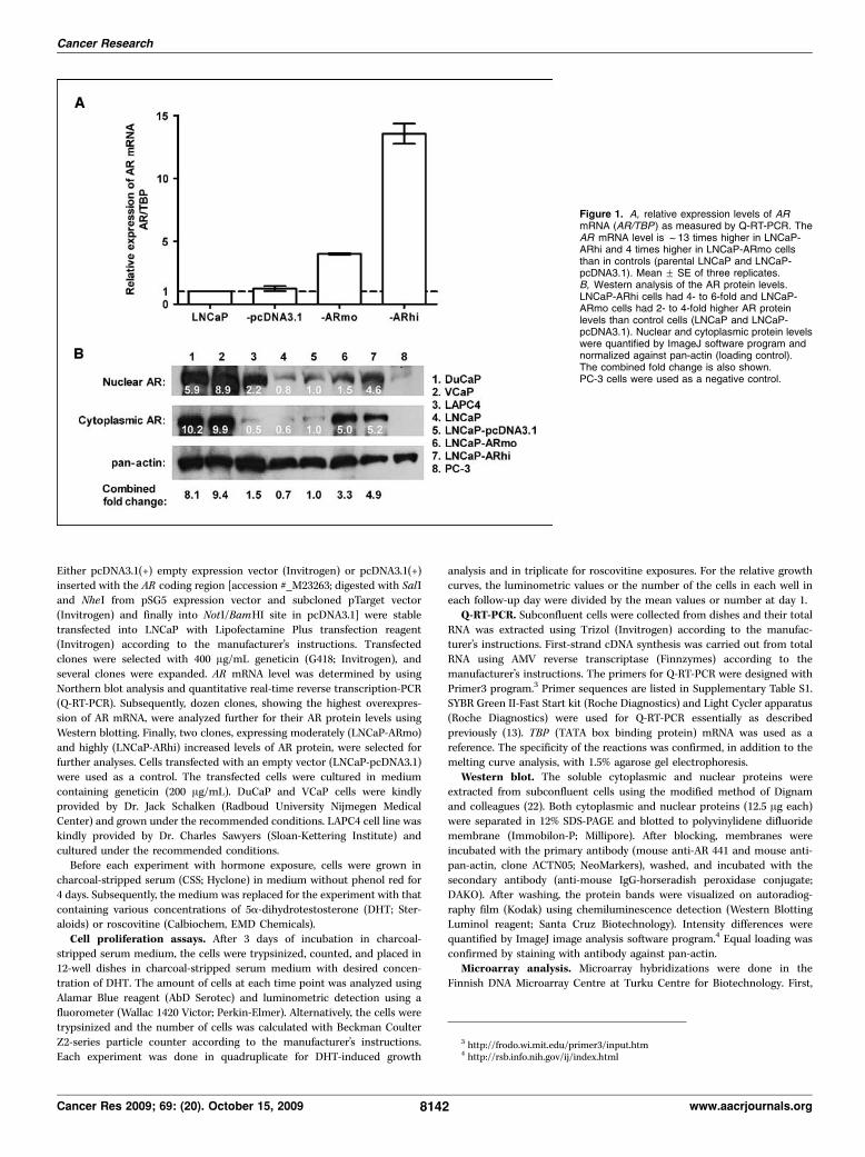
Either pcDNA3.1(+) empty expression vector (Invitrogen) or pcDNA3.1(+)inserted with the AR coding region [accession #_M23263; digested with SalI
and NheI from pSG5 expression vector and subcloned pTarget vector
(Invitrogen) and finally into NotI/BamHI site in pcDNA3.1] were stable
transfected into LNCaP with Lipofectamine Plus transfection reagent(Invitrogen) according to the manufacturer’s instructions. Transfected
clones were selected with 400 Ag/mL geneticin (G418; Invitrogen), and
several clones were expanded. AR mRNA level was determined by using
Northern blot analysis and quantitative real-time reverse transcription-PCR(Q-RT-PCR). Subsequently, dozen clones, showing the highest overexpres-
sion of AR mRNA, were analyzed further for their AR protein levels using
Western blotting. Finally, two clones, expressing moderately (LNCaP-ARmo)
and highly (LNCaP-ARhi) increased levels of AR protein, were selected forfurther analyses. Cells transfected with an empty vector (LNCaP-pcDNA3.1)
were used as a control. The transfected cells were cultured in medium
containing geneticin (200 Ag/mL). DuCaP and VCaP cells were kindlyprovided by Dr. Jack Schalken (Radboud University Nijmegen Medical
Center) and grown under the recommended conditions. LAPC4 cell line was
kindly provided by Dr. Charles Sawyers (Sloan-Kettering Institute) and
cultured under the recommended conditions.Before each experiment with hormone exposure, cells were grown in
charcoal-stripped serum (CSS; Hyclone) in medium without phenol red for
4 days. Subsequently, the medium was replaced for the experiment with that
containing various concentrations of 5a-dihydrotestosterone (DHT; Ster-aloids) or roscovitine (Calbiochem, EMD Chemicals).
Cell proliferation assays. After 3 days of incubation in charcoal-
stripped serum medium, the cells were trypsinized, counted, and placed in12-well dishes in charcoal-stripped serum medium with desired concen-
tration of DHT. The amount of cells at each time point was analyzed using
Alamar Blue reagent (AbD Serotec) and luminometric detection using a
fluorometer (Wallac 1420 Victor; Perkin-Elmer). Alternatively, the cells weretrypsinized and the number of cells was calculated with Beckman Coulter
Z2-series particle counter according to the manufacturer’s instructions.
Each experiment was done in quadruplicate for DHT-induced growth
analysis and in triplicate for roscovitine exposures. For the relative growthcurves, the luminometric values or the number of the cells in each well in
each follow-up day were divided by the mean values or number at day 1.
Q-RT-PCR. Subconfluent cells were collected from dishes and their total
RNA was extracted using Trizol (Invitrogen) according to the manufac-turer’s instructions. First-strand cDNA synthesis was carried out from total
RNA using AMV reverse transcriptase (Finnzymes) according to the
manufacturer’s instructions. The primers for Q-RT-PCR were designed with
Primer3 program.3 Primer sequences are listed in Supplementary Table S1.SYBR Green II-Fast Start kit (Roche Diagnostics) and Light Cycler apparatus
(Roche Diagnostics) were used for Q-RT-PCR essentially as described
previously (13). TBP (TATA box binding protein) mRNA was used as a
reference. The specificity of the reactions was confirmed, in addition to themelting curve analysis, with 1.5% agarose gel electrophoresis.
Western blot. The soluble cytoplasmic and nuclear proteins were
extracted from subconfluent cells using the modified method of Dignamand colleagues (22). Both cytoplasmic and nuclear proteins (12.5 Ag each)
were separated in 12% SDS-PAGE and blotted to polyvinylidene difluoride
membrane (Immobilon-P; Millipore). After blocking, membranes were
incubated with the primary antibody (mouse anti-AR 441 and mouse anti-pan-actin, clone ACTN05; NeoMarkers), washed, and incubated with the
secondary antibody (anti-mouse IgG-horseradish peroxidase conjugate;
DAKO). After washing, the protein bands were visualized on autoradiog-
raphy film (Kodak) using chemiluminescence detection (Western BlottingLuminol reagent; Santa Cruz Biotechnology). Intensity differences were
quantified by ImageJ image analysis software program.4 Equal loading was
confirmed by staining with antibody against pan-actin.Microarray analysis. Microarray hybridizations were done in the
Finnish DNA Microarray Centre at Turku Centre for Biotechnology. First,
Figure 1. A, relative expression levels of ARmRNA (AR/TBP) as measured by Q-RT-PCR. TheAR mRNA level is f13 times higher in LNCaP-ARhi and 4 times higher in LNCaP-ARmo cellsthan in controls (parental LNCaP and LNCaP-pcDNA3.1). Mean F SE of three replicates.B, Western analysis of the AR protein levels.LNCaP-ARhi cells had 4- to 6-fold and LNCaP-ARmo cells had 2- to 4-fold higher AR proteinlevels than control cells (LNCaP and LNCaP-pcDNA3.1). Nuclear and cytoplasmic protein levelswere quantified by ImageJ software program andnormalized against pan-actin (loading control).The combined fold change is also shown.PC-3 cells were used as a negative control.
3 http://frodo.wi.mit.edu/primer3/input.htm4 http://rsb.info.nih.gov/ij/index.html
Cancer Research
Cancer Res 2009; 69: (20). October 15, 2009 8142 www.aacrjournals.org

300 ng total RNA of each sample was transcribed in vitro , biotinylated andamplified with Illumina RNA TotalPrep Amplification kit (Ambion), and
hybridized to Illumina Sentrix HumanRef-8_V2 Expression BeadChip accord-
ing to themanufacturer’s instructions. The probes of Illumina HumanRef-8 v2
chip are based on the content from the National Center for BiotechnologyInformation RefSeq database 1, release 17 containing >22,000 well-annotated
transcripts. The data were analyzed with GeneSpring Analysis Platform
version GX 7.3.1 (Agilent Technologies). First, lowest signal value was set to be
3-fold higher compared with negative control (water) signals. All individualvalues below that were set to this lowest signal value.
For unsupervised hierarchical clustering, samples were normalized per
chip by the 50th percentile and per gene by the median. Average linkage
method was used for clustering, and the similarities were estimated withPearson’s correlation. For the analyses of DHT dose responses, data were
normalized with repeated median polishing per chip and gene. Subse-
quently, signal values in each treatment were divided by the signal value ofthe 0 nmol/L DHT treatment of the same cell line at the same time point.
To identify differently expressed genes in the faster-growing LNCaP-ARhi
cells at 1 nmol/L DHT, the data were normalized with intensity dependent
Lowess normalization. Twenty percent of the data were used to calculatethe Lowess fit at each point. This curve was used to adjust the control value
(LNCaP-pcDNA3.1 or LNCaP-ARmo were used as control samples for
LNCaP-ARhi) for each measurement. For ontology classifications, all gene
ontology lists containing at least 10 genes with P < 0.001 (hypergeometricP value without multiple testing correction) in either LNCaP-pcDNA3.1, or
LNCaP-ARmo, or LNCaP-ARhi were filtered and organized with GeneSpringOntology browser.
The array data were submitted using MIAMExpress to the ArrayExpress
database (accession number E-MEXP-2286).
ChIP-on-chip assays. ChIP-on-chip assays were done with anti-AR anti-body (BJ14-AR3; ref. 23) in nontransfected LNCaP-1F5 cells that were cul-
tured in the absence of hormone for 4 days and then exposed to 100 nmol/L
DHT or vehicle for 2 h. The immunoprecipitation enriched and input
chromatin samples were amplified by ligation-mediated PCR followed byfragmentation and labeling of DNA. Three micrograms of samples from
input and immunoprecipitated samples were hybridized to Affymetrix
whole-genome tiling arrays (GeneChip Human Tiling 2.OR Array Set;
Affymetrix). The regions enriched for AR-binding sites were identified byMAT algorithm (24) and mapped to the most recent human genome
sequence (Hg18), and sites that were enriched above input were scored at a
P value of 10�4. These AR-binding sites were then subsequently mapped tothe differentially expressed genes in the AR overexpression model.
Results
Establishment of AR-overexpressing LNCaP. Two AR-over-expressing clones were selected for experiments. These wereLNCaP-ARmo, with f4-fold higher AR mRNA and f2- to 4-foldhigher AR protein level, and LNCaP-ARhi, with 13-fold higher AR
Figure 2. Relative growth of the LNCaP-ARmodel cells in the presence of (A ) 0 nmol/L DHT,(B) 0.1 nmol/L DHT, (C ) 1 nmol/L DHT, and(D ) 10 nmol/L DHT. The greatest advantage inproliferation rate for AR-overexpressing cells wasseen in 1.0 nmol/L DHT. E, growth of cells in charcoal-stripped serum medium without androgens during3 wk. The highest growth rate was seen inLNCaP-ARhi cells. The growth of LNCaP-ARmo wasbetween LNCaP-ARhi and control cells. Mean F SE offour replicates. Y axis, relative growth against day 1.*, P < 0.05; **, P < 0.01; ***, P < 0.001, below thecurves for LNCaP-ARmo versus LNCaP-pcDNA3.1and above the curves for LNCaP-ARhiversus LNCaP-pcDNA3.1.
Mechanisms of Prostate Cancer Progression
www.aacrjournals.org 8143 Cancer Res 2009; 69: (20). October 15, 2009

mRNA and 4- to 6-fold higher AR protein level, than those in thecontrol LNCaP-pcDNA3.1 cells (Fig. 1). Immunofluorescencestaining showed that the AR protein is located in nucleus whencells are grown in the presence of androgens (SupplementaryFig. S1). In addition, overexpression of AR seemed to enhancethe nuclear transport after DHT exposure. The most intensenuclear staining was seen in the LNCaP-ARhi cells after 1 h exposureto 10 nmol/L DHT.Growth curve analyses. The proliferation rates of these cells
were analyzed in the presence of various DHT concentrations. Thegrowth of LNCaP-ARhi cells was stimulated with lower concen-trations of DHT than control LNCaP-pcDNA3.1 or LNCaP-ARmocells (Fig. 2). Especially, in the presence of 1 nmol/L DHT, LNCaP-ARhi cells grew clearly faster than LNCaP-ARmo or LNCaP-pcDNA3.1 cells (P < 0.01). The cells overexpressing AR were alsocapable of growing better in charcoal-stripped serum mediumwithout DHT (Fig. 2E). However, the growth finally plateaued in allcell lines.Expression profiling. For genome-wide expression profiling,
LNCaP-ARhi, LNCaP-ARmo, and LNCaP-pcDNA3.1 cells weregrown in the presence of various DHT concentrations (0, 1, 10,and 100 nmol/L), and total RNA was extracted at 4 and 24 h time
points. In addition, VCaP cells that contain endogenous AR geneamplification and strong overexpression of AR (ref. 24; Fig. 1) weretreated and analyzed in a similar fashion. Unsupervised hierarchi-cal clustering of androgen-regulated transcripts revealed that VCaPand LNCaP-ARhi cells clustered together, whereas androgen-dependent transcripts of LNCaP-ARmo cells clustered togetherwith those of LNCaP-pcDNA3.1 cells (Fig. 3).
More genes were upregulated than downregulated in the LNCaP-AR cells by DHT. Venn diagrams for upregulated and down-regulated genes are shown in Fig. 4. In LNCaP-pcDNA3.1 cells,expression of 52 and 118 genes was changed >2-fold at any DHTconcentration compared with vehicle at 4 and 24 h, respectively. InLNCaP-ARmo and LNCaP-ARhi, the number of genes with alteredexpression was higher; in ARmo 164 and 379 at 4 and 24 h timepoints, respectively, and in ARhi 240 and 475 at 4 and 24 h timepoints, respectively (P < 0.0001, m2 test). In VCaP cells, expressionof 430 genes was changed >2-fold at the 4 h time point and 428genes at the 24 h time point. The upregulated and downregulatedgenes by DHT exposure in LNCaP-pcDNA3.1, LNCaP-ARmo,LNCaP-ARhi, and VCaP are listed in Supplementary Tables S2 to S5.
Next, we analyzed gene ontology categories for all genesupregulated and downregulated at 24 h of DHT treatments. In
Figure 3. Unsupervised hierarchical clustering of mRNA expression in DHT-treated LNCaP-AR and VCaP cells. LNCaP-ARhi cells cluster together with VCaP cells(except 0 mol/L DHT treated), and LNCaP-pcDNA3.1 cells together with LNCaP-ARmo cells.
Cancer Research
Cancer Res 2009; 69: (20). October 15, 2009 8144 www.aacrjournals.org

LNCaP-pcDNA3.1 cells, DHT upregulated transcripts belonged tothe following five gene ontology categories (at least 10 upregulatedgenes with P < 0.001): intracellular signaling cascade, cell cycle, celldivision, protein metabolism, and DNA metabolism (Supplemen-tary Table S6). In LNCaP-ARhi and LNCaP-ARmo cells, the samefive gene ontology categories were also upregulated as those inLNCaP-pcDNA3.1 cells, but the number of upregulated genes wassignificantly higher in each category. In addition, six other mainontologies were enriched in LNCaP-ARmo and/or LNCaP-ARhicells. These categories were lipid metabolism, secretory pathway,cell organization and biogenesis, chromosome segregation, re-sponse to endogenous stimulus, and cell proliferation (Supplemen-tary Table S6). Ontologies that showed highly significantly (P <0.0001, m2 test) more upregulated genes in LNCaP-ARhi cellscompared with LNCaP-pcDNA3.1 or LNCaP-ARmo cells were genesassociated with mitotic cell cycle, regulation of progression throughcell cycle, organelle organization and biogenesis, cellular proteinmetabolism, and DNA metabolism (Supplementary Table S6). Ofupregulated ontologies, intracellular signaling cascade, cell cycle,and lipid metabolism were also upregulated significantly (P < 0.01)already at 4 h time point. Only one ontology category, apoptosis, wassignificantly upregulated at 4 h time point but not at 24 h time point.The downregulated genes were not enriched in any of the categorieswith more than two genes in any of the cell lines.
When upregulated ontologies were examined separately at var-ious DHT concentrations (Fig. 5), particularly DNA metabolism, cellcycle, cell organization and biogenesis, cell division, and intracel-
lular signaling cascade were found to be highly significantlyupregulated in LNCaP-ARhi cells already in 1 nmol/L DHTconcentration (Fig. 5A). At 10 and 100 nmol/L DHT concentrations,the same ontologies were upregulated but with higher number ofgenes. At 10 and 100 nmol/L DHT concentrations, cell cycle, DNAmetabolism, lipid metabolism, cell organization and biogenesis, celldivision, and chromosome segregation were highly significantlyupregulated also in LNCaP-pcDNA3.1 or LNCaP-ARmo cells. Thenumber of upregulated genes in these categories were, however,always highest in LNCaP-ARhi cells. The most significantlyupregulated ontologies in LNCaP-ARmo cells were lipid metabo-lism and secretory pathway.Expression of known AR target genes. Expression of well-
known AR target genes, such as PSA, TMPRSS2, NKX3-1 , andTMEPAI , was found to be increased by 4- to 10-fold in LNCaP-ARhiandLNCaP-ARmocomparedwithLNCaP-pcDNA3.1 at 4 and24 h timepoints (Fig. 6). Likewise, expression of well-known downregulatedAR target genes, such as PAP and PSMA , was attenuated more inLNCaP-ARhi and LNCaP-ARmo cells than in control cells. Onaverage, for an equal level of upregulation of genes, a 10-fold higherDHTconcentration was required for LNCaP-pcDNA3.1 cells than forLNCaP-ARhi or LNCaP-ARmo cells. Q-RT-PCR of PSA and sixselected genes, unknown previously to be regulated by androgens,confirmed the microarray data (Supplementary Fig. S2).
LNCaP and VCaP cells contain genetic rearrangements affectingEts transcription factors, ETV1 and ERG , respectively (25, 26). InVCaP cells, ERG expression was upregulated by androgens
Figure 4. Venn diagrams of DHT responding genes in LNCaP-AR cell panel for upregulated genes at (A) 4 h and (B ) 2 h. Ninety and 180 genes were found to beupregulated (>2-fold) only in LNCaP-ARhi at 4 and 24 h time points, respectively. Venn diagrams of genes downregulated by DHT in LNCaP-AR panel at (C ) 4 hand (D ) 24 h. Forty-four and 65 genes were found to be downregulated (>2-fold) only in LNCaP-ARhi at 4 and 24 h time points, respectively.
Mechanisms of Prostate Cancer Progression
www.aacrjournals.org 8145 Cancer Res 2009; 69: (20). October 15, 2009

(Supplementary Fig. S3). Unfortunately, Illumina RefSeq8-v2 probesfor ETV1 failed to detect any expression. However, Q-RT-PCRanalysis showed >2-fold increase of ETV1 mRNA (SupplementaryFig. S3). Of Ets family members, only ELK4 mRNA level was in-creased with androgens in LNCaP-ARhi cells at 24 h time point(Supplementary Fig. S3). The ELK4 gene is known to be an ARtarget in human prostate cancer cells with association to cell growthin vitro (27).
Kokontis and colleagues (21) have shown previously that LNCaPcells overexpressing endogenous AR show higher expression ofMYC in low levels of androgens, and the expression is decreased inlower concentrations of the androgens than in the parental cellline. In similar fashion, here the highest MYC expression was foundin VCaP and LNCaP-ARhi cells in the low DHT levels, and the
expression was decreased in higher concentrations of DHT(Supplementary Fig. S4).Identification of candidate AR downstream genes. Because
the LNCaP-ARhi cells gained a growth benefit in vitro with 1 nmol/LDHT concentration compared with LNCaP-ARmo cells or emptyvector–transfected LNCaP-pcDNA3.1 cells, we were particularlyinterested in the genes that are upregulated or downregulated inLNCaP-ARhi cells at 1 nmol/L DHT. Expression of 173 genes werefound to be altered >2-fold (127 upregulated and 46 downregulated)in LNCaP-ARhi cells and compared with LNCaP-pcDNA3.1or LNCaP-ARmo cell at 24 h after DHT exposure (SupplementaryTable S7). Of these genes, we examined whether any of theupregulated genes were expressed in VCaP cells to the sameor higher level than in LNCaP-ARhi cells. Ninety-nine such genes
Figure 5. Androgen upregulated (>2-fold)gene ontology classes at different DHTconcentrations. The histograms showthe number of upregulated genes inLNCaP-pcDNA3.1, LNCaP-ARmo,and LNCaP-ARhi at 1 nmol/L(A), 10 nmol/L (B ), and 100 nmol/L(C ) DHT concentrations atthe 24 h time point. Gray columns, highlysignificantly (P < 0.001, a hypergeometricP value without multiple testingcorrection) upregulated ontologies.Different ontology classes are numberedas follows: 1, GO:19538 proteinmetabolism; 2, GO:6259 DNAmetabolism; 3, GO:6629 lipidmetabolism; 4, GO:7049 cell cycle;5, GO:16043 cell organization andbiogenesis; 6, GO:51301 cell division;7, GO:8283 cell proliferation;8, GO:7242 intracellular signalingcascade; 9, GO:9719 response toendogenous stimulus; 10, GO:7059chromosome segregation;11, GO:45045 secretory pathway.
Cancer Research
Cancer Res 2009; 69: (20). October 15, 2009 8146 www.aacrjournals.org
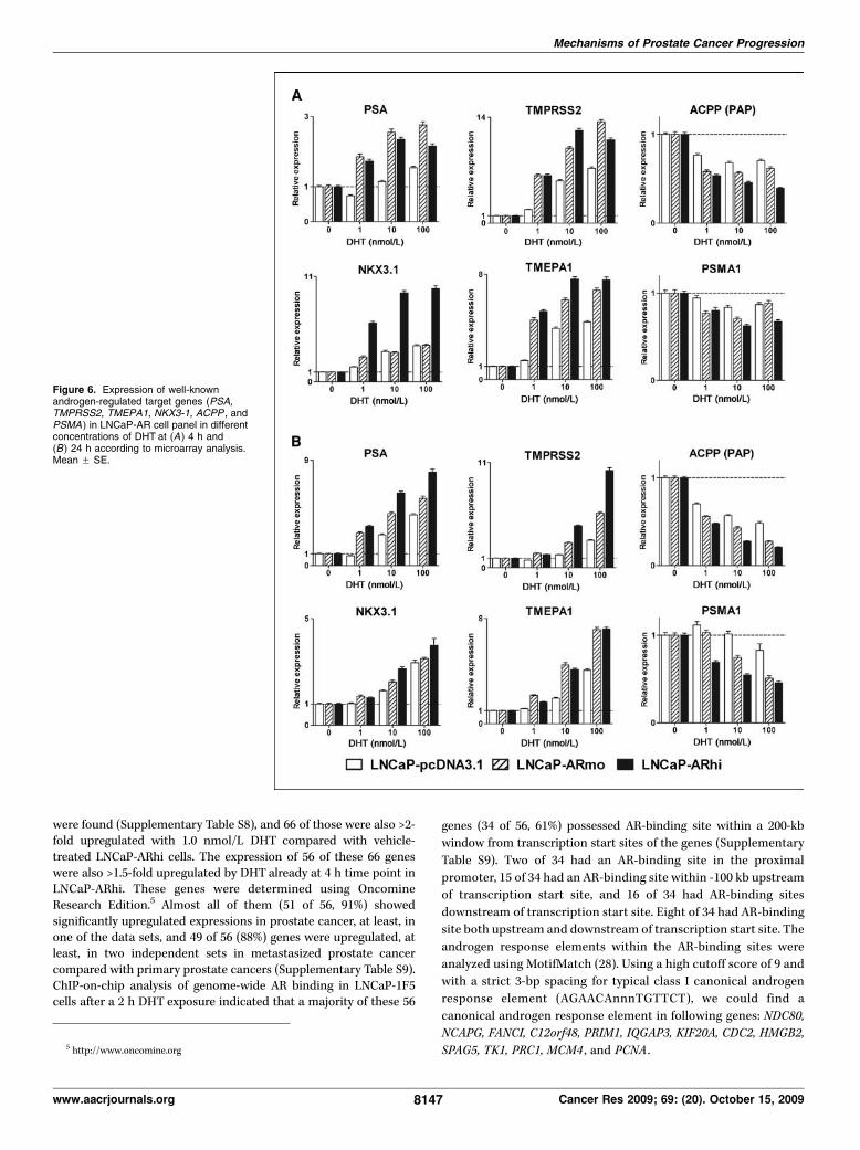
were found (Supplementary Table S8), and 66 of those were also >2-fold upregulated with 1.0 nmol/L DHT compared with vehicle-treated LNCaP-ARhi cells. The expression of 56 of these 66 geneswere also >1.5-fold upregulated by DHT already at 4 h time point inLNCaP-ARhi. These genes were determined using OncomineResearch Edition.5 Almost all of them (51 of 56, 91%) showedsignificantly upregulated expressions in prostate cancer, at least, inone of the data sets, and 49 of 56 (88%) genes were upregulated, atleast, in two independent sets in metastasized prostate cancercompared with primary prostate cancers (Supplementary Table S9).ChIP-on-chip analysis of genome-wide AR binding in LNCaP-1F5cells after a 2 h DHT exposure indicated that a majority of these 56
genes (34 of 56, 61%) possessed AR-binding site within a 200-kb
window from transcription start sites of the genes (Supplementary
Table S9). Two of 34 had an AR-binding site in the proximal
promoter, 15 of 34 had an AR-binding site within -100 kb upstream
of transcription start site, and 16 of 34 had AR-binding sites
downstream of transcription start site. Eight of 34 had AR-binding
site both upstream and downstream of transcription start site. The
androgen response elements within the AR-binding sites were
analyzed using MotifMatch (28). Using a high cutoff score of 9 and
with a strict 3-bp spacing for typical class I canonical androgen
response element (AGAACAnnnTGTTCT), we could find a
canonical androgen response element in following genes: NDC80,
NCAPG, FANCI, C12orf48, PRIM1, IQGAP3, KIF20A, CDC2, HMGB2,
SPAG5, TK1, PRC1, MCM4 , and PCNA .
Figure 6. Expression of well-knownandrogen-regulated target genes (PSA,TMPRSS2, TMEPA1, NKX3-1, ACPP , andPSMA ) in LNCaP-AR cell panel in differentconcentrations of DHT at (A ) 4 h and(B) 24 h according to microarray analysis.Mean F SE.
5 http://www.oncomine.org
Mechanisms of Prostate Cancer Progression
www.aacrjournals.org 8147 Cancer Res 2009; 69: (20). October 15, 2009

Effect of roscovitine on growth of LNCaP-AR cells. Becausethe expression of both CDC2 (alias CDK1) and CDK2 were in-creased in 1 nmol/L DHT, especially in LNCaP-ARhi, and the geneshave AR-binding site according to ChIP-on-chip data, the effect ofCDK1/2 inhibition was tested. The growth of LNCaP sublines wasassayed in different concentrations of CDK1/2 inhibitor roscovitineand DHT. The growth of LNCaP-pcDNA3.1, LNCaP-ARmo, andLNCaP-ARhi cells was significantly (P < 0.001, unpaired two-tailedt test) inhibited with 15 Amol/L roscovitine in 10 nmol/L DHT(Supplementary Fig. S5). The LNCaP-ARhi cells showed the growthinhibition also by 7.5 Amol/L roscovitine (P = 0.0002). In addition, thegrowth of LNCaP-ARhi (P = 0.0253), but not LNCaP-pcDNA3.1 orLNCaP-ARmo, was suppressed by roscovitine in 1 nmol/L DHT.
Discussion
AR is a key protein in both development and progression ofprostate cancer. It is expressed in almost all prostate carcinomasfrom the beginning of the disease to the castration-resistant stage(8, 13, 14). The standard treatment of advanced prostate cancer ishormone ablation. Although androgen withdrawal attenuates AR-mediated signaling and initially prevents tumor growth, severalchanges occur in AR action during the treatment that lead toreactivation of AR signaling and eventually to emergence of thelethal form of the disease, the CRPC (12). One of the key mech-anisms in the emergence of CRPC is amplification of the AR geneleading to overexpression of AR protein (13, 15). This suggests thatCRPC cells are not androgen-independent but may be hypersensitiveto low androgen level. To study the functional consequences of ARoverexpression and the role of the AR level inmore detail, we generatedtwo LNCaP cell lines that overexpressed AR to different levels.
AR overexpression seemed to increase the ability of prostatecancer cells to grow and proliferate in the absence of or at a lowconcentration of DHT. The level of AR affected the growth indifferent androgen concentrations. The LNCaP-ARhi cells with thehighest level of AR expression had the fastest growth rate, whereasthe growth of LNCaP-ARmo cells was between that of the LNCaP-ARhi and the control cells (LNCaP-pcDNA3.1). Both LNCaP-ARhiand LNCaP-ARmo were also able to grow longer in the mediumwithout androgens. Instead, control cell proliferation ceased afterthe first week. The data indicate that increased expression of ARsensitized the growth of the cells to low hormone concentrations.Even a modest increase in AR expression level can help tumor cellsto proliferate at a low androgen concentration as has also beensuggested previously by Chen and colleagues (20). Kokontis andcolleagues (21) have shown previously that androgens havebiphasic effect on the growth of LNCaP cells. Androgens stimulatethe growth, but in higher concentrations the induction ofproliferation is diminished. In addition, they showed that, in theLNCaP cells overexpressing endogenous AR on adaptation togrowth in low levels of androgens, the repression of proliferationtakes place in lower androgen levels than in parental cell lines. Here,we found similar AR level-dependent biphasic effect of androgens(Supplementary Fig. S6). Thus, the optimal level of growth in-duction is dependent on the level of both ligand and the receptor.
Several microarray studies have addressed androgen regulationof gene expression in LNCaP cells (20, 29, 30). However, the data inthese studies are only partially concordant. Reasons for thediscrepant findings could be different time points used, differentligands, different ligand concentrations, and heterogeneity of theLNCaP cells themselves in different laboratories. For example, in
our data, several genes, such as TNFRSF10B, APRIN, TNFAIP3 , andSGK , which were upregulated strongly at the 4 h time point,showed very little, if any, upregulation anymore at 24 h. Animportant aspect is also that AR in the parental LNCaP cells ismutated allowing other steroids and even antiandrogens, such asflutamide, function as agonist (18). To alleviate these problems, wetransfected wild-type AR cDNA into LNCaP cells and used thenatural ligand at two different time points with four differentconcentrations. In our experimental model, we cannot fully separatethe effect of wild-type and mutated AR. However, at least in LNCaP-ARmo and LNCaP-ARhi cells, the majority of AR is wild-type. Thus,the differences between these two sublines can be assumed to be dueto the different levels of the expression of wild-type AR.
In our models, the number of androgen-responsive genes wasclearly associated with AR expression level. LNCaP-ARhi cells pos-sessed more androgen-responsive genes than LNCaP-ARmo cellsthat, in turn, had more those genes than LNCaP-pcDNA3.1 cells. Weanalyzed also VCaP cells, which contain high-level amplification ofthe AR gene leading to strong (up to 12-fold) overexpression of ARprotein (ref. 24; Fig. 1). Its growth is androgen-sensitive (31).Interestingly, the number of the androgen-responsive genes in VCaPcells was even higher than in LNCaP-ARhi. In unsupervisedhierarchical clustering, VCaP and LNCaP-ARhi cells clustered togetherdespite that they are different cell lines with different geneticbackgrounds. This indicates a very strong influence of AR to thegenome-wide expression of prostate cancer cells. Of the well-knownandrogen-regulated genes, such as PSA and TMPRSS2 , induction ofgene expression took place at a 10-fold lower concentrations ofDHTinLNCaP-ARhi and LNCaP-ARmo cells than in LNCaP-pcDNA3.1 cells.Thus, it seems that the level of AR sensitizes the cells to androgens notonly in terms of growth but also by increasing number of genesresponding to DHT.
Because the growth advantage in LNCaP-ARhi cells wasespecially prominent at 1 nmol/L DHT, we were interested ingenes whose expression was altered at that concentration. BecauseVCaP cells are also androgen-sensitive and contain AR gene amplifi-cation, we postulated that androgen target genes that are importantfor progression of CRPC should be detected in both LNCaP-ARhi andVCaP cells. All in all, 56 genes were induced in 1 nmol/L DHT inLNCaP-ARhi (but not in LNCaP-ARmo or LNCaP-pcDNA3.1) alreadyat 4 h and were also highly expressed in VCaP. According to Onco-mine data resource, 51 of 56 (91%) of these genes have been shown toexhibit significant upregulation in primary prostate cancer and/ormetastatic prostate cancer samples (Supplementary Table S9).Unfortunately, the Oncomine data from studies comparing directlyhormone-naive and castration-resistant cancers were not available forall of those genes. The list of genes consists of cell cycle genes, forexample, CDK1, CDK2, cyclin B, cyclin E , and aurora kinase A and B . Allof them are known to have an effect on cell proliferation orchromosome condensation and be upregulated in prostate cancer.ChIP-on-chip assay revealed that themajority (61%) of these genes haveAR-binding sites within a 200-kb window from transcription start sites.Our experiments with roscovitine, a CDK1/2 inhibitor, showed thatLNCaP-ARhi cells were more sensitive to the inhibition than theLNCaP-pcDNA3.1 and LNCaP-ARmo, indicating the importance ofthese androgen-regulated genes for the growth benefit of the AR-overexpressing cells. Because it has been shown previously thatCDK1 phosphorylates and stabilizes AR (32), there may be a positivefeedback mechanism between expression of AR and CDK1.
Previous studies have shown that increased AR expression isassociated with the growth of castration-resistant cancers and with
Cancer Research
Cancer Res 2009; 69: (20). October 15, 2009 8148 www.aacrjournals.org

transformation of androgen-dependent prostate cancer cells toandrogen-independent ones (10, 20, 21). In addition, association ofAR expression level with cell invasion has also been suggested (11).These studies have been performed with different cell lines orxenograft models. In our LNCaP-AR model cells, high AR level wassignificantly associated with increased proliferation at a low andro-gen concentration and with an increased number of genes beingassociated to cell cycle and DNA replication. The finding that onlyLNCaP-ARhi cells showed the growth advance, and high number ofcell proliferation associated genes that were androgen-responsive, isin good agreement with observations that clinical CRPC often have>10-fold overexpression of AR (13, 14).
The results also indicated that AR can have an effect on differentcellular processes depending on the receptor level. In LNCaP-ARmocells, the number of responding genes associated with cell cyclewas not increased; instead, genes in other ontology categories, suchas lipid, sterol, and cholesterol biosynthesis, were highly signifi-cantly enriched. For example, the androgen-regulated lipid meta-bolism genes reviewed by Chen and colleagues (33) were allandrogen-regulated, at least, in one of the cell line used in our data.Of those lipid metabolism genes, especially DHCR24, FASN,HMGCS1, LDLR, PPAP2A , and SCAP were strongly androgen-upregulated in LNCaP-ARmo. Also, the ontology category of secretorypathway, including endoplasmic reticulum to Golgi transport genes,showed higher number of responsive genes in LNCaP-ARmocompared with LNCaP-ARhi. These observations suggest that theamount of AR may also have other effects rather than simplesensitization of cells to lower levels of androgens.
In conclusion, increased expression of AR seems to sensitizeprostate cancer cells in multiple ways and give them severalbiological benefits during hormone ablation. High AR protein levelhelped the cells to sustain and increase their proliferation in envi-ronment with no androgen or a low androgen concentration. Micro-array analyses of AR-regulated genes gave further evidence for thebiological benefits of AR overexpression. They showed enhancedexpression of several cell cycle–associated genes at 1 nmol/L DHT,especially in LNCaP-ARhi cells. In addition, expression of genesassociated with biosynthesis of lipids and other cellular structureswas elevated in LNCaP-ARhi cells. AR expression level seemed also topredict the activity of AR; the more AR expression, the moreandrogen responsive genes. Further studies are warranted toinvestigate whether the genes upregulated at low androgenconcentrations could functions as drug targets for CRPC treatment.
Disclosure of Potential Conflicts of Interest
No potential conflicts of interest were disclosed.
AcknowledgmentsReceived 3/11/09; revised 7/10/09; accepted 7/30/09; published OnlineFirst 10/6/09.
Grant support: Academy of Finland, Cancer Society of Finland, Reino LahtikariFoundation, Sigrid Juselius Foundation, and Medical Research Fund of TampereUniversity Hospital, Finnish Cultural Foundation, and University of TampereFoundation.
The costs of publication of this article were defrayed in part by the payment of pagecharges. This article must therefore be hereby marked advertisement in accordancewith 18 U.S.C. Section 1734 solely to indicate this fact.
We thank Mariitta Vakkuri, Maarit Ohranen, and Paula Vartiainen for the skillfultechnical assistance.
Mechanisms of Prostate Cancer Progression
www.aacrjournals.org 8149 Cancer Res 2009; 69: (20). October 15, 2009
References1. Curado MP, Edwards B, Shin HR, et al. Cancerincidence in five continents, vol. IX. IARC ScientificPublications No. 160. Lyon: IARC; 2007.
2. Coleman MP, Quaresma M, Berrino F, et al.; CONCORDWorking Group. Cancer survival in five continents: aworldwide population-based study (CONCORD). LancetOncol 2008;9:730–56.
3. Isaacs JT, Isaacs WB. Androgen receptor outwitsprostate cancer drugs. Nat Med 2004;10:26–7.
4. Attard G, Reid AH, Yap TA, et al. Phase I clinical trialof a selective inhibitor of CYP17, abiraterone acetate,confirms that castration-resistant prostate cancercommonly remains hormone driven. J Clin Oncol2008;26:4563–71.
5. Holzbeierlein J, Lal P, LaTulippe E, et al. Geneexpression analysis of human prostate carcinoma duringhormonal therapy identifies androgen-responsive genesand mechanisms of therapy resistance. Am J Pathol 2004;164:217–27.
6. Montgomery RB, Mostaghel EA, Vessella R, et al.Maintenance of intratumoral androgens in metastaticprostate cancer: a mechanism for castration-resistanttumor growth. Cancer Res 2008;68:4447–54.7. Locke JA, Guns ES, Lubik AA, et al. Androgen levelsincrease by intratumoral de novo steroidogenesis duringprogression of castration-resistant prostate cancer.Cancer Res 2008;68:6407–15.
8. Hobisch A, Culig Z, Radmayr C, Bartsch G, Klocker H,Hittmair A. Distant metastases from prostatic carcino-ma express androgen receptor protein. Cancer Res 1995;55:3068–72.
9. Gregory CW, Hamil KG, Kim D, et al. Androgenreceptor expression in androgen-independent prostatecancer is associated with increased expression ofandrogen-regulated genes. Cancer Res 1998;58:5718–24.
10. Hara T, Nakamura K, Araki H, Kusaka M, Yamaoka M.Enhanced androgen receptor signaling correlates withthe androgen-refractory growth in a newly established
MDA PCa 2b-hr human prostate cancer cell subline.Cancer Res 2003;63:5622–8.11. Hara T, Miyazaki H, Lee A, Tran CP, Reiter RE.Androgen receptor and invasion in prostate cancer.Cancer Res 2008;68:1128–35.12. Heinlein CA, Chang C. Androgen receptor in prostatecancer. Endocr Rev 2004;25:276–308.13. Linja MJ, Savinainen KJ, Saramaki OR, Tammela TL,Vessella RL, Visakorpi T. Amplification and overexpres-sion of androgen receptor gene in hormone-refractoryprostate cancer. Cancer Res 2001;61:3550–5.14. Latil A, Bieche I, Vidaud D, et al. Evaluation ofandrogen, estrogen (ERa and ERh), and progesteronereceptor expression in human prostate cancer by real-time quantitative reverse transcription-polymerasechain reaction assays. Cancer Res 2001;61:1919–26.15. Visakorpi T, Hyytinen E, Koivisto P, et al. In vivoamplification of the androgen receptor gene and progres-sion of human prostate cancer. Nat Genet 1995;9:401–6.16. Marcelli M, Ittmann M, Mariani S, et al. Androgenreceptor mutations in prostate cancer. Cancer Res 2000;60:944–9.17. Taplin ME, Rajeshkumar B, Halabi S, et al. Androgenreceptor mutations in androgen-independent prostatecancer: Cancer and Leukemia Group B Study 9663.J Clin Oncol 2003;21:2673–8.18. Veldscholte J, Ris-Stalpers C, Kuiper GG, et al. Amutation in the ligand binding domain of the androgenreceptor of human LNCaP cells affects steroid bindingcharacteristics and response to anti-androgens. Bio-chem Biophys Res Commun 1990;173:534–40.19. Heinlein CA, Chang C. Androgen receptor (AR)coregulators: an overview. Endocr Rev 2002;23:175–200.20. Chen CD, Welsbie DS, Tran C, et al. Moleculardeterminants of resistance to antiandrogen therapy. NatMed 2004;10:33–9.21. Kokontis J, Takakura K, Hay N, Liao S. Increasedandrogen receptor activity and altered c-myc expressionin prostate cancer cells after long-term androgen de-privation. Cancer Res 1994;54:1566–73.
22. Dignam DJ, Lebovitz RM, Roeder RG. Nucleic AcidRes 1983;11:1475–89.23. Thompson J, Lepikhova T, Teixido-Travesa N,Whitehead MA, Palvimo JJ, Janne OA. Small carboxyl-terminal domain phosphatase 2 attenuates androgen-dependent transcription. EMBO J 2006;25:2757–67.24. Johnson WE, Li W, Meyer CA, et al. Model-basedanalysis of tiling-arrays for ChIP-chip. Proc Natl AcadSci U S A 2006;103:12457–62.25. Saramaki OR, Harjula AE, Martikainen PM, VessellaRL, Tammela TL, Visakorpi T. TMPRSS2:ERG fusionidentifies a subgroup of prostate cancers with a favor-able prognosis. Clin Cancer Res 2008;14:3395–400.26. Kumar-Sinha C, Tomlins SA, Chinnaiyan AM. Recur-rent gene fusions in prostate cancer. Nat Rev Cancer2008;8:497–511.27. Makkonen H, Jaaskelainen T, Pitkanen-Arsiola T,et al. Identification of ETS-like transcription factor 4as a novel androgen receptor target in prostate cancercells. Oncogene 2008;27:4865–76.28. Wang Q, Li W, Liu XS, et al. A hierarchical networkof transcription factors governs androgen receptor-dependent prostate cancer growth. Mol Cell 2007;27:380–92.29. Nelson PS, Clegg N, Arnold H, et al. The program ofandrogen-responsive genes in neoplastic prostate epi-thelium. Proc Natl Acad Sci U S A 2002;99:11890–5.30. Hendriksen PJ, Dits NF, Kokame K, et al. Evolution ofthe androgen receptor pathway during progression ofprostate cancer. Cancer Res 2006;66:5012–20.31. Korenchuk S, Lehr JE, MClean L, et al. VCaP, a cell-based model system of human prostate cancer. In Vivo2000;15:163–8.
32. Chen S, Xu Y, Yuan X, Bubley GJ, Balk SP. Androgenreceptor phosphorylation and stabilization in prostatecancer by cyclin-dependent kinase 1. Proc Natl Acad SciU S A 2006;103:15969–74.
33. Swinnen JV, Heemers H, van de Sande T, et al.Androgens, lipogenesis and prostate cancer. J SteroidBiochem Mol Biol 2004;92:273–9.