anendohyphalbacterium chitinophaga,bacteroidetes)alters
TRANSCRIPT
ORIGINAL RESEARCHpublished: 14 March 2017
doi: 10.3389/fmicb.2017.00350
Frontiers in Microbiology | www.frontiersin.org 1 March 2017 | Volume 8 | Article 350
Edited by:
Joerg Graf,
University of Connecticut, USA
Reviewed by:
Alessandra Salvioli,
University of Turin, Italy
Ulisses Nunes Da Rocha,
VU University Amsterdam,
Netherlands
*Correspondence:
A. Elizabeth Arnold
Specialty section:
This article was submitted to
Microbial Symbioses,
a section of the journal
Frontiers in Microbiology
Received: 06 November 2016
Accepted: 20 February 2017
Published: 14 March 2017
Citation:
Shaffer JP, U’Ren JM, Gallery RE,
Baltrus DA and Arnold AE (2017) An
Endohyphal Bacterium (Chitinophaga,
Bacteroidetes) Alters Carbon Source
Use by Fusarium keratoplasticum (F.
solani Species Complex, Nectriaceae).
Front. Microbiol. 8:350.
doi: 10.3389/fmicb.2017.00350
An Endohyphal Bacterium(Chitinophaga, Bacteroidetes) AltersCarbon Source Use by Fusariumkeratoplasticum (F. solani SpeciesComplex, Nectriaceae)Justin P. Shaffer 1, Jana M. U’Ren 1, 2, Rachel E. Gallery 3, 4, David A. Baltrus 1 and
A. Elizabeth Arnold 1, 4*
1 School of Plant Sciences, University of Arizona, Tucson, AZ, USA, 2Department of Agricultural and Biosystems Engineering,
University of Arizona, Tucson, AZ, USA, 3 School of Natural Resources and the Environment, University of Arizona, Tucson,
AZ, USA, 4Department of Ecology and Evolutionary Biology, University of Arizona, Tucson, AZ, USA
Bacterial endosymbionts occur in diverse fungi, including members of many lineages
of Ascomycota that inhabit living plants. These endosymbiotic bacteria (endohyphal
bacteria, EHB) often can be removed from living fungi by antibiotic treatment, providing an
opportunity to assess their effects on functional traits of their fungal hosts. We examined
the effects of an endohyphal bacterium (Chitinophaga sp., Bacteroidetes) on substrate
use by its host, a seed-associated strain of the fungus Fusarium keratoplasticum,
by comparing growth between naturally infected and cured fungal strains across 95
carbon sources with a Biolog® phenotypic microarray. Across the majority of substrates
(62%), the strain harboring the bacterium significantly outperformed the cured strain
as measured by respiration and hyphal density. These substrates included many that
are important for plant- and seed-fungus interactions, such as D-trehalose, myo-
inositol, and sucrose, highlighting the potential influence of EHB on the breadth and
efficiency of substrate use by an important Fusarium species. Cases in which the
cured strain outperformed the strain harboring the bacterium were observed in only
5% of substrates. We propose that additive or synergistic substrate use by the fungus-
bacterium pair enhances fungal growth in this association. More generally, alteration of
the breadth or efficiency of substrate use by dispensable EHB may change fungal niches
in short timeframes, potentially shaping fungal ecology and the outcomes of fungal-host
interactions.
Keywords: endobacteria, fusaria, Gram-negative, phenotypic microarray, substrate use, symbiosis
INTRODUCTION
Plant-fungus interactions shape plant health and productivity in all terrestrial ecosystems(Heilmann-Clausen and Boddy, 2008; Kivlin et al., 2011; Tedersoo et al., 2014; Davison et al., 2015).Pathogens can negatively influence photosynthesis, nutrient- and water uptake and transport,structural integrity, reproduction, and seed germination of their hosts (Blanchette, 1991; Agrios,1997; Gallery et al., 2007; Grimmer et al., 2012; Oliva et al., 2014). In turn, mycorrhizal fungi

Shaffer et al. Endohyphal Bacterium Alters Substrate Use
and some endophytes may enhance nutrient uptake and growth,alter plant water relations, or deter antagonistic microbes orherbivores (Arnold et al., 2003; Waller et al., 2005; Arnold andEngelbrecht, 2007; Busby et al., 2015; Estrada et al., 2015; van derHeijden et al., 2015).
Outcomes of such interactions are influenced by geneticand environmental factors (Schafer and Kotanen, 2003; Galleryet al., 2010; see Agrios, 1997; Jones and Dangl, 2006), but alsocan be shaped by additional microorganisms that alter fungalphenotypes (Frey-Klett et al., 2007; Márquez et al., 2007). Suchmicrobes may occur on the exterior surfaces or interior offungal cells, where they can alter sporulation, substrate use,metabolite production, and other features relevant to fungalinteractions with plants (Partida-Martínez and Hertweck, 2005;Salvioli et al., 2010; Hoffman et al., 2013; Spraker et al., 2016).In particular, many plant-associated fungi harbor endosymbioticbacteria (hereafter, endohyphal bacteria, EHB) that can affecthost function and subsequent plant-fungus interactions (Partida-Martínez and Hertweck, 2005; Hoffman et al., 2013; Salvioliet al., 2016). EHB are known among diverse fungi (Hoffman andArnold, 2010; Desirò et al., 2015; Shaffer et al., 2016), but only afew have been developed as model systems in which their effectshave been observed.
The majority of studies to date focus on EHB in diverse root-associated and soilborne fungi, particularly Mucoromycotina,Mortierellomycotina, and Glomeromycotina (Bianciotto et al.,2003; Bertaux et al., 2005; Partida-Martínez et al., 2007b;Sharma et al., 2008; Sato et al., 2010; Desirò et al., 2015).Many of these EHB influence the phenotype of their fungalhosts. For example, the bacterium Burkholderia rhizoxinicaproduces a virulence factor that enables its host fungusRhizopus microsporus (Mucoromycotina) to cause disease onrice (Partida-Martínez and Hertweck, 2005; Partida-Martínezet al., 2007b). When the bacterium is removed, the fungus is nolonger pathogenic and loses the ability to reproduce asexually(Partida-Martínez and Hertweck, 2005; Partida-Martínezet al., 2007a). The bacterium Candidatus Glomeribactergigasporarum increases responsiveness to strigolactonesexuded by roots, enhancing hyphal elongation and branchingrelevant to mycorrhizal establishment by Gigaspora margarita(Gigasporaceae, Glomeromycotina) (Bianciotto et al., 1996,2003, 2004; Lumini et al., 2007; Anca et al., 2009).
EHB also occur in Basidiomycota, with case studies beginningto highlight the functional aspects of their associations inrhizosphere and phyllosphere fungi (Bertaux et al., 2005; Izumiet al., 2005; Sharma et al., 2008; Ruiz-Herrera et al., 2015).For example, Rhizobium radiobacter (syn. Agrobacteriumtumefaciens), like its host, Piriformospora indica (Sebacinales),can promote growth and induce disease resistance inbarley (Sharma et al., 2008). Similarly, a Bacillus sp. fixesand makes available atmospheric nitrogen within cells ofUstilago maydis (Ustilaginomycotina; Ruiz-Herrera et al.,2015).
EHB recently have been documented in diverse Ascomycota,including members of multiple classes (Pezizomycetes,Eurotiomycetes, Dothideomycetes, and Sordariomycetes) andmultiple functional groups (Barbieri et al., 2000; Hoffman and
Arnold, 2010; Shaffer et al., 2016). They are widespread in foliarendophytes and in soilborne Ascomycota (e.g., those that interactwith seeds; Hoffman and Arnold, 2010; Shaffer et al., 2016). Todate their functional significance has been assessed in detail inonly one study system: the foliar endophyte Pestalotiopsis sp.(Amphisphaeriaceae, Xylariales, Sordariomycetes) and its EHB,Luteibacter sp. (Xanthomonadaceae, Gammaproteobacteria;Hoffman et al., 2013; Arendt, 2015). More generally, studiesof EHB in the Ascomycota and other fungi have focusedprimarily on Proteobacteria, Firmicutes, and Mollicutes(e.g., Desirò et al., 2015; see also Baltrus et al., 2016),leaving gaps with regard to the potential for symbioticmodulation of fungal phenotypes by members of other bacteriallineages.
Here, we use a phenotypic microarray (PM) to explorethe influence of an EHB on its fungal host under laboratoryconditions. Approaches to evaluate phenotypic effects of EHBon host fungi range from predictive analyses based on genomicsand related tools to assays that use infected and curedstrains of the same fungus (Anca et al., 2009; Ghignoneet al., 2012; Hoffman et al., 2013). The advantage of ourapproach is that it allows quantification of respiration andgrowth on 95 substrates simultaneously, providing within-experiment controls and comparisons that are not subjectto variation introduced in a lower-throughput framework(Atanasova et al., 2010; Druzhinina et al., 2010; Blumensteinet al., 2015a,b). PMs have been used in diverse studiesinvolving bacteria (reviewed in Bochner, 2008), and sincetheir development have been extended to work with fungi,including yeasts and filamentous strains (reviewed in Bochner,2003; Druzhinina et al., 2006; Atanasova and Druzhinina, 2010;Pfliegler et al., 2014). To our knowledge, PMs have not beenused previously to explore interactions among microbes or morespecifically, the effects of bacterial endosymbionts on fungalphenotypes.
In this study we focus on a lineage of bacteria that hasnot yet been evaluated for phenotypic modulation of fungi:the Bacteroidetes, a diverse clade of Gram-negative, non-endospore forming bacteria that are known from soils andfrom diverse symbiotic associations (Krieg et al., 2010;Thomas et al., 2011). Specifically we examine the effectsof a strain of Chitinophaga sp. (Bacteroidetes) on substrateuse by its host fungus, a strain of Fusarium keratoplasticum(F. solani species complex, FSSC, sensu Short et al., 2013).The fungal strain was originally isolated from the interiorof a seed of a tropical tree that was retrieved from soil in atropical forest. Subcultures of the strain consistently harborChitinophaga sp., and preliminary analyses indicated thatthis bacterium can be removed by antibiotic treatment.Here we show that this endohyphal Chitinophaga alterssubstrate use and substrate-specific growth by its hostfungus. Our results highlight the importance of EHB withregard to shaping fungal phenotypes relevant to plant-fungusinteractions and demonstrate the capacity of a focal memberof the Bacteroidetes to alter the functional traits of a fungalspecies that includes medically and ecologically importantstrains.
Frontiers in Microbiology | www.frontiersin.org 2 March 2017 | Volume 8 | Article 350
Shaffer et al. Endohyphal Bacterium Alters Substrate Use
MATERIALS AND METHODS
As part of a previous study (Sarmiento et al., 2015; Zalamea et al.,2015), a fungus was isolated from a seed of Cecropia insignis(Urticaceae) that had been buried for 1 month in the soil inthe tropical forest understory at Barro Colorado Island, Panama(9◦ 10′N, 79◦ 51′W; 86 m.a.s.l.; for a site description see Leigh,1999). The seed was retrieved from the soil, washed in water,and surface-sterilized by sequential immersion in 95% ethanol(10 s), diluted chlorine bleach (0.525% NaClO; 2 min), and 70%ethanol (2min) (Arnold and Lutzoni, 2007). The seed was cut inhalf and allowed to surface-dry under sterile conditions. One halfof the seed was placed on 2% malt extract agar (MEA) followingGallery et al. (2007). Fungal isolate PS0362A, containing itsbacterial symbiont, grew from the interior of the seed. PS0362Awas deposited as a living voucher at the Robert L. GilbertsonMycological Herbarium, University of Arizona.
Phylogenetic analyses based on three loci identified PS0362Aas F. keratoplasticum, a member of the Fusarium solani speciescomplex (FSSC) (Nectriaceae, Hypocreales, Sordariomycetes,Ascomycota; Shaffer et al., 2016). Its bacterial symbiont wasobserved during screening and was identified by phylogeneticanalysis of the 16S ribosomal RNA (rRNA) gene as Chitinophagasp. PS-EHB01 (Bacteroidetes) (Shaffer et al., 2016). We haverefined the placement of Chitinophaga sp. PS-EHB01 withinthe Chitinophagaceae by focusing taxon sampling and inferringa new 16S phylogeny, confirming its taxonomic identity (seeSupplementary Materials and Methods).
Preparation of Axenic Fungal CultureFusarium keratoplasticum PS0362A was naturally infected byChitinophaga sp. PS-EHB01. We prepared an axenic strain ofthe fungus by subculturing hyphae under sterile conditionson 2% MEA amended with four antibiotics: ampicillin (100µg/mL), kanamycin (50 µg/mL), tetracycline (10 µg/mL), andciprofloxacin (40 µg/mL; see Hoffman and Arnold, 2010;Hoffman et al., 2013; Arendt et al., 2016). We refer to clones ofthe naturally infected fungus as the EHB+ strain, and those ofthe axenic fungus as the EHB− strain.
We confirmed the presence or absence of Chitinophaga sp. PS-EHB01 in F. keratoplasticum PS0362A through light microscopy,molecular analysis, and fluorescence microscopy. We first ruledout extrahyphal bacteria (i.e., contaminants in the medium ormicrobes on hyphal surfaces; see Supplementary Figure 1) byexamining five samples of hyphae per culture at 400X and 1,000Xon a Leica DM400B compound microscope. We did not observeextrahyphal bacteria in any cultures of the EHB+ or EHB−strains used here.
We extracted total genomic DNA from fresh mycelia ofEHB+ and EHB− strains using the REDExtract-N-Amp PlantKit (Sigma-Aldrich, St. Louis, MO, USA) following a modifiedprotocol (see U’Ren, 2016; U’Ren et al., 2016). We preparedfive separate DNA extractions per strain type. We used thepolymerase chain reaction (PCR) to amplify a ca. 1,400 base pair(bp) segment of the 16S rRNA gene (forward primer 27F, reverseprimer 1492R; 10µM; Lane, 1991) with PCR cycling parametersfollowing Hoffman and Arnold (2010) (3 min at 94◦C, 40 cycles
of 30 s at 94◦C, 30 s at 55◦C, and 2 min at 72◦C, followed by 10min at 72◦C). For each reaction we used 10µL of PCR ReadyMix, 0.8 µL of each primer, 4.4µL of molecular grade water, and4 µL of DNA template. We used molecular grade water insteadof template in negative controls. Negative controls never resultedin visible, positive PCR products. Positive controls consistedof a bacterial strain that was amplified consistently with theseprimers. Positive controls yielded amplification as expected inevery PCR.
Positive PCR products were cleaned by adding 1 µL ExoSAP-IT (Affymetrix, Santa Clara, CA, USA) to each of the remainingproducts. Reactions were incubated on a thermal cycler at 37◦Cfor 60 min, and then at 80◦C for 15min to deactivate enzymes.Cleaned PCR products were diluted 1:1 with molecular gradewater and sequenced bidirectionally on an AB3730XL (AppliedBiosystems, Foster City, CA, USA) using PCR primers (5 µM) atthe University of Arizona Genomics Core.
An assembly pipeline consisting of phred and phrap (Ewingand Green, 1998; Ewing et al., 1998) driven by Chromaseq(Maddison and Maddison, 2005) in Mesquite v.2.75 (Maddisonand Maddison, 2009) was used to call bases and assemblebidirectional reads into contigs. Base calls were verified bymanual inspection of all chromatograms in Sequencher v.5.1(Gene Codes Corp., Ann Arbor, MI, USA).
We consistently found Chitinophaga sp. PS-EHB01 in culturesof the EHB+ strain of F. keratoplasticum PS0362A. No otherbacteria were observed in that strain. Chitinophaga sp. PS-EHB01 was not detected in cultures of the EHB− strain ofF. keratoplasticum PS0362A, and we observed no other bacteriain clones of that strain.
To confirm that Chitinophaga sp. PS-EHB01 was viable andthat it occurred within viable hyphae, we examined living hyphaeby microscopy with the Live/Dead BacLight Bacterial ViabilityKit (Invitrogen, Carlsbad, CA, USA) following Hoffman andArnold (2010). The kit provides a two-color fluorescence assay ofbacterial viability that uses two dyes: SYTO 9, a green-fluorescentnucleic acid stain that labels all cells, and propidium iodide,a red-fluorescent nucleic acid stain that labels only those cellswith damaged or otherwise compromised cell membranes (seemanufacturer’s instructions).
We prepared fungal cultures for Live/Dead visualizationby removing a small piece of mycelium (≤2-mm2) from thegrowing edge of a single colony growing on 2%MEA. Fragmentswere aseptically transferred to glass slides containing 20µL of1:1:18 Live/Dead stain (component A: component B: moleculargrade water), teased apart using sterile insect mounting needles(size 00; BioQuip, Rancho Dominguez, CA, USA), coveredwith a coverslip, and incubated in the dark for 15 min. Afterincubation, we washed the mycelium by pulling molecular gradewater through the slide mounts with bibulous paper and sealedthe slides with two coats of nail polish. We used a LeicaDM400B compound microscope with a 100-W mercury arclamp for fluorescent imaging. Samples were viewed at roomtemperature with a Chroma Technology 35002 filter set (480-nmexcitation/520-nm emission) and 100X APO oil objective.
Cultures of the EHB+ strain of F. keratoplasticum PS0362Aconsistently displayed fluorescence of nucleic acids distinct from
Frontiers in Microbiology | www.frontiersin.org 3 March 2017 | Volume 8 | Article 350
Shaffer et al. Endohyphal Bacterium Alters Substrate Use
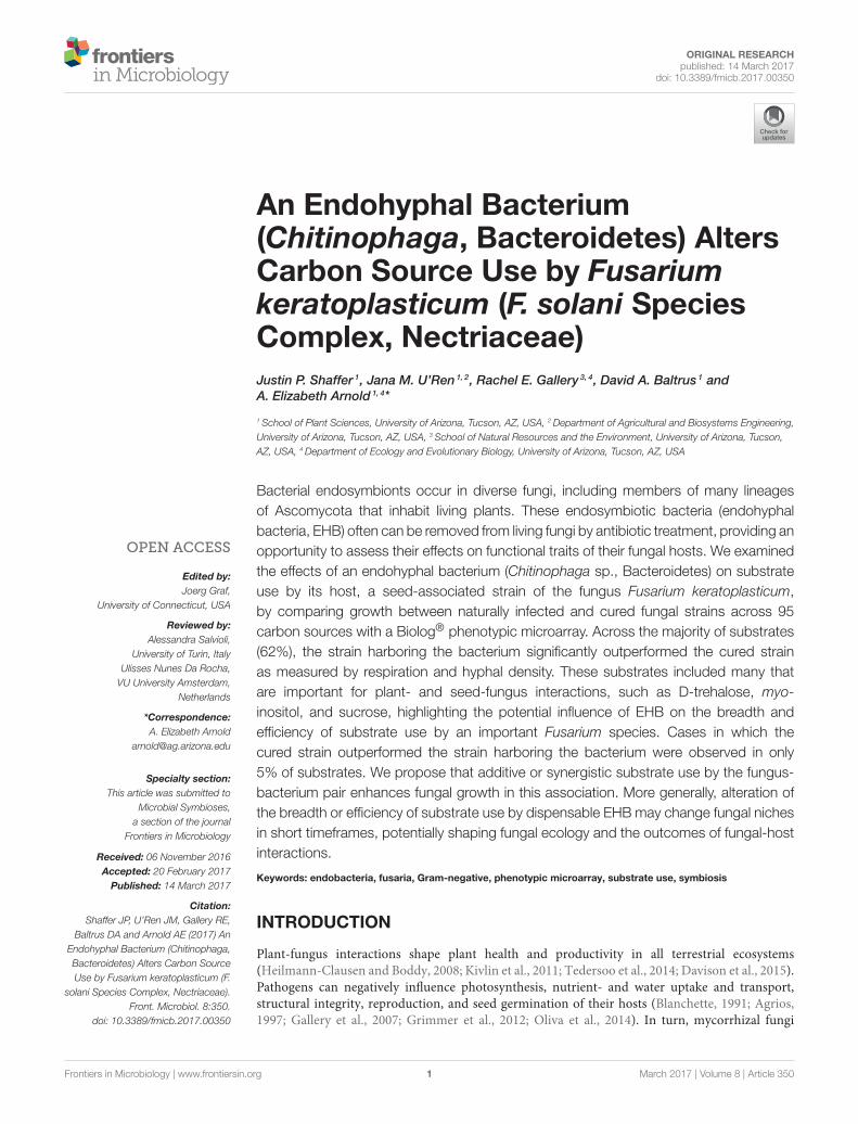
fungal mitochondrial or nuclear DNA (Figure 1). Combinedwith the absence of extrahyphal bacteria and successfulamplification of Chitinophaga sp. PS-EHB01 16S rRNA genesfrom genomic DNA isolated directly from the fungal culture,these results served as evidence of EHB+ status (Hoffman andArnold, 2010; Arendt et al., 2016; Shaffer et al., 2016). Culturesof the EHB− strain did not contain visible fluorescence asabove (Figure 1), and PCR amplification of bacterial 16S rRNAgenes failed in these strains. The EHB+ and EHB− status ofF. keratoplasticum PS0362A strains was confirmed before andafter preliminary assays and Biolog R© assays (below).
Preliminary AssaysWe compared colony diameter and spore production betweenEHB+ and EHB− strains of F. keratoplasticum PS0362A bycomparing five clones of each strain growing on 2% MEA. Foreach clone, we placed a 4-mm plug onto 2% MEA (15 mL) ina 100-mm Petri plate. Each plug was excised from just withinthe growing edge of a fresh culture growing for 5 days on2% MEA in a 100-mm Petri plate. Plates were then incubatedat 25◦C for 10 d. At that point the colony diameter was ca.5mm from the edge of a 100-mm Petri plate, and aerial hyphaewere numerous. To compare colony diameter, we marked thediameter of each clone across two orthogonal axes using a finetip permanent marker, photographed each culture plate with aidfrom a tracing LED lightbox, and obtained the colony diameterfor each clone by taking the average of the two axes as measuredin ImageJ (Schneider et al., 2012). To compare spore production,we obtained spores from each clone by flooding the surface ofthe plate with 5 mL of sterilized milli-q H2O (sH2O), scraped thesurface using a sterile rubber policeperson, and transferred thesuspension to a sterile 50-mL Falcon tube (Corning, NY, USA).For each clone, we quantified the number of spores per mL ofsuspension using a hemocytometer.
Biolog® AssaysWe used commercially available Biolog R© microplates forphenotypic microarray assays. Biolog R© filamentous fungus (FF)microplates (Catalog #1006, Biolog Inc., Hayward, CA, USA) are96-well microtiter plates containing 95 unique carbon sourcesand one negative control (H2O) (Supplementary Table 1).Substrates and reagents are pre-filled and dried into wells.Redox chemistry based on the reduction of iodonitrotetrazoliumviolet (INT) produces a red-colored formazan dye withpeak absorbance at 490 nm (Bochner and Savageau, 1977;Kubicek et al., 2003). This provides a colorimetric measureof mitochondrial activity resulting from substrate use (i.e.,oxidation of succinate to fumarate in the citric acid cycle causesINT to be reduced; Bochner and Savageau, 1977; Bochner, 1989;Bochner et al., 2001; Kubicek et al., 2003). Reduction of INT andproduction of formazan cannot be reversed, and the quantitativemeasure of formazan accumulation by spectrophotometryreflects oxidation of the substrate in a particular well (Bochner,1989; Bochner et al., 2001; Kubicek et al., 2003). In turn, readingthe plates at 750 nm measures turbidity, which reflects growth ofthe fungus through substrate use and production of mycelium(Kubicek et al., 2003; Druzhinina et al., 2006; Atanasova and
Druzhinina, 2010; Blumenstein et al., 2015b). Based on thesemeasurements, the plates can distinguish even closely relatedstrains within fungal species (Singh, 2009; Atanasova andDruzhinina, 2010).
Although FF microplates were designed for use withsporulating fungi, non-sporulating fungi can be evaluatedon the plates following inoculation with homogenous hyphalsuspensions (Singh, 2009). We assessed effects of EHB usinghyphal suspensions for two reasons: first, the fungus producesconidia but we have not yet observed EHB in conidia of PS0362Afollowing staining as described above (Supplementary Figure 2).Second, the fungus appears to colonize seeds as hyphae in naturalconditions (Sarmiento et al., 2015).
We prepared inoculum by incubating mycelium from twoclones of the EHB+ strain and two clones of the EHB− strainof F. keratoplasticum PS0362A on 2% MEA at 25◦C for 10 d.At that point the colony diameter was ca. 5mm from the edgeof a 100-mm Petri plate, and aerial hyphae were numerous,as above. We flooded the surface of each plate with 5 mL ofsH2O, scraped the surface using a sterile rubber policeperson, andcombined suspension from both plates of the same EHB statusby pouring into a sterile 50-mL Falcon tube. We separated andexcluded conidia by filtering the suspensions through three layersof sterile cheesecloth, discarding the filtrate, and transferringtrapped hyphae to new tubes. We brought the total volume ofeach tube up to 20 mL with 0.2% carrageenan type II media(see Hobbie et al., 2003). We then transferred each suspensionto a sterile Waring blender cup and blended for 20 s. Thesuspension was allowed to cool for 20 s and then blended againfor 20 s (see Gale et al., 1960; Orbach et al., 1996). We let eachsuspension rest for 10 min to allow large fragments to fall outof suspension, and diluted each with 0.2% carrageenan type IImedia to obtain an absorbance of 0.22 at 600 nm. We added3 mL of this diluted suspension to 27 mL of 0.2% carrageenantype II media to produce the final hyphal suspension (Hobbieet al., 2003) (Supplementary Figure 3). We allowed suspensionsto sit for 6 h at room temperature, confirming that any remainingconidia had germinated and produced at least one septum distalto the germ tube (Supplementary Figure 3B).
We inoculated 100 µL of suspension into each well of eachmicroplate, pipetting carefully to avoid creating bubbles, andsealed each plate with a double layer of Parafilm (Bemis, Neenah,WI, USA). We prepared five replicate microplates for EHB+ andEHB− strains, wrapped all ten plates in aluminum foil, placedthem into a plastic freezer bag with moistened paper towelsto prevent drying, and incubated them at 25◦C. Preliminaryexamination of hyphal suspension added to a synthetic glucosemedium (20 mM) confirmed viability and growth of the inoculaprepared as above.
We obtained data for substrate use by reading plates at 490nm (absorbance corresponding to cellular respiration) and 750nm (absorbance corresponding to hyphal density) every 12 hfor 7 d. Plates were read using a Synergy H1 hybrid reader andaccompanying Gen5 v.1.11 software package (BioTek, Winooski,VT, USA). We defined absorbance for a given strain on a givensubstrate on a given plate (At
λ) as the value read at a given
wavelength (λ) at a specific time point during the experiment (t),
Frontiers in Microbiology | www.frontiersin.org 4 March 2017 | Volume 8 | Article 350

Shaffer et al. Endohyphal Bacterium Alters Substrate Use
FIGURE 1 | Continued
Frontiers in Microbiology | www.frontiersin.org 5 March 2017 | Volume 8 | Article 350
Shaffer et al. Endohyphal Bacterium Alters Substrate Use
FIGURE 1 | Fusarium keratoplasticum strain PS0362A. (A) Details of culture on 2% MEA: hyphae, conidiophores, and macroconidia. (B) Conidiophores bearing
macroconidia. (C) The EHB+ strain. Fluorescently tagged nucleic acids of viable bacteria appear green, those with damaged membranes in red, and compromised
fungal organelles in orange. (D) Same frame as (C) viewed with differential interference contrast (DIC). In (C,D), orange arrows indicate fungal nuclei, green arrows
indicate viable EHB, and the red arrow indicates inviable EHB. (E) The cured strain (EHB−). Fluorescently tagged nucleic acids of compromised fungal organelles in
orange. (F) Same frame as (E) viewed with DIC. In (E,F), orange arrows indicate fungal nuclei. In all images fungal mycelium was alive at the outset of preparation but
was inactivated during the visualization process. Scale bars = 10 µm.
and total absorbance by a given strain on a given substrate (A tλ)
as the mean absorbance from five replicate plates, measuredat a given wavelength (λ) at a specified time point during theexperiment (t). As previous studies have shown the absorbancespectrum of hyaline mycelium to be level over wavelengths from490 to 750 nm, an adjusted redox value for the production offormazan is obtained by subtracting the absorbance for hyphaldensity (750 nm) from that for cellular respiration (490 nm;Tanzer et al., 2003; Atanasova and Druzhinina, 2010). Wetherefore define the corrected absorbance at 490 nm as Ac490 =
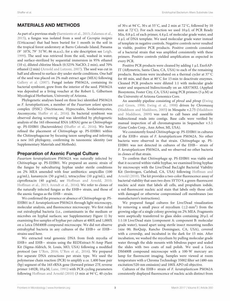
A490 − A750.Absorbance measurements corresponding to respiration
(Ac490) and hyphal density (A750) were highly positivelycorrelated (Kendall’s τ = 0.68, p < 2.2 × 10⁻¹⁶; Figure 2).Furthermore, because similar studies may use indicator dyesother than INT (see Bochner and Savageau, 1977) and redox-based color formation by filamentous fungi does not alwayscorrelate with growth as with bacteria and yeasts (Atanasovaand Druzhinina, 2010), absorbance values corresponding tohyphal density (750 nm) are more consistent across differentgrowth conditions and hyphal morphologies. Absorbance valuescorresponding to hyphal density also aremore often reported andtherefore more comparable among studies (Tanzer et al., 2003;Atanasova and Druzhinina, 2010). Thus, we focus our resultson measurements of absorbance (i.e., turbidity) corresponding tohyphal density (A750), which we refer to as growth (below).
FIGURE 2 | Absorbance measurements corresponding to cellular
respiration (Ac490) and hyphal density (A750) were highly correlated.
Values were log transformed prior to analysis.
Measurable growth was defined as 0.3 < A7750 ≤ 3.0
(see Figure 3), where the upper bound represents 99.9% lightabsorbance and the lower bound the value that best differentiatesgrowth in the lag phase (i.e., negligible growth) from thatreaching the log/exponential phase, across all substrates. We
Frontiers in Microbiology | www.frontiersin.org 6 March 2017 | Volume 8 | Article 350

Shaffer et al. Endohyphal Bacterium Alters Substrate Use
FIGURE 3 | Continued
recognized absorbance values at which strains reach stationaryphase as representing the maximum capacity for growth onthat substrate. We observed a range of growth correspondingto 0.31 ≤ A7
750 ≤ 2.54. Overall we recognized five majoroutcomes: (1) negligible growth by both EHB+ and EHB−strains (A7
750 ≤ 0.3), (2) measurable growth by both, but nodifference in growth between EHB+ and EHB− strains, (3)measurable growth by the EHB+ strain but negligible growthby the EHB− strain, (4) measurable growth by both, with theEHB− strain reaching a higher density, and (5) measurablegrowth by both, with the EHB+ strain reaching a higherdensity.
Statistical AnalysesFor preliminary assays, we compared colony diameter andspore production between EHB+ and EHB− strains usingWelch t-tests. For Biolog R© assays, we compared differencesin global substrate use (i.e., growth across all substrates)between EHB+ and EHB− strains of F. keratoplasticum PS0362Ausing hierarchical clustering and permutational multivariateanalysis of variance (PERMANOVA; Anderson, 2001; Andersonand ter Braak, 2003) based on the Bray-Curtis dissimilarity
metric, as implemented in the R package vegan (R CoreTeam, 2015; Oksanen et al., 2016). For each time point,we first calculated dissimilarities among all replicate platesconsidering growth across all substrates. We then visualizedglobal differences in substrate use among plates by constructingcluster dendrograms, and analyzed differences between platesinoculated with EHB+ and EHB− strains using PERMANOVA(n = 1,000 permutations). We further explored differencesusing non-parametric analysis of similarity (ANOSIM; n =
1,000 permutations; Clarke and Green, 1988; Clarke, 1993)and multi-response permutation procedures (MRPP; n = 1,000permutations; Biondini et al., 1985), but given the normalityof the data we focus here on results from PERMANOVA. Wemade comparisons for each time point in order to determinethe time at which the global effect was largest. We thenused that time point to evaluate differences in A750 betweenEHB+ vs. EHB− strains for focal substrates using Welch t-tests. We controlled for the rate of type I errors inherent inmaking multiple comparisons by using the false discovery rate-controlling method of Benjamini and Hochberg (1995). Theraw data and R scripts for all analyses are available online(Shaffer, 2017).
Frontiers in Microbiology | www.frontiersin.org 7 March 2017 | Volume 8 | Article 350

Shaffer et al. Endohyphal Bacterium Alters Substrate Use
FIGURE 3 | Total absorbance for EHB+ and EHB− strains of Fusarium keratoplasticum PS0362A across all substrates on Biolog® phenotypic
microarrays over 7 d. Light- and dark-orange lines represent absorbance at 490 nm (i.e., cellular respiration). Light- and dark-blue lines represent absorbance at
750 nm (i.e., hyphal density, defined here as growth). Dark, dotted lines with open circles indicate absorbance for EHB+ strains. Light, solid lines with filled circles
indicate absorbance for EHB− strains. Colored squares indicate different substrate classes. The horizontal line at Absorbance = 0.3 indicates the minimum
absorbance value recognized. The vertical line at Day = 7 indicates the time point for which absorbance values were formally compared. (A) Sugar-based substrates.
(B) Amino- and carboxylic acids, their derivatives, and other substrates.
RESULTS
Naturally infected (EHB+) and cured (EHB−) strains ofF. keratoplasticum PS0362A were stable under laboratoryconditions and grew readily on standard growth media and ondiverse substrates in Biolog R© assays. Both colony diameter andspore production by EHB+ and EHB− strains were similar on2% MEA (Supplementary Figures 4, 5). However, in Biolog R©
assays, growth by the fungus was significantly influenced bythe presence of the EHB Chitinophaga sp. PS-EHB01 across themajority of carbon sources (Figure 3 and Supplementary Figure6, Table 1).
Overall the EHB+ strain used 79 of 95 carbon sources(Figure 3, Table 1). The EHB− strain used 77 of 95 of carbonsources, including all of those used by the EHB+ strain except
for one disaccharide and one brominated chemical (see below;Figure 3 and Supplementary Figure 6, Table 1).
Global substrate use (i.e., growth across all substratesconsidered simultaneously) differed significantly between EHB+and EHB− strains after 1 day and differentiated furtherthroughout the remainder of the experiment (Figure 4, Table 2).For many substrates the initial growth rates of EHB+ and EHB−strains were similar; however, the hyphal densities at whichEHB− strains reached stationary phase were often significantlylower than those at which EHB+ strains reached stationary phase(e.g., N-acetyl-D-glucosamine, L-proline, γ-aminobutyric acid,L-ornithine, quinic acid, D-gluconic acid, putrescine; Figure 3,Table 1). PERMANOVA indicated that global differences weregreatest at 7 d (Figure 4D, Table 2); therefore, we used thistime point for comparing growth between EHB+ and EHB−
Frontiers in Microbiology | www.frontiersin.org 8 March 2017 | Volume 8 | Article 350
Shaffer et al. Endohyphal Bacterium Alters Substrate Use
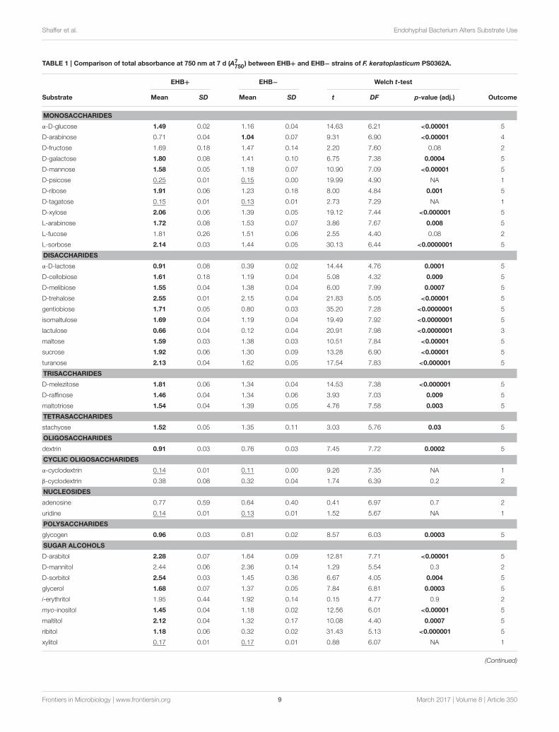
TABLE 1 | Comparison of total absorbance at 750 nm at 7 d (A7750) between EHB+ and EHB− strains of F. keratoplasticum PS0362A.
EHB+ EHB− Welch t-test
Substrate Mean SD Mean SD t DF p-value (adj.) Outcome
MONOSACCHARIDES
α-D-glucose 1.49 0.02 1.16 0.04 14.63 6.21 <0.00001 5
D-arabinose 0.71 0.04 1.04 0.07 9.31 6.90 <0.00001 4
D-fructose 1.69 0.18 1.47 0.14 2.20 7.60 0.08 2
D-galactose 1.80 0.08 1.41 0.10 6.75 7.38 0.0004 5
D-mannose 1.58 0.05 1.18 0.07 10.90 7.09 <0.00001 5
D-psicose 0.25 0.01 0.15 0.00 19.99 4.90 NA 1
D-ribose 1.91 0.06 1.23 0.18 8.00 4.84 0.001 5
D-tagatose 0.15 0.01 0.13 0.01 2.73 7.29 NA 1
D-xylose 2.06 0.06 1.39 0.05 19.12 7.44 <0.000001 5
L-arabinose 1.72 0.08 1.53 0.07 3.86 7.67 0.008 5
L-fucose 1.81 0.26 1.51 0.06 2.55 4.40 0.08 2
L-sorbose 2.14 0.03 1.44 0.05 30.13 6.44 <0.0000001 5
DISACCHARIDES
α-D-lactose 0.91 0.08 0.39 0.02 14.44 4.76 0.0001 5
D-cellobiose 1.61 0.18 1.19 0.04 5.08 4.32 0.009 5
D-melibiose 1.55 0.04 1.38 0.04 6.00 7.99 0.0007 5
D-trehalose 2.55 0.01 2.15 0.04 21.83 5.05 <0.00001 5
gentiobiose 1.71 0.05 0.80 0.03 35.20 7.28 <0.0000001 5
isomaltulose 1.69 0.04 1.19 0.04 19.49 7.92 <0.0000001 5
lactulose 0.66 0.04 0.12 0.04 20.91 7.98 <0.0000001 3
maltose 1.59 0.03 1.38 0.03 10.51 7.84 <0.00001 5
sucrose 1.92 0.06 1.30 0.09 13.28 6.90 <0.00001 5
turanose 2.13 0.04 1.62 0.05 17.54 7.83 <0.000001 5
TRISACCHARIDES
D-melezitose 1.81 0.06 1.34 0.04 14.53 7.38 <0.000001 5
D-raffinose 1.46 0.04 1.34 0.06 3.93 7.03 0.009 5
maltotriose 1.54 0.04 1.39 0.05 4.76 7.58 0.003 5
TETRASACCHARIDES
stachyose 1.52 0.05 1.35 0.11 3.03 5.76 0.03 5
OLIGOSACCHARIDES
dextrin 0.91 0.03 0.76 0.03 7.45 7.72 0.0002 5
CYCLIC OLIGOSACCHARIDES
α-cyclodextrin 0.14 0.01 0.11 0.00 9.26 7.35 NA 1
β-cyclodextrin 0.38 0.08 0.32 0.04 1.74 6.39 0.2 2
NUCLEOSIDES
adenosine 0.77 0.59 0.64 0.40 0.41 6.97 0.7 2
uridine 0.14 0.01 0.13 0.01 1.52 5.67 NA 1
POLYSACCHARIDES
glycogen 0.96 0.03 0.81 0.02 8.57 6.03 0.0003 5
SUGAR ALCOHOLS
D-arabitol 2.28 0.07 1.64 0.09 12.81 7.71 <0.00001 5
D-mannitol 2.44 0.06 2.36 0.14 1.29 5.54 0.3 2
D-sorbitol 2.54 0.03 1.45 0.36 6.67 4.05 0.004 5
glycerol 1.68 0.07 1.37 0.05 7.84 6.81 0.0003 5
i-erythritol 1.95 0.44 1.92 0.14 0.15 4.77 0.9 2
myo-inositol 1.45 0.04 1.18 0.02 12.56 6.01 <0.00001 5
maltitol 2.12 0.04 1.32 0.17 10.08 4.40 0.0007 5
ribitol 1.18 0.06 0.32 0.02 31.43 5.13 <0.000001 5
xylitol 0.17 0.01 0.17 0.01 0.88 6.07 NA 1
(Continued)
Frontiers in Microbiology | www.frontiersin.org 9 March 2017 | Volume 8 | Article 350

Shaffer et al. Endohyphal Bacterium Alters Substrate Use
TABLE 1 | Continued
EHB+ EHB− Welch t-test
Substrate Mean SD Mean SD t DF p-value (adj.) Outcome
METHYL SUGARS
L-rhamnose 2.05 0.04 1.79 0.07 7.72 6.17 0.0005 5
methyl α-D-galactoside 1.89 0.29 1.32 0.16 3.90 6.18 0.01 5
methyl β-D-galactoside 0.58 0.04 0.34 0.02 13.42 5.73 <0.00001 5
methyl α-D-glucoside 2.14 0.04 2.20 0.07 1.77 6.71 0.1 2
methyl β-D-glucoside 2.21 0.06 1.52 0.06 18.02 7.97 <0.000001 5
ALCOHOLIC β-GLUCOSIDES
salicin 0.97 0.02 0.54 0.02 30.08 8.00 <0.0000001 5
GLYCOSIDES
arbutin 0.90 0.06 0.70 0.03 7.16 5.99 0.0007 5
MISC. CARBOHYDRATES
sedoheptulosan 0.12 0.01 0.14 0.03 1.63 4.77 NA 1
AMINO SUGARS
D-glucosamine 0.39 0.11 0.59 0.05 3.69 5.71 0.02 4
N-acetyl-D-galactosamine 0.11 0.00 0.10 0.00 7.00 5.82 NA 1
N-acetyl-D-glucosamine 2.38 0.08 1.06 0.04 34.01 5.89 <0.0000001 5
N-acetyl-D-mannosamine 0.11 0.00 0.10 0.00 3.04 5.77 NA 1
AMINO ACIDS
L-alanine 1.65 0.13 0.95 0.20 6.61 6.75 0.0007 5
L-proline 2.12 0.04 1.24 0.20 9.86 4.31 0.0008 5
L-serine 1.50 0.11 0.79 0.23 6.36 5.71 0.002 5
L-threonine 1.97 0.04 0.79 0.01 65.09 4.49 <0.000001 5
L-aspartic acid 1.18 0.06 1.18 0.11 0.03 5.92 1.0 2
L-glutamic acid 1.51 0.06 0.88 0.04 18.40 6.94 <0.000001 5
N-acetyl-L-glutamic acid 0.13 0.00 0.13 0.01 0.34 6.90 NA 1
glycyl-L-glutamic acid 0.13 0.00 0.13 0.01 1.79 7.22 NA 1
L-pyroglutamic acid 1.77 0.07 1.25 0.12 8.26 6.39 0.0003 5
γ-aminobutyric acid 2.24 0.03 0.84 0.12 24.28 4.63 <0.00001 5
L-asparagine 1.86 0.02 0.91 0.11 18.56 4.35 <0.00001 5
L-phenylalanine 1.21 0.05 1.23 0.29 0.12 4.26 0.9 2
L-ornithine 1.81 0.03 0.92 0.15 13.18 4.24 0.0003 5
L-alanyl-glycine 1.09 0.05 1.06 0.07 0.61 7.64 0.6 2
AMINO ACID DERIVATIVES
amygdalin 1.03 0.04 0.83 0.03 8.55 7.63 0.0001 5
CARBOXYLIC ACIDS
2-keto-D-gluconic acid 0.84 0.02 0.70 0.07 4.43 4.46 0.01 5
α-ketoglutaric acid 0.69 0.06 0.62 0.13 1.16 5.38 0.3 2
β-hydroxybutyric acid 1.09 0.07 0.72 0.06 9.35 7.90 <0.00001 5
D-glucuronic acid 0.79 0.04 0.49 0.06 9.03 7.21 0.0001 5
D-galacturonic acid 1.14 0.07 0.92 0.07 5.10 8.00 0.002 5
D-gluconic acid 0.98 0.02 0.67 0.06 11.77 4.84 0.0002 5
D-saccharic acid 0.87 0.04 0.57 0.04 13.52 7.99 <0.000001 5
D-malic acid 1.23 0.04 1.47 0.04 9.28 7.91 <0.00001 4
L-malic acid 0.81 0.02 0.94 0.09 3.06 4.40 0.04 4
L-lactic acid 0.54 0.02 0.40 0.02 11.45 7.91 <0.00001 5
γ-hydroxybutyric acid 0.23 0.10 0.15 0.01 1.71 4.08 NA 1
fumaric acid 1.09 0.05 1.13 0.15 0.62 4.99 0.6 2
p-hydroxyphenylacetic acid 1.46 0.08 1.24 0.24 1.94 4.92 0.1 2
quinic acid 1.89 0.09 1.15 0.17 8.41 6.04 0.0003 5
sebacic acid 1.43 0.06 0.94 0.15 6.70 5.05 0.002 5
succinic acid 0.70 0.02 0.52 0.05 7.70 4.90 0.001 5
(Continued)
Frontiers in Microbiology | www.frontiersin.org 10 March 2017 | Volume 8 | Article 350

Shaffer et al. Endohyphal Bacterium Alters Substrate Use
TABLE 1 | Continued
EHB+ EHB− Welch t-test
Substrate Mean SD Mean SD t DF p-value (adj.) Outcome
ESTERS
D-lactic acid methyl ester 0.22 0.00 0.17 0.00 15.96 8.00 NA 1
succinic acid monomethyl
ester
1.20 0.11 0.79 0.16 4.76 6.87 0.004 5
AMIDES
alaninamide 0.21 0.01 0.20 0.01 1.14 5.81 NA 1
glucuronamide 0.10 0.01 0.11 0.00 3.21 5.48 NA 1
succinamic acid 1.13 0.10 1.82 0.13 9.78 7.48 <0.00001 4
ethanolamine 0.92 0.04 1.05 0.21 1.41 4.23 0.3 1
putrescine 1.90 0.06 0.77 0.18 13.66 4.76 0.0001 5
PHOSPHORYLATED CHEMICALS
glucose-1-phosphate 0.14 0.01 0.14 0.00 0.35 7.71 NA 1
adenosine-5′-
monophosphate
0.12 0.01 0.11 0.00 2.28 5.97 NA 1
BROMINATED CHEMICALS
bromosuccinic acid 0.38 0.02 0.25 0.01 13.54 7.90 <0.000001 3
SURFACTANTS
Tween® 80 0.62 0.06 0.55 0.03 2.25 5.63 0.09 2
water 0.11 0.00 0.10 0.00 2.98 6.86 NA 1
Values are means and standard deviations. Underlined means indicate substrates for which absorbance was negligible for both strains (i.e., A7750 ≤ 0.3). For the remaining substrates,
results of Welch t-tests are shown. Adjusted p-values were corrected following Benjamini and Hochberg (1995). Significant p-values (i.e., ≤0.05) are bolded. Means in bold are those
that were found to be significantly greater in comparisons. For each substrate, one of five observed outcomes is listed (see Section Results).
strains on individual substrates. We classified our results intofour general outcomes, as described below.
The 17 substrates on which we observed negligible growth byboth the EHB+ and EHB− strains (Outcome 1) included diversemonosaccharides (e.g., D-psicose), amino sugars (e.g., N-acetyl-D-galactosamine), amino acids (e.g., glycyl-L-glutamic acid),amides (e.g., alaninamide), and phosphorylated chemicals (e.g.,glucose-1-phosphate), in addition to water (Figure 3, Table 1).
EHB+ and EHB− strains both used, but did not differin growth on, 15 substrates (Outcome 2; Figure 3, Table 1).These included diverse monosaccharides (e.g., D-fructose), sugaralcohols (e.g., D-mannitol), amino acids (e.g., phenylalanine),and carboxylic acids (e.g., fumaric acid).
On two substrates we observed measurable growth by theEHB+ strain and negligible growth by the EHB− strain(Outcome 3). These were lactulose and bromosuccinic acid, onwhich the EHB+ strain only grew to A7
750 = 0.66 and 0.39,respectively (Figure 3, Table 1).
We observed measurable and significantly different growthbetween EHB+ and EHB− strains on 64 carbon sources(Figure 3, Table 1). The EHB− strain grew to a higher densityon five substrates (Outcome 4), including one monosaccharide(D-arabinose), two stereoisomeric forms of one carboxylic acid(D- and L-malic acid), and one amide (succinamic acid; Figure 3,Table 1). The EHB+ strain grew to a higher density on 59substrates (Outcome 5), including over three-quarters of allsugar-based substrates (77%), and most amino- and carboxylicacids and their derivatives (58 and 60%, respectively) (Figure 3,Table 1).
We repeated the experiment by re-curing the naturallyinfected strain and performing the Biolog R© trial a second time.The raw data and code for analyses are available online (Shaffer,2017). Results were consistent with those reported here.
DISCUSSION
Endohyphal bacteria (EHB) have been documented as symbiontsin phylogenetically and ecologically diverse lineages of fungi(Barbieri et al., 2000; Bianciotto et al., 2003; Bertaux et al., 2005;Partida-Martínez et al., 2007b; Sharma et al., 2008; Hoffmanand Arnold, 2010; Sato et al., 2010; Desirò et al., 2015; Shafferet al., 2016). Only in a few cases have their effects been explored.Comparative genomics and phenotypic assays have recentlyhighlighted the importance of certain proteobacterial EHBamong foliar endophytic Ascomycota (Arendt, 2015; Baltruset al., 2016). Here we provide the first insight to the influenceof EHB on broad-spectrum substrate use by a member of a cladeof fungi known for their widespread pathogenicity on plants (i.e.,the F. solani species complex), with a focus on a strain affiliatedwith seeds from tropical forest soil. The strain considered hereis a member of a lineage that is known for ecologically andmedically important strains (i.e., F. keratoplasticum; Short et al.,2013). Its endohyphal bacterium belongs to a genus known fortheir production of secondary metabolites with antimicrobialactivity (i.e., Chitinophaga). More broadly, it is a member ofthe Bacteroidetes, a phylogenetically diverse phylum of Gram-negative bacteria that are globally distributed, exhibit many
Frontiers in Microbiology | www.frontiersin.org 11 March 2017 | Volume 8 | Article 350

Shaffer et al. Endohyphal Bacterium Alters Substrate Use
FIGURE 4 | Cluster dendrograms summarizing differences in global substrate use at 1, 2, 4.5, and 7 d among EHB+ and EHB− replicate Biolog® PMs.
Distances represent Bray-Curtis dissimilarities. Results from a permutational multivariate analysis of variance (PERMANOVA) are shown for each time point. Panels
show differences at (A) 1 d, (B) 2 d, (C) 4.5 d, and (D) 7 d.
biological functions, and are well-known symbionts of mammalsand insects as well as degraders of organic matter (Moranet al., 2005; Thomas et al., 2011). Similar to Proteobacteria,Bacteroidetes are often one of the most representative taxarecovered from environmental sampling of freshwater, soil,animals guts and skin, and especially the phyllosphere (Redfordet al., 2010; Thomas et al., 2011). To our knowledge, noendohyphal member of the Bacteroidetes has been examinedpreviously for its associations with fungi or its phenotypic effectson a fungal host.
Phenotypic MicroarraysPhenotypic microarrays (PMs) provide a means to obtainquantitative data in a reproducible and highly controlled mannerwith respect to biomass accumulation from the metabolism ofspecific compounds. Such data can inform diverse and emergingfields concerning microbial ecology such as community systemsbiology and metametabolomics, and can provide the basis for
hypotheses that can be evaluated in the context of genomics andtranscriptomics analyses.
Recently PMs have been used to address questions infungal ecology and evolution, including those relevant tobiotechnology applications (Greetham, 2014; Blumenstein et al.,2015a), evolutionary relationships and species concepts (Rice andCurrah, 2005; Atanasova et al., 2010), carbon dynamics and nichedifferentiation (Lee and Magan, 1999; Hobbie et al., 2003; Friedlet al., 2008), genetic and functional diversity (Dobranic and Zak,1999; Druzhinina et al., 2006; Grizzle and Zak, 2006; Friedl et al.,2008), and ecophysiology (Druzhinina et al., 2010). Here we useda Biolog R© plate assay as a rapid and simple method for use incharacterizing the influence of EHB on broad-spectrum carbonsource use by a filamentous fungus.
We found that the presence of Chitinophaga sp. PS-EHB01significantly influenced substrate use across over two-thirds(67%) of carbon sources. The EHB+ strain grew to a higherabsorbance compared to the EHB− strain across the majority
Frontiers in Microbiology | www.frontiersin.org 12 March 2017 | Volume 8 | Article 350

Shaffer et al. Endohyphal Bacterium Alters Substrate Use
TABLE 2 | Summary of differences in global substrate use between EHB+
and EHB− strains over 7 d.
PERMANOVA ANOSIM MRPP
Day F R2 p-value R p-value A p-value
0.5 0.97 0.11 0.4 0.004 0.5 <0.0 0.5
1.0 2.48 0.24 0.07 0.33 0.07 0.07 0.08
1.5 6.86 0.46 0.009 1.00 0.003 0.23 0.02
2.0 22.60 0.74 0.008 1.00 0.005 0.42 0.01
2.5 18.10 0.69 0.009 1.00 0.01 0.40 0.009
3.0 35.19 0.81 0.008 1.00 0.01 0.51 0.009
3.5 57.42 0.88 0.01 1.00 0.007 0.59 0.005
4.0 75.49 0.90 0.007 1.00 0.01 0.63 0.01
4.5 74.62 0.90 0.007 1.00 0.008 0.63 0.01
5.0 75.12 0.90 0.01 1.00 0.01 0.64 0.006
5.5 71.26 0.90 0.01 1.00 0.004 0.63 0.007
6.0 72.46 0.90 0.01 1.00 0.009 0.63 0.02
6.5 78.24 0.91 0.006 1.00 0.007 0.64 0.01
7.0 79.72 0.91 0.008 1.00 0.01 0.65 0.01
Bray-Curtis dissimilarities based on growth across all substrates were calculated for each
pair of replicate phenotypic microarrays, which were grouped based on inoculation with
EHB+ vs. EHB− strains. Results from three different methods (permutational multivariate
analysis of variance [PERMANOVA], analysis of similarity [ANOSIM], and multi-response
permutation procedure [MRPP]) confirm that significant differences in global substrate use
between EHB+ and EHB– strains were observed andmaintained after one day. Significant
p-values (i.e., ≤ 0.05) are bolded.
of substrates (62%). In general, initial growth rates of EHB+and EHB− strains were similar; however, the absorbance valuesat times after which the EHB− strain reached stationaryphase were significantly lower than for those for when theEHB+ strain reached stationary phase (Figure 3, Table 1).We speculate that the bacterium may serve as a metabolicenhancer, possibly releasing compounds that serve as growthfactors for the host fungus, or by detoxifying or metabolizingotherwise harmful waste or growth byproducts such as reactiveoxygen species known to accumulate from catabolism ofcertain compounds (e.g., L-ornithine, putrescine; Pegg andCasero, 2011; Salvioli et al., 2016; Vannini et al., 2016). ThatChitinophaga sp. PS-EHB01 appears to be consistently associatedwith F. keratoplasticum PS0362A but cannot be isolated intopure culture on standard media (Shaffer, unpublished) suggeststhat this EHB may rely on its host fungus to acquire essentialnutrients.
Perspectives from Related SpeciesAlthough their genomes have not yet been sequenced, genomicdata are available for close relatives of the bacterium and fungusevaluated here (see Coleman et al., 2008; Del Rio et al., 2010).The focal bacterium is closely related to Chitinophaga pinensis(Supplementary Figure 7), which was isolated originally from soiland is known for its ability to degrade chitin (Sangkhobol andSkerman, 1981). Chitinophaga pinensis also produces antibioticswith activity against a diversity of filamentous fungi (Mohret al., 2015). That species possesses multiple genes predictedto be involved in the metabolism of carbohydrates (n = 330)and amino acids (n = 301; Del Rio et al., 2010). The focalbacterium is also closely related to C. arvensicola (Supplementary
Figure 7), a bacterium associated with amphibian skin thatproduces metabolites with inhibitory effects on the notoriousfungal pathogen Batrachochytrium dendrobatidis, causal agent ofchytridiomycosis (Loudon et al., 2014). Whether such traits arecommon in the Chitinophaga strain examined here remains to bedetermined.
Using a three-locus dataset, we showed previously that F.keratoplasticum PS0362A is part of the F. solani species complex(FSSC). The fungus studied here is closely related to FSSC Clade3 haplotype group 2 (Shaffer et al., 2016). The closest relative withpublicly available genomic data, Nectria haematococca MPVI, isin group 11-c (Short et al., 2013; Shaffer et al., 2016). Nectriahaematococca MPVI occurs as a saprotroph and plant pathogenin diverse habitats (Coleman et al., 2008). Many membersof the FSSC have conditionally dispensable, supernumerarychromosomes (CD chromosomes) that can influence the use ofspecific carbon sources (Covert, 1998; Coleman et al., 2008).CD chromosomes are mitotically stable in N. haematococcaMPVI (Covert, 1998). Therefore, it is likely that the phenotypicresults observed here do not reflect differential presence of CDchromosomes between EHB+ and EHB− strains, but rather adifference in the presence of the bacterium between them.
The genome of N. haematococca is highly enriched with genescoding for carbohydrate-active enzymes, including glycosidehydrolase and polysaccharide lyase genes (Coleman et al.,2008). Nectria haematococca also possesses a high number ofATP-binding cassette (ABC) transporter genes, second onlyto Aspergillus oryzae when compared to 10 other membersof the Dikarya (Coleman et al., 2008). That the fungusstudied here was able to use the majority of carbon sourcesregardless of EHB infection status may reflect similar genecomposition. These substrates included some synthetic, non-natural compounds such as lactulose, bromosuccinic acid, andTween R© 80, emphasizing the metabolic breadth of this fungus.Strikingly, that breadth is increased markedly by the presence ofChitinophaga sp. PS-EHB01 as an EHB. Once data for both thebacterium and fungus are available, comparative genomics andtranscriptomics can be used to understandmetabolic interactionsbetween the pair. More broadly, the pair could be developedto become a model system for understanding EHB of plant-associated Ascomycota.
Implications for Seed-Fungus InteractionsFungi recruit from soil to seeds that have been dispersed to thesoil seed bank, thus undergoing horizontal transmission (ratherthan being vertically transmitted from mother to offspring)(U’Ren et al., 2009; Sarmiento et al., 2015; Zalamea et al., 2015;Sarmiento et al., unpublished). Given this life history, seed-fungus interactions at the soil-seed interface (i.e., those involvingthe seed coat) are of primary interest with regard to communityassembly of fungi in seeds. Furthermore, similar to bud-breakor wounding (Agrios, 1997; Schädel et al., 2009; Gordon andLeveau, 2010; Savatin et al., 2014), seed germination representsa key event during which nutrients that may attract potentialsymbionts are released into the environment. The potential forEHB to influence seed-fungus interactions during colonization of
Frontiers in Microbiology | www.frontiersin.org 13 March 2017 | Volume 8 | Article 350
Shaffer et al. Endohyphal Bacterium Alters Substrate Use
seeds by fungi at the soil-seed interface, and during key plant life-stage transitions such as seed germination, should be investigatedfurther.
Here we showed that the presence vs. absence of Chitinophagasp. PS-EHB01 led to differential growth by F. keratoplasticum onmost sugars, amino acids, and carboxylic acids, nearly all of whichare relevant in plant biology. In particular, a number of substratesare important in the ecology of seeds, such as importantglobal regulators (e.g., D-trehalose andmyo-inositol; Loewus andMurthy, 2000; Grennan, 2007; Henry et al., 2014; Lunn et al.,2014), thosemetabolized or produced during seed imbibition andgermination (e.g., D-trehalose, sucrose, D-raffinose, stachyose,dextrin, and L-asparagine; Atkins et al., 1975; Bewley and Black,1978; Kuo et al., 1990; Queiroz and Cazetta, 2016), as well asthose important in themetabolism of seed structural componentssuch as the seed coat (e.g., D-mannose, L-arabinose, sucrose,D-raffinose, stachyose, myo-inositol, and L-alanine; Herold andLewis, 1977; Bewley and Black, 1978; Kuo et al., 1990; Buckeridgeet al., 2000; Loewus andMurthy, 2000; Lahuta et al., 2007; Kosinaet al., 2013). The average difference in growth between EHB+and EHB− strains, considering only those substrates on whichwe observed significant differences (n = 64; Table 1) was A7
750 =
0.3. Whether this difference scales to meaningful changes withregard to interacting with plants in nature is not yet known, andwill be assessed in future work using seed-infection and seed-germination experiments. We anticipate that changes in fungalsubstrate use by EHB will alter phenotypes that in turn defineboth the fungal niche and the outcomes of interactions withhosts.
AUTHOR CONTRIBUTIONS
JU modified and tested the phenotypic microarray experimentalprotocol for use with fungal hyphal fragments; JSmade additional
modifications to exclude fungal spores, and performed allexperimental work and related data analysis; AA and DB advisedaspects of the data analysis; JS and AA led the development of themanuscript, with contributions from DB, RG, and JU.
FUNDING
We thank the National Science Foundation (NSF DEB-1119758to AA, NSF DEB-1120205 to James W. Dalling, NSF IOS-1354219 to DB, AA, and RG, NSF-IGERT Fellowship to JS),the Smithsonian Tropical Research Institute (STRI) (Short-termFellowship to JS), the Mycological Society of America (ForestFungal Ecology Award to JS), and the Graduate and ProfessionalStudent Council (Research Award to JS) and School of PlantSciences (Pierson Fellowship to JS) at The University of Arizonafor supporting this work. Additional support from the School ofPlant Sciences and College of Agriculture and Life Sciences at theUniversity of Arizona is gratefully acknowledged.
ACKNOWLEDGMENTS
We thank J. DeVore, K. Hockett, and especially A. Ndobegangfor lab assistance, K. Arendt, Y.-L. Huang, J. Carlson, K. Hockett,J. Dalling, J. Wright, E. Leigh, G. Gilbert, and N. Zimmermanfor helpful discussion, and N. Zimmerman for bioinformaticsassistance. This paper represents a portion of the doctoraldissertation research of JS in Plant Pathology and Microbiologyat the University of Arizona.
SUPPLEMENTARY MATERIAL
The Supplementary Material for this article can be foundonline at: http://journal.frontiersin.org/article/10.3389/fmicb.2017.00350/full#supplementary-material
REFERENCES
Agrios, G. N. (1997). Plant Pathology. San Diego, CA: Academic Press.Anca, I., Lumini, E., Ghignone, S., Salvioli, A., Bianciotto, V., and Bonfante, P.
(2009). The ftsZ gene of the endocellular bacterium ‘CandidatusGlomeribactergigasporarum’ is preferentially expressed during the symbiotic phases ofits host mycorrhizal fungus. Mol. Plant-Microbe Interact. 22, 302–310.doi: 10.1094/MPMI-22-3-0302
Anderson, M. J. (2001). A new method for non-parametric multivariateanalysis of variance. Austral Ecol. 26, 32–46. doi: 10.1111/j.1442-9993.2001.01070.pp.x
Anderson, M. J., and ter Braak, C. J. F. (2003). Permutational tests formulti-factorial analysis of variance. J. Stat. Comput. Simul. 73, 85–113.doi: 10.1080/00949650215733
Arendt, K. A. (2015). Symbiosis Establishment and Ecological Effects of
Endohyphal Bacteria on Foliar Fungi. Master’s thesis, University of Arizona,Tucson, AZ.
Arendt, K. A., Hockett, K. L., Araldi-Brondolo, S. J., Baltrus, D. A., and Arnold,A. E. (2016). Isolation of endohyphal bacteria from foliar Ascomycota and in
vitro establishment of their symbiotic associations. Appl. Environ. Microbiol.
82, 2943–2949. doi: 10.1128/AEM.00452-16Arnold, A. E., and Engelbrecht, B. M. J. (2007). Fungal endophytes nearly double
minimum leaf conductance in seedlings of a neotropical tree species. J. Trop.Ecol. 23, 369–372. doi: 10.1017/S0266467407004038
Arnold, A. E., and Lutzoni, F. (2007). Diversity and host range of foliar fungalendophytes: are tropical leaves biodiversity hotspots? Ecology 88, 541–549.doi: 10.1890/05-1459
Arnold, A. E., Mejia, L. C., Kyllo, D., Rojas, E. I., Maynard, Z., Robbins, N., et al.(2003). Fungal endophytes limit pathogen damage in a tropical tree. Proc. Natl.Acad. Sci. U.S.A. 100, 15649–15654. doi: 10.1073/pnas.2533483100
Atanasova, L., andDruzhinina, I. S. (2010). Global nutrient profiling by PhenotypicMicroArrays: a tool complementing genomic and proteomic studies in conidialfungi. J. Zhejiang Univ. Sci. B 11, 151–168. doi: 10.1631/jzus.B1000007
Atanasova, L., Jaklitsch, W. M., Komon-Zelazowska, M., Kubicek, C. P., andDruzhinina, I. S. (2010). Clonal species of Trichoderma parareesei sp. nov. likelyresembles the ancestor of the cellulase producer Hypocrea jecorina/T. reesei.
Appl. Environ. Microbiol. 76, 7259–7267. doi: 10.1128/AEM.01184-10Atkins, C. A., Pate, J. S., and Sharkey, P. J. (1975). Asparagine metabolism – key to
the nitrogen nutrition of developing legume seeds. Plant Physiol. 56, 807–812.doi: 10.1104/pp.56.6.807
Baltrus, D. A., Dougherty, K., Arendt, K. R., Huntemann, M., Clum, A., Pillay, M.,et al. (2016). Absence of genome reduction in diverse, facultative endohyphalbacteria.Microb. Genom. doi: 10.1099/mgen.0.000101. [Epub ahead of print].
Barbieri, E., Potenza, L., Rossi, I., Sisti, D., Giomaro, G., Rossetti, S.,et al. (2000). Phylogenetic characterization and in situ detection ofCytophaga-Flexibacter-Bacteroides phylogroup bacterium in Tuber borchii
Vittad. ectomycorrhizal mycelium. Appl. Environ. Microbiol. 66, 5035–5042.doi: 10.1128/AEM.66.11.5035-5042.2000
Frontiers in Microbiology | www.frontiersin.org 14 March 2017 | Volume 8 | Article 350
Shaffer et al. Endohyphal Bacterium Alters Substrate Use
Benjamini, Y., and Hochberg, Y. (1995). Controlling the false discovery rate:a practical and powerful approach to multiple testing. J. R. Stat. Soc. B 57,289–300.
Bertaux, J., Schmid, M., Hutzler, P., Hartmann, A., Garbaye, J., and Frey-Klett, P.(2005). Occurrence and distribution of endobacterial in the plant-associatedmycelium of the ectomycorrhizal fungus Laccaria bicolor S238N. Environ.Microbiol. 7, 1786–1795. doi: 10.1111/j.1462-2920.2005.00867.x
Bewley, J. D., and Black, M. (1978). Physiology and Biochemistry of Seeds in
Relation to Germination, Vol. 1. Development, Germination, and Growth. Berlin:Springer-Verlag.
Bianciotto, V., Bandi, C., Minerdi, D., Sironi, M., Tichy, H. V., and Bonfante,P. (1996). An obligately endosymbiotic mycorrhizal fungus itself harborsobligately intracellular bacteria. Appl. Environ. Microbiol. 62, 3005–3010.
Bianciotto, V., Genre, A., Jargeat, P., Lumini, E., Bécard, G., and Bonfante, P.(2004). Vertical transmission of endobacteria in the arbuscular mycorrhizalfungus Gigaspora margarita through generation of vegetative spores. Appl.Environ. Microbiol. 70, 3600–3608. doi: 10.1128/AEM.70.6.3600-3608.2004
Bianciotto, V., Lumini, E., Bonfante, P., and Vandamme, P. (2003). ‘CandidatusGlomeribacter gigasporarum’ gen. nov., sp. nov., an endosymbiont ofarbuscular mycorrhizal fungi. Int. J. Syst. Evol. Microbiol. 53, 121–124.doi: 10.1099/ijs.0.02382-0
Biondini, M. E., Bonham, C. D., and Redente, E. F. (1985). Secondarysuccessional patterns in a sagebrush (Artemisia tridentata) community as theyrelate to soil disturbance and soil biological activity. Vegetatio 60, 25–36.doi: 10.1007/BF00053909
Blanchette, R. A. (1991). Delignification by wood-decay fungi. Annu. Rev.
Phytopathol. 29, 381–398. doi: 10.1146/annurev.py.29.090191.002121Blumenstein, K., Albrectsen, B. R., Martín, J. A., Hultberg, M., Sieber, T.
N., Helander, M., et al. (2015a). Nutritional niche overlap potentiates theuse of endophytes in biocontrol of a tree disease. Biocontrol 60, 655–667.doi: 10.1007/s10526-015-9668-1
Blumenstein, K., Macaya-Sanz, D., Martín, J. A., Albrectsen, B. R., and Witzell, J.(2015b). Phenotype MicroArrays as a complementary tool to next generationsequencing for characterization of tree endophytes. Front. Microbiol. 6:1033.doi: 10.3389/fmicb.2015.01033
Bochner, B. R. (1989). Sleuthing out bacterial identities. Nature 339, 157–158.doi: 10.1038/339157a0
Bochner, B. R. (2003). New technologies to assess genotype-phenotyperelationships. Nat. Rev. Genet. 4, 309–314. doi: 10.1038/nrg1046
Bochner, B. R. (2008). Global phenotypic characterization of bacteria. FEMS
Microbiol. Rev. 33, 191–205. doi: 10.1111/j.1574-6976.2008.00149.xBochner, B. R., and Savageau, M. A. (1977). Generalized indicator plate for
genetic, metabolic, and taxonomic studies with microorganisms.Appl. Environ.Microbiol. 33, 434–444.
Bochner, B. R., Gadzinksi, P., and Panomitros, E. (2001). Phenotype MicroArraysfor high-throughput phenotypic testing and assay of gene function. Genome
Res. 11, 1246–1255. doi: 10.1101/gr.186501Buckeridge, M. S., dos Santos, H. P., and Tiné, M. A. S. (2000). Mobilisation of
storage cell wall polysaccharides in seeds. Plant Physiol. Biochem. 38, 141–156.doi: 10.1016/S0981-9428(00)00162-5
Busby, P. E., Peay, K. G., and Newcombe, G. (2015). Common foliar fungi ofPopulus trichocarpamodifyMelampsora rust disease severity. New Phytol. 209,1681–1692. doi: 10.1111/nph.13742
Clarke, K. R. (1993). Non-parametric multivariate analyses of changes incommunity structure.Austral Ecol. 18, 117–143. doi: 10.1111/j.1442-9993.1993.tb00438.x
Clarke, K. R., and Green, R. H. (1988). Statistical design and analysisfor a ‘biological effects’ study. Mar. Ecol. Prog. Ser. 46, 213–226.doi: 10.3354/meps046213
Coleman, J. J., Rounsley, S. D., Rodriguez-Carres, M., Kuo, A., Wasmann, C. C.,Grimwood, J., et al. (2008). The genome of Nectria haematococca: contributionof supernumerary chromosomes to gene expansion. PLoS Genet. 5:e1000618.doi: 10.1371/journal.pgen.1000618
Covert, S. F. (1998). Supernumerary chromosomes in filamentous fungi. Curr.Genet. 33, 311–319. doi: 10.1007/s002940050342
Davison, J., Moora, M., Öpik, M., Adholeya, A., Ainsaar, L., Bâ, A., et al. (2015).Global assessment of arbuscular mycorrhizal fungus diversity reveals very lowendemism. Science 349, 970–973. doi: 10.1126/science.aab1161
Del Rio, T. G., Abt, B., Spring, S., Lapidus, A., Nolan, M., Tice, H., et al. (2010).Complete genome sequence ofChitionphaga pinensis type strain (UQM2034T).Stand. Genomic Sci. 2, 87–95. doi: 10.4056/sigs.661199
Desirò, A., Faccio, A., Kaech, A., Bidartondo, M. I., and Bonfante, P. (2015).Endogone, one of the oldest plant-associated fungi, host unique Mollicutes-related endobacteria. New Phytol. 205, 1464–1472. doi: 10.1111/nph.13136
Dobranic, J. K., and Zak, J. C. (1999). A microtiter plate procedure for evaluatingfungal functional diversity.Mycologia 91, 756–765. doi: 10.2307/3761529
Druzhinina, I. S., Komon-Zelazowska, M., Atanasova, L., Seidl, V., and Kubicek,C. P. (2010). Evolution and ecophysiology of the industrial producer Hypocreajecorina (Anamorph Trichoderma reesei) and a new sympatric agamospeciesrelated to it. PLoS ONE 5:e9191. doi: 10.1371/journal.pone.0009191
Druzhinina, I. S., Schmoll, M., Seiboth, B., and Kubicek, C. P. (2006).Global carbon utilization profiles of wild-type, mutant, and transformantstrains of Hypocrea jecorina. Appl. Environ. Microbiol. 72, 2126–2133.doi: 10.1128/AEM.72.3.2126-2133.2006
Estrada, C., Degner, E. C., Rojas, E. I.,Wcislo,W. T., andVan Bael, S. A. (2015). Therole of endophyte diversity in protecting plants from defoliation by leaf-cuttingants. Curr. Sci. 109, 55–61.
Ewing, B., and Green, P. (1998). Base-calling of automated sequencer traces usingPhred. II. Error probabilities. Genome Res. 8, 186–194. doi: 10.1101/gr.8.3.186
Ewing, B., Hillier, L.,Wendl,M. C., andGreen, P. (1998). Base-calling of automatedsequencer traces using Phred. I. Accuracy assessment. Genome Res. 8, 175–185.doi: 10.1101/gr.8.3.175
Frey-Klett, P., Garbaye, J., and Tarkka, M. (2007). The mycorrhiza helper bacteriarevisited. New Phytol. 176, 22–36. doi: 10.1111/j.1469-8137.2007.02191.x
Friedl, M. A., Kubicek, C. P., and Druzhinina, I. S. (2008). Carbon sourcedependence and photostimulation of conidiation inHypocrea atroviridis. Appl.Environ. Microbiol. 74, 245–250. doi: 10.1128/AEM.02068-07
Gale, G. R., Harrington, R. L., and Pate, A. F. (1960). The preparation of mycelialsuspensions of dermatophytes for metabolic studies. J. Invest. Dermatol. 34,167–169. doi: 10.1038/jid.1960.22
Gallery, R. E., Dalling, J. W., and Arnold, A. E. (2007). Diversity, host affinity,and distribution of seed-infecting fungi: a case study with Cecropia. Ecology 88,582–588. doi: 10.1890/05-1207
Gallery, R. E., Moore, D. J. P., and Dalling, J. W. (2010). Interspecific variation insusceptibility to fungal pathogens in seeds of 10 tree species in the neotropicalgenus Cecropia. J. Ecol. 98, 147–155. doi: 10.1111/j.1365-2745.2009.01589.x
Ghignone, S., Salvioli, A., Anca, I.-A., Lumini, E., Ortu, G., Petiti, L., et al.(2012). The genome of the obligate endobacterium of an AM fungus revealsan interphylum network of nutritional interactions. ISME J. 6, 136–145.doi: 10.1038/ismej.2011.110
Gordon, T. R., and Leveau, J. H. J. (2010). Plant pathology: a story aboutbiology. Annu. Rev. Phytopathol. 48, 293–309. doi: 10.1146/annurev-phyto-080508-081919
Greetham, D. (2014). Phenotype microarray technology and itsapplication in industrial biotechnology. Biotechnol. Lett. 36, 1153–1160.doi: 10.1007/s10529-014-1481-x
Grennan, A. K. (2007). The role of trehalose biosynthesis in plants. Plant Physiol.144, 3–5. doi: 10.1104/pp.104.900223
Grimmer, M. K., Foulkes, M. J., and Paveley, N. D. (2012). Foliar pathogenesisand plant water relations: a review. J. Exp. Bot. 63, 4321–4331.doi: 10.1093/jxb/ers143
Grizzle, H. W., and Zak, J. C. (2006). A microtiter plate procedure for evaluatingfungal functional diversity on nitrogen substrates. Mycologia 98, 353–363.doi: 10.3852/mycologia.98.2.353
Heilmann-Clausen, J., and Boddy, L. (2008). “Distribution patterns of wood-decaybasidiomycetes at the landscape to global scale,” in Ecology of Saprotrophic
Basidiomycetes (British Mycological Society Symposia Series Volume 28), eds L.Boddy, J. C. Frankland, and P. van West (London: Academic Press), 3–372.
Henry, C., Bledsoe, S. W., Siekman, A., Kollman, A., Waters, B. M., Feil, R., et al.(2014). The trehalose pathway in maize: conservation and gene regulation inresponse to the diurnal cycle and extended darkness. J. Exp. Bot. 65, 5959–5973.doi: 10.1093/jxb/eru335
Herold, A., and Lewis, D. H. (1977). Mannose and green plants:occurrence, physiology and metabolism, and use as a toolto study the role of orthophosphate. New Phytol. 79, 1–40.doi: 10.1111/j.1469-8137.1977.tb02178.x
Frontiers in Microbiology | www.frontiersin.org 15 March 2017 | Volume 8 | Article 350

Shaffer et al. Endohyphal Bacterium Alters Substrate Use
Hobbie, E. A., Watrud, L. S., Maggard, S., Shiroyama, T., and Rygiewicz, P. T.(2003). Carbohydrate use and assimilation by litter and soil fungi assessedby carbon isotopes and BIOLOG R© assays. Soil Biol. Biochem. 35, 303–311.doi: 10.1016/S0038-0717(02)00281-X
Hoffman, M. T., and Arnold, A. E. (2010). Diverse bacteria inhabit living hyphaeof phylogenetically diverse fungal endophytes. Appl. Environ. Microbiol. 76,4063–4075. doi: 10.1128/AEM.02928-09
Hoffman, M. T., Gunatilaka, M. K., Wijeratne, K., Gunatilaka, L., andArnold, A. E. (2013). Endohyphal bacterium enhances production ofindole-3-acetic acid by a foliar fungal endophyte. PLoS ONE 8:e73132.doi: 10.1371/journal.pone.0073132
Izumi, H., Anderson, I. C., Alexander, I. J., Killham, K., and Moore, E.R. B. (2005). Endobacteria in some ectomycorrhiza of Scotspine (Pinussylvestris). FEMS Microbiol. Ecol. 56, 34–43. doi: 10.1111/j.1574-6941.2005.00048.x
Jones, J. D. G., and Dangl, J. L. (2006). The plant immune system. Nature 444,323–329. doi: 10.1038/nature05286
Kivlin, S. N., Hawkes, C. V., and Treseder, K. K. (2011). Global diversityand distribution of arbuscular mycorrhizal fungi. Soil Biol. Biogeochem. 43,2294–2303. doi: 10.1016/j.soilbio.2011.07.012
Kosina, S. M., Schnebly, S. R., and Obendorf, R. L. (2013). Are Raffinose andStachyose unloaded from soybean seed coats to developing embryos? Open
Plant Sci. J. 7, 10–16. doi: 10.2174/1874294701307010010Krieg, N. R., Staley, J. T., Brown, D. R., Hedlund, B. P., Paster, B. J., Ward, N.
L., et al. (2010). “Volume Four: The Bacteroidetes, Spirochaetes, Tenericutes(Mollicutes), Acidobacteria, Fibrobacteres, Fusobacteria, Dictyoglomi,Gemmatimonadetes, Lentisphaerae, Verrucomicrobia, Chlamydiae, andPlanctomycetes,” in Bergey’s Manual of Systematic Bacteriology 2nd Edn., edsM. Goodfellow, P. Kämpfer, J. Chun, P. De Vos, F. Rainey, andW. B. Whitman(New York, NY: Springer), 25–467.
Kubicek, C. P., Bissett, J., Druzhinina, I., Kullnig-Gradinger, C., and Szakacs,G. (2003). Genetic and metabolic diversity of Trichoderma: a casestudy on South-East Asian isolates. Fungal Genet. Biol. 38, 310–319.doi: 10.1016/S1087-1845(02)00583-2
Kuo, T. M., Doehlert, D. C., and Crawford, C. G. (1990). Sugarmetabolism in germinating soybean seeds. Plant Physiol. 93, 1514–1520.doi: 10.1104/pp.93.4.1514
Lahuta, L. B., Górecki, R. J., Zalewski, K., and Hedley, C. L. (2007). Sorbitolaccumulation during natural and accelerated ageing of pea (Pisum sativum L.)seeds. Acta Physiol. Plant 29, 527–534. doi: 10.1007/s11738-007-0063-0
Lane, D. J. (1991). “16S/23S rRNA sequencing,” in Nucleic Acid Techniques in
Bacterial Systematics, eds E. Stackebrandt andM. Goodfellow (Chichester: JohnWiley and Sons), 115–175.
Lee, H. B., and Magan, N. (1999). Environmental factors and nutritionalutilization patterns affect niche overlap indices between Aspergillus
ochraceus and other spoilage fungi. Lett. Appl. Microbiol. 28, 300–304.doi: 10.1046/j.1365-2672.1999.00521.x
Leigh, E. G. Jr. (1999). Tropical Forest Ecology: A View from Barro Colorado Island.New York, NY: Oxford University Press.
Loewus, F. A., and Murthy, P. P. N. (2000). myo-Inositol metabolism in plants.Plant Sci. 150, 1–19. doi: 10.1016/S0168-9452(99)00150-8
Loudon, A. H., Holland, J. A., Umile, T. P., Burzynski, E. A., Minbiole, K. P. C., andHarris, R. N. (2014). Interactions between amphibians’ symbiotic bacteria causethe production of emergent anti-fungal metabolites. Front. Microbiol. 5:441.doi: 10.3389/fmicb.2014.00441
Lumini, E., Bianciotto, V., Jargeat, P., Novero, M., Salvioli, A., Faccio, A.,et al. (2007). Presymbiotic growth and sporal morphology are affectedin the arbuscular mycorrhizal fungus Gigaspora margarita cured of itsendobacteria. Cell. Microbiol. 9, 1716–1729. doi: 10.1111/j.1462-5822.2007.00907.x
Lunn, J. E., Delorge, I., Figueroa, C. M., Van Dijck, P., and Stitt, M. (2014).Trehalose metabolism in plants. Plant, J. 79, 544–567. doi: 10.1111/tpj.12509
Maddison, D. R., and Maddison, W. P. (2005). ChromaSeq Module. Mesquite: A
Modular System for Evolutionary Analysis. Version 1.06. Available online at:http://mesquiteproject.org/
Maddison, W. P., and Maddison, D. R. (2009). Mesquite: A Modular
System for Evolutionary Analysis. Version 2.6. Available online at:http://mesquiteproject.org/
Márquez, L. M., Redman, R. S., Rodriguez, R. J., and Roossink, M. L. (2007). Avirus in a fungus in a plant: three-way symbiosis required for thermal tolerance.Science 315, 513–515. doi: 10.1126/science.1136237
Mohr, K. I., Volz, C., Jansen, R., Wray, V., Hoffman, J., Bernecker, S., et al.(2015). Pinensins: the first antifungal lantibiotics. Angew. Chem. Int. Ed. 54,11254–11258. doi: 10.1002/anie.201500927
Moran, N. A., Tran, P., and Gerardo, N. M. (2005). Symbiosis and insectdiversification: an ancient symbiosis of sap-feeding insects from thebacterial phylum Bacteroidetes. Appl. Environ. Microbiol. 71, 8802–8810.doi: 10.1128/AEM.71.12.8802-8810.2005
Oksanen, J., Blanchet, F. G., Kindt, R., Legendre, P., Minchin, P. R., O’Hara, R.B., et al. (2016). vegan: Community Ecology Package. R Package Version 2.3-3.Available online at: http://CRAN.R-project.org/package=vegan
Oliva, J., Stenlid, J., and Martínez-Vilalta, J. (2014). The effect of fungal pathogenson the water and carbon economy of trees: implications for drought-inducedmortality. New Phytol. 203, 1028–1035. doi: 10.1111/nph.12857
Orbach, M. J., Chumley, F. G., and Valent, B. (1996). Electrophoretic karyotypes ofMagnaporthe grisea pathogens of diverse grasses. Mol. Plant Microbe Interact.
9, 261–271. doi: 10.1094/MPMI-9-0261Partida-Martínez, L. P., Groth, I., Schmitt, I., Richter, W., Roth, M., and
Hertweck, C. (2007b). Burkholderia rhizoxinica sp. nov. and Burkholderia
endofungorum sp. nov., bacterial endosymbionts of the plant-pathogenicfungus Rhizopus microsporus. Int. J. Syst. Evol. Microbiol. 57, 2583–2590.doi: 10.1099/ijs.0.64660-0
Partida-Martínez, L. P., and Hertweck, C. (2005). Pathogenic fungus harboursendosymbiotic bacteria for toxin production. Nature 437, 884–888.doi: 10.1038/nature03997
Partida-Martínez, L. P., Monajembashi, S., Greulich, K.-O., and Hertweck, C.(2007a). Endosymbiont-dependent host reproduction maintains bacterial-fungal mutualism. Curr. Biol. 17, 773–777. doi: 10.1016/j.cub.2007.03.039
Pegg, A. E., and Casero, R. A. Jr. (2011). Current status of the polyamine researchfield.Methods Mol. Biol. 720, 3–35. doi: 10.1007/978-1-61779-034-8_1
Pfliegler, W. P., Atanasova, L., Karanyicz, E., Sipiczki, M., Bond, U., Druzhinina,I. S., et al. (2014). Generation of new genotypic and phenotypic featuresin artificial and natural yeast hybrids. Food Technol. Biotechnol. 52, 46–57.doi: 10.1007/s00253-016-7481-0
Queiroz, R. J. B., and Cazetta, J. O. (2016). Proline and trehalose in maize seedsgerminating under low osmotic potentials. Rev. Bras. Eng. Agríc. Ambient. 20,22–28. doi: 10.1590/1807-1929/agriambi.v20n1p22-28
R Core Team (2015). R: A Language and Environment for Statistical Computing.
Vienna: R Foundation for Statistical Computing. Available online at:http://www.R-project.org/
Redford, A. J., Bowers, R. M., Knight, R., Linhard, Y., and Fierer, N. (2010).The ecology of the phyllosphere: geographic and phylogenetic variability inthe distribution of bacteria on tree leaves. Environ. Microbiol. 12, 2885–2893.doi: 10.1111/j.1462-2920.2010.02258.x
Rice, A. V., and Currah, R. S. (2005). Profiles from Biolog FF plates andmorphological characteristics support recognition ofOidiodendron fimicola sp.nov. Stud. Mycol. 53, 75–82. doi: 10.3114/sim.53.1.75
Ruiz-Herrera, J., León-Ramírez, C., Vera-Nu-ez, A., Sánchez-Arreguín, A., Ruiz-Medrano, R., Salgado-Lugo, H., et al. (2015). A novel intracellular nitrogen-fixing symbiosis made by Ustilago maydis and Bacillus spp. New Phytol. 207,769–777. doi: 10.1111/nph.13359
Salvioli, A., Chiapello, M., Fontaine, J., Hadj-Sahraoui, A. L., Grandmougin-Ferjani, A., Lanfranco, L., et al. (2010). Endobacteria affect the metabolic profileof their host Gigaspora margarita, an arbuscular mycorrhizal fungus. Environ.Microbiol. 12, 2083–2095. doi: 10.1111/j.1462-2920.2010.02246.x
Salvioli, A., Ghignone, S., Novero, M., Navazio, L., Venice, F., Bagnaresi, P.,et al. (2016). Symbiosis with an endobacterium increases the fitness of amycorrhizal fungus, raising its bioenergetic potential. ISME J. 10, 130–144.doi: 10.1038/ismej.2015.91
Sangkhobol, V., and Skerman, V. B. D. (1981). Chitinophaga, a newgenus of chitinolytic myxobacteria. Int. J. Syst. Bacteriol. 31, 285–293.doi: 10.1099/00207713-31-3-285
Sarmiento, C., Zalamea, P.-C., Dalling, J. W., Davis, A. S., and Arnold, A. E. (2015).“Seed-associated fungi: effects on seed survival and germination of tropicalpioneer species,” in Conference for the Association for Tropical Biology and
Conservation, July 12–16 (Honolulu, HI).
Frontiers in Microbiology | www.frontiersin.org 16 March 2017 | Volume 8 | Article 350

Shaffer et al. Endohyphal Bacterium Alters Substrate Use
Sato, Y., Narisawa, K., Tsuruta, K., Umezu, M., Nishizawa, T., Tanaka,K., et al. (2010). Detection of Betaproteobacteria inside the myceliumof the fungus Mortierella elongata. Microbes Environ. 25, 321–324.doi: 10.1264/jsme2.ME10134
Savatin, D. V., Gramegna, G., Modesti, V., and Cervone, F. (2014). Wounding inthe plant tissue: the defense of a dangerous passage. Front. Plant Sci. 5:470.doi: 10.3389/fpls.2014.00470
Schädel, C., Blöchl, A., Richter, A., and Hoch, G. (2009). Short-term dynamicsof nonstructural carbohydrates and hemicelluloses in young branchesof temperate forest trees during bud break. Tree Physiol. 29, 901–911.doi: 10.1093/treephys/tpp034
Schafer, M., and Kotanen, P. M. (2003). The influence of soil moisture on losses ofburied seeds to fungi. Acta Oecol. 24, 255–263. doi: 10.1016/j.actao.2003.09.001
Schneider, C. A., Rasband, W. S., and Eliceiri, K. W. (2012). NIH toImageJ: 25 years of image analysis. Nat. Methods 9, 671–675. doi: 10.1038/nmeth.2089
Shaffer, J. (2017). Justinshaffer/Endohyphal_Bacterium_Alters_Substrate_Use_by_
Fusarium_Keratoplasticum: Second Release of Code for Analyzing
Biolog FF Microplate Data [Data Set]. Zenodo. doi: 10.5281/zenodo.250931
Shaffer, J. P., Sarmiento, C., Zalamea, P.-C., Gallery, R. E., Davis, A. S., Baltrus,D. A., et al. (2016). Diversity, specificity, and phylogenetic relationships ofendohyphal bacteria in fungi that inhabit tropical seeds and leaves. Front. Ecol.Evol. 4:116. doi: 10.3389/fevo.2016.00116
Sharma, M., Schmid, M., Rothballer, M., Hause, G., Zucarro, A., Imanl, J.,et al. (2008). Detection and identification of bacteria intimately associatedwith fungi in the order Sebacinales. Cell. Microbiol. 10, 2235–2246.doi: 10.1111/j.1462-5822.2008.01202.x
Short, D. P. G., O’Donnell, K., Thrane, U., Nielson, K. F., Zhang, N., Juba, J. H.,et al. (2013). Phylogenetic relationships amongmembers of the Fusarium solani
species complex in human infections and the descriptions of F. keratoplasticumsp. nov. and F. petroliphilum stat. nov. Fungal Genet. Biol. 53, 59–70.doi: 10.1016/j.fgb.2013.01.004
Singh, M. P. (2009). Application of Biolog FF MicroPlate for substrate utilizationand metabolite profiling of closely related fungi. J. Microbiol. Methods 77,102–108. doi: 10.1016/j.mimet.2009.01.014
Spraker, J. E., Sanchez, L. M., Lowe, T. M., Dorrestein, P. C., and Keller,N. P. (2016). Ralstonia solanacearum lipopeptide induces chlamydosporedevelopment in fungi and facilitates bacterial entry into fungal tissues. ISME
J. 10, 2317–2330. doi: 10.1038/ismej.2016.32Tanzer, M.M., Arst, H. N., Skalchunes, A. R., Coffin,M., Darveaux, B. A., Heiniger,
R.W., et al. (2003). Global nutritional profiling for mutant and chemical mode-of-action analysis in filamentous fungi. Funct. Integr. Genomics 3, 160–170.doi: 10.1007/s10142-003-0089-3
Tedersoo, L., Bahram, M., Põlme, S., Kõljalg, U., Yorou, N. S., Wijesundera, R.,et al. (2014). Global diversity and geography of soil fungi. Science 346:1256688.doi: 10.1126/science.1256688
Thomas, F., Hehemann, J.-H., Rebuffet, E., Czjzek, M., and Michel, G. (2011).Environmental and gut Bacteroidetes: the food connection. Front. Microbiol.
2:93. doi: 10.3389/fmicb.2011.00093U’Ren, J. (2016). DNA Extraction from Fungal Mycelium using Extract-n-Amp.
Available online at: www.protocols.ioU’Ren, J. M., Dalling, J. W., Gallery, R. E., Maddison, D. R., Davis, E.
C., Gibson, C. M., et al. (2009). Diversity and evolutionary origins offungi associated with seeds of a neotropical pioneer tree: a case studyfor analyzing fungal environmental samples. Mycol. Res. 113, 432–449.doi: 10.1016/j.mycres.2008.11.015
U’Ren, J. M., Miadlikowska, J., Zimmerman, N. B., Lutzoni, F., Stajich,J. E., and Arnold, A. E. (2016). Contributions of North Americanendophytes to the phylogeny, ecology, and taxonomy of Xylariaceae(Sordariomycetes, Ascomycota). Mol. Phylogenet. Evol. 98, 210–232.doi: 10.1016/j.ympev.2016.02.010
van der Heijden, M. G. A., Martin, F. M., Selosse, M.-A., and Sanders, I. R. (2015).Mycorrhizal ecology and evolution: the past, the present, and the future. NewPhytol. 205, 1406–1423. doi: 10.1111/nph.13288
Vannini, C., Carpentieri, A., Salvioli, A., Novero, M., Marsoni, M., Testa, L., et al.(2016). An interdomain network: the endobacterium of a mycorrhizal funguspromotes antioxidative responses in both fungal and plant hosts. New Phytol.
211, 265–275. doi: 10.1111/nph.13895Waller, F., Achatz, B., Baltruschat, H., Fodor, J., Becker, K., Fischer, M., et al. (2005).
The endophytic fungus Piriformospora indica reprograms barley to salt-stresstolerance, disease resistance, and higher yield. Proc. Natl. Acad. Sci. U.S.A. 102,13386–13391. doi: 10.1073/pnas.0504423102
Zalamea, P.-C., Sarmiento, C., Arnold, A. E., Davis, A. S., and Dalling, J. W. (2015).Do soil microbes and abrasion by soil particles influence persistence and lossof physical dormancy in seeds of tropical pioneers? Front. Plant Sci. 5:799.doi: 10.3389/fpls.2014.00799
Conflict of Interest Statement: The authors declare that the research wasconducted in the absence of any commercial or financial relationships that couldbe construed as a potential conflict of interest.
Copyright © 2017 Shaffer, U’Ren, Gallery, Baltrus and Arnold. This is an open-access
article distributed under the terms of the Creative Commons Attribution License (CC
BY). The use, distribution or reproduction in other forums is permitted, provided the
original author(s) or licensor are credited and that the original publication in this
journal is cited, in accordance with accepted academic practice. No use, distribution
or reproduction is permitted which does not comply with these terms.
Frontiers in Microbiology | www.frontiersin.org 17 March 2017 | Volume 8 | Article 350