anti-infectious immunity prof. ilona hromadníková 3 rd year of the curriculum
TRANSCRIPT

Anti-infectious immunityAnti-infectious immunity
Prof. Ilona HromadníkováProf. Ilona Hromadníková
33rdrd year of the curriculum year of the curriculum

Most frequent causes of death

Most frequent infectious diseases causing mortality

Role of Role of normal floranormal flora in Host Defense in Host Defense Against Infectious DiseaseAgainst Infectious Disease
Presence of normal flora at a body site has Presence of normal flora at a body site has several benefits:several benefits:
Major host immune defenseMajor host immune defense Primes immune systemPrimes immune system
Antibodies produced and cell mediated immunity Antibodies produced and cell mediated immunity stimulates the immune systemstimulates the immune system
Exclusion of potential pathogensExclusion of potential pathogens Competition for space and nutrientsCompetition for space and nutrients

OpportunistsOpportunists – usually normal flora but – usually normal flora but can cause disease when host is can cause disease when host is compromised or normal environment compromised or normal environment disturbeddisturbed
Trauma (accident or surgery) – NF Trauma (accident or surgery) – NF introduced to sterile siteintroduced to sterile site
Immunosuppressive disease or drug – Immunosuppressive disease or drug – reduce or alter immune responsereduce or alter immune response
Chronic illness in host (i.e., diabetes)Chronic illness in host (i.e., diabetes)
Role of Normal Flora in Role of Normal Flora in Infectious DiseasesInfectious Diseases

Pathogenicity and VirulencePathogenicity and Virulence PathogenicityPathogenicity – ability to produce disease – ability to produce disease True pathogensTrue pathogens – can cause disease in healthy – can cause disease in healthy
host; examples: host; examples: B. anthracisB. anthracis (anthrax) (anthrax), , N. gonorrhoeae, M. tuberculosisN. gonorrhoeae, M. tuberculosis (TBC)(TBC)
OpportunistsOpportunists – don’t usually cause disease, but – don’t usually cause disease, but can when host compromised; examples: can when host compromised; examples: S. aureus, S. pneumoniae S. aureus, S. pneumoniae (meningitis, sepsis, (meningitis, sepsis, pneumonia, etc.)pneumonia, etc.) Modern medical advances allow these Modern medical advances allow these
organisms to cause serious infectionsorganisms to cause serious infections VirulenceVirulence – degree of harm produced by the – degree of harm produced by the
organism in the hostorganism in the host

Humoral (antibody-dependent) immune response
Neutralisation Opsonisation Opsonisation followed by C activation Antibody-dependent cytotoxicity (ADCC) –
NK cells

Neutralisation
Antibodies may neutralize the activity of bacterial exotoxines (Clostridium tetani - tetanus a C. botulinum -botulism, Corynebacterium diphteriae - diphteria)virusesother microorganisms
Mechanisms: blockage of critical epitopes – prevention of adherence onto the cell surface or the entrance into the cell

NeutralisationNeutralisation

Opsonisation
Microorganisms are coated with antibodies IgG, IgA (neutrophiles) or IgE (eosinophiles), those are recognised by phagocytes via appropriate FcR
C system activation on antibody-coated microorganism, phagocytosis of complement- opsonized particules (C3b) via CR on phagocytes

OpsonisationOpsonisation

Opsonisation Opsonisation C system activation – classical pathwayC system activation – classical pathway Antibody-coated microorganism, C1 binds to Fc
fragment of IgM or IgG, creation of C3-convertase C3a (chemotaxis) and C3b, further C5-convertase C5a (chemotaxis) and C5b (membrane attack complex, cell lysis)
Phagocytosis of opsonized microorganisms by neutophiles and eosinophiles via binding of C3b onto CR3 and CR4 (degradation can be oxygen-dependent - production of reactive oxygen species via via NADPH-complex, or oxygen-independent)

Lytic phase Lytic phase complement activationcomplement activation
C5b678(9)C5b678(9)n n
membrane attack membrane attack
complex = MACcomplex = MAC
(n = 13-18)(n = 13-18)

Antibody-dependent cell-mediated cytotoxicity (ADCC)
Interaction of NK cells via CD16 (FCR) with IgG opsonized cell – lysis (perforines, granzymes) or production of reactive oxygen metabolites
Macrophages, eosinophiles and platelets participate in cytotoxic mechanisms, when target cell is too big for phagocytosis

Immunopatologic reaction type II Immunopatologic reaction type II dependent on IgG and IgMdependent on IgG and IgM

Immune response against extracellular bacteria
Toxigenic bacterial infection
Exotoxines – exclusive virulent factors, immune response against them eliminate sign of infection (mainly produced by G+ bacteria, less by G- bacteria)
Endotoxines – components of bacteria cell surface (LPS with toxic Lipid A) in all G-bacteria
Ab amplify phagocytosis or activate C system
Different antigens (polysaccharides) of various bacteria

Types of bacteriaTypes of bacteria

Endotoxinescomplexes of LPS-protein (toxic
Lipid A), in all G- bacteria

SpecificSpecific b baaccteriterialal exotexotoxinoxineses

SpecificSpecific b baaccterterial enzymes ial enzymes contributing to microbial invasivenesscontributing to microbial invasiveness

Encapsulated bacteriaEncapsulated bacteria Streptococcus pneumoniae, Neisseria
meningitides, Klebsiella pneumoniae, Haemophilus influenzae type B
An outer covering, a capsule, made of polysaccharide - - antigens
In pathogens contributes to their virulency and invasivity
Resistent to phagocytosis. Capsular polysaccharide inhibits phagocytosis (macrophages, neutrophiles)FcR mediated phagocytosis is inefficientEffective phagocytosis requires both Fc and CR (C3b a C3bi) opsonisation

Electric Charge and hydrophilicity of encapsulated bacteria restrict their
phagocytosis

Identification of bacteria by cells of Identification of bacteria by cells of innate immunityinnate immunity
Cells of innate immunity express Pattern Cells of innate immunity express Pattern Recognition Receptors (PPR) which recognise Pathogen Recognition Receptors (PPR) which recognise Pathogen Associated Molecular Patterns (PAMPs) Associated Molecular Patterns (PAMPs) stimulation of stimulation of phagocytosis or activation of other inflammatory phagocytosis or activation of other inflammatory mechanismsmechanisms
G- bacteria – LPS ( contains lipid A)- potent activator of G- bacteria – LPS ( contains lipid A)- potent activator of inflammatory immune responseinflammatory immune response
G+ bacteria – peptidoglycan, lipoteichoic acid, G+ bacteria – peptidoglycan, lipoteichoic acid, muramyldipeptidmuramyldipeptid
Mycobacteria – component of Freund´s complete Mycobacteria – component of Freund´s complete adjuvans – strong stimulation of non-specific and specific adjuvans – strong stimulation of non-specific and specific immunity (peptidoglykan, lipoteichoic acid, immunity (peptidoglykan, lipoteichoic acid, lipoarabinomanan)lipoarabinomanan)
Yeasts – glucanes of cell wall (mannoproteins)Yeasts – glucanes of cell wall (mannoproteins)

Interaction of PPR with PAMPsInteraction of PPR with PAMPsPPR– membrane and/or solublePPR– membrane and/or soluble


Eradication of extracellular bacteria Eradication of extracellular bacteria and parasitesand parasites
Opsonization by antibodies Opsonization by antibodies streptococcus, staphylococcus, Neisseria, streptococcus, staphylococcus, Neisseria,
Hemophilus, enteric bacteria – Hemophilus, enteric bacteria – Salmonella, Shigella, Helicobacter, etc.)Salmonella, Shigella, Helicobacter, etc.)
Entamoeba histolytica, Giardia lambliaEntamoeba histolytica, Giardia lamblia
Further Further stimulation of Th1 (cell-mediated stimulation of Th1 (cell-mediated cytotoxicity) and Th2-B lymphocytescytotoxicity) and Th2-B lymphocytes (production of specific IgM, IgG, IgA and (production of specific IgM, IgG, IgA and IgE opsonizing antibodies)IgE opsonizing antibodies)

Eradication of intracellular bacteria, Eradication of intracellular bacteria, fungi and parasitesfungi and parasites
Mycobacteria, Yersinia, Listeria, BrucellaMycobacteria, Yersinia, Listeria, Brucella Candida albicans, Aspergillus, Candida albicans, Aspergillus,
Pneumocystis carinniiPneumocystis carinnii Leishmania, Plasmodium, Toxoplasma Leishmania, Plasmodium, Toxoplasma
gondii, Trypanosomagondii, Trypanosoma
Phagocytosis by macrophagesPhagocytosis by macrophages (iNOS (iNOS bactericidal NO), bactericidal NO), stimulation of Th1 stimulation of Th1 lympho, mutual co-operationlympho, mutual co-operation

Eradication of intracellular bacteria, Eradication of intracellular bacteria, fungi and parasitesfungi and parasites
They elude immune system via growing IC - particularly in phagocytes. T lymho immune response is fundamental. A result of incomplete eradication of pathogen is latent, persistent infection
Inducement of delayed-type hypersensitivity (DTH), characterised by accumulation of senzibilised T lympho and activated macrophages (activatory role of IFN-)
When escape of pathogen from phagolysosome to When escape of pathogen from phagolysosome to cytoplasm occurs (Listeria) – eradication of infected cells cytoplasm occurs (Listeria) – eradication of infected cells by Tcby Tc

Mycobacteria Species Cell wall is rich of lipids M. tuberculosis (TBC), M. leprae (leprosy), M. avium
(atypic mycobacteria in AIDS) Relevant pathogen in AIDS and immunocompromised
patients Czech Republic - vaccination against TBC by Bacillus
Calmette-Guérin (BCG) – atenuated strain of M.bovis, the effectivity is not 100 %
PPD (Purified Protein Derivate) used for tuberculin skin test (induration after 48 h.), identification of infected individuals and/or the status after vaccination, pozitive reaction does not protect against M. tuberculosis, it gives only information about the presence of IV. type of immune reaction

Bacteria Rezistence Mechanisms Bacteria generally escape from the destruction in
phagocytes becouse they enter the other cells (epithelial cells, fibroblasts)
Escape from killing and degradation in phagolysosome (opsonization after binding to CR only does not activate bactericidal mechanisms) – St. Aureus, Str. pyogenes, mycobacteria
Mycobacteria – expression of C3b-like molecules, enter to macrophages without their activation
Escape from phagosome to cytoplasm – Listeria, Shigella, Rikettsia, prevention of phagosome-lysosome fusion (Salmonella)
Leishmania, Coxiella – able to live in low pH Legionella – converts phagosome into vacuole

Mechanisms of pathogenic bacteria to inhibit phagocytosis

Transmission of infection by parasites
Direct invasion – transfusion of parasitic worms through skin (Schistosoma, Ankylostoma, Nematodes - Strongyloides stercoralis)
By food – tapeworms, ascarids, pintleworms, some protozoa (Toxoplasma, Giardia)
By insect–malaria- mosquitos–trypanosomes - tse-tse fly
–Trypanosoma cruzi – reduviid bug (Triatoma, Rhodnius)
- Leishmania – transmission of Phlebotoma

Prevalence aPrevalence andnd distribu distribution of common parasitosistion of common parasitosis
Types of Types of helminthshelminths
Numbers infected (million)Numbers infected (million) DistributionDistribution
Intestinal helminthesIntestinal helminthesAscaris Ascaris lumbricoideslumbricoides
12211221 WorldwideWorldwide
Hookworm spp.Hookworm spp.[a] 740740 WorldwideWorldwideStrongyloides Strongyloides stercoralisstercoralis
7575 WorldwideWorldwide
Trichuris Trichuris trichiuratrichiura
795795 WorldwideWorldwide
SchistosomesSchistosomesSchistosoma Schistosoma mansonimansoni
6767 South America, AfricaSouth America, Africa
Schistosoma Schistosoma japonicumjaponicum
11 Eastern AsiaEastern Asia
Schistosoma Schistosoma haematobiumhaematobium
119119 AfricaAfrica
Filarial parasitesFilarial parasitesOnchocerca Onchocerca volvulusvolvulus
3737 Africa, Central and South AmericaAfrica, Central and South America
Wuchereria Wuchereria bancroftibancrofti
120120 WorldwideWorldwide
Brugia malayiBrugia malayi 1010 AsiaAsiaLoa loaLoa loa 1313 AfricaAfricaAnimal parasitesAnimal parasites[b]
Taenia soliumTaenia solium 11 WorldwideWorldwideEchinococcus Echinococcus granulosusgranulosus
1.51.5 WorldwideWorldwide
Toxocara canisToxocara canis 77 WorldwideWorldwide

Basic mechanisms of parasitic pathogen escape

Eradication of Helminths
Complex eucaryotic organisms keeping long-time infection in human hosts lasting even decades
Immune response is mediated by Th2 lymphocytes, IgE mastocytes, basophiles and eosinophiles

Anti-viral immune responseAnti-viral immune responseCourse of viral infectionCourse of viral infection
Overcoming of barriersOvercoming of barriers Entry to the cells (via appropriate receptor) Entry to the cells (via appropriate receptor)
and virus replicationand virus replication Inducement of non-specific immune Inducement of non-specific immune
mechanismsmechanisms Inducement of specific immune mechanismsInducement of specific immune mechanisms Memory T and B cellsMemory T and B cells Eventually virus perzistence Eventually virus perzistence

Paths of virus entry to the human body
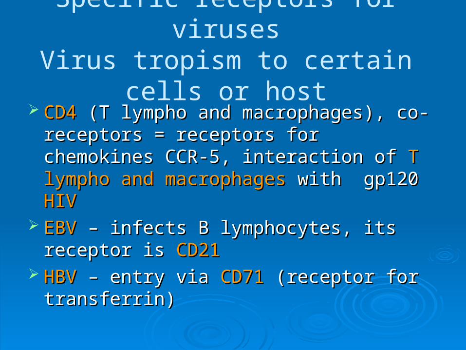
Specific receptors for virusesVirus tropism to certain cells or host
CD4CD4 (T lympho and macrophages), co- (T lympho and macrophages), co-receptors = receptors for chemokines receptors = receptors for chemokines CCR-5, interaction of CCR-5, interaction of T lympho and T lympho and macrophagesmacrophages with gp120 with gp120 HIVHIV
EBVEBV – infects B lymphocytes, its receptor – infects B lymphocytes, its receptor is is CD21 CD21
HBVHBV – entry via – entry via CD71CD71 (receptor for (receptor for transferrin)transferrin)

Anti-viral immune response First lineFirst line – non-specific immunity - – non-specific immunity - interferonsinterferons (INF (INF,, - -
inhibition of virus replication) and inhibition of virus replication) and NK cells NK cells Antibodies Th2 – B lympho co-operationAntibodies Th2 – B lympho co-operation
mucousmucous IgAIgA – blockage of adherence onto epithelium – blockage of adherence onto epithelium (Respiratory viruses, enteroviruses)(Respiratory viruses, enteroviruses)in circulation – neutralisating Abin circulation – neutralisating Ab – IgG and IgM, C – IgG and IgM, C system activation, virus lysissystem activation, virus lysis
Tc Tc – eradication of virus- infected cells – eradication of virus- infected cells Some aggressive viruses may overcome immune
responses and cause severe morbidity and mortality Immunocompromised individuals (adaptive and native
immunity) suffer from more severe viral infection

Anti-viral immunity
Lifetime perzistenceLifetime perzistence (especially herpetic viruses (especially herpetic viruses in nerve in nerve ganglia) ganglia) re-activation during weakening of ISre-activation during weakening of IS
EBV EBV – malignancies of haematopoetic system (Hodgkin – malignancies of haematopoetic system (Hodgkin lymphoma)lymphoma)CMV, VzV, HHV-6, HHV-7CMV, VzV, HHV-6, HHV-7 – recurrent aphthae – recurrent aphthae HSV-1 (herpes labialis)HSV-1 (herpes labialis) - stomatitis herpetica, - stomatitis herpetica, gingivostomatis herpetica gingivostomatis herpetica
virus varicela-zostervirus varicela-zoster– herpes zoster, gingivitis– herpes zoster, gingivitispapilomaviruses papilomaviruses – cervix carcinoma, skin tumors– cervix carcinoma, skin tumors

PrionesPriones Discovery - S.B. Prusiner – Nobel prizeDiscovery - S.B. Prusiner – Nobel prize Rare, transmissible, fatal, spongiform Rare, transmissible, fatal, spongiform
neurodegenerative diseasesneurodegenerative diseases Protein infectious particule able to reproduce, Protein infectious particule able to reproduce,
absence of nucleic acids – PrPabsence of nucleic acids – PrP Organismus codes gene for normal protein -
PrPc, present in normal brain tissue (CNS), heart and skeletal muscles and other organs, unknown function, obviously regulation of ciarcadian rhythm, neuro-muscular synapsis, ion transport via membrane
Abnormal infectious protein - PrPSC found in brains of animals with scrapie (lamb and goats)

PrionesPriones PrPc and PrPSC differ in primary structure
just by 1 aminoacid, however significant difference in secondary and tertiary structures, resistent to proteolytic degradation, creates insoluble aggregates similarly like amyloid deposits, damage of nervous tissue via acumulation of abnormal PrPSC and/or by lack of normal
PrPc

Priones and human diseasesPriones and human diseases
Creutzfeldt-Jakob disease (CJD) Variant CJD (vCJD) Gerstmann-Sträussler-Scheinker disease
similar to CJD,but amyloid deposits in cerebelum
Fatal Familial Insomnia – in families with CJD, neurone loss
Kuru – tribesmen New Guinea – cannibalism– Nobel price Dr. Gajdusek

Creutzfeldt-Jakob disease Most frequent between diseases caused by Most frequent between diseases caused by
priones priones Sporadic formSporadic form – 87 % - unknown reason, clinical – 87 % - unknown reason, clinical
symptoms after the age of 60 – dementia, symptoms after the age of 60 – dementia, ataxia, myoclonusataxia, myoclonus
Inborn (familial) formInborn (familial) form – 8 % - mutation in gene – 8 % - mutation in gene coding PrPcoding PrPcc
Transmission by iatrogennic way Transmission by iatrogennic way – 5 % - – 5 % - aplication of GF from hypophysis, Tx of cornea, aplication of GF from hypophysis, Tx of cornea, brain operation, etc. brain operation, etc.
Neurone loss, astrocyte and microglia Neurone loss, astrocyte and microglia hyperplasia, amyloid depositshyperplasia, amyloid deposits

Variant CJD Breeding of cattle in UK – Breeding of cattle in UK – BSE – Bovinne Spongiforme BSE – Bovinne Spongiforme
Encephalopathie – mad cow disease – transmission from Encephalopathie – mad cow disease – transmission from cattle to man by foodcattle to man by food, since 1986 sicken 180 thousands of , since 1986 sicken 180 thousands of animals, the brain looks like spondgeanimals, the brain looks like spondge, loss of muscle control, dezorientation, death. Infection – feeding mixture – meat and bone meal – transmission of scrapie agens (goats) to cattle, now EC legislation prohibition of nutrition of livestock and fish, obligation to test cattle for meat for the presence of BSE
Human form of this diseaseHuman form of this disease, so-called , so-called variant Creutzfeldt-variant Creutzfeldt-Jakob disease (vCJD),Jakob disease (vCJD), in UK death of 90 people in UK death of 90 peopleSlower progression of the disease, deterioration of psyche, Slower progression of the disease, deterioration of psyche, parastesia, ataxia, myoclonus, dementia parastesia, ataxia, myoclonus, dementia CNS (basal ganglia), tonzila, spleen, nodules – plaque and CNS (basal ganglia), tonzila, spleen, nodules – plaque and detection of PrPdetection of PrPSCSC