antigen of polyoma virus
TRANSCRIPT
Proc. Nat Acad. Sci. USAVol. 79, pp. 800-804, February 1982Biochemistry
Expression of transforming region of Moloney murine sarcomavirus in Escherichia coli as a fusion protein with small tumorantigen of polyoma virus
(recombinant DNA/transforming retrovirus/nucleotide sequence/lac operon/mos gene)
DANIEL J. DONOGHUE AND TONY HUNTERTumor Virology Laboratory, The Salk Institute, P.O. Box 85800, San Diego, California 92138
Communicated by Dan L. Lindsley, October 28, 1981
ABSTRACT Bacterial expression of the transforming regionof Moloney murine sarcoma virus, designated mos, was obtainedas a fusion protein with a portion of the small tumor antigen ofpolyoma virus. This was accomplished by fusing the entire mosopen reading frame, encoding a 41,000-dalton protein, with a plas-mid that expresses a (3-galactosidase-polyoma fusion. protein un-der lac operon control. The resulting plasmid directed synthesisof the predicted polyoma antigen-sarcoma virus fusion protein of59;000 daltons. This protein was immuneprecipitated by an anti-polyoma tumor antigen antiserum that recognized polyoma de-terminants at the NH2 terminus ofthe hybrid protein. This proteinwas also immunoprecipitated by an antiserum directed against asynthetic peptide containing the 12 COOH-terminal amino acidsencoded by the mom open reading frame. This work confirms theexistence ofa long open reading frame in the mos gene and resolvesa discrepancy between different nucleotide sequences for itsCOOH-terminal coding region.
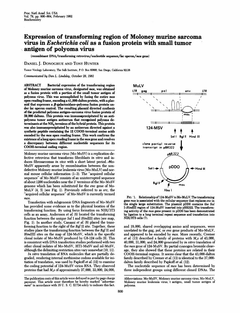
Moloney murine sarcoma virus (Mo-MuSV) is a replication-de-fective retrovirus that transforms fibroblasts in vitro and in-duces fibrosarcomas in vivo with a short latent period. Mo-MuSV apparently arose by recombination between the non-defective Moloney murine leukemia virus (Mo-MuLV) and nor-mal mouse cellular information (1-3). The "acquired cellularsequence of Mo-MuSV consists of an uninterrupted sequenceofabout 1200 nucleotides near the 3' terminus ofthe Mo-MuSVgenome which has been substituted for the env gene of Mo-MuLV (4, 5) (see Fig. 1). Previously referred to as src, the"acquired cellular sequence" of Mo-MuSV is currently termedmos.
Transfection with subgenomic DNA fragments of Mo-MuSVhas provided some evidence as to the physical location of thetransforming function. By using focus formation on NIH/3T3cells as an assay, Andersson et al. (6) located the transformingfunction between the unique Sal I and HindIII sites (see map,Fig. 1). In another study, Canaani et al. (8) placed the trans-forming function to the right of the Bgl II site. Together, thesestudies place the transforming function between the Bgl II andHindIII sites on the map of 124-MuSV, which is the specificclonal isolate of Mo-MuSV produced by G8-124 cells (9). Thisis consistent with DNA transfection studies performed with twoother clonal isolates of Mo-MuSV, HT1-MuSV and ml-MuSV,although.the delimiting restriction sites vary somewhat (10, 11).
In vitro translation of RNA molecules that are partially de-graded, rendering internal methionine codons available for ini-tiation of translation, was used by Papkoff et aL (12) to examinethe coding potential of 124-MuSV virion RNA. They observedproteins that had Mrs of approximately 37,000, 33,000, 24,000,
MuLVLTR gag pol env LTR
N\
N%N N
mos
== A= =I1 24-MSV
clone partial reversetranscript in pBR322
Sal 1_
Sal I Bgl 11 Hind Ill
*s_ Hind IlI
Bgl 11
Xba I
FIG. -1. Relationship of124-MuSV to Mo-MuLV. The transforminggene mos is associated with the cellular sequence that replaces env inthe single large substitution. The plasmid pDDO contains the SalI-HindEI region of 124-MuSV inserted into pBR322. The transform-ing activity of the mos gene present in pDDO has been demonstratedby ligation to a long terminal repeat sequence and transfection intoNIH/3T3 cells (7).
.and 18,000, shared overlapping amino acid sequences, wereunrelated to the gag, pol, or env gene products of Mo-MuLV,and appeared to be encoded by mos. More recently, Cremeret aL (13) described a family of proteins with Mrs of 43,000,40,000, 31,000, and 24,000 generated by in vitro translation ofthe mos gene of 124-MuSV. By partial cyanogen bromide cleav-age, they also showed that these proteins are related in theirCOOH-terminal regions. It seems clear that the 43,000-daltonfamily described by Cremer et aL (13) is identical to the 37,000-dalton family described by Papkoff et aL (12).The nucleotide sequence of. mos has been determined by
three independent groups using different cloned DNAs. The
Abbreviations: Mo-MuSV, Moloney murine sarcoma virus; Mo-MuLV,Moloney murine leukemia virus; t antigen, small tumor antigen ofpolyoma.
800
The publication costs ofthis article were defrayed in part by page chargepayment. This article must therefore be hereby marked "advertise-ment" in accordance with 18 U. S. C. §1734 solely to indicate this fact.

Proc. NatL Acad. Sci. USA 79 (1982) 801
mos sequence of the biologically active clone pDDO (7) agrees
with that ofVan Beveren et al. (14) for the clone pMSV-1; thesesequences predict a single large open reading frame that couldencode a protein of Mr 41,000. However, these two sequences
differ from the mos sequence ofReddy et al. (15) as to the COOHterminus of the predicted polypeptide.
In order to resolve this discrepancy, we wished to expressthe mos open reading frame as a bacterial protein in Escherichiacoli. To this end, we exploited the plasmid pgt-7 in which thesmall tumor antigen (t antigen) of polyoma virus is expressedunder lac operon control (16). In the hybrid plasmid pDD21,the entire mos open reading frame was fused to the coding re-gion of the polyoma antigen. Characterization of the predictedt antigen-MuSV fusion protein, encoded by pDD21, serves toresolve the discrepancy concerning the sequence of the mosopen reading frame.
MATERIALS AND METHODSConstruction of pDD21. The plasmid pDDO containing the
Sal I-HindIII Mo-MuSV fragment was constructed by StephenGoffand Mitchell Goldfarb (at Massachusetts Institute ofTech-nology). Plasmid pgt7-wt6f, abbreviated pgt-7 (16), and the bac-terial strain HB101 (17) were provided by Art Horwich andWalter Eckhart.The desired Xba I-HindII1 fragment ofpDDO was purified
by agarose gel electrophoresis, and DNA was recovered fromthe gel slice by the glass powder technique (18). The Xba I endwas partially filled in by incubation for 20 min at 17'C in 50 mMTris HCl, pH 7.6/10 mM MgCl2/L mM dithiothreitol/20 A.tMdCTP/5 ,uM dTTP with 3 units of E. coli DNA polymerase I.After ethanol precipitation, the DNA was resuspended in 30mM NaOAc, pH 4.6/0.16 M NaCl/0.5 mM ZnCl2 and was di-gested with S1 nuclease. After incubation at 370C for 30 min,EDTA was added to a final concentration of 2 mM. The samplewas extracted once with phenol and three times with ether andwas precipitated with ethanol prior to ligation. The DNA ofpgt-7 was cleaved with Sst I, after which gel purification, S1 nu-
clease treatment, and inactivation of the S1 nuclease were as
for the pDDO DNA above.The resuspended Xba I-HindIII fragment of pDDO was li-
gated (19) to the resuspended Sst I fragment of pgt-7. Aftertransfection, ampicillin-resistant colonies were screened (20) forMo-MuSV-specific sequences by using a probe prepared bynick-translation (21) of an aliquot of the gel-purified XbaI-HindIII fragment. Several clones were obtained with the de-sired insert in either orientation with respect to the polyomasequence; pDD21 bears the insert in the correct orientation forbacterial expression. The mos insert in pDD21 was character-ized by digestion with Ava I, Bgl II, Kpn I, Pst I, Hinfl, andHha I and appeared to be identical to the mos insert in the pa-rental plasmid pDDO.
Immunoprecipitations from Bacterial Lysates. In general,1 ml of exponentially growing bacteria at 2 x 10⁸ cells per mlwere labeled with [3S]methionine (100 ,uCi/ml; 1 Ci = 3.7x 10'0 becquerels) for 45 min in the presence of 1 mM isopropylthiogalactoside (16). Immunoprecipitates of lysates, preparedas described (16), were analyzed by electrophoresis in Na-DodSOJpolyacrylamide gels containing 15% acrylamide and0.09% bisacrylamide. 'S-Labeled proteins were detected byfluorography.
RESULTSMuSV Reading Frames. Fig. 2A presents the methionine
codons and terminator codons for the three reading frames ofthe + strand of the mos sequence ofpDDO between the unique
Bgl II and HindIII sites (having the same sense as the viral RNAgenome). A single large open reading frame is revealed in frame1, containing five methionine residues and encoding a proteinofMr 41,000. Fig. 2B schematically aligns the predicted in vitrotranslation products offrame 1 with the sequence. Five in vitrotranslation products are predicted, differing at their NH2 ter-mini but sharing a common COOH terminus, with calculatedMrs of 41,000, 38,000, 32,000, 22,000, and 8000. The sizes ofthese predicted proteins are in good agreement with those ob-served by in vitro translation of 124-MuSV virion RNA (12, 13).However, the schema presented in Fig. 2B disagrees with themodel presented by Cremer et al. (13); they propose that the41,000-dalton family of proteins has a different COOH termi-nus. This is based on the mos sequence of Reddy et al. (15),which lacks two base pairs present in the sequences of pDDOand pMSV-1; these are located 1226 and 1232 base pairs fromthe Bgl II site. As a result, their proposed reading frame wouldconsist of the NH2-terminal region of reading frame 1 joined tothe COOH-terminal region of reading frame 3, which wouldthen terminate downstream from the HindIII site. This wouldrender each of the predicted proteins slightly larger than thoseshown in Fig. 2B because they would be longer (and differ inamino acid composition) at their COOH termini.
Construction of the Polyoma-MuSV Hybrid Plasmid. Giventhese different nucleotide sequences of mos and the potentialbiological significance of the mos reading frame, we wished toconfirm this reading frame by some technique other than DNAsequence analysis. Our approach involved fusing the entire mosopen reading frame to the coding region of some protein that
FIG. 2. (A) Position of initiator and terminator codons. The three+ strand reading frames refer to the sequence read from the Bgl II siteto the HindII site-i.e., the same sense as the Mo-MuSV viral RNAgenome. (B) Predicted in vitro translation products. Each ATG in themos open reading frame potentially could be used by the in vitro re-ticulocyte lysate translation system to yield the proteins indicated(sizes shown in kilodaltons). The first ATG is actually donated by theenv gene of Mo-MuLV-i.e., the recombination point between Mo-MuLV and c-mos occurs 15 base pairs downstream from the start ofthe mos open reading frame. *, ATG; I, TAG, TAA, or TGA.
A1 11 I L . . I
2011 kMI IIIl I Il-II.1k II III13 loll I . so I
o Distance, bases 1000, . . . 1
BGLII XBA I HI NDI II
B11II ' ' . I
ATG ATG ATG ATG ATG TGA
Met 8--8-
Met 22
Met _ _ _ _ _ _ _ _lt-t 32
Met 38
Met 41
DERIVED DERIVED FROM C-MOSFROM
Mo-MULV
Biochemistry: Donoghue and Hunter
802 Biochemistry: Donoghue and Hunter
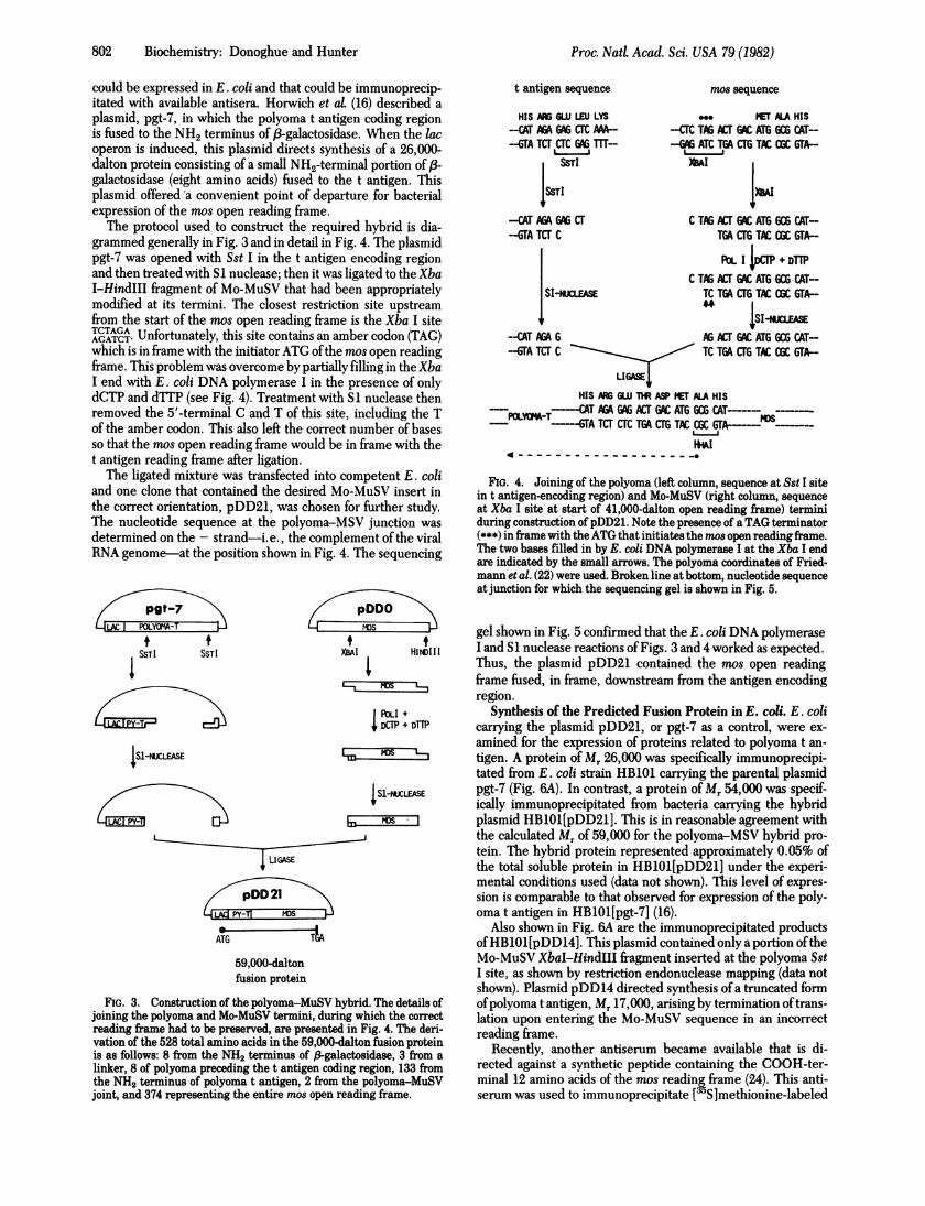
could be expressed in E. coli and that could be immunoprecip-itated with available antisera. Horwich et al. (16) described aplasmid, pgt-7, in which the polyoma t antigen coding regionis fused to the NH2 terminus of f-galactosidase. When the lacoperon is induced, this plasmid directs synthesis of a 26,000-dalton protein consisting of a small NH2-terminal portion of /3-galactosidase (eight amino acids) fused to the t antigen. Thisplasmid offered a convenient point of departure for bacterialexpression of the mos open reading frame.The protocol used to construct the required hybrid is dia-
grammed generally in Fig. 3 and in detail in Fig. 4. The plasmidpgt-7 was opened with Sst I in the t antigen encoding regionand then treated with SI nuclease; then it was ligated to the Xba1-HindIII fragment of Mo-MuSV that had been appropriatelymodified at its termini. The closest restriction site upstreamfrom the start of the mos open reading frame is the Xba I siteTCTAGA Unfortunately, this site contains an amber codon (TAG)which is in frame with the initiator ATG ofthe mos open readingframe. This problem was overcome by partially filling in the XbaI end with E. coli DNA polymerase I in the presence of onlydCTP and dTTP (see Fig. 4). Treatment with Si nuclease thenremoved the 5'-terminal C and T of this site, including the Tof the amber codon. This also left the correct number of basesso that the mos open reading frame would be in frame with thet antigen reading frame after ligation.The ligated mixture was transfected into competent E. coli
and one clone that contained the desired Mo-MuSV insert inthe correct orientation, pDD21, was chosen for further study.The nucleotide sequence at the polyoma-MSV junction wasdetermined on the - strand-i.e., the complement of the viralRNA genome-at the position shown in Fig. 4. The sequencing
t
I SSTI SSTI
pDDO
XBAI HiNDI II
RIb ++ DCIP + DrrP
IS-NUCLEASE
IS1-NUCLEASE
LIGASE
4DD;j159,000-daltonfusion protein
FIG. 3. Construction of the polyoma-MuSV hybrid. The details ofjoining the polyoma and Mo-MuSV termini, during which the correctreading frame had to be preserved, are presented in Fig. 4. The deri-vation of the 528 total amino acids in the 59,000-dalton fusion proteinis as follows: 8 from the NH2 terminus of f3galactosidase, 3 from alinker, 8 of polyoma preceding the t antigen coding region, 133 fromthe NH2 terminus of polyoma t antigen, 2 from the polyoma-MuSVjoint, and 374 representing the entire mos open reading frame.
t antigen sequence
HIS AG GLIU LEU LYS-CAT AGA GAG CrC MA--GTA TCT CrC GG mrr-
SSTI
-CAT A GA CT--GTA TCT C
mos sequence
MET ALA HIS-CTC TAG AC1 GC ATG GCG CAT---C ATC TGA CTG TAC CGC GA-
XBAI
C TAG ALT GC ATG GCG CAT-TGA CrG TC OGC GTA-
POL I M + DTilC TAG ACT[AC ATG GCG CAT--
SI-MMEASE TC TGA CTG TAC CGC GTA-
-CAT AGA G AGLT AC ATG GCG CAT--GTA TCT C TC TGA CTG TAC OC GTA-
LIGASE
HIS AR GLIJ TM ASP MET ALA HIS
POLY--XT AA GM ACT GAC ATG GCGM4IA TCT CTC TGA CMG TAC CGTA
FIG. 4. Joining of the polyoma (left column, sequence at Sst I sitein t antigen-encoding region) and Mo-MuSV (right column, sequenceat Xba I site at start of 41,000-dalton open reading frame) terminiduring construction of pDD21. Note the presence of aTAG terminator(***) in frame with the ATG that initiates the mos open reading frame.The two bases filled in by E. coli DNA polymerase I at the Xba I endare indicated by the small arrows. The polyoma coordinates of Fried-mann et al. (22) were used. Broken line at bottom, nucleotide sequenceat junction for which the sequencing gel is shown in Fig. 5.
gel shown in Fig. 5 confirmed that the E. coli DNA polymeraseI and S1 nuclease reactions of Figs. 3 and 4 worked as expected.Thus, the plasmid pDD21 contained the mos open readingframe fused, in frame, downstream from the antigen encodingregion.
Synthesis of the Predicted Fusion Protein in E. coli. E. colicarrying the plasmid pDD21, or pgt-7 as a control, were ex-amined for the expression of proteins related to polyoma t an-tigen. A protein of Mr 26,000 was specifically immunoprecipi-tated from E. coli strain HB101 carrying the parental plasmidpgt-7 (Fig. 6A). In contrast, a protein of Mr 54,000 was specif-ically immunoprecipitated from bacteria carrying the hybridplasmid HB101[pDD21]. This is in reasonable agreement withthe calculated Mr of 59,000 for the polyoma-MSV hybrid pro-tein. The hybrid protein represented approximately 0.05% ofthe total soluble protein in HB101[pDD21] under the experi-mental conditions used (data not shown). This level of expres-sion is comparable to that observed for expression of the poly-oma t antigen in HB101[pgt-7] (16).
Also shown in Fig. 6A are the immunoprecipitated productsofHB101[pDD14]. This plasmid contained only a portion oftheMo-MuSV XbaI-HindIII fragment inserted at the polyoma SstI site, as shown by restriction endonuclease mapping (data notshown). Plasmid pDD14 directed synthesis of a truncated formofpolyoma t antigen, Mr 17,000, arising by termination oftrans-lation upon entering the Mo-MuSV sequence in an incorrectreading frame.
Recently, another antiserum became available that is di-rected against a synthetic peptide containing the COOH-ter-minal 12 amino acids of the mos reading frame (24). This anti-serum was used to immunoprecipitate [ S]methionine-labeled
Proc. Nad Acad. Sci. USA 79 (1982)

Proc. Natl. Acad. Sci. USA 79 (1982) 803
A
TG
-AT
4N;m- - T
-- -G
B1 2 3
- 4-+54
1 2 3
_--4-54M^a._
No Eq*e5e
R -_
d. *26
..-.J.....- %* +17
-A- C
G A C C
C T
FIG. 5. Nucleotide sequence across the polyoma-MuSV junction.DNA ofplasmid pDDO was 32P-labeled at the 5' termini of the HhaIsite GcG shown in Fig. 4, and the sequence was determined upstreaminto the t antigen encoding region. The products of Maxam-Gilbert(23) reactions were analyzed by electrophoresis in a 24% polyacryla-mide sequencing gel. The location of this sequence is indicated by thedashed arrow at the bottom of Fig. 4. The two bases at the Xba I sitethat were filled in by E. coli DNA polymerase I are indicated by thesmall arrows. To isolate a DNA fragment uniquely labeled at the HhaI site, an Alu I site at polyoma position 539 (22) was utilized for sec-ondary digestion.
lysates ofHB101 harboring either pDD21 or pgt-7 as a negativecontrol. This antiserum precipitated a 54,000-dalton proteinfrom lysates of HB101[pDD21] (Fig. 6B), which comigratedwith the protein specifically immunoprecipitated from the samelysate by the polyoma antitumor serum. As expected, theCOOH-terminal antiserum fitiled to immunoprecipitate the26,000-dalton t antigen produced in HB101[pgt-7].
DISCUSSION
There exists the possibility of a frameshift error in any nucleo-tide sequence of significant length, particularly when a se-
quence is determined with no a priori knowledge of the geneproduct. In the case of Mo-MuSV, we have used bacterialexpression of the mos gene product as a means of confirmingthe open reading frame predicted by the nucleotide sequence.This entailed construction of a hybrid between the coding re-
FIG. 6. Immunoprecipitations of [35S]methionine-labeled bacte-rial lysates. (A) Polyoma antitumor serum was used to immunopre-cipitate lysates of HB101[pgt-7] (lane 1), HB101[pDD21I (lane 2), andHB101[pDD14I (lane 3). (B) An antiserum against a synthetic peptidecontainingthe COOH-terminal 12 amino acids of the mos open readingframe was used to immunoprecipitate lysates of HB101[pgt-71 (lane1) and HB101[pDD21I (lane 2). For comparison, lane 3 showsHB101[pDD21I immunoprecipitated with polyoma antitumor antiserum.
gion of polyoma t antigen and the mos open reading frame. E.coli harboring this plasmid directed synthesis ofa protein oftheexpected size. This protein was specifically immunoprecipi-tated by polyoma antitumor antiserum through recognition ofpolyoma determinants at its NH2 terminus. The hybrid proteinwas also immunoprecipitated by an antiserum directed againstthe predicted COOH terminus of the mos open reading frame.These immunoprecipitation studies demonstrate that the ter-mini of the mos open reading frame are as predicted by thenucleotide sequences ofpDDO and ofpMSV-1 (14). The alter-native COOH terminus predicted by Reddy et al. (15) is incom-patible with these results.The results reported here are in agreement with recent re-
sults of Papkoff et at (24). Using the aforementioned COOH-terminal mos antiserum directed against a synthetic peptidepredicted by the sequences ofpDDO and pMSV-1, they wereable to immunoprecipitate in vitro translation products of 124-MuSV virion RNA.What is the biological significance of this open reading frame
in mos, with regard to transformation? For the following reasonswe suggest that the 41,000-dalton reading frame is used in vivoto encode a transforming gene product. First, all retroviraltransforming genes examined thus far are devoid ofinterveningsequences in their coding regions, although intervening se-quences are often found in the cellular homologues of these
Biochemistry: Donoghue and Hunter

804 Biochemistry: Donoghue and Hunter
genes-e.g., Rous sarcoma virus (25), Abelson murine leukemiavirus (26), and Harvey murine sarcoma virus (27). By analogy,one expects the coding region for the Mo-MuSV transformingfunction to be uninterrupted by intervening sequences. Asshown in Fig. 2A, reading frame 1 is the sole reading frame thatcontains a large uninterrupted coding region.
Second, the NH2-terminal region of the mos open readingframe is derived from the env gene of Mo-MuLV (7, 14,15)-i.e., the recombinational event between Mo-MuLV andthe cellular homologue c-mos occurred 15 base pairs down-stream from the env gene NH2 terminus. Recently, we exam-ined the nucleotide sequence of other isolates of murine sar-coma virus, including Gazdar murine sarcoma virus (28) and ml-MuSV; in these cases, the NH2 terminus of the Mo-MuLV envgene initiates the mos open reading frame (unpublished data).Thus, we infer that proper expression of the viral mos gene iscontingent upon its fusion to the NH2 terminus ofthe env gene,analogous to many other transforming retroviruses in whichcellular information has been fused to the retroviral gaggene -.g., Abelson murine leukemia virus (29), feline sarcomavirus (30), or Y73 virus (31).
At the present time, no conclusive data exist concerning amos-specific mRNA in transformed cells. Recently, however,evidence has been obtained that the 41,000-dalton product isexpressed in Mo-MuSV-transformed cells (J. Papkoff, personalcommunication). Ultimately, characterization of mutants thatinterrupt the mos open reading frame will be required to resolveits significance.
Note Added in Proof. Recently, Reddy et aL (32) revised their earliersequence of mos (15). In their latest sequence, three additional basepairs are present. Two ofthese base pairs, at 1226 and 1232 nucleotidesfrom the Bgl II site, are now in agreement with the sequences ofpDDO(7) and pMSV-1 (14). However, the third additional base pair in theirrevised sequence (32) was not observed in these other sequencs ofmos.Thus, both sequences of Reddy et aL (15, 32) are in disagreement withthe results presented here as to the correct reading frame in the COOH-terminal region of mos.
We thank Mary Anne Hutchinson and Walter Eckhart for providingthe polyoma antitumor serum, Jackie Papkoffand Inder Verma for pro-viding the COOH-terminal Mo-MuSV antiserum, Art Horwich for pro-viding pgt-7 DNA, Steve Goff for providing the Mo-MuSV-specificplasmid, Peter Geiduschek for valuable criticism ofthe manuscript, andArnie Berk, Jon Cooper, Art Horwich, Bart Sefton, and Dennis Tem-pleton for stimulating conversations. D.J.D. gratefully acknowledgesa Helen Hay Whitney postdoctoral fellowship. This work was supportedby U.S. Public Health Service Grants CA-14195, CA-17096, and CA-28458.
1. Moloney, J. B. (1966) NatL Cancer Inst. Monogr. 22, 139-142.2. Scolnick, E. J., Howk, R. A., Anisowicz, A., Peebles, P. T.,
Scher, C. D. & Parks, W. P. (1975) Proc. NatL Acad. Sci. USA 72,4650-4654.
3. Frankel, A. E. & Fischinger, P. J. (1976) Proc. NatL Acad. Sci.USA 73, 3705-3709.
4. Hu, S., Davidson, N. & Verma, I. M. (1977) Cell 10, 469-477.5. Donoghue, D. J., Sharp, P. A. & Weinberg, R. A. (1979) J. Virol
32, 1015-1027.6. Andersson, P., Goldfarb, M. P. & Weinberg, R. A. (1979) Cell
16, 63-75.7. Donoghue, D. J. (1982)J. ViroL, in press.8. Canaani, E., Robbins, K. C. & Aaronson, S. A. (1979) Nature
(London) 282, 378-383.9. Ball, J. K., McCirter, J. A. & Sunderland, S. M. (1973) Virology
56, 268-284.10. Blair, D. G., McClements, W. L., Oskarsson, M. K., Fischin-
ger, P. J. & Van de Woude, G. F. (1980) Proc. NatL Acad. Sci.USA 77, 3504-3508.
11. Oskarsson, M., McClements, W. L., Blair, D. G., Maizel, J. V.& Van de Woude, G. F. (1980) Science 207, 1222-1224.
12. Papkoff, J., Hunter, T. & Beemon, K. (1980) Virology 101,91-103.
13. Cremer, K., Reddy, E. P. & Aaronson, S. A. (1981)J. Virol 38,704-711.
14. Van Beveren, C., Galleshaw, J. A., Jonas, V., Berns, A. J. M.,Doolittle, R. F., Donoghue, D. J. & Verma, I. M. (1981) Nature(London) 289, 258-262.
15. Reddy, E. P., Smith, M. J., Canaani, E., Robbins, K. C., Tron-ick, S. R., Zain, S. & Aaronson, S. A. (1980) Proc. Natl Acad. Sci.USA 77, 5234-5238.
16. Horwich, A., Koop, A. H. & Eckhart, W. (1980) J. Virol 36,125-132.
17. Boyer, H. W. & Roulland-Dussoix, D. (1969) J. Mol Biol 41,459-472.
18. Vogelstein, B. & Gillespie, D. (1979) Proc. Nati Acad. Sci. USA76, 615-619.
19. Ferretti, L. & Sgaramella, V. (1981) Nucleic Acids Res. 9, 85-93.20. Grunstein, M. & Hogness, D. S. (1975) Proc. Natl Acad. Sci. USA
72, 3461-3465.21. Rigby, P. W. J., Dieckmann, M., Rhodes, C. & Berg, P. (1977)
J. Mol Biol 113, 237-247.22. Friedmann, R., Esty, A., LaPorte, P. & Deininger, P. (1979) Cell
17, 715-724.23. Maxam, A. & Gilbert, W. (1980) Methods Enzymol 65, 499-560.24. Papkoff, J., Lai, M. H.-T., Hunter, T. & Verma, I. M. (1981) Cell
27, 109-119.25. Shalloway, D., Zelenetz, A. D. & Cooper, G. M. (1981) Cell 24,
531-541.26. Goff, S. P., Gilboa, E., Witte, 0. N. & Baltimore, D. (1980) Cell
22, 777-785.27. DeFeo, D., Gonda, M. A., Young, H. A., Chang, E. H., Lowy,
D. R., Scolnick, E. M. & Ellis, R. W. (1981) Proc. NatL Acad. Sci.USA 78, 3328-3332.
28. Gazdar, A. F., Chopra, H. C. & Sarma, P. S. (1972) Int. J. Can-cer 9, 219-233.
29. Witte, 0. N., Rosenberg, N., Paskind, M., Shields, A. & Balti-more, D. (1978) Proc. Natl Acad. Sci. USA 75, 2488-2492.
30. Barbacid, M., Lauver, A. V. & Devare, S. G. (1980)J. Virol 33,196-207.
31. Kawai, S., Yoshida, M., Segawa, K., Sugiyama, H., Ishizaki, R.& Toyoshima, K. (1980) Proc. NatL Acad. Sci. USA 77, 6199-6203.
32. Reddy, E. P., Smith, M. J. & Aaronson, S. A. (1981) Science 214,445-450.
Proc. Nad Acad. Sci. USA 79 (1982)