antimicrobial activity of essential oils against...
TRANSCRIPT

Universidade de Lisboa
Faculdade de Ciências
Departamento de Biologia Vegetal
Antimicrobial activity of essential oils
against multiresistant
aeromonads and enterococci
Ana Patrícia Páscoa Quendera
Dissertação
MESTRADO EM MICROBIOLOGIA APLICADA
2014

ii
Universidade de Lisboa
Faculdade de Ciências
Departamento de Biologia Vegetal
Antimicrobial activity of essential oils
against multiresistant
aeromonads and enterococci
Ana Patrícia Páscoa Quendera
Dissertação
MESTRADO EM MICROBIOLOGIA APLICADA
Dissertação orientada pela Doutora Teresa Maria Leitão Semedo-Lemsaddek e pela
Professora Doutora Ana Maria Gonçalves Reis
2014

iii
Antimicrobial activity of essential oils
against multiresistant
aeromonads and enterococci
Ana Patrícia Páscoa Quendera
2014
This thesis was fully performed at the Department of Animal Production and Food Safety,
Faculty of Veterinary Medicine of the University of Lisbon, under the direct supervision of Dr.
Teresa Maria Leitão Semedo-Lemsaddek in the scope of the Master in Applied Microbiology
of the Faculty of Sciences of the University of Lisbon.
Prof. Dr. Ana Maria Gonçalves Reis was the internal designated supervisor in the scope of
the Master in Applied Microbiology of the Faculty of Sciences of the University of Lisbon.

iv
Agradecimentos
Em primeiro lugar, agradeço à minha orientadora, a Doutora Teresa Semedo-Lemsaddek,
pela partilha de valiosos conhecimentos, pelo apoio, pelas críticas construtivas, pela
paciência e pelo incentivo;
À Professora Doutora Ana Maria Reis, pela disponibilidade, pelo apoio, pela motivação e
pelo pensamento positivo;
À Zé e à Lena, por todo o carinho e apoio que deram diariamente, por nos ouvirem e por nos
incentivarem;
À Mariana, à Clenira, ao Ângelo, à Cynthia, à Ana, à Cláudia e à Mónica, por fazerem do
3.99 o gabinete mais divertido daquele piso, pela amizade, pela disponibilidade, pela ajuda e
(como não podia deixar de ser) pelos bolos;
A todos os meus amigos pelo apoio e pensamento positivo, em particular, à Ana Catarina, à
Ema e à Vanessa, pela força e motivação e por acreditarem em mim;
Ao Filipe, pela enorme paciência, pelos conselhos, pela motivação, pelas discussões/trocas
de ideias, pelo grande apoio, pelo carinho e por não me deixar desistir;
Aos meus gatos, Yoshi e Luna, por terem sido os meus companheiros durante a escrita da
dissertação e por serem boas distrações;
Por último, agradeço imenso ao meu irmão e aos meus pais por acreditarem em mim, por
todos os esforços feitos e por me apoiarem sempre. Em especial, agradeço à minha mãe
toda a paciência que teve para ouvir as minhas “queixas” e desabafos, mesmo quando não
percebia tudo o que eu fazia.
Muito obrigada a todos.
v
Abstract
Aeromonas and Enterococcus are ubiquitous microorganisms associated with
infections in humans and animals. The emergence of multiresistant strains and biofilm
formation in clinical and food industry settings are major problems to public health worldwide.
In the present study, in order to contribute to the evaluation of alternatives to
antibiotics/disinfectants, eleven commercial essential oils were assessed for their
antimicrobial and anti-biofilm activity against four aeromonads and four enterococci, sampled
from clinical and environmental settings, all of which presented a multiresistant phenotype.
Evaluation of the antimicrobial activity by the disc diffusion method, led to the selection
of lemongrass and thyme essential oils as the compounds presenting the highest inhibitory
activities. Then, broth microdilution was used to assess minimum inhibitory concentrations
for both essential oils, which were lower for aeromonads (0.05-0.20%) than for enterococci
(0.20-1.56%), suggesting that the Gram negative bacteria under analysis must harbor a
cellular mechanism, or target, highly susceptible to the action of the aforementioned
essential oils.
The effect of lemongrass and thyme essential oils was also tested on previously
formed biofilms using an adaptation of the Calgari Biofilm Device. The biofilms were
subjected to 30 minutes or 1 hour of exposure to each essential oil and the results were
assessed by colony counting. The essential oils showed high levels of eradication-ability
against Aeromonas biofilms, but were unable to successfully eradicate enterococcal biofilms
in the tested concentrations. Thus, biofilms formed by the bacteria under analysis
(Aeromonas - Gram negative and Enterococcus - Gram positive) must possess distinct
characteristics which could help explain the different biofilm-eradication outcomes.
In conclusion, essential oils applicability as antimicrobial agents was assessed against
multiresistant bacteria and in the future they should be regarded as potential alternatives for
antibiotics, disinfectants or detergents used in the clinical and food industry settings.
Moreover, studies should be directed towards a better understanding of the mechanism of
action of essential oils against bacteria and their toxicity to human cell lines.
Keywords: Aeromonas spp., Enterococcus spp., essential oils, multiresistance, biofilms

vi
Resumo
Os membros dos géneros Aeromonas e Enterococcus são microrganismos ubíquos,
presentes em variados ambientes, como água, solo, animais e plantas. Inicialmente,
aeromonas foram caracterizadas como bactérias patogénicas de seres aquáticos, contudo,
hoje em dia, são também consideradas como bactérias patogénicas emergentes que
provocam infeções em humanos. Por seu lado, enterococos eram bactérias consideradas
inofensivas e benéficas para os humanos, sendo muitas vezes utilizadas na indústria
alimentar. Porém, tal como aeromonas, nas últimas décadas, emergiram como
microrganismos causadores de graves infeções associadas aos cuidados de saúde.
A problemática associada a estes dois géneros deve-se ao facto de estarem altamente
disseminados no ambiente, existindo em elevados números na água e em alimentos, bem
como com a sua capacidade de produzir fatores de virulência, de adquirir genes de
resistência a antibióticos e de formar biofilmes.
A produção de fatores de virulência com propriedades/ações distintas é de extrema
importância durante o processo infecioso, uma vez que estes vão permitir a invasão das
células do hospedeiro e a evasão às suas defesas.
Nos últimos anos, o uso inadequado e abusivo de antibióticos em meio clínico, em
veterinária, na agricultura e na produção animal provocou a emergência de estirpes
resistentes a uma variada gama de antibióticos. Estudos de programas de controlo e
vigilância de saúde pública concluíram que a maioria das infeções associadas aos cuidados
de saúde é causada por estirpes multirresistentes a antibióticos, como por exemplo,
enterococos resistentes à vancomicina.
Adicionalmente, os membros destes dois géneros têm a capacidade de formar
biofilmes, comunidades de microrganismos agregadas a superfícies e envolvidas por uma
matriz polissacarídica. Estas comunidades conferem proteção acrescida contra condições
ambientais desfavoráveis, como falta de nutrientes, stress oxidativo e mecanismos de
defesa do hospedeiro, e além disso a matriz impede ou dificulta a entrada de agentes
antimicrobianos. Assim, a presença de biofilmes em superfícies e/ou instrumentos utilizados
tanto na indústria alimentar como nos hospitais, pode ser considerada como uma das
proveniências de bactérias patogénicas responsáveis por contaminações cruzadas e/ou
infeções em humanos.
Atualmente, os processos de higiene e sanitização não são suficientes para eliminar
os biofilmes. Este facto, aliado à ocorrência de microrganismos multirresistentes a
antibióticos/desinfetantes, levou a uma crescente necessidade de encontrar estratégias para
combater estes problemas de saúde pública.

vii
Desde a antiguidade que os produtos naturais extraídos de plantas têm sido usados
pelos humanos com variados propósitos. Nomeadamente, os óleos essenciais, sendo
compostos aromáticos, são utilizados na indústria alimentar como aditivos de sabor, em
cosméticos, em medicamentos e até mesmo em detergentes. Para além disso, as suas
propriedades antibacterianas, antifúngicas e antivirais são conhecidas há muitas décadas,
sendo utilizados com esse fim em medicina tradicional por todo o mundo. Como os óleos
essenciais são misturas de vários compostos, as suas propriedades antimicrobianas podem
dever-se à interação destes compostos com múltiplos alvos celulares, tornando mais
complexo para os microrganismos o desenvolvimento de mecanismos de resistência.
Desta forma, no presente estudo, quatro isolados de aeromonas e quatro isolados de
enterococos foram escolhidos com base no seu fenótipo de multirresistência, a partir de
coleções bacterianas pré-existentes compostas por isolados provenientes de amostras
clínicas e ambientais.
Inicialmente, a atividade antimicrobiana de onze óleos essenciais (alecrim, alfazema,
alho, artemísia, árvore do chá, coentros, curcuma, erva limeira, gengibre, poejo e tomilho) foi
avaliada contra os isolados ambientais e clínicos multirresistentes de Aeromonas e
Enterococcus, utilizando o método da difusão em disco. Os óleos essenciais com maior
atividade inibitória contra os dois grupos bacterianos foram a erva limeira, o tomilho e a
árvore do chá. Os diâmetros das zonas de inibição com enterococos foram relativamente
menores (máximo: 26.7 mm) do que os das zonas de inibição obtidos para aeromonas (que
atingiram os 42 mm), mas em ambos os casos, os óleos essenciais erva limeira e tomilho
foram selecionados como os óleos mais promissores, sendo escolhidos para utilização nos
ensaios subsequentes.
De seguida, o método das microdiluições foi utilizado para determinar as
concentrações mínimas inibitórias (CMI) e as concentrações mínimas bactericidas (CMB)
dos dois óleos essenciais selecionados. Uma vez que os óleos não se misturam de forma
homogénea com o meio de cultura, foi necessário utilizar um solvente para que a interação
entre o óleo e os microrganismos fosse potenciada. O solvente escolhido foi agar 0.15%
(v/v), uma vez que permitiu a obtenção de uma mistura homogénea e não apresenta
toxicidade para o crescimento bacteriano.
De uma forma geral, as concentrações mínimas de erva limeira e de tomilho
necessárias para inibir o crescimento bacteriano foram menores para aeromonas (0.05-
0.20%) do que para enterococos (0.20-1.56%). Assim, apesar de terem a dupla camada
membranar característica das bactérias Gram-negativas que lhes poderia conferir maior
resistência a estes compostos, deverá existir outro mecanismo ou outro alvo nos isolados de
Aeromonas que os torna mais suscetíveis à atividade antimicrobiana dos dois óleos
essenciais testados.

viii
Por último, foi avaliada a capacidade de erradicação de biofilmes por parte dos óleos
essenciais de erva limeira e de tomilho, tendo sido utilizada uma adaptação do Calgari
Biofilm Device que consistiu na utilização de tampas de microplacas com pinos onde os
biofilmes se formaram. Após 24 horas para estabelecimento dos biofilmes, os mesmos
foram colocados em contacto com diferentes concentrações de óleos essenciais (0.20-
3.13%) e foram testados dois tempos de erradicação (30 minutos e 1 hora). Mais uma vez,
os isolados de Aeromonas foram mais suscetíveis à ação dos óleos essenciais,
apresentando elevados níveis de erradicação de biofilme a 3.13 e a 1.78% com 30 minutos
de tempo de contacto, demonstrando que este período é suficiente para erradicar o biofilme.
O isolado A206 foi o mais sensível, sendo que 0.78% de cada um dos óleos foi suficiente
para a remoção de biofilme previamente formado. Os biofilmes de Enterococcus não foram
erradicados quando submetidos ao contacto com o óleo essencial de erva limeira e
apresentaram níveis muito baixos de erradicação sob tratamento com tomilho,
independentemente do tempo de erradicação. Estes resultados podem dever-se às
diferentes características dos biofilmes produzidos por estes dois géneros bacterianos, de
modo que os primeiros sejam mais facilmente removidos pelos óleos, resultado a necessitar
de comprovação futura.
No geral, é importante realçar que uma das grandes desvantagens do presente estudo
tem a ver com a inexistência de normalização para testes dirigidos à análise da atividade
antimicrobiana de compostos naturais, o que dificulta a comparação e a confirmação da
veracidade dos dados de forma inequívoca, como acontece com os testes de suscetibilidade
aos antibióticos.
Assim, apesar de os óleos essenciais não terem sido igualmente eficazes em biofilmes
de Enterococcus em comparação com os de Aeromonas, os resultados obtidos
demonstraram que os óleos essenciais são alternativas promissoras aos agentes
antimicrobianos usados atualmente, em meio clínico e na indústria alimentar. No entanto,
uma melhor compreensão dos mecanismos antimicrobianos envolvidos na atuação dos
óleos essenciais sobre as bactérias e qual a sua toxicidade em linhas celulares humanas,
são assuntos a necessitar de ser aprofundados em estudos futuros.
Palavras-chave: Aeromonas spp., Enterococcus spp., óleos essenciais, multirresistência,
biofilmes
ix
Index
1. Introduction.........................................................................................................................1
1.1. General characteristics of Aeromonas spp. and Enterococcus spp. .............................1
1.1.1. Taxonomy ..............................................................................................................2
1.1.2. Ecology and epidemiology......................................................................................2
1.1.3. Virulence factors ....................................................................................................3
1.1.4. Susceptibility to antibiotics......................................................................................4
1.1.5. Biofilm formation ....................................................................................................5
1.2. Alternatives to antibiotics and disinfectants: essential oils ............................................7
1.3. Aims of the study ..........................................................................................................9
2. Materials and Methods .....................................................................................................10
2.1. Bacterial strains ..........................................................................................................10
2.2. Essential oils ..............................................................................................................10
2.3. Disc diffusion method .................................................................................................10
2.4. Broth microdilution method .........................................................................................11
2.5. Biofilm eradication assay ............................................................................................11
3. Results and Discussion ....................................................................................................14
3.1. Antimicrobial activity of essential oils against planktonic cells ....................................14
3.2. Biofilm eradication by essential oils ............................................................................19
4. Concluding remarks ..........................................................................................................23
5. References .......................................................................................................................24

x
Index of Figures
Figure 1 – Biofilm formation as a five-stage process ..............................................................5
Figure 2 – Scheme of a microplate lid with pegs. .................................................................11
Figure 3 - Representative scheme of biofilm eradication microplates. ..................................12
Index of Tables
Table 1 – Aeromonads and enterococci chosen for this study. .............................................14
Table 2 – Antimicrobial activity of EOs by disc diffusion method for Aeromonas isolates. ....16
Table 3 – Antimicrobial activity of EOs by disc diffusion method for Enterococcus isolates. .16
Table 4 – MICs and MBCs of lemongrass and thyme EOs against aeromonads and
enterococci by broth microdilution method. ...........................................................................17
Table 5 - Aeromonads decimal log reduction achieved by lemongrass and thyme EOs. ......20
Table 6 - Enterococci decimal log reduction achieved by lemongrass and thyme EOs. ........21

1
1. Introduction
Over the past decades, Aeromonas and Enterococcus species have received
increased attention due to their association with many human diseases and to their
ubiquitous worldwide distribution. The misuse and abuse of antibiotics in clinical, agricultural,
veterinary and animal production settings provides favorable conditions for the selection and
spread of antibiotic resistance, a problem that continues to challenge the healthcare sector.
Results of WHO surveillance program (2014) indicate that an elevated percentage of
healthcare-associated infections are caused by multiresistant strains, such as vancomycin-
resistant enterococci.
Additionally, both aeromonads and enterococci have the ability to produce biofilm, a
complex microbial structure difficult to eradicate by antimicrobial agents and a source of
bacterial infections, both in the clinical and food industry contexts.
New and viable antimicrobial products are needed to address these challenges. Plants
natural products, in particular essential oils, have been used since antiquity due to their
aromatic properties in several areas, such as cosmetics, pharmaceutics, food industry and
detergents. Hence, in the past years, essential oils have emerged as promising alternatives
to antibiotics/disinfectants, due to their effects on the inhibition of bacterial growth and biofilm
eradication.
1.1. General characteristics of Aeromonas spp. and Enterococcus spp.
Aeromonas spp. are Gram-negative, rod-shaped, chemoorganotrophic and facultative
anaerobic bacteria. Their growth temperature ranges from 0 to 45ºC with the optimum growth
varying between 22ºC and 37ºC, although some species are unable to grow at 35ºC (Martin-
Carnahan and Joseph, 2005). Aeromonads were first characterized as pathogens of several
aquatic organisms, but nowadays, they are also considered emerging pathogens associated
with human infection (Igbinosa et al., 2012; Janda and Abbott, 2010).
Enterococci are Gram-positive, oval cocci, facultative anaerobic bacteria and belong to
the group of lactic acid bacteria. Most species are resilient and versatile, being able to
survive at 6.5% NaCl, at pH 9.6 and at a wide range of temperatures (10 to 45ºC), with the
optimum growth at 35-37ºC (Ludwig et al., 2009). Initially, Enterococcus spp. were
considered as harmless commensal inhabitants of the gastrointestinal tract of humans,
widely used in the food industry as probiotic or starter cultures (Moreno et al., 2006).
However, for the last two decades, enterococci became one of the most common pathogens
to be associated with healthcare-associated infections.

2
1.1.1. Taxonomy
Originally included in the Pseudomonadaceae family, Aeromonas genus was
transferred to the Vibrionaceae family in 1974. Phylogenetic studies based on 16S rRNA
sequence analysis showed differences between vibrio and aeromonads, leading to the
formation of a new taxonomic family, Aeromonadaceae, as part of the subclass Gamma-
Proteobacteria (Martínez-Murcia et al., 1992 in Martin-Carnahan and Joseph, 2005).
During the past decade, the number of species assigned to the genus has increased,
but in many cases their validity is not universally accepted, since debates regarding species
delineation still remain (Martin-Carnahan and Joseph, 2005; Nhung et al., 2007). Currently
there are thirty one recognized Aeromonas species and twelve subspecies
(http://www.bacterio.cict.fr/a/aeromonas.html, on 14th August 2014).
On the other hand, Enterococcus was first described in 1899, when it was identified as
an intestinal organism and included in the streptococci group (Stiles and Holzapfel, 1997). In
1984, results of DNA-DNA and rDNA-DNA hybridization studies transferred the species
Streptococcus faecium and S. faecalis from the genus Streptococcus to the genus
Enterococcus (Schleifer and Kilpper-Balz, 1984 in Ludwig et al., 2009), hence creating a new
family, Enterococcaceae, as part of the order Lactobacillales. According to J. P. Euzéby,
there are currently fifty three recognized Enterococcus species and two subspecies
(http://www.bacterio.net/enterococcus.html, on 14th August 2014).
1.1.2. Ecology and epidemiology
Aeromonas have been isolated from various environments worldwide, including
surface, drinking and sewage waters, soil, plants and animals (Janda and Abbott, 2010).
As it was previously mentioned, aeromonads are responsible for a wide range of
infectious diseases in humans, in both immunocompromised and immunocompetent
patients, gastroenteritis being the most frequently associated disease (Parker and Shaw,
2011). Aeromonas species are known to cause severe diarrheal disease in children, in the
elderly and in immunocompromised individuals and they have also been implicated in
travelers’ diarrhea (Igbinosa et al., 2012). Moreover, these microorganisms can cause
septicemia, wound, eye, respiratory tract and urogenital tract infections (Parker and Shaw,
2011). Although rare, there are reports of hemolytic-uremic syndrome (Figueras et al., 2007)
and necrotizing fasciitis (Abuhammour, et al., 2006; Angel et al., 2002) associated with
aeromonads.
Colonization of the human gastrointestinal tract by Aeromonas occurs most likely
through ingestion of contaminated drinking water (Scoaris et al., 2008; Sen and Rodgers,

3
2004) or food (Nagar et al., 2011; Shakir et al., 2012), animal feces being probably the major
source of food contamination (Igbinosa et al., 2012). Infections may be caused by exposure
of skin wounds to contaminated water, soil and/or animal bites, in particular, reptile bites
(Angel et al., 2002).
Enterococci, like aeromonads, are widespread in nature. Most species are part of the
intestinal microbiota of mammals and birds, but being ubiquitous, they can also be isolated
from food, plants, soil and water (Ludwig et al., 2009). Enterococci are common in food
products of animal origin, such as dairy products, meat and fermented sausages, and certain
strains are beneficial and influence the taste and aroma of some cheeses (Franz et al., 2011;
Moreno et al., 2006).
These bacteria are also recognized as human and animal opportunistic pathogens.
Enterococci are responsible for several infections in immunocompromised patients, such as
urinary tract infections, endocarditis, surgical wound infection, bacteremia and neonatal
sepsis (Billington et al., 2014; Fisher and Phillips, 2009a; Heintz et al., 2010; Reyes and
Zervos, 2013). Studies have shown that most infecting strains appear to be exogenously
acquired, usually by strains endemic in a hospital where the patient is being hospitalized.
These strains can come from other patients, from the hospital personnel or from the hospital
settings and they possess one or more virulence traits and/or antibiotic resistances (Kayser,
2003).
1.1.3. Virulence factors
The pathogenicity of aeromonads is a complex and yet not well understood mechanism
and their virulence is considered to be multifactorial (Senderovich et al., 2012). The
production of flagella, pili and adhesins allows the attachment and invasion of host cells,
while enterotoxins, proteases, phospholipases and hemolysins cause damages to host cells,
leading to cell death, which allows the multiplication and proliferation of the microorganism
(Gavin et al., 2003 in Igbinosa et al., 2012). Several virulence factors have been identified in
both clinical (Senderovich et al., 2012) and environmental strains, namely from drinking
water (Carvalho et al., 2012; Sen and Rodgers, 2004) and retail food (Ottaviani et al., 2011).
Regarding enterococcal virulence factors, these features are known to play a role in
pathogenicity, since they are associated with colonization and invasion of host tissues,
resistance to host defense mechanisms and production of pathological changes, such as
toxin production or inflammation (Franz et al., 2011). Major virulence determinants include
the enterococcal hemolysin/cytolysin, adhesins, gelatinase, lipase, surface carbohydrates,
superoxide production and hyaluronidase (Jett et al., 1994; Mundy et al., 2000). Various
studies show the presence of virulence factors not only in clinical strains (Medeiros et al.,

4
2014; Soheili et al., 2014), but also in environmental isolates, being found in food (Hammad
et al., 2014; Medeiros et al., 2014), animals (Novais et al., 2013; Semedo-Lemsaddek et al.,
2013) and untreated waters (Macedo et al., 2011).
1.1.4. Susceptibility to antibiotics
Aeromonas species are intrinsically resistant to penicillins (e.g. penicillin, ampicillin,
carbenecillin and ticarcillin) and to first and second-generation cephalosporins due to the
expression of chromosomally encoded β-lactamases (Jones and Wilcox, 1995). In general,
most strains are susceptible to third and fourth-generation cephalosporins, aminoglycosides,
tetracycline, chloramphenicol, quinolones and trimethoprim-sulfamethoxazole (Martin-
Carnahan and Joseph, 2005) however, many investigators have already found strains
resistant to these antibiotics.
Enterococcus species may present intrinsic resistance to several antibiotics; namely β-
lactams, lincosamides, streptogramins, trimethoprim-sulfamethoxazole and low
concentrations of aminoglycosides (Hollenbeck and Rice, 2012; Ludwig et al., 2009).
Acquired resistance has already been reported and may include high concentrations of
aminoglycosides, glycopeptides, macrolides, tetracyclines, chloramphenicol and quinolones
(Hollenbeck and Rice, 2012).
Nowadays, emergence of acquired resistance to several antimicrobial agents has
become a significant public health issue. The worldwide excessive use in human/veterinary
medicine and in agriculture helps explains the increasing number of resistant bacteria,
probably due to elimination of susceptible strains and selection of resistant variants (Davies
and Davies, 2010). Presently, resistant aeromonads and enterococci can be found not only
in the clinical settings (Aravena-Roman et al., 2012; Esteve et al., 2014; Lins et al., 2013),
but also in food (Hammad et al., 2014; Jahan et al., 2013; Nagar et al., 2011; Shakir et al.,
2012), animals (Agersø et al., 2007; Esteve et al., 2014; Novais et al., 2013; Semedo-
Lemsaddek et al., 2013) and water (Khanjanchi et al., 2010; Scoaris et al., 2008). Likewise,
many multiresistant bacteria are being found on these sources (Esteve et al., 2014; Jahan et
al., 2013; Kaskhedikar and Chhabra, 2010; Novais et al., 2013), due to the dissemination of
resistance genes by horizontal gene transfer events, facilitated by mobile genetic elements,
like plasmids and/or transposons (Agersø et al., 2007; Arias and Murray, 2012).
In this context, vancomycin-resistant enterococci (VRE) represent a major problem
since this antibiotic is used as last-resort for the treatment of severe enterococcal infections,
due to limited therapeutic options. Vancomycin-resistance is growing and VRE are becoming
endemic in an increasing number of intensive care facilities worldwide (Arias and Murray,
2012; Cattoir and Leclerq 2013). The percentage of vancomycin-resistance in E. faecium

5
from invasive isolates (blood and cerebrospinal fluid) shows large inter-country variations in
Europe. During recent years, most countries reported resistance percentages of less than
5% and only six out of twenty-nine reported estimates above 10%, being Portugal one of
them with 23.3% of vancomycin-resistant E. faecium invasive isolates (ECDC, 2013).
1.1.5. Biofilm formation
Biofilms are defined as communities of microorganisms of one or more species
irreversibly attached to a surface, which are enclosed in hydrated extracellular polymeric
substances (EPS), like proteins, polysaccharides, phospholipids and nucleic acids, and show
different growth rate and gene transcription from planktonic cells (Donlan and Costerton,
2002; Lindsay and von Holy, 2006; Shi and Zhu, 2009).
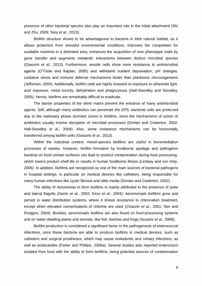
Biofilm formation is a complex developmental process involving attachment and
immobilization on a surface, cell-to-cell interaction, microcolony formation, formation of a
confluent biofilm and developmental of a three-dimensional biofilm structure (Figure 1)
(Mohamed and Huang, 2007).
Figure 1 – Biofilm formation as a five-stage process.
1) Initial attachment; 2) Irreversible attachment; 3) Proliferation; 4) Maturation; 5) Dispersion.
(Image credit: D. Davis (http://www.binghamton.edu/biology/faculty/davies/research.htm))
Various factors influence the formation and development of biofilms. Physiochemical
properties of the bacterial cell and surface materials are strongly correlated with bacterial
initial attachment. For example, porous surfaces entrap more bacteria than smoother
materials and surfaces with a layer of macromolecules will enhance the attachment of cells.
Environmental parameters like pH, nutrient levels, temperature, oxygen levels and the
6
presence of other bacterial species also play an important role in the initial attachment (Shi
and Zhu, 2009; Srey et al., 2013).
Biofilm structure shows to be advantageous to bacteria in their natural habitat, as it
allows protection from stressful environmental conditions, improves the competition for
available nutrients in a delimited area, enhances the acquisition of new phenotypic traits by
gene transfer and augments metabolic interactions between distinct microbial species
(Giaouris et al., 2013). Furthermore, sessile cells show more resistance to antimicrobial
agents (O’Toole and Kaplan, 2000) and withstand nutrient depravation, pH changes,
oxidative stress and immune defense mechanisms better than planktonic microorganisms
(Jefferson, 2004). Additionally, biofilm cells are highly resistant to exposure to ultraviolet light,
acid exposure, metal toxicity, dehydration and phagocytosis (Hall-Stoodley and Stoodley,
2005). Hence, biofilms are remarkably difficult to eradicate.
The barrier properties of the slime matrix prevent the entrance of many antimicrobial
agents. Still, although many antibiotics can penetrate the EPS, bacterial cells are protected
due to the stationary phase dormant zones in biofilms, since the mechanisms of action of
antibiotics usually involve disruption of microbial processes (Donlan and Costerton, 2002;
Hall-Stoodley et al., 2004). Also, some resistance mechanisms can be horizontally
transferred among biofilm cells (Giaouris et al., 2013).
Within the industrial context, mixed-species biofilms are useful in bioremediation
processes of wastes. However, biofilm formation by foodborne spoilage and pathogenic
bacteria on food contact surfaces can lead to product contamination during food processing,
which lowers product shelf-life or results in human foodborne illness (Lindsay and von Holy,
2006). In addition, biofilms are recognized as one of the main sources of bacterial pathogens
in hospital settings, in particular on medical devices like catheters, being responsible for
many human infections like cystic fibrosis and otitis media (Donlan and Costerton, 2002).
The ability of Aeromonas to form biofilms is mainly attributed to the presence of polar
and lateral flagella (Gavín et al., 2002; Kirov et al., 2004). Aeromonads biofilms grow and
persist in water distribution systems, where it shows resistance to chlorination treatment,
except when elevated concentrations of chlorine are used (Chauret et al., 2001; Sen and
Rodgers, 2004). Besides, aeromonads biofilms are also found on food-processing systems
and on water-dwelling plants and animals, like fish, leeches and frogs (Scoaris et al., 2008).
Biofilm production is considered a significant factor in the pathogenesis of enterococcal
infections, once these bacteria are able to produce biofilms in medical devices, such as
catheters and surgical prostheses, which may cause endodontic and urinary infections, as
well as endocarditis (Fisher and Phillips, 2009a). Several studies also reported enterococci
isolated from food with the ability to form biofilms, being potential sources of contamination

7
that might lead to food spoilage and/or transmission of diseases (Jahan and Holley, 2014;
Medeiros et al., 2014; Marinho et al., 2013).
1.2. Alternatives to antibiotics and disinfectants: essential oils
Essential oils (EOs) and other plant extracts have been screened as potential sources
of new antimicrobial compounds, alternatively to current antibiotics/disinfectants; or as
agents used to promote food conservation (Kon and Rai, 2012; Rios and Recio, 2005; Seow
et al., 2014).
Complex mixtures of volatile compounds produced by plants, EOs are characterized by
a strong odor and are formed as secondary metabolites (Bakkali et al., 2008). In nature, they
can act as internal messengers, as defensive substances against herbivores or as volatiles
to attract pollinating insects to their host (Harrejin et al., 2001 in Franz and Novak 2010).
Essential oils are soluble in lipids and organic solvents, presenting lower density than
water. They can be synthesized by all plant organs (e.g. flowers, leaves, buds, stems, seeds,
fruits, roots, twigs or wood) and are stored in secretory cells, cavities, epidermic cells and
glandular trichomes (Bakkali et al., 2008). Steam or water distillation is the most commonly
used method for commercial production of EOs (Van de Braak and Leijten, 1999 in Burt,
2004).
These oils harbor two or three major components, which make up to 20-70% of the
composition, but other elements may be present in vestigial amounts. The main components
generally determine the biological properties of the EOs and can be divided into two groups:
terpenes and aromatic compounds (Bakkali et al., 2008).
The antibacterial properties of EOs have been known for a long time (Guenther, 1948
in Burt, 2004). Basil, cinnamon, clove, mint, oregano, salvia, tea tree and thyme EOs have
been found to possess relevant antibacterial properties, being the most studied (Burt, 2004;
Rios and Recio, 2005). The broad activity of EOs can make them a valued weapon against
multiresistant strains. Especially considering that, until now, there has been no evidence of
emergence of resistance against these compounds; but also due to their low mammalian
toxicity and easy biodegradability in water and soil, making them relatively environmentally
friendly (Isman, 2000).
In several studies performed in recent years, Aeromonas spp. have been considered
highly vulnerable to EOs. Iturriaga et al. (2012) showed A. hydrophila/caviae was more
susceptible to oregano and thyme than Pseudomonas fluorescens and Listeria innocua.
Likewise, Klein et al. (2013) reported A. hydrophila susceptibility towards six EOs
components, in comparison with Escherichia coli and Brochothrix thermosphacta. In addition,
oregano and rosemary EOs, used singly and in combination at sub-inhibitory concentrations,
8
inhibited the cell viability of A. hydrophila, leading to the release of cytoplasmic material and
altering cellular morphology (Azerêdo et al., 2012).
Concerning enterococci, EOs from Eucalyptus globules, Kadsura longipenduculata and
Sideritis erythrantha showed marked in vitro inhibition against VRE (Solórzano-Santos and
Miranda-Novales, 2012). Fisher and Phillips (2009b) showed that a blend of orange and
bergamot EOs (1:1 v/v) affected the cell membrane and homeostasis of E. faecium and E.
faecalis strains, resulting in inhibition of growth or cell death. Thyme showed a very strong
activity against Enterococcus reference and multiresistant clinical strains (Sienkiewicz et al.,
2012) as well as lemongrass against VRE and MRSA strains (Warnke et al., 2013).
Although EOs antibacterial properties have been studied in the past years, their
mechanism of action is yet to be fully known. However, since EOs are lipophilic they are
likely to surpass the cell wall and cytoplasmic membrane, disrupting and permeabilizing the
structure. Extensive leakage of critical molecules and ions disrupts cell homeostasis,
resulting in cell death (Burt, 2004). Moreover, EOs can coagulate the cytoplasm (Gustafson
et al., 1998) and damage lipids and proteins (Burt, 2004). Hence, considering the variety of
compounds present in EOs, it is most likely that their antibacterial activity is not due to one
specific mechanism but they should target many cellular mechanisms (Carson et al., 2002 in
Burt, 2004).
Essential oils also exhibit antiviral (Astani et al., 2011; Elaissi et al., 2012; Ocazionez et
al., 2010), antifungal (Dias Ferreira et al., 2013; Martins et al., 2014; Pinto et al., 2013),
antiparasitic (Echeverrigaray et al., 2010; Silva et al., 2014) and insecticidal (Bossou et al.,
2013; Chu et al., 2012) properties, which are probably related with their function in the
producing plants. Recently, studies showed new properties, such as antioxidant (Amorati et
al., 2013; Martins et al., 2014), anticancer (Formagio et al., 2013; Sharma et al., 2009) and
anti-inflammatory (Formagio et al., 2013; Silva et al., 2012).
Additionally, foodborne illnesses are still a problem in public health, despite increasing
improvements in food production and food safety. Nowadays, due to concerns related to
chemical preservatives, food industries are developing natural preservatives as safer
alternatives, being widely available and better biodegradable. As a consequence, there has
been an increase of studies related to the use of EOs in food products and/or packaging,
since they could be useful in preventing the proliferation of foodborne pathogens and also
increasing shelf life (Dussault et al., 2014; Moore-Neibel et al., 2013; Oliveira et al., 2013;
Seow et al., 2014).
Sanitization processes are often insufficient to eradicate biofilms, hence effective
elimination and biofilm control strategies are still needed. Moreover, due to negative impacts
of detergents and sanitizers on the environment, there is a growing interest in natural
antimicrobial compounds, like essential oils, as viable alternatives against sessile cells (Burt,

9
2004; Rhoades et al., 2013). Several examples of this effective role of natural compounds
against biofilms have already been reported. Husain et al. (2013) showed that the biofilm
forming capability of A. hydrophila WAF-79 was considerably reduced by clove oil. The use
of sanitizing detergents containing thyme (Thymus vulgaris) and lemongrass (Cymbopogon
citratus) essential oils diminished the biofilm formed by A. hydrophila on stainless steel
coupons (Millezi et al., 2013). Citrus vapour, a vaporized blend of citrus essential oils
(orange:bergamot, 1:1 (v/v)), reduced surface contamination by VRE and methicilin-resistant
Staphylococcus aureus (MRSA) and has a potential application in clinical settings (Laird et
al., 2012). Other study showed that Myrcia ovata essential oil was effective against E.
faecalis biofilm after 5, 10 and 30 min of exposure (Cândido et al., 2010). Veras et al. (2014)
reported in vitro enterococcal biofilm reduction with Lippia sidoides essential oil, as a basis
for the possible utilization as adjuvant in the treatment of root canals colonized by E. faecalis.
1.3. Aims of the study
Aeromonas spp. and Enterococcus spp. are both recognized as emergent pathogens,
responsible for many serious infections on humans. Thus, the increasing incidence of drug-
resistant aeromonads/enterococci in both clinical and food-related settings can be
considered a threat to public health worldwide. Moreover, these bacteria are able to form
biofilms in various surfaces (e.g. indwelling medical device, food-contact surfaces), which
turn microbial eradication a complex process.
Recently, various strategies have been implemented to control the spread of drug-
resistant pathogens and to prevent and/or eradicate biofilm formation, including the research
of alternative antimicrobial compounds, such as essential oils.
Hence, the present study aimed to screen for the antimicrobial activity of eleven
commercial EOs against environmental and clinical multiresistant aeromonads and
enterococci. Furthermore, after selection of the most promising compounds, their MICs and
MBCs were assessed, as well as their eradicating action on already established biofilm.

10
2. Materials and Methods
2.1. Bacterial strains
The aeromonads and enterococci used in this study belong to collections gathered by
Barroco (2013) and by Santos (2011), respectively. Additional bacteria were included as
reference strains: E. faecalis V583 and A. hydrophila DSMZ 30187T (R5). All strains were
stored at -80ºC in Brain Heart Infusion (BHI; Scharlau, Barcelona, Spain) broth with 20%
(v/v) glycerol and routinely grown on BHI agar at 30ºC for aeromonads and 37ºC for
enterococci.
2.2. Essential oils
The essential oils used in this study were purchased from New Directions Aromatics,
Portugal: artemisia (Artemisia alba), coriander (Coriandrum sativum), curcuma (Curcuma
aromaticum), garlic (Allium sativum), ginger (Zingiber officinalis), lavender (Lavandula
angustifolia), lemongrass (Cymbopogon flexuosus), pennyroyal (Mentha pulegium),
rosemary (Rosmarinus officinalis), tea tree (Melaleuca alternifolia) and thyme (Thymus
vulgaris).
2.3. Disc diffusion method
Disc diffusion assays were carried out following the methodology outlined in the Clinical
and Laboratory Standard Institute’s guideline (CLSI, 2012). For each isolate, the inoculum
was prepared by making a bacterial suspension in 0.1 M phosphate buffer (PB) in order to
achieve a turbidity equivalent to a 0.5 McFarland standard, containing approximately 1-2x10⁸
CFU/mL. The suspension was spread with a sterile cotton swab onto Mueller-Hinton agar
(MHA; Scharlau, Barcelona, Spain). Subsequently, sterile filter paper discs (5 mm diameter)
were placed on the surface of the agar and impregnated with 10 µL of essential oils, without
dilution. Plates, after remaining at 4ºC for 2 h to facilitate the EO diffusion into the medium
(Alim et al., 2009), were incubated at 30ºC for aeromonads and at 37ºC for enterococci for
24 h. After incubation, the diameters of inhibition zones were measured with a ruler and
recorded in mm. All assays were performed in triplicate. The two EOs showing higher
inhibition zones for both aeromonads and enterococci were selected for further analysis.

11
2.4. Broth microdilution method
Minimum inhibitory concentrations (MICs) were determined in liquid culture using 96-
well microplate assays. Stock solutions of the two selected EOs were prepared in 0.15%
(v/v) agar (Agar-Agar; Scharlau, Barcelona, Spain) to a concentration of 3.13% (v/v) and two-
fold serially diluted in 96-well plates in 90 µL of BHI broth. Inoculum was prepared as
described by CLSI (2012). Briefly: a loopfull of bacterial culture was suspended in 0.1 M PB,
in order to achieve a turbidity equivalent to a 0.5 McFarland standard, diluted 1:20 in PB and
10 µL of the suspension used to inoculate the wells, resulting in a final bacterial
concentration of 5x10⁵ CFU/mL. The microplates were incubated at 30ºC for aeromonads
and at 37ºC for enterococci for 24 h. MICs were determined as the lowest concentration of
EO at which no visible growth could be observed. Triplicates were performed for each EO
and for each microplate the following controls were added: growth control (bacteria and BHI
broth), sterility control (non-inoculated BHI broth) and solvent control (bacteria and BHI broth
with 0.15% (v/v) agar).
To determine the minimum bactericidal concentrations (MBCs), 10 µL of inoculum was
taken aseptically from three consecutive wells without visible turbidity, spot inoculated onto
BHI plates, and incubated at 30/37ºC overnight. In parallel, 10 µL of positive control wells
were also inoculated in BHI plates. After incubation, the number of colonies was assessed
and MBC determined as the lowest concentration of EO which reduced the viability of the
initial bacterial inoculum by ≥ 99.9% (corresponding to the absence of growth in BHI plates).
2.5. Biofilm eradication assay
To evaluate biofilm eradication capability of the two selected EOs, an adaptation of the
Calgary Biofilm Device (Ceri et al., 1999) was applied in this study, corresponding to the use
of sterile lids with pegs (Nunc™ Immunoassay Transferable Solid Phases, Thermo Fisher
Scientific Inc, USA), represented in Figure 2.
Figure 2 – Scheme of a microplate lid with pegs.
Biofilm is represented by green circles on the highlighted peg (scheme adapted from Harrison et al., 2010).

12
The methodology was performed as follows: an overnight culture of each isolate was
grown in 5 mL of BHI broth at 30/37ºC. The optical density (OD) was measured at 600 nm to
ensure initial cellular concentration of 109 CFU/mL on each well. The corresponding volume
was centrifuged for 5 min at 13 200 rpm, supernatant discarded, pellet resuspended in 1 mL
of PB, washed by centrifugation for 5 min at 13 200 rpm (this step was repeated twice),
followed by bacterial resuspension in 100 µL of PB. Each well of two sterile 96-well
microplates (growth microplates) containing 150 μL of BHI broth was inoculated with 3 μL of
bacterial suspension, in triplicate. The growth microplates were covered with lids harboring
pegs and incubated at 30/37ºC for 24 h. For each growth microplate the following controls
were added: growth control for each strain (bacteria and BHI broth) and sterility control (non-
inoculated BHI broth).
After incubation, pegs were washed twice by immersion in washing microplates
containing 150 μL of PB. Then, pegs were immersed in the eradication microplates with EOs
(previously diluted in 0.15% (v/v) agar) at different concentrations (always in triplicates), for
30 min and for 1 h at 25ºC. For a more detailed visualization, microplates prepared for the
biofilm eradication assays are shown on Figure 3.
Figure 3 - Representative scheme of biofilm eradication microplates.
Note: 1/2, 1/4, 1/8 and 1/16 represent the final concentration on each well, in relation to the stock concentration;
blue – EO A; yellow – EO B.
Subsequently the pegs were rinsed in washing microplates with PB and placed in the
recovery microplates containing 150 μL of 0.1 M PB with 0.1% Tween-80 (v/v), followed by
sonication for 20 minutes in an ultrasonic bath (Grant Instruments Ltd, Cambridge, England)
in order to disrupt the formed biofilms (Extremina et al., 2011). Afterwards, tenfold serial
dilutions were performed from the sonicated wells on PB. A 5/10 μL drop (for aeromonads
and enterococci, respectively) of each dilution was plated on BHI and incubated at 30/37ºC
1:2
1:2
1:2
1:2
1:2
1:2

13
overnight. After incubation, biofilm cell survival was assessed by colony counting and
calculation of colonies forming units (CFU) per mL.
Decimal log reduction was calculated using the following equation: logR = logN0 –
logNA (R - reduction; N0 - number of CFU per mL in the biofilm (growth control); NA – number
of surviving CFU per mL). EOs were categorized into three groups in terms of their level of
biofilm eradication: 1 to 3-logR: low level; 4 to 5-logR: medium level; ≥6-logR: high level.
For a better visualization the protocol is summarized in Figure 4.
Figure 4 - Biofilm eradication assay.
(Scheme adapted from Harrison et al., 2008)
14
3. Results and Discussion
Infections caused by bacterial pathogens are a significant problem worldwide. In this
context, enterococci and aeromonads, the bacteria selected for the present study, share
important common features, since both are ubiquitous in nature, exist in high numbers in
water/food, are able to produce biofilms and are responsible for numerous human/animal
infections. Furthermore, the isolates chosen for this analysis also share a multiresistant
phenotype, which according to Magiorakos et al. (2011) means they presented a resistance
phenotype to at least one antibiotic from three or more categories with different bacterial
cellular targets. Thus, for this investigation centered on the antimicrobial effects of essential
oils against multiresistant bacteria, four enterococci and four aeromonads were selected from
a larger collection (Barroco, 2013; Santos, 2011); representing distinct countries and sources
of origin (environmental versus clinical), as well as dissimilar antibiotic resistance
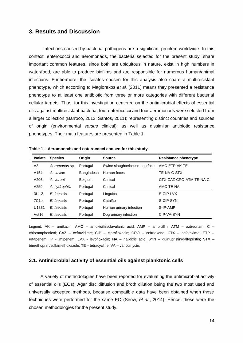
phenotypes. Their main features are presented in Table 1.
Table 1 – Aeromonads and enterococci chosen for this study.
Isolate Species Origin Source Resistance phenotype
A3 Aeromonas sp. Portugal Swine slaughterhouse - surface AMC-ETP-AK-TE
A154 A. caviae Bangladesh Human feces TE-NA-C-STX
A206 A. veronii Belgium Clinical CTX-CAZ-CRO-ATM-TE-NA-C
A259 A. hydrophila Portugal Clinical AMC-TE-NA
3L1.2 E. faecalis Portugal Linguiça S-CIP-LVX
7C1.4 E. faecalis Portugal Catalão S-CIP-SYN
U1881 E. faecalis Portugal Human urinary infection S-IP-AMP
Vet16 E. faecalis Portugal Dog urinary infection CIP-VA-SYN
Legend: AK – amikacin; AMC – amoxicillin/clavulanic acid; AMP – ampicillin; ATM – aztreonam; C –
chloramphenicol; CAZ – ceftazidime; CIP – ciprofloxacin; CRO – ceftriaxone; CTX – cefotaxime; ETP –
ertapenem; IP - imipenem; LVX - levofloxacin; NA – nalidixic acid; SYN – quinupristin/dalfopristin; STX –
trimethoprim/sulfamethoxazole; TE – tetracycline; VA – vancomycin.
3.1. Antimicrobial activity of essential oils against planktonic cells
A variety of methodologies have been reported for evaluating the antimicrobial activity
of essential oils (EOs). Agar disc diffusion and broth dilution being the two most used and
universally accepted methods, because compatible data have been obtained when these
techniques were performed for the same EO (Seow, et al., 2014). Hence, these were the
chosen methodologies for the present study.

15
Disc diffusion assay is a simple and rapid method which requires small amounts of the
EOs. It is regularly used as a preliminary test for the antimicrobial activity of a large number
of compounds, in order to select the ones with the highest inhibitory activity, i.e., larger
inhibitory zones (Burt, 2004). However, disc diffusion methods present certain
disadvantages. First, using volatile EOs could lead to reduced zones of inhibition, since they
evaporate very quickly. Also, poorly soluble compounds do not diffuse uniformly through the
agar medium plates (Seow et al., 2014). Therefore, the convenience of disc diffusion method
is limited to the attainment of preliminary and qualitative data, turning essential the
performance of other complementary methodologies.
Broth microdilution can be used to further evaluate the antimicrobial activity of the most
efficient EOs and is commonly used to determine the extent of inhibitory activity, namely by
determining MIC (minimum inhibitory concentration) and MBC (minimum bactericidal
concentration). Nonetheless, broth dilution assays also present problems, mainly due to the
hydrophobic nature of EOs, which result in immiscibility with broth media. For instance,
surfactants, emulsifiers or solvents are usually applied to ensure even contact between the
test organism and the agent during the experiment. The most commonly used are Tween-20,
Tween-80, DMSO (dimethyl sulfoxide) and ethanol (Burt, 2004).
In the present study, DMSO and Tween-20 were used in preliminary assays in order to
test which concentrations could prevent the immiscibility of the EOs with the aqueous
bacterial growth media. When broth medium was added to the mixture of 0.5% Tween-
20:EO, micelles were formed and the oil-water mixture would separate within few minutes.
Using EOs in a concentration of 20% (v/v) in DMSO prevented the separation of the aqueous
phase from the oil for a longer period of time and the mixture appeared to be translucent.
However, these agents are known to be responsible for changes in the interactions between
EO and bacteria, resulting in either increase or reduction of the antimicrobial activity, and
DMSO cytotoxicity could be a problem in subsequent applications (e.g. direct contact with
the human skin). So, in order to overcome these problems, Mann and Markham (1998)
suggested the use of 0.15% bacteriological agar, previously sterilized by autoclaving, as a
stabilizer.
Regarding the disc diffusion assay, in general, the majority of the EOs under analysis
showed inhibitory zones larger than 10 mm on aeromonads, with the exception of curcuma,
garlic and ginger, which showed few or none antimicrobial activity, since the inhibitory zones
had diameters lower than 10 mm (Table 2). Lemongrass (34-42 mm), thyme (29.7-37 mm),
tea tree (30.3-36 mm) and pennyroyal (21.7-36 mm) EOs showed the highest inhibitory
zones against Aeromonas spp.

16
Table 2 – Antimicrobial activity of EOs by disc diffusion method for Aeromonas isolates.
EOs Tested aeromonads (diameter of inhibitory zone in mm)
A3 A154 A206 A259 R5
Artemisia 14.7 ± 4.7 15.3 ± 4.5 12.0 ± 2.7 15.3 ± 1.5 14.0 ± 0
Coriander 19.0 ± 1.7 25.7 ± 7.8 17.3 ± 7.5 32.0 ± 14.0 36.0 ± 0
Curcuma NA NA NA NA NA
Garlic 6.0 ± 5.2 5.3 ± 4.6 6.7 ± 5.8 6.7 ± 5.8 10.0 ± 8.7
Ginger 2.7 ± 4.6 5.3 ± 4.6 7.0 ± 6.1 6.3 ± 5.5 NA
Lavender 18.7 ± 1.2 16.0 ± 2.0 16.0 ± 4.0 22.0 ± 3.6 27.5 ± 14.9
Lemongrass 42.0 ± 2.0 34.0 ± 6.0 36.0 ± 6.0 38.7 ± 4.2 34.0 ± 0
Pennyroyal 29.0 ± 7.0 26.0 ± 5.3 21.7 ± 12.4 28.7 ± 3.1 36.0 ± 0
Rosemary 28.3 ± 6.4 23.0 ± 2.0 19.7 ± 2.1 26.7 ± 6.8 31.7 ± 10.4
Tea tree 34.3 ± 11.6 36.0 ± 8.7 30.3 ± 1.5 36.0 ± 5.3 36.0 ± 11.3
Thyme 37.0 ± 5.6 29.7 ± 1.5 23.7 ± 7.2 34.0 ± 4.0 35.3 ± 4.6
Legend: Each value represents the mean of triplicate experiments ± standard deviation; NA: no activity
On the other hand, EOs were less inhibitory against enterococci, having in most cases
inhibitory zones smaller than 10 mm (Table 3). Lemongrass (13.3-22.3 mm), thyme (16.3-
26.7 mm) and tea tree (12.7-14.7 mm) showed the largest inhibitory zones against the
Enterococcus isolates. Also, Warnke et al. (2013), using a very similar method, reported
lemongrass inhibition zones from 13 to 18 mm against VRE (vancomycin-resistant
enterococci), comparable to the results obtained in the present study.
Table 3 – Antimicrobial activity of EOs by disc diffusion method for Enterococcus isolates.
EOs Tested enterococci (diameter of inhibitory zone in mm)
3L1.2 7C1.4 U1881 Vet16 V583
Artemisia 6.0 ± 5.3 2.3 ± 4.0 5.7 ± 4.9 6.0 ± 5.2 6.3 ± 5.5
Coriander 10.7 ± 1.2 11.7 ±2.1 5.7 ± 5.1 7.7 ± 1.2 5.7 ± 4.9
Curcuma NA NA NA NA NA
Garlic NA 6.7 ± 1.2 7.7 ± 0.6 6.7 ± 1.2 7.3 ± 0.6
Ginger 2.7 ± 4.6 4.3 ± 3.8 7.3 ± 0.6 7.0 ± 0 4.7 ± 4.0
Lavender 7.7 ± 0.6 3.3 ± 5.8 10.0 ± 1.0 8.3 ± 2.1 8.7 ± 0.6
Lemongrass 17.7 ± 2.9 22.3 ± 4.9 15.7 ± 3.1 13.3 ± 11.6 16.7 ± 2.1
Pennyroyal 8.7 ± 1.2 10.7 ±1.2 9.7 ± 0.6 6.7 ± 5.8 9.7 ± 1.5
Rosemary 6.3 ± 5.5 NA 5.0 ± 4.4 4.7 ± 4.0 6.3 ± 5.5
Tea tree 12.7 ± 4.0 14.3 ± 3.1 14.7 ± 4.9 12.7 ± 5.8 14.0 ± 5.2
Thyme 20.0 ± 1.7 26.7 ± 1.2 16.3 ± 1.5 16.7 ± 0.6 16.7 ± 2.1
Legend: Each value represents the mean of triplicate experiment ± standard deviation; NA: no activity

17
Overall, comparison between inhibition of bacterial growth observed for aeromonads
and enterococci, led to the selection of lemongrass and thyme for subsequent assays of
broth microdilution and biofilm eradication.
Broth microdilution method was performed in this study to further evaluate the
antimicrobial activity of lemongrass and thyme EOs. The two EOs were solubilized in 0.15%
agar to a concentration of 3.13% (v/v), followed by 15 min of agitation by vortex and two-fold
serial dilutions in 90 μL of BHI broth on 96-well microplates The range of concentrations of
the studied oils was between 0.012% and 1.56%. Table 4 presents MICs and MBCs of
lemongrass and thyme EOs.
Table 4 – MICs and MBCs of lemongrass and thyme EOs against aeromonads and enterococci
by broth microdilution method.
Lemongrass EO Thyme EO
MIC MBC MIC MBC
Aero
mo
nad
s A3 0.10 0.20 0.05 0.10
A154 0.10 0.10 0.10 0.10
A206 0.10 0.10 0.05 0.05
A259 0.20 0.20 0.05-0.10 0.05-0.10
R5 0.10 0.10 0.05 0.05
En
tero
co
cci 3L1.2 1.56 >1.56 0.20 0.20
7C1.4 0.78 0.78 0.20 0.39
U1881 0.39 0.78 0.20 0.39
Vet16 0.78 1.56 0.20-0.39 0.39
V583 0.78 0.78 0.20 0.39
Legend: MIC (minimum inhibitory concentration) and MBC (minimum bactericidal concentration) values in % (v/v);
range of concentrations was 0.012-1.56% (v/v)
In general, MICs observed for thyme (0.05-0.39%) were lower than for lemongrass
(0.10-1.56%), in both aeromonads and enterococci. Hence, thyme showed higher
antimicrobial activity against both bacterial groups.
Curiously, in the previous disc diffusion assay, lemongrass showed the highest
inhibitory effect of the tested oils against Aeromonas, while in the microdilution assay it
presented MICs between 0.10 and 0.20%, a superior value in comparison with thyme (0.05-
0.10%). These results could indicate lemongrass diffuses better on Muller Hinton agar than
on BHI broth, due to their different characteristics.
Furthermore, aeromonads inhibition required lower MICs for both EOs (<0.20% for
lemongrass; <0.10% for thyme), whereas higher MICs were required to inhibit the
Enterococcus strains under analysis (<1.56% for lemongrass; <0.39% for thyme). Previous
18
studies reported that Gram-negative bacteria are more resistant to EOs (Delaquis et al.,
2002; Lambert et al., 2001; Martins et al., 2014; Pintore et al., 2002), while few claim the
same for Gram-positive bacteria (Kim et al., 1995; Tassou et al., 1995); interestingly, studies
showing no significant differences between the two bacterial groups can also be found
(Prabuseenivasan et al., 2006; Teixeira et al., 2013).
The resistance of Gram-negative bacteria against EOs has already been attributed to
the complexity of their double layer cell membrane (Teixeira et al., 2013), but in the present
study, enterococci were found to be more resistant, meaning there should be another
property or mechanism responsible for the observed resistance and/or another cellular target
in aeromonads which turns them more susceptible to EO action; further analyses being
needed to determine which.
Overall, these results showed that within the Aeromonas genus, there were no major
differences between MICs for each EO. On the other hand, enterococci showed more
differences between MICs obtained for lemongrass, with a food strain (3L1.2) presenting
higher MIC than clinical strains (U1881 and Vet16). This example could be considered very
problematic, because, since isolate 3L1.2 could be a source of food contamination, a bigger
concentration than 1.56% of lemongrass would be necessary to inhibit its growth. Further
assays should be performed in order to assess the effects of using that concentration of
lemongrass as a disinfectant on a surface or a food-industry device, or of including the EO as
a food preserver in the food package. Hence, the use of thyme would be preferable since its
MICs against Enterococcus isolates were lower.
Regarding MBCs determination, lemongrass showed bactericidal effect from 0.10 to
0.20% on Aeromonas isolates and from 0.78 to 1.56% (or above) for Enterococcus isolates.
MBCs of thyme ranged from 0.05 to 0.10% and from 0.20 to 0.39% on aeromonads and
enterococci, respectively (Table 4). In some cases MICs were the same as MBCs, which
could mean at that concentration the EO had a bactericidal effect instead of a bacteriostatic
one on those isolates. Another possibility could be that the MIC was between the obtained
value and the lower concentration. For example, isolate A154 had equal MIC and MBC for
lemongrass (0.10%), so the actual MIC could be between 0.05 and 0.10%, further tests
being necessary.
The antimicrobial activity of these two essential oils has been evaluated in previous
studies referred bellow. However, comparing published data is complicated since
experimental results of EOs antimicrobial tests depend on several factors, such as
composition, physical and chemical properties, the method and culture conditions applied,
species and strain of the microorganisms under analysis. Temperature, time of incubation,
type and volume of broth and even concentration and age of inoculums are also factors with
a great influence on the EO strength and overall outcome (Seow et al., 2014). Hence, it is

19
peremptory the standardization of the methodologies used to assess the antimicrobial activity
of EOs and natural products in general, similarly to what already occurs with antibiotic
susceptibility tests.
No studies could be found regarding lemongrass (Cympogon flexuosus) effect on
aeromonads or enterococci, though the following studies reported lemongrass antimicrobial
activity against other bacterial species. Oussalah et al. (2007) found that this EO showed
antibacterial effect against Listeria monocytogenes, E. coli O157:H7, S. aureus and
Salmonella Typhimurium. Likewise, Oliveira et al. (2012) found that lemongrass EO had
inhibitory effect against L. monocytogenes (MIC=0.12%) and E. coli (MIC=0.25%). According
to Adukwu et al. (2012), MIC for lemongrass against S. aureus strains (susceptible or
resistant to methicillin) was 0.06%, while at concentrations of 0.125% the effect was
bactericidal.
According to Ahmad et al. (2014), thyme MIC against E. faecalis was 0.125 mg mL-1,
while against E. coli and S. aureus was 0.500 mg mL-1. Uyttendaele et al. (2004) showed that
MIC of thyme towards Aeromonas ranged from 0.025 to 0.040% (v/v). Sienkiewicz et al.
(2012) used the agar diffusion method to study the inhibitory effect of thyme on clinical
multiresistant strains of Enterococcus and Pseudomonas aeruginosa. They obtained MICs
ranging from 0.25 to 1.25 μL/mL for enterococci and from 0.5 to 2.5 μL/mL for
pseudomonads. Although they both belong to the Gammaproteobacteria class,
Pseudomonas susceptibility to EOs is different from aeromonads, being the latter one of the
most sensible bacteria to EOs (Iturriaga et al., 2012), which could explain why their MICs
were lower than the enterococcal ones.
In conclusion, low concentrations of lemongrass and thyme were sufficient to inhibit
aeromonads planktonic growth, while thyme was more effective against enterococci than
lemongrass.
3.2. Biofilm eradication by essential oils
The ability of bacteria to produce biofilms poses a major problem in various industrial
and medical settings. Eradicating biofilms is very difficult since sessile cells are protected
and more resistant to external aggressions, in particular to the entrance of antimicrobial
agents (Jefferson, 2004). So, nowadays, new and effective agents against biofilms are of
great interest.
As previously mentioned, an adaptation of the Calgari Biofilm Device was used to
assess if lemongrass and thyme EOs would be capable of eradicating biofilms formed by
aeromonads and enterococci. This device represents a rapid and less laborious way of
analyzing biofilm formation, since it establishes on the pegs of the lids of the microplates
20
instead of on the bottom of the wells, facilitating the exchange between microplates with
growth medium and microplates with EOs for eradication. However, biofilm assays have
inherent problems related to the difficulty to obtain reproducible data, since biofilm
development is a stochastic process (Heydorn et al., 2000) due to the fact that, as complex
living beings, microorganisms may not always show the same behavior, i.e., each peg may
not have the same initial biofilm growth as the growth control. Moreover, the small area of the
pegs can complicate the biofilm attachment to their surface and less biofilm is formed on the
pegs.
In the present study aeromonads and enterococcal biofilms, formed over a 24-hour-
period of incubation, were subjected to concentrations ranging from 0.20 to 3.13% for both
EOs. These concentrations were chosen based on the fact that sessile cells are more
resistant to antibacterial agents than planktonic cells (O’Toole and Kaplan, 2000).
Preliminary biofilm eradication tests performed using the selected EOs against isolates A3
and R5, evaluated four exposure times (15 min, 30 min, 1 h and 24 h) and led to the
following conclusions: 15 min of exposure showed less decimal log reduction than 30 min
and 1 h periods, while after 24 h of exposure there were no colonies formed on BHI plates,
except for growth controls (data not shown). Thus, 30 min and 1 h were the selected times of
exposure to the EOs. Results obtained during the present study are presented in Table 5 and
Table 6.
Table 5 - Aeromonads decimal log reduction achieved by lemongrass and thyme EOs.
Aeromonads
EOs Time Concentration A3 A154 A206 A259 R5 Average
Lem
on
gra
ss E
O
1 h
3.13 7 6 6 7 7 7
1.56 5 6 6 5 7 6
0.78 5 2 6 3 5 4
0.39 2 2 2 2 2 2
30 min
3.13 7 7 7 7 7 7
1.56 7 7 7 7 7 7
0.78 3 5 7 5 5 5
0.39 2 2 2 2 2 2
Th
ym
e E
O 1 h
3.13 5 6 6 6 7 6
1.56 5 4 6 6 5 5
0.78 3 3 6 4 4 4
0.39 2 2 5 2 4 3
30 min
3.13 8 7 7 7 7 7
1.56 8 6 7 7 7 7
0.78 2 3 7 4 6 4
0.39 2 2 3 3 2 2
Legend: Concentration values in % (v/v); logR = logN0 - logNA (R - reduction; N0 - number of CFU per mL in the
biofilm (growth control); NA – number of surviving CFU per mL)

21
According to Table 5, both lemongrass and thyme showed high levels of eradication of
aeromonads biofilms at 3.13 and 1.78%, except for 1 h treatment with thyme, which showed
medium levels. At 0.78% the levels of eradication were medium, while at 0.39% were low.
These results indicate that EOs could be used as disinfectants in food industry and
indwelling medical devices contaminated with pre-formed aeromonads biofilms. Also, there
were slight differences between the two exposure times, namely at higher concentrations.
These results could mean that 30 min was sufficient time for eradication and 1 h in contact
with the EO could enhance the bacterial biofilm resistance mechanisms and prevent the
entrance and/or removal by the EO.
Regarding the aeromonads under analysis, A206 biofilm was the most susceptible to
both lemongrass and thyme, as no colonies were observed at the majority of the tested
concentrations, exception being 0.39%, which could mean that concentrations above 0.78%
of EOs efficiently remove pre-formed biofilm. The susceptibility of this isolate is curious
because this clinical isolate presented a resistance phenotype against six antibiotics from
different classes. This indicates that even the most resistant isolates can be susceptible to
the eradicating action of EOs.
Table 6 - Enterococci decimal log reduction achieved by lemongrass and thyme EOs.
Enterococci
EOs Time Concentration 3L1.2 7C1.4 U1881 Vet16 V583 Average
Lem
on
gra
ss E
O
1 h
3.13 1 1 0 0 0 0
1.56 0 1 0 0 0 0
0.78 0 1 0 0 0 0
0.39 0 1 0 0 0 0
30 min
3.13 0 1 0 0 0 0
1.56 0 1 0 0 0 0
0.78 0 1 0 0 0 0
0.39 0 1 0 0 0 0
Th
ym
e E
O
1 h
0.78 1 3 1 1 0 1
0.39 1 2 1 1 0 1
0.20 1 1 1 1 0 1
30 min
0.78 1 1 1 0 0 1
0.39 1 3 1 0 0 1
0.20 1 1 1 0 0 1
Legend: Concentration values in % (v/v); logR = logN0 - logNA (R - reduction; N0 - number of CFU per mL in the
biofilm (growth control); NA – number of surviving CFU per mL)
According to Table 6, lemongrass showed no eradication effect on biofilms produced
by the enterococcal isolates 3L1.2, U1881, Vet16 and V583. In fact, 7C1.4 was the only

22
isolate for which an average reduction of 1-log (CFU/mL) was observed for all tested EO
concentrations at both periods of contact.
On preliminary assays, no enterococcal colonies were observed in BHI plates when the
range of concentrations of thyme used was 0.39-3.13%, which means there was complete
eradication; in order to better evaluate the effects of thyme on enterococcal biofilms, the
concentrations were decreased to 0.20-0.78%.
As before, 7C1.4 was the enterococcal isolate most susceptible to biofilm removal
activity by thyme, with average reduction of 3-log (CFU/mL) at 0.78%, for the 1 h treatment,
but still considered as low level eradication. The other four strains showed very low (or none)
levels of eradication when submitted to the treatment with thyme. Their inability in eradicating
biofilms formed by food isolates could allow their persistence in surfaces and/or devices and
be a source of foodborne illnesses. The same could happen in clinical settings, allowing the
dissemination of multiresistant strains by not removing their biofilms. Despite it was
previously mentioned that, on preliminary assays, thyme eradicated completely enterococcal
biofilms, these data should not be considered. By lowering the concentrations of thyme, very
low or none eradication of enterococcal biofilms was verified, which indicates that even
though triplicates were used, the preliminary data were not reliable. In the future, more
repetitions should be performed in order to assess the effect of EOs on biofilms of
enterococcus.
Adukwu et al. (2012) showed that lemongrass EO was unable to eradicate pre-formed
staphylococcal biofilms using concentrations from 0.06-4%, like it occurred in the present
study with Enterococcus biofilms. As biofilms develop, the initial cells undergo irreversible
attachment which leads to maturation and, at this stage, removal of sessile cells is said to be
difficult, requiring mechanical force or chemical disruption. Kavanaugh and Ribbeck (2012)
demonstrated that thyme EO was more effective in eradicating Pseudomonas and S. aureus
biofilms than selected important antibiotics.
In conclusion, the EOs under study presented very different inhibitory effects against
aeromonads and enterococci pre-formed biofilms. Intrinsic characteristics of the aeromonads
biofilms may have facilitated biofilm detachment from the pegs by the EOs, either because
these biofilms adhere less efficiently to the surface of the pegs or due to the interactions
between the used EOs and the components of the matrix. On the other hand, enterococcal
biofilms properties could allow a stronger attachment to the pegs, which prevented EOs
action. Hence, to eradicate Enterococcus biofilms, thyme should not be disregarded since
higher concentrations were not tested, whereas lemongrass was unable to eradicate them.
Instead both EOs could be used in aeromonads case. However, since rarely there are
biofilms constituted by only one microbial species during in vivo conditions, the EOs
analyzed in the present study may be insufficient to guarantee complete biofilm eradication.
23
4. Concluding remarks
In the present study we aimed to (a) assess the antimicrobial activity of eleven
commercial EOs against clinical and environmental multiresistant aeromonads and
enterococci by disc diffusion method, (b) select the two most promising EOs and, by using
broth microdilution method, calculate their MICs and MBCs, as well as (c) assess the efficacy
of the two EOs in removing already formed biofilms. In brief, the following conclusions were
accomplished:
- In the preliminary disc diffusion assay, lemongrass and thyme showed the highest
inhibitory zones against both aeromonads and enterococci, being the former more
susceptible to the tested EOs;
- In planktonic state, low concentrations of both EOs were sufficient to inhibit
aeromonads growth, while thyme was more effective in inhibiting enterococcal growth than
lemongrass;
- The two EOs presented high ability for Aeromonas biofilm eradication;
- Lemongrass was unable to eradicate Enterococcus biofilm, while thyme showed
inconclusive results.
Overall, the present study confirmed the putative applicability of natural products from
plant origin as antimicrobial agents, allowing the creation of a path for the development of
new therapeutic antimicrobial strategies. Nonetheless, eradication of enterococcal biofilms
using thyme should be addressed in future works regarding antimicrobial activity of natural
products.
Further studies should be undertaken to identify the active antimicrobial compounds
within the EOs, as well as the molecular mechanisms responsible for their inhibiting and
eradicating properties, and the cellular targets. Additionally, natural compounds should be
considered potential substitutes for current disinfectants and cleaning products used in both
clinical and food industry settings. Cytotoxicity assays on human cell lines are necessary, in
order to use the EOs as alternatives for the antibiotics in infectious diseases treatments, as
topical therapy for wound infections. Having these tests performed, another approach could
be to combine existing antimicrobial drugs with essential oils, since there is a great difficulty
in acquiring new antibiotics.
To find alternatives for current therapeutic treatments against infectious diseases,
studies to inhibit the expression of virulence factors and not the bacterial growth should be
addressed, as antimicrobial agents harm not only the pathogenic targets but also the human
microbiota.

24
5. References
Abuhammour W., Hasan R.A. and Rogers D. (2006) Necrotizing fasciitis caused by Aeromonas hydrophila in an immunocompetent child. Pediatric Emergency Care 22:48–51. Adukwu E.C., Allen S.C.H. and Phillips C.A. (2012) The anti-biofilm activity of lemongrass (Cymbopogon flexuosus) and grapefruit (Citrus paradisi) essential oilds against five strains of Staphylococcus aureus. Journal of Applied Microbiology 113:1217-1227. Agersø Y., Bruun M.S., Dalsgaard I., Larsen J.L. (2007) The tetracycline resistance gene tet(E) is frequently occurring and present on large horizontally transferable plasmids in Aeromonas spp. from fish farms. Aquaculture 266:47–52. Ahmad A., van Vuuren S. and Viljoen A. (2014) Unravelling the complex antimicrobial interactions of essential oils – the case of Thymus vulgaris (thyme). Molecules 19:2896-2910. Alim, A., Goze, I., Cetin, A., Atas, A.D., Vural, N. and Donmez, E. (2009) Antimicrobial activity of the essential oil of Cyclotrichium niveum (Boiss.) Manden. Et Scheng. African Journal of Microbiology Research 3(8):422-425. Amorati R., Foti M.C. and Valgimigli L. (2013) Antioxidant activity of essential oils. Journal of Agricultural and Food Chemistry 61(46):10835-10847. Angel M. F., Zhang F., Jones M., J. Henderson J. and Chapman S. W. (2002). Necrotizing fasciitis of the upper extremity resulting from a water moccasin bite. Southern Medical Journal 95:1090-1094. Aravena-Roman M, Inglis TJJ, Henderson B, Riley TV, Chang BJ. (2012). Antimicrobial susceptibilities of Aeromonas strains isolated from clinical and environmental sources to 26 antimicrobial agents. Antimicrobial Agents and Chemotherapy 56:1110–1112 Arias C.A. and Murray B.E. (2012) The rise of the Enterococcus: beyond vancomycin resistance. Nature Reviews Microbiology 10(4):266-278. Astani A., Reichling J. and Schnitzler P. (2010) Screening for antiviral activities of isolated compounds from essential oils. Evidence-Based Complementary and Alternative Medicine 11:253646. Azerêdo G.A., Stamford T.L.M., Figueiredo R.C.B.Q. and Souza E.L. (2012) The cytotoxic effect of essential oils from Origanum vulgare L. and/or Rosmarinus officinalis L. on Aeromonas hydrophila. Foodborne Pathogens and Disease 9(4):298-304. Bakkali F., Averbeck S., Averbeck D. and Idaomar M. (2008) Biological effects of essential oils – A review. Food and Chemical Toxicology 46:446-475. Barroco, C. (2013) Antibiorresistência e pesquisa de factores de virulência em Aeromonas spp. Dissertação de Mestrado em Microbiologia Aplicada. Lisboa: Faculdade de Ciências - Universidade de Lisboa. Billington E.O., Phang S.H., Gregson D.B., Pitout J.D.D., Ross T., Church D.L., Laupland K.B. and Parkins M.D. (2014) Incidence, risk factors, and outcomes of Enterococcus spp blood stream infections: a population-based study. International Journal of Infectious Diseases 26C:76-82. Article in press. Bossou A.D., Mangelinckx S., Yedomonhan H., Boko P.M., Akogbeto M.C., De Kimpe N., Avlessi F. and Sohounhloue D.C.K. (2013) Chemical composition and insecticidal activity of plant essential oils from Benin against Anopheles gambiae (Giles). Parasites and Vectors 6:337. Burt S. (2004) Essential oils: their antibacterial properties and potential applications in foods – a review. International Journal of Food Microbiology 94:223-253. Cândido C.S., Portella C.S.A., Laranjeira B.J., Silva S.S., Arriaga A.M.C., Santiago G.M.P, Gomes G.A., Almeida P.C. and Carvalho C.B.M. (2010) Effects of Myrcia ovate Cambess. essential oil on planktonic growth of gastrointestinal microorganisms and biofilm formation of Enterococcus faecalis. Brazilian Journal of Microbiology 41:621-627. Carvalho M.J., Martínez-Murcia A., Esteves A.C., Correia A. and Saavedra M.J. (2012) Phylogenetic diversity, antibiotic resistance and virulence traits of Aeromonas spp. from untreated waters for human consumption. International Journal of Food Microbiology 159:230-239.
25
Cattoir V. and Leclerq R. (2013) Twenty-five years of shared life with vancomycin-resistant enterococci: is it time to divorce? Journal of Antimicrobial Chemotherapy 68:731-742. Ceri, H., Olson, M.E., Stremick, C., Read, R.R., Morck, D. and Buret, A. (1999) The Calgary Biofilm Device: New Technology for Rapid Determination of Antibiotic Susceptibilities of Bacterial Biofilms. Journal of Clinical Microbiology 37(6):1771-1776 Chauret C., Volk C., Creason R., Jarosh J., Robinson J. and Warnes C. (2001) Detection of Aeromonas hydrophila in a drinking-water distribution system: a field and pilot study. Canadian Journal of Microbiology 47:782-786. Chu S.S., Liu Z.L., Du S.S., Deng Z.W. (2012) Chemical composition and insecticidal activity against Sitophilus zeamais of the essential oils derived from Artemisia giraldii and Artemisia subdigitata. Molecules 17(6):7255-7265. CLSI (2012) Methods for Dilution Antimicrobial Susceptibility Tests for Bacteria That Grow Aerobically; Approved Standard – Ninth Edition. CLSI document M07-A9. Wayne, PA: Clinical and Laboratory Standards Institute. CLSI (2013) Performance Standards for Antimicrobial Susceptibility Testing; Twenty-Third Informational Supplement. CLSI document M100-S23. Wayne, PA: Clinical and Laboratory Standards Institute. Davies J. and Davies D. (2010) Origins and evolution of antibiotic resistance. Microbiology and Molecular Biology Reviews 74(3):417-433. Delaquis P.J., Stanich K., Girard B. and Mazza G. (2002) Antimicrobial activity of individual and mixed fractions of dill, cilantro, coriander and eucalyptus essential oils. International Journal of Food Microbiology 74:101-109. Dias Ferreira F., Mossini S.A., Dias Ferreira F.M., Arrotéia C.C, da Costa C.L., Nakamura C.V. and Machinski M. Jr. (2013) The inhibitory effects of Curcuma longa L. essential oil and curcumin on Aspergillus flavus link growth and morphology. Scientific World Journal 343804. Donlan R. and Costerton J.W. (2002) Biofilms: survival mechanisms of clinically relevant microorganisms. Clinical Microbiology Reviews 15(2):167-193. Dussault D., Vu K.D. and Lacroix M. (2014) In vitro evaluation of antimicrobial activities of various commercial essential oils, oleoresin and pure compounds against food pathogens and application in ham. Meat Science 96:514-520. ECDC (2013) Antimicrobial resistance surveillance in Europe 2012. Annual Report of the European Antimicrobial Resistance Surveillance Network. Stockholm. Echeverrigaray S., Zacaria J. and Beltrão R. (2010) Nematicidal activity of monoterpenoids against root-knot nematode Meloidogyne incognita. Phytopathology Journal 100(2):199-203. Elaissi A., Rouis Z., Salem N.A., Mabrouk S., ben Salem Y., Salah K.B., Aouni M. Farhat F., Chemli R., Harzallah-Skhiri F. and Khouka M.L. (2012) Chemical composition of 8 eucalyptus species' essential oils and the evaluation of their antibacterial, antifungal and antiviral activities. BMC Complementary and Alternative Medicine 12:81. Esteve C., Alcaide E. and Giménez M.J. (2014) Multidrug-resistant (MDR) Aeromonas recovered from the metropolitan area of Valencia (Spain): diseases spectrum and prevalence in the environment. European Journal of Clinical Microbiology and Infectious Diseases (Epub ahead of print) Extremina, C.I., Costa, A., Aguiar, A.J., Peixe, L., Fonseca, A.P. (2011) Optimization of processing conditions for the quantification of enterococci biofilms using microtitre-plates. Journal of Microbiological Method 84(2):167-173. Figueras, M. J., M. J. Aldea, N. Fernandez, C. Aspiroz, A. Alperi, and J. Guarro. 2007. Aeromonas hemolytic uremic syndrome. A case and a review of the literature. Diagnostic Microbiology and Infectious Diseases 58:231–234. Fisher K. and Phillips C. (2009a) The ecology, epidemiology and virulence of Enterococcus. Microbiology 155:1749-1757. Fisher K. and Phillips C. (2009b) The mechanism of action of a citrus oil blend against Enterococcus faecium and Enterococcus faecalis. Journal of Applied Microbiology 106:1343-1349.

26
Formagio A.S.N., Vieira M.C., Santos L.A.C., Cardoso C.A.L., Foglio M.A., Carvalho J.E., Andrade-Silva M. and Kassuya C.A.L. (2013) Composition and evaluation of the anti-inflammatory and anticancer activities of the essential oils from Annona sylvatica A.St.-Hill. Journal of Medicinal Food 16(1):20-25. Franz C. and Novak J. (2010) Sources of essential oils. In: Baser K.H.C. and Buchbauer G. (Eds.) Handbook of Essential Oils: Science, Technology, and Applications. (pp. 39-82). Boca Raton: CRC Press/Taylor & Francis Group. Franz C.M.A.P., Huch M., Abriouel H., Holzapfel W. and Gálvez A. (2011) Enterococci as Probiotics and their implications in food safety. International Journal of Food Microbiology 151:125-140. Gavín R., Rabaan A.A., Merino S., Tomás J.M., Gryllos I. and Shaw J.G. (2002) Lateral flagella of Aeromonas species are essential for epithelial cell adherence and biofilm formation. Molecular microbiology 43(2):383-397. Giaouris E., Heir E., Hébraud M., Chorianopoulos N., Langsrud S., Møretrø T., Habimana O., Desvaux M., Renier S. and Nychas G.-J. (2014) Attachment and biofilm formation by foodborne bacteria in meat processing environments: Causes, implications, role of bacterial interactions and control by alternative novel methods. Meat Science 97(3):298-309. Gustafson J.E., Liew Y.C., Chew S., Markham J., Bell H.C., Wyllie S.G. and Warmington J.R. (1998) Effects of tea tree oil on Escherichia coli. Letters in Applied Microbiology 26:194-198. Hammad A.M., Shimamoto T. and Shimamoto T. (2014) Genetic characterization of antibiotic resistance and virulence factors in Enterococcus spp. from Japanese retail ready-to-eat raw fish. Food Microbiology 38:62-66. Hall-Stoodley L. and Stoodley P. (2005) Biofilm formation and dispersal and the transmission of human pathogens. Trend in Microbiology 13(1):7-10. Hall-Stoodley L., Costerton J.W. and Stoodley P. (2004) Bacterial biofilms: from the natural environment to infections diseases. Nature Reviews Microbiology 2:95-108. Harrison J.J., Stremick C.A., Turner R.J., Allan N.D., Olson M.E. and Ceri H. (2010) Microtiter susceptibility testing of microbes growing on peg lids: a miniaturized biofilm model for high-throughput screening. Nature Protocols 5(7):1236-1254. Harrison J.J., Turner R.J., Joo D.A., Stan M.A., Chan C.S., Allan N.D., Vrionis H.A., Olson M.E. and Ceri H. (2008) Copper and quaternary ammonium cations exert synergistic bactericidal and antibiofilm activity against Pseudomonas aeruginosa. Antimicrobial Agents and Chemotherapy 52(8):2870-2881. Heintz B.H., Halilovic J. and Christensen C.L. (2010) Vancomycin-resistant enterococcal urinary tract infections. Pharmacotherapy 30(11):1136-1149. Heydorn A., Ersbøll B.K., Hentzer M., Parsek M.R., Givskov M., Molin S. (2000) Experimental reproducibility in flow-chamber biofilms. Microbiology 146:2409–2415. Hollenbeck B.L. and Rice L.B. (2012) Intrinsic and acquired resistance mechanisms in enterococcus. Virulence 3(5):421-433. Husain F.M., Ahmad I., Asif M. and Tahseen Q. (2013) Influence of clove oil on certain quorum-sensing-regulated functions and biofilm of Pseudomonas aeruginosa and Aeromonas hydrophila. Journal of Biosciences 38:835-844. Igbinosa I.H., Igumbor E.U., Aghdasi F., Tom M. and Okoh A.I. (2012) Emerging Aeromonas species infections and their significance in public health. The Scientific World Journal 2012:625023. Isman M.B. (2000) Plant essential oils for pest and disease management. Crop Protection 19:603-608. Iturriaga L., Olabarrieta I. and de Marañón I.M. (2012) Antimicrobial assays of natural extracts and their inhibitory effect against Listeria innocua and fish spoilage bacteria, after incorporation into biopolymer edible films. International Journal of Food Microbiology 158:58-64. Jahan M. and Holley R.A. (2014) Incidence of virulence factors in enterococci from raw and fermented meat and biofilm forming capacity at 25ºC and 37ºC. International Journal of Food Microbiology 170:65-69. Jahan M., Krause D.O. and Holley R.A. (2013) Antimicrobial resistance of Enterococcus species from meat and fermented meat products isolated by a PCR-based rapid screening method. International Journal of Food Microbiology 163:89-95.
27
Janda J.M. and Abbott S.L. (2010) The Genus Aeromonas: Taxonomy, Pathogenicity, and Infection. Clinical Microbiology Reviews 23(1):35-73. Jefferson K.K. (2004) What drives bacteria to produce a biofilm? FEMS Microbiology Letters 236:163-173. Jett B.D., Huycke M.M. and Gilmore M.S. (1994) Virulence of enterococci. Clinical Microbiology Reviews 7(4):462-478. Jones B.L. and Wilcox M.H. (1995) Aeromonas infections and their treatment. Journal of Antimicrobial Chemotherapy. 35:453-461. Kaskhedikar M and Chhabra D. (2010) Multiple drug resistance in Aeromonas hydrophila isolates of fish. Veterinary World 3(2):76-71. Kavanaugh N.L. and Ribbeck K. (2012) Selected antimicrobial essential oils eradicate Pseudomonas spp. and Staphylococcus aureus biofilms. Applied and Environmental Microbiology 78(11):4057-4061. Kayser F.H. (2003) Safety aspects of enterococci from the medical point of view. International Journal of Food Microbiology 88:255-262. Kim J, Marshall MR, Wei C (1995) Antibacterial activity of some essential oil components against five foodborne pathogens. Journal of Agricultural and Food Chemistry 43:2839–2845. Kirov S.M., Castrisios M. and Shaw J.G. (2004) Aeromonas flagella (polar and lateral) are enterocyte adhesins that contribute to biofilm formation on surfaces. Infection and Immunity 72(4):1939-1945. Klein G., Rüben C. and Upmann M. (2013) Antimicrobial activity of essential oil components against potential food spoilage microorganisms. Current Microbiology 67:200-208. Kon K.V. and Rai M.K. (2012) Plant essential oils and their constituents in coping with multidrug-resistant bacteria. Expert Reviews of Anti-Infective Therapy 10(7):775-790. Laird K., Armitage D. and Phillips C. (2012) Reduction of surface contamination and biofilms of Enterococcus sp. and Staphylococcus aureus using a citrus-based vapour. Journal of Hospital Infection 80:61-66. Lambert RJW, Skandamis PN, Coote PJ, Nychas G-JE (2001) A study of the minimum inhibitory concentration and mode of action of oregano essential oil, thymol and carvacrol. Journal of Applied Microbiology 91:453–462 Lindsay D. and von Holy A. (2006) Bacterial biofilms within the clinical setting: what healthcare professionals should know. Journal of Hospital Infection 64:313-325. Lins R.X., Andrade A.O., Hirata-Junior R., Wilson M.J., Lewis M.A.O., Williams D.W. and Fidel R.A.S. (2013) Antimicrobial resistance and virulence traits of Enterococcus faecalis from primary endodontic infections. Journal of Dentistry 41:779-786. Ludwig W., Schleifer K.-H., Whitman W.B. (2009) Family IV. Enterococcaceae fam. nov. In: Vos P., Garrity G. M., Jones D., Krieg N. R., Ludwig W., Rainey F.A., Schleifer K.-H. and Whitman W.B. (Eds) Bergey's Manual of Systematic Bacteriology, Vol. 3, The Firmicutes. (2
nd ed.). (pp. 594-623). New York: Springer.
Magiorakos A.P., Srinivasan A., Carey R.B., Carmeli Y., Falagas M.E., Giske C.G., Harbarth S., Hindler J.F., Kahlmeter G., Olsson-Liljequist B., Paterson D.L., Rice L.B., Stelling J., Struelens M.J., Vatopoulos A., Weber J.T., Monnet D.L. (2011) Multidrug-resistant, extensively drug-resistant and pandrug-resistant bacteria: an international expert proposal for interim standard definitions for acquired resistance. Clinical Microbiology and Infection 18(3):268-281. Macedo A.S., Freitas A.R., Abreu C., Machado E., Peixe L., Sousa J.C. and Novais C. (2011) Characterization of antibiotic resistant enterococci isolated from untreated waters for human consumption in Portugal. International Journal of Food Microbiology 145:315-319. Mann C.M. and Markham J.L. (1998) A new method for determining the minimum inhibitory concentration of essential oils. Journal of Applied Microbiology 84:538:544. Marinho A.R., Martins P.D., Ditmer E.M., d’Azevedo P.A., Frazzon J., Van Der Sand S.T. and Frazzon A.P.G. (2013) Biofilm formation on polystyrene under different temperatures by antibiotic resistant Enterococcus faecalis and Enterococcus faecium isolated from food. Brazilian Journal of Microbiology 44(2):423-426.
28
Martin-Carnahan A. and Joseph S.W. (2005) Family I. Aeromonadaceae Colwell, MacDonell and De Ley 1986, 474
VP. In: Brenner D.J, Krieg N.R., Staley J.T. and Garrity G.M. (Eds). Bergey's Manual of Systematic
Bacteriology, Vol. 2, The Proteobacteria, Part B, The Gammaproteobacteria. (2nd
ed.). (pp. 556-580). New York: Springer. Martins M.R., Arantes S., Candeias F., Tinoco M.T. and Cruz-Morais J. (2014) Antioxidant, antimicrobial and toxicological properties of Schinus molle L. essential oils. Journal of Ethnopharmacology 151(1):485-492. Medeiros A.W., Pereira R.I., Oliveira D.V., Martins P.D., d’Azevedo P.A., Van der Sand S., Frazzon J. and Frazzon A.P.G. (2014) Molecular detection of virulence factors among food and clinical Enterococcus faecalis strains in South Brazil. Brazilian Journal of Microbiology 45(1):327-332. Millezi A.F., Cardoso M.G., Alves E. and Piccoli R.H. (2013) Reduction of Aeromonas hydrophila biofilm on stainless steel surface by essential oils. Brazillian Journal of Microbiology 44(1):73-80. Mohamed J.A. and Huang D.B. (2007) Biofilm formation by enterococci. Journal of Medical Microbiology 56:1581-1588. Moore-Neibel K., Gerber C., Patel J., Friedman M., Jaroni D. and Ravishankar S. (2013) Antimicrobial activity of oregano oil against antibiotic-resistant Salmonella enteric on organic leafy greens at varying exposure times and storage temperatures 34:123-129. Moreno M.R.F., Sarantinopoulos P., Tsakalidou E. and De Vuyst L. (2006) The role and application of enterococci in food and health. International Journal of Food Microbiology 106:1-24. Mundy L.M., Sahm D.F. and Gilmore M. (2000) Relationships between enterococcal virulence and antimicrobial resistance. Clinical Microbiology Reviews 13(4):513-522. Nagar V., Shashidhar R. and Bandekar J.R. (2011) Prevalence, characterization, and antimicrobial resistance of Aeromonas strains from various retail food products in Mumbai, India. Journal of Food Science 76(7):M486-M492. Nhung P.H., Hata H., Ohkusu M., Noda K., Shah M.M., Goto K. and Ezaki T., (2007) Use of the novel phylogenetic marker dnaJ and DNA – DNA hybridization to clarify interrelationships within the genus Aeromonas. International Journal of Systematic and Evolutionary Microbiology 57:1232–1237. Novais C., Freitas A.R., Silveira E., Antunes P., Silva R., Coque T.M. and Peixe L. (2013) Spread of multidrug-resistant Enterococcus to animals and humans: an underestimated role for the pig farm environment. Journal of Antimicrobial Chemotherapy 68(12):2746-2754. Ocazionez R.E., Meneses R., Torres F.A. and Stashenko E. (2010) Virucidal activity of Colombian Lippia essential oils on dengue virus replication in vitro. Memórias do Instituto Oswaldo Cruz 105(3):304-309. Oliveira M.M.M., Brugnera D.F., Nascimento J.A. and Piccoli R.H. (2012) Control of planktonic and sessile bacteria cells by essential oils. Food and Bioproducts Processing 90:809-818. Oliveira M.M.M., Brugnera D.F. and Piccoli R.H. (2013) Essential oils of thyme and rosemary in the control of Listeria monocytogenes in raw beef. Brazilian Journal of Microbiology 44(4):1181-1188. Ottaviani D., Parlani C., Citterio B., Masini L., Leoni F., Canonico C., Sabatini L., Bruscolini F. and Pianetti A. (2011) Putative virulence properties of Aeromonas strains isolated from food, environmental and clinical sources in Italy: a comparative study. International Journal of Food Microbiology 144:538-545. O’Toole G.A. and Kaplan H.B. (2000) Biofilm formation as microbial development. Annual Reviews in Microbiology 49-79. Oussalah M., Caillet S., Saucier L. and Lacroix M. (2007) Inhibitory effects of selected plant essential oils on the growth of four pathogenic bacteria: E. coli O157:H7, Salmonella Typhimurium, Staphylococcus aureus and Listeria monocytogenes. Food Control 18:414-420. Parker, J.L. and Shaw, J.G. (2011) Aeromonas spp. clinical microbiology and disease. Journal of Infection 62:109-118 Pinto E., Hrimpeng K., Lopes G., Vaz S., Gonçalves M.J., Cavaleiro C. and Salgueiro L. (2013) Antifungal activity of Ferulago capillaris essential oil against Candida, Cryptococcus, Aspergillus and dermatophyte species. European Journal of Clinical Microbiology and Infectious Diseases 32(10):1311-1320.
29
Pintore G., Usai M., Bradesi P., Juliano C., Boatto G., Tomi F., Chessa M., Cerri R. and Casanova J. (2002) Chemical composition and antimicrobial activity of Rosmarinus officinalis L. oils from Sardinia and Corsica. Flavour and Fragance Journal 17:15-19. Prabuseenivasan S., Jayakumar M. and Ignacimuthu S. (2006) In vitro antibacterial activity of some plant essential oils. BMC Complementary and Alternative Medicine 6:39. Reyes K. and Zervos M. (2013) Endocarditis caused by resistant enterococcus: an overview. Current Infectious Disease Reports 15(4):320-328. Rhoades J., Gialagkolidou K., Gogou M., Mavridou O., Blatsiotis N., Ritzoulis C. and Likotrafiti E. (2013) Oregano essential oil as an antimicrobial additive to detergent for hand washing and food contact surface cleaning. Journal of Applied Microbiology 115:987-994. Rios J.L. and Recio M.C. (2005) Medicinal plants and antimicrobial activity. Journal of Ethnopharmacology 100:80-84. Santos, S. (2011) The dual role of enterococci in food technology: bacteriocin production versus pathogenicity potential. Dissertação de Mestrado em Microbiologia Aplicada. Lisboa: Faculdade de Ciências - Universidade de Lisboa. Scoaris D.O., Colacit J., Nakamura C.V. Ueda-Nakamura T., Abreu, B.A. and Dias B.P., (2008). Virulence and antibiotic susceptibility of Aeromonas spp. Isolated from drinking water. Antonie Van Leeuwenhoek 93 (1-2): 111 - 122. Semedo-Lemsaddek T., Nóbrega C.S., Ribeiro T., Pedroso N.M., Sales-Luís T., Lemsaddek A., Tenreiro R., Tavares L., Vilela C.L. and Oliveira M. (2013) Virulence traits and antibiotic resistance among enterococci isolated from Eurasian otter (Lutra lutra). Veterinary Microbiology 163(3-4):378-382. Sen K. and Rodgers M., (2004). Distribution of six virulence factors in Aeromonas species isolated from US drinking water utilities: a PCR identification. Journal of Applied Microbiology 97 (5): 1077 - 1086. Senderovich Y., Ken-Dror S., Vainblat I., Blau D., Izhaki I. and Halpern M. (2012) A molecular study on the prevalence and virulence potential of Aeromonas spp. recovered from patients suffering from diarrhea in Israel. PLoS One 7(2):e30070. Seow Y.X., Yeo C.R., Chung H.L. and Yuk H.-G. (2014) Plant essential oils as active antimicrobial agents. Critical Reviews in Food Science and Nutrition 54(5):625-644. Shakir Z., Khan S., Sung K., Khare S., Khan A., Steele R. and Nawaz M. (2012) Molecular characterization of fluoriquinolone-resistant Aeromonas spp. isolated from imported shrimp. Applied and Environmental Microbiology 78(22):8137-8141. Sharma P.R., Mondhe D.M., Muthiah S., Pal H.C., Shahi A.K., Saxena A.K. and Qazi G.N. (2009) Anticancer activity of an essential oil from Cymbopogon flexuosus. Chemico-Biological Interactions 179:160-168. Shi X. and Zhu X. (2009) Biofilm formation and food safety in food industries. Trends in Food Science and Technology 20:407-413. Sienkiewicz M., Lysakowska M., Denys P. and Kowalczyk E. (2012) The antimicrobial activity of thyme essential oils against multidrug resistant clinical bacterial strains. Microbial Drug Resistance 18(2):137-148. Silva F.V., Guimarães A.G., Silva E.R.S., Sousa-Neto B.P., Machado F.D.F., Quintans-Júnior L.J., Arcanjo D.D.R., Oliveira F.A. and Oliveira R.C.M. (2012) Anti-inflammatory and anti-ulcer activities of carvacrol, a monoterpene present in the essential oil of oregano. Journal of Medicinal Food 15(11):984-991. Silva M.P.N., Oliveira G.L.S., Rusbene R.B.F., Sousa D.P., Freitas R.M., Pinto P.L.S. and Moraes J. (2014) Antischistosomal activity of the terpene nerolidol. Molecules 19(3):3793-3803. Soheili S., Ghafourian S., Sekawi Z., Neela V., Sadeghifard N., Ramli R. and Hamat R.A. (2014) Wide distribution of virulence genes among Enterococcus faecium and Enterococcus faecalis clinical isolates. The Scientific World Journal 2014:623174. Solórzano-Santos F. and Miranda-Novales M.G. (2012) Essential oils from aromatic herbs as antimicrobial agentes. Current Opinion in Biotechnology 23:136-141.

30
Srey S., Jahid I.K. and Ha S.-D. (2013) Biofilm formation in food industries: A food safety concern. Food Control 13:572-585. Stiles M.E. and Holzapfel W.H. (1997) Lactic acid bacteria of foods and their current taxonomy. International Journal of Food Microbiology 36:1-29. Teixeira B., Marques A., Ramos C., Neng N.R., Nogueira J.M.F., Saraiva J.A., and Nunes M.L. (2013) Chemical composition and antibacterial and antioxidant properties of commercial essential oils. Industrial Crops and Products 43:587-595. Uyttendaele M., Neyts K., Vanderswalmen H., Notebaert E. and Debevere J. (2004) Control of Aeromonas on minimmaly processed vegetables by decontamination with lactic acid, chlorinated water, or thyme essential oil solution. International Journal of Food Microbiology 90:263-271. Veras H.N.H., Rodrigues F.F.G., Botelho M.A., Menezes I.R.A., Coutinho H.D.M. and da Costa J.G.M. (2014) Antimicrobial effect of Lippia sidoides and thymol on Enterococcus faecalis biofilm of the bacterium isolated from root canals. The Scientific World Journal 2014:471580. Warnke P.H., Lott A.J.S., Sherry E., Wiltfang J. and Podschun R. (2013) The ongoing battle against multi-resistant strains: In-vitro inhibition of hospital-acquired MRSA, VRE, Pseudomonas, ESBL E. coli and Klebsiella species in the presence of plant-derived antiseptic oils. Journal of Cranio-Maxillo-Facial Surgery 41:321-326. WHO (2014) Antimicrobial resistance: global report on surveillance. World Health Organization. Geneva.