antimicrobial and antioxidant surface modification of cellulose fibers
DESCRIPTION
Antimicrobial and Antioxidant Surface Modification of Cellulose FibersTRANSCRIPT

7/17/2019 Antimicrobial and Antioxidant Surface Modification of Cellulose Fibers
http://slidepdf.com/reader/full/antimicrobial-and-antioxidant-surface-modification-of-cellulose-fibers 1/8
Carbohydrate Polymers 124 (2015) 35–42
Contents lists available at ScienceDirect
Carbohydrate Polymers
journal homepage: www.elsevier .com/ locate /carbpol
Antimicrobial and antioxidant surface modification of cellulose fibersusing layer-by-layer deposition of chitosan and lignosulfonates
Hui Li a,b,c,∗, Lincai Peng b,d
a Food SafetyResearch Institute, Kunming University of Science andTechnology, Kunming 650500, Chinab State Key Laboratory of Pulp andPaper Engineering, South China University of Technology, Guangzhou510640, Chinac Key Laboratory of Pulp andPaper Science & Technology ofMinistry of Education of China, Qilu University of Technology, Jinan 250353, Chinad Faculty of Chemical Engineering, Kunming University of Science andTechnology, Kunming 650500, China
a r t i c l e i n f o
Article history:
Received 26 June 2014
Received in revised form 18 January 2015
Accepted 26 January 2015
Available online 12 February 2015
Keywords:
Layer-by-layer
Chitosan
Lignosulfonates
Antimicrobial activity
Antioxidant activity
Cellulose fibers
a b s t r a c t
To confer cellulose fibers antimicrobial and antioxidant activities, chitosan (CS)/lignosulfonates (LS) mul-
tilayers were constructed on fibers surfaces through layer-by-layer deposition technique. The formation
of CS/LS multilayers on cellulose fibers surfaces was verified by X-ray photoelectron spectroscopy (XPS)
and zeta potential measurement. The surface morphologies of CS/LS multilayers on fibers surfaces were
observed by atomic force microscopy (AFM). The results showed that characteristic element (i.e. N and
S element) content increased with increasing bilayers number, the surface LS content increased linearly
as a function of bilayers. Zeta potential of modified fibers was inversed after deposition of each layer.
AFM phase images indicated that the cellulose microfibrils on fibers surfaces were gradually covered
by granular LS aggregate. The antimicrobial testing results demonstrated that CS/LS multilayers modi-
fied fibers with CS in the outermost layer exhibited higher antimicrobial activity against Escherichia coli.
The antioxidant testing results showed that antioxidant activity of CS/LS multilayers modified fibers was
better than that of original fibers under the same oxidation conditions.
© 2015 Elsevier Ltd. All rights reserved.
1. Introduction
Cellulose fibers are one of the most abundant, renewable,
biodegradable and biocompatible natural polymers. Cellulose
fibers and their derivatives have been used in a variety of appli-
cations in several areas, such as textile industry, paper industry,
packaging industry and medical field (Kalia, Thakur, Celli, Kiechel,
& Schauer, 2013; Vuoti et al., 2013). However, the cellulose fibers-
based materials are particularly easy to be attacked by fungi and
bacterial during use and storage (Vartiainen et al., 2004). Micro-
bial growth on the fibers-based materials leads to irreversible
changes of a destructive character, which are the results of oxi-
dation, hydrolysis and fission of cellulose chains (Silva et al.,
2011). These chemical changes ultimately give rise to the mate-
rials’ degradation, strength loss and even increasing the risk of
infection in the medical field (Szostak-Kotowa, 2004; Dong et al.,
2014). These detrimental effects can be avoided or controlled by
∗ Corresponding author at: FoodSafety Research Institute,Kunming Universityof
Science and Technology, Jingming South Road 727, Kunming 650500, China.
Tel.: +86 087165920293.
E-mail address: [email protected] (H. Li).
antimicrobial and antioxidant modification of cellulose fibers using
specific agents (Martins et al., 2012).
The selection of antimicrobial and antioxidant agent depended
on the mechanism of antimicrobial and antioxidant activities, tox-
icity and cost (Ammayappan & Moses, 2009). In recent years,
natural and eco-friendly antimicrobialand antioxidant agents have
attracted considerable attentions with increasing awareness of
environment protection and concernfor infectiousdiseases control
(Kenny et al.,2014;Brewer,2011). Chitosan(CS) is a natural cationic
polysaccharide. Owing to its antimicrobial, nontoxic, hemostatic,
biocompatible and biodegradable properties, chitosan has been
widely used in various scientific fields, including biotechnology
(Suginta, Khunkaewla, & Schulte, 2013), agriculture (Coqueiro,
Maraschin, & Piero, 2011), food-preservation (Aider, 2010), medi-
cal and pharmaceutical areas (Célineet al., 2013). Lignins areone of
most useful natural resources andmillions tons of technical lignins
are produced globally each year, mainly as a by-product of the
pulping industry (Calvo-Flores & Dobado, 2010). Lignins are nat-
ural polyphenolic compounds that contain phenolic groups, which
possess antioxidant characteristics (Dizhbite, Telysheva, Jurkjane,
& Viesturs, 2004; García, Toledano, Andrés, & Labidi, 2010). Most
studies have revealed the efficacy of different source technical
lignins (such as kraft lignin, lignosulfonates, ethanol lignin) as
http://dx.doi.org/10.1016/j.carbpol.2015.01.071
0144-8617/© 2015 Elsevier Ltd. All rightsreserved.

7/17/2019 Antimicrobial and Antioxidant Surface Modification of Cellulose Fibers
http://slidepdf.com/reader/full/antimicrobial-and-antioxidant-surface-modification-of-cellulose-fibers 2/8

7/17/2019 Antimicrobial and Antioxidant Surface Modification of Cellulose Fibers
http://slidepdf.com/reader/full/antimicrobial-and-antioxidant-surface-modification-of-cellulose-fibers 3/8
H. Li, L. Peng / Carbohydrate Polymers 124 (2015) 35–42 37
Fig. 1. XPS spectra of original and modified cellulose fibers: (a) original cellulose fibers, (b)–(d) (CS/LS)1, (CS/LS)3 and (CS/LS)5 multilayers modified cellulose fibers,
respectively.
to form 3% consistency fiber suspension, the pH of fiber
suspension was adjusted to 4 by using HCl and NaOH. Each
sample was tested in triplicate and the average value was reported
in this work.
2.3.3. Atomic force microscopy (AFM) analysis
The fibers for AFM analysis were taken from the same pulp
sheets for XPS analysis, and the sample preparation method was
as described by Liu, Fu, Zhu, Li, and Zhan (2009). A commercial
Multimode Nanoscope IIIa AFM system (Veeco, Santa Barbara, CA,
US) was used to observe the surfaces characteristics of modified
cellulose fibers. The AFM system was equipped with a J-type scan-
ner and a standard silicon cantilever with a resonance frequency
of 290–320 kHz. The scan was operated in a tapping mode in air atroom temperature with a relative humidity of 65%. Several scans
were performed from different parts of the samples and represen-
tative images were chosen for presentation. The scanning size was
1.5×1.5m. Software Version 5.12r3 (Veeco Co., USA) was used
for online data recording and software WSxM (Nnaotec Electron-
ica, Spain) was used for offline data analysis. No image processing
except flattening was made.
2.4. Antimicrobial activity of CS/LS multilayers modified cellulose
fibers
The method used for testing the antimicrobial activity of mod-
ified cellulose fibers was as described by Qian et al. (2009).
Gram-negative Escherichia coli was selected as representativemicroorganism. The procedure is as follows: The pulp sheet (0.1 g)
cut from XPS analysis sheet sample sterilized by autoclaving and
5 mL E. coli suspension (ca. 106 CFU/mL) were mixed and shaken
at 200 rpm at 37◦C for 1 h. After shaking, a series of dilutions were
made and then 100L of dilution was spread on Luria–Bertani agar
in a Petri dish. The plates were incubated at 37◦C for 24h and the
number of colonies wascounted. At least three repeated tests were
carried out for each sample. The growth inhibition degree of E. coli
can be quantified by the following equation:
Degree of growth inhibition for E. coli = A− B
A × 100% (1)
where A and B are the number of colonies of the control and tested
samples, respectively.
2.5. Antioxidant activity of CS/LS multilayers modified cellulose
fibers
2.5.1. ABAP-initiated oxidation treatment
The oxidation treatment was carried out in a reactor kettle
equipped with an automatic temperature control system, pressure
control and mechanical stirring. ABAP-initiated oxidation treat-
ment conditions were as follows: 10% ABAP (relative oven-dried
fibers), O2 pressure 140kPa, temperature 60◦C, reaction time (0, 2,
6, 12, 24, 48h), stirring speed 120 rpm, 10% consistency in 50mM
phosphate buffer (pH 7.0). After oxidation treatment, the fibers
were washed thoroughly with ultrapure water and filtered.
2.5.2. Physical properties testing The cellulose fibers treated by ABAP-initiated oxidation were
made into handsheets with a grammage of 80g/m2 using a semi-
automatic sheet former equipped with circulation water. Prior to
test,the handsheets werekept at constanttemperatureand humid-
ity (23 ◦C, 50% relative humidity) for at least 24h. The zero-span
tensile strength was determined using a Pulmac zero-span ten-
sile tester (Pulmac International Inc, Middlesex, USA) according to
ISO 15361:2000 standard. The intrinsic viscosity values of cellu-
lose fibers were determined using a cupri-ethylenediamine (CED)
solution according to the standard method SCAN-CM 15:88. The
degree of polymerization (DP) was calculated using the following
Mark–Houwink–Sakurada equation (Sihtola, Kyrklund, Laamanen,
& Palenius, 1963).
DP0.905 = 0.75 []
where [] is the intrinsic viscosity value of cellulose fibers.
3. Results and discussion
3.1. Formation of CS/LS multilayers on cellulose fibers surfaces
XPS was used to examine the surface chemical composition of
original and modified cellulose fibers during the LBL deposition
process. The survey XPS spectra of original and modified fibers
are shown in Fig. 1. The original fibers almost consisted of cel-
lulose, distinctive peaks at 284.6 eV and 532.6eV indicated the
presence of carbonand oxygen, respectively (Fig. 1a). For the mod-
ified fibers, distinctive peaks at 167.7eV, 284.6eV, 399.2eV, and

7/17/2019 Antimicrobial and Antioxidant Surface Modification of Cellulose Fibers
http://slidepdf.com/reader/full/antimicrobial-and-antioxidant-surface-modification-of-cellulose-fibers 4/8
38 H. Li, L. Peng/ Carbohydrate Polymers 124 (2015) 35–42
Fig. 2. Chemical structure of chitosan.
532 eV indicated the existence of sulfur, carbon, nitrogen and oxy-
gen (Fig. 1b–d). Here, the S 2p peak observed was attributed to
sulfonic groups in LS macromolecule, whereas N 1s peak observed
belonged to amino groups in CS macromolecule (Fig. 2). Besides,
there were obvious increase in peaks intensities of sulfur and
nitrogen with the increasing bilayers number, demonstrating the
growth of CS/LS multilayers on cellulose fibers surfaces.
XPS has been shown to be a useful tool to determine lignin con-
tent of fibers surfaces ( Johansson, Campbell, Koljonen, & Stenius,
1999). The surface lignin content of cellulose fibers can be cal-
culated from the components relative amount of C 1s peak after
deconvolution (Gustafsson, Ciovica, & Peltonen, 2003). A high res-
olution C1s spectrum can be deconvoluted into different carbon
components, i.e. carbon with different chemical environments
according to Dorris and Gray(1978). Thetypesof chemical bonds of
carbon inpure LS can be categorized intofive groups: C1 (C C), C2
(C O), C3 (O C O, C O), C4 (O C O) and C5 (C S). On the other
hand, purecellulosecontains onlyC2 andC3 carbons.Therefore, the
surface LS content canbe evaluated by determining the percentage
of C1, C4 and C5 if the sample only consisted of cellulose and LS. As
reported by ourprevious work (Li, Liu,Fu, & Zhan, 2011), theC4and
C5 cannot be used for calculation the LS content due their low con-
tent in LS and poor fitting accuracy (because of a small difference
between C2 and C5,they were fitted in one peak), respectively. The
fiber surface LS content (ØLS) can by quantified by determining the
relative amount of C1 carbon, as shown by the following equation:
LS =C1 − ˛
ˇ × 100% (2)
where C1 represents the area of C1 peak divided by the total area
of the C 1s peak, ˛ is a correction factor for the presence of con-
taminants, and ˇ is the area of the C1 peak divided bythe total are
of C1s peak for the purified LS, here ˇ is 47% as reported before (Li
et al., 2011). It is worth noting that the CS also presented on the
modified fibers surfaces, which contains C2, C3 and C bonded to N
(287.3 eV). These three carbons in CS molecule also have contribu-
tion to division of C 1s peak. However, they affectC2 andC3 carbons
more than C1 carbon, so the calculation error for Eq. (2) is minor.
XPS analysis results are illustrated in Table 1. For original cellu-lose fibers,a small C1 peak was detected.This is dueto presentation
of a very small amountof residual ligninon fully bleached pulp sur-
face. Hence, 3.7% was employed for ˛ value in Eq. (2) to eliminate
the C1 contaminants from sample. With the LBL process pro-
ceeded, the peaksintensities of C1 and C2 increased, demonstrating
increased amount and coverage of building blocks on original cel-
lulose fiber surface since both CS and LS have C1 and C2 carbons.
Furthermore, the calculated surface LS content were plotted as a
function of bilayers number in Fig. 3. There is an almost linear
relationship between surface LS content and number of bilayers
(R2 = 0.972), suggesting that the consecutive deposition of cationic
CS and anionic LS on fiber surface is stepwise and the LBL depo-
sition process is very consistent from layer to layer and highly
reproducible.
Fig. 3. Surface content of LS on cellulosefibersas a function of bilayers number.
The surface charge of modified cellulose fibers was monitored
with zeta potential measurements. The zeta potential of modified
fibers duringLBL deposition process is presented in Fig.4. The orig-
inal cellulose fibers had a negative potential of −29.7mV. When
the CS was deposited on the fiber surface, the zeta potential of
fiber surface reversed to positively charge of 13.6 mV, subsequent
deposition of LS onto cellulose fibers reversed the zeta potentialto −10.9mV. Alternating and regular zeta potential reversals were
observed with the further deposition of each oppositely charged CS
andLS, further demonstrating that the deposition process of CS and
LS on fiber surface could be achieved in a reproducible way, which
is consistent with the results from XPS analysis.
3.2. Surface morphologies of CS/LS multilayers on cellulose fibers
surfaces
The surface morphologiesof cellulosefibers modified withCS/LS
multilayers investigated by AFM are shown in Fig. 5. The original
cellulose fiber surface composed of regular ordered microfibrils
exhibited a uniform topography with a root-mean-square (RMS)
roughness of about 13.57 nm (Fig. 5a). As the number of bilayersincreased, some granular substances gradually appeared on the
fibrillar surface (Fig. 5b and d). When (CS/LS)5 multilayers were
formed on cellulose fiber surface, the fibrillar surface was almost
completely covered with granular substance. The amount of these
granular substances increasedwith the increasing number of bilay-
ers, resulting in the increase in surface RMS roughness of modified
cellulosefibers. Thecellulosefiberssurfacesmodifiedwith (CS/LS)1,
(CS/LS)3 and (CS/LS)5 multilayers have RMS roughness of 21.51 nm,
25.83 nm and 32.68 nm, respectively. These granular substances
Fig. 4. Zetapotentialchanges of modified cellulosefibers during the LBL deposition
process.
7/17/2019 Antimicrobial and Antioxidant Surface Modification of Cellulose Fibers
http://slidepdf.com/reader/full/antimicrobial-and-antioxidant-surface-modification-of-cellulose-fibers 5/8
H. Li, L. Peng / Carbohydrate Polymers 124 (2015) 35–42 39
Table 1
XPSresults from cellulose fibers modified by CS/LS multilayers.
Numberof
bilayers
C 1s total= 100% ϕLS (%)
C1 (%) C2, C5 (%) C3 (%) C4 (%)
0 3.7 80.3 15.1 0.9 0
1 10.3 68.2 19.4 2.1 14.1
2 18.1 61.5 18.0 2.4 30.6
3 24.2 62.6 10.7 2.5 43.6
4 29.5 65.4 3.4 1.7 54.9
5 31.7 65.7 1.5 1.1 59.6
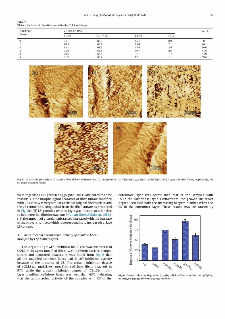
Fig. 5. Surface morphologies of original and modified cellulose fibers: (a) original fiber, (b)–(d) (CS/LS)1, (CS/LS)3 and (CS/LS)5 multilayers modified fibers, respectively, (e)
CS alone-modified fibers.
were regarded as LS granules aggregate. This is attributed to three
reasons: (i) the morphological character of fiber surface modified
with CS alone was very similar to that of original fiber surface and
the CS cannot be distinguished from the fiber surface as presented
in Fig. 5e; (ii) LS granules tend to aggregate in acid solution due
to hydrogen-bonding interactions (Nyman, Rose, & Ralston, 1986);
(iii) the amount of granular substances increased with the increase
in the bilayers number, which is correspondingto increasedsurfaceLS content.
3.3. Assessment of antimicrobial activity of cellulose fibers
modified by CS/LS multilayers
The degree of growth inhibition for E. coli was examined in
CS/LS multilayers modified fibers with different surface compo-
sitions and deposited bilayers. It was found from Fig. 6 that
all the modified cellulose fibers had E. coli inhibition activity
because of the presence of CS. The growth inhibition degree
of (CS/LS)4.5 multilayer modified cellulose fibers reached to
97%, while the growth inhibition degree of (CS/LS)5 multi-
layer modified cellulose fibers was less than 63%, indicating
that the antimicrobial activity of the samples with CS in the
outermost layer was better than that of the samples with
LS in the outermost layer. Furthermore, the growth inhibition
degree increased with the increasing bilayers number when the
CS in the outermost layer. These results may be caused by
Fig. 6. Growth inhibitiondegreefor E.coliby cellulosefibers modified with(CS/LS)n
multilayers having different bilayers number.

7/17/2019 Antimicrobial and Antioxidant Surface Modification of Cellulose Fibers
http://slidepdf.com/reader/full/antimicrobial-and-antioxidant-surface-modification-of-cellulose-fibers 6/8
40 H. Li, L. Peng/ Carbohydrate Polymers 124 (2015) 35–42
Fig. 7. DP (a) and zero-span tensile strength (b) of original and modified cellulosefibersversus oxidation reaction time.
Scheme 2. Schematic diagram illustrating ABAP radical induced oxidation mecha-
nism.
three reasons: (1) CS has antimicrobial activity; (2) the E. coli
absorption and immobilization capacities of CS were increased
because of its hydrophilicity and higher positive charge; (3) the
increase in surface RMS roughness of modified cellulose fibers led
to higherspecific area, whichmay causeefficientcontactand inter-action between CS andE. coli.
3.4. Assessment of antioxidant activity of cellulose fibers modified
by CS/LS multilayers
Degree of polymerization reflects the average length of cellulose
chains. The zero-span tensile strength is a widely used index for
evaluating the average strength of individual fiber. Fig. 7 shows
plots of DP and zero-span tensile strength of original and modified
cellulose fibers against oxidation reaction times. It is clearly seen
that DP and zero-span tensile strength of original cellulose fibers
dramatically decreased as the oxidation reaction time increased,
whichis dueto attackof radical to cellulosechains,thereby causing
depolymerization and strength loss of cellulosefibers. For the CS/LSmultilayers modified cellulose fibers, the DP and zero-span tensile
strength gently decreased with increasing oxidation reaction time.
As the number of bilayers increased, the changes in the DP and
zero-span tensile strength of modified fibers became level off. This
can be explained that LS layers on modified fibers surfaces acted
as natural radical scavenger, which protected cellulose fiber from
radicals attacking.
In this study, a probable radical oxidation mechanism is shown
in Scheme 2. Radical initiator ABAP is a water-soluble azo, which
can rapidly decomposed at 60◦C, producing two carbon-centerd
radicals (R •). R • reacted with oxygen to generate peroxyl radical
(ROO•), which attracted the cellulose, resulting in cellulose chain
fragmentation and generating cellulose radical (Cell•). The Cell•
reacted with oxygen to produce Cell-OO•
and attracted cellulose.
The presence of ROO• and Cell-OO• will cause both depolymeriza-
tionand propagationof theradicalchain.LS are a phenolicpolymers
which inhibit ABAP-initiated oxidation reactions by trapping the
chain-propagating ROO•. The LS phenolic polymers gives up their
phenolic hydrogen atom to ROO•, producing phenoxyl radical
PhO•, terminating chain reaction between ROO• and cellulose.
Simultaneously, colored non-radical products such as quinones
were formed by PhO• coupling reaction.
4. Conclusions
The antimicrobial and antioxidant surfaces modification of cel-
lulose fibers were achieved through constructing CS/LS multilayers
on fibers surfaces via layer-by-layer deposition technique. The
increase in characteristic elements (N and S) of CS/LS multilay-
ers with the number of bilayers was observed, and the surface LS
content of modified cellulose fiber linearly increased as a function
of bilayers. The surface zeta potential of modified cellulose fibers
was inversed after each deposition step. The AFM phase images
showed that the granular LS aggregate gradually covered the cel-
lulose fibers surfaces as the LBL deposition proceeded, resulting in
the increase in fibers surfaces roughness. The antimicrobial testingresults demonstrated that cellulose fibers modified by CS/LS mul-
tilayers exhibited higher antimicrobial activity against E. coli, the
degree of E. coli growth inhibition for a (CS/LS)4.5 multilayer modi-
fied cellulose fibers reached up to 97%. TheE. coligrowth inhibition
degree increased with the increasing bilayers number, especially
E. coli growth inhibition degree of modified cellulose fibers with
CS in the outmost layer was better than that of the cellulose fibers
withLS in theoutmostlayer. Theantioxidanttestingresultsshowed
that there was almost no change in the DP and zero-span tensile
strength of modified fibers after ABAP-initiated oxidations due to
the presence of radical scavenger LS. Moreover, the antioxidant
activity of CS/LS multilayers modified cellulose fibers increased
with the bilayers number increased.
Acknowledgements
This work wassupportedby theApplied BasicResearchProgram
of Yunnan Province (no. 2014FD008), Talent Training Program of
Yunnan Province (no.KKSY201305002), State Key Laboratory Open
Foundation of Pulp and Paper Engineering of China (no. 201323)
and Open Foundation of Key Lab of Pulp and Paper Science &Tech-
nology of Ministry of Education (no. 08031349).
References
Agarwal, K., Prasad, M., Sharma, R. B., & Setua, D. K. (2014). Novel bio-degradablelignin reinforced NBR composite. International Journal of Energy Engineering , 4,
47–62.

7/17/2019 Antimicrobial and Antioxidant Surface Modification of Cellulose Fibers
http://slidepdf.com/reader/full/antimicrobial-and-antioxidant-surface-modification-of-cellulose-fibers 7/8

7/17/2019 Antimicrobial and Antioxidant Surface Modification of Cellulose Fibers
http://slidepdf.com/reader/full/antimicrobial-and-antioxidant-surface-modification-of-cellulose-fibers 8/8
42 H. Li, L. Peng/ Carbohydrate Polymers 124 (2015) 35–42
Zahran, M. K., Ahmed, H. B., & EI-Rafie, M. H. (2014). Surface modification of cottonfabrics forantibacterial application by coating with AgNPs–alginate composite.Carbohydrate Polymers, 108, 145–152.
Zh ang, M., W ang, S . L., Wang, C. Y., & Li, J . ( 2012). A facile method to fabricatesuperhydrophobic cotton fabrics. Applied Surface Science, 261, 561–566.
Zhang, X., Shi, F., Yu, X., Liu, H., Fu, Y., Wang, Z. Q., et al. (2004). Polyelec-trolyte multilayer as matrix for electrochemical desposition of gold clusters:
Toward super-hydrophobicsurface. Journalof theAmericanChemicalSociety,126,3064–3065.
Zhao, Y., Tang, Y. W., Wang, X. G., & Lin,T. (2010). Superhydrophobic cotton fabricfabricated by electrostatic assembly of silica nanoparticles and its remarkablebuoyancy. Applied Surface Science, 256, 6736–6742.