antioxidative effects of caffeine in a hyperoxia-based rat
TRANSCRIPT

RESEARCH Open Access
Antioxidative effects of caffeine in ahyperoxia-based rat model ofbronchopulmonary dysplasiaStefanie Endesfelder* , Evelyn Strauß, Till Scheuer, Thomas Schmitz and Christoph Bührer
Abstract
Background: While additional oxygen supply is often required for the survival of very premature infants inintensive care, this also brings an increasing risk of progressive lung diseases and poor long-term lung outcomes.Caffeine is administered to neonates in neonatal intensive care for the prevention and treatment of apneas and hasbeen shown to reduce BPD incidence and the need for mechanical ventilation, although it is still unclear whetherthis is due to a direct pulmonary action via antagonism of adenosine receptors and/or an indirect action. Thisexperimental study aims to investigate the action of caffeine on the oxidative stress response in pulmonary tissuein a hyperoxia-based model of bronchopulmonary dysplasia in newborn rats.
Methods: Newborn Wistar rats were exposed to 21% or 80% oxygen for 3 (P3) or 5 (P5) postnatal days with orwithout recovery on room air until postnatal day 15 (P15) and treated with vehicle or caffeine (10 mg/kg) every 48h beginning on the day of birth. The lung tissue of the rat pups was examined for oxidative stress response at P3and P5 immediately after oxygen exposure or after recovery in ambient air (P15) by immunohistological stainingand analysis of lung homogenates by ELISA and qPCR.
Results: Lungs of newborn rats, corresponding to the saccular stage of lung development and to the human lungdevelopmental stage of preterms, showed increased rates of total glutathione and hydrogen peroxide, oxidativedamage to DNA and lipids, and induction of second-phase mediators of antioxidative stress response (superoxidedismutase, heme oxygenase-1, and the Nrf2/Keap1 system) in response to hyperoxia. Caffeine reduced oxidativeDNA damage and had a protective interference with the oxidative stress response.
Conclusion: In addition to the pharmacological antagonism of adenosine receptors, caffeine appears to be apotent antioxidant and modulates the hyperoxia-induced pulmonary oxidative stress response and thus protectiveproperties in the BPD-associated animal model. Free-radical-induced damage caused by oxidative stress seems tobe a biological mechanism progress of newborn diseases. New aspects of antioxidative therapeutic strategies topassivate oxidative stress-related injury should be in focus of further investigations.
Keywords: Caffeine, Hyperoxia (oxygen), Preterm, Animal model, Oxidative stress, Antioxidant
© The Author(s). 2019 Open Access This article is distributed under the terms of the Creative Commons Attribution 4.0International License (http://creativecommons.org/licenses/by/4.0/), which permits unrestricted use, distribution, andreproduction in any medium, provided you give appropriate credit to the original author(s) and the source, provide a link tothe Creative Commons license, and indicate if changes were made. The Creative Commons Public Domain Dedication waiver(http://creativecommons.org/publicdomain/zero/1.0/) applies to the data made available in this article, unless otherwise stated.
* Correspondence: [email protected] of Neonatology, Charité Universitätsmedizin Berlin,Augustenburger Platz 1, 13353 Berlin, Germany
Endesfelder et al. Respiratory Research (2019) 20:88 https://doi.org/10.1186/s12931-019-1063-5

BackgroundPreterm birth and administration of supplemental oxygenhave a profound impact on pulmonary lung development[1–5]. Disruption of postnatal development is associatedwith pulmonary dysfunction, higher risk of respiratory dis-eases such as respiratory distress syndrome or broncho-pulmonary dysplasia (BPD), and higher rates of lifelongmorbidities [6–9]. The incidence of BPD correlates closelywith the immaturity of the lung, such as poor oxygen de-toxification enzymes and oxidative stress response [10,11]. Besides the prematurity and the surfactant deficiency,the mechanical ventilation, the oxygen toxicity, and thusthe effect of high oxygen concentrations with the resultingoxidative stress is an important cause of BPD [11–14].Oxidative stress indicates an imbalance between reactiveoxygen species (ROS) and corresponding antioxidantcounterparts, resulting in toxic levels of ROS and damageto lipids, proteins, and DNA [15]. It is necessary to differ-entiate between physiological ROS for redox-sensitivesignal transduction pathways and pathologically relevantROS levels, which could affect vulnerable processes suchas lung development [16], aggravated by inflammatoryside effects [11]. Preterm infants are particularly exposedto the oxidative stress due to the transition from intrauter-ine hypoxia to extrauterine hyperoxia and have an in-creased risk of BPD. Besides avoiding premature birth,prenatal glucocorticoids, reducing oxygen content in ven-tilated air, gentle ventilation techniques, and surfactanttherapy, antioxidative therapy strategies represent anotherpossibility for preventing BPD. [17]. Existing therapies canreduce the rate of BPD but show minimal effects onBPD-typical arrest of lung development [18, 19]. Newinsights could be offered by antioxidant therapies if theyreduce oxidative stress per se and thus could minimizethe pathological consequences [20].The methylxanthine caffeine is the standard and most
frequently used medication for treatment of apnea due toprematurity. Caffeine acts mainly as a nonspecific inhibitorof adenosine receptors subtypes A1 and A2a and with abroad spectrum of pharmacokinetic activity [21]. In a largeclinical trial, caffeine has been shown to reduce the rate ofBPD in very preterm infants, shortening the duration timeof mechanical ventilation and oxygen supplementation[22]. Caffeine appears to be most effective when startedwithin the first few days of life [23–25].In experimental animals, BPD-like lung damage can be
precipitated by postnatal hyperoxia [26], with similar histo-logical changes in hyperoxia-exposed rodent pups andhuman preterm infants [18, 27, 28]. These models can beused to study the effect of therapeutic interventions suchas caffeine, which are not completely understood [29–31].In addition to antagonism of adenosine receptors [32],anti-inflammatory effects, and the reduction of stress onthe endoplasmic reticulum, anti-oxidant properties per se
have also been discussed [33–36]. Therefore, this studyaims to investigate the action of caffeine on the oxidativestress response in pulmonary tissue in a hyperoxia-basedmodel of BPD in the newborn rat.
MethodsOxygen exposure and drug administrationTime-pregnant Wistar rat dams were obtained from theDepartment of Experimental Medicine (FEM, Charité –Universitätsmedizin Berlin, Germany). The adult rats werehoused in individual cages under environment-controlledconditions with a constant 24 h light/dark cycle, ambienttemperature, and relative humidity of 80% with ad libitumaccess to the same food and water. All procedures were ap-proved by the local animal welfare authorities (LAGeSo,approval number G-0088/16) and followed institutionalguidelines. The pups were pooled and randomized within12 h of birth and returned to the dams. According to theexperimental conditions (see Fig. 1), the newborn rats wererandomly assigned to room air (normoxia) oroxygen-enriched atmosphere (hyperoxia) treatment. Thepups in hyperoxia subgroups (HY) were reared with thedams in an atmosphere containing 80% oxygen (OxyCyclerBioSpherix, Lacona, NY) from A) postnatal day (P)0 to P3(n = 7–8) or B) P0 to P5 (n = 6–8); in parallel, the pups innormoxia (NO) groups were reared with the dams underroom air conditions. To avoid oxygen toxicity in the nurs-ing mothers, they were rotated between the oxygen treat-ment and normoxia litters every 24 h. The rats are dividedinto four groups, each for both exposure times with 1)normoxia (NO, control group): 21% oxygen application ofvehicle (phosphate buffered saline, PBS), 2) normoxia withcaffeine (NOC): 21% oxygen with caffeine (10mg/kg,Sigma, Steinheim, Germany), 3) hyperoxia (HY): 80% oxy-gen with vehicle (PBS), and 4) hyperoxia with caffeine(HYC): 80% oxygen with caffeine (10mg/kg). 10mg/kg ofpure caffeine is equivalent to 20mg/kg caffeine citrate,which used clinically.Rat pups received either drug or vehicle injection intra-
peritoneally (i.p.) as a fixed proportion of their body weight(100 μl/10 g) every 48 h beginning on the day of birth (P0).The administration of caffeine or vehicle took place for thepups with a total of three postnatal days of oxygen exposure(P0-P3) on the day of birth (P0) and on P2; for the rat pupswith a total of five days of postnatal oxygen exposure(P0-P5) on the day of birth (P0) and on P2 and P4. The ratpups were first examined after the oxygen exposure (P3,P5). Further examinations occurred after recovery in roomair at P15 (P3_P15, P5_P15). No pups died duringhyperoxia.
Plasma and tissue preparationAt the experimental endpoints (P3; P3_P15; P5; P5_P15),rat pups were anaesthetized with an i.p. injection of
Endesfelder et al. Respiratory Research (2019) 20:88 Page 2 of 13

ketamine (100mg/kg), xylazine (20mg/kg), and acepro-mazine (3mg/kg), blood samples were collected intoheparin-coated centrifugation microtubes (MicrovetteCB300LH, Sarstedt International, Nümbrecht, Germany),and then transcardially perfused. The heart lung blockwas immediately removed and the lungs were snap-frozenin liquid nitrogen and stored at − 80 °C. The perfusionwas carried out with PBS (pH 7.4) for the molecular ana-lysis and for immunohistochemical analysis followed byperfusion with 4% paraformaldehyde (pH 7.4), the lungswere postfixed at 4 °C for 1 day, embedded in paraffin, andprocessed for histological staining. The blood sampleswere centrifuged at 4 °C, the plasma samples were sepa-rated and stored at − 80 °C. The animals were weighed ateach time point of application (P0, P2 and/or P4) and atthe experimental endpoints (P3/P5 and P15).
Caffeine plasma levelThe concentrations of caffeine in the plasma were deter-mined externally using the LC-MS/MS analytical method(Institut für Veterinärmedizinische Diagnostik GmbH,Berlin, Germany).
RNA extraction and quantitative real-time PCRThe gene expression analysis was performed as previ-ously described [37]. The PCR products of
hypoxanthine-guanine phosphoribosyl-transferase (Hprt),Kelch-like ECH-associated protein 1 (Keap1), NFE2-re-lated factor 2 (Nrf2), and superoxide dismutase (Sod) 1,2, and 3 were quantified in real time with the sequences(synthesized by BioTeZ, Berlin, Germany) summarizedin Additional file 2: Table S1. PCR and detection wereperformed with qPCR BIO Mix Hi-ROX (NIPPON Gen-etics Europe, Düren, Germany) with Hprt used as an in-ternal reference. The expression of target genes wasanalyzed with the StepOnePlus real-time PCR system(Applied Biosystems, Carlsbad, CA, USA) according tothe 2-ΔΔCT method [38].
Protein extractionProtein was extracted were determined using the PierceBCA kit (Pierce/Thermo Scientific, Rockford, IL, USA)as described in [39].
Oxidative stress marker ELISA assayConcentrations of total glutathione (OxiSelect Total Gluta-thione Assay Kit, Cell Biolabs Inc., San Diego, CA, USA),lipid peroxidation (TBARS assay kit, Cayman Chemical,Ann Arbor, MI, USA), heme oxygenase (HO)-1 (Rat HemeOxygenase-1 EIA Kit, Precoated, Takara Bio Europe/SAS,Saint-Germain-en-Laye, France), and hydrogen peroxide(H2O2; OxiSelect Hydrogen peroxide assay kit, Cell Biolabs
Fig. 1 Time course of hyperoxic exposure and recovery of rat pups. The pups were divided into four groups with exposure to room air orhyperoxia occurring for either a) 3 postnatal days (P0-P3) or b) 5 postnatal days (P0-P5) with normoxia (NO) and vehicle application (PBS),normoxia with caffeine application (NOC), hyperoxia (HY) and vehicle application, and hyperoxia with caffeine (HYC). Rat pups received caffeine orvehicle injection intraperitoneally as a fixed portion of body weight (100 μl/10 g) every 48 h beginning on the day of birth (P0, red arrow). Theexperimental endpoints (black arrow) are directly after the oxygen exposure (P3 or P5) and after recovery in room air at P15 (P3_P15 or P5_P15)
Endesfelder et al. Respiratory Research (2019) 20:88 Page 3 of 13

Inc.) were measured in lung homogenates according tomanufacturer’s instructions. Values were calculated from astandard curve and normalized to the amount of total pro-tein. Superoxide dismutase (Sod) 1, 2, and 3 concentrationswere measured using rat colorimetric ELISA kits (antibo-dies-online GmbH, Aachen, Germany).
ImmunohistochemistryTen-micrometer-thick paraffin lung sections were depar-affinized in xylene, rehydrated in ethanol, and subjected tohematoxylin and eosin (HE) staining or immunostaining.The pulmonary morphometric changes were viewed by
light microscopy. Statistical data on structural changes hasnot been compiled because other perfusion techniquesmust be used according to the required standard. The ex-emplary HE stains are only intended to support the state-ment about the effectiveness of oxygen toxicity as adamage model of the lung (Additional file 1: Figure S1).Deparaffinized sections were immunostained with an
anti-8-oxodG monoclonal antibody (clone 2E2; Trevi-gen, Gaithersburg, MD, USA) as previously described in[34]. Sections were analyzed blind using a Keyence com-pact fluorescent microscope BZ 9000 with BZ-II Viewersoftware and BZ-II Analyzer software (Keyence, Osaka,Japan). 8-oxodG positive cells were counted in 4non-overlapping separate fields per animal and meanvalues of all images were used for statistical analysis.
Statistical analysesAll data are expressed as the mean ± standard error of themean (SEM). Groups were compared assuming a non-Gaussian distribution with the Mann Whitney test. Atwo-sided p value of < 0.05 was considered significant. All
graphics and statistical analyses were performed using theGraphPad Prism 8.0 software (GraphPad Software, LaJolla, CA, USA).
ResultsCaffeine alters postnatal weight gainThe effects of caffeine treatment on the weight gain of new-born rats were monitored for the different experimentalgroups at different time points. It was found that caffeinereduced weight gain in control rats (normoxia) at day 2(P2), day 4 (P4), and day 5 (P5), while in hyperoxic pups,caffeine treatment inhibited weight gain only at P2 and P4(Fig. 2 a and b). By age P15, no difference in body weightwas found for any of the experimental groups, which indi-cating temporary effects of caffeine on early postnatalweight gain.
Plasma caffeine levelPlasma caffeine levels were undetectable in the saline-treated pups at P3 and P5, as well in all groups after re-covery at P3_P15 and P5_P15. The levels for caffeine-treated normoxic exposed rat pups were 9.4 ± 0.8 μg/ml atP3 and 10.4 ± 0.8 μg/ml at P5, and for caffeine-treatedhyperoxic exposed rat pups 8.8 ± 0.5 μg/ml and 11.2 ±0 μg/ml, respectively, without significant differences be-tween groups. Validation of plasma caffeine levels showedthat caffeine was sufficient to be within the therapeuticallycomparable range (5.5–23.7mg/L) [21].
Caffeine attenuates morphological changesHyperoxia for 3 or 5 days immediately after birth causedsubstantial alterations in lung morphology. After oxygenexposure, the lungs of rats pups contained fewer alveoli
Fig. 2 Weight gain of rat pups exposed to normoxia or hyperoxia and treated or not with caffeine for the examination groups a) oxygenexposure from P0 to P3 with monitored body weight on P2, P3, and P15, and b) oxygen exposure from P0 to P5 with monitored body weighton P2, P4, P5, and P15. Values are the mean of the difference in weight of all animals used for immunohistochemical and molecular biologicalexperiments (n = 14–16). Data are expressed relative to the control group at each time point as mean ± SEM. *p < 0.05, **p < 0.01, and ***p <0.001 versus control (NO); ###p < 0.001 versus hyperoxia group (HY) with Mann Whitney test
Endesfelder et al. Respiratory Research (2019) 20:88 Page 4 of 13
than those of control pups under normoxia conditions,with generalized enlargement of alveolar airspaces.These histological changes were even more pronouncedafter 5 days of exposure. After recovery under normoxicconditions to postnatal day 15, morphological changeswere still detected in the oxygen-damaged lungs. Caf-feine effectively counteracted these structural changes(Additional file 1: Figure S1).
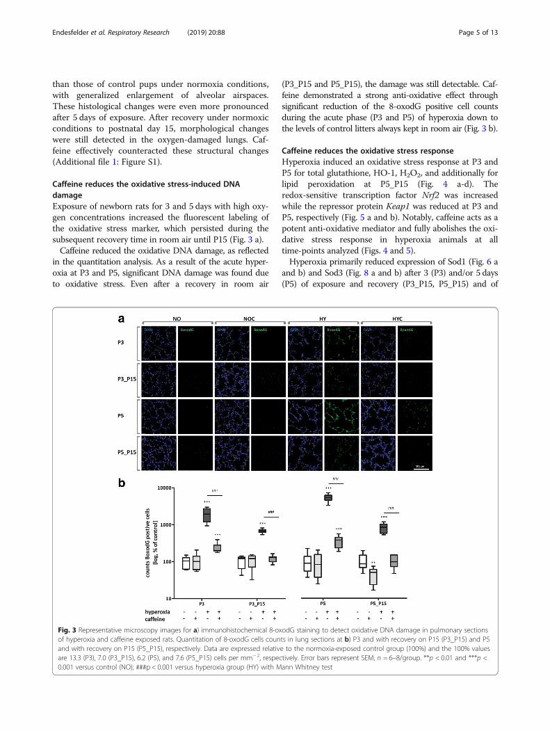
Caffeine reduces the oxidative stress-induced DNAdamageExposure of newborn rats for 3 and 5 days with high oxy-gen concentrations increased the fluorescent labeling ofthe oxidative stress marker, which persisted during thesubsequent recovery time in room air until P15 (Fig. 3 a).Caffeine reduced the oxidative DNA damage, as reflected
in the quantitation analysis. As a result of the acute hyper-oxia at P3 and P5, significant DNA damage was found dueto oxidative stress. Even after a recovery in room air
(P3_P15 and P5_P15), the damage was still detectable. Caf-feine demonstrated a strong anti-oxidative effect throughsignificant reduction of the 8-oxodG positive cell countsduring the acute phase (P3 and P5) of hyperoxia down tothe levels of control litters always kept in room air (Fig. 3 b).
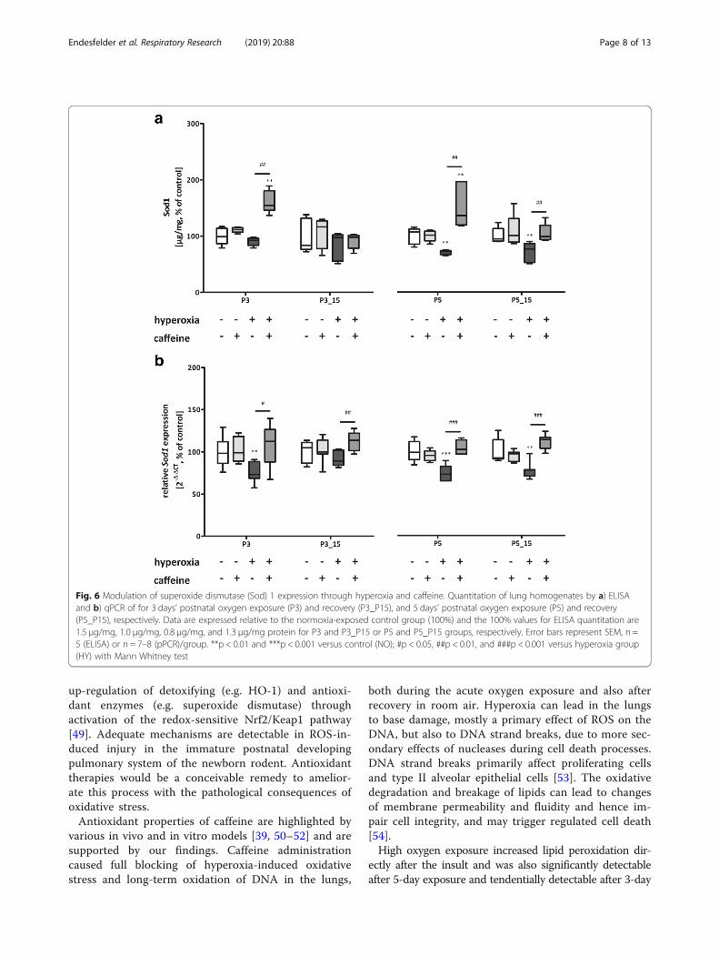
Caffeine reduces the oxidative stress responseHyperoxia induced an oxidative stress response at P3 andP5 for total glutathione, HO-1, H2O2, and additionally forlipid peroxidation at P5_P15 (Fig. 4 a-d). Theredox-sensitive transcription factor Nrf2 was increasedwhile the repressor protein Keap1 was reduced at P3 andP5, respectively (Fig. 5 a and b). Notably, caffeine acts as apotent anti-oxidative mediator and fully abolishes the oxi-dative stress response in hyperoxia animals at alltime-points analyzed (Figs. 4 and 5).Hyperoxia primarily reduced expression of Sod1 (Fig. 6 a
and b) and Sod3 (Fig. 8 a and b) after 3 (P3) and/or 5 days(P5) of exposure and recovery (P3_P15, P5_P15) and of
Fig. 3 Representative microscopy images for a) immunohistochemical 8-oxodG staining to detect oxidative DNA damage in pulmonary sectionsof hyperoxia and caffeine exposed rats. Quantitation of 8-oxodG cells counts in lung sections at b) P3 and with recovery on P15 (P3_P15) and P5and with recovery on P15 (P5_P15), respectively. Data are expressed relative to the normoxia-exposed control group (100%) and the 100% valuesare 13.3 (P3), 7.0 (P3_P15), 6.2 (P5), and 7.6 (P5_P15) cells per mm− 2, respectively. Error bars represent SEM, n = 6–8/group. **p < 0.01 and ***p <0.001 versus control (NO); ###p < 0.001 versus hyperoxia group (HY) with Mann Whitney test
Endesfelder et al. Respiratory Research (2019) 20:88 Page 5 of 13

Sod2 (Fig. 7 a and b) for both exposure times with (P3_P15,P5_P15) and without (P3, P5) recovery at RNA levels.Treatment with caffeine prevented the oxidative debt frommanifesting itself in changed Sod levels (Fig. 6-8).
DiscussionCaffeine is the most common methylxanthine universallyused in current neonatal practice. Its efficacy and toler-ability with a wide therapeutic index and high safetyhave made it the drug of choice for respiratory instabil-ities and it is often used for many weeks with potentialcatabolic effects that could impact the initial weight gainof preterms. The effect of caffeine on weight loss ofcaffeine-treated newborn rats according to our in vivoresults matches observations made in preterm infants[22, 40, 41]. This can be explained by the increased diur-esis in kidneys caused by caffeine. It can only be specu-lated whether the amount of weight loss affects thehealth status of the animals investigated or of humanpatients being treated with caffeine. However, since thisis of a temporary nature, and all treated pups after sometime returned to the range of body weight similar to un-treated controls, the impact of weight loss (or water loss)seems negligible and in fact could easily be treated inthe context of patient medical care. From clinical studiesit is known that prolonged caffeine insult may lead to areduction in weight gain, due to a higher energy expend-iture and higher oxygen consumption of the organism[42], and influenced by caffeine concentration [41].Early oxygen exposure is one of the most important
factors implicated in the development of BPD [43].Experimental hyperoxia-induced lung injury mimics thehallmarks of human BPD [18, 27, 28], which are character-ized by the damaging effect of high oxygen concentrationsand the resulting structural changes, such as alveolar sim-plification and thickening of the alveolar septum [44–46].Postnatal hyperoxia led to structural changes in the imma-ture lung at exposure times over the first few days of life(P3 and P5), furthermore the damage persisted even afterrecovery under normoxic conditions until the transitionfrom the saccular to the alveolar phase of lung develop-ment. Caffeine reduced this structural damage, as shown ina previous study of acute hyperoxia [47, 48].The primary goal of the current study was the investiga-
tion of the antioxidative capacity of caffeine in ahyperoxic-based rat model of BDP, which clearly demon-strated the effective prevention of oxidative DNA damageand oxidative stress responses. While the early postnatalexposure to oxygen induced long-lasting oxidative damageto DNA and lipids after recovery in room air, no such dam-age was found in hyperoxic animals if they were treatedwith caffeine.It is important for this animal study on hyperoxia-in-
duced injury in the developing lung that the relevant
Fig. 4 Acute hyperoxia resulted in an adequate oxidative stress responseand caffeine reduced the response. Quantitation of lung homogenates byELISA of a) total glutathione, b) H2O2, c) HO-1, and d) MDA/ lipidperoxidation with 3 days’ postnatal oxygen exposure (P3) and recovery(P3_P15) and 5days’ postnatal oxygen exposure (P5) and recovery (P5_P15),respectively. Data are expressed relative to the normoxia-exposed controlgroup (100%) and the 100% values are a) 19.2μM/mg, 13.4μM/mg,17.8μM/mg, and 13.7μM/mg protein, b) 1.8μM/mg, 3.1μM/mg, 1.0μM/mg,and 3.0μM/mg protein, c) 17.2 ng/mg, 5.9 ng/mg, 20.0 ng/mg, and 5.2 ng/mg protein, and d) 3.3μM/mg, 3.7μM/mg, 1.3μM/mg, and 3.3μM/mgprotein for P3 and P3_P15 or P5 and P5_P15 groups, respectively. Error barsrepresent SEM, n =5/group. *p <0.05 and **p<0.01 versus control (NO); #p< 0.05 and ##p<0.01 versus hyperoxia group (HY) with Mann Whitney test
Endesfelder et al. Respiratory Research (2019) 20:88 Page 6 of 13

time points of pulmonary developmental lay within thesaccular phase, after the termination of the saccularphase, as well as with a recovery period after completionof the alveolar phase. The human saccular phase termi-nates between gestation weeks 24 and 38 (correspondingto rats P0 to P4), which correlates with very low birthweight (VLBW) infants, and the alveolar phase overlapsat about 36 weeks (corresponding to rats’ P5 to P15).Thus, our model with hyperoxia from day-of-birth topostnatal day 3 (P3) and day 5 (P5) and recovery time today 15 (P15) provides an adequate model of lung injuryto very prematurely born infants and allows studies ofhyperoxic or caffeine-relevant modulations at differentstages of lung development. The oxygen exposure in thefirst 3 and 5 days of life in rats can be regarded asequivalent to 3 to 5 weeks in humans [27]. Oxidative
stress due to the transition from intrauterine hypoxia toextra-uterine hyperoxia, the need for mechanical ventila-tion and additional oxygen, and birth during the saccularphase of lung development are the major risk factors forrespiratory distress in preterm infants, in addition to theincomplete detoxification response to free radicals [14].Our data demonstrated clear antioxidant actions ofcaffeine that effectively prevent oxidative damage in thedeveloping lung even at the high levels of oxygen thatmight need to be applied in preterm infants sufferingfrom lung disease.Hyperoxia leads to excessive generation of ROS, in-
cluded hydrogen peroxide (H2O2) and hydroxyl radicals,and causes oxidative stress. The antioxidative networkincludes direct responses, like radical scavengers orchemical modifications, and indirect responses with
Fig. 5 Acute hyperoxia resulted in an adequate oxidative stress response and caffeine reduced the response. Quantitation of lung homogenatesby qPCR of a) Nrf2, and b) Keap1 for 3 days’ postnatal oxygen exposure (P3) and recovery (P3_P15), and 5 days’ postnatal oxygen exposure (P5)and recovery (P5_P15), respectively. Data are expressed relative to the normoxia-exposed control group (100%). Error bars represent SEM,n = 7–8/group. *p < 0.05, **p < 0.01, and ***p < 0.001 versus control (NO); #p < 0.05, ##p < 0.01, and ###p < 0.001 versus hyperoxia group(HY) with Mann Whitney test
Endesfelder et al. Respiratory Research (2019) 20:88 Page 7 of 13
up-regulation of detoxifying (e.g. HO-1) and antioxi-dant enzymes (e.g. superoxide dismutase) throughactivation of the redox-sensitive Nrf2/Keap1 pathway[49]. Adequate mechanisms are detectable in ROS-in-duced injury in the immature postnatal developingpulmonary system of the newborn rodent. Antioxidanttherapies would be a conceivable remedy to amelior-ate this process with the pathological consequences ofoxidative stress.Antioxidant properties of caffeine are highlighted by
various in vivo and in vitro models [39, 50–52] and aresupported by our findings. Caffeine administrationcaused full blocking of hyperoxia-induced oxidativestress and long-term oxidation of DNA in the lungs,
both during the acute oxygen exposure and also afterrecovery in room air. Hyperoxia can lead in the lungsto base damage, mostly a primary effect of ROS on theDNA, but also to DNA strand breaks, due to more sec-ondary effects of nucleases during cell death processes.DNA strand breaks primarily affect proliferating cellsand type II alveolar epithelial cells [53]. The oxidativedegradation and breakage of lipids can lead to changesof membrane permeability and fluidity and hence im-pair cell integrity, and may trigger regulated cell death[54].High oxygen exposure increased lipid peroxidation dir-
ectly after the insult and was also significantly detectableafter 5-day exposure and tendentially detectable after 3-day
Fig. 6 Modulation of superoxide dismutase (Sod) 1 expression through hyperoxia and caffeine. Quantitation of lung homogenates by a) ELISAand b) qPCR of for 3 days’ postnatal oxygen exposure (P3) and recovery (P3_P15), and 5 days’ postnatal oxygen exposure (P5) and recovery(P5_P15), respectively. Data are expressed relative to the normoxia-exposed control group (100%) and the 100% values for ELISA quantitation are1.5 μg/mg, 1.0 μg/mg, 0.8 μg/mg, and 1.3 μg/mg protein for P3 and P3_P15 or P5 and P5_P15 groups, respectively. Error bars represent SEM, n =5 (ELISA) or n = 7–8 (pPCR)/group. **p < 0.01 and ***p < 0.001 versus control (NO); #p < 0.05, ##p < 0.01, and ###p < 0.001 versus hyperoxia group(HY) with Mann Whitney test
Endesfelder et al. Respiratory Research (2019) 20:88 Page 8 of 13
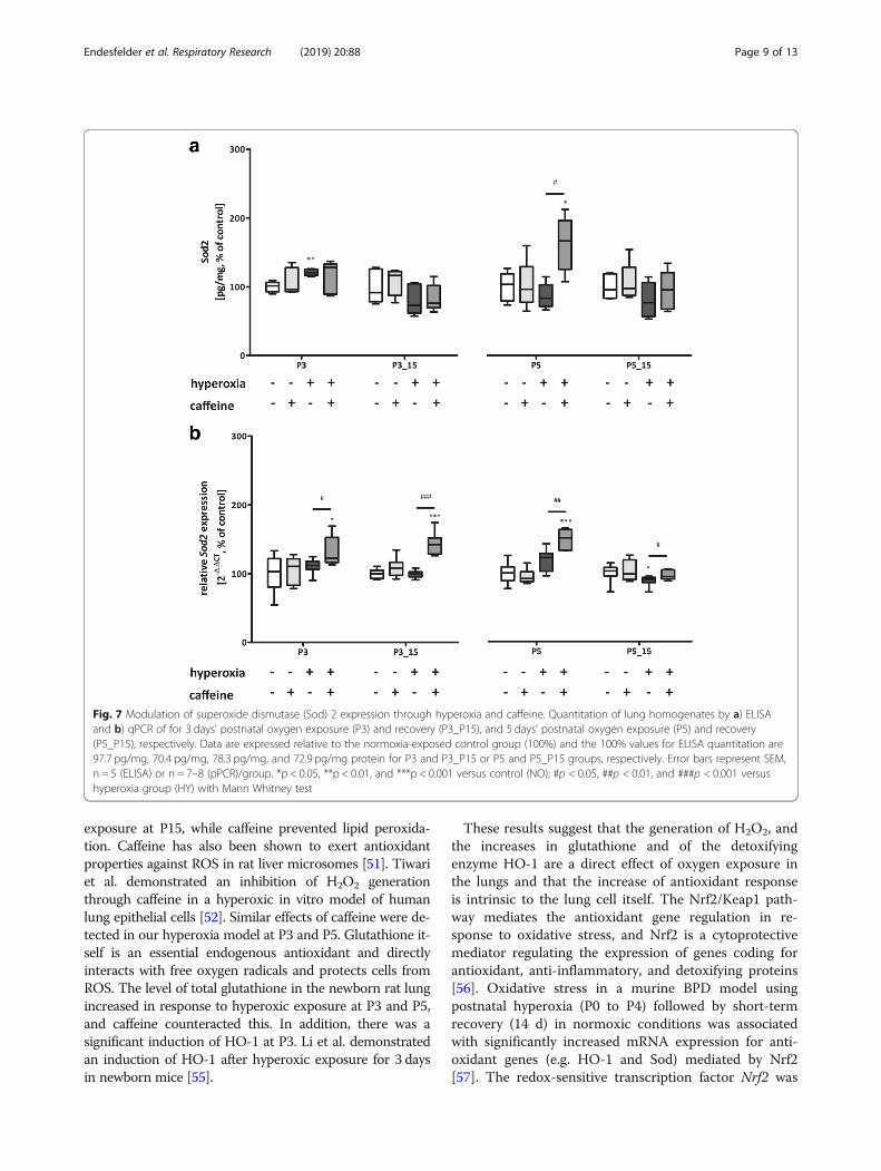
exposure at P15, while caffeine prevented lipid peroxida-tion. Caffeine has also been shown to exert antioxidantproperties against ROS in rat liver microsomes [51]. Tiwariet al. demonstrated an inhibition of H2O2 generationthrough caffeine in a hyperoxic in vitro model of humanlung epithelial cells [52]. Similar effects of caffeine were de-tected in our hyperoxia model at P3 and P5. Glutathione it-self is an essential endogenous antioxidant and directlyinteracts with free oxygen radicals and protects cells fromROS. The level of total glutathione in the newborn rat lungincreased in response to hyperoxic exposure at P3 and P5,and caffeine counteracted this. In addition, there was asignificant induction of HO-1 at P3. Li et al. demonstratedan induction of HO-1 after hyperoxic exposure for 3 daysin newborn mice [55].
These results suggest that the generation of H2O2, andthe increases in glutathione and of the detoxifyingenzyme HO-1 are a direct effect of oxygen exposure inthe lungs and that the increase of antioxidant responseis intrinsic to the lung cell itself. The Nrf2/Keap1 path-way mediates the antioxidant gene regulation in re-sponse to oxidative stress, and Nrf2 is a cytoprotectivemediator regulating the expression of genes coding forantioxidant, anti-inflammatory, and detoxifying proteins[56]. Oxidative stress in a murine BPD model usingpostnatal hyperoxia (P0 to P4) followed by short-termrecovery (14 d) in normoxic conditions was associatedwith significantly increased mRNA expression for anti-oxidant genes (e.g. HO-1 and Sod) mediated by Nrf2[57]. The redox-sensitive transcription factor Nrf2 was
Fig. 7 Modulation of superoxide dismutase (Sod) 2 expression through hyperoxia and caffeine. Quantitation of lung homogenates by a) ELISAand b) qPCR of for 3 days’ postnatal oxygen exposure (P3) and recovery (P3_P15), and 5 days’ postnatal oxygen exposure (P5) and recovery(P5_P15), respectively. Data are expressed relative to the normoxia-exposed control group (100%) and the 100% values for ELISA quantitation are97.7 pg/mg, 70.4 pg/mg, 78.3 pg/mg, and 72.9 pg/mg protein for P3 and P3_P15 or P5 and P5_P15 groups, respectively. Error bars represent SEM,n = 5 (ELISA) or n = 7–8 (pPCR)/group. *p < 0.05, **p < 0.01, and ***p < 0.001 versus control (NO); #p < 0.05, ##p < 0.01, and ###p < 0.001 versushyperoxia group (HY) with Mann Whitney test
Endesfelder et al. Respiratory Research (2019) 20:88 Page 9 of 13
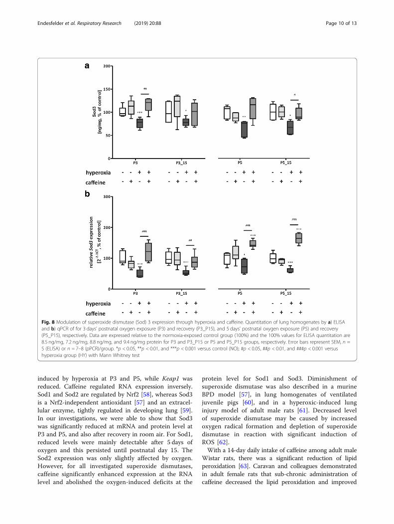
induced by hyperoxia at P3 and P5, while Keap1 wasreduced. Caffeine regulated RNA expression inversely.Sod1 and Sod2 are regulated by Nrf2 [58], whereas Sod3is a Nrf2-independent antioxidant [57] and an extracel-lular enzyme, tightly regulated in developing lung [59].In our investigations, we were able to show that Sod3was significantly reduced at mRNA and protein level atP3 and P5, and also after recovery in room air. For Sod1,reduced levels were mainly detectable after 5 days ofoxygen and this persisted until postnatal day 15. TheSod2 expression was only slightly affected by oxygen.However, for all investigated superoxide dismutases,caffeine significantly enhanced expression at the RNAlevel and abolished the oxygen-induced deficits at the
protein level for Sod1 and Sod3. Diminishment ofsuperoxide dismutase was also described in a murineBPD model [57], in lung homogenates of ventilatedjuvenile pigs [60], and in a hyperoxic-induced lunginjury model of adult male rats [61]. Decreased levelof superoxide dismutase may be caused by increasedoxygen radical formation and depletion of superoxidedismutase in reaction with significant induction ofROS [62].With a 14-day daily intake of caffeine among adult male
Wistar rats, there was a significant reduction of lipidperoxidation [63]. Caravan and colleagues demonstratedin adult female rats that sub-chronic administration ofcaffeine decreased the lipid peroxidation and improved
Fig. 8 Modulation of superoxide dismutase (Sod) 3 expression through hyperoxia and caffeine. Quantitation of lung homogenates by a) ELISAand b) qPCR of for 3 days’ postnatal oxygen exposure (P3) and recovery (P3_P15), and 5 days’ postnatal oxygen exposure (P5) and recovery(P5_P15), respectively. Data are expressed relative to the normoxia-exposed control group (100%) and the 100% values for ELISA quantitation are8.5 ng/mg, 7.2 ng/mg, 8.8 ng/mg, and 9.4 ng/mg protein for P3 and P3_P15 or P5 and P5_P15 groups, respectively. Error bars represent SEM, n =5 (ELISA) or n = 7–8 (pPCR)/group. *p < 0.05, **p < 0.01, and ***p < 0.001 versus control (NO); #p < 0.05, ##p < 0.01, and ###p < 0.001 versushyperoxia group (HY) with Mann Whitney test
Endesfelder et al. Respiratory Research (2019) 20:88 Page 10 of 13
the antioxidant defense in the blood and brain [64]. Tenget al. demonstrated a significant reduction of DNA oxida-tion with caffeine in rat pups raised in hyperoxia from P1to P10 and recovery in room air until P21 [35].In summary, caffeine shows established clinical effects
[22, 65, 66] with high efficacy and is one of the mostwidely used pharmacologic agents in the neonatal inten-sive care unit. Recent studies have provided new insightsinto the mechanisms by which the reduction of respiratorydistress symptoms per se or, as shown in in vivo and invitro studies, the anti-inflammatory or antioxidant effectsof caffeine may be causative [47, 52, 67, 68].
LimitationsSome limitations of this study are to be mentioned. Pre-dominant injurious stimuli for BPD models are hyperoxiaand mechanical ventilation (reviewed in [26]). Our modelused hyperoxia to induce oxidative stress with a focus onthe antioxidant properties of caffeine. No statements canbe made on quantitation of the structural changes, asother perfusion techniques would need to be used accord-ing to the recommended standard [69]. Paraformaldehydeis a good general-purpose fixative for immunohistochem-istry and immunocytochemistry, because it does not com-pletely destroy protein immunogenicity [70]. However,paraformaldehyde does not adequately stabilize tissuestructure; the fixed lung is subject to significant mechan-ical distortion and collapse and pulmonary cell structuresare not adequately fixed. Fixation with glutaraldehyde andis more suitable for morphological quantitative analysisand aims to preserve mainly lung volume in a defined in-flation state, architectural integrity of lung parenchyma,airways, and vessels, ultrastructure of lung cells, organ-elles, and matrix [69]. Structural and morphologicalchanges aspects in unity with the cellular mechanisms arecurrently being further investigated in this project.Caffeine was administered every 48 h, and not in a
once-daily regimen, in order to avoid elevated doses ofcaffeine outside of the therapeutic range that might inducepro-inflammatory responses [70]. Caffeine per se inducesthe inflammatory cascade and cytokine levels were highestin the time window after the application. Chavez Valdez etal. could show this clinically as well [70, 71]. Caffeine hasbeen shown to be anti-inflammatory only in a therapeuticplasma level. The measured caffeine plasma levels wereshown to be in the therapeutically comparable range of5,5–23,7mg/L [21] two days after administration. We de-liberately opted for non-daily application to avoid the dailyinflammatory insults for the rats, but also to reduce thestress on the juveniles and the dams.
ConclusionCaffeine treatment modulated the antioxidative responsein hyperoxic-induced lung injury and suggested that
caffeine acts as a potent antioxidant. Effective radicalscavenging properties of caffeine could have an essentialand crucial function in the treatment of oxidative stress-induced respiratory diseases. Finally, the pathogenesis ofneonatal respiratory distress syndrome or BPD involvesthe formation of ROS and may be amenable to reduc-tions in exposure to oxygen as well as treatment withantioxidants [17, 22, 66, 72].
Additional files
Additional file 1: Figure S1. Representative haematoxylin and eosin(H&E)-stained sections of uninjured and oxygen-injured animals at alltime-points with and without caffeine application. (TIF 6680 kb)
Additional file 2: Table S1. Sequences of oligonucleotides.(DOCX 13 kb)
Abbreviations8-oxodG: 8-oxo-2′-deoxyguanosine; BPD: Bronchopulmonary dysplasia;FAM: 6-carboxyfluorescein; HO-1: Heme oxygenase-1; Hprt: Hypoxanthine-guanine phosphoribosyl-transferase; Keap1: Kelch-like ECH-associated protein1; MDA: Malondialdehyde; Nrf2: NFE2-related factor 2; P: Postnatal day;PBS: Phosphate buffered saline; ROS: Reactive oxygen species; ROX: 6-carboxy-X-rhodamine; SOD/Sod: Superoxide dismutase;TAMRA: Tetramethylrhodamine; TBARS: Thiobarbituric acid reactivesubstances
AcknowledgmentsWe thank Ruth Herrmann for technical assistance.
FundingExcept for intra-mural support, this research did not receive any specificgrant from funding agencies in the public, commercial, or non-profit sectors.
Availability of data and materialsAll data generated or analyzed during this study are included in thispublished article.
Authors’ contributionsSE conceived the ideas, designed and executed the experiments, and wrotethe first draft of the manuscript and approved the final draft; SE and ESperformed the animal studies; ES performed and analyzed ELISAexperiments; ES performed immunostaining; ES and TS helped with analysesand provided technical assistance; TSchm revised the manuscript andcontributed to the critical discussion; CB interpreted the results and revisedthe manuscript. All authors read and approved the final manuscript.
Ethics approval and consent to participateAll animal procedures were approved by the local animal welfare authorities(LAGeSo, approval number G-0088/16) and followed institutional guidelines.
Consent for publicationNot applicable.
Competing interestsThe authors declare that they have no competing interests.
Publisher’s NoteSpringer Nature remains neutral with regard to jurisdictional claims inpublished maps and institutional affiliations.
Endesfelder et al. Respiratory Research (2019) 20:88 Page 11 of 13

Received: 23 January 2019 Accepted: 30 April 2019
References1. Farstad T, Bratlid D, Medbo S, Markestad T. Bronchopulmonary dysplasia -
prevalence, severity and predictive factors in a national cohort of extremelypremature infants. Acta paediatrica (Oslo, Norway : 1992). 2011;100(1):53–8.
2. Velten M, Heyob KM, Rogers LK, Welty SE. Deficits in lung alveolarizationand function after systemic maternal inflammation and neonatal hyperoxiaexposure. J Appl Physiol. 2010;108(5):1347–56.
3. Abman SH, Bancalari E, Jobe A. The evolution of bronchopulmonarydysplasia after 50 years. Am J Respir Crit Care Med. 2017;195(4):421–4.
4. Stocks J, Hislop A, Sonnappa S. Early lung development: lifelong effect onrespiratory health and disease. Lancet Respir Med. 2013;1(9):728–42.
5. McEvoy CT, Jain L, Schmidt B, Abman S, Bancalari E, Aschner JL.Bronchopulmonary dysplasia: NHLBI workshop on the primary prevention ofchronic lung diseases. Annals of the American Thoracic Society. 2014;11(Suppl 3):S146–53.
6. Baraldi E, Filippone M. Chronic lung disease after premature birth. N Engl JMed. 2007;357(19):1946–55.
7. Narang I. Review series: what goes around, comes around: childhoodinfluences on later lung health? Long-term follow-up of infants with lungdisease of prematurity. Chron Respir Dis. 2010;7(4):259–69.
8. Cheong JLY, Doyle LW. An update on pulmonary and neurodevelopmentaloutcomes of bronchopulmonary dysplasia. Semin Perinatol. 2018;42(7):478–84.
9. Bhandari A, McGrath-Morrow S. Long-term pulmonary outcomes of patientswith bronchopulmonary dysplasia. Semin Perinatol. 2013;37(2):132–7.
10. Davis JM, Auten RL. Maturation of the antioxidant system and the effects onpreterm birth. Semin Fetal Neonatal Med. 2010;15(4):191–5.
11. Morty RE. Recent advances in the pathogenesis of BPD. Semin Perinatol.2018;42(7):404–12.
12. Bhandari V. Hyperoxia-derived lung damage in preterm infants. Semin FetalNeonatal Med. 2010;15(4):223–9.
13. Wang J, Dong W. Oxidative stress and bronchopulmonary dysplasia. Gene.2018;678:177–83.
14. Perrone S, Santacroce A, Longini M, Proietti F, Bazzini F, Buonocore G. Thefree radical diseases of prematurity: from cellular mechanisms to bedside.Oxidative medicine and cellular longevity, vol. 2018; 2018. p. 14.
15. Cross CE, Halliwell B, Borish ET, Pryor WA, Ames BN, Saul RL, et al. Oxygenradicals and human disease. Ann Intern Med. 1987;107(4):526–45.
16. Iliodromiti Z, Zygouris D, Sifakis S, Pappa KI, Tsikouras P, Salakos N, et al.Acute lung injury in preterm fetuses and neonates: mechanisms andmolecular pathways. The journal of maternal-fetal & neonatal medicine : theofficial journal of the European Association of Perinatal Medicine, theFederation of Asia and Oceania Perinatal Societies, the International Societyof Perinatal Obstet. 2013;26(17):1696–704.
17. Poggi C, Dani C. Antioxidant strategies and respiratory disease of thepreterm newborn: an update. Oxidative Med Cell Longev. 2014;2014:721043.
18. Silva DM, Nardiello C, Pozarska A, Morty RE. Recent advances in themechanisms of lung alveolarization and the pathogenesis ofbronchopulmonary dysplasia. American journal of physiology Lung cellularand molecular physiology. 2015;309(11):L1239–72.
19. Surate Solaligue DE, Rodriguez-Castillo JA, Ahlbrecht K, Morty RE. Recentadvances in our understanding of the mechanisms of late lungdevelopment and bronchopulmonary dysplasia. American journal ofphysiology Lung cellular and molecular physiology. 2017;313(6):L1101–l53.
20. Darlow BA, Graham PJ, Rojas-Reyes MX. Vitamin A supplementation toprevent mortality and short- and long-term morbidity in very low birthweight infants. The Cochrane database of systematic reviews. 2016;(8):Cd000501.
21. Abdel-Hady H, Nasef N, Shabaan AE, Nour I. Caffeine therapy in preterminfants. World J Clin Pediatr. 2015;4(4):81–93.
22. Schmidt B, Roberts RS, Davis P, Doyle LW, Barrington KJ, Ohlsson A, et al.Caffeine therapy for apnea of prematurity. N Engl J Med. 2006;354(20):2112–21.
23. Kua KP, Lee SW. Systematic review and meta-analysis of clinical outcomesof early caffeine therapy in preterm neonates. Br J Clin Pharmacol. 2017;83(1):180–91.
24. Lodha A, Entz R, Synnes A, Creighton D, Yusuf K, Lapointe A, et al. Earlycaffeine administration and neurodevelopmental outcomes in preterminfants. Pediatrics. 2019;143(1).
25. Hand I, Zaghloul N, Barash L, Parris R, Aden U, Li HL. Timing of caffeinetherapy and neonatal outcomes in preterm infants: a retrospective study.International journal of pediatrics. 2016;2016:9478204.
26. Nardiello C, Mižíková I, Morty RE. Looking ahead: where to next foranimal models of bronchopulmonary dysplasia? Cell Tissue Res. 2017;367(3):457–68.
27. Burri PH. Structural aspects of postnatal lung development - alveolarformation and growth. Biol Neonate. 2006;89(4):313–22.
28. Kassim Z, Greenough A, Rafferty GF. Effect of caffeine on respiratory musclestrength and lung function in prematurely born, ventilated infants. Eur JPediatr. 2009;168(12):1491–5.
29. Millar D, Schmidt B. Controversies surrounding xanthine therapy. SeminNeonatol. 2004;9(3):239–44.
30. Gentle SJ, Travers CP, Carlo WA. Caffeine controversies. Curr Opin Pediatr.2018;30(2):177–81.
31. Dobson NR, Hunt CE. Caffeine: an evidence-based success story in VLBWpharmacotherapy. Pediatr Res. 2018;84(3):333–40.
32. Fredholm BB, Battig K, Holmen J, Nehlig A, Zvartau EE. Actions of caffeine inthe brain with special reference to factors that contribute to its widespreaduse. Pharmacol Rev. 1999;51(1):83–133.
33. Deliktas M, Ergin H, Demiray A, Akca H, Ozdemir OMA, Ozdemir MB.Caffeine prevents bilirubin-induced cytotoxicity in cultured newborn ratastrocytes. J Matern Fetal Neonatal Med. 2018:1–7.
34. Endesfelder S, Zaak I, Weichelt U, Bührer C, Schmitz T. Caffeine protectsneuronal cells against injury caused by hyperoxia in the immature brain.Free Radic Biol Med. 2014;67:221–34.
35. Teng RJ, Jing X, Michalkiewicz T, Afolayan AJ, Wu TJ, Konduri GG.Attenuation of endoplasmic reticulum stress by caffeine ameliorateshyperoxia-induced lung injury. American journal of physiology Lung cellularand molecular physiology. 2017;312(5):L586–l98.
36. Rath P, Nardiello C, Morty RE. A new target for caffeine in the developinglung: endoplasmic reticulum stress? American journal of physiology Lungcellular and molecular physiology. 2017;313(4):L659–l63.
37. Endesfelder S, Weichelt U, Schiller C, Winter K, von Haefen C, Buhrer C.Caffeine protects against anticonvulsant-induced impaired neurogenesis inthe developing rat brain. Neurotox Res. 2018.
38. Livak KJ, Schmittgen TD. Analysis of relative gene expression data usingreal-time quantitative PCR and the 2(−Delta Delta C(T)) method. Methods.2001;25(4):402–8.
39. Endesfelder S, Weichelt U, Strauss E, Schlor A, Sifringer M, Scheuer T, et al.Neuroprotection by caffeine in Hyperoxia-induced neonatal brain injury. IntJ Mol Sci. 2017;18(1).
40. Shrestha B, Jawa G. Caffeine citrate - is it a silver bullet in neonatology?Pediatrics and neonatology. 2017;58(5):391–7.
41. Philip RK, Ismail A, Murphy B, Mirza A, Quinn C, Dunworth M. Caffeinetreatment for apnea of prematurity and the influence on dose-dependentpostnatal weight gain observed over 15 years. J Caffeine Adenosine Res.2018;8(3):99–106.
42. Bauer J, Maier K, Linderkamp O, Hentschel R. Effect of caffeine on oxygenconsumption and metabolic rate in very low birth weight infants withidiopathic apnea. Pediatrics. 2001;107(4):660–3.
43. Ozsurekci Y, Aykac K. Oxidative stress related diseases in newborns.Oxidative Med Cell Longev. 2016;2016:2768365.
44. Coalson JJ. Pathology of bronchopulmonary dysplasia. Semin Perinatol.2006;30(4):179–84.
45. Jobe AH. The new bronchopulmonary dysplasia. Curr Opin Pediatr. 2011;23(2):167–72.
46. Buczynski BW, Maduekwe ET, O'Reilly MA. The role of hyperoxia in thepathogenesis of experimental BPD. Semin Perinatol. 2013;37(2):69–78.
47. Weichelt U, Cay R, Schmitz T, Strauss E, Sifringer M, Buhrer C, et al.Prevention of hyperoxia-mediated pulmonary inflammation in neonatal ratsby caffeine. Eur Respir J. 2013;41(4):966–73.
48. Nagatomo T, Jimenez J, Richter J, De Baere S, Vanoirbeek J, Naulaers G, etal. Caffeine prevents Hyperoxia-induced functional and structural lungdamage in preterm rabbits. Neonatology. 2016;109(4):274–81.
49. Dinkova-Kostova AT, Talalay P. Direct and indirect antioxidant properties ofinducers of cytoprotective proteins. Mol Nutr Food Res. 2008;52(Suppl 1):S128–38.
50. Barcelos RP, Souza MA, Amaral GP, Stefanello ST, Bresciani G, Fighera MR, etal. Caffeine supplementation modulates oxidative stress markers in the liverof trained rats. Life Sci. 2014;96(1–2):40–5.
Endesfelder et al. Respiratory Research (2019) 20:88 Page 12 of 13
51. Devasagayam TP, Kamat JP, Mohan H, Kesavan PC. Caffeine as anantioxidant: inhibition of lipid peroxidation induced by reactive oxygenspecies. Biochim Biophys Acta. 1996;1282(1):63–70.
52. Tiwari KK, Chu C, Couroucli X, Moorthy B, Lingappan K. Differentialconcentration-specific effects of caffeine on cell viability, oxidative stress,and cell cycle in pulmonary oxygen toxicity in vitro. Biochem Biophys ResCommun. 2014;450(4):1345–50.
53. Barker GF, Manzo ND, Cotich KL, Shone RK, Waxman AB. DNA damageinduced by Hyperoxia: quantitation and correlation with lung injury. Am JRespir Cell Mol Biol. 2006;35(3):277–88.
54. Dix TA, Aikens J. Mechanisms and biological relevance of lipid peroxidationinitiation. Chem Res Toxicol. 1993;6(1):2–18.
55. Li Q, Wall SB, Ren C, Velten M, Hill CL, Locy ML, et al. Thioredoxinreductase inhibition attenuates neonatal Hyperoxic lung injury andenhances nuclear factor E2-related factor 2 activation. Am J Respir CellMol Biol. 2016;55(3):419–28.
56. Loboda A, Damulewicz M, Pyza E, Jozkowicz A, Dulak J. Role of Nrf2/HO-1system in development, oxidative stress response and diseases: anevolutionarily conserved mechanism. Cell Mol Life Sci. 2016;73(17):3221–47.
57. Poonyagariyagorn HK, Metzger S, Dikeman D, Mercado AL, Malinina A, CalviC, et al. Superoxide dismutase 3 dysregulation in a murine model ofneonatal lung injury. Am J Respir Cell Mol Biol. 2014;51(3):380–90.
58. Taylor RC, Acquaah-Mensah G, Singhal M, Malhotra D, Biswal S. Networkinference algorithms elucidate Nrf2 regulation of mouse lung oxidativestress. PLoS Comput Biol. 2008;4(8):e1000166.
59. Nozik-Grayck E, Dieterle CS, Piantadosi CA, Enghild JJ, Oury TD. Secretion ofextracellular superoxide dismutase in neonatal lungs. Am J Phys Lung CellMol Phys. 2000;279(5):L977–L84.
60. Olivant Fisher A, Husain K, Wolfson MR, Hubert TL, Rodriguez E, Shaffer TH,et al. Hyperoxia during one lung ventilation: inflammatory and oxidativeresponses. Pediatr Pulmonol. 2012;47(10):979–86.
61. Tao W, Shu YS, Miao QB, Zhu YB. Attenuation of hyperoxia-induced lunginjury in rats by adrenomedullin. Inflammation. 2012;35(1):150–7.
62. Oury TD, Schaefer LM, Fattman CL, Choi A, Weck KE, Watkins SC. Depletionof pulmonary EC-SOD after exposure to hyperoxia. American journal ofphysiology Lung cellular and molecular physiology. 2002;283(4):L777–84.
63. Nikolic J, Bjelakovic G, Stojanovic I. Effect of caffeine on metabolism of L-arginine in the brain. Mol Cell Biochem. 2003;244(1–2):125–8.
64. Caravan I, Sevastre Berghian A, Moldovan R, Decea N, Orasan R, Filip GA.Modulatory effects of caffeine on oxidative stress and anxiety-like behaviorin ovariectomized rats. Can J Physiol Pharmacol. 2016;94(9):961–72.
65. Doyle LW, Anderson PJ. Pulmonary and neurological follow-up of extremelypreterm infants. Neonatology. 2010;97(4):388–94.
66. Schmidt B, Roberts RS, Davis P, Doyle LW, Barrington KJ, Ohlsson A, et al.Long-term effects of caffeine therapy for apnea of prematurity. N Engl JMed. 2007;357(19):1893–902.
67. Jing X, Huang YW, Jarzembowski J, Shi Y, Konduri GG, Teng RJ. Caffeineameliorates hyperoxia-induced lung injury by protecting GCH1 function inneonatal rat pups. Pediatr Res. 2017;82(3):483–9.
68. Rath P, Nardiello C, Surate Solaligue DE, Agius R, Mizikova I, Huhn S, et al.Caffeine administration modulates TGF-beta signaling but does notattenuate blunted alveolarization in a hyperoxia-based mouse model ofbronchopulmonary dysplasia. Pediatr Res. 2017;81(5):795–805.
69. Hsia CC, Hyde DM, Ochs M, Weibel ER. An official research policy statementof the American Thoracic Society/European Respiratory Society: standardsfor quantitative assessment of lung structure. Am J Respir Crit Care Med.2010;181(4):394–418.
70. Chavez Valdez R, Ahlawat R, Wills-Karp M, Nathan A, Ezell T, Gauda EB.Correlation between serum caffeine levels and changes in cytokine profilein a cohort of preterm infants. J Pediatr. 2011;158(1):57–64 e1.
71. Chavez-Valdez R, Wills-Karp M, Ahlawat R, Cristofalo EA, Nathan A, Gauda EB.Caffeine modulates TNF-alpha production by cord blood monocytes: therole of adenosine receptors. Pediatr Res. 2009;65(2):203–8.
72. Schwartz E, Zelig R, Parker A, Johnson S. Vitamin a supplementation for theprevention of bronchopulmonary dysplasia in preterm infants: an update.Nutr Clin Pract. 2017;32(3):346–53.
Endesfelder et al. Respiratory Research (2019) 20:88 Page 13 of 13