anupstreamopenreadingframeregulatestranslationof ... ·...
TRANSCRIPT
An Upstream Open Reading Frame Regulates Translation ofGADD34 during Cellular Stresses That InduceeIF2� Phosphorylation*□S
Received for publication, September 2, 2008, and in revised form, December 16, 2008 Published, JBC Papers in Press, January 8, 2009, DOI 10.1074/jbc.M806735200
Yun-Young Lee‡, Randal C. Cevallos§, and Eric Jan‡1
From the ‡Department of Biochemistry and Molecular Biology, University of British Columbia, Vancouver, British Columbia V6T 1Z3,Canada and the §Department of Microbiology and Immunology, Stanford University School of Medicine, Stanford, California 94305
Cellular stress such as endoplasmic reticulum stress, hypoxia,and viral infection activates an integrated stress response, whichincludes the phosphorylation of the eukaryotic initiation factor2� (eIF2�) to inhibit overall protein synthesis. Paradoxically,this leads to translation of a subset ofmRNAs, like transcriptionfactorATF4,which in turn induces transcriptionof downstreamstress-induced genes such as growth arrest DNA-inducible gene34 (GADD34). GADD34 interacts with protein phosphatase 1 todephosphorylate eIF2�, resulting in a negative feedback loop torecover protein synthesis and allow translation of stress-in-duced transcripts. Here, we show that GADD34 is not only tran-scriptionally induced but also translationally regulated toensure maximal expression during eIF2� phosphorylation.GADD34 mRNAs are preferentially associated with polysomesduring eIF2� phosphorylation, which is mediated by its 5�-un-translated region (5�UTR). The human GADD34 5�UTR con-tains two non-overlapping upstream open reading frames(uORFs), whereas the mouse version contains two overlappingand out of frame uORFs. Using 5�UTR GADD34 reporter con-structs, we show that the downstream uORF mediates repres-sion of basal translation and directs translation during eIF2�phosphorylation. Furthermore, we show that the upstreamuORF is poorly translated and that a proportion of scanningribosomes bypasses the upstream uORF to recognize the down-stream uORF. These findings suggest thatGADD34 translation isregulated by a unique 5�UTR uORF mechanism to ensure properGADD34 expression during eIF2� phosphorylation. This mecha-nism may serve as a model for understanding how other 5�UTRuORF-containingmRNAs are regulated during cellular stress.
Phosphorylation of Ser51 in eIF2� is a key cellular response toenvironmental stresses such as hypoxia, endoplasmic reticu-lum (ER)2 stress and viral infection. The stress-induced phos-
phorylation of eIF2� represses general protein synthesis, whichinduces the expression of specific genes involved in the stressresponse (1, 2). Reprogramming of gene expression is vital forcellular survival and can trigger apoptosis, if the stress is severeand prolonged.Inmammals, four distinct eIF2� kinases have been identified
(2). These include protein kinase R, which is activated uponbinding to double-stranded RNAs or through the antiviralinterferon response (3), the heme-regulated inhibitor, whichsenses heme availability and responds to oxidative stress (4, 5),the general control nonderepressible-2, which is regulated byamino acid availability (6), and the protein kinase R-like ERkinase, PERK, which is activated in response to an accumula-tion of unfolded proteins in the ER (7). Although protein kinaseR, heme-regulated inhibitor, general control nonderepress-ible-2, and PERK can all catalyze the phosphorylation of eIF2�to halt protein synthesis, they do so in response to distinct envi-ronmental cues. For instance, the accumulation of unfoldedproteins in the ER activates PERK to repress translation to easethe load of unfolded proteins in the ER, whereas translationalrepression during amino acid depletion is achieved throughgeneral control nonderepressible-2 activation, which providescells sufficient time for recovery from nutrient starvation. Thebiological importance of each eIF2� kinase signaling pathway isreflected in their association with several diseases such as dia-betes, cancer, neurodegenerative diseases, and viral infections,including hepatitis C virus and cytomegalovirus infections(8–12).The ternary complex, composed of initiator Met-tRNAi,
GTP, and the heterotrimeric initiation factor eIF2, mediatesrecognition of the AUG codon by scanning 40 S ribosomal sub-units (2, 13). Upon correct basepairing of the codon-anticodonof the Met-tRNAi, eIF2-GTP is hydrolyzed to eIF2-GDP andsubsequently released. Free eIF2-GDP is recycled to the greenfluorescent protein-bound form by the guanine nucleotideexchange factor, eIF2B. Phosphorylation of the � subunit ofeIF2� on Ser51 prevents the exchange of GDP for GTP bysequestering eIF2B, thus lowering the available pool of eIF2-GTP and repressing protein synthesis (14, 15).Although general protein synthesis is repressed when eIF2�
is phosphorylated, a subset of mRNAs remains actively trans-
* This work was funded by a Canadian Institutes for Health Research operat-ing grant (to E. J.). The costs of publication of this article were defrayed inpart by the payment of page charges. This article must therefore be herebymarked “advertisement” in accordance with 18 U.S.C. Section 1734 solely toindicate this fact.
□S The on-line version of this article (available at http://www.jbc.org) containssupplemental Figs. S1–S3.
1 To whom correspondence should be addressed: 5457-2350 Health SciencesMall, Vancouver, BC V6T 1Z3, Canada. Tel.: 604-827-4226; Fax: 604-822-5227; E-mail: [email protected].
2 The abbreviations used are: ER, endoplasmic reticulum; PERK, protein kinaseR-like ER kinase; eIF2�, eukaryotic initiation factor 2�; ORF, open readingframe; uORF, upstream ORF; UTR, untranslated region; UPR, unfolded pro-
tein response; RACE, rapid amplification of cDNA ends; YFP, yellow fluores-cent protein; DTT, dithiothreitol; GAPDH, glyceraldehyde-3-phosphatedehydrogenase; IRES, internal ribosome entry site; C/EBP, CAAT/enhancer-binding protein; GADD34, growth arrest DNA-inducible gene 34.
THE JOURNAL OF BIOLOGICAL CHEMISTRY VOL. 284, NO. 11, pp. 6661–6673, March 13, 2009© 2009 by The American Society for Biochemistry and Molecular Biology, Inc. Printed in the U.S.A.
MARCH 13, 2009 • VOLUME 284 • NUMBER 11 JOURNAL OF BIOLOGICAL CHEMISTRY 6661
by guest on July 14, 2018http://w
ww
.jbc.org/D
ownloaded from

lated under these conditions. SuchmRNAs includemammalianATF4, ATF5, and yeast GCN4mRNAs (6, 16–20). Translationof ATF4, a member of the bZIP family of transcription factorsresponsible for inducing transcription of downstream stress-inducible genes, is governed by two uORFs within its 5�UTR(17, 20). Following translation of the upstream uORF, underbasal conditions, ribosomes resume scanning and reinitiatetranslation at the downstream uORF, which leads to ribosomedisassembly and prevents ATF4 translation. In contrast, underconditions where eIF2� is phosphorylated, reinitiating ribo-somes have a higher probability of recruiting another eIF2 ter-nary complex downstream after the AUG codon of the down-stream uORF and thereby initiate translation at theATF4ORF.This mechanism is reminiscent of the classic model of reinitia-tion exemplified by the translational regulation of the yeastGCN4 transcription factor (21). The expression of ATF5,another bZIP family member, is also regulated at the transla-tional level through a similarmechanism involving uORFs in its5�UTR, indicating that such a mechanism is conserved andlikely important for regulating many mRNAs (18, 19).The accumulation of unfolded proteins in the ER activates a
multitude of intracellular signaling pathways, collectivelyreferred to as the unfolded protein response (UPR) (22). Onearm of the UPR switches on the ER-resident unfolded proteinsensor, PERK, to repress global protein synthesis via eIF2�phosphorylation, which in turn induces ATF4 translation (16).ATF4 activates the transcription of downstream stress-inducedgenes, including CHOP and GADD34 (23). GADD34 interactswith protein phosphatase 1 to dephosphorylate eIF2�, whichrelieves the inhibition of translation (24, 25). This negativefeedback loop is critical for translation of stress-induced genesand cell adaptation to ER stress (24–27). Although it is clearthat GADD34 transcriptional induction is important, themechanism by which theGADD34 transcript is translated dur-ing eIF2� phosphorylation remains obscure.
In this study, we demonstrate that translation of the humanand mouse GADD34 mRNAs are governed through an uORFwithin its 5�UTR, which is responsible for translational repres-sion during unstressed conditions and directs translation ofGADD34 during eIF2� phosphorylation. Our results suggestthat the human andmouse 5�UTRs use a distinctmechanism toensureGADD34 expression during cellular stresses that induceeIF2� phosphorylation.
EXPERIMENTAL PROCEDURES
5�RACE and Plasmid Construction—The human 5�UTRGADD34-YFP, mouse 5�UTR GADD34-YFP, and human5�UTR ATF4-YFP reporter plasmids were engineered using atwo-part ligation strategy in the pcDNA3 vector. The humanGADD34 5�UTR cDNA was synthesized by 5�RACE from totalHepG2 RNA using the FirstChoice RLM-RACE system(Ambion) and PCR-amplified using a gene-specific primer,PrEJ18, and the 5�RACE-nested inner primer. The PCR prod-uct was TA-cloned (Invitrogen) and sequence-verified. ThehumanGADD34 5�UTR fragment was digested with EcoRI andNcoI. The mouse GADD34 5�UTR cDNA was PCR-amplifiedusing primers, PrEJ481 and PrEJ482, which contain a 5� EcoRIand a 3�NcoI site. The humanATF4 5�UTRwas PCR-amplified
using primers, PrEJ279 and PrEJ420RR, and subsequently TA-cloned.ATF4 5�UTRwas digested with a 5� EcoRI and a 3� BbsIsite, which contains complementary ends with NcoI. Thereporter enhanced YFP, eYFP (Clontech), was PCR-amplifiedusing primers containing a 5� NcoI and a 3� XbaI site. Thedigested 5�UTR and amplified eYFP were cloned into EcoRIandXbaI sites of pcDNA3, resulting in fusion of the 5�UTRwithYFP via an NcoI site. The NcoI site contains the AUG codon ofeYFP. These ligations yielded the human GADD34 5�UTR-YFP(hGADD34-YFP), themouseGADD345�UTR-YFP (mGADD34-YFP), and the human ATF4-YFP (hATF4-YFP) constructs. Allmutant 5�UTRs, including mutations that either knocked outthe AUG codon of each uORF, inserted codons between thestart codon and stop codon of the mouse uORFs, or fused eachuORF with the YFP ORF, were mutated using the QuikChangesite-directed mutagenesis kit (Stratagene). All mutations weresequenced and verified.The sequence of the mGADD34 uORFs overlap is 5�-..cga-
cAUGAacc..-3�. The AUG start codon of uORF2 overlaps withthe UGA of uORF1. To create the 1 and 2 codon insertionmutants, a codon (underlined) was inserted to produce 5�- . . .cgacAUGUUGAacc . . . -3� (1cod) and 5�- . . . cgacAUGUU-GUUGAacc . . . -3� (2cod).Cell Culture and Stable Cell Lines—Mouse Hepa (1–6C)
cells were generously provided by Maria Hatzoglou (CaseWestern University, Cleveland). HepG2 and Hepa cells weregrown in Dulbecco’s modified Eagle’s medium (Sigma) supple-mentedwith 10% fetal bovine serum (v/v), 2mM glutamine, and100 units of penicillin/streptomycin. Plasmid transfections inHepG2 cells were performed using Lipofectamine (Invitrogen)at 60% confluency. Stable cell lines were selected using Geneti-cin, and cells were passaged as pooled cell lines. Cells weretreated either with 1 �M thapsigargin (Sigma), 2 mM DTT, or100 �g/ml arsenite (Riedel de Haen) to activate eIF2� kinasesand induce eIF2� phosphorylation.Western Blot Analysis—Cells were washed two times with
phosphate-buffered saline and scraped into lysis buffer con-taining 20 mM Hepes, pH 7.5, 150 mM NaCl, 1% Triton X-100(v/v), 10% glycerol, 1 mM EDTA, 10 mM NaF, 17.5 mM �-glyc-erophosphate, and a protease inhibitormixture (RocheAppliedScience). The lysates were freeze-thawed three times and cen-trifuged to clear cell debris and nuclei. Protein concentrationwas determined using Bradford reagent (Bio-Rad). Proteinswere separated by SDS-PAGE and transferred to a polyvinyli-dene difluoride Immobilon-P or Immobilon-FL membrane(Millipore). Total eIF2� was detected using a polyclonal anti-body to the C terminus of eIF2� (Cell Signaling, #9722), andphosphorylated eIF2� was detected with an epitope-specificantiserum (Cell Signaling, #9721). Immunoblots were probedwith GADD34 (Santa Cruz Biotechnology, H-193), CHOP(Affinity BioreagentsMAI-250), ATF4 (Santa Cruz Biotechnol-ogy, SC-200), and green fluorescent protein (Roche AppliedScience) antibodies. The anti-green fluorescent protein anti-body cross-reacts with the YFP reporter protein.Sucrose Gradient Centrifugation and Polysome Analysis—
Sucrose gradient centrifugation and polysome analysis was pre-pared as described (28). Briefly, prior to drug treatment and celllysis, fresh media was added to cells 4–6 h earlier. Cells were
5�UTR GADD34 Translational Control
6662 JOURNAL OF BIOLOGICAL CHEMISTRY VOLUME 284 • NUMBER 11 • MARCH 13, 2009
by guest on July 14, 2018http://w
ww
.jbc.org/D
ownloaded from
incubated with 100 �g/ml cycloheximide for 3 min at 37 °C,washed three times with 1� phosphate-buffered saline, andharvested directly on the plate using lysis buffer (15 mM Tris-HCl, pH 7.4, 15 mM MgCl2, 200 mM NaCl, 1% Triton X-100(v/v), 100 �g/ml cycloheximide, 1 mg/ml heparin). Nuclei andcell debris were cleared by centrifugation at 12,000 � g for 10min at 4 °C, and the resulting supernatant was loaded onto a10–50% (w/v) sucrose gradient composed of the lysis bufferminus theTritonX-100. The gradientwas centrifuged at 35,000rpm for 3 h in an SW41 rotor at 4 °C. Fractions were collectedfrom the top using an ISCO fraction collector and a Brandelsyringe pump system. To collect RNA, 3 ml of 8 M guanidine-HCl and 5 ml of EtOH were added to each fraction, precipi-tated, and resuspended in water. Equal volumes of fractionatedRNA were subjected to Northern blot analysis.Radiolabel Incorporation and Immunoprecipitation of Newly
Synthesized Proteins—To examine newly synthesized proteins,cells were grown inDulbecco’smodified Eagle’smediumminusmethionine and cysteine supplemented with 10% (v/v) dialyzedfetal bovine serum for 25 min and metabolically labeled with100 �Ci/ml [35S]methionine-cysteine for 20 min. To inducestress, 1 �M thapsigargin, 2 mM DTT, or 100 �g/ml arsenitewere incubated at the same time when cells were grown in Dul-becco’s modified Eagle’s medium minus methionine and cys-teine. Radiolabeled cells were washed two times with 1� phos-phate-buffered saline and lysed in buffer containing 20 mMTris-HCl, pH 7.4, 150 mMNaCl, 1 mMDTT, 1 mM EDTA, 0.5%(v/v) Nonidet P-40, and protease inhibitor mixture. Equalamounts of proteins, determined by Bradford reagent, wereincubated with anti-green fluorescent protein antibody (RocheApplied Science) and agarose-protein G beads according to theprotocol recommended by Roche Applied Science. Proteinsattached to the agarose-beads were recovered by boiling in Lae-mmli sample buffer and separated by SDS-PAGE. The gel wasdried, subjected to PhosphorImager analysis, and quantified byImageQuant (Storm, Amersham Biosciences).Northern Blot Analysis—Total RNA (TRIzol, Invitrogen) or
RNA purified from sucrose gradient centrifugation fractionswere separated on a denaturing agarose gel and transferred toZeta-probe membrane (Bio-Rad). Radiolabeled DNA hybrid-ization probes were generated using the Radprime kit (Invitro-gen). The amount of radiolabeled probe was quantitated byPhosphorImager analysis (Storm, Amersham Biosciences).Oligonucleotide Sequences—The sequences were PrEJ18 (5�-
CTACCCATGGGTCTGGGCGGCTGGGGGC-3�), PrEJ279(5�-TTTCTACTTTGCCCGCCCACAG-3�), PrEJ420 (RR-5�-CTAGGAAGACCCCATGGTTTCTTCAGCCCC-3�), PrEJ481(5�-CTAGGAATTCGCTCTGAGTTTGTGGAAGATT-3�),and PrEJ482(5�-CTAGCCATGGGTCTGGGCGGCGGGCTGCAC-3�).
RESULTS
Translational Control during ER Stress in Liver Cells—Toinvestigate the mechanisms of translational control during ERstress, human hepatoma HepG2 cells and mouse hepatomaHepa cells were treated with UPR-inducing agents, thapsigar-gin or DTT. These cells were chosen because they are highlysecretory and are sensitive to drugs that accumulate unfolded
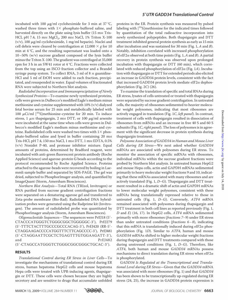
proteins in the ER. Protein synthesis was measured by pulsedlabeling with [35S]methionine for the indicated times followedby quantitation of the total radioactive incorporation intonewly synthesized polypeptides. Both thapsigargin and DTTtreatment inhibited general protein synthesis as early as 15minafter incubation and was sustained for 30 min (Fig. 1, A and B).Notably, inhibition correlated with increased phosphorylationof eIF2� observed at both time points (Fig. 1,A andB). A partialrecovery in protein synthesis was observed upon prolongedincubation with thapsigargin or DTT (60 min), which corre-lated with reduced phosphorylation of eIF2� (Fig. 1A). Incuba-tionwith thapsigargin orDTT for extendedperiods also elicitedan increase in GADD34 protein levels, consistent with the factthat increased GADD34 protein levels mediate eIF2� dephos-phorylation (Fig. 2C) (26).To examine the translation of specific and total RNAs during
ER stress, lysates of cells untreated or treated with thapsigarginwere separated by sucrose gradient centrifugation. In untreatedcells, the majority of ribosomes sedimented to heavier molecu-lar weight polysomes, indicating that most ribosomes areactively engaged in translation (Fig. 1C, left panel). In contrast,treatment of cells with thapsigargin resulted in dissociation ofribosomes from mRNAs and an increase in free 40 S and 60 Ssubunits (Fig. 1C, right panel). The loss of polysomes is in agree-ment with the significant decrease in protein synthesis duringthapsigargin treatment.PolysomeAssociation ofGADD34 inHumanandMouse Liver
Cells during ER Stress—We next asked whether GADD34mRNAs are associated with polysomes during ER stress. Tomonitor the association of specific mRNAs with ribosomes,individual mRNAs within the sucrose gradient fractions wereprobed by Northern blot analysis. In untreated human HepG2and mouse Hepa cells, actin and GAPDH mRNAs sedimentedprimarily to heavymolecular weight fractions 9 and 10, indicat-ing that these mRNAs associated with many ribosomes and areactively translated (Fig. 1, D–G). Thapsigargin and DTT treat-ment resulted in a dramatic shift of actin and GAPDHmRNAsto lower molecular weight polysomes, consistent with thesemRNAs being translationally repressed relative to those inuntreated cells (Fig. 1, D–G). Conversely, ATF4 mRNAremained associated with polysomes during thapsigargin andDTT treatment in both cell lines as reported previously (Fig. 1,D and E) (16, 17). In HepG2 cells, ATF4 mRNA sedimentedprimarily with more ribosomes (fractions 7–9) under ER stressthan under untreated conditions (fractions 6–8), indicatingthat this mRNA is translationally induced during eIF2� phos-phorylation (Fig. 1D). Similar to ATF4, human and mouseGADD34mRNAs shifted to higher molecular weight fractionsduring thapsigargin and DTT treatments compared with thoseduring unstressed conditions (Fig. 1, D–G). Therefore, likeATF4, both human and mouse GADD34 mRNAs possessmechanisms to direct translation during ER stress when eIF2�is phosphorylated.GADD34 Is Regulated at the Transcriptional and Transla-
tional Level during ER Stress—Given that the GADD34mRNAwas associated with more ribosomes (Fig. 1) and thatGADD34has been shown to be transcriptionally up-regulated during ERstress (24, 25), the increase in GADD34 protein expression is
5�UTR GADD34 Translational Control
MARCH 13, 2009 • VOLUME 284 • NUMBER 11 JOURNAL OF BIOLOGICAL CHEMISTRY 6663
by guest on July 14, 2018http://w
ww
.jbc.org/D
ownloaded from
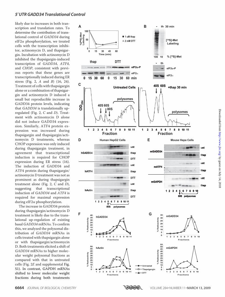
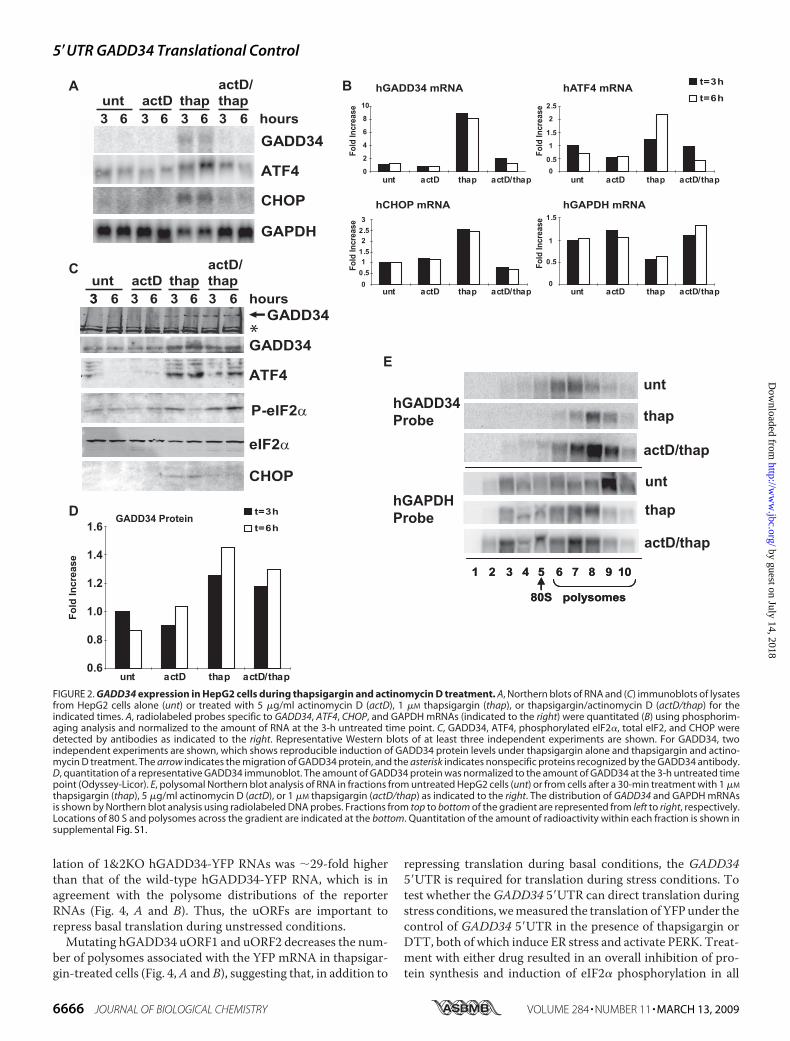
likely due to increases in both tran-scription and translation rates. Todetermine the contribution of trans-lational control of GADD34 duringeIF2� phosphorylation, we treatedcells with the transcription inhibi-tor, actinomycin D, and thapsigar-gin. Incubation with actinomycin Dinhibited the thapsigargin-inducedtranscription of GADD34, ATF4,and CHOP, consistent with previ-ous reports that these genes aretranscriptionally induced during ERstress (Fig. 2, A and B) (16, 24).Treatment of cells with thapsigarginalone or a combination of thapsigar-gin and actinomycin D induced asmall but reproducible increase inGADD34 protein levels, indicatingthat GADD34 is translationally up-regulated (Fig. 2, C and D). Treat-ment with actinomycin D alonedid not induce GADD34 expres-sion. Similarly, ATF4 protein ex-pression was increased duringthapsigargin and thapsigargin/acti-nomycin D treatments, whereasCHOP expression was only inducedduring thapsigargin treatment, inagreement that transcriptionalinduction is required for CHOPexpression during ER stress (16).The induction of GADD34 andATF4 protein during thapsigargin/actinomycin D treatment was not asprominent as during thapsigargintreatment alone (Fig. 2, C and D),suggesting that transcriptionalinduction of GADD34 and ATF4 isrequired for maximal expressionduring eIF2� phosphorylation.The increase in GADD34 protein
during thapsigargin/actinomycin Dtreatment is likely due to the trans-lational up-regulation of existingbasalGADD34mRNAs. To confirmthis, we analyzed the polysomal dis-tribution of GADD34 mRNAs incells treated with thapsigargin aloneor with thapsigargin/actinomycinD. Both treatments elicited a shift ofGADD34 mRNAs to higher molec-ular weight polysomal fractions ascompared with that in untreatedcells (Fig. 2E and supplemental Fig.S1). In contrast, GAPDH mRNAsshifted to lower molecular weightfractions during both treatments
5�UTR GADD34 Translational Control
6664 JOURNAL OF BIOLOGICAL CHEMISTRY VOLUME 284 • NUMBER 11 • MARCH 13, 2009
by guest on July 14, 2018http://w
ww
.jbc.org/D
ownloaded from
(Fig. 2E and supplemental Fig. S1). Together with the Westernblot analyses in Fig. 2C, these results demonstrated thatGADD34mRNAs are translationally activated under thapsigar-gin-induced ER stress.Interestingly, a 3-h thapsigargin treatment induced eIF2�
phosphorylation but by 6 h post-treatment, eIF2� phosphoryl-ation levels had decreased, likely due to maximal induction ofGADD34 protein (Fig. 2C). In contrast, during thapsigargin/actinomycin D incubation, eIF2� remained phosphorylated at6 h post-treatment, suggesting that the moderate induction ofGADD34 protein during this treatment was insufficient toreduce eIF2� phosphorylation (Fig. 2C). In summary, the opti-mal induction of GADD34 protein during eIF2� phosphoryla-tion is mediated through both transcriptional and translationalcontrols of GADD34.The Human andMouse GADD34 5�UTRsMediate Polysome
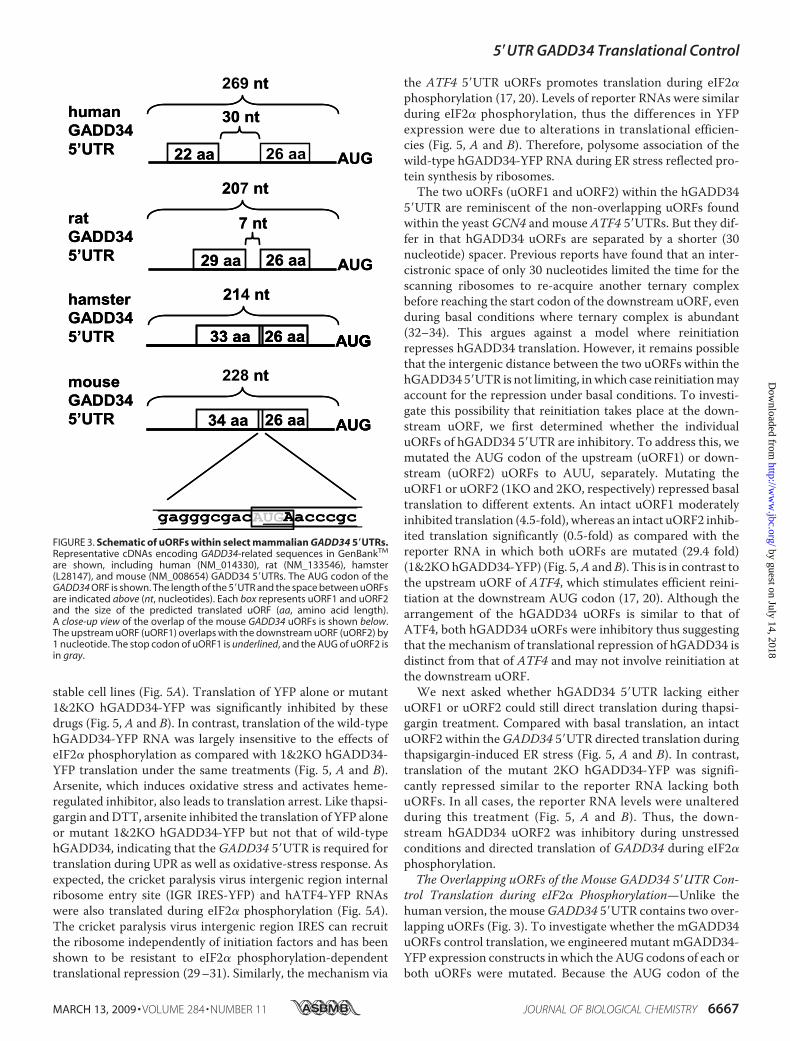
Association during eIF2� Phosphorylation—To begin elucidat-ing the mechanism ofGADD34 translation, we first focused onits 5�UTR. An analysis of annotated mammalian GADD34mRNAs revealed two upstream open reading frames (uORFs)within the 5�UTR. The human (hGADD34), chimp and ratGADD34 5�UTRs contain two non-overlapping uORFs sepa-rated by 7–30 nucleotides, whereas the two uORFs of mouse(mGADD34), hamster, and bovine 5�UTRs are overlapping andout of frame by a single nucleotide (Fig. 3). Specifically, theuORFs overlap within the stop codon of the upstream uORF(uORF1) and the start AUG codon of the downstream uORF(uORF2) (Fig. 3).To determine whether the GADD34 5�UTR is sufficient to
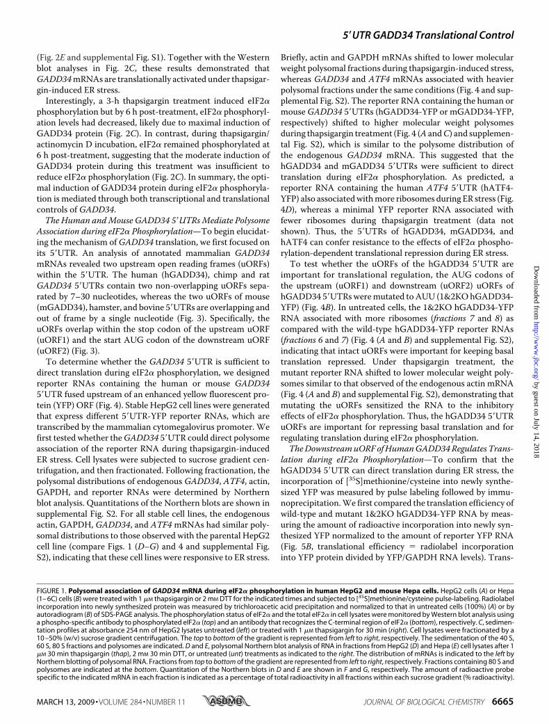
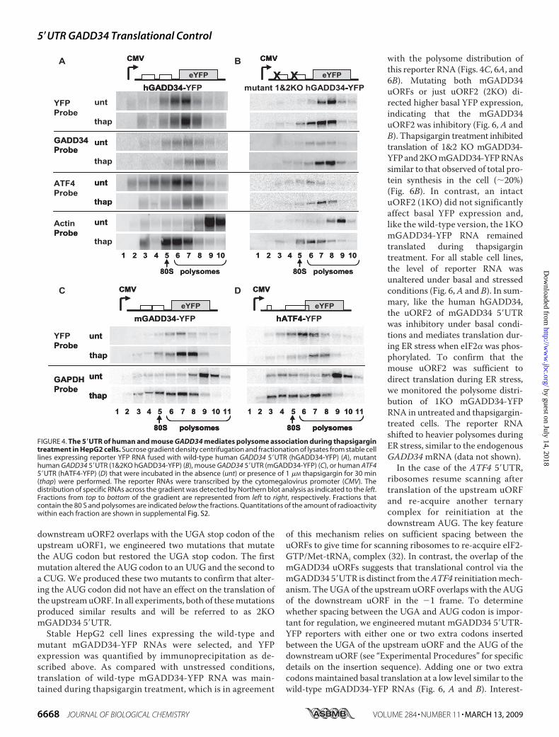
direct translation during eIF2� phosphorylation, we designedreporter RNAs containing the human or mouse GADD345�UTR fused upstream of an enhanced yellow fluorescent pro-tein (YFP) ORF (Fig. 4). Stable HepG2 cell lines were generatedthat express different 5�UTR-YFP reporter RNAs, which aretranscribed by the mammalian cytomegalovirus promoter. Wefirst tested whether theGADD34 5�UTR could direct polysomeassociation of the reporter RNA during thapsigargin-inducedER stress. Cell lysates were subjected to sucrose gradient cen-trifugation, and then fractionated. Following fractionation, thepolysomal distributions of endogenous GADD34, ATF4, actin,GAPDH, and reporter RNAs were determined by Northernblot analysis. Quantitations of the Northern blots are shown insupplemental Fig. S2. For all stable cell lines, the endogenousactin, GAPDH, GADD34, and ATF4mRNAs had similar poly-somal distributions to those observed with the parental HepG2cell line (compare Figs. 1 (D–G) and 4 and supplemental Fig.S2), indicating that these cell lines were responsive to ER stress.
Briefly, actin and GAPDH mRNAs shifted to lower molecularweight polysomal fractions during thapsigargin-induced stress,whereas GADD34 and ATF4 mRNAs associated with heavierpolysomal fractions under the same conditions (Fig. 4 and sup-plemental Fig. S2). The reporter RNA containing the human ormouseGADD34 5�UTRs (hGADD34-YFP or mGADD34-YFP,respectively) shifted to higher molecular weight polysomesduring thapsigargin treatment (Fig. 4 (A andC) and supplemen-tal Fig. S2), which is similar to the polysome distribution ofthe endogenous GADD34 mRNA. This suggested that thehGADD34 and mGADD34 5�UTRs were sufficient to directtranslation during eIF2� phosphorylation. As predicted, areporter RNA containing the human ATF4 5�UTR (hATF4-YFP) also associatedwithmore ribosomes during ER stress (Fig.4D), whereas a minimal YFP reporter RNA associated withfewer ribosomes during thapsigargin treatment (data notshown). Thus, the 5�UTRs of hGADD34, mGADD34, andhATF4 can confer resistance to the effects of eIF2� phospho-rylation-dependent translational repression during ER stress.To test whether the uORFs of the hGADD34 5�UTR are
important for translational regulation, the AUG codons ofthe upstream (uORF1) and downstream (uORF2) uORFs ofhGADD34 5�UTRsweremutated toAUU (1&2KOhGADD34-YFP) (Fig. 4B). In untreated cells, the 1&2KO hGADD34-YFPRNA associated with more ribosomes (fractions 7 and 8) ascompared with the wild-type hGADD34-YFP reporter RNAs(fractions 6 and 7) (Fig. 4 (A and B) and supplemental Fig. S2),indicating that intact uORFs were important for keeping basaltranslation repressed. Under thapsigargin treatment, themutant reporter RNA shifted to lower molecular weight poly-somes similar to that observed of the endogenous actin mRNA(Fig. 4 (A and B) and supplemental Fig. S2), demonstrating thatmutating the uORFs sensitized the RNA to the inhibitoryeffects of eIF2� phosphorylation. Thus, the hGADD34 5�UTRuORFs are important for repressing basal translation and forregulating translation during eIF2� phosphorylation.TheDownstreamuORFofHumanGADD34Regulates Trans-
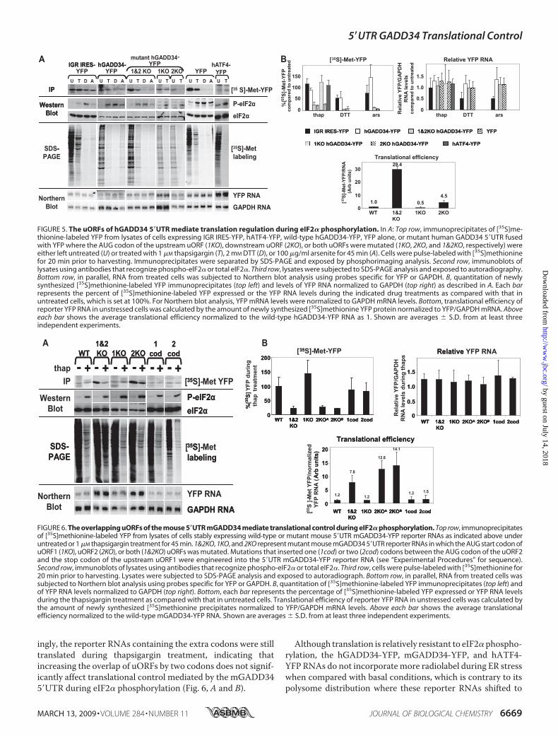
lation during eIF2� Phosphorylation—To confirm that thehGADD34 5�UTR can direct translation during ER stress, theincorporation of [35S]methionine/cysteine into newly synthe-sized YFP was measured by pulse labeling followed by immu-noprecipitation.We first compared the translation efficiency ofwild-type and mutant 1&2KO hGADD34-YFP RNA by meas-uring the amount of radioactive incorporation into newly syn-thesized YFP normalized to the amount of reporter YFP RNA(Fig. 5B, translational efficiency � radiolabel incorporationinto YFP protein divided by YFP/GAPDH RNA levels). Trans-
FIGURE 1. Polysomal association of GADD34 mRNA during eIF2� phosphorylation in human HepG2 and mouse Hepa cells. HepG2 cells (A) or Hepa(1– 6C) cells (B) were treated with 1 �M thapsigargin or 2 mM DTT for the indicated times and subjected to [35S]methionine/cysteine pulse-labeling. Radiolabelincorporation into newly synthesized protein was measured by trichloroacetic acid precipitation and normalized to that in untreated cells (100%) (A) or byautoradiogram (B) of SDS-PAGE analysis. The phosphorylation status of eIF2� and the total eIF2� in cell lysates were monitored by Western blot analysis usinga phospho-specific antibody to phosphorylated eIF2� (top) and an antibody that recognizes the C-terminal region of eIF2� (bottom), respectively. C, sedimen-tation profiles at absorbance 254 nm of HepG2 lysates untreated (left) or treated with 1 �M thapsigargin for 30 min (right). Cell lysates were fractionated by a10 –50% (w/v) sucrose gradient centrifugation. The top to bottom of the gradient is represented from left to right, respectively. The sedimentation of the 40 S,60 S, 80 S fractions and polysomes are indicated. D and E, polysomal Northern blot analysis of RNA in fractions from HepG2 (D) and Hepa (E) cell lysates after 1�M 30 min thapsigargin (thap), 2 mM 30 min DTT, or untreated (unt) treatments as indicated to the right. The distribution of mRNAs is indicated to the left byNorthern blotting of polysomal RNA. Fractions from top to bottom of the gradient are represented from left to right, respectively. Fractions containing 80 S andpolysomes are indicated at the bottom. Quantitation of the Northern blots in D and E are shown in F and G, respectively. The amount of radioactive probespecific to the indicated mRNA in each fraction is indicated as a percentage of total radioactivity in all fractions within each sucrose gradient (% radioactivity).
5�UTR GADD34 Translational Control
MARCH 13, 2009 • VOLUME 284 • NUMBER 11 JOURNAL OF BIOLOGICAL CHEMISTRY 6665
by guest on July 14, 2018http://w
ww
.jbc.org/D
ownloaded from
lation of 1&2KO hGADD34-YFP RNAs was �29-fold higherthan that of the wild-type hGADD34-YFP RNA, which is inagreement with the polysome distributions of the reporterRNAs (Fig. 4, A and B). Thus, the uORFs are important torepress basal translation during unstressed conditions.Mutating hGADD34 uORF1 and uORF2 decreases the num-
ber of polysomes associated with the YFP mRNA in thapsigar-gin-treated cells (Fig. 4,A and B), suggesting that, in addition to
repressing translation during basal conditions, the GADD345�UTR is required for translation during stress conditions. Totest whether theGADD34 5�UTR can direct translation duringstress conditions, wemeasured the translation of YFPunder thecontrol of GADD34 5�UTR in the presence of thapsigargin orDTT, both of which induce ER stress and activate PERK. Treat-ment with either drug resulted in an overall inhibition of pro-tein synthesis and induction of eIF2� phosphorylation in all
FIGURE 2. GADD34 expression in HepG2 cells during thapsigargin and actinomycin D treatment. A, Northern blots of RNA and (C) immunoblots of lysatesfrom HepG2 cells alone (unt) or treated with 5 �g/ml actinomycin D (actD), 1 �M thapsigargin (thap), or thapsigargin/actinomycin D (actD/thap) for theindicated times. A, radiolabeled probes specific to GADD34, ATF4, CHOP, and GAPDH mRNAs (indicated to the right) were quantitated (B) using phosphorim-aging analysis and normalized to the amount of RNA at the 3-h untreated time point. C, GADD34, ATF4, phosphorylated eIF2�, total eIF2, and CHOP weredetected by antibodies as indicated to the right. Representative Western blots of at least three independent experiments are shown. For GADD34, twoindependent experiments are shown, which shows reproducible induction of GADD34 protein levels under thapsigargin alone and thapsigargin and actino-mycin D treatment. The arrow indicates the migration of GADD34 protein, and the asterisk indicates nonspecific proteins recognized by the GADD34 antibody.D, quantitation of a representative GADD34 immunoblot. The amount of GADD34 protein was normalized to the amount of GADD34 at the 3-h untreated timepoint (Odyssey-Licor). E, polysomal Northern blot analysis of RNA in fractions from untreated HepG2 cells (unt) or from cells after a 30-min treatment with 1 �M
thapsigargin (thap), 5 �g/ml actinomycin D (actD), or 1 �M thapsigargin (actD/thap) as indicated to the right. The distribution of GADD34 and GAPDH mRNAsis shown by Northern blot analysis using radiolabeled DNA probes. Fractions from top to bottom of the gradient are represented from left to right, respectively.Locations of 80 S and polysomes across the gradient are indicated at the bottom. Quantitation of the amount of radioactivity within each fraction is shown insupplemental Fig. S1.
5�UTR GADD34 Translational Control
6666 JOURNAL OF BIOLOGICAL CHEMISTRY VOLUME 284 • NUMBER 11 • MARCH 13, 2009
by guest on July 14, 2018http://w
ww
.jbc.org/D
ownloaded from
stable cell lines (Fig. 5A). Translation of YFP alone or mutant1&2KO hGADD34-YFP was significantly inhibited by thesedrugs (Fig. 5, A and B). In contrast, translation of the wild-typehGADD34-YFP RNA was largely insensitive to the effects ofeIF2� phosphorylation as compared with 1&2KO hGADD34-YFP translation under the same treatments (Fig. 5, A and B).Arsenite, which induces oxidative stress and activates heme-regulated inhibitor, also leads to translation arrest. Like thapsi-gargin andDTT, arsenite inhibited the translation of YFP aloneor mutant 1&2KO hGADD34-YFP but not that of wild-typehGADD34, indicating that the GADD34 5�UTR is required fortranslation during UPR as well as oxidative-stress response. Asexpected, the cricket paralysis virus intergenic region internalribosome entry site (IGR IRES-YFP) and hATF4-YFP RNAswere also translated during eIF2� phosphorylation (Fig. 5A).The cricket paralysis virus intergenic region IRES can recruitthe ribosome independently of initiation factors and has beenshown to be resistant to eIF2� phosphorylation-dependenttranslational repression (29–31). Similarly, the mechanism via
the ATF4 5�UTR uORFs promotes translation during eIF2�phosphorylation (17, 20). Levels of reporter RNAs were similarduring eIF2� phosphorylation, thus the differences in YFPexpression were due to alterations in translational efficien-cies (Fig. 5, A and B). Therefore, polysome association of thewild-type hGADD34-YFP RNA during ER stress reflected pro-tein synthesis by ribosomes.The two uORFs (uORF1 and uORF2) within the hGADD34
5�UTR are reminiscent of the non-overlapping uORFs foundwithin the yeast GCN4 and mouse ATF4 5�UTRs. But they dif-fer in that hGADD34 uORFs are separated by a shorter (30nucleotide) spacer. Previous reports have found that an inter-cistronic space of only 30 nucleotides limited the time for thescanning ribosomes to re-acquire another ternary complexbefore reaching the start codon of the downstream uORF, evenduring basal conditions where ternary complex is abundant(32–34). This argues against a model where reinitiationrepresses hGADD34 translation. However, it remains possiblethat the intergenic distance between the two uORFs within thehGADD345�UTR is not limiting, inwhich case reinitiationmayaccount for the repression under basal conditions. To investi-gate this possibility that reinitiation takes place at the down-stream uORF, we first determined whether the individualuORFs of hGADD34 5�UTR are inhibitory. To address this, wemutated the AUG codon of the upstream (uORF1) or down-stream (uORF2) uORFs to AUU, separately. Mutating theuORF1 or uORF2 (1KO and 2KO, respectively) repressed basaltranslation to different extents. An intact uORF1 moderatelyinhibited translation (4.5-fold), whereas an intact uORF2 inhib-ited translation significantly (0.5-fold) as compared with thereporter RNA in which both uORFs are mutated (29.4 fold)(1&2KOhGADD34-YFP) (Fig. 5,A andB). This is in contrast tothe upstream uORF of ATF4, which stimulates efficient reini-tiation at the downstream AUG codon (17, 20). Although thearrangement of the hGADD34 uORFs is similar to that ofATF4, both hGADD34 uORFs were inhibitory thus suggestingthat the mechanism of translational repression of hGADD34 isdistinct from that of ATF4 and may not involve reinitiation atthe downstream uORF.We next asked whether hGADD34 5�UTR lacking either
uORF1 or uORF2 could still direct translation during thapsi-gargin treatment. Compared with basal translation, an intactuORF2 within theGADD34 5�UTR directed translation duringthapsigargin-induced ER stress (Fig. 5, A and B). In contrast,translation of the mutant 2KO hGADD34-YFP was signifi-cantly repressed similar to the reporter RNA lacking bothuORFs. In all cases, the reporter RNA levels were unalteredduring this treatment (Fig. 5, A and B). Thus, the down-stream hGADD34 uORF2 was inhibitory during unstressedconditions and directed translation of GADD34 during eIF2�phosphorylation.The Overlapping uORFs of the Mouse GADD34 5�UTR Con-
trol Translation during eIF2� Phosphorylation—Unlike thehuman version, themouseGADD34 5�UTR contains two over-lapping uORFs (Fig. 3). To investigate whether the mGADD34uORFs control translation, we engineered mutant mGADD34-YFP expression constructs in which the AUG codons of each orboth uORFs were mutated. Because the AUG codon of the
FIGURE 3. Schematic of uORFs within select mammalian GADD34 5�UTRs.Representative cDNAs encoding GADD34-related sequences in GenBankTM
are shown, including human (NM_014330), rat (NM_133546), hamster(L28147), and mouse (NM_008654) GADD34 5�UTRs. The AUG codon of theGADD34 ORF is shown. The length of the 5�UTR and the space between uORFsare indicated above (nt, nucleotides). Each box represents uORF1 and uORF2and the size of the predicted translated uORF (aa, amino acid length).A close-up view of the overlap of the mouse GADD34 uORFs is shown below.The upstream uORF (uORF1) overlaps with the downstream uORF (uORF2) by1 nucleotide. The stop codon of uORF1 is underlined, and the AUG of uORF2 isin gray.
5�UTR GADD34 Translational Control
MARCH 13, 2009 • VOLUME 284 • NUMBER 11 JOURNAL OF BIOLOGICAL CHEMISTRY 6667
by guest on July 14, 2018http://w
ww
.jbc.org/D
ownloaded from
downstream uORF2 overlaps with the UGA stop codon of theupstream uORF1, we engineered two mutations that mutatethe AUG codon but restored the UGA stop codon. The firstmutation altered the AUG codon to an UUG and the second toa CUG. We produced these two mutants to confirm that alter-ing the AUG codon did not have an effect on the translation ofthe upstreamuORF. In all experiments, both of thesemutationsproduced similar results and will be referred to as 2KOmGADD34 5�UTR.Stable HepG2 cell lines expressing the wild-type and
mutant mGADD34-YFP RNAs were selected, and YFPexpression was quantified by immunoprecipitation as de-scribed above. As compared with unstressed conditions,translation of wild-type mGADD34-YFP RNA was main-tained during thapsigargin treatment, which is in agreement
with the polysome distribution ofthis reporter RNA (Figs. 4C, 6A, and6B). Mutating both mGADD34uORFs or just uORF2 (2KO) di-rected higher basal YFP expression,indicating that the mGADD34uORF2 was inhibitory (Fig. 6, A andB). Thapsigargin treatment inhibitedtranslation of 1&2 KO mGADD34-YFPand2KOmGADD34-YFPRNAssimilar to that observed of total pro-tein synthesis in the cell (�20%)(Fig. 6B). In contrast, an intactuORF2 (1KO) did not significantlyaffect basal YFP expression and,like the wild-type version, the 1KOmGADD34-YFP RNA remainedtranslated during thapsigargintreatment. For all stable cell lines,the level of reporter RNA wasunaltered under basal and stressedconditions (Fig. 6,A and B). In sum-mary, like the human hGADD34,the uORF2 of mGADD34 5�UTRwas inhibitory under basal condi-tions and mediates translation dur-ing ER stress when eIF2� was phos-phorylated. To confirm that themouse uORF2 was sufficient todirect translation during ER stress,we monitored the polysome distri-bution of 1KO mGADD34-YFPRNA in untreated and thapsigargin-treated cells. The reporter RNAshifted to heavier polysomes duringER stress, similar to the endogenousGADD34mRNA (data not shown).In the case of the ATF4 5�UTR,
ribosomes resume scanning aftertranslation of the upstream uORFand re-acquire another ternarycomplex for reinitiation at thedownstream AUG. The key feature
of this mechanism relies on sufficient spacing between theuORFs to give time for scanning ribosomes to re-acquire eIF2-GTP/Met-tRNAi complex (32). In contrast, the overlap of themGADD34 uORFs suggests that translational control via themGADD34 5�UTR is distinct from theATF4 reinitiationmech-anism. TheUGAof the upstream uORF overlaps with the AUGof the downstream uORF in the �1 frame. To determinewhether spacing between the UGA and AUG codon is impor-tant for regulation, we engineered mutant mGADD34 5�UTR-YFP reporters with either one or two extra codons insertedbetween the UGA of the upstream uORF and the AUG of thedownstream uORF (see “Experimental Procedures” for specificdetails on the insertion sequence). Adding one or two extracodonsmaintained basal translation at a low level similar to thewild-type mGADD34-YFP RNAs (Fig. 6, A and B). Interest-
YFPProbe
1 2 3 4 5 6 7 8 9 10 11 1 2 3 4 5 6 7 8 9 10 11
unt
thap
thap
untGAPDHProbe
eYFP
CMV
YFPeYFP
CMV
hATF4-YFP
80S polysomes 80S polysomes
1 2 3 4 5 6 7 8 9 10
YFPProbe
GADD34Probe
ActinProbe
1 2 3 4 5 6 7 8 9 10
ATF4Probe
eYFP
CMV
hGADD34-eYFP
CMV
X Xmutant 1&2KO hGADD34-YFP
80S polysomes 80S polysomes
unt
thap
unt
thap
unt
unt
thap
Probe
1 2 3 4 5 6 7 8 9 10 11 1 2 3 4 5 6 7 8 9 10 11
unt
thap
thapthap
untuntGAPDHProbe
eYFP
CMV
mGADD34eYFP
CMV
hATF4-eYFP
CMV
mGADD34-eYFP
CMV
hATF4-
80S polysomes80S polysomes 80S polysomes80S polysomes
1 2 3 4 5 6 7 8 9 10
GADD34Probe
Probe
1 2 3 4 5 6 7 8 9 10
eYFP
CMV
hGADD34eYFP
CMV
X X-
eYFP
CMV
hGADD34-YFPeYFP
CMV
X X-
80S polysomes 80S polysomes
untunt
thap
thap
unt
unt
A B
C D
FIGURE 4. The 5�UTR of human and mouse GADD34 mediates polysome association during thapsigargintreatment in HepG2 cells. Sucrose gradient density centrifugation and fractionation of lysates from stable celllines expressing reporter YFP RNA fused with wild-type human GADD34 5�UTR (hGADD34-YFP) (A), mutanthuman GADD34 5�UTR (1&2KO hGADD34-YFP) (B), mouse GADD34 5�UTR (mGADD34-YFP) (C), or human ATF45�UTR (hATF4-YFP) (D) that were incubated in the absence (unt) or presence of 1 �M thapsigargin for 30 min(thap) were performed. The reporter RNAs were transcribed by the cytomegalovirus promoter (CMV). Thedistribution of specific RNAs across the gradient was detected by Northern blot analysis as indicated to the left.Fractions from top to bottom of the gradient are represented from left to right, respectively. Fractions thatcontain the 80 S and polysomes are indicated below the fractions. Quantitations of the amount of radioactivitywithin each fraction are shown in supplemental Fig. S2.
5�UTR GADD34 Translational Control
6668 JOURNAL OF BIOLOGICAL CHEMISTRY VOLUME 284 • NUMBER 11 • MARCH 13, 2009
by guest on July 14, 2018http://w
ww
.jbc.org/D
ownloaded from
ingly, the reporter RNAs containing the extra codons were stilltranslated during thapsigargin treatment, indicating thatincreasing the overlap of uORFs by two codons does not signif-icantly affect translational control mediated by the mGADD345�UTR during eIF2� phosphorylation (Fig. 6, A and B).
Although translation is relatively resistant to eIF2�phospho-rylation, the hGADD34-YFP, mGADD34-YFP, and hATF4-YFP RNAs do not incorporatemore radiolabel during ER stresswhen compared with basal conditions, which is contrary to itspolysome distribution where these reporter RNAs shifted to
FIGURE 5. The uORFs of hGADD34 5�UTR mediate translation regulation during eIF2� phosphorylation. In A: Top row, immunoprecipitates of [35S]me-thionine-labeled YFP from lysates of cells expressing IGR IRES-YFP, hATF4-YFP, wild-type hGADD34-YFP, YFP alone, or mutant human GADD34 5�UTR fusedwith YFP where the AUG codon of the upstream uORF (1KO), downstream uORF (2KO), or both uORFs were mutated (1KO, 2KO, and 1&2KO, respectively) wereeither left untreated (U) or treated with 1 �M thapsigargin (T), 2 mM DTT (D), or 100 �g/ml arsenite for 45 min (A). Cells were pulse-labeled with [35S]methioninefor 20 min prior to harvesting. Immunoprecipitates were separated by SDS-PAGE and exposed by phosphorimaging analysis. Second row, immunoblots oflysates using antibodies that recognize phospho-eIF2� or total eIF2�. Third row, lysates were subjected to SDS-PAGE analysis and exposed to autoradiography.Bottom row, in parallel, RNA from treated cells was subjected to Northern blot analysis using probes specific for YFP or GAPDH. B, quantitation of newlysynthesized [35S]methionine-labeled YFP immunoprecipitates (top left) and levels of YFP RNA normalized to GAPDH (top right) as described in A. Each barrepresents the percent of [35S]methionine-labeled YFP expressed or the YFP RNA levels during the indicated drug treatments as compared with that inuntreated cells, which is set at 100%. For Northern blot analysis, YFP mRNA levels were normalized to GAPDH mRNA levels. Bottom, translational efficiency ofreporter YFP RNA in unstressed cells was calculated by the amount of newly synthesized [35S]methionine YFP protein normalized to YFP/GAPDH mRNA. Aboveeach bar shows the average translational efficiency normalized to the wild-type hGADD34-YFP RNA as 1. Shown are averages � S.D. from at least threeindependent experiments.
FIGURE 6. The overlapping uORFs of the mouse 5�UTR mGADD34 mediate translational control during eIF2� phosphorylation. Top row, immunoprecipitatesof [35S]methionine-labeled YFP from lysates of cells stably expressing wild-type or mutant mouse 5�UTR mGADD34-YFP reporter RNAs as indicated above underuntreated or 1 �M thapsigargin treatment for 45 min. 1&2KO, 1KO, and 2KO represent mutant mouse mGADD34 5�UTR reporter RNAs in which the AUG start codon ofuORF1 (1KO), uORF2 (2KO), or both (1&2KO) uORFs was mutated. Mutations that inserted one (1cod) or two (2cod) codons between the AUG codon of the uORF2and the stop codon of the upstream uORF1 were engineered into the 5�UTR mGADD34-YFP reporter RNA (see “Experimental Procedures” for sequence).Second row, immunoblots of lysates using antibodies that recognize phospho-eIF2� or total eIF2�. Third row, cells were pulse-labeled with [35S]methionine for20 min prior to harvesting. Lysates were subjected to SDS-PAGE analysis and exposed to autoradiograph. Bottom row, in parallel, RNA from treated cells wassubjected to Northern blot analysis using probes specific for YFP or GAPDH. B, quantitation of [35S]methionine-labeled YFP immunoprecipitates (top left) andof YFP RNA levels normalized to GAPDH (top right). Bottom, each bar represents the percentage of [35S]methionine-labeled YFP expressed or YFP RNA levelsduring the thapsigargin treatment as compared with that in untreated cells. Translational efficiency of reporter YFP RNA in unstressed cells was calculated bythe amount of newly synthesized [35S]methionine precipitates normalized to YFP/GAPDH mRNA levels. Above each bar shows the average translationalefficiency normalized to the wild-type mGADD34-YFP RNA. Shown are averages � S.D. from at least three independent experiments.
5�UTR GADD34 Translational Control
MARCH 13, 2009 • VOLUME 284 • NUMBER 11 JOURNAL OF BIOLOGICAL CHEMISTRY 6669
by guest on July 14, 2018http://w
ww
.jbc.org/D
ownloaded from
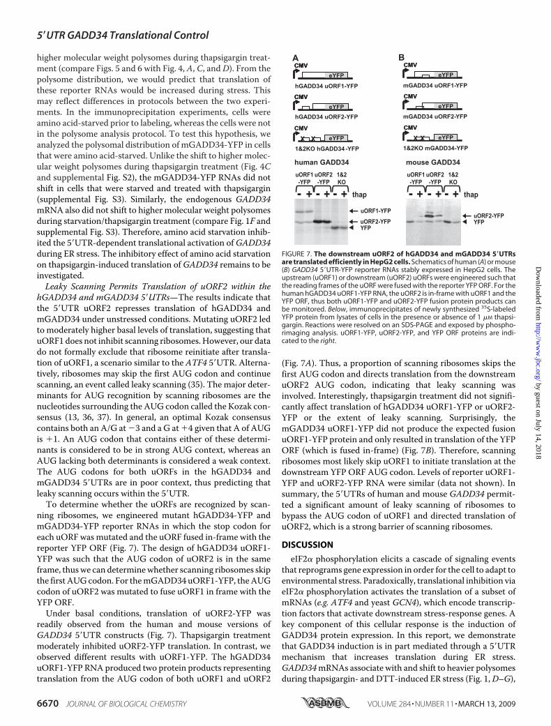
higher molecular weight polysomes during thapsigargin treat-ment (compare Figs. 5 and 6 with Fig. 4, A, C, andD). From thepolysome distribution, we would predict that translation ofthese reporter RNAs would be increased during stress. Thismay reflect differences in protocols between the two experi-ments. In the immunoprecipitation experiments, cells wereamino acid-starved prior to labeling, whereas the cells were notin the polysome analysis protocol. To test this hypothesis, weanalyzed the polysomal distribution of mGADD34-YFP in cellsthat were amino acid-starved. Unlike the shift to higher molec-ular weight polysomes during thapsigargin treatment (Fig. 4Cand supplemental Fig. S2), the mGADD34-YFP RNAs did notshift in cells that were starved and treated with thapsigargin(supplemental Fig. S3). Similarly, the endogenous GADD34mRNA also did not shift to higher molecular weight polysomesduring starvation/thapsigargin treatment (compare Fig. 1F andsupplemental Fig. S3). Therefore, amino acid starvation inhib-ited the 5�UTR-dependent translational activation ofGADD34during ER stress. The inhibitory effect of amino acid starvationon thapsigargin-induced translation ofGADD34 remains to beinvestigated.Leaky Scanning Permits Translation of uORF2 within the
hGADD34 and mGADD34 5�UTRs—The results indicate thatthe 5�UTR uORF2 represses translation of hGADD34 andmGADD34 under unstressed conditions. Mutating uORF2 ledto moderately higher basal levels of translation, suggesting thatuORF1 does not inhibit scanning ribosomes.However, our datado not formally exclude that ribosome reinitiate after transla-tion of uORF1, a scenario similar to the ATF4 5�UTR. Alterna-tively, ribosomes may skip the first AUG codon and continuescanning, an event called leaky scanning (35). The major deter-minants for AUG recognition by scanning ribosomes are thenucleotides surrounding the AUG codon called the Kozak con-sensus (13, 36, 37). In general, an optimal Kozak consensuscontains both an A/G at �3 and a G at �4 given that A of AUGis �1. An AUG codon that contains either of these determi-nants is considered to be in strong AUG context, whereas anAUG lacking both determinants is considered a weak context.The AUG codons for both uORFs in the hGADD34 andmGADD34 5�UTRs are in poor context, thus predicting thatleaky scanning occurs within the 5�UTR.To determine whether the uORFs are recognized by scan-
ning ribosomes, we engineered mutant hGADD34-YFP andmGADD34-YFP reporter RNAs in which the stop codon foreach uORF was mutated and the uORF fused in-frame with thereporter YFP ORF (Fig. 7). The design of hGADD34 uORF1-YFP was such that the AUG codon of uORF2 is in the sameframe, thuswe can determinewhether scanning ribosomes skipthe first AUGcodon. For themGADD34uORF1-YFP, theAUGcodon of uORF2 was mutated to fuse uORF1 in frame with theYFP ORF.Under basal conditions, translation of uORF2-YFP was
readily observed from the human and mouse versions ofGADD34 5�UTR constructs (Fig. 7). Thapsigargin treatmentmoderately inhibited uORF2-YFP translation. In contrast, weobserved different results with uORF1-YFP. The hGADD34uORF1-YFP RNA produced two protein products representingtranslation from the AUG codon of both uORF1 and uORF2
(Fig. 7A). Thus, a proportion of scanning ribosomes skips thefirst AUG codon and directs translation from the downstreamuORF2 AUG codon, indicating that leaky scanning wasinvolved. Interestingly, thapsigargin treatment did not signifi-cantly affect translation of hGADD34 uORF1-YFP or uORF2-YFP or the extent of leaky scanning. Surprisingly, themGADD34 uORF1-YFP did not produce the expected fusionuORF1-YFP protein and only resulted in translation of the YFPORF (which is fused in-frame) (Fig. 7B). Therefore, scanningribosomes most likely skip uORF1 to initiate translation at thedownstream YFP ORF AUG codon. Levels of reporter uORF1-YFP and uORF2-YFP RNA were similar (data not shown). Insummary, the 5�UTRs of human and mouse GADD34 permit-ted a significant amount of leaky scanning of ribosomes tobypass the AUG codon of uORF1 and directed translation ofuORF2, which is a strong barrier of scanning ribosomes.
DISCUSSION
eIF2� phosphorylation elicits a cascade of signaling eventsthat reprograms gene expression in order for the cell to adapt toenvironmental stress. Paradoxically, translational inhibition viaeIF2� phosphorylation activates the translation of a subset ofmRNAs (e.g. ATF4 and yeast GCN4), which encode transcrip-tion factors that activate downstream stress-response genes. Akey component of this cellular response is the induction ofGADD34 protein expression. In this report, we demonstratethat GADD34 induction is in part mediated through a 5�UTRmechanism that increases translation during ER stress.GADD34mRNAs associate with and shift to heavier polysomesduring thapsigargin- and DTT-induced ER stress (Fig. 1,D–G),
FIGURE 7. The downstream uORF2 of hGADD34 and mGADD34 5�UTRsare translated efficiently in HepG2 cells. Schematics of human (A) or mouse(B) GADD34 5�UTR-YFP reporter RNAs stably expressed in HepG2 cells. Theupstream (uORF1) or downstream (uORF2) uORFs were engineered such thatthe reading frames of the uORF were fused with the reporter YFP ORF. For thehuman hGADD34 uORF1-YFP RNA, the uORF2 is in-frame with uORF1 and theYFP ORF, thus both uORF1-YFP and uORF2-YFP fusion protein products canbe monitored. Below, immunoprecipitates of newly synthesized 35S-labeledYFP protein from lysates of cells in the presence or absence of 1 �M thapsi-gargin. Reactions were resolved on an SDS-PAGE and exposed by phospho-rimaging analysis. uORF1-YFP, uORF2-YFP, and YFP ORF proteins are indi-cated to the right.
5�UTR GADD34 Translational Control
6670 JOURNAL OF BIOLOGICAL CHEMISTRY VOLUME 284 • NUMBER 11 • MARCH 13, 2009
by guest on July 14, 2018http://w
ww
.jbc.org/D
ownloaded from
indicating that GADD34 is translated under conditions of gen-eral translational arrest when eIF2� is phosphorylated. Specif-ically, the 5�UTR of both human and mouse GADD34 mRNAsis essential and sufficient to direct efficient translation dur-ing ER stress. Through reporter constructs, we showed thatthe GADD34 5�UTR induced re-distribution of reportermRNAs to heavier polysomes during eIF2� phosphoryla-tion, which is similar to the distribution of endogenousGADD34 mRNAs (Fig. 4 and supplemental Fig. S2). Ourdata, together with earlier reports (38), support the notionthat induction of GADD34 translation during eIF2� phos-phorylation plays an important role in the general stressresponse, which alleviates cellular stress, including ER stress,oxidative stress, and hypoxia.GADD34 is an essential component of the UPR (24, 26).
GADD34 interacts with protein phosphatase 1 via its C-termi-nal region to dephosphorylate eIF2�, leading to translationalrecovery (24, 26, 39, 40). Ectopic expression of a truncatedGADD34 protein lacking the catalytic C-terminal region inmouse embryo fibroblasts prevents dephosphorylation eIF2�and blocks translational recovery during ER stress, resulting inpremature apoptosis (24). The heightened sensitization to celldeath is likely due to translational repression of stress-inducedtranscripts, such as the chaperone BiP, which are normallyexpressed during ER stress (26). Thus, the increased expressionof GADD34 results in a negative feedback loop to enhance thetranslation of stress-induced mRNAs during eIF2� phospho-rylation, which are essential for cellular survival and adaptationto environmental stress. GADD34 is also regulated at the tran-scriptional level, which is induced in part by ATF4 and CHOP,which are expressed during ER stress (25, 27). However, it wasunclear whether the increase inGADD34mRNA alone yieldedsufficient GADD34 protein during eIF2� phosphorylation topromote the negative feedback loop. Our data demonstratesthat GADD34 is translationally induced to ensure maximalexpression during ER stress.We showed thatGADD34mRNAsassociated with more ribosomes during ER stress, suggestingthat eIF2� phosphorylation leads to increased GADD34 trans-lation (Fig. 1, D–G). In addition, GADD34 protein increasedunder thapsigargin treatment in the presence of the transcrip-tion inhibitor actinomycin D (Fig. 2, C and D). This indicatedthat ongoing transcription was not required for GADD34induction and that existing basal GADD34 mRNAs can betranslated during ER stress. In support of this, we showed thatGADD34 mRNAs associated with higher molecular weightpolysomes in cells treatedwith thapsigargin and actinomycinD(Fig. 2E and supplemental Fig. S2). However, the induction ofGADD34 protein was moderate in thapsigargin/actinomycinD-treated cells compared with that of cells treated with thapsi-gargin alone and was not sufficient to reduce eIF2� phospho-rylation at later time points (Fig. 2D). These results are in agree-ment with previous observations that impaired expression ofGADD34 in CHOP�/� mice embryonic fibroblasts leads topersistent eIF2� phosphorylation and loss of protein synthesisrecovery (27). Therefore, the coordinated transcriptional andtranslational controls of GADD34 are necessary for optimalexpression to dephosphorylate eIF2� and for translationalrecovery.
Our results revealed that the uORFs of the human andmouseGADD34 5�UTRplayed a significant role under basal and stressconditions. 5�UTR uORFs have been shown previously to beimportant for translational control of several mRNAs (41). Ingeneral, uORFs act as barriers to scanning ribosomes therebymodulating translation of the authentic ORF. Under certaincellular conditions, ribosomes can bypass the uORFs and reini-tiate translation at the main ORF. The best studied mechanismof translational reinitiation is the one governing ATF4, ATF5,and yeastGCN4 translation. The major premise of this mecha-nism is that the translation of the upstream uORF stimulatestranslation or reinitiation at a downstream AUG, whereastranslation of the downstreamuORF leads to translation termi-nation and dissociation of ribosomes. Following translation ofthe upstream uORF, if eIF2 levels become limiting (i.e. eIF2� isphosphorylated), the recruitment of the ternary complex by theribosome is markedly reduced and as a result ribosomes have ahigher probability of reinitiating translation after the down-stream uORF and thereby reinitiate translation at the authenticORF.Major determinants that control the extent of reinitiationare the intercistronic space between the uORFs and the lengthof the upstream uORF. After translation of uORF1, the proba-bility of reinitiating at the downstream uORF2 depends on thelength of the intercistronic space to allow for scanning ribo-somes to re-acquire the ternary complex (32–34). The shorterthe intercistronic space, the greater chance that scanning ribo-someswill bypass uORF2. Another feature of thismechanism isthat the extent of reinitiation decreases the longer the uORF,whereas efficient reinitiation occurs with translation of veryshort uORFs (2–3 amino acids long) (21, 42). ATF4, ATF5, andGCN4 all contain short uORFs. It has been proposed that ribo-somes that translate a short uORF are still bound to certaininitiation factors (i.e. eIF4G), which promotes reinitiation (43).However, when longer uORFs are translated, these factors falloff the ribosome, thus preventing reinitiation (43).Our data indicate that the two uORFs within the human and
mouseGADD34 5�UTRs control translation through a mecha-nism that is distinct from the well studied reinitiation mecha-nism. First, neither the humanGADD34 uORF1 nor themouseGADD34 5�UTR can promote reinitiation (Fig. 5 and 6). In fact,the human uORF1moderately inhibited scanning ribosomes ascompared with a 5�UTR with no uORFs (Fig. 5). Secondly,human and mouse uORF1s are poorly translated and a signifi-cant proportion of ribosomes scan past uORF1 to initiate trans-lation at a downstream AUG (Fig. 7). In contrast, the uORF1 ofATF4 is readily translated, which is a prerequisite for reinitia-tion at the uORF2 under basal conditions and at the mainATF4 ORF during eIF2� phosphorylation (17, 20). Instead,the presence of the GADD34 uORF2 was sufficient for trans-lational control. An intact uORF2 reduces translation duringunstressed conditions and is important for translational induc-tion during stressed conditions, which incidentally are all prop-erties that are observed with the endogenous GADD34mRNA(Fig. 4–6, data not shown).In addition, the features and arrangement of the GADD34
uORFs do not fit themodel of reinitiation. ThemouseGADD34uORFs overlap by one nucleotide, whereas the human uORFsare separated by 30 nucleotides. In the case of the mouse
5�UTR GADD34 Translational Control
MARCH 13, 2009 • VOLUME 284 • NUMBER 11 JOURNAL OF BIOLOGICAL CHEMISTRY 6671
by guest on July 14, 2018http://w
ww
.jbc.org/D
ownloaded from
GADD34 uORFs, if reinitiation plays a role in this regulation,ribosomes would have to reinitiate backwards (following trans-lation of uORF1) to start translation at the AUG codon ofuORF2. Although there have been reports of backward reinitia-tion, the extent of this was either very inefficient or requires aspecialized RNA sequence that binds to ribosome-recruitingfactors (i.e. eIF3) (42, 44). When extra codons were introducedto extend the overlap between the start and stop codons, themutant mouse GADD34 5�UTR was still functional like thewild-type version, arguing against backward reinitiation (Fig.6). In the case of the humanGADD34, the uORFs are separatedby a 30-nucleotide intercistronic spacer, which is considerablyshorter than those separating uORFs in humanATF4 (87 nucle-otides), ATF5 (110 nucleotides), and yeast GCN4 (198 nucleo-tides between uORF1 and uORF4) uORFs. Furthermore,whereas the upstream uORF of ATF4, ATF5, and GCN45�UTRs are short, uORF1 of the human GADD34 is relativelylong (22 amino acids), which is predicted to greatly decrease theefficiency of reinitiation (32–34). These features argue thatleaky scanning occurs on theGADD34 5�UTR to bypass uORF1and initiate translation at uORF2.The uORF2 of GADD34 is a strong barrier for scanning
ribosomes, which efficiently represses GADD34 translationunder basal conditions. A probable scenario is that, aftertranslating uORF2, ribosomes dissociate, thus inhibitingtranslation of the GADD34 ORF. Alternatively, the uORF2may mediate other effects. It is has been previously shownthat the uORF in fungal mRNAs that encode a subunit ofArg-specific carbamoyl phosphate synthetase controlstranslation by stalling scanning ribosomes and mediatingnonsense-mediated decay (45, 46). In this case, the trans-lated coding sequence of the fungal uORF dictates transla-tional control (47, 48). Interestingly, a comparison of mam-malian GADD34 5�UTRs reveal that uORF2 is highlyconserved. The human, chimp, and rat GADD34 5�UTRscontain non-overlapping uORFs, and the mouse and ham-ster versions contain overlapping uORFs. Although theuORF1s differ in length and are poorly conserved, theuORF2s are exactly 26 amino acids long and contain 18/26identical amino acids. It remains to be determined whetherthe amino acid or nucleotide sequence composition withinuORF2 plays a significant role in translational control duringeIF2� phosphorylation. The high degree of conservation ofuORF2 further strengthens the idea that translational con-trol is mediated by this uORF specifically. However, it isunclear why a dispensable uORF1 has evolved within theGADD34 5�UTR. It is possible that uORF1 has an unex-plored role under a different set of cellular stress conditions.An unresolved question is how do scanning ribosomes
bypass uORF2 and reach the GADD34 ORF when eIF2� isphosphorylated? Other than the reinitiation mechanism, addi-tionalmechanisms that can bypass eIF2� phosphorylation havebeen described. One suchmechanism proposes that ribosomesare directly recruited to the 5�UTRdownstreamof uORF2, pos-sibly through an IRES. The most unique example is via thecricket paralysis virus intergenic region IRES, which can bypassthe requirement for all initiation factors to recruit the ribosomeand can induce translation during eIF2� phosphorylation (31,
49). Another example is cat-1. In response to amino acid star-vation, translation of cat-1 is induced, which ismediated in partby an IRES within its 5�UTR and requires translation of anuORF (50). However, our data indicate that an IRES-likemech-anism does not direct GADD34 translation.3Interestingly, an inhibitory uORF within the 5�UTR of ATF4
and GCN4 can also induce translation during eIF2� phospho-rylation (17, 51). Moreover, the transcription factors C/EBP�and C/EBP�, are also regulated by a single uORF in response toeIF2� phosphorylation (52, 53). These mechanisms are cur-rently poorly understood and appear to be distinct from thereinitiation mechanism (17). Further studies are required todeterminewhether themechanisms via these single uORFs andthe GADD34 5�UTR share similar properties.The finding that both transcriptional and translational
mechanisms control GADD34 expression suggests thatGADD34 is tightly regulated. Indeed, overexpression ofGADD34 leads to apoptosis in some cell lines (54–56), andforced expression of the C-terminal region ofGADD34 in micecauses dysfunction in glucose metabolism in the liver (53). Ourresults indicate that uORF2 regulation within the 5�UTRmain-tains low basal GADD34 expression during unstressed condi-tions and is important in the translational induction for optimalexpression during cellular stress. Given that uORFs are pre-dicted in �25% of all 5�UTRs, further characterization ofGADD34 uORF translational control is warranted (57). Theelucidation of the GADD34 5�UTR mechanism may shed lighton how other stress-induced mRNAs are translated duringeIF2� phosphorylation.
Acknowledgments—We thank Bruno Fonseca and Julianne Garreyfor critical reading of the manuscript. Maria Hatzoglou kindly pro-vided the mouse Hepa cells and Jim Johnson provided the CHOPantibody.
REFERENCES1. Ron, D., andHarding, H. P. (2007) inTranslational Control in Biology and
Medicine (Mathews, M. B., Sonenberg, N., and Hershey, J., eds)pp. 345–368, Cold Spring Harbor Laboratory Press, Cold Spring Harbor,NY
2. Dever, T. E., Dar, A. C., and Sicheri, F. (2007) in Translational Control inBiology and Medicine (Mathews, M. B., Sonenberg, N., and Hershey, J.,eds) pp. 319–344, Cold SpringHarbor Laboratory Press, Cold SpringHar-bor, NY
3. Kostura, M., and Mathews, M. B. (1989)Mol. Cell. Biol. 9, 1576–15864. Han, A. P., Yu, C., Lu, L., Fujiwara, Y., Browne, C., Chin, G., Fleming, M.,
Leboulch, P., Orkin, S. H., and Chen, J. J. (2001) EMBO J. 20, 6909–69185. Chen, J. J., Throop, M. S., Gehrke, L., Kuo, I., Pal, J. K., Brodsky, M., and
London, I. M. (1991) Proc. Natl. Acad. Sci. U. S. A 88, 7729–77336. Dever, T. E., Feng, L., Wek, R. C., Cigan, A. M., Donahue, T. F., and
Hinnebusch, A. G. (1992) Cell 68, 585–5967. Harding, H. P., Zhang, Y., and Ron, D. (1999) Nature 397, 271–2748. Tardif, K. D., Mori, K., and Siddiqui, A. (2002) J. Virol. 76, 74539. Harding, H. P., Zeng, H., Zhang, Y., Jungries, R., Chung, P., Plesken, H.,
Sabatini, D. D., and Ron, D. (2001)Mol. Cell 7, 1153–116310. Schneider, R., and Sonenberg, N. (2007) in Translational Control in Biol-
ogy and Medicine (Mathews, M. B., Sonenberg, N., and Hershey, J., eds)pp. 401–432, Cold SpringHarbor Laboratory Press, Cold SpringHarbor, NY
3 E. Jan, unpublished data.
5�UTR GADD34 Translational Control
6672 JOURNAL OF BIOLOGICAL CHEMISTRY VOLUME 284 • NUMBER 11 • MARCH 13, 2009
by guest on July 14, 2018http://w
ww
.jbc.org/D
ownloaded from
11. Scheuner, D., Song, B.,McEwen, E., Liu, C., Laybutt, R., Gillespie, P., Saun-ders, T., Bonner-Weir, S., and Kaufman, R. J. (2001) Mol. Cell 7,1165–1176
12. Alwine, J. C. (2008) Curr. Top. Microbiol. Immunol. 325, 263–27913. Pestova, T. V., Lorsch, J. R., and Hellen, C. U. (2007) in Translational
Control in Biology and Medicine (Mathews, M. B., Sonenberg, N., andHershey, J., eds) pp. 87–128, Cold Spring Harbor Laboratory Press, ColdSpring Harbor, NY
14. Proud, C. G. (2005) Semin. Cell Dev. Biol. 16, 3–1215. Rowlands, A. G., Panniers, R., and Henshaw, E. C. (1988) J. Biol. Chem.
263, 5526–553316. Harding, H. P., Novoa, I., Zhang, Y., Zeng, H., Wek, R., Schapira, M., and
Ron, D. (2000)Mol. Cell 6, 1099–110817. Lu, P. D., Harding, H. P., and Ron, D. (2004) J. Cell Biol. 167, 27–3318. Zhou, D., Palam, L. R., Jiang, L., Narasimhan, J., Staschke, K. A., andWek,
R. C. (2008) J. Biol. Chem. 283, 7064–707319. Watatani, Y., Ichikawa, K., Nakanishi, N., Fujimoto, M., Takeda, H.,
Kimura, N., Hirose, H., Takahashi, S., and Takahashi, Y. (2008) J. Biol.Chem. 283, 2543–2553
20. Vattem, K. M., and Wek, R. C. (2004) Proc. Natl. Acad. Sci. U. S. A 101,11269–11274
21. Jackson, R. J., Kaminski, A., and Poyry, T. A. A. (2007) in TranslationalControl in Biology and Medicine (Mathews, M. B., Sonenberg, N., andHershey, J., eds) pp. 197–224, Cold Spring Harbor Laboratory Press, ColdSpring Harbor, NY
22. Kenzelmann, M., Maertens, S., Hergenhahn, M., Kueffer, S., Hotz-Wagenblatt, A., Li, L., Wang, S., Ittrich, C., Lemberger, T., Arribas, R.,Jonnakuty, S., Hollstein, M. C., Schmid, W., Gretz, N., Grone, H. J., andSchutz, G. (2007) Proc. Natl. Acad. Sci. U. S. A 104, 6164
23. Harding, H. P., Zhang, Y., Zeng, H., Novoa, I., Lu, P. D., Calfon, M., Sadri,N., Yun, C., Popko, B., Paules, R., Stojdl, D. F., Bell, J. C., Hettmann, T.,Leiden, J. M., and Ron, D. (2003)Mol. Cell 11, 619–633
24. Novoa, I., Zeng, H., Harding, H., and Ron, D. (2001) J. Cell Biol. 153,1011–1022
25. Ma, Y., and Hendershot, L. M. (2003) J. Biol. Chem. 278, 34864–3487326. Novoa, I., Zhang, Y., Zeng, H., Jungreis, R., Harding, H. P., and Ron, D.
(2003) EMBO J. 22, 118027. Marciniak, S., Yun, C., Oyadomari, S., Novoa, I., Zhang, Y., Jungreis, R.,
Nagata, K., Harding, H., and Ron, D. (2004) Genes Dev. 18, 3066–307728. Johannes, G., Carter, M. S., Eisen, M. B., Brown, P. O., and Sarnow, P.
(1999) Proc. Natl. Acad. Sci. U. S. A 96, 13118–1312329. Jan, E., Kinzy, T. G., and Sarnow, P. (2003) Proc. Natl. Acad. Sci. U. S. A
100, 15410–1541530. Jan, E., and Sarnow, P. (2002) J. Mol. Biol. 324, 889–90231. Fernandez, J., Yaman, I., Sarnow, P., Snider, M. D., and Hatzoglou, M.
(2002) J. Biol. Chem. 277, 19198–1920532. Grant, C.M.,Miller, P. F., andHinnebusch,A.G. (1994)Mol. Cell. Biol.14,
2616–262833. Kozak, M. (1987)Mol. Cell. Biol. 7, 3438–344534. Child, S. J., Miller, M. K., and Geballe, A. P. (1999) J. Biol. Chem. 274,
24335–2434135. Kozak, M. (1991) J. Biol. Chem. 266, 19867–1987036. Kozak, M. (1984) Nature 308, 241–24637. Kozak, M. (1986) Cell 44, 283–29238. Koritzinsky, M., Magagnin, M. G., van den Beucken, T., Seigneuric, R.,
Savelkouls, K., Dostie, J., Pyronnet, S., Kaufman, R. J., Weppler, S. A.,Voncken, J. W., Lambin, P., Koumenis, C., Sonenberg, N., and Wouters,B. G. (2006) EMBO J. 25, 1114–1125
39. Brush, M. H., Weiser, D. C., and Shenolikar, S. (2003)Mol. Cell. Biol. 23,1292–1303
40. Connor, J. H., Weiser, D. C., Li, S., Hallenbeck, J. M., and Shenolikar, S.(2001)Mol. Cell. Biol. 21, 6841–6850
41. Sachs, M. S., and Geballe, A. P. (2006) Genes Dev. 20, 915–92142. Kozak, M. (2001) Nucleic Acids Res. 29, 5226–523243. Poyry, T. A., Kaminski, A., and Jackson, R. J. (2004) Genes Dev. 18, 62–7544. Poyry, T. A., Kaminski, A., Connell, E. J., Fraser, C. S., and Jackson, R. J.
(2007) Genes Dev. 21, 3149–316245. Gaba, A., Jacobson, A., and Sachs, M. S. (2005)Mol. Cell 20, 449–46046. Gaba, A., Wang, Z., Krishnamoorthy, T., Hinnebusch, A. G., and Sachs,
M. S. (2001) EMBO J. 20, 6453–646347. Fang, P., Wang, Z., and Sachs, M. S. (2000) J. Biol. Chem. 275,
26710–2671948. Fang, P., Spevak, C. C., Wu, C., and Sachs, M. S. (2004) Proc. Natl. Acad.
Sci. U. S. A 101, 4059–406449. Jan, E. (2006) Virus Res. 119, 16–2850. Yaman, I., Fernandez, J., Liu, H., Caprara, M., Komar, A. A., Koromilas,
A. E., Zhou, L., Snider,M. D., Scheuner, D., Kaufman, R. J., andHatzoglou,M. (2003) Cell 113, 519–531
51. Mueller, P. P., and Hinnebusch, A. G. (1986) Cell 45, 201–20752. Calkhoven, C. F., Muller, C., and Leutz, A. (2000) Genes Dev. 14,
1920–193253. Oyadomari, S., Harding, H. P., Zhang, Y., Oyadomari, M., and Ron, D.
(2008) Cell Metab. 7, 520–53254. Hollander, M. C., Poola-Kella, S., and Fornace, A. J., Jr. (2003) Oncogene
22, 3827–383255. Adler, H. T., Chinery, R., Wu, D. Y., Kussick, S. J., Payne, J. M., Fornace,
A. J., Jr., and Tkachuk, D. C. (1999)Mol. Cell. Biol. 19, 7050–706056. Hollander, M. C., Zhan, Q., Bae, I., and Fornace, A. J., Jr. (1997) J. Biol.
Chem. 272, 13731–1373757. Iacono, M., Mignone, F., and Pesole, G. (2005) Gene (Amst.) 349, 97–105
5�UTR GADD34 Translational Control
MARCH 13, 2009 • VOLUME 284 • NUMBER 11 JOURNAL OF BIOLOGICAL CHEMISTRY 6673
by guest on July 14, 2018http://w
ww
.jbc.org/D
ownloaded from
Yun-Young Lee, Randal C. Cevallos and Eric Jan PhosphorylationαCellular Stresses That Induce eIF2
An Upstream Open Reading Frame Regulates Translation of GADD34 during
doi: 10.1074/jbc.M806735200 originally published online January 8, 20092009, 284:6661-6673.J. Biol. Chem.
10.1074/jbc.M806735200Access the most updated version of this article at doi:
Alerts:
When a correction for this article is posted•
When this article is cited•
to choose from all of JBC's e-mail alertsClick here
Supplemental material:
http://www.jbc.org/content/suppl/2009/01/09/M806735200.DC1
http://www.jbc.org/content/284/11/6661.full.html#ref-list-1
This article cites 52 references, 32 of which can be accessed free at
by guest on July 14, 2018http://w
ww
.jbc.org/D
ownloaded from