aphasia clinics – part 2 differential impairments of
TRANSCRIPT

Aphasia Clinics – Part 2 Differential impairments of semantic cognition
in progressive vs. stroke-related aphasias
Matthew A. LAMBON
RALPHNeuroscience and Aphasia Research Unit (NARU)
School of Psychological SciencesUniversity of Manchester, UK.
And many others
Neuroscience & Aphasia Research Unit

Semantic cognition
•
Semantic cognition is a part of both verbal and nonverbal activities.
•
At least three principal components:–
Sensation ↔ meaning [agnosia/word deafness]
–
Semantic memory [degraded representations]–
Semantic control [deregulated processing]

•
Semantic impairment found in:–
Progressive disorders: e.g., semantic dementia
–
Stroke-related aphasia: 1/3 have measurable semantic impairment (Lambon Ralph et al., Aphasiology, 2002)
–
Other disorders (e.g., TLE, HSVE, head injury)
•
Approach:–
Direct, case-series comparisons –
using same materials*
–
Embedded in same cognitive framework–
Use other neuroscience methods*
to confirm and extend
patient findings, including neuroanatomy specificity
Introduction

Semantic dementia
•
Semantic dementia: temporal lobe variant of frontotemporal dementia
•
Selective neuropsychological impairment:
Comprehension impairment
Anomia
Spared phonology and syntax
Spared nonverbal reasoning
Spared perceptual and spatial skills
Excellent memory for current events

Semantic dementia
Voxel-based
morphometryStructural MRI
Mummery et al. (2000)
• Atrophy & hypometabolism focussed in anterolateral temporal lobes • Bilaterally but often asymmetric
FDG PET
Nestor et al. (2005)
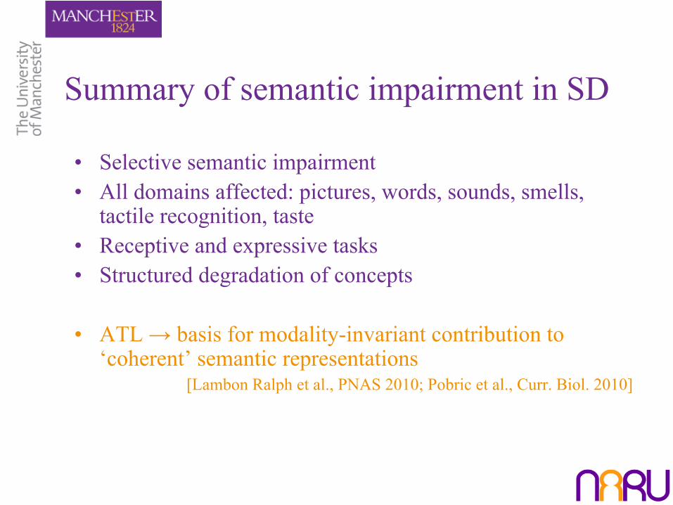
Summary of semantic impairment in SD
•
Selective semantic impairment•
All domains affected: pictures, words, sounds, smells, tactile recognition, taste
•
Receptive and expressive tasks•
Structured degradation of concepts
•
ATL → basis for modality-invariant contribution to ‘coherent’
semantic representations
[Lambon Ralph et al., PNAS 2010; Pobric et al., Curr. Biol. 2010]

Converging evidence for ATL contribution to semantic cognition
•
Implications from SD questioned (e.g., Martin, 2007)•
Does not appear in classic neurological models of language
•
If ATL is critical in producing impaired semantic memory –
what about its normal functioning in intact
people?•
Convergent evidence:–
Repetitive transcranial magnetic stimulation (rTMS)
–
Functional neuroimaging

rTMS methods: equipment

Locating the temporal pole

Offline rTMS: “virtual lesion”
method
•
Repeated stimulation produces refractory period in underlying cortex:–
120% hand motor threshold
–
1Hz for 10 minutes (600 pulses)–
Refractory window ~ 15 minutes
•
Use timed analogues of neuropsychological assessments for convergent evidence
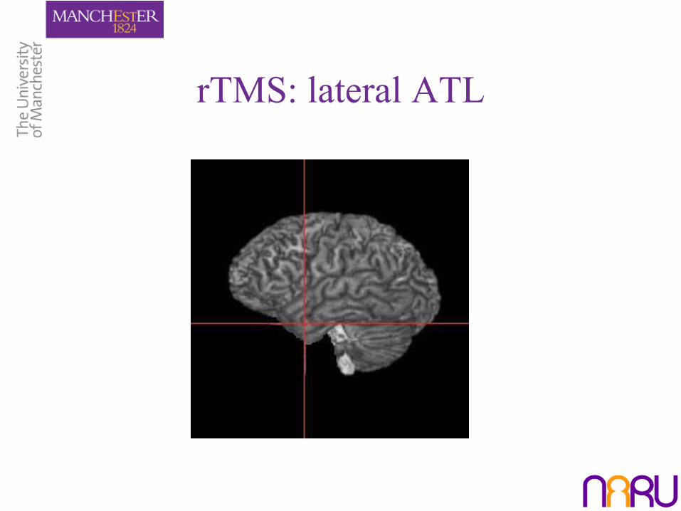
rTMS: lateral ATL
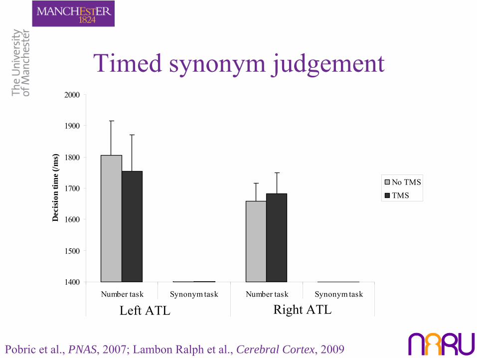
1400
1500
1600
1700
1800
1900
2000
Number task Synonym task Number task Synonym task
Left temporal pole Right temporal pole
Dec
isio
n tim
e (/m
s)
No TMSTMS
Timed synonym judgement
Pobric et al., PNAS, 2007; Lambon Ralph et al., Cerebral Cortex, 2009
Left ATL Right ATL

Functional neuroimaging and the anterior temporal lobe
Missing the meaning•
ATL notable by its absence in fMRI studies
•
Absence of ATL self-reinforcing in fMRI-based studies•
ATL-semantics literature review (Visser et al., JoCN 2010):
–
PET > fMRI–
Field of view
–
Control task: higher baseline > low level/rest
•
Magnetic field distortion:–
EPI gradient –
signal split & shifted

ATL semantics across methods: SD, rTMS & distortion-corrected fMRI
Binney et al., Cerebral Cortex, 2010 Pobric et al., PNAS, 2007
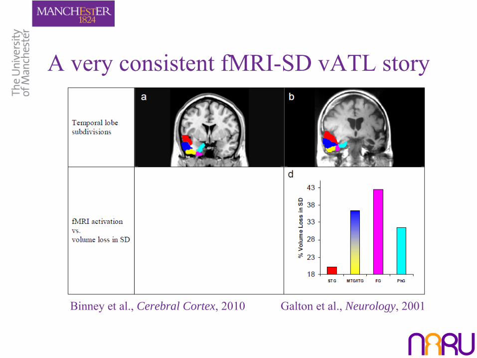
A very consistent fMRI-SD vATL story
Binney et al., Cerebral Cortex, 2010 Galton et al., Neurology, 2001

Graded variation of function across ATL (Visser & Lambon Ralph, JoCN, in press)


Semantic impairments across diseases
•
Semantic dementia: anterior temporal, bilateral•
HSVE: anterior temporal, bilateral
(Lambon Ralph et al., Brain, 2007)
•
Status of multi-modal semantics in CVA aphasia? (Jefferies & Lambon Ralph et al., Brain, 2006)

Transcortical Sensory Aphasia (TSA)
•
Poor verbal comprehension, fluent speech and good repetition
•
Status of non-verbal comprehension unclear•
How does this compare to SD given different location of damage?
(Berthier, 2001)

Multimodal semantic impairment: Semantic dementia vs. semantic aphasia
(Jefferies & Lambon Ralph, Brain, 2006)
•
Ten chronic aphasic CVA patients•
All impaired on picture semantic association test (PPT/CCT)–
Verbal + nonverbal impairment
–
Not just language comprehension deficit
•
Compared to SD cases (matched overall)–
Different neurology
–
Qualitatively different performance

Selected patients with stroke aphasiaCase Aphasia ATL lesion LIPFC lesion T-P lesion
SC Anomic/TSA
ME TSA
JM TSA
PG TSA
LS TSA
NY Mixed transcortical
KH Mixed transcortical
BB Mixed transcortical
KA Global
MS Global

SD: Intra-
& Inter-task comparisons
20
24
28
32
36
40
44
48
52
20 24 28 32 36 40 44 48 52Word PPT
Pic
ture
PP
T
Within task
20
24
28
32
36
40
44
48
52
12 16 20 24 28 32 36 40 44 48
Word-picture matching
Pict
ure
PPT
Between task

SA: Intra-
& Inter-task comparisons
20
24
28
32
36
40
44
48
52
20 24 28 32 36 40 44 48 52Word PPT
Pic
ture
PP
T
Within task
20
24
28
32
36
40
44
48
52
12 16 20 24 28 32 36 40 44 48Word-picture matching
Pic
ture
PP
T
Between task

Effect of external constraint: phonemic cueing (BNT)
0
10
20
30
40
50
60
SD semCVA
Nam
ing
accu
racy
Without cueWith cue
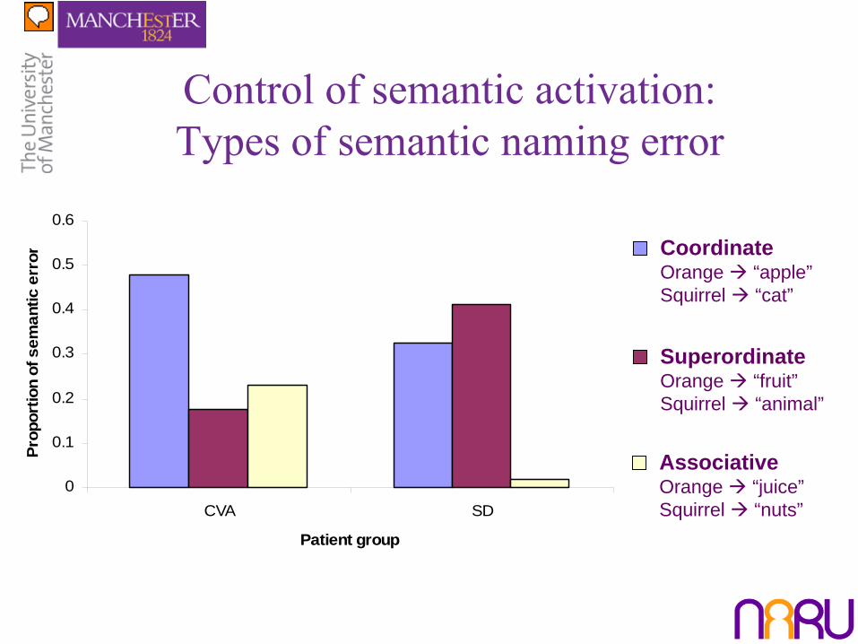
Control of semantic activation: Types of semantic naming error
0
0.1
0.2
0.3
0.4
0.5
0.6
CVA SD
Patient group
Prop
ortio
n of
sem
antic
err
or CoordinateOrange “apple”Squirrel “cat”
SuperordinateOrange “fruit”Squirrel “animal”
AssociativeOrange “juice”Squirrel “nuts”

Semantic control impairment
–
LIPFC & angular gyrus: cognitive & semantic control–
Areas damage in SA
–
Semantic control (Noonan et al., JCN, 2010):•
Inflexible semantic access
•
Poor inhibition of semantic competitors•
Improved with external constraint
e.g.,o
Understanding of dominant > subordinate meaning (e.g., BANK)
o
Performance on subordinate equalised with sentence context

Semantic control: Convergence of patients & rTMS
(Hoffman et al, J. Neurosci, 2010)
[-54, 24, 3]

Semantic control: Convergence of patients & imaging
Noonan et al. (submitted): ALE meta-analysis of 53 imaging studies of semantic control
(Berthier, 2001)

Semantic control: Convergence of patients & rTMS
(Whitney et al, Cereb. Cortex, in press)

Semantic control: Convergence of patients & rTMS
(Whitney et al, Cereb. Cortex, in press)
Stimulation sites

Semantic control: Convergence of patients & rTMS
(Whitney et al, Cereb. Cortex, in press)

Semantic control: Convergence of patients & rTMS
(Whitney et al, Cereb. Cortex, in press)
*

Semantic control: Convergence of patients & rTMS
(Whitney et al, Cereb. Cortex, in press)
*

Semantic aphasia vs. semantic dementia
SD SA
Multimodal comprehension impairment
ATL damage
PFC and/or temporoparietal
Correlation of performance across different tasks
Item familiarity + word frequency
Item consistency ~Correlation with ‘executive’
assessments
Improvement with cueing (verbal and nonverbal)
Jefferies & Lambon Ralph (2006); Corbett et al (2009); Noonan et
al (2010)
Where does Wernicke’s Aphasia fit in?
•
SA selected for SD comparison (cf. Chertkow et al, 1999)•
No WA patients in SA group
•
WA –
the classical comprehension impairment in neurology
•
WA –
associated with pSTG and pMTG damage

Wernicke’s Aphasia participants•
9 Wernicke’s aphasia participants (3 female), mean age 74
•
“Classical”
chronic WA = impaired auditory comprehension + jargon (BDAE: Goodglass et al., 2003)
•
Comparison Groups: SA n=10, SD n=10
Case Age Sex Time post-onset at testingAB 85 F 6-12 monthsLB 78 F 6 yearsEL 60 M 4-6 monthsMR 64 M 5-7 monthsDH 74 M 5-7 monthsDM 86 M 5-7 monthsRD 86 M 12 –
14 months
AC 53 M 5-9 monthsHS 81 F 6-8 months

Comparative temporal lobe lesion distributions

Comparative patient results
Environmental Sounds Battery: Three Group Comparison
0
12
24
36
48
sWPM sounds-pictures sounds-wordsCondition
Acc
urac
y
SASDcontrol
64 Item Battery: Three Group Comparison
0
16
32
48
64
sWPM picture CCT written CCT
Condition
Acc
urac
y SA
SD
control
Robson et al. (submitted)
Environmental Sounds Test Cambridge Semantic Battery

Comparative patient results
Environmental Sounds Battery: Three Group Comparison
0
12
24
36
48
sWPM sounds-pictures sounds-wordsCondition
Acc
urac
y
WASASDcontrol
64 Item Battery: Three Group Comparison
0
16
32
48
64
sWPM picture CCT written CCT
Condition
Acc
urac
y WASASDcontrol
Robson et al. (submitted)
Environmental Sounds Test Cambridge Semantic Battery

Three-way comparison
SD SA WAMultimodal impairment ~Acoustic-phonological deficit
ATL damage
PFC or temporoparietal
Item familiarity
Item consistency ~ ~Correlation with ‘executive’
assessments
Robson et al. (submitted)

WA vs. SA

Summary and conclusions•
Clear patterns revealed by comparative, case-series approach:–
Intra-group consistency; inter-group differences
•
SD: progressive degradation of modality-invariant, basal ATL representations leading to pan-modal receptive and expressive semantic impairment.
•
SA: multi-modal semantic impairment reflects damaged semantic/cognitive control [prefrontal and/or pMTG-AG region]
•
WA: combination of acoustic-phonological impairment and semantic control deficit [key extension to pSTG/iSMG]

The End
For further information and papers: www.psych-sci.manchester.ac.uk/naru/