apoptosis-induced release of mature sterol regulatory ... · apoptosis-induced release of mature...
TRANSCRIPT
Journal of Lipid Research
Volume 42, 2001
1939
Apoptosis-induced release of mature sterolregulatory element-binding proteins activatessterol-responsive genes
Maureen E. Higgins and Yiannis A. Ioannou
1
Department of Human Genetics, P.O. Box 1498, Mount Sinai School of Medicine, Fifth Avenue at 100th Street, New York, NY 10029
Abstract It is well established that during the execution ofthe apoptotic cascade, activated caspase 3 releases sterolregulatory element-binding proteins (SREBP) from themembrane of the endoplasmic reticulum in a proteolytic re-action that is distinct from their normal sterol-dependentactivation. However, it is not known whether these tran-scription factors are capable of activating sterol-responsivegenes under such conditions. The construction of SRE ex-pression vectors has permitted characterization of the apo-ptotic activation of SREBP. Cell lines stably expressing theplasma membrane marker CD32, or GFP, under the controlof the SRE promoter were shown to modulate SRE gene ex-pression on the basis of the levels of available sterols. How-ever, during the induction of apoptosis, expression of CD32and GFP was highly induced, even in the presence of amplesterols. Apoptotic induction of sterol-regulated genes wasdue to activation of caspase 3 and was impervious to treat-ment with sphingomyelinase, indicating that activation ofSRE genes during apoptosis is sterol independent. Furthercharacterization of this apoptotic response indicated that ste-rol-regulated genes are activated at an early stage in the apo-ptotic cascade, preceding the externalization of phosphati-dylserine on the plasma membrane of apoptotic cells.These results suggest that activation of sterol-responsivegenes early during apoptosis may play a role in the properexecution of this program.
—Higgins, M. E., and Y. A. Ioan-nou.
Apoptosis-induced release of mature sterol regulatoryelement-binding proteins activates sterol-responsive genes.
J. Lipid Res.
2001.
42:
1939–1946.
Supplementary key words
annexin V
•
caspase 3
•
CD32
•
cholesterol
•
SREBP
Cholesterol is the predominant sterol of the plasmamembrane in animal cells and is critical for cell growthand proliferation. Sphingomyelin is also an importantcomponent of plasma membranes, and in fact membranecholesterol solubilization depends largely on its sphingo-myelin content. The interactions between cholesterol andsphingomyelin appear to be critical for the regulation ofcellular cholesterol levels (1, 2), and it has been suggestedthat sphingomyelin modulates the amount of cholesterol
found in the various cellular pools (3, 4). In addition,these two molecules have been implicated in the forma-tion of microdomains or rafts within membrane regionsand are believed to function in signal transduction andmembrane trafficking (5).
Cholesterol is obtained from plasma lipoproteins viaendocytosis of the LDL receptor or synthesized de novofrom acetyl-coenzyme A. The rate-limiting steps in thecholesterol biosynthetic and salvage pathways, HMG-CoAreductase (6) and the LDL receptor (7), respectively, areboth transcriptionally regulated by sterol regulatory ele-ment binding proteins (SREBP) (8). These proteins be-long to the c-Myc family of transcription factors and havebeen shown to play an important role in the regulation ofgenes involved in intracellular cholesterol homeostasis inmammalian cells. They are synthesized as 125-kDa precur-sor proteins that are inserted into the membrane of theendoplasmic reticulum (ER) envelope (9–11). In sterol-deficient cells, proteases cleave the precursor proteins,thus releasing the mature transcription factors (12),which can enter the nucleus and activate target genes bybinding to the sterol regulatory element (SRE) sequencesin their promoters.
It was demonstrated that apopain (caspase 3), a cysteineprotease that plays a key role in the induction of apopto-sis, cleaves the SREBP when activated during apoptosis(13). These observations have led to the hypothesis thatcholesterol may be required in the early stages of apo-ptosis to maintain plasma membrane integrity (14). However,this cleavage of SREBP is distinct from the sterol-regulatedcleavage, and it is not clear yet whether caspase 3-cleavedSREBP are biologically relevant and capable of activatingsterol-regulated gene expression (13).
Abbreviations: ALLN,
N
-acetyl-leucinal-leucinal-norleucinal; ER,endoplasmic reticulum; GFP, green fluorescent protein; SRE, ste-rol regulatory element; SREBP, sterol regulatory element-bindingproteins.
1
To whom correspondence should be addressed.e-mail: [email protected]
at PE
NN
ST
AT
E U
NIV
ER
SIT
Y, on F
ebruary 21, 2013w
ww
.jlr.orgD
ownloaded from
1940 Journal of Lipid Research
Volume 42, 2001
Studies described in this report demonstrate thatSREBP released by apoptosis-induced proteolysis activatesterol-responsive genes during the early stages of apopto-sis. Furthermore, activation of these genes by the apoptotic-cleaved forms of SREBP precedes one of the earliest phe-notypes of apoptotic cell death, the externalization ofphosphatidylserine onto the outer layer of the plasmamembrane. These results suggest that activation ofcholesterol-regulated genes upon induction of the apo-ptotic cascade may be an important step in the executionof the death program.
MATERIALS AND METHODS
Materials
All reagents were purchased from Sigma (St. Louis, MO) un-less otherwise stated. DMEM and FBS were purchased from Media-tech (Herndon, VA) and HyClone (Logan, UT), respectively.
l
-Glutamine, gentamicin, G418, and the pGreen Lantern-1 plas-mid were from GIBCO-BRL (Grand Island, NY). VectaShield waspurchased from Vector Laboratories (Burlingame, CA). U18666Awas from BioMol (Plymouth Meeting, PA). Restriction endonu-cleases, polymerases, and ligases were from New England Bio-Labs (Beverly, MA). The ApoAlert annexin V apoptosis kit waspurchased from Clontech Laboratories (Palo Alto, CA). The flu-orescein isothiocyanate (FITC)-conjugated fluorescent anti-bodies were from Roche Molecular Biochemicals (Indianapolis,IN). The mouse monoclonal antibody to SREBP-2 was obtainedfrom Transduction Laboratories (Lexington, KY). The anti-human CD32 monoclonal antibody was produced by the hybri-doma cell line IV.3 (generous gift of J. Unkeless, Mount SinaiSchool of Medicine, New York, NY). Oligonucleotides were syn-thesized with phosphoramidite chemistry on a 380B DNA synthe-sizer (Applied Biosystems, Foster City, CA).
Cell culture
HeLa cells (CRL-1651) were purchased from the AmericanType Culture Collection (Manassas, VA). Cell lines were main-tained in complete DMEM (DMEM supplemented with 10%FBS, 2 mM
l
-glutamine, and gentamicin at 50
�
g/ml) in a hu-midified incubator at 37
�
C and 5% CO
2
.
Construction of the SRE expressionplasmids and derivatives
All molecular biology manipulations were per formed accord-ing to standard procedures (15). All vector constructs were con-firmed by restriction digestion and dideoxynucleotide sequenc-ing. For the construction of the SRE-containing plasmids,equimolar amounts of SRE primers, 5
�
-TCGAGGACATTTGAAAATCACCCCACTGCAAACTCCTCCCCC TGCTAG GCGTGTACGGTGGGAGGCCTG-3
�
and 5
�
-TCGACAGGCCTCCCACCGTACACGCCTAGCAGGGGGAGGAGTTTGCAGTGGGGTGATT TTCAAATGTCC-3
�
, were mixed together, boiled for 5 min, andallowed to cool to room temperature for 20 min. The double-stranded product was phosphorylated with T4 polynucleotide ki-nase and cloned in the sense orientation into the
Xho
I site ofplasmid pUHD10-3 (generous gift of Dr. H. Bujard, Universityof Heidelberg, Heidelberg, Germany). The resulting productwas digested with
Stu
I to remove the tet operator, and the largerfragment was gel purified. The eluted DNA was self-ligated, redi-gested with
Xho
I, and another phosphorylated double-strandedoligonucleotide was cloned into the
Xho
I site, resulting in plas-mid pSRE
��
. Sequence analysis confirmed that both SRE ele-
ments were inserted in the plus (
�
) orientation. The pSRE
��
plasmid was digested with
Bam
HI and a cassette containing theneomycin resistance gene driven by the simian virus 40 (SV40)promoter-enhancer was inserted to generate pSRE
��
.neo. Sub-sequently, a human CD32 cDNA fragment (generous gift of J.Unkeless, Mount Sinai School of Medicine) was inserted into the
Eco
RI site of SRE
��
.neo to generate pSRE
��
.CD32.neo.Similarly, to generate the pSRE
��
.GFP plasmid, a green fluo-rescent protein (GFP)-encoding cDNA was excised from thepGreen Lantern-1 plasmid and was inserted into the pSRE
��
.neoplasmid. The integrity of the resulting vector, pSRE
��
.GFP.neo,was confirmed by restriction digestion.
Generation of HeLa-SRE-CD32 andHeLa-SRE-GFP cell lines
HeLa cells at 80% confluence were harvested by trypsinizationand electroporated with 10
�
g of either pSRE.CD32.neo orpSRE.GFP.neo plasmid DNA, using a GenePulser apparatus (Bio-Rad, Hercules, CA) at 250 V and 500
�
F in complete DMEM. Stableclones were isolated in G418 antibiotic medium (500
�
g/ml), ex-panded, and analyzed for sterol-inducible expression of CD32 orGFP after growth in high or low cholesterol-containing medium.
Western blot analysis
On day 0, cells were seeded in 60-mm dishes in complete me-dium. On day 1, the medium was refreshed with the followingadditions:
1
) none (control),
2
) 25 mM mevastatin,
3
) U18666A(2
�
g/ml), or
4
) 25 mM mevastatin and U18666A (2
�
g/ml).Three hours before harvesting on day 2,
N
-acetyl-leucinal-leucinal-norleucinal (ALLN, 25
�
g/ml) was added to each dish. Cellswere harvested with PBS-2 mM EDTA and centrifuged at 800
g
for 10 min. The cell pellet was resuspended in buffer [150 mMNaCl, 50 mM Tris (pH 8.0), 1% Triton X-100, leupeptin (10
�
g/ml), pepstatin A (5
�
g/ml), ALLN (25
�
g/ml), 1
�
M phenyl-methylsulfonyl fluoride, and 0.1 mM Pefabloc], incubated on icefor 10 min, and centrifuged at 800
g
for 10 min. The supernatantwas boiled for 5 min in SDS loading buffer, and proteins wereseparated on an 8% polyacrylamide gel. Proteins were trans-ferred onto nitrocellulose membranes with a Novex (San Diego,CA) Xcell II Blot module for 2 h at 20 V. Proteins were visualizedby chemiluminescence according to the manufacturer recom-mendations (Roche Diagnostics, Indianapolis, IN).
Fluorescence microscopy
On day 0, cells were seeded on coverslips in complete medium.On day 1, the medium was changed to complete medium contain-ing
1
) no addition (control),
2
) 10
�
M camptothecin, or
3
) 10
�
M camptothecin and neutral sphingomyelinase (100
�
U/ml).After 4 h, cells were washed with chilled PBS and fixed in 3.7%paraformaldehyde for 20 min at room temperature. Immunofluo-rescence analysis was carried out on fixed cells as previously de-scribed (16). Hoechst 33258 (10
�
g/ml) or propidium iodide (1.5
�
g/ml) was added to cells as indicated. For annexin V detection ofphosphatidylserine, cells were washed with chilled PBS and incu-bated with FITC (2
�
g/ml)-conjugated annexin in 1
�
bindingbuffer for 20 min at room temperature. After a wash with chilledPBS, cells were fixed in 3.7% paraformaldehyde for 20 min at roomtemperature. CD32 expression was visualized with an anti-mouserhodamine-conjugated antibody as described above.
DNA fragmentation assay
On day 0, cells were placed in complete medium. On day 1,the medium was changed to complete medium containing
1
) noaddition (control),
2
) 1
�
M camptothecin, or
3
) 1
�
M camp-tothecin and neutral sphingomyelinase at 100
�
U/ml. On day 2,the cells were harvested with PBS-2 mM EDTA and collected by
at PE
NN
ST
AT
E U
NIV
ER
SIT
Y, on F
ebruary 21, 2013w
ww
.jlr.orgD
ownloaded from

Higgins and Ioannou
Sterol response in apoptosis 1941
centrifugation. Cells were lysed in DNA fragmentation lysisbuffer, and DNA fragmentation was quantitated as previously de-scribed (17).
RESULTS
Construction of sterol-inducible expression vectors
Vectors were engineered in which a cDNA encodingeither the plasma membrane marker CD32 or GFP wasplaced under the control of a synthetic sterol-regulatedpromoter. To generate the pSRE plasmids, two copies ofthe SRE-42 element found in the LDL receptor gene pro-moter were inserted 22 bp upstream from the TATA boxof a minimal human cytomegalovirus (CMV) promoter(
Fig. 1A
). A modified human IgG Fc
�
receptor II cDNA(hFcRII, CD32) or a GFP cDNA was inserted in thepolylinker under the control of the sterol-responsive pro-moter (Fig. 1B) to generate pSRE-CD32 and pSRE-GFP,respectively. Because CD32 is expressed only on B lym-phocytes, granulocytes, and platelets (18), it is an idealplasma membrane marker for expression and biopanningin all other cell types, whereas GFP is useful for cell sort-ing and qualitative analysis of positive cells. Finally, a cas-sette containing the neomycin resistance gene driven bythe SV40 promoter-enhancer was inserted into these vec-tors for positive selection.
Expression of sterol-inducible vectors in HeLa cells
HeLa cells were transfected with either the pSRE-CD32or the pSRE-GFP vector to generate stable cell lines thatcould respond to changes in cellular cholesterol levels. A
number of clones were identified that expressed high levelsof CD32 or GFP after growth in reduced sterol mediumbut completely suppressed the expression of these markerproteins after growth in sterol-rich medium. A CD32-expressing clone (HeLa-CD32) and a GFP-expressing clone(HeLa-GFP) that exhibited optimal sterol-inducible ex-pression were chosen for all subsequent work.
Both cell lines expressed the markers in the sterol-dependent manner previously described for the LDL recep-tor, indicating that the cell lines could be used to monitorSRE-induced cellular responses. HeLa-CD32 or HeLa-GFPcells grown in high sterol medium effectively downregu-lated the expression of CD32/GFP (
Fig. 2
, FCS). In fact,continuous growth of these cells in medium containingsoluble cholesterol effectively maintained the expression vec-tor in the “off” state (data not shown). The expression levelof CD32/GFP was only slightly upregulated when the de
Fig. 1. Sterol-regulated mammalian expression vectors. A: Twocopies of the SRE 42 element found in the LDL receptor gene wereplaced in front of a minimal CMV promoter (TATA box) in the (�)orientation (see Materials and Methods). B: The SRE-regulatedpromoter drives the expression of a cDNA encoding either aplasma membrane marker (CD32) or GFP. Stable expression is ob-tained by selection for the SV40-neo cassette.
Fig. 2. Sterol-regulated expression of CD32 or GFP in stable celllines. Both CD32-expressing (HeLa-CD32) and GFP-expressing(HeLa-GFP) cell lines exhibited sterol-regulated expression of themarker genes. In sterol-rich medium, expression of the genes is notinduced (FCS), but in the presence of U18666A, an inhibitor of ly-sosomal cholesterol efflux, both cell lines turn on the expression ofthe marker gene. Mevastatin, an inhibitor of de novo synthesisof cholesterol, also induces expression, albeit at a lower level. The ad-dition of both inhibitors, however, causes a dramatic increase in theexpression of both markers (U18�Mev). Magnification bar: 10 �m.
at PE
NN
ST
AT
E U
NIV
ER
SIT
Y, on F
ebruary 21, 2013w
ww
.jlr.orgD
ownloaded from
1942 Journal of Lipid Research
Volume 42, 2001
novo biosynthetic pathway was blocked by inhibitingHMG-CoA reductase with mevastatin (Fig. 2, mevastatin).Treatment of cells with U18666A, a potent inhibitor oflysosomally derived cholesterol (19), caused a significantincrease in CD32/GFP expression (Fig. 2, U18666A), in-dicating that U18666A is an effective inducer of thesesterol-regulated markers. Simultaneous treatment of cellswith both inhibitors caused a dramatic increase in CD32/GFP expression even though the cells were bathed in ahigh cholesterol medium (Fig. 2; U18
�
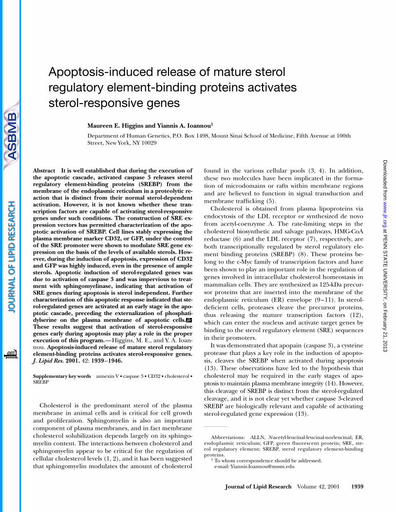
Mev).Western blot analysis of HeLa-CD32 cell lysates demon-
strated that CD32 expression levels correlated with the levelsof mature SREBP (
Fig. 3
). There was negligible expres-sion of mature SREBP-2 and CD32 in HeLa-CD32 cellsmaintained in a high cholesterol medium (FCS), whereasinhibition of cholesterol egress from the endosomal/lysosomal system with U18666A resulted in the appear-ance of mature SREBP-2 and high levels of CD32 (Fig.3). This high level of expression persisted when the cellswere treated with both mevastatin and U18666A. Takentogether, these results indicate that these vectors can beused as monitors or “sensors” of cellular cholesterol levelsor, alternatively, as high level sterol-inducible expres-sion vectors.
Cholesterol-independent activationof sterol-responsive genes during apoptosis
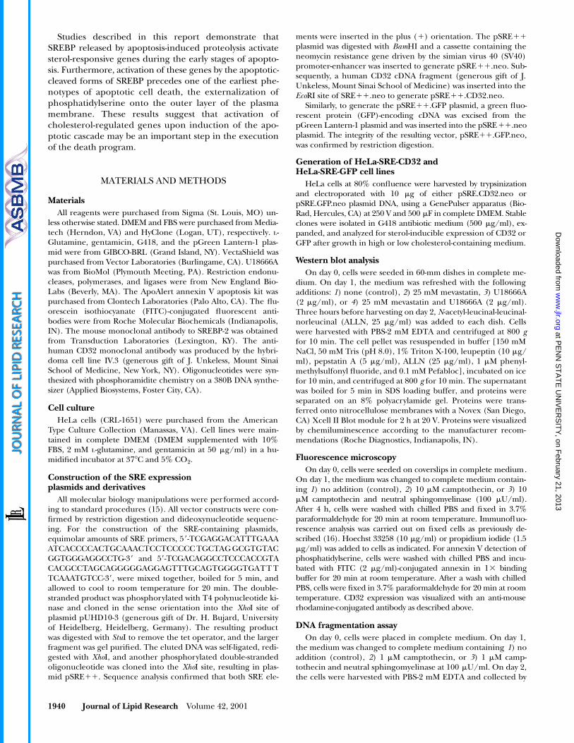
During the characterization of the above-described celllines, it was observed that cells exhibiting signs of apoptosisalso expressed the SRE-regulated markers in the absence ofsterol induction. Previous reports demonstrated that dur-ing apoptosis, activated caspase 3 cleaves SREBP (13, 14).However, it was not determined whether caspase 3-cleavedSREBP were capable of activating SRE-responsive genes, be-cause the caspase 3 cleavage site is different from that pro-duced by sterol induction. To address this question, HeLa-GFP cells were treated with the topoisomerase inhibitorcamptothecin to induce apoptosis. Expression of the sterol-responsive marker GFP could be clearly seen in apoptoticcells even though the presence of ample sterols in thegrowth medium should have suppressed the SRE response(
Fig. 4A
, arrows). Cells expressing GFP displayed the char-
acteristic morphological features of apoptosis, includingmembrane blebbing and nuclear and cytoplasmic conden-sation. Staining these cells with propidium iodide revealeda condensed, bright-red nuclear morphology (Fig. 4B, ar-rows), whereas in the absence of any SRE-driven GFP ex-pression in the nonapoptotic cells, the DNA appears as adiffuse red fluorescence (Fig. 4B). To ensure that theSREBP cleavage was not due to sterol regulation, neutralsphingomyelinase was added to the LDL-enriched culturemedium. Treatment of cultured cells with neutral sphingo-myelinase abolishes sterol-regulated maturation of SREBP
Fig. 3. Immunoblot of CD32 and SREBP-2 expression in HeLa-CD32 cells. HeLa-CD32 cells were grown in the presence of 10%FCS and either of the inhibitors U18666A and mevastatin (see leg-end of Fig. 2). In sterol-rich medium (FCS), the mature form ofSREBP-2 is absent, along with little, if any, expression of CD32.U18666A and mevastatin, however, cause an increase in the levels ofmature SREBP-2, with a concomitant increase in CD32 expression.
Fig. 4. HeLa-GFP cells treated with camptothecin to induce apo-ptosis. HeLa cells grown in sterol-rich medium to suppress sterol-regulated expression of GFP were treated with camptothecin to in-duce apoptosis. A: Sterol-independent induction of GFP expressionis seen in many cells that also exhibit signs of apoptotic cell death,such as membrane blebbing (arrows). B: Cells were stained withpropidium iodide to detect nuclei undergoing apoptosis. Arrowsindicate cells exhibiting the characteristic condensed nuclear mor-phology of apoptotic cells. Magnification bar: 10 �m.
at PE
NN
ST
AT
E U
NIV
ER
SIT
Y, on F
ebruary 21, 2013w
ww
.jlr.orgD
ownloaded from
Higgins and Ioannou
Sterol response in apoptosis 1943
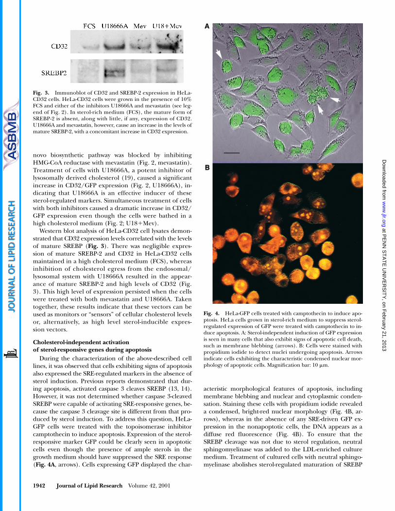
(1) because cholesterol is translocated to the ER after de-pletion of plasma membrane sphingomyelin. Cells grownin sterol-rich medium and treated with both neutral sphin-gomyelinase and camptothecin still expressed the SREmarker CD32 (
Fig. 5A
), and to confirm that these cellswere apoptotic, a DNA fragmentation assay was performed.As shown in Fig. 5B, cells treated with camptothecinshowed a significant increase in DNA fragmentation, whichwas not affected by the addition of sphingomyelinase to thegrowth medium. These results indicate that the apoptosis-induced cleavage of SREBP can activate the expression ofSRE-responsive genes in a sterol-independent manner.
SREBP activation by caspase 3 at an early pointin the apoptotic cascade
Although these results demonstrated that SRE-respon-sive genes are activated during apoptosis, it is not clear atwhich point in the apoptotic cascade SREBP are activated.To determine when SREBP maturation occurs, cells weregrown in sterol-rich medium with or without camptothe-
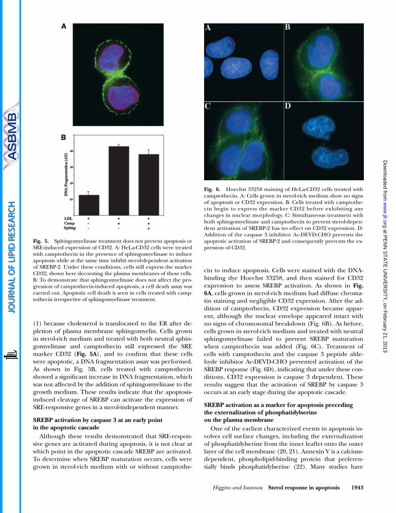
cin to induce apoptosis. Cells were stained with the DNA-binding dye Hoechst 33258, and then stained for CD32expression to assess SREBP activation. As shown in
Fig.6A
, cells grown in sterol-rich medium had diffuse chroma-tin staining and negligible CD32 expression. After the ad-dition of camptothecin, CD32 expression became appar-ent, although the nuclear envelope appeared intact withno signs of chromosomal breakdown (Fig. 6B). As before,cells grown in sterol-rich medium and treated with neutralsphingomyelinase failed to prevent SREBP maturationwhen camptothecin was added (Fig. 6C). Treatment ofcells with camptothecin and the caspase 3 peptide alde-hyde inhibitor Ac-DEVD-CHO prevented activation of theSREBP response (Fig. 6D), indicating that under these con-ditions, CD32 expression is caspase 3 dependent. Theseresults suggest that the activation of SREBP by caspase 3occurs at an early stage during the apoptotic cascade.
SREBP activation as a marker for apoptosis precedingthe externalization of phosphatidylserineon the plasma membrane
One of the earliest characterized events in apoptosis in-volves cell surface changes, including the externalizationof phosphatidylserine from the inner leaflet onto the outerlayer of the cell membrane (20, 21). Annexin V is a calcium-dependent, phospholipid-binding protein that preferen-tially binds phosphatidylserine (22). Many studies have
Fig. 5. Sphingomyelinase treatment does not prevent apoptosis orSRE-induced expression of CD32. A: HeLa-CD32 cells were treatedwith camptothecin in the presence of sphingomyelinase to induceapoptosis while at the same time inhibit sterol-dependent activationof SREBP-2. Under these conditions, cells still express the markerCD32, shown here decorating the plasma membranes of these cells.B: To demonstrate that sphingomyelinase does not affect the pro-gression of camptothecin-induced apoptosis, a cell death assay wascarried out. Apoptotic cell death is seen in cells treated with camp-tothecin irrespective of sphingomyelinase treatment.
Fig. 6. Hoechst 33258 staining of HeLa-CD32 cells treated withcamptothecin. A: Cells grown in sterol-rich medium show no signsof apoptosis or CD32 expression. B: Cells treated with camptothe-cin begin to express the marker CD32 before exhibiting anychanges in nuclear morphology. C: Simultaneous treatment withboth sphingomyelinase and camptothecin to prevent sterol-depen-dent activation of SREBP-2 has no effect on CD32 expression. D:Addition of the caspase 3 inhibitor Ac-DEVD-CHO prevents theapoptotic activation of SREBP-2 and consequently prevents the ex-pression of CD32.
at PE
NN
ST
AT
E U
NIV
ER
SIT
Y, on F
ebruary 21, 2013w
ww
.jlr.orgD
ownloaded from
1944 Journal of Lipid Research
Volume 42, 2001
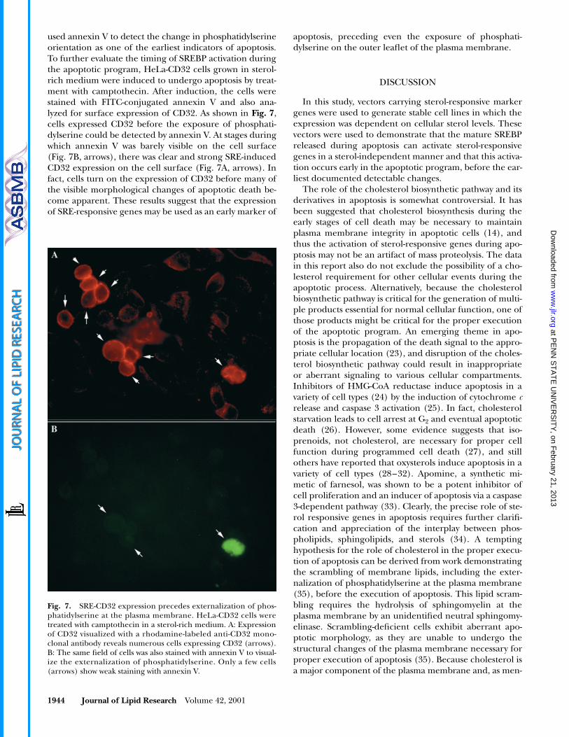
used annexin V to detect the change in phosphatidylserineorientation as one of the earliest indicators of apoptosis.To further evaluate the timing of SREBP activation duringthe apoptotic program, HeLa-CD32 cells grown in sterol-rich medium were induced to undergo apoptosis by treat-ment with camptothecin. After induction, the cells werestained with FITC-conjugated annexin V and also ana-lyzed for surface expression of CD32. As shown in
Fig. 7
,cells expressed CD32 before the exposure of phosphati-dylserine could be detected by annexin V. At stages duringwhich annexin V was barely visible on the cell surface(Fig. 7B, arrows), there was clear and strong SRE-inducedCD32 expression on the cell surface (Fig. 7A, arrows). Infact, cells turn on the expression of CD32 before many ofthe visible morphological changes of apoptotic death be-come apparent. These results suggest that the expressionof SRE-responsive genes may be used as an early marker of
apoptosis, preceding even the exposure of phosphati-dylserine on the outer leaflet of the plasma membrane.
DISCUSSION
In this study, vectors carrying sterol-responsive markergenes were used to generate stable cell lines in which theexpression was dependent on cellular sterol levels. Thesevectors were used to demonstrate that the mature SREBPreleased during apoptosis can activate sterol-responsivegenes in a sterol-independent manner and that this activa-tion occurs early in the apoptotic program, before the ear-liest documented detectable changes.
The role of the cholesterol biosynthetic pathway and itsderivatives in apoptosis is somewhat controversial. It hasbeen suggested that cholesterol biosynthesis during theearly stages of cell death may be necessary to maintainplasma membrane integrity in apoptotic cells (14), andthus the activation of sterol-responsive genes during apo-ptosis may not be an artifact of mass proteolysis. The datain this report also do not exclude the possibility of a cho-lesterol requirement for other cellular events during theapoptotic process. Alternatively, because the cholesterolbiosynthetic pathway is critical for the generation of multi-ple products essential for normal cellular function, one ofthose products might be critical for the proper executionof the apoptotic program. An emerging theme in apo-ptosis is the propagation of the death signal to the appro-priate cellular location (23), and disruption of the choles-terol biosynthetic pathway could result in inappropriateor aberrant signaling to various cellular compartments.Inhibitors of HMG-CoA reductase induce apoptosis in avariety of cell types (24) by the induction of cytochrome
c
release and caspase 3 activation (25). In fact, cholesterolstarvation leads to cell arrest at G
2
and eventual apoptoticdeath (26). However, some evidence suggests that iso-prenoids, not cholesterol, are necessary for proper cellfunction during programmed cell death (27), and stillothers have reported that oxysterols induce apoptosis in avariety of cell types (28–32). Apomine, a synthetic mi-metic of farnesol, was shown to be a potent inhibitor ofcell proliferation and an inducer of apoptosis via a caspase3-dependent pathway (33). Clearly, the precise role of ste-rol responsive genes in apoptosis requires further clarifi-cation and appreciation of the interplay between phos-pholipids, sphingolipids, and sterols (34). A temptinghypothesis for the role of cholesterol in the proper execu-tion of apoptosis can be derived from work demonstratingthe scrambling of membrane lipids, including the exter-nalization of phosphatidylserine at the plasma membrane(35), before the execution of apoptosis. This lipid scram-bling requires the hydrolysis of sphingomyelin at theplasma membrane by an unidentified neutral sphingomy-elinase. Scrambling-deficient cells exhibit aberrant apo-ptotic morphology, as they are unable to undergo thestructural changes of the plasma membrane necessary forproper execution of apoptosis (35). Because cholesterol isa major component of the plasma membrane and, as men-
Fig. 7. SRE-CD32 expression precedes externalization of phos-phatidylserine at the plasma membrane. HeLa-CD32 cells weretreated with camptothecin in a sterol-rich medium. A: Expressionof CD32 visualized with a rhodamine-labeled anti-CD32 mono-clonal antibody reveals numerous cells expressing CD32 (arrows).B: The same field of cells was also stained with annexin V to visual-ize the externalization of phosphatidylserine. Only a few cells(arrows) show weak staining with annexin V.
at PE
NN
ST
AT
E U
NIV
ER
SIT
Y, on F
ebruary 21, 2013w
ww
.jlr.orgD
ownloaded from
Higgins and Ioannou
Sterol response in apoptosis 1945
tioned above, associates with sphingomyelin, it may thus beimportant in facilitating the hydrolysis of sphingomyelinleading to the proper execution of membrane scramblingand cell death.
Programmed cell death is a fundamental biological pro-cess used by multicellular organisms to eliminate un-wanted cells. Together with cell proliferation and differen-tiation, apoptosis is responsible for the arrangement andmaintenance of almost all tissues. The connection betweencholesterol metabolism and embryogenesis has proven tobe critical in mammalian development (36, 37). Choles-terol activates the signaling of hedgehog proteins that areresponsible for the patterning behavior of many multicel-lular organisms (38). Disruptions in cholesterol synthesiscan lead to many developmental deformities. In addition,there is a growing awareness that inappropriate initiationor regulation of apoptosis may be associated with a varietyof neurodegenerative diseases. Malfunctions in the apop-totic cascade have been proposed to contribute to thepathogenesis of such diseases as Alzheimer’s disease andParkinson’s disease.
Although the significance of the activation of cholesterol-regulated genes during apoptosis is not yet established, itis becoming clear that cholesterol plays a far greater rolein cell growth and differentiation than is currently known.Cholesterol influences signaling paths that guide develop-ment. Because apoptosis is a developmental tool, it istempting to speculate that sterols serve as molecules thatimpact the functioning of critical signaling proteins dur-ing development. Thus, it is probable that such activationplays a role in the proper execution of programmed celldeath. As these studies demonstrate, the role of sterol-regulated genes in the regulation of cell growth, differen-tiation, and death needs to be evaluated.
These studies were supported in part by NIH grant R01DK54736 and grants from the March of Dimes Foundation andthe AVA Pavseghian Medical Research Foundation.
Manuscript received 19 July 2001.
REFERENCES
1. Scheek, S., M. S. Brown, and J. L. Goldstein. 1997. Sphingomyelindepletion in cultured cells blocks proteolysis of sterol regulatoryelement binding proteins at site 1.
Proc. Natl. Acad. Sci. USA.
94:
11179–11183.2. Koval, M., and R. E. Pagano. 1991. Intracellular transport and me-
tabolism of sphingomyelin.
Biochim. Biophys. Acta.
1082:
113–125.3. Wattenberg, B. W., and D. F. Silbert. 1983. Sterol partitioning
among intracellular membranes. Testing a model for cellular ste-rol distribution.
J. Biol. Chem.
258:
2284–2289.4. Slotte, J. P. 1997. Cholesterol-sphingomyelin interactions in cells—
effects on lipid metabolism.
Subcell. Biochem.
28:
277–293.5. Simons, K., and E. Ikonen. 1997. Functional rafts in cell mem-
branes.
Nature.
387:
569–572.6. Rodwell, V. W., J. L. Nordstrom, and J. J. Mitschelen. 1976. Regula-
tion of HMG-CoA reductase.
Adv. Lipid Res.
14:
1–74.7. Brown, M. S., and J. L. Goldstein. 1986. A receptor mediated path-
way for cholesterol homeostasis.
Science.
232: 34–47.8. Brown, M. S., and J. L. Goldstein. 1997. The SREBP pathway: regu-
lation of cholesterol metabolism by proteolysis of a membrane-bound transcription factor. Cell. 89: 331–340.
9. Briggs, M. R., C. Yokoyama, X. Wang, M. S. Brown, and J. L. Gold-stein. 1993. Nuclear protein that binds sterol regulatory elementof low density lipoprotein receptor promoter. I. Identification ofthe protein and delineation of its target nucleotide sequence. J.Biol. Chem. 268: 14490–14496.
10. Rajavashisth, T. B., A. K. Taylor, A. Andalibi, K. L. Svenson, and A. J.Lusis. 1989. Identification of a zinc finger protein that binds to thesterol regulatory element. Science. 245: 640–643.
11. Wang, X., M. R. Briggs, X. Hua, C. Yokoyama, J. L. Goldstein,and M. S. Brown. 1993. Nuclear protein that binds sterol regula-tory element of low density lipoprotein receptor promoter. II.Purification and characterization. J. Biol. Chem. 268: 14497–14504.
12. Hua, X., J. Sakai, M. S. Brown, and J. L. Goldstein. 1996. Regulatedcleavage of sterol regulatory element binding proteins requires se-quences on both sides of the endoplasmic reticulum membrane. J.Biol. Chem. 271: 10379–10384.
13. Pai, J-T., M. S. Brown, and J. L. Goldstein. 1996. Purification andcDNA cloning of a second apoptosis-related cysteine protease thatcleaves and activates sterol regulatory element binding proteins.Proc. Natl. Acad. Sci. USA. 93: 5437–5442.
14. Wang, X., N. G. Zelenski, J. Yang, J. Sakai, M. S. Brown, and J. L.Goldstein. 1996. Cleavage of sterol regulatory element bindingproteins (SREBPs) by CPP32 during apoptosis. EMBO J. 15: 1012–1020.
15. Sambrook, J., E. F. Fritsch, and T. Maniatis. 1989. In MolecularCloning. C. Nolan, editor. Cold Spring Harbor Laboratory Press,Cold Spring Harbor, NY.
16. Higgins, M. E., J. P. Davies, F. W. Chen, and Y. A. Ioannou. 1999.Niemann-Pick C1 is a late endosome-resident protein that tran-siently associates with lysosomes and the trans-Golgi network. Mol.Genet. Metab. 68: 1–13.
17. Ioannou, Y. A., and F. W. Chen. 1996. Quantitation of DNA frag-mentation in apoptosis. Nucleic Acids Res. 24: 992–993.
18. Brooks, D. G., W. Q. Qiu, A. D. Luster, and J. V. Ravetch. 1989.Structure and expression of human IgG FcRII(CD32). J. Exp. Med.170: 1369–1385.
19. Harmala, A-S., M. I. Porn, P. Mattjus, and J. P. Slotte. 1994. Choles-terol transport from plasma membranes to intracellular mem-branes is inhibited by 3�-[2-(diethylamino)ethoxy]androst-5-en-17-one. Biochim. Biophys. Acta. 1211: 317–325.
20. Fadok, V. A., J. S. Savill, C. Haslett, D. L. Bratton, D. E. Doherty, P. A.Campbell, and P. M. Henson. 1992. Different populations ofmacrophages use either the vitronectin receptor or the phosphati-dylserine receptor to recognize and remove apoptotic cells. J. Im-munol. 149: 4029–4035.
21. Fadok, V. A., D. R. Voelker, P. A. Campbell, D. L. Bratton, J. J.Cohen, P. W. Noble, D. W. Riches, and P. M. Henson. 1993. Theability to recognize phosphatidylserine on apoptotic cells is an in-ducible function in murine bone marrow-derived macrophages.Chest. 103(Suppl. 2): 102S.
22. Bratton, D. L., V. A. Fadok, D. A. Richter, J. M. Kailey, L. A. Guth-rie, and P. M. Henson. 1997. Appearance of phosphatidylserine onapoptotic cells requires calcium-mediated nonspecific flip-flopand is enhanced by loss of the aminophospholipid translocase. J.Biol. Chem. 272: 26159–26165.
23. Porter, A. G. 1999. Protein translocation in apoptosis. Trends CellBiol. 9: 394–401.
24. Mo, H., and C. E. Elson. 1999. Apoptosis and cell-cycle arrest inhuman and murine tumor cells are initiated by isoprenoids. J.Nutr. 129: 804–813.
25. Wang, I. K., S. Y. Lin-Shiau, and J. K. Lin. 2000. Induction of apo-ptosis by lovastatin through activation of caspase-3 and DNase II inleukaemia HL-60 cells. Pharmacol. Toxicol. 86: 83–91.
26. Martinez-Botas, J., Y. Suarez, A. J. Ferruelo, D. Gomez-Coronado,and M. A. Lasuncion. 1999. Cholesterol starvation decreasesp34(cdc2) kinase activity and arrests the cell cycle at G2. FASEB J.13: 1359–1370.
27. Michikawa, M., and K. Yanagisawa. 1998. Apolipoprotein E4 in-duces neuronal cell death under conditions of suppressed de novocholesterol synthesis. J. Neurosci. Res. 54: 58–67.
28. Nishio, E., S. Arimura, and Y. Watanabe. 1996. Oxidized LDL in-duces apoptosis in cultured smooth muscle cells: a possible rolefor 7-ketocholesterol. Biochem. Biophys. Res. Commun. 223: 413–418.
29. Nishio, E., and Y. Watanabe. 1996. Oxysterols induced apoptosis incultured smooth muscle cells through CPP32 protease activation
at PE
NN
ST
AT
E U
NIV
ER
SIT
Y, on F
ebruary 21, 2013w
ww
.jlr.orgD
ownloaded from
1946 Journal of Lipid Research Volume 42, 2001
and bcl-2 protein downregulation. Biochem. Biophys. Res. Commun.226: 928–934.
30. Lizard, G., S. Monier, C. Cordelet, L. Gesquiere, V. Deckert, S.Gueldry, L. Lagrost, and P. Gambert. 1999. Characterization andcomparison of the mode of cell death, apoptosis versus necrosis,induced by 7beta-hydroxycholesterol and 7-ketocholesterol in thecells of the vascular wall. Arterioscler. Thromb. Vasc. Biol. 19: 1190–1200.
31. Lizard, G., V. Deckert, L. Dubrez, M. Moisant, P. Gambert, and L.Lagrost. 1996. Induction of apoptosis in endothelial cells treatedwith cholesterol oxides. Am. J. Pathol. 148: 1625–1638.
32. Yang, L., and M. S. Sinensky. 2000. 25-Hydroxycholesterol activatesa cytochrome c release-mediated caspase cascade. Biochem. Biophys.Res. Commun. 278: 557–563.
33. Flach, J., I. Antoni, P. Villemin, C. L. Bentzen, and E. J. Niesor.2000. The mevalonate/isoprenoid pathway inhibitor apomine
(SR-45023A) is antiproliferative and induces apoptosis similar tofarnesol. Biochem. Biophys. Res. Commun. 270: 240–246.
34. Ridgway, N. D., D. M. Byers, H. W. Cook, and M. K. Storey. 1999.Integration of phospholipid and sterol metabolism in mammaliancells. Prog. Lipid Res. 38: 337–360.
35. Tepper, A. D., P. Ruurs, T. Wiedmer, P. J. Sims, J. Borst, and W. J. vanBlitterswijk. 2000. Sphingomyelin hydrolysis to ceramide during theexecution phase of apoptosis results from phospholipid scramblingand alters cell-surface morphology. J. Cell Biol. 150: 155–164.
36. Herz, J., T. E. Willnow, and R. V. Farese, Jr. 1997. Cholesterol,hedgehog and embryogenesis. Nat. Genet. 15: 123–124.
37. Farese, R. V., and J. Herz. 1998. Cholesterol metabolism and em-bryogenesis. Trends Genet. 14: 115–120.
38. Porter, J. P., K. E. Young, and P. A. Beachy. 1996. Cholesterol modi-fication of hedgehog signaling protein in animal development.Science. 274: 255–259.
at PE
NN
ST
AT
E U
NIV
ER
SIT
Y, on F
ebruary 21, 2013w
ww
.jlr.orgD
ownloaded from