appendectomy - chelsma.ru · appendicitis: ¾ more often appears at the age of 10-30 ¾ 89,1% of...
TRANSCRIPT

Appendectomy
Speaker:Barysheva V.O.,401 grLeader:Peshikov O.V.

Appendectomy
Indications for operation :
Acute appendicitis Chronic appendicitis Tumors of appendix

Appendicitis
Inflammation of appendix

Appendicitis:
More often appears at the age of 10-3089,1% of all surgical diseases of abdominal visceraThe most often reason of peritonitis

Aetiology and pathogenesis :The mechanical theory — activation of intestinal flora against the background of obturation the appendix’s gap is considered the main reason of appendicitis.Obturation of gap is caused by fecal boluses and hyperplasia of lymphoid folliclesфолликулов.

Casuistical reasons of obturation:
• Swallowed foreign bodes
• Presence of helminthes (often askarids) in the inflamed gap
• Tumors of appendix(more often it’s carcinoid)

Clinicoanatomical forms of appendicitis:
AcuteAcute inflammatory-necrotic disease of appendix, usually caused by obturation the gap of appendix with the assistance of intestinal flora
ChronicA rare form of appendicitis.Develops after having the acute appendicitis.It’s characterized by sclerotic and atrophic changes in the wall of appendix
Disclaimed by many authors

Morphological forms:
Simple (catarrhal)SuperficialDestructive forms:
PhlegmonousApostematous
Ulcerophlegmonous GangrenousPerforative

Clinical picture:
Andominal acheMurphy’s triad :
- Loss of appetite- Vomiting( one-two
times)- The rise of temperature
(37-38º С )

Complications:
Rupture of appendixPeriappendicular infiltratePeriappendicular abscess PeritonitisAbscess of abdominal cavityPhlegmon of retroperitoneal spacePylephlebitis Trombophlebitis of pelvis minor’s veinsSepsis

Appendectomy
— the extraction of appendix

Surgical approachNowadays, McBurney-Volkovich-Dyakonov approach is most often used.Section line passes through McBurney's point . It is situated on the border between external and middle thirds of the line, that connects the umbilicus and anterior superior iliac spine at the right side. The section passes perpendicularly to this line, and one third of it lays upper this line and two thirds are situated lower. The length ot the section should provide a good field of view and depends on thickness of patient’s subcutaneous fat. Usually it is 6-8 cm

The dissection of aponeurosis:Subcutaneous fat lays after skin. It can be dissected with scalpel or moved in a blunt way by swab ( or by the opposite side of scalpel). Superficial fascia slightly incised and under it we may see fibers of aponeurosis of abdominal external oblique muscle. This fibers should be cut along by Cooper’s scissors.
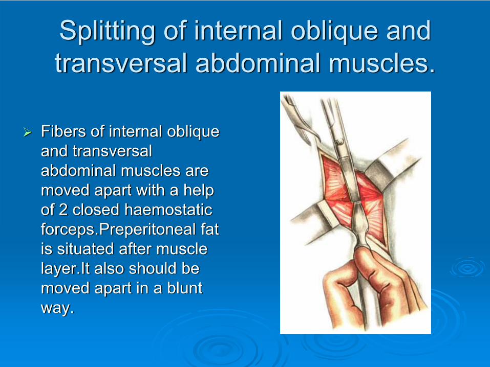
Splitting of internal oblique and transversal abdominal muscles.
Fibers of internal oblique and transversal abdominal muscles are moved apart with a help of 2 closed haemostatic forceps.Preperitoneal fat is situated after muscle layer.It also should be moved apart in a blunt way.

Surgical approach:Parietal peritoneum is picked up by 2 haemostatic forceps. Surgeon should check, that intestine is not under the forceps. After it, the peritoneum should be cut.
Gauze tissues are fixed to the brims of peritoneum by Mikulicz's clamps
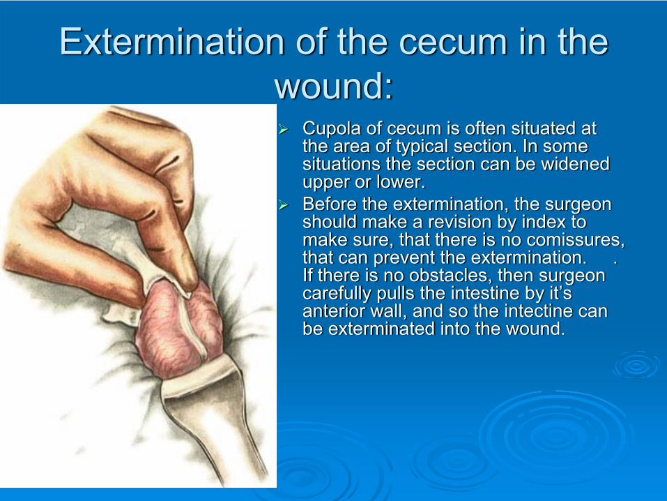
Extermination of the cecum in the wound:
Cupola of cecum is often situated at the area of typical section. In some situations the section can be widened upper or lower.Before the extermination, the surgeon should make a revision by index to make sure, that there is no comissures, that can prevent the extermination. . If there is no obstacles, then surgeon carefully pulls the intestine by it’s anterior wall, and so the intectine can be exterminated into the wound.

The extraction of appendix:
Appendix often comes into the wound after the cupola of cecum.Surgeon carefully takes the appendix by mouse-tooth forceps and pulls it from the abdominal cavity. In some cases, appendix can be pulled out by index. Friable unions should be carefully divided, dense unions should be cut between 2 forceps Extracted appendix is fixed by soft clamp, which should be placed on the mesentery near the top of appendix.
Methods of appendectomy:
- Antegrade- Retrograde

Antegrade:
Surgeon puts one clamp on the mesentery near the top of appendix and pierses the mesentery with another clamp near the base of appendix.Through this opening the mesentery should be compressed with a haemostatic clamp and tied up with a capron thread.After this, the mesentery can be cut.Surgeon puts a clamp near the base of appendix and removes it so
that on the wall of appendix forms a furrow. A catgut ligature is put in the area of this furrow.The next stage of operation is putting in a purse-string suture. A purse-string seromuscular suture is put at the distance near 1 cm from the base of appendix.
Surgeon puts a clamp apper the catgut ligature and cuts the appendix. The he dippes the stump of the appendix into with a clamp and tightens the purse-string suture
Seromuscular Z-shaped suture is put over the purse-string suture.
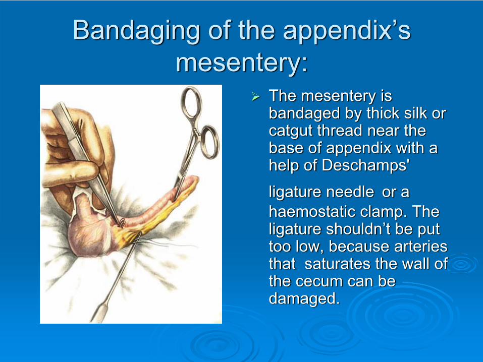
Bandaging of the appendix’s mesentery:
The mesentery is bandaged by thick silk or catgut thread near the base of appendix with a help of Deschamps' ligature needle or a haemostatic clamp. The ligature shouldn’t be put too low, because arteries that saturates the wall of the cecum can be damaged.

Putting in a purse-string suture:
A seromuscular purse-string suture is put on the cecum at the distance near 1-1,5 cm from the base of appendix

Bandaging of the appendix:
Surgeon puts 2 clamps near the base of appendix and removes one of them so that on the wall of appendix forms a furrow. A catgut ligature is put in the area of this furrow.

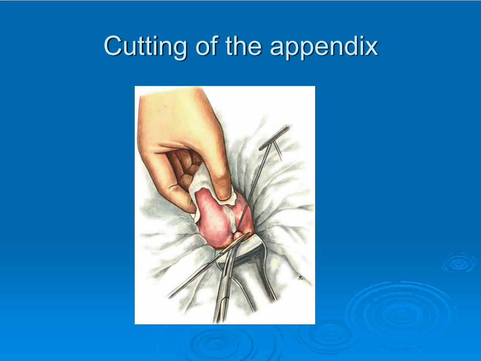
Cutting of the appendix:
Appendix is cut between the ligature and another clamp. The stump of appendix should be seared by iodine and dipped in the purse-string suture.

Dipping of the stump into the purse-string suture

Putting in a Z-shaped suture
Sometimes a seromuscular Z-shaped suture is put over the purse-string suture for more leaktightness

Retrograde:
Retrograde appendectomy takes place when appears some hardships in extermination of the appendix, for examle, retrocecal or retroperitoneal position of the appendix, comissural process in the abdominal cavity.In this case, first of all a catgut ligature is put through the opening in the mesentery. Appendix is cut under the clamp, it’s stump is dipped in the cecum and a purse-string and a Z-shaped sutures are put. And only after that a surgeon starts a gradual bandaging of mesentery.

Cross-clamping of appendix:
Surgeon puts a clamp near the base of appendix and removes it so that on the wall of appendix forms a furrow.

Bandaging of the appendix
A catgut ligature is put in the area of this furrow.
Cutting of the appendix

Dipping of the stump into the purse-string suture
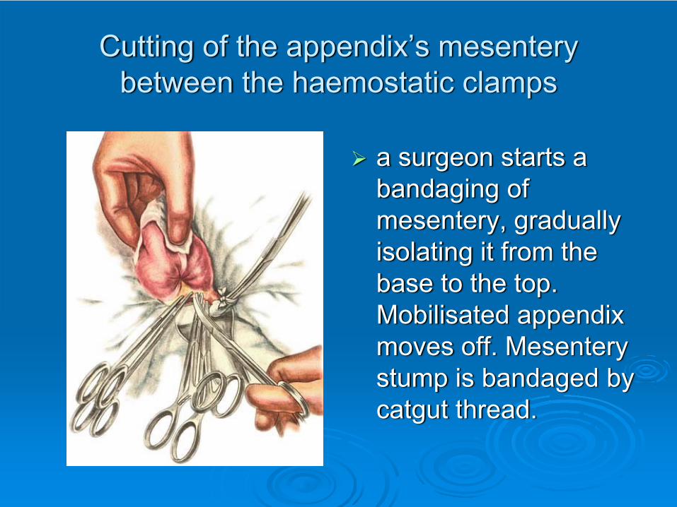
Cutting of the appendix’s mesentery between the haemostatic clamps
a surgeon starts a bandaging of mesentery, gradually isolating it from the base to the top. Mobilisated appendix moves off. Mesentery stump is bandaged by catgut thread.

Sewing and bandaging of the mesentery

Appendectomy. Retroperitoneal position of appendix
If there is no comissuras in the abdominal cavity and the appendix can not be found, then a surgeon should think about the retroperitoneal position of appendix. In this case appendix is situated behind the ascending colon and it’s top can reach the lower pole of kidney

The section line of parietal peritoneum:
Surgeon cuts the parietal peritoneum for a distance of 10-15 cm, stepping back on 1 cm outside from cecum and ascending colon.

Bringing of gauze handle under the base of appendix:
Cecum should be moved inside, founding the appendix/ It shoud be taken on the gauze handle near its’ base

Ligation of appendix vessels:

Cutting of the appendix:
Appendix is cut under the clamp
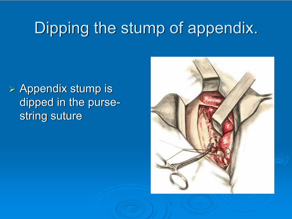
Dipping the stump of appendix.
Appendix stump is dipped in the purse-string suture
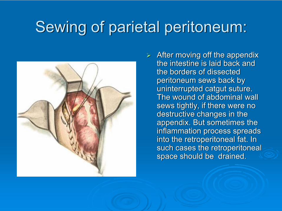
Sewing of parietal peritoneum:After moving off the appendix the intestine is laid back and the borders of dissected peritoneum sews back by uninterrupted catgut suture. The wound of abdominal wall sews tightly, if there were no destructive changes in the appendix. But sometimes the inflammation process spreads into the retroperitoneal fat. In such cases the retroperitoneal space should be drained.

The final stage:After moving out the appendix cecum moves back in the abdominal cavity. Surgeon should check that there is no bleeding from the mesentery and then the wound of the abdominal wall sews tightly in layers. Peritoneum sews by uninterrupted catgut suture, musles, aponeurosis and subcutaneous fat - by nodal catgut suture, skin – by nodal silk suture.In some cases abdominal cavity should be drained by thin rubber or polyvinyl chloride tube. Putting in a rubber tube is indicated in such cases, when there was purulent exudate in the abdominal cavity of phlegmonous changes of cecum.

Laparoscopic appendectomy:Nowadays,
laparoscopic appendectomy becomes very popular. This variant is considered to be less traumatical, but not always technically can be done. Even if the operation started from laparoscopic method, surgeon must always be ready to make the traditional appendectomy.

Laparoscopic appendectomy:

Possible aftereffects:
1. Bleeding2. Contamination of the wound3. Postoperative peritonitis4. Acute intestinal obstruction5. Pylephlebitis6. Abscesses of different locations7. Intestinal fistula

Thank you for your attention!