applications of particle accelerators in medical physics
TRANSCRIPT

CER
N-K
TT-2
013-
001
10/0
9/20
08
Applications of Particle Accelerators in Medical Physics.
G. Cuttone, Istituto Nazionale di Fisica Nucleare-Laboratori Nazionali del Sud
V. S. Sofia, 44 Catania Italy- [email protected]
1. INTRODUCTION
Particle accelerators are often associated to high energy or nuclear physics. As well pointed out in literature [1] if we kindly analyse the number of installation worldwide we can easily note that about 50% is mainly devoted to medical applications (radiotherapy, medical radioisotopes production, biomedical research). Particle accelerators are also playing an important indirect role considering the improvement of the technical features of medical diagnostic. In fact the use of radionuclide for advanced medical imaging is strongly increasing either in conventional radiography (CT and MRI) and also in nuclear medicine for Spect an PET imaging. In this paper role of particle accelerators for medical applications will be presented together with the main solutions applied.
2. THE ROLE OF PARTICLE ACCELERATORS COMBATING CANCER. At the beginning of the third millennium one European citizen out of three will have to deal with a cancer episode in the course of his/her life. Worldwide the estimated number of new cancer cases each year is expected to rise from 10 millions in 2000 to 15 millions by 2020. Cancer is currently the cause of 12% of all deaths worldwide. Within the European union it is over 1,5 million new cancer cases that are diagnosed every year and over 920000 people die of cancer with the two leading cause of cancers in Europe are Breast and Prostate. Therefore combating cancer is a major societal and economical issue for Europe and to face up these new challenges strong mobilisation among the scientific community and industrial manufacturers is needed. Today’s approaches to treat cancer are the surgical removal of the tumour tissue, radiotherapy, chemotherapy, and immunotherapy. Most scientists are confident that in the long run significant improvements in cancer cure will come from immunotherapy and/or gene therapy and drug targeting ; research towards such systemic treatments is and will be of the utmost importance. However, in the mean time, radiotherapy either combined with surgery or as main treatment modality still remains the most effective technique to treat cancer.
More than a half of all cancer patients are now treated by radiation therapy thanks to the technical progress made with irradiation equipment in the last ten years. For external radiation therapy (RT), for instance, high energy photon or electron beams are mainly produced by linear accelerators, while a very limited number of proton synchrotrons or cyclotrons are used for the treatment of cancers close to vulnerable organs such as the eyes and the optical and auditory nerves, spinal cords. For internal radiation therapy, brachytherapy, radioactive sources are put in the tumour with undeniable advantages for the patient in given situations. In an abstract from Annex A of "Europe Against Cancer" [2] in 1994, the present status of cured patients following a specific treatment shows that in Europe at present 45% of all the treated patients are "cured", which means that these patients have a symptom-free survival period exceeding five years. About 90% of the cured patients (i.e. 40% of the total) are cured because of loco-regional control of the primary tumour, i.e. because of surgery and radiotherapy. Of course, the treatments are almost always accompanied by chemotherapy to prevent the spreading of metastasis. In fact surgery and radiotherapy alone are successful in 22% and 12% of the cases respectively. When combined, they account for another 6% of the cases so that radiotherapy is involved in almost half of the curative treatments of loco-regional type. Despite a widespread belief, all the other systemic treatments account for 5% only of the cured patients. There is ample space for improvements here, because 37 % of the tumours are metastatised at the moment of diagnosis and cannot be cured with loco-regional treatments alone.
Three strategic approaches are generally proposed:
• Early detection and improved diagnosis based on widespread screening with the aim of reducing the number of late diagnoses.

• Improved local treatment, avoiding poor treatments, to treat tumours with difficult localisations and tumours which are radio-resistant to conventional radiotherapy.
• Improved systemic treatments combined with local treatments which are able to reduce the tumour mass significantly.
Improving loco-regional treatments is essential for at least two reasons. First, 18% of
all the patients die because of a primary tumour without metastases. This implies that the percentage of the cured patients could pass from 45% to about 65 %, if all primary tumours could be locally controlled. Secondly, in the long term “immunochemotherapy might be extremely useful in cancer treatment; it will, however, only be effective against a relatively small number of cells and such treatments will thus need to be used in conjunction with other anti-cancer modalities such as surgery, radiotherapy and chemotherapy".
For all these reasons it is generally approved that to increase the dose at the target or to get a better dose conformation to spare surrounding health tissue it is mandatory to implement new techniques like Conformal Radiotherapy, Intensity Modulation Radiotherapy (IMRT) or hadrontherapy (HT). These techniques together with stereotactic treatment (ST) or brachytherapy (BT) represent nowadays the new frontier of the radiotherapy daily implemented in the clinical reality of European Union (EU) countries.
The development of three-dimensional (3D) conformal radiotherapy, in which the high-dose volume matches the target volume and avoids radiosensitive normal tissues, has been a major theme for improving the physical basis of radiotherapy [3]. Intensity-modulated radiation therapy (IMRT) is the most advanced form of conformal radiotherapy. It is anticipated [4] that in the next ten years it will be routinely possible to automatically shape radiation fields, modulate the intensity such modulation under computer control, verify that radiotherapy is accurately delivered, predict the clinical outcome via biological models, and if not eliminate uncertainties, quantify them. It may become possible to customize radiotherapy to radio sensitivities of individual patients. Conformal radiotherapy may be broadly divided into two classes of techniques, i.e. those which employ field-shaping alone and those which also modulate the intensity of fluence across the geometrically-shaped field. The basic idea behind IMRT is that by sparing more volume of the organs at risk the dose to the planning treatment volume can be escalated and thus result in improved tumour control probability. The types of radiation used in IMRT may be photons, protons and light ions.
Protontherapy (PT) and heavier charged beam therapy (IT) shares with photon IMRT the characteristics of an enhanced precision in the dose delivery to the target. For these reason protontherapy scores, as photon IMRT, better clinical results than conventional radiotherapy but also faces the same challenges of achieving a higher precision in target localisation. Moreover also from the radiobiological point of view protons and especially heavy ions have demonstrated major effects giving possibility to change the fractioning scheme (ipofractioning) and the total dose.
An other challenge concerns brachytherapy where high quality treatment requires
development of new radiation delivery systems and sensors for checking the main

biological and dosimetric parameters of the patient. Short life radioactive substances are incorporated in the cancerous tumour allowing the patient to spend a quasi normal life with minor limitations. Brachytherapy, also known as Curie therapy is undergoing rapid changes in technology. In Europe, high dose rate (HDR) brachytherapy using after-loaders with Iridium sources is the most common technology. But, specially for the treatment of prostate cancer, low dose rate (LDR) brachytherapy using sealed seeds filled with local action radioisotope such as Iodine 125 or Palladium 103 are increasingly used. Innovative research have to be carried out in order to ensure a better quality and efficiency of treatments. This advanced brachytherapy (BT) treatment needs validation and new requirements for patient safety. On the other hand accelerator based facilty have to be developed in order to get reliable production of those radionuclides.
Although IMRT, external beam radiotherapy (photons, protons, hadrons) and advanced brachytherapy (BT) promise great benefits for optimised cancer treatment, a considerable number of technical restrictions and basic questions have to be resolved to realise the expected benefits.
In radiation oncology (IMRT, PT and BT), the main difficulty is to deliver both a precise and accurate large dose of radiation to destroy cancer cells in a diseased organ and a dose as low as possible to sound organs. Delivered doses to tumours being highly above the lethal level for a total body irradiation, thus doses have to be delivered with extreme precaution. In coming years, it will be necessary to find treatment protocols based on IMRT and advanced BT techniques. It is therefore crucial to be able to more accurately locate the tumour in treatment, to accurately determine the dose required and to fully spare organs that are vulnerable or present a high structural heterogeneity, in order to reduce the doses delivered to healthy organs with proper monitoring throughout the duration of the treatment.
Functional imaging by means of positron emission tomography (PET) or single photon emission computerized tomography (SPECT) can facilitate the evaluation of tumour physiology, metabolism and proliferation [5]. These are parameters determining outcome to radiotherapy treatment. PET can be used to get also quantitative information about the the in vivo distribution of positron-emitting radioisotopes (Fluorine-18, Carbon-11, Oxygen-15) that can be added or substituted into biological relevant compounds. SPECT may be used to image compounds labelled with indium-111, technetium-99m, and Iodine-123 in vivo. The application of these techniques represents a new frontier in combating cancer permitting not only the early diagnosis of tumour but also the biological target definition in radiotherapy like tumour metabolism, hypoxia, growth factor receptor expression, tumour apoptosis. These parameters can be used as predictors or early markers of response to radiotherapy, opening a new frontier of study and clinical application.
3. ACCELERATORS FOR MEDICAL APPLICATIONS: A SHORT HISTORY The development of particle accelerators started in the past century and was well
summarized by P.J Bryant [6]. It is mainly based on three acceleration mechanism: DC Acceleration Resonant Acceleration Betatron Mechanism
Following the first mechanism a wide set of electrostatic machines (Cockroft Walton
generator, Van Der Graaf generator single or double stage) have been developed. Their use for medical applications has been very limited.
Resonant acceleration proposed by Ising in 1924 is based on the use of alternating electric fields. He suggested to accelerate particles with a linear series of conducting drift tubes and in 1928 Wideroe demonstrates his validity building a 1 MhZ, 25 kV oscillator able to accelerate 50 keV potassium ions. He realized the first linear accelerator (LINAC) prototype. At that time the realization of a LINAC was really difficult so that we have to wait until 1930 for the realization of the first real particle accelerator based on this mechanism. In fact at that time Lawrence realized the first fixed frequency cyclotron accelerating 1.25 MeV proton beam. Cyclotron was limited in energy by relativistic effects so that we had to wait for increasing energy until 1944. At that time Veksler discovered the principle of phase stability and invented the synchrotron. LINAC, CYCLOTRON and SYNCHROTRON nowadays are particle accelerators used for medical applications.
Betatron mechanism originally discovered by Wideroe in 1923 and never published, found his proof in 1940 when Kerst re-invents the betatron and built the first working machine for 2.2 MeV electrons. Betatrons were used in radiotherapy for many years. Today they have been fully replaced by electron LINAC.
4. ACCELERATORS FOR RADIOTHERAPY In conventional radiotherapy, electron LINAC are mainly used. A 3 GHz accelerating
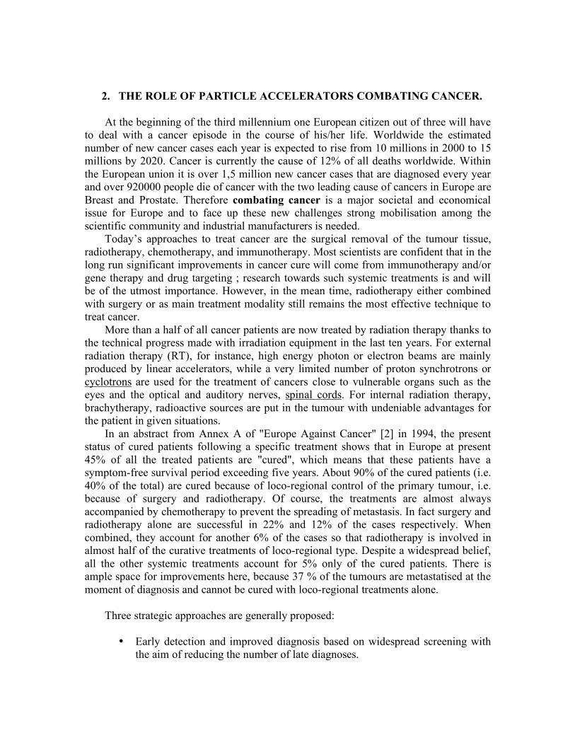
structure is used. The electrons are thermo-ionically emitted from a concave metal cathode at 1000 °C and accelerated in the gun to about ¼ the velocity of light by a pulsed DC electric field. Then they are formed into a pencil beam by a convergent electric filed between the gun electrodes. The RF electric field in the accelerating tube then forms the electron into bunches and accelerates them to more than 99% of light velocity, increasing their mass. The electrons are then used to bombard a target tipically done in tungsten. Electrons hitting the target will produce Bremstralung Radiation in the forward direction. X-rays so emitted are used for the therapy. Two sets of tungsten blocks allow the sizes of the radiation beam (square or rectangular sections) to be adjusted. All modern medical electron linacs employ an isocentric gantry. The accelerator wave guide is mounted in the gantry, either parallel to the gantry axis if a beam bending magnet is employed or perpendicular to the gantry axis if a beam bending magnet is not required. A simplified block diagram of a medical linear accelerator is represented in fig. 1. The new frontier of these machines is represented by multileaf collimator consisting of two of leaves (up to 120) which can be moved independently allowing the application of irregularly shaped beams better adapted to the complex shape of the target volume. Moreover they permit the application of special techniques like the intensity modulation (IMRT), dynamic therapy and radiosurgery.
New frontier in radiotherapy is nowadays represented by the use of hadrons. In fact heavy particles (protons, light ions, neutrons) offer a unique opportunity for improving the quality of radiation therapy. Their use has been pioneered over four decades at Berkley where there was a very active collaboration among nuclear physicist, accelerator
physicist, physicians and biologist. This is well proven looking to the history of the accelerators, especially of cyclotrons. E. Lawrence, the cyclotron father, started in thirties a collaboration with physicians and biologist in the use of neutrons for cancer treatment. The rationale base for the use of neutrons was not available at that time even if an expected high incidence of late morbidity was shown. Only in sixties biologist showed the role of the LET (Linear Energy Transfer) and the concept of radioresistance was developed. In 1946 R. Wilson mentioned that the properties of mono—energetic charged particles such as protons and Ions, i.e. the deposition of a large fraction of their kinetic energy in a small volume at the end of their energy (BRAGG Peak and distal dose fall-off), small lateral scattering, could lead to a new radiotherapic tool. The therapy with heavy particles is based on two factors:
-Balistic effect i.e. improved physical selectivity for charged particles, which means the delivery of a homogeneous dose to the tumour volume while minimizing the dose to surrounding healthy tissues.
-Radiobiological effect i.e. the improved biological effectiveness (RBE) of hadrons due to dense ionising tracks produced by these particles.
In fig.2 you can see the comparison of depth-dose distribution for various types of radiation used in radiotherapy. It is very clear and completely accepted that the advantages of charged particles (protons, carbon ions) with respect to photons and electrons are well summarized in the Bragg Peak and very reduced lateral scattering permitting the better dose conformation to the tumour sparing surrounding healthy tissues. RBE is assumed to be unitary for photons and electrons. It is well clear as biological effectiveness can be significantly higher for hadrons especially for carbon ions. According to that we can state that high-LET treatment is particularly useful for radioresistent tumours, insensitive to conventional. These statements represent the rationale that are convincing and forcing to develop hadrontherapy facility. These facilities are typically based on the use of Cyclotrons and Synchrotrons. In the PTCOG website [7]is reported a short list of these facilities. According to the already gained clinical experience main advantages can be carried for tumours close to organ at risk, like isolated brain metastases, pituitary adenomas, arteriovenous malformations, base of skull tumours, meningiomas, acoustic neuromas chordomas and chondrosarcomas, uveal melanomas, macular degeneration head and neck tumours, chest and abdomen, medically inoperable non-small-cell lung cancer, prostate, pediatric tumours (Brain, Orbital and ocular tumors, Sarcomas of the base of skull and spine, Acoustic neuromas).
Wide experience has been gained in the treatment of eye melanoma, considering the reduced proton energy required, (around 62 MeV, corresponding to a beam range in water of about 3 cm). In the following the experience gained at INFN-LNS will be detailed reported.
Beam produced by particle accelerators are not suitable for clinical applications if a dedicated beam delivery system is not available. Any beam delivery system must accomplish a three-dimensional scanning of the tumour requiring lateral beam deflection, variable range and exposure time to achieve a uniform dose. Moreover position sensitive monitors and fast beam switch-off in case of malfunction have to be available too. The lateral beam deflection should be obtained either by passive scattering [8] or by active methods using magnetic field deflecting the beam realizing a beam spot scanning . The

first method is implemented in fixed beam treatment line. At PSI a discrete spot scanning system based on the use of a kicker magnet located within a gantry has been developed and used for clinical application. The system is shown in fig.3. In order to reduce the dose to surrounding healthy tissues we can take advantage by using different multiple entrance beam port coming from different directions. A device performing this task is a rotating gantry: the beam comes in along the axis, is guided away from the centre and bent back towards the axis orthogonally. If the beam line ends at the axis so that the patient is located opn the axis, the gantry is called isocentric. Isocentric gantry are significantly different when applied at cyclotrons, characterized by a fixed extracted beam energy or at synchrotrons able to change the extracted beam energy. In fig. 3 the PSI Gantry is showed.
5. HADRONTHERAPY FACILITIES Different commercial solutions are now available. A 235 MeV proton beam room-
temperature cyclotron has been studied and developed by IBA in Belgium. This cyclotron with associated gantry has been installed at Massachusetts General Hospital in Boston (USA) going in clinical operation in 2002.
In 2001 PSI, Switzerland, announced officially to have chosen ACCEL Instruments GmbH as the supplier of a 250 MeV superconducting cyclotron. The cyclotron will be part of PSI´s proton therapy program PROSCAN. It includes the development of a dedicated proton source and beamlines to supply up to 250 MeV protons to the existing PSI Spot-Scanning-Gantry for deep-seated tumor treatment, a new industrialized Gantry with advanced technical features and a horizontal beam for treatment of eye tumours. ACCEL had proposed a very powerful proton accelerator based on a conceptual design elaborated earlier by Prof. Henry Blosser and his team at the National Superconducting Cyclotron Laboratory of Michigan State University. The 250 MeV cyclotron is designed for highest extraction efficiency, low energy consumption, high reliability and most modern operation features, e.g. spot scanning and beam intensity modulation. It uses superconducting main coils allowing for moderate HV and RF power levels. The design nevertheless provides full accessibility to the cyclotron parts relevant for operation and maintenance. The cyclotron will be operable as a turn key system in mid 2004.
In Italy an hadrontherapy facility is foreseen, the ‘Centro Nazionale di Adroterapia’(CNA). It is a hospital based facility dedicated to advanced tumour treatments with hadron beams and to advanced research (clinical, radiobiological and technical) made possible by their availability. The main requirements, considered during the CNA design, and the solutions adopted fully satisfy the following main criteria: safety, reliability, maintainability and availability. These criteria have driven the technological choices in so many respects that it is not possible to enumerate all of them in this short introduction, but they will be the core arguments of the authorisation documents that will be presented to obtain the permissions to run the centre and to treat patients. The CNA design (a preliminary view is shown in Fig. 4) is based on the following assumptions:

• the Centre will be devoted to the treatment of deep-seated tumours (up to a depth of 27 cm of water equivalent) with light ion beams (mainly C6+, but also protons and possibly He2+, Li3+, Be4+, B5+ and O8+, but this last with a reduced range). All available light ions will be delivered in each treatment room with the exception of the room foreseen to be equipped, in a second stage, with a gantry for protons;
• the full-size CNA will have 5 treatment rooms: 3 rooms with fixed beams and 2 rooms with gantries. To reduce the initial investment it is proposed to first equip 3 treatment rooms with 4 fixed beams, three horizontal and one vertical beams (CNA - Phase 1). At a later stage (CNA - Phase 2), two other rooms will be equipped with gantries: one gantry for protons and one gantry for light ions.
The eye melanoma treatment with proton beam represents the more significative clinical experience. More than 7000 patients have been successfully treated in different facilities. More significative experience has been gained in Europe at PSI, CCO and Nice, Orsay and Berlin while in USA MGH and Loma Linda are the main centers [9]. Also in Italy is now available a protontherapy center. In fact at the INFN Laboratori Nazionali del Sud in Catania (Italy) the first Italian protontherapy facility, named CATANA (Centro di AdroTerapia e Applicazioni Nucleari Avanzate) has been realized in collaboration with the University of Catania. It is based on the use of the 62 MeV proton beam delivered by the K=800 Superconducting Cylotron installed and working at LNS since 1995. The facility is mainly devoted for the treatment of ocular diseases like uveal melanoma. A beam treatment line in air has been realized together wth a dedicated positioning patient system. The facility is in operation since the beginning of 2002 and 40 patients have been successfully treated up to now. The CATANA proton beam line has been entirely realized at LNS and its global view is shown in Fig.5. The proton beam exits in air through 50 µm Kapton window placed at about 3 meters from isocenter. Before the window, under vacuum, is placed the first scattering foil made by a 15 µm tantalum. The first element of the beam in air is a second tantalum foil 25 µm thick provided with a central brass stopper of 4 mm in diameter The double foils scattering system is optimized to obtain a good homogeneity in terms of lateral dose distribution, minimizing the energy loose. Range shifter and range modulator are placed downstream the scattering system and mounted on two different boxes. Two diode lasers, placed orthogonally, provide a system for the isocenter identification and for patient centering during the treatment. The emission light of a third laser is spread out to obtain the simulation field.
A key element of the treatment line is represented by the two transmission monitor chambers and by the four sector chamber, implemented to have an on-line control of the dose furnished to the patients and an information on beam symmetry respectively. The last element before isocenter is a patient collimator located at 8 cm upstream of the isocenter. Finally two, back and lateral, Philips Practics X-Rays tubes are mounted for the verification of the treatment fields.
Inside CATANA collaboration particular care is going to be devoted to the development of dosimetric techniques for the determination of absorbed dose in clinical proton beams and 2D and 3D dose distribution reconstruction. A parallel-plate calibrated Markus ionization chamber has been chosen as reference detector for the absolute dose measurement, while gaf chromic and radiographic films, TLD (ThermoLuminescent Detectors), natural diamond and silicon detectors are the choices for the relative one.

Depth dose curves and transverse dose distributions, either for the full energy and modulated proton beams, are acquired with a water-tank system provided of three fully computer-controlled step motors. This system, entirely developed at Laboratori Nazionali del Sud, is controlled by a software providing the acquisition and dosimetric analysis of data. Figure 6 shows a depth dose distribution peak in water obtained with the water-tank system and Markus chamber for an unmodulated 25 mm diameter beam at the energy of 62 AMeV.
The Full Width at Half Maximum of the Bragg Peak is 2.76 mm while the 90-10 % and 80-20% distal fall-off are 0.8 mm and 0.6 mm respectively. The entrance to peak ratio is 4.72. We have realized, in collaboration with the Clatterbridge Center for Oncology (UK), a set of wheel modulators to obtain a spread out therapeutic Bragg peak. To do this various Bragg peaks, for different proton beam energies ranging from 62 AMeV to 10 AMeV, were acquired with the Markus chamber in the water phantom. Proton beam energy lower than 62 AMeV are obtained inserting PMMA range shifters of different thickness along the beam path. Finally Figure 7 shows the Spread Out Bragg Peak obtained with the first prototype of the modulator wheel. Actually a first set of modulator wheels (10, 12, 15, 20, 25 mm SOBP), developed for therapeutic purposes, are available. Even if CATANA should not be the clinical answer for all the Italian patients affected by this kind of disease it represents the first successfully Italian example of the collaboration between Nuclear and Medical Physicist together with Medical Doctors in fighting tumours with hadrons. CATANA is the first milestone in Italy trough the extensive use of hadrontherapy in cancer treatment.
Following this experience recently at LNS a new design of superconducting cyclotron for medical applications has been proposed.. [12]. The design of the machine model has been done to accelerate H21+, He2+,B5+, C6+up to 250 MeV/amu, corresponding to a maximum average field at extraction radius of 3.8 tesla. The accelerators will be the core of a new proposed hadrontherapy center that should be realized in Catania in the next years, mainly devoted to the treatment of deep sited tumours with proton beams and to clinical studies of some particular tumoural form with carbon ions.
6. ACCELERATOR BASED FACILITY FOR RADIOISOTOPES PRODUCTION.
Both Radioisotopes and enriched stable isotopes are essential to a wide variety of applications in medicine, where they are used in the diagnosis and treatment of illness. The radioisotopes produced for medical applications are tipically used in nuclear medicine for diagnosis providing dynamic and functional information to study the organ functions. The labelled biomolecules and radioisotopes involved should have as ideal features the absence of beta particle emission, a half-life as short as possible and gamma energy emission in a range between 100 and 300 keV. For a long period the production of radioactive isotopes for medical applications was mainly based on neutron induced nuclear reactions. This was essentially done in nuclear reactors but their availability is slowly decreasing so that the accelerators based production facility are growing up. It is well known and proven that Short life radioisotopes (Fluorine-18, carbon-11, Nitrogen-13

and oxygen-15) are tipically accelerator produced radionuclides. Also others Radionuclides (gallium-67, Indium-111, iodine-123, thallium-201) are produced by means of 30 MeV cyclotrons for loco-regional distribution. According to those a wide market of turn-key accelerators is nowadays available. The main choice in this field is represented by cyclotrons. Infact looking to the time structure of the extracted beam, cyclotrons represent the golden standard with respect to linacs. It si possible to state that the power deposition in a production target is significantly better using a cyclotron which it is characterized by a beam time structure that can be assumed as a “ practically continuous beam”. Linacs are characterized by a low repetition rate so that the instantaneous power deposition in the target so high that force to implement very complex solution expecially in the material choice for thermal dissipation. Even if it is possible to state that with a 30 MeV should be possible to produce many radioisotopes of interest for medical application, it is growing the demand of production of radionuclides requiring a primary proton energy at energy higher then 30 MeV like Magnesium-28, iron-52, germanium-68, strontium-82, xenon-122/127, astanium-201. These demands are also forcing to try to use already existing high intensity nuclear facilities that should be partially involved in this task. Modern isotope production facilities consists of a compact H- cyclotron in the nergy range between 10 and 30 MeV with extracted current up to 400 microA and highly sophisticated target technology and chemistry. Target can be made solid, gas or liquid. In any case they have to be designed in order to dissipate up to 10 kW. A temperature rise of 150 °C is well accepted. The choice of material is dependent on the particular nuclide process. Although a general rule does not exist, there are some aspects of target body like activation, contamination, corrosion and cooling, have to be kindly considered. Target can be either internal and external, placed at the end of dedicated beam transport lines. In tab. 1 the most common cyclotron produced radioisotopes for medical diagnostics are reported. Different commercial solutions are available. IBA, General Electric, CTI, EBCO and SUMITOMO are the main cyclotron producers. They also offer dedicated radiochemistry modules for the production of the labelled molecula to be used. A typical production plant is reported in fig. 8 7. CONCLUSIONS The role of particle accelerators is rising for medical applications. The synergy between medicine (radiotherapy, radiology, nuclear medicine, oncology) and physics (nuclear and accelerator physics) is growing even more and will represent the future challenge to get a better quality of life in the third millennium. The number of potential patients for hadrontherapy have been determined in many studies with different results [9,10,11]. It is possible to state that the percentage of patients getting advantage with respect to conventional radiotherapy is of the order of 30%. Moreover the exploitation of accelerator based facility for the radioisotope production should significantly rise the medical imaging performance permitting an early stage diagnosis of many important diseases, first of all tumours. In conclusion we want to stress again as many advantage can be carried out using particle accelerators for medical applications.

REFERENCE [1]U. Amaldi, Nuclear Physics A654 (1999)375C-399C [2]A.J.M. Vrmoken and F.A.T.M. Schermer (eds), Amsterdam, Oxford, Washnghton DC, Tokyo: IOS press, (1994) [3]S. Webb, IMRT, IOP 2001 [4]S. Webb, The physics of conformal radiotherapy, IOP 2001 [5]C. Van de Wiele, Int. J. Radiation Oncology Biol. Phys, Vol. 55 No1, pp5-15, 2003 [6]CAS Cern Accelerator school: Cyclotrons, linacs and their applications, CERN 96-02 [7] www.ptcog.web.psi.ch [8]B. Gottshalck et al., Harvard Cyclotron Laboratory, report 3/29/89 (1989) [9]Advances in Hadrontherapy, U. Amaldi, B. Larsson and Y. Lemoigne eds, Excerpta Medica, 1996 [10]G. Gademan, ref [10], p.59 [11]R. Orecchia et al. Eur. J. Cancer (1998), p.459 [12] L. Calabretta, XXXV PTCOG Abstract, 2002

Tab. 1. List of some radioisotope tipically used for medical applications
Radioisot.
Nuclear Reaction
Proton Energy (MeV)
Decay Product
T1/2 Emission
Application
15O
15N(p,n)
15O
10→0
14N
2.03min
β+
PET
13N
13C(p,n)
13N
10→0
13C
9.96 min
β+
PET
11C
11B(p,n)
11C
10→0
11B
20.38 min
β+
PET
18F
18O(p,n)
18F
16→3
18O
109.8 min
β+
PET
111In
112Cd(p,2n)1
11In
22
111Cd
68 h
EC
Radioimmun
123I
124Xe(p,2n)1
23Cs→ 123Xe→123I
30
123Te
13.2 h
EC
Brain Spect
67Cu
68Zn(p,2p)
67Cu
40
67Zn
61.9 h
β-
Radioimmun
122Xe
127I(p,6n) 122Xe
70
122I
20.1 h
EC
Father positron emitter 122I
68Ge
natGe(p,pxn) 68Ge
70
68Ga
271 d
EC
PET and tumoral marker
Fig
Fig. 1 Schematic view of a conventional LINAC (Courtesy by VARIAN)
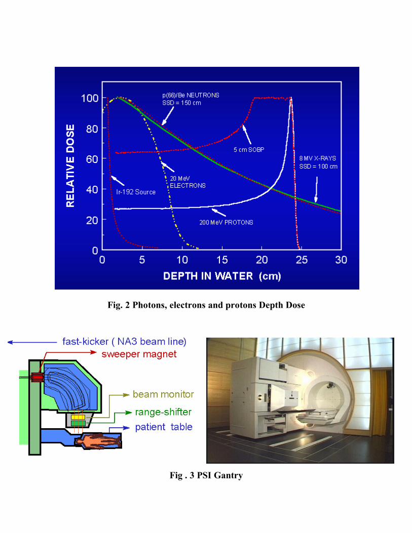
Fig. 2 Photons, electrons and protons Depth Dose
Fig . 3 PSI Gantry

Fig. 4 – CNA: preliminary design of the CNA – Phase 1. The synchrotron and the lines that serve three treatment rooms are evidenced.
Figure 5. View of the CATANA beam line: 1.Treatment chair for patient immobilization, 2. Final collimator, 3.Positioning laser, 4. Light field simulator; 5. A Monitor chamber; 6. Intermediate collimator, 7-8.Boxes for the location of modulator wheel and range shifter, 9.Proton beam output window.

0
0.5
1
1.5
2
2.5
3
3.5
4
4.5
5
0 5 10 15 20 25 30 35
Depth in water [mm]
Ion
izati
on
[a.u
.]
Figure 6. Bragg peak of 62 AMeV proton Beam acquired with a water-tank system and a Markus chamber ionization chamber at CATANA
facility
0
10
20
30
40
50
60
70
80
90
100
110
0 5 10 15 20 25 30 35
Depth in water [mm]
Do
se n
orm
alize
d a
t m
axim
um
valu
e
[a.u
.]
Figure 7 Spread Out Bragg Peak obtained with a modulator wheel;
Data acquired in water
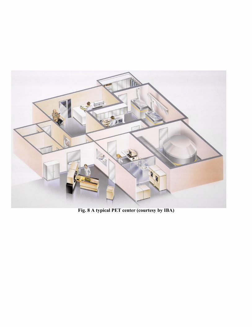
Fig. 8 A typical PET center (courtesy by IBA)