arabidopsis wee1 kinase controls cell cycle … wee1 kinase controls cell cycle arrest in response...
TRANSCRIPT
Arabidopsis WEE1 Kinase Controls Cell Cycle Arrest inResponse to Activation of the DNA Integrity Checkpoint W OA
Kristof De Schutter,1 Jerome Joubes,1,2 Toon Cools, Aurine Verkest, Florence Corellou,3 Elena Babiychuk,Els Van Der Schueren, Tom Beeckman, Sergeı Kushnir, Dirk Inze, and Lieven De Veylder 4
Department of Plant Systems Biology, Flanders Interuniversity Institute for Biotechnology, Ghent University,
B-9052 Gent, Belgium
Upon the incidence of DNA stress, the ataxia telangiectasia–mutated (ATM) and Rad3-related (ATR) signaling kinases
activate a transient cell cycle arrest that allows cells to repair DNA before proceeding into mitosis. Although the ATM-ATR
pathway is highly conserved over species, the mechanisms by which plant cells stop their cell cycle in response to the loss
of genome integrity are unclear. We demonstrate that the cell cycle regulatory WEE1 kinase gene of Arabidopsis thaliana is
transcriptionally activated upon the cessation of DNA replication or DNA damage in an ATR- or ATM-dependent manner,
respectively. In accordance with a role for WEE1 in DNA stress signaling, WEE1-deficient plants showed no obvious cell
division or endoreduplication phenotype when grown under nonstress conditions but were hypersensitive to agents that
impair DNA replication. Induced WEE1 expression inhibited plant growth by arresting dividing cells in the G2-phase of the
cell cycle. We conclude that the plant WEE1 gene is not rate-limiting for cycle progression under normal growth conditions
but is a critical target of the ATR-ATM signaling cascades that inhibit the cell cycle upon activation of the DNA integrity
checkpoints, coupling mitosis to DNA repair in cells that suffer DNA damage.
INTRODUCTION
Genome integrity of cells is threatened by DNA damage that is
the consequence of environmental stresses and endogenous
causes. To cope with these stress conditions, cells have devel-
oped a set of surveillance mechanisms to monitor the status and
structure of DNA during cell cycle progression. In Schizosac-
charomyces pombe (fission yeast) and mammals, DNA damage
activates the ataxia telangiectasia–mutated (ATM) and Rad3-
related (ATR) signaling cascades that simultaneously turn on
DNA repair complexes and arrest cell division; this mechanism
allows cells to repair damaged DNA before proceeding into
mitosis (Zhou and Elledge, 2000; Abraham, 2001; Bartek and
Lukas, 2001; Kurz and Lees-Miller, 2004). ATM responds spe-
cifically to double-stranded breaks, whereas ATR primarily
senses replication stress caused by a persistent block of repli-
cation fork progression. The ATM and ATR kinases transduce the
DNA stress signal to the checkpoint kinases CHK1 and CHK2,
which, in turn, arrest the cell cycle by directly modulating the
activity of the effectors that control cell cycle progression (Chen
and Sanchez, 2004; Sancar et al., 2004), the cyclin-dependent
kinase (CDK) complexes.
CDK complexes consist of a catalytic kinase subunit and a
regulatory cyclin. The sequential activation of different CDK/
cyclin complexes drives the cell cycle through the phosphoryl-
ation of many different target substrates. CDK/cyclin activity is
highly regulated at multiple levels. Control mechanisms include
the regulated synthesis and destruction of the cyclin subunits
(Peters, 1998; Murray, 2004), which are thought to target the
CDKs to the substrates (Ohi and Gould, 1999), and the associ-
ation of CDKs with inhibitory proteins and docking factors (Lees,
1995). Moreover, CDK activity is positively regulated by phos-
phorylation of a conserved residue (Thr-161 or equivalent) within
the T loop and negatively regulated through phosphorylation of
Tyr-15 and Thr-14 by WEE1 family kinases (Berry and Gould,
1996). Phosphorylation of Tyr-15 and Thr-14 residues of the CDK
subunit inhibits ATP binding and blocks substrate recognition.
In fission yeast and mammals, rapid activation of the CDK/
cyclin activity at the G2-M boundary is mediated by a dual-
specificity phosphatase CDC25. Maintenance of the inhibition of
CDK activity by Tyr-15 phosphorylation is the ultimate target of
DNA damage checkpoint signaling. By activation of CHK1 and
CHK2, CDC25 is phosphorylated and targeted for ubiquitin-
dependent destruction or association with a 14-3-3 protein,
resulting in nuclear export and exclusion of CDC25 from the
nuclear pool of CDK/cyclin complexes (Boutros et al., 2006).
Both WEE1 and the functionally related kinase MIK1 have been
implicated as targets of the DNA damage and replication check-
points as well. In Xenopus laevis (African frog) egg extracts,
1 These authors contributed equally to this work.2 Current address: Laboratoire de Biogenese Membranaire, CentreNational de la Recherche Scientifique, Unite Mixte de Recherche 5200,Universite Victor Segalen Bordeaux 2, 146, rue Leo Saignat, F-33076Bordeaux Cedex, France.3 Current address: Laboratoire Arago, Centre National de la RechercheScientifique Paris VI, Unite Mixte de Recherche 7628, BP 44, F-66651Banyuls sur Mer Cedex, France.4 To whom correspondence should be addressed. E-mail [email protected]; fax 32-9-3313809.The author responsible for distribution of materials integral to thefindings presented in this article in accordance with the policy describedin the Instructions for Authors (www.plantcell.org) is: Lieven De Veylder([email protected]).W Online version contains Web-only data.OA Open Access articles can be viewed online without a subscription.www.plantcell.org/cgi/doi/10.1105/tpc.106.045047
The Plant Cell, Vol. 19: 211–225, January 2007, www.plantcell.org ª 2007 American Society of Plant Biologists
activation of the DNA replication checkpoint stabilizes exoge-
nously added WEE1 (Michael and Newport, 1998), whereas in
fission yeast, MIK1 is a target for both the DNA damage and DNA
replication checkpoints (Rhind and Russell, 2001). In response to
the DNA replication checkpoint, MIK1 mRNA levels accumulate
to high levels and, simultaneously, the MIK1 protein is stabilized,
leading to dramatic increases in protein levels (Boddy et al.,
1998; Baber-Furnari et al., 2000; Christensen et al., 2000).
The basic machinery that controls cell cycle progression in
plants is similar to that of yeast and mammals (De Veylder et al.,
2003; Dewitte and Murray, 2003; Inze and De Veylder, 2006).
Multiple CDKs and cyclins are encoded by the genomes of
Arabidopsis thaliana and Oryza sativa (rice) (Vandepoele et al.,
2002; Wang et al., 2004; La et al., 2006). In addition, a WEE1-
related kinase has been described for maize (Zea mays), tomato
(Solanum lycopersicum), and Arabidopsis (Sun et al., 1999;
Sorrell et al., 2002; Gonzalez et al., 2004). Although the plant
WEE1 gene is unable to complement mutations in its yeast
homolog, its overexpression inhibits cell division in fission yeast.
Additionally, recombinant purified WEE1 protein from maize is
capable of inhibiting the kinase activity of biochemically purified
CDKs (Sun et al., 1999). However, the in vivo role of WEE1 in plant
cell cycle progression and growth is not well defined.
Our first insights into the role of DNA replication and damage
checkpoints in plants came with the identification and charac-
terization of Arabidopsis mutants in genes encoding orthologous
ATM and ATR kinases (Garcia et al., 2003; Culligan et al., 2004). A
defective DNA damage checkpoint is the reason why ATM-
deficient plants are primarily hypersensitive to DNA-damaging
agents, such as g-irradiation, but rather insensitive to replication-
blocking agents, such as hydroxyurea or aphidicolin (Garcia et al.,
2003). In contrast, ATR mutants are hypersensitive to replication-
blocking agents but also mildly sensitive toward g-irradiation
(Culligan et al., 2004). These results strongly indicate that the DNA
checkpoint signaling pathways are conserved in plants. How-
ever, it is still unclear how activation of these signaling cascades
leads to the arrest of the cell cycle in response to DNA damage.
Here, we identify WEE1 as an important target of the DNA re-
plication and DNA damage checkpoints. WEE1-deficient plants
grow normally under optimal growth conditions but are hyper-
sensitive to DNA-damaging agents. In accordance with a role for
WEE1 in arresting the cell cycle in response to replication stress,
WEE1 transcripts are found to be strongly upregulated by
replication-inhibiting drugs in an ATR-dependent manner. Anal-
ogously, g-irradiation and radiomimetic drugs induce WEE1
transcription in an ATM-dependent manner. The cell cycle arrest
observed upon induction of WEE1 expression indicates that WEE1
is part of the mechanism that couples the onset of mitosis with
the completion of DNA repair in cells that have suffered DNA
damage.
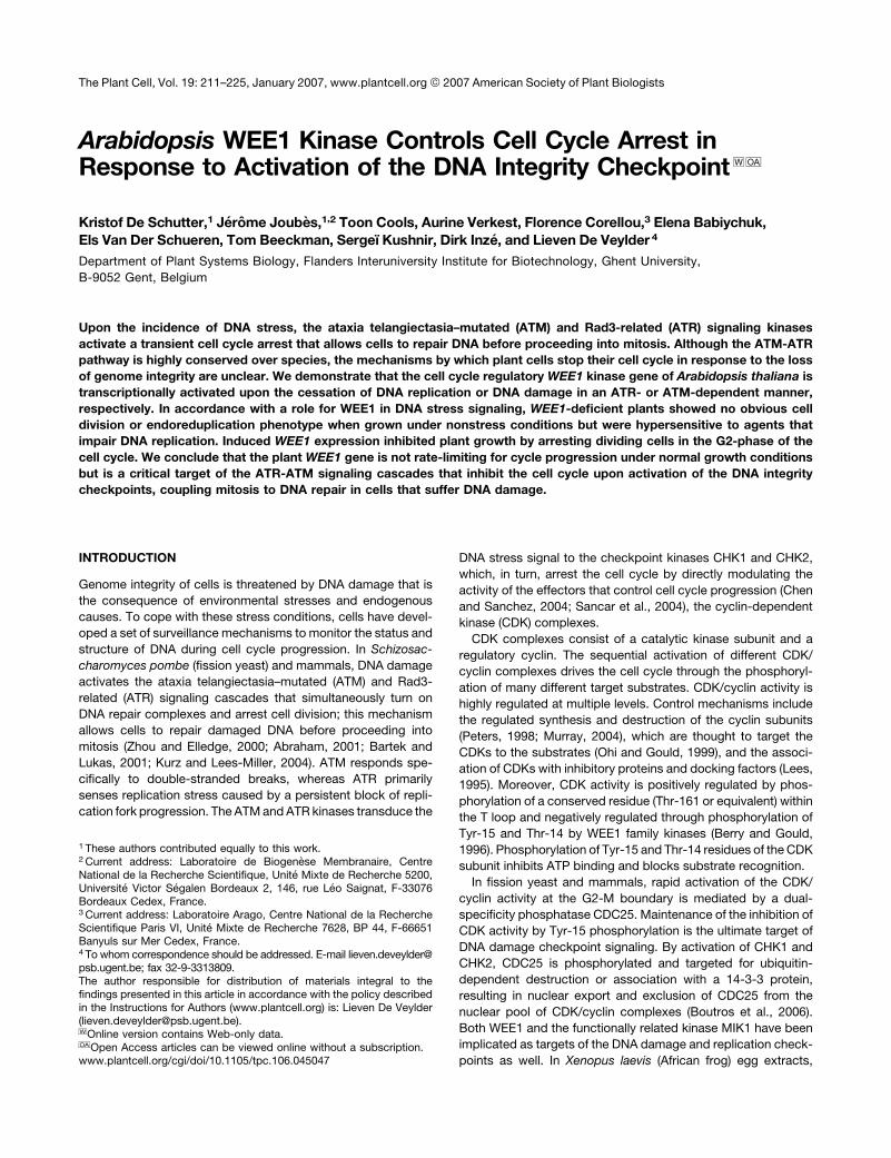
Figure 1. CDKA;1 Target for Tyr Phosphorylation and Binding WEE1.
(A) CDK phosphorylation in response to checkpoint activation. Arabidopsis cell cultures were treated with 10 mg/mL aphidicolin (A), with 3 mM
propyzamide (P), or mock-treated in controls (C). CDKs were purified from total protein extracts (300 mg/sample) with a p10CKS1At-Sepharose matrix,
resolved by SDS-PAGE, and immunoblotted with the indicated antisera.
(B) Interaction of CDKA;1 with WEE1 in the yeast two-hybrid system. Yeast PJ69-4 cells containing a CDKA;1 or CDKB1;1 bait plasmid in combination
with an empty control or WEE1 prey plasmid were spotted on plates with (þ) or without (�) His. Only when the two proteins interact do cells grow on
�His medium.
212 The Plant Cell
RESULTS
Tyr Phosphorylation of Arabidopsis CDKA;1 upon
Activation of the DNA Replication Checkpoint
Tyr phosphorylation of CDKs has been shown to take place upon
cytokinin deprivation in tobacco (Nicotiana tabacum) cells, appli-
cation of water stress to wheat (Triticum aestivum) leaves, and
stimulation of the DNA replication checkpoint in zygotes of the
brown alga Fucus (Zhang et al., 1996; Schuppler et al., 1998;
Corellou et al., 2000). To test whether Tyr phosphorylation occurs
upon the activation of the DNA replication checkpoint in higher
plants, cultured suspension cells of Arabidopsis were treated with
aphidicolin, which inhibits all replicative DNA polymerases. Pro-
pyzamide was used to block cell cycle progression into mitosis by
depolymerizing the mitotic spindle (Planchais et al., 2000). The
efficiency of the drugs to stop cell cycle progression was con-
firmed byflow cytometric analysis (data notshown).After the drugs
had been applied for 24 h, the CDK complexes were purified and
analyzed on protein gel blots with specific antibodies against
CDKA;1andCDKB1;1 (Hemerlyetal., 1995;Porcedduetal., 2001).
CDKA;1 belongs to the archetypical CDKs, characterized by the
presence of a PSTAIRE amino acid sequence motif in the cyclin
binding protein domain, and CDKB1;1 belongs to the group of
plant-specific CDKs (De Veylder et al., 2003; Boudolf et al., 2004a,
2004b). Neither drug treatment had an effect on the abundance of
CDKA;1 protein compared with that of control cells (Figure 1A). In
contrast, CDKB1;1 levels increased slightly in the propyzamide-
treated cells, probably because of the preferential expression of
the CDKB1;1 geneduring M-phase (Porceddu et al., 2001; Boudolf
et al., 2004b). Next, the protein blots were probed with an anti-
phosphotyrosine antibody. Whereas no antibody binding was
detected in protein samples from control or propyzamide-treated
cells (Figure 1A), a polypeptide band with the same electrophoretic
mobility as that of CDKA;1 cross-reacted with the antibody in
extracts prepared from the aphidicolin-treated cells. This analysis
strongly indicates that CDKA;1 is the target for Tyr phosphorylation
upon activation of the DNA replication checkpoint.
To analyze whether the Arabidopsis WEE1 kinase might be
responsible for the observed Tyr phosphorylation of CDKA;1,
both proteins were tested for their interaction using the yeast two-
hybrid system. CDKA;1 and CDKB1;1 in fusion with the GAL4
DNA binding domain were cotransformed in an appropriate yeast
reporter strain with an empty control vector or a vector encoding
a fusion protein between the GAL4 transactivation domain and
WEE1. Transformants were streaked on medium with or without
His. Cells expressing CDKA;1 and WEE1 grew in the absence of
His, indicating that both gene products interacted. No associa-
tion was observed between CDKB1;1 and WEE1 (Figure 1B).
WEE1 Gene Expression Is Induced in Response
to Activation of the DNA Replication Checkpoint
in Cultured Arabidopsis Cells and Seedlings
The observed phosphorylation of CDKA;1 upon DNA replication
blockage and its association with WEE1 suggested an involve-
ment of the WEE1 kinase in the checkpoint pathway. Therefore,
we analyzed the transcriptional response of the WEE1 gene in
cell suspensions treated with either the ribonucleotide reductase
inhibitor hydroxyurea (HU) or aphidicolin (Figures 2A and 2B).
Drugs were added to exponentially growing cells and samples
were collected at different time points for the next 20 h, after which
WEE1 and CDKA;1 transcript levels were analyzed by semiquan-
titative RT-PCR. The actin 2 (ACT2) gene was used as a loading
control. CDKA;1 transcript levels remained relatively constant
upon HU treatment or decreased slightly after aphidicolin addition.
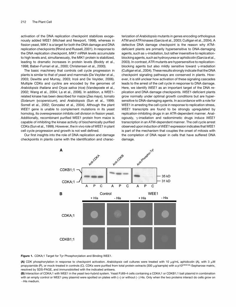
Figure 2. Transcriptional Response of the WEE1 Gene after Activation of
the DNA Replication Checkpoint.
(A) and (B) Transcript levels of WEE1 and CDKA;1 in Arabidopsis cells
treated with 40 mM HU (A) and 10 mL/mL aphidicolin (B). Samples were
harvested at the indicated time points after addition of the drugs. Gene
expression was analyzed by semiquantitative RT-PCR. The ACT2 gene
was used as a loading control.
(C) and (D) Transgenic Arabidopsis roots harboring the WEE1 promoter
fused to the GUS gene grown in the absence (C) or presence (D) of
10 mM HU. Plants were stained for GUS activity 20 h after drug applica-
tion. Both images are at the same magnification. Bar ¼ 50 mm.
WEE1 Checkpoint Control 213
By contrast, WEE1 transcript levels increased dramatically within
4 h after drug treatment, reaching maximum levels at 6 and 10 h
after addition of HU and aphidicolin, respectively, indicating that
replication inhibition transcriptionally activated the WEE1 gene.
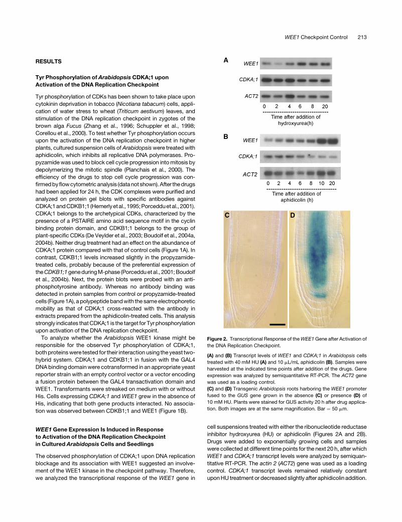
To investigate the transcriptional induction of the WEE1 gene
in response to DNA replication stress at the tissue level, trans-
genic Arabidopsis lines were generated that expressed the
b-glucuronidase (GUS) reporter gene under the control of the
WEE1 promoter. A reproducible expression pattern was found in
three independent reporter lines. Under standard growth condi-
tions, promoter activity was detected locally in the shoot apex
of seedlings as well as in the vasculature of the cotyledons and
roots (Figures 3A and 3B). Also in older seedlings, expression
was confined to apex and vascular tissues of roots and leaves
(Figure 3C). Surprisingly, only occasionally, a faint GUS signal
was detected in the apex of both main and lateral roots (Figures
3D and 3E). By contrast, GUS staining was strong in developing
flowers, particularly in the anthers and gynoecia (Figure 3F), but
not in mature flowers (Figure 3G).
To characterize the transcriptional induction of the WEE1 gene
in response to HU treatment, seeds of plants harboring the
WEE1:GUS reporter were germinated on control medium. After
2 weeks, seedlings were transferred onto fresh control medium
or a medium supplemented with HU. After 20 h, plants were
harvested and assayed for GUS activity. No GUS staining was
observed in the primary and lateral root meristems of plants
transferred to the control medium (Figure 2C). In contrast, roots
of the plants treated with HU for 20 h showed strong GUS
staining in the root apical meristem, mostly confined to the cells
of the central cylinder (Figure 2D). Similarly, WEE1:GUS reporter
expression in the shoot apical meristem and vascular tissues
was clearly induced by the HU treatment (data not shown). From
these observations, we conclude that transcriptional control
seems to play a major role in the regulation of WEE1 kinase
activity during DNA replication stress.
WEE1 Activity Is Not Required for Cell Division
or Endoreduplication
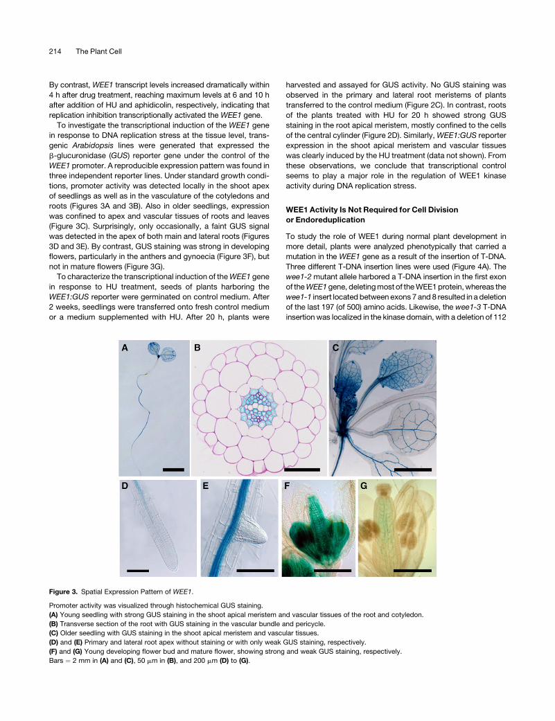
To study the role of WEE1 during normal plant development in
more detail, plants were analyzed phenotypically that carried a
mutation in the WEE1 gene as a result of the insertion of T-DNA.
Three different T-DNA insertion lines were used (Figure 4A). The
wee1-2 mutant allele harbored a T-DNA insertion in the first exon
of the WEE1 gene, deleting most of the WEE1 protein, whereas the
wee1-1 insert located between exons 7 and 8 resulted in a deletion
of the last 197 (of 500) amino acids. Likewise, the wee1-3 T-DNA
insertion was localized in the kinase domain, with a deletion of 112
Figure 3. Spatial Expression Pattern of WEE1.
Promoter activity was visualized through histochemical GUS staining.
(A) Young seedling with strong GUS staining in the shoot apical meristem and vascular tissues of the root and cotyledon.
(B) Transverse section of the root with GUS staining in the vascular bundle and pericycle.
(C) Older seedling with GUS staining in the shoot apical meristem and vascular tissues.
(D) and (E) Primary and lateral root apex without staining or with only weak GUS staining, respectively.
(F) and (G) Young developing flower bud and mature flower, showing strong and weak GUS staining, respectively.
Bars ¼ 2 mm in (A) and (C), 50 mm in (B), and 200 mm (D) to (G).
214 The Plant Cell
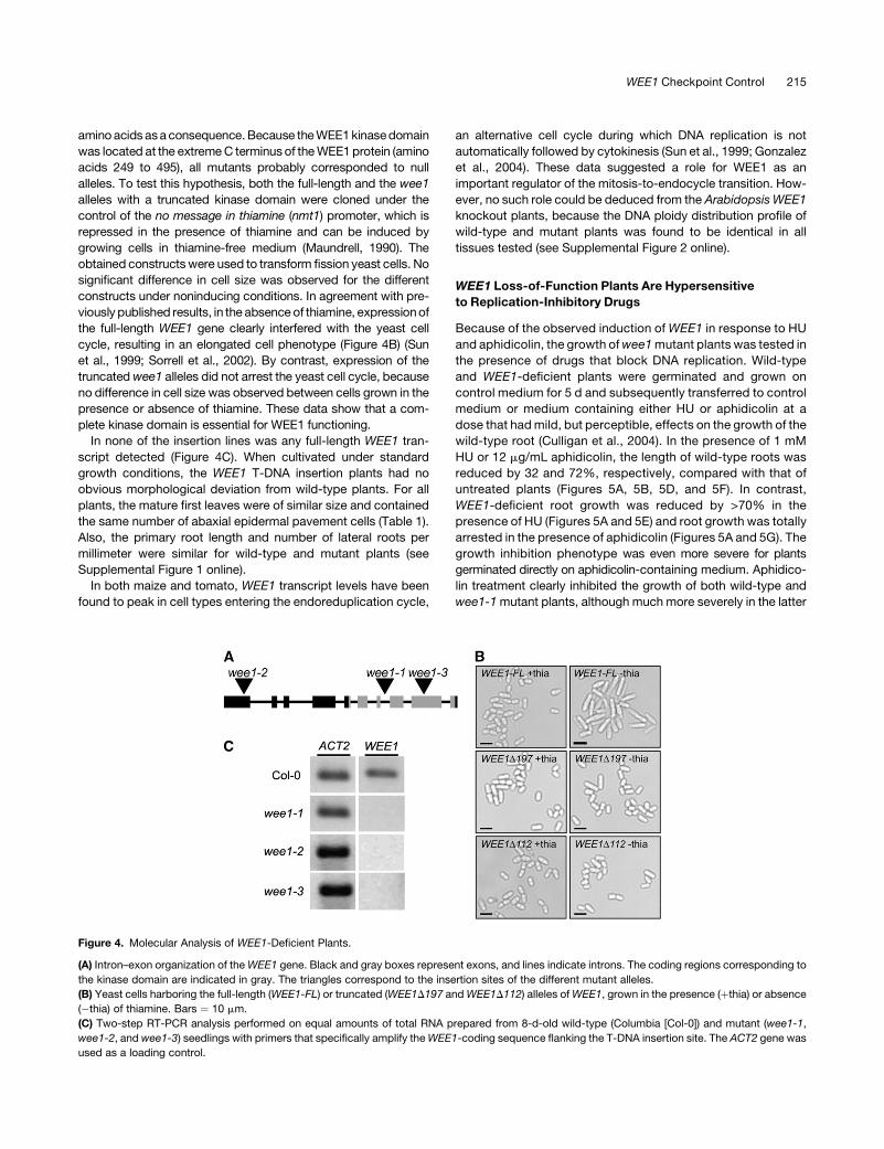
amino acids as a consequence. Because the WEE1 kinase domain
was located at the extreme C terminus of the WEE1 protein (amino
acids 249 to 495), all mutants probably corresponded to null
alleles. To test this hypothesis, both the full-length and the wee1
alleles with a truncated kinase domain were cloned under the
control of the no message in thiamine (nmt1) promoter, which is
repressed in the presence of thiamine and can be induced by
growing cells in thiamine-free medium (Maundrell, 1990). The
obtained constructs were used to transform fission yeast cells. No
significant difference in cell size was observed for the different
constructs under noninducing conditions. In agreement with pre-
viously published results, in the absence of thiamine, expression of
the full-length WEE1 gene clearly interfered with the yeast cell
cycle, resulting in an elongated cell phenotype (Figure 4B) (Sun
et al., 1999; Sorrell et al., 2002). By contrast, expression of the
truncated wee1 alleles did not arrest the yeast cell cycle, because
no difference in cell size was observed between cells grown in the
presence or absence of thiamine. These data show that a com-
plete kinase domain is essential for WEE1 functioning.
In none of the insertion lines was any full-length WEE1 tran-
script detected (Figure 4C). When cultivated under standard
growth conditions, the WEE1 T-DNA insertion plants had no
obvious morphological deviation from wild-type plants. For all
plants, the mature first leaves were of similar size and contained
the same number of abaxial epidermal pavement cells (Table 1).
Also, the primary root length and number of lateral roots per
millimeter were similar for wild-type and mutant plants (see
Supplemental Figure 1 online).
In both maize and tomato, WEE1 transcript levels have been
found to peak in cell types entering the endoreduplication cycle,
an alternative cell cycle during which DNA replication is not
automatically followed by cytokinesis (Sun et al., 1999; Gonzalez
et al., 2004). These data suggested a role for WEE1 as an
important regulator of the mitosis-to-endocycle transition. How-
ever, no such role could be deduced from the Arabidopsis WEE1
knockout plants, because the DNA ploidy distribution profile of
wild-type and mutant plants was found to be identical in all
tissues tested (see Supplemental Figure 2 online).
WEE1 Loss-of-Function Plants Are Hypersensitive
to Replication-Inhibitory Drugs
Because of the observed induction of WEE1 in response to HU
and aphidicolin, the growth of wee1 mutant plants was tested in
the presence of drugs that block DNA replication. Wild-type
and WEE1-deficient plants were germinated and grown on
control medium for 5 d and subsequently transferred to control
medium or medium containing either HU or aphidicolin at a
dose that had mild, but perceptible, effects on the growth of the
wild-type root (Culligan et al., 2004). In the presence of 1 mM
HU or 12 mg/mL aphidicolin, the length of wild-type roots was
reduced by 32 and 72%, respectively, compared with that of
untreated plants (Figures 5A, 5B, 5D, and 5F). In contrast,
WEE1-deficient root growth was reduced by >70% in the
presence of HU (Figures 5A and 5E) and root growth was totally
arrested in the presence of aphidicolin (Figures 5A and 5G). The
growth inhibition phenotype was even more severe for plants
germinated directly on aphidicolin-containing medium. Aphidico-
lin treatment clearly inhibited the growth of both wild-type and
wee1-1 mutant plants, although much more severely in the latter
Figure 4. Molecular Analysis of WEE1-Deficient Plants.
(A) Intron–exon organization of the WEE1 gene. Black and gray boxes represent exons, and lines indicate introns. The coding regions corresponding to
the kinase domain are indicated in gray. The triangles correspond to the insertion sites of the different mutant alleles.
(B) Yeast cells harboring the full-length (WEE1-FL) or truncated (WEE1D197 and WEE1D112) alleles of WEE1, grown in the presence (þthia) or absence
(�thia) of thiamine. Bars ¼ 10 mm.
(C) Two-step RT-PCR analysis performed on equal amounts of total RNA prepared from 8-d-old wild-type (Columbia [Col-0]) and mutant (wee1-1,
wee1-2, and wee1-3) seedlings with primers that specifically amplify the WEE1-coding sequence flanking the T-DNA insertion site. The ACT2 gene was
used as a loading control.
WEE1 Checkpoint Control 215
(Figures 5H and 5I). All three mutant lines had the same recessive
phenotype that segregated with the respective insertions in
WEE1.
We reasoned that the root growth arrest observed for the
WEE1-deficient plants was attributable to a failure to block their
cell cycle in response to DNA stress. To test this hypothesis, we
compared the number of dividing cells in the root tips of the wild-
type and WEE1-deficient plants. Under control growth condi-
tions, the number of cells in mitosis was similar in both genotypes
(Figure 6A). When control plants were treated with HU, they ex-
perienced a dramatic decrease in the number of mitotic cells. This
decrease correlated with the appearance of a Tyr-phosphorylated
CDK that migrated with the same electrophoretic mobility as that
of CDKA;1 (Figure 6B). Moreover, a decrease in CDK activity was
observed within 5 h after transfer to HU-containing medium
(Figure 6C). By contrast, in the WEE1-deficient plants, the
number of mitotic cells decreased only slightly upon HU treat-
ment (Figure 6A). In addition, neither CDK Tyr phosphorylation
nor a decrease in CDK activity was seen (Figures 6B and 6C).
These data suggest that the WEE1-deficient plants failed to
activate a G2 arrest and progressed with a not fully replicated
genome into mitosis.
Table 1. Size and Number of Abaxial Pavement Cells in Leaves of
WEE1-Deficient and Control Plants
Line
Leaf Size
(mm2)
Abaxial Pavement
Cell Size (mm2)
Estimated
Number
Columbia 22.4 6 0.6 2129 6 90 10.746 6 298
wee1-1 22.1 6 1.0 2183 6 140 10.334 6 373
wee1-2 23.0 6 1.5 2090 6 92 10.981 6 519
wee1-3 20.0 6 1.3 1946 6 76 10.250 6 452
All measurements were performed on leaves harvested 21 d after
sowing. The indicated values are means 6 SE (n ¼ 14 to 30).
Figure 5. Phenotypic Analysis of WEE1-Deficient Plants under Replication Stress.
(A) Root elongation rates of plants shown in (B) to (G). Error bars indicate SE (n ¼ 14 to 20).
(B) to (G) Wild-type ([B], [D], and [F]) and wee1-1 ([C], [E], and [G]) plants grown for 5 d and then transferred for 5 d to control medium ([B] and [C]),
medium supplemented with 1 mM HU ([D] and [E]), or medium supplemented with 12 mg/mL aphidicolin ([F] and [G]).
(H) and (I) Wild-type and wee1-1 seeds germinated on 12 mg/mL aphidicolin, respectively.
216 The Plant Cell
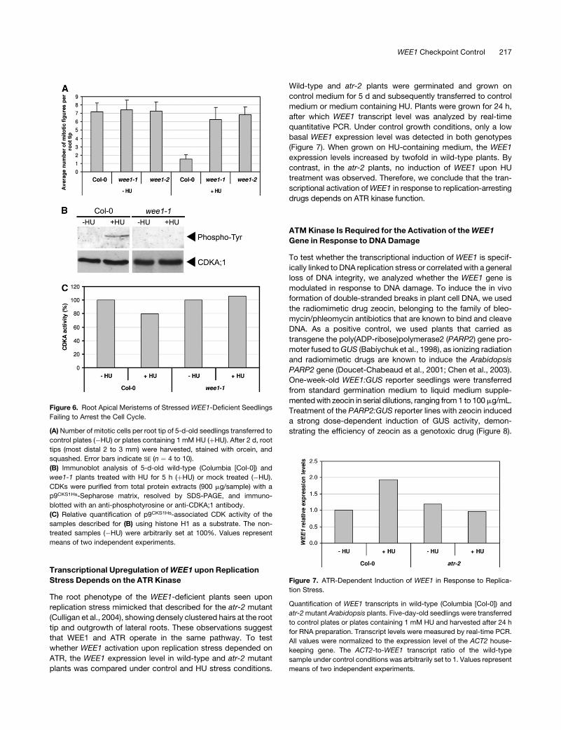
Transcriptional Upregulation of WEE1 upon Replication
Stress Depends on the ATR Kinase
The root phenotype of the WEE1-deficient plants seen upon
replication stress mimicked that described for the atr-2 mutant
(Culligan et al., 2004), showing densely clustered hairs at the root
tip and outgrowth of lateral roots. These observations suggest
that WEE1 and ATR operate in the same pathway. To test
whether WEE1 activation upon replication stress depended on
ATR, the WEE1 expression level in wild-type and atr-2 mutant
plants was compared under control and HU stress conditions.
Wild-type and atr-2 plants were germinated and grown on
control medium for 5 d and subsequently transferred to control
medium or medium containing HU. Plants were grown for 24 h,
after which WEE1 transcript level was analyzed by real-time
quantitative PCR. Under control growth conditions, only a low
basal WEE1 expression level was detected in both genotypes
(Figure 7). When grown on HU-containing medium, the WEE1
expression levels increased by twofold in wild-type plants. By
contrast, in the atr-2 plants, no induction of WEE1 upon HU
treatment was observed. Therefore, we conclude that the tran-
scriptional activation of WEE1 in response to replication-arresting
drugs depends on ATR kinase function.
ATM Kinase Is Required for the Activation of the WEE1
Gene in Response to DNA Damage
To test whether the transcriptional induction of WEE1 is specif-
ically linked to DNA replication stress or correlated with a general
loss of DNA integrity, we analyzed whether the WEE1 gene is
modulated in response to DNA damage. To induce the in vivo
formation of double-stranded breaks in plant cell DNA, we used
the radiomimetic drug zeocin, belonging to the family of bleo-
mycin/phleomycin antibiotics that are known to bind and cleave
DNA. As a positive control, we used plants that carried as
transgene the poly(ADP-ribose)polymerase2 (PARP2) gene pro-
moter fused to GUS (Babiychuk et al., 1998), as ionizing radiation
and radiomimetic drugs are known to induce the Arabidopsis
PARP2 gene (Doucet-Chabeaud et al., 2001; Chen et al., 2003).
One-week-old WEE1:GUS reporter seedlings were transferred
from standard germination medium to liquid medium supple-
mented with zeocin in serial dilutions, ranging from 1 to 100 mg/mL.
Treatment of the PARP2:GUS reporter lines with zeocin induced
a strong dose-dependent induction of GUS activity, demon-
strating the efficiency of zeocin as a genotoxic drug (Figure 8).
Figure 6. Root Apical Meristems of Stressed WEE1-Deficient Seedlings
Failing to Arrest the Cell Cycle.
(A) Number of mitotic cells per root tip of 5-d-old seedlings transferred to
control plates (�HU) or plates containing 1 mM HU (þHU). After 2 d, root
tips (most distal 2 to 3 mm) were harvested, stained with orcein, and
squashed. Error bars indicate SE (n ¼ 4 to 10).
(B) Immunoblot analysis of 5-d-old wild-type (Columbia [Col-0]) and
wee1-1 plants treated with HU for 5 h (þHU) or mock treated (�HU).
CDKs were purified from total protein extracts (900 mg/sample) with a
p9CKS1Hs-Sepharose matrix, resolved by SDS-PAGE, and immuno-
blotted with an anti-phosphotyrosine or anti-CDKA;1 antibody.
(C) Relative quantification of p9CKS1Hs-associated CDK activity of the
samples described for (B) using histone H1 as a substrate. The non-
treated samples (�HU) were arbitrarily set at 100%. Values represent
means of two independent experiments.
Figure 7. ATR-Dependent Induction of WEE1 in Response to Replica-
tion Stress.
Quantification of WEE1 transcripts in wild-type (Columbia [Col-0]) and
atr-2 mutant Arabidopsis plants. Five-day-old seedlings were transferred
to control plates or plates containing 1 mM HU and harvested after 24 h
for RNA preparation. Transcript levels were measured by real-time PCR.
All values were normalized to the expression level of the ACT2 house-
keeping gene. The ACT2-to-WEE1 transcript ratio of the wild-type
sample under control conditions was arbitrarily set to 1. Values represent
means of two independent experiments.
WEE1 Checkpoint Control 217
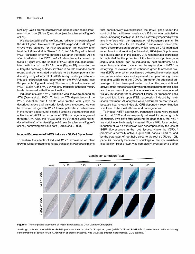
Similarly, WEE1 promoter activity was induced upon zeocin treat-
ment in both root (Figure 8) and shoot (see Supplemental Figure 3
online).
We also tested the effects of ionizing radiation on expression of
the WEE1 gene. Two-week-old plantlets exposed to 20 Gray of
g-rays were sampled for RNA preparation immediately after
treatment (0 h) and after 30 min, 1, 3, 5, and 8 h. Only a low basal
WEE1 transcript level was detected just after treatment. At 1 h
after irradiation, the WEE1 mRNA abundance increased by
fivefold (Figure 9A). The kinetics of WEE1 gene induction corre-
lated with that of the RAD51 gene (Figure 9B), encoding an
eukaryotic homolog of RecA, involved in double-stranded break
repair, and demonstrated previously to be transcriptionally in-
duced by g-rays (Garcia et al., 2003). A very similar g-irradiation–
induced expression was observed for the PARP2 gene (see
Supplemental Figure 4 online). This transcriptional activation of
WEE1, RAD51, and PARP2 was only transient, although mRNA
levels decreased with different kinetics.
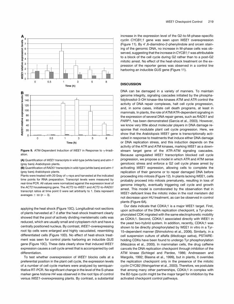
Induction of RAD51 by g-irradiation was shown to depend on
ATM (Garcia et al., 2003). To test the ATM dependence of the
WEE1 induction, atm-1 plants were treated with g-rays as
described above and transcript levels were measured. As can
be observed in Figure 9A, WEE1 transcript levels did not increase
in the mutant background, clearly illustrating that transcriptional
activation of WEE1 in response of DNA damage is regulated
through ATM. Also, the RAD51 and PARP2 genes were not in-
duced in the atm-1 mutant (Figure 9B; see Supplemental Figure 3
online), confirming previous data (Garcia et al., 2003).
Induced Expression of WEE1 Induces a G2 Cell Cycle Arrest
To analyze the effects of induced WEE1 expression on plant
growth, we attempted to generate transgenic Arabidopsis plants
that constitutively overexpressed the WEE1 gene under the
control of the cauliflower mosaic virus 35S promoter but failed to
do so, indicating that high WEE1 levels severely impaired growth
and interfered with the regeneration of transgenic plants. To
overcome this difficulty, we decided to use a switch-on consti-
tutive overexpression approach, which relies on CRE-mediated
recombination at lox sites (Joubes et al., 2004) (see Supplemen-
tal Figure 5 online). In this design, CRE recombinase expression
is controlled by the promoter of the heat-shock protein gene
hsp90 and, hence, can be induced by heat treatment. CRE
recombinase is able to switch on the expression of WEE1 by
catalyzing the excision of the enhanced green fluorescent pro-
tein (EGFP) gene, which was flanked by two colinearly orientated
lox recombination sites and separated the open reading frame
encoding WEE1 from the CDKA;1 promoter. An additional ad-
vantage of the developed system is that the transcriptional
activity of the transgene at a given chromosomal integration locus
and the success of recombinational excision can be monitored
visually by scoring the fluorescent tissues. All transgenic lines
behaved identically upon WEE1 expression induced by heat-
shock treatment. All analyses were performed on root tissues,
because heat shock–inducible CRE-dependent recombination
was found to be most efficient and homogeneous.
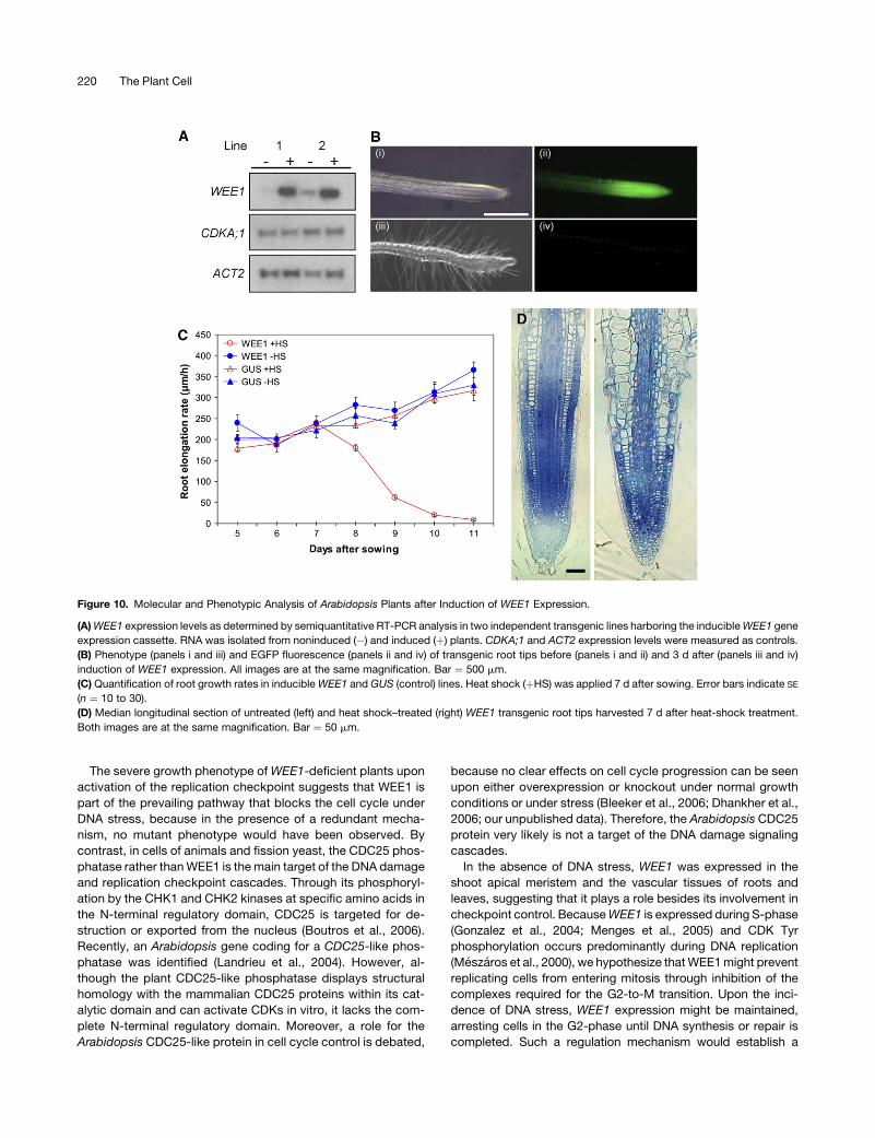
To induce WEE1 expression, transgenic plants were treated
for 2 h at 378C and subsequently returned to normal growth
conditions. Two days after applying the heat shock, the WEE1
transcript level had clearly increased (Figure 10A). As expected,
induction of WEE1 expression was accompanied by the loss of
EGFP fluorescence in the root tissues, where the CDKA;1
promoter is normally active (Figure 10B, panels ii and iv), and
by the outgrowth of root hairs close to the root tip (Figure 10B,
panel iii), probably because of shrinkage of the root meristem
(see below). Root growth was completely arrested by 3 d after
Figure 8. Transcriptional Activation of WEE1 in Response to DNA Damage Checkpoint.
Seedlings harboring the WEE1 or PARP2 promoter fused to the GUS reporter gene (WEE1:GUS and PARP2:GUS) were treated with increasing
concentrations of zeocin for 24 h. Activation of promoter activity was visualized through histochemical GUS staining.
218 The Plant Cell
applying the heat shock (Figure 10C). Longitudinal root sections
of plants harvested at 7 d after the heat-shock treatment clearly
showed that the pool of actively dividing meristematic cells was
reduced, which are usually small and cytoplasm-rich and have a
centrally positioned nucleus. By contrast, WEE1-overexpressing
root tip cells were enlarged and highly vacuolated, resembling
differentiated cells (Figure 10D). No effect of heat-shock treat-
ment was seen for control plants harboring an inducible GUS
gene (Figure 10C). These data clearly show that induced WEE1
expression causes a cell cycle arrest that is accompanied by cell
differentiation.
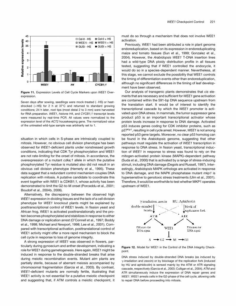
To test whether overexpression of WEE1 blocks cells at a
preferential position in the plant cell cycle, the expression levels
of a number of cell cycle markers were analyzed by semiquan-
titative RT-PCR. No significant change in the level of the S-phase
marker gene histone H4 was observed in the root tips of control
versus WEE1-overexpressing plants. By contrast, a substantial
increase in the expression level of the G2-to-M-phase–specific
cyclin CYCB1;1 gene was seen upon WEE1 overexpression
(Figure 11). By 49,6-diamidino-2-phenylindole and orcein stain-
ing of the genomic DNA, no increase in M-phase cells was ob-
served, suggesting that the increase in CYCB1;1 was attributable
to a block of the cell cycle during G2 rather than to a post-G2
mitotic arrest. No effect of the heat-shock treatment on the ex-
pression of the reporter genes was observed in a control line
harboring an inducible GUS gene (Figure 11).
DISCUSSION
DNA can be damaged in a variety of manners. To maintain
genome integrity, signaling cascades initiated by the phospha-
tidylinositol-3-OH kinase–like kinases ATM and ATR control the
activity of DNA repair complexes, halt cell cycle progression,
and, in some cases, initiate cell death programs, at least in
mammals. In plants, the role of ATM/ATR-dependent signaling in
the expression of several DNA repair genes, such as RAD51 and
PARP1, has been demonstrated (Garcia et al., 2003). However,
we know very little about molecular players in DNA damage re-
sponse that modulate plant cell cycle progression. Here, we
show that the Arabidopsis WEE1 gene is transcriptionally acti-
vated in response to treatments that induce either DNA damage
or DNA replication stress, and this induction depends on the
activity of the ATR and ATM kinases, marking WEE1 as a down-
stream target gene of the ATR-ATM signaling cascades.
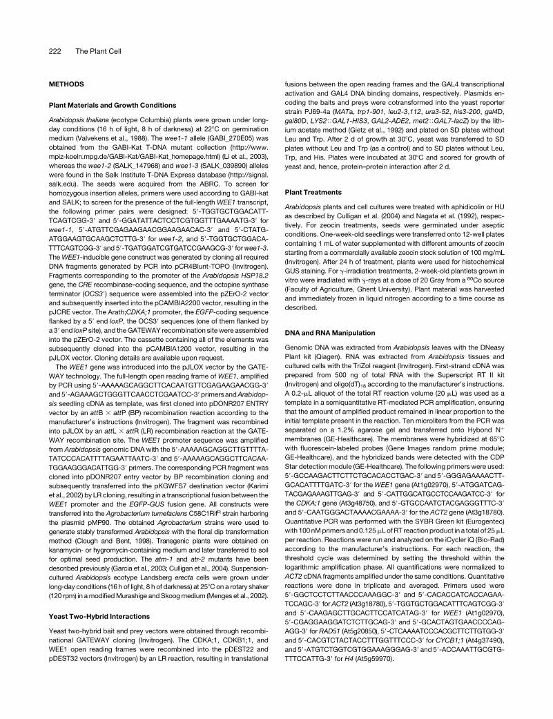
Because upregulated WEE1 transcription blocked cell cycle
progression, we propose a model in which ATR and ATM sense
genotoxic stress and enforce a G2 cell cycle phase arrest by
activating WEE1 expression, allowing cells to complete the
replication of their genome or to repair damaged DNA before
proceeding into mitosis (Figure 12). In plants lacking WEE1, cells
probably proceed into mitosis prematurely, resulting in loss of
genome integrity, eventually triggering cell cycle and growth
arrest. This model is corroborated by the observation that in
WEE1-deficient lines the mitotic index in the root meristem did
not decrease upon HU treatment, as can be observed in control
plants (Figure 6A).
Our data indicate that CDKA;1 is a major WEE1 target. First,
upon activation of the DNA replication checkpoint, a Tyr-phos-
phorylated CDK migrated with the same electrophoretic mobility
as CDKA;1. Second, CDKA;1 associated directly with WEE1 in
the yeast two-hybrid system. In addition, recently, CDKA;1 was
shown to be directly phosphorylated by WEE1 in vitro in a Tyr-
15–dependent manner (Shimotohno et al., 2006). Similarly, in a
cell suspension culture of alfalfa (Medicago sativa), PSTAIRE-
holding CDKs have been found to undergo Tyr phosphorylation
(Meszaros et al., 2000). In mammalian cells, the drug caffeine
cancels the DNA replication checkpoint through inhibition of the
ATM kinase (Schlegel and Pardee, 1986; Andreassen and
Margolis, 1992; Blasina et al., 1999), but in plants, it overrides
the replication checkpoint only in the presence of the mitotic
cyclin CYCB2 (Weingartner et al., 2003). Therefore, we postulate
that among many other partnerships, CDKA;1 in complex with
the B2-type cyclin might be the major target for inhibition by the
activated checkpoint control pathways.
Figure 9. ATM-Dependent Induction of WEE1 in Response to g-Irradi-
ation.
(A) Quantification of WEE1 transcripts in wild-type (white bars) and atm-1
(gray bars) Arabidopsis plants.
(B) Quantification of RAD51 transcripts in wild-type (white bars) and atm-1
(gray bars) Arabidopsis plants.
Plants were treated with 20 Gray of g-rays and harvested at the indicated
time points for RNA preparation. Transcript levels were measured by
real-time PCR. All values were normalized against the expression level of
the ACT2 housekeeping gene. The ACT2-to-WEE1 and ACT2-to-RAD51
transcript ratios at time point 0 were set arbitrarily to 1. Data represent
averages 6 SD (n ¼ 3).
WEE1 Checkpoint Control 219
The severe growth phenotype of WEE1-deficient plants upon
activation of the replication checkpoint suggests that WEE1 is
part of the prevailing pathway that blocks the cell cycle under
DNA stress, because in the presence of a redundant mecha-
nism, no mutant phenotype would have been observed. By
contrast, in cells of animals and fission yeast, the CDC25 phos-
phatase rather than WEE1 is the main target of the DNA damage
and replication checkpoint cascades. Through its phosphoryl-
ation by the CHK1 and CHK2 kinases at specific amino acids in
the N-terminal regulatory domain, CDC25 is targeted for de-
struction or exported from the nucleus (Boutros et al., 2006).
Recently, an Arabidopsis gene coding for a CDC25-like phos-
phatase was identified (Landrieu et al., 2004). However, al-
though the plant CDC25-like phosphatase displays structural
homology with the mammalian CDC25 proteins within its cat-
alytic domain and can activate CDKs in vitro, it lacks the com-
plete N-terminal regulatory domain. Moreover, a role for the
Arabidopsis CDC25-like protein in cell cycle control is debated,
because no clear effects on cell cycle progression can be seen
upon either overexpression or knockout under normal growth
conditions or under stress (Bleeker et al., 2006; Dhankher et al.,
2006; our unpublished data). Therefore, the Arabidopsis CDC25
protein very likely is not a target of the DNA damage signaling
cascades.
In the absence of DNA stress, WEE1 was expressed in the
shoot apical meristem and the vascular tissues of roots and
leaves, suggesting that it plays a role besides its involvement in
checkpoint control. Because WEE1 is expressed during S-phase
(Gonzalez et al., 2004; Menges et al., 2005) and CDK Tyr
phosphorylation occurs predominantly during DNA replication
(Meszaros et al., 2000), we hypothesize that WEE1 might prevent
replicating cells from entering mitosis through inhibition of the
complexes required for the G2-to-M transition. Upon the inci-
dence of DNA stress, WEE1 expression might be maintained,
arresting cells in the G2-phase until DNA synthesis or repair is
completed. Such a regulation mechanism would establish a
Figure 10. Molecular and Phenotypic Analysis of Arabidopsis Plants after Induction of WEE1 Expression.
(A) WEE1 expression levels as determined by semiquantitative RT-PCR analysis in two independent transgenic lines harboring the inducible WEE1 gene
expression cassette. RNA was isolated from noninduced (�) and induced (þ) plants. CDKA;1 and ACT2 expression levels were measured as controls.
(B) Phenotype (panels i and iii) and EGFP fluorescence (panels ii and iv) of transgenic root tips before (panels i and ii) and 3 d after (panels iii and iv)
induction of WEE1 expression. All images are at the same magnification. Bar ¼ 500 mm.
(C) Quantification of root growth rates in inducible WEE1 and GUS (control) lines. Heat shock (þHS) was applied 7 d after sowing. Error bars indicate SE
(n ¼ 10 to 30).
(D) Median longitudinal section of untreated (left) and heat shock–treated (right) WEE1 transgenic root tips harvested 7 d after heat-shock treatment.
Both images are at the same magnification. Bar ¼ 50 mm.
220 The Plant Cell
situation in which cells in S-phase are intrinsically coupled to
mitosis. However, no obvious cell division phenotype has been
observed for WEE1-deficient plants under nonstressed growth
conditions, indicating that CDK Tyr phosphorylation and WEE1
are not rate-limiting for the onset of mitosis. In accordance, the
overexpression of a mutant cdka;1 allele in which the putative
phosphorylated Tyr residue is mutated also did not result in an
obvious cell division phenotype (Hemerly et al., 1995). These
data suggest that a redundant control mechanism couples DNA
replication with mitosis. A putative candidate to coordinate this
event together with WEE1 is CDKB1;1, whose activity has been
demonstrated to limit the G2-to-M onset (Porceddu et al., 2001;
Boudolf et al., 2004b, 2006).
Alternatively, the discrepancy between the observed high
WEE1 expression in dividing tissues and the lack of a cell division
phenotype for WEE1 knockout plants might be explained by
posttranscriptional control of WEE1 levels. In fission yeast and
African frog, WEE1 is activated posttranslationally and the pro-
tein becomes phosphorylated and stabilizes in response to either
DNA damage or replication arrest (O’Connell et al., 1997; Boddy
et al., 1998; Michael and Newport, 1998; Lee et al., 2001). Com-
pared with transcriptional activation, posttranslational control of
WEE1 activity might offer a more rapid mechanism to block the
cell cycle in response to loss of genome integrity.
A strong expression of WEE1 was observed in flowers, par-
ticularly during gynoecium and anther development, indicating a
role for WEE1 during gametogenesis. Here again, WEE1 might be
induced in response to the double-stranded breaks that arise
during meiotic recombination events. Mutant atm plants are
partially sterile, because of aberrant meiosis accompanied by
chromosomal fragmentation (Garcia et al., 2003). By contrast,
WEE1-deficient mutants are normally fertile, illustrating that
WEE1 activity is not essential for a putative meiotic checkpoint
and suggesting that, if ATM controls a meiotic checkpoint, it
must do so through a mechanism that does not involve WEE1
activation.
Previously, WEE1 had been attributed a role in plant genome
endoreduplication, based on its expression in endoreduplicating
maize and tomato tissues (Sun et al., 1999; Gonzalez et al.,
2004). However, the Arabidopsis WEE1 T-DNA insertion lines
had a wild-type DNA ploidy distribution profile in all tissues
tested, suggesting that if WEE1 controlled the endocycle, it
would do so in a species-dependent manner. Nevertheless, at
this stage, we cannot exclude the possibility that WEE1 controls
the timing of differentiation events other than endoreduplication,
although no significant differences in the timing of leaf develop-
ment have been observed.
Our analysis of transgenic plants demonstrates that cis ele-
ments that are necessary and sufficient for WEE1 gene activation
are contained within the 591-bp DNA sequence upstream from
the translation start. It would be of interest to identify the
transcriptional cascade by which the WEE1 promoter is acti-
vated upon DNA stress. In mammals, the tumor suppressor gene
product p53 is an important transcriptional activator whose
protein levels increase in response to DNA damage. Activated
p53 induces genes coding for CDK inhibitor proteins, such as
p27Kip1, resulting in cell cycle arrest. However, WEE1 is not among
reported p53 gene targets. Moreover, no clear p53 homolog can
be found in the Arabidopsis genome, suggesting that other
pathways must regulate the activation of WEE1 transcription in
response to DNA stress. In fission yeast, transcriptional induc-
tion of WEE1 in response to stress was found to rely on the
mitogen-activated protein kinase (MAPK)–dependent pathway
(Suda et al., 2000) that is activated by a range of stress-inducing
stimuli, including DNA damage (Degols and Russell, 1997). Inter-
estingly, Arabidopsis MAPK orthologs are activated in response
to DNA damage, and the MAPK phosphatase mutant mkp1 is
hypersensitive to genotoxic stress treatments (Ulm et al., 2001).
Therefore, it would be worthwhile to test whether MKP1 operates
upstream of WEE1.
Figure 11. Expression Levels of Cell Cycle Markers upon WEE1 Over-
expression.
Seven days after sowing, seedlings were mock-treated (�HS) or heat-
shocked (þHS) for 2 h at 378C and returned to standard growing
conditions; 24 h later, root tips (most distal 2 to 3 mm) were harvested
for RNA preparation. WEE1, histone H4, and CYCB1;1 transcript levels
were measured by real-time PCR. All values were normalized to the
expression level of the ACT2 housekeeping gene. The normalized value
of the untreated wild-type sample was arbitrarily set to 1.
Figure 12. Model for WEE1 in the Control of the DNA Integrity Check-
point.
DNA stress induced by double-stranded DNA breaks (as induced by
g-irradiation and zeocin) or by blockage of the replication fork (induced
by HU and aphidicolin) is sensed mainly by the ATM or ATR signaling
cascade, respectively (Garcia et al., 2003; Culligan et al., 2004). ATM and
ATR simultaneously induce the expression of DNA repair genes and
WEE1. WEE1 arrests cells in the G2-phase of the cell cycle, allowing cells
to repair DNA before proceeding into mitosis.
WEE1 Checkpoint Control 221
METHODS
Plant Materials and Growth Conditions
Arabidopsis thaliana (ecotype Columbia) plants were grown under long-
day conditions (16 h of light, 8 h of darkness) at 228C on germination
medium (Valvekens et al., 1988). The wee1-1 allele (GABI_270E05) was
obtained from the GABI-Kat T-DNA mutant collection (http://www.
mpiz-koeln.mpg.de/GABI-Kat/GABI-Kat_homepage.html) (Li et al., 2003),
whereas the wee1-2 (SALK_147968) and wee1-3 (SALK_039890) alleles
were found in the Salk Institute T-DNA Express database (http://signal.
salk.edu). The seeds were acquired from the ABRC. To screen for
homozygous insertion alleles, primers were used according to GABI-kat
and SALK; to screen for the presence of the full-length WEE1 transcript,
the following primer pairs were designed: 59-TGGTGCTGGACATT-
TCAGTCGG-39 and 59-GGATATTACTCCTCGTGGTTTGAAAATG-39 for
wee1-1, 59-ATGTTCGAGAAGAACGGAAGAACAC-39 and 59-CTATG-
ATGGAAGTGCAAGCTCTTG-39 for wee1-2, and 59-TGGTGCTGGACA-
TTTCAGTCGG-39 and 59-TGATGGATCGTGATCCGAAGCG-39 for wee1-3.
The WEE1-inducible gene construct was generated by cloning all required
DNA fragments generated by PCR into pCR4Blunt-TOPO (Invitrogen).
Fragments corresponding to the promoter of the Arabidopsis HSP18.2
gene, the CRE recombinase–coding sequence, and the octopine synthase
terminator (OCS39) sequence were assembled into the pZErO-2 vector
and subsequently inserted into the pCAMBIA2200 vector, resulting in the
pJCRE vector. The Arath;CDKA;1 promoter, the EGFP-coding sequence
flanked by a 59 end loxP, the OCS39 sequences (one of them flanked by
a 39 end loxP site), and the GATEWAY recombination site were assembled
into the pZErO-2 vector. The cassette containing all of the elements was
subsequently cloned into the pCAMBIA1200 vector, resulting in the
pJLOX vector. Cloning details are available upon request.
The WEE1 gene was introduced into the pJLOX vector by the GATE-
WAY technology. The full-length open reading frame of WEE1, amplified
by PCR using 59-AAAAAGCAGGCTTCACAATGTTCGAGAAGAACGG-39
and 59-AGAAAGCTGGGTTCAACCTCGAATCC-39 primersand Arabidop-
sis seedling cDNA as template, was first cloned into pDONR207 ENTRY
vector by an attB 3 attP (BP) recombination reaction according to the
manufacturer’s instructions (Invitrogen). The fragment was recombined
into pJLOX by an attL 3 attR (LR) recombination reaction at the GATE-
WAY recombination site. The WEE1 promoter sequence was amplified
from Arabidopsis genomic DNA with the 59-AAAAAGCAGGCTTGTTTTA-
TATCCCACATTTTAGAATTAATC-39 and 59-AAAAAGCAGGCTTCACAA-
TGGAAGGGACATTGG-39 primers. The corresponding PCR fragment was
cloned into pDONR207 entry vector by BP recombination cloning and
subsequently transferred into the pKGWFS7 destination vector (Karimi
et al., 2002) by LR cloning, resulting in a transcriptional fusion between the
WEE1 promoter and the EGFP-GUS fusion gene. All constructs were
transferred into the Agrobacterium tumefaciens C58C1RifR strain harboring
the plasmid pMP90. The obtained Agrobacterium strains were used to
generate stably transformed Arabidopsis with the floral dip transformation
method (Clough and Bent, 1998). Transgenic plants were obtained on
kanamycin- or hygromycin-containing medium and later transferred to soil
for optimal seed production. The atm-1 and atr-2 mutants have been
described previously (Garcia et al., 2003; Culligan et al., 2004). Suspension-
cultured Arabidopsis ecotype Landsberg erecta cells were grown under
long-day conditions (16 h of light, 8 h of darkness) at 258C on a rotary shaker
(120 rpm) in a modified Murashige and Skoog medium (Menges et al., 2002).
Yeast Two-Hybrid Interactions
Yeast two-hybrid bait and prey vectors were obtained through recombi-
national GATEWAY cloning (Invitrogen). The CDKA;1, CDKB1;1, and
WEE1 open reading frames were recombined into the pDEST22 and
pDEST32 vectors (Invitrogen) by an LR reaction, resulting in translational
fusions between the open reading frames and the GAL4 transcriptional
activation and GAL4 DNA binding domains, respectively. Plasmids en-
coding the baits and preys were cotransformed into the yeast reporter
strain PJ69-4a (MATa, trp1-901, leu2-3,112, ura3-52, his3-200, gal4D,
gal80D, LYS2TGAL1-HIS3, GAL2-ADE2, met2TGAL7-lacZ) by the lith-
ium acetate method (Gietz et al., 1992) and plated on SD plates without
Leu and Trp. After 2 d of growth at 308C, yeast was transferred to SD
plates without Leu and Trp (as a control) and to SD plates without Leu,
Trp, and His. Plates were incubated at 308C and scored for growth of
yeast and, hence, protein–protein interaction after 2 d.
Plant Treatments
Arabidopsis plants and cell cultures were treated with aphidicolin or HU
as described by Culligan et al. (2004) and Nagata et al. (1992), respec-
tively. For zeocin treatments, seeds were germinated under aseptic
conditions. One-week-old seedlings were transferred onto 12-well plates
containing 1 mL of water supplemented with different amounts of zeocin
starting from a commercially available zeocin stock solution of 100 mg/mL
(Invitrogen). After 24 h of treatment, plants were used for histochemical
GUS staining. For g-irradiation treatments, 2-week-old plantlets grown in
vitro were irradiated with g-rays at a dose of 20 Gray from a 60Co source
(Faculty of Agriculture, Ghent University). Plant material was harvested
and immediately frozen in liquid nitrogen according to a time course as
described.
DNA and RNA Manipulation
Genomic DNA was extracted from Arabidopsis leaves with the DNeasy
Plant kit (Qiagen). RNA was extracted from Arabidopsis tissues and
cultured cells with the TriZol reagent (Invitrogen). First-strand cDNA was
prepared from 500 ng of total RNA with the Superscript RT II kit
(Invitrogen) and oligo(dT)18 according to the manufacturer’s instructions.
A 0.2-mL aliquot of the total RT reaction volume (20 mL) was used as a
template in a semiquantitative RT-mediated PCR amplification, ensuring
that the amount of amplified product remained in linear proportion to the
initial template present in the reaction. Ten microliters from the PCR was
separated on a 1.2% agarose gel and transferred onto Hybond Nþ
membranes (GE-Healthcare). The membranes were hybridized at 658C
with fluorescein-labeled probes (Gene Images random prime module;
GE-Healthcare), and the hybridized bands were detected with the CDP
Star detection module (GE-Healthcare). The following primers were used:
59-GCCAAGACTTCTTCTGCACACCTGAC-39 and 59-GGGAGAAAACTT-
GCACATTTTGATC-39 for the WEE1 gene (At1g02970), 59-ATGGATCAG-
TACGAGAAAGTTGAG-39 and 59-CATTGGCATGCCTCCAAGATCC-39 for
the CDKA;1 gene (At3g48750), and 59-GTGCCAATCTACGAGGGTTTC-39
and 59-CAATGGGACTAAAACGAAAA-39 for the ACT2 gene (At3g18780).
Quantitative PCR was performed with the SYBR Green kit (Eurogentec)
with 100 nM primers and 0.125 mL of RT reaction product in a total of 25 mL
per reaction. Reactions were run and analyzed on the iCycler iQ (Bio-Rad)
according to the manufacturer’s instructions. For each reaction, the
threshold cycle was determined by setting the threshold within the
logarithmic amplification phase. All quantifications were normalized to
ACT2 cDNA fragments amplified under the same conditions. Quantitative
reactions were done in triplicate and averaged. Primers used were
59-GGCTCCTCTTAACCCAAAGGC-39 and 59-CACACCATCACCAGAA-
TCCAGC-39 for ACT2 (At3g18780), 59-TGGTGCTGGACATTTCAGTCGG-39
and 59-CAAGAGCTTGCACTTCCATCATAG-39 for WEE1 (At1g02970),
59-CGAGGAAGGATCTCTTGCAG-39 and 59-GCACTAGTGAACCCCAG-
AGG-39 for RAD51 (At5g20850), 59-CTCAAAATCCCACGCTTCTTGTGG-39
and 59-CACGTCTACTACCTTTGGTTTCCC-39 for CYCB1;1 (At4g37490),
and 59-ATGTCTGGTCGTGGAAAGGGAG-39 and 59-ACCAAATTGCGTG-
TTTCCATTG-39 for H4 (At5g59970).
222 The Plant Cell
Antibodies and Protein Gel Blot Analysis
Protein extracts were prepared by grinding material in homogenization
buffer (De Veylder et al., 1997). Protein concentrations were determined
with the protein assay kit (Bio-Rad). CDKs were purified by affinity
chromatography with either p10CKS1At- or p9CKS1Hs-Sepharose beads as
described (De Veylder et al., 1997). Protein gel blotting was performed
according to standard procedures with primary anti-CDKA;1 and anti-
CDKB1;1 antibodies (Porceddu et al., 2001) diluted 1:5000 and 1:2500,
respectively, and a secondary horseradish peroxidase–conjugated
sheep anti-rabbit antibody (GE-Healthcare) diluted 1:5000 or with the
mouse monoclonal horseradish peroxidase–conjugated anti-phospho-
tyrosine p-Tyr antibody (PY99; Santa Cruz Biotechnology) diluted 1:5000.
Proteins were detected by a chemiluminescence procedure (NEN Life
Science Products). Kinase assays were performed as described previ-
ously (De Veylder et al., 1997).
Histochemical GUS Assays
Complete seedlings or tissue cuttings were stained on multiwell plates
(Falcon 3043; Becton Dickinson). GUS assays were performed as de-
scribed by Beeckman and Engler (1994). Samples mounted in lactic acid
were observed and photographed with a stereomicroscope (Stemi SV11;
Zeiss) or with a differential interference contrast microscope (Leica).
Microscopy and Flow Cytometric Analyses
Leaves were harvested at 21 d after sowing, cleared overnight in ethanol,
stored in lactic acid for microscopy, and observed with a microscope
fitted with differential interference contrast optics (Leica). The total (blade)
area was determined from images digitized directly with a digital camera
(Axiocam; Zeiss) mounted on a binocular (Stemi SV11; Zeiss). From
scanned drawing-tube images of outlines of at least 30 cells of the abaxial
epidermis located 25 and 75% from the distance between the tip and the
base of the leaf, halfway between the midrib and the leaf margin, the
following parameters were determined: total area of all cells in the draw-
ing and total numbers of pavement and guard cells, from which the
average cell area was calculated. The total number of cells per cotyledon
was estimated by dividing the leaf area by the average cell area. To
visualize mitotic cells, seeds were germinated and grown for 5 d as
described above and then transferred to control plates or plates con-
taining HU. Root tips were harvested 2 d after transfer and fixed in three
parts ethanol and one part acetic acid, macerated in 0.1 N HCl, and
squashed in orcein in 45% acetic acid. The number of mitotic cells was
determined by observing mitotic figures through a Leica microscope with
a 633 oil lens. For flow cytometric analysis, tissues were chopped with a
razor blade in 300 mL of 45 mM MgCl2, 30 mM sodium citrate, 20 mM
MOPS, pH 7, and 0.1% Triton X-100 (Galbraith et al., 1991). One micro-
liter of 4,6-diamidino-2-phenylindole from a stock of 1 mg/mL was added
to the filtered supernatants. The nuclei were analyzed with the CyFlow
flow cytometer using FloMax (Partec) software. Sections of root tips for
histological analysis were prepared according to Beeckman and Viane
(2000).
WEE1 Analysis in Yeast
WEE1 alleles were amplified by adapter PCR with 59-GGGCATATGTTC-
GAGAAGAACGGAAGAACAC-39 as the universal forward primer and
59-GGGGGATCCTTATCAACCTCGAATCCTATCAAACATG-39, 59-GGG-
GGATCCTTAGCAAGAGCTTGCACTTCCATC-39, and 59-GGGGGATCC-
TTAACCAAGCTTGCAAACACCGTTC-39 as reverse primers for the full-
length, WEE1D197, and WEE1D112 alleles, respectively. The obtained
PCR fragments were cut with NdeI and BamHI and cloned under the
control of the attenuated nmt1 promoter in the pREP41 HA-N vector
(Craven et al., 1998). All constructs were transferred into the Schizo-
saccharomyces pombe h� leu1-32 strain with the frozen-EZ yeast trans-
formation kit (Zymo Research). For analysis, yeast cells were grown
overnight in rich YPB medium (10 g of yeast extract, 10 g of peptone, 10 g
of beef extract, and 2.5 g of NaCl per liter of water at pH 7.2), then diluted
in Edinburgh minimal medium 2 (Moreno et al., 1991) with or without
thiamine (5 mg/mL).
Accession Numbers
Arabidopsis Genome Initiative locus identifiers for the genes mentioned in
this article are as follows: WEE1 (At1g02970), CDKA;1 (At3g48750),
CDKB1;1 (At3g54180), ATM (At3g48190), and ATR (At5g40820).
Supplemental Data
The following materials are available in the online version of this article.
Supplemental Figure 1. Root Growth Analysis of WEE1-Deficient
Plants.
Supplemental Figure 2. DNA Ploidy Level Distribution of Wild-Type
and WEE1-Deficient Plants in Different Tissues.
Supplemental Figure 3. Transcriptional Activation of WEE1 in Seed-
lings in Response to Zeocin Treatment.
Supplemental Figure 4. ATM-Dependent Transcriptional Induction of
PARP2 in Response to g-Irradiation.
Supplemental Figure 5. Scheme Depicting the CRE-Inducible Tran-
scriptional Activation of the WEE1 Transgene.
ACKNOWLEDGMENTS
We thank Mirande Naudts for technical support and Martine De Cock for
help in preparing the manuscript. This research was supported by
grants from the Interuniversity Poles of Attraction Program–Belgian
Science Policy (Grant P5/13), the Research Fund of Ghent University
(Geconcerteerde Onderzoeksacties Grant 12051403), and the European
Union Marie Curie Research Training Networks (Grant MRTN-CT-2004-
005336). J.J. and F.C. are indebted to the European Molecular Biology
Organization (Heidelberg, Germany) for postdoctoral fellowships. L.D.V.
is a Postdoctoral Fellow of the Research Foundation–Flanders.
Received June 19, 2006; revised August 21, 2006; accepted November
14, 2006; published January 5, 2007.
REFERENCES
Abraham, R.T. (2001). Cell cycle checkpoint signaling through the ATM
and ATR kinases. Genes Dev. 15: 2177–2196.
Andreassen, P.R., and Margolis, R.L. (1992). 2-Aminopurine overrides
multiple cell cycle checkpoints in BHK cells. Proc. Natl. Acad. Sci.
USA 89: 2272–2276.
Baber-Furnari, B.A., Rhind, N., Boddy, M.N., Shanahan, P., Lopez-
Girona, A., and Russell, P. (2000). Regulation of mitotic inhibitor Mik1
helps to enforce the DNA damage checkpoint. Mol. Biol. Cell 11: 1–11.
Babiychuk, E., Cottrill, P.B., Storozhenko, S., Fuangthong, M., Chen,
Y., O’Farrell, M.K., Van Montagu, M., Inze, D., and Kushnir, S.
(1998). Higher plants possess two structurally different poly(ADP-
ribose) polymerases. Plant J. 15: 635–645.
Bartek, J., and Lukas, J. (2001). Pathways governing G1/S transition
and their response to DNA damage. FEBS Lett. 490: 117–122.
WEE1 Checkpoint Control 223
Beeckman, T., and Engler, G. (1994). An easy technique for the
clearing of histochemically stained plant tissue. Plant Mol. Biol. Rep.
12: 37–42.
Beeckman, T., and Viane, R. (2000). Embedding thin plant specimens
for oriented sectioning. Biotech. Histochem. 75: 23–26.
Berry, L.D., and Gould, K.L. (1996). Regulation of Cdc2 activity by
phosphorylation at T14/Y15. In Progress in Cell Cycle Research, Vol.
2, L. Meijer, S. Guidet, and L. Vogel, eds (New York: Plenum Press),
pp. 99–105.
Blasina, A., Van de Weyer, I., Laus, M.C., Luyten, W.H.M.L., Parker,
A.E., and McGowan, C.H. (1999). A human homologue of the check-
point kinase Cds1 directly inhibits Cdc25 phosphatase. Curr. Biol.
9: 1–10.
Bleeker, P.M., Hakvoort, H.W.J., Bliek, M., Souer, E., and Schat, H.
(2006). Enhanced arsenate reduction by a CDC25-like tyrosine phos-
phatase explains increased phytochelatin accumulation in arsenate-
tolerant Holcus lanatus. Plant J. 45: 917–929.
Boddy, M.N., Furnari, B., Mondesert, O., and Russell, P. (1998).
Replication checkpoint enforced by kinases Cds1 and Chk1. Science
280: 909–912.
Boudolf, V., Barroco, R., de Almeida Engler, J., Verkest, A., Beeckman,
T., Naudts, M., Inze, D., and De Veylder, L. (2004a). B1-type cyclin-
dependent kinases are essential for the formation of stomatal com-
plexes in Arabidopsis thaliana. Plant Cell 16: 945–955.
Boudolf, V., Inze, D., and De Veylder, L. (2006). What if higher plants
lack a CDC25 phosphatase? Trends Plant Sci. 11: 474–479.
Boudolf, V., Vlieghe, K., Beemster, G.T.S., Magyar, Z., Torres
Acosta, J.A., Maes, S., Van Der Schueren, E., Inze, D., and De
Veylder, L. (2004b). The plant-specific cyclin-dependent kinase
CDKB1;1 and transcription factor E2Fa-DPa control the balance of
mitotically dividing and endoreduplicating cells in Arabidopsis. Plant
Cell 16: 2683–2692.
Boutros, R., Dozier, C., and Ducommun, B. (2006). The when and
wheres of CDC25 phosphatases. Curr. Opin. Cell Biol. 18: 185–191.
Chen, I.-P., Haehnel, U., Altschmied, L., Schubert, I., and Puchta, H.
(2003). The transcriptional response of Arabidopsis to genotoxic
stress—A high-density colony array study (HDCA). Plant J. 35:
771–786.
Chen, Y., and Sanchez, Y. (2004). Chk1 in the DNA damage response:
Conserved roles from yeasts to mammals. DNA Repair (Amst.) 3:
1025–1032.
Christensen, P.U., Bentley, N.J., Martinho, R.G., Nielsen, O., and
Carr, A.M. (2000). Mik1 levels accumulate in S phase and may medi-
ate an intrinsic link between S phase and mitosis. Proc. Natl. Acad.
Sci. USA 97: 2579–2584.
Clough, S.J., and Bent, A.F. (1998). Floral dip: A simplified method for
Agrobacterium-mediated transformation of Arabidopsis thaliana. Plant
J. 16: 735–743.
Corellou, F., Bisgrove, S.R., Kropf, D.L., Meijer, L., Kloareg, B., and
Bouget, F.-Y. (2000). A S/M DNA replication checkpoint prevents
nuclear and cytoplasmic events of cell division including centrosomal
axis alignment and inhibits activation of cyclin-dependent kinase-like
proteins in fucoid zygotes. Development 127: 1651–1660.
Craven, R.A., Griffiths, D.J.F., Sheldrick, K.S., Randall, R.E., Hagan,
I.M., and Carr, A.M. (1998). Vectors for the expression of tagged
proteins in Schizosaccharomyces pombe. Gene 221: 59–68.
Culligan, K., Tissier, A., and Britt, A. (2004). ATR regulates a G2-phase
cell-cycle checkpoint in Arabidopsis thaliana. Plant Cell 16: 1091–1104.
Degols, G., and Russell, P. (1997). Discrete roles of the Spc1 kinase
and the Atf1 transcription factor in the UV response of Schizosac-
charomyces pombe. Mol. Cell. Biol. 17: 3356–3363.
De Veylder, L., Joubes, J., and Inze, D. (2003). Plant cell cycle
transitions. Curr. Opin. Plant Biol. 6: 536–543.
De Veylder, L., Segers, G., Glab, N., Casteels, P., Van Montagu, M.,
and Inze, D. (1997). The Arabidopsis Cks1At protein binds the cyclin-
dependent kinases Cdc2aAt and Cdc2bAt. FEBS Lett. 412: 446–452.
Dewitte, W., and Murray, J.A.H. (2003). The plant cell cycle. Annu. Rev.
Plant Biol. 54: 235–264.
Dhankher, O.P., Rosen, B.P., McKinney, E.C., and Meagher, R.B.
(2006). Hyperaccumulation of arsenic in the shoots of Arabidopsis
silenced for arsenate reductase (ACR2). Proc. Natl. Acad. Sci. USA
103: 5413–5418.
Doucet-Chabeaud, G., Godon, C., Brutesco, C., de Murcia, G., and
Kazmaier, M. (2001). Ionising radiation induces the expression of PARP-1
and PARP-2 genes in Arabidopsis. Mol. Genet. Genomics 265: 954–963.
Galbraith, D.W., Harkins, K.R., and Knapp, S. (1991). Systemic
endopolyploidy in Arabidopsis thaliana. Plant Physiol. 96: 985–989.
Garcia, V., Bruchet, H., Camescasse, D., Granier, F., Bouchez, D.,
and Tissier, A. (2003). AtATM is essential for meiosis and the somatic
response to DNA damage in plants. Plant Cell 15: 119–132.
Gietz, D., St. Jean, A., Woods, R.A., and Schiestl, R.H. (1992). Im-
proved method for high efficiency transformation of intact yeast cells.
Nucleic Acids Res. 20: 1425.
Gonzalez, N., Hernould, M., Delmas, F., Gevaudant, F., Duffe, P.,
Causse, M., Mouras, A., and Chevalier, C. (2004). Molecular char-
acterization of a WEE1 gene homologue in tomato (Lycopersicon
esculentum Mill.). Plant Mol. Biol. 56: 849–861.
Hemerly, A., de Almeida Engler, J., Bergounioux, C., Van Montagu,
M., Engler, G., Inze, D., and Ferreira, P. (1995). Dominant negative
mutants of the Cdc2 kinase uncouple cell division from iterative plant
development. EMBO J. 14: 3925–3936.
Inze, D., and De Veylder, L. (2006). Cell cycle regulation in plant
development. Annu. Rev. Genet. 40: 77–105.
Joubes, J., De Schutter, K., Verkest, A., Inze, D., and De Veylder, L.
(2004). Conditional, recombinase-mediated, expression of genes in
plant cell cultures. Plant J. 37: 889–896.
Karimi, M., Inze, D., and Depicker, A. (2002). GATEWAY� vectors for
Agrobacterium-mediated plant transformation. Trends Plant Sci. 7:
193–195.
Kurz, E.U., and Lees-Miller, S.P. (2004). DNA damage-induced acti-
vation of ATM and ATM-dependent signaling pathways. DNA Repair
(Amst.) 3: 889–900.
La, H., Li, J., Ji, Z., Cheng, Y., Li, X., Jiang, S., Venkatesh, P.N., and
Ramachandran, S. (2006). Genome-wide analysis of cyclin family in
rice (Oryza sativa L.). Mol. Genet. Genomics 275: 374–386.
Landrieu, I., da Costa, M., De Veylder, L., Dewitte, F., Vandepoele,
K., Hassan, S., Wieruszeski, J.-M., Corellou, F., Faure, J.-D., Van
Montagu, M., Inze, D., and Lippens, G. (2004). A small CDC25 dual-
specificity tyrosine-phosphatase isoform in Arabidopsis thaliana. Proc.
Natl. Acad. Sci. USA 101: 13380–13385. Erratum. Proc. Natl. Acad.
Sci. USA 101: 16391.
Lee, J., Kumagai, A., and Dunphy, W.G. (2001). Positive regulation of
Wee1 by Chk1 and 14-3-3 proteins. Mol. Biol. Cell 12: 551–563.
Lees, E. (1995). Cyclin dependent kinase regulation. Curr. Opin. Cell
Biol. 7: 773–780.
Li, Y., Rosso, M.G., Strizhov, N., Viehoever, P., and Weisshaar, B.
(2003). GABI-Kat SimpleSearch: A flanking sequence tag (FST) data-
base for the identification of T-DNA insertion mutants in Arabidopsis
thaliana. Bioinformatics 19: 1441–1442.
Maundrell, K. (1990). nmt1 of fission yeast. A highly transcribed gene
completely repressed by thiamine. J. Biol. Chem. 265: 10857–10864.
Menges, M., de Jager, S.M., Gruissem, W., and Murray, J.A.H.
(2005). Global analysis of the core cell cycle regulators of Arabidopsis
identifies novel genes, reveals multiple and highly specific profiles of
expression and provides a coherent model for plant cell cycle control.
Plant J. 41: 546–566.
224 The Plant Cell
Menges, M., Hennig, L., Gruissem, W., and Murray, J.A.H. (2002).
Cell cycle-regulated gene expression in Arabidopsis. J. Biol. Chem.
277: 41987–42002.
Meszaros, T., Miskolczi, P., Ayaydin, F., Pettko-Szandtner, A., Peres,
A., Magyar, Z., Horvath, G.V., Bako, L., Feher, A., and Dudits, D.
(2000). Multiple cyclin-dependent kinase complexes and phosphatases
control G2/M progression in alfalfa cells. Plant Mol. Biol. 43: 595–605.
Michael, W.M., and Newport, J. (1998). Coupling of mitosis to the
completion of S phase through Cdc34-mediated degradation of
Wee1. Science 282: 1886–1889.
Moreno, S., Klar, A., and Nurse, P. (1991). Molecular genetic analysis
of fission yeast Schizosaccharomyces pombe. Methods Enzymol.
194: 795–826.
Murray, A.W. (2004). Recycling the cell cycle: Cyclins revisited. Cell
116: 221–234.
Nagata, T., Nemoto, Y., and Hasezawa, S. (1992). Tobacco BY-2 cell
line as the ‘‘HeLa’’ cell in the cell biology of higher plants. Int. Rev.
Cytol. 132: 1–30.
O’Connell, M.J., Raleigh, J.M., Verkade, H.M., and Nurse, P. (1997).
Chk1 is a wee1 kinase in the G2 DNA damage checkpoint inhibiting
cdc2 by Y15 phosphorylation. EMBO J. 16: 545–554.
Ohi, R., and Gould, K.L. (1999). Regulating the onset of mitosis. Curr.
Opin. Cell Biol. 11: 267–273.
Peters, J.-M. (1998). SCF and APC: The Yin and Yang of cell cycle
regulated proteolysis. Curr. Opin. Cell Biol. 10: 759–768.
Planchais, S., Glab, N., Inze, D., and Bergounioux, C. (2000). Chemi-
cal inhibitors: A tool for plant cell cycle studies. FEBS Lett. 476: 78–83.
Porceddu, A., Stals, H., Reichheld, J.-P., Segers, G., De Veylder, L.,
De Pinho Barroco, R., Casteels, P., Van Montagu, M., Inze, D., and
Mironov, V. (2001). A plant-specific cyclin-dependent kinase is in-
volved in the control of G2/M progression in plants. J. Biol. Chem. 276:
36354–36360.
Rhind, N., and Russell, P. (2001). Roles of the mitotic inhibitors Wee1
and Mik1 in the G2 DNA damage and replication checkpoints. Mol.
Cell. Biol. 21: 1499–1508.
Sancar, A., Lindsey-Boltz, L.A., Unsal-Kacmaz, K., and Linn, S.
(2004). Molecular mechanisms of mammalian DNA repair and the
DNA damage checkpoints. Annu. Rev. Biochem. 73: 39–85.
Schlegel, R., and Pardee, A.B. (1986). Caffeine-induced uncoupling of
mitosis from the completion of DNA replication in mammalian cells.
Science 232: 1264–1266.
Schuppler, U., He, P.-H., John, P.C.L., and Munns, R. (1998). Effect of
water stress on cell division and cell-division-cycle 2-like cell-cycle
kinase activity in wheat leaves. Plant Physiol. 117: 667–678.
Shimotohno, A., Ohno, R., Bisova, K., Sakaguchi, N., Huang, J.,
Koncz, C., Uchimiya, H., and Umeda, M. (2006). Diverse phospho-
regulatory mechanisms controlling cyclin-dependent kinase-activating
kinases in Arabidopsis. Plant J. 47: 701–710.
Sorrell, D.A., Marchbank, A., McMahon, K., Dickinson, J.R., Rogers,
H.J., and Francis, D. (2002). A WEE1 homologue from Arabidopsis
thaliana. Planta 215: 518–522.
Suda, M., Yamada, S., Toda, T., Miyakawa, T., and Hirata, D. (2000).
Regulation of Wee1 kinase in response to protein synthesis inhibition.
FEBS Lett. 486: 305–309.
Sun, Y., Dilkes, B.P., Zhang, C., Dante, R.A., Carneiro, N.P., Lowe,
K.S., Jung, R., Gordon-Kamm, W.J., and Larkins, B.A. (1999).
Characterization of maize (Zea mays L.) Wee1 and its activity in de-
veloping endosperm. Proc. Natl. Acad. Sci. USA 96: 4180–4185.
Ulm, R., Revenkova, E., di Sansebastiano, G.-P., Bechtold, N., and
Paszkowski, J. (2001). Mitogen-activated protein kinase phospha-
tase is required for genotoxic stress relief in Arabidopsis. Genes Dev.
15: 699–709.
Valvekens, D., Van Montagu, M., and Van Lijsebettens, M. (1988).
Agrobacterium tumefaciens-mediated transformation of Arabidopsis
thaliana root explants by using kanamycin selection. Proc. Natl. Acad.
Sci. USA 85: 5536–5540.
Vandepoele, K., Raes, J., De Veylder, L., Rouze, P., Rombauts, S.,
and Inze, D. (2002). Genome-wide analysis of core cell cycle genes in
Arabidopsis. Plant Cell 14: 903–916.
Wang, G., Kong, H., Sun, Y., Zhang, X., Zhang, W., Altman, N.,
dePamphilis, C.W., and Ma, H. (2004). Genome-wide analysis of the
cyclin family in Arabidopsis and comparative phylogenetic analysis of
plant cyclin-like proteins. Plant Physiol. 135: 1084–1099.
Weingartner, M., Pelayo, H.R., Binarova, P., Zwerer, K., Melikant, B.,
de la Torre, C., Heberle-Bors, E., and Bogre, L. (2003). A plant
cyclin B2 is degraded early in mitosis and its ectopic expression
shortens G2-phase and alleviates the DNA-damage checkpoint.
J. Cell Sci. 116: 487–498.
Zhang, K., Letham, D.S., and John, P.C.L. (1996). Cytokinin controls
the cell cycle at mitosis by stimulating the tyrosine dephosphoryla-
tion and activation of p34cdc2-like H1 histone kinase. Planta 200:
2–12.
Zhou, B.-B.S., and Elledge, S.J. (2000). The DNA damage response:
Putting checkpoints in perspective. Nature 408: 433–439.
WEE1 Checkpoint Control 225
DOI 10.1105/tpc.106.045047; originally published online January 5, 2007; 2007;19;211-225Plant Cell
Els Van Der Schueren, Tom Beeckman, Sergeï Kushnir, Dirk Inzé and Lieven De VeylderKristof De Schutter, Jérôme Joubès, Toon Cools, Aurine Verkest, Florence Corellou, Elena Babiychuk,
Integrity Checkpoint WEE1 Kinase Controls Cell Cycle Arrest in Response to Activation of the DNAArabidopsis
This information is current as of July 29, 2018
Supplemental Data /content/suppl/2007/01/05/tpc.106.045047.DC1.html
References /content/19/1/211.full.html#ref-list-1
This article cites 69 articles, 29 of which can be accessed free at:
Permissions https://www.copyright.com/ccc/openurl.do?sid=pd_hw1532298X&issn=1532298X&WT.mc_id=pd_hw1532298X
eTOCs http://www.plantcell.org/cgi/alerts/ctmain
Sign up for eTOCs at:
CiteTrack Alerts http://www.plantcell.org/cgi/alerts/ctmain
Sign up for CiteTrack Alerts at:
Subscription Information http://www.aspb.org/publications/subscriptions.cfm
is available at:Plant Physiology and The Plant CellSubscription Information for
ADVANCING THE SCIENCE OF PLANT BIOLOGY © American Society of Plant Biologists