arginine vasopressin modulates electrical activity and
TRANSCRIPT

RESEARCH Open Access
Arginine vasopressin modulates electricalactivity and calcium homeostasis inpulmonary vein cardiomyocytesJen-Hung Huang1,2, Yao-Chang Chen3, Yen-Yu Lu4,5, Yung-Kuo Lin1,2, Shih-Ann Chen6 and Yi-Jen Chen1,7,8*
Abstract
Background: Atrial fibrillation (AF) frequently coexists with congestive heart failure (HF) and arginine vasopressin(AVP) V1 receptor antagonists are used to treat hyponatremia in HF. However, the role of AVP in HF-induced AF stillremains unclear. Pulmonary veins (PVs) are central in the genesis of AF. The purpose of this study was to determineif AVP is directly involved in the regulation of PV electrophysiological properties and calcium (Ca2+) homeostasis aswell as the identification of the underlying mechanisms.
Methods: Patch clamp, confocal microscopy with Fluo-3 fluorescence, and Western blot analyses were used toevaluate the electrophysiological characteristics, Ca2+ homeostasis, and Ca2+ regulatory proteins in isolated rabbitsingle PV cardiomyocytes incubated with and without AVP (1 μM), OPC 21268 (0.1 μM, AVP V1 antagonist), or OPC41061 (10 nM, AVP V2 antagonist) for 4–6 h.
Results: AVP (0.1 and 1 μM)-treated PV cardiomyocytes had a faster beating rate (108 to 152%) than the controlcells. AVP (1 μM) treated PV cardiomyocytes had higher late sodium (Na+) and Na+/Ca2+ exchanger (NCX) currentsthan control PV cardiomyocytes. AVP (1 μM) treated PV cardiomyocytes had smaller Ca2+i transients, andsarcoplasmic reticulum (SR) Ca2+ content as well as higher Ca2+ leak. However, combined AVP (1 μM) and OPC21268 (0.1 μM) treated PV cardiomyocytes had a slower PV beating rate, larger Ca2+i transients and SR Ca2+ content,smaller late Na+ and NCX currents than AVP (1 μM)-treated PV cardiomyocytes. Western blot experiments showedthat AVP (1 μM) treated PV cardiomyocytes had higher expression of NCX and p-CaMKII, and a higher ratio of p-CaMKII/CaMKII.
Conclusions: AVP increases PV arrhythmogenesis with dysregulated Ca2+ homeostasis through vasopressin V1signaling.
Keywords: Atrial fibrillation, Arginine vasopressin, Calcium homeostasis, Pulmonary vein
IntroductionArginine vasopressin (AVP), is a nonapeptide producedby the posterior pituitary gland, mainly synthesized andsecreted in the hypothalamus. AVP binds to its receptorto promote vasoconstriction through modulation of ad-enosine triphosphate-sensitive potassium channel func-tion and nitric oxide (NO) production, and enhances the
vascular response to catecholamines [1]. AVP is an essen-tial stress hormone released in response to hyperosmolar-ity, hypotension, or hypovolemia [2]. AVP has severalphysiological functions, including V1a receptor-mediatedregulation of blood pressure and V2 receptor-mediatedcontrol of body water [3]. AVP has been clinically provento be related to the severity of heart failure (HF) [4, 5].AVP V2 receptor antagonists are also used to treat hypo-natremia associated with HF [6]. OPC 41061, AVP V2 re-ceptor antagonist, had been demonstrated to suppressmultiple potassium currents on GH3 (a rat clonal pituitarycell line) [7]. Additionally, AVP is a nonsympathomimeticvasopressor, which causes vasoconstriction via activation
© The Author(s). 2019 Open Access This article is distributed under the terms of the Creative Commons Attribution 4.0International License (http://creativecommons.org/licenses/by/4.0/), which permits unrestricted use, distribution, andreproduction in any medium, provided you give appropriate credit to the original author(s) and the source, provide a link tothe Creative Commons license, and indicate if changes were made. The Creative Commons Public Domain Dedication waiver(http://creativecommons.org/publicdomain/zero/1.0/) applies to the data made available in this article, unless otherwise stated.
* Correspondence: [email protected] of Cardiovascular Medicine, Department of Internal Medicine, WanFang Hospital, Taipei Medical University, 111 Hsin-Lung Road, Sec. 3, Taipei116, Taiwan7Graduate Institute of Clinical Medicine, College of Medicine, Taipei MedicalUniversity, Taipei, TaiwanFull list of author information is available at the end of the article
Huang et al. Journal of Biomedical Science (2019) 26:71 https://doi.org/10.1186/s12929-019-0564-3

of the V1 vasopressin receptor [8]. The V1a receptor, as-sociated with a Gq/11 protein, hydrolyses phos-phatidylinositol 4,5-bisphosphate into inositoltrisphosphate (IP3) and diacylglycerol (DAG) via the acti-vation of phospholipase C [9]. IP3 induces calcium (Ca2+)release from the sarcoplasmic reticulum (SR) [10, 11],whereas DAG activates protein kinase C, which opensvoltage-gated Ca2+ channels and closes potassium chan-nels [11].HF and atrial fibrillation (AF) are closely interrelated
with sharing similar risk factors and pathophysiology[12]. Patients with concomitant HF and AF suffer fromeven worse symptoms and poorer prognosis. Centrallyliberated AVP increases sympathetic outflow to thecardiovascular system and may increase the risk ofarrhythmia and sudden death [13]. However, the rela-tionship between AF and AVP has not been clearlyelucidated. Earlier studies showed that increased AFoccurrence in patients with postoperative vasoplegicshock more frequently experienced new AF with pro-longed AVP therapy [14, 15].Pulmonary vein (PV) myocardium consists of a mix-
ture of working cardiomyocytes and pacemaker cells. Itplays a critical role in the genesis and maintenance ofAF [16]. PVs are important sources of ectopic beats thatcan initiate paroxysmal AF and ectopic atrial tachycardia[17]. PV cardiomyocytes exhibit distinct electrophysio-logical characteristics that include spontaneous activityand triggers, which may contribute to PV arrhythmogen-esis [18]. Abnormal Ca2+ regulation plays a pivotal rolein PV electrical activity. Late sodium currents (INa-late)and intracellular Ca2+ (Ca2+i) dynamics play an import-ant role in PV arrhythmogenesis and HF. DysregulatedCa2+ homeostasis, and increased Ca2+ spark frequencypromotes arrhythmogenesis of PV cardiomyocytes inHF, which may play an important role in the developmentof AF [19]. Interestingly, in vitro and in vivo studiesshowed that AVP produces relatively less vasoconstrictionin pulmonary circulation [20, 21]. However, the role ofAVP in PV arrhythmogenesis was not clear. Since AVPsignaling may modulate Ca2+ homeostasis, we suggest thatenhanced AVP might contribute to PV arrhythmogenesis.The aim of this study was an evaluation of the effects ofAVP on the PV electrophysiological characteristics andCa2+ handling and an investigation of the potentialmechanisms.
Materials and methodsIsolation of single cardiomyocytes and cell preparationsThe investigation was approved by the local ethics re-view board and conformed to the institutional Guide forthe Care and Use of Laboratory Animals published bythe US National Institutes of Health. Male rabbits(2.0~3.0 kg) were euthanized using intramuscular
injections of a mixture of Zoletil 50 (10 mg/kg) and xyla-zine (5 mg/kg) with an overdose of isoflurane (5% inoxygen) from a precision vaporizer. Single cardiomyo-cytes from rabbit PVs were enzymatically dissociatedthrough a previously described procedure [18]. In brief,a mid-line thoracotomy was performed, and the heartand lungs were removed. PVs were perfused in a retro-grade manner via polyethylene tubing cannulatedthrough the aorta and left ventricle into the left atrium.The free end of the polyethylene tube was connected toa Langendroff perfusion column for perfusion with oxy-genated normal Tyrode’s solution (containing (in mM):NaCl 137, KCl 5.4, CaCl2 1.8, MgCl2 0.5, HEPES 10 andglucose 11; with pH adjusted to 7.4 by titration with 1 NNaOH. The perfusate was replaced with oxygenatedCa2+-free Tyrode’s solution containing 300 units/ml col-lagenase (Sigma Type I) and 0.25 units/ml of protease(Sigma, Type XIV) for 8~12min. Proximal PVs were cutaway from the atrium and lung, and were gently shakenin 5~10ml of Ca2+-free oxygenated Tyrode’s solutionuntil single cardiomyocytes were obtained. The solutionwas then gradually changed to oxygenated normal Tyr-ode’s solution. Cells were allowed to stabilize in the bathfor at least 30 min before the experiments were started.Single cardiomyocytes with spontaneous activity wereidentified by the presence of constant beating duringperfusion with Tyrode’s solution. PV cardiomyocyteswere incubated without (control) and with AVP (0.1 and1 μM), AVP (1 μM) combined with or without OPC21268 (AVP V1 receptor antagonist, 0.1 μM), OPC41061 (AVP V2 receptor antagonist, 10 nM) [22, 23] for4~6 h before patch clamp or western blot.
Patch clamp experimentsA whole-cell patch-clamp was used to record ioniccurrents and action potentials (APs) in isolated PVcardiomyocytes with an Axopatch 1D amplifier (AxonInstruments, Foster City, CA, USA) at 35 °C ± 1 afterrupture or perforation (for L-type Ca2+ current, ICa-L) atan approximately similar period, as previously described[18]. The micropipette resistance was 3~5MΩ. A smallhyperpolarizing step from a holding potential of − 50mV to a testing potential of − 55 mV for 80ms was de-livered at the start of each experiment. The area underthe capacitive current curve was divided by the appliedvoltage step to obtain the total cell capacitance. Nor-mally, 60%~ 80% series resistance (Rs) was electronicallycompensated. APs were recorded in the current-clampmode and ionic currents in the voltage-clamp mode.Spontaneous beating rate was defined as the constantoccurrence of spontaneous APs in the absence of anyelectrical stimuli. APs were analyzed for maximumdiastolic potential (MDP), amplitude (APA), thresholdpotential (the inflection point on the rising phase of
Huang et al. Journal of Biomedical Science (2019) 26:71 Page 2 of 11

action potential) [24, 25], early and late diastolicdepolarization (linear and non-linear components of theinterval between MDP and threshold potential) [26, 27],and AP duration (APD) at 75, 50 and 20% repolarizationof the amplitude (APD75, APD50, and APD20) [28].Micropipettes were filled with a solution containing (in
mM) CsCl 130, MgCl2 1, MgATP 5, HEPES 10, Na2GTP0.1, and Na2 phosphocreatine 5, titrated to a pH of 7.2with CsOH for the experiments on the ICa-L; with a solu-tion containing (in mM) NaCl 20, CsCl 110, MgCl2 0.4,CaCl2 1.75, tetraethylammonium chloride (TEA-Cl) 20,BAPTA 5, glucose 5, MgATP 5, and HEPES 10, titrated toa pH of 7.25 with CsOH for experiments on the Na+/Ca2+ exchanger (NCX) current; and with a solution con-taining (in mM) CsCl 130, Na2ATP 4, MgCl2 1, EGTA 10,and HEPES 5 at a pH of 7.3 with NaOH for the INa-Late;with a solution containing (in mM) KCl 20, K aspartate110, MgCl2 1, MgATP 5, HEPES 10, EGTA 0.5, Na2GTP0.1, and Na2 phosphocreatine 5, titrated to a pH of 7.2with KOH for experiments on the AP.The INa-Late was recorded at room temperature with an
external solution containing (in mM): NaCl 130, CsCl 5,MgCl2 1, CaCl2 1, HEPES 10, and glucose 10 at a pH of7.4 with NaOH by a step/ramp protocol (− 100mVstepped to + 20mV for 100ms, then ramped back to −100mV over 100ms) to enhance INa-Late since the non-equilibrium gating gives rise to a fast recovery of the Na+
channel from inactivated state and reactivation during re-polarization ramp [29]. The external solution containingCaCl2 has been shown to block all movement of monova-lent ions through Na+ channels, which may avoid persist-ent Ca2+ current through Na+ channels [30]. Theapplication of tetrodotoxin (30 μM) results inhibition oflarger fraction of the plateau current of the Na+ current,which has been shown to dissect the full measure of INa--
Late [31]. The INa-late was measured as the tetrodotoxin(30 μM)-sensitive part of the current traces obtained whenthe voltage was ramped back to − 100 Mv [32].The ICa-L was measured as an inward current during
depolarization from a holding potential of − 50 mV totest potentials ranging from − 40 to + 60mV in 10 mVsteps for 300 ms at a frequency of 0.1 Hz using a perfo-rated patch clamp with amphotericin B. NaCl and KClin the external solution were replaced with TEA-Cl andCsCl. To avoid run-down effect (the spontaneous de-crease of voltage-activated current after onset of dialysisby the pipette solution) [33], the ICa-L was measured at5~15min after perforating the membrane patch in eachPV cardiomyocyte.The NCX current was elicited by test pulses of be-
tween − 100 and + 100 mV from a holding potential of −40mV for 300 ms at a frequency of 0.1 Hz. The ampli-tudes of the NCX current were measured as 10mMnickel-sensitive currents. The external solution (in mM)
consisted of NaCl 140, CaCl2 2, MgCl2 1, HEPES 5, andglucose 10 with a pH of 7.4, and contained strophanthi-din (10 μM), nitrendipine (10 μM), and niflumic acid(100 μM).
Measurement of intracellular and SR calcium contentAs described previously, PV cardiomyocytes were loadedwith fluorescent Ca2+ (10 μM, fluo-3/AM) for 30min atroom temperature [34]. The Fluo-3 fluorescence was excitedusing the 488-nm line of an argon ion laser and emissionwas recorded at > 515 nm. Cells were repetitively scanned at2-ms intervals. Fluorescence imaging was performed with alaser scanning confocal microscope (Zeiss LSM 510, CarlZeiss, Jena, Germany) and an inverted microscope (Axiovert100, Carl Zeiss). The fluorescent signals were corrected forvariations in dye concentrations by normalizing the fluores-cence (F) against the baseline fluorescence (F0), to obtain re-liable information about transient intracellular Ca2+ (Ca2+i)changes from baseline values (F - F0 (ΔF)/F0) and to excludevariations in the fluorescence intensity by different volumesof injected dye. The Ca2+i transient, peak systolic Ca2+i, anddiastolic Ca2+i were measured during spontaneous beatingand pacing with a 2-Hz field-stimulation with 10-ms twice-threshold strength square-wave pulses. The SR Ca2+ contentwas estimated by the rapid addition of 20mM caffeine afterelectric stimulation at 2Hz for at least 30 s. The total SRCa2+ content was determined from the peak amplitude ofthe caffeine-induced Ca2+i transients.For the measurement of SR Ca2+ leak, PV cardiomyocytes
were incubated with AVP (1 μM) or AVP (1 μM) plus KN-93 (a Ca2+/calmodulin-dependent protein kinase II, CaM-KII inhibitor, 1 μM) for 4~6 h before experiments. PV car-diomyocytes were stimulated at 2 Hz in normal Tyrode’ssolution to bring the cellular Ca2+ content to a steady state.In the control condition, Ca2+i was monitored while 0 Na+,0 Ca2+ Tyrode’s solution (without tetracaine) was perfusedfor a minimum of 20 s. In the test condition, the superfu-sate was rapidly switched to 0 Na+, 0 Ca2+ Tyrode’s solu-tion with 1mM tetracaine after the last pulse for aminimum of 20 s. Under this condition, the ryanodine re-ceptor (RyR) is abruptly blocked and the Ca2+ leak can bemeasured as a drop in Ca2+i.
Western blot analysisControl and AVP (1 μM)-treated PV cardiomyocytes werecentrifuged and washed with cold PBS, and lysed on ice for30min in RIPA buffer containing 50mM Tris, pH 7.4, 150mM NaCl, 1% NP40, 0.5% Na+ deoxycholate, 0.1% Na+ do-decyl sulfate (SDS), and protease inhibitor cocktails (Sigma-Aldrich, St Louis, MO, USA). The protein concentrationwas determined using Bio-Rad protein assay reagent (Bio-Rad, Hercules, CA, USA). Proteins were separated on 4%~12% SDS-polyacrylamide gel by electrophoresis (PAGE)under reducing conditions and electrophoretically
Huang et al. Journal of Biomedical Science (2019) 26:71 Page 3 of 11

transferred to an equilibrated polyvinylidene difluoridemembrane (Amersham Biosciences, Buckinghamshire,UK). All blots were probed with primary antibodiesagainst NCX (Swant, Bellinzona, Switzerland), Ca2+/cal-modulin-dependent protein kinase II (CaMKII; Abcam,Cambridge, UK), pCaMKII at Thr 286, glyceraldehyde-3-phosphate dehydrogenase (GAPDH) (MBL, Nagoya,Japan), AVP V1a receptor (AVPR1a, Biorbyt, Riverside,UK), AVP V2 receptor (AVPR2, Abbexa, Cambridge, UK),and all secondary antibodies conjugated with horseradishperoxidase. All bound antibodies were detected with anenhanced chemiluminescence detection system (Millipore,Billerica, MA, USA) and analyzed with AlphaEaseFC soft-ware. All targeted bands were normalized to GAPDH toconfirm equal protein loading.
Data and statistical analysisAll continuous parameters were expressed as mean ± thestandard error of the mean (SEM). An unpaired t-test,one-way or two-way analysis of variance (ANOVA) witha Duncan post-hoc test was used to compare differencesbetween the groups. A P value of < 0.05 was consideredstatistically significant.
ResultsEffects of AVP and AVP receptor antagonists on PVelectrical activity, and AVP receptor expressions on PVcardiomyocytesAs shown in Fig. 1a, AVP (0.1 and 1 μM)-treated PV car-diomyocytes had a faster dose dependent beating rate than
control PV cardiomyocytes by 4 and 37% respectively.AVP (1 μM)-treated PV cardiomyocytes had a greaterslope of late diastolic depolarization and a shorter beatingrate interval than other groups. The AP features, thresholdpotential, and the slope of early diastolic depolarization ofPV cardiomyocytes were similar among different groups(Table 1).The beating rate in OPC 21268 (0.1 μM) or OPC-
41061 (10 nM)-treated PV cardiomyocytes was similar tothat in control PV cardiomyocytes. However, combinedOPC 21268 (0.1 μM) and AVP (1 μM)-treated PV cardi-omyocytes had similar beating rate and the slope of latediastolic depolarization as compared to the control(Table 1), suggesting that OPC 21268 (0.1 μM) may at-tenuate the effects of AVP on PV electrical activity. Thebeating rate in combined OPC 41061 (10 nM) and AVP(1 μM)-treated PV cardiomyocytes was similar to that inAVP (1 μM)-treated PV cardiomyocytes. This findingsuggests that OPC 41061 (10 nM) did not change theelectrophysiological effects of AVP on PV cardiomyo-cytes (Fig. 1a). Moreover, western blot expressionsshowed that both AVP V1 and V2 receptors wereexpressed in rabbit PV cardiomyocytes (Fig. 1b).
Effect of AVP and AVP receptor antagonists on ioniccurrents of PV cardiomyocytesFigure 2 shows that AVP (1 μM)-treated PV cardiomyo-cytes had a 58% larger INa-Late than the control cells. Asshown in Fig. 3, AVP (1 μM)-treated PV cardiomyocyteshad larger increases in the forward and reverse modes of
Fig. 1 Effects of arginine vasopressin (AVP) and its antagonist OPC 21268 and OPC 41061 on the spontaneous activity of pulmonary vein (PV)cardiomyocytes. a Examples and average data of spontaneous activity from control (n = 12) and from PV cardiomyocytes treated with either AVP(0.1 μM, n = 11), AVP (1 μM, n = 13), OPC 21268 (0.1 μM, n = 10), AVP (1 μM) plus OPC 21268 (0.1 μM) (n = 11), OPC 41061 (10 nM, n = 10), or AVP(1 μM) plus OPC 41061 (10 nM) (n = 12). b Expression of AVP V1a receptor (AVPR1a) and V2 receptor (AVPR2) on PV cardiomyocytes. *P < 0.05
Huang et al. Journal of Biomedical Science (2019) 26:71 Page 4 of 11

NCX current (by 202% in the peak forward and 143% inthe peak reverse mode current elicited from − 40 to −100 mV). However, control and AVP (1 μM)-treated PVcardiomyocytes had similar ICa-L. Compared to the con-trol, OPC 21268 (0.1 μM) did not change the currentdensity of INa-Late and NCX of PV cardiomyocytes. How-ever, OPC 21268 (0.1 μM) can reverse the effects of AVP(1 μM) on ILate-Na and NCX of PV cardiomyocytes.
Effects of AVP on calcium homeostasisAs can be seen in Fig. 4, AVP (1 μM)-treated PV cardio-myocytes had smaller Ca2+ transients and caffeine-induced Ca2+ transients than the control by 59 and 60%,which suggests they had stored less Ca2+. Similarly,spontaneous Ca2+ transients in AVP (1 μM)-treated PVcardiomyocytes (n = 20) were smaller than those in thecontrol (n = 22) by 67% (P < 0.001). OPC 21268(0.1 μM)-treated PV cardiomyocytes combined with orwithout AVP (1 μM) had similar Ca2+ transients andcaffeine-induced Ca2+ transients to the control, suggest-ing that the effects of AVP (1 μM) on Ca2+ transientsand SR Ca2+ content can be attenuated by OPC 21258.Moreover, AVP (1 μM)-treated PV cardiomyocytes hadgreater Ca2+ leak than the control, which was attenuatedby the presence of KN-93 (1 μM).AVP (1 μM)-treated PV cardiomyocytes had larger
protein expressions of NCX and p-CaMKII, but theprotein expression of CaMKII was similar between thecontrol and AVP (1 μM)-treated PV cardiomyocytes(Fig. 5). Compared to control, the ratios of p-CaMKII/CaMKII were increased in AVP (1 μM)-treated PVcardiomyocytes.
DiscussionIn this study, it was found for the first time, that AVPhas direct effects on electrical activity and Ca2+ homeo-stasis in PV cardiomyocytes. AVP increased the PV
beating rate with a dose-dependent response. Theincreased PV beating rate caused by AVP can be amelio-rated by V1 receptor antagonist OPC 21268. OPC 21268did not change PV electrophysiological properties orCa2+ homeostasis, suggesting that AVP directly regulatesPV spontaneous activity via V1 receptor signaling. Previ-ous study had shown that OPC 41061 displayed aninhibitory effect on AVP-induced cAMP increasing andmodulated intracellular Ca2+ at the concentration of 10nM [23]. The IC50 values for OPC 41061-mediatedinhibition of voltage-gated potassium currents rangedbetween 2 and 7 μM in GH3 cells derived from rat pitu-itary tumors [7]. In addition, OPC 41061 at higher con-centrations may have biological effects in renal ciliaryfunction independent of its binding to V2 receptor [35].Therefore, the presence of these vasopressin antagonistscould potentially exert direct perturbations on the func-tional activities in different types of cells. The effect ofvasopressin antagonists on PV arrhythmogenesis in vivostill remains to be further investigated. Previous studieshave shown that AVP V1a and V2 receptor wereexpressed in human and rodent hearts [36–38]. Simi-larly, by western blot, this study found both AVP V1aand V2 receptors were expressed in rabbit PV cardio-myocytes. However, V2 receptor antagonist did notchange the effects of AVP on PV spontaneous activity,suggesting that V2 receptor signaling may not contributeto the electrical effects of AVP on PV cardiomyocytes.Previous study was found that AVP at 1 μM mobilized60% of cell-associated Ca2+ and decreased protein syn-thesis by 50% within 20–30 min [39]. Since the concen-tration (1 μM) used in this study was clinically relevant[39], our findings suggest that AVP may increase thepotential risk of AF by escalating PV arrhythmogenesis.We previously found that enhanced INa-Late increases
PV arrhythmogenesis [17]. In the present study, it wasfound that AVP-treated PV cardiomyocytes showed
Table 1 Action potential (AP) parameters of pulmonary vein cardiomyocytes treated with or without (control) arginine vasopressin(AVP) and/or AVP receptor antagonist (V1,OPC 21268 and V2 OPC 41061)
APA(mV)
APD75
(msec)APD50
(msec)APD20
(msec)Beating rateinterval (msec)
EDD(mV/msec)
LDD(mV/msec)
MDP(mV)
TP (mV)
Control, (n = 12) 80 ± 6 140 ± 13 99 ± 13 54 ± 8 453 ± 28 0.13 ± 0.05 0.21 ± 0.04 −55 ± 4 −39 ± 3
AVP (0.1 μM), (n = 11) 82 ± 8 164 ± 18 109 ± 16 65 ± 11 432 ± 35# 0.08 ± 0.01 0.33 ± 0.05# −60 ± 2 −32 ± 5
AVP (1 μM), (n = 13) 71 ± 7 121 ± 12 82 ± 8 43 ± 5 305 ± 21* 0.09 ± 0.01 0.43 ± 0.06* −53 ± 3 −32 ± 2
AVP (1 μM) + OPC 21268 (0.1 μM),(n = 11)
80 ± 6 131 ± 8 91 ± 7 48 ± 4 813 ± 213# 0.08 ± 0.01 0.25 ± 0.03# −56 ± 4 −36 ± 3
AVP (1 μM) + OPC 41061 (10 nM),(n = 12)
90 ± 4 142 ± 4 104 ± 3 58 ± 4 329 ± 22* 0.11 ± 0.02 0.37 ± 0.02* −54 ± 5 −36 ± 3
OPC 21268 (0.1 μM), (n = 10) 90 ± 4 148 ± 13 97 ± 10 57 ± 8 581 ± 100 0.09 ± 0.02 0.29 ± 0.03 −62 ± 3 −34 ± 3
OPC 41061 (10 nM), (n = 10) 94 ± 4 158 ± 9 118 ± 8 70 ± 5 468 ± 52§ 0.09 ± 0.01 0.24 ± 0.04§ −59 ± 2 −30 ± 3
APD75, APD50, and APD20 = AP duration at 75, 50 and 20% repolarization of the amplitude, EDD Early diastolic depolarization, LDD Late diastolic depolarization,MDP Maximum diastolic potential, TP Threshold potential. * P < 0.05 vs Control, # P < 0.05 vs AVP (1 μM), § P < 0.05 OPC 41461 (10 nM) vs AVP (1 μM) + OPC41461 (10 nM),
Huang et al. Journal of Biomedical Science (2019) 26:71 Page 5 of 11

larger INa-Late and NCX than control PV cardiomyocytes.There are different methods of measuring the INa-Late
[40, 41]. To dissect out INa-Late to a greater degree, weapplied high concentration of tetrodotoxin (30 μM) toblock Na+ current during a repolarizing voltage ramp[29, 31], which was expected to enhance INa-Late. Increas-ing INa-Late plays an important role in PV arrhythmogen-esis by reducing the repolarization reserve. This opposes
the repolarizing potassium currents and delays repolari-zation [42]. A reduction of the repolarization reserve inPV cardiomyocytes with elevated INa-Late is more likelyto develop early afterdepolarization in response to trig-gers [42]. INa-Late would result in an increase of intracel-lular Na+ concentration, which would activate reversedNCX [17], subsequently inducing the genesis of trig-gered activity [43]. We found that AVP-treated PV
Fig. 2 Effects of arginine vasopressin (AVP) on the late sodium current (INa-Late) in pulmonary vein (PV) cardiomyocytes with and without AVP(1 μM) or OPC 21268 (0.1 μM). An example and the average data of the INa-Late from (a) control (n = 12) and from PV cardiomyocytes treated witheither (b) AVP (1 μM, n = 12), (c) OPC 21268 (0.1 μM, n = 9), or (d) AVP (1 μM) plus OPC 21268 (0.1 μM) (n = 11). INa-Late was measured as thetetrodotoxin (TTX)-sensitive current during ramp pulse from + 20mV to − 100mV. *P < 0.05
Huang et al. Journal of Biomedical Science (2019) 26:71 Page 6 of 11
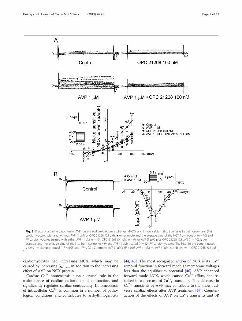
cardiomyocytes had increasing NCX, which may becaused by increasing INa-Late in addition to the increasingeffect of AVP on NCX protein.Cardiac Ca2+ homeostasis plays a crucial role in the
maintenance of cardiac excitation and contraction, andsignificantly regulates cardiac contractility. Inhomeostasisof intracellular Ca2+i is common in a number of patho-logical conditions and contributes to arrhythmogenicity
[44, 45]. The most recognized action of NCX is its Ca2+
removal function in forward mode at membrane voltagesless than the equilibrium potential [46]. AVP enhancedforward mode NCX, which caused Ca2+ efflux, and re-sulted in a decrease of Ca2+i transients. This decrease inCa2+i transients by AVP may contribute to the known ad-verse cardiac effects after AVP treatment [47]. Counter-action of the effects of AVP on Ca2+i transients and SR
Fig. 3 Effects of arginine vasopressin (AVP) on the sodium/calcium exchanger (NCX), and L-type calcium (ICa-L) currents in pulmonary vein (PV)cardiomyocytes with and without AVP (1 μM) or OPC 21268 (0.1 μM). a An example and the average data of the NCX from control (n = 10) andPV cardiomyocytes treated with either AVP (1 μM, n = 10), OPC 21268 (0.1 μM, n = 9), or AVP (1 μM) plus OPC 21268 (0.1 μM) (n = 10). b Anexample and the average data of the ICa-L from control (n = 9) and AVP (1 μM)-treated (n = 12) PV cardiomyocytes. The inset in the current tracesshows the clamp protocol. * P < 0.05 and **P < 0.01 Control vs AVP (1 μM); #P < 0.05 AVP (1 μM) vs AVP (1 μM) combined with OPC 21268 (0.1 μM
Huang et al. Journal of Biomedical Science (2019) 26:71 Page 7 of 11

Ca2+ content in PV cardiomyocytes by the antagonistOPC 21268, suggests that this effect is mainly V1 signaldependent. Previous study has found that AVP can elicitCa2+ entry through a receptor-mediated Ca2+-membranenon-selective cation channel in aortic smooth musclecells, which regulates smooth muscle contractility andenhances vascular tone. OPC 21268 was noted to reverseAVP-induced activation of nonselective cation currents inaortic smooth muscle cells [48]. PVs contain vascularstructure and cardiomyocytes. Previous studies haveshown that stretch increased PV arrhythmogenesisthrough mechano-electrical feedback [49]. Therefore,Ca2+ influx through vasopressin-induced nonselective cat-ion currents of PV smooth muscle cells may increase vas-cular stretch, further increasing PV arrhytmogenesisin vivo. In addition, the activity of vascular smooth musclecells may influence the membrane potential of PV cardio-myocytes via intercellular transfer of electrical signalsoccurring between PV cardiomyocytes and vascularsmooth myocytes of PVs, thereby exacerbating the pro-pensity of PV arrhythmogenesis or cardiac dysrhythmias.Moreover, several previous studies have demonstrated theability of caffeine to activate intermediate-conductanceCa2+-activated potassium channels [50–52], which are alsofunctionally expressed in PV cardiomyocytes [53]. There-fore, part of caffeine-mediated changes in cytosolic Ca2+
transient could be secondarily attributed to its activationof these channels.SR Ca2+ leak, the release of small amounts of Ca2+, oc-
curs when altered RyR spontaneously opens in diastole[54]. Diastolic Ca2+ release activates the forward modeof NCX current on the late diastolic depolarization. Thislate diastolic depolarization acceleration by NCX isrequired for the subsequent timely rapid AP upstroke[55, 56]. In the present study, AVP increased forwardmode of NCX as well as SR Ca2+ leak resulting steepnessof late diastolic potential, which could be responsible forincreasing automaticity [57]. Blocking AVP V1 receptorattenuated AVP-increased PV spontaneous activity,NCX and late diastolic potential, which implies AVP V1receptor may play a role in AVP-increased PV spontan-eous activity. In HF, RyR channels have increased single-channel open probability, which results in diastolic SRCa2+ leak and depletion of SR Ca2+ content, contributingto impaired contractility and HF progression [58, 59].We found that AVP-treated PV cardiomyocytes had sig-nificantly larger SR Ca2+ leak than control cells. Clinic-ally, greater SR Ca2+ leak was well demonstrated in atrialmyocytes in AF patients [44]. Greater Ca2+ leak may alsocontribute to a decrease in Ca2+i transients and SR Ca2+
content as well. CaMKII regulates several Ca2+-handlingproteins and has been shown to be a central regulator of
Fig. 4 Effects of arginine vasopressin (AVP) and its antagonist OPC 21268 on intracellular calcium (Ca2+) homeostasis and sarcoplasmic reticulum(SR) Ca2+ leak in pulmonary vein (PV) cardiomyocytes. a An example and average data of Ca2+i transients from control (n = 20), AVP (1 μM)-treated (n = 22), AVP (1 μM) combined with OPC 21268 (0.1 μM)-treated (n = 25), and OPC 21268 (0.1 μM)-treated (n = 11) PV cardiomyocytes. b Anexample and average data of caffeine-induced Ca2+i transients in control (n = 13) and PV cardiomyocytes treated with either AVP (1 μM, n = 15),AVP (1 μM) plus OPC 21268 (0.1 μM) (n = 21), or OPC 21268 (0.1 μM, n = 11). c An example and average data of SR Ca2+ leak from control (n = 14),AVP (1 μM)-treated (n = 16), and AVP (1 μM) combined with KN-93 (1 μM)-treated (n = 10) PV cardiomyocytes. *P < 0.05; **P < 0.01; ***P < 0.005
Huang et al. Journal of Biomedical Science (2019) 26:71 Page 8 of 11

excitation-contraction coupling [60], and increased CaM-KII expression was found in AF [61]. CaMKII-dependenthyperphosphorylation of the RyR leads to elevated SRCa2+ leak [61, 62], and triggers delayed afterdepolarizationvia activation of the NCX [63]. Diastolic SR Ca2+ leak canbe amplified by NCX, triggering ectopic focal dischargesor facilitating microreentry circuits promoting AF main-tenance [64]. In this study, the higher pCaMKII in AVP-treated PV cardiomyocytes may result in the increasedCa2+ leak. The attenuation effects of KN-93 on AVP-induced Ca2+ leak in PV cardiomyocytes also suggests thatactivation of CaMKII is important for the effects of AVPon PV cardiomyocytes. In addition, the greater INa-Late inAVP-treated PV cardiomyocytes may also arise from thehigher pCaMKII because activation of CaMKII is an im-portant activator of INa-Late. Enhanced INa-Late synergistic-ally increases the risk of cardiac arrhythmias by theactivation of CaMKII [17]. Accordingly, blocking the ef-fects of AVP on PV cardiomyocytes may reduce the riskfor HF-induced AF.Our study may be limited in some respects. First, the
PV cardiomyocytes received AVP for a relatively shorttime and AVP treatment of different duration may nothave the same effects. Furthermore, AVP is usuallyassociated with stress and pathological conditions. Weonly studied the effects of AVP on healthy PV
cardiomyocytes, it is unclear whether our findings ortheory applies to pathological settings such as HF. Fi-nally, the details of the molecular regulation responsiblefor the effects of AVP in PV cardiomyocytes has notbeen fully elucidated.
ConclusionsAVP increases PV arrhythmogenesis with dysregulatedCa2+ homeostasis through vasopressin V1 signaling.
AbbreviationsAF: Atrial fibrillation; APs: Action potentials; AVP: Arginine vasopressin;Ca2+i: Intracellular Ca
2+; CaMKII: Ca2+/calmodulin-dependent protein kinase II;DAG: Diacylglycerol; GAPDH: Glyceraldehyde-3-phosphate dehydrogenase;HF: Heart failure; ICa-L: L-type calcium current; IP3: Inositol trisphosphate;NCX: Sodium/calcium exchanger; NO: Nitric oxide; PAGE: Polyacrylamide gelby electrophoresis; PVs: Pulmonary veins; Rs: Series resistance; RyR: Ryanodinereceptor; SDS: Sodium dodecyl sulfate; SR: Sarcoplasmic reticulum
AcknowledgmentsNot applicable.
Authors’ contributionsJHH, YYL and YJC participated in the design and coordination of the study,performed experiments, analyzed data, and contributed to writing of themanuscript. YKL and SAC participated in the design and coordination of thestudy as well as helped to draft the manuscript. YCC performed experimentsand analyzed data. All authors read and approved the final manuscript.
Fig. 5 Effects of arginine vasopressin (AVP) on sodium (Na+)/calcium (Ca2+) exchanger (NCX) and Ca2+/calmodulin-dependent protein kinase II(CaMKII) in pulmonary vein (PV) cardiomyocytes. Representative immunoblot and average data of Na+/Ca2+ exchanger (NCX), Ca2+/calmodulin-dependent protein kinase II (CaMKII), and phosphorylated ratio of CaMKII from control (n = 6) and AVP (1 μM)-treated PV cardiomyocytes(n = 6). *P < 0.05
Huang et al. Journal of Biomedical Science (2019) 26:71 Page 9 of 11

FundingThis work was supported by grants from the Ministry of Science andTechnology (MOST 103–2314-B-038-055, MOST107–2314-B-038-101-MY3,MOST107–2314-B-038-095, MOST107–2314-B-281-009, and MOST107–2314-B-038-097-MY2), Taipei Medical University-Wan Fang Hospital (105-swf-02, 107-wf-swf-02, and 107-wf-eva-13), Chi-Mei Medical Center (107CM-TMU-04 andCMNDMC10804), and the Ministry of National Defense-Medical Affairs Bureau,Taiwan (MAB-107-044).
Availability of data and materialsAll data generated or analyzed during the current study are included in thispublished article.
Ethics approval and consent to participateThis study was approved by Institutional Animal Care and Use Committee atNational Defense Medical Center, Taipei (IACUC-15-006).
Consent for publicationNot Applicable.
Competing interestsThe authors declare that they have no competing interests.
Author details1Division of Cardiovascular Medicine, Department of Internal Medicine, WanFang Hospital, Taipei Medical University, 111 Hsin-Lung Road, Sec. 3, Taipei116, Taiwan. 2Department of Internal Medicine, School of Medicine, Collegeof Medicine, Taipei Medical University, Taipei, Taiwan. 3Department ofBiomedical Engineering, and Institute of Physiology, National DefenseMedical Center, Taipei, Taiwan. 4Division of Cardiology, Department ofInternal Medicine, Sijhih Cathay General Hospital, New Taipei City, Taiwan.5School of Medicine, Fu-Jen Catholic University, New Taipei City, Taiwan.6Heart Rhythm Center and Division of Cardiology, Department of Medicine,Taipei Veterans General Hospital, Taipei, Taiwan. 7Graduate Institute ofClinical Medicine, College of Medicine, Taipei Medical University, Taipei,Taiwan. 8Cardiovascular Research Center, Wan Fang Hospital, Taipei MedicalUniversity, Taipei, Taiwan.
Received: 29 March 2019 Accepted: 10 September 2019
References1. Barrett LK, Singer M, Clapp LH. Vasopressin: mechanisms of action on the
vasculature in health and in septic shock. Crit Care Med. 2007;35:33–40.2. Chen X, Lu G, Tang K, Li Q, Gao X. The secretion patterns and roles of
cardiac and circulating arginine vasopressin during the development ofheart failure. Neuropeptides. 2015;51:63–73.
3. Holmes CL, Landry DW, Granton JT. Science review: vasopressin and thecardiovascular system part 1--receptor physiology. Crit Care. 2003;7:427–34.
4. Goldsmith SR, Francis GS, Cowley AW Jr, Levine TB, Cohn JN. Increasedplasma arginine vasopressin levels in patients with congestive heart failure.J Am Coll Cardiol. 1983;1:1385–90.
5. Nakamura T, Funayama H, Yoshimura A, Tsuruya Y, Saito M, Kawakami M,Ishikawa SE. Possible vascular role of increased plasma arginine vasopressinin congestive heart failure. Int J Cardiol. 2006;106:191–5.
6. Costello-Boerrigter LC, Boerrigter G, Burnett JC Jr. Pharmacology ofvasopressin antagonists. Heart Fail Rev. 2009;14:75–82.
7. Lu TL, Chang WT, Chan CH, Wu SN. Evidence for effective multiple K(+)-current inhibitions by Tolvaptan, a non-peptide antagonist of vasopressinV2 receptor. Front Pharmacol. 2019;10:76.
8. Walker BR, Haynes JJ, Wang HL, Voelkel NF. Vasopressin-induced pulmonaryvasodilation in rats. Am J Phys. 1989;257:H415–22.
9. Nemenoff RA. Vasopressin signaling pathways in vascular smooth muscle.Front Biosci. 1998;3:d194–207.
10. Henderson KK, Byron KL. Vasopressin-induced vasoconstriction: twoconcentration-dependent signaling pathways. J Appl Physiol (1985). 2007;102:1402–9.
11. Mani BK, Brueggemann LI, Cribbs LL, Byron KL. Opposite regulation ofKCNQ5 and TRPC6 channels contributes to vasopressin-stimulatedcalcium spiking responses in A7r5 vascular smooth muscle cells. CellCalcium. 2009;45:400–11.
12. Lu YY, Cheng CC, Chen YC, Lin YK, Chen SA, Chen YJ. Electrolytedisturbances differentially regulate sinoatrial node and pulmonary veinelectrical activity: a contribution to hypokalemia- or hyponatremia-inducedatrial fibrillation. Heart Rhythm. 2016;13:781–8.
13. Weiss ML, Kenney MJ, Musch TI, Patel KP. Modifications to central neuralcircuitry during heart failure. Acta Physiol Scand. 2003;177:57–67.
14. Cheng Y, Pan T, Ge M, Chen T, Ye J, Lu L, Chen C, Zong Q, Ding Y, Wang D.Evaluation of vasopressin for Vasoplegic shock in patients with preoperativeleft ventricular dysfunction after cardiac surgery: a propensity-score analysis.Shock. 2018;50:519–24.
15. Personett HA, Stollings JL, Cha SS, Oyen LJ. Predictors of prolonged vasopressininfusion for the treatment of septic shock. J Crit Care. 2012;27:318 e7–12.
16. Chen YC, Lu YY, Cheng CC, Lin YK, Chen SA, Chen YJ. Sinoatrial nodeelectrical activity modulates pulmonary vein arrhythmogenesis. Int J Cardiol.2014;173:447–52.
17. Ma J, Luo A, Wu L, Wan W, Zhang P, Ren Z, Zhang S, Qian C, Shryock JC,Belardinelli L. Calmodulin kinase II and protein kinase C mediate the effectof increased intracellular calcium to augment late sodium current in rabbitventricular myocytes. Am J Physiol Cell Physiol. 2012;302:C1141–51.
18. Lu YY, Wu WS, Lin YK, Cheng CC, Chen YC, Chen SA, Chen YJ. Angiotensin1-7 modulates electrophysiological characteristics and calciumhomoeostasis in pulmonary veins cardiomyocytes via MAS/PI3K/eNOSsignalling pathway. Eur J Clin Investig. 2018;48:e12854.
19. Chang SL, Chen YC, Yeh YH, Lin YK, Wu TJ, Lin CI, Chen SA, Chen YJ. Heartfailure enhanced pulmonary vein arrhythmogenesis and dysregulatedsodium and calcium homeostasis with increased calcium sparks. JCardiovasc Electrophysiol. 2011;22:1378–86.
20. Hussain A, Bennett R, Haqzad Y, Qadri S, Chaudhry M, Cowen M, Loubani M,Morice A. The differential effects of systemic vasoconstrictors on humanpulmonary artery tension. Eur J Cardiothorac Surg. 2017;51:880–6.
21. Currigan DA, Hughes RJ, Wright CE, Angus JA, Soeding PF. Vasoconstrictorresponses to vasopressor agents in human pulmonary and radial arteries: anin vitro study. Anesthesiology. 2014;121:930–6.
22. Miyazaki T, Fujiki H, Yamamura Y. Tolvaptan, an orally active non-peptidearginine vasopressin V2 receptor antagonist, reduces ascites in rats withchronic liver injury. Hepatol Res. 2013;43:1224–30.
23. Tamma G, Di Mise A, Ranieri M, Geller A, Tamma R, Zallone A, Valenti G. TheV2 receptor antagonist tolvaptan raises cytosolic calcium and preventsAQP2 trafficking and function: an in vitro and in vivo assessment. J Cell MolMed. 2017;21:1767–80.
24. Vasylyev DV, Waxman SG. Membrane properties and electrogenesis in thedistal axons of small dorsal root ganglion neurons in vitro. J Neurophysiol.2012;108:729–40.
25. Tse G. Mechanisms of cardiac arrhythmias. J Arrhythm. 2016;32:75–81.26. Bogdanov KY, Maltsev VA, Vinogradova TM, Lyashkov AE, Spurgeon HA,
Stern MD, Lakatta EG. Membrane potential fluctuations resulting fromsubmembrane Ca2+ releases in rabbit sinoatrial nodal cells impart anexponential phase to the late diastolic depolarization that controls theirchronotropic state. Circ Res. 2006;99:979–87.
27. Carmeliet E. Pacemaking in cardiac tissue. From IK2 to a coupled-clocksystem. Physiol Rep. 2019;7:e13862.
28. Ma J, Guo L, Fiene SJ, Anson BD, Thomson JA, Kamp TJ, Kolaja KL, SwansonBJ, January CT. High purity human-induced pluripotent stem cell-derivedcardiomyocytes: electrophysiological properties of action potentials andionic currents. Am J Physiol Heart Circ Physiol. 2011;301:H2006–17.
29. Clancy CE, Tateyama M, Liu H, Wehrens XH, Kass RS. Non-equilibrium gatingin cardiac Na+ channels: an original mechanism of arrhythmia. Circulation.2003;107:2233–7.
30. Zygmunt AC, Eddlestone GT, Thomas GP, Nesterenko VV, Antzelevitch C. Largerlate sodium conductance in M cells contributes to electrical heterogeneity incanine ventricle. Am J Physiol Heart Circ Physiol. 2001;281:H689–97.
31. Horvath B, Banyasz T, Jian Z, Hegyi B, Kistamas K, Nanasi PP, Izu LT, Chen-IzuY. Dynamics of the late Na(+) current during cardiac action potential and itscontribution to afterdepolarizations. J Mol Cell Cardiol. 2013;64:59–68.
32. Lu YY, Cheng CC, Tsai CF, Lin YK, Lee TI, Chen YC, Chen SA, Chen YJ.Discrepant effects of heart failure on electrophysiological property in rightventricular outflow tract and left ventricular outflow tract cardiomyocytes.Clin Sci (Lond). 2017;131:1317–27.
33. Ono K, Fozzard HA. Phosphorylation restores activity of L-type calciumchannels after rundown in inside-out patches from rabbit cardiac cells. JPhysiol. 1992;454:673–88.
Huang et al. Journal of Biomedical Science (2019) 26:71 Page 10 of 11

34. Lu YY, Chen YC, Kao YH, Wu TJ, Chen SA, Chen YJ. Extracellular matrix ofcollagen modulates intracellular calcium handling and electrophysiologicalcharacteristics of HL-1 cardiomyocytes with activation of angiotensin II type1 receptor. J Card Fail. 2011;17:82–90.
35. Sherpa RT, Mohieldin AM, Pala R, Wachten D, Ostrom RS, Nauli SM.Sensory primary cilium is a responsive cAMP microdomain in renalepithelia. Sci Rep. 2019;9:6523.
36. Gutkowska J, Miszkurka M, Danalache B, Gassanov N, Wang D, Jankowski M.Functional arginine vasopressin system in early heart maturation. Am JPhysiol Heart Circ Physiol. 2007;293:H2262–70.
37. Kaufmann JE, Iezzi M, Vischer UM. Desmopressin (DDAVP) induces NOproduction in human endothelial cells via V2 receptor- and cAMP-mediatedsignaling. J Thromb Haemost. 2003;1:821–8.
38. Wasilewski MA, Myers VD, Recchia FA, Feldman AM, Tilley DG. Argininevasopressin receptor signaling and functional outcomes in heart failure. CellSignal. 2016;28:224–33.
39. Reilly BA, Brostrom MA, Brostrom CO. Regulation of protein synthesis inventricular myocytes by vasopressin. The role of sarcoplasmic/endoplasmicreticulum Ca2+ stores. J Biol Chem. 1998;273:3747–55.
40. Lin YK, Chen YC, Chen JH, Chen SA, Chen YJ. Adipocytes modulate theelectrophysiology of atrial myocytes: implications in obesity-induced atrialfibrillation. Basic Res Cardiol. 2012;107:293.
41. Suenari K, Chen YC, Kao YH, Cheng CC, Lin YK, Chen YJ, Chen SA.Discrepant electrophysiological characteristics and calcium homeostasis ofleft atrial anterior and posterior myocytes. Basic Res Cardiol. 2011;106:65–74.
42. Sicouri S, Belardinelli L, Antzelevitch C. Antiarrhythmic effects of the highlyselective late sodium channel current blocker GS-458967. Heart Rhythm.2013;10:1036–43.
43. Schotten U, Greiser M, Benke D, Buerkel K, Ehrenteidt B, Stellbrink C,Vazquez-Jimenez JF, Schoendube F, Hanrath P, Allessie M. Atrial fibrillation-induced atrial contractile dysfunction: a tachycardiomyopathy of a differentsort. Cardiovasc Res. 2002;53:192–201.
44. Hove-Madsen L, Llach A, Bayes-Genis A, Roura S, Rodriguez Font E, Aris A,Cinca J. Atrial fibrillation is associated with increased spontaneous calciumrelease from the sarcoplasmic reticulum in human atrial myocytes.Circulation. 2004;110:1358–63.
45. Huke S, Bers DM. Ryanodine receptor phosphorylation at serine 2030,2808 and 2814 in rat cardiomyocytes. Biochem Biophys Res Commun.2008;376:80–5.
46. Blaustein MP, Lederer WJ. Sodium/calcium exchange: its physiologicalimplications. Physiol Rev. 1999;79:763–854.
47. Wang D, Luo P, Wang Y, Li W, Wang C, Sun D, Zhang R, Su T, Ma X, Zeng C,Wang H, Ren J, Cao F. Glucagon-like peptide-1 protects against cardiacmicrovascular injury in diabetes via a cAMP/PKA/rho-dependentmechanism. Diabetes. 2013;62:1697–708.
48. Nakajima T, Hazama H, Hamada E, Wu SN, Igarashi K, Yamashita T,Seyama Y, Omata M, Kurachi Y. Endothelin-1 and vasopressin activateca (2+)-permeable non-selective cation channels in aortic smoothmuscle cells: mechanism of receptor-mediated Ca2+ influx. J Mol CellCardiol. 1996;28:707–22.
49. Chang SL, Chen YC, Chen YJ, Wangcharoen W, Lee SH, Lin CI, Chen SA.Mechanoelectrical feedback regulates the arrhythmogenic activity ofpulmonary veins. Heart. 2007;93:82–8.
50. Schroder RL, Jensen BS, Strobaek D, Olesen SP, Christophersen P. Activationof the human, intermediate-conductance, Ca2+−activated K+ channel bymethylxanthines. Pflugers Arch. 2000;440:809–18.
51. Diaz P, Wood AM, Sibley CP, Greenwood SL. Intermediate conductanceCa2+−activated K+ channels modulate human placental trophoblastsyncytialization. PLoS One. 2014;9:e90961.
52. Wulff H, Kolski-Andreaco A, Sankaranarayanan A, Sabatier JM, ShakkottaiV. Modulators of small- and intermediate-conductance calcium-activatedpotassium channels and their therapeutic indications. Curr Med Chem.2007;14:1437–57.
53. Chen WT, Chen YC, Lu YY, Kao YH, Huang JH, Lin YK, Chen SA, Chen YJ.Apamin modulates electrophysiological characteristics of the pulmonaryvein and the sinoatrial node. Eur J Clin Investig. 2013;43:957–63.
54. Yano M, Yamamoto T, Kobayashi S, Matsuzaki M. Role of ryanodinereceptor as a ca (2)(+) regulatory center in normal and failing hearts. JCardiol. 2009;53:1–7.
55. Monfredi O, Maltsev VA, Lakatta EG. Modern concepts concerning the originof the heartbeat. Physiology (Bethesda). 2013;28:74–92.
56. Lakatta EG, Maltsev VA, Vinogradova TM. A coupled SYSTEM of intracellularCa2+ clocks and surface membrane voltage clocks controls the timekeepingmechanism of the heart's pacemaker. Circ Res. 2010;106:659–73.
57. Kim JJ, Yang L, Lin B, Zhu X, Sun B, Kaplan AD, Bett GC, Rasmusson RL, LondonB, Salama G. Mechanism of automaticity in cardiomyocytes derived fromhuman induced pluripotent stem cells. J Mol Cell Cardiol. 2015;81:81–93.
58. Marx SO, Reiken S, Hisamatsu Y, Jayaraman T, Burkhoff D, Rosemblit N,Marks AR. PKA phosphorylation dissociates FKBP12.6 from the calciumrelease channel (ryanodine receptor): defective regulation in failing hearts.Cell. 2000;101:365–76.
59. Marks AR. Calcium cycling proteins and heart failure: mechanisms andtherapeutics. J Clin Invest. 2013;123:46–52.
60. Maier LS, Bers DM. Role of Ca2+/calmodulin-dependent protein kinase(CaMK) in excitation-contraction coupling in the heart. Cardiovasc Res.2007;73:631–40.
61. Dobrev D, Wehrens XH. Calmodulin kinase II, sarcoplasmic reticulum Ca2+leak, and atrial fibrillation. Trends Cardiovasc Med. 2010;20:30–4.
62. Neef S, Dybkova N, Sossalla S, Ort KR, Fluschnik N, Neumann K, Seipelt R,Schondube FA, Hasenfuss G, Maier LS. CaMKII-dependent diastolic SR Ca2+leak and elevated diastolic Ca2+ levels in right atrial myocardium ofpatients with atrial fibrillation. Circ Res. 2010;106:1134–44.
63. Voigt N, Li N, Wang Q, Wang W, Trafford AW, Abu-Taha I, Sun Q, Wieland T,Ravens U, Nattel S, Wehrens XH, Dobrev D. Enhanced sarcoplasmicreticulum Ca2+ leak and increased Na+−Ca2+ exchanger function underliedelayed afterdepolarizations in patients with chronic atrial fibrillation.Circulation. 2012;125:2059–70.
64. Dobrev D, Voigt N, Wehrens XH. The ryanodine receptor channel as amolecular motif in atrial fibrillation: pathophysiological and therapeuticimplications. Cardiovasc Res. 2011;89:734–43.
Publisher’s NoteSpringer Nature remains neutral with regard to jurisdictional claims inpublished maps and institutional affiliations.
Huang et al. Journal of Biomedical Science (2019) 26:71 Page 11 of 11