arterial anatomy and hemodynamics - nwprimarycare articles/arterial_anatomy_and...arterial anatomy...
TRANSCRIPT

ProSono copyright 2006
Arterial Anatomy and Hemodynamics 1
Three layers of vascular tissue.
Arterial Anatomyand
Hemodynamics
Arterial AnatomyThe architectural arrangement and structure of arteries and veins are
important in the distribution of blood to and from the capillary beds where the realwork of the vascular system is accomplished. The arteries deliver blood to thecapillaries and the veins return it to the heart.
The arteries are distributed throughout the body in a systematic manner. Thevessels leaving the heart are large but soon divide into branches. This divisioncontinues until minute branches are distributed to all parts of the body. At eachdivision the branches are smaller, but since they are numerous, the total of theirdiameters is much greater than that of the artery from which they sprang. Thismeans that as the blood flows from the heart toward the capillaries it flows in anever-widening bed. The diameter of the aorta at the heart is about 2-3cm. Thebranches of the large arteries leave them at abrupt angles; the branches of thesmaller arteries take progressively less abrupt changes of direction.
An inner coat (tunica intima)consists of three layers—a layer ofendothelial cells, a layer of delicateconnective tissue, which is found onlyin vessels of considerable size, and anelastic layer consisting of a membraneor network of elastic fibers.
A middle coat (tunica media)consists mainly of smooth-musclefibers with various amounts of elasticand collagenous tissue. In the largerarteries elastic fibers form layerswhich alternate with the layers ofmuscle fibers. In the largest arterieswhite connective-tissue fibers havebeen found in this coat. The externalcoat (tunica externa, or adventitia) is

ProSono copyright 2006
Arterial Anatomy and Hemodynamics 2
composed of loose connective tissue in which there are scattered smooth musclecells or bundles of cells arranged longitudinally. In all but the smallest arteriesthis coat contains some elastic tissue. The structure and relative thickness varywith the size of the artery.
The great extensibility of the arteries enables them to receive the additionalamount of blood forced into them at each contraction of the heart. Elasticity ofarteries serves as a buffer to the large volume of blood forced into the system bythe heartbeat. If these vessels were rigid (as is true in arteriosclerosis), thesystolic blood pressure would be markedly increased. The strength of an arterydepends largely upon the outer coat; it is far less easily cut or torn than the othercoats and serves to resist undue expansion of the vessel.
The arteries do not collapse when empty, and when an artery is severed, theorifice remains open. The muscular coat, however, contracts somewhat in theregion of the opening, and the elastic fibers cause the artery to retract a littlewithin its sheath, so as to diminish its caliber and permit a blood clot to plug theorifice. This property of a severed artery is an important factor in the arrest ofhemorrhage. Most of the arteries are accompanied by a nerve and one or twoveins, all surrounded by a sheath of connective tissue, which helps to supportand hold these structures in position.
SIZE OF THE ARTERIES The largest arteries in the body, the aorta andpulmonary artery, measure more than 3 cm in diameter at their connection withthe heart. These arteries give off branches, which divide and subdivide intosmaller branches. The smallest arteries are called arterioles, and at their distalends, where only the internal coat remains, the capillaries begin. The arteriolarwalls contain a great proportion of smooth muscle in relation to elastic tissue, andthey are to be thought of as muscular rather than elastic.
THE ELASTIC ARTERIES. These include the large arteries and are calledconducting arteries because they conduct blood from the heart to the medium-sized arteries. The middle coat contains a large amount of elastic tissue, and thewall is comparatively thin for the size of the vessel.
THE MUSCULAR ARTERIES. These include the arteries of medium size, andtheir middle coat is chiefly muscular. Muscular arteries are also called distributingarteries because they distribute the blood to the various organs and bycontraction or relaxation they aid in regulating the volume of blood passing tostructures to meet varying functional demands.
DIVISION OF ARTERIES: The way in which the arteries divide varies. (1) Anartery may give off several branches in succession and still continue as a maintrunk, e.g., the thoracic or abdominal portion of the aorta. (2) A short trunk maysubdivide into several branches at the same point, e.g., the celiac artery. (3) Anartery may divide into two branches of nearly equal size, e.g., the division of theaorta into the two common iliacs.

ProSono copyright 2006
Arterial Anatomy and Hemodynamics 3
BLOOD SUPPLY OF THE ARTERIES. The blood which flows through thearteries nourishes only the inner coat. The external and middle coats aresupplied with arteries, capillaries, and veins, called vasa vasorum, or bloodvessels of the blood vessels.
VASOMOTOR NERVES. The muscular tissue in the walls of the blood vessels iswell supplied with nerve fibers, chiefly from the sympathetic portion of theautonomic system. These nerve fibers are called vasomotor and are divided intotwo sets: (1) vasoconstrictor and (2) vasodilator. A center in the medullaoblongata (vasoconstrictor center) is constantly sending impulses to the vessels,thus keeping them in a state of tone. The vasoconstrictor center is a reflex centerand is connected with afferent fibers coming from all parts of the body.Vasoconstrictor fibers are sympathetic and are widely distributed to arteries andarterioles. They mediate constriction of vessels, and by tonic action speed ofblood flow is controlled. Vasodilator nerve fibers have several origins and arefound on the sympathetic, parasympathetic, and somatic sensory nerves. Thereis no direct evidence that they are tonically active, but they appear to “discharge selectively” when a local increase in blood flow is needed.
There is a diffuse network of sympathetic nerve fibers in the adventitia of allarteries, called the periarterial plexus. Nerve fibers are also present in themuscular coat. Arterioles are directly and completely under nervous control.Pressure from increased volume, exerted on the blood stream in the musculararteries, causes relaxation of the arterioles and more blood can move through tothe capillary bed. The exact function of vasodilator nerve fibers is not wellunderstood. Sudden, widespread relaxation of arterioles lowers blood pressureby decreasing peripheral resistance and shock may result.As the arteries decrease in size and approach the capillary network, they arecalled arterioles. Proximal to the capillary channel there are modified arteriolescalled metarterioles. They have a wall which contains widely separated smooth-muscle cells. A precapillary sphincter is located around the arteriole before itenters the capillary net. Arterioles are well supplied with vasoconstrictor fibers.
The capillaries are exceedingly minute vessels which average about 7 to 9min diameter. They connect the arterioles (smallest arteries) with the venules(smallest veins).
STRUCTURE. The walls of the capillaries consist of one layer of endothelialcells continuous with the layer that line the arteries, the veins, and the heart.These cells are held together by cell “cement.” There is a substance called hyaluronic acid that forms a gelatinous material in the cell membrane and tissuespaces. It holds cells together and binds water in the tissues.
Capillaries

ProSono copyright 2006
Arterial Anatomy and Hemodynamics 4
Vascular connections in thecapillary bed.
DISTRIBUTION. The capillaries communicate freely with one another andform interlacing networks of variable form and size in the different tissues. All thetissues, with the exception of the cartilages, hair, nails, cuticle and cornea of theeye, are traversed by networks of capillary vessels. The capillary diameter is sosmall that the blood cells often must pass through them in single file, and veryfrequently the cell islarger than the caliber ofthe vessel and becomesdistorted as it passesthrough. In many partsthe capillaries lie soclose together that apin’s point cannot be inserted between them.They are most abundantand form the finestnetworks in those organswhere the blood is neededfor purposes other than
local nutrition, such as,for example, secretion or absorption
FUNCTION. It is in the capillaries that the chief work of the blood is done,and the object of the vascular mechanism is to cause the blood to flow throughthese vessels in a steady volume. There are an estimated 7,000 sq meters ofblood capillaries in the adult body. This gives a large area for exchange ofsubstances between the blood and tissue fluid. In the glandular organs thecapillaries supply the substances requisite for secretion; in the ductless glandsthey also take up the products of secretion; in the alimentary canal they take upsome of the digested food; in the lungs they absorb oxygen and give up carbondioxide; in the kidneys they discharge the waste products collected from otherparts; all of the time, everywhere in the body, through their walls an interchangeis going on which is essential to the life of the body. The greater the metabolicactivity of the tissue, the denser the capillary nets.
The shorter circulation, from the rightventricle to the left atrium, is called thepulmonary circulation. The purpose of thepulmonary circulation is to carry the bloodwhich has been through the body, giving upoxygen and collecting carbon dioxide, to theair sacs of the lungs, where the red cells arerecharged with oxygen and the carbondioxide is reduced to the normal amount.
Pulmonary Circulation
ProSono copyright 2006
Arterial Anatomy and Hemodynamics 5
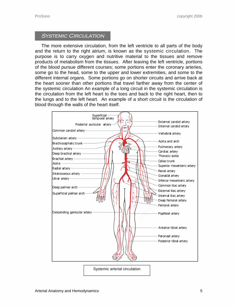
The more extensive circulation, from the left ventricle to all parts of the bodyand the return to the right atrium, is known as the systemic circulation. Thepurpose is to carry oxygen and nutritive material to the tissues and removeproducts of metabolism from the tissues. After leaving the left ventricle, portionsof the blood pursue different courses; some portions enter the coronary arteries,some go to the head, some to the upper and lower extremities, and some to thedifferent internal organs. Some portions go on shorter circuits and arrive back atthe heart sooner than other portions that travel farther away from the center ofthe systemic circulation An example of a long circuit in the systemic circulation isthe circulation from the left heart to the toes and back to the right heart, then tothe lungs and to the left heart. An example of a short circuit is the circulation ofblood through the walls of the heart itself.
Systemic Circulation
Systemic arterial circulation

ProSono copyright 2006
Arterial Anatomy and Hemodynamics 6
Arterial physiologyEach cell of the body is dependent on the blood for its very existence, and it is
the work of the heart, arteries, capillaries, and veins that makes possible thetransporting of all substances to and from cells.
The function of the heart is to adjust circulation in relation to the metabolicrate of body cells. By chemical and nervous control, the needs of these cells aremet promptly through adjustments of pulse rate and pulse volume, whichincrease and decrease the volume of blood reaching the tissue capillaries perminute. Variable cellular needs are thus met by changes in the number, size, andarea of the open capillaries and in the temperature and the minute volume of theblood in these open capillaries.
The blood is contained in a closed set of vessels, which it completely fills.Interposed in this set of vessels is the heart, which fills with blood from the veinsand then contracts, thereby, forcing this blood into the capillaries of all parts ofthe body.
DISTRIBUTION OF BLOOD TO DIFFERENT PARTS OF THE BODY.In healthily tissue the distribution of blood varies, as determined by the needs
of the different parts. When the digestive organs are active, they need an extrasupply of blood, which may be supplied by redistribution of blood from less activeorgans. Other causes may result in an increased supply of blood to an organ. Ifthe skin is exposed to high temperatures, the arterioles which bring blood to it aredilated, and the blood flow near the surface is increased. This aids in theradiation of heat and in the control of body temperature. On the other hand, slightchilling causes contraction of the skin arterioles and resulting paleness. Theblood supply to the brain is relatively constant. According to recent studies, bloodsupply to the brain is not reduced during sleep and not increased by mentalactivity
TONUS OF BLOOD VESSELSNormally the blood vessels maintain a state of tonus about halfway between
contraction and dilatation. It is thought that adjustments in the blood supply tovarious parts are brought about by increasing or decreasing the tone of the localblood vessels. Two factors are important, (1) vasomotor nerve fibers and (2)chemical stimuli.1. The vasomotor nerve fibers consist of two antagonistic sets. The
vasoconstrictors cause the muscular coats of the blood vessels to contract,lessen the diameter of the vessels, and thereby increase resistance to bloodflow. The vasodilator fibers increase the diameter of the blood vessels,probably by allowing the muscular coats to relax, and thereby decreaseresistance to blood flow.
2. Chemical substances, such as the lactic acid and carbon dioxide producedduring muscular activity, may lessen the tonus of the blood vessels in the partaffected, resulting in local dilatation and an increased supply of blood to thepart needing it. At the same time, chemoreceptors convey impulses to the

ProSono copyright 2006
Arterial Anatomy and Hemodynamics 7
vasoconstrictor center, stimulate it, and thereby increase the tonus of bloodvessels in other parts of the body. On the other hand, angiotensin andhormones, such as epinephrine and vasopresssin, cause constriction of theblood vessels.Epinephrine and many other drugs are used medicinally to cause
vasoconstriction, and amyl nitrite is inhaled to bring about vasodilatation,particularly when a condition like angina pectoris makes quick relief necessary.Both the arteries and the veins are capable of dilatation or constriction under theinfluence of nerve fibers or chemical stimuli.
In surgical shock there is marked interference with the circulation of the bloodowing to dilatation of the arteriolar bed and consequent decrease in arterialpressure, which may fall below the level essential to the welfare of the tissues.The pulse becomes rapid and weak, and respiration increases. It is thought thatdilatation of the arterioles may be brought about by substances such ashistamine formed in injured tissues.
Several physiological factors, which control or affect blood flow into humantissues, have a direct impact on alterations of blood flow patterns seen duringsonographic evaluation of the arteries.
LOCAL CONTROL of blood flow into tissue is governed largely by nutritionalneeds of the tissue itself. Metabolically active tissue requires a constant supplyof oxygen and nutrients and removal of waste products. Therefore perfusionaldemands of organs such as the brain, kidneys and heart are constant.Perfusional demands of tissue that is not constantly active at the same metabolicrate will vary; blood flow rates and patterns into these tissues will vary accordingto current needs.
NEURAL CONTROLof blood flow is mediated through the sympathetic nervoussystem that innervates arterial and venous systems. The sympathetic nervoussystem, which is centered in the spinal cord and ganglia innervate viscera, heart,lungs. It controls motor, vasomotor and secretory reflexes. It produces thefollowing physiologic reactions:
1. Dilatation of pupils2. Bronchodilation/constriction3. Vasodilation/constriction4. Sweating, increase/decrease salivary production5. Increased adrenal secretions6. Sphincter/bladder control7. Uterine contractions8. Hair stands on end (piloerector muscles)
Physiological Controls

ProSono copyright 2006
Arterial Anatomy and Hemodynamics 8
HUMORAL CONTROL is the control of arterial tonus by chemical substancesthat typically circulate in the blood stream. Humoral regulation causes vessels toconstrict or dilate for example, angiotensin and vasopressin are potentvasoconstrictors whereas histamine and bradykinin are vasodilators.
AUTOREGULATION is the ability of tissues to control their own blood flow. Lackof oxygen or accumulation of metabolites (lactic acid and carbon dioxide) causesdilatation of capillary channels in an attempt to channel more blood into thehypoxic area. There are two components of physiologic autoregulation thatimpact flow dynamics encountered in arterial pathology:
1. Hyperemia which is a generalized increase in blood flow to a part. Thisincrease in flow volume may be either:
Reactive: when blood supply to an area has been chronically diminishedand is suddenly restored, increased blood volume flows into the tissuecausing it to become red and warm and sometimes painful. Thisphenomenon is frequently seen in a condition called Reynaud’s disease. Reactive hyperemia is also responsible for the red, hot fingers seen inhands that have come in from the cold.
Functional which is an increase in flow based on physiological needs.Examples include exercising skeletal muscle and increases inmetabolism in rapidly growing tissues such as cancer or earlypregnancies.
2. Collateral circulation is a mechanism for long-term re-perfusion oftissues. When mechanical or physiological obstruction blocks normalperfusional channels to living tissue, anatomic anastomotic channels carryblood to that area. Over time, these channels dilate and blood flowincreases to meet nutritional needs of the tissue being perfused.Collateral perfusion works best when obstruction to flow is gradual(chronic) and not sudden (acute). Examples of collateral circulatory flowcan be seen in almost all major human organ systems when primary flowpaths are diminished or obliterated.

ProSono copyright 2006
Arterial Anatomy and Hemodynamics 9
Cardiovascular FunctionThe most important factors maintaining arterial circulation are:
Pumping action of the heart (See Chapter 4, section onHemodynamics–Cardiac Energy)
Elasticity and extensibility of the arterial wallsPeripheral resistance in distal vascular bedsQuantity of blood in the body
During each systole the ventricles force blood into arteries that are already full(about 30 to 60 ml each). The extensibility of the arteries enables them to distendand receive this extra supply of blood. The period of distention corresponds tothe systole of the heart. Just as soon as the force is removed, the elasticity of thearteries causes them to contract to their former diameter, and this exerts such apressure on the contained blood that the blood is forced into the capillaries justrapidly enough to allow the arteries time to reach their usual size during diastoleof the heart. The arteries thus not only serve as conducting vessels but exert aforce that assists the heart in driving the blood into the capillaries.
The extensibility and elasticity of the arteries change with the health and ageof the individual. Sometimes as the result of disease, and usually with age, thearterial walls become less elastic and less well adapted for the unceasing workthey are called upon to perform.
Blood flow is opposed by frictional forces within the vessels. Friction resultsfrom the relationships between the layers of fluid wetting the vessel walls and themore central layers of the moving stream. Frictional resistance to flow variestherefore with the character of the fluid, that is, with its viscosity. This means thatthere is direct relationship between viscosity of the blood and peripheralresistance. The greater the viscosity, the greater the resistance to blood flow.It is the function of the vasomotor fibers to “set” the diameters of the muscular
arterioles in relation to constantly varying local needs for blood. The elasticarteries compensate for heart systole and diastole, thus maintaining a steadyflow of blood in the capillaries, the arteries accommodating the extra blood forcedinto them during heart systole and by their contraction forcing the blood towardthe capillaries during heart diastole. Inasmuch as local needs for blood vary con-stantly and through constantly varying limits, it is the function of the autonomicnervous system (and locally produced chemical substances such as carbondioxide), reflecting these needs, to set the diameters of the arterioles so that theperipheral resistance meets these local needs (much blood needed, wide
Elasticity of Arterial Walls
Peripheral Resistance
ProSono copyright 2006
Arterial Anatomy and Hemodynamics 10
arterioles; less blood needed, narrower arterioles). On the basis of peripheralresistance thus established, it is the function of the arterioles, to expand andcontract, changing an intermittent flow in the arteries to a steady flow incapillaries. It is easily seen that this is a fine adjustment, the elastic arteriesgiving a steady flow in capillaries on many bases of diameter of arterioles set bylocal needs. This fine adjustment (associated with optimum activity of the heart)is the mechanism by means of which homeostasis, or state of constancy, of bodyfluids is maintained.
The kidneys play an important role in the regulation of arterial pressure. It isknown that the kidneys can regulate arterial pressure by increasing urine output,which decreases volume and lowers pressure, or by decreasing urine output,thereby increasing blood volume, which raises arterial pressure. If the kidney isdeprived of part of its blood supply from any cause, blood pressure rises. Whenthe kidney is deprived of part of its blood supply, it secretes renin. Reninactivates a globulin of a plasma protein which eventually becomes a substancecalled angiotensin II. This is the most active vasopressor substance known. Italso acts on the adrenal cortex to increase aldosterone secretion, which stimu-lates the kidney to retain sodium and water. The net effect is an increase in bloodpressure.
It is evident that, other things being equal, the quantity of blood to be moved isan important factor. Except in cases of severe hemorrhage, loss of blood iscompensated for by a transfer of liquid from the tissues into the blood vessels.
Quantity of Blood
ProSono copyright 2006
Arterial Anatomy and Hemodynamics 11
Doppler Measurement of Arterial FlowSpectral Doppler techniques permit a variety of measurements to be made of
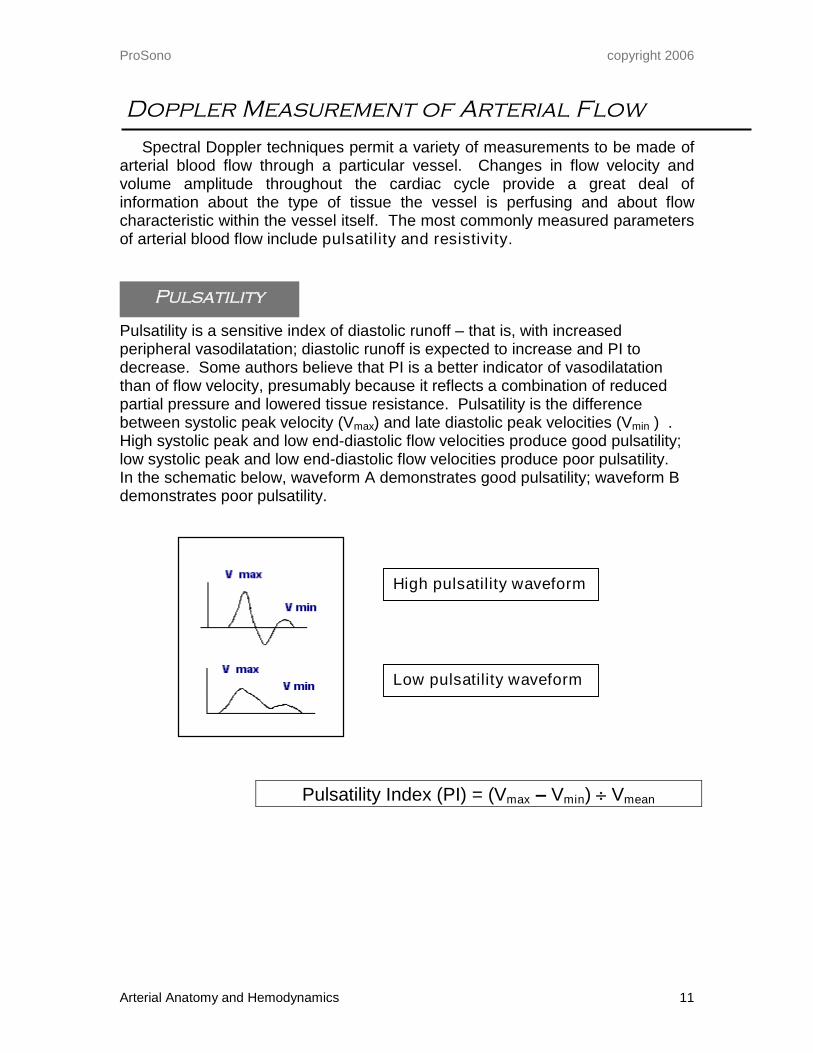
arterial blood flow through a particular vessel. Changes in flow velocity andvolume amplitude throughout the cardiac cycle provide a great deal ofinformation about the type of tissue the vessel is perfusing and about flowcharacteristic within the vessel itself. The most commonly measured parametersof arterial blood flow include pulsatility and resistivity.
Pulsatility is a sensitive index of diastolic runoff–that is, with increasedperipheral vasodilatation; diastolic runoff is expected to increase and PI todecrease. Some authors believe that PI is a better indicator of vasodilatationthan of flow velocity, presumably because it reflects a combination of reducedpartial pressure and lowered tissue resistance. Pulsatility is the differencebetween systolic peak velocity (Vmax) and late diastolic peak velocities (Vmin ) .High systolic peak and low end-diastolic flow velocities produce good pulsatility;low systolic peak and low end-diastolic flow velocities produce poor pulsatility.In the schematic below, waveform A demonstrates good pulsatility; waveform Bdemonstrates poor pulsatility.
Pulsatility Index (PI) = (Vmax –Vmin) Vmean
High pulsatility waveform
Low pulsatility waveform
Pulsatility

ProSono copyright 2006
Arterial Anatomy and Hemodynamics 12
Resistivity
Resistivity is a measure of difficulty in forcing blood through vessels intotissue.
LOWLY RESISITVE tissue provides little resistance to blood flow.Consequently, there is antegrade flow into the tissue throughout the cardiaccycle. Tissue and organs that require a constant supply of oxygen and nutrientsto meet metabolic needs usually possess a large number of small blood vesselswhich create a low resistance vascular bed. Examples include the brain,kidneys, trophoblastic and malignant tissue.
HIGHLY RESISTIVE tissue and organs do not require a constant flow of oxygenand nutrients and typically contain far fewer blood vessels. Blood has a difficulttime flowing easily into them, especially during diastole. Examples includeskeletal muscle at rest and the non-dominant ovary.
Resistivity Index (RI) = (Vpeak systole –Vend diastole) Vpeak systole
5.5 Low Resistance WaveformVelocities accelerate during systoleand begin to slowly taper off duringdiastole. Forward flow is maintainedthroughout the cardiac cycle.
5.6 High Resistance WaveformVelocities accelerate rapidly duringsystole and decelerate quickly. Littleor no forward flow is presentthroughout diastole.

ProSono copyright 2006
Arterial Anatomy and Hemodynamics 13
Concentric laminae in normalarterial laminar flow patterns.
Arterial Hemodynamic PatternsThe flow of blood in the arterial circulation is governed by the basic laws of
fluid dynamics. Hemodynamics relates the forces and motion of blood flow andthe science concerned with the study of the circulation of blood. The forcesfound on the arterial side of the human circulatory system include: the cardiacpump, gravity, hydrostatic pressure and the presence of pressure differences, orgradients, between two points in a column of flowing fluid. Some of the physicalcharacteristics of blood, such as viscosity and inertial mass, also play animportant role in flow patterns in arteries, as do the physical characteristics of thearteries themselves. Diameter of the blood vessel, smoothness of the vascularlumen, the elasticity of the muscular layer, and the vascular bed being suppliedby the artery determine, to a great extent, the volume velocity and laminarity ofblood flow within each vessel.
The following are the basic types of blood flow patterns found in normal anddiseased human arteries.
LAMINAR FLOW is smooth flow in which the blood components are layered sothat the plasma is adjacent to the smooth surface of the vessel wall and thecellular components are in the center of the blood stream. This reduces frictionby allowing the blood layers to slide smoothly over each other in concentriclayers, or laminae. Each very thin layer flows at a different velocity with highervelocities in the center of the stream and slower velocities toward the vessel wall.All flow is in the same direction and laminar flow is stable with “streamline” formations staying intact. Friction andenergy losses, as described below,increase to the extent that laminar flowis disturbed. Laminar flow issometimes referred to as parabolicflow because the velocity profile takeson the shape of a parabola.
The flow of blood in the bloodvessels, like the flow of liquids innarrow rigid tubes, is normally laminar(streamline). Within the blood vessels,an infinitely thin layer of blood incontact with the wall of the vesseldoes not move. The next layer withinthe vessel has a small velocity, the
next a higher velocity, and so forth, velocitybeing greatest in the center of the.
Typical Arterial Flow Patterns

ProSono copyright 2006
Arterial Anatomy and Hemodynamics 14
Spectral waveform showing goodlaminar flow. Spectral window isclear; spectral envelope is crisp.
Laminar flow occurs at velocities up to a certain critical velocity. At or above thisvelocity, flow is turbulent. Streamline flow is silent but turbulent flow createssound, frequently presenting in clinical practice as a bruit
The probability of turbulence is also related to the diameter of the vessel andthe viscosity of the blood. This probability can be expressed by the ratio of inertialto viscous forces. The mathematical relationship of these forces yields a unitlessnumber called the Reynolds number, named for the man who described therelationship. Basically, the higher the value of the Reynolds, the greater theprobability of turbulence. In humans, critical velocity is sometimes exceeded inthe ascending aorta at the peak of systolic ejection, but it is usually exceededonly when an artery is constricted. Turbulence occurs more frequently in anemiabecause the viscosity of the blood is lower. This may be the explanation of thesystolic murmurs that are common in anemia.
Plug FlowPlug flow is a hemodynamic pattern where all layers of blood move along at
the same velocity. All flow is in the same direction. Plug flow is frequently seenin arterial vessels close to the heart that have low resistance, i.e., renals,vertebrals, aorta.
Disturbed FlowWall roughening or non-obstructive plaques causes alteration of normal
laminar flow with random irregularities in velocities throughout the vessel. Theremay be mild alterations in flow direction. Doppler audio signal is mildly gruff.Spectrum demonstrates random irregularities in the upper maximum frequencyrange. A soft bruit may or may not be auscultated with the stethoscope.
Turbulent FlowFlow disturbances characterized by multi-directional, multi-velocity streams
and eddies of blood within an arterial lumen are called turbulent. Commoncauses of turbulence include vessel tortuosity and the collapse of high velocityjets distal to an arterial stenosis. In clinical practice, this phenomenon iscommonly seen just distal (downstream) to a critical arterial stenosis. These jetscreate excessive wall vibrations and the presence of many different velocitiesflowing through the vessel in many different directions. Doppler audio signal is
CDI demonstration of normalarterial laminar flow in the
aorta and superiormesenteric artery.

ProSono copyright 2006
Arterial Anatomy and Hemodynamics 15
gruff and whining. Doppler frequency spectrum shows spectral broadening, non-laminar profile, flow separation and/or reverse flow. It may also be heard as agruff, moderately loud bruit with a stethoscope.
Stenotic Flow Patterns:Definition of a hemodynamically significant, or critical, stenosis:
75% cross-sectional area reduction50% diameter reduction
A critically stenosis channel produces higher velocities consistent withBernoulli’s Principle that relates vessel diameter and flow velocity. According to Bernoulli, fluid will flow faster through a narrower vessel than it will through awider vessel. This assumes that other factors influencing hemodynamics remainconstant such as pressure and viscosity of blood. Doppler audio signal istypically high-pitched and clean. Doppler frequency spectrum shows elevatedpeak systolic velocities (>140 cm/sec) with flow usually in one direction. Theremay be flow reversals and turbulence distal to the stenotic site. High velocityflow patterns may be heard with the stethoscope as a loud, harsh or wheezingbruit. As the degree of narrowing increases, peak systolic velocities continue toincrease.
Pre-occlusive Flow:As blood strikes a pre-occlusive lesion (less than 1 mm in diameter) kinetic
energy is transmitted to the arterial walls during systole causing excessive vesselmotion. Doppler audio signal demonstrates a characteristic dampened thumping.Doppler frequency spectrum shows very high velocity signal with elevated peaksystolic (>200 cm/sec) and end diastolic components (>140 cm/sec), andcomplete spectral broadening. The Doppler signals distal to the stenosis maydemonstrate decreased velocities and absent diastolic flow. The audible bruit isusually “sea-gull” in nature.
CDI demonstration ofturbulent flow in the
carotid artery. Randomflow directions and
velocities are present.
Spectral waveformdemonstrating turbulent flow.Filling in of spectral window;shaggy spectral envelope.

ProSono copyright 2006
Arterial Anatomy and Hemodynamics 16
Monophasic waveform
Triphasic: Each waveform consists of three components: 1) forward flow,usually with rapid acceleration and deceleration rates corresponding to systolicmyocardial contraction; 2) a short, low velocity flowreversal during which blood is ejected back up the arteryas a result of vascular wall rebound during early diastole;and 3) a second forward flow component during latediastole. Triphasic waveforms represent normal bloodflow in the arteries in the extremities at rest. Loss oftriphasicity suggests decreased peripheral resistance inthe distal vascular bed or loss of vascular wallcompliance.
Biphasic: Biphasic waveforms consist of two componentsas described above: forward flow followed by a flowreversal. In the peripheral arterial tree, biphasic flowsuggests mild loss of vascular wall compliance.
Monophasic: Flow in a single direction throughout the cardiac cycle is calledmonophasic flow. It is normal in arterial vessels supplying a lowly resistive distalvascular bed, such as the brain. In arteries that normally display a triphasicpattern, monophasic flow suggests significant loss of vascular wall complianceand reduced pressure from a proximal flow disturbance (significant stenosis orocclusion).
Arterial Phasicity Patterns
Triphasic waveform
Biphasic waveform