article (pdf, 3.24 mb)
TRANSCRIPT

240
Advances in Science and Technology Research JournalVolume 10, No. 31, Sept. 2016, pages 240–246DOI: 10.12913/22998624/64064
Research Article
STRUCTURAL ANALYSIS OF ARTICULAR CARTILAGE OF THE HIP JOINT USING FINITE ELEMENT METHOD
Robert Karpiński1, Łukasz Jaworski2, Jarosław Zubrzycki3
1 Faculty of Electrical Engineering and Computer Science, Lublin University of Technology, Nadbystrzycka 38A St., 20-618 Lublin, Poland, e-mail: [email protected]
2 Faculty of Mechanical Engineering and Management, Poznan University of Technology, Piotrowo 3 St., 60-965 Poznań, Poland, e-mail: [email protected]
3 Biomedical Engineering Department, Faculty of Mechanical Engineering, Lublin University of Technology, Nadbystrzycka 36 St., 20-618 Lublin, Poland, e-mail: [email protected]
ABSTRACTThe paper presents the results of a preliminary study on the structural analysis of the hip joint, taking into account changes in the mechanical properties of the articular cartilage of the joint. Studies have been made due to the need to determine the ten-sion distribution occurring in the cartilage of the human hip. These distribution are the starting point for designing custom made human hip prosthesis. Basic anatomy, biomechanical analysis of the hip joint and articular cartilage are introduced. The mechanical analysis of the hip joint model is conducted. Final results of analysis are presented. Main conclusions of the study are: the capability of absorbing loads by ar-ticular cartilage of the hip joint is preliminary determined as decreasing with increas-ing degenerations of the cartilage and with age of a patient. Without further informa-tion on changes of cartilage’s mechanical parameters in time it is hard to determine the nature of relation between mentioned capability and these parameters.
Keywords: hip joint, pelvis, femur, cartilage, finite element method.
INTRODUCTION
For many years a phenomenon called popula-tion ageing, linked with continuing low or nega-tive population growth rate, is being observed. The most tangible repercussion is growing demand for medical services dedicated for elder people.
Thanks to major breakthroughs and con-stant development on the field of medical imag-ing (mostly computed tomography and magnetic resonance imaging) it is possible to obtain more and more precise data on the anatomical structure of the hip joint. One of possible ways to make use of these informations is to perform a number of mechanical analyses of the hip joint and articular cartilage in particular, which then can be used as a base of assessment during the process of qualify-ing patients for hip replacement procedure.
Anatomy of the hip joint
A joint (or articulation) is a location of con-tact and movement of bones. A hip joint (Fig. 1) is a connection between femur and pelvis, with three degrees of freedom in three planes – coronal, median and axial [10]. Its articu-lar surfaces have got the most regular shape compared to other joints in human body. The cortycoloid cavity (or acetabulum) is formed on the pelvisand covered with articular carti-lage, but only in peripheral area [16]. Middle part, free of cartilage, creates the acetabular notchfilled with fat tissueand synovial villi, whose purpose is to decrease pressure and friction between the head of the femur and ac-etabulum. The acetabular notch is a place of implantation of the ligament of the head of the femur [5, 11 ,13, 16].
Received: 2016.05.25Accepted: 2016.07.05Published: 2016.09.01

241
Advances in Science and Technology Research Journal Vol. 10 (31), 2016
The head of the femur is globularwith ap-proximately 25 mm of radius for adult people. Practically all its surface is covered witharticular cartilage, with the exception of the fovea capitis-femoris, which gives attachment to the ligament of head of femur [11, 13, 16].
Mechanics and loads on the hip joint
The hip joint is one of the most vulnerable to overstraining and degenerations structures in human. Its main role is to transfer body weight onto legs and enable rotational moves in all ana-tomical axis – horizontal, sagittal and vertical. These moves are: flexion and extension around a horizontal axis, lateral rotation and medial ro-tation around a vertical axis, and abduction and adduction around a sagittal axis [2, 16]. Thanks to the specific structure of bones and cartilages along with strong muscles and ligaments, the hip joint is capable of carrying high dynamic loads.
These loads influencing the hip jointare a complex system of forces and torquesdepending on body weight and structure, strength of active muscles, gravity and human activities. There are a lot of models of hip biomechanics, eg. Pauwels, Maquet, or Husikes [2, 16].
Structure of the articular cartilage
In a prenatal stage, human skeleton consists mostly of cartilage, while after birth its presence
is reduced tosurfaces of joints, larynx, trachea, bronchi, nose and ears. Articular cartilage has an important role in the human musculoskeletal sys-tem. It is located on surfaces of joints, where most of the movement of the bones is executed. Carti-lage transfers and spreads loads between bones, while preserving appropriate stress distribution on a surface of the joint, reduces friction, absorbs sudden overstrains and protects bones from sur-face wear. Fully developed cartilage does not contain blood vessels or nerves [3, 15, 16].
Articular cartilage has a layer structure (Fig. 2) and consists of 4 zones:superficial, middle, deep and calcified.In 75-80% it consists ofwater-and in 20% of proteoglycans, collagens, glycolip-idsand chondrocytes forming cartilage matrix.Su-perficial zone, located in direct proximity of the articular cavity,is deprived of cells in favor of col-lagen fibers with the diameter between 5 and 100 nm, parallel to articular surface. This layer com-prises 10% of cartilage thickness, and its main function is to protect structures below. Inmiddle zone (40% of cartilage thickness) collagen fibers are diagonally oriented. Deep zonecomprises 30-40% of cartilage thickness andcollagen fibers are perpendicularly oriented. Calcified layeris a part of cartilage having direct contact with bone. Thanks to mixed orientation of collagen fibers ar-ticular cartilage maintains its proper mechanical properties, ensuring protection of a bone thanks to effective stress distribution and overstrain ab-sorption. [3, 15, 16].
Fig. 1. Anatomical structure of the hip joint [18]

Advances in Science and Technology Research Journal Vol. 10 (31), 2016
242
Biomechanics of articular cartilage
The articular cartilage, while appearing smooth, is in fact a porous material. Its biome-chanical properties depends on movement of fluids inside and outside the tissue, while the joint is loaded [16]. From the mechanics point of viewthe articular cartilage is treated as het-erogenous material with anisotropic mechani-cal properties. Fibers create a complex spatial structure, andthe highest strength of the carti-lage occurs in areas most affected with loads. Distinguished layers’differences aren’t only in the direction of collagen fibers, but also in their concentration. In superficial layer of healthy cartilage concentration of fibers varies between 16% and 31%of tissue’s volume, while in deep zone its average value is 14%to 42% [1, 3]. Estimated longtitudinal strength of cartilage fibers on the head of the femur is 35 MN/m2, whereas lateral strength stands at 18 MN/m2. Damage of the cartilage may occurunder in-fluence of loadsbetween 1 and 30N/mm2. The strength of subchondral bone is approximately 20 to 35 times larger than the strength of the cartilage. [1, 2, 4]
The surface layer exhibits the highest value of Young’s modulus and also the minimum com-pressive strength [6, 7]. The intermediate layer is characterized by high compressive strength, the strength is 25 times higher compared to the superficial layer [6]. Through a combination of layers of varying structure is formed tissue to withstand large compressive and shear stress to protect the elements of the movement. The stiffness of cartilage evaluated using a summary module is in the range of from 0.5 to 0.9 MPa [6, 12]. At equilibrium, Young’s modulus has a value from 0.45 to 0.8 MPa [6].
Damage caused to the articular cartilage
Disturbances in the metabolism and struc-ture of articular cartilage are most often the re-sult of an overload of the passage organ motion and its injuries. These include osteoarthritis and chondromalacia. The main causes leading to joint damage and changes in their mechanical performance is usually their mechanical over-load lesions of rheumatoid ground, changing the properties of the synovial fluid, bone or joint deformity, local destruction of the articular car-tilage [1, 15].
The term chondromalacia often identified with the so-called. dry the cartilage, it is a disease lead-ing to the destruction of cartilage. Early pathologi-cal changes of chondromalacia include the limited field-drying cartilage and its swelling. As the dis-ease progresses cartilage surfaces are separated from deeper layers. This results in the formation of cysts and ulcers and consequently local damage to the collagen fibers which contributes to the forma-tion of fistulas and fragmentation of the cartilage. If the process continues may be completely expose the subchondral bone. If the joint surfaces of the part altered by disease processes cartilage structure and properties of the subchondral bone may be other mechanical properties [1, 15].
Software tools used in the study
The model used in the study was created by using specialized software. Firstly, multiple CT scans were used as a source for creating a base 3D model in Materialise Mimics. Thereafter, the obtained model was processed and modified in Solid Edge ST8 in order to approximate the ar-ticular surface of the hip joint. The conducted approximation was crucial to achieve more ac-curate analysis.
Fig. 2. Zonal features of human articular cartilage and subchondral bone [19]

243
Advances in Science and Technology Research Journal Vol. 10 (31), 2016
Materialise Mimics is software specially de-veloped by Materialise for medical image pro-cessing. It is used for the segmentation of 3D medical images (coming from CT, MRI, 3D Ul-trasound), resulting in highly accurate 3D mod-els of patient’s anatomy. These patient-specific models can be implemented in a variety of en-gineering applications directly in Materialise Mimics or Materialise 3-matic, or exported to 3D models and anatomical landmark points to 3rd party software, like statistical, CAD, or FEA packages [17, 20].
Solid Edge is a 3D CAD, parametric fea-ture and synchronous technology solid model-ing software. It runs on Microsoft Windows and provides solid modeling, assembly modeling and 2D orthographic view functionality for me-chanical designers. Through third party applica-tions it has links to many other Product Lifecy-cle Management technologies.
Implementation of two highly-efficient graphic modelers – Parasolid and D-Cubed that allows combining direct modeling with precise control of geometry and gives engineers op-portunity to conduct the designing process with speed and simplicity on a level, that has never been seen before [9].
STUDY ON STRESS DISTRIBUTION AND DEFORMATIONS IN THE HIP JOINT
Given model was used to perform a series of preliminary studies including the stress distribu-tion and deformations with the use of Finite Ele-ment Analysis method.
FEA method
Finite Element Analysis (FEA) is one of basic methods of conducting a computer aided engi-neering calculations. It is one of the techniques of digitization of geometric systems, ie. dividing a continuum into a finite amount of subareas.
Main principle of FEA is to divide geometric model into finite elements uniting in nodes, which effects in creating discrete geometric model, split in simply shaped subareas, called the finite ele-ments. During performing of calculations with the use of FEA other physical quantities are also being digitized: loads, tensions, restraints or other examples represented in the system with the use of continuous function. While performing the process of digitization software aims at maximally approx-imation of discreet and continuous form using ap-proximation methods. After converting of the data analysis follows, consisting in uniting individual elements as a whole using equilibrium conditions and displacement compatibilities, which results in receiving a set of algebraic, simultaneous equa-tions, posing as mathematical description of ana-lyzed problem. Afterwards mentioned equations are being solved using values of equilibrium con-ditions, and their outcome used to compute sought quantities, ie. tensions [9, 8, 14].
Studies were performer in the environment of Solidworks Simulation software. Solidworks Simulation provides core simulation tools to test designs and make the decisions to improve their quality. The full integration creates a short learn-ing curve and eliminates the redundant tasks re-quired with traditional analysis tools. Component materials, connections, and relationships defined during design development are fully understood for simulation. Its main advantage is its cost, much lower than any other FEA software [9].
In order to achieve credible results, materials were assigned to each element. Properties of ma-terials are presented in Table 1.
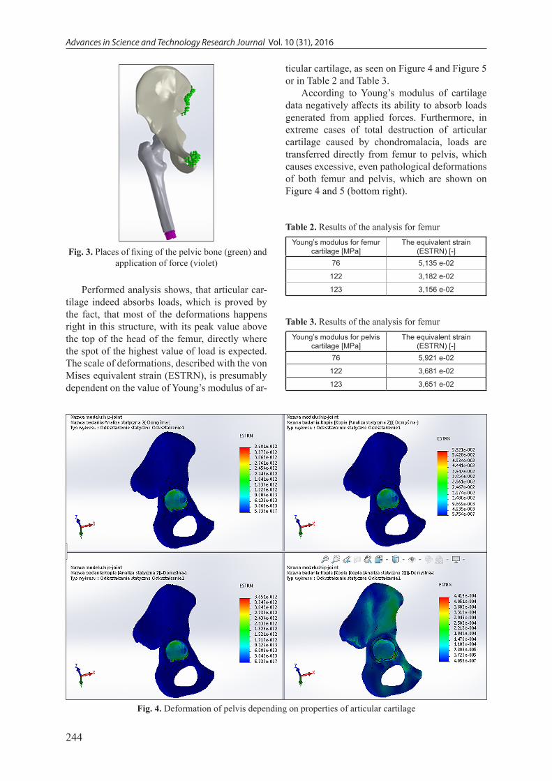
In order to receive correct results it was cru-cial to properly fix the model. For maintaining the best coherence with anatomical structure, the pel-vic bone was fixed in the pubic symphisis and the sacroiliac (Fig. 3).
Next, the force was applied to the body of the femur with value matching the body weight (in N) of a person weighing approximately 80 kg, i.e. 800 N.
Table 1. Table of materials [2, 4, 7, 11, 13]
Material Element Young’s modulus [MPa] Poisson’s ratio Density [kg/m3]
Cortical bone Pelvic bone 17400 0.39 1020
Cortical bone Femur bone 17600 0.3 1020
Cartilage Hip joint articular surfaces
Age 16-39 122 0.35 500
Age 40-59 123 0.35 500
Age 60-83 76 0.35 500
Advances in Science and Technology Research Journal Vol. 10 (31), 2016
244
Performed analysis shows, that articular car-tilage indeed absorbs loads, which is proved by the fact, that most of the deformations happens right in this structure, with its peak value above the top of the head of the femur, directly where the spot of the highest value of load is expected. The scale of deformations, described with the von Mises equivalent strain (ESTRN), is presumably dependent on the value of Young’s modulus of ar-
ticular cartilage, as seen on Figure 4 and Figure 5 or in Table 2 and Table 3.
According to Young’s modulus of cartilage data negatively affects its ability to absorb loads generated from applied forces. Furthermore, in extreme cases of total destruction of articular cartilage caused by chondromalacia, loads are transferred directly from femur to pelvis, which causes excessive, even pathological deformations of both femur and pelvis, which are shown on Figure 4 and 5 (bottom right).
Fig. 3. Places of fixing of the pelvic bone (green) and application of force (violet)
Fig. 4. Deformation of pelvis depending on properties of articular cartilage
Table 2. Results of the analysis for femurYoung’s modulus for femur
cartilage [MPa]The equivalent strain
(ESTRN) [-]76 5,135 e-02
122 3,182 e-02
123 3,156 e-02
Table 3. Results of the analysis for femurYoung’s modulus for pelvis
cartilage [MPa]The equivalent strain
(ESTRN) [-]76 5,921 e-02
122 3,681 e-02
123 3,651 e-02

245
Advances in Science and Technology Research Journal Vol. 10 (31), 2016
CONCLUSIONS
The capability of absorbing loads by articu-lar cartilage of the hip joint is preliminary deter-mined as decreasing with increasing degenera-tions of the cartilage and with age of a patient. Without further information on changes of car-tilage’s mechanical parameters in time it is hard to determine the nature of relation between men-tioned capability and these parameters. Given the way of obtaining results, method presented in the paper may provide additional informa-tions about a condition of the hip joint, espe-cially whether the progress of chondromalacia, without performing surgical procedures, which can be crucial for elder patients.
REFERENCES
1. Będziński R., Kędzior K., Kiwerski J., Morecki A., Skalski K., Wall A., Wit A.: Biomechanika i inżynieria rehabilitacyjna. Akademicka Oficyna Wydawnicza EXIT, Warszawa 2004.
2. Będziński R.: Biomechanika inżynierska. Zagad-nienia wybrane. Oficyna Wydawnicza Politechniki Wrocławskiej, Wrocław 1997.
3. Błaszczyk J.W.: Biomechanika Kliniczna. Podręcznik dla studentów medycyny i fizjoterapii. Wydawnictwo Lekarskie PZWL, Warszawa 2014.
4. Bochenek A., Reicher M.: Anatomia człowieka. Wydawnictwo Lekarskie PZWL, Warszawa 1978.
5. Braniewska M., Zubrzycki J., Karpiński R.: Kom-puterowo wspomagane projektowanie i wytwar-zanie implantu stawu biodrowego. Innowacje w fizjoterapii, 2, 2015, 147–170.
6. Gzik M., Lewandowska-Szumieł M., Pawlikowski M., Wychowański M.: Biomechanika i inżynieria rehabilitacyjna, Akademicka Oficyna Wydawnicza EXIT, Warszawa 2015.
7. Holzapfel A.G., Sommer G., Gasser T.C., Regit-ing P.: Determination of layer-specific mechanical properties of human coronary artheries with non-atherosclerotic intimal thickening and related con-stitutive modeling. AJP-Heart and Circ Phys., 289 (5), 2005, 2048-2058.
8. Hughes T.J.R.: The Finite Element Method: Linear Static and Dynamic Finite Element Analysis. Do-
Fig. 5. Deformation of femur depending on properties of articular cartilage

Advances in Science and Technology Research Journal Vol. 10 (31), 2016
246
ver Publications, New York 2000.9. Karpiński R., Jaworski Ł., Szabelski J.: The de-
sign and structural analysis of the endoprosthesis of the hip joint. Applied Computer Science, 12 (1), 2016, 87–95.
10. Lippert H.: Anatomia. Tom I & II. Elsevier Urban & Partner, Wrocław 1998.
11. Maciejewski R., Torres K.: Anatomia czynnościowa - podręcznik dla studentów pielęgniarstwa, fiz-joterapii, ratownictwa medycznego, analityki me-dycznej i dietetyki. Wyd. 1. Wydawnictwo Czelej, Lublin 2007.
12. Ozolanta I., Tetere G., Purinaya B., Kasyanov V.: Changes in the machanical properties, biochemical contents and wall structure of the human coronary arteries with age and sex. Med. Eng. Phys., 20 (7), 1998, 523-33.
13. Reicher M.: Anatomia człowieka. Tom I, Anatomia ogólna, kości, stawy i więzadła, mięśnie. Wyd. X (VI). Wydawnictwo Lekarskie PZWL, Warszawa 1990.
14. Różyło P.: Numerical analysis of crack propaga-tion in a steel specimen under bending. Applied Computer Science, 11 (4), 2015, 20-29.
15. Spodaryk K.: Patologia narządu ruchu, Wydawnic-two lekarskie PZWL, Warszawa 2002.
16. Tejszerska D., Świtoński E., Gzik M.: Biomechan-ika narządu ruchu człowieka, Praca zbiorowa. Wydawnictwa Naukowe Instytutu Technologii Eksploatacji – Państwowego Instytutu Badawc-zego, Gliwice 2011.
17. Developer’s information on Mimics, http://bio-medical.materialise.com/mimics.
18. Brief anatomy of the hip joint, http://probecure.com/b/brief-anatomy-of-hip-joint/.
19. Classifications and definitions of normal joints, http://www.intechopen.com/books/osteoarthritis-progress-in-basic-research-and-treatment/classifi-cations-and-definitions-of-normal-joints.
20. Zubrzycki J., Smidova N.: Computer-aided de-sign of human knee implant. Industrial and service robotics. In: Hajduk Mikulas, Koukolová Lucia (Eds) 13th International Conference on Industrial, Service and Humanoid Robotics (ROBTEP): Con-ference, Stafa-Zurich; Switzerland: Trans Tech Publications, Applied Mechanics and Materials, No. 913, 2014, 172-181.